Embed Size (px)
Citation preview
Structural and Magnetic Properties of Gold and Silica Doubly Coatedγ-Fe2O3
Nanoparticles
Keeseong Park,† Gan Liang,*,‡ Xiaojun Ji, § Zhi-Ping Luo, | Chun Li, § Mark C. Croft, ⊥,# andJohn T. Markert †
Department of Physics, UniVersity of Texas at Austin, Austin, Texas 78712, Department of Physics,Sam Houston State UniVersity, HuntsVille, Texas 77341, Department of Experimental Diagnostic Imaging,M. D. Anderson Cancer Center, UniVersity of Texas, Houston, Texas 77030, Microscopy and Imaging Center,Texas A&M UniVersity, College Station, Texas 77843-2257, Department of Physics and Astronomy,Rutgers UniVersity, Piscataway, New Jersey 08854, and National Synchrotron Light Source,BrookhaVen National Laboratory, Upton, New York 11973
ReceiVed: July 22, 2007; In Final Form: September 25, 2007
Extensive structural and magnetic characterization measurements were carried out on gold and silica doublycoatedγ-Fe2O3 nanoparticles, which were recently demonstrated to have an efficient photothermal effect andhigh transverse relaxivities for MRI applications. Powder X-ray diffraction and X-ray absorption spectroscopyshow the phase of the uncoated and coated nanoparticles to be that of theγ-Fe2O3 structure. The sizes, structure,and chemical compositions of the nanoparticles were determined by transmission electron microscopy. Themagnetization results indicate that coating of the iron oxide nanoparticles by gold/silica decreases the blockingtemperature from 160 to 80 K. Such a decrease can be well-explained by spin disorder, causing reduction ofthe effective volume of theγ-Fe2O3 core. Moreover, it was found that in the temperature (T) range between100 K and room temperature, the gold/silica coating can cause a slight magnetic change in theγ-Fe2O3 coresfrom superparamagnetic to almost superparamagnetic. Finally, it was found that the coercivity for both theuncoated and the coated nanoparticles decreases almost linearly withT1/2 with the former decreasing fasterthan the latter, and this coercivity result confirms that the blocking temperature is decreased by gold/silicacoating. These results are valuable for evaluating the future applications of this class of multifunctional,hybrid magnetic nanoparticles in biomedicine.
1. Introduction
Superparamagnetic nanoparticles (SPNPs) have great potentialfor magnetic resonance imaging (MRI), due to their hightransverse relaxivity,1,2 and for a wide range of other biomedicalapplications such as drug delivery.3-6 Indeed, SPNP-enhancedMRI techniques have been under development in recent yearsfor imaging of macrophage activity, atherosclerotic lesions,detection of lymph node metastasis, staging of liver tumors,andmonitoringthe invivodistributionofcellular trafficking.1,2,7-11
For in vivo applications, the SPNPs must be coated with abiocompatible and diamagnetic material (such as organicpolymers or silica matrixes) to prevent the formation of largeaggregates of SPNPs and to facilitate functions such as theattachment of drugs. For example, the dispersion of uncoatedγ-Fe2O3 NPs in aqueous solution is stable only in highly acidicor basic media. In recent years, much effort has been devotedto the syntheses and characterization of silica coated superpara-magnetic iron oxide (SPIO) nanoparticles (NPs) includingγ-Fe2O3 NPs.12-17
It has been proven that silica coated SPIO NPs have anexcellent biocompatibility and can homogeneously disperse in
aqueous solutions with a wide range of pH values.12-17 Thus,they can be used for the MRI diagnosis of cells or tissues alteredby diseases such as cancer. However, to kill the cancer cells orcure the affected tissues by photothermal therapy, the silicacoated SPIO NPs need to be coated with particles that have anexcellent heat generating ability under controllable conditions.In recent years, nanostructures with highly controlled opticalproperties have attracted great interest due to their potentialdiagnostic and therapeutic applications in biological and bio-medical systems.18 Among such nanostructures, the gold derivedNPs are especially attractive due to their ease of preparation,good biocompatibility, ready bioconjugation, and unique opticalproperties.19-23 Gold nanoshells have a strong absorbance forthe wavelength tunable in the near-infrared (NIR) region of theelectromagnetic spectrum. They are, therefore, very promisingcandidates for localized photothermal therapy because they canmediate strong plasmon-induced surface heat flux upon absorp-tion of the NIR radiation.23-25 Thus, if the Au nanoparticlescan be coated on the surface or dispersed into the surface layerof the silica matrix of the silica coatedγ-Fe2O3 NPs, then suchAu/SiO2 doubly coatedγ-Fe2O3 NPs could be made bifunctional(i.e., diagnosis and cure of the cancer cells).
Recently, we successfully synthesized Au/SiO2 doubly coatedγ-Fe2O3 NPs and found that they can form a stable solution inwater and have high transverse relaxivities for MRI applica-tions.26 Furthermore, the broad absorbance between 100 and900 nm in the UV-vis spectra indicates that these doubly coatedparticles are suitable for photothermal therapy with light in the
* Corresponding author. Tel.: (936) 294-1608; fax: (936) 294-1585;e-mail: [email protected].
† University of Texas at Austin.‡ Sam Houston State University.§ M. D. Anderson Cancer Center, University of Texas.| Texas A&M University.⊥ Rutgers University.# Brookhaven National Laboratory.
18512 J. Phys. Chem. C2007,111,18512-18519
10.1021/jp0757457 CCC: $37.00 © 2007 American Chemical SocietyPublished on Web 11/29/2007
NIR region.26 However, in that work, a detailed structural andmagnetic characterization of the uncoated and Au/SiO2 doublycoatedγ-Fe2O3 NPs was absent. A better understanding of thestructural and magnetic properties of this class of doubly coated(bifunctional) NPs is urgently needed for their applications inbiomedicine. In this paper, we present detailed results ofcharacterization by temperature- and field-dependent magnetiza-tion, transmission electron microscopy (TEM), X-ray diffraction(XRD), and X-ray absorption spectroscopy (XAS) measurementson these Au/SiO2 doubly coatedγ-Fe2O3 NPs.
2. Experimental Procedures
The synthesis of gold and silica doubly coatedγ-Fe2O3 NPshas been described in detail elsewhere.26 The first step is thesynthesis of the silica coatedγ-Fe2O3 NPs using the well-knownStober process.27 An aqueous ammonia solution (30 wt %, 7mL) and 0.5 mL of tetraethylorthosilicate (TEOS) were addedto theγ-Fe2O3 (or SPIO) solution at room temperature to formsilica NPs. The functional groups at the surface of the silicananoparticles are primarily silanol (Si-OH) or ethoxy (Si-OEt)groups.28 These silica nanoparticles were then treated with0.04 mL of 3-minopropyl trimethoxysilane (APTMS) for 6 hto introduce the amino-terminated silica surface. The reactionmixture was refluxed for 30 min to complete the reaction.28 Inthe second step of the synthesis, 1 mL ofγ-Fe2O3-embeddedsilica solution was added to 5 mL of undiluted tetrakis-(hydroxymethyl) phosphonium chloride (THPC) gold solution,causing the THPC gold nanocrystals to attach onto the silicasurface. The THPC gold solution was prepared by reduction ofchloroauric acid (HAuCl4) with THPC, which produces smallgold particles with a net negative interfacial charge.29 Finally,the gold nanoshell was prepared by reduction of the K-Goldsolution with formaldehyde (37%). UV-vis absorption spectraof the nanoshells were measured with a Beckman Coutler DU-800 UV-vis spectrometer after the reaction to verify theformation of the nanoshell. The spectra are identical to thosereported in our previous article.26 The broad absorbance between100 and 900 nm in the UV-vis spectra indicates that these Au/SiO2 doubly coated particles are suitable for photothermaltherapy with light in the NIR region.26 The water-based Fe2O3
NPs (MEG 304) were purchased from Ferrotech (Nashua, NH).TEOS, APTMS, ammonia solution, THPC, HAuCl4, andformaldehyde were purchased from Sigma-Aldrich (St. Louis,MO).
X-ray diffraction (XRD) measurements were performed atroom temperature using a Rigaku 2005 X-ray diffractometerwith Cu KR radiation. The TEM measurements were carriedout using a JEOL 2010 TEM system at a working voltage of200 kV. All imaging magnifications were calibrated using thestandards of SiC lattice fringes30 for high magnifications andcommercial cross-line grating replica for low magnifications.To prepare the TEM samples, a small drop of the samplesolution was transferred to the top surface of a carbon filmsupported Cu grid that was previously glow discharged toachieve better dispersion and then dried in air. The average sizeof the particles was estimated based on a sufficient number ofsampling, typically over 50. The EDS measurements wereperformed using an Oxford Instruments EDS detector with anINCA energy platform.
The magnetic properties of the NPs were studied using aQuantum Design MPMS SQUID magnetometer at temperaturesranging from 5 to 300 K and in magnetic fields (H) rangingfrom 0 to up to 50 kOe. In the magnetization measurement,both the uncoated and the Au/SiO2 coated Fe2O3 NPs were in
the form of dried powders. Gelatin capsules (from Capsuli-ne.com) and water-resistive polycarbonate capsules (from Uni-pec Inc., Rockville, MD) were used as the sample containersfor the uncoated and Au/SiO2 coated Fe2O3 NPs, respectively.Since both the amount of the Au/SiO2 coated Fe2O3 particles(0.6 mg) and the volume fraction of the magnetic Fe2O3 coresin the sample were very small, the magnetic moment of thepolycarbonate capsule was measured for background subtraction.
The Fe K-edge XAS data were taken in fluorescence modeat room temperature at beamline X-19A at the NationalSynchrotron Light Source (NSLS) at Brookhaven National Lab.A double-crystal Si (111) monochromator was used for energyselection, which was detuned by reducing the incident photonflux 20% from its maximum value to suppress contaminationfrom harmonics. The energy resolution (∆E/E) of the X-19Abeam line was 2× 10⁻⁴, corresponding to about 1.4 eV for theedge energy of the Fe K-edge. The energy calibration of thespectra was made by simultaneously measuring the spectrumof a FeO slide as a reference. The XAS spectra presented inthis paper were background subtracted and normalized to unityin the continuum region.
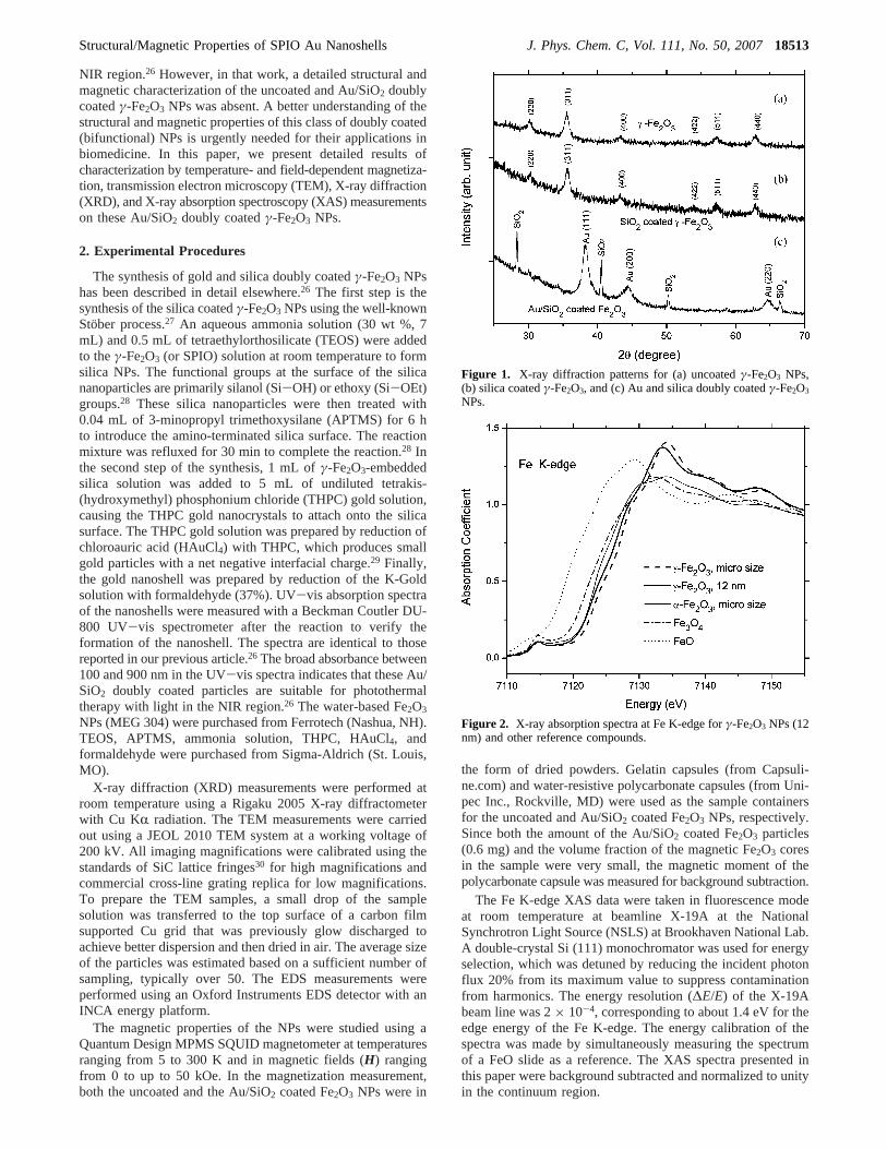
Figure 1. X-ray diffraction patterns for (a) uncoatedγ-Fe2O3 NPs,(b) silica coatedγ-Fe2O3, and (c) Au and silica doubly coatedγ-Fe2O3
NPs.
Figure 2. X-ray absorption spectra at Fe K-edge forγ-Fe2O3 NPs (12nm) and other reference compounds.
Structural/Magnetic Properties of SPIO Au Nanoshells J. Phys. Chem. C, Vol. 111, No. 50, 200718513
3. Results and Discussion
(A) XRD and XAS Results.Figure 1 shows the powder XRDpatterns for (a) commercial IONPs, (b) SiO2 coated IONPs, and(c) Au/SiO2 doubly coated IONPs, with each pattern normalizedto its maximum intensity. All of the peaks in the patterns ofthe commercial IONPs can be indexed with the cubic structurecorresponding to eitherγ-Fe2O3 or Fe3O4 phase. Since magnetite(Fe3O4) has a cubic spinel structure and lattice constant similarto maghemite (γ-Fe2O3), it is quite difficult to distinguishγ-Fe2O3 from Fe3O4 using only XRD data. Thus, the existenceof Fe3O4 in the commercial IONPs cannot be excluded basedonly on the XRD data. On the other hand, the X-ray absorptionnear-edge structure (XANES) of XAS offers a powerful meansto distinguish the iron oxide species. Fe lattice sites withdifferent formal valences and local coordination can manifestdifferent chemical shifts and near-edge spectral features in theirFe K-edge spectra. In Figure 2, we compare the Fe K-edgespectra (labeled byγ-Fe2O3, 12 nm) of the commercial IONPsto the powder spectra of four reference compounds: micrometer-sizedγ-Fe2O3, R-Fe2O3, Fe3O4, and FeO. It can be seen thatboth the shape and the edge energy (defined as the energy atabsorption coefficientµ ) 0.5) for the spectrum of thecommercial IONPs are very close to those for the micrometer-
sizedγ-Fe2O3 but very different from the rest of the spectra inFigure 2. This clearly demonstrates that the commercial IONPsused in this study are in theγ-Fe2O3 phase.
Figure 1b shows that the XRD pattern of the SiO2 (silica)coatedγ-Fe2O3 NPs is very similar to that (Figure 1a) of theuncoatedγ-Fe2O3 NPs. This indicates that the coated silica isin an amorphous form before the step of mixing theγ-Fe2O3-embedded silica solution with the THPC gold solution duringthe synthesis process. For the Au and silica doubly coatedγ-Fe2O3 particles, the XRD pattern shown in Figure 1c displaysthree wide peaks that can be identified as the (111), (200), and(220) reflection lines of the Au fcc-cubic phase, indicating thatthe gold particles are crystallized. In addition, there are fournarrow peaks in the pattern that can be attributed to thecrystallized SiO2 phase (PDF 31-1233), indicating that a certainportion of the silica is crystallized during the steps of coatingAu on the surface of theγ-Fe2O3-embedded silica NPs. Atpresent, while the mechanism of the crystallization of silica dueto the coating of the Au particles is unclear, some recent researchindicates that Au nanoparticles on silica spheres can inducecrystallization of silica at low temperatures.31 The backgroundof this pattern is very similar to the XRD pattern of amorphousSiO2, which decreases rapidly with the increase of 2θ from 23
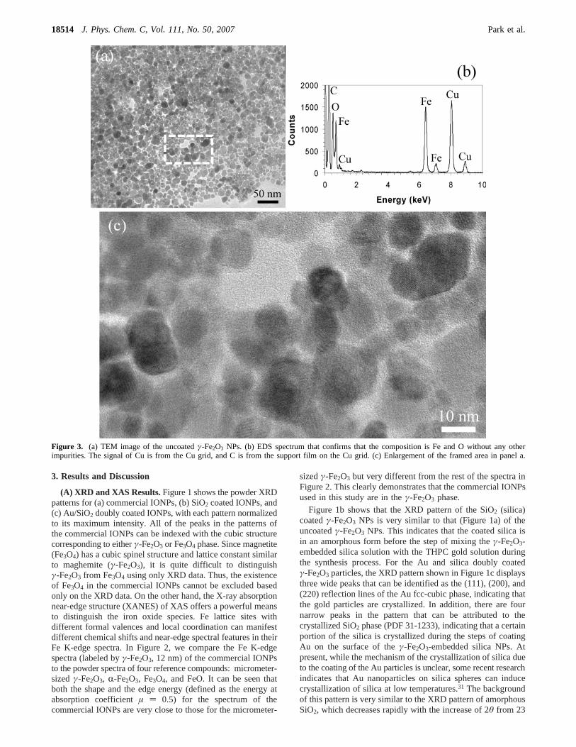
Figure 3. (a) TEM image of the uncoatedγ-Fe2O3 NPs. (b) EDS spectrum that confirms that the composition is Fe and O without any otherimpurities. The signal of Cu is from the Cu grid, and C is from the support film on the Cu grid. (c) Enlargement of the framed area in panel a.
18514 J. Phys. Chem. C, Vol. 111, No. 50, 2007 Park et al.
to 40° and then slowly beyond 40°. Considering the fact thatmost of these coated particles are spherical (see TEM results)and the observation that usually spherical SiO2 particles are inthe amorphous form, we believe that only a small fraction ofthe silica is crystallized as a minor phase and that most of thesilica nanoshells are in the amorphous phase.
Interestingly, the Fe2O3 peaks (Figure 1a,b) appearing in thepatterns of the SiO2 coated and uncoated Fe2O3 particles arealmost unnoticeable in the pattern of the Au/SiO2 coatedparticles shown in Figure 1c. This very small intensity of theFe2O3 peaks can be explained by the strong absorption of andscattering from the coated Au particles. Indeed, such a washing-out effect was also observed in XRD patterns for some Aupassivated Fe NPs.32
(B) TEM and EDS Results.Figure 3a shows a TEM imageof the pure or uncoated Fe2O3 particles at low magnification.The average size of the particles is measured to beDFe2O3 )12.4 nm with a standard deviation of 4.5 nm, based on themeasurements of 412 particles. This value of average size isslightly larger than (but within one standard deviation of) thenominal size of 10 nm given by the vendor (Ferrotech). TheEDS data in Figure 3b confirm that the particle composition isFe and O without any other impurities. The signal of Cu is fromthe Cu grid, and C is from the support film on the Cu grid. Theframed area (enclosed by the dashed rectangular loop) in Figure3a is enlarged in Figure 3c, where lattice fringes of thecrystalline Fe2O3 crystals are visible.
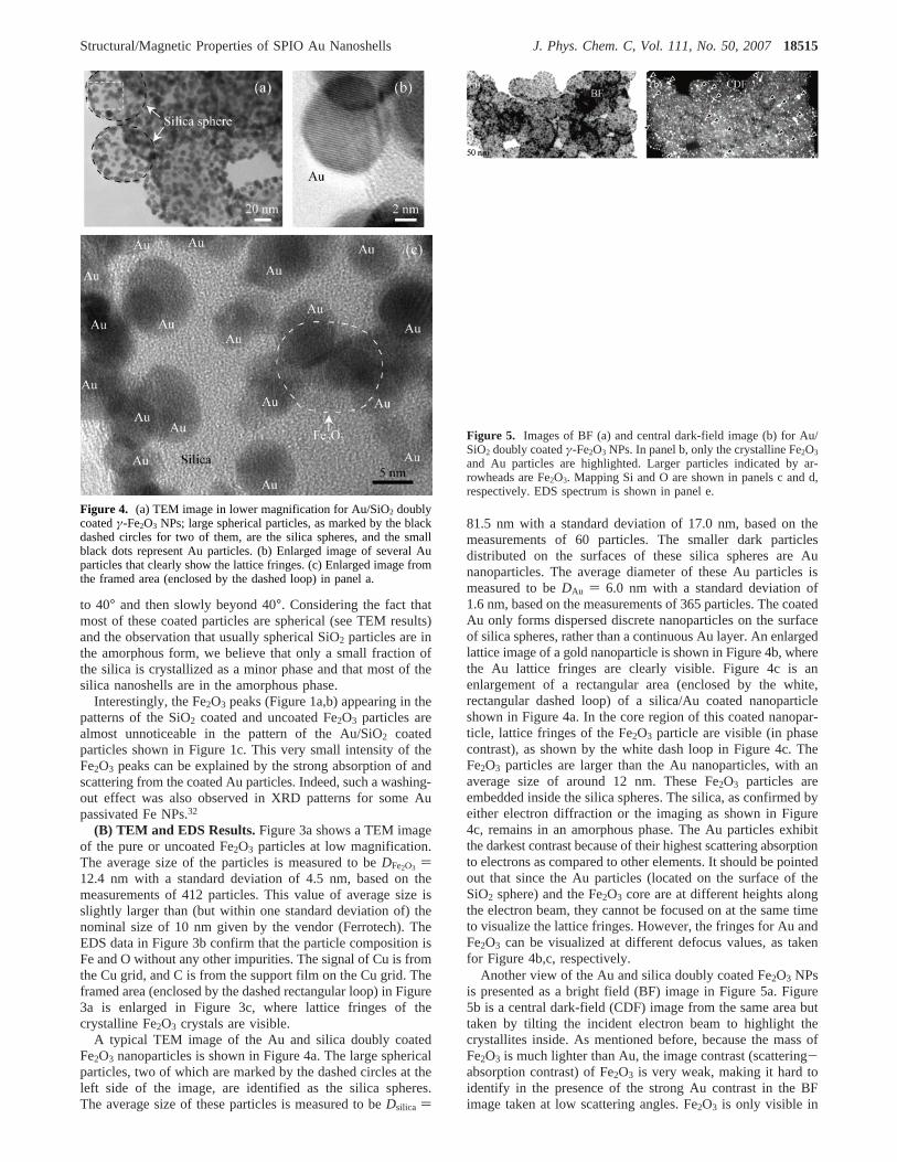
A typical TEM image of the Au and silica doubly coatedFe2O3 nanoparticles is shown in Figure 4a. The large sphericalparticles, two of which are marked by the dashed circles at theleft side of the image, are identified as the silica spheres.The average size of these particles is measured to beDsilica )
81.5 nm with a standard deviation of 17.0 nm, based on themeasurements of 60 particles. The smaller dark particlesdistributed on the surfaces of these silica spheres are Aunanoparticles. The average diameter of these Au particles ismeasured to beDAu ) 6.0 nm with a standard deviation of1.6 nm, based on the measurements of 365 particles. The coatedAu only forms dispersed discrete nanoparticles on the surfaceof silica spheres, rather than a continuous Au layer. An enlargedlattice image of a gold nanoparticle is shown in Figure 4b, wherethe Au lattice fringes are clearly visible. Figure 4c is anenlargement of a rectangular area (enclosed by the white,rectangular dashed loop) of a silica/Au coated nanoparticleshown in Figure 4a. In the core region of this coated nanopar-ticle, lattice fringes of the Fe2O3 particle are visible (in phasecontrast), as shown by the white dash loop in Figure 4c. TheFe2O3 particles are larger than the Au nanoparticles, with anaverage size of around 12 nm. These Fe2O3 particles areembedded inside the silica spheres. The silica, as confirmed byeither electron diffraction or the imaging as shown in Figure4c, remains in an amorphous phase. The Au particles exhibitthe darkest contrast because of their highest scattering absorptionto electrons as compared to other elements. It should be pointedout that since the Au particles (located on the surface of theSiO2 sphere) and the Fe2O3 core are at different heights alongthe electron beam, they cannot be focused on at the same timeto visualize the lattice fringes. However, the fringes for Au andFe2O3 can be visualized at different defocus values, as takenfor Figure 4b,c, respectively.
Another view of the Au and silica doubly coated Fe2O3 NPsis presented as a bright field (BF) image in Figure 5a. Figure5b is a central dark-field (CDF) image from the same area buttaken by tilting the incident electron beam to highlight thecrystallites inside. As mentioned before, because the mass ofFe2O3 is much lighter than Au, the image contrast (scattering-absorption contrast) of Fe2O3 is very weak, making it hard toidentify in the presence of the strong Au contrast in the BFimage taken at low scattering angles. Fe2O3 is only visible in
Figure 4. (a) TEM image in lower magnification for Au/SiO2 doublycoatedγ-Fe2O3 NPs; large spherical particles, as marked by the blackdashed circles for two of them, are the silica spheres, and the smallblack dots represent Au particles. (b) Enlarged image of several Auparticles that clearly show the lattice fringes. (c) Enlarged image fromthe framed area (enclosed by the dashed loop) in panel a.
Figure 5. Images of BF (a) and central dark-field image (b) for Au/SiO2 doubly coatedγ-Fe2O3 NPs. In panel b, only the crystalline Fe2O3
and Au particles are highlighted. Larger particles indicated by ar-rowheads are Fe2O3. Mapping Si and O are shown in panels c and d,respectively. EDS spectrum is shown in panel e.
Structural/Magnetic Properties of SPIO Au Nanoshells J. Phys. Chem. C, Vol. 111, No. 50, 200718515
the previous phase contrast image in Figure 4b. However, inthe CDF image in Figure 5b, as the contrast is formed bydiffracted beams at high scattering angles (diffraction contrast),both crystalline Fe2O3 and Au particles exhibit high contrast inthe CDF image formed from their common diffracted beampositions. It should be pointed out that only those particles thathave their diffracted beams along this tilted incident beamdirection are imaged (i.e., only selective fractions of the Fe2O3
and Au particles show up in the CDF image in Figure 5b).Importantly, it can be recognized that larger Fe2O3 particles areat the center of the silica spheres, as pointed out by somearrowheads in the Figure 5. The maps of Si (Figure 5c) and O(Figure 5d) are similar, along the silica spheres in the BF imagein Figure 5a. The EDS spectrum taken over this entire area (withthe background signals of Cu and C removed) is shown in Figure5e, and it clearly shows the evidence of O, Si, Fe, and Au. Aquantitative analysis yields a composition of 62.6% O, 26.4%Si, 1.7% Fe, and 9.3% Au (all in atom percent). The very lowconcentration of Fe confirms that only small Fe2O3 particlesare present inside the silica.
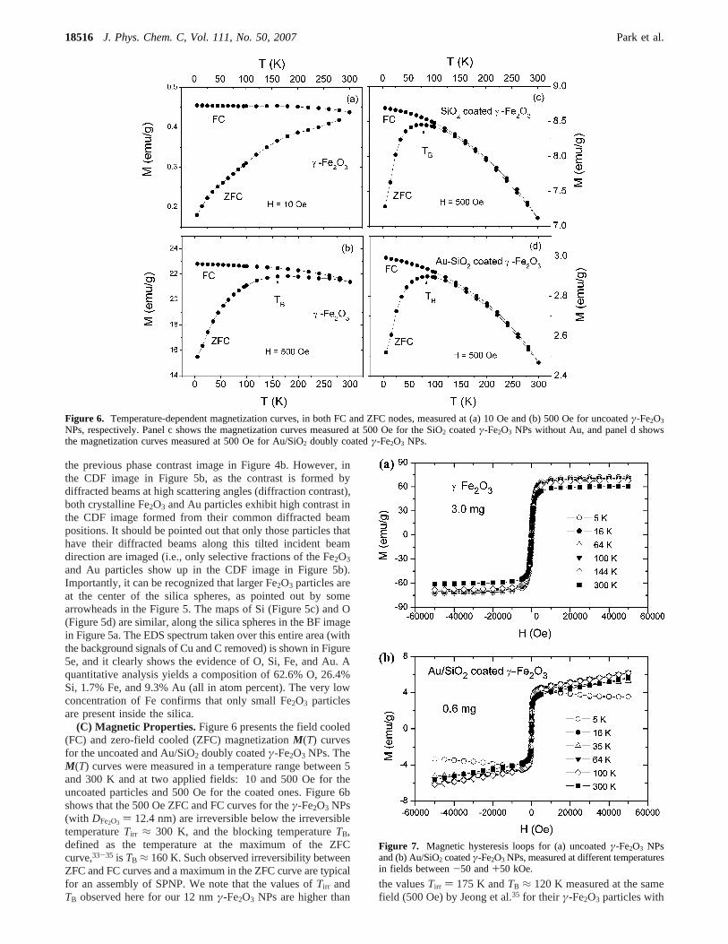
(C) Magnetic Properties.Figure 6 presents the field cooled(FC) and zero-field cooled (ZFC) magnetizationM(T) curvesfor the uncoated and Au/SiO2 doubly coatedγ-Fe2O3 NPs. TheM(T) curves were measured in a temperature range between 5and 300 K and at two applied fields: 10 and 500 Oe for theuncoated particles and 500 Oe for the coated ones. Figure 6bshows that the 500 Oe ZFC and FC curves for theγ-Fe2O3 NPs(with DFe2O3 ) 12.4 nm) are irreversible below the irreversibletemperatureTirr ≈ 300 K, and the blocking temperatureTB,defined as the temperature at the maximum of the ZFCcurve,33-35 is TB ≈ 160 K. Such observed irreversibility betweenZFC and FC curves and a maximum in the ZFC curve are typicalfor an assembly of SPNP. We note that the values ofTirr andTB observed here for our 12 nmγ-Fe2O3 NPs are higher than
the valuesTirr ) 175 K andTB ≈ 120 K measured at the samefield (500 Oe) by Jeong et al.35 for their γ-Fe2O3 particles with
Figure 6. Temperature-dependent magnetization curves, in both FC and ZFC nodes, measured at (a) 10 Oe and (b) 500 Oe for uncoatedγ-Fe2O3
NPs, respectively. Panel c shows the magnetization curves measured at 500 Oe for the SiO2 coatedγ-Fe2O3 NPs without Au, and panel d showsthe magnetization curves measured at 500 Oe for Au/SiO2 doubly coatedγ-Fe2O3 NPs.
Figure 7. Magnetic hysteresis loops for (a) uncoatedγ-Fe2O3 NPsand (b) Au/SiO2 coatedγ-Fe2O3 NPs, measured at different temperaturesin fields between-50 and+50 kOe.
18516 J. Phys. Chem. C, Vol. 111, No. 50, 2007 Park et al.
a smaller particle size of 5-8 nm. This particle size caused achange inTB and can be explained by the theoretical relation34,35
whereV [(1/6)πDav3 ] is the average volume of the NPs,K is the
uniaxial anisotropy constant, andkB the Boltzmann constant.Indeed, such an increase ofTB due to the increase of particlesize has also been observed previously forγ-Fe2O3 NPs withan average diameter in the range between 5 and 13 nm.35-37
For example, Mukadam et al.36 found that theTB of theirγ-Fe2O3 NPs increased from 80 to 160 K with increasing particlesize from 8 to 13 nm, when measured at 100 Oe. The 40 Kdifference inTB observed between our∼12 nm NPs and the∼8 nm NPs of Jeong et al.35 appears to be comparable butslightly smaller than the∆TB/∆Dav rate observed by Mukadamet al.36
A comparison between the curves shown in Figure 6a,bindicates that with the decrease of the applied fieldH from 500Oe to a much lower value of 10 Oe,Tirr andTB both increaseto above 300 K. Similar highTirr andTB values (measured atthe same fieldH ) 10 Oe) were also observed by anothergroup34 for γ-Fe2O3 with a similar particle sizeDav ) 11.1 nm.The observed increase ofTB with decreasing applied field wasalso previously observed forγ-Fe2O3 NPs of other particlesizes,36,38and it can be explained by the theoretical relation39,40
whereTB0 is the blocking temperature at zero-field andC is afield-independent parameter.
By comparing the the ZFC curve in Figure 6c with that inFigure 6b, it can be seen that the blocking temperatureTB
decreases from 160 K to about 80 K with the coating of Au/
Figure 8. Magnetic hysteresis loops of Figure 8 plotted in range of applied fields between-2 and 2 kOe.
TB ) KV25kB
(1)
TB ) TB0(1 - CH2
TB0) (2)
Structural/Magnetic Properties of SPIO Au Nanoshells J. Phys. Chem. C, Vol. 111, No. 50, 200718517
SiO2 on theγ-Fe2O3 NPs. Comparison between the magnetiza-tion curves in Figure 6c,d shows that further coating of Auparticles on the surface of the SiO2 spheres does not changeTB. We can attribute the 80 K decrease ofTB mainly to thereduction of the average effective volume of theγ-Fe2O3 cores,which is caused by the interfacial interaction between the silicaand the outer layer of the iron oxide core. Our TEM resultshave shown that the SiO2 nanoshells (about 35 nm thick) arecoated onγ-Fe2O3 cores and that the Au particles (Dav ≈ 6nm) are only dispersed in a thin layer near the outer surface ofthe SiO2 nanoshells. This result means that the Fe ions locatednear the surface of theγ-Fe2O3 cores can only interact withSiO2 (silica) near theγ-Fe2O3/SiO2 interface, not with the Auparticles. Such an interaction between Fe ions and SiO2 couldproduce a thin layer of misaligned or disordered Fe spins nearthe surface of the sphericalγ-Fe2O3 cores. The spins in thismagnetically disordered layer should have a negligible contribu-tion to the total magnetizationM for the sample and thus canbe excluded from the particle volumeV in eq 1. Thus, if theaverage thickness of the spin disordered layers ist, we can definean average effective volume,Veff,41 for the γ-Fe2O3 cores inthe Au/SiO2 coated NPs
Then, eq 1 should be replaced by
Combining eqs 3 and 4, we have
whereTB(0) is the blocking temperature att ) 0.Recently, Rosa et al.41 measured the value oft for γ-Fe2O3
NPs embedded in an amorphous SiO2 matrix using a Faradayrotation technique. They found thatt was 1.25( 0.07 nm andwas almost unchanged for allγ-Fe2O3 NPs with the averagediameter in the range of 6.2 nme Dav e 21.8 nm. For our
γ-Fe2O3 NPs,Dav (12.4 nm) falls into this range, and thus,t )1.25 nm can be used for estimatingTB. Using eq 5 and thevaluesTB(0) ) 160 K,Dav ) 12.4 nm, andt ) 1.25 nm,TB forthe Au/SiO2 coatedγ-Fe2O3 NPs is estimated to be 81 K, whichis in excellent agreement with the experimental value of 80 K.
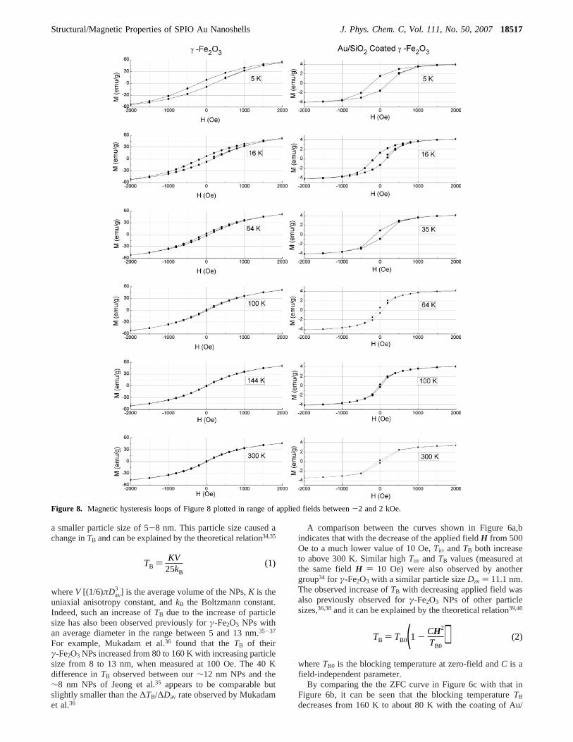
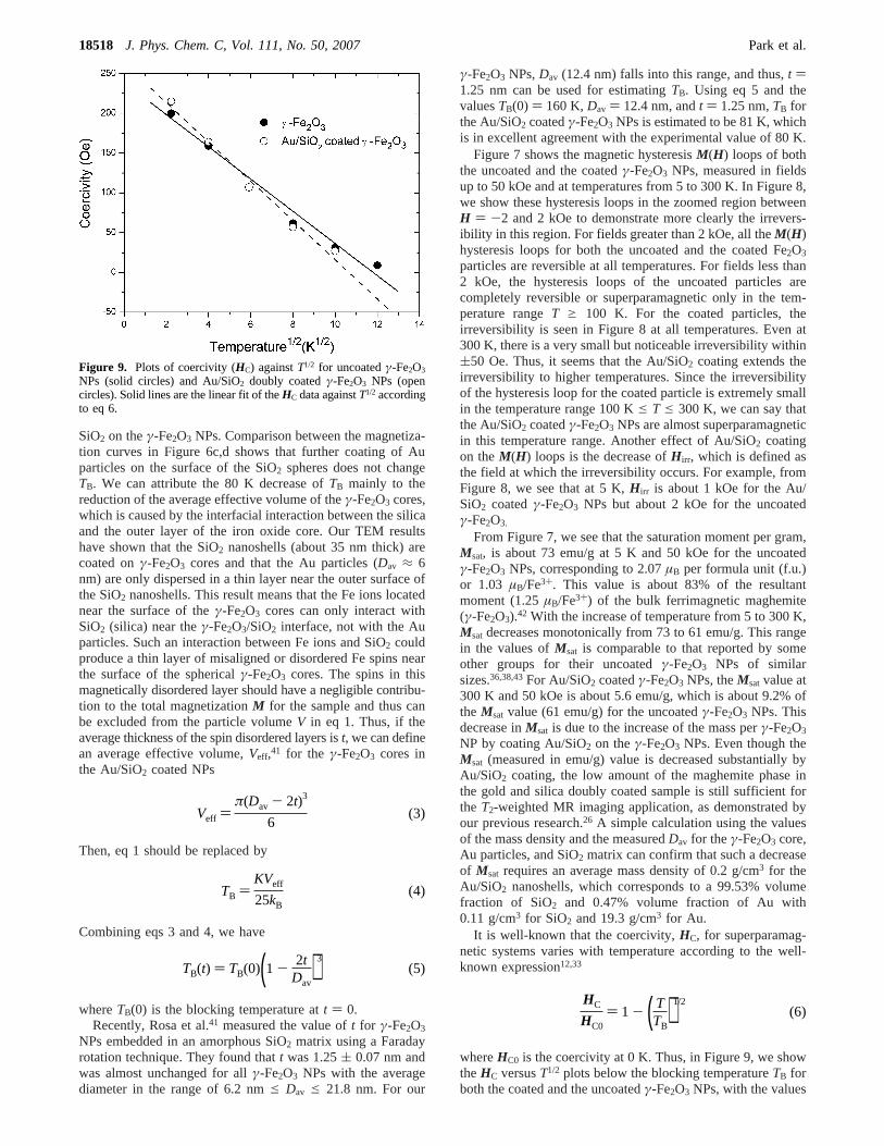
Figure 7 shows the magnetic hysteresisM(H) loops of boththe uncoated and the coatedγ-Fe2O3 NPs, measured in fieldsup to 50 kOe and at temperatures from 5 to 300 K. In Figure 8,we show these hysteresis loops in the zoomed region betweenH ) -2 and 2 kOe to demonstrate more clearly the irrevers-ibility in this region. For fields greater than 2 kOe, all theM(H)hysteresis loops for both the uncoated and the coated Fe2O3
particles are reversible at all temperatures. For fields less than2 kOe, the hysteresis loops of the uncoated particles arecompletely reversible or superparamagnetic only in the tem-perature rangeT g 100 K. For the coated particles, theirreversibility is seen in Figure 8 at all temperatures. Even at300 K, there is a very small but noticeable irreversibility within(50 Oe. Thus, it seems that the Au/SiO2 coating extends theirreversibility to higher temperatures. Since the irreversibilityof the hysteresis loop for the coated particle is extremely smallin the temperature range 100 Ke T e 300 K, we can say thatthe Au/SiO2 coatedγ-Fe2O3 NPs are almost superparamagneticin this temperature range. Another effect of Au/SiO2 coatingon theM(H) loops is the decrease ofH irr, which is defined asthe field at which the irreversibility occurs. For example, fromFigure 8, we see that at 5 K,H irr is about 1 kOe for the Au/SiO2 coatedγ-Fe2O3 NPs but about 2 kOe for the uncoatedγ-Fe2O3.
From Figure 7, we see that the saturation moment per gram,Msat, is about 73 emu/g at 5 K and 50 kOe for the uncoatedγ-Fe2O3 NPs, corresponding to 2.07µB per formula unit (f.u.)or 1.03 µB/Fe3+. This value is about 83% of the resultantmoment (1.25µB/Fe3+) of the bulk ferrimagnetic maghemite(γ-Fe2O3).42 With the increase of temperature from 5 to 300 K,Msat decreases monotonically from 73 to 61 emu/g. This rangein the values ofMsat is comparable to that reported by someother groups for their uncoatedγ-Fe2O3 NPs of similarsizes.36,38,43For Au/SiO2 coatedγ-Fe2O3 NPs, theMsatvalue at300 K and 50 kOe is about 5.6 emu/g, which is about 9.2% ofthe Msat value (61 emu/g) for the uncoatedγ-Fe2O3 NPs. Thisdecrease inMsat is due to the increase of the mass perγ-Fe2O3
NP by coating Au/SiO2 on theγ-Fe2O3 NPs. Even though theMsat (measured in emu/g) value is decreased substantially byAu/SiO2 coating, the low amount of the maghemite phase inthe gold and silica doubly coated sample is still sufficient forthe T2-weighted MR imaging application, as demonstrated byour previous research.26 A simple calculation using the valuesof the mass density and the measuredDav for theγ-Fe2O3 core,Au particles, and SiO2 matrix can confirm that such a decreaseof Msat requires an average mass density of 0.2 g/cm3 for theAu/SiO2 nanoshells, which corresponds to a 99.53% volumefraction of SiO2 and 0.47% volume fraction of Au with0.11 g/cm3 for SiO2 and 19.3 g/cm3 for Au.
It is well-known that the coercivity,HC, for superparamag-netic systems varies with temperature according to the well-known expression12,33
whereHC0 is the coercivity at 0 K. Thus, in Figure 9, we showtheHC versusT1/2 plots below the blocking temperatureTB forboth the coated and the uncoatedγ-Fe2O3 NPs, with the values
Figure 9. Plots of coercivity (HC) againstT1/2 for uncoatedγ-Fe2O3
NPs (solid circles) and Au/SiO2 doubly coatedγ-Fe2O3 NPs (opencircles). Solid lines are the linear fit of theHC data againstT1/2 accordingto eq 6.
Veff )π(Dav - 2t)3
6(3)
TB )KVeff
25kB(4)
TB(t) ) TB(0)(1 - 2tDav
)3(5)
HC
HC0) 1 - ( T
TB)1/2
(6)
18518 J. Phys. Chem. C, Vol. 111, No. 50, 2007 Park et al.
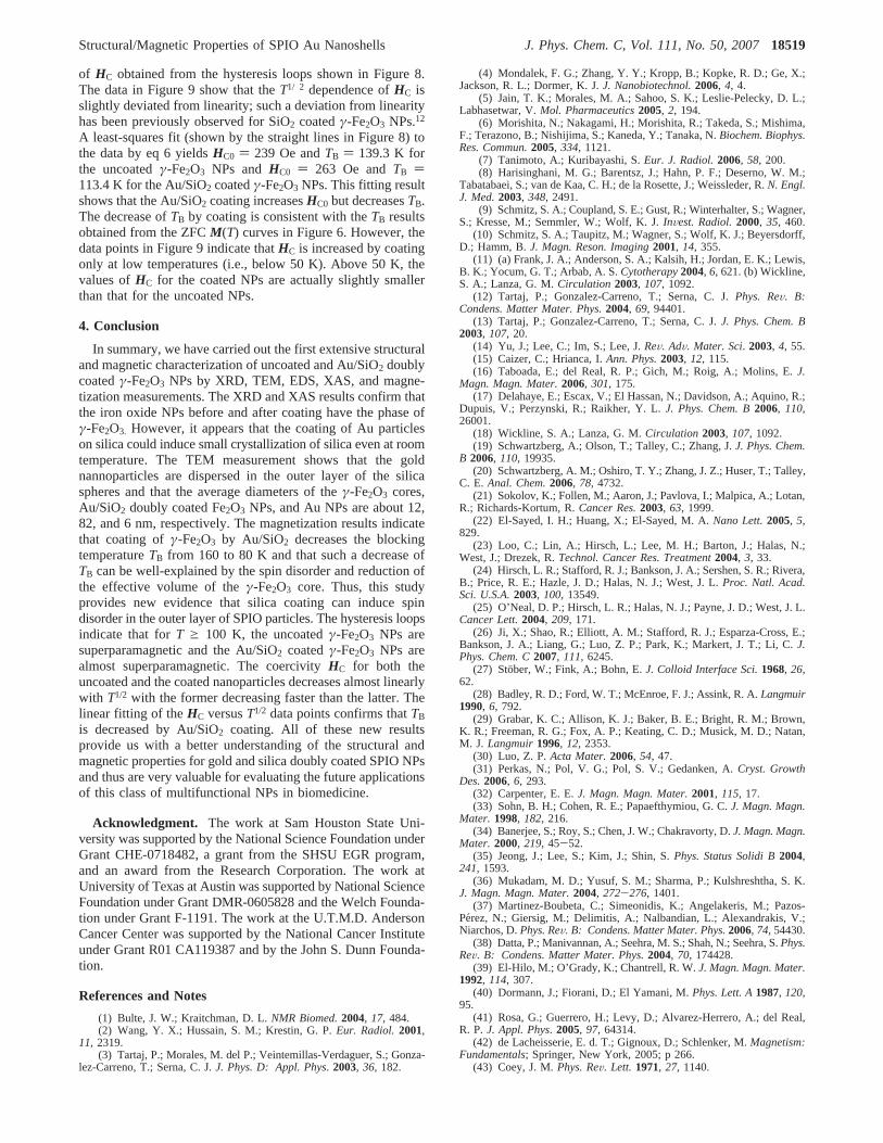
of HC obtained from the hysteresis loops shown in Figure 8.The data in Figure 9 show that theT1/ 2 dependence ofHC isslightly deviated from linearity; such a deviation from linearityhas been previously observed for SiO2 coatedγ-Fe2O3 NPs.12
A least-squares fit (shown by the straight lines in Figure 8) tothe data by eq 6 yieldsHC0 ) 239 Oe andTB ) 139.3 K forthe uncoatedγ-Fe2O3 NPs andHC0 ) 263 Oe andTB )113.4 K for the Au/SiO2 coatedγ-Fe2O3 NPs. This fitting resultshows that the Au/SiO2 coating increasesHC0 but decreasesTB.The decrease ofTB by coating is consistent with theTB resultsobtained from the ZFCM(T) curves in Figure 6. However, thedata points in Figure 9 indicate thatHC is increased by coatingonly at low temperatures (i.e., below 50 K). Above 50 K, thevalues ofHC for the coated NPs are actually slightly smallerthan that for the uncoated NPs.
4. Conclusion
In summary, we have carried out the first extensive structuraland magnetic characterization of uncoated and Au/SiO2 doublycoatedγ-Fe2O3 NPs by XRD, TEM, EDS, XAS, and magne-tization measurements. The XRD and XAS results confirm thatthe iron oxide NPs before and after coating have the phase ofγ-Fe2O3. However, it appears that the coating of Au particleson silica could induce small crystallization of silica even at roomtemperature. The TEM measurement shows that the goldnannoparticles are dispersed in the outer layer of the silicaspheres and that the average diameters of theγ-Fe2O3 cores,Au/SiO2 doubly coated Fe2O3 NPs, and Au NPs are about 12,82, and 6 nm, respectively. The magnetization results indicatethat coating ofγ-Fe2O3 by Au/SiO2 decreases the blockingtemperatureTB from 160 to 80 K and that such a decrease ofTB can be well-explained by the spin disorder and reduction ofthe effective volume of theγ-Fe2O3 core. Thus, this studyprovides new evidence that silica coating can induce spindisorder in the outer layer of SPIO particles. The hysteresis loopsindicate that forT g 100 K, the uncoatedγ-Fe2O3 NPs aresuperparamagnetic and the Au/SiO2 coatedγ-Fe2O3 NPs arealmost superparamagnetic. The coercivityHC for both theuncoated and the coated nanoparticles decreases almost linearlywith T1/2 with the former decreasing faster than the latter. Thelinear fitting of theHC versusT1/2 data points confirms thatTB
is decreased by Au/SiO2 coating. All of these new resultsprovide us with a better understanding of the structural andmagnetic properties for gold and silica doubly coated SPIO NPsand thus are very valuable for evaluating the future applicationsof this class of multifunctional NPs in biomedicine.
Acknowledgment. The work at Sam Houston State Uni-versity was supported by the National Science Foundation underGrant CHE-0718482, a grant from the SHSU EGR program,and an award from the Research Corporation. The work atUniversity of Texas at Austin was supported by National ScienceFoundation under Grant DMR-0605828 and the Welch Founda-tion under Grant F-1191. The work at the U.T.M.D. AndersonCancer Center was supported by the National Cancer Instituteunder Grant R01 CA119387 and by the John S. Dunn Founda-tion.
References and Notes
(1) Bulte, J. W.; Kraitchman, D. L.NMR Biomed.2004, 17, 484.(2) Wang, Y. X.; Hussain, S. M.; Krestin, G. P.Eur. Radiol.2001,
11, 2319.(3) Tartaj, P.; Morales, M. del P.; Veintemillas-Verdaguer, S.; Gonza-
lez-Carreno, T.; Serna, C. J.J. Phys. D: Appl. Phys. 2003, 36, 182.
(4) Mondalek, F. G.; Zhang, Y. Y.; Kropp, B.; Kopke, R. D.; Ge, X.;Jackson, R. L.; Dormer, K. J.J. Nanobiotechnol.2006, 4, 4.
(5) Jain, T. K.; Morales, M. A.; Sahoo, S. K.; Leslie-Pelecky, D. L.;Labhasetwar, V.Mol. Pharmaceutics2005, 2, 194.
(6) Morishita, N.; Nakagami, H.; Morishita, R.; Takeda, S.; Mishima,F.; Terazono, B.; Nishijima, S.; Kaneda, Y.; Tanaka, N.Biochem. Biophys.Res. Commun.2005, 334, 1121.
(7) Tanimoto, A.; Kuribayashi, S.Eur. J. Radiol.2006, 58, 200.(8) Harisinghani, M. G.; Barentsz, J.; Hahn, P. F.; Deserno, W. M.;
Tabatabaei, S.; van de Kaa, C. H.; de la Rosette, J.; Weissleder, R.N. Engl.J. Med.2003, 348, 2491.
(9) Schmitz, S. A.; Coupland, S. E.; Gust, R.; Winterhalter, S.; Wagner,S.; Kresse, M.; Semmler, W.; Wolf, K. J.InVest. Radiol.2000, 35, 460.
(10) Schmitz, S. A.; Taupitz, M.; Wagner, S.; Wolf, K. J.; Beyersdorff,D.; Hamm, B.J. Magn. Reson. Imaging2001, 14, 355.
(11) (a) Frank, J. A.; Anderson, S. A.; Kalsih, H.; Jordan, E. K.; Lewis,B. K.; Yocum, G. T.; Arbab, A. S.Cytotherapy2004, 6, 621. (b) Wickline,S. A.; Lanza, G. M.Circulation 2003, 107, 1092.
(12) Tartaj, P.; Gonzalez-Carreno, T.; Serna, C. J.Phys. ReV. B:Condens. Matter Mater. Phys.2004, 69, 94401.
(13) Tartaj, P.; Gonzalez-Carreno, T.; Serna, C. J.J. Phys. Chem. B2003, 107, 20.
(14) Yu, J.; Lee, C.; Im, S.; Lee, J.ReV. AdV. Mater. Sci. 2003, 4, 55.(15) Caizer, C.; Hrianca, I.Ann. Phys.2003, 12, 115.(16) Taboada, E.; del Real, R. P.; Gich, M.; Roig, A.; Molins, E.J.
Magn. Magn. Mater.2006, 301, 175.(17) Delahaye, E.; Escax, V.; El Hassan, N.; Davidson, A.; Aquino, R.;
Dupuis, V.; Perzynski, R.; Raikher, Y. L.J. Phys. Chem. B2006, 110,26001.
(18) Wickline, S. A.; Lanza, G. M.Circulation 2003, 107, 1092.(19) Schwartzberg, A.; Olson, T.; Talley, C.; Zhang, J.J. Phys. Chem.
B 2006, 110, 19935.(20) Schwartzberg, A. M.; Oshiro, T. Y.; Zhang, J. Z.; Huser, T.; Talley,
C. E. Anal. Chem.2006, 78, 4732.(21) Sokolov, K.; Follen, M.; Aaron, J.; Pavlova, I.; Malpica, A.; Lotan,
R.; Richards-Kortum, R.Cancer Res.2003, 63, 1999.(22) El-Sayed, I. H.; Huang, X.; El-Sayed, M. A.Nano Lett.2005, 5,
829.(23) Loo, C.; Lin, A.; Hirsch, L.; Lee, M. H.; Barton, J.; Halas, N.;
West, J.; Drezek, R.Technol. Cancer Res. Treatment2004, 3, 33.(24) Hirsch, L. R.; Stafford, R. J.; Bankson, J. A.; Sershen, S. R.; Rivera,
B.; Price, R. E.; Hazle, J. D.; Halas, N. J.; West, J. L.Proc. Natl. Acad.Sci. U.S.A.2003, 100, 13549.
(25) O’Neal, D. P.; Hirsch, L. R.; Halas, N. J.; Payne, J. D.; West, J. L.Cancer Lett.2004, 209, 171.
(26) Ji, X.; Shao, R.; Elliott, A. M.; Stafford, R. J.; Esparza-Cross, E.;Bankson, J. A.; Liang, G.; Luo, Z. P.; Park, K.; Markert, J. T.; Li, C.J.Phys. Chem. C2007, 111, 6245.
(27) Stober, W.; Fink, A.; Bohn, E.J. Colloid Interface Sci.1968, 26,62.
(28) Badley, R. D.; Ford, W. T.; McEnroe, F. J.; Assink, R. A.Langmuir1990, 6, 792.
(29) Grabar, K. C.; Allison, K. J.; Baker, B. E.; Bright, R. M.; Brown,K. R.; Freeman, R. G.; Fox, A. P.; Keating, C. D.; Musick, M. D.; Natan,M. J. Langmuir1996, 12, 2353.
(30) Luo, Z. P.Acta Mater.2006, 54, 47.(31) Perkas, N.; Pol, V. G.; Pol, S. V.; Gedanken, A.Cryst. Growth
Des.2006, 6, 293.(32) Carpenter, E. E.J. Magn. Magn. Mater.2001, 115, 17.(33) Sohn, B. H.; Cohen, R. E.; Papaefthymiou, G. C.J. Magn. Magn.
Mater. 1998, 182, 216.(34) Banerjee, S.; Roy, S.; Chen, J. W.; Chakravorty, D.J. Magn. Magn.
Mater. 2000, 219, 45-52.(35) Jeong, J.; Lee, S.; Kim, J.; Shin, S.Phys. Status Solidi B2004,
241, 1593.(36) Mukadam, M. D.; Yusuf, S. M.; Sharma, P.; Kulshreshtha, S. K.
J. Magn. Magn. Mater.2004, 272-276, 1401.(37) Martinez-Boubeta, C.; Simeonidis, K.; Angelakeris, M.; Pazos-
Perez, N.; Giersig, M.; Delimitis, A.; Nalbandian, L.; Alexandrakis, V.;Niarchos, D.Phys. ReV. B: Condens. Matter Mater. Phys.2006, 74, 54430.
(38) Datta, P.; Manivannan, A.; Seehra, M. S.; Shah, N.; Seehra, S.Phys.ReV. B: Condens. Matter Mater. Phys.2004, 70, 174428.
(39) El-Hilo, M.; O’Grady, K.; Chantrell, R. W.J. Magn. Magn. Mater.1992, 114, 307.
(40) Dormann, J.; Fiorani, D.; El Yamani, M.Phys. Lett. A1987, 120,95.
(41) Rosa, G.; Guerrero, H.; Levy, D.; Alvarez-Herrero, A.; del Real,R. P.J. Appl. Phys. 2005, 97, 64314.
(42) de Lacheisserie, E. d. T.; Gignoux, D.; Schlenker, M.Magnetism:Fundamentals; Springer, New York, 2005; p 266.
(43) Coey, J. M.Phys. ReV. Lett. 1971, 27, 1140.
Structural/Magnetic Properties of SPIO Au Nanoshells J. Phys. Chem. C, Vol. 111, No. 50, 200718519


