Embed Size (px)
Citation preview

STRUCTURAL AND BIOPHYSICAL
CHARACTERIZATION OF PENICILLIN V ACYLASE
FROM BACILLUS SUBTILIS AND COMPARISON WITH
RELATED HYDROLASES AS WELL AS STUDY OF
SELECTED PROTEINS FROM MALARIAL PARASITE
THESIS SUBMITTED TO THE
UNIVERSITY OF PUNE
FOR THE DEGREE OF
DOCTOR OF PHILOSOPHY
IN
BIOTECHNOLOGY
BY
PRIYA R
DIVISION OF BIOCHEMICAL SCIENCES
NATIONAL CHEMICAL LABORATORY
PUNE – 411 008
DECEMBER 2006

DECLARATION
I hereby declare that the thesis entitled ‘Structural and Biophysical
Characterization of Penicillin V Acylase from Bacillus subtilis and Comparison
with Related Hydrolases as well as Study of Selected Proteins from Malarial
Parasite’ submitted for Ph.D. degree to the University of Pune has not been
submitted by me to any other university for a degree or diploma.
Priya R Date:
Biochemical Sciences Division
National Chemical Laboratory
Dr. Homi Bhabha Road
Pune 411 008
India

CERTIFICATE
Certified that the work incorporated in this thesis entitled, ‘Structural and
Biophysical Characterization of Penicillin V Acylase from Bacillus subtilis and
Comparison with Related Hydrolases as well as Study of Selected Proteins
from Malarial Parasite’ submitted by Ms. Priya R was carried out by the
candidate under my supervision. The materials obtained from other sources
have been duly acknowledged in the thesis.
(Dr. C. G. Suresh) Date:
Research Guide

Acknowledgments I thank the Director and Head, Biochemical Sciences Division, NCL for permission to conduct
my work here. I acknowledge financial support from Council of Scientific and Industrial Research,
New Delhi. I thank the Commonwealth and the University of York for the split-site PhD scholarship.
I wish to express my sincere gratitude to my research Guide, Dr. C.G. Suresh, mainly for the
academic freedom. He has been most understanding, considerate and caring. I am indebted to Dr.
Archana V. Pundle for her able guidance and encouragement and Dr. Asmita A. Prabhune for constant
support and care. I am grateful to Dr. Sushama Gaikwad for her help and guidance during crucial times.
I have been fortunate to work with Prof. Guy G. Dodson and Prof. Eleanor Dodson, who have
been most kind; Dr. James A. Brannigan, who showed exemplary care, often bailing me out of cloning
catastrophes and Prof. Antony J. Wilkinson, who provided kind support throughout the tenure at the
University of York.
Dr. Shama Barnabas - I cherish the innumerable instances when she shared her wisdom –she
is such a scholarly person, with lots of positive attitude, warmth and understanding. Dr. Anil Lachke
has been an admirable source of advice and encouragement. I am most grateful to Dr C. SivaRaman for
his scholarly suggestions and advice. I am thankful to Drs. Mala Rao and M I Khan for permission to
use the spectrophotometer and fluorimeter.
I thank Prof. Marek Brozozowski for helping me even at unearthly hours, sorting out
crystallisation problems. Dr. Claudia Schnick was the epitome of friend, philosopher and guide during
my tenure at York. My thanks to Dr. Johan Turkenberg for help with collecting data and
troubleshooting and Mr. Sam Hart for being so patient and helpful. I am also thankful to Dr. Elena
Blagova, Mrs. Shirley Roberts for help with crystallisation. I thank Mr. Simon Grist, Mrs. Sally Lewis,
Mrs. Wendy Offen and Mr Tim Kirk for all the ready help. I should mention Eric for magnanimously
sharing work space. I thank Ms Caroline and Mrs Louise and the staff at commonwealth for ironing out
difficulties. I should acknowledge Mrs Indira, Mr Trehan, Mr Karanjkar Mr Mari and Satyali for their
help during all these years.
Dinesh, Rao, Bhushan, Rachna, Meena, Meena, Jayanthi, Jaspreet, Manish, Shivappa, Sulatha
and all others have kept company and helped on innumerable occasions. I wish to acknowledge them
and everyone from my lab and the division, especially Uma, Poorva, Sharmili, Suresh, Nishant, Anu,
Raamesh, Urvashi, Nitin for all the timely help and Ashvini and Mrunal for being such great friends. I
also am thankful to all the lovely friends, outside work who made life easier. I am indebted to Uming
and her endearing family, especially Appa, Amma, Vekatesh and Sonali for constant support,
encouragement and care. Though I have not mentioned all of them, I remember all the goodness each
of my friends and family have showered on me.
I wish to mention with gratitude, my teachers at school and graduate studies, especially, Mr.
H.H. Sheriff and Drs. K . Nirmala, S. Saroja and S. Annapurani –they were so caring and encouraging.
My family has always had an all- pervading, invisible presence in me, in the form of Hope I
carry. I am privileged to be indebted to Aajima for unflinching love and to my TM, Ba for everything I
treasure. I dedicate whatever little I have achieved to the loving memory of my beloved grandparents
and for M-P, who stood by me, through thick and thin.
- Priya

1
Abbreviations 6APA 6-aminopenicillanic acid
AMoRe Automated Molecular Replacement
AS Ammonium sulphate
BLAST Basic Local Alignment Search Tool
BSA Bovine Serum Albumin
BspPVA Bacillus sphaericus Penicillin V Acylase
BsuPVA Bacillus subtilis Penicillin V Acylase
CA Cephalosporin acylase
CBH Conjugated bile acid hydrolase
CCP4 Colloborative computational Project 4
CD Circular Dichroism
CPB Citrate Phosphate buffer
DNA Deoxyribo Nucleic Acid
DTNB 5,5’-dithiobis-(2-nitrobenzoic) acid
DTT DiThioThreitol
EDTA Ethylene Diamine Tetra Acetic acid
FPLC Fast protein liquid chromatography
GFC Gel filtration chromatography
h /hr hour
HEPES N-(2-hydroxyethyl)-piperazine-N’-2-ethane sulfonic acid
HPLC High performance liquid chromatography
IEF Iso-electric focussing
IPTG Isopropyl-β-D-thiogalactoside
kDa kilo Dalton
Ki Inhibition constant
Km Michaelis-Menten constant
LB Lineweaver-Burke
M W / Mr Molecular Weight
M molar
MAD Multi-wavelength anomalous dispersion
MALDI -MS Matrix Assisted Laser Desorption Ionisation Mass Spectrometry
min minute
MIR Multiple isomorphous replacement
MR Molecular replacement
NCS Non-crystallographic symmetry

2
NIPOAB 2-nitro-5-(phenoxyacetamido)-benzoic acid
NMR Nuclear Magnetic Resonance
Ntn N-terminal hydrolase
O.D Optical density
PAGE Poly-acrylamide gel electrophoresis
PCR Polymerase Chain Reaction
PDAB Para Dimethyl Amino Benzaldehyde
PDB Protein DataBank
PEG poly ethylene glycol
PenG Penicillin G
PenV Penicillin V
Gdn HCl Guanidine hydrochloride
PIPES Piperazine -1,4–bis - (2-Ethane) Sulphonic Acid
PVA Penicillin V Amidase
rmsd root mean square deviation
rpm revolution per minute
s second
SDS sodium dodecyl sulphate
SV Stern-Volmer
TCA TriChloro Acetic Acid
Tris Tris-hydroxymethyl amino methane
VM Matthews number
α (h,k,l) phase angle
μ micro-
σ sigma
Σ summation
α alpha
Å Angstrom
β beta
F(hkl) structure factor
l litre oC degree centigrade

3
Abstract
The thesis is a result of the effort at characterising some proteins by using
biochemical and biophysical techniques, especially crystallography to elucidate their
structure and function. The aim was to characterize and conduct comparative study
of a class of hydrolases called choloylglycine hydrolases. Understanding the atomic
level contributions of various structural constituents towards activity and stability of
penicillin acylase will help to improve the deacylase activity for the production of 6-
aminopenicillanic acid, used in the semi-synthetic manufacture of various β-lactams.
Similar objectives prompted studies on malarial proteins, which could find application
in drug targeting.
The structure of Bacillus subtilis penicillin V acylase (BsuPVA) has been elucidated
and its enzymatic properties characterized. Biophysical studies using fluorimetry and
circular dichroism have been conducted on this enzyme as well as a related
hydrolase from Bacillus sphaericus (BspPVA) whose crystal structure was already
known. By combining with kinetic studies, these studies have explained the
differences between related enzymes at various levels considering molecular level
observations obtained from X-ray crystallographic studies. Results will be discussed
juxtaposed with the closely related conjugated bile salt hydrolases, which also belong
to the family of choloylglycine hydrolases along with PAs. Three malarial proteins
were cloned, another one purified and crystallization and crystal characterisation
were carried out on one.
The thesis is divided into 9 chapters and structured in three parts. In the first part,
characterization of a hypothetical protein from B. subtilis, its identification as a PVA
and its three-dimensional structure determination are described in detail. Extensive
comparative studies on related hydrolases from the choloylglycine family, by
generating the relevant data on BspPVA, form the second part of the thesis. The third
and final part deals with various studies carried out on proteins from the malarial
parasite.
Chapter 1: General Introduction Penicillin acylases (also called penicillin amidases or amidohydrolases) are enzymes
that reversibly cleave the amide bond between the side chain and the β-lactam
nucleus of penicillins, without affecting the amide bond of the β-lactam nucleus. PVA

4
is an industrially important enzyme that can be used in the manufacture of semi-
synthetic penicillins. PVAs are remarkably similar to conjugated bile salt hydrolases
in several aspects and the enzyme belongs to the same family. Comparative studies
might help us to assign a function to penicillin acylases whose physiological role is
still not understood.
The complete genome of the malarial parasite Plasmodium falciparum and that of the
vector, mosquito have been reported. Along with the success of human genome
project, the triumvirate genome data have been a shot in the arm for efforts to
combat the disease. Various structural genomics consortia are trying to elucidate the
proteome of the parasite and rational drug design. In an opportunity to work in one
such consortium the author has tried to characterise selected proteins from the
parasite. These efforts are described in the last chapter.
Chapter 2: Biochemical characterization of YxeI as a Penicillin V acylase This chapter describes the biochemical characterization of YxeI, a hypothetical
protein from B. subtilis, subsequently characterised as a PVA by us. E. coli
BL21(DE3) cells were transformed with the yxeI gene, the protein was overproduced
and purified. Molecular weight, pI and optimal conditions were estimated. PVA
activity was detected against penicillin V and for the synthetic substrate 2-nitro-5-
(phenoxyacetamido)-benzoic acid (NIPOAB). Chemical modification and site-directed
mutation studies identified cysteine and arginine as active site residues.
Chapter 3: Crystallographic studies on YxeI from B. subtilis characterised as a PVA BsuPVA was crystallized and diffraction data were collected using R-AXIS IV++ Image
Plate system under cryo conditions up to a resolution of 2.5 Å. Data were processed
and scaled using Denzo and Scalepack. The structure has been determined using
molecular replacement method using BspPVA (PDB code 2pva) as the search
model. BsuPVA forms a homotetramer by two symmetry-related dimers, in
agreement with the existence of tetramer in gel filtration studies. The structure has
the classical αββα Ntn hydrolase fold. Cysteine1 is the N-terminal nucleophilic
residue. The final Rmerge and R factor were within the acceptable range. Geometry
of the molecule, checked using PROCHECK, had no residue in the disallowed
regions of the Ramachandran plot.

5
Chapter 4: Fluorimetric and kinetic studies of BsuPVA The decrease in fluorescence intensity upon titration with the substrates NIPOAB and
penicillin V (PenV), without change in emission maxima, was made use of to
calculate binding constants for these substrates at different temperatures. The
temperature dependence of the association constants was used to determine the
thermodynamic parameters. The accessibility of Trp fluorophores and their local
environment were investigated using acrylamide, potassium iodide (KI) and cesium
chloride (CsCl).
Chapter 5: Studies on conformational stability of BsuPVA using fluorimetry and circular dichroism Temperature, pH and Guanidine hydrochloride (Gdn HCl)- induced conformational
changes were studied by monitoring the changes observed in Trp emission.
Evidence for molecular aggregation at higher temperatures was obtained by the
binding of 1-anilinonaphthalene-8-sulphonic acid (ANS) to the protein in a range of
acidic pH 1-4. The secondary structure of the protein was studied using circular
dichroism. Activity and denaturation studies were conducted at different
concentrations of Gdn HCl.
Chapter 6: Comparative studies on related hydrolases- Part I. Specificity and substrate binding of the PVA from Bacillus sphaericus
This part of the thesis compares the kinetic, conformational and structural aspects
and stability of BsuPVA with other known hydrolases. Many PVAs have been
reported but the only other PVA whose structure has been elucidated is the one from
B. sphaericus (BspPVA). Conformational studies have not been reported on this
enzyme. Such comparative studies have been carried out on this enzyme and the
observed data are presented here. The binding constants for PenV and NIPOAB
were calculated. Acrylamide was by far the most efficient quencher (81% quenching).
Quenching by KI and CsCl are 20% and 14%, respectively.
Chapter 7: Comparative studies on related hydrolases- Part II. Comparison with the conformational stability of BspPVA BspPVA has 3 Trp residues in hydrophobic environment. There are disturbances in
secondary structure with increase in temperature and extremes of pH. The presence
of molten globule as indicated by ANS binding is seen in the acidic pH range 1-4.
PVA is active in this pH range. Increasing concentrations of Gdn HCl resulted in

6
decreased 333/356 intensity ratio. There is substantial loss of activity (80%) at 1 M
Gdn HCl a complete loss of secondary structure at 6 M.
Chapter 8: Comparative studies on related hydrolases- Part III. Structure, function and evolution BsuPVA has good sequence homology with other PVAs and Conjugated Bile Acid
Hydrolases. This chapter deals with comparison of structural, functional and
evolutionary characteristics of BsuPVA with other members of the choloylglycine
hydrolase family. Major differences in the structure vis-à-vis substrate specificities,
efficiency and relatedness of their sequences are discussed.
In conclusion, our studies have established that YxeI from B. subtilis is a PVA,
belongs to choloylglycine hydrolase family of the Ntn-hydrolase superfamily and
shares the αββα fold called the Ntn-fold and conserved residues especially at the
oxyanion hole. Despite sharing relatively good sequence and structure homology,
BsuPVA and BspPVA are significantly different in their kinetic properties with respect
to their hydrolase activities on penicillin V. Evidences show that the enzyme
originated through divergent evolution from a common ancestor that possessed both
penicillin acylase and conjugated bile acid hydrolase activities. This study
enumerates the reasons and rationale behind designating the protein YxeI as a PVA.
Chapter 9: Studies on malarial proteins Three calmodulin–like proteins from Plasmodium falciparum, MALP71.69,
PF14_0181 and PF10_0301 were identified from PlasmoDB. Two of the genes could
be expressed in E. coli BL21 cells, but were insoluble. Solubilization and purification
using various methods were attempted. Macrophage migration inhibitory factor was
purified, refolded and crystallisation attempted. A very crystalline precipitate was got.
A translationally controlled tumour protein was purified and crystallized. Diffraction
data were collected at the European Synchrotron Radiation Facility in France and
could be processed very well. Though no new structures could be solved in the case
of malarial proteins, the study helps in highlighting the challenges while working with
eukaryotic proteins and the problems pertaining to malarial proteins in particular.

7
CHAPTER 1
INTRODUCTION
Background
The work presented in this thesis can be broadly segregated into two parts. The first
part of this work is on penicillin V acylases, the enzymes used in the industrial
production of semi-synthetic penicillins. The second part of the work is on selected
proteins from the malarial parasite, Plasmodium. Accordingly, the background on this
work is presented in two parts respectively.
Part I
1.1.1. Introduction to enzymes
Proteins, along with carbohydrates, lipids and nucleic acids are the major biological
macromolecules. Enzymes are proteins specialized to perform biological catalytic
functions. Some ribonucleic acids called ribozymes also can perform catalysis. The
first commercial application of enzymes was in the form of organisms that possessed
them, as in leavening or brewing with yeast. The word ‘enzyme’ originates from
Greek énsymo, én meaning "at" or "in" and simo means "leaven" or "yeast" (Stryer,
1995). More than 2500 enzymes are known and 250 commercially used (Woodley,
2000). Enzymes are sometimes synthesized in inactive form called zymogen. E.g.
Pepsinogen. Such zymogens need to be activated, often involving proteolytic
cleavage, in this case, to pepsin. Two popular theories are used to explain binding of
enzyme to its substrate:
Lock and key theory: Emil Fischer postulated in 1894 that the correct substrate of an
enzyme acts as a key that fits into a specific pocket in the enzyme.
Induced fit theory: It was proposed by Daniel Koshland in 1958 that the substrate
induces a change in the shape of the enzyme, which then fits it.

8
Enzymes speed up reactions by providing an alternate pathway of lower activation
energy for a reaction without altering reaction equilibrium. The rates of enzyme
turnover (number of substrate molecules converted to product per second) vary from
36 million per second for carbonic anhydrase to lysozyme, which processes merely 2
molecules per second. Their lifetime varies from a few minutes to weeks. They have
optimal pH and temperatures where their efficiency is maximum. Some require
cofactors or coenzymes for functioning- in such cases, the protein part is called
apoenzyme and together with the cofactor, constitutes the holoenzyme. The main
advantages of enzymes compared to most other catalysts are their chemoselectivity
and steriospecificity. They are described by maximal velocity of the reaction they
catalyze, Vmax, and the affinity they have for the substrate, measured as substrate
concentration at half maximal velocity, Km, generally calculated using Michaelis-
Menten equations proposed in 1913.
Enzymes are sensitive to molecules, which step up reaction called activators and
inhibitors that reduce their activity. Inhibition can be reversible or irreversible. The
different types are:
Competitive: Inhibitor binds to the same site as the substrate, Km increases but Vmax
remains the same.
Uncompetitive: inhibitor binds only to the enzyme-substrate complex (ES), not to the
free enzyme. This causes a decrease in both Vmax and the Km value.
Noncompetitive: Inhibitor binds to a site other than substrate binding site resulting in
decreased Vmax, but leaving Km unchanged. The inhibition is mostly irreversible. E.g.
Suicide inhibitors.
Partially competitive: The mechanism is similar to that of non-competitive inhibition
except that the enzyme substrate inhibitor complex is active.

9
Mixed: Mixed inhibitors can bind to both the enzyme and the ES complex. It has the
properties of both competitive and uncompetitive inhibition. Both, decrease in Vmax
and increase in Km value occur.
Apart from in vivo functions, some enzymes have great commercial applications.
Since reaction conditions are mild, and enzymes are specific, no unwanted by-
products accumulate. Immobilization of enzymes has permitted repeated use of
costly enzymes. Enzyme catalyzed reactions are thus cheaper than their chemical
counterparts and the process more environment-friendly. Since enzymes help
overcome energy barrier of activation, it has viable and economical industrial
applications in textiles, petrochemicals, food, animal feed, detergents, pulp and
paper, leather etc. For example, β-lactam acylases such as penicillin acylases, are
used in the industrial production of semisynthetic β-lactam antibiotics.
1.1.2 Antibiotics
Antibiotics are chemicals produced by microorganisms or fungi that acts on other
microorganisms. Antibiotics kill the bacteria (bactericidal) or arrest its growth
(bacteriostatic). Majority of the clinically used antibiotics have been obtained from
actinomycetes, especially Streptomyces species. Bacillus species and fungi also
have yielded few useful antibiotics. They can be classified based on their chemical
structure, microbial origin, spectrum of activity or mode of action. One class of
antibiotics that work by inhibiting the synthesis of peptidoglycan in bacterial cell walls
are β-lactam antibiotics, which can be classified based on their structure (nucleus) as
1. Penicillin. This can be classified into two types according to the source.
Natural penicillins are penicillin G (PenG) and penicillin V (PenV)
Semi-synthetic penicillins E.g. Amoxicillin, penicillinase-resistant cloxacillin,
methicillin.

10
2. Beta lactam inhibitors. E.g. clavulanic acid, sulbactam
3. Cephalosporin. Generations – I, II, III and IV. Along with cephamycin, it forms a
sub-group called cephems.
4. Carbapenems. E.g. imipenem and meropenem
5. Monobactams. E.g. Aztreonam
Penicillin G (6APA) Clavulanic acid (Clavam)
Cephalosporin C (Cephem)
Imipenem (Carbapenem) Carumonam (Monobactam)
Fig. 1.1: Structures of different β-lactams. The nuclei of the different antibiotics are
coloured black and indicated in brackets. The side chains are in blue. In another type
of classification, the antibiotics are grouped according to their side chains.
N
O
COOHO
OHNO
O
H
N
S
COOHO
NH
ON
S
O CH3
O
ONOH
O
NO
OH
SN
NH2COOH N
O SO3H
O
NH2
NH
O
N
S
NNH2
O
COOH

11
Penicillins and cephalosporins are widely found in organisms especially, in fungi
belonging to Penicillium and Cephalosporium. The basic component of penicillins
and cephalosporins is a β-lactam nucleus which is formed by the fusion of a 4-
membered β-lactam ring to a thiazolidine ring or a six carbon ring to form 6-
aminopenicillanic acid (6APA) or 7-amino cephalosporanic acid (7ACA), respectively.
They are the most widely used group of antibiotics. Modification of side chains yields
different penicillins and cephalosporins. It is obvious that the side chain determines
the antibacterial range and pharmacological properties of the β-lactams. The
penicillin β-lactam nucleus is derived from valine and cysteine via a tripeptide
intermediate. The β-lactams inhibit the transpeptidylation step in peptidoglycan
(murein) synthesis.
Penicillin was discovered initially by a French medical student, Ernest Duchesne, in
1896, and subsequently rediscovered by Scottish physician Alexander Fleming in
1928, for which he was awarded the Nobel Prize along with Howard Florey and Ernst
Chain who prepared the antibiotic in large quantities (Abraham, 1981; Bennet and
Chung, 2001). Determination of X-ray structure of penicillin by Dorothy Crowfoot
Hodgkin and co-workers in 1949 was made use of to find and develop more effective
penicillins. Penicillins are used in the treatment of leptospirosis, syphilis, etc. The
main disadvantage of using penicillin is allergy it causes in some people and narrow
range of activity. They cause fatal allergies in around 300-500 people every year.
Also, many penicillins display little activity against Gram negative bacteria, since they
do not penetrate the outer membrane.
Cephalosporins are a group of broad-spectrum β-lactams. Cephalosporins and other
newer penicillins are active against Gram negative bacteria. They are used as
penicillin substitutes, and in surgical prophylaxis, and in the treatment of gonorrhea,

12
meningitis, pneumococcal, staphylococcal and streptococcal infections.
Pharmacological characteristics of the antibiotic can be modified by substitution at 3
and 7 positions of its β-lactam ring. Newer generations of cephalosporin have
progressively broader range of activity against gram-negative organisms but a
narrower range of activity against gram-positive organisms than the preceding
generation. They also have longer half-lives, reducing the dosing frequency. The
advantages of cephalosporins over penicillins include low toxicity, resistance to β-
lactamase (Abraham, 1987) and different antibacterial spectrum.
The peptidoglycan layer of bacteria is formed by cross-linked peptidoglycan chains,
which are repeating alternating units of the sugars N-acetylglucosamine (NAG) and
N-acetylmuramic acid (NAM) with a variable peptide chain attached to the carboxyl
group of the NAM-unit. Individual peptidoglycan chains are covalently cross-linked to
each other via short pentapeptide bridges connecting the third position of the first
peptide chain to the carboxyl group of a D-alanyl-D-alanine residue from another
peptide chain, catalyzed by transpeptidases (Fig. 1.2). Transpeptidases first bind to a
D-alanyl-D-alanine unit forming a covalent acyl-enzyme complex with the release of
terminal D-Ala. Attack on this complex by the terminal glycine of the cross-linking
pentaglycine results in the recovery of active enzyme and formation of the bond
between glycine and alanine (Scheme I, Fig. 1.3). Penicillin, being a structural analog
of D-alanyl-D-alanine, interferes in this reaction by binding irreversibly to the enzyme
(Scheme II, Fig. 1.3). As a result of faulty cell wall, cells are unable to divide inspite of
growing, leading to accumulation of pressure and subsequent lysis of the cell by
autolysins (Abraham, 1981).

13
1.1.3 Antibiotic Resistance
After a lot of initial failures, penicillin was finally prepared in large amounts. During
the Second World War, it saved a lot of lives that could have succumbed to serious
war wound infections. Complacency set in in the 1980s. Slowly, resistant microbes
began to appear (Abraham, 1981). Now, diseases like tuberculosis, which were
claimed to be vanquished before, are making a come back with renewed resistance.
Antibiotics themselves increase the prevalence of resistance by selecting naturally
occurring variants of organisms that are resistant. One can contract a resistant bug
due to infection or the resistance can emerge within the body during treatment due to
selection pressure of the antibiotic. The resistant organisms then transfer the
resistance to other non-resistant organisms. Bacteria acquire genes conferring
resistance by spontaneous DNA mutation, transformation, or from plasmids.
Resistance may also arise from a change in the structure of penicillin binding
proteins such that the antibiotic does not bind efficiently (Essack, 2001; Antignac et
al., 2003). In the case of Gram negative bacteria, penicillins pass across the outer
membrane using porins. Resistance may develop from mutation leading to modified
porins. The most efficient way for bacteria to withstand penicillin action is by
producing ß-lactamases, which cleave ß-lactams, rendering them ineffective (Jacoby
and Munoz-Price, 2005) (Scheme III, Fig. 1.3).

14
Fig. 1.2. The repeating unit of bacterial cell wall. The NAM units from different chains
are joined together by a pentaglycine linker.
D-Ala-D-Ala Transpeptidase
D-Ala
Covalent acyl-enzymeintermediate
Glycine pentapeptide
Cross-linking
TranspeptidasePenicillin
Transpeptidase
Inactive penicilloyl-enzyme intermediate
Penicillin Penicillinase Penicilloic acid Cross-linking
No cross-linking
Beta-lactamaseinhibitor
Penicillinase Inactive enzyme
TranspeptidasePenicillin Inactive enzyme
No cross-linking
I.
II.
III.
IV.
Fig. 1.3. Penicillin action
NAG NAM
NH
CH3
O
OCH2OH
O
CH2OH
NH
O
OH O
O
CH3
O
L-Ala
D-Glu
L-Lys
D-Ala
D-Ala
Gly
Gly
Gly
Gly
Gly
O
CH3
D-Ala
NH
CH3
O
O
CH2OH
NH
O
OCH2OH
OH O
O
CH3
L-Ala
D-Glu
L-Lys
D-Ala
Gly
O
Gly
Gly
Gly
Gly
O
CH3

15
Fig. 1.4. Site of action of the enzymes. The backbone of penicillin and D-Alanyl
alanine are very similar (coloured Blue), enabling penicillin to act as a structural
analog for transpeptidase.
Consequently, β-lactamase resistant penicillins like flucloxacillin, dicloxacillin and
methicillin and later vancomycin were developed and used. Methicillin and
vancomycin resistant Staphylococcus aureus (referred to as MRSA and VRSA) have
emerged now. This prompted the use of a group of structural analogs of ß-lactams,
called ß-lactamase inhibitors in combination with β-lactams. Though they do not have
antibacterial activity themselves, but bind tightly to ß-lactamases, allowing the other
ß-lactam to act. E.g., the natural clavulanic acid or the synthetic sulbactam. These
are effective against beta-lactamases commonly produced by a variety of organisms
including Staphylococcus species, the Enterobacteriaceae, Pseudomonas
aeruginosa, Acinetobacter species and some anaerobes. While using combination
therapy with beta-lactamase inhibitors, the two ß-lactams act synergistically - one
competitively inhibiting beta-lactamase, thus protecting the other from inactivation
NR
O
H
N
S
COOHO
CH3
CH3
Penicillinase cleavage site
NR
O
H
NCH3
COOHO
CH3HH
Transpeptidase cleavage site
Penicillin D-Ala-D-Ala
Penicillin acylase cleavage site

16
(Scheme IV, Fig. 1.3). However, in some cases, penicillins antagonize each other;
the antagonist blocks the more sensitive site but acts additively with the antagonized
antibiotic at the less sensitive site (Acar et al., 1975). A promising lead to new
mechanism-based and transition state analogue inhibitors could be the retro-amide
side chain of aryl malonamates, which fits the active site of P99 beta-lactamase
(Cabaret et al., 2003).
Though bacterial antibiotic resistance is a natural phenomenon, other factors can
abet the problem. The most serious preventable cause is inappropriate antibiotic use.
The need of the hour is firstly, to prevent the spread of resistance and the selection
of resistant organisms, using narrow spectrum antibiotics and following proper
regimen for the usage of antibiotics. Secondly, newer and effective antibiotics have to
be found or developed. In the quest for new antibiotics, semi-synthetic penicillins,
with a β-lactam nucleus and a custom-designed side chain, look promising, though
resistance to some semi-synthetic penicillins are already reported.
1.1.4 Semi-synthetic penicillins
Since known natural penicillins were increasingly becoming ineffective for treatment
of various pathogens, other forms were scouted for. It was first observed during
fermentation of E. coli that it produced a mixture of penicillin isoforms. Later, it was
discovered that it could be preferably induced to produce PenG by adding its side
chain, phenylester, to the culture medium. It was observed that variations in the side
chain alter the properties of a β-lactam antibiotic (Abraham, 1981; Vandamme and
Voets, 1974) providing the first clue for more effective antibiotics. However, modifying
the side chain chemically is expensive and generates by-products that have to be
treated before disposal. In a multi-step reaction, natural penicillins have to be cleaved
to yield the β-lactam nucleus 6APA and in the next scheme, the nucleus has to be

17
derivatised with the desired side chain. The amide link between the side chain and
the nucleus can be hydrolyzed conventionally to yield 6APA but β-lactam ring also
gets hydrolyzed. A significant discovery was that in the absence of any precursors, a
particular Penicillium could produce the free β-lactam nucleus (Bruggink, 2001;
Vandamme and Voets, 1974). Alternatively, Penicillin acylases can selectively
hydrolyze the amide bond leaving the β-lactam ring intact. Penicillin acylase from E.
coli could catalyze the breakdown of PenG to 6APA and its organic acid (Shewale,
1997). Later it was found that it could catalyze the second step also when it was
found that under different conditions reverse reaction could occur (Vandamme and
Voets, 1974). Thus the green method of semi-synthetic penicillin production was
born. Other interesting discoveries included that Isopenicillin N synthase creates the
bicyclic nucleus of penicillins in one step and deacetoxycephalosporin C synthase
catalyses the expansion of the penicillin nucleus into the nucleus of cephalosporins.
The enzymatic method is regio- and stereo-specific and the reaction conditions are
milder. Advances in immobilization techniques facilitated easy recovery and reuse of
enzymes and increased their stability making it cost-effective (Woodley, 2000). In
fact, in the hydrolysis of racemic iso-propylamide of mandelic acid, immobilization
improved the enantioselectivity of PGA (Rocchietti et al., 2002). Thus, enzymatic
process is cheaper and safer than chemical processes. Today, penicillin G and V are
fermentatively produced at an estimated annual market volume of 16,000 tons
(Bruggink and Roy, 2001) worldwide using Penicillium strains. About 10,000 tons of
this along with 30 tons of penicillin acylases are used for production of semi-synthetic
β-lactam antibiotics (Demain, 2000, Elander, 2003). Major chunk of the 6APA
produced is via the PGA route. The structures of all semi-synthetic penicillins and
cephalosporins known are based on their respective beta-lactam nuclei, 6APA and
7ACA, respectively. Totally synthetic analogs are also known.

18
Fig. 1.5. Synthesis of semi-synthetic penicillin
Fig. 1.6. Ampicillin, a semi-synthetic penicillin
The advantages of semi-synthetic penicillins are increased tolerance and diminished
toxicity in humans, so they have fewer side effects; increased effectiveness due to
greater selectivity against pathogens; increased resistance to β-lactamases; broader
spectrum of antimicrobial activity, e.g., ampicillin is more useful than penicillin due to
N
S
COOHO
CH3
CH3
NH
O
NH2
N
O
H
N
S
COOHO
CH3
CH3
R1
NH2
N
S
COOHO
CH3
CH36APA
Natural penicillin
Fermentation
Penicillin acylase
N
O
H
N
S
COOHO
CH3
CH3
2
Penicillin acylase /Chemical Synthesis
Synthetic side chain
Semi synthetic penicillin
R

19
broader spectrum (Bruggink and Roy, 2001); improved pharmacological properties
like increased stability, better absorption from the gastro-intestinal tract resulting in
less dosage and lower elimination rates from the patient, thus decreasing the
frequency of administration of the drugs. The β-lactam acylases commonly used in
pharmaceutical industry in the bulk manufacture of semi-synthetic β-lactams include
penicillin acylases, cephalosporin acylase and glutaryl 7-aminocephalosporanic acid
acylase reported from Arthobacter viscosus, Bacillus laterosporus, Bacillus
megaterium, Bacillus sphaericus, Kluyvera citrophila, Proteus rettgeri and
Pseudomonas sp. (Deshpande et al., 1994).
1.1.5 β-lactam acylases
1.1.5.1 Penicillin acylases
Penicillin amidohydrolases (EC 3.5.1.24) also called penicillin acylases or penicillin
amidases are enzymes that catalyze the hydrolysis of penicillin to a carboxylate and
6-aminopenicillanic acid (Sakaguchi and Murao, 1950). The EC number denotes that
they are hydrolases acting on carbon-nitrogen bonds other than peptide bonds in
linear amides. Apart from penicillin production, PAs are also used in other industries,
in peptide synthesis or acyl group transfer reactions (Van Langen et al., 2000).
Amidase from E. coli is used in the synthesis of artificial sweetener aspartame
(Fuganti et al., 1986) and diphenyl dipeptides, whose derivatives are used as food
additives, fungicidal, antiviral and anti-allergic compounds (Van Langen et al., 2000).
PAs can be used to resolve racemic mixtures of chiral compounds such as
aminoacids (Bossi et al., 1998), β-amino esters, amines and secondary alcohols
(Svedas et al., 1996).
According to the type of substrate preferably hydrolyzed, penicillin acylases are
classified into Penicillin V acylase (PVA), Penicillin G acylase (PGA) and ampicillin

20
acylase which preferably cleave PenV, PenG and ampicillin, respectively. However,
some PAs have broad substrate specificity, hydrolyzing more than one type of
penicillin. PVA from Streptomyces lavendulae also acts on aliphatic penicillins like
Penicillin F, dihydroF and K (Torres-Guzman et al., 2002). Both PGA and PVA were
found to be members of Ntn hydrolase superfamily.
1.1.5.1.1 Penicillin G acylase
PGA catalyses the hydrolysis of PenG and the condensation of Cα-substituted
phenylacetic acids with β-lactam nucleophiles. PGA has been reported from
Achromobacter xylosoxidans (Cai et al., 2004), Alcaligenes faecalis (Verhaert et al.,
1997), Arthrobacter viscosus (Ohashi et al.,1988, Ohashi et al.,1989; Verhaert et al.,
1997). Bacillus megaterium (Martin et al., 1995), Bacillus megaterium, Escherichia
coli, Kluyvera citrophila (Barbero et al., 1986; Martin et al., 1991), Proteus rettgeri
(McDonough et al.,1999), Streptomyces lavendulae. Homologs of PAs are found
throughout the whole kingdom of prokaryotes (Arroy et al., 2003). Most PGAs in
gram negative organisms are usually periplasmic. PGA of B. megaterium is
extracellular (Martin et al., 1995).
PGA from E. coli has been thoroughly investigated. In 1974, Kutzbach and
Rauenbusch studied its general properties. The mature enzyme is a 80 kDa
heterodimer of 24 kDa α-subunit and 64 kDa β-subunit comprising 209 and 566
aminoacids, respectively (Duggleby, 1995; McVey, 1997). It is produced as a 96 kDa
cytoplasmic precursor pre-pro-protein. Post-translational processing requires
translocation through the cytoplasmic membrane (Schumacher et al., 1986, Hewitt et
al., 2000; Burtscher and Schumacher, 1992) to the periplasm using the 26-amino-
acid signal peptide that is subsequently cleaved off. The 54-amino-acid spacer
peptide that connects the α and β chains which may influence the final folding of the

21
chains (Oliver et al., 1985), is cleaved on the carboxyl side first between Thr263 and
Ser264 (Choi et al., 1992), giving rise to the N-terminal of the β-subunit, serine, which
is the active catalytic residue. However, the β-chain alone is not catalytic (Daumy,
1985). The C-terminal of the α-peptide is generated by another endopeptide
cleavage (Bock et al., 1983; Merino, et al., 1992) and the α and β units are
reconstituted to give an active enzyme. Kinetic studies showed that the
autoproteolysis in PGA is intramolecular (Kasche et al., 1999). These endopeptidase
cleavages require an intact carboxy terminus. This type of processing is found in the
synthesis and processing of preproinsulin and other eukaryotic hormones and is
unique for a prokaryotic enzyme.
PGA from Kluyvera citrophila ATCC21285 is reported to be composed of two non-
identical subunits of 23 and 62 kDa (Barbero et al., 1986), in contrast with the
previous findings (Shimizu et al., 1975). Its nucleotide sequence is 80% similar to E.
coli ATCC11105 PGA (Schumacher et al., 1986), indicating a common ancestor.
Proteus rettgeri is an 86 kDa enzyme composed of two essential non-identical
subunits. Like E. coli PGA, the beta subunit contained a serine residue required for
enzymatic activity, and the alpha subunit contained the domain that imparts
specificity for the penicillin side chain (Daumy et al., 1985; Klei et al., 1985). The
enzymes from different sources had similar substrate specificity but differed in
molecular weight, isoelectric point, and electrophoretic mobility in polyacrylamide
gels and did not antigenically cross-react. However, most PGAs have a similar
subunit configuration, structure and substrate range as E. coli PGA. This indicates
divergent evolution although they have evolved beyond any obvious sequence
homology.

22
Mechanism of catalysis of PGA was elucidated in detail through solving its native
structure and structure with bound complexes including substrate analogs and
inhibitors. The native structure of PGA from E. coli has been reported (Hunt et al.,
1995; Duggleby et al., 1995). In 2000, Hewitt et al solved the structure of inactive
precursor mutant of E. coli PGA, showing the spacer peptide blocking the active-site
cleft lending support to an autocatalytic mechanism of maturation. The enzyme
structure solved with complexes and mutants (Done et al., 1998; Brannigan et al.,
2000) has helped advance our understanding of the enzyme’s substrate binding,
specificity, mechanism of action and stability. The study reported by McVey et al.,
(2001) study on the structure of PGA in complex with PenG sulphoxide, a poor
substrate has also thrown light on the catalytic mechanism of the enzyme. They have
also shown that the reaction proceeds via direct nucleophilic attack of SerB1 on the
scissile amide and not as previously proposed via a tightly H-bonded water molecule
acting as a "virtual" base.
PGA catalysis proceeds via a acyl-enzyme intermediate. Their mode of action is
similar to proteases like chymotrypsin (Kato, 1980) though, the role of intermediary
water molecule has been disputed (McVey et al., 2001). The hydroxyl group of serine
that is located at the N-terminal of β-subunit (Serβ1) acts as a nucleophile.

23
Fig. 1.7. The heteromeric PGA from E. coli (PDB code 1PNK). The β-sheets
(magenta) which constitute the active site, are sandwiched between α-helices (cyan)
on either sides (Top half in the figure). Figure generated using PyMol.
NH2
OH
BSer1
O
R' NR''
OH
H
NH2
O
BSer1
O
R'
OH
H
NH2
OH
BSer1
O
R' OHO
H
H
H2O
NH
R''
Fig. 1.8. Mechanism of catalysis of PGA (coloured black). The substrate is coloured
blue, R’ and R’’ denote variable groups.

24
It is activated by its own α-amino group via a bridging water molecule, analogous to
activation of serine by histidine-aspartic acid in serine proteases (Duggleby et al.,
1995). The serine hydroxyl oxygen forms an oxyanion tetrahedral intermediate with
the acyl carbon of the substrate, which is stabilized via hydrogen bonds by the main
chain amide of Ala69 and the side chain nitrogen of Asn241. The enzyme forms a
covalent acyl intermediate and is subsequently deacylated by a nucleophile, through
another tetrahedral intermediate to yield free enzyme and the acylation product of the
nucleophile. When the nucleophilic attack is performed by water (hydrolysis), β-
lactam antibiotics yield the side-chain carboxylic acid and the free β-lactam nucleus.
When the acyl donor is an activated synthetic side chain like an amide or an ester, a
β-lactam nucleus such as 6APA will yield a semi-synthetic β-lactam antibiotic through
aminolysis. Studies have shown that PGA is specific for hydrolysis of
phenylacetamide derivatives, but is more tolerant of other features in the rest of the
substrate. Thus β-lactam acylases are very useful in industry.
Duggleby et al showed that there is no histidine equivalent that is found in serine
proteases, distinguishing PGAs from them. The base is contributed by the N-terminal
residue’s own α-amino group. This led to the classification of these type of enzymes
as a new type of hydrolases- the N-terminal nucleophile (Ntn) hydrolses (Brannigan
et al., 1995). Several other Ntn hydrolases have since been identified. Fermentation
(DeLeon et al., 2003) biochemical and other characterization studies identified
residues that determine the catalysis, stability and other critical properties of the
enzyme. PA from Alcaligenes faecalis has a very high affinity for PenG (Km 2 μM)
(Verhaert et al., 1997, Svedas et al., 1997) than other PGAs. For e.g., the one from
B. megaterium ATCC14945 has a Km of 4.5 mM (Chang and Bennet, 1967). By
comparing the aminoacid sequences and also the different substrate specificities of
different acylases it may be possible to identify critically important residues for strain

25
improvement and mutagenesis. Such studies have been conducted on
Achromobacter xylosoxidans (Cai et al., 2004) and Kluyvera citrophila (Martin et al.,
1993). Some of the studies point to the presence of arginine at or near the catalytic
site (Prabhune and Sivaraman, 1990). Morillas et al., (1999) have identified that α-
amino group of the catalytic Serβ1 and the guanidinium group of Argβ263 are
important for activity. Alkema et al (2002) have found that Argα145 is involved in
binding of β-lactam substrates and Argβ263 is important both for stabilizing the
transition state and for processing in E. coli PGA. Conformational studies have also
been carried out on PGA (Lindsay and Pain, 1990). Modifying the N-terminal serine
of the β-chain to a cysteine inactivates the enzyme, whereas Thr, Arg or Gly
substitution prevents in vivo processing of the enzyme, underlining the importance of
this residue for activity and cleavage (Duggleby et al., 1995). In Bacillus megaterium
PGA, the mutation of Lys at β427 and 430 to Ala was found to enhance stability in
acidic or organic solvent environment (Yang et al., 2000). The thermostability of A.
faecalis PGA compared to other known PGAs, could be attributed to the unusual
presence of two cysteines that form a disulfide bond (Verhaert et al., 1997). The
combined results of the inactive Nβ241A mutant structural and kinetic studies show
the importance of Fα146 in the beta-lactam binding site and provide leads for
engineering mutants with improved synthetic properties (Alkema et al., 2000). Using
selection pressure specifically designed for the compound of interest it is possible to
change the specificity of PA by laboratory evolution to obtain new enzymes for
industrial application (Roa et al., 1994; Rajendhran and Gunasekaran, 2004). The
reported results might be used to alter the substrate specificity of penicillin acylase in
order to hydrolyze substrates of industrial significance other than penicillins. Greater
knowledge of the enzyme's structure and specificity could facilitate engineering of the
enzyme, enhancing its potential for chemical and industrial applications (Done et al.,
1998).

26
Structures of PGAs solved from other sources (Providencia rettgeri (Klei et al., 1995),
P. rettgeri (McDonough et al., 1999)) also confirmed the conserved nature of the
active site of the enzymes where, at the same time, regions elsewhere were very
dissimilar. It is this property that has led to many other applications for the enzyme.
P. rettgeri and E. coli PA crystal structures suggests several mutations that could
potentially allow penicillin acylase to accept charged beta-lactam side chain R-groups
and to function as a cephalosporin acylase and thus be used in the manufacture of
semi-synthetic cephalosporins (McDonough et al., 1999; Oh et al., 2004).
The physiological function of this enzyme is not clear. Valle et al (1986), reported the
nucleotide sequence of the regulatory region of this gene, the identification of a
functional promoter and transcriptional start point. Oh et al (1987), observed that the
synthesis of active PGA and precursor processing in E.coli are affected by growth
temperature. In E. coli, penicillin acylase activity was found to confer the ability to use
penicillin G as a metabolic substrate, by detaching the phenylacetic group which can
be used as a carbon source (Merino et al., 1992). In Pseudomonas fluorescens, the
nucleus of the benzylpenicillin (PenG) molecule is reported to be utilized as carbon,
nitrogen and energy source (Johnsen et al., 1977). The regulation of PGA expression
is controlled by both temperature and phenylacetic acid (Valle et al., 1991). It is
envisaged that PGA may function during the free-living mode of the organism as a
scavenger enzyme for phenylacetylated compounds to metabolize aromatic
compounds to generate a carbon source.
1.1.5.1.2 Penicillin V acylase
PVA reversibly cleave the amide bond between the side chain phenoxymethyl group
and the β-lactam nucleus of PenV, without affecting the amide bond of the β-lactam
nucleus. PVA are mostly found in fungi. They have been reported from Beijerinckia
indica var. (Ambedkar et al., 1991) Penicillium, Fusarium sp., Pseudomonas

27
acidovorans. PVA are mostly produced intracellularly (Sudhakaran and Borkar,
1985). The production is either constitutive or enhanced by phenoxyacetic acid in
growth medium. The molecular weight ranges from 88,000 (Schneider and Roehr,
1976), 83,200 in Fusarium sp.SKF 235 to 140,000 in Bacillus sphaericus, and their
subunit composition varies from monomer to tetramer. In 1991, Ambedkar et al
isolated Beijerinckia indica var. penicillanicum mutant UREMS-5, which produced
168% more PVA than B. sphaericus.
PVA differ from PGA in reaction conditions, substrate specificity, etc. The optimum
pH values for PVA range between pH 5.6 - 8.5, as opposed to 6.5 - 8.5 for PGA
(Margolin et al., 1980; Schumacher et al., 1986). This can be advantageous in 6APA
production since the chemical degradation of 6APA is less at lower pH values
(Shewale and Sudhakaran, 1997). Also, compared to PenG, PenV is more stable in
aqueous solutions and at the conditions of extraction. From the clinical point of view,
the advantages of PenV over PenG are resistance to stomach acids so that they can
be taken orally, resistance to penicillinase and broader range of activity against some
Gram-negative bacteria, making PenV/PVA the preferred system over PenG/PGA
(Shewale and Sudhakaran, 1997). However, PGA is still more often used than PVA
for want of new and better strains.
For PVA, the gene from B. sphaericus (Olsson et al., 1985) has been cloned and
studied in biochemical and structural (Suresh et al., 1999) detail. This PVA and E.
coli PGA are very different from each other in their molecular properties. PVA is
much simpler in architecture than PGA. PVA is a homotetramer of 148 kDa whereas
E coli PGA is a 80 kDa heterodimer. PGA is produced as an inactive precursor which
needs extensive post-translational modifications to form an active enzyme. PGA is
usually periplasmic whereas PVA is cytoplasmic. More importantly, the two enzymes
do not have detectable aminoacid sequence homology. Despite this, when PVA
28
structure was solved, it was discovered that the two not only have a good structural
similarity- they share the Ntn hydrolase fold- the structurally and biochemically
equivalent atoms in both enzymes (the oxyanion hole) were found to overlap. The
geometry of the active site was similar. However, the active and substrate binding
sites of PVA is bigger and more polar to accommodate PenV. According to sequence
similarity, PVA belongs to choloylglycine hydrolase family of Ntn hydrolase
superfamily. PVA crystal structure demonstrated that the N-terminal nucleophile is a
cysteine. DNA sequence revealed that Cys is the fourth aminoacid from N-terminal
methionine, indicating that the three preceding residues including Met were removed
during maturation, a post-translational processing shared with other Ntn hydrolases.
1.1.5.2 Cephalosporin acylases
Cephalosporin acylases (CAs) act on β-lactams with a cephalosporin nucleus, such
as cephalosporin C and /or the more preferred substrate glutaryl 7-
aminocephalosporanic acid to produce 7-aminocephalosporanic acid (7-ACA) and β-
lactams with a charged side chain (Fritz-Wolf et al., 2002). Cephalosporin C acylase
from Pseudomonas sp. strain N176, a heterodimer of 25 and 58 kDa, has been
crystallized (Kinoshita et al., 2000). Like PGA, the enzyme is a heterodimer of two
nonidentical subunits, α and β, which are derived from a nascent precursor
polypeptide that is cleaved proteolytically through a two-step autocatalytic process
upon folding. The two subunits of the acylase, separately are inactive (Li et al.,
1999). CA works using the same mechanism as PGA except for involving two
catalytic dyads. His and Glu residues in the beta chain are involved in catalysis (Kim
et al., 2003; Mao et al., 2004). Histidine interacts indirectly with Oγ through a
hydrogen bond relay network involving the α-amino group of the serine and a bound
water molecule. The crystal structures of intracellular heterodimeric CA from
Pseudomonas diminuta (Fritz-Wolf et al., 2002; Kim et al., 2000) and Pseudomonas

29
sp. resemble the Ntn-hydrolase fold of the E. coli PGA structure, but their overall
structures are different elsewhere (Kim et al., 2000). The acylase from Pseudomonas
sp. 130 is highly active on glutaryl-7ACA and glutaryl 7-
aminodesacetoxycephalosporanic acid, but much less active on cephalosporin C and
PenG. According to MEROPS database, both PGA and CA have been grouped
under Peptidase family S45 which contains self-cleaving precursor proteins of Ntn
hydrolases. However, unlike PGA (Duggleby et al., 1995) whose Ser gets inactivated
with phenylmethyl sulphonyl fluoride, neither the cephalosporin acylase precursor nor
its mature enzyme was found to be affected by this reagent (Park et al., 2004).
7-ACA is obtained from the natural antibiotic cephalosporin C, either chemically or by
a two-step enzymatic process involving the enzymes D-aminoacid oxidase and
glutarylamidase. The chemical production is expensive and produces harmful by-
products. Cephalosporin C acylases (CCA) are very interesting from an industrial
point of view (Kim et al., 2000). However, the problem with enzymatic cleavage using
CA is that it has low substrate specificity for cephalosporin C than glutaryl-7ACA.
Studies have been conducted on improving the performance of P. diminuta CA (Kim
et al., 2000) and redesigning the enzyme to produce 7-ACA from cephalosporin C in
a single enzymatic step (Fritz-Wolf et al., 2002). By site-directed mutagenesis relative
activity of Pseudomonas sp. N176’s CCA on cephalosporin C could be further
improved to 6% as that of glutaryl 7-ACA (Ishii et al., 1995). Structure of CA in native
form and in complex with phosphate, ethylene glycol and glycerol has been studied
recently. An enzyme-substrate complex model might help us to predict mutants of
glutarylamidase that deacylate cephalosporin C into 7-ACA. The industrially used
cephalosporin producing fungus Acremonium chrysogenum has provided a new tool,
promoting the design of alternative biosynthetic pathways making it possible to obtain
new antibiotics and to improve cephalosporin production (Diez et al., 1996). CA was
also found to be a Ntn hydrolase.

30
Fig. 1.9. A PVA monomer from B. sphaericus. PDB ID 2PVA. The structural make up
of PVA is simpler than PGA.
Fig. 1.10. Heterodimeric CA from Brevundimonas diminuta PDB ID (1FM2). The N-
terminal residue is in the centre of the figure.

31
1.1.6 Ntn hydrolase superfamily
Ntn hydrolase superfamily of proteins are characterised by a distinct αββα fold (Ntn
fold) (Brannigan et al., 1995). In all the structures elucidated so far, the two central
anti-parallel β-sheets are sandwiched between two layers of anti-parallel α-helices.
Between them, the β-sheets have a packing angle of from 5º in aspartyl
glucosaminidase to 35º in proteasome. Eight totally conserved secondary structure
units are found (Oinonen and Rouvinen, 2000). The arrangement of structural
elements indicates a common ancestor. However, the composition of this core varies
as do the oligomeric states. For example, PVA is a tetramer of a single type of
subunit whereas proteasome comprises 28 subunits of two types α and β making up
a mammoth four-layered αββα barrel. Each layer has seven subunits. The outer α-
layers do not have catalytic activity. The catalytic β-chains of proteasome have a Thr
as their Ntn residue. Members of the family whose three-dimensional structures are
known are listed in the table.
D-aminopeptidase A Fanuel et al., 1999
aspartylglucosaminidase Oinonen et al., 1995, Guo et al.,
1998
cephalosporin acylases Kim et al., 2001
conjugated bile acid hydrolase Rossocha et al., 2005
gamma-glutamyltranspeptidase Okada et al., 2006
glucosamine 6-phosphate synthase Isupov et al., 1996
glutamine PRPP amidotransferase Smith et al., 1994
glutaryl 7-aminocephalosporanic acid acylase Lee et al., 2000
L-aminopeptidase-DAla-esterase/amidase Bompard-Gilles et al., 2000
penicillin G acylase Duggleby et al., 1995
penicillin V acylase Suresh et al., 1999
20S proteasomes lowe et al., 1995, Groll et al 1997

32
Based on sequence homology with B. sphaericus PVA, Pei and Grishin (2003) have
grouped peptidase U34 family with Ntn hydrolase family. It is purported to also
include acid ceramidases, isopenicillin N acyltransferases and choloyglycine
hydrolases (Kim et al., 2000; Oinonen and Rouvinen, 2000; Pei and Grishin, 2003;
Suzuki and Kumagai, 2002).
All Ntn hydrolases catalyze amide bond hydrolysis but their substrates vary diversely.
As a result, the shape, size, nature of interacting residues and their locations differ in
the binding pocket. The mechanism is like serine proteases, but instead of a catalytic
triad, a single N-terminal residue functions as a nucleophile and catalytic base. Till
date the nucleophile has been only one of the three-Ser, Thr and Cys, whose
reactivity is influenced by neighbouring residues. The reaction proceeds though a
covalent intermediate via a transition state, which is stabilized by residues from
oxyanion hole formed normally by two residues. The catalytic nucleophile residue at
the N-terminal is revealed by autocatalysis. Even though the nucleophilic residue
differs (PGA-Ser, PVA-Cys, Proteasome-Thr), it occupies a spatially similar position
in these proteins. In the oxyanion hole all the enzymes contain equivalent critical
groups and a similar catalytic arrangement indicating similar mechanistic design of
catalysis. Among the members there is lack of any discernible sequence similarity.
Post-translational autoproteolysis is a mechanism used to activate many proteins via
self-catalyzed peptide bond rearrangements, which play an essential role in a wide
variety of biological processes. They include activation cascades such as blood
coagulation and fibrinolysis, cell death, embryonic development, protein targeting and
degradation, viral protein processing, zymogen activation (Neurath, 1986), caspase
(Salvesen and Dixit, 1999). Post-translational editing mechanisms were first
uncovered in prokaryotes in 1992 by Thony-Meyer et al., among others in E. coli
PGA. Autocatalytic processes were described in prokaryotes by Perler et al in 1997.

33
Earlier, non-protease zymogens were believed to be incapable of autoproteolysis. In
Ntn hydrolases, the processing is generally intramolecular. Propeptide processing in
the proteasome from Thermoplasma acidophilum is autocatalytic, but is probably
intermolecular (Seemuller et al., 1996). Precursor structures are reported for
glycosylasparaginase (Xu et al., 1999), PGA (Hewitt et al., 2000) and β-subunit of
proteasome (Ditzel et al., 1998), which has helped to decipher processing
mechanisms. PVA from B. subtilis and CBH from Clostridium perfringens however,
possess their Ntn residue at the second position, requiring nothing but removal of N-
formyl methionine from the precursor to activate the enzyme.
1.1.7 Choloylglycine hydrolases
This group of Ntn hydrolases comprise PVA and CBH which share extensive
sequence and structural similarity (Christiaens, 1992) (Fig. 1.12). Their Ntn Cys is
revealed by the simple removal of N-terminal Met. CBH is a 3α,7α,12α-trihydroxy-5β-
cholan-24-oylglycine amidohydrolase (bile salt hydrolase, choloyltaurine hydrolase,
glycocholase). CBHs are a class of microbial enzymes hydrolyzing the amide linkage
that conjugates bile acids to glycine or taurine (Hylemon, 1985). CBH activity was
detected in many autochthonous gastrointestinal microbiota of animals including
Bifidobacterium (Grill et al., 1995, Tanaka et al., 2000), Clostridium (Batta et al.,
1984, Coleman et al., 1995), Lactobacillus (Elkins and Savage, 1998; Christiens et
al., 1992, Lundeen and Savage, 1990) and Bacteroides (Kawamoto et al., 1989).
Bile acids are produced de novo in the liver from cholesterol. The steroid nucleus is
conjugated through an amide bond at the carboxyl C-24 position to one of the two
aminoacids, glycine or taurine to facilitate biliary excretion and concentrated in the
gall bladder (Baron and Hylemon, 1997). These conjugated bile acids are secreted
via the common bile duct into the duodenum in those salt forms. Though the released
bile salts are absorbed back into blood, a portion of it evades absorption and reacts

34
with the microbial flora of the intestinal tract where they undergo biotransformation in
a number of ways. The hydrolysis of conjugated bile acids is one of the most
common microbial bile salt transformations. Conjugated bile acids aid in digestion,
emulsification and absorption of fats and lipids from the small intestine. Bile salts
inhibit HIV infection (Herold et al., 1999) and induce apoptosis (Gumpricht et al.,
2005; Payne et al., 1995). They may act as gall-stone-dissolving agents (Batta et al.,
1984). Extensive bile salt metabolism is implicated in steatorrhoea and tumour
production. Bile acids may be regulators of gene expression in the liver and
intestines and modulate a variety of other functions. Bile acids are reported to be
ligands for farnesoid X receptor (Wang et al., 1999).
The advantage this enzyme confers on the harbouring bacteria is not clear. The
autochthonous microbiota might have evolved CBH related functions to gain
advantage over allochthonous species in anaerobic environments. Though uptake of
conjugated bile acids is regulated by bacteria, which in high levels is toxic to the cell,
the enzyme also helps in reducing toxicity (Floch et al., 1971; Baron, 1997; Ellkins
and Savage, 1998). Though it was believed that CBH protects the cells that produce
it from the toxicity of conjugated bile salts, recent reports are contradictory. CBH from
Listeria monocytogenes is reported to be a virulence factor (Dussurget et al., 2002).
Deconjugated bile salts are excreted more rapidly than conjugated ones (Gilliland
and Speck, 1977). Since free bile acids are excreted from the body this will increase
the synthesis of new bile salts from cholesterol leading to a reduction in the total
cholesterol concentration in the body resulting in lower serum cholesterol levels
(Chikai et al., 1987; Tannock et al., 1997; DeSmet et al., 1998). The possibility of
using bile salt deconjugation by the intestinal microbes to reduce the level of serum
cholesterol levels in vivo has received great attention in recent years (DeSmet et al.,
1998), especially with regard to hypercholesterolemic patients. In research labs, the

35
Fig. 1.11. CBH monomer from Clostridium perfringens (PDB code 2BJF).
Fig. 1.12. Superposed monomers of PVA (2PVA) (magenta) and CBH (2BJF) (blue)
clearly show the structural similarity between the two enzymes. The Ntn residue and
sandwished β sheets are in the centre of the figure. The extensions in the structure
interact with other monomers forming globular tetrameric proteins.

36
enzyme is used for deconjugation of bile acids for structural analysis and clinically to
quantify bile acids secreted in biological fluids. Though the in vivo role of PAs are yet
to be elucidated, the remarkable sequence similarity between PVA and CBH
provides a clue to the evolutionary relatedness of the enzymes.
1.1.8 Hypothetical proteins
The advent of numerous genome sequencing projects has caused a flooding of
genome data, awash with information about potential proteins. Picking protein targets
for various studies has been rendered much easier with the advent of bioinformatics
and various programs that are available to study and screen sequences. The basic
local alignment search tool (BLAST) (Altschul et al., 1997) is a very powerful and
useful program that sifts through vast databases and returns any protein or gene that
is similar to the query. Similarly, sequence alignment programs like ClustalW
(Thompson et al., 1994) enable us to zero in on conserved- and so, critical- residues.
Such programs are invaluable to access and meaningfully extract needed information
from the ever-increasing pool of targets. Such information can predict sequences that
are not known to code for any known protein. Such ‘hypothetical proteins’ are
aplenty. They are considered very interesting targets to study, especially the ones
that seem to be conserved over a large number of organisms. Not only will knowing
the function of the gene product be interesting, but it will also give us an idea of the
evolutionary path of these products. The protein YxeI from the gram positive
bacterium B. subtilis ATCC 33234 was selected as it had good sequence similarity
with PVA from B. sphaericus (Christiaens, 1992) which has been studied previously.
1.1.9 Three-dimensional structure of proteins and X-ray crystallography
Activities of enzymes are directly related to their three-dimensional conformation,
which is dictated by its aminoacid sequence. Differential activities of similar enzymes
can be traced back to small differences in their sequences. This means that enzymes

37
that do not have adequate activities can be improvised by changing a few crucial
aminoacids in its sequence. The seriousness of just one point mutation is exemplified
in sickle cell anaemia where substitution of a glutamic acid to valine at position 6 in β-
globin gene of hemoglobin leads to impairment of function. Phenylketonuria is
caused by a single aminoacid mutation in the enzyme phenylalanine hydroxylase.
However, identification of such aminoacid residues is difficult as enzymes are made
up of a multitude of residues and many are oligomeric. This task will be much easier
if we know the active site of the enzyme and the structure-function relationships of
different aminoacids. This can be achieved by solving the three-dimensional structure
of proteins by NMR or X-ray crystallography. Though a lot of information can be
collated through various chemical and physical studies on macromolecules, fine
details like catalytic mechanism cannot be understood until the atomic level structure
is known. The molecular interactions between the substrate and the enzymes may be
studied by ligand binding studies. Post-translational modifications, if any, can also be
studied. Site-directed mutagenesis will let us know the various important active site
residues. Once the atomic level structure is known, it is easier to identify and select
residues for site-directed mutagenesis. Solving structures of unknown proteins might
help assign a function to them or stumble on a hitherto unknown structure or function.
Structure can be used to explain observed activities of naturally occurring proteins
and also repercussions of changes in disease-related proteins. Structural details of
important proteins from pathogenic organisms will help us to design molecules that
might regulate them, like inhibitors. This is called rational drug design because it
significantly reduces the number of molecules that need to be tested by utilizing
structural information.
After the discovery of X-rays by Roentgen, Max von Laue discovered diffraction of X-
ray by crystals in 1912. The usefulness of the property was promptly made use of to
study crystals of small molecules. It was not until 1958 that the first protein crystal

38
structure, that of myoglobin, was solved by Kendrew and co-workers. Then it took
years to solve the structure. With the advent of computers and superior digitized data
collection equipment and synchronization of the whole process from data collection
to refinement, today structures can be solved in a matter of months or even weeks.
As protein structures began to be solved, the protein data bank (PDB) was
established at Brookhaven National Laboratory, New York in 1971. From then, it has
served as a repository of structures solved around the world. Today, it has over
40,000 structures and is maintained by Research Collaboratory for Structural
Bioinformatics (RCSB) in collaboration with European Bioinformatics Institute
(Macromolecular structure Database) and Protein Data Bank Japan. It can be
accessed over the internet at www.pdb.org.
Part II
1.2 Malaria
1.2.1 Prevalence
Malaria is an endemic disease in developing countries in tropical and subtropical
regions. Approximately 40% of the world’s population lives in areas prone to malaria.
Each year, there are about 500 million cases and over 2 million deaths from malaria.
The mortality levels are greatest in sub-Saharan Africa, where children under 5 years
of age account for 90% of all deaths due to malaria. In India, malaria is endemic
except at elevations above 1800 meters and in some coastal areas (Sharma, 1996a).
Periodic epidemics of malaria occur every five to seven years (Sharma et al., 1994).
In 2005, 2.5 to 3.5 billion people were at possible risk of transmission of Plasmodium
falciparum and P. vivax (Guerra et al., 2006). Distribution of malaria is influenced by
temperature, precipitation and relative humidity of an area (Pampana, 1969; Bouma
and van der Kaay, 1996). Due to acquired immunity to malaria in some humans, the
correlation between rates of morbidity and mortality, and malaria transmission rates
cannot be accurately established (Brewster, 1999). Resistance to anti-malarial drugs

39
and insecticides, the decay of public health infrastructure, population movements,
political unrest and environmental changes are contributing to the spread of malaria.
The need of the hour is to develop new and effective methods of vector control, new
drugs and vaccines to combat disease and improved diagnostics. Malaria has
received increased attention and several new schemes to curb the disease like
Global Fund to Fight AIDS, Tuberculosis and Malaria, Multilateral Initiative on Malaria
in Africa, National Malaria Eradication Programme in India, Medicines for Malaria,
Malaria Vaccine Initiative, Mapping Malaria Risk in Africa and Plasmodium Genome
Sequencing Project and Roll Back Malaria campaign have beed initiated.
1.2.2 The pathogen
Human malaria is caused by infection with intracellular Plasmodium parasites which
is transmitted to man by the female anopheles mosquito. The four species that cause
the disease in humans are P. falciparum, P. vivax, P. ovale and P. malariae. The
disease caused by each species is different in terms of response to drugs, morbidity
and mortality, the length of the incubation period, behaviour in different mosquito
phases and in man (Kreier et al., 1976). P. falciparum is the most virulent species. It
causes malignant tertian malaria, which is responsible for maximum deaths due to
malaria (Pampana, 1969). P. vivax remains in the body longer than P. falciparum,
causing a more gradual health deterioration, the course of which is more predictable.
At elevated temperatures, Plasmodium take less time to complete the extrinsic cycle.
P. vivax and P. falciparum, which have the shortest extrinsic incubation times
transmit faster and are more common than P. ovale and P. malariae. Also, higher
temperatures increase the number of blood meals taken and the number of times
eggs are laid by the mosquitoes (Martens et al., 1995). 65% of malarial infections in
India are caused by P. vivax and 35% are caused by P. falciparum (Kabilan, 1997).
P. malariae causes the third most common type of malaria in the world, although it

40
grows slower than the other three species. P. ovale is the least common and least
pathogenic of the four human malaria species (Kreier et al., 1976).
1.2.3 Life cycle of Plasmodium
In the mosquito the two gametocytes from the human host, taken up with a blood
meal, fuse to form the ookinete or egg which develops in the midgut. It develops into
sporozoites, which circulate and end up at the salivary glands from where they can
be injected into a human during the next blood meal completing the extrinsic cycle. In
the human host, the sporozoites asexually reproduce to form merozoites in liver cells,
which then spread through the blood and invade red blood cells (RBC). Inside the
RBCs, the merozoites multiply ferociously eventually breaking the RBCs open and
invading new ones. This phase causes fevers and other symptoms which we call
malaria. Some of these merozoites differentiate into gametocytes which are picked
up by feeding female Anopheline mosquitos.
The main symptom of malaria is a fever or paroxysm which occurs in a cyclic cold
shivering, hot and sweating stages. In addition, a patient might experience chills,
headache, malaise, weakness, hepatomegaly, splenomegaly, and dehydration which
could lead to complications like anemia, anorexia, nausea and diarrhea. Deaths from
malaria are normally caused by cerebral, renal or pulmonary failure or a combination
of these. After the first fever attack, relapses occur with a pattern dependent on the
species of the parasite (Pampana, 1969).
1.2.4 Drug resistance
The earliest methods to control malaria involved the eradication of mosquitoes,
especially using insecticides like DDT (dichloro-diphenyl-trichloroethane) and use of
treated bed nets. Malaria eradication programmes around the world were very
successful in the beginning until the mosquitoes circumvented the problem by

41
developing resistance (Sharma, 1996a). The pathogen itself has become resistant to
chloroquine and other widely used antimalarials. Once a parasite mutates into a form
that can resist the effects of an anti-malarial drug, that form is naturally selected for
as it rapidly multiplies and spreads from host to host. Mechanisms developed by the
parasite to evade an inhospitable environment may differ widely between species. P.
falciparum has become resistant to chloroquine in many parts of the world and there
are now strains of multi-drug resistant P. falciparum that were first discovered in Asia
but now spreading around the world (Kidson and Indaratna, 1998). Artemisinin, a
product of the tree Artemisia annua is a promising medicine and is now used to treat
parasites resistant to known drugs.
1.2.5 Resistance to infection
In individuals who are heterozygous for sickle-cell hemoglobin, the internal
environment of RBCs does not favour the development of merozoites (Wakelin,
1996). This infected person does not suffer from disease nor can spread it. The Duffy
antigen, which is expressed on the surface of RBCs, is necessary for P. vivax to
enter RBCs. Individuals lacking this antigen do not develop the disease. Such
people, who are infected with the parasite but do not have the disease have acquired
immunity (Wakelin, 1996). Much research is being conducted currently into the
mechanisms of this acquired immunity to malaria in order to create a malaria
vaccine. However, acquired immunity is not foolproof against infection due to
antigenic variation in the parasite. The parasites display a large diversity in antigens
that vary between species, strains, stages, and during the course of an infection. A
person immune to one type of pathogen might be prone to a different one not
recognised by the immune system (Wakelin, 1996; Kabilan, 1997).
1.2.6 Genome sequence
An international effort was launched in 1996 to sequence the P. falciparum genome

42
with the expectation that the genome sequence would open new avenues for
research. After the success of the project along with advances in sequencing
technology, genomes of P. vivax and other related parasites are being sequenced
(Gardner et al., 2002). The sequencing of the genomes of malarial parasite, the
vector- mosquito and the host- human, has provided a shot in the arm for
understanding molecular mechanisms and treating/ preventing the disease. The
genome of P. falciparum 3D7 has been sequenced (Gardner et al., 1998; Gardner et
al., 2002; Bowman et al., 1999) and analyzed by Gardner et al (2002). The 23-
megabase nuclear genome consists of 14 chromosomes ranging in size from
approximately 643 to 3290 Kb and encodes about 5300 genes. The AT content of the
genome 80.6%, was found to be maximum among genomes sequenced to date and
it is 90% in introns and intergenic regions. The average gene density in P. falciparum
is 1 gene per 4-5000 bases. Introns were predicted in 54% of P. falciparum genes.
1.2.7 Vaccine
A large proportion of genes are devoted to immune evasion and host–parasite
interactions. Genes involved in antigenic variation are concentrated in the
subtelomeric regions of the chromosomes (Gardner et al., 2002). The genome was
also found to contain low-complexity regions. These could be variable
immunodominant epitopes of transmembrane proteins (Reeder and Brown, 1996).
This enables the parasite to evade the immune response of the host by switching
among different antigenic phenotypes. Although, the protozoan itself undergoes a lot
of antigenic variation, thereby making any vaccine prospects bleak, the genome
sequence will stimulate vaccine development by the identification of hundreds of
potential antigens that could be scanned for desired properties such as surface
expression or limited antigenic diversity. This could be combined with data on stage-
specific expression to identify potential antigens that are expressed in one or more
stages of the life cycle. To date, about 30 P. falciparum antigens are being evaluated

43
for use in vaccines, and several have been tested in clinical trials. Partial protection
with one vaccine has recently been attained in a field setting.
1.2.8 Structural genomics
The structural genomics consortia is an ambitious project to solve the structures of
several selected important proteins coded by the genome. Discovering the structure
of proteins provides invaluable assistance in understanding the molecular
mechanisms of drug targets. Many clinically important drugs are small, highly
hydrophobic molecules. The structure will shed light on the affinity of these types of
molecules for sites in proteins and on important regions/ residues in the target
proteins, which could be exploited for screening and designing drugs using structure-
function relationships. The targets selected for the present study are calmodulin,
Macrophage migration inhibitory factor and translationally controlled tumour protein.
1.2.9 Calmodulin
Many proteins are sensitive to intra and extracellular calcium levels. Troponin C was
the first such protein discovered. Its structure is remarkably similar to calmodulin
except for the length of the linker connecting the two calcium-binding globular
domains. Similar motifs have also been discovered in other calcium-sensitive
proteins. Calmodulin is so named as it is a calcium modulated protein. It is abundant
in the cytoplasm of cells of all higher organisms and is highly conserved through
evolution implying a very important role for it. Calmodulin acts as an intermediary
protein that senses calcium levels and relays signals to various proteins. For
instance, calmodulin binds and activates kinases and phosphatases that play
significant roles in cell signaling, ion transport and cell death.
Calmodulin is a small dumbbell-shaped protein composed of two globular domains
connected together by a flexible linker (Fig. 1.13). It contains four EF hands and

44
identical sites capable of binding calcium even at micromolar concentrations. Each
end binds two calcium ions. Upon binding of calcium, non-polar surfaces of
calmodulin are exposed which then bind to non-polar regions on the target proteins
wrapping it from either side using non-polar interactions usually involving
methionines. The generic nature of non-polar grooves enable calmodulin to act as a
versatile regulatory protein and bind to numerous targets. The target proteins come
in a range of shapes and sizes and encompass a wide array of functions. The
structure of calmodulin has been studied from various sources. Calmodulin structure
from the malarial parasite will enable us to exploit any critical differences between
different organisms, specifically, between humans and the parasite that may be
exploited to develop drugs.
1.2.10 Macrophage migration inhibitory factor
Cytokines are the messengers of the vertebrate immune system. The macrophage
migration inhibitory factor (MIF), a major proinflammatory cytokine with many unusual
properties, was characterized at a molecular level from T lymphocytes by Weiser et
al., in 1989. MIF is an important immune system regulator in humans and plays an
active role in inflammation. It inhibits macrophage migration and is associated with
delayed hypersensitivity type reactions. Originally named for its inhibition of
mononuclear cell migration in vitro, localizing macrophages to sites of delayed-type
hypersensitivity reactions, MIF is now recognized as a pivotal proinflammatory
mediator in systemic reactions such as those induced by bacterial endotoxins. In
addition to the immune system, MIF is expressed in many nonhematopoietic tissues
such as the brain. It is a pituitary hormone and endogenous antagonist of
glucocorticoid action (Bernhagen et al., 1993; Calandra et al., 1995). In what appears
to be a novel pathway of secretion, MIF is secreted by cells inspite of the absence of
a signal sequence. In another interesting observation, the cytokine seems to lack a
membrane receptor (atleast not yet identified), but interacts with a intracellular

45
signaling protein. Despite lacking a receptor, MIF can be endocytosed by immune
and other target cells. It interacts with the transcription factor Jab1, a transcriptional
coactivator which is an intracellular re-gulatory protein. MIF is the only cytokine
known to be induced rather than inhibited by glucocorticoids.
Structure of MIF from different sources and with complexes have been reported
(Suzuki et al., 1996, Lubetsky et al., 1999, Taylor et al., 1999, Orita et al., 2001, Zang
et al., 2002). It is a trimer although in physiological conditions it was found to exist as
monomer and dimers also. Each monomer of 13 kDa is structurally comprised of two
antiparallel helices and a four stranded sheet revealing a trimeric assembly with an
inner pore created by beta-stranded sheets from each subunit, the significance of
which is ill-understood. MIF has been shown to contain glutathione S-transferase
activity (BIocki et al., 1992). However, crystal structure (Suzuki et al., 1996) (Fig.
1.14) showed that they are different eventhough anti-MIF antibodies were shown to
cross-react with GST. But it was shown to possess similarities with other bacterial
isomerases 4-oxalocrotonate tautomerase and 5-(carboxymethyl)-2-
hydroxymuconate isomerase (Sun et al., 1996; Murzin, 1996). It was found to
possess phenyl pyruvate tautomerase activity. MIF is also the only cytokine to also
possess enzymatic activity despite being less than 120 aminoacids in length.
However, the substrates are all synthetic- no physiological substrate has yet been
identified. Many compounds have been shown to inhibit tautomerase activity. Such
specific inhibitors of this molecule could be good candidates as antiinflammatory
therapeutics. The N-terminal proline is necessary for the phenyl tautomerase activity
(Lubetsky et al., 1999; Taylor et al., 1999). The use of N-terminal residue for catalysis
is a property shared by the Ntn hydrolase enzymes.
Zang et al (2002) have shown the conservation of both biological activity and
molecular structure between eukayotic pathogen Brugia malahi and its human host

46
despite the considerable phylogenetic divide among these organisms. The human
and nematode MIF homologues share a tautomerase enzyme activity, which is in
each case abolished by the mutation of the N-terminal proline residue. This
remarkable similarity shows how parasites have developed efficient strategies to
evade and manipulate host defenses by co-evolving with their immune systems. MIF
is an important modulator of immune system with enzymatic properties. It is critically
involved in the host immune and stress response during acute and chronic
inflammatory reactions and infections. Anti-MIF antibody could be used
therapeutically in animal models in a number of inflammatory diseases like Sepsis
(Calandra et al., 1998), adult respiratory distress syndrome (Donnelly and Bucala,
1997), rheumatoid arthritis (Mikulowska, 1997), glomerulonephritis (Lan et al., 1997)
etc. Inhibitors of this protein could be a valuable antinflammatory therapeutic agent in
diseases involving inflammation like malaria. Such an important protein is an obvious
candidate as a drug target.
1.2.11 Translationally controlled tumor protein
Endoperoxide-containing antimalarials such as artemisinin and its derivatives
artemether and artesunate are becoming increasingly important to treat multidrug-
resistant strains of Plasmodium falciparum. Because these drugs are structurally
unrelated to the classical quinoline and antifolate antimalarials, there is little or no
cross-resistance. Like quinoline antimalarials, artemisinin appears to be selectively
toxic to malaria parasites by interacting with heme, a byproduct of hemoglobin
digestion, which is present in the parasite in high amounts.
Regulation of gene expression at the translational level has been demonstrated for
many important cellular proteins, and various mechanisms are known to be involved.
One such protein, translationally controlled tumor protein (TCTP), is a known target
of artemisinin. It reacts with artemisinin in situ and in vitro in the presence of hemin

47
and appears to bind to hemin. The function of the malarial TCTP and the role of this
reaction in the mechanism of action of artemisinin await elucidation (Bhisutthibhan et
al., 1998). TCTP is variously described as “P21”, “Q23”, P23 (Boehm et al., 1989)
and TCTP (Gross et al., 1989). It was named so when it was observed that its
synthesis is under translational control and from the source of the first known cDNA
sequence, a mammary tumour (Gross et al., 1989). However, now it is known that its
synthesis is not exclusively regulated at the translational level. It is widely expressed
in mammalian tissues (Sanchez et al., 1997; Chung et al., 2000; Thiele et al., 2000).
TCTP shows a high degree of conservation between all eukaryotic kingdoms and
wide spread expression indicating that it is an important cellular protein.
The exact physiological function of this protein is yet to be found although several
interesting observations have been made. Yeast TCTP is similar to the Mss4/Dss4
family of proteins, which interact with Rab proteins (Thaw et al., 2001). P23 has been
reported to be a tubulin-binding protein (Gachet et al., 1999). It is preferentially
expressed in proliferating cells of the polyp Hydra vulgaris (Yan et al., 2000). It might
be a calcium-binding protein (Sanchez et al., 1997; Bhisutthibhan et al., 1999; Xu et
al., 1999). It could be involved in IgE-dependent histamine and interleukin-4 release
in allergy (Schroeder et al., 1996) and could act as a B-cell growth factor (Kang et al.,
2001). Its expression is highly regulated in response to alterations of various
physiological conditions like growth induction of mammalian cells (Boehm et al.,
1989; Bommer et al., 1994), activation of macrophages (Walsh et al., 1995), increase
in cytoplasmic calcium concentration (Xu et al., 1999), circadian variations in plants
(Sage-Ono et al., 1998), different stress responses (Sturzenbaum et al., 1998), and
induction of apoptosis (Baudet et al., 1998). Here, the haem and artemisinin-binding
activity of malarial TCTP (Bhisutthibhan et al., 1998; Bhisutthibhan and Meshnick,
2001) is taken into consideration in the pursuit of new anti-malarials.

48
Fig. 15.Human TCTP monomer (2HR9) (Feng et al., to be published)
Fig. 13. Calmodulin from chicken (PDB ID 1UP5) (Rupp et al., 1996).
Fig. 14. MIF from rat liver (Suzuki et al., 1996) (1FIM)

49
1.3 Scope and Objective
PVA application properties improved using directed evolution or site-directed
mutagenesis can be used not only for enhancing their catalytic efficiency but also to
expand their substrate specificity to accommodate other related, more useful β-
lactams. The work is the continuation of that quest. PVA also makes a preferred
target for research since it has all the essential structural and biochemical features of
PGA without being as complex in terms of structure and post-translational
modifications. The expression and production of active enzyme are also much easier.
In the work described here, comparative studies have been conducted on PVA from
Bacillus subtilis (BsuPVA) and Bacillus sphaericus (BspPVA). Although sharing good
sequence and structural similarity BsuPVA is less efficient compared to BspPVA,
which is more conformationally stable too. Kinetic studies carried out on the two
enzymes showed that BsuPVA has less affinity for PVA than BspPVA. The
association constant for PVA also showed that BsuPVA has lesser affinity for
substrate. The two also differ in the optimal conditions of activity and stability. The
study shows how and why BspPVA is better than BsuPVA and has helped to identify
factors important for a better PVA.
To understand the structural basis of this difference, structure of BsuPVA has been
solved. Structure of BspPVA was already known. The similarities in the three-
dimensional structure between the two enzymes as elucidated by X-ray
crystallography and the differences in their biochemical and conformational stabilities
has enabled us to significantly close in on residues that seem to be so crucial for
catalysis, paving way for a detailed understanding of the structure – function
relationship. Both the enzymes are remarkably similar to CBH, both in sequence and
structure (Rossocha et al., 2005). Inhibition studies indicate that BspPVA is sensitive
to the presence of various bile salts, natural substrates of CBH, whereas, BsuPVA

50
was not significantly affected. This shows that BspPVA represents an intermediate
between CBH and PVAs. Interestingly, BspPVA shows CBH activity, justifying the
close-relatedness of their sequences. The study has provided a platform for
elucidating functional details of PVA while highlighting the phylogenetic and
evolutionary relationships between members of the choloylglycine hydrolase family-
between PVA and conjugated bile salt hydrolases. Extensive studies on PGA and
PVA have not resulted in any conclusive evidence for the physiological function of
the protein. The remarkable similarity between these PVAs and CBH might just
provide us the opportunity to arrive at some conclusion.
Ever since the genome sequence of Plasmadium falciparum was made available,
several proteins have been targetted for structure elucidation in the hope that they
can be exploited to design drugs or devise vaccine strategies to control the pest.
Such work is fraught with difficulties due to peculiar features of the parasites
genome. This work reports attempts made on understanding and targeting
calmodulin-like proteins, a macrophage migration inhibitory factor and a
translationally controlled tumour protein from another species of Plasmodium.

51
CHAPTER – II
BIOCHEMICAL CHARACTERIZATION OF YXEI AS A
PENICILLIN V ACYLASE
Summary
Recombinant E. coli BL21 cells, containing the yxeI hypothetical protein gene from
Bacillus subtilis grown in Luria Bertani medium and induced with Isopropyl
thiogalactoside, overproduced a protein that was purified using phenyl sepharose
column. It was found to be a homotetramer with a native molecular weight of
148,000. The protein showed penicillin V amidase activity (PVA) and was inhibited by
the transition metal ions Cu2+, Zn2+, Fe2+ and Co2+. The reducing agent dithiothreitol
enhanced amidase activity. Chemical modification and mutation studies proved that
cysteine was an active site residue. Modification of arginine reduced amidase
activity. The optimum activity was between pH 6.6 - 7.5 and the catalytic residues
had estimated pKa values of 6.5 and 8.5. The enzyme had an apparent Km of 40 mM
at pH 6.0 for Penicillin V and 0.67 mM for the synthetic substrate 2-nitro-5-
(phenoxyacetamido)-benzoic acid (NIPOAB) at pH 7.0.
2. 1 Introduction
Post-genomics era has seen a plethora of genes sequenced from various species
hitherto functionally uncharacterized. High-throughput gene sequencing has simply
outpaced any characterization attempts. Some of these uncharacterized genes have
been found spread over a large number of species. Such ‘conserved hypothetical
proteins’ are mostly believed to be involved in important basic functions of the cell.
They, therefore, make potentially good targets to study as they can contribute to our
basic understanding, unlike genes that are species-specific. Especially so in model
organisms like Bacillus subtilis or Eshcherichia coli, which still have several

52
uncharacterized genes. Complete characterization of their full proteome complement
is essential for a systems approach.
The gene of YxeI was originally sequenced as part of an international project for the
sequencing of the entire Bacillus subtilis genome. The gene yxeI lies in the region of
DNA of the bacterium B. subtilis occurring between the iol and hut operons and has
been reported to code for a hypothetical protein (Yoshida et al., 1995). The open
reading frame (ORF) is annotated as a hypothetical protein /choloylglycine hydrolase
and a highly conserved putative penicillin amidase, based on sequence. The protein
is predicted to contain the domains of penicillin V acylase (PVA) and Conjugated bile
acid hydrolase (CBH). Homology with other bacterial CBH and PVA is high (Tanaka
et al., 2000). A Basic Local Alignment Search Tool (BLAST) search of the sequence
against various databases revealed that the protein had significant levels of
homology with both PVA from Bacillus sphaericus (BspPVA) and CBH from various
sources including Clostridium perfringens and Bifidobacterium longum, whose crystal
structures have been solved, and a number of hypothetical proteins from different
organisms.
The physiological role of penicillin acylase in the host organisms is still not clearly
established, despite years of research. Conjugated bile acid hydrolases have been
thought to help the host bacteria present in the mammalian intestinal flora
deconjugate the conjugated bile salts, thereby detoxifying them. Both the enzymes,
however, have commercial applications- penicillin acylases are used in the industrial
production of semisynthetic penicillins and CBHs are used clinically for assaying
levels of bile acids in biological fluids. Interest in YxeI arose primarily because of the
PVA activity of the protein. The present chapter describes the biochemical
characterization of YxeI from Bacillus subtilis.

53
2. 2 Materials and Methods
2. 2. 1 Cloning
YxeI from B. subtilis, cloned and transformed into E. coli, was available for the study
through a collaboration. The chromosomal DNA of B. subtilis was amplified by PCR
using the following forward and reverse oligonucleotide primers synthesized in
automatic DNA synthesizer at University of York (Applied Biosystems 373).
5’ GGGACTGATCATATGTGCACAAGTCTTAC 3’ 3’ TCTCATAAGTACTGGAATAAATTCCTAGGAGTTA 5’
The primers were designed to have restriction sites for the enzymes Nde I and Bam
HI (shown in bold) (New England Biolabs). The purified PCR product was restriction
digested and cloned into plasmid pET26b (Invitrogen) and transformed into E. coli
XL-1 cells. The protein was expressed in E. coli BL21 (DE3) cells after further
transformation. DNA sequencing was done at MWG Biotech, Germany. This clone
was used for protein preparation, characterization and structural studies. Two
mutants C1A and C1S were also engineered using appropriate base substitution in
the primers.
2. 2. 2 Expression and purification of enzyme
Luria Bertani medium containing 30 μg ml-1 kanamycin was inoculated with 5%
overnight shake flask cultures of E. coli BL21 (DE3)-pET26-yxeI cells. The cells were
grown in Erlenmeyer flasks at 37 oC in shaker cultures at 200 rev min-1 till they
attained an optical density of 0.8 at 600 nm. The mid-logarithmic growth phase
culture was supplied with isopropyl-β-D-thiogalactoside (IPTG) at a final
concentration of 1 mM to induce production of the enzyme. The cells were allowed
to incubate at 30 oC for another 4 hours with shaking (200 rev min-1). The culture
medium was centrifuged at 10 000g in a Sorvall RC 5B refrigerated centrifuge for 20
min to sediment the cells and the supernatant was discarded. The cells were

54
weighed and stored frozen overnight. The cells were re-suspended in 10 ml of 50
mM potassium phosphate buffer pH 6.0 containing 1 mM ethylene diamine tetra
acetic acid (EDTA) and 0.5 mM dithiothreitol (DTT) (buffer A) and sonicated in a
Biosonik III sonic oscillator at 5 × 1 min at 20KHz, 300W, 10 min intervals between
pulses. The enzyme was maintained on ice during sonication and all subsequent
steps were carried out at 4 oC unless otherwise specified. The sonicate was
centrifuged at 10 000g for 20 min and the cell debris was removed. The supernatant
was mixed with 7 mg ml-1 streptomycin sulphate and stirred in the cold for 1 hour, to
precipitate and remove nucleic Acids. The resulting sample was centrifuged for 30
min at 8000g to remove the precipitated DNA. The supernatant was mixed slowly
with ammonium sulphate (AS) in the cold till a final concentration of 80% saturation
was attained. After centrifugation (10 000g, 20 min), the precipitate was dialyzed
against buffer A containing 24% AS. This sample was loaded at 4 oC on a phenyl
sepharose column (phenyl sepharose CL 4B, Sigma, USA) that had been pre-
equilibrated with buffer A containing 24% AS. The column was washed with 50 ml of
the same buffer. The bed volume of the column was 20 ml. The protein was eluted
with buffer A, as 30 min fractions at a column flow rate of 5 ml hr-1. The fractions
were checked for enzyme activity and the active fractions were subjected to sodium
dodecyl sulphate polyacrylamide gel electrophoresis (SDS-PAGE). Fractions
showing pure protein were pooled and dialyzed against 50 mM potassium phosphate
buffer containing 10 mM DTT and concentrated by ultrafiltration using a PM-10
membrane in an Amicon unit. A 12% SDS-PAGE was run to check the purity of the
protein as described by Laemmli (1970). The final total yield of the homogeneous
protein was >10 mg per liter of cell culture. The purified enzyme was aliquoted (0.5
ml, 10 mg ml-1) and stored at -20 oC. The C1A and C1S mutants were also purified
using the same procedure.

55
2. 2. 3 Protein estimation
Protein concentration of samples was estimated by the method of Lowry et al (1951)
using bovine serum albumin (BSA) as the calibration standard.
2. 2. 4 Determination of molecular weight
The molecular weight of native protein was determined using Gel filtration
chromatography and High Performance Liquid Chromatography. Gel filtration
chromatography was performed on a Sephadex G200 (Pharmacia) column. The
marker proteins (Sigma) used were Thyroglobulin (MW 669000), β-amylase
(200000), Alcohol Dehydrogenase (150000), BSA (66000) and Carbonic anhydrase
(29000). The native molecular weight was calculated from the ratio of elution volume
(Ve) of the protein to void volume (Vo) of the gel filtration column with reference to
protein markers of known molecular weights. The column was equilibrated with 50
mM pH 7.5 potassium phosphate buffer and run at a flow rate of 4 ml hr-1 at 4 oC.
High performance liquid chromatography (HPLC) separations were performed on a
Waters 2690 separations module equipped with a waters 2487 dual wavelength
absorbance detector operated in the 20 –21 nm range. An octadecyl silica column
50×5 mm was eluted with 50 mM potassium phosphate buffer pH 7.0 after loading
sample. Fractions were collected manually at the detector outlet. The molecular
weight was calculated from the retention time in the case of HPLC. The markers
(Sigma) used were BSA (66000), Alcohol Dehydrogenase (150000), β-amylase
(200000), Cytochrome-C (12400) and Ovalbumin (45000).
The subunit molecular weight of the protein was determined by SDS-PAGE.
Electrophoresis was performed on the protein samples (~5-10 μg) under reducing
conditions on a 12% polyacrylamide gel in a Biorad unit. Gels were stained with

56
0.25% Coomassie brilliant blue R250 in 40% (v/v) methanol and 10% (v/v) glacial
acetic acid. Electrophoresis was conducted at 25 oC at 200 V (constant voltage) till
the bromophenol blue tracking dye reaches the end of the gel. The apparent
molecular weight was calculated by comparing the migration of the protein with that
of marker proteins of known molecular weights.
The subunit molecular weight of the protein was also determined using Matrix
Assisted Laser Desorption Ionisation Mass Spectrometry using a sinipinic acid matrix
at the CSL facility, York. Protein samples were prepared by buffer exchange
overnight by dialysis against deionised water at 4 oC.
2. 2. 5 Determination of the isoelectric point of the protein
The isoelectric point of the protein was determined in a Biorad unit at a constant
voltage of 400 V for 8 h at 4 oC using ampholines in the pH range 3.5 – 10.25. The
markers were bought from Biorad.
2. 2. 6 Penicillin V Acylase assay
Penicillin V (PenV) was a kind gift from Hindustan Antibiotics Limited, Pimpri, India.
The assay of penicillin acylase activity was carried out spectrophotometrically by
measuring the amount of 6-amino penicillanic acid (6APA) released when 100 mM
PenV, pre-incubated for 2 min, was incubated for 10 min with the enzyme at 40 oC at
pH 6.6 using the method of Shewale et al (1987). One unit of penicillin acylase
activity was defined as the amount of enzyme required to liberate 1 μmol of 6APA per
minute using potassium salt of phenoxymethyl penicillanic acid as substrate. The
reaction was initiated with the addition of the enzyme in a total reaction volume of 0.5
ml and subsequently quenched with equal volume of citrate phosphate buffer (CPB)
pH 2.5. All assays were carried out in triplicates. Colour reactions were performed
using p-dimethyl amino benzaldehyde (PDAB) reagent (1 g PDAB dissolved in 170

57
ml methanol and 0.01% hydroquinone as stabilizer). The intensity of the colour was
read at 415 nm exactly after 2 min of addition of an equal amount of PDAB to
samples quenched with CPB. Standard graphs were drawn using pure 6APA (sigma)
(Figure 2.6).
Olsson et al (1985) reported the use of 6-nitro-3-(phenoxyacetamido) benzoic acid
(NIPOAB) as a substrate for measuring the hydrolytic activity of B. sphaericus PVA.
The compound has been further studied by Kerr (1993) using PVA from the fungus
Fusarium oxysporum and also by Alkema et al (1999). The compound NIPOAB was
synthesized by Schotten-Bauman reaction and the end product purified by
crystallization.
In the case of NIPOAB, the reaction mixture contained enzyme and different
concentrations of the substrate in 1 ml of 50 mM potassium phosphate buffer pH 7.0
at 30 oC. The rate of change of absorbance at 405 nm was recorded. Activity was
calculated using an extinction coefficient of 8980 M-1 cm-1 for the reaction product 5-
amino-2-nitrobenzoic acid. At that wavelength, absorbance due to the substrate was
relatively negligible. Dimethyl sulphoxide was used in minimal amounts to dissolve
NIPOAB. Advantage of this substrate is the absence of any end product inhibition
unlike PenV where end-product inhibition is reported.
2. 2. 7 Determination of optimum conditions for penicillin acylase activity
The progress of PenV hydrolysis over time was monitored for 2 hours at 40 oC. The
assay was carried out as outlined above. Samples were removed at suitable time
intervals, reaction arrested using CPB and the product reacted with PDAB for 2 min.
The enzyme was assayed at various pHs between 1.0 to 12.0 at 40 °C as described
above. The buffers used were glycine-HCl (1.0 - 3.0), CPB (4.0 – 6.0), potassium

58
phosphate (7.0), Tris-HCl (8.0 –9.0) and glycine-NaOH (10.0 - 12.0). The pHs of the
buffers were adjusted at 25 oC. All the buffers had the same final ionic strength of
100 mM in the reaction mixture. Different buffers such as potassium phosphate,
sodium phosphate, Piperazine-1,4-bis(2-ethanesulfonic acid) and sodium cacodylate
were tested near the optimal pH. Activity was measured in triplicates with a 10 min
reaction time, 100 mM PenV and 2 min colour development with PDAB. The pH
stability of the enzyme was studied by incubating the enzyme in the corresponding
buffers for different time periods at room temperature and the residual activity
measured as outlined above at 40 °C at pH 7.0 using 100 mM potassium phosphate
buffer.
The enzyme was assayed for PVA activity at different temperatures over the range
30 – 70 oC. The temperature stability of the enzyme was studied by incubating the
enzyme at different temperatures for 30 min to 4 h, at pH 7.0 and residual activity
measured at 40 °C. PenV (potassium salt) was used as the substrate in these
studies. Standard assay conditions also had 10 mM DTT in the reaction mixture.
2. 2. 8 Effect of DTT and EDTA
PVA was assayed after incubating with different concentrations of the reducing agent
DTT and the chelating agent EDTA for 15 min at room temperature. PVA assayed
without the addition of DTT or EDTA served as control.
2. 2. 9 Effect of divalent metal ions
PVA samples (150 μg) were diluted with 50 mM potassium phosphate buffer pH 6.6
and incubated with various divalent metal ions for 15 min at room temperature (25
oC). PVA assay was then done in triplicates at 40 oC for 10 min after saturating with
100 mM PenV and 2 min reaction of the samples with PDAB to quantify the residual
activity by detection of the chromogenic Schiff base formed.

59
2. 2. 10 Substrate specificity
The hydrolase activity was checked with different β- lactam antibiotics as substrates,
purchased from Sigma, USA. Penicllin V, Penicillin G, Ampicillin, Amoxicillin,
Cephalosporin C Zinc salt and Cephalosporin G were tested. A typical incubation
mixture contained substrate at a concentration of 50 mM as free acid or sodium salt
in 100 mM potassium phosphate buffer pH 6.6. Enzyme samples were diluted in the
same buffer and assayed by initiating the reaction with addition of enzyme to different
substrates. After incubating at 40 oC for 10 min, the solutions containing the potential
alternate substrates were quenched and the presence of products was determined.
Each substrate was assayed in triplicates, along with appropriate controls from which
the addition of enzyme was withheld. In the case of NIPOAB, the reaction mixture
contained 10 μg of enzyme with 500 μM NIPOAB in 1 ml of 100 mM phosphate
buffer pH 7.0. The rate of change in absorption at 405 nm was recorded. Bile salts
tested were sodium salts of glycocholic acid, glycodeoxycholic acid,
glycochenodeoxycholic acid, taurocholic acid, taurodeoxycholic acid and
taurochenodeoxycholic acid.
2. 2. 11 Kinetic studies
Measurements of the rates of enzyme reactions as a function of substrate
concentration provide information about the kinetic parameters that characterize the
reaction. Two of them are most important: Km is an approximate measure of the
tightness of binding of the substrate to the enzyme and Vmax is the theoretical
maximum velocity of the enzyme reaction. Measurements are made at several
substrate concentrations while holding all other conditions constant.
The kinetic studies were carried out using PenV and the synthetic substrate NIPOAB
as substrates. Reactions with PenV were performed at 40 oC. Substrate

60
concentrations typically ranged from a Km of 0.2 to 5 units. After 10 min, reactions
were quenched at which point an estimated less than 5 % of the substrate had been
consumed. Initial rates (Vo) were determined in triplicates and fitted to standard
Lineweaver-Burk (LB) plot to determine values of Km and Vmax. The kinetics of the
enzyme was studied at various pH. Atleast three measurements at each substrate
concentration and pH were made. In the case of NIPOAB, the enzyme was allowed
to react with different concentrations of NIPOAB as described earlier and the data
were fitted to LB plot.
2. 2. 12 Chemical modification and CD spectra measurements
2. 2. 12. 1 Chemical modification of cysteine
Prior to chemical modification, protein samples were extensively dialyzed against
deionised water to remove any DTT present. The cysteine residues of the enzyme
were modified using Ellman’s reagent - 5,5’-dithiobis-(2-nitrobenzoic acid) (DTNB)
(Sigma, USA). Samples (150 μg) were incubated for different time intervals with
different concentrations of DTNB (0.5-10 mM) in 100 mM Tris buffer pH 8.0. Excess
reagent was removed by passing through Sephadex G-25 columns.
The apparent first-order rate constant of inactivation, Kapp, depends upon the
concentration of DTNB according to the relationship
Kapp = K [M] n
where K is the second-order rate constant, [M] is the concentration of the modifier
and n is the average order of the reaction with respect to concentration of DTNB.
Kapp can be calculated from a semi-logarithmic plot of residual enzyme activity as a
function of time. The order of the reaction can be experimentally determined by
estimating Kapp at different concentrations of DTNB. A plot of log Kapp against [M]
gives a straight line with slope equal to n, where n is the number of molecules of
DTNB reacting with each active unit of the enzyme to produce an enzyme-inhibitor

61
complex (Levy, 1963, Ramakrishna and Benjamin, 1981). Kinetic studies were
conducted on 80% modified enzyme.
2. 2. 12. 2 Chemical modification of arginine
The enzyme was treated with arginine modifying reagent phenylglyoxal (Takahashi,
1968) at various concentrations (100 μM – 10 mM) for different time periods (2 min –
1 h) and the residual activity measured. The alpha-dicarbonyl reagent phenylglyoxal
was dissolved in ethanol and incubated at various concentrations with the enzyme in
50 mM potassium phosphate buffer pH 8.0. Samples were removed after definite
time intervals and assayed for PVA activity as outlined above after removing the
excess reagent by passing through a Sephadex G-25 column (10 x 0.5 cm).
2. 2. 12. 3 Substrate protection studies
Substrate protection studies for cysteine modification were conducted by incubating
the enzyme with different concentrations of PenV before modifying with 2 mM DTNB
for 6 min and measuring the residual activity.
Substrate protection studies for arginine modification were conducted thus: The
enzyme was incubated with different concentrations of PenV in ice cold conditions for
10 min. A concentration of 0.1 mM phenylglyoxal was then added and allowed to
incubate for 2 min and the residual activity was measured. Kinetic studies were
conducted on 50% modified enzyme to determine the Km and Vmax values.
2. 2. 12. 4 Circular dichroism measurements
Far UV CD spectra were recorded in a Jasco-J715 spectropolarimeter at room
temperature using a cell of 0.1 cm pathlength. Triplicate scans were run at 1 nm
resolution, 1 nm bandwidth and a scan speed of 200 nm /min. Data were averaged to
reduce noise and the baseline was subtracted. The sensitivity was 20 mdeg and the

62
response time was 2 sec. Each spectrum obtained was the average of 3 scans with
the baseline subtracted spanning 250 to 200 nm in 0.1 nm increments. The CD
spectrum of the enzyme was recorded in 50 mM potassium phosphate buffer pH 7.0
and compared with that of the treated enzyme. Protein concentration used was
around 3 μM. The secondary structure composition was calculated using k2d
software available at http://www.embl-heidelberg.de/~andrade/k2d/
2. 2. 13 Bile salt hydrolase assay
CBH activity was measured by estimating glycine or taurine produced on hydrolysis
of glycine or taurine-conjugated bile acids. Samples were incubated at 37 oC with 100
mM substrate in 50 mM potassium phosphate buffer containing 10 mM DTT in a total
volume of 100 μl. 50 μl of this was removed after 10 min incubation and mixed with
50 μl 15% TCA and centrifuged. 50 μl of the supernatant was boiled for 14 min in the
presence of 50 μl 2% ninhydrin reagent (Sigma, USA) (Moore, 1968), rapidly cooled
for 3 min under running water. The volume was made up to 2 ml and the absorbance
was read at 570 nm. Glycine (Sigma, USA) was used as the standard. All substrates
were procured from Sigma, USA.

63
2. 3. Results and Discussion
2. 3. 1 Overexpression and purification
The recombinant E. coli cells, containing the yxeI gene cloned from the bacterium B.
subtilis NCIMB 11621 into plasmid pET26, grown in Luria Bertani medium,
overproduced a protein that had a molecular weight of 148,000 as estimated from gel
filtration chromatography. The recombinant protein cloned with a 6 × histidine tag for
affinity purification on Nickel- Nitrilo tri acetate matrix, aggregated on purification and
storage. Though aggregation could be reduced by adding glycerol, keeping optimal
conditions of enzyme action in mind (Arroyo et al., 1999), the protein without the His
tag was eventually used for our studies. Optimal conditions of expression were found
to be 1 mM IPTG at 30 oC for 4 h after induction. The protein was purified using
phenyl sepharose column chromatography. The specific activity of the purified
protein was 5 μmol mg-1 min-1. The protein remained active for atleast a month when
stored at –20 oC in aliquots. Purification details for a typical 1 L culture are
summarised in Table 2. 1.
Table 2. 1: Purification of PVA from recombinant E. coli
Purification step Total amt of protein
(mg)
Activity
(U)
Sp act
(U/mg)
Yield
(%)
Crude extract 1980 634 0.32 100
Phenyl sepharose 20 101 5.06 16

64
2. 3. 2 Estimation of molecular weight
SDS-PAGE of YxeI under reducing conditions showed a single band at MWcalc =
37,000 (Fig. 2. 1). The molecular weight of the subunit of the enzyme was found to
be 37,008 as determined by MALDI as can be predicted from the sequence. The
native enzyme was found to have a molecular weight of 148,000 as determined by
gel filtration chromatography and HPLC (Fig. 2. 2), indicating that the protein is
composed of four identical monomers. The native molecular weight was further
supported by observation from crystallographic data (Rathinaswamy et al., 2005).
The molecular weight of the closely related homotetrameric PVA from B. sphaericus
is 140,000 (Olsson et al., 1985, Pundle and SivaRaman, 1997). Various molecular
weights have been reported for related CBH and are elaborately discussed by
Tanaka et al (2000). Crystallographic studies also showed a tetrameric association
for CBH.
Fig. 2. 1. SDS-PAGE of YxeI: A 12% gel stained with Coomassie brilliant blue R250.
The molecular weight of the markers (lane 1) is shown on the left of the lane, in kDa.
Lane 2 shows single band of PVA after purification on Phenyl sepharose column.
97 66
45
31
2114
PVA
1 2

65
Fig. 2. 2: Molecular weight determination by a) MALDI
b) Gel filtration chromatography- plot of Ve/Vo versus log (molecular weight) where Ve
and Vo are elution volumes of individual proteins and column void volume
respectively. The marker proteins (Sigma) used were a. Cytochrome-C (Mr 12400),
b. Carbonic anhydrase (29000), c. BSA (66000), d. Alcohol Dehydrogenase (150000)
and β-amylase (200000).
c) HPLC – plot of log (molecular weight) versus retention time of proteins in the
column. The markers (Sigma) used were a. β-amylase (200000), b. Alcohol
Dehydrogenase (150000), c. BSA (66000), d. Ovalbumin (45000) and e.
Cytochrome-C (12400).
14 16 18 20 22 24 26 28
4.0
4.2
4.4
4.6
4.8
5.0
5.2
5.4
e
d
c
b
a
log
M W
Retention time (min)
1.0
1.2
1.4
1.6
1.8
2.0
2.2
2.4
2.6
4.0 4.2 4.4 4.6 4.8 5.0 5.2 5.4
e
dc
b
a
log MW
Ve
/Vo
cb
a

66
2. 3. 3 Determination of pI
The isoelectric point of the protein was found to be 5.3, which is in agreement with
the value predicted using YxeI protein sequence deduced from our nucleotide
sequence.
Fig. 2. 3: Isoelectric focussing of purified YxeI: The pI of the markers are shown on
the left of lane 1. Lane 2 shows PVA.
2. 3. 4 N-terminal sequence information
The amino terminal sequence of the protein was found to be Cys-Thr-Ser-Leu-Thr-
Leu. Comparison with the DNA-deduced sequence shows the loss of N-terminal
methionine. The N-terminal aminoacid residue determined was to be a cysteine, the
N-formyl methionine being lost during processing. Autocatalytic processing is a
characteristic feature of Ntn hydrolases, whereby the amino group of their active Ntn
residue is revealed. The sequence of our protein differs from the translated sequence
reported by Yoshida et al (1995) at four residues – alanine-69, phenylalanine-70,
methionine-91 and serine-219 were glycine, isoleucine, threonine and proline,
respectively. The total number of aminoacids was 327. CD spectra revealed that the
protein had 6% alpha helix and 46% beta strands. The complete aminoacid
sequence deduced from nucleotide sequence is presented overleaf:
PVA
3.5
4.6 5.2
5.9
6.6
7.4
8.5
9.3 10.3

67
CTSLTLETADRKHVLARTMDFAFQLGTEVILYPRRYSWNSEADGRAHQTQYAFIGMGRKLG
NILFADAFNESGLSCAALYFPGYAEYEKMIREDTVHIVPHEFVTWVLSVCQSLEDVKEKIRSLT
IVEKKLDLLDTVLPLHWILSDRTGRNLTIEPRADGLKVYDNQPGVMTNSPDFIWHVTNLQQYT
GIRPKQLESKEMGGLALSAFGQGLGTVGLSGDYTPPSRFVRAVYLKEHLEPAADETKGVTA
AFQILANMTIPKGAVITEEDEIHYTQYTSVMCNETGNYYFHHYDNRQIQKVNLFHEDLDCLEP
KVFSAKAEESIHELN
Fig. 2. 4. Standard graph of 6APA detection. Different moles of pure 6APA were
reacted with PDAB for 2 min as described in methods and the Schiff base formed
was quantified by reading the resultant yellow colour at 415 nm.
Fig. 2. 5: Time course of PVA activity. Purified enzyme was subjected to assay as
described above and the amount of 6APA produced was quantified to study the
variation of enzyme activity with time.
0
0.1
0.2
0.3
0.4
0.5
0.6
0.7
0 0.05 0.1 0.15 0.2 0.25 0.36APA (u mol)
A 4
15 n
m
0 10 20 30 40 50 60 70 80 90 1001101200
20
40
60
80
100
Rel
ativ
e am
ount
of 6
APA
Time (min)

68
2. 3. 5 Optimum pH and stability
The enzyme was active in the pH range from pH 5.5 to 9.0 with maximal penicillin
acylase activity between pH 6.6 and 7.5 at 40 oC when incubated for 10 min, using
substrate PenV (Fig. 2. 6). At pH 9.0, there is a sudden drop in activity. The protein
was stable at pH 8.0 where it showed only 5% loss of activity after incubation at 25
oC for 2 h.
Fig. 2. 6: Relative activity of BsuPVA plotted as a function of pH
2. 3. 6 Optimum temperature and stability
The enzyme was stable at the optimum temperature of 40 °C; at the end of 4 h, it
showed 80% residual activity. It steadily lost activity at higher temperatures and at 50
°C, it retained only 40% activity at the end of 4 h. At 60 °C and higher, the enzyme
was rapidly inactivated (Fig. 2.7). Though the enzyme was unstable at higher
temperatures, CD spectra did not show any significant changes in the secondary
structure composition of the protein when heated at 55 oC for 15 min at pH 7. PVA
was assayed at different temperatures retaining other standard conditions; Vmax
increased with temperature (Table. 2. 7) in the range where enzyme was stable.
4 5 6 7 8 9 100
20
40
60
80
100
Per
cent
rel
ativ
e a
ctiv
ity
pH

69
Fig 2. 7 : Effect of temperature on PVA activity. The activity was measured after
incubation for different time periods at the following temperatures: ─♦─ 40 oC ─■─
50 oC ─▲─ 60 oC ─○─ 70 oC
Temp (oC) Vmax (μmol mg-1 min-1)
30
35
40
2.8
3.5
5.06
Table 2. 2: PVA activity is shown as a function of temperature. PVA activity was
assayed at different temperatures and Vmax calculated.
2. 3. 7 Effect of reducing agents and chelating agents on activity
Presence of 10 mM DTT was found to enhance enzyme activity by 12%, indicating
the presence of an active sulfhydryl group in the enzyme. Therefore, DTT was used
at a concentration of 10 mM throughout the experiments, from sonication onwards,
unless otherwise indicated. EDTA inhibited the activity of PVA in a concentration-
dependant manner. At 1 mM, it decreased PVA activity by 4% (Table 2. 3).
0
20
40
60
80
100
0 1 2 3 4Time (hours)
Per
cent
resi
dual
act
ivity

70
In the case of CBH, Grill et al (1995) reported that the presence of DTT enhanced
enzyme activity indicating that the thiol groups were present at important sites and a
reduced state was necessary for activity. Nair et al (1967) have also reported the
isolation of a choloylglycine hydrolase from C. perfringens, which has an active
sulfhydryl group. CBH from Bifidobacterium longum was inhibited by thiol enzyme
inhibitors indicating essential cysteine residues. PVA is also known to be oxygen
sensitive. Presence of DTT enhanced penicillin acylase activity in YxeI, indicating the
presence of active sulfhydryl group in the enzyme, which as shown from chemical
modification and mutation studies, is a cysteine.
2. 3. 8 Effect of divalent metal ions on activity
The Group 2A metals Ca2+ and Mg2+ had no appreciable effect on activity. The
enzyme was inhibited by the transition metals Cu2+, Zn2+, Fe2+, Ni2+, Mn2+ and Co2+
(Table 2. 3).
2. 3. 9 Substrate specificity
Based on significant sequence similarity between this protein and penicillin V acylase
from B. sphaericus, the enzyme was tested with penicillin and various related β-
lactam antibiotics for its hydrolytic activity. We found that the enzyme showed
maximum hydrolytic activity with phenoxymethyl penicillin. It did not exhibit significant
activity when benzyl penicillin was used as substrate. There was no measurable
cephalosporin acylase activity. It did not hydrolyze ampicillin or amoxicillin. The
synthetic substrate 2-Nitro-5-(phenoxyacetamido)-benzoic acid (NIPOAB) has been
reported to be a good chromogenic substrate for PVA (Kerr, 1993). On testing it with
YxeI, it was found to give significant reactivity. However, the enzyme did not show
any hydrolytic activity against the conjugated bile salts tested.

71
Table 2. 3: PVA activity in the presence of various metals, reducing and chelating
agents.
Additive Concentration (mM) Percent residual activity
Cu2+
Zn2+
Ni2+
Fe2+
Mn2+
Co2+
DTT
EDTA
5
10
5
10
5
10
5
10
10
5
10
5
10
1
5
10
5
6
53
37
67
26
61
72
91
93
81
107
112
96
89
69
2. 3. 10 Kinetic constants
Kinetic studies were conducted on the enzyme and the experimental data were
analyzed by LB plots. The apparent Km of the enzyme for PenV was 40 mM and the
Vmax was 5.06 μmol mg-1 min-1. The Km for NIPOAB was 0.63 mM and Vmax was 1.2
μmol mg-1 min-1.

72
Fig. 2. 8: Plot of log Vmax/Km at different pH. Vmax and Km were calculated in the pH
range where PVA was active.
The buffers used were of constant ionic strengths, which eliminated variation in
activity contributed by ionic strength. There were two ionizable groups in the free
enzyme with apparent pKa 6.5 and 8.5. The pKa of 6.5 indicated probable
involvement of ionization of a histidine or a carboxyl group in non-polar environment.
The pKa of 8.5 could be contributed by changes in the microenvironment of basic
aminoacid. This could be arginine as indicated by chemical modification, sequence
alignment and structure studies. Figure 2. 8 shows the pH rate profile of YxeI.
2. 3. 11 Aminoacid modification studies
2. 3. 11. 1 Modification of cysteine
Sequence similarity searches showed that YxeI was similar to proteins that have
been characterized as Ntn hydrolases, which have a catalytically active N-terminal
residue. The presence of the reducing agent DTT, enhanced the enzyme activity
acknowledging the importance of a sulfhydryl group. Thus, cysteine could be one of
0
0.5
1
1.5
2
2.5
3
3 4 5 6 7 8 9 10 11 12 13
pH
log
Vm
ax/K
m

73
the active site residues involved in the PVA activity, as in B. sphaericus PVA.
Therefore, cysteine modification using DTNB was tried.
Enzyme inactivation followed pseudo first-order kinetics. Kapp was calculated from
time versus log (percent residual activity) plot. The reaction order was calculated
from the plot of log of DTNB concentration versus respective Kapp. Linear fit of the
plot gave a slope of 0.96 indicating the reaction of DTNB with one cysteine residue
per mole of enzyme (Fig 2. 9a, 2. 9b). The enzyme mutants in which the N-terminal
cysteine was mutated to alanine or serine did not show any penicillin acylase activity,
indicating that the N-terminal cysteine was a catalytic residue.
Fig. 2. 9: Chemical modification of cysteine
2. 3. 11. 2 Kinetic studies on partially inactivated PVA
Kinetic studies on PenV with 80% modified enzyme showed that Km remained
constant whereas Vmax was reduced (1.1 μ mol min-1) as determined using LB plots
indicating modification of aminoacid residue at the substrate binding site.
0 2 4 6 8 10 121.2
1.4
1.6
1.8
2.0
a
log
perc
ent r
esid
ual a
ctiv
ity
Time (min)
0 mM 0.5 mM 1 mM 2 mM 4 mM
-0.4 -0.2 0.0 0.2 0.4 0.6 0.8
-2.2
-2.0
-1.8
-1.6
-1.4
-1.2
-1.0 b
log
k
log [DTNB] mM

74
2. 3. 11. 3 Modification of arginine
Enzymes acting on anionic substrates have essential basic aminoacids (Riordan,
1977). Pundle et al (1997) have shown the involvement of lysine in B. sphaericus
PVA activity. An arginine residue was found in the vicinity of Cys1 in the active site of
the enzyme structure of YxeI (Chapter 3). Both Cys and Arg were among the active
residues of the closely related penicillin acylase from B. sphaericus (Suresh et al.,
1999). The aminoacid sequence analysis with BLAST showed that Cys and Arg are
among the invariant residues among these proteins, which share sequence similarity
with those of the Ntn hydrolase family. Sequence alignments indicated Arg17 was
conserved among various proteins and was subsequently found to be important for
product binding in CBH (Rossocha et al., 2005).
Modification of arginine residues with phenylglyoxal, which eliminates charge in the
cationic residues, rapidly reduced the activity (Fig. 2. 10). A concentration of 2 mM
phenylglyoxal reduced the enzyme activity by 47% in 5 min, indicating that Arg was
indeed very important for catalytic activity. The reduction in enzyme activity was
protected upto 80% by adding 50 mM PenV.
Fig. 2. 10: PVA activity as a function of phenylglyoxal concentration
0 2 4 6 8 100
20
40
60
80
100
Per
cent
resi
dual
act
ivity
Phenylglyoxal (mM)

75
The CD spectra of the modified enzyme did not show any change in secondary
structure from the untreated active enzyme (Fig. 2. 11). This showed that the
reduction in activity is not due to disruption in secondary structure but due to
modification of the active site aminoacid residue. Protection offered by the substrate
against modification indicated that those residues might be involved in substrate
binding or activity.
Fig. 2. 11: CD spectra of PVA. ─── native enzyme ------- enzyme modified with
phenylglyoxal.
In conclusion, YxeI was found to posess PVA activity. Molecular weight and subunit
composition was found to be similar to the closely related PVA from B. sphaericus
(BspPVA) which also shares 40% sequence homology. However, it has
comparatively lesser affinity for PenV. Chemical modification and site-directed
mutagenesis experiments of PVA from Bacillus subtilis (BsuPVA) confirm cysteine1
as the nucleophilic residue pointing to the fact that this enzyme belongs to the Ntn
hydrolase family. These observations are further confirmed from the crystal structure,
described in the next chapter.
200 210 220 230 240 250-6
-4
-2
0
2
4
6
8Arginine modification
ellip
ticity
wavelength (nm)

76
CHAPTER III
CRYSTALLOGRAPHIC STUDIES ON YXEI FROM B. SUBTILIS
CHARACTERISED AS A PVA
Summary
The single crystal X-ray structure of penicillin V acylase from Bacillus subtilis has
been determined at 2.5 Å resolution. The protein was crystallized using the hanging
drop vapour diffusion method at a concentration of 10 mg ml-1. The precipitant
solution contained 4M sodium formate in 100 mM Tris-HCl buffer pH 8.2. Diffraction
data were collected using Rigaku copper rotating anode X-ray generator on a R-AXIS
IV++ Image Plate system under cryo conditions up to a resolution of 2.5 Å. Data
processing was done using Denzo and Scalepack. The crystals belonged to the
orthorhombic space group C2221 having unit cell dimensions a= 111.0, b= 308.0 and
c= 56.0 Å. It forms a symmetric homotetramer, in agreement with gel filtration
studies. The Matthews number estimated, assuming one dimer per asymmetric unit
was 3.23 Da Å-3 and corresponded to 62% solvent content. The structure has been
solved by molecular replacement method using Bacillus sphaericus PVA (PDB code
3PVA) as the search model. The final R factor is 16. The structure comprises a
typical αββα Ntn fold.
Introduction
X-ray crystallography is one of the most powerful methods to elucidate atomic level
three dimensional structures of proteins. This has practical implications for some
industrially important enzymes like penicillin acylase. Site-directed mutagenesis
experiments can be more carefully planned for altering specific desired traits once
the structure is known and structure-function relationship established. In PVA, such

77
experiments can be carried out, particularly, to increase the acylase efficiency in the
synthesis of semi-synthetic penicillins, and introduce specificity for cephalosporins,
one of the most promising antibiotic class.
Ntn (N- terminal nucleophile) hydrolases are a superfamily of proteins that are
functionally varied but share a common αββα fold, called the Ntn-fold. Though this
group shares remarkable structural similarity, the primary sequences of these
proteins are devoid of any recognizable sequence similarity (Brannigan et al., 1995).
Most of the family members could be identified as Ntn hydrolases only after their
structures were solved. Known members include l-aminopeptidase D-ala-
esterase/amidase (Bompard-Gilles et al., 2000. PDB accession number 1B65),
penicillin G acylase (Duggleby et al., 1995), proteasome (Groll et al., 2000, 1GOU),
putative plant-type asparaginase (Michalska et al., 2006, 1K2X)), conserved protein
MTH1020 from Methanobacterium thermoautotrophicum (Saridakis, et al., 2002,
1KUU), conjugated bile acid hydrolase (Rossocha et al., 2005, 2BJF), recently1–3:
glutamine PRPP amidotransferase and PVA (Suresh et al., 1999).
Since this family of proteins lack obvious sequence similarity, evolutionary or
structural relatedness cannot be established until the structure is solved. For
example, the sequence similarity between penicillin acylase and proteasome is as
low as 8% and that between the PVAs from B. sphaericus and B. subtilis is 40%,
notwithstanding remarkably similar structure and mechanism. In this group, the active
amino terminal nucleophile has been found to be differing- serine (PGA), threonine
(proteasomes) or cysteine (PVA /CBH). PVA belongs to the choloylglycine hydrolase
family which comprises CBH and PVA.
The crystal structure of a penicillin V acylase was first solved from B. sphaericus
(PDB code 3PVA) (Suresh et al., 1999). It was found to belong to the then newly

78
classified superfamily of proteins called the Ntn hydrolases which on post-
translational autocatalytic cleavage exposes the N-terminal amino group of the
nucleophile residue and this free terminal amino group in turn acts as the base in
catalysis. The sequence of the hypothetical protein YxeI from B subtilis was found to
be 40% similar to BspPVA. Biochemical studies proved penicillin acylase activity for
the protein. PVA exhibits maximum sequence similarity with other PVAs as well as
with CBHs - the enzymes that catalyse the hydrolysis of bonds conjugating bile acids
to glycine or taurine. This chapter reports the crystallization and X-ray diffraction
analysis of a newly characterized PVA from B. subtilis. The structure has been
solved using Molecular Replacement (MR) method inputting BspPVA as the model.
3.2 Materials and methods
Structure can be put to use for applications like drug discovery or for basic research.
Proteins are fine targets for drugs since they are functional molecules, which allow
functions to be fine-tuned without causing any heritable change in the cell. Knowing
the structure provides invaluable information for rational drug design. On a
fundamental level, structure–function relationships throw light on incompletely
understood cellular mechanisms. Two widely used methods of determining protein
structure are Nuclear Magnetic Resonance (NMR) and X-ray crystallography and to a
lesser extent cryo electron microscopy and theoretical modelling. Though NMR has
the advantage of studying proteins in their soluble state and does not have the
disadvantage of requirement of crystals, which is a major bottle-neck with X-ray
crystallography, it cannot be used for molecules bigger than 30 kDa. In such cases
X-ray diffraction is the method of choice. Apart from the requirement of protein in
crystalline form, another drawback with this method as far as macromolecular
crystallography is concerned, is the large solvent content of protein crystals as
compared to small molecule counterparts. Therefore, protein crystals are fragile and
are labile to dehydration. Proteins, membrane and other hydrophobic proteins in

79
particular, also have solubility problems, whereas nucleation and crystal growth
require high degree of supersaturation, which depends on equilibrium solubility of the
protein. Different variables affecting the solubility of the protein can be used to
manipulate supersaturation in the system, thereby, influencing the rates of nucleation
and growth. A variety of chemicals can be used as precipitants. The biological
macromolecular crystallization database http://wwwbmcd.nist.gov:8080/bmcd/
bmcd.html provides an exhaustive list of crystallisation conditions for different
macromolecules.
X-ray crystallography requires a source for producing X-rays, a single phi-axis
goniostat, and an area detector for measuring the location and intensity of diffraction.
In home sources, a collimated X-ray beam is produced using a rotating anode X-ray
generator or a sealed tube. The metal that the anode is made of determines the
wavelength of resulting X-rays. In rotating anode generators, it is usually copper with
a wavelength of 1.542 Å. The heat produced in the anode is carried away by chilled
water supply. When X-rays impinge atoms on a regular crystal lattice, they produce
an interference pattern. Nowadays, the reflections data are recorded as simple digital
images in a desktop computer. In synchrotrons, high intensity X-rays are produced
by the particle accelerators and the reflections are recorded on a charge coupled
device (CCD) detectors. The resolution is often better than home sources and data
collection time is much lesser. They are particularly useful with very large molecules
or small crystals (Drenth, 1994). The electron density of the molecule is calculated by
inverse transform of phased reflection data.
3.2.1 Crystallization
Protein crystallisation was used first as a method of purification which led to the
establishment of enzymes as proteins (though non-protein enzymes were discovered
later). Though protein crystallisation is presently mainly associated with

80
crystallography, the phenomenon also occurs in other contexts such as in purification
of protein and diseases like madcow. For crystallographic studies, a single, high-
quality crystal is needed. Protein diffraction data need to be collected from a single
crystal to avoid problems from scaling multiple data sets which leads to error in final
structure. This means that a protein crystal should be large enough and diffract till the
required amount of data is collected. Sometimes crystals grow from multinucleation
sites, or there can be twinning, some crystals do not diffract or diffract poorly for
reasons still unclear. The fact that crystal surfaces are flat and uniform show
underlying order and regularity in arrangement of individual molecules that constitute
the crystal. Such order is required for XRD studies. Crystals are regular repeating
array of molecules in three- dimensional lattices.
Protein crystallization is largely a trial and error process. Factors thought to influence
crystallization include pH, temperature, ionic strength of salts present, presence of
substrates/ analogs (for enzymes), impurities, pressure, gravity, mixing, and above
all the precipitating agent. The thermodynamic variables of temperature, pressure,
and composition are typically used to alter the solubility of proteins. Temperature
directly affects protein solubility. Generally proteins are more soluble at higher
temperatures. This is reversed in the presence of high salt concentrations. Protein
solubility will change dramatically as pH is altered by even 0.5 pH units; however,
some systems are sensitive to pH changes as small as 0.1 pH units. Many proteins
are not stable near their isoelectric point and require crystallization at pH values far
from the pI. When salts are used as precipitants, crystallisation can be achieved by
salting in and salting out phenomena.
Protein crystallization can be carried out using a number of methods: vapour
diffusion, liquid-liquid diffusion, dialysis, or microbatch. In microbatch, small droplets
of protein and precipitant are dispensed under oil and allowed to equilibrate. Vapour

81
diffusion is the most common method used. It can be performed with hanging or
sitting drop (Ducruix & Giege, 1992; McPherson, 1982). Small droplets of protein and
precipitant are mixed and allowed to equilibrate with the precipitant well solution in an
air-tight chamber. Slowly, due to difference in concentration, water vaporizes from
the drop, and the precipitant concentration increases till a level optimal for
crystallization is reached depending on well solution (Rhodes, 1993; McRee, 1993).
Drops were set up on coverslips siliconised with Sigmacote (Sigma) using 1 μl each
of protein (PVA) and precipitant solution and allowed to equilibrate in multiwell trays
rendered air-tight using silicone grease. Concentration of protein was estimated
using the method of Lowry et al (1951). A number of conditions were tried for
crystallisation - ammonium sulphate and polyethylene glycols of different molecular
weights, 2-methylpentan-2,4-diol and various other salts by varying parameters of
precipitant concentration, pH, temperature, concentration of protein etc.
3.2.2 Data collection
Growing diffraction quality crystals is considered to be the most crucial stage in the
study of X-ray structure of macromolecules. However, there are several bottlenecks
down the line. Data collection requires an X-ray source. At present, data are
collected in-house using home sources. A copper anode generator produces X-rays
with a wavelength of 1.5418 Å. Goniometer mounting can be done in thin walled
glass capillaries for room temperature data collection and in cryo loops for cryogenic
collection. The crystal is properly aligned in the path of the beam by viewing through
a CCD camera. For data collection at low temperatures, a jet of liquid nitrogen is
sprayed on the crystal from close quarters. Sensitive detectors are helpful with
proteins that do not give adequately bright intensities. The crystal to detector
distance has to be carefully adjusted in order not to miss any spots, without
compromising resolution and appropriate separation of spots. X-rays are considered

82
to be reflected against lattice planes and the lattice plane distance d and the
diffraction angle θ are related by Bragg’s law 2d sinθ = nλ.
Alternatively particle accelerators like synchrotrons are used as X-ray source.
Synchrotrons are very powerful sources. They were first used for particle physics, but
custom-made ones for biological macromolecules are being built such as the third
generation Spring-8 in Japan and diamond in UK. Today, synchrotron radiation is
used in nearly half of the new structures that are solved. The benefits of synchrotron
radiation include substantial increase in resolution over those available with
laboratory sources and the ability to study crystals that are too small or have a unit
cell too large to be studied using home X-ray sources. (Structural Biology and
Synchrotron Radiation: Evaluation of Resources and Needs - Report of BioSync- The
Structural Biology Synchrotron Users Organization, 1997). Though synchrotron
sources with high intensity radiation were used, crystals were often damaged due to
the intensity of radiation. Help arrived in the form of cryogenics. Data collection done
at temperatures as low as –160 °C minimise radiation damage to crystal. Crystals
grown normally are soaked momentarily in a cryoprotectant solution before being
mounted on the goniometer using a loop. The data is then collected at cryogenic
temperatures. Cryoprotectants help in preventing crystalline ice formation which
would otherwise damage the crystal when frozen. Different cryoprotectants used are:
salts at high concentration, organic solvents such as glycerol. A variety of
cryoprotectants are also commercially available.
In-house X-ray diffraction data from the crystal were collected using Rigaku Cu
rotating anode generator at National Chemical Laboratory, Pune, on a Raxis IV++
Image plate detector to a resolution of 2.5 Å, under cryo conditions. The beam was
focussed using osmic mirrors. The detector is a X-ray storage phosphor image plate

83
which can be used repeatedly. 350 frames of data were collected with an oscillation
angle of 0.5°.
3.2.3 Data processing
Different frames of data are read, peaks searched and auto-indexed. The program
DENZO of HKL package was used for data indexing, integrating and processing.
Scaling and merging was done using SCALEPACK (Otwinowski & Minor, 1997).The
HKL suite is a package of programs intended for the analysis of X-ray diffraction data
collected from single crystals. It consists of three parts: XdisplayF for visualization of
the diffraction pattern, Denzo for data reduction and integration, and Scalepack for
merging and scaling of the intensities obtained by Denzo or other programs.
3.2.4 Solvent content
Mathews number, VM (Matthews, 1968), is an estimation of number of molecules per
unit cell and is calculated by using the formula
VM = unit cell volume / (M × Z )
Where Z is the number of molecules in the unit cell and M is molecular weight of the
protein in daltons. For proteins, VM is usually between 1.7 and 3.5 Å3 Da -1, mostly
~2.15 Å3 Da -1. The solvent content of the crystal is equal to (1 – (1.23/ VM)).
3.2.5 Structure solution
The phase problem in crystallography is determining the phase angle of each
reflection. There are basically 3 techniques used to solve phase problem-
Isomorphous replacement (heavy atom method), Multiwavelength anomalous
dispersion and Molecular replacement. In isomorphous replacement method a heavy
atom derivative of the crystal, which should be isomorphous to the native crystals is
used to calculate phases. In multiple isomorphous replacement, more than one

84
heavy atom derivative is used. In multiwavelength anomalous dispersion technique,
data are collected on the same crystal using different wavelengths. A protein
containing a natural heavy atom or proteins with selenomethionine instead of
methionine can be used.
Molecular replacement technique is the simplest of all: It makes use of available
structure of a protein (phasing model) that has good sequence homology to the one
being studied. This makes use of the fact that primary sequences of proteins
determine their three-dimensional structure and therefore, proteins possessing
homologous sequences could be expected to share structural homology as well.
Several programs employ this technique including MOLREP and AMoRe (Automated
MOlecular REplacement) (Navaza, 1991), which was used in the present case.
BspPVA, which has 40% sequence similarity was used as the search model.
3.2.6 Structure refinement
Structure refinements are carried out to minimise errors in phase estimates.
Crystallographic R factor provides an index of the correctness of the structure and is
calculated using the formula
As a result of refinement, the difference between the calculated and observed
structure factors are minimised. The structure was refined using the maximum
likelihood program REFMAC (Murshudov et al., 1997), implemented in the CCP4
suite. Restrained refinement involves restraints on bond lengths, angles, torsion
angles and van der Waals contact. The noncrystallographic symmetry is made use of

85
to refine the molecules in the asymmetric unit. 5% reflections are set aside for cross-
validation. Visualisation of density maps and graphics were done using QUANTA
(Accelrys).
3.2.7 Analysis of structure
The Ramachandran plot (Ramachandran et al., 1963, Ramachandran and
Sasishekaran, 1968) gives the stereochemistry of the folding of main chain. The
dihedral angles Φ and Ψ assume values that home them in the allowed regions of the
plot. Any deviation from the allowed regions needs careful analysis. PROCHECK
(Laskowski, 1993) implemented in the CCP4 suite was used to check the geometry
of the model. Crystal contacts were calculated using CONTACT and the buried
surface area by AREAIMOL implemented in CCP4. Graphical representations were
constructed using Pymol.

86
3.3 Results and Discussion
3.3.1. Crystallization
The recombinant protein was purified using procedures explained in Chapter 2. The
initial crystallization trials using conventional precipitants such as different
polyethylene glycols, ammonium sulphate and 2-methyl pentan-2,4-diol did not yield
good quality crystals. On further screening for favourable conditions, the protein
eventually crystallized using sodium formate as precipitant. Crystals were grown by
vapour diffusion at 291 K. Concentration of the protein sample was 10 mg ml-1. The
reservoir solution contained 4 M sodium formate solution in 100 mM Tris-HCl buffer
pH 8.6. The protein was dialysed against 100 mM NaCl and hanging drops
containing 1 μl of protein and 1 μl of well solution were set up. The crystals grew in a
week’s time. A single crystal measuring 0.54 mm in the longest dimension was used
to collect data. Crystals with different morphology also grew at slightly different pHs
and buffers, but were of poor quality (Rathinaswamy et al., 2005). The crystals
belonged to the space group C2221. Crystallization pH is in the active range of the
enzyme.
Fig. 3.1: A BsuPVA crystal. Crystals typically measured ~0.5 mm in the longest
dimension.
3.3.2. Data collection and data quality
Diffraction data were collected in-house at 113 K. The cryoprotectant used was 30%
glycerol in crystal growth buffer. A single crystal, scooped using a cryoloop was
momentarily soaked in cryoprotectant solution and immediately flash frozen in a

87
stream of liquid nitrogen and mounted on the goniometer head. The diffraction data
up to a Bragg spacing of 2.5 Å were recorded on a R-AXIS IV++ Image Plate system,
mounted on Rigaku rotating anode X-ray generator operated at 50 kV and 100 mA.
The crystal was rotated through 0° to 180° in oscillations of 0.5°, and 5 min exposure
per frame. The crystal to detector distance was kept at 260 mm. The Cu Kα radiation
was monochromated and focussed using osmic mirrors and the data collection
control and initial Indexing were done using program CRYSTALCLEAR (Rigaku).
The protein crystallized in orthorhombic space group C2221 with cell dimensions a=
111.0, b= 308.0 and c= 56.0 Å and unit cell volume 1913716 Å3. The completeness
of the data was 98.8% (97.8% for the last shell) and the R merge 0.084 (0.196) with
a total of 114421 reflections of which 33573 were unique. The crystal had a mosaicity
of 0.7. Diffraction pattern is shown in Fig.1b. Data were processed for the 354
images collected. The signal to noise ratio was 11.11 (4.14). The HKL program
package DENZO and SCALEPACK (Otwinowski & Minor, 1997) was used for
integrating, processing, scaling and merging the reflections. Data collection and
processing statistics are tabulated in Table 3.1.
Fig. 3.2: A typical diffraction pattern of the crystal

88
Table 3.1 Summary of crystal properties and diffraction data statistics
Experimental Conditions
Source
Wavelength (Å)
Detector
Temperature (K)
Crystal to detector distance (mm)
Crystallization conditions
Crystal Parameters
Space group
Unit cell parameters (Å)
a
b
c
Data Processing
Resolution (Å)
Data completeness (%)
Number of measured reflections
Number of unique reflections
Molecules per asymmetric unit
Matthews number
Merging R# factor
I / sigma
Mosaicity (°)
Rigaku rotating anode generator
1.5418
R-AXIS IV ++
113
250
4M sodium formate, 0.1 M Tris buffer pH 8.6
C2221
110.96
307.95
56.00
50 - 2.5 (last shell)
98.8 (97.8)
114421
33573
2
3.23
0.08 (0.20)
11.11 (4.14)
0.7
(Values in parentheses are for the highest resolution shell.)
# Rmerge= Σhkl Σi |Ii(hkl)-<I(hkl)>|/ΣhklΣiIi(hkl)

89
3.3.3. Solvent content of crystals
Matthews number (VM) (Matthews, 1968) was calculated assuming a dimer in the
asymmetric unit. The estimated VM was 3.23 Å3 Da -1, corresponding to 62% solvent
content. The crystal packing parameter is within the range commonly observed for
globular proteins. The enzyme in solution was found to be a tetramer with a
molecular weight of approximately 148,000 as estimated by gel fitration
chromatography. Thus it could be expected that a crystallographic 2-fold related
dimers of the tetramer existed.
3.3.4. Structure determination by Molecular replacement
The AMoRe program package (Navaza, 1994) implemented in the CCP4 suite
(Collaborative Computational Project, Number 4, 1994) was used to solve the
structures by MR method. A dimer of BspPVA was used as a model, with appropriate
changes in the sequence. A CBH from Clostridium perfringens, whose crystal
structure is known, also had good similarity with the protein. However, it was
available only later and BspPVA had slightly higher similarity. The CLUSTAL
alignment (Thompson et al., 1994) of the sequences of 3PVA and 2BJF with BspPVA
is shown in Fig. 2. Rotation and translation functions were calculated followed by a
rigid body fit. Details about the molecular replacement solutions are given in Table
3.2. As expected from Matthews number, the asymmetric unit turned out to be a
dimer and the two dimers of the stable tetramer were indeed related by a
crystallographic 2-fold axis. For diffraction data between 50 and 2.5 Å, the model
gave clear solutions with a correlation coefficient of 46.9 and an R factor of 48.8%.
The electron-density map was of sufficient quality to trace the polypeptide chains,
excluding residues 199-208.

90
Table 3.2a Details of the molecular replacement solutions. Resolution used for
rotation function and initial translation is 10.0 – 3.0 Å
Eulerian angles Corre-
lation
factor
Translations (fractional)
Soln. α β γ Cc Tx Ty Tz Cc Rfactor
1 18.56 35.25 13.22 17.0 0.3602 0.2993 0.3486 34.0 55.8
2 51.00 12.07 336.50 13.8 0.3529 0.0599 0.2050 31.3 56.8
3 21.58 65.16 185.59 13.2 0.0656 0.4767 0.3324 24.2 59.9
4 149.33 21.02 234.00 11.8
Table 3.2b
Translation function fixing one solution and searching for the second (10 - 3 Å)
Soln. Tx Ty Tz Cc R factor
1 0.3602 0.2993 0.3486 Fixed
2 0.8530 0.0600 0.7050 45.1 51.8
Table 3.2c
Rigid body fit (10 - 3 Å)
Soln. α β γ Tx Ty Tz Cc R factor
1 18.68 36.17 13.28 0.3603 0.2993 0.3468
2 57.73 12.53 331.29 0.8529 0.0601 0.7062 62.2 46.4

91
Fig. 3.3 Alignment of the aminoacid sequences of penicillin V acylases from B.
subtilis and B. sphaericus using the program CLUSTALW-1.82 (Thompson et al.,
1994) "*" denotes that the residues in that column are identical, ":" conserved
substitutions, "." semi-conserved substitutions.
3PVA CSSLSIRTTDDKSLFARTMDFTMEPDSKVIIVPRNYGIRLLEKENVVINNSYAFVGMGST 60 BsuPVA CTSLTLETADRKHVLARTMDFAFQLGTEVILYPRRYS-WNSEADGRAHQTQYAFIGMGRK 59 2BJF CTGLALETKDGLHLFGRNMDIEYSFNQSIIFIPRNFK-CVNKSNKKELTTKYAVLGMGTI 59 *:.*::.* * ::.*.**: . . .:*: **.: : : ..**.:*** 3PVA DITSPVLYDGVNEKGLMGAMLYYATFATYADEPKKGTTGINPVYVISQVLGNCVTVDDVI 120 BsuPVA LGNI-LFADGINESGLSCAALYFPGYAEYEKTIRADTVHIVPHEFVTWVLSVCQSLEDVK 118 2BJF FDDYPTFADGMNEKGLGCAGLNFPVYVSYSKEDIEGKTNIPVYNFLLWVLANFSSVEEVK 119 . : **:**.** * * :. :. * . ... * .: **. ::::* 3PVA EKLTSYTLLNEANIILGFAPPLHYTFTDASGESIVIEPDKTGITIHRKTIGVMTNSPGYE 180 BsuPVA EKIRSLTIVEKKLDLLDTVLPLHWILSDRTGRNLTIEPRADGLKVYDNQPGVMTNSPDFI 178 2BJF EALKNANIVDIPISENIPNTTLHWMISDITGKSIVVEQTKEKLNVFDNNIGVLTNSPTFD 179 * : . .::: .**: ::* :*..:.:* :.:. : **:**** : 3PVA WHQTNLRAYIGVTPNPPQDIMMGDLDLTPFGQGAGGLGLPGDFTPSARFLRVAYWKKYTE 240 BsuPVA WHVTNLQQYTGIRPKQLE----------AFGQGLGTVGLPGDYTPPSRFVRAVYLKEHLE 228 2BJF WHVANLNQYVGLRYNQVPEFKLGDQSLTALGQGTGLVGLPGDFTPASRFIRVAFLRDAMI 239 ** :**. * *: : ..:*** * :*****:**.:**:*..: :. 3PVA KAKNETEGVTNLFHILSSVNIPKGVVLTNEGKTDYTIYTSAMCAQSKNYYFKLYDNSRIS 300 BsuPVA PAADETKGVTAAFQILANMTIPKGAVITEEDEIHYTQYTSVMCNETGNYYFHHYDNRQIQ 288 2BJF KNDKDSIDLIEFFHILNNVAMVRGSTRTVEEKSDLTQYTSCMCLEKGIYYYNTYENNQIN 299 .:: .: *:** .: : :* . * * : . * *** ** :. **:: *:* :*. 3PVA AVSLMAENLNSQDLITFEWDRKQDIKQLNQVN 332 BsuPVA KVNLFHEDLDCLEPKVFSAKAEESIHELN--- 317 2BJF AIDMNKENLDGNEIKTYKYNKTLSINHVN--- 328 :.: *:*: : .:. . .*:.:* :
3.3.5. Structure refinement
The three-dimensional structure of PVA obtained using molecular replacement
method was refined at 2.5 Å resolution (50 - 2.5 Å). The initial atomic coordinates for
the molecules were obtained from that of BspPVA model. Refinement was carried
out using the program REFMAC. After each refinement cycle, the model was visually
inspected using the program QUANTA using (2Fo – Fc) and (Fo – Fc) difference
maps. The two monomers in the asymmetric unit were labelled A and B; they were
symmetrically related and B could be generated from A through non-crystallographic
symmetry. Atomic coordinate superpositioning was carried out using LSQKAB. The
non-crystallographic symmetry was made use of to restrain the molecules A and B

92
together. 5% reflections were set aside for R free. All data from 50 Å to 2.5 Å were
used applying a bulk-solvent correction.
After the electron density maps were calculated, side chain conformations were
progressively fitted into the densities and refined. The structure was subjected to
several rounds of computational refinement and map calculation with CCP4 and
manual model inspection and modification with QUANTA. This process was repeated
till Rfree and Rcryst converged. Hydrogen atoms were used if present in file. The
program PROCHECK was run to check the geometry of the model. The free R factor
calculated from reflections set aside at the outset was used to monitor the progress
of refinement in the correct direction. In the regions where the maps were not well
defined initially, and the residue not fitting the density even after a few cycles, the
residues were deleted, fresh maps generated and the residues rebuilt progressively
into the densities.
The interactive X-SOLVATE module of QUANTA was used to add water molecules.
When the Rcryst value reached 25%, water molecules were placed into 3σ peaks in
(Fo - Fc) maps using QUANTA when they were within a suitable hydrogen-bonding
distance of the existing model (2.5 – 3.5 Å). After refinement, water molecules whose
positions were not supported by the electron density at 1σ contouring in a weighted
(2Fo - Fc) map were deleted. Waters which tend to reach the limiting value of 100 set
for B-factor were also deleted from the coordinate file. Non-crystallographic
symmetry constraints were maintained throughout the refinement.
3.3.6. The model
In all 31,859 reflections were used. Mean B factor was 44. The final model, refined to
2.5 Å, incorporates 5504 non-hydrogen atoms. The total number of water is 215. A

93
stretch of 10 aminoacid residues could not be fitted due to insufficient electron
density, and are absent in the final model. These residues form part of a loop and
their B-factor was very large to begin with. The final R factor of the model is 15.6%
(free R factor = 21.2%). The coordinates of the final model and the merged structure
factors have been deposited with the Protein Data Bank (Bernstein et al., 1977).
Analysis of the geometry of the model using the program PROCHECK (Laskowski et
al., 1993) were favourable with 91.4% of the 327 residues in the most favoured
regions of the Ramachandran (phi-psi) plot (Ramachandran et al., 1963,
Ramachandran and Sasishekaran, 1968). 8.1% residues are located in the
additionally allowed regions and 0.5% in generously allowed regions. There are no
residues in the disallowed regions of the plot. The overall G-factor is –0.32 indicating
good sterochemistry for the model.
Table 3.3
Refinement statistics
No. of non-H protein atoms 5504
No. of solvent atoms 215
Resolution range 50 - 2.5
σ cutoff None
R (%) 15.6
Rfree (%) 21.2
R.m.s. deviation from ideal values
Bond lengths (Å) 0.032
Bond angles (°) 2.378
Ramachandran plot statistics
Most favoured (%) 91.4
Additionally allowed (%) 8.1
Generously allowed (%) 0.5
Disallowed region (%) 0.0
R.m.s.d. between monomers A and B (Å) 0.123

94
Fig. 3.4 Ramachandran plot. Triangles and squares represent glycine and non-
glycine residues respectively.
3.3.7. Primary structure
The functional unit of BsuPVA is a homotetramer, as observed in gel filtration
studies, with 327 residues per monomer. The N-terminal is cysteine, as expected.
The crystallization conditions did not include any reducing agents; therefore Cys1 is
present in the oxidized form of cysteic acid. The sequence shows similarity to PVAs
and CBHs. This is described in detail in Chapter 8. The sequence is different from
the one reported before (Chapter 2) and the changes are retained in the structure.

95
Alignment with choloylglycine hydrolases whose structures are known shows that the
oxyanion hole members are invariant.
3.3.8. Secondary structure
Each monomer contains two antiparallel β-sheets sandwiched between two sets of α-
helices on either sides in the form αββα organization, the classical Ntn fold.
3.3.9. Tertiary structure
Two monomers related by a two-fold symmetry form a dimeric asymmetric unit. The
monomer has a single globular domain with approximate dimensions of 47 x 44 x 47
Å. A loop consisting of residues 320 - 329 stretches out 19 Å, and another loop
consisting of residues 210-214 stretches > 13 Å into the other dimer. The C-terminus
323-329 and the loop region extend into another subunit. The B values are ~ 35 Å2.
The core is composed of two sandwiched anti-parallel β-sheets that contain the N-
terminus and the oxyanion hole residues and are flanked by α-helices on either
sides. The N-terminal β-sheet is made up of 6 anti-parallel β-strands and the other β-
sheet II comprises 7 anti-parallel β-strands. The β-sheets are flat and the angle
between the two sheets is 30º. Leu207 and Gly218, highly conserved in the loop in
2BJF (Rossocha et al., 2005) are found interacting with other subunits at surface in
this protein. The intervening loops that connect the secondary structural elements are
short and ordered and principally comprise residues with flexible side chains. There
are a total of 10 loops. The β-sheet constituents are predominantly hydrophobic in
nature. The aminoacid residues whose φ and ϕ angles were found in generously
allowed regions, are in loops or surface – numbers 197, 177, 175, 44, 96, 11, 271,
except 174.

96
3.3.10 The fold and the active site
BsuPVA contains the classical 4-layered Ntn hydrolase fold as expected from
sequence similarity and conserved residues. This fold was first observed in PGAs.
Since then the fold has been found in many other proteins leading to the
classification of a new superfamily of proteins- the Ntn hydrolases (Brannigan et al.,
1995). Whereas in PGA, two different subunits form part of the Ntn fold, in PVA, it is
from a single subunit. This active site also includes Cys1, the N-terminal nucleophile.
It is highly conserved among related sequences and CBH. The geometry is identical
in all monomers and is similar to CBH and BspPVA. The conserved reisdues forming
the oxyanion hole are found in relatively similar positions as in PVA /CBH. From
sequence similarity with homologous Ntn hydrolases, the oxyanion hole residues can
be assumed to be Cys1, Tyr 81 and Asn175.
Fig. 3.5 Monomer view of Ntn fold
3.3.11. Quaternary structure
Overall structure shows that BsuPVA, like BspPVA, forms a symmetric
homotetramer. The overall dimensions of the tetramer are 98 Å × 95 Å × 56 Å. The
asymmetric unit contains two identical subunits A and B that are related by a non-

97
crystallographic symmetry. The functional molecule appears to be a dimer of this
dimer with a two-fold symmetry. The subunits C and D were generated by a
symmetry operation on A and B respectively using PDBSET implemented in CCP4
suite. Structural superpositioning of chains A and B shows that the RMSD for the Cα
of aminoacids is 0.122. The surface area buried on oligomerization was calculated
from the difference in accessible surface areas in the subunit and the oligomer using
AREAIMOL (CCP4). The total solvent accessible surface area and the surface area
buried upon association of the subunits were computed to be 45,532 Å2 and 13,692
Å2 respectively. About 26% of the accessible surface area is buried upon subunit
association. The loops are slightly different in arrangement from CBH. The residues
181-220 interact to form the tetramer. Each monomer has residues that interact with
the other three monomers of the functional molecule. During dimer formation, the
residues that are in close contact with the other subunit are Thr184, Tyr86, Gln188
and Thr224. Trp181 of both the subunits are in close contact.
3.3.12. Solvent structure
A total of 215 water molecules were found for each subunit. These waters were
found within a radius of 2.5 - 3.5 Å and constitute the primary hydration shell.
Secondary hydration shell is constituted by waters at a distance of 7 Å. All water
molecules either had contact with themselves or with protein molecules. Invariant
water molecules are the ones which have 1.8 Å between the oxygens of the
constituent waters.

98
Fig. 3.6 Dimer in the asymmetric unit of BsuPVA
Fig. 3.7 Tetramer of BsuPVA
A
B

99
3.3.13. Crystal packing
Fig. 3.8. Crystal packing diagram for PVA showing a single cell
3.3.14. Structural comparison with similar proteins in DALI database
Structural analysis using DALI algorithm (http://www.ebi.ac.uk/dali/ ) (Holm & Sander,
1995) returned 16 matches that were structurally homologous to BsuPVA.
Superposition of such proteins showed that the residues that make up the oxyanion
hole are structurally well conserved and occupy similar positions in the tertiary
structure. Least-square superpositions of the Cα coordinates of the monomers
(restrained refinement) show remarkable similarity with CBH from Clostridium
perfringens and BspPVA crystal structures and revealed Cα r.m.s.d values of 1.158 Å
and 2.62 Å respectively. However, differences were also noted. In BJF, there was
shortening of the α-helix at residue 249. The continuation of this loop which ends in
another α-helix, starts earlier than the others by 242-249. The regions composed of
residues 84-86 and 134 -137 are also different. Prolines found in the vicinity of loops
in CBH (198) and BspPVA (209) might contribute to more order in the loops in the
respective structures whereas BsuPVA lacks an equivalent proline residue.

100
The secondary structural elements overlay closely in the conserved motif region.
They are slightly deviant in the outer regions of the molecule. The corresponding
region of loop 238-245 of 2BJF is slightly away from the complementary regions in
2PVA and YxeI, which overlay closely. Unlike the other two molecules, loop starts
from 250 in BJF, shortening the α-helix. BsuPVA further extends at C-terminal. There
are six α-helices: 180I-185N, 116L-122K, 105F-112V, 245E-259M, 225P-238H and
180I-185N. There are 2 β-sheets composed of individual strands:
32 28
54 59
72 69
77 82
147 142
154 159
167 162
303 300
288 293
283 275
13 21
7 3
other βstrands are contributed by
39 35
50 47
100 98
129 126

101
Table 3.4 Structural alignment statistics
PROTEIN STR ID Z RMSD Similar total %ID
penicillin v acylase 3pva-A 46.9 1.2 316 334 40
glutaryl 7-aminocephalosporanic acid acylase 1fm2-B 15.0 3.5 221 520 11
penicillin amidohydrolase 1ajq-B 14.7 3.3 212 557 12
proteasome 1pma-P 10.5 3.3 159 203 8
proteasome 1pma-A 9.2 3.3 156 221 10
20s proteasome 1ryp-2 9.1 4.0 159 233 6
Unknown protein 1kuu-A 9.1 3.6 163 202 12
hslv (Escherichia coli) 1ned-A 8.4 2.9 141 180 10
20s proteasome 1ryp-1 8.2 3.3 146 222 10
proteasome alpha-type subunit 1 1q5q-A 7.7 3.4 147 219 9
putative bacterial enzyme 1txo-A 2.9 4.3 121 230 9
phosphatase 2c human 1a6q 2.9 4.3 129 363 5
glucosamine 6-phosphate synthase domain 1gdo-A 2.7 4.2 104 238 12
glutamine amidotransferase 1ecf-B 2.4 5.7 116 500 8
6-pyruvoyl tetrahydro 1b66-A 2.2 4.0 85 138 5
aspartylglucosaminidase 1apy-B 2.2 3.9 95 141 9
STR ID: PDB code, Similar: No of residues that are similar to PVA in the sequence
Conclusion
Though very similar, CBHs have an Asn instead of Tyr conserved at position 82. It is
interesting that subtle changes in specificity of aminoacids lead to different activities.
Given close sequence homology, it appears possible that small crucial changes in
sequence might lead to cross-reactivity between these choloylglycine members (PVA
/CBH). Crystal structures of members of Ntn superfamily share an Ntn fold, including

102
the newly solved structure of CBH from Clostridium perfringens. In these structures,
oxyanion residues conserved at primary level are also conserved at tertiary level.
Such evidences prompted us to check YxeI for potential PVA activity. Biochemical
analyses show that YxeI is a PVA. The crystal structure provides unequivocal
evidence for the same.

103
CHAPTER – IV
FLUORIMETRIC AND KINETIC STUDIES OF BsuPVA
Summary
The decrease in fluorescence intensity upon titration with the substrates NIPOAB and
PenV was made use of to calculate binding constants for these substrates at different
temperatures. The association constants were found to be 8.93 × 10² M –1 and 2.51
×105 M –1 for PenV and NIPOAB, respectively at 40 °C. The ligand associations were
found to be enthalpically driven with a favourable entropic contribution. The
accessibility of Trp fluorophores and its local environment were investigated using
acrylamide, potassium iodide (KI) and cesium chloride (CsCl). The Stern-Volmer
plots of native enzyme showed downward curvature in quenching experiments with
KI and upward curvatures with CsCl and acrylamide. Acrylamide was more efficient
than both CsCl and KI. Complete quenching by acrylamide in native protein indicates
that all the Trps lie in flexible, accessible environments. Presence of Gdn HCl
increased the accessibility of the fluorophores to KI but CsCl was a poor quencher
even after unfolding of protein.
4.1. Introduction
The functions of most proteins, especially those of enzymes, depend on their ability
to bind various ligands. Such binding studies, based on fluorescence and CD
spectroscopy, often provide information about both structure and function. The high
sensitivity of fluorescence spectroscopy makes it amenable to study binding of
ligands with very high affinity. The aminoacid Trp is a very sensitive fluorophore that
can be used to follow changes undergone by the whole protein. Trp residues in the
vicinity of active sites can be monitored to study ligand binding because a ligand may
physically interact with Trp by changing its polarity or accessibility to the solvent;

104
alternatively, it can interact with a remote site on the protein, induce conformational
changes in the protein that then alters the microenvironment of the Trp resulting
either in enhancement or quenching of fluorescence or a red or blue shift in the
emission spectrum. Eventhough it is not straight forward to obtain inference from
these results when more than one Trp is present in the protein, or, when the protein
is oligomeric, it is comparatively straightforward when applied to binding studies,
since the effects can be quantified by following changes at one fixed wavelength of
the emission spectrum. Accessibility of Trps in the protein to soluble quenching
agents can be studied from which one can predict the average microenvironment
around the Trps. This chapter describes the determination of association constants
for the enzyme’s substrates and determination of thermodynamic parameters for their
binding at different temperatures. Tryptophan environment has been characterized
using solute quenchers.
105
4. 2. Materials and Methods
4. 2. 1. Materials
Acrylamide was procured from Sigma (St. Louis, USA). Sodium thiosulphate and
buffers were from Qualigens. Cesium chloride (CsCl) was procured from Sisco
Research Laboratories. Potassium iodide (KI) and methanol were from Merck. All
buffers and solutions were filtered through a 0.45 μM filter (Sartorius). All quenching
solutions were prepared as 5 M stocks of the quencher. KI was dissolved in 200 μM
potassium thiosulphate solution to prepare the 5 M stock. All solutions were filtered
and centrifuged before use. Buffers were prepared as 1 M stocks, pH adjusted at
room temperature and filtered.
4. 2. 2. Preparation of protein samples
Production and purification of the protein, enzyme assay and protein estimation were
carried out as described in Chapter 2. For the fluorescence studies, protein was
dialyzed against 100 mM NaCl and centrifuged (10 000g, 10 min). The clear
supernatant was used for the studies.
4. 2. 3. Fluorescence measurements
Fluorescent chromophores such as phenylalanine, tyrosine and tryptophan present in
proteins, get excited on absorption of energy. The return of the excited residue to
ground state involves dissipation of excess energy by nonradiative processes like
photoionization, intersystem crossing, exciplex formation, excited state proton and
electron transfer etc. The resulting light emission enables us to follow conformational
changes in protein using these sensitive fluorophores. Fluorescence can be studied
in terms of
i) fluorescence excitation spectrum which represents fluorescence intensity as a
function of the wavelength of exciting radiation

106
ii) fluorescence emission spectrum represents fluorescence intensity as a function of
emission wavelength. The emission is due to transition from lowest vibrational level
of the excited state to the ground state. Spectral characteristics of this emission is
sensitive to chemical reactions or solvent perturbations that has occurred in the
excited state.
iii) Quantum yield of fluorescence which is the ratio of numbers of quanta emitted to
quanta absorbed (Quantum yield = quanta emitted/ quanta absorbed).
Quantum yield depends on the amount of internal and external conversion. The
excited chromophore can transfer energy to other solute or solvent molecules by
different mechanisms or by internal conversion when the interaction leads to
modification of potential energy surfaces.
Fluorescence quenching refers to external conversion processes. The complex
between chromophore and quencher can be formed in two ways leading to two types
of quenching.
a) Dynamic or differential quenching must take place during excited state. The
intensity of fluorescence emission is determined by the statistical chance of meeting
between excited fluorescence and quencher
b) Static quenching equilibrium is established in ground state.
In static model the quencher approaches the chromophore, indole ring of Trp, by
describing a path while the protein behaves as a rigid sphere whereas in dynamic
quenching the buried Trps are quenched by the solute quencher whose manouvre
through the solution is facilitated by dynamic state of the protein which is supposed to
have fluid-like character (Eftink, 1997).
Studies were conducted on BsuPVA by monitoring the fluorescence emission
spectra. Steady-state fluorescence measurements were made on a Perkin Elmer LS
50 B fluorimeter with slit width of 7 nm for both the monochromators. 2 ml samples

107
were maintained at constant temperature (± 0.1°C) in a quartz cuvette with the help
of a Julabo F 25 circulating cryobath. Samples were excited at 280 nm and the
emission spectra were recorded from 300 to 400 nm. All samples were checked for
inner filter effect. The fluorescence of buffers, quenchers and various additives were
measured at identical wavelengths and corrected for in the observed fluorescence of
samples. Corrections were also made for any dilutions due to addition of solutions to
samples. Interference due to ionic strength were minimised by using a low
concentration of buffer.
4. 2. 4. Determination of binding constants
The binding of non-fluorescent substrate to the enzyme was studied by intrinsic
fluorescence titrations. Emission at a particular wavelength can be used instead of
the whole spectra as relative signals are studied during binding. Change in
fluorescence intensity after the binding of substrate was monitored at λmax of the
protein (333 nm). Corrections were made to compensate for the dilution effect upon
addition of the substrate. At the highest concentration of substrate, volume change
was less than 5% of the solution in the cuvette. To 2 ml of the protein sample (200
μg) in 50 mM potassium phosphate buffer pH 7.0, 2-5 μl aliquots of substrate stock
solution were added in increments and the fluorescence intensity monitored before
and after addition. The binding reactions were carried out at various temperatures
thermoregulated (± 0.1°C) in a quartz cuvette with the help of a Julabo F25
circulating cryobath between 20 – 45 °C. Enzyme activity on the substrates was
negligible at these substrate concentrations; it did not interfere with determination of
the association constants. The fluorescence of buffers and substrates were
measured at identical wavelengths and corrected for in the observed fluorescence.
Linear fits and graphs were generated using Microcal Origin 6.1 software.

108
The fluorescence intensity of PVA saturated with substrate (F∞) was extrapolated
from the plot Fo /(Fo-Fc) against 1/[C] where Fo and Fc are the fluorescence of the
enzyme in the absence and presence of substrate at a given concentration, C. The
association constants (Ka) for the protein-substrate interactions were determined
from the titration data by assuming the relation that pKa of the complex equals the
value of [C] when log [(Fc-Fo)/ (F∞-Fc)] = 0. The abscissa intercept of the log [(Fo-
Fc)/(Fc-F∞)] versus log[C] plot was used to calculate Ka.
Free energy changes of association (ΔG) were determined by the equation
ΔG = -RT ln Ka
Changes in enthalpy (ΔH) were determined from the vant-Hoff plots (ln Ka against
1000/T) by using the equation
ln Ka = (-ΔH/RT) + ΔS/R
The slope of the plot equals (-ΔH/R) from which the enthalpy change was calculated.
The entropy change was obtained from the equation
ΔG = ΔH – TΔS.
4. 2. 5. Solute perturbation studies
4. 2. 5. 1. Solute quenching
Fluorescence quenching can be used to study the environment of Trp (Lehrer, 1971).
Quenching of fluorescence upon addition of various solute quenchers was monitored
at 333 nm. Corrections were made to compensate for the dilution effect upon addition
of quencher. At the highest concentration of the quencher, volume change was less
than 5% of the total volume of the solution in the cuvette. The fluorescence intensity
change is linear at these dilutions. Acrylamide (a neutral quencher), KI (anionic
quencher) and CsCl (cationic quencher) were used. The iodide solution contained
sodium thiosulphate (200 μM) to supress tri-iodide formation, which otherwise would
interfere with spectra due to its absorbance at a wavelength of 350 nm. Defined

109
amounts of the quencher (2-5 μl aliquots) from a stock of 5 M were used to titrate 2
ml protein solution in the cuvette. All quenching experiments were carried out in 50
mM phosphate buffer pH 7 at 30 °C. Quenching studies were repeated on enzyme
treated with Gdn HCl buffered with 50 mM potassium phosphate buffer pH 7. The
corrected fluorescence signal was calculated using the formula (Fo × total volume in
μl) / 2000, where Fo is the observed fluoresence intensity.
4. 2. 5. 2. Analysis of fluorescence quenching data
Accessibility of the quenchers to the fluorophores depends on exposure as well as
lifetime. Accessibility of residues are reflected in the Stern-Volmer constant. The
fluorescence data were analyzed using Stern-Volmer (SV) and modified Stern-
Volmer equations:
Fo/Fc = 1+ Ksv[Q]
Fo/(Fo-Fc) = 1/fa + 1/fa Kq [Q]
Where Fo and Fc are the fluorescence intensities at λmax in the absence and presence
of the quencher at concentration [Q], respectively. fa is the fractional degree of
fluorescence at concentration [Q], Ksv is the effective quenching or Stern-Volmer
constant and (Fo-Fc) is the difference between fluorescence intensities in the absence
and presence of quencher. The plot of Fo/Fc versus [Q] is linear and the slope, Ksv is
the product of lifetime and bimolecular rate constant Kq.
From the ordinate intercept of the modified SV plot (double reciprocal plot of Fo/(Fc-
Fo) versus 1/[Q]), where Fo and Fc are the fluorescence intensities of the free protein
and of the protein at a quencher concentration [Q], F∞ the fluorescence intensity at
infinite concentration of quencher was obtained. SV plots were constructed using
Fo/Fc versus [Q]. Linear fits of the data and graphs were generated using Microcal
Origin 6.1 software.

110
4. 3. Results and Discussion
BsuPVA has four Trp residues per monomer. When excited at wavelength 280 nm,
the fluorescence emission spectrum showed maximum intensity between 330 and
350 nm indicating that it contained two populations of Trp. One of the populations is
in nonpolar environment or buried while the other one is in a polar environment.
Tryptophans completely exposed to aqueous solutions show a λmax around 356 nm
whereas those in hydrophobic milieu have a λmax around 330 nm (Eftink, 1997).
4. 3. 1. Determination of association constants
The intrinsic fluorescence intensity of BsuPVA decreased upon binding of substrates.
There was a small blue shift upon binding of PenV. Titration of the enzyme with PenV
resulted in quenching of the intrinsic fluorescence with a pronounced decrease in the
emission maximum at the red end of the peak (Fig. 4.1). Since fluorescence of Trp is
influenced by its microenvironment, changes in fluorescence in the presence of PenV
shows changes in hydrophilic environment of Trp.
The slope of the plot of log [ΔF/(Fc-F∞)], versus log[C] was unity for both substrates
(PenV and NIPOAB) indicating formation of one-to-one complex. There was also a
large difference in entropy. Temperature dependence of association constants (Ka)
was made use of to determine the thermodynamic parameters. The values of Ka for
the binding of PenV were determined at different temperatures. Fig. 4.1 shows the
pattern of quenching. The association decreased with increasing temperature
indicating that the affinity of the enzyme for PenV decreased with increase in
temperature (Table 4.1). Binding experiments repeated for NIPOAB indicated that the
patterns of binding of the two substrates are different. The Ka for PenV was much
lower than that for NIPOAB. For PenV, the Ka decreased with increase in
temperature corresponding to a negative change in enthalpy (ΔH). The increase in
the affinity was accompanied by a positive entropy change in this case (Table 4.2).

111
Fig. 4. 1: Determination of association constants and thermodynamic parameters for
the binding of PenV to BsuPVA
Fig. 4. 1. 1: Fluorescence emission spectrum on titration with PenV
Fig. 4. 1. 2: Fo/ ΔF versus 1/[PenV] (M-1) plot. dF indicates (Fo-Fc) or (ΔF)
Fig. 4. 1. 3: log (ΔF/ (Fc-F∞)) versus log C plot. fin denotes F∞
Fig. 4. 1. 4: vant-Hoff plot for the binding of PenV to BsuPVA
300 320 340 360 380 40050
100
150
200
250
300
350Fig. 4.1.1
Inte
nsity
wavelength (nm)
Native 2 mM 4 mM 6 mM 8 mM 10 mM 12 mM 14 mM 16 mM
3.10 3.15 3.20 3.25 3.30 3.35 3.40 3.456.4
6.6
6.8
7.0
7.2
7.4
7.6Fig. 4.1.4
ln K
a
1000 / T-3.8 -3.6 -3.4 -3.2 -3.0 -2.8
-0.8
-0.6
-0.4
-0.2
0.0
0.2 Fig. 4.1.3
log
( dF
/ (Fc
-Fin
) )
log C [PenV] (M)
0 1000 2000 3000 4000 50002
4
6
8
10
12
14 Fig. 4.1.2
Fo /
dF
1 / [PenV] (M-1)

112
Fig. 4. 2: Binding studies with NIPOAB on BsuPVA
Fig. 4. 2. 1: Fluorescence emission spectrum on titration with 1μM aliquots of
NIPOAB
Fig. 4. 2. 2: Fo/ ΔF versus 1/[NIPOAB] (M-1) plot. dF indicates (Fo-Fc) or (ΔF)
Fig. 4. 2. 3: log (ΔF/ (Fc-F∞)) versus log C plot. Fin denotes F∞
Fig. 4. 2. 4: vant-Hoff plot for the binding of NIPOAB to BsuPVA
3.20 3.25 3.30 3.35 3.40 3.45
11.2
11.4
11.6
11.8
12.0
12.2 Fig. 4.2.4
ln K
a
1000/T
0 2 4 6 8 10 120
2
4
6
8
10
12
14
16
18
20 Fig. 4.2.2
Fo/(F
o-Fc
)
1/[NIPOAB] X 105 (M-1)
320 340 360 380 400100
150
200
250
300
350
400
450 Fig. 4.2.1Increasing NIPOAB concentrations
NativeIn
tens
ity
Wavelength (nm)
300 320 340 360 380 400
0
50
100
150
200
250
300
350
400
450
Inte
nsity
nm
native 5 uM

113
Table 4. 1: Association constants for the binding of PenV and NIPOAB to BsuPVA at
different temperatures.
Temp (oC) PenV
(× 102 M –1)
NIPOAB
(× 104 M –1)
20
30
35
40
17.8
12
9.44
8.93
7.25
12.55
15.17
19.21
Table 4. 2: Thermodynamic parameters for the binding of substrates to BsuPVA at
40 oC.
Ligand Ka (M –1 ) ΔH (kJ mol-1) -ΔG (kJ mol-1) ΔS (J mol-1 K-1)
PenV 8.93 × 10² -29.1 17.68 -36.95
NIPOAB 1.92 × 10⁵ 45.73 31.05 46.91
4. 3. 2. Solute quenching
Acrylamide was the most efficient quencher of the native protein at room
temperature. Addition of acrylamide resulted in 100% quenching of fluorescence with
no shift in the emission maximum of the native protein. The modified SV plot was
linear while the direct SV plot showed upward curvature. This indicates static and
dynamic quenching. In the modified SV plots, the value of fa–1 was given by the Y-
intercept. Accordingly, the fraction of Trps accessible to quenchers (fa) in the free

114
protein was expressed in values relative to the Trp residues contributing to the initial
fluorescence of the protein. Acrylamide is an uncharged and soluble molecule and is
capable of effectively quenching indole derivatives present in aqueous environment.
It does not quench the fluorescence of completely buried Trp residues, but quenches
the surface-exposed and partially buried Trp residues. The holes formed in the
protein to allow quenching with acrylamide should be as large as to allow a molecule
as big as acrylamide to diffuse through. Thus quenching gives an idea about the
flexibility of the molecule also. Complete quenching of fluorescence by acrylamide
indicated that all the four Trp residues are accessible in BsuPVA. The SV constant
was calculated to be 14.38 and the quenching constant was 9.2.
Trp environment was studied also using anionic (iodide) and cationic quenchers
(cesium ion). Titration of protein with CsCl and KI resulted in quenching of the native
protein with no shift in the emission maximum showing that there was no change in
protein conformation upon binding of quenchers. KI showed 23% quenching while
cesium ion showed very little quenching (5%). The modified SV plots for both the
quenchers were linear while the direct SV plot showed curvature. The downward
curvature of the SV plot for KI indicates that all the Trps are not accessible to polar
phase. The SV plot for CsCl curved upwards. All Trps were thus not accessible to
ionic quenchers. Higher quenching efficiency of KI compared to CsCl is an indication
of the presence of an average electropositive environment around Trp.
Quenching of enzyme denatured with 1 and 6 M Gdn HCl led to marginal increase in
quenching compared to native enzyme. KI and CsCl showed 55% and 25%
quenching respectively in the presence of 1 M Gdn HCl. However, CsCl showed
lesser quenching of even fully unfolded PVA in 6 M Gdn HCl. This may be an effect
of charge repulsion due to presence of excess Gdn HCl rather than due to
accessibility of Trp to CsCl.

115
Table 4. 3: Quenching parameters for PVA from B subtilis on denaturation with Gdn
HCl.
Protein Quencher Ksv KQ Fa (eff) % quenching
BsuPVA
alone
BsuPVA +
1.0 M
Gdn HCl
BsuPVA +
6.0 M
Gdn HCl
Acrylamide
KI
CsCl
Acrylamide
KI
CsCl
Acrylamide
KI
CsCl
14.38
1.29
1.12
21.49
13.63
4.03
29.29
12.89
6.36
9.2
17.28
37.68
13.59
26.34
17.2
43.85
28.56
271.85
1.19
0.23
0.095
1.54
0.55
0.25
0.76
0.46
0.055
100
23
9.5
100
55
25
76
46
5.5
Fig. 4. 3: Quenching of fluorescence of native PVA by acrylamide.
(A) SV and (B) modified SV plots
5 10 15 20 25 30 35 40 450.0
0.5
1.0
1.5
2.0
2.5
3.0
3.5
4.0
4.5
5.0Fig. 4.3(B)
Fo/d
F
1/ Acrylamide [ M -1 ]0.00 0.02 0.04 0.06 0.08 0.10
0.8
1.0
1.2
1.4
1.6
1.8
2.0
2.2Fig. 4.3(A)
Fo /
F
Acrylamide [M]

116
Fig. 4. 4: SV and modified SV plots for quenching of native PVA by A) KI B) CsCl
Fluorescence measurement studies show that BsuPVA has higher association
constant for NIPOAB (2.51 ×10⁵ M–1) than PenV (8.93 ×10² M–1) which explains the
higher affinity towards the synthetic substrate NIPOAB (apparent Km 0.6 mM) than for
Penicillin V (PenV) (40 mM). Trps appear to be in both hydrophobic and polar
environments but completely accessible to the neutral quencher acrylamide. The
average charge of Trp microenvironment is positive. In continuation, conformational
stability studies were also conducted using Trp fluorescence and CD spectroscopy.
The results from these studies are presented in the next chapter.
0.00 0.02 0.04 0.06 0.08 0.10 0.12 0.14 0.16 0.180.90
0.95
1.00
1.05
1.10
1.15
1.20
1.25 Fig. 4.4(B)
Fo /
Fc
Potassium Iodide [M]
0 10 200
2
4
6
8
10
12
Fo /
dF
1/ Potassium Iodide [ M -1 ]
0.01 0.02 0.03 0.04 0.05 0.06 0.07
1.03
1.04
1.05
1.06
1.07
1.08
1.09Fig. 4.4(A)
Fo/F
c
X Axis Title
10 15 20 25 305
10
15
20
25
Fo /
dF
1/ Cesium chloride [ M-1 ]

117
CHAPTER V
STUDIES ON CONFORMATIONAL STABILITY OF BsuPVA
USING FLUORIMETRY AND CIRCULAR DICHROISM
Summary
Temperature, pH and guanidine hydrochloride (Gdn HCl)- induced conformational
changes were studied in BsuPVA, by monitoring the changes observed in Trp
emission. Increasing temperatures progressively quenched fluorescence intensity.
Temperature related changes were partially reversible as evidenced by partial
restoration of PVA activity. Molecular aggregation was observed at higher
temperatures when the protein solution was incubated at acidic pH. The loss of
fluorescence and enzyme activity were both reversible even at extreme alkaline pH
but not at acidic pH. PVA showed presence of solvent-accessible nonpolar clusters
at acidic pH as evidenced by the binding of 1-anilinonaphthalene-8-sulphonic acid
(ANS). It could be inferred from circular dichroism spectra that the secondary
structure of the protein was preserved at extremes of pH and at temperatures as high
as 55 oC. The enzyme was irreversibly inactivated at a concentration of 2 M Gdn HCl.
The loss of PVA activity and structure was complete at 6 M Gdn HCl.
5.1 Introduction
Physiological functions of proteins are linked to their three-dimensional structures
and native conformations. Since three-dimensional structures of proteins are
stabilized by disulphide bridges and non-covalent interactions such as hydrogen
bonds, ionic and hydrophobic interactions and van der Waals forces, those
conditions that interfere with such interactions tend to destabilize proteins, affecting
their native and active conformation. This can be caused by increase in temperature,

118
pH changes and denaturation by chemical agents like urea and Gdn HCl.
Perturbations in structure throw light on how proteins might behave at a given
condition and thus supply probable explanations for observed effects. Unlike using
crystallography which has the limitation of requiring protein in crystal form, by using
fluorescence and circular dichroism (CD) spectroscopy, dynamic studies on proteins
can be conducted in solution itself under varied conditions of temperature, pH, ionic
strength, denaturation etc. Progressively the protein denaturation can be monitored.
Surface properties can be mapped using dyes and fluorimetric assays can be
performed for detecting the binding of different ligands. Moreover, comparatively
lesser sample is required for these methods, an advantage when protein is not
available in large quantities.
Atoms are perpetually in motion because of thermal agitation or vibration. Since
crystallographic experiments are carried out in solid state the dynamics of the protein
to a large extent are not captured. However, regions of protein that are not
discernable in electron density can be considered as mainly due to disorder arising
from dynamics. Dynamics is necessary for substrate binding and release. Binding
and release of substrates cannot be achieved without destroying the crystal
structure. Moreover, native states of proteins are actually an ensemble of different
states that are energetically stable and viable. Structural dynamics of the protein can
be studied depending on the accessibility of Trps to soluble quenching agents,
monitoring Gdn HCl induced changes etc.
The crystal structures of two penicillin V acylases, one from Bacillus subtilis and the
other from Bacillus sphaericus are known. Until now, there are no reports of
biophysical studies on any PVA. It is shown in Chapter 4 that together with crystal
structure, these studies can be powerful tools for gathering information on the
interaction of substrate with the protein and to elucidate its structure-function

119
relationship. Such studies in tandem with conformational stability studies are
important for the biotechnological applications of the enzyme. This chapter describes
the studies on conformational stability of BsuPVA conducted using fluorescence and
CD spectroscopy.
5. 2 Materials and Methods
5. 2. 1 Materials
Guanidine hydrochloride (Gdn HCl) and 1-anilino-8-naphthalene sulfonic acid (ANS)
were procured from Sigma (St. Louis, USA). Sodium thiosulphate and buffers were
from Qualigens. Methanol was from Merck. All buffers and solutions were filtered
through a 0.45 μM filter (Sartorius). All solutions were filtered and centrifuged before
use. Gdn HCl was prepared as 8 M solution in deionised water, adjusted the pH to 7
and then filtered. Buffers were prepared as 1 M stocks, pH adjusted at room
temperature and filtered. ANS was prepared fresh every time at a concentration of 10
mM in methanol and centrifuged before use. All the other reagents were also
prepared fresh before use.
5. 2. 2 Preparation of protein samples
Production and purification of the protein, enzyme assay and protein estimation were
carried out as described in Chapter 2. For the fluorescence studies, protein was
dialyzed against 100 mM NaCl and centrifuged (10 000g, 10 min). The clear
supernatant was used for the studies.
5. 2. 3 Fluorescence measurements
Details of collection of fluorescence emission spectra are given in Chapter 4.
5. 2. 4 Hydrophobic dye binding studies
ANS is a fluorescent molecule used as a hydrophobic extrinsic probe for examining
subtle changes in nonpolar character of proteins at the surface. ANS has a weak

120
fluorescence at around 515 nm at acidic pH. The ANS fluorescence intensity
increases significantly upon binding to exposed hydrophobic patches on proteins with
a concomitant blue shift of λmax to 495 nm. The fluorescence yield is high in apolar
environment and water molecules quench fluorescence of ANS. This property can be
made use of to detect surface exposed nonpolar patches in proteins (Daniel &
Weber, 1966, Cardamone & Puri, 1992). Since Trp residues are usually buried inside
the protein and are not exposed unless unfolding takes place, subtle changes on
surface of the protein can be easily traced by following its fluorescence. It is very
useful to detect molten globule states which are partially folded states of protein
characterized by accessible ordered hydrophobic surfaces (Semisotnov et al., 1991).
Assessment of ANS binding can also be applied to study the interaction between a
protein and its ligand (Masui & Kuramitsu, 1998).
Protein samples were allowed to react with the hydrophobic dye ANS and the spectra
recorded. 15 μl aliquots of 10 mM ANS stock were used for 2 ml samples. The
samples were excited at wavelength 375 nm and the emission spectra were recorded
from 400 to 550 nm. Bandwidths were kept at 5.0 for both excitation and emission
monochromators. ANS blanks were also run simultaneously and were subtracted
from the respective spectra of the samples.
5. 2. 5 Rayleigh light scattering studies
When light travels in transparent solids, liquids and especially in gases, the light is
scattered by particles much smaller than its own wavelength. This is known as
Rayleigh scattering and the amount of scattering of a beam of light is dependent
upon the size of the particles and the wavelength of the incident light and the
scattering coefficient of light in the medium. The intensity of the scattered light, varies
inversely with the fourth power of the wavelength according to Rayleigh law. The

121
intensity I of light scattered by a single small particle from a beam of light of
wavelength λ and intensity I0 is given by:
where R is the distance to the particle, θ is the scattering angle, n is the refractive
index of the particle, and d is the diameter of the particle (Ditchburn, 1953). This
property can be used to detect presence of any aggregates in the protein solution.
PVA suspended in 50 mM potassium phosphate buffer pH 7 was studied under
various conditions for Rayleigh light scattering. The excitation and emission
wavelengths were fixed at 400 nm or 350 nm with excitation and emission slit widths
at 5.0 and 2.5 and scattering was recorded for 30 s at intervals of 1 s.
5. 2. 6 Circular Dichroism
Circular dichroism refers to the phenomenon where different quantities of left and
right polarised light are absorbed by molecules when circularly polarised light is
incident upon them. This is a property exhibited by all optically active molecules.
Biological molecules like sugars and aminoacids are examples of molecules
exhibiting CD. The secondary structures of proteins and nucleic acids also exhibit
signature CD spectra. The CD spectra of proteins in the ultraviolet region is
commonly used to follow secondary structural changes and to estimate the fractions
of different types of secondary structure present. Though spatial coordinates of the
secondary structural elements cannot be estimated using CD, it is an invaluable tool
in following conformational changes in protein. Along with fluorescence study, which
is also conducted on proteins in solution, CD analysis nicely complements solid state
studies like X-ray crystallography.
Near UV (250 - 300 nm) CD signals are contributed by the aromatic aminoacid
sidechains and disulfide bonds. The CD signals they produce are sensitive to the

122
overall tertiary structure of the protein. The aminoacids responsible for the signal are
phenylalanine (250 - 270 nm), tyrosine (270 - 290 nm) and tryptophan (280 - 300
nm). Weak signals are contributed by disulfide bonds throughout the near-UV CD
spectrum. The contributions from individual aminoacids can vary and it is not feasible
to separate the contributions of individual aromatic residues from the spectra.
Far UV CD (200 – 250 nm) spectra were recorded in a Jasco-J715
spectropolarimeter at room temperature using a cell of 0.1 cm pathlength. Scans
were run at 1 nm resolution, 1 nm bandwidth and a scan speed of 200 nm/min. The
sensitivity was 20 mdeg and the response time was 2 sec. The spectra obtained
were the average of 5 scans with the baseline subtracted. Temperature was
maintained using a circulating Julabo waterbath. 200 μl samples were used for
recording the spectra. Near UV CD spectra were also collected by extending the
range to 250 to 300 nm. A cuvette of pathlength 1 cm was used with the same
settings. CD spectra were simultaneously recorded for appropriate blanks containing
only buffer and subtracted from those of the respective samples. The changes in far
UV CD spectra were monitored by following changes in ellipticity at 222 nm. Mean
residue ellipticity was calculated as follows
Mean Residue Ellipticity = (100 × θ × Mr) / (l × c × N ) deg cm2 dmol-1
Where θ is ellipticity in mdeg, Mr is the molecular weight of the protein, l is the length
of the light path in cm, c is the concentration in mg mL-1 and N is number of
aminoacids.
5. 2. 7 Temperature treatment
The stability of the enzyme at different temperatures was studied by incubating 2 ml
samples of protein in 50 mM potassium phosphate buffer pH 7 for 15 min at different
temperatures (30 to 55 °C) with the help of a variable circulating cryobath and the

123
collecting emission spectra in the range 300 to 400 nm. Samples were allowed to
renature at room temperature and the spectra were re-recorded at 30 °C. Enzyme
samples treated identically were tested for PVA activity as outlined in Chapter 2. ANS
binding studies were carried out as described above. In a similar manner, far and
near UV CD spectra were recorded for selected samples after heating and then
cooling.
5. 2. 8 Effect of pH variation
PVA was incubated at room temperature in different buffers at varying pH from 1.0 to
12.0. The buffers used were glycine-HCl in the pH range 1.0 to 3.0, citrate phosphate
in the pH range 4.0 to 6.0, potassium phosphate for pH 7.0, Tris from 8.0 to 9.0 and
glycine-NaOH from 10.0 to 12.0. Protein was incubated for 2 h in 100 mM
concentration of different buffers and spectra recorded as outlined above at 30 °C.
Experiments were repeated with samples incubated at different temperatures for 15
min. ANS binding at different temperatures was also studied as previously described.
The pH of samples incubated in buffers of different pH were adjusted to pH 7 and the
spectra and ANS binding were studied at 30 °C. CD spectra were also recorded as
described above for selected samples. All the samples were checked for PVA activity
and aggregation.
5. 2. 9 Denaturation by Guanidine hydrochloride
PVA was incubated for 4 h with different concentrations of Gdn HCl at room
temperature. Spectra and ANS binding were studied as outlined above. CD spectra
were also recorded. For renaturation studies, samples were first incubated in
different concentrations of Gdn HCl in a total volume of 100 μl and then renatured by
diluting out Gdn HCl to different residual concentrations by making up the solution to
2 ml and equilibrating for 24 h. The spectra were recorded and ANS binding was
studied for renatured samples. Rayleigh light scattering studies were also carried out

124
on these samples. Aggregation was checked by using scattering at 400 and 350 nm.
PVA activity assays were carried out on all the samples.
5. 3 Results and Discussion
5. 3. 1. Temperature stability of BsuPVA
The effects of thermal treatment on the conformation of protein were studied at pH 7
using Trp fluorescence intensity changes. Increase in temperature reduced the
fluorescence intensity of the protein linearly, without any shift in λmax. At 65 °C,
fluorescence intensity reduced to 55% of the original (Fig. 5. 1). The protein regained
intensity upon cooling with a little loss in intensity at higher temperatures. There was
a remarkable reduction in PVA activity with temperatures above 55 °C (Fig. 5. 2). The
loss of activity at this temperature could not be reversed completely by allowing the
enzyme to cool to room temperature.
There was no aggregation observed as no change was recoded in light scattering
intensity. The protein did not bind ANS at any of the temperatures tested or after it
was allowed to cool to 30 oC, indicating that no hydrophobic patches were accessible
to solvent. Fluorescence was studied at 55 °C at different pH from 1 to 12. The trend
appeared to be the same as that of the enzyme kept at room temperature. Compared
to room temperature, there was reduction in intensity at all of the pH studied
compared to 55 °C.
The decrease in fluorescence intensity may be due to thermal deactivation of
fluorophores. The primary amine group of lysine is known to quench fluorescence
(Brand & Withold, 1967). BsuPVA has 16 lysine residues per monomer. Thermal
quenching of fluorophores indicates that such groups are repositioned due to
increase in temperature, in such a way as to effectively quench Trp fluorescence. If
the heat in the system is converted into excess chemical potential energy, weak
bonds might collapse with ensuing alterations in the three-dimensional structure.

125
Such conformational changes often lead to exposure of buried Trp residues to
aqueous environment resulting in changes in the intensity of emission spectra
accompanied by shift in the emission maximum (λmax). Such changes were not
observed upon raising temperature till 65°C, ruling out unfolding in this range.
Fig. 5. 1: Fluorescence intensity as a function of temperature -•- upon heating and -
ο- upon cooling.
Fig. 5. 2: PVA activity measured as percentage of original activity upon heating -♦-
and subsequent cooling -■- plotted as a function of temperature.
0
20
40
60
80
100
25 30 35 40 45 50 55 60Temperature (oC)
Per
cent
act
ivity
30 35 40 45 50 55 60 65200
250
300
350
400
450
500
550
Flu
ores
cenc
e In
tens
ity
Temp (°C)

126
Far UV CD (200–250 nm) of native PVA exhibited characteristic spectrum of αβ
proteins at pH 7, with minima at 218 nm and 222 nm. There was no significant
change in the secondary structure composition at pH 7 and 55 °C when compared to
the spectrum at room temperature. However, when the heated protein was cooled,
there was a split in minima (Fig. 5.3). Near UV CD spectrum showed negative
minima at 284 to 294 nm showing contributions from tyrosine and tryptophan side
chains and a positive maximum at 259 nm where the signal contribution is mainly
due to phenylalanine (Fig. 5.4). There was gradual loss of native structure on
heating. At 70 °C, there was complete loss of structure as indicated by significant
decrease in negative minima. After cooling, negative ellipticity decreased further;
there was no discernible tertiary structure indicating that the loss of structure at 70 °C
is irreversible. This is in contrast to fluorescence intensity, which is regained upon
cooling. Therefore, changes occuring in microenvironment of Trps on heating is
reversible though the overall tertiary structure was lost.
Fig. 5. 3: CD spectra of PVA ⎯ at room temperature, - - at 55 oC, - -■- - after
cooling.
200 210 220 230 240-8
-4
0
4
8
Ellip
ticity
Wavelength (nm)

127
Fig. 5. 4: Near UV CD spectra of PVA at different temperatures. ⎯⎯30 °C −−−50 °C
−···− 70 °C ---- cooled.
5. 3. 2 Stability of BsuPVA in different pH and presence of molten globule state
Fluorescence intensity increased with increase in pH till pH 9 and then started
dropping beyond pH 9 (Fig. 5.5). The enzyme was stable in a wide range of pH from
5 to 12 as indicated by reversible loss of PVA activity (Fig. 5.6). the loss of activity
was irreversible.
Fig. 5. 5: Fluorescence spectrum of BsuPVA at different pH at room temperature.
It is well-known that the intensity of Trp fluorescence decreases on protonation of
neighbouring water molecules or in the presence of protonated acidic groups. In the
250 260 270 280 290 300
-4
-3
-2
-1
0
70 oC
50 oC
30 oC
cooled
Wavelength (nm)
Elli
ptic
ity
300 320 340 360 380 400100
200
300
400
500
600
700
800
Wavelength (nm)
Inte
nsity
(arb
itrar
y un
its) 7
11
3
109
5
1

128
zwitterionic states, the residues aspartic and glutamic acid act as dynamic
quenchers. Protonation of imidazole rings of histidine residues with pK of 6.5 may
also cause reduction in fluorescence intensity (Bushueva et al., 1974). Evidences
from crystal structures suggest that there is a histidine (His182) residue next to
Trp181. Trp181 sidechains from subunits A and B (and C and D) are only 3.457 Å
away from each other and are solvent accessible. This indicates possibility of energy
transfer between the residues (Ercelen et al., 2001). However, it should be
remembered that Trp found in a given environment in the crystal structure might
change conformations due to dynamics in solution, even if only for a fleeting moment.
In strong alkaline conditions, deprotonation of amino group of aminoacid residues
occur. The ε-amino group of lysine, guanidino group of arginine and phenol group of
tyrosine are also known to quench the fluorescence of Trp residues in their
deprotonated form (Bushueva, 1975 & Steinberg, 1971) which explains decrease in
intensity at extreme alkaline pH. Deprotonation might induce charge destabilization in
the local environment by inducing unfavourable changes in the native state like
electrostatic repulsions, breakage of salt bridges or formation of isolated buried
charges, leading to loss of function. The loss in activity above pH 10 could also be
due to unfolding of the protein as indicated by decrease in 330/350 intensity ratio.

129
Fig. 5. 6: Relative activity of PVA as a function of pH. ⎯▲⎯ after incubation at
respective pH. ⎯■⎯ after readjusting the pH to 7.
The CD spectrum of native PVA at pH 7, was typical of a protein with β-sheet and α-
helix structure (Fig. 5. 7). At pH 1 and 12, there was splitting of the far UV CD spectra
around 219 nm in the room temperature sample. Incubation at 55 °C for 15 min at pH
1, 7 or 12 did not affect the secondary structure but the splitting in the spectra at
extreme pH was reversed indicating the formation of compact molecule though slight
rearrangement of structure might be possible. There was some rearrangement of the
structure with prominent minima present at 216 and 222 nm at pH 12 whereas the
minima were at 216 and 224 nm at pH 1 which might involve a native-like molten
globule state. Due to the rearrangement, there was exposure of hydrophobic patches
at pH 1, but not at pH 12 (see below). The CD spectrum in the aromatic region (250-
300 nm) was featureless for the acid-denatured (pH 1) whereas pH 7 showed
structural features with a negative minimum at 284 nm (Fig. 5. 8). Thus, at pH 1, PVA
has intact, although rearranged, secondary structure but no defined tertiary structure.
2 4 6 8 10
0
20
40
60
80
100
Perc
ent a
ctiv
ity
pH

130
Fig. 5. 7: CD spectra of PVA at different pH ···········pH 1 −−− pH 7 —— pH 12
Fig. 5. 8: Near UV CD spectra of PVA at −−− pH 1 and ——pH 7.
No aggregation of the protein was observed in Rayleigh light scattering studies when
excited at 400 nm at different pH and 30 °C. However, aggregation was observed
when the experiment was repeated with protein incubated at 55 °C, in pH range of 3-
5 (Fig. 5. 9). The observed effects may be because the protein solution is closer to its
isoelectric point which is 5.3. Here, there was very little aggregation at lower
temperatures but substantial aggregation was observed at 55 °C (Data not shown).
Aggregation is indicative of the fact that the protein is not in its native conformation.
200 210 220 230 240 250
-6
-4
-2
0
2
4
6
Wavelength (nm)
Elip
ticity
(mde
g)
250 260 270 280 290 300
-3.5
-3.0
-2.5
-2.0
-1.5
-1.0
-0.5
0.0
1
7
Elli
ptic
ity (m
deg)
wavelength (nm)

131
Fig. 5. 9: Aggregation of PVA at ⎯⎯55 oC as compared to aggregation at ----30 oC.
Fig. 5. 10: ANS binding of PVA at acidic pH −···− 1 ⎯⎯ 2 −−−3 ---- 4.
At pH 1-4, there was increase in fluorescence intensity of ANS as well as blue shift of
λmax to 480 nm, both of which indicate exposure of hydrophobic surfaces on PVA
(Fig. 5. 10). Maximum binding of ANS was at pH 1; it gradually decreased with
increase in pH upto 4. This binding occured at 30 °C. Same pattern of ANS binding
was observed when similar experiment was conducted at 55 °C; no further increase
in ANS binding was observed after heating for 15 min. This showed that irrespective
0 2 4 6 8 10 120
200
400
600
800
1000
pH
400 420 440 460 480 500 520 5400
20
40
60
80
100
4
3
2
1
Fluo
resc
ence
inte
nsity
Wavelength (nm)

132
of the temperature, there was perturbance in the hydrophobic pockets at pH 1-4 and
the binding is contributed mainly by a protonated environment and is independent of
temperature. Due to change in pH there could be a change in polarity of the Trp
environment. So, apart from hydrophobic interaction, there could also be electrostatic
forces in play helping the binding of ANS to the protein (Zhengping et al., 2005).
There was no binding of ANS to protein upon readjustment at pH 7, showing that the
binding is pH dependant and that the changes caused in the hydrophobic pockets
are reversible. ANS binding indicated rearrangement of the globular structure of the
protein. Very mild change in ANS binding indicated that there is no substantial
change in the structure around hydrophobic pockets due to pH. This is also
confirmed from the spectrum of the refolded protein. Interestingly, the renatured
protein has a sharp λmax at 333 nm indicating only buried Trp residues in nonpolar
environment. Protein also regained its original intensity (Data not shown).
Many proteins exist in stable conformations that are neither fully folded nor unfolded.
One of these states is the molten globule state, characterized by partly folded
species with high proportion of native-like secondary structure but lacking persistent
tertiary interactions. The main chain folds similarly and as compactly as the native
state but there is a high degree of freedom for the side chains and the intramolecular
non covalent bonds are fewer. These partly folded intermediates can be made to
accumulate in equilibrium by mild concentrations of chemical denaturants, low pH,
covalent trapping or by protein engineering. The molten globule can be
experimentally found at conditions which show a high content of native-like
secondary structure, overall compactness with buried but highly mobile aromatic
side-chains, exposure of hydrophobic surface, as indicated by dye binding ability and
susceptibility to aggregation. Forming molten globule state by globular protein
molecules is a general trend (Ohgushi & Wada, 1983).

133
Although ANS is widely used to study structural changes, there are reports that ANS
can distort structure by itself. This calls for careful interpretation of observations lest
simple ANS distortion will be attributed to other parameters, especially pH. In horse
heart cytochrome c, it was found that ANS induced a native-like secondary structure
in the acid-unfolded protein while disrupting tertiary structure, thus stabilizing a
molten globule state at acidic pH (Ali et al., 1999). In pectate lyase C also ANS was
found to induce partially folded molecular species (Kamen & Woody, 2001).
Electrostatic interaction mainly between negatively charged ANS and positively
charged aminoacid residues in acid-unfolded proteins results in a compact molten
globule-like state (Haskard & Li-Chan, 1998, Matulis & Lovrien, 1998).
5. 3. 3 Effect of Guanidine hydrochloride
In presence of Gdn HCl, the 330/350 ratio of fluorescence intensity decreased with
increased concentration of the denaturant indicating increased exposure of Trp
residues to the polar environment (Fig. 5.11). The decrease in the ratio from 0.94 to
0.85 and increase in intensity at 350 nm indicated unfolding of protein. Unfolding of
enzyme was maximum in 2 M Gdn HCl and with higher concentrations, only emission
of exposed Trp increased. There was no ANS binding on treatment with Gdn HCl or
after removal of Gdn HCl indicating that there was no exposure of hydrophobic
patches during unfolding or refolding.

134
Fig. 5. 11: Fluorescence spectra of PVA treated with different concentrations of Gdn
HCl ----- untreated protein.
Fig. 5. 12: Hydrolase activity of PVA on PenV as a function of Gdn HCl
concentration. ⎯♦⎯ denatured enzyme - -♦- - after removal of Gdn HCl.
The enzyme lost significant activity when the concentration of the denaturant reached
1 M (Fig. 5.12). The far UV CD data showed disturbances in the secondary structure
as Gdn HCl concentration increased (Data not shown). There was rearrangement at
0
20
40
60
80
100
0 1 2 3 4 5 6Gdn HCl (M)
Per
cent
act
ivity
300 320 340 360 380 40050
100
150
200
250
300
350
400
450
2 M
6 M
5 M
4 M
3 M
1 M
Inte
nsity
Wavelength (nm)

135
1 M Gdn HCl. Loss of secondary structure was complete at 2M, where the protein
lost activity fully. At 1 M Gdn HCl, the positively charged guanidinium group of Gdn
HCl might interact with negatively charged active site residues, resulting in reduced
activity without major change in the conformation of the protein. An aspartic acid
residue found in the active site of PVA could interact with Gdn HCl (Chapter 3). Also
at low concentrations, Gdn HCl is known to confer effective charge shielding (Monera
et al., 1994).
The enzyme was renatured by diluting Gdn HCl-treated enzyme samples with 50 mM
potassium phosphate buffer, pH 7 so that the samples eventually had slightly varied
insignificant residual concentrations of Gdn HCl. The unfolded protein, on
renaturation, regained partial structure as indicated by increase in 330/350 ratio of
fluorescence though the fluorescence intensity was significantly lower when
compared with untreated enzyme. However, inactivation of hydrolase activity was not
completely reversible. The duration and temperature of incubation with Gdn HCl prior
to refolding is reported to affect the yield of native protein on renaturation (Kathir et
al., 2005).
BsuPVA was unstable in Gdn HCl solutions. This indicated the importance of ionic
bonds in maintaining the overall structure of the functional enzyme. It was also
unstable in acidic pH at high temperatures. These properties of BsuPVA were
compared with a related hydrolase, BspPVA which is described in the next part of the
thesis.

136
CHAPTER – VI
COMPARATIVE STUDIES ON RELATED HYDROLASES- Part-I
Specificity and Substrate Binding of the PVA from
Bacillus sphaericus
Summary
Titration with PenV and NIPOAB caused slight but progressive blue shift in emission
maximum of the spectrum and also led to quenching of fluorescence intensity. The
binding constants for PenV and NIPOAB were calculated to be 2.74 × 10⁴ M-1, and
3.99 × 10⁴ M-1 respectively at 40 oC, which is comparatively higher than that of
BsuPVA. Acrylamide was by far the most efficient quencher (81% quenching in
native protein), as in the case of BsuPVA. Quenching by KI and CsCl were 20% and
14% respectively.
6.1 Introduction
In the present and the next chapter of the thesis, comparative studies conducted on a
related hydrolase- PVA from Bacillus sphaericus (BspPVA) is reported. The present
chapter describes substrate binding studies conducted on the enzyme using
fluorimetry and tryptophan environment. The substrates studied were PenV and
NIPOAB. Solute quenching studies were conducted using the neutral acrylamide and
the ionic quenchers KI and CsCl. Data are presented in comparison with the results
obtained on BsuPVA.
137
6. 2 Materials and Methods
6. 2. 1 Materials
Various materials used and solution and stock preparations are as described in 4. 2.
1. section of Chapter 4.
6. 2. 2 Cloning
BspPVA was cloned and expressed in E. coli as described by Chandra et al (2005).
6. 2. 3 Production and purification of enzyme
The recombinant cells were grown in Luria Bertani medium containing 50 μg ml-1
ampicillin till they attained OD600 of 0.8. Expression of the recombinant protein in the
mid-logarithmic growth phase E. coli cells was induced with 1 mM isopropyl
thiogalactoside. The cells were allowed to incubate at 30 °C for further 4 hours with
shaking at 200 rpm. The culture medium was centrifuged at 10 000g in a Sorvall RC
5B refrigerated centrifuge for 20 minutes to sediment the cells and the supernatant
was discarded. The cells were weighed and stored frozen overnight. Cells were
suspended in 50 mM potassium phosphate buffer, pH 6.0 containing 1 mM ethylene
diamine tetraacetic acid (EDTA) and 10 mM dithiothreitol (DTT) (buffer A) and
sonicated using a Biosonik III sonic oscillator for 5 x 1 min at 20 KHz, 300 W, with 10
min intervals between pulses. Samples were always held on ice. After centrifugation
(10 000g for 20 min), ammonium sulphate (AS) was slowly added to the supernatant
to a final concentration of 80% saturation. The precipitate was dissolved in buffer A
and dialyzed against 24% AS in buffer A (buffer B) and purified on a phenyl
sepharose column (CL 4B, Sigma) that had been pre-equilibrated with buffer B, using
buffer A for elution. The fractions were checked for enzyme activity and positive
fractions were subjected to Polyacrylamide Gel Electrophoresis under denaturing
conditions. Pure fractions were pooled, dialyzed against buffer A and concentrated

138
by ultrafiltration in an Amicon unit using PM-10 membrane. Protein was dialyzed
against 100 mM NaCl and centrifuged (10 000g, 10 min) immediately before use, to
remove any precipitate. Amount of protein in the samples was estimated by the
method of Lowry et al (1951) using bovine serum albumin (Sigma) as the calibration
standard.
6. 2. 4 Pencillin V acylase assay
Assay for penicillin acylase activity with the substrate PenV was carried out at pH 5.8
at 40 °C as described by Shewale et al (1987). Full details are given in Chapter 2
section 2. 2. 6.
6. 2. 5 Fluorescence measurements
Steady-state fluorescence measurements were made on a Perkin Elmer LS 50 B
fluorimeter as described in section 4. 2. 3. of Chapter 4.
6. 2. 6 Determination of binding constants
Association constants for PenV and NIPOAB were determined at various
temperatures and thermodynamic parameters were calculated. The details of
procedures are given in Chapter 4 section 4. 2. 4.
6. 2. 7 Solute quenching
Fluorescence quenching studies on the protein upon addition of the quenchers
acrylamide, potassium iodide and cesium chloride, were carried out in 50 mM
phosphate buffer pH 7 at 30 °C. The fluorescence signal was corrected for dilution
upon addition of quenchers. The data were analyzed using Stern-Volmer and
modified Stern-Volmer equations described in section 4. 2. 5. 2 of Chapter 4.

139
6. 3 Results and Discussion
The fluorescence emission spectrum of the enzyme showed maximum at 333 nm
indicating that Trp residues lie in a predominantly hydrophobic environment with low
mobility. Though Kyte-doolittle plot showed that the three Trps in BspPVA- at
positions 181, 235 and 319 are all in aqueous milieu (data not shown), emission
maximum of fluoresence indicated that they are indeed in hydrophobic environment.
This is in contrast to BsuPVA where three of the four Trps are predicted to be in
relatively polar environment accessible to the aqueous milieu. This is also clear from
the fluorescence spectra of the two enzymes; BspPVA has a sharp peak at 333 nm
whereas BsuPVA has a plateau from 330-350 nm.
6. 3. 1 Determination of binding constants
Binding of substrate PenV and the synthetic substrate NIPOAB resulted in reduction
of fluorescence intensity. This was made use of to determine the binding constants
for both the substrates. The binding and resultant decrease in intensity were linear.
Binding of substrate induced a conformational change in the enzyme as evidenced
from the blue shift of the λmax from 333 nm in normal protein to 330 nm for protein in
the presence of PenV. Association constants were calculated at different
temperatures and vant-Hoff plots were constructed. Thermodynamic parameters for
the association at different temperatures were calculated (Table 6.1). The association
constant for PenV (2.7×10⁴ M –1) was lesser than that for NIPOAB (4×10⁴ M –1) at 40
oC. As temperature increased, association increased for both the substrates. The
association was found to be enthalpically driven.
Free energy of binding (ΔG) was negative for both the substrates. This means that
the binding of substrates to the enzyme is a spontaneous process and the values
indicate that both substrates are equally favoured. Enthalpy (ΔH) was positive
indicating that the process of binding is endothermic.

140
Fig. 6. 1 Determination of association constants and thermodynamic parameters for
the binding of PenV to BspPVA at 40 °C
Fig. 6. 1. 1: Fluorescence emission spectrum on titration with 250 μM aliquots of
PenV
Fig. 6. 1. 2: Fo/ ΔF versus 1/[PenV] (M-1) plot. dF indicates (Fo-Fc) or (ΔF)
Fig. 6. 1. 3: log (ΔF/ (Fc-F∞)) versus log C plot. Fin denotes F∞
Fig. 6. 1. 4: vant-Hoff plot for the binding of PenV to BspPVA
300 320 340 360 380 400
100
200
300
400
500
600Fig. 6.1.1 Native protein
Increasing PenV concentrations
Inte
nsity
wavelength (nm)
500 1000 1500 20002
3
4
5
6
7
8Fig. 6.1.2
Fo/
(Fo-
Fc)
1/[Penicillin V] (M-1)
3.15 3.20 3.25 3.30 3.35
7
8
9
10
11
Fig. 6.1.4
ln K
a
1000/T-3.4 -3.3 -3.2 -3.1 -3.0 -2.9 -2.8 -2.7
0.5
0.6
0.7
0.8
0.9
1.0Fig. 6.1.3
log
(Fo/
(Fc-
Fin)
)
log [PenV]

141
6. 2: Binding studies on BspPVA with NIPOAB
Fig. 6. 2. 1: Fluorescence emission spectrum on titration with NIPOAB
Fig. 6. 2. 2: Fo/ ΔF versus 1/[NIPOAB] (M-1) plot. dF indicates (Fo-Fc) or (ΔF)
Fig. 6. 2. 3: log ΔF/ (Fc-F∞) versus log C plot. Fin denotes F∞
Fig. 6. 2. 4: vant-Hoff plot for the binding of NIPOAB to PVA
3.10 3.15 3.20 3.25 3.30 3.35 3.40 3.45
10.0
10.1
10.2
10.3
10.4
10.5
10.6
10.7 NIPOABln
Ka
1000/ T
300 320 340 360 380 400100
200
300
400
500
600
700 Fig. 6.2.1
Increasing NIPOAB concentrations
Native proteinIn
tens
ity (a
rbitr
ary
units
)
Wavelength (nm)
0 100000 200000 300000 4000000
5
10
15
20
25 Fig. 6.2.2
Fo/ (
Fo-F
c)
1/ [NIPOAB]
-5.6 -5.4 -5.2 -5.0 -4.8 -4.6
-1.0
-0.8
-0.6
-0.4
-0.2
0.0Fig. 6.2.3
log
((Fo
-Fc)
/ (F
c-Fi
n))
log [NIPOAB] (M)
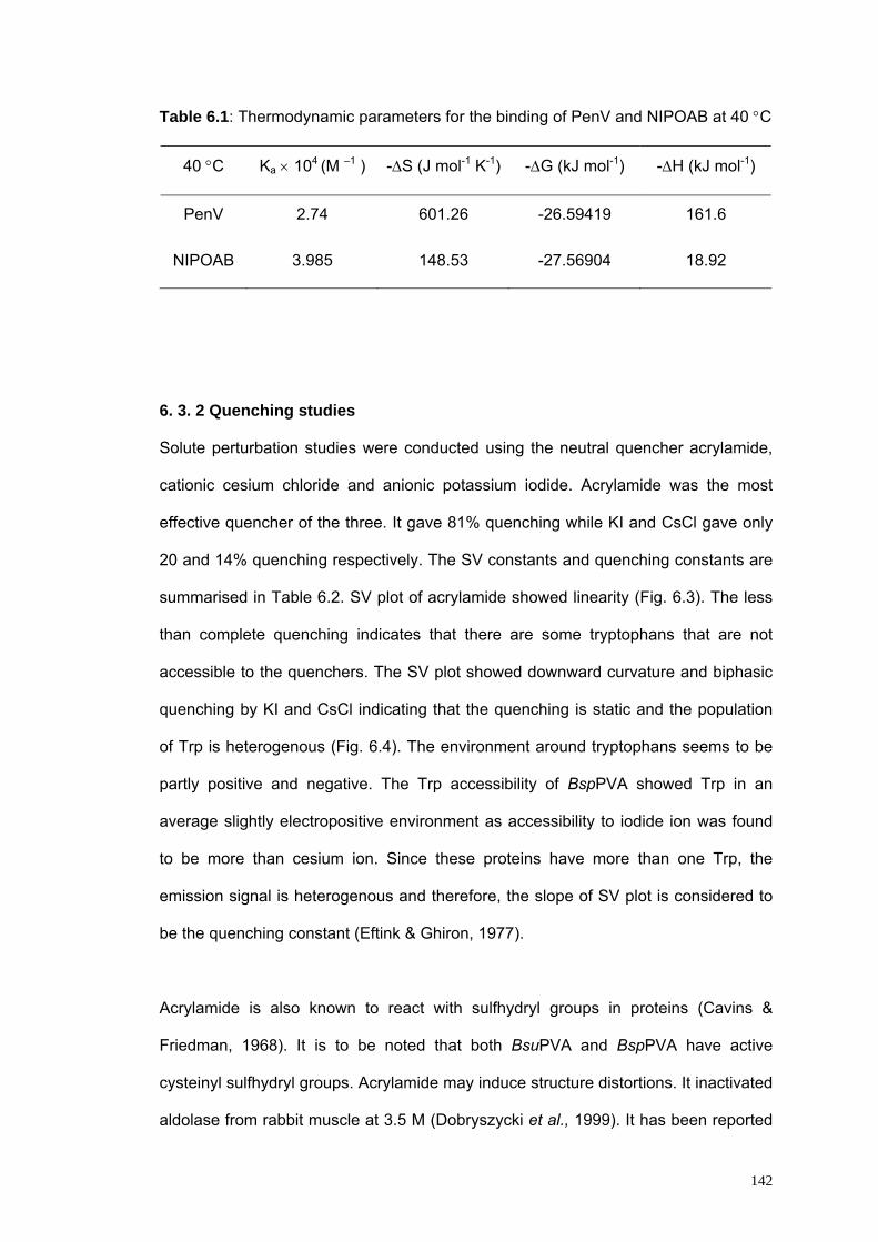
142
Table 6.1: Thermodynamic parameters for the binding of PenV and NIPOAB at 40 °C
40 °C Ka × 104 (M –1 ) -ΔS (J mol-1 K-1) -ΔG (kJ mol-1) -ΔH (kJ mol-1)
PenV
NIPOAB
2.74
3.985
601.26
148.53
-26.59419
-27.56904
161.6
18.92
6. 3. 2 Quenching studies
Solute perturbation studies were conducted using the neutral quencher acrylamide,
cationic cesium chloride and anionic potassium iodide. Acrylamide was the most
effective quencher of the three. It gave 81% quenching while KI and CsCl gave only
20 and 14% quenching respectively. The SV constants and quenching constants are
summarised in Table 6.2. SV plot of acrylamide showed linearity (Fig. 6.3). The less
than complete quenching indicates that there are some tryptophans that are not
accessible to the quenchers. The SV plot showed downward curvature and biphasic
quenching by KI and CsCl indicating that the quenching is static and the population
of Trp is heterogenous (Fig. 6.4). The environment around tryptophans seems to be
partly positive and negative. The Trp accessibility of BspPVA showed Trp in an
average slightly electropositive environment as accessibility to iodide ion was found
to be more than cesium ion. Since these proteins have more than one Trp, the
emission signal is heterogenous and therefore, the slope of SV plot is considered to
be the quenching constant (Eftink & Ghiron, 1977).
Acrylamide is also known to react with sulfhydryl groups in proteins (Cavins &
Friedman, 1968). It is to be noted that both BsuPVA and BspPVA have active
cysteinyl sulfhydryl groups. Acrylamide may induce structure distortions. It inactivated
aldolase from rabbit muscle at 3.5 M (Dobryszycki et al., 1999). It has been reported

143
that acrylamide denatures proteins at higher temperatures in which case, the SV plot
will be upper curving (Eftink & Ghiron, 1977). However, such distortions were not
observed in PVA in the concentration range of acrylamide used as evidenced by
absence of any red shift in the fluorescence spectrum.
Table 6. 2: Quenching by different solutes
Quencher Ksv KQ fa % quenching
Acrylamide
KI
CsCl
12.87
1.47
0.51
17.43
24.7
12.2
0.81
0.2
0.14
81
20
14
Fig. 6. 3. : SV and modified SV plot for quenching of BspPVA by acrylamide.
0.00 0.01 0.02 0.03 0.04
1.0
1.1
1.2
1.3
1.4
1.5
Fo/F
c
Acrylamide (M)0 25 50 75 100 125 150 175 200 225
0
2
4
6
8
10
12
14
16
Fo/(F
o-Fc
)
1/[acrylamide]

144
Fig. 6. 4 : SV and modified SV plot for quenching of BspPVA by A) KI and B) CsCl.
In contrast to BsuPVA, all the Trp in BspPVA lie in hydrophobic environments with
low mobility. Efficiency of acrylamide towards quenching of Trp in native BspPVA is
less as compared to BsuPVA where it is complete. The association constants of the
substrates are higher for BspPVA. This also follows the fact that the Km of this
enzyme (10 mM) is much lower than BsuPVA (40 mM). This shows that BspPVA is a
more efficient enzyme in PenV hydrolysis than BsuPVA. The conformational stability
of BspPVA is described in the next chapter.
0.02 0.03 0.04 0.05 0.06 0.07 0.08
1.08
1.10
1.12
1.14
1.16Fig. 6.4(A)
[KI] (M)
Fo/F
c
0 5 10 15 20 25 30 35 40 45 504
6
8
10
12
14
Fo/ (
Fo-F
c)
1/[KI]
0.00 0.02 0.04 0.06 0.08 0.10 0.12 0.141.02
1.04
1.06
1.08
1.10Fig. 6.4(B)
Fo/F
c
[CsCl] M
0 2 4 6 8 10 12 14 16 18 20 226
8
10
12
14
16
18
20
22Fo
/(Fo-
Fc)
1/ [CsCl]

145
CHAPTER – VII
COMPARATIVE STUDIES ON RELATED HYDROLASES: Part–II
Comparison with the Conformational Stability of BspPVA
Summary
Tryptophan fluorescence intensity decreased with increase in temperature. Both
decrease in intensity and PVA activity were partially reversible. After cooling, they
were 85% and 90% of the initial values respectively. There was decrease in ellipticity
values with increase in temperature as seen from the far UV CD spectra pointing to
disturbances in secondary structure. Upon incubation in acidic pH range 1-4, the
protein showed presence of molten globule as indicated by ANS binding. This range
also had good PVA activity. Far UV CD showed loss of secondary structure at
extreme acidic and alkaline pH. Increasing concentrations of Gdn HCl resulted in
decreased fluorescence intensity with decrease in 333/355 ratio. There was
substantial loss of activity (80%) at 1 M Gdn HCl. Secondary structure losses started
from 0.75 M and at 6 M Gdn HCl, there was complete loss of secondary structure.
7. 1. Introduction
BsuPVA has been studied in comparison with other related hydrolases based on
structure, conformation and biochemical characteristics. BspPVA is the other PVA
whose crystal structure is available. The biochemical characteristics of BspPVA have
been already reported. However, conformational changes of the enzyme in solution
have not been studied. This enzyme being significantly similar to BsuPVA,
comparative studies on their behaviour in solution will help elucidate structural basis
of observations. This chapter reports conformational stability of BspPVA as a function
of temperature, pH and Gdn HCl concentration. This is the first instance of such
studies on the choloylglycine hydrolase family of the Ntn hydrolase superfamily.

146
7. 2. Materials and Methods
7. 2. 1. Materials
The materials and preparation of solutions are described in chapter 5 under Materials
and Methods section 4. 2. 1.
7. 2. 2. Preparation of protein samples
Production and purification of the protein, enzyme assay and protein estimation were
carried out as described in Chapter 6. The recombinant cells were grown in Luria
Bertani medium containing 50 μg ml-1 ampicillin. Expression of the recombinant
protein was induced with 1 mM IPTG. Protein was purified on phenylsepharose
column. Pure protein samples were dialyzed against 100 mM NaCl and centrifuged
(10 000g, 10 min) immediately before use, to remove any precipitate.
7. 2. 3. Assay of Penicillin V acylase activity
Assay for penicillin acylase activity against PenV was carried out at pH 5.8 at 40 °C
as described earlier.
7. 2. 4. Fluorescence measurements
Steady-state fluorescence measurements were made on a Perkin Elmer LS 50 B
fluorimeter as previously described (Chapter 4).
7. 2. 5. Hydrophobic dye binding studies
ANS binding studies were carried out with samples treated as in chapter 5 section 5.
2. 4. and were excited at wavelength 375 nm and the emission spectra were
recorded from 400 to 550 nm.

147
7. 2. 6. Circular dichroism
Far and near UV CD spectra were recorded in a Jasco-J715 spectropolarimeter at
room temperature as outlined in Chapter 5.
7. 2. 7. Light scattering studies
Aggregation of the protein was studied under various conditions using Rayleigh light
scattering. The excitation and emission wavelengths were fixed at 400 nm and
scattering was recorded for 30 s in 1 s intervals.
7. 2. 8. Effect of temperature
The stability of the enzyme at different temperatures was studied by holding 2 ml
samples of protein in 50 mM potassium phosphate buffer pH 7 at required
temperature with the help of a variable circulating cryobath and the spectra collected
from 300 to 400 nm. Samples were slowly heated and spectra collected at different
temperatures. Samples were allowed to cool and the spectra were recorded again.
Similarly treated enzyme samples were tested for PVA activity as outlined in 2.1.
ANS binding studies were carried out as described above. Far and near UV circular
dichroism spectra were recorded for selected samples after heat treatment as well as
cooling. The effect of temperature was studied for every 5 °C rise and decrease in
temperature from 25 to 65 °C and back to 25 °C at pH 7.
7. 2. 9. Effect of pH
PVA was incubated at room temperature in the following buffers: glycine-HCl in the
pH range 1.0 to 3.0, citrate phosphate in the pH range 4.0 to 6.0, potassium
phosphate for pH 7.0, Tris from 8.0 to 9.0 and glycine-NaOH from 10.0 to 12.0. after
incubation for 2 h in 100 mM concentration of buffers, spectra were recorded as
previously outlined at 30 °C. ANS binding was also studied as previously described.
These samples, incubated in different pH buffers, were adjusted to pH 7 and the

148
spectra and ANS binding were studied at 30 °C. CD spectra were collected for
selected samples. All samples were parallely checked for PVA activity and
aggregation.
7. 2. 10. Effect of guanidine hydrochloride
There are various techniques to monitor unfolding of proteins- Ultraviolet difference
spectroscopy, CD, optical rotation, fluorimetry and NMR. Denaturation using urea or
guanidine hydrochloride can be used to estimate the conformational stability ΔGD of
the protein. Denaturation curves for different proteins obtained under identical
conditions are helpful in measuring differences in their conformational stabilities
because of aminoacid sequence or chemical modification inspite of significant
similarities between them, especially, in relation to three-dimensional structure. From
the denaturation curves, it is possible to unravel the mechanism of folding and to
predict whether the protein is made of single domain or if it is made up of more than
one domain. The extent of reversibility of Gdn HCl denaturation is dependent on time
for which the protein is allowed to equilibrate in the denaturant and pH at which the
denaturation and refolding are conducted (Pace, 1986).
Most globular proteins unfold in a two-state mechanism:
F (folded state) ↔ U (unfolded state)
The spectroscopic property at each concentration used for the calculation, y, is the
ratio of fluorescence intensities at emission λmax of native and denatured protein.
Where yu and yf are values of parameters obtained at unfolded and folded by
extrapolation from linear portions of unfolding curve at low and high unfolding
concentrations.
For a two-state mechanism,
ff + fu = 1

149
Where ff and fu denote fraction of the protein in folded and unfolded conformations
respectively.
y = yfff + yufu
Therefore,
fu = (y-yf) / (yu – yf)
ff = (yu-y)/(yu-yf)
The equilibrium constant Ku is calculated using the relation
Ku = e -ΔGu / RT = fu/ff = (y-yf)/ (yu-y)
Where ΔGu is the energy of unfolding.
In linear extrapolation, the simplest method of estimating ΔGuH2O (energy of unfolding
in the absence of denaturant), linear dependence of ΔGu on denaturant concentration
observed in transition region is assumed to continue to zero concentration.
ΔGu = ΔGuH2O – m [Denaturant]
The difference in conformational stability is estimated by multiplying the difference
between the D1/2 values by m, where m is the slope of the
ΔGu = ΔGuH2O – m[Denaturant]
plot and D1/2 is the denaturant concentration where unfolding is halfway.
PVA was incubated for 4 h with different concentrations of Gdn HCl at room
temperature. For renaturation studies, samples were incubated in different
concentrations of Gdn HCl in a total volume of 100 μl and renatured by diluting out
Gdn HCl to slightly different residual concentrations by making up the solution to 2 ml
and equilibrating for 24 h. Fluorescence spectra, CD spectra and ANS binding were
studied for both denatured and renatured samples. Light scattering studies were also
carried out. Aggregation was checked at 400 nm. PVA activity assays were carried
out on all the samples. The methods used are described in detail in chapter 4.

150
7. 3. Results and Discussion
7. 3. 1. Temperature stability of BspPVA
The fluorescence intensity at 333 nm decreased with increase in temperature (Fig. 7.
1). The change in intensity was gradual and linear in the temperature range tested.
At 70 °C, the intensity was 63% as that of the enzyme at 25 °C. As temperature was
increased, the 333/350 emission intensity ratio, which is indicative of folded
conformation, decreased marginally (0.03). On gradual cooling from 70 oC to 25 oC,
the intensity again increased, but the final intensity was 85% of the initial intensity.
This might indicate some minor changes in the Trp environment in the enzyme
structure or thermal inactivation of fluorophores though loss in hydrolase activity was
not documented. Upon cooling from 70 oC to room temperature and then assaying,
there was a loss of 10% activity underlining the minor changes during cooling.
Far UV CD data showed that ellipticity was gradually lost during heating at pH 7 (Fig.
7. 2). At 45 °C, there was significant transition at 218 and 222 nm. The negative
ellipticity peak at 218 nm disappeared at 50 °C. On gradually cooling the protein
again, ellipticity increased but the final ellipticity was 10% lesser than that of the
native protein. This trend is similar to fluorescence where intensity after cooling was
less than the initial intensity. The secondary structure showed a change in alpha helix
content upon cooling; the negative peak became pronounced at 222 nm.
There was no evidence of aggregation at any of the temperatures tested either upon
heating or cooling, though there was marginal increase in scattering upon heating, as
indicated by Rayleigh light scattering studies. There was no ANS binding indicating
that there is no exposure of surface hydrophobic pockets.

151
Fig. 7. 1: Intensity at emission maxima as a function of temperature upon heating to
70 oC and cooling back to room temperature.
Fig. 7. 2: Far UV CD: Fraction unfolded calculated using ellipticity at 222 nm at
different temperatures.
7. 3. 2. Effect of pH variation
The effect of pH was studied from pH 1 to 12 in 100 mM buffers at 30 °C. To
investigate the functional stability of PVA at different pH, we determined its acylase
activity on PenV at 40 °C as a function of pH. The enzyme retains more than 80% of
its activity over the whole range of pH from 1 to 12 after 4 h of incubation. On
readjustment of pH to 7, the enzyme on the acidic side (pH 2 - 6) regains activity
20 30 40 50 60 70450
500
550
600
650
700
750
800
Inte
nsity
Temperature (o C)
30 35 40 45 50 55 60 65
0.0
0.2
0.4
0.6
0.8
1.0
f u
Temperature (oC)

152
whereas in the alkaline range does not regain full activity. This indicates that there is
a slight irreversible modification of the protein in alkaline pH. The activity profile of
PVA is shown in (Fig. 7. 3). The structural changes under these conditions were
studied using fluorescence spectroscopy, CD spectroscopy and ANS binding.
Emission spectrum collected in wavelength range 300 - 400 nm upon excitation at
280 nm showed that the intensity was minimum at pH 5. It increased towards the
acidic range. On the other side towards neutral and alkaline pH, it increased till pH 7
and there was no significant change thereafter.
Fig. 7. 3: Activity profile of BspPVA after incubation at different pH (∏) and after
readjusting the pH to 7 (█).
There was significant ANS binding at the acidic pH range in BspPVA at 30 °C (Fig. 7.
4). The binding was maximum at pH 1 and decreased upon increase in pH till 4,
where there was very little ANS fluorescence. The λmax was 488 nm for pH 1-2 which
shifted to 495 at pH 4.
1 2 3 4 5 6 7 8 9 10 11 120
20
40
60
80
100
120
Per
cent
Act
ivity
pH

153
Fig. 7. 4: ANS binding of BspPVA at acidic pH: −···− 1 ⎯⎯ 2 −−−3 ---- 4
Enzyme treated at pH 3 exhibited preserved secondary structure as seen from the
CD data. At pH 4, there was significant loss of secondary structure and negligible
binding of ANS showing lack of molten globule species, but activity was found to be
maximum indicating that exposure of hydrophobic patches alone does not affect the
activity.
There were some interesting observations made in the protein structure at different
pH, which are described below. PVA incubated at pH 5 showed considerable loss in
secondary structure. This is also where fluorescence intensity and 333/355 ratio was
low for the protein. Since the isoelectric point of the protein is 5.2, the protein is
unstable at this pH. At pH 6, there was shift in the ellipticity mimimum to 225 nm from
218 nm at pH 7. The protein showed maximum negative ellipticity at 218 nm at pH 7
in far UV CD spectrum. At pH 9, where the enzyme showed least activity, far UV CD
spectrum showed minimum at 221 nm indicating rearrangement in the secondary
structure elements (Fig. 7.5). There was a dramatic decrease in structural features at
pH 11 as compared to pH 9. This may be because pH 11 is strongly alkaline and
400 420 440 460 480 500 520 5400
10
20
30
40
50
60
70
4
3
2
1
Fluo
resc
ence
inte
nsity
(arb
itrar
y un
its )
Wavelength (nm)

154
might lead to charge repulsion between delicately balanced electrostatic charges
leading to instability.
Fig. 7. 5: Far UV CD spectrum of BspPVA at different pH
From the above results it is seen that the fluorescence and CD data are very different
from the activity trend of the enzyme for the hydrolysis of PenV. For instance, loss of
secondary structure does not lead to loss in activity at pH 1 and 3. At pH 1, though
there was significant activity, fluorescence intensity was minimum. Therefore, the
active state of the enzyme is dependent on the protonated-deprotonated forms of
active residues rather than the overall secondary structure or tertiary conformation of
the protein. There was no aggregation at any of the pH tested at 30 oC. The protein
also did not show any aggregation till 65 °C.
Fluorescence spectra showed that apart from change in the intensity of the protein in
the acidic and alkaline pH range, there is no major change in the Trp environment of
the protein (Data not shown). It is well-known that Trp fluorescence intensity
decreases on protonation of water or neighbouring protonated acidic groups. In the
zwitterionic states, aspartic and glutamic acid act as dynamic quenchers. Protonation
of imidazole rings of histidine residues with a pKa of 6.5 may also contribute to
reduction in fluorescence intensity (Bushueva et al., 1974). In strong alkaline
210 220 230 240
-8
-6
-4
-2
0
2
4
73
1
5
Elli
ptic
ity
Wavelength (nm)210 220 230 240
-8
-6
-4
-2
0
2
4
97
11
Elli
ptic
ity
Wavelength (nm)

155
conditions, deprotonation of side chain amino group of residues can occur. The ε-
amino group of lysine, guanidino group of arginine and phenol group of tyrosine are
also known to quench the fluorescence of Trp residues in their deprotonated form
(Bushueva, 1975, Steinberg, 1971) which explains decrease in intensity at extreme
alkaline pH.
Far UV CD spectra of the protein at pH 7 shows characteristic minima of αβ proteins.
The negative ellipticity at 222 nm is maximum at pH 7 and gradually decreases when
moving towards either side of the pH range. Though there is decrease in ellipticity,
the pH range 1 and 9 showed significant conservation of secondary structure (Fig.
7.5). Comparison of the near UV CD spectra of PVA at pH 7, shows that at pH 1 the
structure completely collapses. Rayleigh light scattering studies showed no
aggregation at any of the pH or temperatures from 30 oC to 65 °C. The ANS binding
properties of the protein was then checked at different pH. PVA strongly bound ANS
at acidic pH 1-4 with resultant increase in fluorescence intensity along with a blue
shift in the λmax from 520 to 480 nm, indicating exposure of hydrophobic patches on
the surface (Fig. 7.4). The binding was maximum at pH 1-2. There was no ANS
binding in the pH range 5 to 12. The binding was lost after readjustment of pH to 7
showing reversibility of structural changes at this pH. These results indicate the
presence of molten globule state of pVA in pH 1-4.
7. 3. 3. Effect of Guanidine hydrochloride
BspPVA underwent progressive unfolding when incubated with increasing
concentrations of Gdn HCl. During unfolding, the fluorescence intensity decreased
gradually (Fig. 7. 6). Since Trp fluorescence can be quenched by water, it decreased
upon unfolding of BspPVA as buried Trps started getting exposed to aqueous
environment. Polar groups of protein themselves are also capable of quenching to a

156
certain extent. The 333/355 ratio of the native protein which was 1.27 in the native
protein, decreased to 0.76 at 6 M Gdn HCl. The emission λmax underwent a
significant shift from 333 nm in the native protein to 355 nm at a Gdn HCl
concentration of 6 M (Fig. 7. 7).
Fig. 7. 6: Fluorescence emission spectrum of BspPVA at different Gdn HCl
molarities.
Fig. 7. 7: Wavelength of emission maxima at different Gdn HCl molarities.
Fig. 7. 8: Effect of Gdn HCl on BspPVA. ---■---inactive fraction —◊—unfolded
fraction calculated from F 333/350 ratio —▲— unfolded fraction calculated from
ellipticity at 222 nm.
0 1 2 3 4 5 6
0.0
0.2
0.4
0.6
0.8
1.0
0.0
0.2
0.4
0.6
0.8
1.0
f inac
t
Gdn HCl (M)
fu
300 320 340 360 380 400
100
200
300
400
500
600
700
Flu
ores
cenc
e in
tens
ity
6 M
4 M
2 M
1 Mnative
Wavelength (nm)
0 1 2 3 4 5 6
335
340
345
350
355
Emis
sion
max
imum
(nm
)
Gdn HCl (M)

157
Hydrolase activity of PVA towards PenV decreased sharply with increasing
concentrations of Gdn HCl. At 0.5 M Gdn HCl, the secondary and tertiary structures
were intact but there was loss of 65% activity. At 1 M Gdn HCl, only 16% of the
original activity was retained although there were no major disruptions in secondary
and tertiary structures. This could be attributed to charged nature of Gdn HCl in
solution which enables it to interact with active site residues of PVA. Positively
charged guanidinium ion can react with negatively charged active residues. It may be
remembered that BspPVA has an aspartic acid at the active site. At 3 M, there was
complete loss of activity with 60% loss in secondary structure and negligible loss in
tertiary structure. Secondary structure decreased gradually whereas there was a
partial unfolding of the protein at 4 M Gdn HCl. Complete unfolding occured only
above 4 M Gdn HCl whereas there was complete loss of activity at a concentration of
Gdn HCl as less as 3 M. This showed that loss of activity at lower concentrations of
Gdn HCl was independent of structural disturbances and at higher concentrations,
the loss was due to modification of secondary structure (Fig. 7. 8). Thus, BspPVA
lost enzyme activity much before it lost secondary or tertiary structures.
Denaturation curves were plotted with fraction of the protein inactivated (finact) and
fraction unfolded (fu) as a function of Gdn HCl concentration (Fig. 7.8). A plot of the
ratio of fluorescence intensities at emission λmax of native (333 nm) and denatured
(355 nm) protein against Gdn HCl molarity was constructed and from the graph a
two-state mechanism of unfolding was assumed. At each concentration, fraction of
the protein in folded and unfolded forms were calculated using equations given in
materials and methods section of this chapter. Free energy of unfolding in the
transition range was calculated from these graphs and plotted as a function of Gdn
HCl molarity from which the difference in free energy between the folded and
unfolded states was determined (Fig. 7.9). The value of ΔGDH2O was found to be

158
12.29 kJ/ mol. Concentration of the denaturant required to unfold 50% of the protein
molecules, D1/2 was found to be 4.44 M at pH 7 at room temperature.
Fig. 7. 9: Free energy of unfolding as a function of Gdn HCl concentration
Upon renaturation, the enzyme regained fluorescence intensity (Fig. 7.10) and λmax
shifted back to 333 nm indicating that unfolding is completely reversible. There was
not much recovery of activity after refolding the enzyme treated with different
concentrations of Gdn HCl. There was no change in ANS binding indicating absence
of exposure of any hydrophobic patches upon unfolding or refolding. Neither was
aggregation observed.
Fig. 7. 10: F 333/350 of BspPVA as a function of Gdn HCl ——♦——after unfolding
and − –■− −refolding
0 1 2 3 4 5-10
-5
0
5
10
15
f inact
fudG
(kJ/
mol
)
Gdn HCl (M)
0 1 2 3 4 5 60.7
0.8
0.9
1.0
1.1
1.2
1.3
333/
355
ratio
Gdn HCl (M)

159
Understanding the protein folding pathways requires structural characterization of
both native structures and stable intermediate states. The crystal structure of the
stable PVA from B. sphaericus was first solved in our laboratory. However, unstable,
non-native intermediate states are less amenable to structure elucidation. This
chapter has reported studies on solution dynamics in different conditions.
During our investigations using spectroscopic techniques, we found that PVA exists
in a molten globule state in acidic pH conditions. Many proteins can exist in stable
conformations that are neither fully folded nor unfolded. One of these states is the
molten globule state, characterized by partly folded species with high proportion of
native-like secondary structure but lacking persistent tertiary interactions. ANS
binding is generally observed in molten globule states where hydrophobic regions
that are usually buried in the native state are exposed to the polar medium due to
flexibility or fluidity of the state (Semisotnov et al., 1991). Forming of molten globule
state is a general trend in globular proteins (Ohgushi and Wada, 1983). Most proteins
are required to undergo conformational changes for functioning and molten globule
states sometimes form intermediary stages during execution of a function. Typical
characteristics of molten globule are observed for BspPVA at pH 1-3, viz., major
retention of secondary structure, substantial distortion of tertiary structure and
exposure of hydrophobic patches to the solvent.
Bychkova et al (1988) have hypothesised that the molten globule state is involved in
the translocation of proteins across membranes. During membrane insertion of the
pore-forming domain of colicin A, a bacteriocin that depolarizes the cytoplasmic
membrane of sensitive cells, the acidic molten globular state could be an
intermediate (van der Goot, et al., 1991). The steroidogenic acute regulatory protein
is believed to exist as a molten globule at reduced pH and the partially unfolded state
may be critical to its biological action of moving cholesterol from outer to inner

160
membrane of adrenal and gonadal mitochondria for synthesis of steroid hormones
(Bose et al., 1999). The groEL chaperone stabilizes proteins, during their folding, in
a conformation resembling the 'molten globule' state (Martin et al., 1991). Protein-
receptor interactions involve certain conformational changes in the molecules
involved to fit each other. For example both insulin and its plasma membrane
receptor (Nakagawa and Tager, 1993), antigenic peptides and immunoglobulins (Rini
et al., 1992), cyclosporin A and cyclophilin (Weber et al., 1991) undergo small
adjustments in their structure to accommodate each other. In a demonstration of
structure-function relationship, the native alpha-lactalbumin structure present in milk
provides nourishment and the multimeric molten globule form (MAL) in acid
conditions fights tumor. Similarly, retinol-binding protein transports retinol in plasma
as a complex with transthyretin. Under acidic conditions it undergoes transformation
to molten globule which releases bound retinol (Bychkova, 1992). Molten globules
are natural conformational states in protein folding process. However, enzymes
rarely occur in an active molten globule form. PVA is now shown here to exist in
molten globule form in acidic conditions which retained 80% of the enzyme activity.
Gdn-HCl and temperature induced molten globules have also been reported for
some proteins. Creatine kinase was reported to exhibit an inactive molten globule
form when denatured with 0.8 M Gdn-HCl. A temperature induced molten globule in
the botulinum toxin has been reported (Kukreja and Singh, 2005). However, when
tested under different conditions of Gdn-HCl during unfolding or refolding and at
different temperatures, PVA did not exhibit any molten globule form or aggregates.
Gdn-HCl denaturation studies show that the protein is relatively unstable. It is widely
acknowledged that due to high flexibility at the active sites compared to the rest of
the protein, most proteins are inactivated before conformational changes occur. In
this case it could also be attributed to charged nature of Gdn-HCl in solution, which
enables it to interact with active site residues of PVA. The positively charged

161
guanidine group can react with negatively charged residues. Interestingly, PVA has
an aspartic acid at the active site. At low concentrations, Gdn-HCl is known to confer
effective charge shielding. The active site seems to involve hydrophobic and nonionic
interactions (Monera et al., 1994).
PVA is commercially used in the manufacture of semi-synthetic penicillins. Search is
on for an enzyme that is more efficient in terms of yield and may function as
cephalosporin acylase since semi-synthetic cephalosporin production is a
complicated multi-step process. The main advantage of PVA is the operability at
acidic pH. Denaturation studies indicate that the core of PVA, an Ntn hydrolase,
which is known to be a compact αββα-layered structure, is well-shielded. The
protonation-deprotonation of active site residues in the core overrides the overall
changes in secondary or tertiary conformations of the protein, as far as activity is
concerned. Though the functional significance of the molten globule form in vivo is
unclear, these observations have implications on the minimal structural requirement
of the protein for activity.
Thus, PVA from B. sphaericus is relatively stable at high temperature and extremes
of pH. It exhibits an active molten globule state in the acidic pH range 1-3. It can be a
good catalyst in the production of 6APA as it gives a lot of flexibility in culture and
extraction conditions because of its functional stability over a wide range of
conditions. Our studies indicate that together with crystal structure, these studies can
be a powerful tool to enhance the utility of such industrially important enzymes and
that such studies can be used to screen and select candidates for such applications.
Results presented in this chapter indicate that BspPVA is comparatively more stable
in the presence of Gdn HCl and in acidic pH and at high temperatures as compared
to BsuPVA. Results obtained in various studies described in this as well as in the
previous chapters are consolidated in the next chapter.

162
CHAPTER – VIII
COMPARATIVE STUDIES ON RELATED HYDROLASES: Part - III
Structure, Function and Evolution
Summary
Comparison of BspPVA and BsuPVA shows that the former is a superior enzyme in
terms of affinity and association constants for the substrates tested. It is more
efficient in tolerating extreme temperatures and pH changes. The conformational
stability in the presence of chaotrope is also better. This observation helps us to
identify factors that make a protein a better PVA. The overall structure and the active
site of the two enzymes superpose particularly well, pointing to minor critical changes
in residues that lead to change in substrate specificity and efficiency. This throws
open avenues for modification experiments to tailor a given protein to our need. The
substrate cross reactivity also emphasises the evolutionary relatedness of the
enzymes. Although we expected BsuPVA to possess CBH activity, it could not be
established with purified protein.
8.1 Introduction
The gene of YxeI was originally sequenced as part of an international project for the
sequencing of the entire Bacillus subtilis genome. The gene yxeI lies in the region of
DNA of the bacterium B. subtilis occurring between the iol and hut operons and has
been reported to code for a hypothetical protein (Yoshida et al., 1995). The open
reading frame (ORF) is annotated as a hypothetical protein /choloylglycine hydrolase
and a highly conserved putative penicillin amidase, based on sequence. Homology
with other bacterial conjugated bile acid hydrolases (CBH) and PVA is high (Tanaka
et al., 2000). A BLAST search of the sequence against various databases revealed
that the protein had significant levels of homology with both BspPVA and CBH from

163
various sources including Clostridium perfringens and Bifidobacterium longum,
whose crystal structures have been reported, and a number of hypothetical proteins
from different organisms.
The physiological role of penicillin acylase in host organisms is still not clearly
established, despite years of research. Conjugated bile acid hydrolases have been
thought to help the host bacteria present in the mammalian intestinal flora
deconjugate the conjugated bile salts, thereby detoxifying them. Both the enzymes,
however, have commercial applications- penicillin acylases are used in the industrial
production of semi-synthetic penicillins and CBHs are used clinically for assaying
levels of bile acids in biological fluids. Interest in YxeI arose primarily because of the
similarity with BsuPVA. Though recombinant expression of PVA was tried in B.
subtilis, there is no report of any indigenous PVA. The present chapter describes the
identification of PVA using sequence information, comparison with related sequences
along with comparison of BsuPVA with BspPVA.
Apart from helping to trace relatedness between the enzymes, which might lead to
establishing a physiological role for PVAs, this comparative study also sheds light on
the two PVAs from industrial stand point. This study explains the rationale behind
superior PVA activity of BspPVA, and thus might help to pick out residues that could
be subjected to site-directed mutagenesis to extend the substrate specificity of PVAs
to cephalosporins. Using information from genomic data, YxeI has been
characterized as a PVA. Observations from kinetic studies and comparison of similar
studies conducted on the two proteins are discussed in this chapter.

164
8.2 Material and methods
8.2.1 Homology studies with other choloylglycine hydrolases
Protein Sequence analysis of YxeI was performed using BLAST (Altschul et al.,
1997) at the Swiss Institute of Bioinformatics site (http://au.expasy.org/tools/blast/)
against UniProt knowledgebase using default settings. A blastp search was also
done at the National Center for Biotechnology Information site (http://www.ncbi.nlm.
nih.gov/BLAST/) against a nonredundant database. After selecting sequences with
significant e values, redundancy was decreased using an upper cutoff of 85%
sequence identity and the resulting sequences were screened and the N-terminal
stretch of sequences that was not part of the alignment were removed from the
analysis. All sequences began with cysteine at the N-terminal - otherwise they were
removed. Multiple sequence alignment of the selected sequences was generated
using the program ClustalW (www.ebi.ac.uk/clustalw) (Thompson, et al 1994).
Phylogenetic trees were constructed using TreeView (Page, 1996) or Molecular
Evolutionary Genetic Analysis (Kumar et al., 2004).
8.2.2 Inhibition kinetics
Kinetic studies were carried out by incubating the enzyme in different substrate
concentrations in the presence of different inhibitors. The assays were carried out as
described in chapter 2. Inhibitors were added to the mixture before the reaction was
initiated with the addition of enzyme. The Ki vaules were calculated using LB plots.
8.2.3 Structure superpositions
Structural coordinates of the proteins were superposed using LSQKAB or superpose
implemented in CCP4 and also manually checked using QUANTA (Accelrys).
8.2.4 Hydropathicity plot

165
A hydropathicity plot of the protein (Kyte and Doolittle, 1982) was constructed using
ExPASy (Swiss Institute of Bioinformatics). Window size was set at 9 amino acids,
when strong negative peaks indicate possible surface regions of globular proteins.
8.3 Results and Discussion
8.3.1 Sequence similarity to other PVAs and CBHs
Sequence homology studies indicate that the protein BsuPVA is very similar to PAs
and CBHs from different organisms. It shared a 40% sequence identity with BspPVA
(Swissprot Accession No.P12256; PDB code 3PVA) and 39% with CBH from
Clostridium perfringens (Swissprot Acc No. P54965; (PDB code 2BJF) whose crystal
structures have been solved. It shared 32% identity with the well-characterised
Lactobacillus plantarum CBH (Swissprot Accession No. Q06115) and 27% sequence
similarity with a hypothetical protein A284L (Swissprot Accession No. P54965) from
Paramecium bursaria Chlorella virus 1, which could also be a PVA /CBH. It has all
the conserved residues of the family except for Asp20.
Phylogenetic analysis shows evolutionary links between CBH and PVA. A tree was
constructed with the sequences found by BLAST as described in materials and
methods section of this chapter. All members of the tree have the residues that are
thought to be essential in PVA /CBH activity. Cys1, Arg17, Asn70, His141, Asp146,
Asn173, Pro175 and Arg226 are conserved; a Glu substitutes Asp20 in some
sequences. Another catalytically implicated residue Tyr80 varies in the sequences.
(Residues are numbered starting from N-terminal cysteine of BsuPVA.) Based on
these observations, we have identified these hypothetical/ putative proteins that
could have choloylglycine hydrolase or penicillin acylase activity. Many of these
sequences, including that of B. subtilis, are part of genome sequencing projects.
Once these proteins are fully characterised, the evolution of these enzymes can be
better understood.

166
Fig. 8.1. A dendrogram showing relationships between proteins that are homologous
to PVA of B. subtilis as found by the BLAST program. The distance of the branches
corresponds to aminoacid substitutions per site. This tree was constructed using
TREEVIEW.
The source of the proteins and their data bank accession numbers are: YXEI
(Penicillin V acylase from Bacillus subtilis, Swissprot accession number P54948),
LACLA (Lactococcus lactis, Trembl Acc no. Q9CGG1), GLOVI (Gloeobacter
violaceus, Trembl Acc. No. Q7NNP2), BACTN (Bacteroides thetaiotaomicron, Trembl
Acc. No. Q8A600), DESPS (Desulfotalea psychrophila, Trembl Acc. No. Q6ARU2),
BACFN (Bacteroides fragilis, Trembl Acc. No. Q5L9F8), SYNP6 (Synechococcus sp.,
Trembl Acc. No. Q5N0N8), RHILO (Rhizobium loti, Trembl Acc. No. Q983W5),
ACIAD (Acinetobacter sp., Trembl Acc. No. Q6FAR4), ERWCT (Erwinia carotovora,
Trembl Acc. No. Q6D291), PSEF5 (Pseudomonas fluorescens, Trembl Acc. No.
Q4KCA8), VIBF1 (Vibrio fischeri, Trembl Acc. No. Q5DZK1), METAC
(Methanosarcina acetivorans, Trembl Acc. No. Q8TSP0), RICFE (Rickettsia azadi,
Trembl Acc. No. Q4UKE3), RHOBA (Rhodopirellula baltica, Trembl Acc. No.
Q7TU08), SYNY3 (Synechocystis sp., Trembl Acc. No. P72765), VIBCH (Vibrio
cholerae, Trembl Acc. No. Q9KL70), 9CYAN (Lyngbya majuscula, Trembl Acc. No.
Q6E7I9), BACLI (Bacillus licheniformis, GenBank Acc. No. 52005709), BACTH
0.1
LISMO BACTH
YXEI BACSU BACLI
PAC BACSH OENOE
LACLA2
LACPL4 LACLA
OENOE2LACPL
LACPL2
STAEPSTAAUSTASA
STAHACLOPE
LACPL3CLOPE2LISMO2
ENTFA ENTFA2 LACAC2 LACJO
LACGA LACAC
LACJO2 LACGA2
BIFBI
BIFAN CLOPE3
ENTFA3
PEDPE VIBFI2
FRATT2
VIBF1 METAC Bdel
GLOVI RICFE SYNP6
BACTH2 ERWCT ACIAD
PSEF5 AGRTUBORPE RHILO
NITWI
RHOPA
BURVI
BACTN BACFN
9CYAN
VIBCH
DESPS DESPS3RHOBA
SYNY3FRATT VIBspPVA
BRUSU
DESPS2
PARCH
SALEN

167
(Bacillus thuringiensis, GenBank Acc. No. 49478365), LISMO (Listeria
monocytogenes, GenBank Acc. No. 16802490), OENOE (Oenococcus oeni,
GenBank Acc. No. 48864776), LACLA (Lactococcus lactis, GenBank Acc. No.
15673817), BACSH (Bacillus sphaericus, GenBank Acc. No. 129549), LACPL
(Lactobacillus plantarum, GenBank Acc. No. 28379706), CLOPE (Clostridium
perfringens, GenBank Acc. No. 18144368), STASA (Staphylococcus saprophyticus,
GenBank Acc. No. 73661455), STAEP (Staphylococcus epidermidis, GenBank Acc.
No. 27467268), LACPL (Lactobacillus plantarum, GenBank Acc. No 28377029)
STAAU (Staphylococcus aureus, GenBank Acc. No 57286722), BRUSU (Brucella
suis, GenBank Acc. No 23348317), STAHA (Staphylococcus haemolyticus, GenBank
Acc. No 70725298), ENTFA (Enterococcus faecium, GenBank Acc. No 46518306),
ENTFA (Enterococcus faecalis, GenBank Acc. No 29375146), LACPL (Lactobacillus
plantarum, GenBank Acc. No 28379847), LACGA (Lactobacillus gasseri, GenBank
Acc. No 12802353), LISMO (Listeria monocytogenes, GenBank Acc. No 16804106),
LACGA (Lactobacillus gasseri, GenBank Acc. No 23003120), LACJO (Lactobacillus
johnsonii, GenBank Acc. No 10732793), LACAC (Lactobacillus acidophilus,
GenBank Acc. No 58337369), DESPS (Desulfotalea psychrophila, GenBank Acc. No
50876071), CLOPE (Clostridium perfringens, GenBank Acc. No 60417955), VIBFI
(Vibrio fischeri, GenBank Acc. No 59713424), LACAC (Lactobacillus acidophilus,
GenBank Acc. No 58337197), LACJO (Lactobacillus johnsonii, GenBank Acc. No
42519073), BIFBI (Bifidobacteium bifidum, GenBank Acc. No 40074455), OENOE
(Oenococcus oeni, GenBank Acc. No 48865090), CLOPE (Clostridium perfringens,
GenBank Acc. No 18144753), BIFAN (Bifidobacterium animalis, GenBank Acc. No
46486762), FRATT (Francisella tularensis, GenBank Acc. No 56708296), PARCH
(Paramecium bursaria chlorella virus 1, GenBank Acc. No 9631852), ENTFA
(Enterococcus faecalis, GenBank Acc. No 29377466), LACPL (Lactobacillus
plantarum, GenBank Acc. No 28379105), PEDPE (Pediococcus pentosaceus,
GenBank Acc. No 48870357), FRATT (Francisella tularensis, GenBank Acc. No
56708187), RHOPA (Rhodopseudomonas palustris, GenBank Acc. No 39934948),
SALEN (Salmonella enterica, GenBank Acc. No 29141908), NITWI (Nitrobacter
winogradskyi, GenBank Acc. No 75675071), BORPE (Bordetella parapertussis,
GenBank Acc. No 33597843), Bdel (Bdellovibrio bacteriovorus, GenBank Acc. No
42522252), AGRTU (Agrobacterium tumefaciens, GenBank Acc. No 1793827),
VIBspPVA (Vibrio sp., GenBank Acc. No 75856500), BURVI (Burkholderia
vietnamiensis, GenBank Acc. No 67548680), DESPS (Desulfotalea psychrophila,
GenBank Acc. No 50875094), BACTH (Bacteroides thetaiotaomicron, GenBank Acc.
No 29338566).

168
Fig. 8.2. CLUSTALW multiple sequence alignment of known proteins belonging to
choloylglycine hydrolase family. BIFLO (Bifidobacterium longum CBH), LACPL
(Lactobacillus plantarum CBH), CLOPE (Clostridium perfringens CBH), BACSH
(Bacillus sphaericus PVA), BACSU (Bacillus subtilis PVA) and PBCV1 (Paramecium
bursaria chlorella virus 1 hypothetical protein). The data bank accession numbers are
given in the results section of the text. * indicates conserved residues. Putative
functional residues conserved in all the members of the dendrogram in Fig 8.1 are
highlighted.
BIFLO CTGVRFSDDEGNTYFGRNLDWSFSYGETILVTPRGYHYDTVFG--AGGKAKPNAVIGVGV 58 LACPL CTAITYQS--YNNYFGRNFDYEISYNEMVTITPR--KYPLVFRK-VENLDHHYAIIGITA 55 CLOPE CTGLALETKDGLHLFGRNMDIEYSFNQSIIFIPR--NFKCVNKSNKKELTTKYAVLGMGT 58 BACSH CSSLSIRTTDDKSLFARTMDFTMEPDSKVIIVPRNYGIRLLEKE-NVVINNSYAFVGMGS 59 BACSU CTSLTLETADRKHVLARTMDFAFQLGTEVILYPRRYSWN-SEAD-GRAHQTQYAFIGMG- 57 PBCV1 CSGLRIIADDGTVVVGRTLEFGEN----------------------ILKFKKFVNGNIRG 38 * * BIFLO VMADRPMYFDCANEHGLAIAGLNFPGYASFVHEPVEGTENVATFEFPLWVARNFDSVDEV 118 LACPL DVESYPLYYDAMNEKGLCIAGLNFAGYADYKKYDAD-KVNITPFELIPWLLGQFSSVREV 114 CLOPE IFDDYPTFADGMNEKGLGCAGLNFPVYVSYSKEDIEGKTNIPVYNFLLWVLANFSSVEEV 118 BACSH TDITSPVLYDGVNEKGLMGAMLYYATFATYADEPKKGTTGINPVYVISQVLGNCVTVDDV 119 BACSU RKLGNILFADGINESGLSCAALYFPGYAEYEKTIREDTVHIVPHEFVTWVLSVCQSLEDV 117 PBCV1 ISTPDGKLLDGMNEHGLVIFVFYFKNYAKYG-CPSQTKLNIKPTEVALFLLQKAKNVKDV 97 * ** ** * BIFLO EETLRNVTLVSQIVPGQQ-ESLLHWFIGDGK-RSIVVEQMADG-MHVHHDDVDVLTNQPT 175 LACPL KKNIQKLNLVNINFSEQLPLSPLHWLVADKQ-ESIVIESVKEG-LKIYDNPVGVLTNNPN 172 CLOPE KEALKNANIVDIPISENIPNTTLHWMISDITGKSIVVEQTKEK-LNVFDNNIGVLTNSPT 177 BACSH IEKLTSYTLLNEANIILGFAPPLHYTFTDASGESIVIEPDKTG-ITIHRKTIGVMTNSPG 178 BACSU KEKIRSLTIVEKKLDLLDTVLPLHWILSDRTGRNLTIEPRADG-LKVYDNQPGVMTNSPD 176 PBCV1 KAIAKTLNVIHESYPPFTETPPMHWLVTDASGKSIVLEPLGNGELTVFDNPMGIFTNAPT 157 * * * ** * BIFLO FDFHMENLRNYMCVSNEMAEPTSWGKASLTAWGAGVGMHGIPGDVSSPSRFVRVAYTNAH 235 LACPL FDYQLFNLNNYRALSNSTPQNSFSEKVDLDSYSRGMGGLGLPGDLSSMSRFVRAAFTKLN 232 CLOPE FDWHVANLNQYVGLRYNQVPEFKLGDQSLTALGQGTGLVGLPGDFTPASRFIRVAFLRDA 237 BACSH YEWHQTNLRAYIGVTPNPPQDIMMGDLDLTPFGQGAGGLGLPGDFTPSARFLRVAYWKKY 238 BACSU FIWHVTNLQQYTGIRPKQLESKEMGGLALSAFGQGLGTVGLPGDYTPPSRFVRAVYLKEH 236 PBCV1 FPEHMESAKKALEHLSPISDP--------NAASQGTGALGLPGDFSSASRFIRLAFFSQT 209 * * * *** ** * BIFLO YPQQNDEAANVSRLFHTLGSVQMVDGMAKMGDGQF-----ERTLFTSGYSSKTNTYYMNT 290 LACPL SLSMQTESGSVSQFFHILGSVEQQKGLCEVTDGKY-----EYTIYSSCCDMDKGVYYYRT 287 CLOPE MIKNDKDSIDLIEFFHILNNVAMVRGSTRTVEEKS-----DLTQYTSCMCLEKGIYYYNT 292 BACSH TEKAKNETEGVTNLFHILSSVNIPKGVVLTNEGKT-----DYTIYTSAMCAQSKNYYFKL 293 BACSU LEPAADETKGVTAAFQILANMTIPKGAVITEEDEI-----HYTQYTSVMCNETGNYYFHH 291 PBCV1 IEIPRTSAGAVNTLFHVLNNFDIPKGVVASINMNTGKHVYEKTIYTVIYNIKSKEIVFKH 269 * * * * BIFLO YDDPAIRSYAMADYDMDSSELISVAR---------------- 316 LACPL YDNSQINSVSLNHEHLDTTELISYPLRSEAQYYAVN------ 323 CLOPE YENNQINAIDMNKENLDGNEIKTYKYNKTLSINHVN------ 328 BACSH YDNSRISAVSLMAENLNSQDLITFEWDRKQDIKQLNQVNVMS 335 BACSU YDNRQIQKVNLFHEDLDCLEPKVFSAKAEESIHELN------ 327 PBCV1 YNDQNIQKL--------------------------------- 278 * *

169
Fig. 8.3. A neighbour-joining tree 'CBH-BACLI'
YxeI BACSU
'CBH-BACCE'
CBH family
CBH BACTH
CBHfamily
CBHfamily2
lmo0446 LI
CBH LACPL
PVA STAEP
CBH STAAU
PVA STAAU
CBH2 STAAU
CBHfamily3
PVA BACSH
PVA OENOE
PVA LACLA
CBH VIBFI
CBH DESPS
CBHfamily4
CBHfamily5
CBH CLOPE
CBH LACJO
CBHbeta LA
PVA2 LACGA
CBH LACAC
BSH BIFAD
CBH BIFLO
BSH BIFLO
PVA BIFLO
BSH2 LACAC
PVA LACGA
BSH LACAC
CBH LACJO3
CBH LACJO2
CBHalpha p
CBH2 CLOPE
CBHfamily6
CBH ENTFA
BSH ENTFA
PVA ENTFA
CBH LACPL
lmo2067 LI
CBH LISMO

170
Fig: 8.4. A rooted UPGMA tree
YxeI BACSU
CBH- BACLI
CBH- BACCE
CBHfamily
CBHfamily2
CBH family
CBH BACTH
lmo0446 LI
PVA OENOE
PVA LACLA
PVA BACSH
CBH LACPL
CBH CLOPE
BSH ENTFA
PVA ENTFA
CBH ENTFA
CBHfamily6
lmo2067 LI
CBH LISMO
CBH2 CLOPE
CBH LACPL
PVA LACGA
BSH LACAC
CBH LACJO3
CBH LACJO2
CBHalpha p
BSH2 LACAC
CBH LACJO
CBHbeta LA
PVA2 LACGA
CBH LACAC
BSH BIFAD
BSH BIFLO
PVA BIFLO
CBH BIFLO
CBHfamily4
CBHfamily5
CBH DESPS
CBH VIBFI
PVA STAEP
PVA STAAU
CBH2 STAAU
CBHfamily3
CBH STAAU

171
8.3.2 Structure similarities with BspPVA
The structure of BsuPVA was compared to the other PVA structure that is available,
that of BspPVA. The main chain of the structures overlap each other extremely well.
The Cα trace shows a rms deviation of 0.88 Å. The catalytic residue Cys1 overlaps
well as do Arg17, Asp20, Asp69, His143, Asp148, Asn175, Pro177 and Arg228.
There are no major differences in the loops. The secondary structural elements
overlap well, particularly in active site region.
Fig. 8.5: Superposition of monomers of BsuPVA (blue) and BspPVA (magenta)
The different Trp emission spectra of the two proteins show that Trps lie in different
environments. The Kyte-Doolittle plots shows the hydropathicity of protein residues.
The sequence of BsuPVA shows the presence of Trps at positions 38, 106, 142 and
179. Hopp-Woods plot (Hopp & Woods, 1981) (data not shown) shows that
trytophan-38 and 179 could be in a hydrophilic local environment. From the crystal
structure it is clear that Trp106 and Trp142 lie in the hydrophobic environment. Trp38
is found in the periphery of the protein and is accessible to solvent. This could be
contributing to fluorescence emission at 350 nm. All the four Trps lie linearly with

172
Fig. 8.6: Kyte-Doolittle plot for hydropathicity of PVA from B. subtilis. Trp are marked
by ‘•’.
Fig. 8.7: Kyte-doolittle hydropathicity plot for BspPVA. Trp are marked by ‘■’.
distances between them in the range 10.3 to 15.6 Å. At distances in the range 7 to 12
the fluorescence emission spectrum of BspPVA shows maximum at 333 nm
indicating that Trp lie in a hydrophobic environment with low mobility. Though the plot
shows that the three Trps at positions 181, 235 and 319 are all in aqueous milieu,
emission maximum of fluoresence shows that they are indeed in hydrophobic Å ,
there is a possibility of energy transfer (Ercelen et al., 2001) between them. The
distance between Trp142 and Cys1 is 11.42 Å, enabling us to follow changes at the
active site through the fluorescence of Trp142. TrpA181 and TrpB181 sidechains are
3.457 Å away and solvent accessible.environment. This is in contrast to BsuPVA
where three of the four Trps are predicted to be in relatively polar environment
0 50 100 150 200 250 300-3
-2
-1
0
1
2
3
0 5 0 1 0 0 1 5 0 2 0 0 2 5 0 3 0 0
-2
-1
0
1
2
Hyd
ropa
thic
ity
A m in o a c id re s id u e n u m b e r

173
accessible to the aqueous milieu. This is also clear from the fluorescence spectra of
the two enzymes; BspPVA has a sharp peak at 333 nm whereas BsuPVA has a
plateau from 330-350 nm. The sequence shows presence of aminoacids known to
quench fluorescence in the vicinity of Trp. VMTNSPGYEW(181)HQTNLRAY,
DFTPSARFLRVAYW(235)KKYTE and
QDLITFEW(319)DRKQDIKQLNQ
8.3.3 Comparison of the two proteins in denaturing conditions
8.3.3.1 Effect of temperature on activity and conformation
BspPVA showed better tolerance to increased temperature. BspPVA retains activity
at 70 °C whereas BsuPVA loses substantial activity at 60 °C. BspPVA retains
significant secondary and tertiary structures at high temperatures. Thus the
interactions that stabilize the two structures must be different in character or number.
8.3.3.2 Tolerance to pH changes
The optimal conditions for enzyme action were slightly different for the two enzymes.
BspPVA was active at a lower pH range than BsuPVA (6.6 - 7.5) highlighting
differences in the nature of participating catalytic residues. The stabilities of the two
proteins, as estimated by enzyme activity at different pH were different. BsuPVA was
unstable at acidic pH whereas, BspPVA retained significant activity. This has got to
do more with the charges on groups of active residues as the secondary structure is
not disturbed (Chapter 5 and 7). This shows that residues involved in stability and
catalysis are different in character and charge.
Both the enzymes exist in a molten globule form in acidic pH. Under these
conditions, they retain secondary structure, lose significant tertiary structure and
show exposure of hydrophobic patches that are buried in native conditions. The
remarkable difference between them is in their function. BspPVA is active under

174
these conditions whereas BsuPVA is not. This shows that irrespective of changes
that may occur in their structure, their function is primarily dictated by the protonation-
deprotonation and environment of the active site.
8.3.3.3 Denaturation studies with guanidine hydrochloride
BspPVA was found to be more stable to Gdn HCl denaturation than BsuPVA. It was
unfolded only when the concentration of Gdn HCl reached beyond 4 M whereas
BsuPVA was unfolded much before. It appears that the hydrophobic and nonionic
interactions that participate in intra or intersubunit interactions that stabilise the
structure in BspPVA are stronger or numerous.
8.3.4 Affinity for substrates
BspPVA showed greater affinity for the substrate PenV. The Km was 10 mM as
opposed to 40 mM for BsuPVA. The Vmax was also higher indicating greater catalytic
efficiency. The association constant, determined using Trp fluorescence (Chapter 4
and 6), was also greater for BspPVA indicating differences in the side chain of active
site residues or their environment, as the main chain backbone of the two enzymes
overlap particularly well.
Although in the absence of any substrate-complexed structure, the catalytic residues
cannot be pin-pointed, from the structure of related PGA, equivalent residues can be
identified. We can assume that the N-terminal cysteine plays the proton donor. This
residue has been shown by chemical modification to be important for activity and
also to bind substrate in BsuPVA. Since such residues are conserved in both the
proteins, the difference in binding efficiency may be due to geometric constraints.
175
8.3.5 Inhibition studies
Conjugated bile acids inhibited both BsuPVA and BspPVA activity (Table 1 and 2).
BspPVA shows significant inhibition in activity in the presence of bile salts.
Conjugated forms of deoxycholic acid showed the maximum inhibition. There was no
significant difference between glycine or taurine conjugated salts. Deoxycholic acid
showed comparable inhibition indicating that the aminoacid moiety is not important
for specificity and is not involved in binding. Chenodeoxycholic acid conjugates
showed slightly lesser inhibition. Cholic acid conjugates showed the least inhibition.
Competitive inhibitions indicate that bile salts compete at the substrate binding site.
Phenoxyacetic acid slightly inhibited PVA activity. Cephalosporin significantly
inhibited the enzyme.
Compared to BsuPVA, the inhibitions are much more profound in the case of
BspPVA indicating differences in the geometry of the active site and chemistry of the
participating aminoacid residues in the two proteins. Inhibition suggests that PVAs
can have CBH activity and vice versa. BspPVA was indeed found to have CBH
activity. However, CBH activity could not be clearly established with purified BsuPVA.
Inhibition studies on BsuPVA showed that the inhibitions very very feeble and so the
inhibition constants could not be calculated. The only exception was cephalosporin
which showed good inhibition. The percent residual activity in the presence of
inhibitors compared to untrated enzyme, is tabulated in Table 8.2.

176
Table 8.1 Kinetics studies for BspPVA
Substrate/ Inhibitor Km / Ki
Penicillin V
Glycocholic acid
Glycodeoxycholic acid
Glycochenodeoxycholic acid
Taurocholic acid
Taurodeoxycholic acid
Taurochenodeoxycholic acid
Deoxycholic acid
Phenoxyacetic acid
Cephalosporin G
10
0.5
0.04
0.09
0.58
0.042
0.05
0.044
10
0.04
Table 8.2 Kinetics studies for BsuPVA
1 mM Percent activity
Glycodeoxycholic acid
Taurodeoxycholic acid
Deoxycholic acid
Phenoxyacetic acid
Amoxicillin
Cephalosporin C
Cephalosporin G
95
94
95
94
90
35
21
8.3.6 Similarities with CBH
Given close sequence homology, it appears possible that small crucial changes in
sequence might lead to cross-reactivity between (PVA/CBH) of these choloylglycine
hydrolase members. The newly solved CBH structure of Clostridium perfringens
enables us to corroborate with structure. The active site cleft of these two PVAs are
similar to the one from C. perfringens, showing possibility of cross-reaction between

177
substrates. Interestingly, BspPVA also showed CBH activity, confirming the
evolutionary relatedness of these enzymes. Subtle changes in specificity and
structure lead to different activities. The important conserved residues for activity, in
CBHs, are found at similar positions in BsuPVA also. It has four out of the five
residues (Cys1, Asp20, Asn173 and Arg226) that are conserved in the oxyanion hole
among the members of choloylglycine hydrolase family, implying structural similarity
between them. The exception is Tyr81, which is common between BsuPVA and other
PVAs, whereas CBHs have Asn conserved at that position.
3PVA CSSLSIRTTDDKSLFARTMDFTMEPDSKVIIVPRNYGIRLLEKENVVINNSYAFVGMGST BsuPA CTSLTLETADRKHVLARTMDFAFQLGTEVILYPRRYS-WNSEADGRAHQTQYAFIGMGRK 2BJF CTGLALETKDGLHLFGRNMDIEYSFNQSIIFIPRNFK-CVNKSNKKELTTKYAVLGMGTI 3PVA DITSPVLYDGVNEKGLMGAMLYYATFATYADEPKKGTTGINPVYVISQVLGNCVTVDDVI BsuPA LGNI-LFADGINESGLSCAALYFPGYAEYEKTIRADTVHIVPHEFVTWVLSVCQSLEDVK 2BJF FDDYPTFADGMNEKGLGCAGLNFPVYVSYSKEDIEGKTNIPVYNFLLWVLANFSSVEEVK 3PVA EKLTSYTLLNEANIILGFAPPLHYTFTDASGESIVIEPDKTGITIHRKTIGVMTNSPGYE BsuPA EKIRSLTIVEKKLDLLDTVLPLHWILSDRTGRNLTIEPRADGLKVYDNQPGVMTNSPDFI 2BJF EALKNANIVDIPISENIPNTTLHWMISDITGKSIVVEQTKEKLNVFDNNIGVLTNSPTFD 3PVA WHQTNLRAYIGVTPNPPQDIMMGDLDLTPFGQGAGGLGLPGDFTPSARFLRVAYWKKYTE BsuPA WHVTNLQQYTGIRPKQLE----------AFGQGLGTVGLPGDYTPPSRFVRAVYLKEHLE 2BJF WHVANLNQYVGLRYNQVPEFKLGDQSLTALGQGTGLVGLPGDFTPASRFIRVAFLRDAMI 3PVA KAKNETEGVTNLFHILSSVNIPKGVVLTNEGKTDYTIYTSAMCAQSKNYYFKLYDNSRIS BsuPA PAADETKGVTAAFQILANMTIPKGAVITEEDEIHYTQYTSVMCNETGNYYFHHYDNRQIQ 2BJF KNDKDSIDLIEFFHILNNVAMVRGSTRTVEEKSDLTQYTSCMCLEKGIYYYNTYENNQIN 3PVA AVSLMAENLNSQDLITFEWDRKQDIKQLNQVN BsuPA KVNLFHEDLDCLEPKVFSAKAEESIHELN 2BJF AIDMNKENLDGNEIKTYKYNKTLSINHVN Fig. 8.8: Alignment of residues according to their structural alignments. A stretch of
10 residues which could not be fitted into the structure are absent in BsuPVA.

178
Fig. 8.9: Superposition of monomers of BsuPVA (3PVA) (blue) and 2BJF (green).
Fig. 8.10: Superposition of monomers of BspPVA (red) and 2BJF (green).

179
The superpositions of structures of BsuPVA with C. perfringens CBH (PDB code
2BJF) shows remarkable similarity. The main chain of the structures overlap each
other extremely well. The Cα trace shows a rms deviation of 2.62 Å. The catalytic
residues Cys1, Arg17, Asp20, Asp67, His141, Asp146, Asn173, Pro175 and Arg226
overlap well.
Another similarity between BsuPVA and CBHs is that the N-terminal nucleophile is
revealed by a simple removal of N-formyl methionine, unlike BspPVA, which involves
autoproteolysis to remove three residues at the N-terminus. This property could be of
particular advantage in studying structure-function relationships of different
aminoacids using site-directed mutagenesis as it represents a simpler model devoid
of autoproteolytic processing requirements. BspPVA also shows good similaity to
CBH. Phylogenetic analysis show that BspPVA could be an evolutionary intermediate
during the divergence of PVAs and CBHs from a common precursor. The secondary
structural elements overlap well particularly in active site region.
Conclusion
The advantage CBHs confer on the harbouring bacteria is still debated. Despite
several years of extensive research, the physiological function of PAs also is not
clear. Work on PGA suggested that they might function during the free-living mode of
the organism as a scavenger for phenylacetylated and other aromatic compounds to
generate a carbon source. The similarity between PVA and CBH gives us an
opportunity to understand the physiological role of these enzymes better.
Comparative studies on two PVAs reported here gives insights into structure-function
relationships of different aminoacid residues and, even when active site aminoacids
are conserved how their environment and binding sites can change the properties of
the enzymes including their specificity.

180
CHAPTER – IX
STUDIES ON MALARIAL PROTEINS
Summary
Three calmodulin–like proteins from Plasmodium falciparum, MALP71.69,
PF14_0181 and PF10_0301 were identified from PlasmoDB, the database for
Plasmodium genome. All three genes were transformed into E. coli. Two of the
genes could be expressed, but were insoluble. Solubilization and purification using
various methods were attempted. Another protein, macrophage migration inhibitory
factor (MIF) was purified, refolded and crystallisation attempted. A very crystalline
precipitate was obtained. Translationally Controlled Tumour Protein from P. knowlesii
was purified and crystallized using sodium citrate. Diffraction data were collected at
the European Synchrotron Radiation Facility in France. The crystal diffracted very
well to a resolution of 2.1 Å.
9. 1. Introduction
Malaria is an endemic disease in tropical countries and accounts for the death of
more than a million people, especially children, every year and infects about 500
million. Malaria is caused by the protozoan Plasmodium; the most virulent species is
Plasmodium falciparum. The vector, which spreads the parasite, is the mosquito
Anopheles gambiae. The earliest methods to control malaria involved the eradication
of mosquitoes, especially using insecticides like DDT. But the mosquitoes have
circumvented the problem by developing resistance. The protozoan itself undergoes
lot of antigenic variation, thereby making any vaccine prospects bleak. The
sequencing of the genomes of malarial parasite, the vector- mosquito and the host-
human, has provided a shot in the arm for understanding molecular mechanisms and
treating / preventing the disease. Discovering the structure of proteins through X-ray

181
crystallographic studies provides invaluable assistance in the understanding of
molecular mechanisms of important drug targets. The structure will shed light on the
affinity of candidate drug molecules for sites in proteins and on important regions /
residues in the target proteins, which could be exploited for screening and designing
new drugs. The targets selected for the study were calmodulin-like proteins and the
Macrophage migration inhibitory factor (MIF) apart from a group of translationally
controlled tumour proteins (TCTPs).
Calmodulin is a protein that is modulated by levels of calcium in cells. It relays
signals to a multitude of target proteins. The structure of calmodulin is known in
various organisms. The structure of this important protein from the malarial parasite
will enable us to exploit any critical differences between different organisms,
specifically, between humans and the parasite, to develop drugs. Three proteins that
had good sequence similarity with calmodulin were studied.
The macrophage migration inhibitory factor is an important immune system regulator
in humans and plays an active role in inflammation. It inhibits macrophage migration
and is associated with delayed hypersensitivity type reactions. It is also a phenyl
pyruvate tautomerase. Studies using anti-migration inhibitory factor antibody have
shown that it is beneficial in many inflammatory diseases like sepsis, adult respiratory
distress syndrome, rheumatoid arthritis, glomerulonephritis etc. Inhibitors of this
protein could be valuable anti-inflammatory therapeutic agents in diseases involving
inflammation like malaria. This enzyme uses its N-terminal residue for catalysis, a
property shared by a superfamily of enzymes called the N-terminal nucleophile
hydrolases.
Artemisinin and its derivatives are important new antimalarial drugs. Artemisinin, the
first drug of this class, was originally identified as a component of a Chinese herbal

182
remedy. Because these drugs are structurally unrelated to the classical quinoline and
antifolate antimalarials, there is little or no cross-resistance. Like quinoline
antimalarials, artemisinin appears to be selectively toxic to malaria parasites by
interacting with heme, a byproduct of hemoglobin digestion, which is present in the
parasite in high amounts. Recently, one of these target proteins was identified as a
translationally controlled tumor protein (TCTP) homolog. The function of the malarial
translationally controlled tumor protein and the role of this reaction in the mechanism
of action of artemisinin await elucidation (Bhisutthibhan et al., 1998).
9. 2. Materials and methods
9. 2. 1. Bacterial strains
The BL21 strain used for transformation is derived from E. coli and lacks the Lon and
OmpT proteases. It is optimsed for high-level protein expression from T7 RNA
polymerase-based expression systems. BL21 CodonPlus RIL strains (Stratagene)
contain extra copies of rare E. coli tRNA genes for optimal expression of proteins
encoded by rare codons. Kanamycin, chloramphenicol and various buffers and salts
were procured from Sigma. Plasmid purification kits were bought from Promega
Wizard or Qiagen. PCR cleaning kit was from Promega. DNA Ligase was from New
England Biolabs.
9. 2. 2. Cloning of MAL7P1.69, PF14_0181 and PF10_0301
The following forward and reverse primers were synthesized at MWG Biotech,
Germany. Forward 5’ CATGCCATGGTATATAATTCTTTTCTAGTTCG 3’
Reverse 5’ CCG CTCGAGTGAATTATTTAAAAGGTAAAAAATTG 3’
The required fragment of DNA was amplified by PCR. The mixture contained 200 μM
of each dNTP, 65 ng of P. falciparum 3D7 DNA, 1 unit pfu polymerase, 10 pmol of
forward and reverse primers in 1× NEB Thermopol buffer made upto a total of 50 μl
with nuclease- free water. A standard 30-cycle amplification was performed, involving

183
a initial heating at 95 °C for 2 min followed by 30 cycles of 95 °C for 30 s, 45 °C for
30 s, 68 °C for 3 min and finally 68 °C for 10 min. Electrophoresis was performed in
1% agarose gels in Tris-Acetate-EDTA buffer at 120 V. Gels were stained with
ethidium bromide and photographed under a UV trans-illuminator. The band of
interest was spliced from the agarose gel and DNA extracted using a Promega
Wizard column. The purified PCR product was digested with NcoI and XhoI in a total
volume of 50 μl by incubating at 37 °C overnight. The restriction digested vector and
insert were ligated using T4 DNA ligase (NEB) by incubating for 2 h at room
temperature.
25 μl of novablue supercompetent cells (Novagen) was incubated with 5 μl of the
ligation mixture on ice for 30 minutes. The cells were heat shocked at 42 °C for 90 s.
Cells were then suspended in 500 μl of SOC medium (invitrogen) and incubated at
37 °C for 30 min. Cells were pelleted out, resuspended in 100 μl medium and plated
onto Luria Bertani agar plates containing 30 μg ml-1 kanamycin and incubated
overnight at 37 °C. 5 ml overnight culture was prepared from individual colonies. The
plasmid was prepared using a Promega wizard kit and digested as described before
and electrophoresed on a 1% agarose gel to ascertain the presence of insert. 1 μl of
this plasmid preparation was used to transform 50 μl of E. coli BL21 codonplus cells.
For the cloning of PF14_0181 gene, the forward and reverse primers were
synthesised at MWG Biotech, Germany.
Forward 5’ TATCATGCCATGGGCATTGTAGATAAGCAA 3’
Reverse 5’ TTTCCGCTCGAGCGAAAGAATTTTTTCTACAAATAC 3’
Novablue colonies transformed using the recombinant plasmid were screened for the
insert and their plasmid preparation was used for transforming BL21 codonplus cells.

184
For the cloning of PF10_0301 gene, the primers used were
Forward 5’ TATCATGCCATGGAGGTGAATTTC 3’
Reverse 5’ TTTCCGCTCGAGACATAAATTTTTAATCATTTTCG 3’
Same procedures were followed as above for transforming BL21cells. The gene of
interest was present in the correct orientation as shown by sequencing.
9. 2. 3. Expression
Cells grown in Luria Bertani medium containing 30 μg ml-1 kanamycin inoculated with
5% overnight culture were allowed to grow at 37 °C with shaking at 200 rev min-1 till
they obtained an optical density of 0.6 at 600 nm. Cells were induced with 1mM IPTG
and allowed to incubate at 30 °C for a further 4 h with shaking. The cells were
pelleted out by centrifuging at 8000g for 30 min and the pellets frozen. Cells were
thawed on ice and suspended in lysis buffer and sonicated on ice for two bursts of 10
s each with 10 s interval at an amplitude of 14 microns. A 12% SDSPAGE was run to
check the presence of the protein of interest in the total and soluble fractions.
9. 2. 4. Denaturation purification
Cells were suspended in buffer containing 100 mM NaH2PO4 10 mM Tris HCl (buffer
A) pH 8.0 containing 6 M Gdn HCl. The suspension was sonicated and spun at
8000g for 30 min. The supernatant was mixed with 50% Ni-NTA column and allowed
to stand for 1 hour. After allowing the liquid to flowthrough and the column was
washed twice with wash buffer A containing 8 M urea pH 6.3. Eluted the protein 4
times with buffer A containing 8 M urea pH 5.9 and then 4 times with buffer A
containing 8M urea pH 4.5. Samples from various steps were run on a 12% SDS-
Polyacrylamide gel.

185
9. 2. 5. Dynamic light scattering studies on MIF
In DLS, scattering of light when a monochromatic beam of light is incident on the
sample is recorded at right angles. DLS studies were carried out on filtered protein
sample containing 0.5 mg ml-1 in 100 mM NaCl, 10 mM Tris pH 8.5, 1 mM DTT at 20
°C using settings for globular proteins on a DynaPro instrument (Protein Solutions)
for monochromatic beam of light of wavelength 825 nm. Data were analysed with
Dynamics software.
9. 2. 6. Purification of MIF by Gel filtration
A prepacked superdex 75 column equilibrated with 20 mM Tris pH 8.5 100 mM NaCl
1 mM DTT was run on Akta FPLC at a maximum pressure of 0.35 MPa at 1 ml min-1.
The protein was concentrated, filtered, loaded and eluted using the same buffer used
for equilibration.
9. 2. 7. Refolding of MIF
Purified MIF (1 mg) was resuspended in 3:30 ml of 6 M Gdn HCl/ 20 mM sodium
dihydrogen phosphate in the presence of 1 µM DTT for 2 h. This was dialysed
against 1 L of sodium phosphate and DTT pH 7.2 for 24 h. Dialysis repeated thrice in
5 L 20 mM sodium dihydrogen phosphate pH 7.2 for 24 h. The protein was then
concentrated using centricons (amicon).
9. 2. 8. MALDI
The molecular weight of the protein was estimated on a sinapinic acid matrix using a
Applied Biosystems Voyager system 4071.
9. 2. 9. Circular dichroism
Circular dichroism studies were carried out on a Jasco J-810 instrument. The settings
were 2 nm band width, 1 s response time. Five accumulations were carried out at

186
100 nm min-1. Measurements were carried out at room temperature in a cell with a
pathlength of 0.1 cm.
9. 2. 10 Purification of TCTP
Recombinant E. coli cells, containing the protein with a His tag and AcTEV protease
site, were grown in LB medium at 37 °C at 200 rpm till they attained an optical
density of 0.6 at 600 nm. They were induced with 1 mM IPTG and incubated at 30 °C
for 4 h with shaking. The cells were pelleted out and sonicated. The clear
supernatant containing the protein was loaded on 5 ml HisTrap Nickel column
(Amersham) at a flow rate of 1 ml min-1 at a pressure of 0.3 MPa on Akta FPLC
(Amersham). The protein was eluted with an imidazole gradient. Fractions were
checked for purity on a 12% SDS-PAGE. Fractions containing pure protein were
pooled and digested using AcTEV Protease to cleave the tag. The tag was then
removed by allowing the digestion mixture to pass again through a nickel column.
The tag was retained on the column whereas the protein flowed through the column
unbound. The fractions were then pooled, concentrated and purified further using
superdex 75 16/60 column on Akta FPLC equilibrated with 20 mM HEPES buffer pH
7.5, 100 mM NaCl and 1mM DTT and concentrated to 10 mg ml-1.
9. 2. 11. Crystallization of TCTP
Hanging drops were set up using 1 μl each of protein (10 mg ml-1) and well solution
made up of 1.36 M sodium citrate in 100 mM HEPES buffer pH 7.5. Rod shaped
crystals grew in 4 days. A single crystal was scooped up from the drop, momentarily
soaked in well solution containing 20% hexanetriol and flash frozen in liquid nitrogen.

187
9. 3. Results and Discussion
9. 3. 1. Cloning of Calmodulin-like proteins
The Plasmodium falciparum strain 3D7 is derived from NF54 line by dilution (Rosario,
1981). There are several putative calmodulin-like proteins in the Plasmodium
falciparum genome database (www.plasmodb.org). Of these, three calmodulins that
have single exons were selected namely, MALP71.69, PF10_0301 and PF14_0181
(Table 9.1). The aim of the investigation was to clone the proteins in order to
overexpress them, purify and crystallise to study the X-ray crystal structure.
Gene name Chromosome Description
MAL7P1.69 7 calmodulin, putative
PF10_0301 10 calmodulin, putative
PF14_0181 14 calmodulin
Table 9.1: Specifications of calmodulins that were selected from Plasmodium
falciparum.
MALP71.69 gene product has an estimated molecular mass of 30.1 kDa. This protein
has a deduced length of 249 aminoacids and is slightly longer compared to the other
calmodulins. PCR products amplified using Pfu DNA polymerase (Fig. 9.1) and
pET28a vector were both digested separately with NcoI and XhoI. Ligation was then
carried out with T4 DNA ligase. Ligated products were transformed with XL-1
novablue supercompetent cells. Transformants were picked up and analysed for the
presence of insert. The plasmid was sequenced using T7 polymerase and the correct
sequence of the gene ascertained. These cells were grown, plasmid harvested which
was then used to transform competent BL-21 cells for expression.

188
Fig. 9.1.: Amplification of MALP71.69. Lane 1 shows PCR-amplified product of
MALP71.69. Lane 2 shows molecular mass markers (Promega). On the right, the
masses are given in kbase.
MALP71.69 was first transformed in the BL-21* strain and C41, but showed no
expression. Since the BL21 expression host series of E. coli, are protease deficient,
when Rare, RareS and RIG of the BL-21 strains were used, they showed good
overexpression of the protein. The BL21-CodonPlus series of strains (Stratagene)
are variants of the BL21-Gold cells and contain extra copies of rare E. coli tRNA
genes. This modification allows for high-level expression of many proteins that are
difficult or impossible to express in conventional E. coli hosts due to the presence of
rare codons like arginine codons AGA and AGG, isoleucine AUA, leucine CUA, and
proline CCC thereby resolving the problem of codon bias for organisms like
Plasmodium whose genome is AT-rich. However, the protein was totally insoluble in
all the strains in which it was expressed and attempts to solubilise it were
unsuccessful (Fig. 9.2). The protein could not be purified using different conditions of
sonication, growth or temperature. Temperature of induction was varied from 30 to
16 °C. Lower temperatures have been reported to enable proper folding of proteins.

189
The period of induction was also varied. Triton X-100 was tried during sonication and
with different buffers and at different pH and in the presence of EDTA. Presence of
lysozyme during sonication did not make any difference either. The speed of shaking
during cell growth was varied. Presence of calcium did not help in the solubilisation of
the protein. IPTG was also tried at different concentrations. Rich medium containing
metals was unsuccessfully tried for growth. The protein was denatured in urea or
guanidine hydrochloride (Qiagen) and tried for solubilisation. The protein was soluble
after Gdn HCl treatment and heating.
Fig. 9.2: Expression of MAL7P1.69. T indicates total cell sonicate and S indicates the
soluble fraction. Lane containing molecular weight markers is indicated by M.
Uninduced cells were used as control (CON).
9.3. 2. Cloning PF14_0181
Calmodulin like proteins present on chromosome 14 (gene PF14_0181) was cloned
in pET28a and overexpression was achieved in BL-21*. It showed expression when
induced with IPTG at 30 °C for 4 hours, but was insoluble. Again, this protein was
totally insoluble and attempts to solubilise it were unsuccessful (Fig. 9.3). Reducing
the temperature to 16 °C and incubation overnight did not produce any improvement
in solubility.
C41 CON Rare RareS RIG T S M T S T S T S

190
Fig. 9.3: Expression of PF14_0181. The protein is present in the total fraction (Lane
3) of sonicated cells (indicated by arrow) but is absent in the soluble fraction (Lane
4). Uninduced cells served as the control (Lane 2).
9. 3. 3. Cloning PF10_0301
The calmodulin gene from chromosome 10 (PF10_0301) was amplified using PCR
(Fig. 9.4). DNA was amplified using pfu polymerase with specific primers and
cleaned the PCR product with the Promega kit. Digestion was with NcoI and XhoI,
vector used was pET28a and successfully transformed in XL-1 blue cells. It,
however, did not express in the strains.
Fig. 9.4: Amplification of PF10_0301. Lane 1 shows molecular mass markers
(Promega). On the left, the masses are given in kbase. Lane 2 shows PCR-amplified
product of PF10_0301.
Marker Control Total Soluble
10 8 6 5 4 3
2.5 2
1.5
1
0.7
5

191
9. 3. 4. Purification and crystallisation trials of macrophage migration inhibitory
factor
Macrophage migration inhibitory factor was available in a purified form through a
collaboration. It was screened for crystallisation conditions using the commercially
available screens as well as some other conditions, mainly based on ammonium
sulphate (AS) precipitation. The commercial screens that were tried were Hampton
crystal screen 1 and Hampton crystal screen 2, Clear Strategy Screen (Molecular
Dimensions Inc.) at pH 5.6 and 8.6, PEG Ion Screen, Stura footprint and the
Hampton Index Screen. Crystallisation trials with AS showed granular precipitate in
the pH range 4.6- 6.0 and at concentration of AS at 1.5 to 2.5 M. Various additives
were tried in combination with AS. Preliminary conditions did not yield any crystals.
Investigation using Dynamic Light Scattering showed presence of precipitate (Fig.
9.5). The hydrodynamic radius of the precipitate was calculated to be 3.99 nm,
indicating the presence of hexamers of MIF.
Fig.9.5 DLS regularisation histogram of MIF. This size distribution histogram shows
presence of aggregates in the protein sample.

192
The protein was then purified on a gel filtration column (a native PAGE showed that
the protein was rid of precipitates (Fig. 9.6), this sample was then used for
crystallisation experiments. The protein was also refolded using the procedure
outlined in methods section of this chapter and the resultant sample used for
crystallisation (Fig. 9.7). The molecular weight of refolded monomeric MIF as
calculated from SDS_PAGE and ascertained from MALDI as 14055 (Fig. 9.8).
Refolding of MIF was followed using CD. Far UV CD spectra were recorded in the
range 185 – 250 nm. The refolded protein showed good structural features (Fig. 9.9).
A very crystalline precipitate was obtained with ammonium sulphate (Fig. 9.10). This
could be reproduced but could not be further improved. The inability of the protein
could arise from the fact that the protein was several months old. Proteins develop a
tendency not to crystallise on keeping.
Fig. 9.6 : Native PAGE showing purification of MIF from aggregates. Arrows indicate different protein
fractions from the column.
Fig. 9.7: Purified and refolded MIF. Left lane shows mass of molecular markers in
kDa. The pure protein is shown in the second lane.
97.4
66.2
45
31
21.5
14.4

193
Fig. 9. 8: Molecular weight determination by MALDI.
Fig. 9.9: CD spectra of MIF. The refolded MIF shows good secondary structural
features
Fig. 9. 10 : Crystalline precipitate of MIF. This was reproducible, but could not be
further improved.
200 210 220 230 240 250
-8
-6
-4
-2
0
2
4
6 refolded MIF
Elli
ptic
ity (m
deg)
wavelength (nm)

194
9. 3. 5. Expression, purification, crystallisation and X-ray diffraction studies on
the translationally controlled tumour protein
TCTP homologs from different species namely, falciparum, vivax, knowlesii, yoelii,
Berghii, were overproduced and purified. The clones gave excellent expression.
Recombinant TCTP from P. knowlesii cloned into E. coli cells was expressed and
purified on a HisTrap HP (Amersham Biosciences) Ni-NTA matrix. The tag was
removed by digestion with AcTEV protease and passing through nickel column. It
was further purified on a superdex 75 gel filtration column (Fig 9.11). The molecular
weight was estimated by MALDI to be 20458. CD showed good secondary structural
features (Fig. 9.12).
Fig. 9.11.: Purified and digested TCTP. Left lane shows molecular mass markers in
kDa.
97.4
66.2
45 31 21.5 14.4
200 210 220 230 240 250-12
-8
-4
0
4
Elli
ptic
ity (m
deg)
Wavelength (nm)

195
Fig. 9.12: Far UV CD of purified TCTP showing good secondary structural features.
Fig. 9.13 : A TCTP crystal.
The TCTP homolog from P. knowlesii was taken up for crystallisation. Preliminary
conditions were screened using Hampton, Marek, PEG ion and Stura screens and
ammonium sulphate and citrate. Needles were obtained with citrate as the
precipitant. The fine tuned condition was supplied through a research collaboration.
The protein gave good diffraction quality, long rod-shaped, crystals with 1.36 M
sodium citrate in 100 mM HEPES buffer pH 7.5 (Fig. 9.13). The concentration of
protein was around 10 mg ml-1 in 20 mM HEPES pH 7.5 with 100 mM NaCl and 1mM
DTT. The diffraction data were collected on the crystal at the European Synchrotron
Radiation Facility at Grenoble, France. The crystal diffracted very well to a resolution
of 2.1 Å.
The study helps in highlighting the challenges while working with eukaryotic proteins
and the problems pertaining to malarial proteins in particular. Of particular concern is
the problem of insolubility of recombinant proteins when expressing in prokaryotic
host systems. Since the genome of plasmodium was sequenced completely, it has
thrown a myriad of opportunities for understanding and combating the pathogenic
organism. However, the practical difficulties in handling the proteins highlight how
daunting the task actually is.

196
Appendix Purification, crystallization, data collection and processing of FemX from
Weissella viridescens and purification of FemB from Staphylococcus aureus
Introduction The imprudent use of broad spectrum antibiotics has resulted in the prevalence of
bacteria that are resistant to a wide variety of antibiotics. This problem can be
overcome by designing drugs that are very specific for target pathogens. Methicillin
resistant Streptococcus aureus is one of the most serious pathogens. One group of
the candidate targets in this organism could be the factor essential for methicillin
resistance, which is very important for the survival of the bacteria and is specific to a
small subgroup of Gram positive organisms, making it a very specific and vital target.
FemABX family of proteins are non-ribosomal peptidyl transferases and have an
active role to play in the synthesis of the interchain pentapeptide of the peptidoglycan
of the bacterial cell wall. FemX is believed to be responsible for the addition of the
first glycine of the pentapeptide (Rohrer et al, 1999), while FemA and FemB catalyse
the second and third, and the fourth and fifth additions, respectively (Tschierske et al,
1999). Among the three enzymes involved in the peptidoglycan synthesis pathway,
the structure of S. aureus FemA (Benson et al., 2002) and Wiesella viridescens
FemX (Biarrotte-Sorin et al, 2004) have been reported before we could complete the
structure analysis.
Expression and purification FemX The gene for the FemX peptidyl transferase cloned in a vector containing ampicillin
and kanamycin as the selectable markers was obtained from the authors (Hegde &
Shrader, 2001), with a 6×His affinity tag at the C-terminal. 30 mg ml-1 kanamycin and
100 mg ml-1 ampicillin stocks were prepared and ultrafiltered using a 2 μm filter.
Recombinant E. coli cells were inoculated in 50 ml Luria-Bertani medium containing
0.1% glucose, 30 μg ml-1 kanamycin and 100 μg ml-1 ampicillin and incubated in a
shaker at 200 rev min-1 at 37 °C overnight. From the overnight cell culture, 2 ml of the
medium was transferred to 100 ml LB media with ampicillin and kanamycin and
incubated for 4 h at 37 °C. This culture was induced with 0.1 mM isopropyl
thiogalactoside and allowed to shake for 4 h at 30 °C. The cells were harvested by
centrifugation at 8000 rev min-1 for 20 min at 4 °C and stored frozen till further
processing.

197
The frozen cells were thawed and suspended in potassium phosphate buffer 50 mM
pH 7.5 containing 20 mM imidazole and 250 mM NaCl (buffer A). Henceforth, the
enzyme was maintained on ice. The suspension was sonicated thrice for 1 minute
with 5 minute intervals. This was centrifuged at 8000 xg for 30 min and the cell debris
was removed. The supernatant was mixed with 7 mg ml-1 streptomycin sulphate and
stirred in the cold for 1 h, to precipitate and remove DNA. The resulting sample was
centrifuged for 30 min at 8000 xg to remove the precipitated DNA. The nickel affinity
column was equilibrated with buffer A and washed with 50 ml of potassium
phosphate buffer 50 mM pH 7.5 containing 35 mM imidazole and 250 mM NaCl
(buffer B). Loaded the supernatant and passed 50 ml of buffer B. The protein was
eluted with potassium phosphate buffer 50 mM pH 7.5 containing 250 mM imidazole
and 250 mM NaCl. The absorbance of the fractions was read at 280 nm and the
fractions containing the protein checked for purity using SDS-PAGE and finally the
pure fractions were pooled and concentrated. It was further dialysed against
potassium phosphate buffer 50 mM pH 7.5 to remove imidazole.
Crystallization of FemX FemX was dialyzed against 100 mM sodium chloride. The protein was crystallized by
the hanging drop vapour diffusion method at a concentration of 10 mg ml-1 with 1 μl
of protein solution to which 1μl of precipitant was added to set up the drop. The
precipitant that gave the crystal was 20% polyethylene glycol (MW 8000) at pH 6.0.
Other precipitants tried yielded extremely thin and very small crystals. The crystals
grew in 10 days time at 18 °C. They were thin long needles or diamond shaped thin
plates. The quality of the crystals deteriorated very quickly upon exposure to X-rays.
It was extremely difficult to collect data either at room temperature or by freezing the
crystal. Eventually good data could be collected at room temperature from a long
crystal in two stages by translating the crystal to expose two different edges of the
crystal for two data sets.
Data Collection and processing for FemX
Data were collected by keeping the detector at 200 mm and giving 0.5° oscillation to
the crystal. Exposure time chosen was 2 min. The mosaicity of the crystal was 0.7.
The unit cell dimensions determined were found to be a= 42.48 Å, b= 102.09, c=
47.44 Å and β=102.63 in a monoclinic space group P21. The processed and scaled
data from the two sets gave R merge 0.106 (0.218) for the resolution range 50-3.0 Å
and completion 82.4 (85%). At this stage, the same crystal structure was reported

198
by another group in the same unit cell (Biarrotte-Sorin, 2004) and so the work was
abandoned.
Fig. 1: Crystals of FemX.
Expression and purification of FemB Since FemX structure was solved, we switched over to studies on FemB as FemA
structure also has been reported earlier. FemB from Staphylococcus aureus was
cloned into E. coli (Rohrer et al, 1996). The recombinant E. coli cells containing the
plasmid were obtained from the authors, grown from 5% overnight cultures in shaker
cultures with Luria-Bertani medium grown till they attained an optical density of 0.8 at
595 nm. The cells were then grown in shaker cultures at 200 rpm for five hours.
Isopropyl thiogalactoside was added at a final concentration of 1 mM to induce the
production of the enzyme. The cells were allowed to incubate at 30 oC for another 4
hours at 200 rev min-1. The culture medium was centrifuged at 8000 xg in a Sorvall
centrifuge for 20 min and the supernatant was discarded. The cells were weighed
and frozen. The recombinant protein was purified using nickel NTA column. The
purified protein had a subunit molecular weight of ~50,000. MALDI and amino-
terminal sequencing were carried out at Indian Institute of Science, Bangalore. The
protein aggregated on purification. The soluble portion was quickly set up for
extensive crystallisation trials. None of the crystallisation conditions were successful.
The crystals that did appear were tested for diffraction. They turned out to be salt
crystals. Further investigations conclusively proved all the protein was precipitated
and there was no protein in the purified protein sample used for crystallization, which
explains why no protein crystals were grown in any of the conditions. The next batch
of protein is under preparation.

199
REFERENCES
Benson, T.E., Prince, D., Mutchler, V., Curry, K., Ho, A., Sarver, R., Hagadorn, J., Choi, G.
and Garlick, R. (2002). X-ray crystal structure of Staphylococcus aureus FemA.
Structure (Camb.) 10: 1107–1115.
Biarrotte-Sorin, S., Maillard, A. P., Delettré, J., Sougakoff, W., Arthur, M. and Mayer, C
(2004). Crystal Structures of Weissella viridescens FemX and Its Complex with UDP-
MurNAc-Pentapeptide Insights into FemABX Family Substrates Recognition.
Structure 12:257-267.
DeLano, W.L. (2002). The PyMOL Molecular Graphics System, DeLano Scientific, San
Carlos, CA.
Hegde, S.S. and Shrader, T.E. (2001). FemABX family members are novel nonribosomal
peptidyltransferases and important pathogen-specific drug targets. J. Biol. Chem.
276:6998–7003.
Rohrer, S. and Berger-Bächi, B. (2003). FemABX peptidyl transferases: a link between
branched-chain cell wall peptide formation and beta-lactam resistance in gram-
positive cocci. Antimicrob. Agents Chemother. 47:837–846.
Rohrer, S., Ehlert, K., Tschierske, M., Labischinski, H. and Berger-Bächi, B. (1999). The
essential Staphylococcus aureus gene fmhB is involved in the first step of
peptidoglycan pentaglycine interpeptide formation. Proc. Natl. Acad. Sci. USA
96:9351–9356.
Tschierske, M., Mori, C., Rohrer, S., Ehlert, K., Shaw, K. J. and Berger-Bachi, B. (1999).
Identification of three additional femAB-like open reading frames in Staphylococcus
aureus. FEMS Microbiol. Lett., 171:97-102.
200
REFERENCES
Abraham, E.P. (1981). The β-lactam antibiotics. Sci. Am. 244: 64-74.
Abraham, E.P. (1987). Cephalosporins 1945-1986. Drugs 34:1-14.
Acar, J.F., Sabath, L.D. and Ruch, P.A. (1975). Antagonism of the antibacterial action of some
penicillins by other penicillins and cephalosporins. J. Clin. Invest. 55:446-53.
Ali, V., Prakash, K., Kulkarni, S., Ahmad, A., Madhusudan, K.P. and Bhakuni, V. (1999). 8-Anilino-
1-naphthalene sulfonic acid (ANS) induces folding of acid unfolded cytochrome c to
molten globule state as a result of electrostatic interactions. Biochemistry 38:13635-
13642.
Alkema, W. B. L., Floris, R. and Janssen, D. B. (1999). The use of chromogenic reference
substrates for the kinetic analysis of Penicillin Acylases. Anal. Biochem. 275:47-53.
Alkema, W.B., de Vries, E., Floris, R., Janssen, D.B. (2003). Kinetics of enzyme acylation and
deacylation in the penicillin acylase-catalyzed synthesis of beta-lactam antibiotics. Eur J
Biochem. 270:3675-83.
Alkema, W.B.L., Hensgens, C.M.H., Kroezinga, E.H., de Vries, E., Floris, R., Laan, v.d., J.-M.,
Dijkstra, B.W. and Janssen, D.B. (2000). Characterization of theβ-lactam binding site of
penicillin acylase of Escherichia coli by structural and sitedirected mutagenesis studies.
Protein Eng.13:857-868.
Altschul, S. F., Madden, T. L., Schaffer, A. A., Zhang, J., Zhang, Z., Miller, W. and Lipman, D. J.
(1997). Gapped BLAST and PSI-BLAST: a new generation of protein database search
programs. Nucleic Acids Res. 25:3389-3402.
Ambedkar, S. S., Deshpande, B.S., Sudhakaran, V.K. and Shewale, J.G. (1991). Beijerinckia
indica var. penicillanicum penicillin V acylase: enhanced enzyme production by catabolite
repression-resistant mutant and effect of solvents on enzyme activity. J. Ind Microbiol.
7:209-214.
Ambedkar, S.S., Deshpande, B.S. and Shewale, J.G. (1997). Separation of cephalosporin C
acylase and penicillin V acylase from Aeromonas ACY 95 by chemical treatment. Proc.
Biochem. 32:305-307.
Antignac, A., Boneca, I.G., Rousselle, J.C., Namane, A., Carlier, J.P., Vazquez, J.A., Fox, A.,
Alonso, J.M. and Taha, M.K. (2003). Correlation between alterations of the penicillin-
binding protein 2 and modifications of the peptidoglycan structure in Neisseria meningitidis
with reduced susceptibility to penicillin G. J Biol Chem. 278:31529-31535.
Arroyo M., de la Mata, I., Acebal, C. and Castillón, P. (2003). Biotechnologicalapplications of
penicillin acylases: state-ofthe-art. Appl. Microbiol. Biotechnol., 60:507-514.
Arroyo, M., Torres, R., de la Mata, I., Castillon, M. P. and Acebal, C. (1999). Interaction of penicillin
V acylase with organic solvents: catalytic activity modulation on the hydrolysis of penicillin
V. Enz. Microb. Tech. 25:378-383.
Barbero, J.L., Buesa, J.M., Gonzalez de Buitrago, G., Mendez, E., Pez-Aranda, A. and Garcia, J.L.
(1986). Complete nucleotide sequence of the penicillin acylase gene from Kluyvera
citrophila. Gene 49:69-80.
Baron, R.L. (1997). Computed tomography of the bile ducts. Semin Roentgenol. 32:172-187.

201
Baron, S.F. and Hylemon, P.B. (1995). Expression of the bile acid-inducible NADH:flavin
oxidoreductase gene of Eubacterium sp. VPI 12708 in Escherichia coli. Biochim Biophys
Acta. 1249:145-154.
Baron, S.F., Franklund, C.V. and Hylemon, P.B. (1991). Cloning, sequencing, and expression of
the gene coding for bile acid 7 alpha-hydroxysteroid dehydrogenase from Eubacterium sp.
strain VPI 12708. J Bacteriol. 173:4558-4569.
Batta, A.K., Salen, G. and Shefer, S. (1984). Substrate specificity of cholylglycine hydrolase for the
hydrolysis of bile acid conjugates. J Biol Chem. 259:15035-15039.
Bennett, J.W. and Chung, K.T. (2001). Alexander Fleming and the discovery of penicillin. Adv Appl
Microbiol. 49:163-184.
Berman, H.M., Westbrook, J., Feng, Z., Gilliland, G., Bhat, T.N., Weissig, H., Shindyalov, I.N. and
Bourne P.E., (2000). The Protein Data Bank. Nucleic Acids Res. 28:235-242.
Bernhagen, J., Calandra, T., Mitchell, R.A., Martin, S.B., Tracey, K.J., Voelter, W., Manogue, K.R.,
Cerami, A. and Bucala, R. (1993). MIF is a pituitary-derived cytokine that potentiates
lethal endotoxaemia. Nature. 365:756-759.
Bhisutthibhan, J. and Meshnick, S.R. (2001). Immunoprecipitation of [(3)H]dihydroartemisinin
translationally controlled tumor protein (TCTP) adducts from Plasmodium falciparum-
infected erythrocytes by using anti-TCTP antibodies. Antimicrob Agents Chemother.
45:2397-2399.
Bhisutthibhan, J., Philbert, M.A., Fujioka, H., Aikawa, M. and Meshnick, S.R. (1999). The
Plasmodium falciparum translationally controlled tumor protein: subcellular localization
and calcium binding. Eur J Cell Biol. 78:665-670.
Bhisutthibhan, J., Xing-Qing, P., Hossler, P. A., Walker, D. J., Yowell, C. A., Carlton, J., Dame, J.
B. and Meshnick, S R. (1998). The Plasmodium falciparum translationally controlled tumor
protein homolog and its reaction with the antimalarial drug artemisinin. J. Biol. Chem.
273:16192-16198.
Blocki, F.A., Ellis, L.B. and Wackett, L.P. (1993). MIF protein are theta-class glutathione S-
transferase homologs. Protein Sci. 2:2095-2102.
Bock, A., Wirth, R., Schmidt, G., Schumacher, G., Lang, G. and Buckel, P. (1983). The two
subunits of penicillin acylase are processed from a common precursor. FEMS Microbiol.
Lett. 20:141-144.
Bohm, H., Benndorf, R., Gaestel, M., Gross, B., Nurnberg, P., Kraft, R., Otto, A. and Bielka, H.
(1989). The growth-related protein P23 of the Ehrlich ascites tumor: translational control,
cloning and primary structure. Biochem Int. 19:277-286.
Bommer, U.A., Lazaris-Karatzas, A., De Benedetti, A., Nurnberg, P., Benndorf, R., Bielka, H. and
Sonenberg, N. (1994). Translational regulation of the mammalian growth-related protein
P23: involvement of eIF-4E. Cell Mol Biol Res. 40:633-641.
Bompard-Gilles, C., Villeret, V., Davies, G.J., Fanuel, L., Joris, B., Frere, J.M., Van Beeumen, J.
(2000). A new variant of the Ntn hydrolase fold revealed by the crystal structure of L-
aminopeptidase D-ala-esterase/amidase from Ochrobactrum anthropi. Structure Fold Des.
8:153-162.
Bomstein, N. J. and Evans, W. G. (1965). Automated colorimetric determination of 6-

202
aminopenicillanic acid in fermentation media. Anal. Chem. 37:576–578.
Borek, D. and Jaskolski, M. (2000). Crystallization and preliminary crystallographic studies of a
new L-asparaginase encoded by the Escherichia coli genome. Acta Crystallogr D Biol
Crystallogr. D56:1505-1507.
Michalska, K., Bujacz, G. and Jaskolski, M. (2006). Crystal Structure of Plant Asparaginase. J.
Mol. Biol. 360:105-116.
Bose, H.S., Whittal, R.M., Baldwin, M.A. and Miller, W.L. (1999). The active form of the
steroidogenic acute regulatory protein, StAR, appears to be a molten globule. Proc Natl
Acad Sci USA. 96:7250-7255.
Bossi, A, Cretich, M. and Righetti, P.G. (1998). Production of D-phenylglycine from racemic (D,L)-
phenylglycine via isoelectrically-trapped penicillin G acylase. Biotechnol Bioeng. 60:454-
61.
Bouma, M.J. and van der Kaay, H.J. (1996). The El Nino Southern Oscillation and the historic
malaria epidemics on the Indian subcontinent and Sri Lanka: an early warning system for
future epidemics? Trop Med Int Health. 1:86-96.
Bouma, M.J., Dye, C. and van der Kaay, H.J. (1996). Falciparum malaria and climate change in
the northwest frontier province of Pakistan. Am J Trop Med Hyg. 55:131-137.
Bowman, S., Lawson, D., Basham, D., Brown, D., Chillingworth, T., Churcher, C.M., Craig, A.,
Davies, R.M., Devlin, K., Feltwell, T., Gentles, S., Gwilliam, R., Hamlin, N., Harris, D.,
Holroyd, S., Hornsby, T., Horrocks, P., Jagels, K., Jassal, B., Kyes, S., McLean, J., Moule,
S., Mungall, K., Murphy, L., Oliver, K., Quail, M.A., Rajandream, M.A., Rutter, S., Skelton,
J., Squares, R., Squares, S., Sulston, J.E., Whitehead, S., Woodward, J.R., Newbold, C.
and Barrell, B.G. (1999). The complete nucleotide sequence of chromosome 3 of
Plasmodium falciparum. Nature 400:532-538.
Brand, L and Withold, B. (1967). Fluorescence measurements. Methods Enzymol. 11:776-856.
Brannigan, J.A., Dodson, G.G., Done, S.H., Hewitt, L., McVey, C.E., and Wilson, K.S. (2000).
Structural studies of penicillin acylase. Appl Biochem Biotechnol 88:313-319.
Brannigan, James A., Dodson, Guy G., Duggleby, Helen J., Moody, Peter C. E., Smith, Janet L.,
Tomchick, Diana R. and Murzin, Alexey G. (1995). A protein catalytic framework with an
N-terminal nucleophile is capable of self-activation. Nature. 378:416-419.
Brewster, D. (1999). Environmental management for vector control. Is it worth a dam if it worsens
malaria? BMJ. 319:651-652.
Bruggink, A. and Roy, P.D. (2001). Industrial synthesis of semi-synthetic antibiotics. In: Synthesis
of β-lactam antibiotics. Chemistry, Biocatalysis & Process Integration (Bruggink, A., ed.).
Dordrecht (The Netherlands): Kluwer Academic Publishers, pp. 13-55.
Burtscher, H. and Schumacher, G. (1992). Reconstitution in vivo of penicillin G acylase activity
from separately expressed subunits. Eur J Biochem 205:77-83.
Bushueva, T. L., Busel E. P., Bushuev, V. N. and Burstein, E. A. (1974). The interaction of protein
functional groups with indole chromophore. I. Imidazole group. Stud. Biophys. 44:129–
132.
Bychkova, V.E., Berni, R., Rossi, G.L., Kutyshenko, V.P. and Ptitsyn, O.P. (1992). Retinol-binding
protein is in the molten globule state at low pH. Biochemistry. 31:7566-7571.

203
Bychkova, V.E., Pain, R.H. and Ptitsyn, O.B. (1988). The 'molten globule' state is involved in the
translocation of proteins across membranes? FEBS Lett. 238:231-234.
Cabaret, D., Adediran, S.A., Pratt, R.F. and Wakselman, M. (2003). New substrates for beta-
lactam-recognizing enzymes: aryl malonamates. Biochemistry 42:6719-6725.
Cai, G., Zhu, S., Yang, S., Zhao, G. and Jiang, W. (2004). Cloning, overexpression, and
characterization of a novel thermostable penicillin G acylase from Achromobacter
xylosoxidans: probing the molecular basis for its high thermostability. Appl Environ
Microbiol. 70:2764-2770.
Calandra, T., Bernhagen, J., Metz, C.N., Spiegel, L.A., Bacher, M., Donnelly, T., Cerami, A. and
Bucala, R. (1995). MIF as a glucocorticoid-induced modulator of cytokine production.
Nature 377:68-71.
Calandra, T., Spiegel, L.A., Metz, C.N. and Bucala, R. (1998). Macrophage migration inhibitory
factor is a critical mediator of the activation of immune cells by exotoxins of Gram-positive
bacteria. Proc Natl Acad Sci USA. 95:11383-11388.
Cardamone, M. and Puri, N. K. (1992). Spectrofluorimetric assessment of the surface
hydrophobicity of proteins. Biochem. J. 282:589-593.
Cavins, J. F. and Friedman, M. (1968). Specific modification of protein sulfhydryl groups with
alpha,beta-unsaturated compounds. J Biol Chem. 243:3357-3360.
Chandra, P.M., Brannigan, J.A., Prabhune, A., Pundle, A., Turkenburg, J.P., Dodson, G.G. and
Suresh, C.G. (2005). Cloning, preparation and preliminary crystallographic studies of
penicillin V acylase autoproteolytic processing mutants. Acta Crystallogr Sect F Struct Biol
Cryst Commun. 61:124-127.
Chiang, C and Bennett, R.E. (1967). Purification and properties of penicillin amidase from Bacillus
megaterium. J Bacteriol. 93:302-308.
Chikai, T., Nakao, H. and Uchida, K. (1987). Deconjugation of bile acids by human intestinal
bacteria implanted in germ-free rats. Lipids. 22:669-671.
Chilov GG, Guranda DT, Svedas VK. The role of hydrophobic interactions on alcohol binding in the
active center of penicillin acylases. Biochemistry (Mosc). 2000 Aug;65(8):963-6.
Choi, K.S., Kim, J.A. and Kang, H.S. (1992). Effects of site-directed mutations on processing and
activities of penicillin G acylase from Escherichia coli ATCC 11105. J Bacteriol. 174:6270-
6276.
Christiaens, H., Leer, R. J., Pouwels, P. H. and Verstrete, W. (1992). Cloning and expression of a
conjugated bile acid hydrolase gene from Lactobacillus plantarum by using a direct plate
assay. Appl. Environ. Microbiol. 58: 3792-3798.
Chung, S., Kim, M., Choi, W., Chung, J. and Lee, K. (2000). Expression of translationally
controlled tumor protein mRNA in human colon cancer. Cancer Lett. 156:185-190.
Coleman, J. P. and Hudson, L. L. (1995). Cloning and characterization of a conjugated bile acid
hydrolase gene from Clostridium perfringens. Appl. Environ. Microbiol. 61:2514-2520.
Collaborative Computational Project Number. 4. (1994). The CCP4 suite: Programs for protein
crystallography. Acta Crystllogr. D50:760-763.
De Leon, A., Breceda, G.B., Barba de la Rosa, A.P., Jimenez-Bremont, J.F. and Lopez-Revilla, R.
(2003). Galactose induces the expression of penicillin acylase under control of the lac

204
promoter in recombinant Escherichia coli. Biotechnol Lett. 25:1397-1402.
Demain, A.L. (2000). Small bugs, big business: the economic power of the microbe. Biotechnol
Adv 18:499-514.
Deshpande, B.S., Ambedkar, S.S., Sudhakaran, V.K. and Shewale, J.G. (1994). Molecular biology
of beta-lactam acylases. World J. Microbiol and Biotech 10:129-138.
Desmet, V.J., van Eyken, P. and Roskams, T. (1998). Histopathology of vanishing bile duct
diseases. Adv Clin Path. 2:87-99.
Diez, B., Mellado, E., Fouces, R., Rodriguez, M. and Barredo, J.L. (1996). Recombinant
Acremonium chrysogenum strains for the industrial production of cephalosporin.
Microbiologia 12:359-370.
Ditchburn, R. W. (1953). Light. John Wiley and Sons. New York.
Ditzel, L., Huber, R., Mann, K., Heinemeyer, W., Wolf, D.H. and Groll, M. (1998). Conformational
constraints for protein self-cleavage in the proteasome. J Mol Biol. 279:1187-1191.
Dixon, M., and Webb, E. C. (1979). Enzymes, 3rd ed. Longman, London, United Kingdom.
Dobryszycki, P., Rymarczuk, M., Gapinski, J. and Kochman, M. (1999). Effect of acrylamide on
aldolase structure. I. Induction of intermediate states. Biochim Biophys Acta 1431:338-
350.
Done, S.H., Brannigan, J.A., Moody, P.C. and Hubbard, R.E. (1998). Ligand-induced
conformational change in penicillin acylase. J. Mol. Biol. 284:463-475.
Donnelly, S.C. and Bucala, R. (1997). Macrophage migration inhibitory factor: a regulator of
glucocorticoid activity with a critical role in inflammatory disease. Mol Med Today 3:502-
507.
Drenth, J. (1994). Principles of protein crystallography. Springer-Verlag; New York, pp. XIII + 305.
Ducruix, A & Giege, R (1999).(ed) Crystallization of nucleic acids and proteins- A practical
approach, II ed., Oxford University Press, Oxford.
Duggleby, H.J., Tolley, S.P., Hill, C.P., Dodson, E.J., Dodson, G. and Moody, P.C. (1995).
Penicillin acylase has a single amino acid catalytic centre. Nature. 373:264-268.
Dussurget, O., Cabanes, D., Dehoux, P., Lecuit, M., Buchrieser, C., Glaser, P. and Cossart, P.
(2002). European Listeria Genome Consortium. Listeria monocytogenes bile salt
hydrolase is a PrfA-regulated virulence factor involved in the intestinal and hepatic phases
of listeriosis. Mol Microbiol. 45:1095-1106.
Eftink, M. R. (1997). Fluorescence Methods for Studying the Kinetics of Protein Folding Reactions,
in Methods Enzymol. edited by L. Brand and M. Johnson, 278:258-286.
Elander, R.P. (2003). Industrial production of β-lactam antibiotics. Appl. Mircobiol.Biotechnol.
61:385-392.
Elkins, C. A. and Savage, D. C. (1998). Identification of genes encoding conjugated bile salt
hydrolase and transport in Lactobacillus johnsonii 100-100. J. Bacteriol. 180:4344-4349.
Ercelen, Sebnem., Kazan, Dilek., Erarslan, Altan and Demchenkoa, Alexander P. (2001). On the
excited-state energy transfer between tryptophan residues in proteins: the case of
penicillin acylase. Biophys. Chem. 90:203-217.
Essack, S.Y. (2001). The development of beta-lactam antibiotics in response to the evolution of
beta-lactamases. Pharm Res 18:1391-1399.

205
Ezra, Daniel and Gregorio, Weber. (1966). Cooperative Effects in Binding by Bovine Serum
Albumin. I. The Binding of 1-Anilino-8-naphthalenesulfonate. Fluorimetric Titrations.
Biochemistry 5:1893-1900.
Fanuel, L., Goffin, C., Cheggour, A., Devreese, B., Van Driessche, G., Joris, B., Van Beeumen, J.
and Frere, J.M. (1999). The DmpA aminopeptidase from Ochrobactrum anthropi
LMG7991 is the prototype of a new terminal nucleophile hydrolase family. Biochem. J.
341:147-155.
Floch, M.H., Gershengoren, W., Elliott, S. and Spiro, H.M. (1971). Bile acid inhibition of the
intestinal microflora--a function for simple bile acids? Gastroenterol. 61:228-233.
Fritz-Wolf, K., Koller, K.P., Lange, G., Liesum, A., Sauber, K., Schreuder, H., Aretz, W. and
Kabsch, W. (2002). Structure-based prediction of modifications in glutarylamidase to allow
single-step enzymatic production of 7-aminocephalosporanic acid from cephalosporin C.
Protein Sci. 11:92-103.
Fuganti, C., Lanati, S. and Servi, S. (1986). Kinetic resolution of substituted 1,3-4H-5,6-
dihydrooxazines with carboxylesterase NP: synthesis of (3S,1'R)-3-(1'-hydroxyethyl)-
azetidin-2-one. Bioorg Med Chem. 2:723-726.
Gachet, Y., Tournier, S., Lee, M., Lazaris-Karatzas, A., Poulton, T., Bommer, U.A. (1999). The
growth-related, translationally controlled protein P23 has properties of a tubulin binding
protein and associates transiently with microtubules during the cell cycle. J Cell Sci.
112:1257-1271.
Gardner, M.J., Hall N, Fung E, White O, Berriman M, Hyman RW, Carlton JM, Pain A, Nelson KE,
Bowman S, Paulsen IT, James K, Eisen JA, Rutherford K, Salzberg SL, Craig A, Kyes S,
Chan MS, Nene V, Shallom SJ, Suh B, Peterson J, Angiuoli S, Pertea M, Allen J,
Selengut J, Haft D, Mather MW, Vaidya AB, Martin DM, Fairlamb AH, Fraunholz MJ, Roos
DS, Ralph SA, McFadden GI, Cummings LM, Subramanian GM, Mungall C, Venter JC,
Carucci DJ, Hoffman SL, Newbold C, Davis RW, Fraser CM, Barrell B. (2002). Genome
sequence of the human malaria parasite Plasmodium falciparum. Nature 419:498–511.
Gardner, M.J., Shallom, S.J., Carlton, J.M., Salzberg, S.L., Nene, V., Shoaibi, A., Ciecko, A., Lynn,
J., Rizzo, M., Weaver, B., Jarrahi, B., Brenner, M., Parvizi, B., Tallon, L., Moazzez, A.,
Granger, D., Fujii, C., Hansen, C., Pederson, J., Feldblyum, T., Peterson, J., Suh, B.,
Angiuoli, S., Pertea, M., Allen, J., Selengut, J., White, O., Cummings, L.M., Smith, H.O.,
Adams, M.D., Venter, J.C., Carucci, D.J., Hoffman, S.L., Fraser, C.M. (2002). Sequence
of Plasmodium falciparum chromosomes 2, 10, 11 and 14. Nature 419:531-534.
Gardner, M.J., Tettelin, H., Carucci, D.J., Cummings, L.M., Aravind, L., Koonin, E.V., Shallom, S.,
Mason, T., Yu, K., Fujii, C., Pederson, J., Shen, K., Jing, J., Aston, C., Lai, Z., Schwartz,
D.C., Pertea, M., Salzberg, S., Zhou, L., Sutton, G.G., Clayton, R., White, O., Smith, H.O.,
Fraser, C.M., Adams, M.D., Venter, J.C. and Hoffman, S.L. (1998). Chromosome 2
sequence of the human malaria parasite Plasmodium falciparum. Science 282:1126-1132.
Garewal, H., Bernstein, H., Bernstein, C., Sampliner, R. and Payne, C. (1996) Reduced bile acid-
induced apoptosis in "normal" colorectal mucosa: a potential biological marker for cancer
risk. Cancer Res. 56:1480-1483.
Gilliland, S.E. and Speck, M.L. (1977). Deconjugation of bile acids by intestinal lactobacilli. Appl

206
Environ Microbiol. 33:15-18.
Grill, J., Schneider, F., Crociani, J. and Ballongue, J. (1995). Purification and characterization of
conjugated bile salt hydrolase from Bifidobacterium longum BB536. Appl. Environ.
Microbiol. 61:2577-2582.
Groll, M., Bajorek, M., Kohler, A., Moroder, L., Rubin, D.M., Huber, R., Glickman, M.H. and Finley,
D. (2000). A gated channel into the proteasome core particle. Nat.Struct.Biol. 7:1062-
1067.
Groll, M., Ditzel, L., Lowe, J., Stock, D., Bochtler, M., Bartunik, H.D. and Huber, R. (1991).
Structure of 20S proteasome from yeast at 2.4 A resolution. Nature 386:463-471.
Gross, B., Gaestel, M., Bohm, H. and Bielka, H. (1989). cDNA sequence coding for a
translationally controlled human tumor protein. Nucleic Acids Res. 17:8367.
Guan, C., Cui, T., Rao, V., Liao, W., Benner, J., Lin, C.L. and Comb, D. (1996). Activation of
glycosylasparaginase. Formation of active N-terminal threonine by intramolecular
autoproteolysis. J Biol Chem. 271:1732-1737.
Guan, C., Liu, Y., Shao, Y., Cui, T., Liao, W., Ewel, A., Whitaker, R. and Paulus, H. (1998).
Characterization and functional analysis of the cis-autoproteolysis active center of
glycosylasparaginase. J Biol Chem. 273:9695-9702.
Guerra, C.A., Snow, R.W., Hay, S.I. (2006). Mapping the global extent of malaria in 2005. Trends
Parasitol. 22:353-358.
Gumpricht, E., Dahl, R., Devereaux, M.W. and Sokol, R.J. (2005). Licorice compounds glycyrrhizin
and 18beta-glycyrrhetinic acid are potent modulators of bile acid-induced cytotoxicity in rat
hepatocytes. J Biol Chem. 280:10556-10563.
Guo, H.C., Xu, Q., Buckley, D. and Guan, C. (1998). Crystal structures of Flavobacterium
glycosylasparaginase. An N-terminal nucleophile hydrolase activated by intramolecular
proteolysis. J Biol Chem. 273:20205-20212.
Haskard, Carolyn A. and Li-Chan, Eunice C. Y. (1998). Hydrophobicity of Bovine Serum Albumin
and Ovalbumin Determined Using Uncharged (PRODAN) and Anionic (ANS-) Fluorescent
Probes. J. Agric. Food Chem. 46:2671-2677.
Hedrick, J. L. and Smith, A. J. (1968). Size and charge isomer separation and estimation of
molecular weights of proteins by disc gel electrophoresis. Arch. Biochem. Biophys.
126:155-164.
Herold, B.C., Kirkpatrick, R., Marcellino, D., Travelstead, A., Pilipenko, V., Krasa, H., Bremer, J.,
Dong, L.J., Cooper, M.D. (1999). Bile salts: natural detergents for the prevention of
sexually transmitted diseases. Antimicrob Agents Chemother 43:745-751.
Hewitt, L., Kasche, V., Lummer, K., Lewis, R.J., Murshudov, G.N., Verma, C.S., Dodson, G.G. and
Wilson, K.S. (2000). Structure of a slow processing precursor penicillin acylase from
Escherichia coli reveals the linker peptide blocking the active-site cleft. J Mol. Biol.
302:887-898.
Holm, L. and Sander, C. (1995). DALI: a network tool for protein structure comparison. Trends
Biochem. Sci., 20:478-480.
Holt, R.A., Subramanian GM, Halpern A, Sutton GG, Charlab R, Nusskern DR, Wincker P, Clark
AG, Ribeiro JM, Wides R, Salzberg SL, Loftus B, Yandell M, Majoros WH, Rusch DB, Lai

207
Z, Kraft CL, Abril JF, Anthouard V, Arensburger P, Atkinson PW, Baden H, de Berardinis
V, Baldwin D, Benes V, Biedler J, Blass C, Bolanos R, Boscus D, Barnstead M, Cai S,
Center A, Chaturverdi K, Christophides GK, Chrystal MA, Clamp M, Cravchik A, Curwen
V, Dana A, Delcher A, Dew I, Evans CA, Flanigan M, Grundschober-Freimoser A, Friedli
L, Gu Z, Guan P, Guigo R, Hillenmeyer ME, Hladun SL, Hogan JR, Hong YS, Hoover J,
Jaillon O, Ke Z, Kodira C, Kokoza E, Koutsos A, Letunic I, Levitsky A, Liang Y, Lin JJ,
Lobo NF, Lopez JR, Malek JA, McIntosh TC, Meister S, Miller J, Mobarry C, Mongin E,
Murphy SD, O'Brochta DA, Pfannkoch C, Qi R, Regier MA, Remington K, Shao H,
Sharakhova MV, Sitter CD, Shetty J, Smith TJ, Strong R, Sun J, Thomasova D, Ton LQ,
Topalis P, Tu Z, Unger MF, Walenz B, Wang A, Wang J, Wang M, Wang X, Woodford KJ,
Wortman JR, Wu M, Yao A, Zdobnov EM, Zhang H, Zhao Q, Zhao S, Zhu SC, Zhimulev I,
Coluzzi M, della Torre A, Roth CW, Louis C, Kalush F, Mural RJ, Myers EW, Adams MD,
Smith HO, Broder S, Gardner MJ, Fraser CM, Birney E, Bork P, Brey PT, Venter JC,
Weissenbach J, Kafatos FC, Collins FH and Hoffman, S.L. (2002). The genome sequence
of the malaria mosquito Anopheles gambiae. Science. 2002 298:129-149.
Hopp, T.P. and Woods, K.R. (1983). A computer program for predicting protein antigenic
determinants. Mol Immunol. 20:483-489.
Hubbard, S. J., Campbell, S. F. and Thoronton, J. M. (1991). Molecular recognition:
Conformational analysis of limited proteolytic sites and serine proteinase protein inhibitors.
J. Mol. Biol. 220:507-530.
Hunt, P.D., Tolley, S.P., Ward, R.J, Hill, C.P. and Dodson, G.G. (1990). Expression, purification
and crystallization of penicillin G acylase from Escherichia coli ATCC 11105. Protein Eng.
3:635-639.
Hylemon, P. B. (1985). In H. Danielson and J. Sjovall (ed.), Sterols and bile acids. Elsevier
Science Publishing Inc., New York.
Ignatova, Z., Mahsunah, A., Georgieva, M. and Kasche, V. (2003). Improvement of
posttranslational bottlenecks in the production of penicillin amidase in recombinant
Escherichia coli strains. Appl Environ Microbiol. 69:1237-1245.
Ishii, Y., Saito, Y., Fujimura, T., Sasaki, H., Noguchi, Y., Yamada, H., Niwa, M. and Shimomura, K.
(1995). High-level production, chemical modification and site-directed mutagenesis of a
cephalosporin C acylase from Pseudomonas strain N176. Eur J Biochem. 230:773-778.
Isupov, M.N., Obmolova, G., Butterworth, S., Badet-Denisot, M.A., Badet, B., Polikarpov, I.,
Littlechild, J.A. and Teplyakov, A. (1996). Substrate binding is required for assembly of the
active conformation of the catalytic site in Ntn amidotransferases: evidence from the 1.8 A
crystal structure of the glutaminase domain of glucosamine 6-phosphate synthase.
Structure. 4:801-810.
Jacoby, G.A. and Munoz-Price, L.S. (2005). The new beta-lactamases. N Engl J Med. 352:380-
391.
Jancarik, J. and Kim, S. H. (1991). Sparse matrix sampling: a screening method for crystallization
of proteins. J. Appl. Cryst. 24:409-411.
Johnsen, J. (1977). Utilization of benzylpenicillin as carbon, nitrogen and energy source by a
Pseudomonas fluorescens strain. Arch Microbiol. 115:271-275.

208
Kabilan, L. (1997). T cell immunity in malaria. Indian J Med Res. 106:130-148.
Kamen, Douglas E. and Woody, Robert W. (2001). A partially folded intermediate conformation is
induced in pectate lyase C by the addition of 8-anilino-1-naphthalenesulfonate (ANS).
Protein Sci., 10:2123-2130.
Kang, H.S., Lee, M.J, Song, H., Han, S.H., Kim, Y.M., Im, J.Y. and Choi, I. (2001). Molecular
identification of IgE-dependent histamine-releasing factor as a B cell growth factor. J
Immunol. 166:6545-6554.
Kasche, V., Lummer, K., Nurk, A., Piotraschke, E., Rieks, A., Stoeva, S. and Voelter, W. (1999).
Intramolecular autoproteolysis initiates the maturation of penicillin amidase from
Escherichia coli. Biochim Biophys Acta 1433:76-86.
Kathir, K. M., Krishnaswamy, T., Kumar, S., Rajalingam, D. and Yu, C. (2005). Time-dependent
Changes in the Denatured State(s) Influence the Folding Mechanism of an All -Sheet
Protein. J. Biol. Chem. 280:29682-29688.
Kato, K. (1980) Kinetics of acyl transfer by α-amino acid ester hydrolase from Xanthomonas citri.
Agric. Biol. Chem. 44:1083-1088.
Kerr, D. E. (1993). A colorimetric assay for Penicillin-V amidase. Anal. Biochem. 209:332-334.
Kidson, C. and Indaratna, K. (1998). Ecology, economics and political will: the vicissitudes of
malaria strategies in Asia. Parassitologia. 40:39-46.
Kim, J.K, Yang, I.S, Rhee, S., Dauter, Z., Lee, Y.S., Park, S.S. and Kim, K.H. (2003). Crystal
structures of glutaryl 7-aminocephalosporanic acid acylase: Insight into autoproteolytic
activation. Biochemistry 42:4084-4093.
Kim, Y. and Hol, W.G. (2001). Structure of cephalosporin acylase in complex with glutaryl-7-
aminocephalosporanic acid and glutarate: insight into the basis of its substrate specificity.
Chem Biol. 8:1253-1264.
Kim, Y., Kim, S., Earnest, T.N. and Hol, W.G. (2002). Precursor structure of cephalosporin
acylase. Insights into autoproteolytic activation in a new N-terminal hydrolase family. J.
Biol. Chem. 277:2823-2829.
Kim, Y., Yoon, K.H., Khang, Y., Turley, S. and Hol, W.G.J. (2000). The 2.0 Å crystal structure of
cephalosporin acylase.Structure 8:1059-1068.
Kinoshita, T., Tada, T., Saito, Y., Ishii, Y., Sato, A. and Murata, M. (2000). Crystallization and
preliminary X-ray analysis of cephalosporin C acylase from Pseudomonas sp. strain
N176. Acta Cryst Section D: Biological Cryst 56:458-459.
Klei, H.E., Daumy, G.O. and Kelly, J.A. (1995). Purification and preliminary crystallographic studies
of penicillin G acylase from Providencia rettgeri. Protein Sci. 4:433-441.
Krier, J.P., Hamburger, J., Seed, T.M., Saul, K. and Green, T. (1976). Plasmodium berghei:
characteristics of a selected population of small free blood stage parasites. Tropenmed
Parasitol. 27:82-88.
Kruskal, J.B. and Sankoff, D. (1983). Time Warps, String Edits, and Macromolecules: The Theory
and Practice of Sequence Comparison. Addison-Wesley, Reading, MA.
Kukreja, R. and Singh, B. (2005). Biologically active novel conformational state of botulinum, the
most poisonous poison. J Biol Chem. 280:39346-39352.
Kumar, S., Tamura, K. and Nei, M. (2004). MEGA3: Integrated software for Molecular Evolutionary

209
Genetics Analysis and sequence alignment. Brief. Bioinform 5:50-163.
Kutzbach, C. and Rauenbusch, E. (1974). Preparation and general properties of crystalline
penicillin acylase from Escherichia coli ATCC 11 105. Hoppe Seylers Z. Physiol. Chem.
355:45-53.
Kyte, J. and Doolittle, R. (1982). A simple method for displaying the hydropathic character of a
protein. J. Mol. Biol. 157:105-132.
Laemmli, U. K. (1970). Cleavage of structural proteins during the assembly of the head of
bacteriophage T4. Nature. 227:680-685.
Lan, H.Y., Bacher, M., Yang, N., Mu, W., Nikolic-Paterson, D.J., Metz, C., Meinhardt, A., Bucala,
R. and Atkins, R.C. (1997). The pathogenic role of macrophage migration inhibitory factor
in immunologically induced kidney disease in the rat. J Exp Med. 185:1455-1465.
Laskowski, R. A., MacArthur. M. W., Moss. D. S. and Thoronton, J. M. (1993). PROCHECK: A
program to check the stereo-chemical quality of protein structures. J. Appl. Cryst. 26:283-
291.
Lee, B. and Richards, F. M. (1971). The interpretation of protein structures: estimation of static
accessiblity. J. Mol. Biol. 55:379-400.
Lee, H., Park, O.K. and Kang, H.S. (2000). Identification of a new active site for autocatalytic
processing of penicillin acylase precursor in Escherichia coli ATCC11105. Biochem
Biophys Res Commun.27:199-204.
Lee, Y.S., Kim, H.W. and Park, S.S. (2000). The role of alpha-amino group of the N-terminal serine
of beta subunit for enzyme catalysis and autoproteolytic activation of glutaryl 7-
aminocephalosporanic acid acylase. J Biol Chem. 275:39200-39206.
Lee, Y.S., Kim, H.W., Lee, K.B. and Park, S.S. (2000). Involvement of arginine and tryptophan
residues in catalytic activity of glutaryl 7-aminocephalosporanic acid acylase from
Pseudomonas sp. strain GK16. Biochim Biophys Acta. 1523:123-127.
Lehrer, S. S. (1971). Solute perturbation of protein fluorescence: the quenching of the trytophyl
fluorescence of model compounds and of lysozyme by iodide ion. Biochemistry. 10:3254-
3263.
Levy, H. M., Leber, P. D. and Ryan, E. M. (1963). Inactivation of myosin by 2,4-Dinitrphenol and
protection by adenosine triphosphate and other phosphate compounds J. Biol. Chem.
238:3654-3659.
Li, Y., Chen, J., Jiang, W., Mao, X., Zhao, G. and Wang, E. (1999). In vivo post-translational
processing and subunit reconstitution of cephalosporin acylase from Pseudomonas sp.
130. Eur J Biochem. 262:713-719.
Lowe, J., Stock, D., Jap, B., Zwickl, P., Baumeister, W. and Huber, R. (1995). Crystal structure of
the 20S proteasome from the archaeon T. acidophilum at 3.4 A resolution. Science.
268:533-539.
Lowry, O. H., Rosebrough, N. J., Farr, A. L. and Randall, R. J. (1951). Protein measurement by the
Folin-Phenol reagent. J. Biol. Chem. 193:265-275.
Lubetsky, J.B., Dios, A., Han, J., Aljabari, B., Ruzsicska, B., Mitchell, R., Lolis, E. and Al-Abed, Y.
(2002). The tautomerase active site of macrophage migration inhibitory factor is a
potential target for discovery of novel anti-inflammatory agents. J Biol Chem. 277:24976-

210
24982.
Lundeen, S. G. and Savage, D. C. (1990). Characterization and purification of bile salt hydrolase
from Lactobacillus sp. strain 100-100. J. Bacteriol. 172:4171-4177.
Mao, X., Wang, W., Jiang, W. and Zhao, G.P. (2004). His23beta and Glu455beta of the
Pseudomonas sp. 130 glutaryl-7-amino cephalosporanic acid acylase are crucially
important for efficient autoproteolysis and enzymatic catalysis. Protein J. 23:197-204.
Margolin, A.L., Svedas, V.K. and Berezin, I.V. (1980). Substrate specificity of penicillin amidase
from E. coli. Biochim Biophys Acta 616:283-289.
Martens, W.J.M., Niessen, L.W., Rotmans, J., Jetten, T.H. and McMichael, A.J. (1995). Potential
Impact of Global Climate Change on Malaria Risk. Environmental Health Perspectives,
103: 458-464.
Martin, J., Langer, T., Boteva, R., Schramel, A., Horwich, A.L. and Hartl, F.U. (1991). Chaperonin-
mediated protein folding at the surface of GroEL through a ‘molten globule’-like
intermediate. Nature. 352:36–42.
Martin, J., Prieto, I., Mancheno, J.M., Barbero, J.L. and Arche, R. (1993). pH studies to elucidate
the chemical mechanism of penicillin acylase from Kluyvera citrophila. Biotechnol Appl
Biochem. 17:311-325.
Martin, L., Prieto, M.A., Cortes, E. and Garcia, J.L. (1995). Cloning and sequencing of the pac
gene encoding the penicillin G acylase of Bacillus megaterium ATCC 14945. FEMS
Microbiol Lett. 125:287-292.
Masui, Ryoji and Kuramitsu, Seiki. (1998). Probing of DNA-Binding Sites of Escherichia coli RecA
Protein Utilizing 1-Anilinonaphthalene-8-sulfonic Acid. Biochemistry, 37:12133-12143.
Matthews, B. W. (1968). Solvent content of protein crystals. J. Mol. Biol. 33:491-97.
Matulis, D. and Lovrien, R. (1998). 1-Anilino-8-naphthalene sulfonate anion-protein binding
depends primarily on ion pair formation. Biophys J. 74:422-429.
McDonough, M.A., Klei, H.E. and Kelly, J.A.(1999) Crystal structure of penicillin G acylase from
the Bro1 mutant strain of Providencia rettgeri. Protein Sci., 8:1971-1981.
McRee, Duncan E. (1993). Practical Protein Crystallography. San Diego: Academic Press, pp. 1-
23.
McVey, C.E., Tolley, S.P., Done, S.H., Brannigan, J.A. and Wilson, K.S. (1997). A new crystal form
of penicillin acylase from Escherichia coli. Acta Crystallogr D Biol Crystallogr. 53:777-779.
McVey, C.E., Walsh, M.A., Dodson, G.G., Wilson, K.S. and Brannigan, J.A. (2001). Crystal
structures of penicillin acylase enzyme-substrate complexes: structural insights into the
catalytic mechanism. J. Mol. Biol. 313:139-150.
Merelo, J.J., M.A. Andrade, A. Prieto and F. Morán. (1994). Proteinotopic Feature Maps.
Neurocomputing. 6:443-454.
Michaelis, L. and Menten, M. L. (1913). Die Kinetik der Invertinwirkung. Biochem. Z. 49:333-369.
Mikulowska, A., Metz, C.N., Bucala, R. and Holmdahl, R. (1997). Macrophage migration inhibitory
factor is involved in the pathogenesis of collagen type II-induced arthritis in mice. J
Immunol. 158:5514-5517.
Monera, O.D., Kay, C. M. and Hodges, R. S. (1994). Protein denaturation with guanidine
hydrochloride or urea provides a different estimate of stability depending on the

211
contributions of electrostatic interactions. Protein Sci. 3:1984-1991.
Moore, S. (1968). Amino acid analysis: aqueous dimethyl sulfoxide for the ninhydrin reaction. J.
Biol. Chem. 243:6281-6283.
Morillas, M., Goble, M.L. and Virden, R. (1999). The kinetics of acylation and deacylation of
penicillin acylase from Escherichia coli ATCC 11105: evidence for lowered pKa values of
groups near the catalytic centre. Biochem J. 338:235-239.
Murshudov, G. N., Vagin, A. A. and Dodson, E. J. (1997). Refinement of macromolecular
structures by the maximum likelihood method. Acta Crystallogr. D53:240-255.
Myers, E.W. (1991). An Overview of Sequence Comparison Algorithms in Molecular Biology.
Technical Report TR 91-29, University of Arizona, Tucson, Department of Computer
Science,
Nair, P.P., Gordon, M. and Reback, J. (1967). The enzymatic cleavage of the carbon-nitrogen
bond in 3α,7α,12α-trihydroxy-5-ß-cholan-24-oylglycine. J. Biol. Chem. 242:7-11.
Nakagawa, S.H. and Tager, H.S. (1993). Importance of main-chain flexibility and the insulin fold in
insulin-receptor interactions. Biochemistry. 32:7237-7243.
Navaza, J. (1994). AMoRe: An automated package for molecular replacement. Acta Crystallogr.,
A50:157-163.
Navaza, J. (2001). Implementation of molecular replacement in AMoRe. Acta Crystallogr.,
D57:1367-1372.
Navaza, J. and Saludjian, P. (1997). AMoRe: An automated molecular replacement program
package. Methods Enzymol. 276:581-594.
Neurath, H. (1986). The versatility of proteolytic enzymes. J Cell Biochem. 32:35-49.
Oh, B., Kim, M., Yoon, J., Chung, K., Shin, Y., Lee, D. and Kim, Y. (2003). Deacylation activity of
cephalosporin acylase to cephalosporin C is improved by changing the side-chain
conformations of active-site residues. Biochem Biophys Res Comm 310:19-27.
Ohashi, H., Katsuta, Y., Hashizume, T., Abe, S.N., Kajiura, H., Hattori, H., Kamei, T. and Yano, M.
(1988). Molecular cloning of the penicillin G acylase gene from Arthrobacter viscosus.
Appl Environ Microbiol. 54:2603-2607.
Ohashi, H., Katsuta, Y., Nagashima, M., Kamei, T. and Yano, M. (1989). Expression of the
Arthrobacter viscosus penicillin G acylase gene in Escherichia coli and Bacillus subtilis.
Appl Environ Microbiol. 55:1351-1356.
Ohgushi, M. and Wada, A. (1983). 'Molten-globule state': a compact form of globular proteins with
mobile side-chains. FEBS Lett. 164:21-24.
Oinonen, C. and Rouvinen, J. (2000). Structural comparison of Ntn-hydrolases. Prot. Sci. 9:2329-
2337.
Okada, T., Suzuki, H., Wada, K., Kumagai, H. and Fukuyama, K. (2006). Crystal structures of
gamma-glutamyltranspeptidase from Escherichia coli, a key enzyme in glutathione
metabolism, and its reaction intermediate. Proc Natl Acad Sci. USA.103:6471-6476.
Oliver, G., Valle, F., Rosetti, F., Gomez-Pedrozo, M., Santamaria, P., Gosset, G. and Bolivar, F.
(1985). A common precursor for the two subunits of the penicillin acylase from
Escherichia coli ATCC11105. Gene 40:9-14.
Olsson, A., and M. Uhlen. (1986). Sequencing and heterologous expression of the gene encoding

212
penicillin V amidase from Bacillus sphaericus. Gene. 45:175-181.
Olsson, A., Hagstrom, T., Nilsson, B., Uhlen, M. and Gatenbeck, S. (1985). Molecular cloning of
Bacillus sphaericus penicillin V amidase gene and its expression in Eshcherichia coli and
Bacillus subtilis. Appl. Environ. Microbiol. 49:1084-1089.
Orita, M., Yamamoto, S., Katayama, N. and Fujita, S. (2002). Macrophage migration inhibitory
factor and the discovery of tautomerase inhibitors. Curr Pharm Des. 8:1297-1317.
Otwinowski, Z. (1993). DENZO. An oscillation data processing program for macromolecular
crystallography. Yale University, New Haven, CT, USA.
Otwinowski, Z. and Minor, W. (1997). Processing of X-ray Diffraction Data Collected in Oscillation
Mode, Methods in Enzymology, Volume 276: Macromolecular Crystallography, part A,
p.307-326, Carter, Jr. C.W. and Sweet, R. M. Eds., Academic Press (New York).
Pace, C N. (1986). Determination and analysis of urea and guanidine hydrochloride denaturation
curves. Methods Enzymol. 131:266-280.
Page, R. D. M. (1996). TREEVIEW: An application to display phylogenetic trees on personal
computers. Comput. Appl. Biosci. 12:357-358.
Pampana, E. (1969). A Textbook of Malaria Eradication. London: Oxford University Press.
Patterson, A. L. (1934). A Fourier series method for the determination of the components of
interatomic distances in crystals. Phys. Rev. 46:372-376.
Patterson, A. L. (1935). A direct method for the determination of the components of interatomic
distances in crystals. Zeitschrift fur Kristallographie., A90:517-542.
Payne, C.M., Bernstein, H., Bernstein, C. and Garewal, H. (1995). Role of apoptosis in biology and
pathology: resistance to apoptosis in colon carcinogenesis. Ultrastruct Pathol 19:221-248.
Pearson, W. R. Comparison of methods for searching protein sequence databases. Prot. Sci.
4:1145-1160.
Pei, J. and Grishin, N.V. (2003). Peptidase family U34 belongs to the superfamily of Nterminal
nucelophile hydrolases. Prot. Sci. 12:1131-1135.
Prabhune, A.A. and Sivaraman, H. (1990). Evidence for involvement of arginyl residue at the
catalytic site of penicillin acylase from Escherichia coli. Biochem Biophys Res Commun.
173:317-322.
Pundle, A. and SivaRaman, H. (1997). Bacillus sphaericus Penicillin V Acylase: Purification,
Substrate Specificity, and Active-Site Characterization. Curr. Microbiol. 34:144-148.
QUANTA (1997). X-ray structure analysis user’s reference. San Diego: Molecular simulations.
Rajendhran, J. and Gunasekaran, P. (2004) Recent biotechnological interventions for developing
improved penicillin G acylases. J Biosci Bioeng. 97:1-13.
Ramachandran, G. N. and Sasishekaran, V. (1968). Conformation of polypeptides and proteins.
Adv. Protein. Chem., 23:283-438.
Ramachandran, G. N., Ramakrishnan, C. and Sasisekharan, V. (1963). Stereochemistry of
Polypeptide Chain Configurations. J. Mol. Biol. 7:95-99.
Ramakrishna, S., and Benjamin, W.B. (1981). Evidence for an essential arginine residue at the
active site of ATP citrate lyase from rat liver. Biochem. J. 195:735–743.
Rathinaswamy, P., Pundle, A. V., Prabhune, A. A., SivaRaman, H., Brannigan, J. A., Dodson G.
G., and Suresh, C. G. (2005). Cloning, purification, crystallization and preliminary

213
structural studies of penicillin V acylase from Bacillus subtilis. Acta Cryst. F61:680-683.
Reeder, J.C. and Brown, G.V. (1996). Antigenic variation and immune evasion in Plasmodium
falciparum malaria. Immunol Cell Biol. 74:546-554.
Rhodes, Gale. Crystallography Made Crystal Clear. San Diego: Academic Press, 1993 pp. 8-10,
29-38.
Rini, J.M., Schulze-Gahmen, U. and Wilson, I.A. (1992). Structural evidence for induced fit as a
mechanism for antibody-antigen recognition. Science. 255:959-965.
Riordan, J. F., McElvany, K. D. and Borders, Jr. C. L. (1977). Arginyl residues: anion recognition
sites in enzymes. Science 195:884-886.
Roa, A, Garcia, J.L., Salto, F. and Cortes, E. (1994). Changing the substrate specificity of penicillin
G acylase from Kluyvera citrophila through selective pressure. Biochem J. 303:869-875.
Rossmann, M. G. (2001). Molecular replacement–historical background. Acta Crystallogr.
D57:1360-1366.
Rossmann, M. G. and Blow, D. M. (1962). The detection of sub-units within the crystallographic
asymmetric unit. Acta Crystallogr. 15:24-31.
Rossmann, M. G. and Blow, D. M. (1963). Determination of phases by the conditions of non-
crystallographic symmetry. Acta Crystallogr. 16:39-45.
Rossmann, M. G., Blow, D. M., Harding, M. M. and Coller, E. (1964). The relative positions of
independent molecules within the same asymmetric unit. Acta Crystallogr. 17:338-342.
Rossocha, M., Schultz-Heienbrok, R., Von Moeller, H., Coleman, J.P. and Saenger, W. (2005).
Conjugated bile acid hydrolase is a tetrameric N-terminal thiol hydrolase with specific
recognition of its cholyl but not of its tauryl product. Biochemistry. 44:5739-5748.
Rupp, B., Marshak, D.R. and Parkin, S. (1996). Crystallization and preliminary X-ray analysis of
two new crystal forms of calmodulin. Acta Crystallogr D Biol Crystallogr. 52:411-413.
Sadasivan, C., Nagendra, H. G. and Vijayan, M. (1998). Plasticity, hydration and accessibility in
Ribonuclease A. The structure of a new crystal form and its low humidity content. Acta.
Crystallogr., D54:1343-1352.
Sage-Ono, K., Ono, M., Harada, H. and Kamada, H. (1998). Accumulation of a clock-regulated
transcript during flower-inductive darkness in pharbitis nil Plant Physiol. 116:1479-1485.
Sakaguchi, K. and S. A. Murao. (1950). Preliminary report on a new enzyme, "penicillin-amidase".
J. Agric. Chem. Soc. Jpn. 23:411.
Salvesen, G.S. and Dixit, V.M. (1999). Caspase activation: the induced-proximity model. Proc Natl
Acad Sci USA. 96:10964-10967.
Sanchez, J.C., Schaller, D., Ravier, F., Golaz, O., Jaccoud, S., Belet, M., Wilkins, M.R., James, R.,
Deshusses, J. and Hochstrasser, D. (1997). Translationally controlled tumor protein: a
protein identified in several nontumoral cells including erythrocytes. Electrophoresis.
18:150-155.
Sander, C. and Schneider, R. (1991). Database of homology-derived protein structures. Proteins
9:56-68.
Saridakis, V., Christendat, D., Thygesen, A., Arrowsmith, C.H., Edwards, A.M. and Pai, E.F.
(2002). Crystal structure of Methanobacterium thermoautotrophicum conserved protein
mth1020 reveals an ntn-hydrolase fold. Proteins: Struct.,Funct.,Genet. 48:141-143.

214
Schneider, W.J. and Roehr, M. (1976). Purification and properties of penicillin acylase of Bovista
plumbea. Biochim Biophys Acta. 452:177-185.
Schumacher, G., Sizmann, D., Haug, H., Buckel, P. and Bock, A. (1986). Penicillin acylase from E.
coli: unique gene-protein relation. Nucleic Acids Res. 14:5713-5727.
Seemuller, E., Lupas, A. and Baumeister, W. (1996). Autocatalytic processing of the 20S
proteasome. Nature. 382:468-471.
Semisotnov, G.V., Rodionova, N.A., Razgulyaev, O.I., Uversky, V.N., Gripas A.F. and Gilmanshin,
R.I. (1991). Study of the "Molten Globule" intermediate state in protein folding by a
hydrophobic fluorescent probe. Biopolymers. 31:119–128.
Sharma, V.P. 1996a. Re-emergence of malaria in India. Ind J Med Res 103: 26-45.
Sharma, V.P., Srivastava, A. and Nagpal, B.N. (1994). A study of the Relationship of Rice
Cultivation and Annual Parasite Incidence of Malaria in India. Social Sci Med 38:165-178.
Shewale, J. G. and SivaRaman, H. (1989). Penicillin Acylase: Enzyme production and its
application in the manufacture of 6-APA. Proc Biochem. 24:146-154.
Shewale, J. G., Kumar, K. K. and Ambedkar, G. R. (1987). Evaluation of determination of 6-
Aminopenicillanic acid by p-dimethylaminobenzaldehyde. Biotechnol Techniq. 1:69-72.
Shewale, J.G. and Sudhakaran, V.K. (1997). Penicillin V acylase: its potential in the production of
6-aminopenicillanic acid.Enzyme Microb.Technol. 20:402-410.
Shimizu, M., Masuike, T., Fujita, H., Okachi, R., Kimura, K. and Nara, T. (1975). Purification and
properties of penicillin acylase from Kluyvera citrophila. Agr. Biol. Chem. 39:1225–1232.
Shimomura, K. (1996). Protein engineering of a cephalosporin C acylase from Pseudomonas
strain N176. Ann N Y Acad Sci. 782:226-240.
Smith, J.L., Zaluzec, E.J., Wery, J.P., Niu, L., Switzer, R.L., Zalkin, H. and Satow, Y. (1994).
Structure of the allosteric regulatory enzyme of purine biosynthesis. Science. 264:1427-
1433.
Steinberg, I. Z. (1971). Long-Range Nonradiative Transfer of Electronic Excitation Energy in
Proteins and Polypeptides Annu. Rev. Biochem. 40:83-114.
Stellwag, E.J. and Hylemon, P.B. (1976). Purification and characterization of bile salt hydrolase
from Bacteroides fragilis subsp. fragilis. Biochim. Biophys. Acta 452:165-176.
Stryer, L. (1995) Biochemistry, W H Freeman and Co, New York.
Sturzenbaum, S.R., Kille, P. and Morgan, A.J. (1998). Identification of heavy metal induced
changes in the expression patterns of the translationally controlled tumour protein (TCTP)
in the earthworm Lumbricus rubellus1. Biochim Biophys Acta. 1398:294-304.
Sudhakaran, V.K. and Borkar, P.S. (1985a). Microbial transformation of β-lactam antibiotics:
enzymes from bacteria, sources and study - a summary. Hind. Antibiot. Bull. 27:63-119.
Sudhakaran, V.K. and Borkar, P.S. (1985b). Phenoxymethyl penicillin acylases: sources and study
- a summary. Hind Antibiot Bull 27:44-62.
Sudhakaran, V.K., B. S. Deshpande, Ambedkar, S. S. and Shewale, J. G. (1992). Proc. Biochem.
27:131-143.
Suresh, C.G., Pundle, A.V., SivaRaman, H., Rao, K.N., Brannigan, J.A., McVey, C.E., Verma,
C.S., Dauter, Z., Dodson, E.J. and Dodson, G.G. (1999). Penicillin V acylase crystal
structure reveals new Ntn-hydrolase family members. Nat.Struct.Biol. 6:414-416.

215
Suzuki, H., Miwa, C., Ishihara, S. and Kumagai, H. (2004). A single amino acid substitution
converts gamma-glutamyltranspeptidase to a class IV cephalosporin acylase (glutaryl-7-
aminocephalosporanic acid acylase). Appl Environ Microbiol. 70:6324-6328.
Suzuki, M., Sugimoto, H., Nakagawa, A., Tanaka, I., Nishihira, J. and Sakai, M. (1996). Crystal
structure of the macrophage migration inhibitory factor from rat liver. Nat Struct Biol.
3:259-266.
Svedas, V., Guranda, D., van Langen, L., van Rantwijk, F. and Sheldon, R. (1997). Kinetic study of
penicillin acylase from Alcaligenes faecalis. FEBS Lett. 417:414-418.
Svedas, V.K., Savchenko, M.V., Beltser, A.I. and Guranda, D.F. (1996). Enantioselective penicillin
acylase-catalyzed reactions. Factors governing substrate and stereospecificity of the
enzyme. Ann N Y Acad Sci. 799:659-669.
Takahashi, K. (1968). The reaction of phenyl glyoxal with arginine residue in proteins. J. Biol.
Chem. 243:6171-6179.
Tanaka, H., Hashiba, H., Kok, J. and Mierau, I. (2000). Bile Salt Hydrolase of Bifidobacterium
longum-Biochemical and Genetic Characterization. Appl. Environ. Microbiol. 66:2502-
2512.
Tannock, G. W., Bateup, J. M., and Jenkinson, H. F. (1997). Effect of sodium taurocholate on the
in vitro growth of lactobacilli. Microb. Ecol. 33:163-167.
Tannock, G. W., M. P. Dashkevicz, and S. D. Feighner. (1989). Lactobacilli and bile salt hydrolase
in the murine intestinal tract. Appl Environ Microbiol. 55:1848–1851.
Taylor, A.B, Johnson, W.H. Jr, Czerwinski, R.M., Li, H.S., Hackert, M.L. and Whitman, C.P. (1999).
Crystal structure of macrophage migration inhibitory factor complexed with (E)-2-fluoro-p-
hydroxycinnamate at 1.8 A resolution: implications for enzymatic catalysis and inhibition.
Biochemistry 38:7444-7452.
Thanassi, D. G., Cheng, L. W. and Nikaido, H. (1997). Active efflux of bile salts by Escherichia coli.
J. Bacteriol. 179:2512-2518
Thaw, P., Baxter, N.J., Hounslow, A.M., Price, C., Waltho, J.P., Craven, C.J. (2001). Structure of
TCTP reveals unexpected relationship with guanine nucleotide-free chaperones. Nat
Struct Biol. 8:701-704.
Thiele, H., Berger, M., Skalweit, A. and Thiele, B.J. (2000). Expression of the gene and processed
pseudogenes encoding the human and rabbit translationally controlled tumour protein
(TCTP). Eur J Biochem. 267:5473-5481.
Thompson, J. D., Higgins, D.G. and Gibson, T. J. (1994). CLUSTAL W: improving the sensitivity of
progressive multiple sequence alignment through sequence weighting, position-specific
gap penalties and weight matrix choice. Nucleic Acids Res. 22:4673-4680.
Torres-Guzman, R., de la Mata, I., Torres-Bacete, J., Arroyo, M., Castillon, M.P. and Acebal, C.
(2002). Substrate specificity of penicillin acylase from Streptomyces lavendulae. Biochem
Biophys Res Commun. 291:593-597.
Valle, F., Balbas, P., Merino, E. and Bolivar, F. (1991) The role of penicillin amidases in nature and
in industry. Trends Biol Sci. 16:36-40.
Valle, F., Gosset, G., Tenorio, B., Oliver, G. and Bolivar, F. (1986). Characterization of the
regulatory region of the Escherichia coli penicillin acylase structural gene. Gene 50:119-

216
122.
van der Goot, F.G., Gonzalez-Manas, J.M., Lakey, J.H. and Pattus, F.A. (1991). A 'molten-globule'
membrane-insertion intermediate of the pore-forming domain of colicin A. Nature.
354:408-410.
Van Langen, L. M., Oosthoek, N. H. P., Guranda, D. T., Van Rantwijk, F., Svedas, V. K. and
Sheldon R. A.. (2000b). Penicillin acylase- catalyzed resolution of amines in aqueous
organic solvents. Tetrahedron Asym. 11:4593-4600.
Van Langen, L. M., Van Rantwijk, F., Svedas, V. K. and Sheldon R. A.. (2000a). Penicillin acylase-
catalysed peptide synthesis: a chemoenzymatic route to stereoisomers of 3,6-
diphenylpiperazine-2,5-dione. Tetrahedron Asym 11:1077-1083.
Van Langen, L.M., Oosthoek, N.H.P., van Rantwijk, F. and Sheldon, R.A. (2003). Penicillin acylase
catalysed synthesis of ampicillin in hydrophilic organic solvents. Adv Syn Cat 345:797-
801.
Vandamme, E.J. and Voets, J.P. (1974). Microbial penicillin acylases. Adv. Appl. Microbiol 17:311-
369.
Verhaert, R.M., Riemens, A.M., van der Laan, J.M., van Duin, J. and Quax, W.J. (1997). Molecular
cloning and analysis of the gene encoding the thermostable penicillin G acylase from
Alcaligenes faecalis. Appl Environ Microbiol. 63:3412-3418.
Verhaert, R.M.D, van Duin, J. and Quax, W.J. (1999). Processing and functional display of the 86
kDa heterodimeric penicillin G acylase on the surface of phage fd. Biochem J 342:415-
422.
Wakelin, D. (1978). Genetic control of susceptibility and resistance to parasitic infection. Adv
Parasitol. 16:219-308.
Walsh, B.J., Gooley, A.A., Williams, K.L. and Breit, S.N. (1995). Identification of macrophage
activation associated proteins by two-dimensional gel electrophoresis and
microsequencing. J Leukoc Biol. 57:507-512.
Wang, H., Chen, J., Hollister, K., Sowers, L.C. and Forman, B.M. (1999). Endogenous bile acids
are ligands for the nuclear receptor FXR/BAR. Mol Cell. 3:543-553.
Waterman. M.S. Introduction to Computational Biology. Chapman and Hall, London, 1995.
Waterman. M.S. Sequence Alignments. In M.S. Waterman, editor, Mathematical Methods for DNA
Sequences, pages 53-92. CRC-Press, Boca Raton, FL, 1989.
Weber, C., Wider, G., von Freyberg, B., Traber, R., Braun, W., Widmer, H. and Wuthrich, K.
(1991). The NMR structure of cyclosporin A bound to cyclophilin in aqueous solution.
Biochemistry 30:6563-6574.
Weiser, W.Y., Temple, P.A., Witek-Giannotti, J.S., Remold, H.G., Clark, S.C. and David, J.R.
(1989). Molecular cloning of a cDNA encoding a human macrophage migration inhibitory
factor. Proc Natl Acad Sci USA. 86:7522-7526.
Wilbur, W.J. and D.J. Lipman. (1983). Rapid Similarity Searches of Nucleic Acid and Protein Data
Banks. Natl. Acad. Sci. USA 80:726-730.
Woodley, J.M. (2000). Advances in Enzyme Technology - UK contributions. Adv.Biochem. Eng.,
70:93-108.
Xu, Q., Buckley, D., Guan, C. and Guo, H.C. (1999). Structural insights into the mechanism of

217
intramolecular proteolysis. Cell. 98:651-661.
Yan, L., Leontovich, A., Fei, K. and Sarras, M.P. Jr. (2000). Hydra metalloproteinase 1: a secreted
astacin metalloproteinase whose apical axis expression is differentially regulated during
head regeneration. Dev Biol. 219:115-128.
Yang, S., Huang, H., Li, S.Y., Ye, Y.Z., Wan, L., Zhang, F.W. and Yuan, Z.Y. (2000). Enhancing
Penicillin G Acylase Stability by Site-directed Mutagenesis. Sheng Wu Hua Xue Yu Sheng
Wu Wu Li Xue Bao (Shanghai). 32:581-585.
Yoshida, K., Fujimura, M., Yanai, N. and Fujita, Y. (1995). Cloning and sequencing of a 23-kb
region of the Bacillus subtilis genome between the iol and hut operons. DNA Res. 6:295-
301.
Zang, X., Taylor, P., Wang, J.M., Meyer, D.J., Scott, A.L., Walkinshaw, M.D. and Maizels, R.M.
(2002). Homologues of human macrophage migration inhibitory factor from a parasitic
nematode. Gene cloning, protein activity, and crystal structure. J Biol Chem. 277:44261-
44267.
Zhengping, L., Chenghui, L., Yuqin, S. and Xinrui, D. (2005). Study on the interaction between
protein and 8-anilino-1-naphthalenesulfonic acid by resonance light scattering technique.
Chemical J Internet. 7:83.


