Embed Size (px)
Citation preview


InternationalR E V I E W O F
Neurobiology
Volume 52
NeurobiologyO F T H E
Immune System

InternationalR E V I E W O F
NeurobiologyVolume 52
SERIES EDITORS
RONALD J. BRADLEYDepartment of Psychiatry, School of MedicineLouisiana State University Medical Center
Shreveport, Louisiana, USA
R. ADRON HARRISWaggoner Center for Alcohol and Drug Addiction Research
The University of Texas at AustinAustin, Texas, USA
PETER JENNERDivision of Pharmacology and Therapeutics
GKT School of Biomedical SciencesKing’s College, London, UK
EDITORIAL BOARD
PHILIPPE ASCHER KINYA KURIYAMAROSS J. BALDESSARINI BRUCE S. MCEWENTAMAS BARTFAI HERBERT Y. MELTZERCOLIN BLAKEMORE NOBORU MIZUNOFLOYD E. BLOOM SALVADOR MONCADADAVID A. BROWN TREVOR W. ROBBINSMATTHEW J. DURING SOLOMON H. SNYDERKJELL FUXE STEPHEN G. WAXMANPAUL GREENGARD CHIEN-PING WUSUSAN D. IVERSEN RICHARD J. WYATT

NeurobiologyO F T H E
Immune System
EDITED BY
ANGELA CLOWDepartment of PsychologyUniversity of Westminster
London W1B 2UW, United Kingdom
FRANK HUCKLEBRIDGEDepartment of Biomedical Sciences
University of WestminsterLondon W1M 8JS, United Kingdom
Amsterdam Boston London New York Oxford ParisSan Diego San Francisco Singapore Sydney Tokyo

This book is printed on acid-free paper. ©∞
Copyright C© 2002, Elsevier Science (USA).
All Rights Reserved.No part of this publication may be reproduced or transmitted in any form or by anymeans, electronic or mechanical, including photocopy, recording, or any informationstorage and retrieval system, without permission in writing from the Publisher.
The appearance of the code at the bottom of the first page of a chapter in this bookindicates the Publisher’s consent that copies of the chapter may be made forpersonal or internal use of specific clients. This consent is given on the condition,however, that the copier pay the stated per copy fee through the Copyright ClearanceCenter, Inc. (222 Rosewood Drive, Danvers, Massachusetts 01923), for copyingbeyond that permitted by Sections 107 or 108 of the U.S. Copyright Law. This consentdoes not extend to other kinds of copying, such as copying for general distribution, foradvertising or promotional purposes, for creating new collective works, or for resale.Copy fees for pre-2002 chapters are as shown on the title pages. If no fee codeappears on the title page, the copy fee is the same as for current chapters.0074-7742/2002 $35.00
Explicit permission from Academic Press is not required to reproduce a maximum oftwo figures or tables from an Academic Press chapter in another scientific or researchpublication provided that the material has not been credited to another source and thatfull credit to the Academic Press chapter is given.
Academic PressAn imprint of Elsevier Science.525 B Street, Suite 1900, San Diego, California 92101-4495, USAhttp://www.academicpress.com
Academic Press84 Theobalds Road, London WC1X 8RR, UKhttp://www.academicpress.com
International Standard Book Number: 0-12-366853-0
PRINTED IN THE UNITED STATES OF AMERICA02 03 04 05 06 07 MM 9 8 7 6 5 4 3 2 1

CONTENTS
CONTRIBUTORS. . . . . . . . . . . . . . . . . . . . . . . . . . . . . . . . . . . . . . . . . . . . . . . . . . xi
PREFACE . . . . . . . . . . . . . . . . . . . . . . . . . . . . . . . . . . . . . . . . . . . . . . . . . . . . . . . . xiii
Neuroimmune Relationships in PerspectiveFRANK HUCKLEBRIDGE AND ANGELA CLOW
I. Introduction. . . . . . . . . . . . . . . . . . . . . . . . . . . . . . . . . . . . . . . . . . . . . . . . . . . . . . . . . . . . . . . . . . . . . 1II. Innate and Acquired Immunity . . . . . . . . . . . . . . . . . . . . . . . . . . . . . . . . . . . . . . . . . . . . . . . . 2
III. The Neuroendocrine System and the Balance of the Immune System . . . . . 9IV. Conclusions . . . . . . . . . . . . . . . . . . . . . . . . . . . . . . . . . . . . . . . . . . . . . . . . . . . . . . . . . . . . . . . . . . . . . 14
Sympathetic Nervous System Interaction with the Immune SystemVIRGINIA M. SANDERS AND ADAM P. KOHM
I. Introduction. . . . . . . . . . . . . . . . . . . . . . . . . . . . . . . . . . . . . . . . . . . . . . . . . . . . . . . . . . . . . . . . . . . . . 17II. Anatomy and Physiology . . . . . . . . . . . . . . . . . . . . . . . . . . . . . . . . . . . . . . . . . . . . . . . . . . . . . . . 18
III. Adrenergic Receptor Expression on Immune Cells . . . . . . . . . . . . . . . . . . . . . . . . . . 21IV. Effect of β2AR Stimulation on CD4+ T Lymphocytes. . . . . . . . . . . . . . . . . . . . . . . . 25V. Effect of β2AR Stimulation on B Lymphocytes . . . . . . . . . . . . . . . . . . . . . . . . . . . . . . . 28
VI. Conclusion . . . . . . . . . . . . . . . . . . . . . . . . . . . . . . . . . . . . . . . . . . . . . . . . . . . . . . . . . . . . . . . . . . . . . . 31References. . . . . . . . . . . . . . . . . . . . . . . . . . . . . . . . . . . . . . . . . . . . . . . . . . . . . . . . . . . . . . . . . . . . . . . 32
Mechanisms by Which Cytokines Signal the BrainADRIAN J. DUNN
I. Introduction. . . . . . . . . . . . . . . . . . . . . . . . . . . . . . . . . . . . . . . . . . . . . . . . . . . . . . . . . . . . . . . . . . . . . 43II. Interleukin-1 (IL-1) . . . . . . . . . . . . . . . . . . . . . . . . . . . . . . . . . . . . . . . . . . . . . . . . . . . . . . . . . . . . . 49
III. Conclusions . . . . . . . . . . . . . . . . . . . . . . . . . . . . . . . . . . . . . . . . . . . . . . . . . . . . . . . . . . . . . . . . . . . . . 58References. . . . . . . . . . . . . . . . . . . . . . . . . . . . . . . . . . . . . . . . . . . . . . . . . . . . . . . . . . . . . . . . . . . . . . . 59
v

vi CONTENTS
Neuropeptides: Modulators of Immune Responses in Health and DiseaseDAVID S. JESSOP
I. Introduction. . . . . . . . . . . . . . . . . . . . . . . . . . . . . . . . . . . . . . . . . . . . . . . . . . . . . . . . . . . . . . . . . . . . . 67II. CRH and Related Peptides . . . . . . . . . . . . . . . . . . . . . . . . . . . . . . . . . . . . . . . . . . . . . . . . . . . . . 68
III. Pro-opiomelanocortin (POMC) Peptides. . . . . . . . . . . . . . . . . . . . . . . . . . . . . . . . . . . . . 70IV. Opioid Peptides Other Than β-Endorphin . . . . . . . . . . . . . . . . . . . . . . . . . . . . . . . . . . . 75V. Trophic Peptides . . . . . . . . . . . . . . . . . . . . . . . . . . . . . . . . . . . . . . . . . . . . . . . . . . . . . . . . . . . . . . . . 79
VI. Other Immunoneuropeptides . . . . . . . . . . . . . . . . . . . . . . . . . . . . . . . . . . . . . . . . . . . . . . . . . 80VII. Quantitative Analysis of Immunoneuropeptides by MACS. . . . . . . . . . . . . . . . . . . 81
VIII. Conclusions . . . . . . . . . . . . . . . . . . . . . . . . . . . . . . . . . . . . . . . . . . . . . . . . . . . . . . . . . . . . . . . . . . . . . 82References. . . . . . . . . . . . . . . . . . . . . . . . . . . . . . . . . . . . . . . . . . . . . . . . . . . . . . . . . . . . . . . . . . . . . . . 84
Brain–immune Interactions in SleepLISA MARSHALL AND JAN BORN
I. Sleep . . . . . . . . . . . . . . . . . . . . . . . . . . . . . . . . . . . . . . . . . . . . . . . . . . . . . . . . . . . . . . . . . . . . . . . . . . . . . 93II. Microbial Products and Cytokines: A Brief Overview. . . . . . . . . . . . . . . . . . . . . . . . . 98
III. Effects of Microbial Products and Cytokines on Sleep . . . . . . . . . . . . . . . . . . . . . . . 102IV. Effect of Sleep–Wake Activity on Cytokines and the Immune System. . . . . . . 111V. Summary . . . . . . . . . . . . . . . . . . . . . . . . . . . . . . . . . . . . . . . . . . . . . . . . . . . . . . . . . . . . . . . . . . . . . . . . 121
References. . . . . . . . . . . . . . . . . . . . . . . . . . . . . . . . . . . . . . . . . . . . . . . . . . . . . . . . . . . . . . . . . . . . . . . 123
Neuroendocrinology of AutoimmunityMICHAEL HARBUZ
I. Introduction. . . . . . . . . . . . . . . . . . . . . . . . . . . . . . . . . . . . . . . . . . . . . . . . . . . . . . . . . . . . . . . . . . . . . 133II. The Hypothalamic-Pituitary-Adrenal (HPA) Axis . . . . . . . . . . . . . . . . . . . . . . . . . . . . 134
III. The HPA Axis in Autoimmune Disease. . . . . . . . . . . . . . . . . . . . . . . . . . . . . . . . . . . . . . . . 138IV. Neurotransmitters in Disease . . . . . . . . . . . . . . . . . . . . . . . . . . . . . . . . . . . . . . . . . . . . . . . . . . 151V. Summary . . . . . . . . . . . . . . . . . . . . . . . . . . . . . . . . . . . . . . . . . . . . . . . . . . . . . . . . . . . . . . . . . . . . . . . . 154
References. . . . . . . . . . . . . . . . . . . . . . . . . . . . . . . . . . . . . . . . . . . . . . . . . . . . . . . . . . . . . . . . . . . . . . . 155
Systemic Stress-Induced Th2 Shift and Its Clinical ImplicationsILIA J. ELENKOV
I. Introduction. . . . . . . . . . . . . . . . . . . . . . . . . . . . . . . . . . . . . . . . . . . . . . . . . . . . . . . . . . . . . . . . . . . . . 163II. Th1/Th2 Paradigm: Role of Type 1 and Type 2 Cytokines. . . . . . . . . . . . . . . . . . . 164
III. Stress Hormones and Th1/Th2 Balance . . . . . . . . . . . . . . . . . . . . . . . . . . . . . . . . . . . . . . 166

CONTENTS vii
IV. Clinical Implications. . . . . . . . . . . . . . . . . . . . . . . . . . . . . . . . . . . . . . . . . . . . . . . . . . . . . . . . . . . . 171V. Conclusions . . . . . . . . . . . . . . . . . . . . . . . . . . . . . . . . . . . . . . . . . . . . . . . . . . . . . . . . . . . . . . . . . . . . . 179
References. . . . . . . . . . . . . . . . . . . . . . . . . . . . . . . . . . . . . . . . . . . . . . . . . . . . . . . . . . . . . . . . . . . . . . . 180
Neural Control of Salivary S-IgA SecretionGORDON B. PROCTOR AND GUY H. CARPENTER
I. Introduction. . . . . . . . . . . . . . . . . . . . . . . . . . . . . . . . . . . . . . . . . . . . . . . . . . . . . . . . . . . . . . . . . . . . . 187II. Nerve-Mediated Increases in Salivary Secretion of S-IgA. . . . . . . . . . . . . . . . . . . . . 189
III. Autonomimetic Stimulation of pIgR-Mediated IgA Transcytosis . . . . . . . . . . . . 192IV. Studies of Reflex IgA Secretion . . . . . . . . . . . . . . . . . . . . . . . . . . . . . . . . . . . . . . . . . . . . . . . . 199V. Long-Term Influences of Nerves on S-IgA Secretion. . . . . . . . . . . . . . . . . . . . . . . . . 204
VI. Effects of Nerves on Plasma Cell Production of IgA. . . . . . . . . . . . . . . . . . . . . . . . . . 206VII. Conclusion . . . . . . . . . . . . . . . . . . . . . . . . . . . . . . . . . . . . . . . . . . . . . . . . . . . . . . . . . . . . . . . . . . . . . . 208
References. . . . . . . . . . . . . . . . . . . . . . . . . . . . . . . . . . . . . . . . . . . . . . . . . . . . . . . . . . . . . . . . . . . . . . . 208
Stress and Secretory ImmunityJOS A. BOSCH, CHRISTOPHER RING, ECO J. C. DE GEUS, ENNO C. I. VEERMAN,
AND ARIE V. NIEUW AMERONGEN
I. Introduction. . . . . . . . . . . . . . . . . . . . . . . . . . . . . . . . . . . . . . . . . . . . . . . . . . . . . . . . . . . . . . . . . . . . . 214II. Saliva as a Model for Mucosal Secretory Defense . . . . . . . . . . . . . . . . . . . . . . . . . . . . . 215
III. An Outline of Secretory Immunity . . . . . . . . . . . . . . . . . . . . . . . . . . . . . . . . . . . . . . . . . . . . 216IV. The Neurobiological Basis for Stress-Induced Changes in Salivary
Secretory Immunity. . . . . . . . . . . . . . . . . . . . . . . . . . . . . . . . . . . . . . . . . . . . . . . . . . . . . . . . . . . . . 218V. Stress and Secretory IgA. . . . . . . . . . . . . . . . . . . . . . . . . . . . . . . . . . . . . . . . . . . . . . . . . . . . . . . . 223
VI. Stress and Innate Secretory Immunity. . . . . . . . . . . . . . . . . . . . . . . . . . . . . . . . . . . . . . . . . 240VII. Stress and Microbial Colonization Processes. . . . . . . . . . . . . . . . . . . . . . . . . . . . . . . . . . 242
VIII. Future Perspectives . . . . . . . . . . . . . . . . . . . . . . . . . . . . . . . . . . . . . . . . . . . . . . . . . . . . . . . . . . . . . 243References. . . . . . . . . . . . . . . . . . . . . . . . . . . . . . . . . . . . . . . . . . . . . . . . . . . . . . . . . . . . . . . . . . . . . . . 245
Cytokines and DepressionANGELA CLOW
I. Introduction. . . . . . . . . . . . . . . . . . . . . . . . . . . . . . . . . . . . . . . . . . . . . . . . . . . . . . . . . . . . . . . . . . . . . 255II. Depression and Immune System Dysregulation . . . . . . . . . . . . . . . . . . . . . . . . . . . . . . 256
III. Neuroendocrine Dysregulation . . . . . . . . . . . . . . . . . . . . . . . . . . . . . . . . . . . . . . . . . . . . . . . . 260IV. Relationship between Immune and Neuroendocrine System Changes . . . . . 262V. Effect of Cytokines on Mood. . . . . . . . . . . . . . . . . . . . . . . . . . . . . . . . . . . . . . . . . . . . . . . . . . . 263
VI. Role of Brain Monoamines. . . . . . . . . . . . . . . . . . . . . . . . . . . . . . . . . . . . . . . . . . . . . . . . . . . . . 264

viii CONTENTS
VII. Conclusions . . . . . . . . . . . . . . . . . . . . . . . . . . . . . . . . . . . . . . . . . . . . . . . . . . . . . . . . . . . . . . . . . . . . . 267References. . . . . . . . . . . . . . . . . . . . . . . . . . . . . . . . . . . . . . . . . . . . . . . . . . . . . . . . . . . . . . . . . . . . . . . 268
Immunity and Schizophrenia: Autoimmunity, Cytokines, andImmune ResponsesFIONA GAUGHRAN
I. Background . . . . . . . . . . . . . . . . . . . . . . . . . . . . . . . . . . . . . . . . . . . . . . . . . . . . . . . . . . . . . . . . . . . . . 275II. Autoimmune Diseases and Schizophrenia . . . . . . . . . . . . . . . . . . . . . . . . . . . . . . . . . . . . 277
III. T and B Lymphocytes in Schizophrenia. . . . . . . . . . . . . . . . . . . . . . . . . . . . . . . . . . . . . . . 281IV. HLA Antigens in Schizophrenia . . . . . . . . . . . . . . . . . . . . . . . . . . . . . . . . . . . . . . . . . . . . . . . 288V. HPA Axis in Schizophrenia. . . . . . . . . . . . . . . . . . . . . . . . . . . . . . . . . . . . . . . . . . . . . . . . . . . . . 289
VI. Effects of Antipsychotic Medication . . . . . . . . . . . . . . . . . . . . . . . . . . . . . . . . . . . . . . . . . . . 290VII. Treatment of Schizophrenia with Immunosuppressants . . . . . . . . . . . . . . . . . . . . . 291
VIII. Vaccine Response . . . . . . . . . . . . . . . . . . . . . . . . . . . . . . . . . . . . . . . . . . . . . . . . . . . . . . . . . . . . . . . 291IX. Th1/Th2 Imbalance in Schizophrenia . . . . . . . . . . . . . . . . . . . . . . . . . . . . . . . . . . . . . . . . 292X. Summary . . . . . . . . . . . . . . . . . . . . . . . . . . . . . . . . . . . . . . . . . . . . . . . . . . . . . . . . . . . . . . . . . . . . . . . . 294
References. . . . . . . . . . . . . . . . . . . . . . . . . . . . . . . . . . . . . . . . . . . . . . . . . . . . . . . . . . . . . . . . . . . . . . . 294
Cerebral Lateralization and the Immune SystemPIERRE J. NEVEU
I. Introduction. . . . . . . . . . . . . . . . . . . . . . . . . . . . . . . . . . . . . . . . . . . . . . . . . . . . . . . . . . . . . . . . . . . . . 303II. Opposite Effects of Left- and Right-Cortical Ablations on Immunity . . . . . . . 305
III. Association between Behavioral Lateralization and Immune Reactivity . . . . 309IV. General Conclusion and Perspectives . . . . . . . . . . . . . . . . . . . . . . . . . . . . . . . . . . . . . . . . . 317
References. . . . . . . . . . . . . . . . . . . . . . . . . . . . . . . . . . . . . . . . . . . . . . . . . . . . . . . . . . . . . . . . . . . . . . . 318
Behavioral Conditioning of the Immune SystemFRANK HUCKLEBRIDGE
I. Introduction. . . . . . . . . . . . . . . . . . . . . . . . . . . . . . . . . . . . . . . . . . . . . . . . . . . . . . . . . . . . . . . . . . . . . 325II. Conditioned Immunosuppression Using Cyclosporin A . . . . . . . . . . . . . . . . . . . . . 332
III. Conditioned Immunopotentiation . . . . . . . . . . . . . . . . . . . . . . . . . . . . . . . . . . . . . . . . . . . . 335IV. Antigen as the Unconditioned Stimulus . . . . . . . . . . . . . . . . . . . . . . . . . . . . . . . . . . . . . . 338V. Human Studies. . . . . . . . . . . . . . . . . . . . . . . . . . . . . . . . . . . . . . . . . . . . . . . . . . . . . . . . . . . . . . . . . . 342
VI. Conclusions . . . . . . . . . . . . . . . . . . . . . . . . . . . . . . . . . . . . . . . . . . . . . . . . . . . . . . . . . . . . . . . . . . . . . 347References. . . . . . . . . . . . . . . . . . . . . . . . . . . . . . . . . . . . . . . . . . . . . . . . . . . . . . . . . . . . . . . . . . . . . . . 348

CONTENTS ix
Psychological and Neuroendocrine Correlates of Disease ProgressionJULIE M. TURNER-COBB
I. Introduction. . . . . . . . . . . . . . . . . . . . . . . . . . . . . . . . . . . . . . . . . . . . . . . . . . . . . . . . . . . . . . . . . . . . . 353II. Psychological Correlates Associated with Disease Progression. . . . . . . . . . . . . . . 355
III. Intervention Studies . . . . . . . . . . . . . . . . . . . . . . . . . . . . . . . . . . . . . . . . . . . . . . . . . . . . . . . . . . . . 363IV. Neuroendocrine and Immunological Correlates That Underpin
Disease Progression. . . . . . . . . . . . . . . . . . . . . . . . . . . . . . . . . . . . . . . . . . . . . . . . . . . . . . . . . . . . . 364V. Neuroimmune Alterations Associated with Psychological Interventions. . . . 369
VI. Other Explanatory Mechanisms in Disease Progression . . . . . . . . . . . . . . . . . . . . . 372References. . . . . . . . . . . . . . . . . . . . . . . . . . . . . . . . . . . . . . . . . . . . . . . . . . . . . . . . . . . . . . . . . . . . . . . 373
The Role of Psychological Intervention in Modulating Aspectsof Immune Function in Relation to Health and Well-Being
J. H. GRUZELIER
I. Introduction. . . . . . . . . . . . . . . . . . . . . . . . . . . . . . . . . . . . . . . . . . . . . . . . . . . . . . . . . . . . . . . . . . . . . 383II. Relaxation Training. . . . . . . . . . . . . . . . . . . . . . . . . . . . . . . . . . . . . . . . . . . . . . . . . . . . . . . . . . . . . 384
III. Guided Imagery of the Immune System. . . . . . . . . . . . . . . . . . . . . . . . . . . . . . . . . . . . . . . 388IV. Hypnosis and Immunity . . . . . . . . . . . . . . . . . . . . . . . . . . . . . . . . . . . . . . . . . . . . . . . . . . . . . . . . 403V. Research Directions . . . . . . . . . . . . . . . . . . . . . . . . . . . . . . . . . . . . . . . . . . . . . . . . . . . . . . . . . . . . 406
References. . . . . . . . . . . . . . . . . . . . . . . . . . . . . . . . . . . . . . . . . . . . . . . . . . . . . . . . . . . . . . . . . . . . . . . 412
INDEX . . . . . . . . . . . . . . . . . . . . . . . . . . . . . . . . . . . . . . . . . . . . . . . . . . . . . . . . . . 419
CONTENTS OF RECENT VOLUMES . . . . . . . . . . . . . . . . . . . . . . . . . . . . . . . . . . 433

This Page Intentionally Left Blank

CONTRIBUTORS
Numbers in parentheses indicate the pages on which the authors’ contributions begin.
Jan Born (93), Department of Clinical Neuroendocrinology, Medical Uni-versity of Lubeck, 23538 Lubeck, Germany
Jos A. Bosch (213), Department of Oral Biology, College of Dentistry, TheOhio State University, Columbus, Ohio 43218 and Academic Centre forDentistry Amsterdam Section of Oral Biochemistry, Vrije Universiteit,1081 BT Amsterdam, The Netherlands
Guy H. Carpenter (187), Salivary Research Group, Guy’s, King’s andSt. Thomas’ School of Dentistry, King’s College London, The RayneInstitute, London SE5 9NU, United Kingdom
Angela Clow (1, 255), Department of Psychology, University of Westminster,London W1B 2UW, United Kingdom
Eco J. C. de Geus (213), Department of Biological Psychology, Vrije Uni-versiteit, 1081 BT Amsterdam, The Netherlands
Adrian J. Dunn (43), Department of Pharmacology and Therapeutics,Louisiana State University Health Sciences Center, Shreveport, Louisiana71130
Ilia J. Elenkov (163), Division of Rheumatology, Immunology, and Allergy,Georgetown University Medical Center, Washington, D.C. 20007
Fiona Gaughran (275), Ladywell Unit, University Hospital, Lewisham, Lon-don SE13 6LH, United Kingdom
J. H. Gruzelier (383), Department of Cognitive Neuroscience and Behavior,Imperial College London, London W6 8RF, United Kingdom
Michael Harbuz (133), University Research Center for Neuroendocrinology,University of Bristol, Bristol BS2 8HW, United Kingdom
Frank Hucklebridge (1, 325), Department of Biomedical Sciences, Univer-sity of Westminster, London W1M 8JS, United Kingdom
David S. Jessop (67), University Research Center for Neuroendocrinology,University of Bristol, Bristol BS2 8HW, United Kingdom
Adam P. Kohm (17), Department of Microbiology and Immunology,Northwestern University Medical Center, Chicago, Illinois 60611
Lisa Marshall (93), Department of Clinical Neuroendocrinology, MedicalUniversity of Lubeck, 23538 Lubeck, Germany
xi

xii CONTRIBUTORS
Pierre J. Neveu (303), Neurobiologie Integrative, INSERM U394, InstitutFrancois Magendie, 33077 Bordeaux, France
Arie V. Nieuw Amerongen (213), Academic Centre for Dentistry Amsterdam,Section of Oral Biochemistry, Vrije Universiteit, 1081 BT Amsterdam, TheNetherlands
Gordon B. Proctor (187), Salivary Research Group, Guy’s, King’ andSt. Thomas’ School of Dentistry, King’s College London, The RayneInstitute, London SE5 9NU, United Kingdom
Christopher Ring (213), School of Sport and Exercise Sciences, Universityof Birmingham, Edgbaston, Birmingham B15 2TT, United Kingdom
Virginia M. Sanders (17), Department of Molecular Virology, Immunology,and Medical Genetics, The Ohio State University, Columbus, Ohio 43210
Julie M. Turner-Cobb (353), Department of Psychology, University of Kentat Canterbury, Canterbury, Kent CT2 7NP, United Kingdom
Enno C. I. Veerman (213), Academic Centre for Dentistry Amsterdam,Section of Oral Biochemistry, Vrije Universiteit, 1081 BT Amsterdam,The Netherlands

PREFACE
This book addresses the issue of coordination between the brain and theimmune system. Although at first it seemed too remarkable to be true it isnow widely acknowledged that the immune system and brain “talk” to eachother. There are good teleological arguments about why this should be so.The immune system changes its behavior in relation to the light–dark, sleep–wake, cycle. The only way the immune system knows about circadian pat-terns is that the brain keeps it informed, principally via the neuroendocrinesystem. Likewise we change our behavior in relation to sickness. The onlyway the brain knows about sickness is that the immune system can signal di-rectly to it via its own afferent pathways. These relationships and responsesare adaptive but a perturbation in either system can have an impact on thefunctioning of the other with consequences for health and well-being. Rel-atively speaking it is still early in our understanding of the neurobiologyof the immune system. However, what once might have been considered arandom collection of phenomenological, disjointed, and uninterpretablefindings has now matured into a substantial body of evidence that providesan integrated view of communication pathways between these systems. Ourcurrent understanding draws upon the investigative disciplines of neuro-science, immunology, psychology, behavioral and developmental biology,pharmacology, endocrinology, and molecular biology. It is our view that atthe current time some of the most exciting developments in immunologyand neurobiology are to be found at this interface and are represented inthis book.
Early chapters describe both efferent and afferent pathways of commu-nication between the brain and the immune system and their physiologicalsignificance. Chapter 5 describes how these pathways are implicated in onevery important aspect of normal physiological function, namely the circa-dian cycle of sleep and wakefulness. Chapter 6 is concerned with abnormalfunction—the neuroendocrine and immune dysregulation associated withautoimmunity.
One of the most commonly researched but frequently misunderstoodaspects of brain–immune system interaction, the association between psy-chological stress and immune function, is explored in Chapter 7, whereassucceeding chapters deal specifically with neuronal and psychological de-terminants of mucosal defence. The putative role of the immune system
xiii

xiv PREFACE
in the major psychological disorders of depression and schizophrenia isconsidered respectively in Chapters 10 and 11.
Perhaps some of the most important and direct evidence that justifiesthe concept of the “neurobiology of the immune system” derives from thestudies that show that immune function is modulated at the level of thecerebral cortex and various distinct subcortical structures. Chapters con-cerning cerebral lateralization in relation to immune system functioningand behavioral conditioning of the immune system chart these waters.
Although the clinical implications of our deepening knowledge ofbrain–immune system interactions are alluded to throughout, the final twochapters deal with these issues specifically.
We acknowledge and thank all of our contributors and hope that thereaders will share our enthusiasm and our wonder.
Frank HucklebridgeAngela Clow

NEUROIMMUNE RELATIONSHIPS IN PERSPECTIVE
Frank Hucklebridge
Department of Biomedical Sciences, University of Westminster,London W1M 8JS, United Kingdom
Angela Clow
Department of Psychology, University of Westminster,London W1B 2UW, United Kingdom
I. IntroductionII. Innate and Acquired Immunity
A. LymphocytesB. Th1/Th2 Cells
III. The Neuroendocrine System and the Balance of the Immune SystemA. StressB. InflammationC. Psychological Stress
IV. Conclusions
I. Introduction
Functional communication between elements of the nervous and im-mune systems is now so well established that it is appropriate to speak of theneurobiology of the immune system. It is equally relevant to talk of the immuno-biology of the nervous system: signaling of information is bidirectional. Manyimmunologists have considered the immune system to be autonomous, in-dependent of neuronal modulation. In the same way many neuroscientistshad not considered the possibility that the brain may be affected by periph-eral cells of the immune system. There were good reasons for these views asthe immune system provides protection from infection—a role quite distinctfrom cognitive, motor, emotional, or even sensory function. Furthermorethe predominant view was that the brain is essentially isolated and protectedfrom changes in the circulating milieu by the blood–brain barrier, respond-ing to changes in the internal environment via specialized nerve endings.These conventional views have been radically subverted: the role of the cen-tral nervous system (CNS) in monitoring external and internal environmen-tal cues must now be seen to include the vast and diverse world of immune
INTERNATIONAL REVIEW OFNEUROBIOLOGY, VOL. 52
1 Copyright 2002, Elsevier Science (USA).All rights reserved.
0074-7742/02 $35.00

2 HUCKLEBRIDGE AND CLOW
system responses. In essence, the immune system is a dispersed sensory or-gan, not only handling input signals and generating adaptive responses inits own right, but also informing the CNS of its state of alertness and engage-ment. At the same time, efferent pathways from the central nervous systembias the activity and sensitivity of the immune system. The recognition thatthe nervous and immune systems operate as an integrated whole opens upintriguing new areas for investigation that will provide insight into the re-lationship between psychological variables, immune challenge, and health.The chapters in this book explore the evidence, the mechanisms, and theconsequences of these relationships.
II. Innate and Acquired Immunity
The immune system consists of those organs, cells, and secreted mole-cules whose primary role is “protection from infection,” which implies pro-tection from foreign pathogens. A slightly more sophisticated definitionwould suggest “protection of the cellular integrity of the body” since theimmune system is also equipped to recognize damaged or subversive cells,whatever the cause or origin, and terminate their activities.
Immune defenses are generally discriminated into innate/natural andacquired/adaptive. The distinction lies in the degree to which defensive ac-tivity depends upon prior acquaintance with the antigenic stimulus. Innateimmune defense does not require priming by such initial contact and is fullymobilized regardless. In contrast, acquired immune defense is more specific:initially a small number of cells are alerted by contact with the antigen. Thesecells then undergo a time-consuming period of cellular expansion and dif-ferentiation before targeted effector mechanisms are brought into play.
Phagocytic cells are an important first line of defense in the innate branchof the immune system’s armory against bacterial infection. Neutrophils andmacrophages can destroy bacteria by internalization and enzymatic destruc-tion within a phagolysosome. These cells possess innate receptors that rec-ognize molecular configurations commonly expressed on bacterial surfacesand which distinguish them from mammalian cells (pattern recognition);once bound by the pathogen the phagocytic process is initiated. Naturalkiller (NK) cells are also part of the innate immune armory, destroyingvirally infected or transformed (potentially cancerous) cells of the host body.Molecular changes on the surface of such cells are recognized by receptorson the NK cells which target them for cytotoxic activity and hence destruc-tion by induction of apoptotic pathways.
Acquired or adaptive immune responses are the special province of lym-phocytes. These cells recognize small details of molecular organization to
NEUROIMMUNE RELATIONSHIPS IN PERSPECTIVE 3
distinguish self from nonself. Thus molecular recognition by lymphocytesis precise and high resolution. The details of molecular structure, whichare the focus for this recognition, are referred to as epitopes. Since theepitopic universe is almost limitless, we require a vast number of lympho-cytes to ensure that potentially every different epitope expressed by themicrobial world of potentially invading organisms should be duly seen.Complex and unique gene rearrangement events are organized to gener-ate receptor diversity during the development of a lymphocyte to ensurethat an enormously diverse repertoire of epitope recognition specificity isexpressed by the totality of our lymphocyte population; but generally eachindividual cell is monospecific, expressing just one receptor specificity, thesingular product of gene rearrangement in that cell. To encompass thedemands for epitope recognition on this scale and at this degree of reso-lution the immune system continually generates, from lymphopoietic tis-sue in the bone marrow, a vast number of cells. The total number of ourlymphocyte pool is some ×1012, which is equal to the cellular mass of thebrain.
There are a number of distinct lymphocyte populations which performdifferent roles in the immune system; primarily B lymphocytes and T lym-phocytes are distinguished and referred to often simply as B cells and T cells.All lymphocytes (and all blood cells) are generated from stem cells in thebone marrow. Mature B cells are released directly from the bone marrow butT cells migrate to the thymus gland where their development into matureand immunocompetent cells is completed, hence the term T cell—thymicdependent. There are subpopulations of both B cells and T cells; mostimportant, the major population of T cells can be distinguished by the sur-face expression of either the CD4 molecule or the CD8 molecule. They aretherefore said to be either CD4+ or CD8+ T cells. CD4+ cells also divergeto perform either CD4+ Th1 or CD4+ Th2 functions (see later discussion).CD8+ T cells predominantly have cytotoxic (cell killing) functions in theimmune system whereas CD4+ cells provide stimulatory signals that activateother effector cells. CD8+ cells are therefore often referred to as cytotoxicT cells (Tcyt.) and CD4+ cells are referred to as helper T cells (Th).
The contribution that lymphocytes make to immunological defense issaid to be acquired or adaptive since it alters in relation to repeated stimula-tion. The number of cells that can specifically recognize an epitope are few,and hence clonal expansion of these cells is required to mount an effectorresponse. Clonal expansion amplifies not only effector cells but also progenythat differentiate into long-lived memory cells that retain the capacity fordistinct epitope recognition and can orchestrate more rapid and potent re-sponses on future contact with antigens expressing the original epitope(s)(the basis of immunization). The fundamental role that T cells and B cellsplay in acquired immunity now will be described.

4 HUCKLEBRIDGE AND CLOW
A. LYMPHOCYTES
1. B Cells and Antibodies
B cells express antibody (otherwise referred to as immunoglobulin) ontheir surface, as their epitope-recognition receptor, the product of rear-ranged immunoglobulin genes. B cells also have the capacity to divide anddifferentiate into populations of plasma cells that secrete antibody into bodyfluids as a soluble defensive effector molecule. Several different kinds of anti-body (classes or isotypes) can be secreted depending on the predispositionof the cell, the microenvironment in which it is stimulated, and the pre-sentation of various costimulatory signals. Antibody that protects mucosalsurfaces is largely of the IgA isotype. The role of this kind of antibody inimmunological defense of mucosal surfaces and the influence of the auto-nomic nervous system (ANS) and psychological variables on this aspect ofimmune defense are detailed in Chapters 8 and 9 of this volume.
B cell–derived plasma cells also secrete the isotypes IgM, IgG, and IgE.IgM and IgG are predominant in the vascular circulation although IgG isalso transported to extravascular interstitial spaces and indeed crosses theplacental maternal/fetal barrier to confer fetal immunological protection.IgE interacts with tissue mast cells and induces atopic, or allergic, responses.Antibody secreting cells can switch isotype from IgM, which is the primaryclass of antibody, to more specialized isotypes: IgG, or IgA, or IgE.
2. Helper T Cells and Cytotoxic T Cells
As already described, T cells fall primarily into two distinct populations:helper T cells (CD4+) and cytotoxic T cells (CD8+). Helper T cells inter-act with several kinds of antigen-presenting cells (APCs) and in variousand different ways activate these cells or other collaborative cells. Threedifferent populations of specialized (professional) APCs are recognized:dendritic cells, macrophages, and B cells (the type of lymphocyte referredto in the previous section). Dendritic cells are thought to belong to themacrophage lineage but, as the name implies, have fine cytoplasmic projec-tions that increase surface area thus maximizing antigen-presentating capac-ity. Dendritic cells are very potent APCs and are specialized entirely for thepresentation of antigen to T cells. By contrast macrophages and B cells aregifted amateurs and play other effector roles in addition to acting as APCs.Similar to dendritic cells, macrophages and B cells can acquire antigen andpresent it to T cells, but their capacity to contribute to an immunological re-sponse, by clearing the antigen, is limited unless they work in collaborationwith T cells and receive CD4+ T cell stimulatory signals. Thus macrophagecapacity for intracellular phagocyte killing of internalized microorganisms

NEUROIMMUNE RELATIONSHIPS IN PERSPECTIVE 5
and their production of pro-inflammatory mediators is enhanced as a resultof interaction with CD4+ cells. Likewise the capacity of B cells to differen-tiate into antibody-secreting plasma cells is almost entirely dependent oninteraction with CD4+ T cells. Similarly dendritic cell/CD4+ cell interac-tions promote the cytotoxic effector mechanisms that are protective againstviral infection. By contrast cytotoxic T cells (CD8+) recognize different sig-nals expressed on the surface of their interacting cell, which potentially isany infected or otherwise diseased nucleated cell, and respond dramaticallydifferently. These cells are programmed to directly kill the aberrant celltarget and thus confer immunological protection.
T cells recognize epitopic detail but only as peptide sequences derivedfrom foreign protein antigens (this is in contrast to B cells which recognizesurface features associated with a variety of molecular structures, includingprotein). Peptide recognition requires the display of peptide on a cellu-lar surface in association with special peptide presentation molecules calledclass I or class II major histocompatibility complex (MHC) molecules. CD4+
T helper cells recognize peptides in association with class II MHC molecules,whereas CD8+ T cytotoxic cells recognize peptides in association with classI MHC molecules. Since peptides are derived from proteins, cellular pro-cessing pathways are required to access the peptide sequences and associatethem with the membrane display molecules. There are two distinct process-ing pathways. Extracellular proteins derived from exogenous (captured)antigen are processed via an endosomal pathway and expressed on the cellsurface in association with a class II MHC molecule (this is the particularrole of the APCs), whereas proteins synthesized within the cell itself areprocessed within the cytoplasm and expressed in association with the classI MCH molecule. This marks a fundamental distinction in the way the im-mune system works. The normal protein synthesis machinery of the cell willtend to be corrupted by viral infection (or cells may otherwise lose theirnormal internal metabolic regulatory machinery). A manifestation of thissubversion is expressed as foreign peptide displayed by the class I MHCmolecule on the surface of the diseased cell. Such cells mark themselves astargets for cytotoxic T cell killing since epitope-specific cytotoxic T cells canrecognize the peptide–class I MHC signal. In contrast, cells that have cap-tured exogenous antigenic protein as a result of their phagocytic/endocyticactivities can process it via the endocytic pathway into peptide sequencesfor display in association with the class II MHC molecule. All nucleated cellscan express foreign peptide together with the class I MHC molecule andare potential targets for CD8+ T cell killing, but only the specialized APCsof the immune system display the peptide–class II MHC molecular signal insuch a way as to invite T cell help. The three different types of APC are spe-cialized for presenting peptides from different pathogens. Dendritic cells

6 HUCKLEBRIDGE AND CLOW
present peptides derived from viral antigen, macrophages process bacterialantigen, and B cells handle soluble protein antigens such as bacterial toxins.Dendritic cells also activate CD8+ T cells in viral protection via the class IMHC pathway.
a. MHC Polymorphism. In the human body, the MHC gene locus, which islocated on chromosome 6, is known as the human leukocyte antigen (HLA).For class I molecules three genes are expressed: HLA-A, HLA-B, and HLA-C;similarly for class II molecules the designations are HLA-DP, HLA-DQ, andHLA-DR. The MHC is the most highly polymorphic gene system in thehuman body; that is, numerous alternative alleles exist within the humangene pool for expression within these regions. Each individual, in an outbredpopulation such as the human, inherits a more or less unique combinationof alleles. In humans the different alleles are designated by a number systemHLA-B15, HLA-DR-4, etc. Certain aspects of disease susceptibility and themanifestation of immunological disorders, such as autoimmune diseases,tend to associate with the expression of particular HLA alleles (see Chapter 6in this volume). Certain neurological disorders such as schizophrenia alsoshow interesting associations with the inheritance of particular HLA allelespointing to some immunological involvement in the etiology of the disease(explored in detail in Chapter 11 of this volume). It is not entirely clearwhy these associations arise but a possibility is that the differing capacities ofvarious HLA alleles to express peptide products of different microorganismsmight provoke the immune system in ways that lead to these manifestationsof disorder.
B. Th1/Th2 CELLS
1. The Th1/Th2 Dichotomy
There are two distinct populations of CD4+ T helper cells associatedwith two importantly different avenues for the progress of an immunologi-cal response. B cells associate with so-called Th2 cells, whereas macrophagesand dendritic cells associate with so-called Th1 cells. Th1 cells drive cellularimmunity and are stimulated by intracellular organisms. Cellular defensemechanisms largely provide protection against this kind of infection. Bycontrast Th2 cells induce antibody production (humoral immunity), whichis effective against organisms that invade the body but exploit the extra-cellular environment. This distinction between alternative Th1 and Th2response has become enormously important in the conceptualization ofhow the immune system responds and is controlled and informs much ofour understanding of the way that the nervous system and neuroendocrinesystems modulate immunological responses.

NEUROIMMUNE RELATIONSHIPS IN PERSPECTIVE 7
a. Th2 Immunity. CD4+ Th2 cells interact with B cells expressing pep-tide derived from foreign soluble protein, originally trapped by specificepitope binding to the B cell surface immunoglobulin (Ig). This interac-tion involves intimate communication between the Th2 cell and the B cellwhich takes place within designated regions of secondary lymphoid organs:lymph nodes, spleen, or organized mucosal lymphoid tissue. Cellular con-tact is established between the peptide recognition receptor of the T cell,the T cell receptor (TcR) and the peptide–class II MHC molecular com-plex expressed on the surface of the B cell. Interaction between adhesionmolecules expressed on the surface of both cells stabilizes the cellular asso-ciation. Mutual stimulatory signals are provided by ligand receptor interac-tions. An activation signal to the T cell is modulated through CD4 bindingto an extracellular domain on the class II MHC molecule. A T cell costim-ulatory molecule, CD28, expressed on the T cell surface, interacts with amolecule designated B7 (B7.1 and B7.2) expressed on the B cell surface.This molecular interaction amplifies signaling between the two cells, indeedabsence of the B7 signal leads to T cell paralysis (anergy).
Interestingly one of the ways the neuronal signaling influences immuneresponsiveness is via a β-adrenoreceptor–mediated upregulation of B7 onthe B cell triggered by the sympathetic neurotransmitter norepinephrine(NE) (see Chapter 2 herein). Likewise, an activation molecule CD40, ex-pressed on the B cell, is triggered by binding to a T cell CD40 ligand(CD154). Collectively these signals result in mutual activation of both T andB cells. This activation results in the release of cytokines, by the T cells thatstimulate B cells, as well as upregulation of cytokine receptors on the partof the B cell. Cytokines are soluble mediators of communication betweendifferent cells of the immune system and indeed other cell types. Many aredesignated as interleukins (IL-1, IL-2, etc.), whereas others belong to theinterferon (IFN), tumor necrosis factor (TNF), and transforming growthfactor (TGF) families. These areas of cellular contact and signaling spe-cialization between the T and B cells are appropriately referred to as the“immunological synapse.” The term synapse seems apt since “synaptic” pro-cesses in the communication between these immune cells are subject toregulatory plasticity as exemplified by the influence of NE on B7 upregu-lation and in a sense parallel an important attribute of classical neuronalsynapses. Activation results in clonal proliferation of both cell types (B cellsand CD4+ T cells) in order to amplify the response. Th2 cytokines alsodrive the B cells to differentiate into antibody-secreting plasma cells and in-duce maturation of the immune response in terms of isotype switching andmemory B cell differentiation. Important Th2 cytokines that mediate theseresponses are IL-4 and IL-5. The Th2 cytokine IL-4 is particularly proactive indriving cognate B cell activation and differentiation to antibody-secreting

8 HUCKLEBRIDGE AND CLOW
plasma cells. In addition it plays an important role in mediating isotypeswitch to selective and adaptive isotypes, notably IgG and IgE. IL-5 togetherwith the anti-inflammatory cytokine, TGF-β, promote isotype class switch toIgA within the mucosal immune system.
b. Th1 Immunity. Th1 immunity is driven by intracellular organisms.These are pathogens that can survive and flourish within the cellular do-main of the host. Viruses are facultative residents of the host’s internalcellular environment. In addition some bacterial and protosoal parasitescan subvert the defenses of the macrophage, which is the cell type that willinitially internalize them, succeed in avoiding phagocytic killing, and surviveand propagate within this cellular sanctuary. Activation of Th1 cells is theadaptive response to this form of microbial challenge. Th1 cells interrelatewith antigen-presenting cells (APCs) in much the same way that Th2 cellsinteract with B cells. Similar cognate recognition and ligand-receptor acti-vation at the “immunological synapse” are involved, although here the cellcooperation is between Th1 CD4+ cells and peptide antigen presented byeither macrophages or dendritic cells. Activation of either cell type inducesthe secretion of the cytokine IL-12 which is strongly Th1 promoting andcontributes to the release by these activated Th1 cells of cytokines that pro-mote cellular defenses. The principle Th1 cytokines are IL-2, IFN-γ , andTNF-β. In various ways these Th1 cytokines promote cellular immune activ-ity. This includes macrophage activation and arming, to promote phagocytickilling, and activation of cytotoxic activities of both the innate componentof immune defenses NK cells, and the adaptive arm, CD8+ cytotoxic T cells.Whereas Th2 cells remain within the lymphoid environment to maintainclose association with their B cell partners, Th1 cells can migrate to theperiphery to engage cellular cooperation at the local sites of inflammatoryand cellular activation.
2. The Th1/Th2 Balance
These two pathways of immunological response are balanced in an im-munological equilibrium. Th1 and Th2 immune activities are mutuallycounterregulatory. Cytokines that promote Th1 activity in relation to intra-cellular pathogens tend to downregulate Th2 activity and in turn Th2 dom-ination inhibits Th1 activity. The balance between T helper cell productionof IFNγ as opposed to IL-4 and IL-10 is particularly important since theTh2 cytokines IL-4 and IL-10 inhibit Th1 activity and the Th-1 cytokineIFNγ inhibits Th2 activity. Th1 and Th2 cytokines also cross-regulate eachother’s effector mechanisms, for instance, IL-4, IL-10, and IL-13 suppressmacrophage activation and are said to be anti-inflammatory. Il-4 is stronglyTh2 promoting, stimulating Th2 activity and downregulating Th1 activity.

NEUROIMMUNE RELATIONSHIPS IN PERSPECTIVE 9
This dynamic equilibrium within the immune system is fundamentally un-stable. Balance is normally maintained as a result of circadian oscillation.Th1 activity is promoted at nighttime, during sleep and rest, whereas theimmune system swings toward Th2 domination in relation to awakening andpreparation for daytime physical activity. (The relationship between sleepand immune activity is discussed in Chapter 5 of this volume). The relation-ship between the Th1 and Th2 pathways of the immune system is illustratedin Fig. 1.
Of necessity this dynamic equilibrium leads us to the role that the neuro-endocrine system plays in immunomodulation.
III. The Neuroendocrine System and the Balance of the Immune System
The pivotal roles of two neuroendocrine systems in relation to immuno-modulation are explored in particular detail in the following chapters. Thefirst of these is the hypothalamic-pituitary-adrenal (HPA) axis. Central con-trol of the HPA axis resides in the paraventricular nucleus (PVN) of thehypothalamus. This system is characterized by a profound circadian cyclesuch that HPA axis activity, as evidenced by cortisol secretory activity, is atits nadir during nocturnal sleep and increases upon diurnal wakefulness.In contrast activity in the second and related neuroendocrine system, cen-tered on the pineal gland (the product of which is melatonin), shows thereverse cycle being active in darkness and during nocturnal sleep. Thesecycles are synchronized by input from the hypothalamic suprachiasmaticnucleus (SCN). The secretory products of these neuroendocrine systemsserve to balance the immune system: nighttime melatonin promotes Th1domination whereas daytime cortisol secretory activity, which peaks about30 min following awakening, is thought to switch the balance toward Th2domination. Equilibrium is maintained by the circadian cycle.
Although the immune system is balanced in this way, between Th1-mediated type 1 activity and Th2-mediated type 2 immune activity, certain as-pects of disease susceptibility and progression are associated with an overallskew in this equilibrium. Chronic inflammatory disorders such as rheuma-toid arthritis (RA) and type 1 insulin-dependent diabetes mellitus (IDDM)are associated with an overall shift toward type 1 domination, whereas aller-gic atopic conditions such as hay fever or asthma are, in part, manifestationsof a skew toward type 2 immune domination. The fairly complex associa-tions between the HPA axis and inflammatory autoimmune disorders suchas rheumatoid arthritis are explored later in Chapter 6.


NEUROIMMUNE RELATIONSHIPS IN PERSPECTIVE 11
A. STRESS
The stress response system consists of two major efferent arms: the HPAaxis already described, but outlined in more detail in Chapter 6, and thesympatho-adreno-medullary (SAM) system given focus next in Chapter 2.Central control of the HPA axis resides in the PVN of the hypothalamusand the SAM response is orchestrated in the locus ceruleus (LC). Thesetwo centers of the stress response system are coordinated such that nora-drenergic neuronal pathways from the LC stimulate PVN activity; likewisethe central driver of the HPA axis, corticotropin-releasing factor (CRF) pro-duced by parvocellular cells in the PVN, stimulates LC noradrenergic ac-tivity. Serotonergic pathways emanating from the raphe nuclei augmentactivity in both nuclei. Further details of the central organization of thisneuroendocrine system are given later in Chapter 6.
Stressors (various challenges to emotional and physiological equilib-rium) mobilize the HPA and SAM stress response systems. Peripheral effec-tors of this central activation are the adrenocortical hormone cortisol andrelease of the postganglionic sympathetic neurotransmitter norepinephrine(NE) and additionally the adrenal medullary hormone epinephrine (E) to-gether comprising the peripheral catecholamines. Immunocytes expressspecific receptors for these soluble stress mediators, as is outlined in thisvolume in Chapters 2 and 7. Activation of these receptors induces trans-duction mechanisms that tend to bias the immune balance in favor of Th2domination. These principle features of these neuroimmune relationshipare considered in detail in above-mentioned Chapters 2 and 7.
B. INFLAMMATION
Inflammatory processes mark the acute response to infection. At sitesof microbial infection and tissue damage, activated macrophages releasethe pro-inflammatory cytokines, IL-1, IL-6, and TNF-α. These peripherally
FIG. 1. The immune system is balanced between type I (cell-mediated) and type 2 (humoral-or antibody-mediated) responses. This depends on the relative activities of two populations ofCD4+ T helper cells—Th1 and Th2—and the cytokines they secrete. Type 1 activity, promotingcytotoxic functions, is a response to intracellular invasion, whereas type 2 immunity protectsagainst extracellular organisms for which antibody is the main effector. Macrophage, CD8+,and NK cell activity is promoted by Th1 cytokines, whereas antibody secretion (with the excep-tion of an IgG subclass referred to as cytotoxic antibody) is driven by Th2 cytokines. Certainautoimmune disorders are associated with a skew toward type 1 activity (e.g., rheumatoid arthri-tis, RA) or by contrast toward type 2 activity (e.g., systemic lupus erythematosus, SLE). Thisbalance and its implications are referred to throughout.

12 HUCKLEBRIDGE AND CLOW
generated cytokines signal to the brain, importantly influencing mood(affect) and motivation, inducing what is termed malaise or sickness be-havior (see Chapters 5 and 12 of this volume). Behavioral changes areenergy conserving and considered to be adaptive in infection. Generallythere is behavioral withdrawal with a reduction in social and exploratorybehavior—loss of appetite and sexual motivation. These cytokines are alsopyrogenic (inducing fever) which is an adaptive response to infection sincea few degrees in elevation of body temperature favors lymphocyte prolif-eration and hampers microbial growth and expansion. The energy con-servation associated with behavioral changes is necessary to support themetabolic demands of raised body temperature. The behavioral responseis referred to as malaise or sickness behavior but is part of what is knownas the acute phase response to infection. Evidence has accumulated thatdisregulation or inappropriate activation of this system may be implicatedin the etiology of melancholic depression and perhaps also schizophrenia.These important aspect of the neuroimmune relationship are discussedherein, in Chapters 10 and 11. Physiological aspects of the acute phaseresponse, mediated by pro-inflammatory cytokines are the induction ofacute phase, antibacterial proteins, released from the liver, so-called posi-tive acute phase proteins, and chemotactic attraction of phagocytic cells,neutrophils, and monocytes to the site of infection. These cytokines alsoinduce an increase in circulating neutrophils that are summoned from thebone marrow. Although initially the acute phase response is innate, Th1cells support inflammatory processes; TGF-β and certain Th2 cytokines areanti-inflammatory.
Pro-inflammatory cytokines also stimulate the HPA axis. The pathwaysby which these peripherally generated signals influence the HPA axis aredescribed in detail in Chapter 3 and also referred to in Chapters 6 and 12(within this volume) and constitute one of the important avenues by whichthe immune system signals to the brain. As far as the brain and the stress neu-roendocrine system are concerned infections that induce pro-inflammatorycytokines are “stressors.” It is generally considered that this linkage hasevolved to marshal the anti-inflammatory potential of the steroid hormonecortisol in limiting inflammatory processes. There is little doubt that this isan important pathway in the resolution of the inflammatory process, which ifleft unchecked can cause severe and irreversible tissue damage and erosion.In the shorter term inflammatory processes can reduce mobility and one roleof anti-inflammatory steroids might be to suspend inflammatory processesduring periods of vigorous physical activity: Cannon’s “fight–flight,” and di-rect these processes toward periods of rest (see discussion in Chapter 5).Chronic inflammatory conditions are aggravated during the cortisol nadir(see later in Chapter 6). It has been argued, with some teleological merit,

NEUROIMMUNE RELATIONSHIPS IN PERSPECTIVE 13
that the primary role of the stress neuroendocrine system, in all its com-plexity, is the adaptive response to infection; the cueing of this response toother perceptions of threat (primarily psychological in nature) are a latteracquisition.
Acute inflammation can become chronic in circumstances where res-olution, and the various mediators of resolution fail. This may be as aresult, at least initially, of persistent and localized infection that continu-ously provokes the Th1-mediated inflammatory response. The role of thestress neuroendocrine system in the regulation of inflammatory autoimmu-nity is discussed later in Chapter 6. Adverse immunological responses areimplicated not only in what are classically understood to be “autoimmunediseases” but also in major neurological disorders such as depression (seeChapter 10) and schizophrenia (see Chapter 11). In addition to cortisola number of neuropeptide-secreted products of the stress neuroendocrinesystem play roles in the regulation of inflammatory processes. Fascinatingly,immune cells themselves can synthesize and secrete various neuropeptidesin physiologically significant amounts. The putative roles of these peptidesin immuno- and inflammatory regulation are discussed later in Chapter 4.
C. PSYCHOLOGICAL STRESS
Among the stressors that can stimulate the HPA axis and the autonomicnervous system are psychological stressors. Psychological stressors are noteasy to define and often are identified in terms of the physiological re-sponse: increase in levels of circulating cortisol and catecholamines. Thereare a number of animal models of stress (see discussion in Chapter 6) butin the human context psychological stressors are usually social stimuli withpotentially unpleasant outcomes (negative valence) that are a threat to self-esteem and over which the individual has little behavioral control. Since theprocess requires cognitive appraisal there is much individual variation in therelationship between the stimulus cues, coping mechanism and psychophys-iological response. However, since the response invokes immunoregulatorymechanisms it should be no surprise that the immune system is influencedby psychological stress.
The nature and direction of immune responses to psychosocial stress arecommonly mediated by the duration of the stressor. Brief acute stress chal-lenges such as can be engineered under controlled laboratory conditions of-ten induce immunological changes, which differ from those associated withmore sustained long-term, enduring life-style, or occupational stress. Acutestress challenges include difficult or paced mental arithmetic, public speak-ing, and various computer tasks. These generally include the engineering

14 HUCKLEBRIDGE AND CLOW
of observed social failure in relation to expected performance criteria. Theimmune response to these psychological manipulations can often be inter-preted as representing mobilization or upregulation of various aspects ofimmunological defense particularly those arms of the immune system thatcharacterize innate or natural defense. These include increases in periph-erally circulating cytotoxic cells, CD8+ and NK cells, and increased NK cellactivity; increases in circulating pro-inflammatory cytokines; and, within themucosal immune system, increases in secretion of S-IgA and other defensivemucosal proteins. These acute responses are generally attributable to auto-nomic nervous system (ANS) regulatory mechanisms. Detailed discussionof these mechanisms in relation to the mucosal immune defense system isprovided herein, in Chapters 8 and 9.
By contrast, enduring chronic stress such as bereavement, marital dis-cord, caring for a relative suffering dementia, or a period of important aca-demic examinations is associated with immune changes which seem to beopposite in nature. These tend to be described as manifestations of immunesuppression although evidence is accumulating that these might best be in-terpreted as representing a shift in Th1/Th2 balance toward a type 2 domi-nated immune system (see Chapter 7 for detailed discussion). This wouldbe compatible with the influence that elevated cortisol and β2-adrenergicagonists such as norepinephrine have on the Th1/Th2 balance.
In light of these changes in immune cell function that accompany psy-chological stress the possibility exists that psychological factors might havean important bearing on disease susceptibility and disease progression. Avery large body of evidence has accumulated to suggest that this is indeedthe case. This evidence is reviewed in Chapter 14. Of parallel interest is thepossibility that various aspects of neurological organization are associatedwith a biasing of immune responding. In this context hemispheric lateral-ization of various cortical structures seems of importance. Individuals mightbe predisposed to respond to various immunological challenges in differ-ent ways (with different health outcomes) depending on the degree anddirection of hemispheric dominance within the cerebral cortex. Evidencefor this view and the various pathways which cortical structures might use toinfluence the immune system are reviewed in Chapter 12.
IV. Conclusions
The scientific arguments presented in the following chapters providecompelling evidence that the immune system is sensitive to psychologi-cal variables and, in turn, exerts its own influence on central neurological

NEUROIMMUNE RELATIONSHIPS IN PERSPECTIVE 15
processes. One of the earliest and most influential lines of evidence show-ing that the nervous system might bias immunological responding stemsfrom the pioneering investigations of Cohen and Ader into classical (Pavlo-vian) conditioning of immunological activity (referred to in Chapter 2 andthe subject of Chapter 13). Pairing of an immunologically neutral stimulus(saccharine-flavored water) with an immunosuppressive drug could trans-fer (condition) immunosuppression to presentation of the neutral stimulusalone. Such conditioning also applied to immuno-enhancing agents and, al-though first demonstrated in the rat, was shown to be applicable in humanstudies. This leant persuasive evidence that immune system activity could bemodulated by autonomic control.
The possibility therefore exists that psychological intervention mightusefully contribute to the armory of therapeutic approaches to disease man-agement. Although still controversial, evidence is beginning to accumulatethat psychological intervention strategies are indeed important not just inimproving the sense of well-being and quality of life but also the course ofinfection and pathology itself. Striking examples of beneficial immunoma-nipulation by classical conditioning paradigms are reported in the litera-ture. Various forms of relaxation therapy and stress management have beenshown to have positive outcomes in determining the course of chronic dis-ease. Various aspects of these considerations are discussed in this volume’sfinal chapters, 14 and 15.
Issues raised and experimental evidence discussed in this volume throwimportant light on the physiology of both the immune system and the ner-vous system. We hope that for many specialists in either field some knowl-edge of what their other self is up to might prove illuminating.

This Page Intentionally Left Blank

SYMPATHETIC NERVOUS SYSTEM INTERACTIONWITH THE IMMUNE SYSTEM
Virginia M. Sanders
Department of Molecular Virology, Immunology, and Medical GeneticsThe Ohio State University, Columbus, Ohio 43210
Adam P. Kohm
Department of Microbiology and ImmunologyNorthwestern University Medical Center
Chicago, Illinois 60611
I. IntroductionII. Anatomy and Physiology
III. Adrenergic Receptor Expression on Immune CellsA. CD4+ T LymphocytesB. B Lymphocytes
IV. Effect of β2AR Stimulation on CD4+ T LymphocytesV. Effect of β2AR Stimulation on B Lymphocytes
VI. ConclusionReferences
I. Introduction
Some of the first approaches used to indicate that an interaction existedbetween the nervous and immune systems were those involving behavioral-conditioning paradigms (Ader and Cohen, 1975; Rogers et al., 1976; Wayneret al., 1978; Cohen et al., 1979; Exton et al., 1998). Since then, other researchhas confirmed and extended these early findings, although a definitive rolefor such an interaction in the etiology or progression of disease states isinconclusive. This chapter will review the key findings that confirm the exis-tence of a link between these two distinctly different organ systems. Specialemphasis will be given to findings that indicate the presence of sympatheticnerve terminals within the parenchyma of lymphoid tissues, the release ofnorepinephrine following antigen or cytokine administration, the expres-sion of adrenergic receptors on immune cells, and the regulation of immunecell function at both the cellular and the molecular level’s by adrenergic re-ceptor stimulation.
INTERNATIONAL REVIEW OFNEUROBIOLOGY, VOL. 52
17 Copyright 2002, Elsevier Science (USA).All rights reserved.
0074-7742/02 $35.00

18 SANDERS AND KOHM
II. Anatomy and Physiology
The brain communicates with the periphery via two different pathwaysthat include the activation of both the hypothalamo-pituitary-adrenal axisand the sympathetic nervous system (SNS). In this chapter, activation ofthe SNS, and the effects of this activation on immune cell function, will bereviewed. Sympathetic neurotransmission from the central nervous system(CNS) to the periphery occurs via projections extending from the paraven-tricular nucleus of the hypothalamus, rostral ventrolateral medulla, ventro-medial medulla, and caudal raphe nucleus to preganglionic neurons of thespinal cord where preganglionic cell bodies of sympathetic nerves reside inthe intermediolateral cell column of the lateral horn of the spinal cord atT1-L2 (Sawchenko and Swanson, 1982). These cell bodies send myelinatedprojections that exit from the spinal cord via the ventral roots to synapseon the superior mesenteric ganglia which send projections following thevasculature to innervate target organs. Within the target organ, sympatheticnerves form terminals from which the sympathetic neurotransmitter nor-epinephrine (NE) is released to bind to adrenergic receptors expressed byvarious cell populations.
Both primary and secondary lymphoid organs are innervated by sympa-thetic fibers (Calvo, 1968; Reilly et al., 1979; Williams and Felten, 1981; vanOosterhout and Nijkamp, 1984; Felten et al., 1988). Studies report the pres-ence of sympathetic innervation in the splenic capsule, in trabeculae, andin the white pulp areas containing the T cell–rich periarteriolar lymphoidsheath (PALS), the B cell–rich marginal zone, and the marginal sinus (Livnatet al., 1985; Felten and Olschowka, 1987; Felten et al., 1985, 1987; Ackermanet al., 1987). Sympathetic nerve terminals are in direct apposition to T cellsand are adjacent to both interdigitating dendritic cells and B cells (Feltenet al., 1987). Therefore, the close proximity of sympathetic nerve terminalsto immune cells provides a mechanism for targeting norepinephrine releaseto immune cells. Human lymphoid tissue is also innervated with sympatheticfibers. Thus, the presence of efferent sympathetic nerve fibers in lymphoidorgans provides for the delivery of a message from the brain to immunecells residing within lymphoid organs (Besser and Wank, 1999; Kohm andSanders, 2000).
However, for norepinephrine to influence immune cell function, it mustbe released at the immediate site of action because it is either rapidly de-graded or taken up into the nerve terminal following release (reviewed inGlowinski and Baldessarini, 1966). Therefore, if norepinephrine is to influ-ence immune cell function in response to antigen, mechanisms must exist

SYMPATHETIC–IMMUNE INTERACTION 19
for increasing norepinephrine release within the immediate microenviron-ment of a lymphoid cell responding to antigen. During normal homeostasis,the rate of norepinephrine release is balanced by the rate of norepinephrinesynthesis, resulting in a constant tissue level of norepinephrine over a widerange of sympathetic nerve activity. As a result, all studies will need to providean estimate of the dynamic changes in sympathetic nerve activity instead ofmaking a determination of tissue norepinephrine concentration alone.
Lipopolysaccharide (LPS)-induced activation of immune cell popula-tions increases the rate of norepinephrine release in both the heart andthe spleen during the first 12 h of exposure (Pardini et al., 1982), whileinfection with Pseudomonas aeruginosa increases the rate of norepinephrineturnover in both the heart and the bone marrow (Tang et al., 1999). As op-posed to changes in the rate of norepinephrine release, immunization ofanimals with the particulate T cell–dependent antigen sheep red blood cells(sRBCs) appears to decrease the total norepinephrine content of the spleenin comparison to controls (Besedovsky et al., 1979). However, since nore-pinephrine content may not be reflective of a change in sympathetic nerveactivity, the level of the dopamine metabolite 3,4-dihydroxyphenylaceticacid (DOPAC) which correlates with the rate of norepinephrine synthesis,was found to increase in the spleens of mice immunized with sRBCs (Fuchset al., 1988b). This finding suggests that a particulate antigen precipitates anincrease in sympathetic nerve activity and release of norepinephrine withinthe spleen. Likewise, a cognate-soluble protein antigen increases the rate ofnorepinephrine release in lymphoid organs in the spleen and bone marrow18–25 h, but not 1–8 h, following immunization, when measured by nor-epinephrine turnover analysis in an antigen-specific model system in mice(Kohm et al., 2000). Taken together, these findings suggest that infectious,particulate, and soluble protein antigens precipitate an increase in the rateof norepinephrine release in the spleen.
However, the question remained—What mechanism was used by the im-mune cells responding to antigen to induce the release of norepinephrinefrom sympathetic nerve terminals residing with their immediate micro-environment? The hallmark experiments of Besedovsky et al. suggest thatactivated immune cells secrete “soluble factors” into the circulation thatultimately enter the CNS to stimulate neuronal activity in both thehypothalamus and the brainstem (Besedovsky et al., 1983). These studieswere some of the first to show that soluble factors produced by cells of theimmune system, such as IL-1, were able to alter noradrenergic nerve inputinto the hypothalamus, affecting both hypothalamic nerve activity and thelevel of corticotropin-releasing hormone secreted from the hypothalamus(Sapolsky et al., 1987; Akiyoshi et al., 1990; Dunn, 1992; Fleshner et al., 1995),

20 SANDERS AND KOHM
effects that are now known to translate into alterations in efferent sympa-thetic nerve activity. Peripheral administration of IL-1β increases the rateof norepinephrine turnover in the spleen 15 min to 6 h following exposure(Akiyoshi et al., 1990; Niijima et al., 1991; Takahashi et al., 1992), peakingwithin 40 min (Ichijo et al., 1992; Shimizu et al., 1994) and raising the basallevel of norepinephrine in the spleen from ∼1.6–∼3 × 10⁻⁶ M (Shimizuet al., 1994). In addition, the effect of IL-1β on sympathetic nerve activitymay be specific for sympathetic nerves located in specific organs since itincreases the rate of norepinephrine release in the spleen but not in theheart (Akiyoshi et al., 1990). However, in contrast, IL-1β appears to inhibitsplenic and atrial sympathetic nerve activity, as measured by microdialysis(Bognar et al., 1994; Abadie et al., 1997). Thus, IL-1β increases sympatheticnerve activity, but the possible mechanisms responsible for this activationremain unclear.
In contrast, another study suggests that macrophage-derived IL-1β maynot be the only cytokine responsible for the antigen-induced increase insympathetic nerve activity. The finding from this study suggests that a sol-uble mediator produced by a cognate interaction between CD4+ Th cellsand B cells may be necessary for an increase in splenic norepinephrineturnover to occur (Kohm et al., 2000). The identity of this cytokine is un-known, but a number of candidates are suggested. For example, IL-6 inhibits[3H]norepinephrine release from sympathetic nerve terminals within 2 h ofcytokine exposure in vitro (Ruhl et al., 1994), whereas both IL-2 (Bognar et al.,1994) and TNF-α (Foucart and Abadie, 1996; Abadie et al., 1997) inhibit therate of splenic norepinephrine release in vivo. Thus, immune cell–derivedcytokines other than IL-1β may also affect local sympathetic nerve activitywithin the microenvironment of immune cells within lymphoid organs.
However, for cytokines to leave the blood and enter the CNS, a majorobstacle has to be overcome, the blood–brain barrier. While several mecha-nisms exist by which blood-borne cytokines cross into the CNS (Banks andKastin, 1985a,b, 1987), they may not be a primary line of communicationfrom the immune system to the CNS. One alternative mechanism by whichimmune cell–derived cytokines may signal the CNS is through the stimula-tion of cytokine receptors expressed on peripheral sensory nerves. By thismechanism, immune responses occurring near sites of sensory innervationmight more effectively communicate signals to the CNS.
The interleukin-1 receptor (IL-1R) was the first cytokine receptor re-ported to be expressed on peripheral sensory nerves, stemming from thefindings that the peripheral administration of IL-1β increased CNS activity(Saphier and Ovadia, 1990; Dunn, 1992). Other studies reported that pe-ripheral administration of IL-1β increases vagus nerve activity, suggesting

SYMPATHETIC–IMMUNE INTERACTION 21
that the IL-1R is expressed on peripheral nerves and that stimulation ofthese receptors by their specific cytokines induces afferent nerve activity tothe CNS (reviewed in Maier et al., 1998). In addition, the effect of periph-eral IL-1β on changing hypothalamic levels of norepinephrine is blockedby subdiaphragmatic vagotomy, suggesting a role for vagal afferents in me-diating the effect of IL-1β within the CNS (Fleshner et al., 1995). Theseresults were later supported by the finding that vagal paraganglia expressthe IL-1R, providing a mechanism by which IL-1β directly activates vagalnerve afferent fibers (Goehler et al., 1997). In addition to the IL-1R, sym-pathetic neurons express IL-2 receptors (Haugen and Letourneau, 1990)and a low level of IL-6 binding subunits that increases after nerve injury(Dinarello, 1998; Marz et al., 1998). Thus, it appears that the expression ofa functional IL-1 receptor on the vagus nerve, as well as other cytokine re-ceptors on sympathetic neurons, provides alternative mechanisms by whichimmune-derived cytokines signal the CNS.
Taken together, the findings summarized in this section suggest thatmechanisms are in place for the activated immune system to communicatewith the nervous system for inducing the release of norepinephrine fromsympathetic nerve terminals residing within the immediate microenviron-ment of activated, as well as resting, immune cells. At this point, data areneeded to show that receptors exist on the surface of immune cells to bindthe released norepinephrine.
III. Adrenergic Receptor Expression on Immune Cells
A. CD4+ T LYMPHOCYTES
For local norepinephrine release to influence immune cell function,immune cells must express receptors for the neurotransmitter. Almost everycell associated with the immune system expresses adrenergic receptors thatbind norepinephrine, and these findings have been summarized in detailelsewhere (reviewed in Sanders et al., 2001). In this chapter, the findings forCD4+ T cells and B lymphocytes alone will be discussed.
Although few studies have reported the presence of the α-adrenergicreceptor (αAR) on T cells, many studies have reported the presence of afunctional β-adrenergic receptor (βAR). Early reports show that lymphocyteexposure to a βAR agonist results in adenylyl cyclase activation and increasesin cAMP intracellularly, suggesting the presence of a functional βAR tomediate the effects of norepinephrine on signaling intermediates (Makman,

22 SANDERS AND KOHM
1971; Bourne and Melmon, 1971; Bach, 1975). Williams et al. performed theoriginal studies to directly measure the level of βAR expression on humanlymphocyte membranes via radioligand saturation binding assays (Williamset al., 1976). Approximately 2000 βAR binding sites per lymphocyte weremeasured. However, a number of subsequent binding studies report a lowerlevel of βAR expression on purified populations of T cells, as opposedto total lymphocytes, with approximately 200–750 βAR binding sites perT cell and CD8+ cells expressing more binding sites than CD4+ cells (Pochetet al., 1979; Bishopric et al., 1980; Loveland et al., 1981; Krawietz et al., 1982;Bidart et al., 1983; Pochet and Delespesse, 1983a; Khan et al., 1986; Westlyand Kelley, 1987; Fuchs et al., 1988a; Van Tits et al., 1990; Radojcic et al.,1991). Many studies also report that a functional β2AR is expressed on aCD4+ T cell and that stimulation of the receptor activates adenylate cyclase,accumulates cAMP intracellularly, and activates protein kinase A (reviewedin Sanders et al., 2001).
Until the early 1980s, very little was known about specific βAR subtypesexpressed on T cells. But subsequently, using competitive binding assayswith selective β1AR and β2AR antagonists, the primary βAR-subtype ex-pressed on lymphocytes was found to be the β2AR (Bourne and Melmon,1971; Williams et al., 1976; Conolly and Greenacre, 1977; Pochet et al., 1979;Loveland et al., 1981a; Meurs et al., 1982; Ramer-Quinn et al., 1997; Sanderset al., 1997). Immature T cells in the thymus express a significantly lowernumber on their surface in comparison to either circulating peripheralT cells (van de Griend et al., 1983; Pochet and Delespesse, 1983b; Staehelinet al., 1985) or splenic T cells (Fuchs et al., 1988a), suggesting that β2ARexpression may increase on the cell surface during T cell differentiation.The reason for such alterations in β2AR expression on developing T cells isunclear.
In the mid-1980s, the level of βAR expression on a specific subpopu-lation of CD4+ T cells was reported. According to these findings, approxi-mately 750 βAR binding sites were expressed on CD4+ effector T cells (Khanet al., 1986), and they were exclusively of the β2-subtype (Dailey et al., 1988;Robberecht et al., 1989). Importantly, in light of the many current findingsin immunology, we know now that these early studies used mixed popula-tions of CD4+ T cells, containing naive, Th1, and Th2 cell populations. Theexpression of βAR subtypes on CD4+ T cell subsets has been determined atboth the protein and the mRNA level. In general, naive CD4+ T cells andTh1 cells, but not Th2 cells, preferentially express the β2AR as determinedby mRNA and functional analysis (Sanders et al., 1997; Swanson et al., 2001;Kohm et al., 2002b), whether or not the Th1 and Th2 cells are cloned cellsor cells newly generated in vitro from a CD4+ naive cell precursor (Kohmet al., 2002).

SYMPATHETIC–IMMUNE INTERACTION 23
Currently, the mechanism regulating the differential expression of theβ2AR on murine Th1 and Th2 cells is unknown, but a few studies have shedlight on some possibilities. Since the cytokine microenvironment is the onlydifference between Th1- and Th2-promoting conditions, it seems reason-able to propose that intracellular signals resulting from cytokine receptorstimulation during CD4+ T cell differentiation may affect β2AR expres-sion on subsequent generations of effector cells. One mechanism by whichcytokine receptor stimulation regulates gene expression is via alterationsin both the level of histone acetylation (Ohno et al., 1997; Taplick et al.,1998; Gray et al., 2000; Cheung et al., 2000; Ito et al., 2000; Vanden Bergheet al., 2000) and DNA methylation (Hmadcha et al., 1999; Kang et al., 1999),epigenetic mechanisms that appear to regulate the level of βAR expres-sion in nonimmune cells (Kassis et al., 1988; Buscail et al., 1990). A studyshows that exposure of β2AR-negative Th2 cells to the histone deacetylaseinhibitor butyrate results in a dose- and time-dependent induction of β2ARmRNA expression in these Th2 cells (Kohm et al., 2002). Similarly, expo-sure of Th2 cells to the methyltransferase inhibitor 5-azacytidine increasesβ2AR mRNA expression, but with a longer time of onset in comparisonto butyrate. Not surprisingly, when 5-azacytidine DNA hypomethylation isinduced prior to butyrate-induced histone hyperacetylation, a synergistic en-hancement of β2AR mRNA expression occurs in Th2 cells. And finally, pre-treatment of Th2 cells with either the transcription inhibitor actinomycin-Dor the protein synthesis inhibitor cycloheximide shows that the inductionof β2AR mRNA expression following histone hyperacetylation and/or DNAhypomethylation is transcription-dependent, but translation-independent.The latter finding suggests that the basal levels of transcription factor ex-pression are sufficient to induce β2AR gene transcription once the genelocus is accessible. Thus, epigenetic mechanisms such as histone acetylationand DNA methylation may play a critical role in regulating the differen-tial expression of the β2AR in murine Th1 and Th2 cells. Unfortunately,due to the difficulty in obtaining mRNA from absolutely pure Th1 and Th2subpopulations in normal humans, differential expression of the β2AR inhuman CD4+ effector T cells remains unknown.
In addition, the state of T cell activation may affect the level of βARsurface expression. For example, an increased level of βAR expression oc-curs 24 h after mitogen activation, without any effect on the affinity (Kd) ofthe receptor (Westly and Kelley, 1987; Madden et al., 1989; Radojcic et al.,1991). Also, β2AR expression upregulates on the surface of activated Th1cell clones after T cell–receptor stimulation, but remains undetectable onthe surface of activated Th2 cell clones (Ramer-Quinn et al., 1997). However,a contrasting finding that T cell activation downregulates β2AR expression(Cazaux et al., 1995) is supported by a report that mitogen activation of

24 SANDERS AND KOHM
T cells results in an increase in the expression of the βAR kinase-1 (βARK1)and βARK2 mRNA, with no change in G-protein receptor kinase-5 (GRK5)and GRK6 (De Blasi et al., 1995). Since βARK is a serine–threonine kinasethat regulates the level of βAR expression (reviewed in Inglese et al., 1993),βARK activation may downregulate βAR expression following mitogen ac-tivation. Thus, while most studies report that T cell activation increases thelevel of surface βAR expression early after activation, it may decrease levelsof βAR expression later after activation.
Taken together, these studies suggest that subsets of CD4+ T cells dif-ferentially express a functional β2AR, with detectable expression on CD4+
naive T cells and Th1 cells, but not on Th2 cells. Activation of CD4+ T cellsmay result in an initial upregulation of the level of β2AR surface expression,but then a later downregulation by PKC-dependent mechanisms possiblyinvolving βARK activation.
B. B LYMPHOCYTES
An isoproterenol-induced accumulation of intracellular cAMP was thefirst finding to suggest the presence of a functional βAR on the B-cell sur-face (Bach, 1975). A few years later using radioligand binding analysis, anenriched population of murine splenic B cells was shown to express approxi-mately 400–600 βAR binding sites per cell (Pochet et al., 1979). Reports alsosuggest that purified B cells express a higher level of the βAR on their sur-face when compared to peripheral T cells (Pochet et al., 1979; Miles et al.,1981; Krawietz et al., 1982; Bidart et al., 1983; Paietta and Schwarzmeier,1983; Pochet and Delespesse, 1983a,b; Miles et al., 1984a,b, 1985; Korholzet al., 1988; Griese et al., 1988; Fuchs et al., 1988a; Van Tits et al., 1990;Cremaschi et al., 1991). Studies employing salbutamol displacement curvesshow that the βAR expressed by the B cell is of the β2-subtype (Korholzet al., 1988; Griese et al., 1988), which is in agreement with the findingsof others (Krawietz et al., 1982; Pochet and Delespesse, 1983a; Fuchs et al.,1988a). A study of murine antigen-specific B cells freshly isolated from thespleens of unimmunized mice, using radioligand binding analysis, showedthat these B cells express approximately 620 βAR binding sites per cell,while immunofluorescence and mRNA analyses showed the receptor to beβ2-subtype-specific as opposed to β1- or β3-specific (Kohm and Sanders,1999). Functionally, βAR stimulation increases adenylyl cyclase activity andintracellular cAMP accumulation in resting B cells (Bach, 1975; Galant et al.,1978; Bishopric et al., 1980; Pochet and Delespesse, 1983b; Blomhoff et al.,1987; Holte et al., 1988; Kohm and Sanders, 1999). Thus, taken together,these studies suggest that B cells express a functional β2AR.

SYMPATHETIC–IMMUNE INTERACTION 25
IV. Effect of β2AR Stimulation on CD4+ T Lymphocytes
Early studies suggested that either βAR stimulation or increased cAMPinhibited or enhanced the level of T cell proliferation, depending on theconcentration of cAMP generated intracellularly (Smith et al., 1971; Johnsonet al., 1981; Glibetic and Baumann, 1986; Feldman et al., 1987; Scordamagliaet al., 1988; Griese et al., 1988; Carlson et al., 1989; Minakuchi et al., 1990;Bartik et al., 1993; Bauman et al., 1994), and that βAR-induced PKA activationcorrelated with the inhibition of T cell proliferation (Bauman et al., 1994).Although the mechanism responsible for this inhibition is unclear, an earlyelevation in intracellular cAMP following βAR stimulation at the time ofT cell–receptor stimulation inhibits proliferation via disruption of cytoskele-tal events leading to cell division (Parsey and Lewis, 1993; Selliah et al.,1995). Another finding suggests a strain-specific effect of norepinephrine-depletion in vivo on T cells induced to proliferate in vitro (Lyte et al., 1991),with this differential effect in vivo depending on the T cell activation stimulusused (Madden et al., 1994). Thus, most studies suggest that norepinephrineand β2AR stimulation decreases the rate of T cell proliferation in vitroand that this effect may be both strain-dependent and activation stimulus–dependent in vivo.
In addition to proliferation, T cell differentiation is critical for the de-velopment of T cell–effector function. Naive CD4+ T cells differentiatepreferentially into Th1 cells when they receive T cell–receptor stimulationin the presence of IL-12 (Seder et al., 1993), whereas they will differen-tiate preferentially into Th2 effector cells in the presence of IL-4 (Hsiehet al., 1992; Seder et al., 1992). Therefore, in light of the importance of thecytokine microenvironment in naive CD4+ T cell differentiation, any nor-epinephrine-induced change in this cytokine microenvironment may affectthe naive T cell differentiation process. For example, direct addition ofdb-cAMP, norepinephrine, or a β2AR-selective agonist to mitogen- or anti-CD40-activated monocytes decreases the level of IL-12 produced, a cytokinecritical for naive T cell differentiation into a Th1 cell (Van der Pouw-Kraanet al., 1995; Elenkov et al., 1996; Panina-Bordignon et al., 1997). In contrast,when antigen-presenting cells are activated with peptide antigen, as opposedto LPS or anti-CD40 antibody, norepinephrine and β2AR stimulation do notaffect the level of IL-12 produced (Swanson et al., 2001). Thus, stimulationof the β2AR on a professional antigen-presenting cell may favor the devel-opment of a specific effector cell subset, depending on the stimulus used toactivate the antigen-presenting cell.
Norepinephrine and β2AR stimulation also affect CD4+ T cell differen-tiation via direct effects on the naive CD4+ T cell. Activation of naive CD4+
26 SANDERS AND KOHM
T cells by anti-CD3 and anti-CD28 antibodies and IL-12 in the presence ofeither a β2AR agonist or norepinephrine (10−6 M) generates effector Th1cells that produce significantly higher levels of IFN-γ per cell on restimula-tion when compared to effector cells generated without the β2AR stimulus(Swanson et al., 2001). Furthermore, the presence of IL-12 is essential forthe enhancing effect of β2AR stimulation to be seen on Th1 differentiation,at all concentrations of IL-12 studied. In contrast, direct stimulation of theβ2AR on CD4+ naive cells generated in the presence of IL-4 either increasesor decreases the amount of IL-4 produced by restimulated effector cells,depending on the concentration of IL-4 present at the time of naive cellactivation, with low concentrations increasing and high concentrations de-creasing the level of IL-4 produced (unpublished findings). Thus, althoughcontroversial at present, norepinephrine may differentially affect Th1 andTh2 cell function via stimulation of the β2AR on a naive CD4+ T cell.
The level of IL-2 produced by activated Th1 cells is inhibited by theexogenous addition of a β2AR-selective agonist or cAMP analog in vitro(Sekut et al., 1995; Ramer-Quinn et al., 1997; Holen and Elsayed, 1998).The mechanism for this inhibition may involve a decrease in the rate ofcalcineurin-dependent IL-2 gene transcription (Paliogianni et al., 1993), adecrease in the binding of nuclear factor of activated T cells (NF-AT) to theIL-2 promoter (Lacour et al., 1994), and/or a decrease in NF-AT and NF-κBnuclear binding (Tsuruta et al., 1995). Similarly, stimulation of the β2AR onsort-purified naive CD4+ T cells by norepinephrine or a β2AR agonist atthe time of cell activation by anti-CD3 and anti-CD28 antibodies decreasesthe level of IL-2 produced by these cells 24 h later (Ramer-Quinn et al.,2000), effects that are blocked by the β2AR-selective antagonist ICI 118,551,but not by the β1AR-selective antagonist metoprolol (Swanson et al., 2001).Thus, these studies suggest a role for β2AR stimulation on both naive andeffector CD4+ Th1 cells in decreasing the level of IL-2 produced by thesecells.
The role of βAR stimulation on the level of IFN-γ produced by effec-tor CD4+ T cells has also been examined. cAMP-elevating agents (10−6 M)inhibit the level of IFN-γ produced by Th1-like cells (Betz and Fox, 1991;Van der Pouw-Kraan et al., 1992; Snijdewint et al., 1993). High concentra-tions of a β2AR-selective agonist also decrease the level of IFN-γ producedby mitogen-activated T cells (Paul-Eugene et al., 1992; Borger et al., 1998).However, the timing between T cell activation and β2AR stimulation maybe an important factor that determines the effect induced on the level ofcytokine produced. For example, the β2AR-selective agonist terbutaline de-creases the level of IFN-γ produced by Th1 clones when the β2AR is stim-ulated before the Th1 cells are activated (Sanders et al., 1997), whereas thelevel of IFN-γ produced increases when the β2AR is stimulated at the time

SYMPATHETIC–IMMUNE INTERACTION 27
of cell activation (Ramer-Quinn et al., 1997). And finally, norepinephrinemay affect the level of effector T cell cytokine production by stimulating theβ2AR expressed on naive CD4+ T cells as discussed earlier. Thus, the ma-jority of findings suggest that β2AR stimulation affects the level of cytokineproduced by Th1-like cells but that the direction of the effect is dependenton both when the β2AR is stimulated in relation to T cell activation andwhich CD4+ T cell subset is stimulated.
In contrast to effects on naive and Th1 cells, cAMP-elevating agents orthe β2AR-selective agonist salbutamol fail to affect the level of IL-4 produc-tion by either mature T cells or Th2 cell clones (Novak and Rothenberg,1990; Betz and Fox, 1991; Paul-Eugene et al., 1992; Van der Pouw-Kraanet al., 1992; Snijdewint et al., 1993). Since Th2 cells do not express the β2AR(Sanders et al., 1997), it is not surprising that salbutamol does not influenceIL-4 production by Th2 cell clones. However, in contrast to the previousfindings that cAMP appears to cause no effect on the ability of Th2 cells toproduce IL-4, other findings suggest that Th2 cells may respond to cAMPelevations (Lacour et al., 1994; Teschendorf et al., 1996; Wirth et al., 1996).However, high concentrations of cAMP may inhibit the level of IL-4 mRNAexpression (Borger et al., 1996). Therefore, the type of stimulus used to raisethe intracellular concentration of cAMP may determine if the level of IL-4produced by Th2 cells will be affected. If Th2 cells do not express the β2AR,then norepinephrine may not affect Th2 cell cytokine production directly,but rather via an indirect effect on another cell population that influencesTh2 function or stimulation of another adrenergic-receptor subtype thatmay be expressed on the resting or activated Th2 cell.
Unfortunately, the effect of norepinephrine stimulation of the β2AR oncytokine production in vivo is unclear. Findings suggest that norepinephrineand β2AR stimulation differentially affect CD4+ T cell cytokine productionin vivo, depending on the strain of mouse, the mode of T cell activation,and the specific cytokine being measured (Madden et al., 1994; Kruszewskaet al., 1995). In addition, since the β2AR may be differentially expressed bysubpopulations of CD4+ T cells, norepinephrine may selectively influencethe level of cytokine produced in vivo by naive and Th1 cells but not by Th2cells.
Surprisingly, both Th1 and Th2 cytokine responses are affected in dop-amine β-hydroxylase deficient mice lacking the enzyme that converts dop-amine to norepinephrine (Alaniz et al., 1999). When mice are infected withListeria monocytogenes, T cells produce less IFN-γ, TNF-α, and IL-10 in com-parison to cells isolated from wild-type mice. However, when mice are in-fected with Mycobacterium tuberculosis, T cells produce less IFN-γ and TNF-αbut increased levels of IL-10 in comparison to wild-type mice. This studysuggests that norepinephrine differentially affects both Th1- and Th2-like

28 SANDERS AND KOHM
cytokine production and that this effect may be exerted on the naive and/orthe effector T cells located within lymphoid organs at the time of antigenadministration.
V. Effect of β2AR Stimulation on B Lymphocytes
Since few B cells in the body are capable of responding to a specificantigen, it is critical that the antigen-specific B cell population expand innumber in response to antigen so that a suitable number of B cells willdifferentiate into both antigen-specific antibody-secreting cells and mem-ory B cells. Many studies emphasize the fact that the effect of cAMP onB cell proliferation in vitro depends on the activation stimulus used and thecytokines present within the activated B cell microenvironment (Blomhoffet al., 1987; Hoffmann, 1988; Cohen and Rothstein, 1989; Vazquez et al.,1991). Early studies found that cAMP elevation in B cells either enhancesor inhibits the number of antibody-secreting cells produced upon B cellactivation, depending on the concentration of cAMP and the timing ofcAMP exposure in relation to B cell activation. These findings suggest thatcAMP exerts concentration- and time-dependent effects on B cell function,such that early elevations increase the number of antibody-secreting cells,whereas later elevations inhibit this number (Ishizuka et al., 1971; Robisonand Sutherland, 1971; Watson et al., 1973; Melmon et al., 1974; Montgomeryet al., 1975; Marchalonis and Smith, 1976; Teh and Paetkau, 1974; Kishimotoand Ishizaka, 1976; Kishimoto et al., 1977; Cook et al., 1978; Sanford et al.,1979; Koh et al., 1995). Other studies showed that norepinephrine and iso-proterenol both inhibit the number of antibody-secreting cells in responseto the particulate antigen sheep red blood cells (sRBCs) when added attimes well after B cell activation (Melmon et al., 1974). In contrast, earlyaddition of either norepinephrine or terbutaline to sRBC-exposed spleencells enhances the number of antibody-secreting cells, with signals gener-ated during the first 6 h of cell activation being the most critical (Sandersand Munson, 1984). Of importance, the previously discussed studies usedwhole lymphocyte cultures containing various cell populations other thanB cells. Thus, cAMP-elevating agents may have affected either B cells directlyor other accessory cells that affect the level of antibody produced by a B cell.
Therefore, since the frequency of antigen-specific B cells is relativelylow in the spleen, and since other cell types are present in these culturesthat also express adrenergic receptors, later studies use enriched popula-tions of antigen-specific B cells and T cells to determine the effects of nor-epinephrine and β2AR stimulation. A hapten-carrier protocol can be used to

SYMPATHETIC–IMMUNE INTERACTION 29
tease out the activity of B cells compared to CD4+ Th2 cells. In this paradigmthe B cell recognizes the hapten determinant trinitrophenol (TNP) ex-pressed on the surface of a large carrier protein, keyhole-limpet hemocyanin(KLH). In this way specific B cell–T cell interactions can be engineered andanalyzed. When TNP-specific B cells are cocultured with KLH-specific CD4+
Th2 cell clones in the presence of TNP-KLH and the β2AR agonist terbu-taline, the number of anti-TNP IgM-secreting cells increases in comparisonto cells exposed to antigen alone (Sanders and Powell-Oliver, 1992). A laterstudy using the same antigen-specific model system and limiting dilutionanalysis reported that terbutaline also increases the total amount of IgG1and IgE secreted by B cells, without affecting the number of secreting cells(Kasprowicz et al., 2000). In a similar manner, exposure of human periph-eral blood mononuclear cells to the β2AR-selective agonists salbutamol orfenoterol increases the level of IL-4-dependent IgE produced (Paul-Eugeneet al., 1992). Other findings also show that elevations in cAMP increasethe level of IgG1 and IgE produced by lipopolysaccharide (LPS)-activatedB cells in the presence of varying concentrations of IL-4 (Roper et al., 1990;Coqueret et al., 1996), possibly via effects on the level of germline γ 1 tran-script produced (Lycke et al., 1990; Roper et al., 1995). Also, exposure ofIFN-γ -pulsed B cells to cAMP-elevating agents enhances both the number ofIgG2a-producing B cells and the total amount of IgG2a produced followingLPS-induced activation (Stein and Phipps, 1991). Thus, elevations in cAMPwithin a B cell may influence the effects that cytokines exert on the level ofa particular antibody isotype produced by the B cell.
β2AR stimulation on only the B cell augments the level of IL-4-dependentIgG1 and IgE produced by either the Th2 cell- or the CD40L-activated B cell,and this effect did not occur if B cells were isolated from the spleens ofβ2AR −/− mice (Kasprowicz et al., 2000). Importantly, the β2AR agonistterbutaline appears to exert this effect on the B cell by increasing the levelof B cell responsiveness to IL-4, as well as by increasing the level of B7-2(CD86) expressed on the B cell surface (Kasprowicz et al., 2000). The re-lationship between the β2AR-induced increase in B7-2 expression and thelevel of IgG1 and IgE produced by a B cell is an area of intense investigationcurrently.
B7-2 is a protein molecule expressed on the surface of a B cell thatplays an important role in B cell function. B7-2 is a costimulatory moleculethat serves two functions. First, B7-2 stimulates CD28 that is expressed onT cells to increase the level of both the cytokine produced and the CD40Lexpressed, effects which then affect B cell function. Second, stimulation ofB7-2 appears to directly signal the B cell to regulate the level of antibodyproduced ( Jeannin et al., 1997; Kasprowicz et al., 2000). The latter findingthat B7-2 signals directly to a B cell may be dependent on stimulation of

30 SANDERS AND KOHM
the B cell receptor for antigen. However, findings suggest that the require-ment for B cell–receptor stimulation to attain B7-2 competency may be aneffect that depends on when the B cell receives the activation signal throughCD40 in relation to B7-2 stimulation (Podojil and Sanders, submitted forpublication). Thus, changes in the level of B7-2 expression on the B cellmay affect B cell function directly and/or indirectly, and much more in-vestigation needs to be conducted before a definitive mechanism can beascertained.
While the B7-2 protein and mRNA are expressed at very low levels inresting B cells (Lenschow et al., 1993), cell activation upregulates expression(Freeman et al., 1993; Lenschow et al., 1993, 1994). Stimulation of the β2ARalone on resting B cells increases the level of B7-2 expression, and concomi-tant stimulation of both the B cell–antigen receptor and the β2AR result inan additive increase in the level of B7-2 expression on B cells (Kasprowiczet al., 2000), via effects on mRNA stability and NF-κB-dependent gene tran-scription (Kohm et al., 2002a). Thus, the change in B7-2 expression by β2ARstimulation on a B cell may affect not only the level of B7-2 on a B cell, butalso, if B7-2 directly signals the B cell, the level of antibody produced dur-ing a T cell–dependent antibody response. Taken together, these findingssuggest that stimulation of the β2AR on a B cell either prior to or at thetime of cell activation increases the amount of antibody produced by theB cell via a mechanism in which the stimulation of the β2AR on a B cellmay increase signals generated in either the IL-4R or the B7-2 signalingpathways.
In vivo, the effect of norepinephrine and β2AR stimulation on B celldifferentiation into antibody-secreting cells has been studied extensively.Using a low dose of 6-hydroxydopamine (6-OHDA) to selectively destroy pe-ripheral sympathetic nerve terminals, both the primary hemagglutinin titerand the number of plaque-forming cells in response to primary immuniza-tion with sheep red blood cells decreases (Kasahara et al., 1977b; Williamset al., 1981; Hall et al., 1982), without affecting the secondary response toantigen (Kasahara et al., 1977a), However, if 6-OHDA is administered con-currently with the secondary exposure to antigen, the level of antibody pro-duced decreases, suggesting that the level of norepinephrine at the time ofantigen administration may be important. The level of serum TNP-specificantibody produced by dopamine β-hydroxylase deficient mice (i.e., nor-epinephrine-deficient mice) was lower than the level of antibody producedby wild-type mice (Alaniz et al., 1999), suggesting that norepinephrine affectsthe magnitude of an antibody response to antigen. In contrast, one studyreports a strain-specific increase in antibody production in norepinephrine-depleted C57Bl/6 J (Th1-slanted strain) and Balb/c (Th2-slanted strain)mice (Kruszewska et al., 1995). However, the problem with 6-OHDA model

SYMPATHETIC–IMMUNE INTERACTION 31
systems is that resident lymphocytes may be exposed to a burst of nor-epinephrine initially, since the mechanism of action of 6-OHDA is to dis-place norepinephrine before destruction of the sympathetic nerve terminal.
To address this possibility, a model system was developed in whichnorepinephrine-depleted T cell– and B cell–deficient scid mice are reconsti-tuted with KLH-specific Th2 cell clones (β2AR-negative) and TNP-specificB cells (β2AR-positive). When these mice are immunized, the serum level ofTNP-specific IgM and IgG1 decreases, splenic follicles do not expand, andgerminal centers do not form in comparison to norepinephrine-intact mice(Kohm and Sanders, 1999). Importantly, the effect of norepinephrine de-pletion is reversed by the administration of either the β2AR-selective agonistterbutaline or metaproterenol, suggesting that the effect of norepinephrine-depletion on the in vivo antibody response is mediated via a lack of β2ARstimulation on the B cell. In addition, although the level of TNP-specific IgMreturns to control levels following secondary immunization in norepine-phrine-depleted mice, serum levels of TNP-specific IgG1 remain signifi-cantly lower, but seem to increase gradually in a delayed manner. Thus,these data suggest that stimulation of the B cell β2AR by endogenous nor-epinephrine released during the course of a T-dependent immune response(Kohm et al., 2000) is necessary to maintain an optimal level of antibodyproduction in vivo.
VI. Conclusion
The relevance of the reviewed findings to the clinical community is underintense investigation but remains unclear. Collectively, these findings indi-cate that both T cells and B cells express the β2AR and bind norepinephrinethat is released within lymphoid organs, and that norepinephrine plays arole in modulating the activity of CD4+ T cells and B cells participating inan immune response against antigen. This role of norepinephrine in reg-ulating T and B cell activity needs to be fully understood since antibodiespreserve our well-being by defending us against bacteria, viruses, and aller-gens and cytokines provide help to B cells, allowing the B cell to differentiateand thus secrete antibodies of particular isotypes. Given the importance tothe host of having T cells and B cells that function optimally, it is likely thatthe mechanisms regulating and modulating these functions will be variedand interrelated. For example, it will be necessary to understand how prod-ucts released from the hypothalamic-pituitary-adrenal axis, at the same timenorepinephrine is released by sympathetic nerve terminals within lymphoidorgans, will affect immune cell function.
32 SANDERS AND KOHM
By understanding the mechanism by which the SNS modulates the levelof cytokines and antibody produced, we will be able to develop therapeuticapproaches for treating and preventing changes in the immunocompetentstate of persons experiencing any disease that involves an alteration in eithernervous or immune system function. For example, the level of immunocom-petence may vary as a result of changes in (1) the level of locally secretednorepinephrine in lymphoid organs, (2) the level of expression of the β2ARon lymphocytes, or (3) the ratio of T cell subsets participating in a particularimmune response. Such changes in immunocompetence may not be imme-diately life-threatening to an individual, but could alter long-term healthstatus and quality of life.
References
Abadie, C., Foucart, S., Page, P., and Nadeau, R. (1997). Interleukin-1 β and tumor necrosisfactor-α inhibit the release of [3H]-noradrenaline from isolated human atrial appendages.Naunyn-Schmiedeberg’s Arch. Pharmacol. 355, 384–389.
Ackerman, K. D., Felten, S. Y., Bellinger, D. L., and Felten, D. L. (1987). Noradrenergic sym-pathetic innervation of the spleen. III. Development of innervation in the rat spleen.J. Neurosci. Res. 18, 49–54, 123–125.
Ader, R., and Cohen, N. (1975). Behaviorally conditioned immunosuppression. Psychosom. Med.37, 333–340.
Akiyoshi, M., Shimizu, Y., and Saito, M. (1990). Interleukin-1 increases norepinephrineturnover in the spleen and lung in rats. Biochem. Biophys. Res. Commun. 173, 1266–1270.
Alaniz, R. C., Thomas, S. A., Perez-Melgosa, M., Mueller, K., Farr, A. G., Palmiter, R. D., andWilson, C. B. (1999). Dopamine β-hydroxylase deficiency impairs cellular immunity. Proc.Natl. Acad. Sci. USA 96, 2274–2278.
Bach, M.-A. (1975). Differences in cyclic AMP changes after stimulation by prostaglandins andisoproterenol in lymphocyte subpopulations. J. Clin. Invest. 55, 1074–1081.
Banks, W. A., and Kastin, A. J. (1985a). Peptides and the blood–brain barrier: Lipophilicity asa predictor of permeability. Brain Res. Bull. 15, 287–292.
Banks, W. A., and Kastin, A. J. (1985b). Permeability of the blood–brain barrier to neuropep-tides: The case for penetration. Psychoneuroendocrinology 10, 385–399.
Banks, W. A., and Kastin, A. J. (1987). Saturable transport of peptides across the blood–brainbarrier. Life Sci. 41, 1319–1338.
Bartik, M. M., Brooks, W. H., and Roszman, T. L. (1993). Modulation of T cell proliferation bystimulation of the β-adrenergic receptor: Lack of correlation between inhibition of T cellproliferation and cAMP accumulation. Cell. Immunol. 148, 408–421.
Bauman, G. P., Bartik, M. M., Brooks, W. H., and Roszman, T. L. (1994). Inductionof cAMP-dependent protein kinase (PKA) activity in T cells after stimulation of theprostaglandin E2 or the β-adrenergic receptors: Relationship between PKA activity and in-hibition of anti-CD3 monoclonal antibody-induced T cell proliferation. Cell. Immunol. 158,182–194.
Besedovsky, H., Del Rey, A., Sorkin, E., Da Prada, M., Burri, R., and Honegger, C. (1983). Theimmune response evokes changes in brain noradrenergic neurons. Science 221, 564–566.

SYMPATHETIC–IMMUNE INTERACTION 33
Besedovsky, H. O., Del Rey, A., Sorkin, E., Da Prada, M., and Keller, H. H. (1979). Immunoreg-ulation mediated by the sympathetic nervous system. Cell. Immunol. 48, 346–355.
Besser, M., and Wank, R. (1999). Cutting edge: Clonally restricted production of the neu-rotrophins brain-derived neurotrophic factor and neurotrophin-3 mRNA by human im-mune cells and Th1/Th2-polarized expression of their receptors. J. Immunol. 162, 6303–6306.
Betz, M., and Fox, B. S. (1991). Prostaglandin E2 inhibits production of Th1 lymphokines butnot of Th2 lymphokines. J. Immunol. 146, 108–113.
Bidart, J. M., Motte, P., Assicot, M., Bohuon, C., and Bellet, D. (1983). Catechol-O-methyl-transferase activity and aminergic binding sites distribution in human peripheral bloodlymphocyte subpopulations. Clin. Immunol. Immunopathol. 26, 1–9.
Bishopric, N. H., Cohen, H. J., and Lefkowitz, R. J. (1980). β-Adrenergic receptors in lympho-cyte subpopulations. J. Allergy Clin. Immunol. 65, 29–33.
Blomhoff, H. K., Smeland, E. B., Beiske, K., Blomhoff, R., Ruud, E., Bjoro, T., Pfeifer-Ohlsson,S., Watt, R., Funderud, S., Godal, T., and Ohlsson, R. (1987). Cyclic AMP-mediated sup-pression of normal and neoplastic B cell proliferation is associated with regulation of mycand Ha-ras protooncogenes. J. Cell. Physiol. 131, 426–433.
Bognar, I. T., Albrecht, S. A., Farasaty, M., Schmitt, E., Seidel, G., and Fuder, H. (1994). Effectsof human recombinant interleukins on stimulation-evoked noradrenaline overflow fromthe rat perfused spleen. Naunyn-Schmiedeberg’s Arch. Pharmacol. 349, 497–502.
Borger, P., Kauffman, H. F., Postma, D. S., and Vellenga, E. (1996). Interleukin-4 geneexpression in activated human T lymphocytes is regulated by the cyclic adenosinemonophosphate-dependent signaling pathway. Blood 87, 691–698.
Borger, P., Hoekstra, Y., Esselink, M. T., Postma, D. S., Zaagsma, J., Vellenga, E., and Kauffman,H. F. (1998). β-Adrenoceptor-mediated inhibition of IFN-γ , IL-3, and GM-CSF mRNAaccumulation in activated human T lymphocytes is solely mediated by the β2-adrenoceptorsubtype. Am. J. Respir. Cell Mol. Biol. 19, 400–407.
Bourne, H. R., and Melmon, K. L. (1971). Adenyl cyclase in human leukocytes: Evidence foractivation by separate β adrenergic and prostaglandin receptors. J. Pharmacol. Exp. Ther.178, 1–7.
Buscail, L., Robberecht, P., DeNeef, P., Bui, D. N., Hooghe, R., and Christophe, J. (1990). Diver-gent regulation of β2-adrenoceptors and adenylate cyclase in the Cyc-mouse T lymphomacell line TL2–9. Immunobiology 181, 51–63.
Calvo, W. (1968). The innervation of the bone marrow in laboratory animals. J. Anat. 123,315–328.
Carlson, S. L., Brooks, W. H., and Roszman, T. L. (1989). Neurotransmitter-lymphocyte in-teractions: Dual receptor modulation of lymphocyte proliferation and cAMP production.J. Neuroimmunol. 24, 155–162.
Cazaux, C. A., Sterin-Borda, L., Gorelik, G., and Cremaschi, G. A. (1995). Down-regulation ofβ-adrenergic receptors induced by mitogen activation of intracellular signaling events inlymphocytes. FEBS Lett. 364, 120–124.
Cheung, P., Tanner, K. G., Cheung, W. L., Sassone-Corsi, P., Denu, J. M., and Allis, C. D.(2000). Synergistic coupling of histone H3 phosphorylation and acetylation in responseto epidermal growth factor stimulation. Molecular Cell 5, 905–915.
Cohen, D. P., and Rothstein, T. L. (1989). Adenosine 3′, 5′-Cyclic monophosphate modulatesthe mitogenic responses of murine B lymphocytes. Cell. Immunol. 121, 113–124.
Cohen, N., Ader, R., Green, N., and Bovbjerg, D. (1979). Conditioned suppression of a thymus-independent antibody response. Psychosom. Med. 41, 487–491.
Conolly, M. E., and Greenacre, J. K. (1977). The β-adrenoceptor of the human lymphocyteand human lung parenchyma. Br. J. Pharmacol. 59, 17–23.

34 SANDERS AND KOHM
Cook, R. G., Stavitsky, A. B., and Harold, W. W. (1978). Regulation of the in vitro anamnesticantibody response by cyclic AMP. II. Antigen-dependent enhancement by exogenousprostaglandins of the E series. Cell. Immunol. 40, 128–140.
Coqueret, O., Demarquay, D., and Lagente, V. (1996). Role of cyclic AMP in the modu-lation of IgE production by the β2-adrenoceptor agonist, fenoterol. Eur. Respir. J. 9,220–225.
Cremaschi, G. A., Fisher, P., and Boege, F. (1991). β-Adrenoceptor distribution in murinelymphoid cell lines. Immunopharmacology 22, 195–206.
Dailey, M. O., Schreurs, J., and Schulman, H. (1988). Hormone receptors on cloned T lym-phocytes. Increased responsiveness to histamine, prostaglandins, and β-adrenergic agentsas a late stage event in T cell activation. J. Immunol. 140, 2931–2936.
De Blasi, A., Parruti, G., and Sallese, M. (1995). Regulation of G-protein-coupled receptorkinase subtypes in activated T lymphocytes. Selective increase of β-adrenergic receptorkinase 1 and 2. J. Clin. Invest. 95, 203–210.
Dinarello, C. A. (1998). Interleukin-1, interleukin-1 receptors and interleukin-1 receptor an-tagonist. Int. Rev. Immunol. 16, 457–499.
Dunn, A. J. (1992). Endotoxin-induced activation of cerebral catecholamine and serotoninmetabolism: Comparison with interleukin-1. J. Pharmacol. Exp. Ther. 261, 964–969.
Elenkov, I. J., Papanicolaou, D. A., Wilder, R. L., and Chrousos, G. P. (1996). Modulatoryeffects of glucocorticoids and catecholamines on human interleukin-12 and interleukin-10 production: Clinical implications. Proc. Assoc. Am. Physicians 108, 374–381.
Exton, M. S., von Horsten, S., Schult, M., Voge, J., Strubel, T., Donath, S., Steinmuller, C.,Seeliger, H., Nagel, E., Westermann, J., and Schedlowski, M. (1998). Behaviorally condi-tioned immunosuppression using cyclosporine A: Central nervous system reduces IL-2production via splenic innervation. J. Neuroimmunol. 88, 182–191.
Feldman, R. D., Hunninghake, G. W., and McArdle, W. L. (1987). β-Adrenergic-receptor-mediated suppression of interleukin 2 receptors in human lymphocytes. J. Immunol. 139,3355–3359.
Felten, D. L., Felten, S. Y., Carlson, S. L., Olschowka, J. A., and Livnat, S. (1985). Noradrenergicand peptidergic innervation of lymphoid tissue. J. Immunol. 135, 755s–765s.
Felten, D. L., Ackerman, K. D., Wiegand, S. J., and Felten, S. Y. (1987). Noradrenergicsympathetic innervation of the spleen. I. Nerve fibers associate with lymphocytes andmacrophages in specific compartments of the splenic white pulp. J. Neurosci. Res. 18, 28–36.
Felten, S. Y., and Olschowka, J. (1987). Noradrenergic sympathetic innervation of the spleen.II. Tyrosine hydroxylase (TH)-positive nerve terminals form synaptic-like contacts on lym-phocytes in the splenic white pulp. J. Neurosci. Res. 18, 37–48.
Felten, S. Y., Felten, D. L., Bellinger, D. L., and Livnat, S. (1988). Noradrenergic sympatheticinnervation of lymphoid organs. Prog. Allergy 43, 14–36.
Fleshner, M., Goehler, L. E., Hermann, J., Relton, J. K., Maier, S. F., and Watkins, L. R. (1995).Interleukin-1 β-induced corticosterone elevation and hypothalamic NE depletion is vagallymediated. Brain Res. Bull. 37, 605–610.
Foucart, S., and Abadie, C. (1996). Interleukin-1 β and tumor necrosis factor-α inhibit the re-lease of [3H]-noradrenaline from mice isolated atria. Naunyn-Schmiedeberg’s Arch. Pharmacol.354, 1–6.
Freeman, G. J., Gribben, J. G., Boussiotis, V. A., Ng, J. W., Restivo, V. A. J., Lombard, L. A.,Gray, G. S., and Nadler, L. M. (1993). Cloning of B7-2: A CTLA-4 counter-receptor thatcostimulates human T cell proliferation. Science 262, 909–911.
Fuchs, B. A., Albright, J. W., and Albright, J. F. (1988a). β-Adrenergic receptor on murinelymphocytes: Density varies with cell maturity and lymphocyte subtype and is decreasedafter antigen administration. Cell. Immunol. 114, 231–245.

SYMPATHETIC–IMMUNE INTERACTION 35
Fuchs, B. A., Campbell, K. S., and Munson, A. E. (1988b). Norepinephrine and serotonincontent of the murine spleen: Its relationship to lymphocyte β-adrenergic receptor den-sity and the humoral immune response in vivo and in vitro. Cell. Immunol. 117, 339–351.
Galant, S. P., Underwood, S. B., Lundak, T. C., Groncy, C. C., and Mouratides, D. I. (1978).Heterogeneity of lymphocyte subpopulations to pharmacologic stimulation. I. Lymphocyteresponsiveness to β-adrenergic agents. J. Allergy Clin. Immunol. 62, 349–356.
Glibetic, M. D., and Baumann, H. (1986). Influence of chronic inflammation on the level ofmRNA for acute-phase reactants in the mouse liver. J. Immunol. 137(5), 1616–1622.
Glowinski, J., and Baldessarini, R. J. (1966). Metabolism of norepinephrine in the centralnervous system. Pharmacol. Rev. 18, 1201–1238.
Goehler, L. E., Relton, J. K., Dripps, D., Kiechle, R., Tartaglia, N., Maier, S. F., and Watkins, L. R.(1997). Vagal paraganglia bind biotinylated interleukin-1 receptor antagonist: A possiblemechanism for immune-to-brain communication. Brain Res. Bull. 43, 357–364.
Gray, S. G., Svechnikova, I., Hartmann, W., O’Connor, L., Aguilar-Santelises, M., and Ekstrom,T. J. (2000). IGF-II and IL-2 act synergistically to alter HDAC1 expression following treat-ments with trichostatin a. Cytokine 12, 1104–1109.
Griese, M., Korholz, U., Korholz, D., Seeger, K., Wahn, V., and Reinhardt, D. (1988). Densityand agonist-promoted high and low affinity states of the β-adrenoceptor on human B- andT-cells. Eur. J. Clin. Invest. 18, 213–217.
Hall, N. R., McClure, J. E., Hu, S., Tare, S., Seals, C. M., and Goldstein, A. L. (1982). Effects of6-hydroxydopamine upon primary and secondary thymus dependent immune responses.Immunopharmacology 5, 39–48.
Haugen, P. K., and Letourneau, P. C. (1990). Interleukin-2 enhances chick and rat sympa-thetic, but not sensory, neurite outgrowth. J. Neurosci. Res. 25, 443–452.
Hmadcha, A., Bedoya, F. J., Sobrino, F., and Pintado, E. (1999). Methylation-dependent genesilencing induced by interleukin 1β via nitric oxide production. J. Exp. Med. 190, 1595–1604.
Hoffmann, M. K. (1988). The requirement for high intracellular cyclic adenosine monophos-phate concentrations distinguishes two pathways of B cell activation induced with lym-phokines and antibody to immunoglobulin. J. Immunol. 140, 580–582.
Holen, E., and Elsayed, S. (1998). Effects of β2-adrenoceptor agonists on T-cell subpopulations.APMIS 106, 849–857.
Holte, H., Torjesen, P., Blomhoff, H. K., Ruud, E., Funderud, S., and Smeland, E. B. (1988).Cyclic AMP has the ability to influence multiple events during B cell stimulation. Eur.J. Immunol. 18, 1359–1366.
Hsieh, C. S., Heimberger, A. B., Gold, J. S., O’Garra, A., and Murphy, K. M. (1992). Differentialregulation of T helper phenotype by interleukins 4 and 10 in an αβ T cell receptortransgenic system. Proc. Natl. Acad. Sci. USA 89, 6065–6060.
Ichijo, T., Katafuchi, T., and Hori, T. (1992). Enhancement of splenic sympathetic nerve activityinduced by central administration of interleukin-1-β in rats. Neurosci. Res. Suppl. 17, S150–S157.
Inglese, J., Freedman, N. J., Koch, W. J., and Lefkowitz, R. J. (1993). Structure and mechanismof the G protein-coupled receptor kinases. J. Biol. Chem. 268, 23735–23738.
Ishizuka, M., Braun, W., and Matsumoto, T. (1971). Cyclic AMP and immune responses. I.Influence of poly A : U and cAMP on antibody formation in vitro. J. Immunol. 107, 1027–1035.
Ito, K., Barnes, P. J., and Adcock, I. M. (2000). Glucocorticoid receptor recruitment of histonedeacetylase 2 inhibits interleukin-1β-induced histone H4 acetylation on lysines 8 and 12.Mol. Cell. Biol. 20, 6891–6903.

36 SANDERS AND KOHM
Jeannin, P., Delneste, Y., Lecoanet-Henchoz, S., Gauchat, J.-F., Ellis, J., and Bonnefoy, J.-Y.(1997). CD86 (B7-2) on Human B cells: A functional role in proliferation and selectivedifferentiation into IgE- and IgG4-producing cells. J. Biol. Chem. 272, 15613–15619.
Johnson, D. L., Ashmore, R. C., and Gordon, M. A. (1981). Effects of β-adrenergic agents onthe murine lymphocyte response to mitogen stimulation. J. Immunopharmacol. 3, 205–219.
Kang, S. H., Bang, Y. J., Im, Y. H., Yang, H. K., Lee, D. A., Lee, H. Y., Lee, H. S., Kim, N. K.,and Kim, S. J. (1999). Transcriptional repression of the transforming growth factor-β typeI receptor gene by DNA methylation results in the development of TGF-β resistance inhuman gastric cancer. Oncogene 18, 7280–7286.
Kasahara, K., Tanaka, S., and Hamashima, Y. (1977a). Suppressed immune response toT-cell dependent antigen in chemically sympathectomized mice. Res. Commun. Chem.Pathol. Pharmacol. 18, 533–542.
Kasahara, K., Tanaka, S., Ito, T., and Hamashima, Y. (1977b). Suppression of the primaryimmune response by chemical sympathectomy. Res. Comm. Chem. Pathol. Pharmacol. 16,687–694.
Kasprowicz, D. J., Kohm, A. P., Berton, M. T., Chruscinski, A. J., Sharpe, A. H., and Sanders, V. M.(2000). Stimulation of the B cell receptor, CD86 (B7-2) and the β2-adrenergic receptorintrinsically modulates the level of IgG1 produced per B cell. J. Immunol. 165, 680–690.
Kassis, S., Sullivan, M., and Fishman, P. H. (1988). Modulation of the β-adrenergic receptor-coupled adenylate cyclase by chemical inducers of differentiation: Effects on β receptorsand the inhibitory regulatory protein Gi. J. Recept. Res. 8, 627–644.
Khan, M. M., Sansoni, P., Silverman, E. D., Engleman, E. G., and Melmon, K. L. (1986).β-Adrenergic receptors on human suppressor, helper, and cytolytic lymphocytes. Biol.Psychiatry 35, 1137–1142.
Kishimoto, T., and Ishizaka, K. (1976). Regulation of antibody response in vitro. X. Biphasiceffect of cyclic AMP on the secondary anti-hapten antibody response to anti-immunoglobulin and enhancing soluble factor. J. Immunol. 116, 534–541.
Kishimoto, T., Nishizawa, Y., Kikutani, H., and Yamamura, Y. (1977). Biphasic effect of cyclicAMP on IgG production and on the changes of non-histone nuclear proteins induced withanti-immunoglobulin and enhancing soluble factor. J. Immunol. 118, 2027–2033.
Koh, W. S., Yang, K. H., and Kaminski, N. E. (1995). Cyclic AMP is an essential factor in immuneresponses. Biochem. Biophys. Res. Commun. 206, 703–709.
Kohm, A. P., and Sanders, V. M. (1999). Suppression of antigen-specific Th2 cell-dependentIgM and IgG1 production following norepinephrine depletion in vivo. J. Immunol. 162,5299–5308.
Kohm, A. P., and Sanders, V. M. (2000). Norepinephrine: A messenger from the brain to theimmune system. Immunol. Today 21, 539–542.
Kohm, A. P., Tang, Y., Sanders, V. M., and Jones, S. B. (2000). Activation of antigen-specificCD4+ Th2 cells in vivo increases norepinephrine release in the spleen and bone marrow.J. Immunol. 165, 725–733.
Kohm, A. P., Mozaffarian, A., and Sanders, V. M. (2002a). B cell receptor- and beta-2-adrenergicreceptor-induced regulation of B7-2(CD86) expression in B cells. J. Immunol. 168, 6314–6322.
Kohm, A. P., Huber, N., Swanson, M. A., and Sanders, V. M. (2002b). Epigenetic regulationof beta-2-adrenergic receptor expression in CD4
+ effector T cell subsets. (submitted forpublication).
Korholz, D., Seeger, K., Griese, M., Wahn, V., Reifenhauser, A., and Reinhardt, D. (1988).β-Adrenoceptor density and resolution of high and low affinity state on B- and T-cells inasthmatic and non-asthmatic children. Eur. J. Pediatr. 147, 116–120.
Krawietz, W., Werdan, K., Schober, M., Erdmann, E., Rindfleisch, G. E., and Hannig, K. (1982).

SYMPATHETIC–IMMUNE INTERACTION 37
Different numbers of β-receptors in human lymphocyte subpopulations. Biochem. Pharma-col. 31, 133–136.
Kruszewska, B., Felten, S. Y., and Moynihan, J. A. (1995). Alterations in cytokine and antibodyproduction following chemical sympathectomy in two strains of mice. J. Immunol. 155,4613–4620.
Lacour, M., Arrighi, J. F., Muller, K. M., Carlberg, C., Saurat, J. H., and Hauser, C. (1994). cAMPup-regulates IL-4 and IL-5 production from activated CD4+ T cells while decreasing IL-2release and NF-AT induction. Int. Immunol. 6, 1333–1343.
Lenschow, D. J., Su, G. H. T., Zuckerman, L. A., Nabavi, N., Jellis, C. L., Gray, G. S., Miller, J.,and Bluestone, J. A. (1993). Expression and functional significance of an additional ligandfor CTLA-4. Proc. Natl. Acad. Sci. USA 90, 11054–11058.
Lenschow, D. J., Sperling, A. I., Cooke, M. P., Freeman, G., Rhee, L., Decker, D. C., Gray, G.,Nadler, L. M., Goodnow, C. C., and Bluestone, J. A. (1994). Differential up-regulation ofB7-1 and B7-2 costimulatory molecules after Ig receptor engagement by antigen. J. Immunol.153, 1990–1997.
Livnat, S., Felten, S. Y., Carlson, S. L., Bellinger, D. L., and Felten, D. L. (1985). Involve-ment of peripheral and central catecholamine systems in neural–immune interactions.J. Neuroimmunol. 10, 5–30.
Loveland, B. E., Jarrott, B., and McKenzie, I. F. (1981a). The detection of β-adrenoceptors onmurine lymphocytes. Int. J. Immunopharmacol. 3, 45–55.
Lycke, N., Severinson, E., and Strober, W. (1990). Cholera toxin acts synergistically with IL-4to promote IgG1 switch differentiation. J. Immunol. 145, 3316–3324.
Lyte, M., Ernst, S., Driemeyer, J., and Baissa, B. (1991). Strain-specific enhancement of splenicT cell mitogenesis and macrophage phagocytosis following peripheral axotomy. J. Neuroim-munol. 31, 1–8.
Madden, K. S., Felten, S. Y., Felten, D. L., Sundaresan, P. R., and Livnat, S. (1989). Sympatheticneural modulation of the immune system. I. Depression of T cell immunity in vivo andin vitro following chemical sympathectomy. Brain Behav. Immun. 3, 72–89.
Madden, K. S., Moynihan, J. A., Brenner, G. J., Felten, S. Y., Felten, D. L., and Livnat, S.(1994). Sympathetic nervous system modulation of the immune system. III. Alterations inT and B cell proliferation and differentiation in vitro following chemical sympathectomy.J. Neuroimmunol. 49, 77–87.
Maier, S. F., Goehler, L. E., Fleshner, M., and Watkins, L. R. (1998). The role of the vagus nervein cytokine-to-brain communication. Ann. N. Y. Acad. Sci. 840, 289–300.
Makman, M. H. (1971). Properties of adenylate cyclase of lymphoid cells. Proc. Natl. Acad. Sci.USA 68, 885–889.
Marchalonis, J. J., and Smith, P. (1976). Effects of dibutyrylcyclic AMP on the in vitro primaryresponse of mouse spleen cells to sheep erythrocytes. Aust. J. Exp. Biol. Med. Sci. 54, 1–10.
Marz, P., Cheng, J. G., Gadient, R. A., Patterson, P. H., Stoyan, T., Otten, U., and Rose-John, S.(1998). Sympathetic neurons can produce and respond to interleukin 6. Proc. Natl. Acad.Sci. USA 95, 3251–3256.
Melmon, K. L., Bourne, H. R., Weinstein, Y., Shearer, G. M., Kram, J., and Bauminger, S.(1974). Hemolytic plaque formation by leukocytes in vitro : Control by vasoactive amines.J. Clin. Invest. 53, 13–21.
Meurs, H., Van Den Bogaard, W., Kauffman, H. F., and Bruynzeel, P. L. B. (1982). Character-ization of (–)-[3H]Dihydroalprenolol binding to intact and broken cell preparations ofhuman peripheral blood lymphocytes. Eur. J. Pharmacol. 85, 185–194.
Miles, K., Quintans, J., Chelmicka-Schorr, E., and Arnason, B. G. W. (1981). The sympatheticnervous system modulates antibody response to thymus-independent antigens. J. Neuroim-munol. 1, 101–105.

38 SANDERS AND KOHM
Miles, K., Atweh, S., Otten, G., Arnason, B. G., and Chelmicka-Schorr, E. (1984a). β-Adrenergicreceptors on splenic lymphocytes from axotomized mice. Int. J. Immunopharmacol. 6,171–177.
Miles, K., Atweh, S., Otten, G., Arnason, B. G. W., and Chelmicka-Schorr, E. (1984b).β-Adrenergic receptors on splenic lymphocytes from axotomized mice. Int. J. Immunophar-macol. 6(3), 171–177.
Miles, K., Chelmicka-Schorr, E., Atweh, S., Otten, G., and Arnason, B. G. W. (1985). Sympatheticablation alters lymphocyte membrane properties. J. Immunol. 135, 797s–801s.
Minakuchi, R., Wacholtz, M. C., Davis, L. S., and Lipsky, P. E. (1990). Delineation of themechanism of inhibition of human T cell activation by PGE2. J. Immunol. 145, 2616–2625.
Montogomery, P. C., Skandera, C. A., and Kahn, R. L. (1975). Differential effects of cyclicAMP on the in vitro induction of antibody synthesis. Nature (London) 256, 137–138.
Niijima, A., Hori, T., Aou, S., and Oomura, Y. (1991). The effects of interleukin-1 β on theactivity of adrenal, splenic and renal sympathetic nerves in the rat. J. Auton. Nerv. Syst. 36,183–192.
Novak, T. J., and Rothenberg, E. V. (1990). cAMP inhibits induction of interleukin 2 but notof interleukin 4 in T cells. Proc. Natl. Acad. Sci. USA 87, 9353–9357.
Ohno, Y., Lee, J., Fusunyan, R. D., MacDermott, R. P., and Sanderson, I. R. (1997). Macrophageinflammatory protein-2: Chromosomal regulation in rat small intestinal epithelial cells.Proc. Natl. Acad. Sci. USA 94, 10279–10284.
Paietta, E., and Schwarzmeier, J. D. (1983). Differences in β-adrenergic receptor density andadenylate cyclase activity between normal and leukaemic leukocytes. Eur. J. Clin. Invest. 13,339–346.
Paliogianni, F., Kincaid, R. L., and Boumpas, D. T. (1993). Prostaglandin E2 and other cyclicAMP elevating agents inhibit interleukin 2 gene transcription by counteracting calcineurin-dependent pathways. J. Exp. Med. 178, 1813–1817.
Panina-Bordignon, P., Mazzeo, D., Lucia, P. D., D’Ambrosio, D., Lang, R., Fabbri, L., Self, C.,and Sinigaglia, F. (1997). β2-Agonists prevent Th1 development by selective inhibition ofinterleukin 12. J. Clin. Invest. 100, 1513–1519.
Pardini, B. J., Jones, S. B., and Filkins, J. P. (1982). Contribution of depressed reuptake tothe depletion of norepinephrine from rat heart and spleen during endotoxin shock. Circ.Shock 9, 129–143.
Parsey, M. V., and Lewis, G. K. (1993). Actin polymerization and pseudopod reorganizationaccompany anti-CD3-induced growth arrest in Jurkat T cells. J. Immunol. 151, 1881–1893.
Paul-Eugene, N., Kolb, J. P., Calenda, A., Gordon, J., Kikutani, H., Kishimoto, T., Mencia-Huerta,J. M., Braquet, P., and Dugas, B. (1992). Functional interaction between β2-adrenoceptoragonists and interleukin-4 in the regulation of CD23 expression and release and IgE pro-duction in human. Mol. Immunol. 30, 157–164.
Pochet, R., and Delespesse, G. (1983a). β-Adrenoceptors display different efficiency on lym-phocyte subpopulations. Biochem. Pharmacol. 32, 1651–1655.
Pochet, R., and Delespesse, G. (1983b). β-Adrenoreceptors display different efficiency on lym-phocyte subpopulations. Biochem. Pharmacol. 32, 1651–1655.
Pochet, R., Delespesse, G., Gausset, P. W., and Collet, H. (1979). Distribution of β-adrenergicreceptors on human lymphocyte subpopulations. Clin. Exp. Immunol. 38, 578–584.
Podojil, J., and Sanders, V. M. (2002). CD86 (B7-2) and beta-2-adrenergic receptor stimulationon a CD40L/IL-4-activated B cell increases mature IgG1 transcription, without affectinggermline IgG1 transcription. (submitted for publication).
Radojcic, T., Baird, S., Darko, D., Smith, D., and Bulloch, K. (1991). Changes in β-adrenergicreceptor distribution on immunocytes during differentiation: An analysis of T cells andmacrophages. J. Neurosci. Res. 30, 328–335.

SYMPATHETIC–IMMUNE INTERACTION 39
Ramer-Quinn, D. S., Baker, R. A., and Sanders, V. M. (1997). Activated Th1 and Th2 cellsdifferentially express the β2-adrenergic receptor: A mechanism for selective modulationof Th1 cell cytokine production. J. Immunol. 159, 4857–4867.
Ramer-Quinn, D. S., Swanson, M. A., Lee, W. T., and Sanders, V. M. (2000). Cytokine productionby naive and primary effector CD4+ T cells exposed to norepinephrine. Brain Behav.Immun. 14, 239–255.
Reilly, F. D., McCuskey, P. A., Miller, M. L., McCuskey, R. S., and Meineke, H. A. (1979).Innervation of the periarteriolar lymphatic sheath of the spleen. Tissue Cell 11, 121–126.
Robberecht, P., Abello, J., Damien, C., de Neef, P., Vervisch, E., Hooghe, R., and Christophe, J.(1989). Variable stimulation of adenylate cyclase activity by vasoactive intestinal-like pep-tides and β-adrenergic agonists in murine T cell lymphomas of immature, helper, andcytotoxic types. Immunobiology 179, 422–431.
Robison, G. A., and Sutherland, E. W. (1971). Cyclic AMP and the function of eukaryotic cells:An introduction. Ann. N. Y. Acad. Sci. 185, 5–9.
Rogers, M. P., Reich, P., Strom, T. B., and Carpenter, C. B. (1976). Behaviorally conditionedimmunosuppression: Replication of a recent study. Psychosom. Med. 38, 447–451.
Roper, R. L., Conrad, D. H., Brown, D. M., Warner, G. L., and Phipps, R. P. (1990). ProstaglandinE2 promotes IL-4-induced IgE and IgG1 synthesis. J. Immunol. 145, 2644–2651.
Roper, R. L., Brown, D. M., and Phipps, R. P. (1995). Prostaglandin E2 promotes B lymphocyteIg isotype switching to IgE. J. Immunol. 154, 162–170.
Ruhl, A., Hurst, S., and Collins, S. M. (1994). Synergism between interleukins 1 β and 6 onnoradrenergic nerves in rat myenteric plexus. Gastroenterology 107, 993–1001.
Sanders, V. M., and Munson, A. E. (1984). β-Adrenoceptor mediation of the enhancing effectof norepinephrine on the murine primary antibody response in vitro. J. Pharmacol. Exp.Ther. 230(1), 183–192.
Sanders, V. M., and Powell-Oliver, F. E. (1992). β2-Adrenoceptor stimulation increases thenumber of antigen-specific precursor B lymphocytes that differentiate into IgM-secretingcells without affecting burst size. J. Immunol. 148, 1822–1828.
Sanders, V. M., Baker, R. A., Ramer-Quinn, D. S., Kasprowicz, D. J., Fuchs, B. A., and Street,N. E. (1997). Differential expression of the β2-adrenergic receptor by Th1 and Th2clones: Implications for cytokine production and B cell help. J. Immunol. 158, 4200–4210.
Sanders, V. M., Kasprowicz, D. J., Kohm, A. P., and Swanson, M. A. (2001). Neurotransmitterreceptors on lymphocytes and other lymphoid cells. In “Psychoneuroimmunology” 3rded., (Ader, Cohen, and Felten, eds.), Academic Press, San Diego, CA.
Sanford, L. P., Stavitsky, A. B., and Cook, R. G. (1979). Regulation of the in vitro anamnesticantibody response by cyclic AMP. III. Cholera enterotoxin induces lymph node cells torelease soluble factor(s) which enhance(s) antibody synthesis by antigen-treated lymphnode cells. Cell. Immunol. 48, 182–194.
Saphier, D., and Ovadia, H. (1990). Selective facilitation of putative corticotropin-releasingfactor-secreting neurones by interleukin-1. Neurosci. Lett. 114, 283–288.
Sapolsky, R., Rivier, C., Yamamoto, G., Plotsky, P., and Vale, W. (1987). Interleukin-1 stimulatesthe secretion of hypothalamic corticotropin-releasing factor. Science 238, 522–524.
Sawchenko, P. E., and Swanson, L. W. (1982). The organization of noradrenergic pathwaysfrom the brainstem to the paraventricular and supraoptic nuclei in the rat. Brain Res. 257,275–325.
Scordamaglia, A., Ciprandi, G., Ruffoni, S., Caria, M., Paolieri, F., Venuti, D., and Canonica,G. W. (1988). Theophylline and the immune response: In vitro and in vivo effects. Clin.Immunol. Immunopathol. 48, 238–246.

40 SANDERS AND KOHM
Seder, R. A., Paul, W. E., Davis, M. M., and de St. Groth, B. F. (1992). The presence of interleukin4 during in vitro priming determines the lymphokine-producing potential of CD4+ T cellsfrom T cell receptor transgenic mice. J. Exp. Med. 176, 1091–1098.
Seder, R. A., Gazzinelli, R., Sher, A., and Paul, W. E. (1993). Interleukin 12 acts directly onCD4+ T cells to enhance priming for interferon-γ production and diminishes interleukin4 inhibition of such priming. Proc. Natl. Acad. Sci. USA 90, 10188–10192.
Sekut, L., Champion, B. R., Page, K., Menius, J. A. Jr., and Connolly, K. M. (1995). Anti-inflammatory activity of salmeterol: Down-regulation of cytokine production. Clin. Exp.Immunol. 99, 461–466.
Selliah, N., Bartik, M. M., Carlson, S. L., Brooks, W. H., and Roszman, T. L. (1995). cAMPaccumulation in T-cells inhibits anti-CD3 monoclonal antibody-induced actin polymeriza-tion. J. Neuroimmunol. 56, 107–112.
Shimizu, N., Hori, T., and Nakane, H. (1994). An interleukin-1-β-induced noradrenaline re-lease in the spleen is mediated by brain corticotropin-releasing factor: An in vivo micro-dialysis study in conscious rats. Brain Behav. Immun. 7, 14–23.
Smith, J. W., Steiner, A. L., and Parker, C. W. (1971). Human lymphocyte metabolism. Effectsof cyclic and noncyclic nucleotides on stimulation by phytohemagglutinin. J. Clin. Invest.50, 442–448.
Snijdewint, F. G., Kalinski, P., Wierenga, E. A., Bos, J. D., and Kapsenberg, M. L. (1993).Prostaglandin E2 differentially modulates cytokine secretion profiles of human T helperlymphocytes. J. Immunol. 150, 5321–5329.
Staehelin, M., Muller, P., Portenier, M., and Harris, A. W. (1985). β-Adrenergic receptors andadenylate cyclase activity in murine lymphoid cell lines. J. Cyclic Nucleotide Protein Phosphor.Res. 10, 55–64.
Stein, S. H., and Phipps, R. P. (1991). Antigen-specific IgG2a production in response toprostaglandin E2, immune complexes, and IFN-γ . J. Immunol. 147, 2500–2506.
Swanson, M. A., Lee, W. T., and Sanders, V. M. (2001). IFN-γ production by Th1 cells generatedfrom naive CD4+ T cells exposed to norepinephrine. J. Immunol. 166, 232–240.
Takahashi, H., Nishimura, M., Sakamoto, M., Ikegaki, I., Nakanishi, T., and Yoshimura, M.(1992). Effects of interleukin-1 β on blood pressure, sympathetic nerve activity, and pitu-itary endocrine functions in anesthetized rats. Am. J. Hypertens. 5, 224–229.
Tang, Y., Shankar, R., Gamelli, R., and Jones, S. (1999). Dynamic norepinephrine alterationsin bone marrow: Evidence of functional innervation. J. Neuroimmunol. 96, 182–189.
Taplick, J., Kurtev, V., Lagger, G., and Seiser, C. (1998). Histone H4 acetylation duringinterleukin-2 stimulation of mouse T cells. FEBS Lett. 436, 349–352.
Teh, H.-S., and Paetkau, V. (1974). Biphasic effect of cyclic AMP on an immune response.Nature (London) 250, 505–507.
Teschendorf, C., Trenn, G., Hoffkes, H. G., and Brittinger, G. (1996). Differential effect of theactivation of protein kinase A on the protein synthesis and secretion in the T-helper 2 cellline D10. G4.1. Scand. J. Immunol. 44, 150–156.
Tsuruta, L., Lee, H. J., Masuda, E. S., Koyano-Nakagawa, N., Arai, N., Arai, K., and Yokota, T.(1995). Cyclic AMP inhibits expression of the IL-2 gene through the nuclear factor ofactivated T cells (NF-AT) site, and transfection of NF-AT cDNAs abrogates the sensitivityof EL-4 cells to cyclic AMP. J. Immunol. 154, 5255–5264.
van de Griend, R. J., Astaldi, A., Wijermans, P., van Doorn, R., and Roos, D. (1983). Lowβ-adrenergic receptor concentration on human thymocytes. Clin. Exp. Immunol. 51,53–60.
Vanden Berghe, W., Vermeulen, L., De Wilde, G., De Bosscher, K., Boone, E., and Haegeman, G.(2000). Signal transduction by tumor necrosis factor and gene regulation of the inflam-matory cytokine interleukin-6. Biochem. Pharmacol. 60, 1185–1195.

SYMPATHETIC–IMMUNE INTERACTION 41
Van der Pouw-Kraan, T., Van Kooten, C., Rensink, I., and Aarden, L. (1992). Interleukin (IL)-4production by human T cells: Differential regulation of IL-4 vs. IL-2 production. Eur.J. Immunol. 22, 1237–1241.
Van der Pouw-Kraan, T. C. T. M., Boeije, L. C. M., Smeenk, R. J. T., Wijdenes, J., and Aarden,L. A. (1995). Prostaglandin-E2 is a potent inhibitor of human interleukin 12 production.J. Exp. Med. 181, 775–779.
van Oosterhout, A. J. M., and Nijkamp, F. P. (1984). Anterior hypothalamic lesions prevent theendotoxin-induced reduction of β-adrenoceptor number in the guinea pig lung. BrainRes. 302, 277–280.
Van Tits, L. J. H., Michel, M. C., Grosse-Wilde, H., Happel, M., Eigler, F.-W., Soliman, A., andBrodde, O.-E. (1990). Catecholamines increase lymphocyte β2-adrenergic receptors via aβ2-adrenergic, spleen-dependent process. Am. J. Physiol. 258, E191–E202.
Vazquez, A., Auffredou, M. T., Galanaud, P., and Leca, G. (1991). Modulation of IL-2- andIL-4-dependent human B cell proliferation by cyclic AMP. J. Immunol. 146, 4222–4227.
Watson, J., Epstein, R., and Cohn, M. (1973). Cyclic nucleotides as intracellular mediators ofthe expression of antigen-sensitive cells. Nature (London) 246, 405–409.
Wayner, E. A., Flannery, G. R., and Singer, G. (1978). Effects of taste aversion conditioning onthe primary antibody response to sheep red blood cells and Brucella abortus in the albinorat. Physiol. Behav. 21, 995–1000.
Westly, H. J., and Kelley, K. W. (1987). Down-regulation of glucocorticoid and β-adrenergicreceptors on lectin-stimulated splenocytes. Proc. Soc. Exp. Biol. Med. 185, 211–218.
Williams, J. M., and Felten, D. L. (1981). Sympathetic innervation of murine thymus andspleen: A comparative histofluorescence study. Anat. Rec. 199, 531–542.
Williams, J. M., Peterson, R. G., Shea, P. A., Schmedtje, J. F., Bauer, D. C., and Felten, D. L.(1981). Sympathetic innervation of murine thymus and spleen: Evidence for a functionallink between the nervous and immune systems. Brain Res. Bull. 6(1), 83–94.
Williams, L. T., Snyderman, R., and Lefkowitz, R. J. (1976). Identification of β-adrenergicreceptors in human lymphocytes by (–)3H-alprenolol binding. J. Clin. Invest. 57, 149–155.
Wirth, S., Lacour, M., Jaunin, F., and Hauser, C. (1996). Cyclic adenosine monophosphate(cAMP) differentially regulates IL-4 in thymocyte subsets. Thymus 24, 101–109.

This Page Intentionally Left Blank

MECHANISMS BY WHICH CYTOKINES SIGNAL THE BRAIN
Adrian J. Dunn
Department of Pharmacology and TherapeuticsLouisiana State University Health Sciences Center
Shreveport, Louisiana 71130
I. IntroductionA. The MessengersB. The Blood–Brain BarrierC. The MechanismsD. The Responses MeasuredE. The Hypothalamic-Pituitary-Adrenocortical (HPA) Axis
II. Interleukin-1 (IL-1)A. Intracerebral and Peripheral Administration of CytokinesB. IL-1-Induced Changes in Body TemperatureC. IL-1-Induced Activation of the HPA AxisD. IL-1-Induced Effects on Behavior
III. ConclusionsReferences
I. Introduction
A. THE MESSENGERS
Cytokines are proteins or glycoproteins that are synthesized and secretedby immune cells and that act as messengers regulating the immune response.Thus they are the hormones of the immune system. Although the traditionalview of the immune system was that it received little input from other partsof the body, it is now widely accepted that there are extensive interactionsbetween the immune system and other organ systems, and especially thecentral nervous system (CNS). Not surprisingly, cytokines play a role inthis communication process, and they are the principal known messengersfrom the immune system to the brain. However, the mechanisms by whichthis communication occurs are unresolved. Nevertheless, we now know thatthere are multiple possibilities, and that more than one different mechanismmay be used, even for the same or similar responses. Thus the question “howdo cytokines signal the brain?” does not have a simple answer, and it needsto be refined to ask “which cytokine?” and for “what specific message?” In
INTERNATIONAL REVIEW OFNEUROBIOLOGY, VOL. 52
43 Copyright 2002, Elsevier Science (USA).All rights reserved.
0074-7742/02 $35.00

44 ADRIAN J. DUNN
practical terms this requires defining the cytokine and the specific responseto the signal.
Many of the experiments relevant to this topic have been performedusing endotoxin (lipopolysaccharide, LPS). LPS is a degradation productof the cell walls of gram-negative bacteria, which has the ability to inducefever in animals. LPS is a class of molecules with similar structures, ratherthan a uniquely defined molecular entity, thus different preparations ofLPS from different sources may have differing activities. LPS is a potentinducer of the three major pro-inflammatory cytokines, interleukin-1 (IL-1),interleukin-6 (IL-6), and tumor necrosis factor–α (TNF-α). Many of theeffects of administered LPS closely resemble those of these three cytokines(especially IL-1), but it should not be assumed that LPS works only via IL-1(see Section II.D).
B. THE BLOOD–BRAIN BARRIER
Because cytokines are relatively large molecules, it is unlikely that theyenter the brain owing to the existence of the blood–brain barrier. The bar-rier is not an impenetrable shield surrounding the brain. It is a selective, notan absolute, barrier. Some substances have ready access to the brain, andcertain parts of the brain do not exhibit a barrier. To understand how cy-tokines might affect the brain, it is important to understand the nature of theblood–brain barrier and the structures that comprise it. Molecules that caneasily traverse cell membranes are not hampered by the barrier. Thus, lipidsand lipid solvents pass the barrier easily, whereas large or highly chargedmolecules are impeded. For example, anesthetics, such as chloroform, crossreadily into the brain as do gases, such as nitrous oxide as well as oxygen andcarbon dioxide. It should be obvious that ethanol also has access. Neverthe-less, the brain also needs a ready supply of charged molecules as precursorsfor the synthesis of many important brain chemicals. This is accomplishedby selective brain uptake systems for important fuels and synthetic materials,such as glucose and the amino acids, as well as for cations such as Na+ andCa2+. In general, lipids and small molecules can traverse the blood–brainbarrier, whereas larger and highly charged molecules cannot, unless theirstructures match the needs of one of the selective uptake systems.
Certain structures in the brain lack a barrier. The blood–brain barrierwas discovered in the nineteenth century when it was observed that dyesinjected systemically into live animals stained all the tissues in the bodyexcept the brain and spinal cord. Subsequent careful examination revealedthat a small number of brain structures do stain. These structures includethe pituitary (both anterior and posterior) and the pineal, as well as five

CYTOKINE–BRAIN SIGNALING 45
structures known as the circumventricular organs (CVOs) because they allborder the cerebral ventricles: the hypothalamic median eminence, theorganum vasculosum laminae terminalis (OVLT), the area postrema, thesubfornical organ, and the subcommissural organ. Each of these areas isinvolved in regulating functions that require feedback from the periphery.Thus cytokines could act directly on neurons within any of these areas.
The physical basis of the barrier is reasonably clear. The major compo-nent is the endothelial cells that form the blood vessels within the brain.In contrast with the endothelial cells in most bodily tissue, the junctionsbetween brain endothelia are tight and apparently impenetrable to largemolecules. The brain endothelial cells also lack the fenestrations (windows)present in endothelia in other organs which permit passage of macro-molecules. However, the barrier to a molecule originating in the blood anddestined for a neuron may well include crossing several other membranes,such as the plasma membranes of the astrocytes making up the glia limitanswhich surrounds the capillaries as well as those of other neurons and glia enroute, and/or long sojourns through extracellular space. The endothelia ofthe CVOs lack tight junctions and are fenestrated, largely explaining the easyaccessibility. However, because these openings would breach the barrier, inmost cases the glia bordering the CVOs form tight barriers between theCVO and the brain parenchyma.
C. THE MECHANISMS
The potential mechanisms by which cytokines might signal the brain arelisted in Table I. Banks et al. (1995) have provided evidence for selective up-take systems that are capable of transporting a range of different cytokinesinto the brain. The existence of such systems has been criticized but notrefuted. Their significance is not known, but their capacity appears to bequite limited (see later discussion). The second avenue is to exploit the ab-sence of a blood–brain barrier in the CVOs. There is no doubt that circulat-ing cytokines can access the CVOs. It is believed that this may be an importantpathway for the pyrogenic activities of IL-1 and LPS and specifically involvesthe OVLT (see Section II.B). It is also likely that this may be a mechanismfor the rapid activation of the hypothalamic-pituitary-adrenocortical (HPA)axis, but whether the OVLT, the median eminence, or the area postremais the important CVO is unresolved (see Section II.C). The third possiblemechanism is the activation of peripheral neurons that can send afferentsignals to the brain. The principal known pathway is the vagus nerve. Thisradical hypothesis originated with the discovery that lesions of the vagusnerve could prevent the appearance of the immediate-early gene protein

46 ADRIAN J. DUNN
TABLE ITHE PRINCIPAL KNOWN MECHANISMS FOR CYTOKINE SIGNALING OF THE BRAIN
1. Cytokines can be transported into the brain to a limited extent using selective uptakesystems (transporters) that bypass the blood–brain barrier.
2. Cytokines can act on brain tissue at sites where the blood–brain barrier is weak ornonexistent, e.g., the CVOs.
3. Cytokines may act directly or indirectly on peripheral nerves that send afferent signals tothe brain.
4. Cytokines can act on peripheral tissues inducing the synthesis of molecules whose abilityto penetrate the brain is not limited by the barrier. A major target appears to beendothelial cells which bear receptors for IL-1 (and endotoxin).
5. Cytokines can be synthesized by immune cells that infiltrate the brain.
Fos in hypothalamic neurons in response to peripheral injection of LPS(Wan et al., 1994). The lesions were made subdiaphragmatically (belowthe diaphragm), because vagal lesions above this level are life-threatening.Subsequently, Watkins et al. (1995a) showed that subdiaphragmatic lesionsof the vagus attenuated the febrile response of rats to intraperitoneal (ip)IL-1β, and Bret-Dibat et al. (1995) showed that subdiaphragmatic vagotomyin mice attenuated the IL-1- and LPS-induced depression of food-motivatedbehavior. Subdiaphragmatic vagotomy also prevented the IL-1β-inducedreduction in hypothalamic norepinephrine (NE) and attenuated the in-crease in plasma corticosterone (Fleshner et al., 1995). To date, many of theresponses to peripherally injected IL-1 have been shown to be attenuated bysubdiaphragmatic lesions of the vagus (Goehler et al., 2000; Watkins et al.,1995b). However, rarely are the responses blocked completely. The detailedmechanism of this effect is not yet known, but it is believed that adminis-tered IL-1, or IL-1 produced in the periphery, binds to paraganglion cellsin the vagal ganglia, and that this induces a signal in the vagal afferents thatis transmitted to the nucleus tractus solitarius (NTS; Goehler et al., 1999).
The existence of the blood–brain barrier does not preclude the possibil-ity that cytokines may act on cells outside the barrier to produce moleculesfor which there is no barrier. Thus the fourth important possibility is thatcytokines could act on peripheral tissues inducing the synthesis of moleculeswhose ability to penetrate the brain is not limited by the barrier. This possi-bility may take many forms. A major target is thought to be brain endothelialcells which can be induced to produce prostaglandins. Endothelial cells bearreceptors for both IL-1 and LPS, which appear to be coupled to cyclooxy-genase (COX). COX is an enzyme essential for the synthesis of prosta-glandins as well as prostacyclins and thromboxanes. The principal knownpathway is activation by IL-1/LPS of the synthesis of prostaglandin (PG)E2,

CYTOKINE–BRAIN SIGNALING 47
known to be an important inducer of fever. This pathway may be involvedin IL-1-induced fever as well as activation of the HPA axis and perhaps someof the behavioral effects of IL-1. The fifth possibility involves infiltrationof the brain by immune cells, principally macrophages. It is well establishedthat peripheral treatment with LPS results in the appearance of IL-1 withinthe brain. The unresolved question is the nature of the cells involved. It isclear that following LPS injection, microglia immunoreactive for IL-1 ap-pear within the brain (Quan et al., 1998b; Van Dam et al., 1992). The studyby Quan et al. (1998b) suggested a slow migration of IL-1 immunoreactivemicroglia, which appeared to be migrating from the external surface of thebrain inward. However, others have reported IL-1 immunoreactivity in as-troglia (Lee et al., 1993), and even in neurons (Breder et al., 1988), but thisis still highly controversial. More evidence suggests that stimulation of vagalafferents can induce IL-1 synthesis within the brain (Dantzer et al., 1999).Because the only established presence of IL-1 in the brain is in microglia,the mechanism of this is unclear (see Section II.D).
D. THE RESPONSES MEASURED
From an empirical perspective, one must define the CNS response to bemeasured. Such responses could be internal to the brain, for example, mea-suring changes in electrophysiological activity or in various biochemicalsor chemical processes. In vivo studies can be conducted with standard elec-trophysiological techniques. Chemical measures can be made with in vivomicrodialysis or in vivo voltammetry or chronoamperometry. We can nowinclude imaging techniques such as magnetic resonance imaging (MRI)or positron emission tomography (PET) scanning, although we are un-aware of any such studies with cytokines. Yet another possibility is to assessbrain output in terms of physiological and behavioral responses. Thosemost commonly studied have been changes in body temperature; endocrinemeasures, especially those of the HPA axis; and behavioral measures, espe-cially sleep and so-called sickness behaviors.
E. THE HYPOTHALAMIC-PITUITARY-ADRENOCORTICAL (HPA) AXIS
The HPA axis is a hierarchy in which activation of neurons in the par-aventricular nucleus of the hypothalamus (PVN) containing corticotropin-releasing factor (CRF) results in the secretion of CRF in the median emi-nence region of the hypothalamus (Fig. 1). The CRF is secreted into theportal blood supply which carries it to the anterior pituitary where it acts

48 ADRIAN J. DUNN
FIG. 1. A diagram of the relationship between the brain, the HPA axis, and immunecells. Interleukin-1 (IL-1) produced peripherally during immune responses activates thehypothalamic-pituitary-adrenocortical (HPA) axis. Release of corticotropin-releasing factor(CRF) occurs in the median eminence region of the hypothalamus and is secreted into theportal blood system. CRF then stimulates the secretion of ACTH from the anterior lobe ofthe pituitary. The ACTH is carried in peripheral blood to the adrenal cortex where it acti-vates the synthesis and secretion of glucocorticoid hormones. The glucocorticoids provide anegative feedback on cytokine production by lymphocytes. IL-1 may act on circumventricularorgans (CVOs) such as the median eminence or the OVLT, or it may activate vagal afferentswhich in turn stimulate noradrenergic neurons in the brain stem (nucleus tractus solitar-ius) which innervate the hypothalamus, specifically paraventricular neurons containing CRF.Interleukin-6 (IL-6) and tumor necrosis factor–α (TNE-α) can also activate the HPA axis, butthe mechanism(s) are not established.
on specific receptors on corticotrophs resulting in the secretion of adreno-corticotropin (ACTH) into the general circulation. ACTH is transportedthroughout the body, but as it passes through the adrenal cortex it bindsto specific receptors that stimulate the synthesis and subsequent secretionof adrenal glucocorticoids (corticosterone in rodents, cortisol in man andmany other species). The glucocorticoids circulate and are ultimately re-sponsible for a host of metabolic and regulatory responses, including themobilization of glucose stores, the generation of glucose from amino acids,the disposition and distribution of fat in the body, the inhibition of visceralfunctions, and the modulation of immune system activity.

CYTOKINE–BRAIN SIGNALING 49
II. Interleukin-1 (IL-1)
The literature on cytokine signaling of the brain is dominated by studiesof interleukin-1 (IL-1). When the structure of IL-1 was determined and themolecule characterized, it was considered to be an endogenous pyrogen,that is, a substance that induces fever (Dinarello, 1984). Another cytokine,interleukin-6 (IL-6) also qualifies as a pyrogen. In fact, IL-6 may be respon-sible for the pyrogenic activity of IL-1 (Chai et al., 1996; Kozak et al., 1998).TNF-α has been proposed as an endogenous pyrogen, but most of the ex-perimental data suggest that it may be antipyrogenic in rats (Kozak et al.,1995a), and it has little or no effect on body temperature in mice (Wanget al., 1997). It rapidly became apparent that IL-1 had additional physiolog-ical activities. McCarthy et al. (1985) speculated that IL-1 might account forthe reduced appetite that is frequently associated with infections, and wenton to show that IL-1 did indeed possess potent anorexic activity (McCarthyet al., 1986). This was the first demonstration of the behavioral activity ofIL-1, but we now know that IL-1 is potently active across the behavioral spec-trum (see Dantzer et al., 2001). Adding to this in 1986, Besedovsky et al.reported that purified recombinant human IL-1β administered ip to ratspotently activated the HPA axis. This activity was rapidly confirmed by otherresearchers and in other species (Berkenbosch et al., 1987; Dunn, 1988;Sapolsky et al., 1987; Uehara and Arimura, 1987). The phenomenon rapidlycaptured the imagination of many researchers and it is now clear that theability of IL-1 to activate this stress axis is a critical physiological function ofthis cytokine. Thus IL-1 has a spectrum of physiological activities (perhapsunique among the cytokines) and can account for many of the changesobserved in animals sick from infections or other causes. These responsesinclude fever, endocrine responses, sleep, anorexia, and a variety of otherbehavioral activities.
IL-1 exists in two forms, IL-1α and IL-1β, which are closely related buthave significant differences in their amino acid sequences. There are tworeceptors for IL-1, type I and type II, but only the type I receptor is thoughtto be involved in physiological responses. Both IL-1α and IL-1β act on type Ireceptors, and although IL-1α is somewhat less potent than IL-1β, bothinduce similar responses.
The available data suggest that there is more than one mechanism bywhich IL-1 can signal the brain to induce these effects. These may includeall the mechanisms listed in Table I. It may be the case that all the possiblemechanisms by which IL-1 might signal the brain are functional. Thus theactions of IL-1 on the brain may be a model for the actions of cytokines andother large molecular messengers.

50 ADRIAN J. DUNN
A. INTRACEREBRAL AND PERIPHERAL ADMINISTRATION OF CYTOKINES
Intracerebral administration of cytokines has been shown to elicit a vari-ety of physiological responses. Because in many cases these effects are verysimilar to those observed following peripheral administration of cytokines(e.g., fever, HPA activation, behavioral responses), and because these re-sponses could well be initiated by the brain, many researchers leaped tothe conclusion that the cytokines somehow penetrated the brain and acteddirectly on various brain structures, especially in the hypothalamus. Thisconclusion was bolstered by the observation that in many cases the doses ofthe cytokines that were effective intracerebroventricularly (icv) were consid-erably lower than those effective peripherally. However, the effects of intra-cerebral injection of cytokines are complex. The injection procedure itselfcauses tissue damage and subsequent inflammation which may result in thegeneration of inflammatory mediators, including cytokines. In addition theinflammation may cause breakdown of the blood–brain barrier permittingentry of many circulating factors. The local injection of pro-inflammatorycytokines such as IL-1 adds to the inflammation, the net result of whichis that many pro- and anti-inflammatory mediators, including cytokines,are produced. Thus it cannot be assumed that intracerebral administrationof cytokines simply mimics their local production. It is relevant that mostresponses to icv IL-1 are delayed in comparison to those after systemic ad-ministration, again suggesting that the usual physiological signaling is fromthe periphery to the brain.
Another problem is the paucity of receptors for IL-1 in the CNS. Al-though an early report indicated a high density of IL-1 binding to slices ofrat brain (Farrar et al., 1987), subsequent studies have failed to support this.Careful experimentation by Haour et al. (1992) and (Takao et al., 1993) failedto find significant binding of radiolabeled IL-1 to rat brain tissue, except onendothelial cells and the choroid plexus. Interestingly, both groups did findsignificant binding in the mouse brain, but this was limited and almost com-pletely confined to the hippocampus (Ban et al., 1991; Takao et al., 1993). Ithas been suggested that the number of IL-1 receptors necessary to inducea response is very small (perhaps as little as one per cell) and that this smallnumber might not be detectable in binding studies. In fact, mRNA for IL-1receptors has been detected in brain tissue, but the significance of this is dif-ficult to assess without expression of the receptors, especially because a smallnumber of necrotic cells could readily explain the presence of this mRNA.
B. IL-1-INDUCED CHANGES IN BODY TEMPERATURE
As indicated earlier, an early identified physiological function of IL-1 wasits ability to induce fever. Interestingly, this response is relatively slow, and

CYTOKINE–BRAIN SIGNALING 51
in mice, for example, the initial response to peripherally injected IL-1 is adecrease in body temperature, but this is followed by an increase which issignificantly delayed (Long et al., 1992; Wang et al., 1997). Careful analysisindicates that there are multiple phases to the fever response, and it is pos-sible or even likely that each phase is mediated by a different mechanism(Blatteis and Sehic, 1997; Szekely et al., 2000). Changes in body temperatureare controlled by the anterior hypothalamus, specifically the preoptic area(POA). The question then is how does IL-1 signal the POA. The potentialmechanisms for this response to IL-1 were reviewed by Blatteis and Sehic(1997). According to the calculations of these authors, the amount of IL-1that could be accumulated in the POA by the cytokine transporter systemswould not be sufficient to explain the early febrile response. Because thePOA borders the OVLT, the latter is an obvious target. However, studiesusing OVLT lesions have produced conflicting results, ablating the febrileresponse to LPS in sheep and guinea pigs (Blatteis and Sehic, 1998), butenhancing it in rats and rabbits (Stitt, 1985). Radiolabeled IL-1 injectedinto the carotid labeled endothelial cells in the OVLT, but no radioactiv-ity appeared in the POA, suggesting that IL-1 itself did not gain access tothe POA, but instead induced secondary mediators within the OVLT. IL-1and LPS both induce Fos protein in the POA immediately surrounding theOVLT.
It has long been suspected that PGE2 is the mediator of febrile responses,but it was originally thought that PGE2 acted directly on the POA. Theproduction of PGE2 requires cleavage of arachidonic acid from membranephospholipids and subsequent cyclization by COX. IL-1 and LPS have beenshown to increase PGE2 in the OVLT (Komaki et al., 1992) and this is inhib-ited by local application of COX inhibitors. PGE2 is lipophilic and can readilycross cell membranes and thus could diffuse through the tissue to reach thePOA, but it is unresolved whether that is the mechanism, or whether thereis a neural link from the OVLT to the POA (Blatteis and Sehic, 1997). It isalso not known which cells (endothelia, glia, or neurons) provide the COXthat makes the PGE2. COX2 is induced in endothelia in response to IL-1and LPS (Matsumura et al., 1997; Quan et al., 1998a), and this occurs inthe OVLT. This is consistent with the sensitivity of the major component ofIL-1-induced fever to selective COX2 inhibitors (Blatteis and Li, 2000) andits absence in COX2 knockout mice (Li et al., 1999). However, the inductionof COX2 is too slow to account for the early phases of the fever response,and other mechanisms must be involved. There has been speculation fora role of NE and complement in the more rapid responses (Blatteis et al.,2000).
A number of studies has clearly shown that afferents of the vagus nerveconstitute another important mechanism by which IL-1 can elicit fever(Blatteis and Sehic, 1998; Romanovsky et al., 1997; Watkins et al., 1995a).

52 ADRIAN J. DUNN
The hepatic branch of the vagus appears to be the critical one involved ineliciting fever (Romanovsky et al., 1997).
Thus the effects of IL-1 on body temperature may be mediated by mech-anisms 2, 3, and 4 in Table I. Whether mechanisms 1 or 5 are involved is notknown.
C. IL-1-INDUCED ACTIVATION OF THE HPA AXIS
As previously indicated, strong evidence that peripheral cytokines couldaffect the central nervous system was derived from the seminal discovery byBesedovsky’s group that ip administration of microgram amounts of IL-1β
to rats activated the HPA axis (Besedovsky et al., 1986). Although this effectcould have been peripheral (i.e., by action on the pituitary or the adrenalglands), as we shall see later, subsequent studies strongly suggested a centralmechanism of action. The precise mechanism of this action of IL-1 has beenthe subject of considerable research and some aspects are still controversial.
A priori, IL-1 could act directly on the adrenal cortex or the anteriorpituitary or indirectly via the hypothalamus (Fig. 1). Interestingly, there isevidence for actions of IL-1 at all three levels. The evidence for direct actionson the pituitary and adrenal was derived largely from in vitro experimentsand is therefore susceptible to artifact. However, the in vivo evidence stronglyfavors a role for hypothalamic CRF as the major mechanism in normalhealthy animals (Dunn, 1990; Rivier, 1995; Turnbull and Rivier, 1995). Abrief review of this evidence follows.
IL-1 has been shown to stimulate the secretion of corticosterone fromthe adrenal cortex in vitro in some studies (Andreis et al., 1991; Roh et al.,1987; Winter et al., 1990) but not others (Cambronero et al., 1992; Gwosdowet al., 1990; Harlin and Parker, 1991). A direct effect on the adrenal cortex isunlikely to explain the in vivo response because IL-1 administration elevatesboth plasma ACTH and corticosterone in rats (Besedovsky et al., 1986) andmice (Dunn, 2000b). IL-1β elicited increases in plasma ACTH (already ele-vated by adrenalectomy alone) in rats (Rivier and Vale, 1991) and mice (A. J.Dunn, unpublished observations, 1993). Also, IL-1 failed to induce an in-crease in plasma corticosterone in hypophysectomized rats (Gwosdow et al.,1990) and mice (Dunn, 1993), although one study did find some response(Andreis et al., 1991). Moreover, the corticosterone response to IL-1 waslargely inhibited by treatment with an antibody to CRF in rats (Berkenboschet al., 1987; Sapolsky et al., 1987; Uehara et al., 1987) and mice (Dunn, 1993).
Many reports have indicated that IL-1 stimulates ACTH release from theanterior pituitary in vitro (Beach et al., 1989; Bernton et al., 1987; Kehreret al., 1988; Malarkey and Zvara, 1989). However, others have failed to find

CYTOKINE–BRAIN SIGNALING 53
such an effect (Berkenbosch et al., 1987; McGillis et al., 1988; Suda et al.,1989; Tsagarakis et al., 1989). There are earlier reports of IL-1-inducedACTH secretion from AtT20 cells, but studies of abnormal cells as withall in vitro studies cannot answer questions concerning in vivo mechanisms.Interestingly, the conflicting results may be explained by the observationthat prolonged incubation appears to increase the sensitivity to IL-1 whiledecreasing the response to CRF (Suda et al., 1989). A direct pituitary effectappears to be excluded as the normal physiological mechanism because hy-pothalamic lesions (Rivest and Rivier, 1991) and CRF-antibody treatmentprevented the ACTH and glucocorticoid responses to IL-1 (Berkenboschet al., 1987; Dunn, 1993; Sapolsky et al., 1987; Uehara et al., 1987).
That the hypothalamus is involved in the HPA response to peripherallyadministered IL-1 is suggested because lesions of the PVN (Rivest and Riv-ier, 1991) prevented the ACTH and corticosterone responses. CRF is impli-cated by the observation that ip IL-1 elevates concentrations of CRF in portalblood (Sapolsky et al., 1987), as well as the apparent release of CRF from themedian eminence (Berkenbosch et al., 1987; Watanobe et al., 1991). Also,IL-1 increases the electrophysiological activity of CRF neurons (Saphier andOvadia, 1990). Moreover, immunoneutralization of CRF prevented the in-creases in plasma ACTH and corticosterone induced by IL-1 (Berkenboschet al., 1987; Dunn, 1993; Sapolsky et al., 1987; Uehara et al., 1987). Also, CRF-knockout mice showed only a very small increase in plasma corticosteroneafter IL-1 administration (Dunn and Swiergiel, 1999). In support of this, IL-1in vitro stimulates CRF release from hypothalamic slabs (Bernardini et al.,1990; Tsagarakis et al., 1989), although such in vitro studies cannot providedefinitive conclusions.
These studies suggest that the major mechanism of action of IL-1 involveshypothalamic CRF. However, in our studies in mice treated with antibodyto CRF (Dunn, 1993), there was a small increase in plasma corticosteronefollowing ip IL-1, and a similar small increase was also observed in CRF-knockout mice (Dunn and Swiergiel, 1999). This suggests that when theHPA axis is impaired, the pituitary or adrenal cortex may gain the ability tomount a small glucocorticoid response.
The normal mechanism by which the HPA axis is activated involves var-ious inputs to the CRF-containing neurons in the PVN. There may be avariety of such inputs, but a major one involves noradrenergic (and possiblyadrenergic) projections from the NTS and the locus coeruleus (LC) (see A1-Damluji, 1988). Intraperitoneal administration of IL-1β increased the braincontent of the NE catabolite 3-methoxy, 4-hydroxyphenylethyleneglycol(MHPG) in mouse brain, especially in the medial hypothalamus (Dunn,1988; Zalcman et al., 1994). Dopamine and its catabolites were not signifi-cantly affected. This finding was confirmed in rats (Kabiersch et al., 1988),

54 ADRIAN J. DUNN
and concordant data were obtained studying NE turnover using metyrosineto block NE synthesis (Terao et al., 1993). Subsequent microdialysis stud-ies have shown increased extracellular concentrations of NE in the medialhypothalamus following intravenous (iv) (Smagin et al., 1996) or ip admin-istration of IL-1 (Ishizuka et al., 1997; Kaur et al., 1998; Merali et al., 1997;Smagin et al., 1996), suggesting that the increases in NE catabolism indeedreflect increased synaptic release of NE.
The IL-1-induced changes in NE closely parallel the increases in plasmacorticosterone (Smagin et al., 1996). Surprisingly, adrenoreceptor antago-nists did not prevent the IL-1-induced increase in plasma corticosterone.Combined treatment with α- and β-adrenoreceptor antagonists failed toalter the increase in plasma ACTH in response to IL-1 in rats (Rivier, 1995).In mice, the β-adrenoreceptor antagonist, propranolol, did not affect theplasma corticosterone response to IL-1, whereas the α1-adrenoreceptor an-tagonist, prazosin, inhibited the response, but not by more than 50%, evenat high doses (Dunn, 2001). Nevertheless, lesions of the ventral noradren-ergic ascending bundle (VNAB) with 6-hydroxydopamine (6-OHDA) and6-OHDA injected into the PVN depleted PVN NE by approximately 75%and prevented the increase in plasma corticosterone in response to ip injec-tion of IL-1 in rats (Chuluyan et al., 1992). A similar sensitivity to VNAB6-OHDA was observed when the IL-1 was administered icv (Weidenfeld et al.,1989). These results suggest that cerebral NE systems are necessary for theHPA response to ip (and icv) IL-1. Consistent with this, the IL-1-inducedappearance of Fos in the PVN (which normally accompanies HPA activa-tion) is largely prevented when whole brain NE was depleted with 6-OHDA(Swiergiel et al., 1996). However, when mice were injected icv with 6-OHDAdepleting whole brain NE by more than 90%, there was little or no reduc-tion in the plasma corticosterone in response to ip IL-1 (Swiergiel et al.,1996). The reason for the discrepancy between the rat and the mouse re-sults is not known. It could be the species difference, but it is also pos-sible that the direct injection of 6-OHDA into brain tissue causes morenonspecific damage (i.e., to non-noradrenergic systems), and that someof the peptide contransmitters present in NE neurons are involved. In-terestingly, the noradrenergic response to IL-1 is more or less preventedby the cyclooxygenase inhibitor, indomethacin, which, however, attenu-ates but does not prevent the increase in plasma corticosterone (Dunnand Chuluyan, 1992; V. Fillipov and A. J. Dunn, unpublished observations,2001).
Although these studies strongly suggest the involvement of hypothala-mic CRF in the HPA response to IL-1, they do not reveal its site of action.The site of action may well depend on the route of injection. Thus, the HPAresponse to iv IL-1 occurs very rapidly after injection, dissipates rapidly, and

CYTOKINE–BRAIN SIGNALING 55
is selectively sensitive to COX inhibitors (Chuluyan et al., 1992). Thus itseems most likely that iv-injected IL-1 acts primarily on a circumventricularorgan such as the median eminence or the OVLT. An OVLT site was favoredby Katsuura et al. (1990) because IL-1 in the OVLT stimulated the synthesisof PGE2, which in turn may elicit CRF release by stimulating PVN neurons.This hypothesis is consistent with the observations that iv IL-1β adminis-tration increased hypothalamic concentrations of PGE2 as determined bymicrodialysis, and that hypothalamic infusions of PGE2 can stimulate theHPA axis (Komaki et al., 1992). On the other hand, the median eminencecould be considered a more attractive target because of its proximity toCRF-containing nerve terminals (Rivier, 1995). Sharp’s group has proposedthat IL-1 may act directly on CRF-containing terminals in the median emi-nence (Matta et al., 1990). IL-1 injected directly into this region elicits CRFrelease, as indicated by increases of plasma ACTH, and these effects can beprevented by local administration of the IL-1 receptor antagonist (IL-1ra)(Matta et al., 1993). Interestingly this effect appears to be indirect becauseicv administration of 6-OHDA prevents this response to IL-1, suggestingthat noradrenergic neurons in the median eminence normally regulate therelease of CRF. Consistent with this, injection of phenoxybenzamine or pro-pranolol into the median eminence prevented the IL-1-induced elevationof plasma ACTH (Matta et al., 1990).
Yet, another CVO, the area postrema, has been implicated in this re-sponse. The area postrema is located quite close to the nucleus tractussolitarius. Lee et al. (1998) found that lesions of this structure preventedthe induction of c-fos mRNA in the PVN and attenuated the increases inplasma ACTH and corticosterone. However, Ericsson et al. (1997) found nochanges in IL-1-induced c-fos expression following area postrema lesions.
The sensitivity to COX inhibitors could be interpreted to mean that COXin endothelial cells is activated, and that PGE2 (or other COX products)travels through the brain to activate CRF secretion. In either case, this wouldexplain the sensitivity of the HPA response to IL-1 injected intravenously.On the other hand, ip-injected IL-1 probably acts by multiple mechanisms,a rapid response like that to iv IL-1 from the IL-1 that rapidly enters the cir-culation, and slower responses that involve hypothalamic CRF and perhapsvagal afferents (Dunn and Chuluyan, 1992; Watkins et al., 1995b). Moreover,there may be some slower pituitary effects caused by IL-6 production andsome small direct effects on the adrenal cortex.
Thus the effects of IL-1 on the HPA axis may be mediated by mechanisms2, 3, and 4 in Table I. The roles of mechanisms 1 and 5 are uncertain.
IL-6 and TNF have also been shown to activate the HPA axis, althoughboth are considerably less potent than IL-1—the maximum response issmaller, and the duration is shorter (Ando and Dunn, 1999; Wang and Dunn,

56 ADRIAN J. DUNN
1998). The mechanisms are not known, but there is some evidence that IL-6may act directly on the pituitary (Dunn, 2000a).
D. IL-1-INDUCED EFFECTS ON BEHAVIOR
Peripheral administration of IL-1 has been shown to affect a variety ofdifferent behaviors. For the most part these include what are known as clas-sic sickness behaviors, such as decreased general activity, decreased feeding,decreased exploration of the environment, decreased sexual activity, and in-creased sleep (for reviews, see Dantzer et al., 2001; Larson and Dunn, 2001).In addition, there may be effects on social behavior (Dantzer et al., 2001),memory (Pugh et al., 2001), and motivation (Larson et al., 2002). Althoughin most cases IL-1 and LPS have very similar effects, the effects of the lattertend to be more profound, and the two agents should be clearly separatedin considering cytokine actions on behavior, because it is clear that IL-1 isnot responsible for all of the effects of LPS (Bluthe et al., 2000; Fantuzziet al., 1996; Kent et al., 1992; Kozak et al., 1998; Swiergiel et al., 1997b). Ingeneral the behavioral effects of IL-1 are slower than those discussed ear-lier. Thus the onset of inhibition of social investigation of a juvenile rat,for example, tends to occur after 2 h or so (Dantzer et al., 2001). Never-theless, using a model in which mice are allowed to drink sweetened milkfor a limited period each day, we have determined that a response to IL-1can be observed within 30 min (Swiergiel and Dunn, 2002). Such a rapidresponse would tend to implicate IL-1 uptake or action on a CVO as themechanism, but the IL-1-induced reduction in milk intake is sensitive toCOX inhibitors (Swiergiel et al., 1997a), and more specifically to COX2-selective inhibitors (Dunn and Swiergiel, 2000). Data from COX2 knockoutmice are consistent with that mechanism (Swiergiel and Dunn, 2001). How-ever, the induction of COX2 by IL-1 is delayed for at least 1–1.5 h and occurslargely in brain endothelial cells (Matsumura et al., 1998). Additionally, wehave determined that the early response of milk drinking to IL-1 is sensi-tive to COX inhibitors, but that the selectivity is for COX1 (based on selec-tive inhibitors and knockout mice), whereas at later times COX2 appears tobe more important (Swiergiel and Dunn, 2002).
The behavioral responses appear to be particularly sensitive to vagotomy.The first indication that the vagus nerve might be involved in a behavioralresponse was made by Bluthe et al. (1994) who observed that the effectsof LPS on social investigation of a juvenile rat were impaired by vagotomy.Subsequently, Bret-Dibat et al. (1995) found that vagotomy attenuated thereduction in food-motivated behavior induced in mice by IL-1 and LPS,and Bluthe et al. (1996) found that the reductions in social behavior in rats

CYTOKINE–BRAIN SIGNALING 57
induced by IL-1 were similarly attenuated. This was followed by a number ofother observations from several laboratories (see reviews by Dantzer et al.,1999; Kent et al., 1996; Watkins et al., 1995b). It is important that in mostcases subdiaphragmatic vagotomy only attenuated the responses. This hasbeen attributed to the experimental necessity not to lesion the entire vagus.However, some investigators have found that subdiaphragmatic vagotomydid not alter the hypophagic responses to IL-1 (or LPS) in rats (Lavianoet al., 1995; Schwartz et al., 1997). Thus vagal afferents are clearly not theonly mechanism for the behavioral responses to IL-1.
The Bordeaux group has proposed that the induction of sickness be-havior in response to peripheral IL-1 and LPS involves induction of IL-1in the CNS mediated by vagal stimulation (Dantzer et al., 1999). This hy-pothesis was based on the observation that mRNA for IL-1 was induced inthe brain following LPS stimulation and that this appearance was preventedby subdiaphragmatic vagotomy (Laye et al., 1995). This was bolstered bythe observation that central administration of IL-1ra can prevent certainsickness behavioral responses to peripheral IL-1 and LPS (Dantzer et al.,1999). Specifically, this has been reported for sleep (Imeri et al., 1993), ap-petitive behavior (Kent et al., 1996), and food and water intake (Linthorstet al., 1995). Interestingly, such effects were also observed with the febrileresponse (Luheshi et al., 1997). However, fever was induced by LPS in IL-1knockout (Kozak et al., 1995b) and IL-1-receptor knockout mice (Leon et al.,1996), indicating either that IL-1 is not involved or that there are alternativemechanisms. This is an extraordinarily interesting concept, but one whichraises some troubling issues.
Whether or not IL-1 exists in the normal brain is still controversial.Although there have been many reports (e.g., Quan et al., 1996), noneof them has attained the standard biochemical criteria for universal accep-tance. That would require purification of IL-1 from normal brain involvingat least two different chromatographic procedures, followed by at least onegood bioassay and one well characterized immunoassay. No purification hasbeen reported, and proponents have relied on immunoassays and immuno-histochemistry both of which are notoriously prone to error. Surprisingly,to date the presence of IL-1 in normal brain has not even been demon-strated using Western blots. Demonstration of the existence of mRNA forIL-1 is not definitive, because there are many instances in which mRNA issynthesized but not translated (e.g., for Fos). In the absence of this evidence,it must be concluded that there is very little if any IL-1 in the normal healthybrain.
Nevertheless, when brain tissue is damaged by injury or infection, localproduction of cytokines can occur, although the relative contributions of in-vading immune cells and brain cells remain to be determined. Most of the

58 ADRIAN J. DUNN
experimental studies have employed LPS. Following peripheral treatmentwith LPS, there is evidence that IL-1 does appear in discrete cells within theCNS (although even this has not been established by the previous criteria).But, as discussed earlier, the presumed IL-1 immunoreactivity that ap-pears after peripheral administration of LPS or IL-1 is found primarily inmicroglia and not in neurons (Quan et al., 1998b; Van Dam et al., 1992).Moreover, the IL-1-containing microglia appear first on the surface of thebrain, especially in the CVOs, and the IL-1 immunoreactivity detected ap-pears to reflect migration of IL-1-containing microglia, presumably derivedfrom invasion of peripheral macrophages, through the brain parenchyma(Quan et al., 1998b). This raises a number of questions. How does vagalstimulation cause the microglial invasion? Does this occur because of someneural or hormonal signal to the peripheral macrophages? Or, is the effecttransmitted to the brain endothelia allowing it to recognize and grant admis-sion to circulating macrophages? How do the microglia know where to goand when to secrete the IL-1? What is the mechanism of the IL-1 secretion?How can the presumed IL-1-containing microglia carry a specific messagefor brain cells? The idea that glia might talk to neurons is not new, butthis mechanism is cumbersome, and it seems to lack any obvious means forspecificity. Why, when the neural messages have already reached the brain,do macrophages become involved?
There has been no direct demonstration that it is the IL-1 containedin the migrating microglia that is affected by the injection of IL-1ra intothe ventricles. It should be noted that in most cases the intracerebral an-tagonists only attenuate the responses, and that the presumed effect of theendogenously produced IL-1 is always to depress behavioral activity. Couldthis indicate a nonspecific (pathological) depression of cerebral activity?Other questions concern the temporal aspects of the responses. The mi-croglial migration is relatively slow, too slow to account for many of thebehavioral responses. The maximal appearance of IL-1 in the brain occurslong after the maximal expression of the behavioral responses. Until someof these questions are answered, and plausible mechanisms are proposed,this hypothesized mechanism should be regarded as tenuous.
Thus the effects of IL-1 on behavior may be mediated by mechanisms,2, 3, 4, and 5 in Table I. The role of cytokine transporters is unknown.
III. Conclusions
The evidence reviewed earlier clearly indicates that several differentmechanisms exist for cytokines to signal the brain. Interestingly, several

CYTOKINE–BRAIN SIGNALING 59
different mechanisms may exist for the same cytokine to induce the sameor similar responses. Obviously which of these mechanisms is relevant de-pends on the cytokine and the response under consideration. The existingevidence almost exclusively relates to effects of IL-1. This is largely becausebrain effects of IL-6 and TNF and other cytokines are weaker and have beenharder to demonstrate than those of IL-1. Despite the evidence from clinicalstudies of the effects of the interferons, the responses to interferons in an-imals have proven to be inconsistent and/or difficult to study. Mechanisms2 (CVOs), 3 (neural afferents), and 4 (peripheral relays) appear to be rele-vant to the fever-inducing effects of IL-1 as well as to the HPA axis activation.The behavioral effects of IL-1 appear to involve mechanisms 2, 3, 4, and per-haps 5 (immune cell migration). There is currently little evidence directlyimplicating the uptake of cytokines into the brain by cytokine transporters.Current and future studies will undoubtedly reveal an increase in the docu-mented effects of cytokines on the brain, and these findings may bring withthem the recognition of new mechanisms for cytokine signaling of the brain.
References
Al-Damluji, S. (1988). Adrenergic mechanisms in the control of corticotrophin secretion.J. Endocrinol. 119, 5–14.
Ando, T., and Dunn, A. J. (1999). Mouse tumor necrosis factor–α increases brain tryptophanconcentrations and norepinephrine metabolism while activating the HPA axis in mice.Neuroimmunomodulation 6, 319–329.
Andreis, P. G., Neri, G., Belloni, A. S., Mazzocchi, G., Kasprzak, A., and Nussdorfer, G. G.(1991). Interleukin-1β enhances corticosterone secretion by acting directly on the ratadrenal gland. Endocrinology 129, 53–57.
Ban, E., Milon, G., Prudhomme, N., Fillion, G., and Haour, F. (1991). Receptors forinterleukin-1 (α and β) in mouse brain: Mapping and neuronal localization in hippocam-pus. Neuroscience 43, 21–30.
Banks, W. A., Kastin, A. J., and Broadwell, R. D. (1995). Passage of cytokines across the blood–brain barrier. Neuroimmunomodulation 2, 241–248.
Beach, J. E., Smallridge, R. C., Kinzer, C. A., Bernton, E. W., Holaday, J. W., and Fein, H. G.(1989). Rapid release of multiple hormones from rat pituitaries perifused with recombi-nant interleukin-1. Life Sci. 44, 1–7.
Berkenbosch, F., van Oers, J., del Rey, A., Tilders, F., and Besedovsky, H. (1987). Corticotropin-releasing factor–producing neurons in the rat activated by interleukin-1. Science 238, 524–526.
Bernardini, R., Calogero, A. E., Mauceri, G., and Chrousos, G. P. (1990). Rat hypothalamiccorticotropin-releasing hormone secretion in vitro is stimulated by interleukin-1 in aneicosanoid-dependent manner. Life Sci. 47, 1601–1607.
Bernton, E. W., Beach, J. E., Holaday, J. W., Smallridge, R. C., and Fein, H. G. (1987). Releaseof multiple hormones by a direct action of interleukin-1 on pituitary cells. Science 238,519–521.

60 ADRIAN J. DUNN
Besedovsky, H. O., del Rey, A., Sorkin, E., and Dinarello, C. A. (1986). Immunoregulatoryfeedback between interleukin-1 and glucocorticoid hormones. Science 233, 652–654.
Blatteis, C. M., and Li, S. (2000). Pyrogenic signaling via vagal afferents: What stimulates theirreceptors? Autonom. Neurosci. 85, 66–71.
Blatteis, C. M., and Sehic, E. (1997). Fever: How may circulating pyrogens signal the brain.News Physiol. Sci. 12, 1–9.
Blatteis, C. M., and Sehic, E. (1998). Cytokines and fever. Ann. N.Y. Acad. Sci. 840, 608–618.Blatteis, C. M., Sehic, E., and Li, S. (2000). Pyrogen sensing and signaling: Old views and new
concepts. Clin. Infect. Disease 31, S168–S177.Bluthe, R.-M., Walter, V., Parnet, P., Laye, S., Lestage, J., Verrier, D., Poole, S., Stenning, B. E.,
Kelley, K. W., and Dantzer, R. (1994). Lipopolysaccharide induces sickness behaviour inrats by a vagal mediated mechanism. C. R. Acad. Sci. Paris 317, 499–503.
Bluthe, R.-M., Michaud, B., Kelley, K. W., and Dantzer, R. (1996). Vagotomy attenuates be-havioural effects of interleukin-1 injected peripherally but not centrally. NeuroReport 7,1485–1488.
Bluthe, R.-M., Laye, S., Michaud, B., Combe, C., Dantzer, R., and Parnet, P. (2000). Roleof interleukin-1β and tumor necrosis factor-α in lipopolysaccharide-induced sickness be-haviour: A study with interleukin-1 type I receptor-deficient mice. Eur. J. Neurosci. 12, 4447–4456.
Breder, C. D., Dinarello, C. A., and Saper, C. B. (1988). Interleukin-1 immunoreactive inner-vation of the human hypothalamus. Science 240, 321–324.
Bret-Dibat, J.-L., Bluthe, R.-M., Kent, S., Kelley, K. W., and Dantzer, R. (1995). Lipopolysac-charide and interleukin-1 depress food-motivated behavior in mice by a vagal-mediatedmechanism. Brain Behav. Immun. 9, 242–246.
Cambronero, J. C., Rivas, F. J., Borrell, J., and Guaza, C. (1992). Is the adrenal cortex a putativesite for the action of interleukin-1? Horm. Metab. Res. 24, 48–49.
Chai, Z., Gatti, S., Toniatti, C., Poli, V., and Bartfai, T. (1996). Interleukin (IL)-6 gene expressionin the central nervous system is necessary for fever response to lipopolysaccharide or IL-1β:A study on IL-6-deficient mice. J. Exp. Med. 183, 311–316.
Chuluyan, H., Saphier, D., Rohn, W. M., and Dunn, A. J. (1992). Noradrenergic innervation ofthe hypothalamus participates in the adrenocortical responses to interleukin-1. Neuroen-docrinology 56, 106–111.
Dantzer, R., Aubert, A., Bluthe, R.-M., Gheusi, G., Cremona, S., Laye, S., Konsman, J.-P.,Parnet P., and Kelley, K. W. (1999). Mechanisms of the behavioural effects of cytokines.In “Cytokines, Stress, and Depression” (R. Dantzer, E. E. Wollman, and R. Yirmiya, eds.),pp. 83–105. Kluwer Academic/Plenum, New York.
Dantzer, R., Bluthe, R.-M., Castanon, N., Chauvet, N., Capuron, L., Goodall, G., Kelley, K. W.,Konsman, J.-P., Laye, S., Parnet, P., and Pousset, F. (2001). Cytokine effects on behavior.In “Psychoneuroimmunology” (R. Ader, D. Felten, and N. Cohen, eds.), pp. 703–727.Academic Press, San Diego.
Dinarello, C. A. (1984). Interleukin-1. Rev. Infect. Dis. 6, 51–95.Dunn, A. J. (1988). Systemic interleukin-1 administration stimulates hypothalamic nor-
epinephrine metabolism parallelling the increased plasma corticosterone. Life Sci. 43,429–435.
Dunn, A. J. (1990). Interleukin-1 as a stimulator of hormone secretion. Prog. NeuroEndocrinIm-munol. 3, 26–34.
Dunn, A. J. (1993). Role of cytokines in infection-induced stress. Ann. N.Y. Acad. Sci. 697,189–202.
Dunn, A. J. (2000a). Cytokine activation of the HPA axis. Ann. N.Y. Acad. Sci. 917, 608–617.

CYTOKINE–BRAIN SIGNALING 61
Dunn, A. J. (2000b). Effects of the interleukin-1 (IL-1) receptor antagonist on the IL-1- andendotoxin-induced activation of the HPA axis and cerebral biogenic amines in mice. Neu-roimmunomodulation 7, 36–45.
Dunn, A. J. (2001). Effects of cytokines and infections on brain neurochemistry. In “Psychoneu-roimmunology” (R. Ader, D. L. Felten, and N. Cohen, eds.), pp. 649–666. Academic Press,New York.
Dunn, A. J., and Chuluyan, H. (1992). The role of cyclo-oxygenase and lipoxygenase in theinterleukin-1-induced activation of the HPA axis: Dependence on the route of injection.Life Sci. 51, 219–225.
Dunn, A. J., and Swiergiel, A. H. (1999). Behavioral responses to stress are intact in CRF-deficient mice. Brain Res. 845, 14–20.
Dunn, A. J., and Swiergiel, A. H. (2000). The role of cyclooxygenases in endotoxin- andinterleukin-1-induced hypophagia. Brain Behav. Immun. 14, 141–152.
Ericsson, A., Arias, C., and Sawchenko, P. E. (1997). Evidence for an intramedullaryprostaglandin-dependent mechanism in the activation of stress-related neuroendocrinecircuitry by intravenous interleukin-1. J. Neurosci. 17, 7166–7179.
Fantuzzi, G., Zheng, H., Faggioni, R., Benigni, F., Ghezzi, P., Sipe, J. D., Shaw, A. R., andDinarello, C. A. (1996). Effect of endotoxin in IL-1β-deficient mice. J. Immunol. 157, 291–296.
Farrar, W. L., Kilian, P. L., Ruff, M. R., Hill, J. M., and Pert, C. B. (1987). Visualization andcharacterization of interleukin 1 receptor in brain. J. Immunol. 139, 459–463.
Fleshner, M., Goehler, L. E., Hermann, J., Relton, J. K., Maier, S. F., and Watkins, L. R. (1995).Interleukin-1β induced corticosterone elevation and hypothalamic NE depletion is vagallymediated. Brain Res. Bull. 37, 605–610.
Goehler, L. E., Gaykema, R. P. A., Nguyen, K. T., Lee, J. E., Tilders, F. J. H., Maier, S. F., andWatkins, L. R. (1999). Interleukin-1β in immune cells of the abdominal vagus nerve: Alink between the immune and nervous systems? J. Neurosci. 19, 2799–2806.
Goehler, L. E., Gaykema, R. P. A., Hansen, M. K., Anderson, K., Maier, S. F., and Watkins,L. R. (2000). Vagal immune-to-brain communication: A visceral chemosensory pathway.Autonom. Neurosci. 85, 49–59.
Gwosdow, A. R., Kumar, M. S. A., and Bode, H. H. (1990). Interleukin 1 stimulation of thehypothalamic-pituitary-adrenal axis. Am. J. Physiol. 258, E65–E70.
Haour, F., Ban, E., Marquette, C., Milon, G., and Fillion, G. (1992). Brain Interleukin-1 re-ceptors: Mapping, characterization and modulation. In “Interleukin-1 in the Brain” (N. J.Rothwell and R. D. Dantzer, eds.), pp. 13–25. Pergamon Press, Oxford.
Harlin, C. A., and Parker, C. R. (1991). Investigation of the effect of interleukin-1β on steroido-genesis in the human fetal adrenal gland. Steroids 56, 72–76.
Imeri, L., Opp, M. R., and Krueger, J. M. (1993). An IL-1 receptor and an IL-1 receptorantagonist attenuate muramyl dipeptide- and IL-1-induced sleep and fever. Am. J. Physiol.265, R907–R913.
Ishizuka, Y., Ishida, Y., Kunitake, T., Kato, K., Hanamori, T., Mitsuyama, Y., and Kannan, H.(1997). Effects of area postrema lesion and vagotomy on interleukin-1β-induced nor-epinephrine release in the hypothalamic paraventricular nucleus region in the rat. Neurosci.Lett. 223, 57–60.
Kabiersch, A., del Rey, A., Honegger, C. G., and Besedovsky, H. O. (1988). Interleukin-1 induceschanges in norepinephrine metabolism in the rat brain. Brain Behav. Immun. 2, 267–274.
Katsuura, G., Arimura, A., Koves, K., and Gottschall, P. E. (1990). Involvement of organum vas-culosum of lamina terminalis and preoptic area in interleukin 1β-induced ACTH release.Am. J. Physiol. 258, E163–E171.

62 ADRIAN J. DUNN
Kaur, D., Cruess, D. F., and Potter, W. Z. (1998). Effect of IL-1α on the release of norepinephrinein rat hypothalamus. J. Neuroimmunol. 90, 122–127.
Kehrer, P., Turnill, D., Dayer, J.-M., Muller, A. F., and Gaillard, R. C. (1988). Human recombi-nant interleukin-1β and -α, but not recombinant tumor necrosis factor α stimulate ACTHrelease from rat anterior pituitary cells in vitro in a prostaglandin E2 and cAMP indepen-dent manner. Neuroendocrinology 48, 160–166.
Kent, S., Kelley, K. W., and Dantzer, R. (1992). Effects of lipopolysaccharide on food-motivatedbehavior in the rat are not blocked by an interleukin-1 receptor antagonist. Neurosci. Lett.145, 83–86.
Kent, S., Bret-Dibat, J. L., Kelley, K. W., and Dantzer, R. (1996). Mechanisms of sickness-induceddecreases in food-motivated behavior. Neurosci. Biobehav. Rev. 20, 171–175.
Komaki, G., Arimura, A., and Koves, K. (1992). Effect of intravenous injection of IL-1β on PGE2levels in several brain areas as determined by microdialysis. Am. J. Physiol. 262, E246–E251.
Kozak, W., Conn, C. A., Klir, J. J., Wong, G. H. W., and Kluger, M. J. (1995a). TNF solublereceptor and antiserum against TNF enhance lipopolysaccharide fever in mice. Am. J.Physiol. 269, R23–R29.
Kozak, W., Zheng, H., Conn, C. A., Soszynski, D., Van Der Ploeg, L. H. T., and Kluger,M. J. (1995b). Thermal and behavioral effects of lipopolysaccharide and influenza ininterleukin-1β-deficient mice. Am. J. Physiol. 38, R969–R977.
Kozak, W., Kluger, M. J., Soszynski, D., Conn, C. A., Rudolph, K., Leon, L. R., and Zheng, H.(1998). IL-6 and IL-1β in fever. Studies using cytokine-deficient (knockout) mice. Ann.N.Y. Acad. Sci. 856, 33–47.
Larson, S. J., and Dunn, A. J. (2001). Behavioral effects of cytokines. Brain Behav. Immun. 15,371–387.
Larson, S. J., Romanoff, R. L., Dunn, A. J., and Glowa, J. R. (2002). Effects of interleukin-1β
on food-maintained behavior in the mouse. Brain Behav. Immun. 16, 398–410.Laviano, A., Yang, Z. J., Meguid, M. M., Koseki, M., and Beverly, J. L. (1995). Hepatic vagus
does not mediate IL-1α induced anorexia. NeuroReport 6, 1394–1396.Laye, S., Bluthe, R. M., Kent, S., Combe, C., Medina, C., Parnet, P., Kelley, K., and Dantzer,
R. (1995). Subdiaphragmatic vagotomy blocks induction of IL-1β mRNA in mice brain inresponse to peripheral LPS. Am. J. Physiol. 268, R1327–R1331.
Lee, H. Y., Whiteside, M. B., and Herkenham, M. (1998). Area postrema removal abolishesstimulatory effects of intravenous interleukin-1β on hypothalamic-pituitary-adrenal axisactivity and c-fos mRNA in the hypothalamic paraventricular nucleus. Brain Res. Bull. 46,495–503.
Lee, S. C., Liu, W., Dickson, D. W., Brosnan, C. F., and Berman, J. W. (1993). Cytokine produc-tion by human fetal microglial and astrocytes—Differential induction by lipopolysaccha-ride and IL-1β. J. Immunol. 150, 2659–2667.
Leon, L. R., Conn, C. A., Glaccum, M., and Kluger, M. J. (1996). IL-1 type I receptor mediatesacute-phase response to turpentine, but not lipopolysaccharide, in mice. Am. J. Physiol. 40,R1668–R1675.
Li, S., Wang, Y., Matsumura, K., Ballou, L. R., Morham, S. G., and Blatteis, C. M. (1999).The febrile response to lipopolysaccharide is blocked in cyclooxygenase-2-/-, but not incyclooxygenase-1-/- mice. Brain Res. 825, 86–94.
Linthorst, A. C. E., Flachskamm, C., Muller-Preuss, P., Holsboer, F., and Reul, J. M. H. M.(1995). Effect of bacterial endotoxin and interleukin-1β on hippocampal serotonergicneurotransmission, behavioral activity, and free corticosterone levels: An in vivo microdial-ysis study. J. Neurosci. 15, 2920–2934.
Long, N. C., Morimoto, A., Nakamori, T., and Murakami, N. (1992). Systemic injection ofTNF-α attenuates fever due to IL-1β LPS in rats. Am. J. Physiol. 263, R987–R991.

CYTOKINE–BRAIN SIGNALING 63
Luheshi, G., Stefferl, A., Turnbull, A. V., Dascombe, M. J., Brouwer, S., Hopkins, S. J., andRothwell, N. J. (1997). Febrile response to tissue inflammation involves both peripheraland brain IL-1 and TNF-α in the rat. Am. J. Physiol. 272, R862–R868.
Malarkey, W. B., and Zvara, B. J. (1989). Interleukin-1β and other cytokines stimulate adreno-corticotropin release from cultured pituitary cells of patients with Cushing’s disease.J. Clin. Endocrinol. Metab. 69, 196–199.
Matsumura, K., Cao, C., and Watanabe, Y. (1997). Possible role of cyclooxygenase-2 in the brainvasculature in febrile response. Ann. N.Y. Acad. Sci. 813, 302–306.
Matsumura, K., Cao, C., Ozaki, M., Morii, H., Nakadate, K., and Watanabe, Y. (1998). Electronmicroscopic evidence for induction of cyclooxygenase-2 in brain endothelial cells. Ann.N.Y. Acad. Sci. 856, 278–285.
Matta, S. G., Singh, J., Newton, R., and Sharp, B. M. (1990). The adrenocorticotropin responseto interleukin-1β instilled into the rat median eminence depends on the local release ofcatecholamines. Endocrinology 127, 2175–2182.
Matta, S. G., Linner, K. M., and Sharp, B. M. (1993). Interleukin-1-α and interleukin-1-β stimu-late adrenocorticotropin secretion in the rat through a similar hypothalamic receptor(s):Effects of interleukin-1 receptor antagonist protein. Neuroendocrinology 57, 14–22.
McCarthy, D. O., Kluger, M. J., and Vander, A. J. (1985). Suppression of food intake duringinfections: is interleukin-1 involved? Amer. J. Clin. Nutr. 42, 1179–1182.
McCarthy, D. O., Kluger, M. J., and Vander, A. J. (1986). Effect of centrally administeredinterleukin-1 and endotoxin on food intake of fasted rats. Physiol. Behav. 36, 745–749.
McGillis, J. P., Hall, N. R., and Goldstein, A. L. (1988). Thymosin fraction 5 (TF5) stimulatessecretion of adrenocorticotropic hormone (ACTH) from cultured rat pituitaries. Life Sci.42, 2259–2268.
Merali, Z., Lacosta, S., and Anisman, H. (1997). Effects of interleukin-1β and mild stress onalterations of norepinephrine, dopamine and serotonin neurotransmission: A regionalmicrodialysis study. Brain Res. 761, 225–235.
Pugh, C. R., Fleshner, M., Watkins, L. R., Mater, S. F., and Rudy, J. W. (2001). The immunesystem and memory consolidation: A role for cytokine IL-1β. Neurosci. Biobehav. Rev. 25,29–41.
Quan, N., Zhang, Z., Emery, M., Bonsall, R., and Weiss, J. M. (1996). Detection of interleukin-1 bioactivity in various brain regions of normal healthy rats. Neuroimmunomodulation 3,47–55.
Quan, N., Whiteside, M., and Herkenham, M. (1998a). Cyclooxygenase 2 mRNA expressionin rat brain after peripheral injection of lipopolysaccharide. Brain Res. 802, 189–197.
Quan, N., Whiteside, M., and Herkenham, M. (1998b). Time course and localization patternsof interleukin-1β messenger RNA expression in brain and pituitary after peripheral ad-ministration of lipopolysaccharide. Neuroscience 83, 281–293.
Rivest, S., and Rivier, C. (1991). Influence of the paraventricular nucleus of the hypothala-mus in the alteration of neuroendocrine functions induced by intermittent footshock orinterleukin. Endocrinology 129, 2049–2057.
Rivier, C. (1995). Influence of immune signals on the hypothalamic-pituitary axis of the rodent.Front. Neuroendocrinol. 16, 151–182.
Rivier, C., and Vale, W. (1991). Stimulatory effect of interleukin-1 on adrenocorticotropinsecretion in the rat: Is it modulated by prostaglandins? Endocrinology 129, 384–388.
Roh, M. S., Drazenovich, K. A., Barbose, J. J., Dinarello, C. A., and Cobb, C. F. (1987). Directstimulation of the adrenal cortex by interleukin-1. Surgery 102, 140–146.
Romanovsky, A. A., Simons, C. T., Szekely, M., and Kulchitsky, V. A. (1997). The vagus nervein the thermoregulatory response to systemic inflammation. Am. J. Physiol. 273, R407–R413.

64 ADRIAN J. DUNN
Saphier, D., and Ovadia, H. (1990). Selective facilitation of putative corticotropin-releasingfactor–secreting neurones by interleukin-1. Neurosci. Lett. 114, 283–288.
Sapolsky, R., Rivier, C., Yamamoto, G., Plotsky, P., and Vale, W. (1987). Interleukin-1 stimulatesthe secretion of hypothalamic corticotropin-releasing factor. Science 238, 522–524.
Schwartz, G. J., Plata-Salaman, C. R., and Langhans, W. (1997). Subdiaphragmatic vagal deaf-ferentation fails to block feeding-suppressive effects of LPS and IL-1β in rats. Am. J. Physiol.273, R1193–R1198.
Smagin, G. N., Swiergiel, A. H., and Dunn, A. J. (1996). Peripheral administration of interleukin-1 increases extracellular concentrations of norepinephrine in rat hypothalamus: Compar-ison with plasma corticosterone. Psychoneuroendocrinology 21, 83–93.
Stitt, J. T. (1985). Evidence for the involvement of the organum vasculosum laminae terminalisin the febrile response of rabbits and rats. J. Physiol. 368, 501–511.
Suda, T., Tozawa, F., Ushiyama, T., Tomori, N., Sumitomo, T., Nakagami, Y., Yamada, M.,Demura, H., and Shizume, K. (1989). Effects of protein-kinase-C-related adrenocortico-tropin secretagogues and interleukin-1 on proopiomelanocortin gene expression in ratanterior pituitary cells. Endocrinology 124, 1444–1449.
Swiergiel, A. H., and Dunn, A. J. (2001). Cyclooxygenase 1 is not essential for hypophagicresponses to interleukin-1 and endotoxin in mice. Pharmacol. Biochem. Behav. 69, 659–663.
Swiergiel, A. H., and Dunn, A. J. (2002). Distinct roles for cyclooxygenases 1 and 2 ininterleukin-1-induced hypophagia. J. Pharmacol. Exptl. Therap., in press.
Swiergiel, A. H., Dunn, A. J., and Stone, E. A. (1996). The role of cerebral noradrenergicsystems in the Fos response to interleukin-1. Brain Res. Bull. 41, 61–64.
Swiergiel, A. H., Smagin, G. N., and Dunn, A. J. (1997a). Influenza virus infection of miceinduces anorexia: Comparison with endotoxin and interleukin-1 and the effects of in-domethacin. Pharmacol. Biochem. Behav. 57, 389–396.
Swiergiel, A. H., Smagin, G. N., Johnson, L. J., and Dunn, A. J. (1997b). The role of cytokinesin the behavioral responses to endotoxin and influenza virus infection in mice: Effects ofacute and chronic administration of the interleukin-1-receptor antagonist (IL-1ra). BrainRes. 776, 96–104.
Szekely, M., Balasko, M., Kulchitsky, V. A., Simons, C. T., Ivanov, A. I., and Romanovsky, A. A.(2000). Multiple neural mechanisms of fever. Autonom. Neurosci. 85, 78–82.
Takao, T., Newton, R. C., and De Souza, E. B. (1993). Species differences in [125I]interleukin-1binding in brain, endocrine and immune tissues. Brain Res. 623, 172–176.
Terao, A., Oikawa, M., and Saito, M. (1993). Cytokine-induced change in hypothalamicnorepinephrine turnover: Involvement of corticotropin-releasing hormone andprostaglandins. Brain Res. 622, 257–261.
Tsagarakis, S., Gillies, G., Rees, L. H., Besser, M., and Grossman, A. (1989). Interleukin-1directly stimulates the release of corticotrophin-releasing factor from rat hypothalamus.Neuroendocrinology 49, 98–101.
Turnbull, A. V., and Rivier, C. (1995). Regulation of the HPA axis by cytokines. Brain Behav.Immun. 9, 253–275.
Uehara, A., and Arimura, A. (1987). Involvement of interleukin-1 in the hypothalamic/pituitary/adrenocortical axis. Clin. Res. 35, 37A.
Uehara, A., Gottschall, P. E., Dahl, R. R., and Arimura, A. (1987). Interleukin-1 stimulatesACTH release by an indirect action which requires endogenous corticotropin-releasingfactor. Endocrinology 121, 1580–1582.
Van Dam, A. M., Brouns, M., Louisse, S., and Berkenbosch, F. (1992). Appearance of inter-leukin 1 in macrophages and in ramified microglia in the brain of endotoxin-treated rats:A pathway for the induction of non-specific symptoms of sickness? Brain Res. 588, 291–296.

CYTOKINE–BRAIN SIGNALING 65
Wan, W., Wetmore, L., Sorensen, C. M., Greenberg, A. H., and Nance, D. M. (1994). Neural andbiochemical mediators of endotoxin and stress-induced c-fos expression in the rat brain.Brain Res. Bull. 34, 7–14.
Wang, J. P., and Dunn, A. J. (1998). Mouse interleukin-6 stimulates the HPA axis and increasesbrain tryptophan and serotonin metabolism. Neurochem. Int. 33, 143–154.
Wang, J. P., Ando, T., and Dunn, A. J. (1997). Effect of homologous interleukin-1, interleukin-6and tumor necrosis factor α on the core body temperature of mice. Neuroimmunomodulation4, 230–236.
Watanobe, H., Sasaki, S., and Takabe, K. (1991). Evidence that intravenous administrationof interleukin-1 stimulates corticotropin-releasing hormone secretion in the median emi-nence of freely moving rats: Estimation by push–pull perfusion. Neurosci. Lett. 133, 7–10.
Watkins, L. R., Goehler, L. E., Relton, J. K., Tartaglia, N., Silbert, L., Martin, D., and Maier, S. F.(1995a). Blockade of interleukin-1 induced hyperthermia by subdiaphragmatic vagotomy:Evidence for vagal mediation of immune–brain communication. Neurosci. Lett. 183, 27–31.
Watkins, L. R., Maier, S. F., and Goehler, L. E. (1995b). Cytokine-to-brain communication: Areview and analysis of alternative mechanisms. Life Sci. 57, 1011–1026.
Weidenfeld, J., Abramsky, O., and Ovadia, H. (1989). Evidence for the involvement of the cen-tral adrenergic system in interleukin 1-induced adrenocortical response. Neuropharmacology28, 1411–1414.
Winter, J. S. D., Gow, K. W., Perry, Y. S., and Greenberg, A. H. (1990). A stimulatory effect ofinterleukin-1 on adrenocortical cortisol secretion mediated by prostaglandins. Endocrinol-ogy 127, 1904–1909.
Zalcman, S., Green-Johnson, J. M., Murray, L., Nance, D. M., Dyck, D., Anisman, H., andGreenberg, A. H. (1994). Cytokine-specific central monoamine alterations induced byinterleukin-1, -2 and -6. Brain Res. 643, 40–49.

This Page Intentionally Left Blank

NEUROPEPTIDES: MODULATORS OF IMMUNE RESPONSESIN HEALTH AND DISEASE
David S. Jessop
University Research Center for Neuroendocrinology, University of BristolBristol BS2 8HW, United Kingdom
I. IntroductionII. CRH and Related Peptides
III. Pro-opiomelanocortin (POMC) PeptidesIV. Opioid Peptides Other Than β-EndorphinV. Trophic Peptides
VI. Other ImmunoneuropeptidesVII. Quantitative Analysis of Immunoneuropeptides by MACS
VIII. ConclusionsReferences
I. Introduction
Activation of the immune system is a critical defense in response tolife-threatening foreign pathogens. Just as critical are the regulatory mech-anisms which tune an immune response according to the magnitude ofthe antigenic challenge and prevent excessive and potentially damaging re-lease of inflammatory agents. This control system is coordinated throughcirculating cytokines, glucocorticoids, and catecholamines, which composea bidirectional homeostatic network of brain–immune interactions. In ad-dition to systemic and neuronal control, immune responses are also underparacrine regulation by neuropeptides at sites of inflammation. Expressionand release of neuropeptides from neurons or leukocytes within inflamedtissue is increased in many autoimmune diseases, and neuropeptides canalter immune functions in vivo and in vitro.
Despite overwhelming evidence for key physiological roles, immuno-neuropeptides have not always enjoyed a good press, and modern textbooksof immunology either give scant attention to these compounds or ignorethem entirely. This chapter will highlight the principal contentious issueswhich have arisen in this area of research and offer explanations to harmo-nize apparently inconsistent data from different laboratories. The intentionis not to provide an inventory of all neuropeptides in the immune system
INTERNATIONAL REVIEW OFNEUROBIOLOGY, VOL. 52
67 Copyright 2002, Elsevier Science (USA).All rights reserved.
0074-7742/02 $35.00

68 DAVID S. JESSOP
but to critically analyze some functional roles which selected immunoneu-ropeptides might play and to explore potential mechanisms for facilitationof these actions. Evidence for important immunoregulatory functions will bepresented for neuropeptides in (1) control of inflammation in autoimmunediseases, and (2) local modulation of immune functions during stress.
II. CRH and Related Peptides
In addition to its widespread distribution in the central nervous system(CNS), corticotropin-releasing hormone (CRH) is present in peripherallymphocytes (Stephanou et al., 1990; Aird et al., 1993), principally in macro-phages (Brouxhon et al., 1998) although also in T and B cells. CRH ex-pression in the immune system is responsive to acute and chronic stress-ors, notably in rodent models of inflammation such as adjuvant arthritis(AA) ( Jessop et al., 1995a), experimental allergic encephalomyelitis (EAE),and systemic lupus erythematosis (SLE) ( Jessop, 2001). CRH bioactivity(Hargreaves et al., 1989), mRNA, and peptide levels (Crofford et al., 1992)are elevated in inflamed rat synovial tissues and in synovium from patientswith rheumatoid arthritis (RA) (Crofford et al.,1993). CRH in synovial tissueis probably derived both from infiltrating macrophages and from neuronalinput. Dramatic upregulation of CRH-R1 receptors in mouse splenocyteswas observed following injection of lipopolysaccharide (Radulovic et al.,1999), and CRH-R1 receptors within inflamed synovial tissue were increasedin arthritic rats (Mousa et al., 1996). Levels of CRH in synovial fluid frompatients with RA correlated positively with the acute-phase protein serumamyloid A (Nishioka et al., 1996). This evidence for an early involvement ofCRH in the onset of inflammation is corroborated by reports of increasedexpression of CRH-R1 (McEvoy et al., 2001) and CRH mRNA (Murphy et al.,2001) in synovial cells from recently diagnosed RA patients.
Other than sensitivity to stress and inflammation, there is little informa-tion about mechanisms which control immune CRH expression. The CRHgene contains glucocorticoid and estrogen response elements, but few stud-ies have been undertaken into immune CRH sensitivity to glucocorticoidsand estrogens. Interleukin-(IL)-1β stimulation of CRH expression in thethymus, but not in the spleen, is dependent on the presence of circulatingglucocorticoids ( Jessop et al., 1997b). Rat spleen and thymic macrophagespositive for CRH immunoreactivity exist in proximity to noradrenergic fibers(Brouxhon et al.,1998), but the degree of sympathetic control over immuneCRH expression is unknown.

NEUROPEPTIDES: MODULATORS OF IMMUNE RESPONSES 69
Incontrast to theanti-inflammatoryeffectsof hypothalamicCRHthroughstimulation of pituitary ACTH and adrenal glucocorticoid secretion, evi-dence has emerged that immune CRH can act as a pro-inflammatory peptide(Karalis et al.,1997). CRH stimulates lymphocyte proliferation and cytokinerelease (Singh, 1989; Singh and Leu, 1990), immunoneutralization of pe-ripheral CRH reduced inflammation associated with carrageenan-inducedarthritis in rats (Karalis et al., 1991), and oligonucleotide probe blockadeof CRH mRNA translation inhibited splenocyte proliferation ( Jessop et al.,1997a). In a mouse transgenic model, overexpression of hypothalamic CRHwas associated with a reduction in B cells (Stenzel-Poore et al., 1996) andan increase in T lymphocytes (Stenzel-Poore et al., 1996; Boehme et al.,1997), while CRH deficiency was accompanied by increased circulatingIL-6 (Venihaki et al., 2001). It is difficult, however, to assign a direct roleto immune-derived CRH in these models since manipulation of centralCRH expression will alter catecholamine and glucocorticoid release, bothof which are immunomodulatory.
Although there is substantial evidence for pro-inflammatory effects ofCRH in animal models of arthritis, CRH infusion did not alter the severityof inflammation in AA (Harbuz et al.,1996). Furthermore, peripheral injec-tion of CRH prevented inflammation in a rat model of EAE (Poliak et al.,1997). Since CRH was protective in adrenalectomized rats in this study, itseffects may be exerted through direct influence on the immune system,or via pituitary ACTH, rather than through corticosterone release. To rec-oncile these conflicting results, CRH may exert pro- or anti-inflammatoryproperties depending on the dose of CRH, the type (and severity) of inflam-mation, and the timing of administration relative to disease onset. It shouldalso be considered that the effects of CRH injection on inflammation maynot be specific but may be due to stress. CRH is a potent activator of thepituitary-adrenal axis, and application of a stressor, in the form of an acuteimmunological challenge, prior to onset of disease can prevent inflamma-tion (Harbuz et al., 2002).
CRH may act to modulate inflammatory pathways in synergy with sub-stance P (SP). CRH and its receptors are located in sympathetic efferentneurons and primary afferent nociceptors (PAN) (Elenkov and Chrousos,1999; Udelsman et al., 1986) where CRH is colocalized with SP in capsaicin-sensitive fibers (Skofitsch et al., 1984). SP, released from both PAN and im-mune cells, is an important mediator of the acute inflammatory responsethrough stimulation of histamine secretion from mast cells and cytokinesfrom monocytes. CRH also activates mast cells and monocytes (Elenkov andChrousos, 1999), and there is potential for SP stimulation of CRH releasefrom macrophages, thereby potentiating release of histamine and cytokines.

70 DAVID S. JESSOP
In addition to mediating inflammation, both SP (Chancellor-Freeland et al.,1995) and CRH ( Jessop et al.,1997b) in immune cells are responsive to stres-sors and may interact to mediate the multiple effects of stress on immunefunction ( Jessop et al., 2001). Establishing a functional relationship betweenCRH and SP in the onset and severity of inflammation may lead to the de-velopment of SP and CRH antagonists as anti-inflammatory agents, perhapsacting in synergy. The challenge will be to design a CRH antagonist whichis effective at the site of inflammation without passing into the blood whereit has the potential to exert pro-inflammatory effects by inhibiting HPA axisactivity at the anterior pituitary.
Urocortin (Ucn), another member of the peptide CRH family, bindswith high affinity to type 1 and type 2 CRH receptors, both classes of whichare present in the immune system (Baigent, 2001). Ucn mRNA has been de-tected in rat spleen and thymus by riboprobe hybridization (Kageyama et al.,1999) but not by polymerase chain reaction using Ucn cDNA-specific PCRprimers (Park et al., 2000). Upregulation of Ucn mRNA and immunoreacti-vity in synovial tissues from RA patients has been reported (Kohno et al., 2001;Uzuki et al., 2001) but these studies did not determine whether synovial Ucnis of immune or neural origin. There is still a question about whether im-mune cells produce authentic Ucn, since some groups have failed to locateeither Ucn immunoreactivity or mRNA in the immune system. Chromato-graphic studies have revealed multiple CRH-like peptides in immune tissueswhich may be novel products of the CRH or Ucn genes or even undiscoveredgenes of the CRH family. Assumptions that all immunomodulatory actionsof the CRH family can be ascribed to CRH and Ucn alone may not be sus-tained. Further characterization of the physico- and biochemical propertiesof CRH and Ucn isoforms, and their affinities for CRH receptor subgroups,is necessary if this research is to lead to development of small CRH/Ucnanalogs as modulators of inflammation.
III. Pro-opiomelanocortin (POMC) Peptides
Pioneering work by Blalock (1999) and colleagues established that POMCpeptides ACTH and β-endorphin are expressed within the immune sys-tem. Research in this area has not progressed without controversy, withwidely variable amounts and forms of immunoreactivity reported betweenlaboratories. Such discrepancies may be due in part to differing degreesof lymphocyte activation in experimental systems. Only activated T andB lymphocytes contain significant amounts of ACTH, while the expressionof ACTH in macrophages is constitutive and independent of cell activation

NEUROPEPTIDES: MODULATORS OF IMMUNE RESPONSES 71
(Lyons and Blalock, 1995). β-Endorphin contents in a range of lymphocytesubtypes also vary widely throughout the course of inflammation (Rittneret al., 2001). Discrepancies have also arisen from differential POMC precur-sor processing in immune tissues into forms other than those which existwithin the pituitary. Although it has now been convincingly demonstratedthat the immune system is capable of producing full-length POMC mRNAand ACTH (1–39) peptide (Lyons and Blalock, 1997), immune tissues alsocontain a wide range of high molecular weight forms and small fragments ofACTH and β-endorphin (Lolait et al.,1986; Buzzetti et al.,1989; Jessop et al.,1994). Due to differing methods of measurement, levels of total immunore-activity and chromatographic profiles are not always comparable betweenlaboratories.
Anti-inflammatory effects of ACTH, through stimulation of glucocorti-coids, have been recognized for decades but ACTH and other POMC pep-tides can be anti-inflammatory in their own right. A considerable rangeof immunomodulatory effects have been reported in studies using syn-thetic ACTH, β-endorphin, or α-melanocyte-stimulating hormone (α-MSH)( Jessop, 1998). In most of these reports, POMC peptides attenuate immunecell proliferation or block the actions of inflammatory cytokines. The liter-ature, however, is inconsistent, and reports of biological effects of POMCpeptides should be interpreted according to whether the concentration em-ployed is in the range reported within immune tissues, which is frequentlynot the case with in vitro studies. Immune cell status is also critical. Thebiological effects of ACTH are dependent on the degree of cell activation,since activated lymphocytes in culture display a substantially increased num-ber of high affinity ACTH binding sites (Clarke and Bost, 1989; Johnsonet al., 2001). In vivo determination of the degree of POMC expression inimmune tissues, and cellular responses to synthetic POMC peptides, candepend on many factors such as the immunological history of the animal,transportation from breeding facilities, breeding and housing conditions,and prior exposure to stress. Data from in vitro cell culture studies can beinfluenced by the degree of cell activation, the type of antigen employed toactivate cells, and the culture milieu, there being a particular requirementfor a critical number of CD4+ cells (Gaillard et al.,1998). Discrepant reportsin the literature on the immunomodulatory effects of β-endorphin may alsobe explained by immune status. Activation of T cells by cytokines secretedduring inflammatory autoimmune diseases and during infection dramati-cally increases the affinity and number of opioid binding sites (Roy et al.,1991). The well-recognized biphasic effects of β-endorphin (Heijnen et al.,1987; Williamson et al., 1988; Pasnik et al., 1999) may also lead to empiricalconfusion, since, depending on the concentration and in vitro conditions,β-endorphin may inhibit, stimulate, or leave unaffected cell proliferation

72 DAVID S. JESSOP
and activity. Although most reports highlight the anti-inflammatory effectsof POMC peptides, one study which used oligonucleotide antisense cDNAprobes to inhibit total POMC peptide translation in splenocytes in vitroresulted in increased mitosis (Fulford et al., 2000), which suggests a netproliferative synergy of POMC peptides in this experimental system.
Despite these caveats, there have been promising reports that POMCpeptides may form the basis of anti-inflammatory strategies, acting throughmelanocortin (MC) or opioid receptors. MC-1 and MC-3 receptors havebeen reported only in macrophages and monocytes and not in resting Tand B lymphocytes (Smith et al., 1987; Star et al., 1995; Bhardwaj et al., 1997;Getting et al., 1999). MC-2 receptors are present in some, but not all, T andB cells and macrophages ( Johnson et al., 2001). However, it is not possible tobe categorical about whether or not a cell subtype expresses a class of recep-tors, since receptors which are not expressed on resting leukocytes may beupregulated during antigen presentation or inflammation ( Johnson et al.,2001). ACTH (4–10) has an antiproliferative effect on mouse macrophagesthat can be blocked by an MC-3 receptor antagonist (Getting et al., 1999),and MC-3 agonists can attenuate neutrophil migration and macrophagesecretory activity in a mouse model of inflammation (Getting et al., 2001).α-MSH is a potent antagonist of IL-β activity (Lipton and Catania, 1998) andcan induce expression of the Th2 anti-inflammatory cytokine IL-10 throughα-MSH-specific MC-1 receptors on monocytes (Bhardwaj et al., 1996). Useof MC-1 and MC-3 agonists as anti-inflammatory agents has the advantagethat the pituitary MC-2 receptor involved in steroidogenesis will not be acti-vated. Therefore systemic application of these compounds in the treatmentof chronic inflammatory disorders need not result in hypercortisolemia andassociated problems such as obesity, hypertension, and depression.
μ-Opioid receptors are located in all major leukocyte subtypes and thereare many reports of immunomodulatory effects of opioid peptides ( Jessop,1998). In one study, β-endorphin prolonged skin allograft survival in micewhereas naloxone accelerated rejection (Sacerdote et al., 1998), demon-strating that endogenous β-endorphin can tonically inhibit the immuneresponse in tissue transplantation. Although many of the effects of opioidpeptides are naloxone-reversible and therefore mediated through δ-, κ -, orμ-opioid receptors, there is a body of evidence in support of opioid effectswhich cannot be explained through classical receptors. Naloxone specif-ically blocks N-terminal β-endorphin receptor binding, but β-endorphinmay also act through C-terminal binding to a naloxone-insensitive classof opioid receptors (McCain et al., 1982; Shahabi et al., 1990; Owen et al.,1998). Morphine has been shown to bind to activated T lymphocytes butno trace of μ-opioid receptor mRNA was detected, suggesting a novel mor-phine binding site on T cells (Madden et al., 2001). Evidence for novel

NEUROPEPTIDES: MODULATORS OF IMMUNE RESPONSES 73
receptors in immune cells highlights the difficulties in investigating func-tional mechanisms mediating neuropeptide effects on immune functionusing pharmacological tools developed for investigation of neuropeptideswithin the CNS.
Many studies have demonstrated changes in POMC expression in im-mune tissues during inflammation. POMC mRNA and peptide products areelevated in rat immune tissues in inflammatory autoimmune diseases suchas AA, EAE, and SLE ( Jessop, 2001). In a rat model of AA, thymic contentsof ACTH were elevated in response to inflammation whereas ACTH in thespleen was increased well before clinical signs of inflammation ( Jessop et al.,1995a), suggesting tissue-specific control of POMC expression. In contrastto the early increase in splenic ACTH, β-endorphin contents in the spleenwere only elevated around the time of inflammation, which is consistentwith differential POMC processing in the spleen and possible separate func-tions for β-endorphin and ACTH in modulating the course of AA. Increasedβ-endorphin immunoreactivity has been observed in inflamed synovial tissue(Przewlocki et al., 1992). Although β-endorphin contents are increased ininflamed tissues, lymph node contents of β-endorphin are decreased whichmay reflect differential immunocyte trafficking in response to inflammation(Cabot et al., 1997).
There have been few reports on immune ACTH expression in humanpathologies, but biologically active ACTH is produced by HIV-infected lym-phocytes in culture (Hashemi et al.,1998), and β-endorphin levels in mono-nuclear leukocytes are reduced in patients with chronic fatigue syndrome(Conti et al.,1998). Alterations in peripheral human blood lymphocyte con-tents of β-endorphin in the predominantly Th1-mediated diseases mul-tiple sclerosis, RA, and Crohn’s disease have led to the hypothesis thatβ-endorphin may be influential in mediating the switch from the Th1 tothe Th2 profile of predominantly anti-inflammatory cytokines (Panerai andSacerdote, 1997). Naloxone reduced production of IL-4 and increased se-cretion of IL-2 and IFN-γ , a cytokine secretory profile which is consistentwith an endogenous opioid-induced Th2 switch (Sacerdote et al., 2000).β-Endorphin inhibited secretion of IL-6 from mouse spleen slices (Straubet al.,1998) and IL-8 from synovial fibroblasts taken from RA patients (Raapet al., 2000), demonstrating that β-endorphin can attenuate important im-mune responses such as antibody production, neutrophil activation, andT-cell chemotaxis. Leukocyte production of β-endorphin is stimulated bythe pro-inflammatory cytokine IL-1 (Kavelaars et al., 1989) and also by theTh2 cytokine IL-4 (Kraus et al., 2001), indicating that β-endorphin can re-spond to changes in the Th1/Th2 cytokine balance, a balance which is highlysensitive to modulation by catecholamines and glucocorticoids released bystress.

74 DAVID S. JESSOP
An alternative anti-inflammatory mode of action for opioids is the in-duction of apoptosis in immune cells. Morphine was able to induce DNAfragmentation in mouse thymocytes in vivo but not in vitro (Fuchs andPruett, 1993), suggesting an indirect effect, whereas other in vitro studieshave shown that β-endorphin enhances spermidine transport (Ientile et al.,1997) and morphine increases Fas expression (Yin et al.,1999). Induction ofcell death by apoptosis through the Fas/Fas ligand pathway would be a po-tent anti-inflammatory mechanism for immune-based opioid peptides, butthis mechanism remains controversial since a number of groups have failedto demonstrate any effects of opioids on apoptosis ( Jessop et al., 2002).Research into possible interactions of opioids with TNF-α in the NF-κB–mediated antiapoptotic pathway might prove fruitful.
POMC peptides in the immune system, as in the anterior pituitary, areresponsive to acute stressors such as restraint ( Jessop et al., 1997b), hyper-glycemia (D. S. Jessop and R. Kvetnansky, unpublished observations, 1999),or hypoglycemia (Gaillard et al., 1998). ACTH and β-endorphin contentsin splenocytes and thymocytes are increased following central injection ofIL-1β ( Jessop et al.,1997b; Panerai et al.,1997), and ACTH and β-endorphinare secreted in response to CRH (Smith et al.,1986). Stress-induced increasesin immune ACTH and β-endorphin may be secondary to local secretionof immune CRH. Synthetic CRH upregulates POMC gene expression inmouse lymphocytes (Galin et al.,1991) and stimulates the secretion of ACTHand β-endorphin from leukocytes (Smith et al., 1986; Kavelaars et al., 1990).Although some groups have been unable to observe an effect of CRH onimmune POMC expression or ACTH secretion, failure to elicit a POMC re-sponse to CRH may be due to differential activation of lymphocytes and theconsequent variable numbers of CRH receptors. A functional relationshipbetween CRH and POMC expression has been demonstrated in inflamma-tion whereby CRH injected into inflamed tissue produced an analgesic effectwhich was reversed by antisera to β-endorphin (Cabot et al., 1997; Schaferet al., 1997) or to the adhesion molecule selectin (Machelska et al., 1998),indicating complex interactions between CRH, β-endorphin, and selectin inmediating peripheral pain transmission. There was a strong correlation, im-plying interdependence, between splenic contents of ACTH, β-endorphin,and CRH in the acute response to central injection of IL-1β ( Jessop et al.,1997b). CRH receptors are present in mouse splenic macrophages and neu-trophils and in human lymphocytes (Baigent, 2001). Thus the componentsare present for a functional interaction of CRH with POMC expression inthe immune system. However, in AA, splenic ACTH contents are elevatedmany days before CRH ( Jessop et al.,1995a), a time course inconsistent withstimulation of POMC expression by CRH in this disease model.
Immune POMC expression can be regulated by glucocorticoids as in theanterior pituitary (Smith et al., 1986) but not under all circumstances, since

NEUROPEPTIDES: MODULATORS OF IMMUNE RESPONSES 75
the stimulatory effects of IL-1β on POMC expression are glucocorticoid-dependent in the thymus but not in the spleen ( Jessop et al.,1997b). Thereis also potential for SNS regulation of immune POMC, since sympatho-adrenergic neurons abut neuropeptide-positive splenocytes (Felton et al.,1987) and β-endorphin secretion from human mononuclear cells was stimu-lated by the β-adrenergic agonist isoprenaline (Kavelaars et al.,1990). Much,however, remains to be learned about the mechanisms which control POMCexpression in immune cells. Glucocorticoid and catecholaminergic regula-tion of POMC expression in immune tissues is a potentially fertile areaof investigation into mechanisms whereby immune functions can be influ-enced by CNS responses to acute or chronic stress. Neurotransmitters suchas GABA, serotonin, and dopamine can also exert multiple influences onimmune functions. These neurotransmitters are responsive to stress and arelocated in immune cells as well as peripheral neurons, but little is knownabout their effects on POMC or expression of any other immunoneuropep-tides. Selective peripheral lesioning of these neurotransmitter systems withneurotoxic agents should in the future provide valuable information aboutwhich neuronal systems exert tonic control over immunopeptide expres-sion, under normal conditions or during stress.
IV. Opioid Peptides Other Than β-Endorphin
Enkephalins, dynorphins, nociceptins, and endomorphins (EM)—andtheir respective δ-, κ -, nociceptin-, and μ-opioid receptors—have all beenlocated within immune tissues (Peluso et al., 1998; Sharp et al., 1998). Stud-ies using antisense probes to inhibit enkephalin mRNA translation havedemonstrated increased human T-cell proliferation and cytokine secretion(Kamphuis et al., 1998) and inhibition of mouse thymocyte (Linner et al.,1995) and rat splenocyte (Fulford et al., 2000) proliferation, results whichare consistent with anti-inflammatory roles for enkephalins in several speciesand cell types. Preproenkephalin A mRNA and Met-enkephalin are ex-pressed in activated T cells (Linner et al., 1995; Kamphuis et al., 1997) andare abundant in inflamed tissue surrounding the joints of arthritic rats(Przewlocki et al., 1992). Cells containing opioid immunoreactivity wereidentified in this study as T and B lymphocytes, macrophages, and mono-cytes located in tissue adjacent to primary afferent nociceptive neurons. BothMet- and Leu-enkephalin (and β-endorphin) inhibited release of TNF-α,IL-1, and matrix metalloproteinases from synovial cells in patients withRA through a naxolone-reversible mechanism (Takeba et al., 2001). Met-Enkephalin and β-endorphin levels are elevated in synovial tissues from RApatients (Stein et al., 1993) who reported a lower pain threshold following

76 DAVID S. JESSOP
naloxone injection into the inflamed joint compared to intravenous admin-istration, suggesting that endogenous opioids in synovial tissue are capableof modulating neurogenic pain as well as inflammation in RA.
Dynorphin, present in rat lymphocytes (Hassan et al., 1992), is an en-hancer of IL-2 secretion and lymphocyte proliferation (Ni et al., 1999) andat ultralow concentrations inhibits TNF-α secretion from glial cells (Konget al., 1997). Nociceptin (orphanin FQ) mRNA has been reported in theporcine immune system (Pampusch et al., 2000) and nociceptin can mod-ulate immune functions (Peluso et al., 2001; Serhan et al., 2001). Noci-ceptin receptor mRNA, but not functional receptors, has been reportedin porcine lymphocytes (Pampusch et al., 2000), and it is probable that lym-phocyte activation is a prerequisite for receptor expression. Although thereis no doubt that dynorphin and nociceptin of neural origin play importantroles in mechanisms mediating neurogenic pain, these peptides have notbeen investigated in any detail in the immune system, and there is littleinformation about the responses of these peptides, or their receptors, toonset of inflammation either in animal models or in human autoimmunediseases.
The opioid tetrapeptide endomorphins, EM-1 and EM-2, which havevery high selectivity and affinity for μ-receptors, are located within rat andhuman immune tissues as well as in the CNS ( Jessop et al., 2000), and theirsynovial tissue levels are increased during inflammation ( Jessop et al., 2002).Endomorphins exerted opposite effects on superoxide anion production byneutrophils depending on the degree of cell activation (Azuma et al., 2000)and, in a rat model of acute inflammation, EM-1 was effective in attenuatingthe inflammatory response to SP, an effect reversed by naloxone (Khalil et al.,1999). However, one careful study found no evidence for EM-1 involvementover a range of immune function tests in vivo in normal rats (Carriganet al., 2000). These apparently discrepant observations suggest that immuneresponses to endomorphins may only be evident during inflammation whenopioid receptors are upregulated in activated cells.
Although pharmacological studies strongly associate EM-1 and EM-2with μ-opioid receptors through which are mediated the analgesic, respi-ratory, and tolerance effects of morphine, endomorphins may exert someimmunomodulatory effects through novel receptors. Centrally injecting en-domorphins induces phenomena which cannot be explained by a commonreceptor for endomorphins and morphine. EM-1 and EM-2 differentially in-duced analgesia in mice where similar responses were predicted on the basisof action through the μ-receptor (Ohsawa et al., 2001; Sakurada et al., 2001).The central effects of morphine on corticosterone release could not beblocked by preadministration of EM-1 or EM-2 (Coventry et al., 2001). EM-1acted as a potent analgesic without demonstrating any of the immunological

NEUROPEPTIDES: MODULATORS OF IMMUNE RESPONSES 77
effects normally exerted by morphine (Carrigan et al., 2000). This cumula-tive evidence suggests that endomorphins can exert effects independent ofthe morphine receptor. At least two μ-opioid receptor subtypes exist throughwhich EM-1 and EM-2 may act, as well as through other novel μ-opioid re-ceptor variants. Any preferential association of endomorphins with selectiveμ-opioid receptor subtypes remains to be investigated. Endomorphins, suchas dynorphin (Kong et al., 1997) and β-endorphin (Williamson et al., 1988),have been reported to modulate immune functions in vitro at concentrationslower than 10−15 M. Since ligand occupancy of the classical opioid recep-tors at these doses is negligible, this pharmacology supports the existenceof novel opioid receptors in the immune system or heterodimerization ofexisting receptors in immune cells to form complexes with extremely highaffinity for opioid peptides ( Jordan and Devi, 1999). Therefore the potentialexists for endomorphins to exert physiological actions at low concentrationsthrough novel opioid receptors.
Intact undegraded EM-1 and EM-2 have been characterized within circu-lating human lymphocytes (Fig. 1), whereas multiple forms of immunoreac-tivity have been reported in spleen, thymus, and plasma ( Jessop et al., 2000),suggesting extensive degradation. Thus endomorphins can be transportedencapsulated and protected within lymphocytes in biologically active formsto target inflamed tissue. This is consistent with the mechanism proposedfor directing β-endorphin to inflamed tissue through lymphocyte traffick-ing (Cabot et al., 1997) and selectin-dependent sequestration (Machelskaet al., 1998). In this way opioid peptides can be released from circulatinglymphocytes directly at sites of inflammation to act as anti-inflammatoryand analgesic agents through inhibition of cytokine and SP release (Fig. 2).Such targeted delivery would enable low concentrations of opioids to ex-ert maximally effective paracrine actions at inflamed sites. An alternativeto targeted delivery within peripheral lymphocytes is the possibility thatfreely circulating peptides, for example, ACTH and β-endorphin releasedfrom the anterior pituitary, might exert immunomodulatory effects. Whilethis may be a plausible scenario in conditions of excessive ACTH secretionsuch as Cushing’s syndrome, in normal circumstances concentrations aretoo low, and proteolytic degradation too rapid, for opioid peptides to exertphysiologically relevant systemic effects.
Endomorphins and other opioid peptides are located in neuronal pro-jections innervating respiratory tissues (Groneberg and Fischer, 2001) wherethey can exert bronchodilatory effects. Although not yet identified in im-mune cells in this system, circulating opioid-containing lymphocytes mightalso be sequestered in inflamed bronchial tissues in asthma. The role ofopioids in mediating the Th1/Th2 cytokine switch in this predominantlyTh2 disease is worthy of closer investigation.

FIG. 1. Reversed-phase HPLC of (a) EM-1 and (b) EM-2 extracted from normal humanperipheral blood lymphocytes. HPLC fractions were vacuum-dried and reconstituted in phos-phate buffer for measurement of EM-1 and EM-2 by radioimmunoassays. Synthetic EM-1 andEM-2 eluted in fractions 31 to 32, and 28 to 29, respectively.

NEUROPEPTIDES: MODULATORS OF IMMUNE RESPONSES 79
FIG. 2. During the onset of rheumatoid arthritis (RA), synovial tissue becomes inflamed withgreatly increased blood flow. Increased expression of adhesion proteins on vascular endothe-lium leads to recruitment and infiltration of mature leukocytes which release a range of in-flammatory compounds. Endomorphin (EM)-1 and EM-2, transported intact within peripheralblood lymphocytes (PBLs) (see Fig. 1), and β-endorphin are released from leukocytes and canact on T cells, synoviocytes (S), and macrophages (M) within synovial tissue to inhibit the releaseof pro-inflammatory mediators, e.g., IL-1, IL-2, IL-6, IL-8, TNF, IFN, and matrix metallopro-teinases (MMP). This creates a dynamic homeostatic interaction of pro- and anti-inflammatorymediators within synovial tissues in RA. Opioid peptides also act through receptors on ter-minals of primary afferent nociceptive neurons (PAN) within synovial tissues and inhibit therelease of algesic agents substance P (SP) and CGRP. Therefore immune opioids can act as anti-inflammatory agents by directly inhibiting the release of Th1 cytokines, and they can attenuateneurogenic pain associated with RA by indirectly inhibiting SP-induced histamine secretionfrom mast cells. Stress, through release of catecholamines and glucocorticoids which alter theTh1/Th2 cytokine balance, can influence opioid secretion from immune cells and therebymodulate the onset and severity of inflammatory diseases. + = stimulatory; − = inhibitory.
V. Trophic Peptides
Many hypothalamic- and pituitary-derived regulatory peptides such asthyroid-stimulating hormone, somatostatin, prolactin, follicle-stimulatinghormone, growth hormone (GH), and gonadotropin-releasing hormoneare also expressed in immune tissues (Blalock, 1994; Clark, 1997). Receptorsfor GH and prolactin are present in immune tissues, and immunological

80 DAVID S. JESSOP
changes may be induced by either pituitary- or immune-derived GH andprolactin, or both (Clark, 1997; Clevenger et al., 1998). A role has beendemonstrated for immune GH involvement in tonic inhibition of lympho-cyte proliferation (Weigent et al., 1991). The physiological relevance of GHin maintaining immunocompetence in humans has been questioned, sincepituitary-GH-deficient children show no signs of impaired immune function,but it is possible that expression of immune-derived GH may be sufficientto compensate under such conditions.
It is unclear whether lymphocyte GH expression and secretion are reg-ulated by growth hormone-releasing hormone (GHRH) and somatostatinas they are in the anterior pituitary. GHRH and somatostatin are presentin immune tissues along with their receptors (Campbell and Scanes, 1995;ten Bokum et al., 2000). Inhibition of somatostatin mRNA translation usingantisense probes resulted in increased cell proliferation (Aguila et al.,1996).Somatostatin peptide and receptor expression in synovial tissues are upregu-lated during inflammation (ten Bokum et al.,1999a,b), probably in responseto inflammatory agents TNF-α and IFN-γ (Elliott et al.,1998), while SP down-regulates somatostatin expression in splenocytes (Blum et al., 1998). Ananti-inflammatory action has been demonstrated for somatostatin (Sakaneand Suzuki, 1998), possibly through inhibition of TNF-α, IFN-γ , and matrixmetalloproteinase secretion from synovial cells.
The immunological effects of immune GH may be mediated throughinsulin-derived growth factors (IGF) which, along with their receptors, arewidely distributed throughout immune tissues. It has been proposed that im-mune IGF-1 is a potent apoptotic agent which limits tissue damage inducedby glucocorticoids released during stress (Clark, 1997). Immunoneutral-ization of IGF-1 prevents the proliferative effects of GH and prolactin onthymocyte stimulation, demonstrating a dependence of these actions onIGF-1, but GH stimulation of splenocytes does not require IGF-1 (Postel-Vinay et al.,1997). Insulin and nerve growth factor also promote the normaldevelopment and function of immune cells (Savino et al., 1995).
VI. Other Immunoneuropeptides
Calcitonin gene-related peptide (CGRP) and SP, released from periph-eral afferent nociceptors, are important mediators of neurogenic inflam-mation. SP is also expressed in human mononuclear cells and circulatingleukocytes (De Giorgio et al., 1998), while CGRP has been reported in rat thy-mocytes (Wang et al., 1999; Xing et al., 2000). Expression of SP and CGRP isdecreased in immune cells during the course of SLE (Bracci-Laudiero et al.,1998). Although it is well established that regulation of SP release from

NEUROPEPTIDES: MODULATORS OF IMMUNE RESPONSES 81
peripheral neurons is inhibited by peptides such as β-endorphin (Levineet al., 1993) and somatostatin (Karalis et al., 1994), function and control ofSP and CGRP within lymphocytes is poorly understood and the extent, if any,to which lymphocyte CGRP and SP contribute to neurogenic inflammationis unknown. A functional relationship has been established between over-expression of CGRP in pancreatic beta cells and a decreased immune re-sponse (Khachatryan et al.,1997), which culminates in total resistance to dia-betes mellitis in male mice and two-thirds reduced incidence in females. Thistransgenic model was one of the earliest to provide physiological evidencefor a link between peripheral neuropeptide expression and a specific au-toimmune disease. Immunosuppressive effects of CGRP can occur throughinhibition of NF-κB complex aggregation in cytoplasm as observed in mousethymocytes (Millet et al., 2000), a process which promotes apoptotic celldeath.
Structurally related vasoactive intestinal peptide (VIP) and pituitaryadenylate cyclase activating polypeptide (PACAP) and their receptors aredistributed throughout the immune system (Goetzl et al.,1998; Gaytan et al.,1994; Delgado and Ganea, 1999). Expression of VIP, like POMC and GH,is not constitutive and is dependent on thymocyte maturity (Delgado et al.,1996). Evidence for anti-inflammatory effects of VIP and PACAP has beenreported in animal models of lung (Said and Dickman, 2000) and joint(Delgado et al., 2001) inflammation.
Related peptides vasopressin (AVP), oxytocin, and neurophysin are ex-pressed in thymic epithelial cells where they have the potential to influ-ence T-cell maturation (Martens et al., 1996; Vanneste et al., 1997). AVP hasbeen proposed as a pro-inflammatory agent in RA (Chikanza and Grossman,1998). AVP in the spleen is located predominantly in B lymphocytes ( Jessopet al., 1995b), and splenic contents of AVP increase around the time of in-flammation in AA ( Jessop et al., 1995a). Levels of AVP in spleens and thy-muses from female rats are two- to fivefold greater than in males (Chowdreyet al., 1994), and AVP receptor density is significantly higher in peripheralblood lymphocytes from women (Elands et al., 1990)—observations of sex-ual dimorphism which may be of relevance with regard to the considerablyhigher incidence of RA in women.
VII. Quantitative Analysis of Immunoneuropeptides by MACS
One area in the literature which has been beset with difficulties is themeasurement of neuropeptides in immune tissues, especially in nonacti-vated leukocytes, since peptide contents are usually 100–1000 times lowerthan in the CNS. This has contributed to consistency and reproducibility

82 DAVID S. JESSOP
TABLE IMEASUREMENT OF CRH BY RADIOIMMUNOASSAY IN MACS-PURIFIED
SUBPOPULATIONS OF NONACTIVATED RAT SPLENOCYTESa
Monoclonal Splenocyte Percentage CRHantibody subtype purity (pg/million cells)
R 73 T cells 93 0.62
OX 33 B cells 80 0.62
ED 2 Macrophages 95 3.5
a Mouse anti-rat monoclonal antibodies were obtained from Serotec(U.K.) and magnetic microbeads labeled with anti-mouse IgG were ob-tained from Miltenyi (U.K.). Splenocyte subpopulation enrichment wasdetermined by FACS analysis (Becton Dickinson U.K.).
problems within and between laboratories. These problems can be resolvedby application of the technique of magnetic cell sorting and separation(MACS) which can be utilized to prepare T and B cells and macrophagesubpopulations of high purity. Availability of a wide range of monoclonalantibodies to cell surface antigenic markers permits the application of theMACS technique to mouse, rat, or human leukocyte populations. Cell sub-type purification by MACS combined with highly sensitive immunoassayscan thus enhance the power and accuracy of neuropeptide measurementwithin immune cells. This combination of techniques can also determineneuropeptide colocalization within leukocyte subsets, an area of researchwhich has provided a wealth of information about the synergy of neuropep-tide interactions centrally and systemically, but about which little is knownin the immune system.
MACS can also resolve any dispute over whether neuropeptides arepresent at inflamed sites in lymphocytes or in nerve terminals. Many peptidessuch as CRH, Ucn, and SP are probably synthesized in both locations but rel-ative amounts are difficult to establish because of the small amounts of tissueinvolved. Harvesting of cells from inflamed tissues, lysing to eliminate synap-tosomes, purification by MACS, and identification by FACS can provide pu-rified lymphocyte subsets for analysis of mRNA by Northern blotting or PCR,or peptides by immunoassays. This technique has been utilized to confirmthat most CRH in rat splenocytes is contained in macrophages (Table I).
VIII. Conclusions
It is now beyond dispute that neuropeptides possess potent immuno-modulatory properties. Although glucocorticoids remain the most powerful

NEUROPEPTIDES: MODULATORS OF IMMUNE RESPONSES 83
of all anti-inflammatory agents, their clinical application in chronic inflam-mation and autoimmune diseases is limited by systemic side effects. Thechallenge now is to develop selective anti-inflammatory drugs with efficacyequal to or better than glucocorticoids. Immunoneuropeptides have excit-ing potential for development as a new generation of nonglucocorticoidanti-inflammatory drugs. One area of promise is the direct injection intoinflamed tissue of small amounts of a long-acting compound, or local im-plants of slow-release polymer beads impregnated with an anti-inflammatoryagent. This would be a useful strategy for the application of low dose/highpotency opioid or CRH analogs to exert anti-inflammatory effects withoutsystemic release and consequent nonspecific effects.
There is compelling evidence that expression of some immunoneuro-peptides is responsive to glucocorticoids and catecholamines and that theseneuropeptides can modulate the effects of stress on immune functions.Stress can influence the production of CRH and POMC peptides from leuko-cytes and also determine T-cell differentiation into distinctive Th1/Th2cytokine secretory profiles. Therefore neuropeptides may be involved inmediating the effects of stress on production of specific cytokines with pro-or anti-inflammatory activity. It is now accepted that, while most stressors incommon activate the HPA axis and the SNS, each type of stressor—acute, re-peated or chronic, physical or psychological—is characterized by subtle andunique differences in the secretory profile of neuropeptides, neurotrans-mitters, and glucocorticoids which act as stimulatory or inhibitory agents( Jessop, 1999). It might be useful to view the immune system in this multi-farious light whereby selective neuropeptides can mediate specific immuneresponses to different types of stress. Most autoimmune diseases are sen-sitive to stress, yet very little is known of the mechanisms whereby stresscan exacerbate, or ameliorate, inflammation (Solomon, 1997). It is possi-ble that neuropeptides in immune cells, like their counterparts in the HPAaxis, may selectively determine stress-induced predisposition to, or onsetand severity of, some inflammatory diseases through stimulation of a pro-or anti-inflammatory cytokine profile. Identification of these neuropeptidesand their receptors, and their specific effects on cytokine secretion and ac-tions, will represent an important step toward understanding how stressinfluences inflammation.
Acknowledgments
This work has been supported in part by the Wellcome Trust and the Neuroendocrinol-ogy Charitable Trust. We are grateful to Louise Richards for her skillful performance of theendomorphin HPLC studies and radioimmunoassays and to Kathryn Elsegood for MACS andFACS analysis.

84 DAVID S. JESSOP
References
Aguila, M. C., Rodriguez, A. M., Aguila-Mansilla, H. N., and Lee, W. T. (1996). Somatostatin an-tisense oligodeoxynucleotide-mediated stimulation of lymphocyte proliferation in culture.Endocrinology 137, 1585–1590.
Aird, F., Clevenger, C. V., Prystowsky, M. B., and Redei, E. (1993). Corticotropin-releasing factormRNA in rat thymus and spleen. Proc. Natl. Acad. Sci. USA 90, 7104–7108.
Azuma, Y., Wang, P. L., Shinohara, M., and Ohura, K. (2000). Immunomodulation of theneutrophil respiratory burst by endomorphins 1 and 2. Immunol. Lett. 75, 55–59.
Baigent, S. M. (2001). Peripheral corticotropin-releasing hormone and urocortin in the controlof the immune response. Peptides 22, 809–820.
Bhardwaj, R., Becher, E., Mahnke, K., Hartmeyer, M., Schwarz, T., Scholzen, T., and Luger, T. A.(1997). Evidence for the differential expression of the functional α-melanocyte-stimulatinghormone receptor MC-1 on human monocytes. J. Immunol. 158, 3378–3384.
Bhardwaj, R. S., Schwarz, A., Becher, E., Mahnke, K., Aragane, Y., Schwarz, T., and Luger, T. A.(1996). Pro-opiomelanocortin-derived peptides induce IL-10 production in human mono-cytes. J. Immunol. 156, 2517–2521.
Blalock, J. E. (1994). The syntax of immune-neuroendocrine communication. Immunol. Today15, 504–511.
Blalock, J. E. (1999). Proopiomelanocortin and the immune-neuroendocrine connection. Ann.N.Y. Acad. Sci. 885, 161–172.
Blum, A. M., Elliott, D. E., Metwali, A., Li, J., Qadir, K., and Weinstock, J. V. (1998). SubstanceP regulates somatostatin expression in inflammation. J. Immunol. 161, 6316–6322.
Boehme, S. A., Gaur, A., Crowe, P. D., Liu, X. J., Wong, T., Pahuja, A., Ling, N., Vale, W.,De Souza, E. B., and Conlon, P. J. (1997). Immunosuppressive phenotype of corticotropin-releasing factor transgenic mice is reversed by adrenalectomy. Cell Immunol. 176, 103–112.
Bracci-Laudiero, L., Aloe, L., Stenfors, C., Theodorsson, E., and Lundeberg, T. (1998). Devel-opment of systemic lupus erythematosus in mice is associated with alteration of neuropep-tide concentrations in inflamed kidneys and immunoregulatory organs. Neurosci. Lett. 248,97–100.
Brouxhon, S. M., Prasad, A. V., Joseph, S. A., Felten, D. L., and Bellinger, D. (1998). Localizationof corticotropin-releasing factor in primary and secondary lymphoid organs of the rat.Brain Behav. Immun. 12, 107–122.
Buzzetti, R., McLoughlin, L., Lavender, P., Clark, A., and Rees, L. H. (1989). Expression ofpro-opiomelanocortin gene and quantification of adrenocorticotropic hormonelike im-munoreactivity in human normal peripheral mononuclear cells and lymphoid and myeloidmalignancies. J. Clin. Invest. 83, 733–737.
Cabot, P. J., Carter, L., Gaiddon, C., Zhang, Q., Schafer, M., Loeffler, J. P., and Stein, C. (1997).Immune cell–derived β-endorphin. Production, release, and control of inflammatory painin rats. J. Clin. Invest. 100, 142–148.
Campbell, R. M., and Scanes, C. G. (1995). Endocrine hormones “moonlighting” as im-mune modulators: Roles for somatostatin and GH-releasing factor. J. Endocrinol. 147, 383–396.
Carrigan, K. A., Nelson, C. J., and Lysle, D. T. (2000). Endomorphin-1 induces antinociceptionwithout immunomodulatory effects in the rat. Psychopharmacology (Berlin) 151, 299–305.
Chancellor-Freeland, C., Zhu, G. F., Kage, R., Beller, D. I., Leeman, S. E., and Black, P. H.(1995). Substance P and stress-induced changes in macrophages. Ann. N.Y. Acad. Sci. 771,472–484.

NEUROPEPTIDES: MODULATORS OF IMMUNE RESPONSES 85
Chikanza, I. C., and Grossman, A. B. (1998). Hypothalamic-pituitary–mediated immunomod-ulation: Arginine vasopressin is a neuroendocrine immune mediator. Br. J. Rheumatol. 37,131–136.
Chowdrey, H. S., Lightman, S. L., Harbuz, M. S., Larsen, P. J., and Jessop, D. S. (1994). Con-tents of corticotropin-releasing hormone and arginine vasopressin immunoreactivity inthe spleen and thymus during a chronic inflammatory stress. J. Neuroimmunol. 53, 17–21.
Clarke, B. L., and Bost, K. L. (1989). Differential expression of functional adrenocorticotropichormone receptors by subpopulations of lymphocytes. J. Immunol. 143, 464–469.
Clark, R. (1997). The somatogenic hormones and insulin-like growth factor-1: Stimulators oflymphopoiesis and immune function. Endocr. Rev. 18, 157–179.
Clevenger, C. V., Freier, D. O., and Kline, J. B. (1998). Prolactin receptor signal transductionin cells of the immune system. J. Endocrinol. 157, 187–197.
Conti, F., Pittoni, V., Sacerdote, P., Priori, R., Meroni, P. L., and Valesini, G. (1998). Decreasedimmunoreactive β-endorphin in mononuclear leucocytes from patients with chronic fa-tigue syndrome. Clin. Exp. Rheumatol. 16, 729–732.
Coventry, T. L., Jessop, D. S., Finn, D. P., Crabb, M. D., Kinoshita, H., and Harbuz, M. S. (2001).Endomorphins and activation of the hypothalamo-pituitary-adrenal axis. J. Endocrinol. 169,185–193.
Crofford, L. J., Sano, H., Karalis, K., Webster, E., Goldmuntz, E., Chrousos, G., and Wilder, R.(1992). Local secretion of corticotropin-releasing hormone in the joints of Lewis rats withinflammatory arthritis. J. Clin. Invest. 90, 2555–2564.
Crofford, L. J., Sano, H., Karalis, K., Friedman, T. C., Epps, H. R., Remmers, E. F., Mathern, P.,Chrousos, G. P., and Wilder, R. L. (1993). Corticotropin-releasing hormone in synovialfluids and tissues of patients with rheumatoid arthritis and osteoarthritis. J. Immunol. 151,1587–1596.
De Giorgio, R., Tazzari, P. L., Barbara, G., Stanghellini, V., and Corinaldesi, R. (1998). Detectionof substance P immunoreactivity in human peripheral leukocytes. J. Neuroimmunol. 82,175–181.
Delgado, M., and Ganea, D. (1999). Vasoactive intestinal peptide and pituitary adenylatecyclase-activating polypeptide inhibit interleukin-12 transcription by regulating nuclearfactor kappaB and Ets activation. J. Biol. Chem. 274, 31930–31940.
Delgado, M., Martinez, C., Leceta, J., Garrido, E., and Gomariz, R. P. (1996). Differential VIPand VIP1 receptor gene expression in rat thymocyte subsets. Peptides 17, 803–807.
Delgado, M., Abad, C., Martinez, C., Leceta, J., and Gomariz, R. P. (2001 ). Vasoactive in-testinal peptide prevents experimental arthritis by downregulating both autoimmune andinflammatory components of the disease. Nat. Med. 7, 563–568.
Elands, J., Van Woudenberg, A., Resink, A., and de Kloet, R. (1990). Vasopressin receptorcapacity of human blood peripheral mononuclear cells is sex dependent. Brain Behav.Immun. 4, 30–38.
Elenkov, I. J., and Chrousos, G. P. (1999). Stress, cytokine patterns and susceptibility to disease.Baillieres Best Pract. Res. Clin. Endocrinol. Metab. 13, 583–595.
Elliott, D. E., Blum, A. M., Li, J., Metwali, A., and Weinstock, J. V. (1998). Preprosomatostatinmessenger RNA is expressed by inflammatory cells and induced by inflammatory mediatorsand cytokines. J. Immunol. 160, 3997–4003.
Felten, D. L., Felten, S. Y., Bellinger, S. L., Carlson, S. L., Ackerman, K. S., Madden, J. A.,Olschowka, J. A., and Livnat, S. (1987). Noradrenergic sympathetic neural interactionswith the immune system: Structure and function. Immunol. Rev. 100, 225–260.
Fuchs, B. A., and Pruett, S. B. (1993). Morphine induces apoptosis in murine thymocytes in vivobut not in vitro: Involvement of both opiate and glucocorticoid receptors. J. Pharmacol. Exp.Ther. 266, 417–423.

86 DAVID S. JESSOP
Fulford, A. J., Harbuz, M. S., and Jessop, D. S. (2000). Antisense inhibition of pro-opiomelanocortin and proenkephalin A messenger RNA translation alters rat immunecell function in vitro. J. Neuroimmunol. 106, 6–13.
Gaillard, R. C., Daneva, T., Hadid, R., Muller, K., and Spinedi, E. (1998). The hypothalamo-pituitary-adrenal axis of athymic Swiss nude mice. The implications of T lymphocytes inthe ACTH release from immune cells. Ann. N. Y. Acad. Sci. 840, 480–490.
Galin, F. S., LeBoeuf, R. D., and Blalock, J. E. (1991). Corticotropin-releasing factor upregulatesexpression of two truncated pro-opiomelanocortin transcripts in murine lymphocytes.J. Neuroimmunol. 31, 51–58.
Gaytan, F., Martinez-Fuentes, A. J., Garcia-Navarro, F., Vaudry, H., and Aguilar, E. (1994).Pituitary adenylate cyclase-activating peptide (PACAP) immunolocalization in lymphoidtissues of the rat. Cell Tissue Res. 276, 223–227.
Getting, S. J., Gibbs, L., Clark, A. J., Flower, R. J., and Perretti, M. (1999). POMC gene-derivedpeptides activate melanocortin type 3 receptor on murine macrophages, suppress cytokinerelease, and inhibit neutrophil migration in acute experimental inflammation. J. Immunol.162, 7446–7453.
Getting, S. J., Allcock, G. H., Flower, R., and Perretti, M. (2001). Natural and synthetic agonistsof the melanocortin receptor type 3 possess anti-inflammatory properties. J. Leukoc. Biol.69, 98–104.
Goetzl, E. J., Pankhaniya, R. R., Gaufo, G., Mu, Y., Xia, M., and Sreedharan, S. P. (1998).Selectivity of effects of vasoactive intestinal peptide on macrophages and lymphocytes incompartmental immune responses. Ann. N. Y. Acad. Sci. 840, 540–550.
Groneberg, D. A., and Fischer, A. (2001). Endogenous opioids as mediators of asthma. Pulm.Pharmacol. Ther. 14, 383–389.
Harbuz, M. S., Chowdrey, H. S., Lightman, S. L., Wei, E. T., and Jessop, D. S. (1996). Aninvestigation into the effects of chronic infusion of corticotrophin-releasing factor onhind paw inflammation in adjuvant-induced arthritis. Stress 1, 105–111.
Harbuz, M. S., Chover-Gonzalez, A., Gibert-Rahola, J., and Jessop, D. S. (2002). Protectiveeffect of prior acute immune challenge, but not footshock, on inflammation in the rat.Brain. Behav. Immun. 16, 439–449.
Hargreaves, K. M., Costello, A. H., and Joris, J. L. (1989). Release from inflamed tissue of asubstance with properties similar to corticotropin-releasing factor. Neuroendocrinology 49,476–482.
Hashemi, F. B., Hughes, T. K., and Smith, E. M. (1998). Human immunodeficiency virus in-duction of corticotropin in lymphoid cells. J. Clin. Endocrinol. Metab. 83, 4373–4381.
Hassan, A. H., Przewlocki, R., Herz, A., and Stein, C. (1992). Dynorphin, a preferential ligandfor κ -opioid receptors, is present in nerve fibers and immune cells within inflamed tissueof the rat. Neurosci. Lett. 140, 85–88.
Heijnen, C. J., Zijlstra, J., Kavelaars, A., Croiset, G., and Ballieux, R. E. (1987). Modulation ofthe immune response by POMC-derived peptides. I. Influence on proliferation of humanlymphocytes. Brain Behav. Immun. 1, 284–291.
Ientile, R., Ginoprelli, T., Cannavo, G., Picerno, I., and Piedimonte, G. (1997). Effect ofβ-endorphin on cell growth and cell death in human peripheral blood lymphocytes.J. Neuroimmunol. 80, 87–92.
Jessop, D. S. (1998). Neuropeptides: Modulators of the immune system. Curr. Opin. Endocrinol.Diabetes 5, 52–58.
Jessop, D. S. (1999). Central non-glucocorticoid inhibitors of the hypothalamo-pituitary-adrenal axis. J. Endocrinol. 160, 169–180.
Jessop, D. S. (2001). Neuropeptides in the immune system: Functional roles in health anddisease. In “Frontiers of Hormone Research” (R. Gaillard, ed.), Vol. 29, pp. 50–68.S. Karger AG, Basel.

NEUROPEPTIDES: MODULATORS OF IMMUNE RESPONSES 87
Jessop, D. S., Lightman, S. L., and Chowdrey, H. S. (1994). Effects of a chronic inflammatorystress on levels of pro-opiomelanocortin-derived peptides in the rat spleen and thymus.J. Neuroimmunol. 49, 197–203.
Jessop, D. S., Renshaw, D., Lightman, S. L., and Harbuz, M. S. (1995a). Changes in ACTH andβ-endorphin immunoreactivity in immune tissues during a chronic inflammatory stress arenot correlated with changes in corticotropin-releasing hormone and arginine vasopressin.J. Neuroimmunol. 60, 29–35.
Jessop, D. S., Chowdrey, H. S., Lightman, S. L., and Larsen, P. J. (1995b). Vasopressin is locatedwithin lymphocytes in the rat spleen. J. Neuroimmunol. 56, 219–223.
Jessop, D. S., Harbuz, M. S., Snelson, C. L., Dayan, C. M., and Lightman, S. L. (1997a). An anti-sense oligodeoxynucleotide complementary to corticotropin-releasing hormone mRNAinhibits rat splenocyte proliferation in vitro. J. Neuroimmunol. 75, 135–140.
Jessop, D. S., Douthwaite, J. D., Conde, G. L., Lightman, S. L., Dayan, C. M., and Harbuz,M. S. (1997b). Effects of acute stress or centrally injected interleukin-1β on neuropeptideexpression in the immune system. Stress 2, 133–144.
Jessop, D. S., Major, G. N., Coventry, T. L., Kaye, S. J., Fulford, A. J., Harbuz, M. S., andde Bree, F. M. (2000). Opioid peptides endomorphin-1 and endomorphin-2 are presentin mammalian immune tissues. J. Neuroimmunol. 106, 53–59.
Jessop, D. S., Harbuz, M. S., and Lightman, S. L. (2001). CRH in chronic inflammatory stress.Peptides 22, 803–807.
Jessop, D. S., Richards, L. J., and Harbuz, M. S. (2002). Opioid peptides endomorphin (EM)-1and EM-2 in the immune system in a rodent model of inflammation. Ann. N.Y. Acad. Sci.966, 456–463.
Johnson, E. W., Hughes, T. K. Jr, and Smith, E. M. (2001). ACTH receptor distribution andmodulation among murine mononuclear leukocyte populations. J. Biol. Regul. Homeost.Agents 15, 156–162.
Jordan, B. A., and Devi, L. A. (1999). G-protein-coupled receptor heterodimerization modulatesreceptor function. Nature (London) 399, 697–700.
Kageyama, K., Bradbury, M. J., Zhao, L., Blount, A. L., and Vale, W. W. (1999). Urocortinmessenger ribonucleic acid: Tissue distribution in the rat and regulation in thymus bylipopolysaccharide and glucocorticoids. Endocrinology 140, 5651–5658.
Kamphuis, S., Kavelaars, A., Brooimans, R., Kuis, W., Zegers, B. J. M., and Heijnen, C. J. (1997).T helper 2 cytokines induce preproenkephalin mRNA expression and proenkephalin Ain human peripheral blood mononuclear cells. J. Neuroimmunol. 79, 91–99.
Kamphuis, S., Eriksson, F., Kavelaars, A., Zijlstra, J., van de Pol, M., Kuis, W., and Heijnen,C. J. (1998). Role of endogenous pro-enkephalin A-derived peptides in human T cellproliferation and monocyte IL-6 production. J. Neuroimmunol. 84, 53–60.
Karalis, K., Sano, H., Redwine, J., Listwak, J., Wilder, R., and Chrousos, G. (1991). Autocrineor paracrine inflammatory actions of corticotropin-releasing hormone in vivo. Science 254,421–423.
Karalis, K., Mastorakos, G., Chrousos, G. P., and Tolis, G. (1994). Somatostatin analogues sup-press the inflammatory reaction in vivo. J. Clin. Invest. 93, 2000–2006.
Karalis, K., Muglia, L. J., Bae, D., Hilderbrand, H., and Majzoud, J. A. (1997). CRH and theimmune system. J. Neuroimmunol. 72, 131–136.
Kavelaars, A., Ballieux, R. E., and Heijnen, C. J. (1989). The role of IL-1 in the corticotropin-releasing factor and arginine-vasopressin-induced secretion of immunoreactiveβ-endorphin by human peripheral blood mononuclear cells. J. Immunol. 142, 2338–2342.
Kavelaars, A., Ballieux, R. E., and Heijnen, C. J. (1990). In vitro β-adrenergic stimulation oflymphocytes induces the release of immunoreactive β-endorphin. Endocrinology 126, 3028–3032.

88 DAVID S. JESSOP
Khachatryan, A., Guerder, S., Palluault, F., Cote, G., Solimena, M., Valentijn, K., Millet, I.,Flavell, R. A., and Vignery, S. (1997). Targeted expression of the neuropeptide calcitoningene-related peptide to β cells prevents diabetes in NOD mice. J. Immunol. 158, 1409–1416.
Khalil, Z., Sanderson, K., Modig, M., and Nyberg, F. (1999). Modulation of peripheral inflam-mation by locally administered endomorphin-1. Inflamm. Res. 48, 550–556.
Kohno, M., Kawahito, Y., Tsubouchi, Y., Hashiramoto, A., Yamada, R., Inoue, K. I., Kusaka, Y.,Kubo, T., Elenkov, I. J., Chrousos, G. P., Kondo, M., and Sano, H. (2001). Urocortin ex-pression in synovium of patients with rheumatoid arthritis and osteoarthritis: Relation toinflammatory activity. J. Clin. Endocrinol. Metab. 86, 4344–4352.
Kong, L. Y., McMillian, M. K., Hudson, P. M., Jin, L., and Hong, J. S. (1997). Inhibition oflipopolysaccharide-induced nitric oxide and cytokine production by ultralow concentra-tions of dynorphins in mixed glia cultures. J. Pharmacol. Exp. Ther. 280, 61–66.
Kraus, J., Borner, C., Giannini, E., Hickfang, K., Braun, H., Mayer, P., Hoehe, M. R.,Ambrosch, A., Konig, W., and Hollt, V. (2001). Regulation of μ-opioid receptor gene tran-scription by interleukin-4 and influence of an allelic variation within a STAT6 transcriptionfactor binding site. J. Biol. Chem. 276, 43901–43908.
Levine, J. D., Fields, H. L., and Basbaum, A. I. (1993). Peptides and the primary afferentnociceptor. J. Neurosci. 13, 2273–2286.
Linner, K. M., Quist, H. E., and Sharp, B. M. (1995). Met-enkephalin-containing peptidesencoded by proenkephalin A mRNA expressed in activated murine thymocytes inhibitthymocyte proliferation. J. Immunol. 154, 5049–5060.
Lipton, J. M., and Catania, A. (1998). Mechanisms of anti-inflammatory action of the neuroim-munomodulatory peptide α-MSH. Ann. N. Y. Acad. Sci. 840, 373–380.
Lolait, S., Clements, J. A., Markwick, A., Cheng, C., McNulty, M., Smith, A. I., and Funder, J. W.(1986). Pro-opiomelanocortin messenger ribonucleic acid and posttranslational process-ing of β-endorphin in spleen macrophages. J. Clin. Invest. 77, 1776–1779.
Lyons, P. D., and Blalock, J. E. (1995). The kinetics of ACTH expression in rat leukocytesubpopulations. J. Neuroimmunol. 63, 103–112.
Lyons, P. D., and Blalock, J. E. (1997). Pro-opiomelanocortin gene expression and proteinprocessing in rat mononuclear leukocytes. J. Neuroimmunol. 78, 47–56.
Machelska, H., Cabot, P. J., Mousa, S. A., Zhang, Q., and Stein, C. (1998). Pain control ininflammation governed by selectins. Nat. Med. 4, 1425–1428.
Madden, J. J., Whaley, W. L., Ketelsen, D., and Donahoe, R. M. (2001). The morphine-bindingsite on human activated T-cells is not related to the μ-opioid receptor. Drug Alcohol Depend.62, 131–139.
Martens, H., Goxe, B., and Geenen, V. (1996). The thymic repertoire of neuroendocrine self-antigens: Physiological implications in T-cell life and death. Immunol. Today 17, 312–317.
McCain, H. W., Lamster, I. B., Bozzone, J. M., and Grbic, J. T. (1982). β-Endorphin modulateshuman immune activity via non-opiate receptor mechanisms. Life Sci. 31, 1619–1624.
McEvoy, A. N., Bresnihan, B., FitzGerald, O., and Murphy, E. P. (2001). Corticotropin-releasinghormone signaling in synovial tissue from patients with early inflammatory arthritis ismediated by the type 1 α-corticotropin-releasing hormone receptor. Arthritis Rheum. 44,1761–1767.
Millet, I., Phillips, R. J., Sherwin, R. S., Ghosh, S., Voll, R. E., Flavell, R. A., Vignery, A., andRincon, M. (2000). Inhibition of NF-κB activity and enhancement of apoptosis by theneuropeptide calcitonin gene-related peptide. J. Biol. Chem. 275, 15114–15121.
Mousa, S. A., Schafer, M., Mitchell, W. M., Hassan, A. H., and Stein, C. (1996). Local upregu-lation of corticotropin-releasing hormone and interleukin-1 receptors in rats with painfulhindlimb inflammation. Eur. J. Pharmacol. 311, 221–231.

NEUROPEPTIDES: MODULATORS OF IMMUNE RESPONSES 89
Murphy, E. P., McEvoy, A., Conneely, O. M., Bresnihan, B., and FitzGerald, O. (2001). Involve-ment of the nuclear orphan receptor NURR1 in the regulation of corticotropin-releasinghormone expression and actions in human inflammatory arthritis. Arthritis Rheum. 44,782–793.
Ni, X., Lin, B. C., Song, C. Y., and Wang, C. H. (1999). Dynorphin A enhances mitogen-inducedproliferative response and interleukin-2 production of rat splenocytes. Neuropeptides 33,137–143.
Nishioka, T., Kurokawa, H., Takao, T., Kumon, Y., Nishiya, K., and Hashimoto, K. (1996).Differential changes of corticotropin releasing hormone (CRH) concentrations in plasmaand synovial fluids of patients with rheumatoid arthritis (RA). Endocr. J. 43, 241–247.
Ohsawa, M., Mizoguchi, H., Narita, M., Nagase, H., Kampine, J. P., and Tseng, L. F.(2001). Differential antinociception induced by spinally administered endomorphin-1 andendomorphin-2 in the mouse. J. Pharmacol. Exp. Ther. 298, 592–597.
Owen, D. L., Morley, J. S., Ensor, D. M., and Miles, J. B. (1998). The C-terminal tetrapeptide ofβ-endorphin (MPF) enhances lymphocyte proliferative responses. Neuropeptides 32,131–139.
Pampusch, M. S., Serie, J. R., Osinski, M. A., Seybold, V. S., Murtaugh, M. P., and Brown, D. R.(2000). Expression of nociceptin/OFQ receptor and prepro-nociceptin/OFQ in lymphoidtissues. Peptides 21, 1865–1870.
Panerai, A. E., and Sacerdote, P. (1997). β-endorphin in the immune system: A role at last?Immunol. Today 18, 317–319.
Panerai, A. E., Sacerdote, P., Bianchi, M., and Manfredi, B. (1997). Intermittent but not con-tinuous inescapable footshock stress and intracerebroventricular interleukin-1 similarlyaffect immune responses and immunocyte β-endorphin concentrations in the rat. Int. J.Clin. Pharmacol. Res. 17, 115–126.
Park, J. H., Lee, Y. J., Na, S. Y., and Kim, K. L. (2000). Genomic organization and tissue-specificexpression of rat urocortin. Neurosci. Lett. 292, 45–48.
Pasnik, J., Tchorzewski, H., Baj, Z., Luciak, M., and Tchorzewski, M. (1999). Priming effectof Met-enkephalin and β-endorphin on chemiluminescence, chemotaxis and CD11bmolecule expression on human neutrophils in vitro. Immunol. Lett. 67, 77–83.
Peluso, J., LaForge, K. S., Matthes, H. W., Kreek, M. J., Kieffer, B. L., and Gaveriaux-Ruff, C.(1998). Distribution of nociceptin/orphanin FQ receptor transcript in human centralnervous system and immune cells. J. Neuroimmunol. 81, 184–192.
Peluso, J., Gaveriaux-Ruff, C., Matthes, H. W., Filliol, D., and Kieffer, B. L. (2001). OrphaninFQ/nociceptin binds to functionally coupled ORL1 receptors on human immune cell linesand alters peripheral blood mononuclear cell proliferation. Brain Res. Bull. 54, 655–660.
Poliak, S., Mor, F., Conlon, P., Wong, T., Ling, N., Rivier, J., Vale, W., and Steinman, L. (1997).Stress and autoimmunity: The neuropeptides corticotropin-releasing factor and urocortinsuppress encephalomyelitis via effects on both the hypothalamic-pituitary-adrenal axis andthe immune system. J. Immunol. 158, 5751–5756.
Postel-Vinay, M. C., de Mello-Coelho, V., Gagnerault, M. C., and Dardenne, M. (1997). Growthhormone stimulates the proliferation of activated mouse T lymphocytes. Endocrinology 138,1816–1820.
Przewlocki, R., Hassan, A. H. S., Lason, W., Epplen, C., Herz, A., and Stein, C. (1992). Gene ex-pression and localisation of opioid peptides in immune cells of inflamed tissue: Functionalrole in antinociception. Neuroscience 48, 491–500.
Raap, T., Justen, H. P., Miller, L. E., Cutolo, M., Scholmerich, J., and Straub, R. H. (2000).Neurotransmitter modulation of interleukin 6 (IL-6) and IL-8 secretion of synovial fibro-blasts in patients with rheumatoid arthritis compared to osteoarthritis. J. Rheumatol. 27,2558–2565.

90 DAVID S. JESSOP
Radulovic, M., Dautzenberg, F. M., Sydow, S., Radulovic, J., and Spiess, J. (1999). Corticotropin-releasing factor receptor 1 in mouse spleen: Expression after immune stimulation andidentification of receptor-bearing cells. J. Immunol. 162, 3013–3021.
Rittner, H. L., Brack, A., Machelska, H., Mousa, S. A., Bauer, M., Schafer, M., and Stein, C.(2001). Opioid peptide-expressing leukocytes: Identification, recruitment, and simultane-ously increasing inhibition of inflammatory pain. Anesthesiology 95, 500–508.
Roy, S., Ge, B. L., Ramakrishnan, S., Lee, N. M., and Loh, H. H. (1991). [3H]morphine bindingis enhanced by IL-1-stimulated thymocyte proliferation. FEBS Lett. 287, 93–96.
Sacerdote, P., di San Secondo, V. E., Sirchia, G., Manfredi, B., and Panerai, A. E. (1998). Endoge-nous opioids modulate allograft rejection time in mice: Possible relation with Th1/Th2cytokines. Clin. Exp. Immunol. 113, 465–469.
Sacerdote, P., Gaspani, L., and Panerai, A. E. (2000). The opioid antagonist naloxone inducesa shift from type 2 to type 1 cytokine pattern in normal and skin-grafted mice. Ann. N.Y.Acad. Sci. 917, 755–763.
Said, S. I., and Dickman, K. G. (2000). Pathways of inflammation and cell death in the lung:Modulation by vasoactive intestinal peptide. Regul. Pept. 93, 21–29.
Sakane, T., and Suzuki, N. (1998). The role of somatostatin in the pathophysiology of rheuma-toid arthritis. Clin. Exp. Rheumatol. 16, 745–749.
Sakurada, S., Hayashi, T., Yuhki, M., Orito, T., Zadina, J. E., Kastin, A. J., Fujimura, T.,Murayama, K., Sakurada, C., Sakurada, T., Narita, M., Suzuki, T., Tan-no, K., and Tseng,L. F. (2001). Differential antinociceptive effects induced by intrathecally administeredendomorphin-1 and endomorphin-2 in the mouse. Eur. J. Pharmacol. 427, 203–210.
Savino, W., de Mello-Coelho, V., and Dardenne, M. (1995). Control of the thymic micro-environment by growth hormone/insulin-like growth factor-1–mediated circuits. Neuroim-munomodulation 2, 313–318.
Schafer, M., Mousa, S. A., and Stein, C. (1997). Corticotropin-releasing factor in antinocicep-tion and inflammation. Eur. J. Pharmacol. 323, 1–10.
Serhan, C. N., Fierro, I. M., Chiang, N., and Pouliot, M. (2001). Nociceptin stimulates neu-trophil chemotaxis and recruitment: Inhibition by aspirin-triggered-15-epi-lipoxin A4.J. Immunol. 166, 3650–3654.
Shahabi, N. A., Linner, K. M., and Sharp, B. M. (1990). Murine splenocytes express a naloxone-insensitive binding site for β-endorphin. Endocrinology 126, 1442–1448.
Sharp, B. M., Roy, S., and Bidlack, J. M. (1998). Evidence for opioid receptors on cells involvedin host response and the immune system. J. Neuroimmunol. 83, 45–56.
Skofitsch, G., Hamill, G. S., and Jacobowitz, D. M. (1984). Capsaicin depletes corticotropin-releasing factor-like immunoreactive neurons in the rat spinal cord and medulla oblongata.Neuroendocrinology 38, 514–517.
Singh, V. (1989). Stimulatory effect of corticotropin-releasing neurohormone on human lym-phocyte proliferation and interleukin-2 receptor expression. J. Neuroimmunol. 23, 256–262.
Singh, V., and Leu, C. (1990). Enhancing effect of corticotropin-releasing neurohormone onthe production of interleukin-1 and interleukin-2. Neurosci. Lett. 120, 151–154.
Smith, E. M., Morrill, A. C., Meyer, W. J., and Blalock, J. E. (1986). Corticotropin releasing factorinduction of leukocyte derived immunoreactive ACTH and endorphins. Nature (London)321, 881.
Smith, E. M., Brosnan, P., Meyer, W. J., and Blalock, J. E. (1987). An ACTH receptor on humanmononuclear leukocytes. New Engl. J. Med. 317, 1266–1269.
Solomon, G. F. (1997). Clinical and social implications of stress-induced neuroendocrine-immune interactions. In “Stress, Stress Hormones and the Immune System” ( J. C.Buckingham, G. E. Gillies, and A-M. Cowell, eds.), pp. 225–239. John Wiley and Sons,Chichester.

NEUROPEPTIDES: MODULATORS OF IMMUNE RESPONSES 91
Star, R. A., Rajora, N., Huang, J., Stock, R. C., Catania, A., and Lipton, J. M. (1995). Evidenceof autocrine modulation of macrophage nitric oxide synthase by α-melanocyte-stimulatinghormone. Proc. Natl. Acad. Sci. USA 92, 8016–8020.
Stein, C., Hassan, A. H. S., Lehrberger, K., Giefing, J., and Yassouridis, A. (1993). Local analgesiceffect of endogenous opioid peptides. Lancet 342, 321–322.
Stenzel-Poore, M. P., Duncan, J. E., Rittenberg, M. B., Bakke, A. C., and Heinrichs, S. C. (1996).CRH overproduction in transgenic mice. Ann. N.Y. Acad. Sci. 780, 36–48.
Stephanou, A., Jessop, D. S., Knight, R. A., and Lightman, S. L. (1990). Corticotropin-releasingfactor-like immunoreactivity and mRNA in human leucocytes. Brain Behav. Immun. 4,67–73.
Straub, R. H., Dorner, M., Riedel, J., Kubitza, M., Van Rooijen, N., Lang, B., Scholmerich, J.,and Falk, W. (1998). Tonic neurogenic inhibition of interleukin-6 secretion from murinespleen caused by opioidergic transmission. Am. J. Physiol. 274, R997–R1003.
Takeba, Y., Suzuki, N., Kaneko, A., Asai, T., and Sakane, T. (2001). Endorphin andenkephalin ameliorate excessive synovial cell functions in patients with rheumatoidarthritis. J. Rheumatol. 28, 2176–2183.
ten Bokum, A. M., Melief, M. J., Schonbrunn, A., van der Ham, F., Lindeman, J., Hofland,L. J., Lamberts, S. W., and van Hagen, P. M. (1999a). Immunohistochemical localizationof somatostatin receptor sst2A in human rheumatoid synovium. J. Rheumatol. 26, 532–535.
ten Bokum, A. M., Lichtenauer-Kaligis, E. G., Melief, M. J., van Koetsveld, P. M., Bruns, C.,van Hagen, P. M., Hofland, L. J., Lamberts, S. W., and Hazenberg, M. P. (1999b).Somatostatin receptor subtype expression in cells of the rat immune system during adju-vant arthritis. J. Endocrinol. 161, 167–175.
ten Bokum, A. M., Hofland, L. J., and van Hagen, P. M. (2000). Somatostatin and somatostatinreceptors in the immune system: A review. Eur. Cytokine Netw. 11, 161–176.
Udelsman, R., Harwood, J. P., Millan, M. A., Chrousos, G. P., Goldstein, D. S., Zimlichman, R.,Catt, K. J., and Aguilera, G. (1986). Functional corticotropin releasing factor receptors inthe primate peripheral sympathetic nervous system. Nature (London) 319, 147–150.
Uzuki, M., Sasano, H., Muramatsu, Y., Totsune, K., Takahashi, K., Oki, Y., Iino, K., and Sawai,T. (2001). Urocortin in the synovial tissue of patients with rheumatoid arthritis. Clin. Sci.(London). 100, 577–589.
Vanneste, Y., Thome, A. N., Vandermissen, E., Charlet, C., Franchimont, D., Martens, H.,Lliaubet, A.-M., Schimpff, R.-M., Rostene, W., and Geenen, V. (1997). Identification ofneurotensin-related peptides in human thymic epithelial cell membranes with major histo-compatibility complex class 1 molecules. J. Neuroimmunol. 76, 161–166.
Venihaki, M., Dikkes, P., Carrigan, A., and Karalis, K. P. (2001). Corticotropin-releasing hor-mone regulates IL-6 expression during inflammation. J. Clin. Invest. 108, 1159–1166.
Wang, X., Xing, L., Xing, Y., Tang, Y., and Han, C. (1999). Identification and characteri-zation of immunoreactive calcitonin gene-related peptide from lymphocytes of the rat.J. Neuroimmunol. 94, 95–102.
Weigent, D. A., Blalock, J. E., and LeBoeuf, R. D. (1991). An antisense oligodeoxynucleotide togrowth hormone messenger ribonucleic acid inhibits lymphocyte proliferation. Endocrinol-ogy 128, 2053–2057.
Williamson, S. A., Knight, R. A., Lightman, S. L., and Hobbs, J. R. (1988). Effects of β-endorphinon specific immune responses in man. Immunology 65, 47–51.
Xing, L., Guo, J., and Wang, X. (2000). Induction and expression of β-calcitonin gene-relatedpeptide in rat T lymphocytes and its significance. J. Immunol. 165, 4359–4366.
Yin, D., Mufson, A., Wang, R., and Shi, Y. (1999). Fas-mediated cell death promoted by opioids.Nature (London) 397, 218.

This Page Intentionally Left Blank
BRAIN–IMMUNE INTERACTIONS IN SLEEP
Lisa Marshall and Jan Born
Department of Clinical NeuroendocrinologyMedical University of Lubeck, 23538 Lubeck, Germany
I. SleepA. Electroencephalographic Recording in SleepB. Endocrine Activity in Sleep
II. Microbial Products and Cytokines: A Brief OverviewA. Endogenous Sleep–Wake Fluctuations in Cytokine Patterns
III. Effects of Microbial Products and Cytokines on SleepA. EndotoxinB. Interleukins, Tumor Necrosis Factors, Interferons, Colony-Stimulating Factors,
Growth Factors, and NeurotrophinsIV. Effect of Sleep–Wake Activity on Cytokines and the Immune System
A. Effects of Sleep—HumansB. Effects of Sleep—Animals
V. SummaryReferences
I. Sleep
Sleep research began in earnest in 1953 when Aserinsky and Kleitmandiscovered rapid eye movement (REM) sleep. The state of sleep is mostfrequently characterized by neurophysiological, vegetative, and endocrinechanges. The functions of sleep appear to be manifold and related to thebody as well as to the brain/mind. Whereas the former are most easily asso-ciated with cell growth, temperature reduction/energy restoration, and im-mune function, the latter involve memory formation, neuronal repair, andreorganization as well as cerebral metabolism in sleep (e.g., Drucker-Colin,1995; Toth, 1995; Maquet, 2001). Moreover, memory formation is not onlylimited to cognitive processes, immunological memory also reportedly ben-efits from sleep (see Sections IV.A and IV.B.). Characteristic sleep-associatedchanges in neurophysiological (electroencephalographic), vegetative, andendocrine parameters with the main focus on human research are givenin Sections I.A and I.B. For more on sleep regulatory mechanisms, theircircadian and homeostatic components, and sleep functions refer to other
INTERNATIONAL REVIEW OFNEUROBIOLOGY, VOL. 52
93 Copyright 2002, Elsevier Science (USA).All rights reserved.
0074-7742/02 $35.00

94 MARSHALL AND BORN
reviews (e.g., Horne, 1992; Borbely, 1998; Czeisler and Klerman, 1999;Peigneux, 2001).
A. ELECTROENCEPHALOGRAPHIC RECORDING IN SLEEP
In humans the standard procedure to assess sleep in the laboratory isthe polysomnographical recording. This consists of the continuous mea-surement of electroencephalographic activity (EEG), electrooculographicactivity (EOG), and electromyographic activity (EMG). Wakefulness andfive stages of sleep, stages 1–4 and REM sleep, are discriminated, wherebystages 1 and 2 represent light Non-REM (NREM) sleep and stages 3 and4 represent slow-wave sleep (SWS) or also deep NREM sleep. The criteriaof Rechtschaffen and Kales (Rechtschaffen and Kales, 1968) are commonlyused for classification. According to this scoring system, a subject is stillawake when the alpha rhythm in the EEG (8–12 Hz) predominates and theEMG reveals enhanced activity. Stage 1 sleep is associated with the occur-rence of low voltage, mixed frequency EEG with a prominence of 2–7 Hzand decreased EMG activity. Sleep stage 2 begins as soon as the first sleepspindles and/or K complexes appear in the EEG, which are both phasicevents of about 0.5 s duration. Slow-wave sleep is characterized by largeamplitude (>75 μV) slow waves in the delta frequency range (0.5–4 Hz),also termed EEG slow-wave activity (SWA). In sleep stage 3 such slow-waveactivity must occur at least 20% of the time; when slow waves occur 50% ormore of the time, stage 4 is present. EEG SWA is considered an indicator ofsleep intensity. During REM sleep the EEG is characterized by low-voltageEEG with fast oscillations of mixed frequency, similar to the EEG duringattention in awake subjects. However, the EMG is tonically suppressed andepisodic REMs occur.
Hypnograms in humans are usually constructed by assigning each suc-cessive 30-s period of polysomnographic recordings a sleep score value,which thus reveals the temporal pattern (architecture) across the entiresleep period (Fig. 1). Undisturbed nocturnal sleep in humans typically re-veals four to five NREM–REM sleep cycles of about 100 min duration. AsFig. 1 shows SWS is most prominent at the beginning of the night, duringthe first and second sleep cycles. REM sleep episodes, on the other hand,increase in length in the course of nocturnal sleep with marginal amountsduring early sleep and very extended periods over 30 min toward the endof sleep.
Sleep architecture differs between species. Whereas in humans normalsleep occurs during the night, the rat spends about 70% of the light pe-riod and about 30% of the dark period asleep. As in humans, in rats SWS

BRAIN–IMMUNE INTERACTIONS IN SLEEP 95
FIG. 1. Hypnograms of a human and rat subject. (A) Hypnogram with the sleep stages (W,wake; REM, rapid eye movement sleep; S1, S2, S3, S4, sleep stages 1–4; M, movements) ofa healthy human subject. (B) Hypnogram with the sleep stages (W, wake; N, non rapid eyemovement sleep; R, rapid eye movement sleep) of a rat in a baseline condition.
is high at the beginning of the major rest period and declines thereafter.A NREM–REM cycle is much shorter in rats than in humans, lasting typi-cally 7–13 min. The most frequent sleep scoring method in rats is based onthe ratio between EEG delta (0.75–4 Hz) and theta (6–9 Hz) bands, andEMG activity. NREM sleep is characterized by low EMG amplitude and EEGamplitude with high power density in the delta band, REM sleep is charac-terized by low EMG amplitude and low EEG amplitude with high values inthe theta band, and waking is characterized by both high EMG and highEEG amplitudes (Trachsel et al., 1991; Franken et al., 1991). Rabbits, on a12:12-h light:dark cycle, spend approximately only 40–50% of the light pe-riod asleep compared with about 25–35% of the dark period, whereby theperiods of sleep are concentrated around the transitions between the darkand the light period. The 24-h sleep architecture of cats is even more vari-able, in general they sleep more during the light period (Opp et al., 1992).

96 MARSHALL AND BORN
Neuronal substrates underlying sleep-associated brain rhythms are reportedby Steriade (e.g., Steriade, 1993).
B. ENDOCRINE ACTIVITY IN SLEEP
Sleep is accompanied by distinct temporal patterns of endocrine activitythat are probably especially relevant for mediating immunological effects.In particular, the interactions between sleep, immune activity, and severalhormones of the hypothalamic-pituitary-adrenal axis—growth hormone-releasing hormone (GHRH), growth hormone (GH), adrenocorticotropin(ACTH), and cortisol—are well documented (Fig. 2). ACTH affects theimmune system essentially through its stimulatory effect on the adreno-cortical release of glucocorticoids, which at high concentrations generallysuppress immune activation. At the beginning of nocturnal sleep in humans,ACTH and cortisol release initially decreases to a minimum. Toward the sec-ond half of the night, around 180–200 min following sleep onset, episodicACTH/cortisol secretion starts to increase reaching average concentrationsthat are five- to tenfold higher than during the early part of the night. Thenocturnal pituitary–adrenal secretory pattern possesses circadian and sleep-associated components. Much evidence indicates that both componentscontribute to the strong suppression of the hormones at the beginning ofthe night (Born and Fehm, 1998). The episodic ACTH/cortisol release inlater sleep occurs primarily during periods of NREM sleep; however, averageACTH/cortisol concentrations are highest in the REM sleep–dominated sec-ond half of the night. Exogenous cortisol administration suppresses REMsleep and, at low concentrations, increases time spent in SWS in humans(Born et al., 1989). Corticotropin-releasing hormone (CRH) induced non-significant effects in the same direction, a higher dose of CRH significantlyreduced SWS, but enhanced sleep stages 1 and 2 (Tsuchiyama et al., 1995). Inrabbits, cortisone administration also suppressed the amount of time spentin REM sleep but did not affect time spent in SWS. However, the amplitudeof EEG SWA during SWS was reduced (Toth et al., 1992). In contrast toACTH, GH usually peaks concurrent with the first extended epoch of noc-turnal SWS at the beginning of normal nocturnal sleep. Exogenous systemicGHRH administration increases both GH concentration and time spent inSWS in humans (Steiger et al., 1992; Kerkhofs et al., 1993; Marshall et al.,1996) and in rats (Obal et al., 1996). A further sleep-related modulator ofthe immune system is melatonin, which is distinctly increased during thedark hours of nocturnal sleep and inhibited during daytime wakefulness(Maestroni, 2001). It has been suggested that the nocturnal increase in IL-2concentrations could depend at least in part on the promoting actions of

BRAIN–IMMUNE INTERACTIONS IN SLEEP 97
FIG. 2. Relationship between sleep and circulating hormone concentrations during normalnocturnal sleep in a healthy young man. (A) Hypnogram with the sleep stages (W, wake; REM,rapid eye movement sleep; S1, S2, S3, S4, sleep stages 1–4; M, movements). (B) Profiles ofplasma concentrations of cortisol, growth hormone (GH), epinephrine, and norepinephrine.Minimum plasma concentrations of cortisol and maximum concentrations of GH occur duringearly sleep. Catecholamine concentrations are reduced during sleep, in particular during REMsleep. Gray vertical bars indicate periods of REM sleep.
melatonin (Lissoni et al., 1998). In addition, light affects immunomodula-tory neuropeptides such as vasointestinal peptide (VIP) and neuropeptideY (NPY) as well as neurotransmitters such as norepinephrine, acetylcholine,and γ -aminobutyric acid (GABA; for reviews see Poon et al., 1994; Plytyczand Seljelid, 1997; Roberts, 2000).
Cytokines are also known to stimulate endocrine systems, which inturn can affect sleep. For instance IL-1, IL-6, TNF-α, nerve growth factor(NGF), IFN-α, and in some cases IL-2 have been shown to stimulate the

98 MARSHALL AND BORN
hypothalamic-pituitary-adrenocortical axis (Spath-Schwalbe et al., 1989,1998; reviewed in Turnbull and Rivier, 1999; Dunn, 2000). IL-1β is knownfor stimulating the somatotropic axis (Payne et al., 1992). The stimulativeactions of pro-inflammatory cytokines on GHRH have been considered es-sential for the somnogenic effects of these cytokines observed in rats andrabbits (Krueger et al., 1999). Cytokines also influence neurotransmitter ac-tivity (De Simoni et al., 1995); however, there is no clear picture on howneurotransmitters may mediate the effects of cytokines on sleep.
II. Microbial Products and Cytokines: A Brief Overview
Excessive sleepiness and fever are constitutional symptoms associatedwith systemic infection. Sleep patterns are modified by inoculation of ani-mals with bacterial, viral, protozoan, and fungal organisms. The best-studiedmicroorganisms associated with sleep enhancement are the bacteria. Mu-ramyl peptides are fragments of peptidoglycan, a major component of thecell wall in gram-positive bacteria. They were first isolated from rabbit brainsand human urine (Krueger et al., 1982) and were revealed to exert somno-genic effects in rabbits, cats, rats, and monkeys (Wexler and Moore-Ede,1984; Krueger and Majde, 1994). Muramyl peptides appear to induce sleepby binding to serotinergic receptors located peripherally on macrophages,or more likely on glial cells in the brain, which in turn releases somnogenicpro-inflammatory cytokines such as IL-1β (Karnovsky, 1986; Silverman et al.,1989; Andren et al., 1995; Pabst et al., 1999).
Lipopolysaccharides (LPS), the main component of the outer cell wallof gram-negative bacteria together with its lipid moiety (lipid A) are respon-sible for the biological activity of endotoxin. On death, when the bacterialcell wall breaks down endotoxin is set free in large amounts. Both LPS andendotoxin are somnogenic in rats (Krueger et al., 1986) and rabbits (Cadyet al., 1989) and, at only very low concentrations, also in humans (Mullingtonet al., 2000). LPS injected into the general circulation may enter the brainby penetrating the circumventricular organs, which then allows the endo-toxin to trigger locally the synthesis of its own receptor CD14 and stimulatethe transcription of pro-inflammatory cytokines such as IL-1β and TNF-α(Rivest et al., 2000).
Changes in sleep patterns also occur upon infection with viruses, such asinfluenza or HIV in humans. However, the viral factor responsible for trigger-ing the acute phase response, or flulike symptoms, is not defined. One can-didate viral factor is double-stranded RNA which is generated during viralinfection (Kimura-Takeuchi et al., 1992; Majde et al., 1998). Fang et al. (1999)

BRAIN–IMMUNE INTERACTIONS IN SLEEP 99
demonstrated that intracerebroventricular injection of synthetic low molec-ular weight double-stranded RNAs increased NREM sleep and suppressedREM sleep, aside from inducing fever. The ability of double-stranded RNAto induce various cytokines (e.g., interferons) as well as other inflamma-tory mediators such as prostaglandins was reviewed by Krueger and Majde(1995). Infections with pathogenic fungi such as Candida albicans and pro-tozoans such as Trypanosoma brucei (responsible for the sleeping sickness)can alter sleep patterns through immune stimulation of their hosts andsubsequent cytokine induction (Krueger and Majde, 1994).
Cytokines are a heterogeneous group of polypeptide mediators involvedin the regulation of the immune system. Immune cells are the main sourcesof cytokines; however, neurons and other nonimmune cells also producecytokines and express cytokine receptors. Besides membrane-bound recep-tors, soluble receptors have been identified for many cytokines (e.g., sIL-1β,sTNF-αR, sIL-2R, sIL-4R, sIL-10R, sIFN-γ R). Most soluble cytokine receptorsin vivo inhibit the ability of cytokines to bind their membrane receptors andthus prevent them from generating a biological response. However, solubleIL-6 receptor (sIL-6R) is agonistic in that together with IL-6 it can activatetarget cells lacking the complete membrane-bound receptor form (trans-signaling) (Igaz et al., 2000). Table I summarizes some diverse cytokines andtheir subdivision into families as suggested by Hopkins and Rothwell (1995),which appear to be also related to sleep.
There are several routes through which cytokines may influence the cen-tral nervous system sleep processes. When induced systemically, cytokinesmay communicate to the brain via four mechanisms: (1) carrier-mediatedtransport across the blood–brain barrier (BBB), (2) transport across thecircumventricular organs which lack a BBB, (3) neuronal activation via pri-mary sensory neurons with afferents to the brain such as the vagal afferents,
TABLE ISELECTED CYTOKINE FAMILIES AND THEIR MAJOR FEATURESa
Selected cytokine families Major features
Interleukins (e.g., IL-1β, IL-2) Immunoregulatory
Tumor necrosis factors (e.g., TNF-α) Immunoregulatory, tumor cytotoxicity
Interferons (e.g., IFN-α, -β, -γ ) Immunoregulatory
Colony-stimulating factors (e.g., G-CSF) Colony cell formation
Growth factors (e.g., IGF-I) Cell growth and differentiation
Neurotrophins (e.g., BDNF, NGF) Growth and differentiation of neurons
a Modified from Hopkins and Rothwell (1995).

100 MARSHALL AND BORN
and/or (4) binding of cytokines to cerebral vascular endothelial-glial cells,thereby inducing secondary factors such as prostaglandins, nitric oxide, andother cytokines (Reyes and Coe, 1998; Kronfol and Remick, 2000; Dantzeret al., 2001; Maier et al., 2001; Dunn et al., 2002). Alternatively, numer-ous cytokines have been shown to arise from cellular components of brainparenchyma (e.g., neurons, astrocytes, microglia, endothelia), the choroidplexus, and brain vasculature (Reichlin, 2001). The ability of cytokines (de-rived initially either from the periphery or from the brain) to stimulateendocrine systems and thereby modulate sleep was described previously inSection I.B.
A. ENDOGENOUS SLEEP–WAKE FLUCTUATIONS IN CYTOKINE PATTERNS
A sleep–wake or diurnal variation is an indicator for a possible biolog-ical relevance of cytokines in sleep regulation. Unfortunately, the numberof studies, especially on cytokine measurements within the brain are lim-ited. Diurnal variations within cerebrospinal fluid have been reported forconcentrations of IL-1 (Lue et al., 1988), or within the brain for TNF-αmRNA (Bredow et al., 1997; Floyd and Krueger, 1997) and brain-derivedneurotrophic factor (BDNF) (Bova et al., 1998; Berchtold et al., 1999) aswell as for the uptake of circulating IL-1α by the spinal cord and brain(Banks et al., 1998). Enhanced expression of IL-1β mRNA in the hypotha-lamus, hippocampus, and cortex during the light period, the period whenrats sleep the most, and decreased expression during the dark period wasreported by Taishi et al. (Taishi et al., 1997, 1998; Krueger et al., 1998). Oth-ers failed to find a systematic 24-h variation in IL-1β mRNA in the rat brain,although levels were increased significantly in the hypothalamus and brainstem after sleep deprivation (Mackiewicz et al., 1996).
Diurnal patterns of circulating cytokines have been most extensively in-vestigated in humans (Sothern and Roitman-Johnson, 2001). Serum concen-trations of most cytokines are, however, quite low and close to the detectionlimit of the assay. An exception is IL-6; however, studies reveal inconsis-tent results. Born et al. (1997) failed to reveal any sleep-related changes inplasma concentrations over a 48-h period, whereas Vgontzas and colleagues(1999) measured a biphasic circadian pattern. This is probably in part amethodological problem, since IL-6 measurements obtained by frequentblood sampling using an indwelling catheter over extended time periodshave been shown to produce spurious results (Haack et al., 2000). Serumlevels of IL-2 have also been measured and revealed an early morning peak(Lissoni et al., 1998), although this did not reach significance. The produc-tion of IL-2 measured after in vitro stimulation similarly increased distinctlyin the early morning (Born et al., 1997).

FIG. 3. Patterns of cytokine concentrations dependent upon sleep versus sleep deprivation.Mean (±SEM) production (IL-2, IFN-γ , IL-1β, TNF-α) or plasma concentration (IL-6) ofcytokines are shown for a session consisting of two successive nights of normal nocturnal sleep(both horizontal bars); dotted lines, and a session with one night of sleep deprivation (hatchedhorizontal bar) followed by a night of recovery sleep (black horizontal bar); continuous line(n = 10). Cytokines IL-2, IFN-γ , IL-1β, and TNF-α are shown relative to the main cellular sourceof their production in peripheral blood. Asterisks indicate significant differences between thetwo sessions. Modified from Born et al. (1997). Copyright 1997. The American Association ofImmunologists, Inc.
102 MARSHALL AND BORN
The production of IL-1β and TNF-α also revealed sleep-associatedchanges in concentration (Hohagen et al., 1993; Uthgenannt et al., 1995;Darko et al., 1995; Born et al., 1997). During the nocturnal sleep period IL-1β
and IL-6 have been reported to increase (Moldofsky et al., 1986; Gudewillet al., 1992; Covelli et al., 1992; Bauer et al., 1994). However, when concentra-tions of IL-1β and TNF-α were put into relationship with the concentrationof monocytes, the main cellular source of these cytokines in blood, whichitself reveals a 24-h pattern, significant temporal cytokine patterns disap-peared (Fig. 3). This suggests that respective 24-h cytokine variations donot necessarily reflect cytokine production or release per se, but rather thenumber of circulating cells producing these monokines.
Hence, when discussing circulating cytokine concentrations it is impor-tant to be aware of the concentrations of cytokine-producing cells, which arespecific white blood cell subpopulations for most circulating cytokine fami-lies. In humans, across the 24-h day monocytes and lymphocytes, includingthe T helper (CD4+), T cytotoxic (CD8+), and B cell subsets, obtain maxi-mal values during the night, whereby sleep deprivation shifted the nocturnalpeak from the period before midnight to the early morning. Minimal lym-phocyte and monocyte counts occurred within 1–5 h upon morning awake-ning. Natural killer cells obtained maximal counts during late afternoon.The decrease in natural killer cells around midnight was suppressed duringsleep deprivation (Born et al., 1997). Similar to humans, at the beginningof the rat’s active period leukocyte and lymphocyte numbers were signifi-cantly reduced, the decrease in lymphocytes was mirrored by an increasein neutrophils, and peripheral blood leukocyte numbers were inversely re-lated to plasma corticosterone levels (Dhabhar et al., 1994). Collectively thedata indicate a circadian increase in circulating monocytes and lymphocytesduring the rest period that is enhanced by sleep. Also, the production ofT-cell related cytokines appears to be increased by sleep, independent frommigratory changes in T-cell distribution.
III. Effects of Microbial Products and Cytokines on Sleep
This section gives an overview of the effects on sleep of experimentaladministration of microbial products and cytokines in humans and labora-tory animals. When possible, results are discussed comparatively. However,due to the strong interactions between immune, endocrine, circadian, andsleep–wake activity patterns and their species specificity, comparisons mustalways be treated with caution.

BRAIN–IMMUNE INTERACTIONS IN SLEEP 103
A. ENDOTOXIN
The effects of intravenously administered endotoxin on the sleep ofhealthy human subjects was studied extensively by the group of Pollmacherand colleagues (Pollmacher et al., 1993, 1995). The administration of0.4 ng/kg and 0.8 ng/kg Salmonella abortus endotoxin at 1900 h inducedsignificant increases in circulating levels of TNF-α, IL-6, cortisol, and ACTHand in body temperature. The increase in TNF-α was the first of these re-sponses commencing 1 h after endotoxin administration, followed by IL-6.After the increase in cytokine levels, heart rate and rectal temperature in-creased. The hormonal concentrations of cortisol, ACTH, and GH revealedbi- or multiphasic responses. Neither time spent in SWS nor EEG SWA wereaffected by endotoxin administration. However, time in sleep stage 2 in-creased and time spent awake decreased significantly over the entire nightafter the 0.4 ng/kg dosage. The first REM sleep period tended to occurlater, and it was prolonged after endotoxin administration. All of these ef-fects of endotoxin on sleep were most pronounced in the first half of thesleep period. In another study, the same research group compared the ef-fects of the same endotoxin doses and placebo with an even lower dosage of0.2 ng/kg administered at 2300 h, that is, directly before lights were turnedoff to begin the sleep period (Mullington et al., 2000). The effects of thelow 0.2 ng/kg dose on various cytokines, soluble cytokine receptors, cor-tisol, body temperature, and sleep results are shown in Fig. 4. Time spentin NREM sleep as well as EEG SWA was acutely enhanced only after thisvery subtle host defense activation, which was not accompanied by fever.On the other hand, all endotoxin concentrations decreased the time spentin REM sleep. REM sleep suppression was also the most consistent effecton sleep after daytime endotoxin administration (Korth et al., 1996) and indepressed patients treated with endotoxin (Bauer et al., 1995). REM sleepsuppression is very likely a consequence of increased cortisol release afterendotoxin administration (Born et al., 1989).
The administration of LPS and lipid A enhanced similarly the durationof NREM sleep and SWS in rats and rabbits after either intravenous, intra-peritoneal, or intraventricular administration, and LPS also suppressed REMsleep (Krueger et al., 1986; Kapas et al., 1998; Schiffelholz and Lancel, 2001).The effects of LPS on sleep have been shown to depend in part on thetime of administration relative to the light or dark period (Kapas et al.,1998; Mathias et al., 2000) and age (Schiffelholz and Lancel, 2001). In thestudy by Mathias et al., LPS typically increased the duration of NREM sleepwhile increasing the number of NREM sleep episodes and decreasing theirduration, increased EEG SWA, and decreased REM sleep irrespective oftime of LPS administration. Differences in response to LPS administration

104 MARSHALL AND BORN

BRAIN–IMMUNE INTERACTIONS IN SLEEP 105
(i.e., at the beginning of the activity phase or the rest phase) were onlyreflected in the EEG frequencies >5 Hz during NREM sleep. In contrast,the response to LPS between young and middle-aged rats was only reflectedin a decline in EEG SWA in middle-aged subjects (Schiffelholz and Lancel,2001). Concerning possible mechanisms of LPS effects, it was shown thatvagotomy can blunt the NREM sleep responses suggesting the involvementof subdiaphragmal vagi in mediating effects on sleep (Kapas et al., 1998).On the other hand, TNF-α by binding to its p75 receptor does not appearto mediate the effects of endotoxin on either NREM or REM sleep (Lancelet al., 1997). Together, the data reveal a somnogenic action of endotoxindepending strongly on dose. The mechanisms are unclear and may be linkedto the cascade of the pro-inflammatory cytokine induced by the substance.
B. INTERLEUKINS, TUMOR NECROSIS FACTORS, INTERFERONS,COLONY-STIMULATING FACTORS, GROWTH FACTORS, AND NEUROTROPHINS
In humans the effects of cytokines on sleep were first investigated in pa-tients receiving cytokines as treatment against cancer (TNF-α, IFN-α, IFN-γ )and in phase I clinical trials (IL-1β). Enhanced feelings of fatigue and sleepi-ness were observed, yet it was difficult to distinguish between effects result-ing from the disease itself and those from the high doses used. Further-more in these initial studies sleep was assessed subjectively and not throughpolysomnography. Polysomnographically monitored sleep has since been in-vestigated in healthy humans after administration of IFN-α (Spath-Schwalbeet al., 2000), IL-2 (Lange et al., 2002a), IL-6 (Spath-Schwalbe et al., 1998),and granulocyte colony-stimulating factor (G-CSF) (Schuld et al., 1999). Allcytokines were given subcutaneously to healthy male subjects prior to the ex-perimental night, either at 1900 h (IFN-α, IL-2, IL-6) or at 2300 h (G-CSF).Dosages of IFN-α (1000 and 10,000 IU/kg body weight), IL-2 (1000 and10,000 IU/kg), and IL-6 (0.5 μg/kg) were quite low, the intent being to
FIG. 4. Mean (±SEM) difference values between a very low dose of endotoxin and placebo.Immediately after endotoxin administration (0.2 ng/kg at 2300 h) lights were turned off andsubjects allowed to sleep (n = 7). Time of endotoxin or placebo administration is indicatedby the vertical dashed line. (A) Endotoxin-induced changes seen in cytokines and cytokinereceptors, but not in cortisol or body temperature. TNF, tumor necrosis factor; sTNF-R, solu-ble TNF receptor; IL-1Ra, interleukin-1 receptor antagonist. (B) Endotoxin-induced changesseen in sleep disruption (top), non rapid eye movement sleep, NREMS, and rapid eye move-ment sleep, REMS. Asterisks indicate significant differences between endotoxin and placeboadministration. Modified from Mullington et al. (2000), with permission.

106 MARSHALL AND BORN
induce plasma concentrations similar to those occurring during moderatebacterial and viral infections. Whereas none of these cytokines enhancedsleep acutely, fatigue was reported following IFN-α and IL-6. Following IL-6the amount of polysomnographically recorded REM sleep revealed a highlysignificant reduction across the night, and REM sleep onset latency was pro-longed. Most interesting was the reduced amount of SWS in the first half tobe followed by a highly significant increase as compared to placebo in the sec-ond half of the night. Whether this increase reflected a rebound effect afteracute SWS suppression or whether IL-6 induced a cascade of (biochemical)events resulting eventually in the increase in SWS can only be speculatedon at present. Plasma concentrations of cortisol and ACTH were enhancedafter IL-6 which probably explains the decrease in REM sleep (Born et al.,1989). The effects on sleep were paralleled by a moderate increase in bodytemperature (average 1◦C). The effects of IL-6 correspond with results froma study on the effects of endotoxin on daytime sleep: NREM sleep within a30-min nap 1-h following endotoxin injection was negatively correlated withthe individual peak level of plasma IL-6 (Korth et al., 1996). Effects on sleepfollowing IFN-α were similar to those for IL-6 (Spath-Schwalbe et al., 2000).The total amount of REM sleep across the night was reduced, REM sleeplatency and light sleep stage 1 were increased. However, the strongest ef-fect after the higher IFN-α dosage was a highly significant reduction in SWSin the first half of the night. IFN-α increased IL-6 plasma concentrations,ACTH, and cortisol in the first half of the night. Whereas the former mayhave mediated the decrement in SWS, the hormones of the hypothalamic-pituitary-adrenal system presumably accounted for the loss in REM sleep.IL-2 did not have any effect on sleep latencies, or on the amount of timespent in any sleep stage across the night or within either half of the night(Lange et al., 2002a).
The fact that a role in sleep regulation of TNF-α and IL-1β is well estab-lished in animals led Schuld and colleagues (1999) to investigate the effectsof G-CSF on sleep. Subcutaneously administered G-CSF (300 μg) increasesthe circulating levels of both soluble TNF-α receptors, p55 and p75, andIL-1 receptor antagonist (IL-1ra) but, as the same group has revealed ina previous study (Pollmacher et al., 1996), does not affect plasma levels ofcortisol and GH or temperature. G-CSF increased total time spent asleep,as well as REM sleep latency and amount of movement time. Differencesin the amount of time spent in the different sleep stages and EEG powerwere only found after analyses over successive 2-h periods: within the first2 h after lights off (2300–0100 h) the amount of SWS, in particular stage3, and EEG SWA decreased by about 20%. This decrease occurred parallelto steep increases in the plasma levels of IL-1ra and both soluble TNF-αreceptors. During the last 2 h of the night (0500–0700 h) sleep stage 3 and

BRAIN–IMMUNE INTERACTIONS IN SLEEP 107
EEG SWA tended to reveal an increase. Interestingly at this time the TNF-αplasma level was significantly increased. Overall, G-CSF, IFN-α, and IL-6 atleast acutely suppress SWS and also REM sleep, although an enhancementmay eventually occur at a later time period.
In animals the somnogenic effect of various cytokines have been inves-tigated given either systemically (e.g., intravenously or intraperitoneally)or centrally (e.g., intracerebroventricularly). Table II gives an overview ofadministered substances, species, and effects. The pattern of effects of cy-tokine administration on sleep may, however, vary distinctly depending onconcentration and time of administration in relation to sleep and/or thelight–dark cycle. For instance, in the rat low doses of IL-1β (0.5 ng atlights out, 2.5 ng 5 h after lights on) given intracerebroventricularly in-creased both the duration of NREM sleep and EEG SWA. At higher doses(≤10 ng and ≤25 ng, respectively) NREM sleep duration was promoted atnight and EEG SWA during the day, whereas NREM sleep duration duringthe day and EEG slow wave amplitudes at night were suppressed. At the high-est doses REM sleep was also decreased during both day and night (Oppet al., 1991). A distinct example of the strong modulation of sleep responsesby circadian processes in highly circadian species such as the rat is givenby Opp and Imeri (1999). These authors summarize that during the lightperiod of the light–dark cycle, the predominant effect of IL-1β is to increaseEEG SWA without any great change in the duration of NREM sleep. In con-trast, during the dark period NREM duration is generally enhanced withlittle effect on EEG SWA. Furthermore, intracerebroventricular and intra-venous administration of IL-1β or TNF-α induced increases in EEG SWA;however, intraperitoneal injection was found to induce decreases in EEGSWA (Hansen and Krueger, 1997). These findings suggest that indepen-dent regulatory mechanisms exist for EEG SWA and NREM sleep duration.
Interestingly, four cytokines, IL-4, IL-10, IL-13, and TGF-β1 (cp. Table II)which inhibit pro-inflammatory cytokine production also inhibited sponta-neous sleep. Furthermore, mice lacking a functional IL-10 gene spent agreater amount of time in SWS (Toth and Opp, 2001). These inhibitory ac-tions are likely due to an inhibition in the production of endogenous sleepregulatory substances such as IL-1β, TNF-α, and nitric oxide (NO), and/orthrough antagonizing effects for instance via the production of IL-1Ra(Kubota et al., 2000).
Studies on the ability to modulate sleep by cytokines have concentratedmainly on IL-1β and TNF-α. The ability to suppress sleep or sleep reboundfollowing sleep deprivation by administering antibodies which inhibit theactivity of IL-1β or TNF-α (Opp and Krueger, 1994; Takahashi et al., 1995,1997) led these authors to suggest that these cytokines are involved in physi-ological sleep regulation. Interestingly, in the case of IL-1β, central, but not

TABLE IIEFFECTS OF CYTOKINES ON SLEEP IN ANIMALS
Substance Administrationa Effectb Species References
IL-1 Central, systemic Enhanced duration in NREMS, enhancedEEG SWA amplitude, but also:
Rats, cats, rabbits, micemonkeys
Krueger et al. (1998),Krueger et al. (1999)
Systemic Enhanced duration in NREMS, no increasein EEG SWA
IL-2 Central, systemic Enhanced duration in NREMS, enhanced EEGSWA; suppressed REM sleep when administeredat light onset
Rats, rabbits Nistico and De Sarro (1991),Kubota et al. (2001a)
IL-4 Central Suppressed NREMS, no change in EEG SWA,suppressed REM sleep at the highest dose(250 ng); no effect on sleep when administeredat dark onset
Rabbits Kushikata et al. (1998)
IL-6 Central No effect on sleep Rabbits Opp et al. (1989)
IL-10 Central Suppressed NREMS, no change in EEG SWA,suppressed REM sleep at the highest dose(250 ng); no effect on sleep when administeredat dark onset
Rabbits Kushikata et al. (1999b)
IL-13 Central Suppressed NREMS, no change in EEG SWA,suppressed REMS when administeredat dark onset
Rabbits Kubota et al. (2000)
IL-15 Central Enhanced duration in NREMS, enhanced EEGSWA, suppressed REM sleep; no effect onsleep when administered at dark onset
Rabbits Kubota et al. (2001a)
IL-18 Central Enhanced duration in NREMS, (rabbits, rats) Rabbits, rats Kubota et al. (2001b)
Systemic Enhanced duration in NREMS, decreasedEEG SWA (rats), suppressed REM sleep

TNF-α Central, systemic Enhanced duration in NREMS, enhanced EEGSWA but also:
Rats, rabbits, mice, sheep Krueger et al. (1998),Krueger et al. (1999),Dickstein et al., (1999)
Systemic Enhanced duration in NREMS, no increasein EEG SWA
IFN-α Central, systemic Enhanced duration in NREMS, enhanced EEGSWA, slight reductions in REM sleep
Rabbits Krueger et al. (1987),Kimura et al., (1994)
IFN-γ Central Enhanced duration in NREMS, enhancedEEG SWA, suppressed REM sleep at light onset
Rabbits Kubota et al. (2001c)
IGF-I Central Enhanced duration in NREMS at the lowest dose(0.5 μg), acute NREMS suppression after thehigher doses (≤5.0 μg), delayed increase inEEG SWA no effect on REMS
Rats Obal et al. (1999)
TGF-β1 Central Suppressed NREMS, when administered duringthe light period, no change in EEG SWA; noeffect on sleep when administered at dark onset
Rabbits Kubota et al. (2000)
NGF Central Enhanced duration in NREMS, decrease in EEGSWA, enhanced duration in REMS catsc: onlyenhanced duration in REMS
Rabbits, cats Takahashi et al. (1999),Yamuy et al. (1995)
BDNF Central Enhanced duration in NREMS, no effect onEEG SWA; enhanced duration in REMS; rabbits:only enhanced duration in REMS
Rabbits, cats Kushikata et al. (1999a)
a Administration is characterized as central (intracerebroventricular) or systemic (intravenous or intraperitoneal).b NREMS, NREM sleep; REMS, REM sleep.c Microinjection into the pons.

110 MARSHALL AND BORN
systemic, inhibition blocked the enhanced sleep responses in rabbits aftersleep deprivation suggesting a primary central nervous system site of actionfor this cytokine (Opp and Krueger, 1994; Takahashi et al., 1997).
Focusing on the somnogenic activity of IL-1β and TNF-α, Krueger andco-workers compiled a most integrative concept on the immunological reg-ulation of sleep, essentially on the bases of their extensive animal studies.The concept depicted in Fig. 5 attempts to account not only for the effects ofmany of the cytokines depicted in Table II, but also adapts known feedbackloops among the cytokines and activity of other sleep regulatory mediators.Two central pathways mediating the sleep-promoting effects of IL-1β andTNF-α involve NF-κB and the synthesis of NO and prostaglandin on theone hand, and the central nervous system induction of GHRH on the otherhand. The promoting influence of GHRH on SWS and NREM sleep hasbeen well documented in numerous animal and human studies (Krueger
FIG. 5. Cascade of sleep regulatory events. Substances in boxes inhibit both NREMS andthe production or action of substances in the somnogenic pathways. Inhibition of any one stepdoes not result in complete sleep loss; animals likely compensate for the loss of any one step byrelying on parallel somnogenic pathways. Such redundant pathways provide stability to the sleepregulatory system and alternative mechanisms by which a variety of sleep-promoting, or sleepinhibitory, stimuli may affect sleep. Our current knowledge of the biochemical events involvedin sleep regulation is much more extensive than that shown here. For example, the involvementof prolactin and vasoactive intestinal peptide in REMS regulation are not illustrated. TNF,tumor necrosis factor; sTNFR, soluble TNF receptor; anti-TNF, anti-TNF antibody; NGF, nervegrowth factor; NF-κB, nuclear factor-κB; IL-1Ra, IL-1 receptor antagonist; sIL-1R, soluble IL-1receptor; anti-IL-1, antiarginine analog; NOS, nitric oxide synthase; GHRH, growth hormone-releasing hormone; →, stimulation; , inhibition. Modified from Krueger et al. (1999), withpermission.

BRAIN–IMMUNE INTERACTIONS IN SLEEP 111
and Obal, 1993; Obal et al., 1995, 1996; Zhang et al., 1999). In addition, in-hibition of GHRH using antibodies also attenuates IL-1β-induced increasesin NREM sleep (Obal et al., 1995). Also, the concept accounts for other find-ings; for instance, the enhancing effect of IFN-γ on NREM sleep appearsto be mediated at least in part by TNF-α, since TNF-α receptor fragment at-tenuated the NREM sleep-promoting effect of IFN-γ (Kubota et al., 2001c).However, although mechanisms through which sleep is modulated by thesecytokines have been suggested (as shown in Fig. 5), the role of many othercytokines in physiological sleep regulation has yet to be proven. For instanceanti-IL-18 antibody attenuated IL-18 muramyl dipeptide–induced sleep, butnot spontaneous sleep (Kubota et al., 2001b). Thus, from that study it canonly be concluded that IL-18 is involved in sleep responses associated withmicrobial challenge.
IV. Effect of Sleep–Wake Activity on Cytokines and the Immune System
The influence of sleep–wake activity on the immune system is investi-gated by manipulating sleep—typically by sleep deprivation. The depriva-tion can involve the complete sleep period as in total sleep deprivation,partial sleep deprivation when sleep is prohibited during defined intervals(for instance a 2-h suppression of sleep in an 8-h sleep period), or theselective deprivation of a certain sleep stage (for instance REM sleep). Ageneral problem of sleep deprivation studies is related to the nonspecificeffects of stress mediated by increased hypothalamic-pituitary-adrenal axisand sympathetic nervous system activity. Whereas stress can be reduced bythe voluntary participation of human subjects, particular older techniquesused to prohibit sleep in animals frequently induced a state of enhancedarousal and stress (Suchecki et al., 2002). An elegant method for inves-tigating the effects of sleep activity on immune function is by measuring along-term change in immunological function (e.g., to antibody production)subsequent to an acute suppression of sleep. Assessment in humans, in fact,indicates that plasma levels of cortisol and catecholamines during nocturnalsleep disruption, although enhanced in comparison with nocturnal sleep,by far do not reach the levels seen during daytime wakefulness or stress (e.g.,Lange et al., 2002b; Dodt et al., 1994). Thus, generally ascribing differencesin immunological parameters seen in humans between conditions of sleepand sleep deprivation to specific stress is probably not justified. However,in animals, under conditions of forced sleep deprivation, strong enhance-ments of the hypothalamic-pituitary-adrenocortical axis have been observed(Suchecki et al., 2002). So the situation may differ from that in humans.

112 MARSHALL AND BORN
These considerations also underscore the strong circadian features of neu-roendocrine and immune activity in humans, which can normally be dis-sociated from effects of sleep. Accordingly, circadian rhythms interactingwith the effects of sleep need to be taken into account. Circadian fluctua-tions in white blood cell subsets are of relevance when effects of sleep oncytokine concentrations are to be characterized, since the relationship be-tween number of cytokine-producing cells and cytokine concentration isimportant when measuring circulating cytokine levels (Section IV.A). Onthe other hand, apart from circulating blood cells, the brain itself also canserve as a secretory source of cytokines entering the blood (reviewed inReichlin, 2001).
A. EFFECTS OF SLEEP—HUMANS
To distinguish between circadian and sleep-related changes in whiteblood cell counts the sampling rate of blood should be frequent, at leastevery 4 h, and cover at least one 24-h period. Unfortunately, until recentlythese conditions have only rarely been met. Reviewing earlier studies onthe effects of sleep deprivation on sleep Dinges et al. (1995) summarizedthat sleep deprivation is generally associated with an increase in signs ofnonspecific immunity (e.g., phagocyte numbers, NK cell numbers, NK cellactivity) with no effect on humoral immune functions (e.g., B-cell counts).In their own study, Dinges and co-workers (Dinges et al., 1994), based on asingle measurement per day, revealed that numbers of circulating lympho-cytes, monocytes, granulocytes, and NK cells increased with partial sleepdeprivation of 64-h, whereas T-cell numbers in blood decreased. However,in another study with samples collected every 3 h to enable a separationof circadian and sleep-related changes, gross changes in the total numberof white blood cells were not observed (Born et al., 1997). The decreasein lymphocyte counts in peripheral blood observed during sleep in severalhuman studies (Abo et al., 1981; Palm et al., 1996; Suzuki et al., 1997) aswell as in animals (Haus and Smolensky, 1999) might be explained by amigration of these cells into extravascular lymphatic tissue. In fact an exper-iment in sheep reported a decreased flow rate and the release of cells intoefferent lymphatic tissues during sleep (Dickstein et al., 2000). Compara-ble results revealing a decreased lymphocyte release into efferent lymphaticvessels during nocturnal sleep in humans have also been reported (Engesetet al., 1977). These last two results support a sleep-associated increase in theretention of lymphocytes in the lymph nodes. Taking the circadian rhythmsin circulating blood cell counts into account, 24-h sleep deprivation wasacutely associated with an enhanced number of monocytes, NK cells, and

BRAIN–IMMUNE INTERACTIONS IN SLEEP 113
lymphocytes including B cells, T helper cells (CD4+), T suppressor cells(CD8+), and activated T cells (HLA-DR+). In the afternoon and eveningfollowing the night of sleep deprivation the numbers of NK cells, lympho-cytes, and T-cell subsets were, however, decreased. Figure 6 shows that sleepdeprivation mainly induced a shift in the phase shifts of the circadian vari-ations on circulating blood cell levels. Infrequent blood sampling wouldnot only fail to identify this shift, but also, depending on the actual time ofmeasurement, would reveal either higher, lower, or unchanged blood cellcounts following sleep deprivation.
An obvious difficulty in assessing the effects of sleep on cytokine activityare the low levels of endogenous cytokines circulating in blood and theirproximity to the lower detection limit of most assays. In fact, since most cy-tokines probably do not have a hormonelike function as bloodborne messen-gers, concentrations in serum of cytokines may not represent valid measuresof the functional role of these substances. A measure more closely linked tothe biological role, therefore, could be cytokine production, measured afterin vitro stimulation of white blood cells. Such measures also revealed moreconsistent changes in the context of sleep studies. Lipopolysaccharides arecommon mitogen stimulants for cytokines released from monocytes andmacrophages, such as IL-1β and TNF-α, and from B cells, whereas phy-tohemagglutinin and concanavalin A activate, in particular, the cytokinerelease from T lymphocytes. Most studies in humans have analyzed the in-fluence of sleep on these cytokine systems comprising assessments of IL-1β,TNF-α, IL-2, and IL-6.
One night of sleep deprivation was associated with an enhanced pro-duction of the cytokines IL-1β and TNF-α after in vitro mitogen stimulation(Uthgenannt et al., 1995; Dinges et al., 1995; Born and Hansen, 1997). How-ever, for cytokine production relative to the number of circulating mono-cytes, the main source of these cytokines in peripheral blood, effects were nolonger significant (Born et al., 1997). On the other hand, results of variousstudies do suggest interactions between TNF-α and IL-1β cytokine systemsand human sleep regulation. For one, increased endogenous antagonists ofIL-1β and TNF-α activity induced after G-CSF administration in humans havebeen found to suppress NREM sleep and EEG SWA and prolong REM la-tency (Schuld et al., 1999). Since neither TNF-α, GH, nor cortisol increasedduring this time, and an earlier study found that G-CSF does not influ-ence plasma levels of IL-1β, IL-6, or body temperature (Pollmacher et al.,1996), the decrease in sleep intensity may be essentially attributed to theincreased IL-1β and TNF-α antagonistic activity. In another study (Sheareret al., 2001) in which the effects on the TNF-α cytokine system of extendedsleep loss rather than an acute modulation of sleep were investigated, sleeploss was associated with enhanced endogenous antagonists of TNF-α activity.

114 MARSHALL AND BORN
FIG. 6. Patterns of peripheral blood cell counts dependent on sleep versus sleep depriva-tion. Mean ( ± SEM) numbers of neutrophils, monocytes, NK cells, lymphocytes, and majorlymphocyte subsets—B cells, Th cells (CD4+), T cytotoxic cells (CD8+), and activated T cells

BRAIN–IMMUNE INTERACTIONS IN SLEEP 115
Comparing endocrine–immune changes in 4-day total versus partial sleepdeprivation the authors concluded that the increase in plasma levels of thesoluble TNF-α receptor I with total sleep deprivation reflected an elevationof the homeostatic drive for sleep. However, TNF-α levels did not differ be-tween conditions of sleep deprivation. Whereas the latter two studies showedrelations between sleep loss or suppression and cytokine systems, Marshallet al. (2001) investigated the relation between increased sleep and IL-1β
production. In that study the enhancement of NREM sleep stage 4 duringthe first half of the night induced by a single GHRH bolus administration(50 μg) preceded a significant decrease in IL-1β production at 0300 h. Thiseffect could not be attributed to changes in GH or cortisol. Although it wouldbe premature to conclude a direct effect of sleep alone on IL-1β, the datado suggest that GHRH modulated immune functions through brain mech-anisms also involved in the regulation of sleep. Altogether, results of thelatter studies are consistent in that either an association between sleep andenhanced activity of the corresponding cytokine systems, or an associationbetween sleep depression and enhanced activity of endogenous antagonisticof cytokines activity was found.
For IL-2 the most consistent finding with sleep deprivation is a decreasein IL-2 production (Uthgenannt et al., 1995; Dinges et al., 1995; Born et al.,1997). This reduction was evident for absolute cytokine production as well asfor IL-2 production relative to the number of circulating T cells (Fig. 3; Bornet al., 1997). Partial sleep deprivation from 2200 h to 0300 h also reducedIL-2 production (Irwin et al., 1996). Serum IL-2 concentrations, in contrast,were not altered by partial sleep deprivation in the second half of the nightor at sleep onset (Irwin et al., 1999). Soluble IL-2 cytokine receptors whichserve as endogenous IL-2 antagonists also remained unaffected by both 4-daypartial and total sleep deprivation (Shearer et al., 2001), which agrees withthe finding that fluctuations in soluble IL-2 receptors are mainly determinedby a strong circadian rhythm (Lemmer et al., 1992).
Reports also vary for the effects of sleep on IL-6. In part, this may reflectmethodological artifacts resulting from blood sampling with an indwellingcatheter for an extended time period (Haack et al., 2002). In that studyblood was drawn every 30 min for a 24-h period of sleep or sleep depri-vation, either through an indwelling catheter or by a simple needle stick
(HLA-DR+)—are shown for a session consisting of two successive nights of normal nocturnalsleep (both horizontal bars); dotted lines, and a session with one night of sleep deprivation(hatched horizontal bar) followed by a night of recovery sleep (black horizontal bar); continu-ous line (n = 10). Asterisks indicate significant differences between the two sessions. Modifiedfrom Born et al. (1997). Copyright 1997. The American Association of Immunologists, Inc.

A
B
Sleep Stages
Clock Time (h)
awake 1 & 2 3 & 4 REM
22.00 0 2 4 6
IL-6
(pg
/ml)
GH
(pg
/ml)
IL-6
(pg
/ml)
2
1
1.5
0.5
0
Cor
tisol
(pg
/ml)
-4
0
4
8
12
16
22.00 0 2 4 6
3
0
-3
6
9
22.00 0 2 4 6
0
.5
1

BRAIN–IMMUNE INTERACTIONS IN SLEEP 117
sample from the contralateral arm. Plasma IL-6 levels increased linearly withtime for samples drawn from the indwelling catheter, but not for the sticksamples. Furthermore, IL-6 levels were higher in samples rated as difficultto obtain, which occurred predominantly during sleep. Thus, it was con-cluded that local cytokine production at the indwelling catheter markedlyinfluenced measurements. Comparing circadian and sleep–wake changesin IL-6 plasma concentration Born et al. (1997) did not find any sleep–wakeor circadian changes in IL-6. In contrast, using a more sensitive assay andsampling blood every 30 min, IL-6 increased significantly as early as 30 minafter sleep onset from a 2.5-h baseline level (Redwine et al., 2000). More-over, with partial sleep deprivation from 2300 to 0300 h IL-6 levels in thatstudy did not deviate from baseline until after subjects were allowed to sleep(Fig. 7A). Furthermore, changes in IL-6 levels proved to be dependent onsleep stage (Fig. 7B). Circulating concentrations of IL-6 were higher duringstages 1, 2, and REM sleep compared to average waking levels, whereas IL-6concentration during SWS did not differ from waking. Using the same assayas Redwine et al. (2000), Vgontzas et al. (1999) reported an increase in IL-6during the daytime after one night of total sleep deprivation. IL-6 levelsacross the 24-h postdeprivation level, however, did not differ from the con-trol condition in which subjects had slept throughout the night. The findingby Shearer et al. (2001) that after the fourth day of total sleep deprivationIL-6 concentrations increased as compared to partial sleep deprivation overthe same time period let the authors conclude that changes in IL-6 reflectelevations of the homeostatic drive for sleep. Many of the observed relationsbetween increased IL-6 concentration and sleep or even sleep deprivationare, however, challenged by the previously mentioned finding on the effectof an indwelling catheter on local cytokine production (Haack et al., 2002).
A beneficial effect of sleep on immune function was demonstrated bycomparing the effect of acute sleep or sleep deprivation on the night follow-ing a vaccination with hepatitis A on the virus-specific antibody titer after 28days. Subjects with regular sleep following vaccination, displayed a nearlytwofold higher antibody titer four weeks later than subjects who stayed awake
FIG. 7. Effect of sleep and partial sleep deprivation on cytokine and hormone levels. (A)Mean (±SEM) changes from a pre-sleep (2200–2330 h) level of IL-6, growth hormone (GH),and cortisol plasma concentrations for normal nocturnal sleep (filled squares) and partial sleepdeprivation (2200–0300 h) (open circles). The first vertical dashed line indicates the averagetime of sleep onset in the sleep session, and the second vertical dashed line indicates theend of sleep deprivation and sleep onset in the condition of partial sleep deprivation (n = 31).(B) Mean (±SEM) serum levels of IL-6 across the sleep stages: awake, sleep stages 1–4, and rapideye movement sleep (REM). See text for comments on the results. Modified from Redwineet al. (2000), with permission of The Endocrine Society.

118 MARSHALL AND BORN
FIG. 8. Mean (±SEM) anti-hepatitis A virus titer before (day 0) and after (day 5–14, day28) hepatitis A vaccination in subjects who had either regular nocturnal sleep after vaccination(thick line, filled square, n = 9) or were kept awake during that night (thin line, open square,n = 10). Asterisk indicates a significant difference between both conditions. Modified fromLange et al. (2002b).
on that one night (Fig. 8). Compared with sleep deprivation, sleep aftervaccination also distinctly increased release of several immune-stimulatinghormones including growth hormone, prolactin, and dopamine (Langeet al. 2002b). A positive relationship between sleep and infection was alsoreported for human immunodeficiency virus (HIV)–infected patients, aswell as feline immunodeficiency virus–infected cats (Prospero-Garcia et al.,1994; Gemma and Opp, 1999). Persons who were seropositive for HIV, butotherwise healthy demonstrated excessive stage 4 sleep, but that sleep de-teriorates and becomes disrupted as the disease progresses (Norman et al.,1990; Darko et al., 1995). In conjunction with the increase in IL-2 observedrather consistently in humans, these data suggest that a supportive influenceof sleep may in particular pertain to later stages of specific immune functionas mediated by T cells.
B. EFFECTS OF SLEEP—ANIMALS
First systematic investigations on the effects of sleep deprivation reportedon its lethal effects and on the deregulation of temperature homeostasis.Whereas a decrease in white and red blood cells was measured after a fewdays of sleep deprivation in an early study at the end of the nineteenth
BRAIN–IMMUNE INTERACTIONS IN SLEEP 119
century by Manacaine in dogs (cited in Kleitman, 1963) negligible effectson immune parameters (e.g., in vitro lymphocyte proliferation and spleencell counts) were found in rats at the end of a 32-day sleep deprivation periodin a study by Rechtschaffen and colleagues (Benca et al., 1989; Rechtschaffenet al., 1989). An indication that sleep deprivation weakens the host defensemechanism is that rats which died after prolonged periods of sleep depriva-tion showed signs of infection and bacteremia (Everson, 1993).
A correlation between sleep enhancement, in particular in SWS, andsurvival rate was reported by Toth and colleagues (Toth et al., 1993; Toth,1995). Rabbits were inoculated with different bacterial antigens (Escherichiacoli, Staphylococcus aureus) which enhanced sleep. Long periods of enhancedsleep were linked to a more favorable prognosis than short periods of en-hanced sleep, and rabbits that eventually died had slept less than rabbits thatsurvived the infection. However, the positive correlation between enhancedsleep and more efficient host defense could reflect a beneficial effect of sleepon immune defense functions as well as the consequence of a functionallystrong immune system on sleep.
Similar to the study by Lange and colleagues (2002b) (Section IV.A) onthe effects of sleep versus sleep deprivation on the response to immuniza-tion in humans the beneficial effect of sleep on such responses has also beenobserved in animals. However, the animal studies so far concentrated on thesecondary response to antigen challenge. Brown et al. (1989) investigatedmice orally immunized with the influenza virus and the response of theseand unimmunized animals challenged with the influenza virus 1 week later.Within this week half of the animals were sleep deprived, in only a mildlystressful manner (i.e., by tapping gently on the cage as soon as the miceadopted a sleeping position). Serum antibody concentrations measuredthree days after the influenza challenge in the group allowed to sleep withinthe week were dramatically higher than in sleep-deprived immunized mice.The fact that antibody concentrations did not differ between both groupsof unimmunized mice indicates that sleep within the 7-day period had notaffected the strength of the influenza infection itself (e.g., viral replication),but instead the development of the host response system immediately afterimmunization. The experiment was, however, terminated too early, as theauthors themselves state, to examine the primary response to antigen chal-lenge, determining the generation of immunological long-term memories.Two further studies, however, attempting to demonstrate similar effects ofsleep deprivation on secondary antibody responses to influenza infection inmice failed (Renegar et al., 1998; Toth and Rehg, 1998). In fact, a beneficialeffect of sleep deprivation on the mucosal anti-influenza defenses of immu-nized and unimmunized mice, due to an upregulation of antiviral cytokinesIL-1β and IFN-α, was even suggested (Renegar et al., 2000).
120 MARSHALL AND BORN
It has become recognized that neurotrophins are a component of theimmunoregulatory system. Production and release of neurotrophins fromimmune cells including activated T lymphocytes, B lymphocytes, and mono-cytes has been demonstrated (Besser and Wank, 1999; Kerschensteineret al., 1999; Stocker et al., 1999). Furthermore, stimulated human peripheralblood mononuclear cells express trkB and trkC, or functional NGF recep-tors, and receptor expression can be specifically influenced by interleukins(Brodie and Gelfand, 1992; Brodie, 1996; Besser and Wank, 1999). More-over, immune cell functions such as B-cell proliferation (Melamed et al.,1996; Torcia et al., 1996), stimulation of phagocytosis, parasite killing, andIL-1 production by macrophages (Susaki et al., 1996) are influenced by neu-rotrophins (Levi-Montalcini et al., 1996). A major interest in neurotrophinsof the brain arises from their increase in inflammatory infiltrates in thebrain or cerebrospinal fluid of patients with acute disseminated encephali-tis, multiple sclerosis, Alzheimer’s disease, or after traumatic brain injury(Kossmann et al., 1997; Kerschensteiner et al., 1999; Otten et al., 2000).Moreover, beneficial effects of neurotrophins have been reported in animalmodels of neurodegenerative disease (e.g., Mitsumoto et al., 1994; Villosladaet al., 2000). Also, a cross-talk between neurotrophins and other cytokineshas been suggested, in particular the involvement of IL-6 has been hypoth-esized in physiological and pathophysiological processes in the CNS (Ottenet al., 2000). For instance, these authors suggest that IL-6 in conjunctionwith its soluble receptor modulate immune responses in the central ner-vous system by inducing a specific pattern of neurotrophins in astrocytes indefined brain regions. Aside from immunoregulatory functions the involve-ment of neurotrophins in synaptic plasticity and memory functions makethe dependence of this cytokine family on sleep and sleep deprivation sovery interesting. It should be noted here that the involvement in both sleepregulation and synaptic plasticity is a feature shared, for instance, with IL-1,TNF-α, nitric oxide, adenosine, prostaglandins, and nuclear factor-κB (sum-marized in Taishi et al., 2001).
Taishi et al. (2001) revealed that brain-derived neurotrophic factor(BDNF) mRNA was increased by about 120% after 8 h of sleep depriva-tion maintained by gentle handling, in the rat cerebral cortex, whereasBDNF mRNA was indistinguishable from levels in control animals follow-ing 2 h of recovery sleep. However, in the hippocampus neither sleep de-privation nor recovery sleep, in that same study, influenced BDNF mRNAlevels. Cirelli and Tononi (2000) similarily reported higher BDNF mRNAlevels in the cerebral cortex after 8 h of sleep deprivation, yet not after 3 hof sleep deprivation as compared to sleep. Brain structure specific effectsof two neurotrophins were observed as a result of 6 h of selective REMsleep deprivation (Sei et al., 2000). In the cerebellum and brain stem BDNF

BRAIN–IMMUNE INTERACTIONS IN SLEEP 121
protein levels were decreased, whereas in the hippocampus NGF proteinwas decreased. A most plausible explanation for this decreases in BDNF ascompared to the increases after sleep deprivation observed in the formertwo studies is that it reflects structure-specific responses (i.e., cerbral cor-tex vs cerebellum and brain stem). In particular, the authors associated thedecrease in NGF in the hippocampus with the lack of cholinergic input tothe hippocampus which normally occurs during REM sleep (Marrosu et al.,1995). Activity of the noradrenergic neurotransmitter system has proven es-sential for the increase in cortical BDNF expression observed during sleepdeprivation and also during spontaneous periods of waking as comparedto sleep (Cirelli and Tononi, 2000). Thus, sleep related neurotransmitteractivity is involved and may even mediate sleep-dependent effects on neu-rotrophins in the brain. The interaction between sleep, neuronal afferentinput, and the nature of ongoing synaptic reorganization was demonstratedin a study measuring NGF immunoreactivity in cortical pyramidal neuronsof the rat cortex (Brandt et al., 2001). The effect of unilateral trimmingand 6 h of sleep deprivation depended on afferent input: on the side ofthe barrel cortex receiving input from the intact whiskers, NGF increased,whereas NGF immunoreactivity decreased to below the level observed insleeping animals on the contralateral side which was now deprived of affer-ent input.
V. Summary
This chapter discusses various levels of interactions between the brainand the immune system in sleep. Sleep–wake behavior and the architec-ture of sleep are influenced by microbial products and cytokines. On theother hand, sleep processes, and perhaps also specific sleep states, appearto promote the production and/or release of certain cytokines. The effectsof immune factors such as endotoxin and cytokines on sleep reveal speciesspecificity and usually strong dependence on parameters such as substanceconcentration, time relative to administration or infection with microbialproducts, and phase relation to sleep and/or the light–dark cycle. For in-stance, endotoxin increased SWS and EEG SWA in humans only at verylow concentrations, whereas higher concentrations increased sleep stage 2only, but not SWS. In animals, increases in NREM sleep and SWA were moreconsistent over a wide range of endotoxin doses. Also, administration of pro-inflammatory cytokines such as IL-6 and IFN-α in humans acutely disturbedsleep while in rats such cytokines enhanced SWS and sleep. Overall, thefindings in humans indicate that strong nonspecific immune responses are

122 MARSHALL AND BORN
acutely linked to an arousing effect. Although subjects feel subjectively tired,their sleep flattens. However, some observations indicate a delayed enhanc-ing effect on sleep which could be related to the induction of secondary,perhaps T-cell-related factors. This would also fit with results in animalsin which the T-cell-derived cytokine IL-2 enhanced sleep while cytokineswith immunosuppressive functions like IL-4 and L-10 suppressed sleep. Themost straightforward similarity in the cascade of events inducing sleep inboth animals and humans is the enhancing effect of GHRH on SWS, andpossibly the involvement of the pro-inflammatory cytokine systems of IL-1β
and TNF-α.The precise mechanisms through which administered cytokines influ-
ence the central nervous system sleep processes are still unclear, althoughextensive research has identified the involvement of various molecular inter-mediates, neuropeptides, and neurotransmitters (cp. Fig. 5, Section III.B).Cytokines are not only released and found in peripheral blood mononu-clear cells, but also in peripheral nerves and the brain (e.g., Hansen andKrueger, 1997; Marz et al., 1998). Cytokines are thereby able to influencethe central nervous system sleep processes through different routes. In addi-tion, neuronal and glial sources have been reported for various cytokines aswell as for their soluble receptors (e.g., Kubota et al., 2001a). Links betweenthe immune and endocrine systems represent a further important routethrough which cytokines influence sleep and, vice versa, sleep-associatedprocesses, including variations in neurotransmitter and neuronal activitymay influence cytokine levels.
The ability of sleep to enhance the release and/or production of cer-tain cytokines was also discussed. Most consistent results were found forIL-2, which may indicate a sleep-associated increase in activity of the specificimmune system. Furthermore, in humans the primary response to antigensfollowing viral challenge is enhanced by sleep. In animals results are less con-sistent and have focused on the secondary response. The sleep-associatedmodulation in cytokine levels may be mediated by endocrine parameters.Patterns of endocrine activity during sleep are probably essential for theenhancement of IL-2 and T-cell diurnal functions seen in humans: Whereasprolactin and GH release stimulate Th1-derived cytokines such as IL-2, cor-tisol which is decreased during the beginning of nocturnal sleep inhibitsTh1-derived cytokines. The immunological function of neurotrophins, inparticular NGF and BDNF, has received great interest. Effects of sleep andsleep deprivation on this cytokine family are particularly relevant in viewof the effects these endogenous neurotrophins can have not only on spe-cific immune functions and the development of immunological memories,but also on synaptic reorganization and neuronal memory formation (e.g.,Rachal et al., 2001).

BRAIN–IMMUNE INTERACTIONS IN SLEEP 123
Acknowledgment
We gratefully thank Anja Otterbein for her skillful preparation of the figures.
References
Abo, T., Kawate, T., Itoh, K., and Kumagai, K. (1981). Studies on the bioperiodicity of theimmune response. I. Circadian rhythms of human T, B, and K cell traffic in the peripheralblood. J. Immunol. 126, 1360–1363.
Andren, J., Andrews, K., Brown, L., Chidgey, J., Geary, N., King, M. G., and Roberts, T. K.(1995). Muramyl peptides and the functions of sleep. Behav. Brain Res. 69, 85–90.
Aserinsky, E., and Kleitman, W. (1953). Regularly occurring periods of eye motility and con-comitant phenomena during sleep. Science 118, 273–274.
Banks, W. A., Kastin, A. J., and Ehrensing, C. A. (1998). Diurnal uptake of circulatinginterleukin-1α by brain, spinal cord, testis and muscle. Neuroimmunomodulation 5, 36–41.
Bauer, J., Hohagen, F., Ebert, T., Timmer, J., Ganter, U., Krieger, S., Lis, S., Postler, E., Voder-holzer, U., and Berger, M. (1994). Interleukin-6 serum levels in healthy persons correspondto the sleep–wake cycle. Clin. Invest. 72, 315.
Bauer, J., Hohagen, F., Gimmel, E., Bruns, F., Lis, S., Krieger, S., Ambach, W., Guthmann, A.,Grunze, H., Fritsch-Montero, R., Weissbach, A., Ganter, V., Frommberger, U., Riemann,D., and Berger, M. (1995). Induction of cytokine synthesis and fever suppresses REM sleepand improves mood in patients with major depression. Biol. Psychiatry 38, 611–621.
Benca, R. M., Kushida, C. A., Everson, C. A., Kalski, R., Bergmann, B. M., and Rechtschaffen,A. (1989). Sleep deprivation in the rat. VII. Immune function. Sleep 12, 47–52.
Berchtold, N. C., Oliff, H. S., Isackson, P., and Cotman, C. W. (1999). Hippocampal BDNFmRNA shows a diurnal regulation, primarily in the exon III transcript. Brain Res. Mol.Brain Res. 71, 11–22.
Besser, M., and Wank, R. (1999). Cutting edge: Clonally restricted production of the neu-rotrophins brain-derived neurotrophic factor and neurotrophin-3 mRNA by humanimmune cells and Th1/Th2-polarized expression of their receptors. J. Immunol. 162,6303–6306.
Borbely, A. A. (1998). Processes underlying sleep regulation. Horm. Res. 49, 114–117.Born, J., and Fehm, H. L. (1998). Hypothalamus-pituitary-adrenal activity during human sleep:
A coordinating role for the limbic hippocampal system. Exp. Clin. Endocrinol. Diabetes 106,153–163.
Born, J., and Hansen, K. (1997). Dependence of human cytokine production and mononu-clear cell subset counts on circadian rhythm and sleep. In “Immunological Alterationsin Psychiatric Diseases.” (A. E. Henneberg and W. P. Kaschka, eds.), pp. 18–31. KargerBasel.
Born, J., Spath-Schwalbe, E., Schwakenhofer, H., Kern, W., and Fehm, H. L. (1989). Influencesof corticotropin-releasing hormone, adrenocorticotropin, and cortisol on sleep in normalman. J. Clin. Endocrinol. Metab. 68, 904–911.
Born, J., Lange, T., Hansen, K., Molle, M., and Fehm, H. L. (1997). Effects of sleep and circadianrhythm on human circulating immune cells. J. Immunol. 158, 4454–4464.
Bova, R., Micheli, M. R., Qualadrucci, P., and Zucconi, G. G. (1998). BDNF and trkB mRNAsoscillate in rat brain during the light–dark cycle. Brain Res. Mol. Brain Res. 57, 321–324.

124 MARSHALL AND BORN
Brandt, J. A., Churchill, L., Guan, Z., Fang, J., Chen, L., and Krueger, J. M. (2001). Sleepdeprivation but not a whisker trim increases nerve growth factor within barrel corticalneurons. Brain Res. 898, 105–112.
Bredow, S., Guha-Thakurta, N., Taishi, P., Obal, F., Jr., and Krueger, J. M. (1997). Diurnalvariations of tumor necrosis factor α mRNA and α- tubulin mRNA in rat brain. Neuroim-munomodulation 4, 84–90.
Brodie, C. (1996). Differential effects of Th1 and Th2 derived cytokines on NGF synthesis bymouse astrocytes. FEBS Lett. 394, 117–120.
Brodie, C., and Gelfand, E. W. (1992). Functional nerve growth factor receptors on human Blymphocytes. Interaction with IL-2. J. Immunol. 148, 3492–3497.
Brown, R., Pang, G., Husband, A. J., and King, M. G. (1989). Suppression of immunity toinfluenza virus infection in the respiratory tract following sleep disturbance. Reg. Immunol.2, 321–325.
Cady, A. B., Kotani, S., Shiba, T., Kusumoto, S., and Krueger, J. M. (1989). Somnogenic activitiesof synthetic lipid A. Infect. Immun. 57, 396–403.
Cirelli, C., and Tononi, G. (2000). Differential expression of plasticity-related genes in wakingand sleep and their regulation by the noradrenergic system. J. Neurosci. 20, 9187–9194.
Covelli, V., Massari, F., Fallacara, C., Munno, I., Jirillo, E., Savastano, S., Tommaselli, A. P., andLombardi, G. (1992). Interleukin-1 β and β-endorphin circadian rhythms are inverselyrelated in normal and stress-altered sleep. Int. J. Neurosci. 63, 299–305.
Czeisler, C. A., and Klerman, E. B. (1999). Circadian and sleep-dependent regulation of hor-mone release in humans. Recent Prog. Horm. Res. 54, 97–130.
Dantzer, R., Bluthe, R. M., Castanon, N., Chauvet, N., Capuron, L., Goodall, G., Kelley, K. W.,Konsman, J. P., Laye, S., Parnet, P., and Pousset, F. (2001). Cytokine effects on behavior. In“Psychoneuroimmunology” (R. Ader, D. L. Felton, and N. Cohen, eds.), Vol. 1, pp. 703–727. Academic Press, London.
Darko, D. F., Miller, J. C., Gallen, C., White, J., Koziol, J., Brown, S. J., Hayduk, R., Atkinson,J. H., Assmus, J., and Munnell, D. T. (1995). Sleep electroencephalogram δ-frequencyamplitude, night plasma levels of tumor necrosis factor α, and human immunodeficiencyvirus infection. Proc. Natl. Acad. Sci. USA 92, 12080–12084.
De Simoni, M. G., Imeri, L., De Matteo, W., Perego, C., Simard, S., and Terazzino, S. (1995).Sleep regulation: Interactions among cytokines and classical neurotransmitters. Adv. Neu-roimmunol. 5, 189–200.
Dhabhar, F. S., Miller, A. H., Stein, M., McEwen, B. S., and Spencer, R. L. (1994). Diurnal andacute stress-induced changes in distribution of peripheral blood leukocyte subpopulations.Brain Behav. Immun. 8, 66–79.
Dickstein, J. B., Moldofsky, H., Lue, F. A., and Hay, J. B. (1999). Intracerebroventricular in-jection of TNF-α promotes sleep and is recovered in cervical lymph. Am. J. Physiol. 276,R1018–R1022.
Dickstein, J. B., Hay, J. B., Lue, F. A., and Moldofsky, H. (2000). The relationship of lymphocytesin blood and in lymph to sleep/wake states in sheep. Sleep 23, 185–190.
Dinges, D. F., Douglas, S. D., Zaugg, L., Campbell, D. E., McMann, J. M., Whitehouse, W. G.,Orne, E. C., Kapoor, S. C., Icaza, E., and Orne, M. T. (1994). Leukocytosis and natural killercell function parallel neurobehavioral fatigue induced by 64 hours of sleep deprivation.J. Clin. Invest. 93, 1930–1939.
Dinges, D. F., Douglas, S. D., Hamarman, S., Zaugg, L., and Kapoor, S. (1995). Sleep deprivationand human immune function. Adv. Neuroimmunol. 5, 97–110.
Dodt, C., Theine, K. J., Uthgenannt, D., Born, J., and Fehm, H. L. (1994). Basal secretory activityof the hypothalamo-pituitary-adrenocortical axis is enhanced in healthy elderly. An assess-ment during undisturbed night-time sleep [see comments]. Eur. J. Endocrinol. 131, 443–450.

BRAIN–IMMUNE INTERACTIONS IN SLEEP 125
Drucker-Colin, R. (1995). The function of sleep is to regulate brain excitability in order tosatisfy the requirements imposed by waking. Behav. Brain Res. 69, 117–124.
Dunn, A. J. (2000). Cytokine activation of the HPA axis. Ann. N. Y. Acad. Sci. 917, 608–617.Dunn, A. J. (2002). Mechanisms by which cytokines signal the brain. In “Neurobiology of the
Immune System.” (A. Clow, F. Hucklebridge, and P. Evans, eds.); this volume, ch. 3, pp.43–66.
Engeset, A., Sokolowski, J., and Olszewski, W. L. (1977). Variation in output of leukocytesand erythrocytes in human peripheral lymph during rest and activity. Lymphology 10, 198–203.
Everson, C. A. (1993). Sustained sleep deprivation impairs host defense. Am. J. Physiol. 265,R1148–R1154.
Fang, J., Bredow, S., Taishi, P., Majde, J. A., and Krueger, J. M. (1999). Synthetic influenzaviral double-stranded RNA induces an acute-phase response in rabbits. J. Med. Virol. 57,198–203.
Floyd, R. A., and Krueger, J. M. (1997). Diurnal variation of TNF α in the rat brain. Neuroreport8, 915–918.
Franken, P., Dijk, D. J., Tobler, I., and Borbely, A. A. (1991). Sleep deprivation in rats: Effectson EEG power spectra, vigilance states, and cortical temperature. Am. J. Physiol. 261, R198–R208.
Gemma, C., and Opp, M. R. (1999). Human immunodeficiency virus glycoproteins 160 and41 alter sleep and brain temperature of rats. J. Neuroimmunol. 97, 94–101.
Gudewill, S., Pollmacher, T., Vedder, H., Schreiber, W., Fassbender, K., and Holsboer, F. (1992).Nocturnal plasma levels of cytokines in healthy men. Eur. Arch. Psychiatry Clin. Neurosci. 242,53–56.
Haack, M., Reichenberg, A., Kraus, T., Schuld, A., Yirmiya, R., and Pollmacher, T. (2000).Effects of an intravenous catheter on the local production of cytokines and soluble cytokinereceptors in healthy men. Cytokine 12, 694–698.
Haack, M., Kraus, T., Schuld, A., Dalal, M., Koethe, D., and Pollmacher, T. (2002). Diurnalvariations of interleukin 6 plasma levels are confounded by blood drawing procedures.Psychoneuroendocrinology. DOI:10.1016/50306-4530(02)00006-9.
Hansen, M. K., and Krueger, J. M. (1997). Subdiaphragmatic vagotomy blocks the sleep- andfever-promoting effects of interleukin-1β. Am. J. Physiol. 273, R1246–R1253.
Haus, E., and Smolensky, M. H. (1999). Biologic rhythms in the immune system. Chronobiol.Int. 16, 581–622.
Hohagen, F., Timmer, J., Weyerbrock, A., Fritsch-Montero, R., Ganter, U., Krieger, S., Berger,M., and Bauer, J. (1993). Cytokine production during sleep and wakefulness and its rela-tionship to cortisol in healthy humans. Neuropsychobiology 28, 9–16.
Hopkins, S. J., and Rothwell, N. J. (1995). Cytokines and the nervous system. I. Expression andrecognition. Trends Neurosci. 18, 83–88.
Horne, J. (1992). Human slow wave sleep: A review and appraisal of recent findings, withimplications for sleep functions, and psychiatric illness. Experientia 48, 941–954.
Igaz, P., Horvath, A., Horvath, B., Szalai, C., Pallinger, E., Rajnavolgyi, E., Toth, S., Rose-John,S., and Falus, A. (2000). Soluble interleukin-6 receptor (sIL-6R) makes IL-6R negative Tcell line respond to IL-6; it inhibits TNF production. Immunol. Lett. 71, 143–148.
Irwin, M., McClintick, J., Costlow, C., Fortner, M., White, J., and Gillin, J. C. (1996). Partialnight sleep deprivation reduces natural killer and cellular immune responses in humans.FASEB J. 10, 643–653.
Irwin, M., Thompson, J., Miller, C., Gillin, J. C., and Ziegler, M. (1999). Effects of sleep and sleepdeprivation on catecholamine and interleukin- 2 levels in humans: Clinical implications.J. Clin. Endocrinol. Metab. 84, 1979–1985.

126 MARSHALL AND BORN
Kapas, L., Hansen, M. K., Chang, H. Y., and Krueger, J. M. (1998). Vagotomy attenuates butdoes not prevent the somnogenic and febrile effects of lipopolysaccharide in rats. Am. J.Physiol. 274, R406–R411.
Karnovsky, M. L. (1986). Muramyl peptides in mammalian tissues and their effects at thecellular level. Fed. Proc. 45, 2556–2560.
Kerkhofs, M., Van Cauter, E., Van Onderbergen, A., Caufriez, A., Thorner, M. O., and Copin-schi, G. (1993). Sleep-promoting effects of growth hormone-releasing hormone in normalmen. Am. J. Physiol. 264, E594–E598.
Kerschensteiner, M., Gallmeier, E., Behrens, L., Leal, V. V., Misgeld, T., Klinkert, W. E., Kolbeck,R., Hoppe, E., Oropeza-Wekerle, R. L., Bartke, I., Stadelmann, C., Lassmann, H., Wekerle,H., and Hohlfeld, R. (1999). Activated human T cells, B cells, and monocytes producebrain-derived neurotrophic factor in vitro and in inflammatory brain lesions: A neuropro-tective role of inflammation? J. Exp. Med. 189, 865–870.
Kimura, M., Majde, J. A., Toth, L. A., Opp, M. R., and Krueger, J. M. (1994). Somnogeniceffects of rabbit and recombinant human interferons in rabbits. Am. J. Physiol. 267, R53–R61.
Kimura-Takeuchi, M., Majde, J. A., Toth, L. A., and Krueger, J. M. (1992). The role of double-stranded RNA in induction of the acute-phase response in an abortive influenza virusinfection model. J. Infect. Dis. 166, 1266–1275.
Kleitman, N. (1963). “Sleep and Wakefulness” University of Chicago Press, Chicago.Korth, C., Mullington, J., Schreiber, W., and Pollmacher, T. (1996). Influence of endotoxin on
daytime sleep in humans. Infect. Immun. 64, 1110–1115.Kossmann, T., Stahel, P. F., Lenzlinger, P. M., Redl, H., Dubs, R. W., Trentz, O., Schlag, G., and
Morganti-Kossmann, M. C. (1997). Interleukin-8 released into the cerebrospinal fluid afterbrain injury is associated with blood–brain barrier dysfunction and nerve growth factorproduction. J. Cereb. Blood Flow Metab. 17, 280–289.
Kronfol, Z., and Remick, D. G. (2000). Cytokines and the brain: Implications for clinical psy-chiatry. Am. J. Psychiatry 157, 683–694.
Krueger, J. M., and Majde, J. A. (1994). Microbial products and cytokines in sleep and feverregulation. Crit. Rev. Immunol. 14, 355–379.
Krueger, J. M., and Majde, J. A. (1995). Cytokines and sleep. Int. Arch. Allergy Appl. Immunol.106, 97–100.
Krueger, J. M., and Obal, F., Jr. (1993). Growth hormone-releasing hormone and interleukin-1in sleep regulation. FASEB J. 7, 645–652.
Krueger, J. M., Pappenheimer, J. R., and Karnovsky, M. L. (1982). Sleep-promoting effects ofmuramyl peptides. Proc. Natl. Acad. Sci. USA 79, 6102–6106.
Krueger, J. M., Kubillus, S., Shoham, S., and Davenne, D. (1986). Enhancement of slow-wavesleep by endotoxin and lipid A. Am. J. Physiol. 251, R591–R597.
Krueger, J. M., Dinarello, C. A., Shoham, S., Davenne, D., Walter, J., and Kubillus, S. (1987).Interferon α-2 enhances slow-wave sleep in rabbits. Int. J. Immunopharmacol. 9, 23–30.
Krueger, J. M., Fang, J., Taishi, P., Chen, Z., Kushikata, T., and Gardi, J. (1998). Sleep. Aphysiologic role for IL-1 β and TNF-α. Ann. N. Y. Acad. Sci. 856, 148–159.
Krueger, J. M., Obal, F., Jr., and Fang, J. (1999). Humoral regulation of physiological sleep:Cytokines and GHRH. J. Sleep Res. 8 Suppl. 1. 53–59.
Kubota, T., Fang, J., Kushikata, T., and Krueger, J. M. (2000). Interleukin-13 and transforminggrowth factor-β1 inhibit spontaneous sleep in rabbits. Am. J. Physiol. Regul. Integr. Comp.Physiol. 279, R786–R792.
Kubota, T., Brown, R. A., Fang, J., and Krueger, J. M. (2001a). Interleukin-15 and interleukin-2enhance non-REM sleep in rabbits. Am. J. Physiol. 281, R1004–R1012.
Kubota, T., Fang, J., Brown, R. A., and Krueger, J. M. (2001b). Interleukin-18 promotes sleepin rabbits and rats. Am. J. Physiol. Regul. Integr. Comp. Physiol. 281, R828–R838.

BRAIN–IMMUNE INTERACTIONS IN SLEEP 127
Kubota, T., Majde, J. A., Brown, R. A., and Krueger, J. M. (2001c). Tumor necrosis factor receptorfragment attenuates interferon-γ - induced non-REM sleep in rabbits. J. Neuroimmunol. 119,192–198.
Kushikata, T., Fang, J., Wang, Y., and Krueger, J. M. (1998). Interleukin-4 inhibits spontaneoussleep in rabbits. Am. J. Physiol. 275, R1185–R1191.
Kushikata, T., Fang, J., and Krueger, J. M. (1999a). Brain-derived neurotrophic factor enhancesspontaneous sleep in rats and rabbits. Am. J. Physiol. 276, R1334–R1338.
Kushikata, T., Fang, J., and Krueger, J. M. (1999b). Interleukin-10 inhibits spontaneous sleepin rabbits. J. Interferon Cytokine Res. 19, 1025–1030.
Lancel, M., Mathias, S., Schiffelholz, T., Behl, C., and Holsboer, F. (1997). Soluble tumor necro-sis factor receptor (p75) does not attenuate the sleep changes induced by lipopolysaccha-ride in the rat during the dark period. Brain Res. 770, 184–191.
Lange, T., Marshall, L., Spath-Schwalbe, E., Fehm, H. L., and Born, J. (2002a). Systemic immuneparameters and sleep after ultra low dose administration of IL-2 in healthy men. BrainBehav. Immun. In press.
Lange, T., Perras, B., Fehm, H. L., and Born, J. (2002b). Sleep enhances the human antibodyresponse to hepatitis A vaccination. Psychosom. Med. In press.
Lemmer, B., Schwulera, U., Thrun, A., and Lissner, R. (1992). Circadian rhythm of solubleinterleukin-2 receptor in healthy individuals. Eur. Cytokine Netw. 3, 335–336.
Levi-Montalcini, R., Skaper, S. D., Dal Toso, R., Petrelli, L., and Leon, A. (1996). Nerve growthfactor: From neurotrophin to neurokine. Trends Neurosci. 19, 514–520.
Lissoni, P., Rovelli, F., Brivio, F., Brivio, O., and Fumagalli, L. (1998). Circadian secretions ofIL-2, IL-12, IL-6 and IL-10 in relation to the light/dark rhythm of the pineal hormonemelatonin in healthy humans. Nat. Immun. 16, 1–5.
Lue, F. A., Bail, M., Jephthah-Ochola, J., Carayanniotis, K., Gorczynski, R., and Moldofsky, H.(1988). Sleep and cerebrospinal fluid interleukin-1-like activity in the cat. Int. J. Neurosci.42, 179–183.
Mackiewicz, M., Sollars, P. J., Ogilvie, M. D., and Pack, A. I. (1996). Modulation of IL-1 β geneexpression in the rat CNS during sleep deprivation. Neuroreport 7, 529–533.
Maestroni, G. J. M. (2001). Melatonin and immune function. In “Psychoneuroimmunology”(R. Ader, D. L. Felten, and N. Cohen, eds.), Vol. 1, pp. 433–443. Academic Press, London.
Maier, S. F., Watkins, L. R., and Nance, D. M. (2001). Multiple routes of action of interleukin-1on the nervous system. In “Psychoneuroimmunology” (R. Ader, D. L. Felten, and N. Cohen,eds.), 563–583. Academic Press, London.
Majde, J. A., Guha-Thakurta, N., Chen, Z., Bredow, S., and Krueger, J. M. (1998). Spontaneousrelease of stable viral double-stranded RNA into the extracellular medium by influenzavirus-infected MDCK epithelial cells: Implications for the viral acute phase response. Arch.Virol. 143, 2371–2380.
Maquet, P. (2001). The role of sleep in learning and memory. Science 294, 1048–1052.Marrosu, F., Portas, C., Mascia, M. S., Casu, M. A., Fa, M., Giagheddu, M., Imperato, A., and
Gessa, G. L. (1995). Microdialysis measurement of cortical and hippocampal acetylcholinerelease during sleep–wake cycle in freely moving cats. Brain Res. 671, 329–332.
Marshall, L., Molle, M., Boschen, G., Steiger, A., Fehm, H. L., and Born, J. (1996). Greaterefficacy of episodic than continuous growth hormone-releasing hormone (GHRH) ad-ministration in promoting slow-wave sleep (SWS). J Clin. Endocrinol. Metab. 81, 1009–1013.
Marshall, L., Perras, B., Fehm, H. L., and Born, J. (2001). Changes in immune cell counts andinterleukin (IL)-1 production in humans after a somnogenically active growth hormone-releasing hormone (GHRH) administration. Brain Behav. Immun. 15, 227–234.
Marz, P., Cheng, J. G., Gadient, R. A., Patterson, P. H., Stoyan, T., Otten, U., and Rose-John, S.(1998). Sympathetic neurons can produce and respond to interleukin 6. Proc. Natl. Acad.Sci. USA 95, 3251–3256.

128 MARSHALL AND BORN
Mathias, S., Schiffelholz, T., Linthorst, A. C., Pollmacher, T., and Lancel, M. (2000). Diurnalvariations in lipopolysaccharide-induced sleep, sickness behavior and changes in cortico-sterone levels in the rat. Neuroendocrinology 71, 375–385.
Melamed, I., Kelleher, C. A., Franklin, R. A., Brodie, C., Hempstead, B., Kaplan, D., and Gelfand,E. W. (1996). Nerve growth factor signal transduction in human B lymphocytes is mediatedby gp140trk. Eur. J. Immunol. 26, 1985–1992.
Mitsumoto, H., Ikeda, K., Klinkosz, B., Cedarbaum, J. M., Wong, V., and Lindsay, R. M. (1994).Arrest of motor neuron disease in wobbler mice cotreated with CNTF and BDNF. Science265, 1107–1110.
Moldofsky, H., Lue, F. A., Eisen, J., Keystone, E., and Gorczynski, R. M. (1986). The relationshipof interleukin-1 and immune functions to sleep in humans. Psychosom. Med. 48, 309–318.
Mullington, J., Korth, C., Hermann, D. M., Orth, A., Galanos, C., Holsboer, F., and Pollmacher,T. (2000). Dose-dependent effects of endotoxin on human sleep. Am. J. Physiol. Regul. Integr.Comp. Physiol. 278, R947–R955.
Nistico, G., and De Sarro, G. (1991). Behavioral and electrocortical spectrum power effectsafter microinfusion of lymphokines in several areas of the rat brain. Ann. N. Y. Acad. Sci.621, 119–134.
Norman, S. E., Chediak, A. D., Kiel, M., and Cohn, M. A. (1990). Sleep disturbances in HIV-infected homosexual men. AIDS 4, 775–781.
Obal, Jr., F., Floyd, R., Kapas, L., Bodosi, B., and Krueger, J. M. (1996). Effects of systemicGHRH on sleep in intact and hypophysectomized rats. Am. J. Physiol. 270, E230–E237.
Obal, Jr., F., Kapas, L., Gardi, J., Taishi, P., Bodosi, B., and Krueger, J. M. (1999). Insulin-likegrowth factor-1 (IGF-1)-induced inhibition of growth hormone secretion is associated withsleep suppression. Brain Res. 818, 267–274.
Obal, F., Jr., Fang, J., Payne, L. C., and Krueger, J. M. (1995). Growth-hormone-releasing hor-mone mediates the sleep-promoting activity of interleukin-1 in rats. Neuroendocrinology 61,559–565.
Opp, M., Obal, F., Jr., Cady, A. B., Johannsen, L., and Krueger, J. M. (1989). Interleukin-6 ispyrogenic but not somnogenic. Physiol. Behav. 45, 1069–1072.
Opp, M. R., and Imeri, L. (1999). Sleep as a behavioral model of neuro-immune interactions.Acta Neurobiol. Exp. (Warsaw) 59, 45–53.
Opp, M. R., and Krueger, J. M. (1994). Anti-interleukin-1 β reduces sleep and sleep reboundafter sleep deprivation in rats. Am. J. Physiol. 266, R688–R695.
Opp, M. R., Obal, F., Jr., and Krueger, J. M. (1991). Interleukin 1 alters rat sleep: Temporaland dose-related effects. Am. J. Physiol. 260, R52–R58.
Opp, M. R., Kapas, L., and Toth, L. A. (1992). Cytokine involvement in the regulation of sleep.Proc. Soc. Exp. Biol. Med. 201, 16–27.
Otten, U., Marz, P., Heese, K., Hock, C., Kunz, D., and Rose-John, S. (2000). Cytokines andneurotrophins interact in normal and diseased states. Ann. N.Y. Acad. Sci. 917, 322–330.
Pabst, M. J., Beranova-Giorgianni, S., and Krueger, J. M. (1999). Effects of muramyl peptideson macrophages, monokines, and sleep. Neuroimmunomodulation 6, 261–283.
Palm, S., Postler, E., Hinrichsen, H., Maier, H., Zabel, P., and Kirch, W. (1996). Twenty-four-hour analysis of lymphocyte subpopulations and cytokines in healthy subjects. Chronobiol.Int. 13, 423–434.
Payne, L. C., Obal, F., Jr., Opp, M. R., and Krueger, J. M. (1992). Stimulation and inhibitionof growth hormone secretion by interleukin-1 β: The involvement of growth hormone-releasing hormone. Neuroendocrinology 56, 118–123.
Peigneux, P. (2001). Sleeping brain, learning brain. The role of sleep and memory systems.Neuroreport 12, A111–A124.

BRAIN–IMMUNE INTERACTIONS IN SLEEP 129
Plytycz, B., and Seljelid, R. (1997). Rhythms of immunity. Arch. Immunol. Ther. Exp. (Warsaw)45, 157–162.
Pollmacher, T., Schreiber, W., Gudewill, S., Vedder, H., Fassbender, K., Wiedemann, K., Trach-sel, L., Galanos, C., and Holsboer, F. (1993). Influence of endotoxin on nocturnal sleep inhumans. Am. J. Physiol. 264, R1077–R1083.
Pollmacher, T., Mullington, J., Korth, C., and Hinze-Selch, D. (1995). Influence of host defenseactivation on sleep in humans. Adv. Neuroimmunol. 5, 155–169.
Pollmacher, T., Korth, C., Mullington, J., Schreiber, W., Sauer, J., Vedder, H., Galanos, C., andHolsboer, F. (1996). Effects of granulocyte colony-stimulating factor on plasma cytokineand cytokine receptor levels and on the in vivo host response to endotoxin in healthy men.Blood 87, 900–905.
Poon, A. M., Liu, Z. M., Pang, C. S., Brown, G. M., and Pang, S. F. (1994). Evidence for a directaction of melatonin on the immune system. Biol. Signals 3, 107–117.
Prospero-Garcia, O., Herold, N., Phillips, T. R., Elder, J. H., Bloom, F. E., and Henriksen, S. J.(1994). Sleep patterns are disturbed in cats infected with feline immunodeficiency virus.Proc. Natl. Acad. Sci. USA 91, 12947–12951.
Rachal, Pugh C., Fleshner, M., Watkins, L. R., Maier, S. F., and Rudy, J. W. (2001). The immunesystem and memory consolidation: A role for the cytokine IL- 1β. Neurosci. Biobehav. Rev.25, 29–41.
Rechtschaffen, A., and Kales, A. (1968). A manual of standardized terminology, techniquesand scoring system for sleep stages of human subjects. NIH Publ. 204, U.S. GovernmentPrinting Office, Washington, D.C.
Rechtschaffen, A., Bergmann, B. M., Everson, C. A., Kushida, C. A., and Gilliland, M. A. (1989).Sleep deprivation in the rat. X. Integration and discussion of the findings. Sleep 12, 68–87.
Redwine, L., Hauger, R. L., Gillin, J. C., and Irwin, M. (2000). Effects of sleep and sleepdeprivation on interleukin-6, growth hormone, cortisol, and melatonin levels in humans.J. Clin. Endocrinol. Metab. 85, 3597–3603.
Reichlin, S. (2001). Secretion of immunomodulatory mediators from the brain: An alterna-tive pathway of neuroimmunomodulation. In “Psychoneuroimmunology” (R. Ader, D. L.Felten, and N. Cohen, eds.), Vol. 1, pp. 499–516. Academic Press, London.
Renegar, K. B., Floyd, R. A., and Krueger, J. M. (1998). Effects of short-term sleep deprivationon murine immunity to influenza virus in young adult and senescent mice. Sleep 21, 241–248.
Renegar, K. B., Crouse, D., Floyd, R. A., and Krueger, J. (2000). Progression of influenza viralinfection through the murine respiratory tract: The protective role of sleep deprivation.Sleep 23, 859–863.
Reyes, T. M., and Coe, C. L. (1998). The proinflammatory cytokine network: Interactions inthe CNS and blood of rhesus monkeys. Am. J. Physiol. 274, R139–R144.
Rivest, S., Lacroix, S., Vallieres, L., Nadeau, S., Zhang, J., and Laflamme, N. (2000). How theblood talks to the brain parenchyma and the paraventricular nucleus of the hypothalamusduring systemic inflammatory and infectious stimuli. Proc. Soc. Exp. Biol. Med. 223, 22–38.
Roberts, J. E. (2000). Light and immunomodulation. Ann. N. Y. Acad. Sci. 917, 435–445.Schiffelholz, T., and Lancel, M. (2001). Sleep changes induced by lipopolysaccharide in the
rat are influenced by age. Am. J. Physiol. Regul. Integr. Comp. Physiol. 280, R398–R403.Schuld, A., Mullington, J., Hermann, D., Hinze-Selch, D., Fenzel, T., Holsboer, F., and
Pollmacher, T. (1999). Effects of granulocyte colony-stimulating factor on night sleepin humans. Am. J. Physiol. 276, R1149–R1155.
Sei, H., Saitoh, D., Yamamoto, K., Morita, K., and Morita, Y. (2000). Differential effect of short-term REM sleep deprivation on NGF and BDNF protein levels in the rat brain. Brain Res.877, 387–390.

130 MARSHALL AND BORN
Shearer, W. T., Reuben, J. M., Mullington, J. M., Price, N. J., Lee, B. N., Smith, E. O., Szuba,M. P., Van Dongen, H. P., and Dinges, D. F. (2001). Soluble TNF-α receptor 1 and IL-6plasma levels in humans subjected to the sleep deprivation model of spaceflight. J AllergyClin. Immunol. 107, 165–170.
Silverman, D. H., Imam, K., and Karnovsky, M. L. (1989). Muramyl peptide/serotonin receptorsin brain-derived preparations. Pept. Res. 2, 338–344.
Sothern, R. B., and Roitman-Johnson, B. (2001). Biological rhythms and immune function. In“Psychoneuroimmunology” (R. Ader, D. L. Felten, and N. Cohen, eds.), Vol. 1, pp. 445–479.Academic Press, London.
Spath-Schwalbe, E., Porzsolt, F., Digel, W., Born, J., Kloss, B., and Fehm, H. L. (1989). Ele-vated plasma cortisol levels during interferon-γ treatment. Immunopharmacology 17, 141–145.
Spath-Schwalbe, E., Hansen, K., Schmidt, F., Schrezenmeier, H., Marshall, L., Burger, K., Fehm,H. L., and Born, J. (1998). Acute effects of recombinant human interleukin-6 on endocrineand central nervous sleep functions in healthy men. J. Clin. Endocrinol. Metab. 83, 1573–1579.
Spath-Schwalbe, E., Lange, T., Perras, B., Fehm, H. L., and Born, J. (2000). Interferon-α acutelyimpairs sleep in healthy humans. Cytokine 12, 518–521.
Steiger, A., Guldner, J., Hemmeter, U., Rothe, B., Wiedemann, K., and Holsboer, F. (1992).Effects of growth hormone-releasing hormone and somatostatin on sleep EEG and noc-turnal hormone secretion in male controls. Neuroendocrinology 56, 566–573.
Steriade, M. (1993). Cellular substrates of brain rhythms. In “Electroencephalography: BasicPrinciples, Clinical Applications, and Related Fields.” (E. Niedermeyer, and F. Lopes daSilva, eds.), pp. 27–62. Lippincott, Williams & Wilkins, Baltimore.
Stocker, M., Hellwig, M., and Kerschensteiner, D. (1999). Subunit assembly and domain analysisof electrically silent K+ channel α-subunits of the rat Kv9 subfamily. J. Neurochem. 72, 1725–1734.
Suchecki, D., Tiba, P. A., and Tufik, S. (2002). Paradoxical sleep deprivation induces facilitationof the hypothalamic-pituitary-adrenal axis in rats. Neurosci Lett. 320, 45–48.
Susaki, Y., Shimizu, S., Katakura, K., Watanabe, N., Kawamoto, K., Matsumoto, M., Tsudzuki,M., Furusaka, T., Kitamura, Y., and Matsuda, H. (1996). Functional properties of murinemacrophages promoted by nerve growth factor. Blood 88, 4630–4637.
Suzuki, S., Toyabe, S., Moroda, T., Tada, T., Tsukahara, A., Iiai, T., Minagawa, M., Maruyama,S., Hatakeyama, K., Endoh, K., and Abo, T. (1997). Circadian rhythm of leucocytes andlymphocytes subsets and its possible correlation with the function of the autonomic nervoussystem. Clin. Exp. Immunol. 110, 500–508.
Taishi, P., Bredow, S., Guha-Thakurta, N., Obal, F., Jr., and Krueger, J. M. (1997). Diurnalvariations of interleukin-1 β mRNA and β-actin mRNA in rat brain. J. Neuroimmunol. 75,69–74.
Taishi, P., Chen, Z., Obal, F., Jr., Hansen, M. K., Zhang, J., Fang, J., and Krueger, J. M. (1998).Sleep-associated changes in interleukin-1β mRNA in the brain. J. Interferon Cytokine Res. 18,793–798.
Taishi, P., Sanchez, C., Wang, Y., Fang, J., Harding, J. W., and Krueger, J. M. (2001). Conditionsthat affect sleep alter the expression of molecules associated with synaptic plasticity. Am. J.Physiol. Regul. Integr. Comp. Physiol. 281, R839–R845.
Takahashi, S., Kapas, L., Fang, J., and Krueger, J. M. (1995). An anti-tumor necrosis factorantibody suppresses sleep in rats and rabbits. Brain Res. 690, 241–244.
Takahashi, S., Fang, J., Kapas, L., Wang, Y., and Krueger, J. M. (1997). Inhibition of braininterleukin-1 attenuates sleep rebound after sleep deprivation in rabbits. Am. J. Physiol.273, R677–R682.

BRAIN–IMMUNE INTERACTIONS IN SLEEP 131
Takahashi, T., Yamashita, H., Nakamura, S., Ishiguro, H., Nagatsu, T., and Kawakami, H. (1999).Effects of nerve growth factor and nicotine on the expression of nicotinic acetylcholinereceptor subunits in PC12 cells. Neurosci. Res. 35, 175–181.
Torcia, M., Bracci-Laudiero, L., Lucibello, M., Nencioni, L., Labardi, D., Rubartelli, A.,Cozzolino, F., Aloe, L., and Garaci, E. (1996). Nerve growth factor is an autocrine sur-vival factor for memory B lymphocytes. Cell 85, 345–356.
Toth, L. A. (1995). Sleep, sleep deprivation and infectious disease: Studies in animals. Adv.Neuroimmunol. 5, 79–92.
Toth, L. A., and Opp, M. R. (2001). Cytokine- and microbially induced sleep responses ofinterleukin-10 deficient mice. Am. J. Physiol. Regul. Integr. Comp. Physiol. 280, R1806–R1814.
Toth, L. A., and Rehg, J. E. (1998). Effects of sleep deprivation and other stressors on theimmune and inflammatory responses of influenza-infected mice. Life Sci. 63, 701–709.
Toth, L. A., Gardiner, T. W., and Krueger, J. M. (1992). Modulation of sleep by cortisone innormal and bacterially infected rabbits. Am. J. Physiol. 263, R1339–R1346.
Toth, L. A., Tolley, E. A., and Krueger, J. M. (1993). Sleep as a prognostic indicator duringinfectious disease in rabbits. Proc. Soc. Exp. Biol. Med. 203, 179–192.
Trachsel, L., Tobler, I., Achermann, P., and Borbely, A. A. (1991). Sleep continuity and theREM–nonREM cycle in the rat under baseline conditions and after sleep deprivation.Physiol. Behav. 49, 575–580.
Tsuchiyama, Y., Uchimura, N., Sakamoto, T., Maeda, H., and Kotorii, T. (1995). Effects of hCRHon sleep and body temperature rhythms. Psychiatry Clin. Neurosci. 49, 299–304.
Turnbull, A. V., and Rivier, C. L. (1999). Regulation of the hypothalamic-pituitary-adrenal axisby cytokines: Actions and mechanisms of action. Physiol. Rev. 79, 1–71.
Uthgenannt, D., Schoolmann, D., Pietrowsky, R., Fehm, H. L., and Born, J. (1995). Effects ofsleep on the production of cytokines in humans. Psychosom. Med. 57, 97–104.
Vgontzas, A. N., Papanicolaou, D. A., Bixler, E. O., Lotsikas, A., Zachman, K., Kales, A., Prolo,P., Wong, M. L., Licinio, J., Gold, P. W., Hermida, R. C., Mastorakos, G., and Chrousos,G. P. (1999). Circadian interleukin-6 secretion and quantity and depth of sleep. J. Clin.Endocrinol. Metab. 84, 2603–2607.
Villoslada, P., Hauser, S. L., Bartke, I., Unger, J., Heald, N., Rosenberg, D., Cheung, S. W.,Mobley, W. C., Fisher, S., and Genain, C. P. (2000). Human nerve growth factor protectscommon marmosets against autoimmune encephalomyelitis by switching the balance ofT helper cell type 1 and 2 cytokines within the central nervous system. J. Exp. Med. 191,1799–1806.
Wexler, D. B., and Moore-Ede, M. C. (1984). Effects of a muramyl dipeptide on the temperatureand sleep–wake cycles of the squirrel monkey. Am. J. Physiol. 247, R672–R680.
Yamuy, J., Morales, F. R., and Chase, M. H. (1995). Induction of rapid eye movement sleep bythe microinjection of nerve growth factor into the pontine reticular formation of the cat.Neuroscience 66, 9–13.
Zhang, J., Obal, F., Jr., Zheng, T., Fang, J., Taishi, P., and Krueger, J. M. (1999). Intrapreopticmicroinjection of GHRH or its antagonist alters sleep in rats. J. Neurosci. 19, 2187–2194.

This Page Intentionally Left Blank

NEUROENDOCRINOLOGY OF AUTOIMMUNITY
Michael Harbuz
University Research Center for NeuroendocrinologyUniversity of Bristol
Bristol BS2 8HW, United Kingdom
I. IntroductionII. The Hypothalamic-Pituitary-Adrenal (HPA) Axis
A. Negative FeedbackB. Diurnal RhythmC. Acute StressD. Long-Term Effects of a Single StressorE. Chronic Stress
III. The HPA Axis in Autoimmune DiseaseA. A Crucial Role for the HPA AxisB. Diurnal RhythmC. Hypothalamic Regulation and InflammationD. Implications for the Stress ResponseE. Does a Defect in the HPA Axis Predispose to Inflammatory Disease?F. Is Susceptibility Predetermined?
G. A Handle on the HPA Axis?IV. Neurotransmitters in DiseaseV. Summary
References
I. Introduction
Autoimmune diseases such as rheumatoid arthritis (RA) and multiplesclerosis (MS) have a major impact on the quality of life of individuals withthese diseases and are a burden on the health services treating them. Despitemuch effort the origins of these diseases remain obscure. The factors un-derlying the fundamental question of susceptibility are poorly understoodas are those responsible for the differences in severity between individuals.Of considerable interest has been the suggestion that susceptibility may berelated to impaired responsiveness of the hypothalamic-pituitary-adrenal(HPA) axis. This hypothesis suggested that an inability to mount an appro-priate cortisol response with which to downregulate the immune systemallowed the immune system to rampage unchecked and attack the self. Thishypothesis links regulation of the release from the adrenal gland of the
INTERNATIONAL REVIEW OFNEUROBIOLOGY, VOL. 52
133 Copyright 2002, Elsevier Science (USA).All rights reserved.
0074-7742/02 $35.00

134 MICHAEL HARBUZ
potent immunosuppressive glucocorticoids to the disease process itself. Thatglucocorticoids are fundamental in regulating the severity of the diseaseprocess is without question. Blocking endogenous glucocorticoid produc-tion using metyrapone results in a flare in disease activity in patients withRA. Furthermore, surgical removal of the adrenal gland in patients withCushing’s disease has resulted in the onset of autoimmune diseases.
Although the hypothesis proposing a link between a hyporesponsive HPAaxis and susceptibility to disease is intriguing, various strands of evidencehave suggested that this may be too simplistic a notion. In patients withRA, for example, alterations in HPA axis activity have not been consistentlyobserved. However, there are both subtle changes associated with the devel-opment of autoimmune disease and evidence to link behavioral responsesand neurotransmitter systems to differences in susceptibility. This chapterwill review the importance of the HPA axis in determining susceptibility andin regulating the severity of inflammatory processes in autoimmune disease.
II. The Hypothalamic-Pituitary-Adrenal (HPA) Axis
In 1950, Philip S. Hench received the Nobel Prize for his pioneeringwork demonstrating the anti-inflammatory effects of glucocorticoids in pa-tients with RA (Hench, 1949; Hench et al., 1949). Sadly, the initial optimismof this “magic bullet” cure for RA was misplaced, as the unfortunateside effects of long-term, high-dose glucocorticoid use became apparent.Despite the problems associated with their long-term use, glucocorticoidsremain an important weapon in the clinical armory for treating inflam-matory episodes in a variety of conditions. Attention has focused on theimportance of endogenous glucocorticoids, and hence the HPA axis, inregulating the inflammatory process. The endpoint of the activation ofthe HPA axis is the release of glucocorticoids (cortisol in man, cortico-sterone in rodents), from the adrenal cortex into the general circulation(Fig. 1) (Buckingham et al., 1997). This release is under the control ofACTH secreted from the anterior pituitary gland. ACTH is generated fromthe parent peptide pro-opiomelanocortin (POMC), hence activation ofthis pathway can be monitored by determining POMC mRNA expression.ACTH release in turn is regulated by the corticotropin-releasing factors—corticotropin-releasing factor1–41 (CRF) and arginine vasopressin (AVP).These corticotropin-releasing factors are synthesized in the parvocellularcells of the paraventricular nucleus (PVN) and under normal conditionsapproximately 50% of these CRF neurons also contain AVP; the remain-der contain CRF but no AVP (Whitnall et al., 1985; Whitnall et al., 1987).

NEUROENDOCRINOLOGY OF AUTOIMMUNITY 135
FIG. 1. The hypothalamic-pituitary-adrenal (HPA) axis. PVN is the paraventricular nucleuslocated within the hypothalamus. It is within the parvocellular cells of this nucleus that the majorcorticotropin-releasing factors, CRF and AVP, are synthesized. ACTH is adrenocorticotropin,the principal stress hormone released from the corticotrophs of the anterior pituitary. The solidlines represent stimulatory feedforward pathways, and the dashed lines represent inhibitoryfeedback pathways.
The axons of these neurons terminate in the external zone of the me-dian eminence where the neurosecretory peptides are released into thehypophysial portal blood to be carried to the corticotrophs of the anteriorpituitary. CRF and AVP exert a synergistic action on the release of ACTH.Currently, CRF is the only releasing factor known to stimulate synthesis ofPOMC mRNA. The PVN acts as a coordinating center, receiving signals frommany brain areas including the hippocampus and the amygdala, togetherwith a variety of brain stem nuclei including the raphe nucleus, nucleustractus solitarius, and the locus coeruleus. Not only does the PVN coordi-nate signals to initiate activation of the HPA axis, but also neurons aris-ing in the PVN signal to many brain areas including the brain stem wherethese can influence activation of the sympathetic nervous system (SNS), it-self an important contributor to the stress response. The innervation of the

136 MICHAEL HARBUZ
immune tissues by the SNS and the role of neurotransmitters, especially nor-adrenaline and serotonin, are increasingly being investigated as importantregulators of immune activity (Mossner and Lesch, 1998; Baerwald et al.,2000; Kohm and Sanders, 2000; Stefulj et al., 2000).
A. NEGATIVE FEEDBACK
Activation of the HPA axis in response to a challenge is important forlong-term survival. Of equal importance is the ability to terminate theresponse once the threat has passed. The major mechanism involved isnegative feedback where glucocorticoid released from the adrenal cortexis able to act at the pituitary, at the PVN, and at higher centers to inhibitthe synthesis of precursor mRNAs such as POMC, CRF, and AVP and alsoto inhibit the release of the mature peptide products. This activity is medi-ated via two corticosteroid receptors—type I (mineralocorticoid) and type II(glucocorticoid) receptors. The type I receptor has a high affinity for gluco-corticoids and is present in the hippocampus. The type II receptor is foundin the pituitary, PVN, and hippocampus and has a relatively low affinity forglucocorticoids. The presence of both type I and type II receptors in thehippocampus makes this area an important target for negative feedback inaddition to the direct actions at the pituitary and hypothalamic levels. Thehippocampus has an inhibitory input to the PVN, as lesions between theseareas result in an upregulation of HPA axis activity.
B. DIURNAL RHYTHM
In normal humans under baseline conditions the release of ACTH andcortisol follows a diurnal rhythm with peak release occurring in the earlyhours of the morning, at or around the time of awakening, with levels fallingthrough the day. Studies have demonstrated that there is a further peak incortisol release that occurs within 30 min of waking (Edwards et al., 2001a,b).Under resting conditions the type I, high-affinity receptors are fully occu-pied in the hippocampus. At the peak of cortisol secretion (i.e., in the earlymorning) and in response to stress the type II receptors are also occupied.This increase in cortisol release is believed to be involved in activatingmetabolic systems providing energy substrates prior to increased activityafter waking. The diurnal rhythm of cortisol is, however, likely to have farwider reaching consequences. As will be discussed later, many disease condi-tions are associated with a flattened cortisol rhythm and this has implicationsfor the regulation of disease activity.

NEUROENDOCRINOLOGY OF AUTOIMMUNITY 137
C. ACUTE STRESS
Stress results in activation of the HPA axis that is reflected by increasedCRF and AVP release into the hypophysial portal blood, by increased re-lease of ACTH and cortisol into the blood, by increased POMC mRNAin the anterior pituitary, and by increased CRF and AVP mRNAs in thePVN (Buckingham et al., 1997). Stress can be predominantly psychological(e.g., exams, interviews, public speaking); physical (e.g., broken limbs orpainful stimuli); or immunological (e.g., following infection). The propor-tion of CRF-positive/AVP-negative to CRF-positive/AVP-positive cells can bealtered in response to stress or other neuroendocrine manipulations sug-gesting the possibility of a level of control at this point (Whitnall, 1989). Inaddition to these two releasing factors, other peptides such as enkephalins,oxytocin, etc., are colocalized in these cells. These can be differentially ac-tivated depending on the type of stimulus and its duration and frequency.The net effect of this release will determine the signal received by the corti-cotrophs of the anterior pituitary. The potential therefore exists for exquisitecontrol of ACTH release through the regulation of the synthesis and releaseof these factors into the hypophysial portal blood in response to differentsignals. Appropriate activation of the HPA axis to a stimulus is crucial. How-ever, termination of the stress response once the challenge has passed isconsidered important in the regulation of the HPA axis. In addition to glu-cocorticoid negative feedback, a number of neurotransmitters have beenidentified as exerting negative effects on the HPA axis. For a review of cen-tral nonglucocorticoid inhibitors of the HPA axis, see Jessop (1999).
D. LONG-TERM EFFECTS OF A SINGLE STRESSOR
For a long time it has been assumed that the HPA axis represented ahomeostatic mechanism; that following activation of the HPA axis in re-sponse to stress, and subsequent termination of the stressor, homeostaticmechanisms would return hormonal levels back to baseline within an houror so and that mRNA levels would return to baseline within a few hours. Itis generally accepted that the HPA axis would not be permanently alteredby a single acute stress. Studies by a number of groups have, however, ques-tioned this assumption and instead have suggested that following exposureto stress there are long-term alterations to the HPA axis. For example, ithas been shown that exposure to the same stressor days or even weeks afterinitial exposure can alter responsiveness to a variety of stressors includinginterleukin-1, footshock, social defeat, immobilization, etc. (Schmidt et al.,1995; Tilders et al., 1999; Marti et al., 2001). These reports highlight the

138 MICHAEL HARBUZ
important contribution of the stress history of individuals in human andanimal stress studies and of the absolute need for contemporary controls inall such experiments.
E. CHRONIC STRESS
When considering rodent chronic stress studies it is important to definethe type of chronic stress. There are few suitable models of chronic stress andmost of these utilize an acute stress repeated daily for a number of days. Inaddition to the problems mentioned earlier of the long-term implications ofa single acute stress, repeated stress may produce habituation or adaptationto the stressor. Repeated footshock, restraint, and ethanol administrationall elevate plasma corticosterone for up to 7 days but levels return towardbaseline despite repeated exposure. This habituation is stressor-specific, as anovel stress will elicit a normal or even an exaggerated stress response. Thisis likely to be due to stimulation of different afferent pathways resulting in analtered activation profile in the PVN and alteration of factors released intothe hypophysial portal blood. Although hormone levels may be returned tonormal, POMC mRNA levels in the anterior pituitary and adrenal weightare increased reflecting chronic stimulation.
One major alteration observed in repeated stress studies is the increasedimportance of AVP. Chronically restrained rats do not increase plasma ACTHin response to exogenous CRF despite being able to respond to an acutestress (Hashimoto et al., 1988). However, these animals do respond to AVP,suggesting that AVP is necessary for sustaining the stress response when thepituitary is refractory to CRF stimulation (Scaccianoce et al., 1991). Furtherevidence is provided by reports of an increase in AVP stores and of increasedAVP colocalization within CRF-positive terminals in the median eminencetogether with loss of anterior pituitary CRF receptors following chronic re-straint stress (de Goeij et al., 1991). Together these data suggest AVP maybecome more important as a regulator of the HPA axis following repeatedstress as CRF is downregulated.
III. The HPA Axis in Autoimmune Disease
A. A CRUCIAL ROLE FOR THE HPA AXIS
A functioning HPA axis is crucial for regulating autoimmune disease andinflammatory conditions. In the absence of a fully functioning HPA axis the

NEUROENDOCRINOLOGY OF AUTOIMMUNITY 139
prognosis is not good. In RA, treatment with metyrapone, which preventssynthesis of endogenous glucocorticoids by the adrenal cortex, results inan exacerbation of disease activity (Panayi, 1992). Removal of the adrenalgland, as a treatment for Cushing’s syndrome, has resulted in the develop-ment of RA and autoimmune thyroid disease (Yakushiji et al., 1995; Takasuet al., 1990). Similarly, in adjuvant-induced arthritis (AA) and experimentalallergic encephalomyelitis (EAE—the model of choice for multiple sclero-sis research), surgical removal of the adrenal glands results in an earlieronset of disease, a rapid increase in the severity of the disease (irrespectiveof the timing of the adrenalectomy), and a fatal outcome (Mason et al.,1990; Harbuz et al., 1993a). These effects can be prevented if the animalsare treated with steroids. Further evidence is provided by observations thatthe severity of streptococcal cell wall–induced arthritis in the Lewis rat canbe reduced by treatment with corticosterone at physiological doses and thatthe resistant Fischer strain can be made susceptible if treated with a corti-costeroid receptor antagonist (Sternberg et al., 1989a).
B. DIURNAL RHYTHM
As previously noted, a feature of many diseases is the loss of the normaldiurnal rhythm, and this is often associated with increased disease severity inthe early hours of the morning. Individuals with RA are likely to experiencean increase in joint stiffness in the early hours of the morning. It has beensuggested that the variation in disease activity seen throughout the day maybe related to cortisol effects on the immune system. Not only are diurnalvariations noted for ACTH and cortisol, but these have also been reportedfor pro-inflammatory cytokines such as IL-1, IL-6, IL-12, interferon-γ andTNF-α in normal subjects, with peak production in the early morning. Levelsof these cytokines are also increased in the circulation of patients with RAalthough the profiles exhibit a time shift. Thus, in patients with RA, peakACTH and cortisol concentrations tend to occur earlier in the night whereasIL-6 concentrations peak later than in controls (Masi and Chrousos, 1997).Not only is the IL-6 peak later in RA but it is also much greater than thatseen in controls. This exaggerated IL-6 response has been suggested to beresponsible for the increased symptoms such as joint stiffness present in theearly morning in RA. This increase in IL-6 has been targeted by giving low-dose steroids (up to 5-mg prednisolone in women and up to 7.5 mg in men)at 2 a.m. with positive benefits (Arvidson et al., 1997). We have conducteda preliminary study using lower doses of prednisolone (1 mg and 5 mg)delivered at 2 a.m. and found similar positive effects on morning stiffness(M. Harbuz, unpublished observations). Although there is some controversy

140 MICHAEL HARBUZ
over the use of low-dose steroids in RA (Masi and Chrousos, 1997), they havebeen shown to be beneficial in substantially reducing joint destruction inpatients with early, active RA (Kirwan, 1995). There is therefore a real andexciting prospect that such studies may elucidate mechanisms allowing forselective timing of drug delivery using lower doses of steroids that wouldthus minimize the adverse effects of prolonged steroid use.
A loss in diurnal cortisol secretion has also been reported in RA with theeffects dependent on the severity of the disease. With medium severity ofRA there is a shift in the rhythm with peak concentrations occurring earlierin the night. With high disease activity the rhythm is blunted. It is thereforelikely that modification of treatment regimes and dosage reduction may bemore beneficial in the early stages of disease when the delivery of steroidscan augment the normal diurnal rhythm. Once the disease is establishedand the diurnal rhythm lost, such augmentation may be less effective. Othercomponents of the immune system also follow marked diurnal variation innormal individuals (e.g., natural killer cell activity), and this rhythmicity isalso lost in patients with RA. Such changes in immune parameters are likelyto contribute to the exacerbation in disease activity.
Alterations in HPA axis activity are found in many diseases. Not only inautoimmune diseases, but also in psychiatric and other immune-mediatedconditions. One study has highlighted the importance of appropriate reg-ulation of the HPA axis with respect to cancer survival. For individuals ableto maintain a normal cortisol diurnal rhythm, survival time was increased.Where the rhythm was blunted, survival time was decreased. Where a flat-tened profile was recorded, the patients were at increased risk of dying withintwo years (Sephton et al., 2000). These alterations in long-term survival werecorrelated to the changes in cortisol and therefore HPA axis regulation. Itis intriguing to speculate whether treatments targeting the HPA axis andreimposing a normal cortisol rhythm, may not only improve quality of lifeof the individuals but also increase their ability to fight cancer. This has beensuggested by a number of studies but needs to be rigorously investigated (seeChapter 14 and 15, this volume).
Rats with AA also exhibit a loss of circadian rhythm (Persellin et al., 1972;Sarlis et al., 1992). The normal rhythm is blunted with a shallow peak andtrough occurring 6 h earlier than in nonarthritic animals, similar to thatreported in humans. Studies have demonstrated that there is a profoundalteration in the profile of corticosterone release in rats with AA (Windleet al., 2001). Under normal conditions, naive rats release corticosteronein a pulsatile fashion with approximately 13 pulses occurring within 24 h.A greater number of pulses occurs in the early evening (peak of the diurnalrhythm in rodents), and these are of greater magnitude contributing to thepeak in corticosterone release at this time. In AA, there is a doubling in the

NEUROENDOCRINOLOGY OF AUTOIMMUNITY 141
number of pulses over 24 h and although the individual peaks in release ofcorticosterone are not greater than those seen in controls, the greater num-ber contributes to the reported increase in plasma corticosterone associatedwith AA. Whether similar mechanisms underlie the reported elevations inplasma corticosterone associated with peak clinical symptoms in EAE andother autoimmune disease models remains to be determined.
It is apparent that there is a link between alterations in the regulationof the HPA axis and disease severity over a wide range of disorders. It istherefore pertinent to consider how the axis is regulated and how this altersin respect to the disease process at a central level. Such considerations arefraught with difficulties in humans and so a review of findings in preclinicalmodels with suitable corroborative examples from human studies with beprovided.
C. HYPOTHALAMIC REGULATION AND INFLAMMATION
As previously noted, the development of AA results in profound changesin the release of ACTH and corticosterone into the general circulation. Inaddition, there is also a significant increase in POMC mRNA in the anteriorpituitary (Neidhart and Fluckiger, 1992; Harbuz et al., 1992; Stephanouet al., 1992). CRF remains the only factor demonstrated to increase POMCmRNA. It was therefore a considerable surprise to determine a significantdecrease in CRF mRNA associated with the onset of inflammation in AA andfurther decreases in CRF mRNA correlated with increased disease activity(Harbuz et al., 1992). In addition to the decrease in CRF mRNA there wasalso a significant decrease in CRF peptide release into the hypophysial portalblood. This paradoxical decrease in CRF activity has been reported in anumber of strains of rat: the Piebald-Viral-Glaxo (PVG) (Harbuz et al., 1992),the Lewis (Brady et al., 1994), and the Wistar (Chover-Gonzalez et al., 2000),suggesting it is a common mechanism in response to the development ofinflammation in rodents. This decrease is first apparent at Day 11 afteradjuvant injection, when the first indications of inflammation are apparentin the model, and reaches a nadir at Day 21, the peak in inflammatory activity.
In contrast to CRF, AVP concentrations in the portal blood and AVPmRNA in the parvocellular cells of the PVN are both increased suggestingthat in the presence of permissive levels of CRF, AVP is able to take overas the major stimulator of the HPA axis in the AA rat (Harbuz et al., 1992;Chowdrey et al., 1995a). These data support those from other repeatedstress studies that have proposed an increased role for AVP in chronic stresssituations as noted earlier. These data also support the notion that despitethe colocalization of AVP within CRF neurons and indeed the colocalization

142 MICHAEL HARBUZ
of CRF and AVP within the same secretory vesicles of these neurons theyappear to be under independent control and regulation. The role of AVP asa mediator in RA, MS, and other autoimmune diseases has been reviewed(Michelson and Gold, 1998; Chikanza and Grossman, 1998; Chikanza et al.,2000).
This alteration in hypothalamic regulation with a change from CRF con-trol of the HPA axis is not just a feature of AA, but rather appears to be com-mon to a wide range of immune-mediated disorders that crosses speciesbarriers. The adoptive transfer model of EAE involves injecting activatedsplenocytes into naive recipients which develop peak symptoms after 6–7days and show full recovery after 11 days. Peak clinical symptoms are associ-ated with elevated plasma corticosterone and elevated POMC mRNA in theanterior pituitary. At the level of the PVN there is a paradoxical decreasein CRF activity (Harbuz et al., 1993b). These alterations in HPA axis activityreturn to normal following recovery at Day 11. Increased HPA axis activityhas been reported in patients with MS under basal conditions and in dy-namic testing (Michelson et al., 1994; Reder et al., 1994; Grasser et al., 1996;Wei and Lightman, 1997; Fassbender et al., 1998; Then Bergh et al., 2001).Postmortem analysis of MS-affected brains has revealed an increase in thenumber of CRF-containing neurons in the PVN. This increase was specifi-cally located in the CRF-positive/AVP-positive neurons of the PVN (Erkutet al., 1995; Purba et al., 1995) suggesting an increased role for AVP in MS.Further support for an increased role for AVP is provided by the observa-tion that MS patients have an increased relative HPA axis response to AVPcompared with controls (Michelson et al., 1994).
Eosinophilia-myalgia syndrome (EMS) reached epidemic proportionson the West Coast of the United States in 1989. EMS is an immune-mediateddisease characterized by eosinophilia, myalgias, edema, fasciitis, and neu-ropathies. The source of the epidemic was eventually traced to a roguebatch of impure L-tryptophan (L-Trp) derived from a genetically modifiedbacteria. This L-Trp was being used as a diet supplement and found its wayinto a number of health food preparations. To confirm the agent responsi-ble, Lewis rats were treated with comparable doses (w/w) of impure L-Trp tothose ingested by humans. These animals also developed fasciitis and per-imyositis similar to the pathological features of human EMS. The appear-ance of symptoms of EMS in the rat model was associated with a decrease inCRF mRNA in the PVN (Brady et al., 1994; Crofford et al., 1990).
These observations of altered hypothalamic regulation are not solelyconfined to rat models. The MRL strain of mouse has been used as a modelof the autoimmune disease systemic lupus erythematosus. As the animalsage, they develop symptoms of lupus. As the symptoms develop, CRF mRNAlevels in the PVN fall (Shanks et al., 1999). Leishmaniasis is a parasitic liver

NEUROENDOCRINOLOGY OF AUTOIMMUNITY 143
infection caused by the protozoan Leishmania donovani which is associatedwith T-cell activation. This parasite is prevalent in the tropics and in someMediterranean areas. Certain mouse strains are susceptible to infection withthe parasite, and following the inoculation of a susceptible strain there is adecrease in CRF mRNA associated with infection (Harbuz et al., 1995).
These data suggest that an alteration in the hypothalamic control of theHPA axis may be part of the adaptive response to chronic immune-mediateddisease/inflammatory stressors. This change can be seen in a number ofdifferent disease models in different rodent species and may also occur inhumans. From the available evidence it would appear that AVP takes overas the major stimulator of the HPA axis while CRF provides a secondarypermissive role in chronic inflammatory stress.
D. IMPLICATIONS FOR THE STRESS RESPONSE
These alterations in hypothalamic regulation, resulting in a decrease inCRF activity, have a profound influence on the ability of AA rats to respondto acute stress. This is, perhaps, not surprising as CRF is considered to bethe major stimulator of the acute stress response. We have noted an inabilityof rats with AA to respond to the predominantly psychological stressors ofrestraint and noise or to the physical stress of ip hypertonic saline injection(Harbuz et al., 1993a; Aguilera et al., 1997; Windle et al., 2001). This inabilityto respond to acute stress appears to be intimately related to the alterationin pulsatile corticosterone release patterns in AA rats (Windle et al., 2001).A stress response can only be generated either in the rising phase of a cor-ticosterone peak or in the plateau phase immediately prior to the initiationof the rise. In the AA rat, the increased number of pulses associated withinflammation results in the rats spending a considerably greater time in thefalling (inhibitory) phase of the release cycle (Fig. 2). It appears thereforethat the AA rat is not entirely refractory and unable to respond to acutestress, but rather the increased time spent in the inhibitory phase makes itunlikely that the animal will be able to respond to a stressor.
An inability to respond to an acute challenge has been reported in hu-mans with RA. Chikanza and colleagues (1992) reported no increase inplasma cortisol in RA patients following joint replacement surgery. Theyalso found that these patients responded normally to a CRF stimulation testindicating that the pituitary and adrenal responses were intact and furthersupporting the case for a hypothalamic defect associated with inflammatorydisease. However, another study was unable to find any such hyporesponsive-ness (Eijsbouts et al., 1998) suggesting that further investigation is requiredto tease apart these discrepancies in humans with RA.

144 MICHAEL HARBUZ
FIG. 2. (A) Control rats release corticosterone in approximately 13 ± 1 pulses in 24 h. Duringthe plateau or interpulse phase or the rising phase (dashed lines) animals are able to respondto stress and increase corticosterone release. During the falling phase (solid lines) no responseto stress is generated. Following development of inflammation in rats with adjuvant-inducedarthritis (AA) the number of pulses increases to 22 ± 1 in 24 h. There is a correspondingdecrease in the interpulse phase, and the rising phase is also reduced. (B) Stress is representedby the solid arrows associated with (a) the interpulse phase, (b) the rising phase, and (c) thefalling phase. During (a) and (b) a stress response will be generated as represented by thedashed arrows. During (c) there is no significant increase in plasma corticosterone release dueto inhibition of release. See Windle et al., 2001 for details.
These observations are of particular interest as an inability to mount anHPA axis response to either acute or chronic immune stimulation is likelyto be life-threatening. However, no alteration in susceptibility to infectionhas been noted in animal disease models or in patients with autoimmunedisease. We have demonstrated an intact HPA axis response at all levels

NEUROENDOCRINOLOGY OF AUTOIMMUNITY 145
of the axis in response to acute injection with endotoxin (Harbuz et al.,1999). These data show that although the ability to respond to acute stressis impaired in rats with AA, the ability to respond to acute immune stimula-tion is intact reflecting the potentially life-threatening nature of the latterchallenge. These data suggest activation of alternative pathway(s) to thesestimuli and provide a further example of differential control of the responseto acute stress and acute immune stimulation.
E. DOES A DEFECT IN THE HPA AXIS PREDISPOSE TO INFLAMMATORY DISEASE?
A compelling hypothesis first postulated in the late 1980s suggestedthat susceptibility to autoimmune disease resides in an inability to mountan appropriate response to stress. The hypothesis developed from studiesusing the rat model of streptococcal cell wall–induced arthritis and EAE(Sternberg et al., 1989a,b; Mason, 1991). The Lewis strain, which is suscepti-ble to both of these disease models has a hyporesponse to a variety of acutestressors. In contrast, the Fischer and PVG strains of rat are resistant in thesemodels. The major factor influencing this effect was determined to reside atthe level of the hypothalamus and involved a defect in the responsivenessof CRF (Sternberg et al., 1989a). This defect resulted in low basal cortico-sterone levels, a blunted circadian rhythm, and an inability to respond tostress. The Lewis rat also has smaller adrenal glands and a larger thymusthan the other strains reflecting lower corticosteroid production. These dif-ferences correlate well with the increased susceptibility to a wide range ofT-cell-mediated autoimmune-like diseases seen in the Lewis rat.
However, this hypothesis has been questioned. First, despite having ahyperresponse to stress that is believed to protect the PVG strain of rat fromEAE, this strain is susceptible to AA suggesting that the hypothesis does nothold true for all disease models (Harbuz et al., 1993a). Second, a numberof groups have failed to replicate the difference in stress response betweenthe Lewis and the Fischer strains (Rivest and Rivier, 1994; Dhabhar et al.,1993; Grota et al., 1997) suggesting that this difference may only occur inthe immature females used in the original studies. Third, comparing sus-ceptibility and resistance to EAE in a number of rat strains failed to identifyHPA axis characteristics as a predictor of disease susceptibility (Stefferl et al.,1999). Fourth, the use of different inbred strains has an inherent disadvan-tage in that by definition they are genetically different. As a consequencethey are likely to have a number of significant variations in a variety of neu-roendocrine and immune parameters that may contribute to alterations insusceptibility. To rigorously test the hypothesis that a difference in HPA axisresponsivity can alter disease susceptibility or severity, it is pertinent to doso in a single population of the same strain. The use of an outbred strain

146 MICHAEL HARBUZ
guarantees genotypic heterogeneity and is likely to be closer to the humancondition than comparison of inbred strains.
Dividing a single population into subpopulations can be done using be-havioral tests such as the open-field test. This is an established measure ofanxiety and can easily be used to separate animals with high and low anx-iety from within a population. Subsequent analysis of stress-responsivenessrevealed clear differences between these groups. However, despite the dif-ferences in plasma corticosterone response to stress no difference in sus-ceptibility or severity of AA were determined (Chover-Gonzalez et al., 1998).The learned helplessness (LH) paradigm has been used as an animalmodel of depression. Following exposure to uncontrollable footshock, sub-sequent exposure to escapable footshock reveals a group of rats that makeno attempt to escape the shock. These are termed learned helpless (LH+)(i.e., exhibiting depression-like symptoms). In contrast, another subpopu-lation very rapidly learn to escape and receive few footshocks (LH−). Thereis a striking difference in corticosterone responses comparing these groupswith a further significant increase in the LH− rats compared to the increasein the LH+ rats. According to the hypothesis the LH− rats should be pro-tected by the relatively larger increase in plasma corticosterone. In fact, theLH− rats develop inflammation sooner and with increased severity whencompared to the LH+ animals suggesting HPA axis responsiveness is not agood predictor of disease activity (Chover-Gonzalez et al., 2000). In anotherbehavioral study it was demonstrated that the latency of an animal to attackan intruder correlated significantly with the disease score in EAE. Animalsthat did not attack an intruder were more resistant to the disease (Kavelaarset al., 1999). Together, these data suggest a link between behavioral charac-teristics of an individual and subsequent disease severity.
While there is evidence to support a hyporesponsive HPA axis both inanimal models and in autoimmune disease in humans once the disease isestablished, it is difficult in humans to determine if this hyporesponsive-ness was present prior to the onset of diseases. However, impaired HPA axisfunction has been noted in a number of immune-mediated diseases in hu-mans. African sleeping sickness is a potentially lethal parasitic disease inhumans caused by the protozoan Trypanosoma brucei. A variety of symptomsoccur and in the later stages the parasite may invade the CNS and this canproduce a broad spectrum of neurologic and psychiatric symptoms. Leftuntreated the disease is eventually fatal. Individuals with sleeping sicknessare unable to mount a cortisol response to ACTH or CRF injection (Reinckeet al., 1994). However, following antiparasitic treatment and recovery, ACTHand cortisol responses to CRF are normal suggesting that the defective HPAaxis responsiveness was in response to the disease and not inherent to theindividual prior to infection. Overall these data from animal and human
NEUROENDOCRINOLOGY OF AUTOIMMUNITY 147
studies suggest that HPA axis responsivity is unlikely to be a good predictorof susceptibility to autoimmune disease.
F. IS SUSCEPTIBILITY PREDETERMINED?
Much of the literature has been concerned with whether a strain of ratis resistant or susceptible to autoimmune disease models. This may not beentirely appropriate as studies have demonstrated changes in susceptibil-ity dependent on early life events, challenges in adulthood, and housingconditions. Housing and the associated microbiological status of rats havebeen highlighted as important factors in determining resistance or suscep-tibility to experimental arthritis. Germ-free Fischer rats are highly suscep-tible to experimental arthritis whereas conventionally housed rats are re-sistant (van den Broek et al., 1992; Gripenberg-Lerche and Toivanen 1993;van de Langerijt et al., 1993). These observations lend further support to thenotion that there is no inherent resistance of the Fischer strain comparedto the Lewis strain, suggesting instead that the environment plays an impor-tant role in determining these observations. In one study we investigatedwhether there was a difference in susceptibility in rats transferred from aclean breeding facility (fitted with HEPA filters but not a germ-free SPF unit)to a conventional stock room. These animals were resistant to AA for up to2 days after transfer. However, 8 days after transfer and up to 27 days aftertransfer they were susceptible (Fig. 3). These effects are unlikely to be relatedto the stress-related changes 48 h after moving from the colony rooms to theexperimental rooms. These data support previous studies concerning theimportance of bacterial flora in determining susceptibility to, and the abilityto reactivate, experimental arthritis (van de Langerijt et al., 1993; Lichtmanet al., 1995). Indeed, these data extend these observations to suggest thatchanges in bacterial flora in the environment may subtly alter susceptibil-ity and resistance to AA depending on the length of exposure. This sub-tle change suggests this need not be an all-or-none phenomenon wherebypathogen-free rats are highly susceptible and conventionally housed rats arerelatively resistant. The timescale of this alteration in susceptibility suggeststhese changes may involve the immune system. The likely mediator appearsto involve the colonization of the individual by novel bacteria.
These data raise the question of whether acute immune challenges mightinfluence susceptibility to models of autoimmune disease. In this regard ithas been shown that injection of LPS in neonates confers resistance to AA onthese animals when adjuvant is injected in adulthood (Shanks et al., 2000).These data are in keeping with the well-established effects of neonatal treat-ments affecting HPA axis and behavioral activities in adulthood (Francis

148 MICHAEL HARBUZ
FIG. 3. Paw volume (ml) of rats on Day 0 (basal), Day 10 and Day 14 after injection ofadjuvant. Control rats received vehicle alone. Adjuvant was injected either 2 days, 8 days, or27 days after transfer of the animals from a breeding facility to the stockroom. Data representmean ± SEM for n = 5 animals. ∗
P < 0.05 compared to control rats at Day 14. Unpublishedobservations courtesy of A. J. Chover-Gonzalez, Department of Neurosciences, University ofCadiz, Spain.
et al., 1999). Rather more surprising is the observation that exposure ofadults to LPS can confer resistance to AA when the adjuvant is injectedweeks later (Harbuz et al., 2002a). This is not an adaptive stress-related ef-fect as exposure of rats to footshock has no effect on the development ofAA in this paradigm. Taken together, these data suggest that questions ofsusceptibility and resistance are relative and depend on a variety of factorsinvolving behavioral responses, exposure to pathogens in early or later life,and the stress history of the individual. The cause of autoimmune diseasesremains unknown, but, bacteria and viruses continue to be implicated al-though there is little consensus on which may be involved or the mechanismsthrough which they may operate.
G. A HANDLE ON THE HPA AXIS?
Despite the evidence highlighting the importance of the HPA axis inautoimmune disease the precise relationship remains obscure. A reviewof the literature regarding the HPA axis in RA concluded that HPA axis

NEUROENDOCRINOLOGY OF AUTOIMMUNITY 149
activity is not significantly different compared with normal subjects (Harbuzand Jessop, 1999). This was qualified by the observation that any inherentabnormality might lie in the inability of the axis to respond to the onsetof inflammation and that therefore challenging the system might highlightthese differences. Certainly there are differences in diurnal variation andthese are likely to impact on disease as discussed earlier. Teasing out thesedifferences and the underlying mechanisms may yield valuable clues for thecontainment of disease activity. Changes in HPA axis responsiveness havebeen investigated by injection of the hypothalamic releasing factors CRFand AVP and have suggested that in MS AVP may play a more significantrole than CRF in maintaining the activity of the HPA axis (Michelson et al.,1994). Wei and Lightman (1997) found a difference in response to ovineCRF depending on the disease status of the individual with a lower cortisolresponse in patients with secondary progressive MS but a normal responsein those with the less severe primary progressive MS. Cortisol secretion inpatients with RA was also impaired in response to challenge with releasinghormones (Gudbjornsson et al., 1996). In contrast patients with newly di-agnosed, untreated RA showed no difference in plasma ACTH and cortisolto a similar challenge (Templ et al., 1996). These authors note, however,that given the presence of inflammation the response might be consideredinappropriately low.
The dexamethasone-CRF test has been used extensively to assess HPAaxis dysfunction in a variety of psychiatric disorders (Holsboer et al., 1985;Deuschle et al., 1998; Hundt et al., 2001; Zobel et al., 2001). Individuals withdepression or alcoholism appear to have an altered feedback regulatory sys-tem such that dexamethasone suppression of endogenous HPA axis activityis incomplete. When challenged with CRF, these patients are able to escapethe dexamethasone suppression and increases in cortisol are observed. Ithas been suggested that the underlying mechanism involves a shift in thedose–response curve toward lower glucocorticoid receptor sensitivity in de-pressed patients reflecting feedback resistance at the level of the PVN andthe pituitary (Modell et al., 1997). The involvement of corticosteroid re-ceptors is supported by evidence of decreased type I and type II receptormRNAs in suicide victims with a history of depression (Lopez et al., 1998).Following successful treatment dexamethasone suppression prevents the re-sponse to CRF. These data provide further evidence that alterations in HPAaxis activity are not necessarily inherent to these individuals but are insteada consequence of the disorder itself.
In an attempt to tease out the subtleties underlying HPA regulationin autoimmune disease, investigations have used the dexamethasone-CRFtest. Early studies had demonstrated abnormalities in dexamethasone sup-pression with approximately 50% of MS patients not suppressing cortisol

150 MICHAEL HARBUZ
(Reder et al., 1987). This is similar to patients with major depression andin excess of that found in the normal control population. There was noapparent difference in depression scores between the suppressors and thenonsuppressors suggesting this was not a major factor. Combining the dex-amethasone suppression with a CRF challenge revealed a heterogeneousresponse with 15 out of 19 patients responding to CRF with increased cor-tisol (i.e., escaping the dexamethasone suppression) (Grasser et al., 1996).Six of these mounted an exaggerated response. Four of the subjects didnot respond. Once again these patterns were not obviously related to psy-chopathological features. Subsequent studies have confirmed that MS pa-tients are able to escape from dexamethasone suppression following chal-lenge with CRF suggesting a hyperactivity of the HPA axis that is significantlycorrelated to disease activity (Fassbender et al., 1998; Kumpfel et al., 1999;Then Bergh et al., 2001). However, although one study has related this es-cape to higher depression and anxiety scores in MS patients compared tocontrols (Fassbinder et al., 1998), others have not found any evidence torelate the cortisol response to CRF with increased incidence of depression(Kumpfel et al., 1999; Then Bergh et al., 2001). The cortisol response to thedexamethasone-CRF test has been investigated in patients with RA (Harbuzet al., 2002b). Three out of seven patients escaped the dexamethasone sup-pression while four remained suppressed (Fig. 4). No correlation betweendisease activity or severity and nonsuppression was noted in this study sug-gesting the existence of subpopulations of patients with RA. The impact ofthese differences on disease progression have yet to be elucidated.
The observation that the alterations in HPA axis activity, including theescape from cortisol suppression in the dexamethasone-CRF test, seen inpatients with depression can be reversed by antidepressants led one groupto investigate the use of an antidepressant in patients with MS (Then Berghet al., 2001). This study noted that treatment with glucocorticoids alonedid not alter the escape from dexamethasone-CRF when this was tested.However, if the treatment was combined with the antidepressant moclobe-mide then the cortisol response to the test was normalized. Due to the smallsample size and the use of patients with relapsing–remitting MS only, (themildest form of MS), no significant difference in clinical outcome was ob-served, although it was noted that this was not the purpose of the study.It is of interest that the cortisol response in the dexamethasone-CRF testhas been suggested to predict relapse in patients with remitted depression(Zobel et al., 2001). Whether such a mechanism may be operating in pa-tients with autoimmune disease or whether the escape can be predictive ofalterations in severity of inflammation requires further investigation.
Together these data suggest an alteration in the responsiveness of theHPA axis associated with autoimmune disease. However, it appears that the

NEUROENDOCRINOLOGY OF AUTOIMMUNITY 151
FIG. 4. Mean plasma cortisol in patients with active rheumatoid arthritis admitted to hospitalfor bed rest and age-matched control subjects. All subjects were given 1.5 mg dexamethasoneorally at 2300 h on Day 1. On Day 2 at 1300 h an iv catheter was fitted and baseline bloodsamples were taken at 1400 h and 1430 h. The mean of these two samples comprise the Pre-CRF group. At 1500 h, 100μg CRF was infused and samples collected every 15 min for 2 h.The mean of these values comprise the Post-CRF group. The values represent mean + SEM for6 controls, 4 RA nonresponders, and 3 RA responders.
mechanism involved is not simply a predisposition to autoimmune diseasesecondary to a defect in the HPA axis but a more complex interaction be-tween the HPA axis and the disease process associated with the developmentof the disease.
IV. Neurotransmitters in Disease
The observation that monoamine oxidase antidepressants may be ben-eficial in MS is intriguing. MS is seen as a neurological condition whereasmost other autoimmune diseases are considered to predominantly involvemechanisms in the periphery. Increasing evidence suggests that neurotrans-mitters may be important in a variety of inflammatory conditions. Substance
152 MICHAEL HARBUZ
P antagonists have been reported to have protective effects in AA (Chowdreyet al., 1995a), probably through an action at the level of the CRF neurons inthe PVN ( Jessop et al., 2000). The role of serotonin and substance P in theHPA axis response to stress and inflammation has been reviewed (Harbuzand Jessop, 2001). The role of neurotransmitters in modulating, for exam-ple, arthritic disease, has been investigated principally at the level of thejoint that is densely innervated by fibers containing catecholamines, sero-tonin, substance P, and opioids (Levine et al., 1993). So, for example, at theperipheral level adrenal medullectomy decreases the severity of AA (Levineet al., 1988; Coderre et al., 1990), which can be reversed by treatment withadrenaline or β2-agonists (Coderre et al., 1990). The role of a number ofcatecholamine receptor agonists and antagonists have been investigated inthis regard (Levine et al., 1988; Coderre et al., 1991).
The role of central neurotransmitters has been less extensively inves-tigated to date. However, the increased importance of the SNS innerva-tion of the immune tissues and the central control of this activation islikely to focus attention on this area. It is generally accepted that the cate-cholamines exert a stimulatory role at the hypothalamic level on HPA axisactivation to acute stress. There have, however, been relatively few studieson the role of catecholamines in chronic inflammatory stress/autoimmunedisease models. Endogenous catecholamine depletion following neonataltreatment with 6-OHDA has been shown to increase the severity of ex-perimental autoimmune myasthenia gravis and EAE (Agius et al., 1987;Chelmicka-Schoor et al., 1988, 1992). This treatment results in whole bodydepletion of catecholamines producing major effects on peripheral sympa-thetic catecholaminergic systems in addition to central lesions. Irrespectiveof whether this action is exerted at the peripheral or the central level, orinvolves a combination of both, these data demonstrate a protective effectof catecholamines. A similar protective effect was observed following directcatecholamine depletion in the PVN by targeted application of 6-OHDA inAA. This resulted in an increase in the severity of the hind paw inflamma-tion supporting a protective role for central catecholamines in this diseasemodel (Harbuz et al., 1994). In a monoarthritic rat model, tyrosine hydrox-ylase mRNA levels were increased in the pontine noradrenergic cell groupswhich project to the spinal cord suggesting a possible involvement of spinalcord projections (Cho et al., 1995). Noradrenaline concentrations are de-creased in the lumbar and sacral portions of the spinal cord in chronic re-lapsing EAE and are decreased in the cranio-thoracal spinal cord followingrelapses (Krenger et al., 1986). It has also been demonstrated that agonistsdirected against the β-adrenergic receptor alleviate EAE (Weigmann et al.,1995). In contrast to these studies demonstrating an increase in severity fol-lowing depletion of catecholamines, other groups have reported that both

NEUROENDOCRINOLOGY OF AUTOIMMUNITY 153
central and peripheral depletion of catecholamines can reduce the severityof EAE (Leonard et al., 1991). These latter data are not easily resolved andsuggest the need for further investigation. The mechanism by which theseactions are mediated is unknown but may involve alterations in the HPA axisand/or SNS activity. One study has demonstrated that immune depressioninduced by protein calorie malnutrition can be suppressed following centralcatecholaminergic lesions suggesting that central catecholaminergic hyper-activity is one of the mechanisms involved in immunodepression. These dataprovide evidence relating central catecholaminergic activity to an improve-ment of lymphocyte proliferation and IL-1 production (Schlesinger et al.,1995). While noradrenaline has received the most attention, the depletingagents used would have effects on other catecholamines (i.e., dopamine andadrenaline). Dopamine may also be important as pergolide (a dopamine re-ceptor agonist), has anti-inflammatory properties in both an acute model ofcarrageenan-induced paw inflammation and in an arthritis model (Bendeleet al., 1991). These effects are partially inhibited by a centrally acting dop-amine antagonist but not peripherally acting antagonists suggesting theinvolvement of centrally acting dopamine receptors.
Serotonin has a pro-inflammatory role. Direct injection of serotonin intothe hind paw of the rat produces a dose-dependent increase in paw edema(Sufka et al., 1992). An increase in serotonin in plasma, spinal cord, and somemidbrain areas has been noted in AA although the location of these cen-tral changes was not specified (Weil-Fugazza et al., 1979; Garzon et al., 1990;Marlier et al., 1991; Pertsch et al., 1993; Sofia and Vassar., 1974; Godefroy et al.,1987). Depletion of endogenous serotonin using p-chlorophenylalanine(PCPA; a reversible inhibitor of the rate-limiting enzyme of serotonin syn-thesis), at the time of development of inflammation, resulted in a reductionof hind paw inflammation (Harbuz et al., 1996). In addition, the decreasein CRF mRNA was reversed in the serotonin-depleted animals. These datasuggest that serotonin might be implicated in the reduced expression ofCRF mRNA in AA. The role of central serotonin was addressed by deplet-ing serotonin using 5′7′-dihydroxytryptamine, a toxin of serotonin neurons,injected into the lateral ventricle. These central lesions also significantlyreduced inflammation, suggesting that central serotonin may be importantin modulating inflammatory disease activity (Harbuz et al., 1998). Despitemeasuring concentrations of serotonin and the major metabolite 5HIAAin a number of brain areas, no difference in either of these parameters ofserotonin activity were determined comparing control and AA brains. Thelocation of the changes in central serotonin remain to be determined.
Of particular interest is the possibility that serotonin antagonists maybe of clinical benefit in reducing the severity of acute inflammation. Alter-ations in central 5HT2C receptor mRNA have been noted in the CA1–CA3

154 MICHAEL HARBUZ
hippocampal regions in a monoarthritic rat model suggesting that 5HT2Creceptors may be involved in mediating central serotoninergic changes inarthritis (Holmes et al., 1995). However, a 5HT2 antagonist did not alter in-flammation in the AA model (unpublished observations, M. Harbuz), whileother groups have demonstrated an increase in severity of streptococcalcell wall–induced arthritis in the Lewis rat treated with a 5HT2 antagonist(Sternberg et al., 1989b). The large number of serotonin receptor subtypesidentified to date and the lack of specificity of the drugs available makefurther determination of the role of serotonin complicated.
If serotonin is indeed pro-inflammatory then it is likely that selectiveserotonin reuptake inhibitors (SSRIs), through their action in increasingavailable serotonin at the site of release, would increase inflammation. Thishas been shown in the rat model of AA (Harbuz et al., 1998) and also re-ported in patients with arthritis receiving SSRIs as treatment for depression(Hood et al., 2001). Indeed, in one person the onset of RA occurred follow-ing SSRI treatment for depression. These data suggest that SSRIs may becontraindicated in patients with underlying inflammation.
V. Summary
The HPA axis is fundamental for long-term survival and protection fromthe ravages of autoimmune disease. Continuing investigations suggest thatthe hypothesis linking susceptibility to autoimmune disease and a hypore-sponsive HPA axis is somewhat simplistic. Instead, data from a number ofdifferent human diseases and from preclinical studies in a variety of mod-els have suggested a more complicated picture. Alterations in the diurnalrhythms of ACTH, cortisol, and immune parameters appear to be linked toseverity of disease. The use of low doses of steroids timed to target disrupteddiurnal immune system changes in patients with RA may reduce the unfor-tunate side effects of long-term steroid use. Studies in cancer patients haverelated alterations in diurnal cortisol to survival. Whether differences in in-dividual cortisol profiles are predictive of a deterioration in symptoms ofautoimmune disease remains to be established. Responsiveness of the HPAaxis to subtle challenges such as the dexamethasone suppression test andthe related dexamethasone-CRF test suggest that there are different sub-populations of patients with RA and MS and these may have confoundedearlier, apparently contradictory, studies. These different responses may berelated to the severity of the disease. That these HPA axis differences can bealtered beneficially through the use of antidepressants, as has been shownin MS, may impact on future health care strategies. However, reports of

NEUROENDOCRINOLOGY OF AUTOIMMUNITY 155
negative developments in arthritis associated with SSRI use suggest thatthe SSRIs may be unsuitable under some circumstances. The link of be-havioral differences to alterations in neurotransmitter changes associatedwith disease is intriguing and opens new avenues of research. These futurestudies will require input from neuroscientists, neuroendocrinologists, psy-chologists, and immunologists working with the clinical specialities alreadyinvolved in treating patients with autoimmune disease. These multidisci-plinary studies reflecting the increased importance of hormonal and neuro-transmitter involvement with the immune system hold great promise for thefuture.
References
Agius, M. A., Checinski, M. E., Richman, D. P., and Chelmicka-Schorr, E. (1987). Sympa-thectomy enhances the severity of experimental autoimmune myasthenia gravis (EAMG).J. Neuroimmunol. 16, 11–12.
Aguilera, G., Jessop, D. S., Harbuz, M. S., Kiss, A., and and Lightman, S. L. (1997). Biphasicregulation of hypothalamic-pituitary corticotropin releasing hormone receptors duringdevelopment of adjuvant-induced arthritis in the rat. J. Endocrinol. 153, 185–191.
Arvidson, N. G., Gudbjornsson, B., Larsson, A., and Hallgren, R. (1997). The timing of gluco-corticoid administration in rheumatoid arthritis. Ann. Rheum. Dis. 56, 27–31.
Baerwald, C. G., Burmester, G. R., and Krause, A. (2000). Interactions of autonomic nervous,neuroendocrine, and immune systems in rheumatoid arthritis. Rheum. Dis. Clin. North Am.26, 841–857.
Bendele, A. M., Spaethe, S. M., Benslay, D. N., and Bryant, H. U. (1991). Anti-inflammatoryactivity of pergolide, a dopamine receptor agonist. J. Pharmacol. Ther. 259, 169–175.
Brady, L. S., Page, S. W., Thomas, F. S., Rader, J. L., Lynn, A. B., Misiewicz-Poltorak, B.,Zelazowski, E., Crofford, L. J., Zelazowski, P., Smith, C., Raybourne, R. B., Love, L. A.,Gold, P. W., and Sternberg, E. M. (1994). 1’1-Ethylidenebis[L-tryptophan], a contaminantimplicated in L-tryptophan eosinophilia myalgia syndrome, suppresses mRNA expressionof hypothalamic corticotropin-releasing hormone in Lewis (LEW/N) rat brain. Neuroim-munomodulation 1, 59–65.
Buckingham, J. C., Cowell, A-M., Gillies, G., Herbison, A. E., and Steel, J. H. (1997). Theneuroendocrine system: Anatomy, physiology and responses to stress. In “Stress, StressHormones and the Immune System” ( J. C. Buckingham, A.-M. Cowell, G. Gillies, eds.),pp. 9–47, John Wiley and Sons, Chichester.
Chelmicka-Schoor, E., Checinski, M. E., and Arnason, B. G. W. (1988). Chemical sympa-thectomy augments the severity of experimental allergic encephalomyelitis in Lewis rats.J. Neuroimmunol. 17, 347–350.
Chelmicka-Schoor, E., Kwasniewski, M. N., and Wollmann, R. L. (1992). Sympathectomy aug-ments adoptively transferred experimental allergic encephalomyelitis. J. Neuroimmunol. 37,99–103.
Chikanza, I. C., and Grossman, A. S. (1998). Hypothalamic-pituitary-mediated immunomod-ulation: Arginine vasopressin is a neuroendocrine immune mediator. Br. J. Rheumatol. 37,131–136.

156 MICHAEL HARBUZ
Chikanza, I. C., Petrou, P., Kingsley, G., Chrousos, G., and Panayi, G. S. (1992). Defectivehypothalamic response to immune and inflammatory stimuli in patients with rheumatoidarthritis. Arthritis Rheum. 35, 1281–1288.
Chikanza, I. C, Petrou, P, and Chrousos, G. (2000). Perturbations of arginine vasopressinsecretion during inflammatory stress. Pathophysiologic implications. Ann. N.Y. Acad. Sci.917, 825–834.
Cho, H.-J., Lee, H.-S., Bae, M.-A., and Joo, K. (1995). Chronic arthritis increases tyrosine hy-droxylase mRNA levels in the pontine noradrenergic cell groups. Brain Res. 695, 96–99.
Chover-Gonzalez, A. J., Tejedor-Real, P., Harbuz, M. S., Gibert-Rahola, J., Larsen, P. J., andJessop, D. S. (1998). A differential response to stress is not a prediction of susceptibility orseverity in adjuvant-induced arthritis. Stress 2, 221–226.
Chover-Gonzalez, A. J., Jessop, D. S., Tejedor-Real, P., Gibert-Rahola, J., and Harbuz, M. S.(2000). Onset and severity of inflammation in rats exposed to the learned helplessnessparadigm. Rheumatology (Oxford) 39, 764–771.
Chowdrey, H. S., Larsen, P. J., Harbuz, M. S., Lightman, S. L., and Jessop, D. S. (1995a).Endogenous substance P inhibits the expression of corticotropin-releasing hormone dur-ing a chronic inflammatory stress. Life Sci. 57, 2021–2029.
Chowdrey, H. S., Larsen, P. J., Harbuz, M. S., Jessop, D. S., Aguilera, G., Eckland, D. J. A., andLightman, S. L. (1995b). Evidence for arginine vasopressin as the primary activator of theHPA axis during adjuvant-induced arthritis. Br. J. Pharmacol. 116, 2417–2424.
Coderre, T. J., Basbaum, A. I., Dallman, M. F., Helms, C., and Levine, J. D. (1990). Epinephrineexacerbates arthritis by an action at presynaptic β2-adrenoceptors. Neuroscience 34, 521–523.
Coderre, T. J., Chan, A. K., Helms, C., Basbaum, A. I., and Levine, J. D. (1991). Increasing sympa-thetic nerve terminal-dependent plasma extravasation correlates with decreased arthriticjoint injury in rats. Neuroscience 40, 185–189.
Crofford, L. J., Rader, J. I., Dalakas, M. C., Hill, R. H., Page, S. W., Needham, L. L., Brady,L. S., Heyes, M. P., Wilder, R. L., Gold, P. W., Illa, I., Smith, C., and Sternberg, E. M.(1990). L-Tryptophan implicated in human eosinophilia-myalgia syndrome causes fasciitisand perimyositis in the Lewis rat. J. Clin. Invest. 86, 1757–1763.
de Goeij, D. C., Kvetnansky, R., Whitnall, M. H., Jezova, D., Berkenbosch, F., and Tilders,F. J. (1991). Repeated stress-induced activation of corticotropin-releasing factor neuronsenhances vasopressin stores and colocalization with corticotropin-releasing factor in themedian eminence of rats. Neuroendocrinology 53, 150–159.
Deuschle, M., Schweiger, U., Gotthardt, U., Weber, B., Korner, A., Schmider, J.,Standhardt, H., Lammers, C. H., Krumm, B., and Heuser, I. (1998). The combineddexamethasone/corticotropin-releasing hormone stimulation test is more closely asso-ciated with features of diurnal activity of the hypothalamo-pituitary-adrenocortical systemthan the dexamethasone suppression test. Biol. Psychiatry. 43, 762–766.
Dhabhar, F. S., McEwen, B. S., and Spencer, R. L. (1993). Stress response, adrenal steroid re-ceptor levels and corticosteroid-binding globulin levels—A comparison between Sprague-Dawley, Fischer 344 and Lewis rats. Brain Res. 16, 89–98.
Edwards, S., Evans, P., Hucklebridge, F., and Clow, A. (2001a). Association between time ofawakening and diurnal cortisol secretory activity. Psychoneuroendocrinology 26, 613–622.
Edwards, S., Clow A., Evans P., and Hucklebridge, F. (2001b). Exploration of the awakeningcortisol response in relation to diurnal cortisol secretory activity. Life Sci. 68, 2093–2103.
Eijsbouts, A., van den Hoogen, F., Laan, R, de Waal Malefijt, M., Hermus, A., Sweep, C., de Rooij,D. J., and van de Putte, L. (1998). Similar response of adrenocorticotrophic hormone,cortisol and prolactin to surgery in rheumatoid arthritis and osteoarthritis. Br. J. Rheumatol.37, 1138–1139.

NEUROENDOCRINOLOGY OF AUTOIMMUNITY 157
Erkut, Z. A., Hofman, M. A., Ravid, R., and Swaab, D. F. (1995). Increased activity of hypotha-lamic corticotropin-releasing hormone neurons in multiple sclerosis. J. Neuroimmunol. 62,27–33.
Fassbender, K., Schmidt, R., Mossner, R., Kischka, U., Kuhnen, J., Schwartz, A., and Hennerici,M. (1998). Mood disorders and dysfunction of the hypothalamic-pituitary-adrenal axis inmultiple sclerosis: Association with cerebral inflammation. Arch. Neurol. 55, 66–72.
Francis, D. D., Champagne, F. A., Liu, D., and Meaney, M. J. (1999). Maternal care, geneexpression, and the development of individual differences in stress reactivity. Ann. N.Y.Acad. Sci. 896, 66–84.
Garzon, J., Lerida, M., and Sanchez-Blazquez, P. (1990). Effect of intrathecal injection of per-tussis toxin on substance P, norepinephrine and serotonin contents in various neuralstructures of arthritic rats. Life Sci. 47, 1915–1923.
Godefroy, F., Weil-Fugazza, J., and Besson, J.-M. (1987). Complex temporal changes in5–hydroxytryptamine synthesis in the central nervous system induced by experimentalpolyarthritis in the rat. Pain 28, 223–238.
Grasser, A., Moller, A., Backmund, H., Yassouridis, A., and Holsboer, F. (1996). Heterogeneityof hypothalamic-pituitary-adrenal system response to a combined dexamethasone-CRHtest in multiple sclerosis. Exp. Clin. Endocrinol. Diabetes 104, 31–37.
Gripenberg-Lerche, C., and Toivanen, P. (1993). Yersinia associated arthritis in SHR rats: Effectof the microbial status of the host. Ann. Rheum. Dis. 52, 223–228.
Grota, L. J., Bienen, T., and Felten, D. L. (1997). Corticosterone responses of adult Lewis andFischer rats. J. Neuroimmunol. 74, 95–101.
Gudbjornsson, B., Skogseid, B., Oberg, K., Wide, L., and Hallgren, R. (1996). Intact adreno-corticotropic hormone-secretion but impaired cortisol response in patients with activerheumatoid arthritis-effect of glucocorticoids. J. Rheumatol. 23, 596–602.
Harbuz, M. S., and Jessop, D. S. (1999). Is there a defect in cortisol production in rheumatoidarthritis? Rheumatology (Oxford) 38, 298–302.
Harbuz, M. S., and Jessop, D. S. (2001). Stress and inflammatory disease: Widening roles forserotonin and substance P. Stress 4, 57–70.
Harbuz, M. S., Rees, R. G., Eckland, D., Jessop, D. S., Brewerton, D., and Lightman, S. L.(1992). Paradoxical responses of hypothalamic CRF mRNA and CRF-41 peptide and ade-nohypophyseal POMC mRNA during chronic inflammatory stress. Endocrinology 130, 1394–1400.
Harbuz, M. S., Rees, R. G., and Lightman, S. L. (1993a). Hypothalamo-pituitary responses toacute stress and changes in circulating glucocorticoids during chronic adjuvant-inducedarthritis in the rat. Am. J. Physiol. 264, R179–R185.
Harbuz, M. S., Leonard, J. P., Lightman, S. L., and Cuzner, M. L. (1993b). Changes in hy-pothalamic corticotrophin-releasing factor and anterior pituitary pro-opiomelanocortinmRNA during the course of experimental allergic encephalomyelitis. J. Neuroimmunol. 45,127–132.
Harbuz, M. S., Chover-Gonzalez, A. J., Biswas, S., Lightman, S. L., and Chowdrey, H. S. (1994).Role of central catecholamines in the modulation of corticotrophin-releasing factor mRNAduring adjuvant-induced arthritis in the rat. Br. J. Rheumatol. 33, 205–209.
Harbuz, M. S., Jessop, D. S., Chowdrey, H. S., Blackwell, J. M., Larsen, P. J., and Lightman, S. L.(1995). Evidence for altered control of hypothalamic CRF in immune-mediated diseases.Ann. N.Y. Acad. Sci. 771, 449–458.
Harbuz, M. S., Perveen-Gill, Z., Lalies, M. D., Jessop, D. S., Lightman, S. L., and Chowdrey,H. S. (1996). The role of endogenous serotonin in adjuvant-induced arthritis in the rat.Br. J. Rheumatol. 35, 112–116.

158 MICHAEL HARBUZ
Harbuz, M. S., Marti, O., Lightman, S. L., and Jessop, D. S. (1998). Alteration of central sero-tonin modifies onset and severity of adjuvant-induced arthritis in the rat. Br. J. Rheumatol.37, 1077–1083.
Harbuz, M. S., Rooney, C., Jones, M., and Ingram, C. D. (1999). Hypothalamo-pituitary-adrenalaxis responses to lipopolysaccharide in male and female rats with adjuvant-induced arthri-tis. Brain Behav. Immun. 13, 335–347.
Harbuz, M. S., Chover-Gonzalez, A. J., Gibert-Rahola, J., and Jessop, D. S. (2002a). Protectiveeffect of prior acute immune challenge, but not stress, on inflammation in the rat. BrainBehav. and Immun. 16, 439–449.
Harbuz, M. S., Korendowych, E., Jessop, D. S., Crown, A., Lightman, S. L., and Kirwan,J. (2002b). HPA axis dysregulation in patients with rheumatoid arthritis following thedexamethasone-CRF test. Submitted for publication.
Hashimoto, K., Suemaru, S., Takao, T., Sugarwara, M., Makino, S., and Ota, S. (1988).Corticotropin-releasing hormone and pituitary-adrenocortical responses in chronicallystressed rats. Regul. Pep. 23, 117–126.
Hench, P. S. (1949). The potential reversibility of rheumatoid arthritis. Proc. Mayo Clinic 24,167–180.
Hench, P. S., Kendall, E. C., Slocumb, C. H., and Polley, H. F. (1949). The effect of a hormoneof the adrenal cortex (17-hydroxy-11-dehydrocorticosterone: compound E) and of pitui-tary adrenocorticotropic hormone on rheumatoid arthritis. Proc. Mayo Clinic 24, 181–197.
Holmes, M. C., French, K. L., and Seckl, J. R. (1995). Modulation of serotonin and corticosteroidreceptor gene expression in the rat hippocampus with circadian rhythm and stress. Mol.Brain Res. 28, 186–192.
Holsboer, F., Gerken, A., Stalla, G. K., and Muller, O. A. (1985). ACTH, cortisol, and cortico-sterone output after ovine corticotropin-releasing factor challenge during depression andafter recovery. Biol. Psychiatry. 20, 276–286.
Hood, S. D., Argyropoulos, S. V., and Nutt, D. J. (2001). Arthritis and serotonergic antidepres-sants. J. Clin. Psychopharmacol. 21, 458–461.
Hundt, W., Zimmermann, U., Pottig, M., Spring, K., and Holsboer, F. (2001). The combineddexamethasone-suppression/CRH-stimulation test in alcoholics during and after acutewithdrawal. Alcohol Clin. Exp. Res. 25, 687–691.
Jessop, D. S. (1999). Central non-glucocorticoid inhibitors of the hypothalamo-pituitary-adrenal axis. J Endocrinol. 160, 169–180.
Jessop, D. S., Renshaw, D., Larsen, P. J., Chowdrey, H. S., and Harbuz, M. S. (2000). Substance Pis involved in terminating the hypothalamo-pituitary-adrenal axis response to acute stressthrough centrally located neurokinin-1 receptors. Stress 3, 209–220.
Kavelaars, A., Heijnen, C. J., Tennekes, R., Bruggink, J. E., and Koolhaas, J. M. (1999). In-dividual behavioral characteristics of wild-type rats predict susceptibility to experimentalautoimmune encephalomyelitis. Brain Behav. Immun. 13, 279–286.
Kirwan, J. R. (1995). The effect of glucocorticoids on joint destruction in rheumatoid arthritis.The Arthritis and Rheumatism Council Low-Dose Glucocorticoid Study Group. New Engl.J. Med. 333, 142–146.
Kohm, A. P., and Sanders, V. M. (2000). Norepinephrine: A messenger from the brain to theimmune system. Immunol. Today 21, 539–542.
Krenger, W., Honegger, C. G., Feurer, C., and Cammiuli, S. (1986). Changes of neurotransmit-ter systems in chronic relapsing experimental allergic encephalomyelitis in rat brain andspinal cord. J. Neurochem. 47, 1247–1254.
Kumpfel, T., Then Bergh, F., Friess, E., Uhr, M., Yassouridis, A., Trenkwalder, C., and Holsboer,F. (1999). Dehydroepiandrosterone response to the adrenocorticotropin test and thecombined dexamethasone and corticotropin-releasing hormone test in patients with mul-tiple sclerosis. Neuroendocrinology 70, 431–438.

NEUROENDOCRINOLOGY OF AUTOIMMUNITY 159
Leonard, J. P., MacKenzie, F. J., Patel, H. A., and Cuzner, M. L. (1991). Hypothalamic nora-drenergic pathways influence neuroendocrine and clinical status in experimental allergicencephalomyelitis. Brain Behav. Immun. 5, 328–338.
Levine, J. D., Coderre, T. J., Helms, C., and Basbaum, A. I. (1988). β2-Adrenergic mechanismsin experimental arthritis. Proc. Natl. Acad. Sci. USA 85, 4553–4556.
Levine, J. D., Fields, H. L., and Basbaum, A. I. (1993). Peptides and the primary afferentnociceptor. J. Neurosci. 13, 2273–2286.
Lichtman, S. N., Wang, J., Sartor, R. B., Zhang, C., Bender, D., Dalldorf, F. G., and Schwab,J. H. (1995). Reactivation of arthritis induced by small bowel bacterial overgrowthin rats: Role of cytokines, bacteria, and bacterial polymers. Infect. Immun. 63, 2295–2301.
Lopez, J. F., Chalmers, D. T., Little, K. Y., and Watson, S. J. (1998). A. E. Bennett ResearchAward. Regulation of serotonin1A, glucocorticoid, and mineralocorticoid receptor in ratand human hippocampus: Implications for the neurobiology of depression. Biol. Psychiatry.43, 547–573.
Marlier, L., Poulat, P., and Rajaofetra, N. (1991). Modifications of serotonin-, substance P- andcalcitonin gene-related peptide-like immunoreactivities in the dorsal horn of the spinalcord of arthritic rats: A quantitative immunocytochemical study. Exp. Brain Res. 85, 482–490.
Marti, O., Garcia, A., Valles, A., Harbuz, M. S., and Armario, A. (2001). Evidence that a singleexposure to aversive stimuli triggers long-lasting effects in the hypothalamus-pituitary-adrenal axis that consolidate with time. Eur. J. Neurosci. 13, 129–136.
Masi, A. T., and Chrousos, G. P. (1997). Dilemmas of low dosage glucocorticoid treatment inrheumatoid arthritis: Considerations of timing. Ann. Rheum. Dis. 56, 1–4.
Mason, D. (1991). Genetic variation in the stress response; susceptibility to experimental aller-gic encephalomyelitis and implications for human inflammatory disease. Immunol. Today12, 57–60.
Mason, D., MacPhee, I., and Antoni, F. (1990). The role of the neuroendocrine system in de-termining genetic susceptibility to experimental encephalomyelitis in the rat. Immunology70, 1–5.
Michelson, D., Stone, L., Galliven, E., Magiakou, M. A., Chrousos, G. P., Sternberg, E. M.,and Gold, P. W. (1994). Multiple sclerosis is associated with alterations in hypothalamic-pituitary-adrenal axis function. J. Clin. Endocrinol. Metab. 79, 848–853.
Michelson, D., and Gold, P. W. (1998). Pathophysiologic and somatic investigations ofhypothalamo-pituitary-adrenal axis activation in patients with depression. Ann. N.Y. Acad.Sci. 840, 717–722.
Modell, S., Yassouridis, A., Huber, J., and Holsboer, F. (1997). Corticosteroid receptor functionis decreased in depressed patients. Neuroendocrinology 65, 216–222.
Mossner, R., and Lesch, K.-L. (1998). Role of serotonin in the immune system and in neuroim-mune interactions. Brain Behav. Immun. 12, 249–271.
Neidhart, M., and Fluckiger, E. W. (1992). Hyperprolactinaemia in hypophysectomized orintact male rats and the development of adjuvant arthritis. Immunology 77, 449–455.
Panayi, G. S. (1992). Neuroendocrine modulation of disease expression in rheumatoid arthritis.Eular Congress Reports 2, 2–12.
Persellin, R. H., Kittinger, G. W., and Kendall, J. W. (1972). Adrenal response to experimentalarthritis in the rat. Am. J. Physiol. 222, 1545–1549.
Pertsch, M., Krause, E., and Hirschelmann, R. (1993). A comparison of serotonin (5-HT) bloodlevels and activity of 5-HT2 antagonists in adjuvant arthritic Lewis and Wistar rats. AgentsActions 38, C98–C101.
Purba, J. S., Raadsheer, F. C., Hofman, M. A., Ravid, R., Polman, C. H., Kamphorst, W., andSwaab, D. F. (1995). Increased number of corticotropin-releasing hormone expressing

160 MICHAEL HARBUZ
neurons in the hypothalamic paraventricular nucleus of patients with multiple sclerosis.Neuroendocrinology 62, 62–70.
Reder, A. T., Lowy, M. T., Meltzer, H. Y., and Antel, J. P. (1987). Dexamethasone suppressiontest abnormalities in multiple sclerosis: relation to ACTH therapy. Neurology 37, 849–853.
Reder, A. T., Mackowiec, R. L., and Lowy, M. T. (1994). Adrenal size is increased in multiplesclerosis. Arch. Neurol. 51, 151–154.
Reincke, M., Heppner, C., Petske, F., Allolio, B., Arlt, W., Mbulamberi, D., Siekmann, L.,Vollmer, D., Winkelmann, W., and Chrousos, G. P. (1994). Impairment of adrenocorti-cal function associated with increased plasma tumour necrosis factor-α and interleukin-6concentrations in African trypanosomiasis. Neuroimmunomodulation 1, 14–22.
Rivest, S., and Rivier, C. (1994). Stress and interleukin-1 β-induced activation of c-fos, NGFI-Band CRF gene expression in the hypothalamic PVN: Comparison between Sprague-Dawley,Fisher-344 and Lewis rats. J. Neuroendocrinol. 6, 101–117.
Sarlis, N. J., Chowdrey, H. S., Stephanou, A., and Lightman, S. L. (1992). Chronic activationof the hypothalamo-pituitary-adrenal axis and loss of circadian rhythm during adjuvant-induced arthritis in the rat. Endocrinology 130, 1775–1779.
Scaccianoce, S., Muscolo, L. A. A., Cigliana, G., Navarra, D., Nicolai, R., and Angelucci, L.(1991). Evidence for a specific role of vasopressin in sustaining pituitary-adrenocorticalstress response in the rat. Endocrinology 128, 3138–3143.
Schlesinger, L., Arevalo, M., Simon, V., Lopez, M., Munoz, C., Hernandez, A., Carreno, P.,Belmar, J., White, A., and Haffnercavaillon, N. (1995). Immune depression induced byprotein-calorie malnutrition can be suppressed by lesioning central noradrenaline systems.J. Neuroimmunol. 57, 1–7.
Schmidt, E. D., Janszen, A. W., Wouterlood, F. G., and Tilders, F. J. (1995). Interleukin-1-inducedlong-lasting changes in hypothalamic corticotropin-releasing hormone (CRH)—Neuronsand hyperresponsiveness of the hypothalamus-pituitary-adrenal axis. J. Neurosci. 15, 7417–7426.
Sephton, S. E., Sapolsky, R. M., Kraemer, H. C., and Spiegel, D. (2000). Diurnal cortisol rhythmas a predictor of breast cancer survival. J. Natl. Cancer Inst. 92, 994–1000.
Shanks, N., Moore, P. M., Perks, P., and Lightman, S. L. (1999). Alterations in hypothalamic-pituitary-adrenal function correlated with the onset of murine SLE in MRL +/+ andlpr/lpr mice. Brain Behav. Immun. 13, 348–360.
Shanks, N., Windle, R. J., Perks, P. A., Harbuz, M. S., Jessop, D. S., Ingram, C. D., andLightman, S. L. (2000). Early-life exposure to endotoxin alters hypothalamic-pituitary-adrenal function and predisposition to inflammation. Proc. Natl. Acad. Sci. USA 97, 5645–5650.
Sofia, R. D., and Vassar, H. B. (1974). Changes in serotonin (5HT) concentrations inbrain tissue of rats with adjuvant-induced polyarthritis. Arch. Int. Pharmacodyn. 211, 74–79.
Stefferl, A., Linington, C., Holsboer, F., and Reul, J. M. (1999). Susceptibility and resistanceto experimental allergic encephalomyelitis: Relationship with hypothalamic-pituitary-adrenocortical axis responsiveness in the rat. Endocrinology 140, 4932–4938.
Stefulj, J., Jernej, B., Cicin-Sain, L., Rinner, I., and Schauenstein, K. (2000). mRNA expressionof serotonin receptors in cells of the immune tissues of the rat. Brain Behav. Immun. 14,219–224.
Stephanou, A., Sarlis, N. J., Knight, R. A., Lightman, S. L., and Chowdrey, H. S. (1992). Glucocor-ticoid mediated responses of plasma ACTH and anterior pituitary pro-opiomelanocortin,growth hormone and prolactin mRNAs during adjuvant-induced arthritis in the rat. J. Mol.Endocrinol. 9, 273–281.
Sternberg, E. M., Young, W. S., Bernardini, R., Calogero, A. E., Chrousos, G. P., Gold,P. W., and Wilder, R. L. (1989a). A central nervous system defect in biosynthesis of

NEUROENDOCRINOLOGY OF AUTOIMMUNITY 161
corticotropin-releasing hormone is associated with the susceptibility to streptococcal cellwall-induced arthritis in Lewis rats. Proc. Natl. Acad. Sci. USA 86, 4771–4775.
Sternberg, E. M., Hill, J. M., Chrousos, G. P., Kamilaris, T., Listwak, S. J., Gold, P. W., andWilder, R. L. (1989b). Inflammatory mediator-induced hypothalamic-pituitary-adrenal axisactivation is defective in streptococcal cell wall arthritis susceptible Lewis rats. Proc. Natl.Acad. Sci. USA 86, 2374–2378.
Sufka, K. J., Schomburg, F. M., and Giordano, J. (1992). Receptor mediation of 5-HT inducedinflammation and nociception in rats. Pharmacol. Biochem. Behav. 41, 53–56.
Takasu, N., Komiya, I., Nagasawa, Y., Asawa, T., and Yamada, T. (1990). Exacerbation of au-toimmune thyroid dysfunction after unilateral adrenalectomy in patients with Cushing’ssyndrome due to an adrenocortical adenoma. New Engl. J. Med. 322, 1708–1712.
Templ, E., Koeller, M., Riedl, M., Wagner, O., Graninger, W., and Luger, A. N. A. (1996).Anterior pituitary function in patients with newly-diagnosed rheumatoid arthritis. Br. J.Rheumatol. 35, 350–356.
Then Bergh, F., Kumpfel, T., Grasser, A., Rupprecht, R., Holsboer, F., and Trenkwalder, C.(2001). Combined treatment with corticosteroids and moclobemide favors normalizationof hypothalamo-pituitary-adrenal axis dysregulation in relapsing-remitting multiple scle-rosis: A randomized, double blind trial. J. Clin. Endocrinol. Metab. 86, 1610–1615.
Tilders, F. J., Schmidt, E. D., Hoogendijk, W. J., and Swaab, D. F. (1999). Delayed effects ofstress and immune activation. Baillieres Best Pract. Res. Clin. Endocrinol. Metab. 13, 523–540.
van de Langerijt, A. G., van Lent, P. L., Hermus, A. R., Sweep, C. G., Cools, A. R., and vanden Berg, W. B. (1993). Susceptibility to adjuvant arthritis: Relative importance of adrenalactivity and bacterial flora. Clin. Exp. Immunol. 94, 150–155.
van den Broek, M. F., van Bruggen, M. C., Koopman, J. P., Hazenberg, M. P., and van denBerg, W. B. (1992). Gut flora induces and maintains resistance against streptococcal cellwall–induced arthritis in F344 rats. Clin. Exp. Immunol. 88, 313–317.
Wei, T., and Lightman, S. L. (1997). The neuroendocrine axis in patients with multiple sclerosis.Brain 120, 1067–1076.
Weigmann, K., Muthyala, S., Kim, D. H., Arnason, B. G. W., and Chelmicka-Schorr E. (1995).β-Adrenergic agonists suppress chronic/relapsing experimental allergic encephalomyeli-tis (CREAE) in Lewis rats. J. Neuroimmunol. 56, 201–206.
Weil-Fugazza, J., Godefroy, F., and Besson, J. M. (1979). Changes in brain and spinal tryptophanand 5–hydroxyindoleacetic acid levels following acute morphine administration in normaland arthritic rats. Brain Res. 175, 291–301.
Whitnall, M. H. (1989). Stress selectively activates the vasopressin-containing subset ofcorticotropin-releasing hormone neurons. Neuroendocrinology 50, 702–707.
Whitnall, M. H., Mezey, E., and Gainer, H. (1985). Co-localization of corticotropin-releasingfactor and vasopressin in median eminence neurosecretory vesicles. Nature (London) 317,248–250.
Whitnall, M. H., Smith, D., and Gainer, H. (1987). Vasopressin coexists in half of thecorticotropin-releasing factor axons present in the external zone of the median eminencein normal rats. Neuroendocrinology 45, 420–424.
Windle, R. J., Wood, S. A., Kershaw, Y. M., Lightman, S. L., Ingram, C. D., and Harbuz, M. S.(2001). Increased corticosterone pulse frequency during adjuvant-induced arthritis andits relationship to alterations in stress responsiveness. J. Neuroendocrinol. 13, 905–911.
Yakushiji, F., Kita, M., Hiroi, N., Ueshiba, H., Monma, I., and Miyachi, Y. (1995). Exacerbationof rheumatoid arthritis after removal of adrenal adenoma in Cushing’s syndrome. Endocr.J. 42, 219–223.
Zobel, A. W., Nickel, T., Sonntag, A., Uhr, M., Holsboer, F., and Ising, M. (2001). Cortisolresponse in the combined dexamethasone/CRH test as predictor of relapse in patientswith remitted depression: A prospective study. J. Psychiatr. Res. 35, 83–94.

This Page Intentionally Left Blank

SYSTEMIC STRESS-INDUCED Th2 SHIFT AND ITSCLINICAL IMPLICATIONS
Ilia J. Elenkov
Division of Rheumatology, Immunology, and AllergyGeorgetown University Medical Center,
Washington, D.C. 20007
I. IntroductionII. Th1/Th2 Paradigm: Role of Type 1 and Type 2 Cytokines
III. Stress Hormones and Th1/Th2 BalanceA. Systemic EffectsB. Local EffectsC. CRF–Mast Cell–Histamine Axis
IV. Clinical ImplicationsA. Intracellular InfectionsB. Major InjuryC. AutoimmunityD. Allergy/AtopyE. Tumor Growth
V. ConclusionsReferences
I. Introduction
During an immune response the brain and the immune system “talkto each other” in a process which is essential for maintaining homeostasis.Two major pathway systems are involved in this cross-talk: the hypothalamic-pituitary-adrenal (HPA) axis and the systemic/adrenomedullary sympatheticnervous system (SNS) (Besedovsky et al., 1983; Chrousos, 1995; Elenkovet al., 2000). The HPA axis and the SNS represent the peripheral limbsof the stress system. Activation of the stress system occurs within the cen-tral nervous system (CNS) in response to distinct bloodborne, neurosen-sory, and limbic signals. The central components of the stress system arethe corticotropin-releasing factor (CRF) and locus ceruleus-norepinephrine(LC-NE)/autonomic (sympathetic) neurons of the hypothalamus and brainstem, which respectively regulate the peripheral activities of the HPA axisand the SNS (Chrousos, 1995; Elenkov et al., 2000). The stress-inducedrelease of hypothalamic CRF leads ultimately to systemic secretion of
INTERNATIONAL REVIEW OFNEUROBIOLOGY, VOL. 52
163 Copyright 2002, Elsevier Science (USA).All rights reserved.
0074-7742/02 $35.00

164 ILIA J. ELENKOV
glucocorticoids and catecholamines (CAs), mainly epinephrine and nor-epinephrine (NE), which in turn influence immune responses. An immunechallenge that threatens the stability of the internal milieu can be regardedas a stressor. Thus, cell products from an activated immune system, predom-inately the cytokines tumor necrosis factor (TNF)-α, interleukin (IL)-1, andIL-6 stimulate CRF secretion and, hence, activate both the HPA axis andthe SNS (Besedovsky et al., 1986; Kovacs and Elenkov, 1995; Chrousos, 1995;Elenkov et al., 2000).
Several studies during the 1970s and the 1980s revealed that stress hor-mones inhibit lymphocyte proliferation and cytotoxicity and the secretion ofcertain cytokines, such as IL-2 and interferon (IFN)-γ (Boumpas et al., 1993).These early observations, in the context of the broad clinical use of gluco-corticoids as potent anti-inflammatory and immunosuppressive agents, ini-tially led to the conclusion that stress was, in general, immunosuppressive.However, there has been convincing evidence that glucocorticoids and CAs,at levels that can be achieved during stress, selectively suppress cellular butpotentiate humoral immunity. This chapter focuses on this new conceptthat helps explain some well-known, but often contradictory, effects of stresson the immune system and on the onset and course of certain infectious,autoimmune/inflammatory, allergic, and neoplastic diseases.
II. Th1/Th2 Paradigm: Role of Type 1 and Type 2 Cytokines
Immune responses are regulated by antigen-presenting cells (APC), suchas monocytes/macrophages, dendritic cells, and other phagocytic cells, thatare components of innate immunity, and by the T helper (Th) lymphocytesubclasses Th1 and Th2 that are components of acquired (adaptive) immunity.Th1 cells primarily secrete IFN-γ , IL-2, and TNF-β, which promote cellularimmunity, whereas Th2 cells secrete a different set of cytokines, primarilyIL-4, IL-10, and IL-13 which promote humoral immunity (Mosmann andSad, 1996; Fearon and Locksley, 1996; Trinchieri, 1995) (Fig. 1).
Naive CD4+ (antigen-inexperienced) Th0 cells are clearly bipotentialand serve as precursors of Th1 and Th2 cells. Among the factors currentlyknown to influence the differentiation of these cells toward Th1 or Th2,cytokines produced by cells of the innate immune system are the most impor-tant. Thus, IL-12, produced by activated monocytes/macrophages or otherAPCs, is a major inducer of Th1 differentiation and hence cellular immunity.This cytokine acts in concert with NK-derived IFN-γ to further promote Th1responses (Trinchieri, 1995). APC-derived IL-12 and TNF-α, in concert withnatural killer (NK) cell– and Th1-derived IFN-γ , stimulate the functionalactivity of T cytotoxic cells (Tc), NK cells, and activated macrophages, which

STRESS AND THE Th1/Th2 BALANCE 165
FIG. 1. Systemic effects of stress hormones on Th1/Th2 balance. Hypothalamic CRF stim-ulates the secretion of pituitary ACTH, which in turn triggers the secretion of cortisol fromthe adrenal cortex. Upon CRF and LC/NE activation, NE is released from the postganglionicsympathetic nerve terminals in blood vessels and lymphoid organs, and epinephrine is se-creted from the adrenal medulla. Th1 cells primarily secrete IFN-γ and IL-2, which pro-mote cellular immunity, whereas Th2 cells secrete primarily IL-4, IL-10, and IL-13, whichpromote humoral immunity. IL-12, a 75–kDa heterodimeric cytokine produced mostly bymonocytes/macrophages is a central inducer of Th1 responses and cell-mediated immunityby favoring Th1 cell proliferation and differentiation, and by suppressing Th2 responses. Thecellular source of IL-4 that stimulates the Th2 differentiation is not well defined. Note thatglucocorticoids do not affect the production of IL-10 by monocytes/macrophages but upreg-ulate the production of IL-10, IL-4, and IL-13 by Th2 cells. Catecholamines upregulate IL-10production by monocytes/macrophages; however, they do not affect directly Th2 cells (Th2cells have very few or no β-adrenergic receptors). ∗Indirectly, however, catecholamines maypotentiate the cytokine production by Th2 cells, since they remove the inhibitory restraintson these cells exerted mainly by IL-12 and IFN-γ . Solid lines represent stimulation, whiledashed lines represent inhibition. ACTH, Adrenocorticotropic hormone; CRH, corticotropin-releasing hormone; GR, glucocorticoid receptor; IL, interleukin; IFN, interferon; LC, lo-cus ceruleus; NE, norepinephrine; NK, natural killer cell; PVN, paraventricular nucleus; Th,T helper lymphocyte; TNF, tumor necrosis factor.

166 ILIA J. ELENKOV
are the major components of cellular immunity. The type 1 cytokines IL-12,TNF-α, and IFN-γ also stimulate the synthesis of nitric oxide (NO) and otherinflammatory mediators that drive chronic delayed-type inflammatory re-sponses. Because of these crucial and synergistic roles in inflammation IL-12,TNF-α, and IFN-γ are considered the major pro-inflammatory cytokines(Mosmann and Sad, 1996; Fearon and Locksley, 1996; Trinchieri, 1995).
Th1 and Th2 responses are mutually inhibitory. Thus, IL-12 and IFN-γinhibit Th2 cell activities, while IL-4 and IL-10 inhibit Th1 responses. IL-4and IL-10 promote humoral immunity by stimulating the growth and activa-tion of mast cells and eosinophils, the differentiation of B cells into antibody-secreting B cells, and B-cell immunoglobulin switching to IgE. Importantly,these cytokines also inhibit macrophage activation, T-cell proliferation, andthe production of pro-inflammatory cytokines (Mosmann and Sad, 1996;Fearon and Locksley, 1996; Trinchieri, 1995). Therefore, the Th2 (type 2)cytokines IL-4 and IL-10 are the major anti-inflammatory cytokines.
III. Stress Hormones and Th1/Th2 Balance
A. SYSTEMIC EFFECTS
Previous studies have shown that glucocorticoids suppress the produc-tion of TNF-α, IFN-γ , and IL-2 in vitro and in vivo in animals and humans(Beutler et al., 1986; Boumpas et al., 1993). Evidence indicates that gluco-corticoids also act through their classic cytoplasmic/nuclear receptors onAPCs to suppress the production of the main inducer of Th1 responses,IL-12, in vitro and ex vivo (Elenkov et al., 1995; Blotta et al., 1997). SinceIL-12 is extremely potent in enhancing IFN-γ and inhibiting IL-4 synthesisby T cells, the inhibition of IL-12 production by APCs may represent a ma-jor mechanism by which glucocorticoids affect the Th1/Th2 balance. Thus,glucocorticoid-treated monocytes/macrophages produce significantly lessIL-12, leading to their decreased capacity to induce IFN-γ production byantigen-primed CD4+ T cells. This is also associated with an increased pro-duction of IL-4 by T cells, probably resulting from disinhibition from the sup-pressive effects of IL-12 on Th2 activity (DeKruyff et al., 1998). Furthermore,glucocorticoids potently downregulate the expression of IL-12 receptors onT and NK cells. This explains why human peripheral blood mononuclearcells (PBMCs) stimulated with immobilized anti-CD3 lose their ability toproduce IFN-γ in the presence of glucocorticoids (Wu et al., 1998). Thus,although glucocorticoids may have a direct suppressive effect on Th1 cells,the overall inhibition of IFN-γ production by these cells appears to result

STRESS AND THE Th1/Th2 BALANCE 167
mainly from the inhibition of IL-12 production by APCs and from the lossof IL-12 responsiveness of NK and Th1 cells (Fig. 1).
It is particularly noteworthy that glucocorticoids have no effect on theproduction of the potent anti-inflammatory cytokine IL-10 by monocytes(Elenkov et al., 1995; van der Poll et al., 1996); yet, lymphocyte-derived IL-10production appears to be upregulated by glucocorticoids (Fig. 1). Thus,rat CD4+ T cells pretreated with dexamethasone exhibit increased levelsof mRNA for IL-10 (Ramierz et al., 1996). Similarly, during experimentalendotoxemia or cardiopulmonary bypass, or in multiple sclerosis patientshaving an acute relapse, treatment with glucocorticoids is associated withincreased plasma IL-10 secretion (van der Poll et al., 1996; Tabardel et al.,1996; Gayo et al., 1998). This could be the result of a direct stimulatoryeffect of glucocorticoids on T-cell IL-10 production and/or a block onthe restraining inputs of IL-12 and IFN-γ on monocyte/lymphocyte IL-10production.
Catecholamines drive a Th2 shift at the level of both APCs and Th1 cells(Fig. 1). Norepinephrine and epinephrine potently inhibit or enhance theproduction of IL-12 and IL-10, respectively, in human whole blood cul-tures stimulated with bacterial lipopolysaccharide (LPS) ex vivo (Elenkovet al., 1996). These effects are mediated by stimulation of β-adrenoreceptors(ARs) since they are completely prevented by propranolol, a β-AR antag-onist. In addition, the nonselective β- and selective β2-AR agonists inhibitthe production of IL-12 in vitro and in vivo (Panina-Bordignon et al., 1997;Hasko et al., 1998). In conjunction with their ability to suppress IL-12 pro-duction, β2-AR agonists also inhibit the development of Th1-type cells, whilepromoting Th2 cell differentiation (Panina-Bordignon et al., 1997).
β2-ARs are expressed on Th1 cells but not on Th2 cells (Sanders et al.,1997). This might provide an additional mechanistic basis for the differentialeffect of CAs on Th1/Th2 functions. In fact, in both murine and human sys-tems, β2-AR agonists inhibit IFN-γ production by Th1 cells but do not affectIL-4 production by Th2 cells (Sanders et al., 1997; Borger et al., 1998). Im-portantly, the differential effect of CAs on type1/type2 cytokine productionalso operates in in vivo conditions. Thus, increasing sympathetic outflow inmice by selective α2-AR antagonists or application of β-AR agonists resultsin inhibition of LPS-induced TNF-α and IL-12 production (Hasko et al.,1994; Elenkov et al., 1995). In humans, the administration of the β2-ARagonist salbutamol results in inhibition of IL-12 production ex vivo (Panina-Bordignon et al., 1997). Also, acute brain trauma that is followed by massiverelease of CAs triggers secretion of substantial amounts of systemic IL-10(Woiciechowsky et al., 1998).
CAs exert tonic inhibition on the production of pro-inflammatorycytokines in vivo. Application of propranolol, a β-AR antagonist which blocks

168 ILIA J. ELENKOV
their inhibitory effect on cytokine-producing cells, results in a substantial in-crease of LPS-induced secretion of TNF-α and IL-12 in mice (Elenkov et al.,1995; Hasko et al., 1998). Thus, systemically, both glucocorticoids and CAs,through inhibition and stimulation of type 1 and type 2 cytokine secretion,respectively, cause selective suppression of cellular immunity and a shifttoward Th2-mediated humoral immunity. This is further substantiated bystudies showing that stress hormones inhibit effector function of cellular im-munity components (i.e., the activity of NK, Tc, and activated macrophages).For example, CAs are potent inhibitors of NK cell activity, both directly, act-ing on β2-ARs expressed on these cells, or indirectly, through suppressionof the production of IL-12 and INF-γ , which are essential for NK cell activity(Hellstrand and Hermodsson, 1989; Elenkov et al., 1995). It appears that NKcells are the ones most “sensitive” to the suppressive effect of stress; indeed,NK cell activity has been used as an index of stress-induced immunosuppres-sion in many studies (Irwin, 1994; Elenkov et al., 2000).
B. LOCAL EFFECTS
The just-mentioned general conclusion on the effects of stress hormoneson Th1/Th2 balance may not pertain to certain conditions or local responsesin specific compartments of the body. Thus, the synthesis of transform-ing growth factor (TGF)-β, another type 2 cytokine with potent antiinflam-matory activities, is differentially regulated by glucocorticoids: it is en-hanced in human T cells but suppressed in glial cells (Batuman et al., 1995).In addition, NE, via stimulation of α2-ARs can augment LPS-stimulated pro-duction of TNF-α from mouse peritoneal macrophages (Spengler et al.,1990), while hemorrhage, a condition associated with elevations of systemicCA concentrations, increases the expression of TNF-α and IL-1 by lungmononuclear cells via stimulation of α-ARs (Le Tulzo et al., 1997). Becausethe response to β-AR agonist stimulation wanes during maturation of humanmonocytes into macrophages (Baker and Fuller, 1995), it is possible that incertain compartments of the body, the α-AR-mediated effect of CAs becomestransiently dominant. Interestingly, in vitro, long-term incubation with lowconcentrations of the synthetic glucocorticoid dexamethasone can indeedactivate alveolar macrophages, leading to increased LPS-induced IL-1β pro-duction (Broug-Holub and Kraal, 1996. Exposure of rats to mild inescapableelectrical footshock stress also results in increased IL-1β and TNF-α produc-tion by alveolar macrophages (Broug-Holub et al., 1998). The upregulationof pro-inflammatory cytokine production, in in vivo conditions, appears tobe dependent on intact sympathetic innervation and β-ARs. However, theeffect is most likely indirect, since, in vitro, a direct modulatory effect of CAson LPS-induced IL-1β by alveolar macrophages was not demonstrated. It can

STRESS AND THE Th1/Th2 BALANCE 169
be envisaged that the stress-induced changes in alveolar macrophage activityresult from β-AR-mediated alveolar type II epithelial cell activation, leadingto release of surfactant and/or other factors (Broug-Holub et al., 1998).
Through the just-mentioned mechanisms, CAs may actually boost localcellular immune responses in a transitory fashion. This is further substanti-ated by the finding that CAs potentiate the production of IL-8 by monocytesand epithelial cells of the lung (Linden, 1996; Kavelaars et al., 1997), thusprobably promoting recruitment of polymorphonuclear leukocytes in localinflammation. Again, this effect appears to be an indirect one. Evidenceindicates that epinephrine promotes IL-8 production by human leukocytesvia an effect on platelets. Thus, IL-8 levels in samples containing platelets andstimulated with LPS and epinephrine were significantly higher than controlsamples containing no platelets (Engstad et al., 1999). In fact, as shown byKaplanski et al. (1993), activated platelets are able to induce endothelialsecretion of IL-8.
Anatomically, a close spatial relationship between sympathetic and pep-tidergic nerve fibers on one hand, and macrophages and mast cells on theother, is frequently observed (cf. Elenkov et al., 2000). Neuro–macrophageand neuro–mast cell connections not only are restricted to the preformedlymphoid organs and tissues but also are regularly encountered in virtu-ally all somatic and visceral tissues. Substance P (SP) and peripheral CRF(see also next section), which are released from sensory peptidergic neu-rons, are two of the most potent mast cell secretagogues (Foreman, 1987;Church et al., 1989; Theoharides et al., 1995, 1998). Furthermore, evidenceindicates that SP upregulates both TNF-α and IL-12 production by humanand murine monocytes and macrophages (Lotz et al., 1988; Kincy-Cainand Bost, 1997; Ho et al., 1998). This adds further complexity to the localimmunomodulatory effects of stress hormones, in conjunction with otherneurotransmitters and/or mediators (for more details see (Elenkov et al.,2000; Straub and Cutolo, 2001). Thus, in summary, although stress hor-mones suppress Th1 responses and pro-inflammatory cytokine secretionand boost Th2 responses systemically, they may affect certain local responsesdifferently. Further studies are needed to address this question.
C. CRF–MAST CELL–HISTAMINE AXIS
CRF is also secreted peripherally at inflammatory sites (peripheral orimmune CRF) and influences the immune system directly, through localmodulatory actions (Karalis et al., 1991). This adds another complexityto the stress–immune system interactions. Immunoreactive CRF is iden-tified locally in experimental carrageenan-induced subcutaneous asepticinflammation, streptococcal cell wall– and adjuvant-induced arthritis, and

170 ILIA J. ELENKOV
retinol-binding protein (RBP)–induced uveitis, and in human tissues frompatients with various autoimmune/inflammatory diseases, including rheu-matoid arthritis, autoimmune thyroid disease, and ulcerative colitis. Thedemonstration of CRF-like immunoreactivity in the dorsal horn of the spinalcord, dorsal root ganglia, and sympathetic ganglia supports the hypothesisthat most of the immune CRF in early inflammation is of peripheral nerverather than immune cell origin (cf. Elenkov et al., 1999).
Peripheral CRF has pro-inflammatory, vascular permeability-enhancing,and vasodilatory actions. Systemic administration of specific CRF antiserumreduces the inflammatory exudate volume and cell number in carrageenan-induced inflammation and RBP-induced uveitis, and inhibits stress-inducedintracranial mast cell degranulation (Karalis et al., 1991; Theoharides et al.,1995). In addition, CRF administration to humans or nonhuman primatescauses major peripheral vasodilation manifested as flushing and increasedblood flow and hypotension (Udelsman et al., 1986). An intradermal CRFinjection induces a marked increase of vascular permeability and mast celldegranulation (Theoharides et al., 1998). Importantly, this effect is mediatedthrough CRF type 1 receptors and is stronger than the effect of an equimolarconcentration of C48/80, a potent mast cell secretagogue (Theoharideset al., 1998). Therefore, it appears that the mast cell is a major target ofimmune CRF. This concept has an anatomic prerequisite: in blood vessels,periarterial sympathetic plexuses are closely associated with mast cells liningthe perivascular regions, and plexuses of nerve fibers (noradrenergic andpeptidergic) within lymphoid parenchyma are also closely associated withclusters of mast cells.
Interestingly, evidence suggests that urocortin, a newly discovered mem-ber of the CRF family, which binds to the same receptors as CRF, is producedby human lymphocytes and Jurkat T lymphoma cells (Bamberger et al.,1998; Bamberger and Bamberger, 2000). High expression of urocortin im-munoreactivity was demonstrated in the synovial lining cell layer, subsynovialstromal cells, blood vessels endothelial cells and mononuclear inflamma-tory cells from joints of rheumatoid arthritis (RA) patients. In addition,urocortin also stimulated IL-1β and IL-6 secretion by PBMC in vitro (Kohnoet al., 2001). Thus, this peptide may also participate in the peripheral CRFreceptor–mediated inflammatory response.
Histamine, a major product of mast cell degranulation, is a well recog-nized mediator of acute inflammation and allergic reactions. These actionsare mainly mediated by activation of H1 histamine receptors and includevasodilation, increased permeability of the vessel wall, edema, and, in thelungs, bronchoconstriction. Thus, it is conceivable that CRF activates mastcells via a CRF receptor type 1–dependent mechanism leading to release ofhistamine and other contents of the mast cell granules that subsequently

STRESS AND THE Th1/Th2 BALANCE 171
cause vasodilation, increased vascular permeability, and other manifesta-tions of inflammation.
The past 10–15 years have provided strong evidence that histamine mayhave important immunoregulatory functions via H2 receptors expressed onimmune cells. We have found that histamine, via stimulation of H2 receptorson peripheral monocytes and subsequent elevation of cAMP, inhibits thesecretion of human IL-12 and stimulates the production of IL-10 (Elenkovet al., 1998). Our data are consistent with previous studies showing thathistamine, via H2 receptors, also inhibits TNF-α production from monocytesand IFN-γ production by Th1-like cells but has no effect on IL-4 productionfrom Th2 clones (Lagier et al., 1997). Histamine then, similarly to CAs,appears to drive a Th2 shift at the level of both APCs and Th1 cells. Thus,the activation of CRF–mast cell–histamine axis through stimulation of H1receptors may induce acute inflammation and allergic reactions, whereasthrough activation of H2 receptors it may induce suppression of Th1 re-sponses and a Th2 shift.
IV. Clinical Implications
A. INTRACELLULAR INFECTIONS
A major factor governing the outcome of infectious diseases is the selec-tion of Th1- versus Th2-predominant adaptive responses during and afterthe initial invasion of the host by the pathogen. A stress-induced Th2 shiftmay, therefore, have a profound effect on the susceptibility of the host toinfections and/or it may influence the course of infections—particularly theintracellular ones, the defense against which is primarily through cellularimmunity mechanisms (Table I).
Cellular immunity, particularly IL-12 and IL-12-dependent IFN-γ secre-tion in humans, seems essential in the control of mycobacterial infections(Altare et al., 1998). In the 1950s, Thomas Holmes (cf. Lerner, 1996) re-ported that individuals who had experienced stressful life events were morelikely to develop tuberculosis and less likely to recover from it. Althoughit is still a matter of some speculation, stress hormone–induced inhibitionof IL-12 and IFN-γ production and the consequent suppression of cellu-lar immunity, might explain the pathophysiological mechanisms of theseobservations (Elenkov et al., 1996).
Helicobacter pylori infection is the most common cause of chronic gastritis,which in some cases progresses to peptic ulcer disease. The role of stress inpromoting peptic ulcers has been recognized for many years (Levenstein

172 ILIA J. ELENKOV
TABLE ISYSTEMIC STRESS-INDUCED Th2 SHIFT AND ITS CLINICAL IMPLICATIONSa
Activity of the stress system Th1/Th2 balance Clinical implications
Hyperactive stress system Th2 shiftAcute/chronic stress Short/long lasting Th2 shift Increased susceptibility to
intracellular infections, flaresof SLE, allergic reactions, andtumor growth
Excessive exercise Besides stress hormones, other Postexercise infectionMajor injury mediators such as PgE2, Infectious complications
adenosine, and histamine maycontribute to the Th2 shift
Pregnancy (3rd trimester) Additive effects of estradiol, Suppression of RA andprogesterone, and MS disease activity1,25-dihydroxyvitamin D3
Fischer rat Th2 dominance Resistance to experimentallyinduced Th1-relatedautoimmune disease models
Hypoactive stress system Th1 shiftPostpartum period Rebound Th1 shift Exacerbation/onset ofPost–chronic stress ?Rebound Th1 shift RA and MS
Post–glucocorticoid therapy ?Rebound Th1 shiftLewis rat Th1 dominance Increased susceptibility
to experimentally inducedTh1-related autoimmunedisease models
Rheumatoid arthritis ?Facilitates/sustains the Th1 shift Exacerbation of RA
a Th, T Helper; RA, rheumatoid arthritis; MS, multiple sclerosis; SLE, systemic lupus erythematosus.
et al., 1999). Thus, increased systemic stress hormone levels, in concert withan increased local concentration of histamine, induced by inflammatory orstress-related mediators, may skew the local responses toward Th2 and thusmight allow the onset or progression of a H. pylori infection.
HIV+ patients have IL-12 deficiency, while disease progression has beencorrelated with a Th2 shift. The innervation (primarily sympathetic/nor-adrenergic) of lymphoid tissue may be particularly relevant to HIV infec-tion, since lymphoid organs represent the primary site of HIV pathogenesis.In fact, it has been shown that NE, the major sympathetic neurotransmitterreleased locally in lymphoid organs (Elenkov and Vizi, 1991; Vizi et al., 1995),is able to directly accelerate HIV-1 replication by up to 11-fold in acutely in-fected human PBMCs (Cole et al., 1998). The effect of NE on viral replicationis transduced via the β-AR–adenylyl cyclase–cAMP–PKA signaling cascade
STRESS AND THE Th1/Th2 BALANCE 173
(Cole et al., 1998). In another study, Haraguchi et al. found that the in-duction of intracellular cAMP by a synthetic, immunosuppressive, retroviralenvelope peptide caused a shift in the cytokine balance and led to suppres-sion of cell-mediated immunity by inhibiting IL-12 and stimulating IL-10production (Haraguchi et al., 1995).
Progression of HIV infection is also characterized by increased cortisolsecretion in both the early and the late stages of the disease. Increased glu-cocorticoid production, triggered by the chronic infection, was proposed tocontribute to HIV progression (Clerici et al., 1994). Kino et al. (1999) foundthat one of the HIV-1 accessory proteins, Vpr, acts as a potent coactivatorof the host glucocorticoid receptor rendering lymphoid cells hyperrespon-sive to glucocorticoids. Thus, on one hand, stress hormones suppress cel-lular immunity and directly accelerate HIV replication, while, on the otherhand, retroviruses may suppress cell-mediated immunity using the samepathways by which stress hormones, including CAs and glucocorticoids, alterthe Th1/Th2 balance.
In one study, an association was demonstrated between stress and suscep-tibility to the common cold among 394 persons who had been intentionallyexposed to five different upper respiratory viruses. Psychological stress wasfound to be associated in a dose-dependent manner with an increased risk ofacute infectious respiratory illness, and this risk was attributed to increasedrates of infection rather than to an increased frequency of symptoms afterinfection (Cohen et al., 1991). Thus, stress hormones, through their selectiveinhibition of cellular immunity, might play important roles in the increasedrisk to an individual of acute respiratory infections caused by common coldviruses.
B. MAJOR INJURY
Major injury (serious traumatic injury and major burns) or major surgi-cal procedures often lead to severe immunosuppression, which contributesto infectious complications and, in some cases, to sepsis, the most commoncause of late death after trauma. A strong stimulation of the SNS and theHPA axis correlates with the severity of both cerebral and extracerebralinjury and an unfavorable prognosis (cf. Woiciechowsky et al., 1998). In pa-tients with traumatic major injury, and in animal models of burn injury,the suppressed cellular immunity is associated with diminished productionof IFN-γ and IL-12 and increased production of IL-10 (i.e., a Th2 shift)(O’Sullivan et al., 1995). One study indicated that systemic release of IL-10triggered by SNS activation might be a key mechanism of immunosup-pression after injury. Thus, high levels of systemic IL-10 documented in

174 ILIA J. ELENKOV
patients with “sympathetic storm” due to acute accidental or iatrogenic braintrauma were associated with a high incidence of infection (Woiciechowskyet al., 1998). In a rat model, the increase of IL-10 was prevented by β-ARblockade (Woiciechowsky et al., 1998). Therefore, stress hormones andhistamine secretion triggered by major injury, via an induction of a Th2shift, may contribute to the severe immunosuppression observed in theseconditions.
C. AUTOIMMUNITY
Several autoimmune diseases are characterized by common alterationsto the Th1 versus Th2 and the IL-12/TNF-α versus IL-10 balance. In rheuma-toid arthritis (RA), multiple sclerosis (MS), type 1 diabetes mellitus, au-toimmune thyroid disease (ATD), and Crohn’s disease (CD) the balance isskewed toward Th1 and an excess of IL-12 and TNF-α production, whereasTh2 activity and the production of IL-10 are deficient. This appears to bea critical factor that determines the proliferation and differentiation ofTh1-related autoreactive cellular immune responses in these disorders(Segal et al., 1998). On the other hand, systemic lupus erythematosus (SLE)is associated with a Th2 shift and an excessive production of IL-10, whileproduction of IL-12 and TNF-α appears to be deficient. Taking into consid-eration the Th2-driving effects of stress hormones systemically, one couldpostulate that a hypoactive stress system may facilitate or sustain the Th1 shiftin MS or RA, and, vice versa, stress system hyperactivity may intensify the Th2shift and induce or facilitate flares of SLE (Table I). Animal studies andcertain clinical observations support this hypothesis.
1. Stress System Activity in RA and MS
Studies suggest that suboptimal production of cortisol is involved in theonset and/or progression of RA (cf. Wilder, 1995; Wilder and Elenkov,1999; Straub and Cutolo, 2001). Most patients with RA have relatively “in-appropriately normal” plasma cortisol levels in the setting of severe, chronicinflammation, characterized by increased production of TNF-α, IL-1, andIL-6. Since these cytokines are powerful stimulants to the HPA axis andcortisol production, we would have expected significantly elevated plasmacortisol levels in RA patients. The available data suggest that the HPA axisresponse is blunted in these patients. Whether this abnormality is primaryor secondary has not been established (cf. Wilder and Elenkov, 1999).
Several lines of evidence indicate that the sympathetic–immune inter-face is defective in MS and its experimental model, the experimental allergicencephalomyelitis (EAE). Thus, sympathetic skin responses are decreased

STRESS AND THE Th1/Th2 BALANCE 175
and lymphocyte β-ARs are increased in progressive MS (Karaszewski et al.,1990). The density of β-ARs on CD8+ T cells is increased between two- tothreefold, compared with age-matched controls (Arnason et al., 1988a;Karaszewski et al., 1993). Similarly, in the preclinical stage of EAE the NEcontent in spleen is reduced, accompanied by an increase of splenocyteβ-AR density (Mackenzie et al., 1989). A defective or hypoactive SNS ismost likely a “causative” factor for the upregulation of β-ARs observed inMS (Arnason et al., 1988b). Furthermore, isoproterenol and terbutaline,β-AR and β2-AR agonists, respectively, were reported to suppress chronic/relapsing EAE in Lewis rats (Chelmicka-Schorr et al., 1989; Wiegmann et al.,1995). The latter observation might have resulted from the previouslydiscussed effects of CAs and β-AR agonists on the production of type 1cytokines.
Data from several studies suggest a “protective” role of the SNS in RA orits experimental models in animals. Thus, in the arthritis-prone Lewis rats,sympathectomy with 6-OHDA enhanced the severity of adjuvant-inducedarthritis (Felten et al., 1992; Lorton et al., 1996). In this animal model ofarthritis, selective sympathetic denervation of the reactive secondary lym-phoid organs (the popliteal and inguinal lymph nodes) was achieved withlocal injection into the fat pads surrounding these lymph nodes (Feltenet al., 1992; Lorton et al., 1996). This denervation resulted in earlier onsetand enhanced severity of inflammation and bone erosions compared withnondenervated rats. The “protective” role of SNS is further substantiatedby the study of Malfait et al. (1999) demonstrating that the β2-AR agonistsalbutamol is a potent suppressor of established collagen-induced arthritisin mice. This drug had a profound protective effect as assessed by clinicalscore, paw thickness, and joint histology. Additionally, in in vitro experimentssalbutamol reduced IL-12 and TNF-α release by peritoneal macrophages andblocked mast cell degranulation in joint tissues.
Studies in humans also suggest a defective SNS in RA. In patients withRA, diminished autonomic responses were observed after cognitive discrimi-nation and the Stroop Color-Word Interference Tests (Geenen et al., 1996).Miller et al. (2000) demonstrated that patients with long-term RA had ahighly significant reduction of sympathetic nerve fibers in synovial tissues,which was dependent on the degree of inflammation. Thus, the reduction ofsympathetic nerve fibers in the chronic disease may lead to uncoupling of thelocal inflammation from the anti-inflammatory input of SNS. Interestingly,in RA synovial tissues it appears there is a preponderance of about 10:1 forprimary sensory, substance P–positive fibers as compared with sympatheticfibers (Miller et al., 2000). Since substance P is a powerful pro-inflammatoryagent, via release of histamine, TNF-α, and IL-12, such a preponderancemay lead to an unfavorable pro-inflammatory state, supporting the disease

176 ILIA J. ELENKOV
process of RA. In addition, Lombardi et al. (1999) demonstrated a decreasein G-protein-coupled receptor kinase (GRK) activity in lymphocytes of RApatients, particularly GRK2 and GRK6 subtypes. The GRKs are responsiblefor the rapid loss of receptor responsiveness despite continuous presence ofthe agonist, a process known as homologous desensitization. The decreasein GRK2 activity in RA appears to be mediated by cytokines such as IL-6and IFN-γ . Local pro-inflammatory cytokines or a hypoactive SNS may,therefore, mediate the changes in coupling of β-ARs to G-proteins as ob-served in RA patients.
2. Lewis/Fischer Paradigm
Among inbred rats, Lewis (LEW) rats are highly susceptible to type IIcollagen-induced arthritis (CIA), adjuvant-induced arthritis (AIA), EAE,and experimental autoimmune uveitis (EAU), whereas Fischer (F344) ratsare highly resistant to these diseases (Wilder, 1995; Joe et al., 1999). Theseexperimentally induced diseases are mediated by Th1-dominant immuneresponses. In ocular tissues of EAU, LEW express type 1/pro-inflammatorycytokines (IL-12p40, IFN-γ , and TNF-α), coincident with the peak of theresponse, whereas F344 express high basal IL-10 levels of mRNA in the eyes(Sun et al., 2000). Sakamoto et al. (2001) have also demonstrated that lymphnode cells from LEW rats express high levels of IL-12 p40 and there is upreg-ulation of the expression of IL-12 receptors β1 and β2. These data suggestthat LEW mounts a more polarized Th1 response that makes them more sus-ceptible to Th1-mediated diseases, whereas F344 overproduces IL-10, whichmay contribute to a higher resistance to induction of these diseases. LEWrats have globally blunted stress system responses and fail, in response to awide variety of stressors, to activate the hypothalamic CRF neuron appropri-ately (Wilder, 1995). Since F344 are known to have hyperresponsive stressresponses and high corticosteroid production, but LEW rats have bluntedstress responses with subnormal corticosteroid production, it is postulatedthat corticosteroids contribute to the differences in the development ofpathogenic T cells in these two strains (Wilder, 1995).
3. Pregnancy/Postpartum and Autoimmune Disease Activity
Some autoimmune diseases like RA and MS often remit during preg-nancy, particularly during the third trimester, but have an exacerbation ortheir initial onset during the postpartum period (Buyon, 1998; Buyon et al.,1996; Confavreux et al., 1998; Wilder, 1995). The risk of developing newonset RA during pregnancy, compared to nonpregnancy, is decreased byabout 70%. In contrast, the risk of developing RA is markedly increased inthe postpartum period, particularly in the first 3 months (odds ratio of 5.6overall and 10.8 after first pregnancy). In women with multiple sclerosis, the

STRESS AND THE Th1/Th2 BALANCE 177
rate of relapses declines during pregnancy, especially in the third trimester,increases during the first 3 months of the postpartum, and then returnsto the prepregnancy rate (Confavreux et al., 1998). In apparent contrast,autoimmune diseases that present with symptoms associated predominantlywith antibody-mediated damage, such as systemic lupus erythematosus (SLE)and specifically the immune complex–mediated glomerulonephritis, tendto develop or flare during pregnancy (Buyon, 1998; Khamashta et al., 1997;Petri, 1997; Petri et al., 1991).
A decrease in the production of IL-2 and IFN-γ by antigen- and mitogen-stimulated peripheral blood mononuclear cells, accompanied by an increasein the production of IL-4 and IL-10, is observed in normal pregnancy. Thelowest quantities of IL-2 and IFN-γ and the highest quantities of IL-4 andIL-10 are present in the third trimester of pregnancy (Marzi et al., 1996).Placental tissues from mothers at term express high levels of IL-10 (Cadetet al., 1995); although IL-10 is present in the amniotic fluid of the majorityof pregnancies, higher concentrations are found at term compared withthe second trimester (Greig et al., 1995). We have found that during thethird trimester of pregnancy, ex vivo monocytic IL-12 production was aboutthree-fold and TNF-α production approximately 40% lower than postpar-tum values (Elenkov et al., 2001). These studies suggest that type 1/pro-inflammatory cytokine production and cellular immunity are suppressed,and there is a Th2 shift during normal pregnancy, particularly in the thirdtrimester (see Table I).
The third trimester of pregnancy and the early postpartum are also as-sociated with abrupt changes of several hormones. Thus, during the thirdtrimester of pregnancy, urinary cortisol and NE excretion and serum levelsof 1,25-dihydroxyvitamin D3 are about two- to threefold higher than post-partum values (Elenkov et al., 2001). This is accompanied by the well-knownmarked elevations of estradiol and progesterone serum concentrations.The data reviewed here are consistent with the view that the increasedlevels of cortisol, NE, 1,25-dihydroxyvitamin D3, estrogens, and proges-terone in the third trimester of pregnancy might orchestrate the improve-ment in autoimmune diseases, such as RA and MS, via suppression of type1/pro-inflammatory (IL-12, IFN-γ and TNF-α) and potentiation of type2/anti-inflammatory (IL-4 and IL-10) cytokine production. Conversely, thisparticular type of hormonal control of pro-/anti-inflammatory cytokinebalance might contribute to the flare ups of SLE observed during preg-nancy. Postpartum, the hormonal state abruptly shifts. The deficit in hor-mones that inhibit Th1-type cytokines and cell-mediated immunity mightpermit autoimmune diseases such as RA and MS to first develop or estab-lished diseases to flare up (Wilder, 1995; Elenkov et al., 1997; Elenkov et al.,2001).

178 ILIA J. ELENKOV
D. ALLERGY/ATOPY
Allergic reactions of type 1 hypersensitivity (atopy), such as asthma,eczema, hay fever, urticaria and food allergy, are characterized by dominantTh2 responses, overproduction of histamine, and a shift to IgE production.The effects of stress on atopic reactions are complex, at multiple levels,and can be in either direction. Stress hormones acting at the level of APCsand lymphocytes may induce a Th2 shift, and, thus, facilitate or sustainatopic reactions; however, this can be antagonized by their effects on themast cell. Glucocorticoids and CAs (through β2-ARs) suppress the release ofhistamine by mast cells, thus abolishing its pro-inflammatory, allergic, andbronchoconstrictor effects. Thus, reduced levels of epinephrine and corti-sol in the very early morning could contribute to nocturnal wheezing andhave been linked to high circulating histamine levels in asthmatics (Barneset al., 1980). This might also explain the beneficial effect of glucocorti-coids and β2-AR agonists in asthma. Infusion of high doses of epinephrine,however, causes a rise in circulating histamine levels that may be due toan α-adrenergic-mediated increase in mediator release (cf. Barnes et al.,1980). Thus, severe acute stress associated with high epinephrine concen-trations and/or high local secretion of CRF could lead to mast cell degran-ulation. As a result, a substantial amount of histamine could be released,which consequently would not antagonize, but rather would amplify, theTh2 shift through H2 receptors, while in parallel, by acting on H1 re-ceptors it could initiate a new episode or exacerbate a chronic allergiccondition.
Glucocorticoids alone or in combination with β2-AR agonists are broadlyused in the treatment of atopic reactions, and particularly asthma. In vivo,ex vivo, and in vitro exposure to glucocorticoids and β2-AR agonists resultsin a reduction of IL-12 production, which persists for at least several days(Elenkov et al., 1996; DeKruyff et al., 1998; Panina-Bordignon et al., 1997).Thus, glucocorticoid and/or β2-AR-agonist therapy is likely to reduce thecapacity of APC to produce IL-12, to suppress greatly the synthesis of type 2cytokine in activated but not resting T cells, and to abolish eosinophilia(DeKruyff et al., 1998). If, however, resting (cytokine-uncommitted) T cellsare subsequently activated by APCs preexposed to glucocorticoids and/orβ2-AR agonists, enhanced IL-4 production could be induced, with limitedIFN-γ synthesis, (DeKruyff et al., 1998). Thus, although in the short term theeffect of glucocorticoids and β2-AR agonists might be beneficial, their long-term effects might be to sustain the increased vulnerability of the patientto the allergic condition. This is substantiated by the observations that bothglucocorticoids and β2-AR agonists potentiate IgE production in vitro andin vivo (Zieg et al., 1994; Coqueret et al., 1995).

STRESS AND THE Th1/Th2 BALANCE 179
E. TUMOR GROWTH
The amount of IL-12 available at the tumor site appears to be criticalfor tumor regression (cf. Colombo et al., 1996). Thus, low levels of IL-12have been associated with tumor growth, as opposed to tumor regressionobserved with administration of IL-12 delivered in situ or systemically. Onthe other hand, local overproduction of IL-10 and TGF-β, by inhibitingthe production of IL-12 and TNF-α, and the cytotoxicity of NK and Tc cells,seems to play an inappropriate immunosuppressive role, allowing increasedmalignant tumor growth, as seen for example in melanoma (Chouaib et al.,1997). These and others studies suggest that Th1 function is locally down-regulated during tumor growth.
Several lines of evidence suggest that stress can increase the suscep-tibility to tumors, tumor growth, and metastases. In animals, β-ARs stim-ulation suppresses NK cell activity and compromises resistance to tumormetastases (Shakhar and Ben-Eliyahu, 1998); stress decreases the potentialof spleen cells to turn into antitumor Tc against syngeneic B16 melanoma,and it significantly suppresses the ability of tumor-specific CD4+ cells toproduce IFN-γ and IL-2 (Li et al., 1997). In humans, the augmentationof the rate of tumor progression and cancer-related death has been asso-ciated with stress (cf. Li et al., 1997), while treatment with cimetidine, anH2 histamine antagonist, correlated with increased survival in patients withgastric and colorectal cancer ((Matsumoto, 1995). In fact, high concentra-tions of histamine have been measured within colorectal and breast cancertissues and large numbers of mast cells have been identified within cer-tain tumor tissues (cf. Elenkov et al., 1998). These data suggest that stresshormone/histamine-induced suppression of cellular immunity may con-tribute to increased growth of certain tumors.
V. Conclusions
Evidence accumulated over the past decade strongly suggests that stresshormones selectively suppress Th1 responses and cause a Th2 shift ratherthan generalized immunosuppression. The stress hormone–induced Th2shift may have both beneficial and detrimental consequences. Althoughinterest in the Th2 response was initially directed at its protective rolein helminthic infections and its pathogenic role in allergy, this responsemay have important regulatory functions in countering the tissue-damagingeffects of macrophages and Th1 cells (Fearon and Locksley, 1996). Thus,an excessive immune response, through activation of the stress system,

180 ILIA J. ELENKOV
may trigger a mechanism that inhibits Th1 but potentiates Th2 responses.This important feedback mechanism may protect the organism from “over-shooting” by type 1/pro-inflammatory cytokines and other products ofactivated macrophages with tissue damaging potential.
The substantial Th2-driving force, however, of endogenous glucocor-ticoids and CAs can be amplified to a great extent during certain condi-tions such as severe acute or chronic stress, excessive exercise, or pregnancy(see Table I). For example, in major injury, a condition followed by sub-stantial activation of the stress system and a “sympathetic storm”, the effectsof stress hormones may contribute to serious infectious complications. Onthe other hand, in the early postpartum period, a hypoactive stress systemmay determine a rebound increase of pro-inflammatory/type 1 cytokineproduction that may be the driving force for the exacerbations and/or theonset of RA and MS during this period. An abnormal stress system activ-ity, therefore, in either direction, might contribute to the pathophysiologyof common human diseases, where a selection of Th1 (type 1) versus Th2(type 2) responses plays a significant role. These include several intracel-lular infections, major injury and its complications, allergic (atopic) reac-tions, autoimmune/inflammatory diseases, and tumor growth. Clearly thesehypotheses require further investigation, but the answers should providecritical insights into mechanisms underlying a variety of common humandiseases.
References
Altare, F., Durandy, A., Lammas, D., Emile, J. F., Lamhamedi, S., Le Deist, F., Drysdale, P.,Jouanguy, E., Doffinger, R., Bernaudin, F., Jeppsson, O., Gollob, J. A., Meinl, E.,Segal, A. W., Fischer, A., Kumararatne, D., and Casanova, J. L. (1998). Impairment ofmycobacterial immunity in human interleukin-12 receptor deficiency. Science 280, 1432–1435.
Arnason, B. G., Brown, M., Maselli, R., Karaszewski, J., and Reder, A. (1988a). Blood lymphocyteβ-adrenergic receptors in multiple sclerosis. Ann. N.Y. Acad. Sci. 540, 585–588.
Arnason, B. G., Noronha, A. B., and Reder, A. T. (1988b). Immunoregulation in rapidly pro-gressive multiple sclerosis. Ann. N.Y. Acad. Sci. 540, 4–12.
Baker, A. J., and Fuller, R. W. (1995). Loss of response to β-adrenoceptor agonists during thematuration of human monocytes to macrophages in vitro. J. Leukoc. Biol. 57, 395–400.
Bamberger, C. M., and Bamberger, A. M. (2000). The peripheral CRF/urocortin system. Ann.N.Y. Acad. Sci. 917, 290–296.
Bamberger, C. M., Wald, M., Bamberger, A. M., Ergun, S., Beil, F. U., and Schulte, H. M. (1998).Human lymphocytes produce urocortin, but not corticotropin-releasing hormone. J. Clin.Endocrinol. Metab. 83, 708–711.
Barnes, P., FitzGerald, G., Brown, M., and Dollery, C. (1980). Nocturnal asthma and changesin circulating epinephrine, histamine, and cortisol. New Engl. J. Med. 303, 263–267.

STRESS AND THE Th1/Th2 BALANCE 181
Batuman, O. A., Ferrero, A., Cupp, C., Jimenez, S. A., and Khalili, K. (1995). Differentialregulation of transforming growth factor β-1 gene expression by glucocorticoids in humanT and glial cells. J. Immunol. 155, 4397–4405.
Besedovsky, H., Del Rey, A., Sorkin, E., and Dinarello, C. A. (1986). Immunoregulatory feed-back between interleukin-1 and glucocorticoid hormones. Science 233, 652–654.
Besedovsky, H. O., del Rey, A. E., and Sorkin, E. (1983). What do the immune system and thebrain know about each other? Immunol. Today 4, 342–346.
Beutler, B., Krochin, N., Milsark, I. W., Luedke, C., and Cerami, A. (1986). Control ofcachectin (tumor necrosis factor) synthesis: Mechanisms of endotoxin resistance. Science232, 977–980.
Blotta, M. H., DeKruyff, R. H., and Umetsu, D. T. (1997). Corticosteroids inhibit IL-12 produc-tion in human monocytes and enhance their capacity to induce IL-4 synthesis in CD4+lymphocytes. J. Immunol. 158, 5589–5595.
Borger, P., Hoekstra, Y., Esselink, M. T., Postma, D. S., Zaagsma, J., Vellenga, E., and Kauffman,H. F. (1998). β-Adrenoceptor-mediated inhibition of IFN-γ , IL-3, and GM-CSF mRNAaccumulation in activated human T lymphocytes is solely mediated by the β2-Adrenoceptorsubtype. Am. J. Respir. Cell Mol. Biol. 19, 400–407.
Boumpas, D. T., Chrousos, G. P., Wilder, R. L., Cupps, T. R., and Balow, J. E. (1993). Gluco-corticoid therapy for immune-mediated diseases: Basic and clinical correlates. Ann. Intern.Med. 119, 1198–1208.
Broug-Holub, E., and Kraal, G. (1996). Dose- and time-dependent activation of rat alveolarmacrophages by glucocorticoids. Clin. Exp. Immunol. 104, 332–336.
Broug-Holub, E., Persoons, J. H., Schornagel, K., Mastbergen, S. C., and Kraal, G. (1998).Effects of stress on alveolar macrophages: A role for the sympathetic nervous system. Am.J. Respir. Cell Mol. Biol. 19, 842–848.
Buyon, J. P. (1998). The effects of pregnancy on autoimmune diseases. J. Leukoc. Biol. 63,281–287.
Buyon, J. P., Nelson, J. L., and Lockshin, M. D. (1996). The effects of pregnancy on autoimmunediseases. Clin. Immunol. Immunopathol. 78, 99–104.
Cadet, P., Rady, P. L., Tyring, S. K., Yandell, R. B., and Hughes, T. K. (1995). Interleukin-10messenger ribonucleic acid in human placenta: Implications of a role for interleukin-10in fetal allograft protection. Am. J. Obstet. Gynecol. 173, 25–29.
Chelmicka-Schorr, E., Kwasniewski, M. N., Thomas, B. E., and Arnason, B. G. (1989). Theβ-adrenergic agonist isoproterenol suppresses experimental allergic encephalomyelitis inLewis rats. J. Neuroimmunol. 25, 203–207.
Chouaib, S., Asselin-Paturel, C., Mami-Chouaib, F., Caignard, A., and Blay, J. Y. (1997). Thehost–tumor immune conflict: From immunosuppression to resistance and destruction.Immunol. Today 18, 493–497.
Chrousos, G. P. (1995). The hypothalamic-pituitary-adrenal axis and immune-mediated inflam-mation [see comments]. New Engl. J. Med. 332, 1351–1362.
Church, M. K., Lowman, M. A., Robinson, C., Holgate, S. T., and Benyon, R. C. (1989). Inter-action of neuropeptides with human mast cells. Int. Arch. Allergy Appl. Immunol. 88, 70–78.
Clerici, M., Bevilacqua, M., Vago, T., Villa, M. L., Shearer, G. M., and Norbiato, G. (1994).An immunoendocrinological hypothesis of HIV infection [see comments]. Lancet 343,1552–1553.
Cohen, S., Tyrrell, D. A., and Smith, A. P. (1991). Psychological stress and susceptibility to thecommon cold [see comments]. New Engl. J. Med. 325, 606–612.
Cole, S. W., Korin, Y. D., Fahey, J. L., and Zack, J. A. (1998). Norepinephrine accelerates HIVreplication via protein kinase A–dependent effects on cytokine production. J. Immunol.161, 610–616.

182 ILIA J. ELENKOV
Colombo, M. P., Vagliani, M., Spreafico, F., Parenza, M., Chiodoni, C., Melani, C., andStoppacciaro, A. (1996). Amount of interleukin 12 available at the tumor site is critical fortumor regression. Cancer Res. 56, 2531–2534.
Confavreux, C., Hutchinson, M., Hours, M. M., Cortinovis-Tourniaire, P., and Moreau, T.(1998). Rate of pregnancy-related relapse in multiple sclerosis. Pregnancy in MultipleSclerosis Group. New Engl. J. Med. 339, 285–291.
Coqueret, O., Dugas, B., Mencia-Huerta, J. M., and Braquet, P. (1995). Regulation of IgEproduction from human mononuclear cells by β2- adrenoceptor agonists [see comments].Clin. Exp. Allergy 25, 304–311.
DeKruyff, R. H., Fang, Y., and Umetsu, D. T. (1998). Corticosteroids enhance the capacity ofmacrophages to induce Th2 cytokine synthesis in CD4+ lymphocytes by inhibiting IL-12production. J. Immunol. 160, 2231–2237.
Elenkov, I. J., and Vizi, E. S. (1991). Presynaptic modulation of release of noradrenaline fromthe sympathetic nerve terminals in the rat spleen. Neuropharmacology 30, 1319–1324.
Elenkov, I. J., Hasko, G., Kovacs, K. J., and Vizi, E. S. (1995). Modulation of lipopolysaccharide-induced tumor necrosis factor-α production by selective α- and β-adrenergic drugs in mice.J. Neuroimmunol. 61, 123–131.
Elenkov, I. J., Papanicolaou, D. A., Wilder, R. L., and Chrousos, G. P. (1996). Modulatoryeffects of glucocorticoids and catecholamines on human interleukin-12 and interleukin-10production: Clinical implications. Proc. Assoc. Am. Physicians. 108, 374–381.
Elenkov, I. J., Hoffman, J., and Wilder, R. L. (1997). Does differential neuroendocrine controlof cytokine production govern the expression of autoimmune diseases in pregnancy andthe postpartum period? Mol. Med. Today 3, 379–383.
Elenkov, I. J., Webster, E., Papanicolaou, D. A., Fleisher, T. A., Chrousos, G. P., and Wilder, R. L.(1998). Histamine potently suppresses human IL-12 and stimulates IL-10 production viaH2 receptors. J. Immunol. 161, 2586–2593.
Elenkov, I. J., Webster, E. L., Torpy, D. J., and Chrousos, G. P. (1999). Stress, corticotropin-releasing hormone, glucocorticoids, and the immune/inflammatory response: Acute andchronic effects. Ann. N.Y. Acad. Sci. 876, 1–11.
Elenkov, I. J., Wilder, R. L., Chrousos, G. P., and Vizi, E. S. (2000). The sympathetic nerve—An integrative interface between two supersystems: The brain and the immune system.Pharmacol. Rev. 52, 595–638.
Elenkov, I. J., Wilder, R. L., Bakalov, V. K., Link, A. A., Dimitrov, M. A., Fisher, S., Crane, M.,Kanik, K. S., and Chrousos, G. P. (2001). IL-12, TNF-α, and hormonal changes duringlate pregnancy and early postpartum: Implications for autoimmune disease activity duringthese times. J. Clin. Endocrinol. Metab. 86, 4933–4938.
Engstad, C. S., Lund, T., and Osterud, B. (1999). Epinephrine promotes IL-8 production inhuman leukocytes via an effect on platelets. Thromb. Haemost. 81, 139–145.
Fearon, D. T., and Locksley, R. M. (1996). The instructive role of innate immunity in theacquired immune response. Science 272, 50–53.
Felten, D. L., Felten, S. Y., Bellinger, D. L., and Lorton, D. (1992). Noradrenergic and peptider-gic innervation of secondary lymphoid organs: Role in experimental rheumatoid arthritis.Eur. J. Clin. Invest. 22 Suppl 1, 37–41.
Foreman, J. C. (1987). Substance P and calcitonin gene-related peptide: Effects on mast cellsand in human skin. Int. Arch. Allergy Appl. Immunol. 82, 366–371.
Gayo, A., Mozo, L., Suarez, A., Tunon, A., Lahoz, C., and Gutierrez, C. (1998). Glucocorticoidsincrease IL-10 expression in multiple sclerosis patients with acute relapse. J. Neuroimmunol.85, 122–130.
Geenen, R., Godaert, G. L., Jacobs, J. W., Peters, M. L., and Bijlsma, J. W. (1996). Dimin-ished autonomic nervous system responsiveness in rheumatoid arthritis of recent onset.J. Rheumatol. 23, 258–264.

STRESS AND THE Th1/Th2 BALANCE 183
Greig, P. C., Herbert, W. N., Robinette, B. L., and Teot, L. A. (1995). Amniotic fluidinterleukin-10 concentrations increase through pregnancy and are elevated in patientswith preterm labor associated with intrauterine infection. Am. J. Obstet. Gynecol. 173, 1223–1227.
Haraguchi, S., Good, R. A., James-Yarish, M., Cianciolo, G. J., and Day, N. K. (1995). Inductionof intracellular cAMP by a synthetic retroviral envelope peptide: A possible mechanism ofimmunopathogenesis in retroviral infections. Proc. Natl. Acad. Sci. USA 92, 5568–5571.
Hasko, G., Elenkov, I. J., Kvetan, V., and Vizi, E. S. (1995). Differential effect of selective blockof α2-adrenoreceptors on plasma levels of tumour necrosis factor-α, interleukin-6 andcorticosterone induced by bacterial lipopolysaccharide. J. Endocr. 144, 457–462.
Hasko, G., Szabo, C., Nemeth, Z. H., Salzman, A. L., and Vizi, E. S. (1998). Stimulation ofβ-adrenoceptors inhibits endotoxin-induced IL-12 production in normal and IL-10 defi-cient mice. J. Neuroimmunol. 88, 57–61.
Hellstrand, K., and Hermodsson, S. (1989). An immunopharmacological analysis of adrenaline-induced suppression of human natural killer cell cytotoxicity. Int. Arch. Allergy Appl. Im-munol. 89, 334–341.
Ho, W. Z., Stavropoulos, G., Lai, J. P., Hu, B. F., Magafa, V., Anagnostides, S., and Douglas, S. D.(1998). Substance P C-terminal octapeptide analogues augment tumor necrosis factor-αrelease by human blood monocytes and macrophages. J. Neuroimmunol. 82, 126–132.
Irwin, M. (1994). Stress-induced immune suppression: Role of brain corticotropin releasinghormone and autonomic nervous system mechanisms. Adv. Neuroimmunol. 4, 29–47.
Joe, B., Griffiths, M. M., Remmers, E. F., and Wilder, R. L. (1999). Animal models of rheumatoidarthritis and related inflammation. Curr. Rheumatol. Rep. 1, 139–148.
Kaplanski, G., Porat, R., Aiura, K., Erban, J. K., Gelfand, J. A., and Dinarello, C. A. (1993).Activated platelets induce endothelial secretion of interleukin-8 in vitro via an interleukin-1-mediated event. Blood 81, 2492–2495.
Karalis, K., Sano, H., Redwine, J., Listwak, S., Wilder, R. L., and Chrousos, G. P. (1991). Au-tocrine or paracrine inflammatory actions of corticotropin-releasing hormone in vivo.Science 254, 421–423.
Karaszewski, J. W., Reder, A. T., Maselli, R., Brown, M., and Arnason, B. G. (1990). Sympatheticskin responses are decreased and lymphocyte β-adrenergic receptors are increased inprogressive multiple sclerosis. Ann. Neurol. 27, 366–372.
Karaszewski, J. W., Reder, A. T., Anlar, B., and Arnason, G. W. (1993). Increased high affinityβ-adrenergic receptor densities and cyclic AMP responses of CD8 cells in multiple sclerosis.J. Neuroimmunol. 43, 1–7.
Kavelaars, A., van, d.P., Zijlstra, J., and Heijnen, C. J. (1997). β2-adrenergic activation enhancesinterleukin-8 production by human monocytes. J. Neuroimmunol. 77, 211–216.
Khamashta, M. A., Ruiz-Irastorza, G., and Hughes, G. R. (1997). Systemic lupus erythematosusflares during pregnancy. Rheum. Dis. Clin. North Am. 23, 15–30.
Kincy-Cain, T., and Bost, K. L. (1997). Substance P-induced IL-12 production by murinemacrophages. J. Immunol. 158, 2334–2339.
Kino, T., Gragerov, A., Kopp, J. B., Stauber, R. H., Pavlakis, G. N., and Chrousos, G. P. (1999).The HIV-1 virion-associated protein vpr is a coactivator of the human glucocorticoidreceptor. J. Exp. Med. 189, 51–62.
Kohno, M., Kawahito, Y., Tsubouchi, Y., Hashiramoto, A., Yamada, R., Inoue, K. I., Kusaka, Y.,Kubo, T., Elenkov, I. J., Chrousos, G. P., Kondo, M., and Sano, H. (2001). Urocortin ex-pression in synovium of patients with rheumatoid arthritis and osteoarthritis: Relation toinflammatory activity. J. Clin. Endocrinol. Metab. 86, 4344–4352.
Kovacs, K. J., and Elenkov, I. J. (1995). Differential dependence of ACTH secretion inducedby various cytokines on the integrity of the paraventricular nucleus. J. Neuroendocrinol. 7,15–23.

184 ILIA J. ELENKOV
Lagier, B., Lebel, B., Bousquet, J., and Pene, J. (1997). Different modulation by histamine ofIL-4 and interferon-γ (IFN-γ ) release according to the phenotype of human Th0, Th1and Th2 clones. Clin. Exp. Immunol. 108, 545–551.
Lerner, B. H. (1996). Can stress cause disease? Revisiting the tuberculosis research of ThomasHolmes, 1949–1961. Ann. Intern. Med. 124, 673–680.
Le Tulzo, Y., Shenkar, R., Kaneko, D., Moine, P., Fantuzzi, G., Dinarello, C. A., and Abraham, E.(1997). Hemorrhage increases cytokine expression in lung mononuclear cells in mice:Involvement of catecholamines in nuclear factor-κB regulation and cytokine expression.J. Clin. Invest. 99, 1516–1524.
Levenstein, S., Ackerman, S., Kiecolt-Glaser, J. K., and Dubois, A. (1999). Stress and pepticulcer disease. JAMA 281, 10–11.
Li, T., Harada, M., Tamada, K., Abe, K., and Nomoto, K. (1997). Repeated restraint stressimpairs the antitumor T cell response through its suppressive effect on Th1-type CD4+ Tcells. Anticancer Res. 17, 4259–4268.
Linden, A. (1996). Increased interleukin-8 release by β-adrenoceptor activation in humantransformed bronchial epithelial cells. Br. J. Pharmacol. 119, 402–406.
Lombardi, M. S., Kavelaars, A., Schedlowski, M., Bijlsma, J. W., Okihara, K. L., Van de, P. M.,Ochsmann, S., Pawlak, C., Schmidt, R. E., and Heijnen, C. J. (1999). Decreased expressionand activity of G-protein-coupled receptor kinases in peripheral blood mononuclear cellsof patients with rheumatoid arthritis. FASEB J. 13, 715–725.
Lorton, D., Bellinger, D., Duclos, M., Felten, S. Y., and Felten, D. L. (1996). Application of6-hydroxydopamine into the fatpads surrounding the draining lymph nodes exacerbatesadjuvant-induced arthritis. J. Neuroimmunol. 64, 103–113.
Lotz, M., Vaughan, J. H., and Carson, D. A. (1988). Effect of neuropeptides on production ofinflammatory cytokines by human monocytes. Science 241, 1218–1221.
Mackenzie, F. J., Leonard, J. P., and Cuzner, M. L. (1989). Changes in lymphocyte β-adrenergicreceptor density and noradrenaline content of the spleen are early indicators of immunereactivity in acute experimental allergic encephalomyelitis in the Lewis rat. J. Neuroimmunol.23, 93–100.
Malfait, A. M., Malik, A. S., Marinova-Mutafchieva, L., Butler, D. M., Maini, R. N., andFeldmann, M. (1999). The β2-adrenergic agonist salbutamol is a potent suppressor ofestablished collagen-induced arthritis: Mechanisms of action. J. Immunol. 162, 6278–6283.
Marzi, M., Vigano, A., Trabattoni, D., Villa, M. L., Salvaggio, A., Clerici, E., and Clerici, M.(1996). Characterization of type 1 and type 2 cytokine production profile in physiologicand pathologic human pregnancy. Clin. Exp. Immunol. 106, 127–133.
Matsumoto, S. (1995). Cimetidine and survival with colorectal cancer. Lancet 346, 115.Miller, L. E., Justen, H. P., Scholmerich, J., and Straub, R. H. (2000). The loss of sympathetic
nerve fibers in the synovial tissue of patients with rheumatoid arthritis is accompanied byincreased norepinephrine release from synovial macrophages. FASEB J. 14, 2097–2107.
Mosmann, T. R., and Sad, S. (1996). The expanding universe of T-cell subsets: Th1, Th2 andmore. Immunol. Today 17, 138–146.
O’Sullivan, S. T., Lederer, J. A., Horgan, A. F., Chin, D. H., Mannick, J. A., and Rodrick, M. L.(1995). Major injury leads to predominance of the T helper-2 lymphocyte phenotype anddiminished interleukin-12 production associated with decreased resistance to infection[see comments]. Ann. Surg. 222, 482–490.
Panina-Bordignon, P., Mazzeo, D., Lucia, P. D., D’Ambrosio, D., Lang, R., Fabbri, L., Self, C.,and Sinigaglia, F. (1997). β2-Agonists prevent Th1 development by selective inhibition ofinterleukin 12. J. Clin. Invest. 100, 1513–1519.
Petri, M. (1997). Hopkins Lupus Pregnancy Center: 1987 to 1996. Rheum. Dis. Clin. North Am.23, 1–13.

STRESS AND THE Th1/Th2 BALANCE 185
Petri, M., Howard, D., and Repke, J. (1991). Frequency of lupus flare in pregnancy. TheHopkins Lupus Pregnancy Center experience. Arthritis Rheum. 34, 1538–1545.
Ramierz, F., Fowell, D. J., Puklavec, M., Simmonds, S., and Mason, D. (1996). Glucocorti-coids promote a Th2 cytokine response by CD4+ T cells in vitro. J. Immunol. 156, 2406–2412.
Sakamoto, S., Fukushima, A., Ozaki, A., Ueno, H., Kamakura, M., and Taniguchi, T. (2001).Mechanism for maintenance of dominant T helper 1 immune responses in Lewis rats.Microbiol. Immunol. 45, 373–381.
Sanders, V. M., Baker, R. A., Ramer-Quinn, D. S., Kasprowicz, D. J., Fuchs, B. A., and Street, N. E.(1997). Differential expression of the β2-adrenergic receptor by Th1 and Th2 clones:Implications for cytokine production and B cell help. J. Immunol. 158, 4200–4210.
Segal, B. M., Dwyer, B. K., and Shevach, E. M. (1998). An interleukin (IL)-10/IL-12 immuno-regulatory circuit controls susceptibility to autoimmune disease. J. Exp. Med. 187, 537–546.
Shakhar, G., and Ben-Eliyahu, S. (1998). In vivo β-adrenergic stimulation suppresses naturalkiller activity and compromises resistance to tumor metastasis in rats. J. Immunol. 160,3251–3258.
Spengler, R. N., Allen, R. M., Remick, D. G., Strieter, R. M., and Kunkel, S. L. (1990). Stimulationof α-adrenergic receptor augments the production of macrophage-derived tumor necrosisfactor. J. Immunol. 145, 1430–1434.
Straub, R. H., and Cutolo, M. (2001). Involvement of the hypothalamic-pituitary-adrenal/gonadal axis and the peripheral nervous system in rheumatoid arthritis: Viewpoint basedon a systemic pathogenetic role. Arthritis Rheum. 44, 493–507.
Sun, B., Sun, S. H., Chan, C. C., and Caspi, R. R. (2000). Evaluation of in vivo cytokine expressionin EAU-susceptible and resistant rats: A role for IL-10 in resistance? Exp. Eye Res. 70, 493–502.
Tabardel, Y., Duchateau, J., Schmartz, D., Marecaux, G., Shahla, M., Barvais, L., LeClerc, J. L.,and Vincent, J. L. (1996). Corticosteroids increase blood interleukin-10 levels duringcardiopulmonary bypass in men. Surgery 119, 76–80.
Theoharides, T. C., Spanos, C., Pang, X., Alferes, L., Ligris, K., Letourneau, R., Rozniecki, J. J.,Webster, E., and Chrousos, G. P. (1995). Stress-induced intracranial mast cell degranula-tion: A corticotropin-releasing hormone–mediated effect. Endocrinology 136, 5745–5750.
Theoharides, T. C., Singh, L. K., Boucher, W., Pang, X., Letourneau, R., Webster, E., andChrousos, G. (1998). Corticotropin-releasing hormone induces skin mast cell degranula-tion and increased vascular permeability, a possible explanation for its proinflammatoryeffects. Endocrinology 139, 403–413.
Trinchieri, G. (1995). Interleukin-12: A proinflammatory cytokine with immunoregulatoryfunctions that bridge innate resistance and antigen-specific adaptive immunity. Annu. Rev.Immunol. 13, 251–276.
Udelsman, R., Gallucci, W. T., Bacher, J., Loriaux, D. L., and Chrousos, G. P. (1986). Hemody-namic effects of corticotropin releasing hormone in the anesthetized cynomolgus monkey.Peptides 7, 465–471.
van der Poll, T., Barber, A. E., Coyle, S. M., and Lowry, S. F. (1996). Hypercortisolemia increasesplasma interleukin-10 concentrations during human endotoxemia—A clinical researchcenter study. J. Clin. Endocrinol. Metab. 81, 3604–3606.
Vizi, E. S., Orso, E., Osipenko, O. N., Hasko, G., and Elenkov, I. J. (1995). Neurochemical,electrophysiological and immunocytochemical evidence for a noradrenergic link betweenthe sympathetic nervous system and thymocytes. Neuroscience 68, 1263–1276.
Wiegmann, K., Muthyala, S., Kim, D. H., Arnason, B. G., and Chelmicka-Schorr, E.(1995). β-Adrenergic agonists suppress chronic/relapsing experimental allergic encephal-omyelitis (CREAE) in Lewis rats. J. Neuroimmunol. 56, 201–206.

186 ILIA J. ELENKOV
Wilder, R. L. (1995). Neuroendocrine–immune system interactions and autoimmunity. Annu.Rev. Immunol. 13, 307–338.
Wilder, R. L., and Elenkov, I. J. (1999). Hormonal regulation of tumor necrosis factor-α,interleukin-12 and interleukin-10 production by activated macrophages. A disease-modifying mechanism in rheumatoid arthritis and systemic lupus erythematosus? Ann.N.Y. Acad. Sci. 876, 14–31.
Woiciechowsky, C., Asadullah, K., Nestler, D., Eberhardt, B., Platzer, C., Schoning, B.,Glockner, F., Lanksch, W. R., Volk, H. D., and Docke, W. D. (1998). Sympathetic activationtriggers systemic interleukin-10 release in immunodepression induced by brain injury. Nat.Med. 4, 808–813.
Wu, C. Y., Wang, K., McDyer, J. F., and Seder, R. A. (1998). Prostaglandin E2 and dexamethasoneinhibit IL-12 receptor expression and IL-12 responsiveness. J. Immunol. 161, 2723–2730.
Zieg, G., Lack, G., Harbeck, R. J., Gelfand, E. W., and Leung, D. Y. (1994). In vivo effects ofglucocorticoids on IgE production. J. Allergy Clin. Immunol. 94, 222–230.

NEURAL CONTROL OF SALIVARY S-IgA SECRETION
Gordon B. Proctor and Guy H. Carpenter
Salivary Research GroupGuy’s, King’s and St. Thomas’ School of Dentistry
King’s College LondonThe Rayne Institute
London SE5 9NU, United Kingdom
I. IntroductionA. Mucosal Defense in the MouthB. Control of Salivary Function by Autonomic Nerves
II. Nerve-Mediated Increases in Salivary Secretion of S-IgAIII. Autonomimetic Stimulation of pIgR-Mediated IgA Transcytosis
A. Studies on Salivary Cells in vitroB. In vivo StudiesC. Intracellular Coupling of Salivary Cell Stimulation to IgA Secretion
IV. Studies of Reflex IgA SecretionA. Effects of Stress on Reflex IgA SecretionB. Effects of Exercise on Reflex S-IgA Secretion
V. Long-Term Influences of Nerves on S-IgA SecretionVI. Effects of Nerves on Plasma Cell Production of IgA
VII. ConclusionReferences
I. Introduction
The concept of a common mucosal immune system is now established,and the mouth with its associated exocrine (salivary) glands is a part of thissystem (Mestecky, 1993). Immunoglobulin A (IgA) is the most abundantspecific defense factor in saliva and most salivary IgA is in the form of se-cretory IgA (S-IgA) which is secreted by three pairs of major salivary glands(the parotids, submandibulars, and sublinguals) and numerous minor sali-vary glands. It is apparent from a variety of studies that the amounts of S-IgAsecreted onto mucosal surfaces can be regulated (Norderhaug et al., 1999).A clear example is salivary S-IgA secretion and its regulation by autonomicnerves. In this chapter we review the evidence of neural regulation of sali-vary S-IgA secretion and the mechanisms that might be operating in suchregulation.
INTERNATIONAL REVIEW OFNEUROBIOLOGY, VOL. 52
187 Copyright 2002, Elsevier Science (USA).All rights reserved.
0074-7742/02 $35.00

188 PROCTOR AND CARPENTER
A. MUCOSAL DEFENSE IN THE MOUTH
Saliva forms a film over teeth and mucosal surfaces in the mouth (Dawes,1996). The film of saliva performs a number of functions, including lubrica-tion, prevention of soft tissue desiccation, prevention of hard tissue decalci-fication, and maintenance of an ecological balance (Bowen, 1996). Most ofthese functions are dependent on salivary proteins. Microbial colonizationof oral surfaces is influenced by the interactions of salivary proteins with bac-teria, fungi, and viruses. Some salivary proteins are overtly antimicrobial, forexample, peroxidase in association with thiocyanate and hydrogen peroxide(Tenovuo, 1989). Other salivary proteins can aggregate or trap microorgan-isms in saliva or reduce microbial adherence to teeth and the oral mucosa.The submandibular mucins MUC7 and MUC5B are good examples of innatedefense factors that can aggregate microorganisms and modify microbial ad-herence (Wu et al., 1994). Binding of these salivary glycoproteins by micro-organisms is frequently mediated by a microbial adhesin that recognizes aspecific sugar sequence (Murray et al., 1992). In vitro studies have demon-strated that, through specific antigen recognition, S-IgA also aggregates andprevents adherence of oral microorganisms (Marcotte and Lavoie, 1998).Since S-IgA has been shown to be present in the acquired protein pelliclepresent on teeth and in saliva coating mucosal surfaces it is likely to playa similar role in vivo. The role played by S-IgA in oral defense is likely tooverlap with innate defense factors, and together these create an environ-ment favoring the presence of the indigenous microbiota and preventingcolonization by pathogens (Cole, 1985; Marcotte and Lavoie, 1998).
B. CONTROL OF SALIVARY FUNCTION BY AUTONOMIC NERVES
Salivary gland secretion of fluid and proteins is controlled by autonomicnerves and once these nerves have been sectioned secretion ceases almostentirely. A minority of salivary glands are also capable of secreting smallamounts of saliva in the absence of impulses from nerves (Emmelin, 1981).Parasympathetic and sympathetic nerves form the efferent arms of sali-vary reflexes elicited mainly by gustatory and mechanical stimuli in themouth. Eating elicits a large increase in salivary flow over and above aresting flow that is present throughout the day and night (Hector andLinden, 1999). Parasympathetic nerve–mediated stimuli play the princi-ple role, through the release of the neurotransmitter acetylcholine, in ac-tivating salivary acinar cell fluid secretion. Sympathetic innervation variesbetween different salivary glands in different species. In the parotid and

SALIVARY S-IgA 189
submandibular glands of the rat, the most extensively studied experimen-tal models, the sympathetic nerves play an important role in evoking pro-tein secretion. Neuropeptide cotransmitters present in autonomic nervesalso modulate protein secretion by salivary glands (Ekstrom, 1999). Similarmechanisms to those just outlined also appear to operate in humans. Avariety of autonomic receptors exist on salivary epithelial cells and the in-tracellular mechanisms which couple receptor occupation to secretion offluid or proteins appear to be similar to those described for other systems.Thus, β-adrenoceptor activation is linked to increased intracellular cyclicadenosine monophosphate (cAMP) leading principally to stored proteinsecretion. Muscarinic cholinergic receptor activation is linked to formationof inositol triphosphate (IP3) and diacylglycerol (DAG) and subsequent risesin the intracellular calcium that open membrane ion channels leading tofluid secretion (Baum and Wellner, 1999).
II. Nerve-Mediated Increases in Salivary Secretion of S-IgA
Unlike other major protein components of saliva, IgA is not the productof salivary epithelial cells but is produced by plasma cells within glands. Itis generally accepted that the main mechanism by which IgA is secretedonto mucosal surfaces across glandular and nonsquamous epithelial cellsis via the epithelial polymeric immunoglobulin receptor (pIgR). Paracellu-lar movement of IgA is unlikely to make more than a minor contributionexcept during inflammation or when the epithelial cell layer is disrupted(Brandtzaeg et al., 1999a). Figure 1 shows the proposed IgA secretory path-way from plasma cell to saliva.
The influences of autonomic nerves on salivary secretion can be conve-niently studied by electrically stimulating the parasympathetic and sympa-thetic nerve supplies. Such studies have been performed in a number of dif-ferent species but the rat parotid and submandibular glands have been mostfrequently studied (e.g., Anderson et al., 1995; Ekstrom, 1999). Carpenteret al. (1998) used a nerve stimulation protocol that allowed calculation of theunstimulated rate of IgA secretion from rat submandibular gland. When theparasympathetic and sympathetic nerve supplies were stimulated, IgA out-put into saliva was increased above the calculated unstimulated rate (Fig. 2).Similar results were subsequently obtained in the rat parotid gland (Proctoret al., 2000a).
In the previously discussed studies, IgA secretion was compared withmarkers of secretion from submandibular and parotid acinar cells—the

190 PROCTOR AND CARPENTER
FIG. 1. Proposed pathway of S-IgA secretion at the salivary gland epithelium. Production ofJ chain–containing polymeric IgA by plasma cells in glandular interstitium is followed by bind-ing to the polymeric immunoglobulin receptor (pIgR) on the basolateral membrane of epithe-lial cells. Bound IgA is then endocytosed and transported in a membrane bound compartmentto the apical membrane, a process referred to as receptor-mediated transcytosis.
main protein-secreting cells in salivary glands. Such comparisons revealedthat IgA secretion was not increased to the same extent as secretory per-oxidase, a useful enzymatic marker of submandibular acinar cell proteinsecretion (Anderson et al., 1995; Proctor and Chan, 1994), nor amylase, astored secretory product of parotid acinar cells. These results suggest thatIgA is not stored and secreted as are other acinar cell secretory proteins.Figure 2 shows that sympathetic nerve stimulation increased IgA secretionfrom the submandibular gland to a greater extent than the parotid. Thereason for this difference is unclear but may be due to granular duct cellsthat are present in the rat submandibular gland but absent from the parotid

SALIVARY S-IgA 191
FIG. 2. Histogram showing effects of acute nerve stimulation on S-IgA secretion in ratsubmandibular and parotid glands. S-IgA secretion is increased by parasympathetic (ps) andsympathetic (sy) stimulation compared to the unstimulated (un) rate. (a) Statistically signif-icantly different (p < 0.05) compared to the unstimulated rate in the submandibular gland.(b) Statistically significantly different (p < 0.05) from parasympathetically stimulated rate inthe submandibular gland. (c) Statistically significantly different (p < 0.05) compared to theunstimulated rate in the parotid gland.
gland. Granular duct cells can contribute substantial amounts of protein tosaliva, particularly in response to high frequencies of impulses in the sympa-thetic nerve supply (Anderson et al., 1995). Further experiments examinedsubmandibular secretion of S-IgA in response to different frequencies ofimpulses in the sympathetic nerve supply on a background of parasympa-thetic nerve impulses (Carpenter et al., 2000). Maximum secretion of IgAwas obtained at sympathetic nerve impulse frequencies that evoked maxi-mal activation of granular duct cell secretion. The experiments noted earlieragain indicated that the mechanism of IgA secretion differs from acinar cellstored protein secretion. Electrophoresis and Western blotting confirmedthat IgA was present in the form of S-IgA in these experiments and thereforethat pIgR-mediated transport was responsible for increased IgA secretion(see Fig. 9 as example of Western blot of salivary S-IgA). Graded increasesin frequency of either parasympathetic or sympathetic nerve stimulationproduced increased acinar cell protein secretion while S-IgA secretion re-mained relatively unchanged over a range of nerve stimulation frequencies(Carpenter et al., 2000). The use of dual nerve stimulation, that is, sym-pathetic nerve stimulation superimposed on a background of parasympa-thetic nerve stimulation, may provide a more approximate model of events
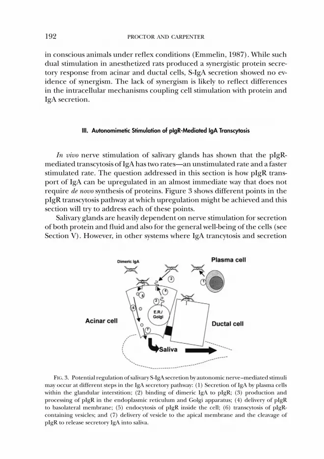
192 PROCTOR AND CARPENTER
in conscious animals under reflex conditions (Emmelin, 1987). While suchdual stimulation in anesthetized rats produced a synergistic protein secre-tory response from acinar and ductal cells, S-IgA secretion showed no ev-idence of synergism. The lack of synergism is likely to reflect differencesin the intracellular mechanisms coupling cell stimulation with protein andIgA secretion.
III. Autonomimetic Stimulation of pIgR-Mediated IgA Transcytosis
In vivo nerve stimulation of salivary glands has shown that the pIgR-mediated transcytosis of IgA has two rates—an unstimulated rate and a fasterstimulated rate. The question addressed in this section is how pIgR trans-port of IgA can be upregulated in an almost immediate way that does notrequire de novo synthesis of proteins. Figure 3 shows different points in thepIgR transcytosis pathway at which upregulation might be achieved and thissection will try to address each of these points.
Salivary glands are heavily dependent on nerve stimulation for secretionof both protein and fluid and also for the general well-being of the cells (seeSection V). However, in other systems where IgA trancytosis and secretion
FIG. 3. Potential regulation of salivary S-IgA secretion by autonomic nerve–mediated stimulimay occur at different steps in the IgA secretory pathway: (1) Secretion of IgA by plasma cellswithin the glandular interstition; (2) binding of dimeric IgA to pIgR; (3) production andprocessing of pIgR in the endoplasmic reticulum and Golgi apparatus; (4) delivery of pIgRto basolateral membrane; (5) endocytosis of pIgR inside the cell; (6) transcytosis of pIgR-containing vesicles; and (7) delivery of vesicle to the apical membrane and the cleavage ofpIgR to release secretory IgA into saliva.

SALIVARY S-IgA 193
is dependent on pIgR, other factors predominate; for example, sex hor-mones affect steady-state levels of pIgR in rodent uteri (Kaushic et al., 1995),and in the kidney, water loading appears to affect pIgR levels (Rice et al.,1998). Secretion from the rat lacrimal gland is dependent upon a back-ground of androgen stimulation which has previously been found to upreg-ulate pIgR expression in lacrimal cells (Sullivan et al., 1984, 1990; Kelleheret al., 1991). Superimposed on tissue specific regulation are the influencesof cytokines such as interferon-γ , interleukin (IL)-4 and IL-6 (Piskurichet al., 1993; Loman, 1997; Nilsen et al., 1999; Norderhaug et al., 1999) which,in vitro at least, increased pIgR levels. Most of these factors affected steady-state synthesis of pIgR, requiring de novo synthesis of pIgR, a process takinghours rather than minutes. However, do increasing amounts of pIgR in-crease the amount of IgA transcytosed? In one study pIgR was geneticallyoverexpressed in mouse mammary epithelial cells with steady-state levels in-creased 60 to 270 times greater than normal and yet S-IgA secretion into milkwas only increased one- to twofold (de Groot et al., 2000). To date, there ap-pear to be only two published studies of fast stimulated (i.e., less than 5 min)increases in pIgR-mediated IgA secretion: the rat salivary glands (Carpenteret al., 1998) and the porcine ileum (Schmidt et al., 1999). Increases in S-IgArelease into both saliva and intestinal secretions were evoked by nerve stim-ulation. We have therefore used the rat submandibular gland to study theprocess of upregulation of pIgR-mediated IgA transport in more detail.
A. STUDIES ON SALIVARY CELLS in vitro
Digested salivary glands have been used many times before for studies ofprotein secretion as they allow secretory mechanisms to be studied in moredetail than can easily be achieved in vivo. Primary salivary cell preparationsof salivary glands are most suitable for such studies since cells are responsiveto a complete range of autonomic agonists and contain measurable amountsof pIgR, although only short-term studies may be attempted as these cellsdo not survive for long in vitro. In comparison, most immortalized salivarycell lines are neither polarized nor express pIgR in sufficient quantities toallow studies of IgA transcytosis. Most cell lines used for IgA transcytosisstudies have been transfected with a pIgR gene to increase pIgR expressionlevels (Cardone et al., 1996; Hirt et al., 1993; Su and Stanley, 1998), raisingquestions about functional expression of the pIgR molecule.
To prepare cells for in vitro studies submandibular glands were excisedfrom the rat and digested with collagenase P (Boehringer MannheimBiochemicals, Indianapolis, Ind.) for 1 h in 100% oxygen, producing a mix-ture of intact acinar and ductal cell units but also containing other cells

194 PROCTOR AND CARPENTER
FIG. 4. The photomicrograph shows the typical appearance of rat submandibular cellsprepared by enzymatic digestion and used for examining the effects of autonomimetics onS-IgA secretion in vitro. Cells are mostly present in intact units of acini (black arrow) and ducts(white arrow), the most prominent ductal component being granular ducts.
(see Fig. 4) such as plasma cells. Given that salivary cells do not synthesizeIgA, an initial question for study was whether parenchymal cells still containIgA following such preparation.
Assays of IgA in cells and the digestion medium before and after cellpreparation revealed that only 5–10% of total glandular IgA is present withinfinal cell preparations. The amount of IgA released during digestion can beestimated as 2–5% of total glandular IgA. It can therefore be concludedthat most IgA present in the rat submandibular gland is in the extracellularspaces (but within the glandular capsule) near the site of production byplasma cells but outside of acinar and ductal cells. The extracellular pool ofIgA is quite large (∼0.3 mg/gland) and presumably sufficient for normalreflex secretion in conscious rats, although it can be diminished followingextensive nerve stimulation when under anesthesia (Carpenter et al., 1998).Thus under normal conditions plasma cells maintain a level of IgA withinthe glands sufficient for normal secretion, the excess presumably drainingvia the lymphatics. It is unlikely that short-term increases in salivary S-IgAsecretion are due to increased secretion from the plasma cells since therealready exists an excess of IgA.
Since we had shown IgA to be present in the salivary cells after di-gestion from the gland, we stimulated primary cell preparations with au-tonomimetics and revealed an immediate secretion of IgA (all extracellularIgA having been washed away) with cholinergic, adrenergic, and peptidergic

SALIVARY S-IgA 195
FIG. 5. S-IgA secretion by in vitro preparations of rat submandibular cells in response tocarbachol (Carb), adrenergic (Ad, adrenaline; Iso, isoprenaline; PhE, phenylephrine), andpeptidergic (VIP- vasoactive intestinal peptide; SP, substance P) autonomimetics at concentra-tions that gave maximal responses. S-IgA secretion is given as a proportion of that secretedfrom control cells in the absence of an agonist. Each agonist significantly (p < 0.05) increasedS-IgA secretion.
stimulation (see Fig. 5). As these cell preparations are unfractionated it wasunclear whether parenchymal and/or plasma cells secrete IgA in response tothe agonists. To answer this question submandibular gland plasma cells wereisolated on a reverse Percoll gradient following a more extensive digestionthan described earlier (Mega et al., 1995), and then stimulated with the sameagonists for 30 min. Over this relatively short period of time there was noagonist-induced increase in IgA secretion. Thus despite being present withinthe unfractionated primary cell preparations plasma cells do not appear toincrease IgA secretion in response to autonomimetics. We can concludethat the immediate secretion of IgA evoked with cholinergic, adrenergic,and peptidergic stimulation (see Fig. 5) was due to parenchymal secretionof IgA taken into the cells while still in vivo.
Although immunocytochemistry of human parotid glands confirms thatboth acinar and ductal cells express pIgR and contain IgA (for a review seeBrandtzaeg et al., 1999a) we wanted to confirm whether both cell types se-crete S-IgA in response to autonomimetics and if so to which type of nervesignal. To answer this question rat submandibular glands were digested withcollagenase P (as earlier) and acinar and ductal units separated using aPercoll density gradient (Amsallem et al., 1996). Morphologically this pro-cedure produced a good (but not complete) separation of acinar and ductal

196 PROCTOR AND CARPENTER
FIG. 6. The photomicrograph shows the typical appearance of rat submandibular cellsprepared by enzymatic digestion and then subjected to Percoll density gradient centrifugationto separate (a) acinar and (b) ductal cells. Separated acinar and ductal cells were then used toexamine S-IgA secretion in response to autonomimetics.
units (see Fig. 6). Stimulation with adrenaline (an α- and β-adrenoceptoragonist) caused a similar secretion of S-IgA from acinar and ductal cellpreparations suggesting that both cell types are capable of secreting S-IgA.Furthermore, the use of α- and β-adrenoceptor specific blockers withadrenaline indicates both acinar and ductal units can secrete S-IgA by stim-ulation of either adrenergic receptor.
To confirm that S-IgA secretion was via transcytosis during stimulationwith autonomimetics, confocal microscopy of live cells prepared as de-scribed earlier has been performed. To date, these studies have shown thatFITC- or TRITC-labeled polymeric IgA is not transported paracellularly by

SALIVARY S-IgA 197
either acinar or ductal cells (G. B. Proctor et al., 2002, submitted). Theseresults are supported by earlier confocal microscopy studies which showedthat large macromolecules such as fluorescently labeled dextrans, do notmove by a paracellular route and only small fluorescent dyes such as LuciferYellow show a paracellular movement with autonomimetic stimulation of ratparotid cells (Segawa and Yamashina, 1999).
B. In vivo STUDIES
The use of specific agonists and inhibitors with cell preparations ofrat submandibular glands in vitro showed that the secretion of S-IgA fromparenchymal cells is mainly in response to adrenergic stimuli. However,these cell preparations only give information about the release of S-IgA fromepithelial cells and not the earlier process of IgA uptake. By combining ag-onists with an in vivo collection of saliva from rat submandibular glands wecould examine the effect of specific α- and β-adrenoceptor and choliner-gic stimuli on the whole process of S-IgA transport into saliva. To do this,the parasympathetic nerve supply to submandibular glands of anesthetizedrats was electrically stimulated at three different frequencies and S-IgA se-cretion rates compared with those obtained during intravenous injectionof methacholine (cholinergic stimulus), isoprenaline (β-adrenergic), orphenylephrine (α-adrenergic), each at three increasing doses. The adrener-gic agonists increased the rate of S-IgA secretion which was sustained abovean unstimulated rate for all doses of agonist. However, methacholine didnot sustain increased S-IgA secretion at the highest doses (see Fig. 7). S-IgAsecretion was relatively constant for each agonist across a range of doseswhile other markers of secretion including flow rate, acinar peroxidase,and ductal cell proteinase all increased with increasing dose.
The use of in vivo autonomimetics also highlighted the differences be-tween parasympathetic and cholinergic stimulation on IgA secretion, imply-ing a role for neuropeptides that can be co-released from parasympatheticnerves upon stimulation (Ekstrom, 1987). Assessing the influence of neuro-peptides on the secretion of IgA into saliva is rather difficult as peptides inisolation often do not evoke sufficient saliva to enable analyses. An alter-native strategy therefore was to give study subjects neuropeptide on top ofparasympathetic stimulation as previously reported (Anderson et al., 1996).Although parasympathetic stimulation and substance P (SP) increased aci-nar cell fluid secretion and parasympathetic stimulation and vasoactive in-testinal peptide (VIP) increased ductal cell proteinase secretion neither ofthe peptides further increased S-IgA output (G. H. Carpenter et al., 2002,unpublished). Unlike the stimulus-dependent increase in stored protein

198 PROCTOR AND CARPENTER
FIG. 7. S-IgA secretion by submandibular gland of the anesthetized rat in response to intra-venous infusion of isoprenaline (Iso; β-adrenergic), phenylephrine (PhE; α-adrenergic), andmethacholine (MeCh; cholinergic). Comparison is made with the effects of electrical stimula-tion of the parasympathetic nerve (Ps). With the exception of MeCh, each stimulus significantly(p < 0.05) increased S-IgA secretion above the calculated unstimulated rate.
(peroxidase and proteinase) secretion, S-IgA appears to be secreted eitherat an unstimulated rate or at a higher stimulated rate independently of dose,suggesting a simpler regulatory system compared to stored proteins. Thus,it may be that the addition of a neuropeptide stimulus to parasympatheticstimulation could not further increase S-IgA secretion. Studies with prepara-tions of digested salivary glands in vitro (see Section III.A) confirmed that thepeptides alone can increase secretion of S-IgA (vasoactive intestinal peptideand substance P increased IgA secretion by three and four times the controlvalue, respectively). Overall, therefore, from these data we may concludethat S-IgA secretion is increased in response to adrenergic, cholinergic, andpeptidergic stimuli, which apparently do not modulate each other’s effects.
C. INTRACELLULAR COUPLING OF SALIVARY CELL STIMULATION
TO IGA SECRETION
The data described earlier (Sections III.A and III.B) suggest that S-IgAsecretion can be stimulated via α- and β-adrenoceptors, peptidergic recep-tors, and, to a lesser extent, muscarinic cholinergic receptors. The mecha-nisms of intracellular signaling within salivary cells which couple autonomic

SALIVARY S-IgA 199
receptor occupation to salivary secretion have been well studied (see Baumand Wellner, 1999). Stimulation through β-adrenoceptors raises intracellu-lar levels of cAMP leading to protein storage granule exocytosis. Stimulationof either muscarinic cholinergic or α-adrenergic receptors initiates an el-evation in intracellular calcium. How such a rise in intracellular calciumcauses increased S-IgA secretion is uncertain; it may be by interacting withpIgR directly, or via calmodulin to phosphorylate pIgR, as found in MDCKcells (Cardone et al., 1996). Likewise, elevations in cAMP may increase S-IgAsecretion by activating the relevant protein kinase. Overall it appears thatS-IgA secretion from salivary cells is mediated by both calcium and cAMPsignal transduction pathways from either adrenergic or muscarinic receptoroccupation.
These studies indicate that parenchymal cells are responsible for theincreased S-IgA secretion into saliva with nerve stimulation. On a cellularlevel this may be achieved by increased availability of pIgR at the basolateralsurface or an increased rate of transcytosis and secretion. Following studiesof a different in vitro model, it was also concluded that basolateral expressionof the pIgR molecule was the regulating factor of IgA transcytosis (Hirt et al.,1993).
IV. Studies of Reflex IgA Secretion
Studies utilizing electrical nerve stimulation or autonomimetics have re-vealed the nature of mechanisms controlling salivary S-IgA secretion, butis S-IgA secretion increased by reflex stimulation in conscious animals? Inhumans, reflex stimulation of salivary secretion evokes an increased secre-tion of S-IgA (Brandtzaeg, 1971; Mandel and Khurana, 1969; Proctor andCarpenter, 2001). Results from each of these studies indicate that S-IgA con-centration decreases during stimulated salivary secretion although S-IgAoutput increases. Proctor and Carpenter (2001) collected parotid salivaunder resting conditions followed by a number of samples collected duringa 10-min period of chewing and compared the pattern of S-IgA secretionwith that of the stored acinar cell protein amylase and the ductal cell pro-tein tissue kallikrein. As can be seen from Fig. 8 the patterns of S-IgA andamylase secretion were very similar whereas tissue kallikrein differed.
Brandtzaeg (1971) found previously that the patterns of S-IgA and amy-lase secretion differed when parotid secretion was stimulated with citric acid.Amylase output increased 16-fold whereas IgA output increased only 2.5-fold. Subsequently the effects of chewing and citric acid on S-IgA secretion

FIG. 8. Secretion of (a) IgA, (b) amylase, and (c) tissue kallikrein from the human parotid gland at rest and in response to chewing on inertplastic tubing. One 10 min period of rest (open bar) was followed by 10 periods of chewing (filled bars) for 1 min and a final 10 min periodof rest (open bar). IgA was assayed by ELISA, amylase and tissue kallikrein were assayed by enzymatic activity, and secretion was expressed asoutputs per minute, that is, the product of concentration or activity and salivary flow rate. Solid horizontal bars indicate values that are statisticallysignificantly different (p < 0.05) from the initial unstimulated rate.

SALIVARY S-IgA 201
TABLE ISECRETION OF IgA AND AMYLASE, THE MAIN STORED SECRETORY PROTEIN,
FROM THE HUMAN PAROTID GLAND AT REST AND IN RESPONSE TO CHEWING
AND CITRIC ACID STIMULATION
Rest Chewing 2% Citric Acid
Flow rate (ml min−1) 7.26 ± 2.48 91.72 ± 11.13a 98.04 ± 14.02a
IgA (μg min−1) 4.9 ± 1.31 96.77 ± 11.29a 101.37 ± 15.29a
Amylase (Units min−1) 3.48 ± 1.34 53.18 ± 9.31a 318.9 ± 109a,b
a Statistically significantly different (p < 0.05) from mean value at rest.b Statistically significantly different (p < 0.05) from mean value with chewing.
were compared and the results are shown in Table I (G. B. Proctor et al., 2002,unpublished observations). As found previously, chewing increased S-IgAand amylase secretion to similar extents compared to resting (unstimulated)secretion. However, citric acid increased stored protein secretion to a muchgreater extent than S-IgA. The secretory mechanism leading to increasedS-IgA secretion was examined in both published studies. Western blotting ofrest- and chew-stimulated saliva samples demonstrated that IgA was secretedin the form of S-IgA, that is, complete with secretory component (Fig. 9).Interestingly, scanning of Western blots and quantification of band inten-sities revealed that the proportion of free secretory component remainedunchanged with stimulation (Fig. 9). Thus it might be that the epithelialtranscytosis mechanism can always cope with the glandular polymeric IgAavailable for secretion (Proctor and Carpenter, 2001). Brandtzaeg (1971)assayed free secretory component by single radial immunodiffusion andfound an excess in both stimulated and resting saliva samples. In experi-ments on rat liver Giffroy et al. (1998) found that increased availability ofpolymeric IgA increased transcytosis across hepatocytes with correspondingdecreases in the ratio of free to bound secretory component. Therefore,it can be tentatively concluded that reflexly stimulated increases in salivaryS-IgA secretion are not driven by increased availability of polymeric IgAsince a relatively constant proportion of pIgR is transcytosed without IgAattached.
Since nerve stimulation studies have revealed that both parasympatheticand sympathetic nerve–mediated stimuli can potentially increase the trans-cytosis of IgA into saliva it is of interest to examine the role played by thesenerves under reflex conditions in conscious rats. Such a study of the in-fluence of the sympathetic nerve under reflex conditions was undertakenby Matsuo et al. (2000). The concentrations of S-IgA in salivas evoked bygrooming, feeding, rejection behavior, and heat were determined beforeand after acute sectioning of the sympathetic chain caudal to the superior

202 PROCTOR AND CARPENTER
FIG. 9. Western blot of unstimulated (un) and citric acid stimulated (st) human parotidsalivary proteins separated on a gradient SDS gel and probed with antibodies, showing im-munoglobulin A, secretory component (SC), and J chain. A sample of J chain–containingmyeloma IgA(m) has been electrophoresed for comparison and the positions of molecularweight standard proteins are shown. High relative molecular weight (Mr) protein in salivasamples contains IgA, J chain, and SC, while the myeloma, which has not been transcytosed, isSC negative. Free SC is seen as a more mobile band (at Mr of 71 kD) in both salivas.
cervical ganglion creating a unilateral sympathetic decentralization (sx).S-IgA secretion on reflex stimulation remained unchanged following de-centralization except for the grooming stimulus that showed a decreasedoutput. These results contrasted with total protein secretion and acinarcell peroxidase that showed a decrease in both concentration and outputfollowing sx. The latter indicated that in the rat submandibular gland, ashas previously been found in the rat and rabbit parotid glands (Speirsand Hodgson, 1974; Gjorstrup, 1980), sympathetic nerve–mediated stim-uli make an important contribution to reflexly induced secretion of sali-vary proteins. When the results of Matsuo et al. (2000) are consideredwith those obtained in nerve stimulation studies in anesthetized animalsit can be concluded that high-frequency sympathetic nerve–mediated stim-uli are unlikely to be contributing significantly to S-IgA secretion underreflex conditions. In the nerve stimulation studies, higher S-IgA secretionwas evoked by high-frequency (50 Hz) electrical stimulation and it wouldseem that such high frequencies were not evoked by the reflex stimuli.It may therefore be that reflexly induced increases in S-IgA secretion intosaliva are regulated mainly by parasympathetic nerve–mediated impulses. Inhumans, sweet or salty, tastes evoke more protein-rich salivas than sour taste,

SALIVARY S-IgA 203
possibly due to differing proportions of impulses mediated by parasympa-thetic and sympathetic nerves. The results with chewing and citric acid stim-ulation in humans (see earlier) suggest that unlike stored protein secretion,S-IgA secretion is not stimulated to different extents by different afferentstimuli.
A. EFFECTS OF STRESS ON REFLEX IgA SECRETION
There is considerable interest in stress-related immune impairment andthe function of the hypothalamic pituitary-adrenocortical (HPA) axis. Anumber of indices of immune competence have been utilized in the field ofpsychoneuroimmunology, including assays of immunoglobulins as indicesof the humoral immune response (Vedhara et al., 1999). Salivary S-IgA hasbeen widely assayed since whole mouth saliva is easy to collect and S-IgAmight provide an index of the mucosal immune response relevant to theincreased incidence of upper respiratory tract infections that have been as-sociated with stress (e.g., Drummond and HewsonBower, 1997). Individualswith increased S-IgA levels in upper respiratory tract secretions, includingsaliva, appear to develop fewer symptoms of infection during viral infection(Rossen et al., 1970; McClelland et al., 1980). In general, saliva samples col-lected in such studies are not overtly stimulated but resting samples collectedusing absorbant material placed in the mouth (e.g., Deinzer et al., 2000).Kugler et al. (1992) studied resting salivary secretion of S-IgA in 85 subjectsand found a negative correlation between S-IgA concentration and salivaryflow rate.
There is a well-recognized drying of the mouth during times of acutestress and it might be expected that saliva sampled at such times wouldhave higher concentrations of S-IgA. Some studies have tried to accountfor changes in salivary flow by expressing secretion as output (i.e., concen-tration × flow rate). It would appear that there are effects of stress onS-IgA secretion which are not dependent on changed flow rate, althoughthe sampling techniques used in many studies makes estimating salivary flowdifficult (Deinzer et al., 2000). Previous studies suggest that salivary S-IgAlevels can be raised or lowered by stress and the differences between studiesmay depend on the nature and definition of stress. Results suggest that thelevels of other stored salivary proteins are affected by stress ( J. Bosch et al.,2002, personal communication). It may therefore be that both salivary fluidand protein secretion are subject to central inhibition from other centersin the brain. Certainly central connections between the salivary centers andother centers in the rat brain have been demonstrated (Matsuo, 1999).

204 PROCTOR AND CARPENTER
B. EFFECTS OF EXERCISE ON REFLEX S-IgA SECRETION
A study of elite cross-country skiers found that their salivary IgA levelswere lower than those of recreational athletes (Tomasi et al., 1982). From anumber of subsequent studies it has been concluded that exhaustive exer-cise can reduce S-IgA levels, although there is usually recovery within 24 h(Gleeson and Pyne, 2000). Occasional studies have examined other aspectsof mucosal immunity, thus it has been found that breast milk S-IgA levelswere decreased in one study (Gregory et al., 1997). It has been suggested,though not proven, that reduced levels of IgA in mucosal secretions, par-ticularly in salivary S-IgA1 levels, are associated with upper respiratory tractinfections in athletes undertaking large volumes and intensities of exercise(Gleeson and Pyne, 2000). The mechanism underlying the effects of exer-cise on salivary S-IgA levels remains unknown. In general, studies in thisarea have expressed S-IgA levels as concentrations, which are decreased.Few studies have addressed whether salivary flow rate is affected by exerciseand how secretion rates of S-IgA are affected, although results of one studysuggest that secretion rates are reduced (Gleeson et al., 2000).
V. Long-Term Influences of Nerves on S-IgA Secretion
The studies described earlier established that nerves can acutely regulateS-IgA transcytosis by salivary epithelial cells. What are the long-term effectsof nerves on S-IgA secretion? Autonomic nerves have previously been shownto exert long-term influences on salivary glands. Removal of the parasym-pathetic nerve supply in particular causes atrophy while secretion of fluidand the synthesis and secretion of major salivary proteins are altered bydenervation (Proctor, 1999).
The effects of 1 week of sympathetic decentralization on S-IgA secre-tion were examined (Proctor et al., 2000b). The right cervical sympatheticchain was sectioned, leaving the superior cervical ganglion intact and thuscreating sx without destruction of postganglionic nerves and the inductionof supersensitivity. Saliva was collected 1 week later from decentralized andcontralateral control glands and as in the studies previously described theprotocol enabled both unstimulated and nerve stimulated secretion of S-IgAto be estimated. The results are summarized in Figure 10a. Sympatheticdecentralization had little effect on the unstimulated rate of S-IgA secre-tion but completely abolished the increase evoked by stimulation of theintact parasympathetic nerve. In contrast the secretion of stored secretoryproteins increased in parasympathetically evoked saliva from decentralized

SALIVARY S-IgA 205
FIG. 10. Secretion of IgA into submandibular saliva from denervated (hatched bars) com-pared to control contralateral (open bars) glands of anesthetized rats. Saliva was collectedfrom denervated glands during electrical stimulation of the remaining intact nerve supplyfollowing 7 days of either (a) sympathectomy or (b) parasympathectomy. The stimulation pro-tocol allowed calculation of the unstimulated rate of IgA secretion for comparison with theoutputs in response to nerve stimulation. The unstimulated rate of IgA secretion was not signif-icantly changed by sympathectomy whereas the parasympathetically stimulated rate was greatlyreduced (p < 0.05) compared to the contralateral control gland.a Both the unstimulatedb
and sympathetically stimulateda rates of IgA secretion were significantly reduced (p < 0.05)by parasympathectomy.
compared to intact glands. Assay of free SC in the same samples showedthat it too was reduced by decentralization and therefore that the effects ofdecentralization were mediated, at least in part, through a reduced rate ofIgA transcytosis in epithelial cells. Certainly decentralization did not lead toa reduction in IgA production by glandular plasma cells since the levels ofIgA in homogenates of decentralized and intact glands were similar (Proctoret al., 2000b). The effects of parasympathectomy (px), achieved by sectioningbranches of the chorda lingual nerve which innervate the submandibulargland, have also been investigated. Stimulation of the intact sympatheticnerve 1 week following px revealed that both the calculated unstimulatedand the sympathetically stimulated rates of S-IgA secretion were decreasedby approximately 30% (Figure 10b). Again this effect contrasted with thatobserved for stored secretory proteins, which showed little change in secre-tion rate following px. The effects of px might be explained by a decreasein glandular levels of pIgR and preliminary evidence suggests that this is so(G. H. Carpenter et al., 2002, unpublished observations). There appeared

206 PROCTOR AND CARPENTER
to be no decrease in plasma cell production of IgA since homogenates ofparasympathectomized and intact glands contained similar levels of IgA.
VI. Effects of Nerves on Plasma Cell Production of IgA
Salivary gland epithelial cells are subject to acute regulation by auto-nomic nerves that have a dominant role in controlling secretion. The in-vestigations described earlier have established that this role extends to acontrol of IgA secretion by salivary glands, most likely through regulationof epithelial cell transcytosis. Do nerves also influence IgA production byplasma cells within salivary glands? Following extensive nerve stimulation ofIgA secretion from the rat salivary glands, glandular levels of IgA were as-sayed in homogenates of both stimulated and contralateral control glandsand compared with the total IgA secreted into saliva. In both glands IgAlevels were reduced following stimulation compared to control glands sug-gesting that during prolonged stimulation, the rate of IgA production byplasma cells does not match the rate of IgA transcytosis and secretion intosaliva. In the submandibular gland there was evidence of IgA productionwithin the gland since IgA levels did not decrease to the same extent aswould be indicated by the amounts secreted. Immunocytochemistry of therat and human salivary glands suggests that there is a reservoir of extracellu-lar IgA within the gland (G. B. Proctor et al., 2002, unpublished observations;Brandtzaeg, 1974). Acute isolation of epithelial cell units as described previ-ously (Section III.A) supports the concept of such an extracellular reservoirsince only a minor proportion of total glandular IgA levels were found withincells. However, this data does not indicate whether nerves upregulate IgAproduction by plasma cells. Following 1 week of px or sx as described ear-lier there was no reduction in the IgA content of submandibular glands.In fact, determination of the intracellular and extracellular levels of IgA inparasympathectomized glands suggests that there has been no reduction inIgA production.
Neuropeptides can increase IgA production by B cells isolated from dif-ferent sources (see Pascual et al., 1994). Substance P may act directly on Band/or T lymphocytes, both of which have been shown to express substanceP receptors (Stanisz et al., 1987). Substance P may act as a co-stimulatory sig-nal for B cells activated by antigen or T-cell-derived (Th2 subset) cytokinessuch as interleukin 5 (IL-5) and IL-6 that have roles in humoral immu-nity. It may be that at mucosal sites receiving an autonomic innervation,nerves can upregulate IgA production through release of neuropeptides.Salivary glands are effector sites of the mucosal immune system that receivea rich autonomic innervation, and substance P and other neuropeptides,

SALIVARY S-IgA 207
such as vasoactive intestinal peptide, have been localized in the parasym-pathetic nerves supplying salivary glands in a number of species (Ekstrom,1999). Peptide release from parasympathetic nerves might upregulate IgAproduction by plasma cells in salivary glands. Cytokines, such as Il-6 andtransforming growth factor–β that are also produced by intestinal epithe-lial cells, have been demonstrated to regulate mucosal B-cell IgA secretion(Goodrich and McGee, 1998). If the regulation of salivary epithelial cellsby autonomic nerves extends to cytokine production then it may be thatautonomic nerves can influence salivary gland plasma cell IgA secretionindirectly through effects on epithelial cell cytokine production.
There have been few studies of the effects of neurotransmitters on non-transformed plasma cells since the latter tend to be widely distributed insmall numbers at mucosal effector sites, which makes isolation of significantnumbers difficult. Brink et al. (1994) overcame this problem by studying theHarderian gland of the chicken, an accessory lacrimal gland that is richin plasma cells and innervated by cholinergic parasympathetic nerves. Incontrast to previous studies of an IgA-producing myeloma (Tartakoff andVassalli, 1977), the Harderian gland–derived plasma cells responded to car-bachol with increased IgG secretion within 20 min of stimulation. IgG wasassayed rather than IgA in order to be able to discount the influence of glan-dular epithelial cells and transcytosis on IgA secretion. Muscarinic choliner-gic receptors were immunocytochemically localized on the cells and carba-chol, although at high doses (100 μM) raised levels of intracellular calciumand induced IgG secretion. Neuropeptides, the cholinergic agonist pilo-carpine, and electrical stimulation of the extrinsic innervation of intestinalsegments have each been found to increase luminal IgA secretion (Schmidtet al., 1999; Wilson et al., 1982). The mechanism operating is not certain butmay represent a direct effect on plasma cells of the lamina propria.
The mechanisms regulating plasma cell migration to and retention atmucosal sites are beginning to be unraveled (Brandtzaeg et al., 1999b). Itseems likely that mucosal epithelial cells have a role to play, probably throughthe local release of cytokines, in retaining plasma cells at mucosal sites.Following systemic chemical sympathectomy using 6-hydroxydopamine, re-tention and accumulation of ovalbumin-primed B cells at mucosal sites wasreduced suggesting a change in postextravasation signals at effector sites(Gonzalez-Ariki and Husband, 1998). Autonomic nerves may well influ-ence, either directly or indirectly through epithelial cells, the retentionof plasma cells within salivary glands. Following 1 week of either sx orpx there was no obvious change in plasma cell numbers, as indicated byimmunocytochemistry or IgA levels in submandibular glands. However, itmay well be that periods of denervation lasting longer than 1 week are re-quired before an effect on plasma cell migration is apparent. As mentionedpreviously, surgical denervation did not appear to alter total IgA-producing

208 PROCTOR AND CARPENTER
plasma cells in the rat submandibular gland. However, a further study mightexamine the effect of denervation on new recruitment of B cells by salivaryglands.
VII. Conclusion
S-IgA secretion into saliva of humans is rapidly (within 15 s) increasedby autonomic nerve–mediated reflex stimuli through increased glandularepithelial cell transcytosis of IgA by pIgR. Investigations in rat salivary glandshave revealed that such autonomic regulation is mediated through both sym-pathetic and parasympathetic nerves and that a number of epithelial cellautonomic receptors are involved, although cholinergic stimuli appear tohave a lesser effect than adrenergic and peptidergic stimuli. The autonomicregulation of S-IgA secretion is different from the major stored secretory pro-teins of salivary glands and we can tentatively conclude from in vivo studiesthat stimulated secretion is on or off and does not show dose dependencyor additive effects with combinations of agonists. Autonomic nerves havelonger term effects on S-IgA secretion through effects on pIgR-mediatedtranscytosis as demonstrated by denervation experiments. Stress or exten-sive, intense physical training have been linked with increased susceptibilityto upper respiratory tract infections and it has been suggested that decreasedS-IgA secretion may play a role. The studies described earlier establish thatS-IgA secretion is under the influence of the autonomic nervous systembut further studies are required to determine whether altered autonomicactivity can change susceptibility to mucosal infection.
Acknowledgments
The authors are grateful to colleagues who appear as coauthors on cited references, to theWellcome Trust for support through a project grant, and to GlaxoSmithKline for support ofstudies of human salivary secretion.
References
Amsallem, H., Metioui, M., van den Abeele, A., Elyamani, A., Moran, A., and Dehae, J. P. (1996).Presence of metabotropic and an ionotrophic purinergic receptor on rat submandibularductal cells. Cell. Physiol. 40, C1546–C1555.
Anderson, L. C., Garrett, J. R., Zhang, X., Proctor, G. B., and Shori, D. K. (1995). Differentialsecretion of proteins by rat submandibular acini and granular ducts on graded autonomicnerve stimulations. J. Physiol. 485.2, 503–511.

SALIVARY S-IgA 209
Anderson, L. C., Garrett, J. R., Zhang, X., Proctor, G. B., and Shori, D. K. (1996). Proteinsecretion from rat submandibular acini and granular ducts: Effects of exogenous VIPand substance P during parasympathetic nerve stimulation. Comp. Biochem. Physiol. 119A,327–331.
Baum, J. B., and Wellner, R. B. (1999). Receptors in salivary glands. In “Neural Mechanismsof Salivary Glands” ( J. R. Garrett, J. Ekstrom, and L. C. Anderson, eds.). Frontiers in OralBiology, pp. 44–58. Karger, Basel.
Bowen, W. H. (1996). Salivary influences on the oral microflora. In “Saliva and Oral Health”(W. M. Edgar and D. M. O’Mullane, eds.), pp. 95–103. British Dental Association,London.
Brandtzaeg, P. (1971). Human secretory immunoglobulins, VII: Concentrations of parotidIgA and other secretory proteins in relation to the rate of flow and duration of secretorystimulus. Arch. Oral. Biol. 16, 1295–1310.
Brandtzaeg, P. (1974). Mucosal and glandular distribution of immunoglobulin components.Immunology 26, 1101–1113.
Brandtzaeg, P., Farstad, I. N., Johansen, F.-E., Morton, H. C., Norderhaug, I. N., and Yamanaka,T. (1999a). The B-cell system of human mucosae and exocrine glands. Immunol. Rev. 171,45–87.
Brandtzaeg, P., Baekkevold, E. S., Farstad, I. N., Jahnsen, F. L., Johansen, F.-E., Nilsen, E. M.,and Yamanaka, T. (1999b). Regional specialization in the mucosal immune system: Whathappens in the microcompartments? Immunol. Today 20, 141–151.
Brink, P. R., Walcott, B., Roemer, E., Grine, E., Pastor, M., Christ, G. J., and Cameron, R. H.(1994). Cholinergic modulation of immunoglobulin secretion from avian plasma cells:The role of calcium. J. Neuroimmunol. 51, 113–121.
Cardone, M. H., Smith, B. L., Mennitt, P. A., Mochly-Rosen, D., Silver, R. B., and Mostov, K. E.(1996). Signal transduction by the polymeric immunoglobulin receptor suggests a role inregulation of receptor transcytosis. J. Cell. Biol. 133, 997–1005.
Carpenter, G. H., Garrett, J. R., Hartley, R. H., and Proctor, G. B. (1998). The influence ofnerves on the secretion of immunoglobulin A into submandibular saliva in rats. J. Physiol.512.2, 567–573.
Carpenter, G. H., Proctor, G. B., Anderson, L. C., Zhang, X. S., and Garrett, J. R. (2000).Immunoglobulin A secretion into saliva during dual sympathetic and parasympatheticnerve stimulation of rat submandibular glands. Exp. Physiol. 85.3, 281–286.
Cole, M. F. (1985). Influence of secretory immunoglobulin A on ecology of oral bacteria. In“Molecular Basis of Oral Microbial Adhesion” (S. E. Mergenhagen and B. Rosan, eds.),pp. 125–130. Am. Soc. Microbiologists, Washington, D.C.
Dawes, C. (1996). Clearance of substances from the oral cavity—Implications for oral health.In “Saliva and Oral Health” (W. M. Edgar and D. M. O’Mullane, eds.), pp. 67–79. BritishDental Association, London.
De Groot, N., van Kuik-Romeiln, P., Lee, S. H., and de Boer, H. A. (2000). Increased im-munoglobulin A levels in milk by overexpressing the murine polymeric immunoglobulinreceptor gene in the mammary gland epithelial cells of transgenic mice. Immunology 101,218–224.
Deinzer, R., Kleineidam, C., Stiller-Winkler, R., Edel, H., and Bachg, D. (2000). Prolongedreduction of salivary immunoglobulin A (S-IgA) after a major academic exam. Int.J. Psychophysiol. 37, 219–232.
Drummond, P. D., and HewsonBower, B. (1997). Increased psychosocial stress and decreasedmucosal immunity in children with recurrent upper respiratory tract infections. J. Psycho-som. Res. 43, 271–278.
Ekstrom, J. (1999). Role of nonadrenergic, noncholinergic autonomic transmitters in sali-vary glandular activities in vivo. In “Neural Mechanisms of Salivary Gland Secretion”

210 PROCTOR AND CARPENTER
(J. R. Garrett, J. Ekstrom, and L. C. Anderson, eds.), Frontiers in Oral Biology, Volume 11,Chapter 6, pp. 196–218. Karger, Basel.
Emmelin, N. (1981). Salivary glands: Secretory mechanisms. In “Scientific Foundations ofGastroenterology” (W. Sircus and A. N. Smith, eds.), pp. 219–225. Heineman, London.
Emmelin, N. (1987). Nerve interactions in salivary glands. J. Dent. Res. 66, 509–517.Giffroy, D., Langendries, A., Maurice, M., Daniel, F., Lardeux, B., Courtoy, P. J., and Vaerman,
J. P. (1998). In vivo stimulation of polymeric Ig receptor transcytosis by circulating poly-meric IgA in rat liver. Int. Immunol. 10, 347–354.
Gjorstrup, P. (1980). Taste and chewing as stimuli for the secretion of amylase from the parotidgland of the rabbit. Acta Physiol. Scand. 110, 295–301.
Gleeson, M., and Pyne, D. B. (2000). Exercise effects on mucosal immunity. Immunol. and CellBiol. 78, 536–544.
Gleeson, M., Ginn, E., and Francis, J. L. (2000). Salivary immunoglobulin monitoring in anelite kayaker. Clin. J. Sport Med. 10, 206–208.
Gonzalez-Ariki, S., and Husband, A. J. (1998). The role of sympathetic innervation of the gutin regulating mucosal immune responses. Brain Behav. Immun. 12, 53–63.
Goodrich, M., and McGee, D. W. (1998). Regulation of mucosal B cell immunoglobulin secre-tion by intestinal epithelial cell-derived cytokines. Cytokine 10, 948–955.
Gregory, R. L., Wallace, J. P., Gfell, L. E., Marks, J., and King, B. A. (1997). Effect of exerciseon milk immunoglobulin A. Med. Sci. Sport Exer. 29, 1596–1601.
Hector, M. P., and Linden, R. W. A. (1999). Reflexes of salivary secretion. In “Neural Mecha-nisms of Salivary Gland Secretion” ( J. R. Garrett, J. Ekstrom, and L. C. Anderson, eds.).Frontiers in Oral Biology, Volume 11, Chapter 12, pp. 196–218. Karger, Basel.
Hirt, R. P., Hughes, G. J., Frutiger, S., Michett, P., Perregaux, C., Poulain-Goaefroy, O.,Jeangvenat, N., Neutra, M. R., Kraehenbuhl, J. P. (1993). Transcytosis of the polymericIg receptor requires phosphorylation of serine 664 in the absence but not the presence ofdimeric IgA. Cell 74, 245–255.
Kaushic, C., Richardson, J. M., and Wira, C. R. (1995). Regulation of polymeric immunoglob-ulin A receptor messenger ribonucleic acid expression in rodent uteri: Effect of sex hor-mones. Endocrinology 136, 2836–2844.
Kelleher, R. S., Hann, L. E., Edwards, J. A., and Sullivan, D. A. (1991). Endocrine, neural, andimmune control of secretory component output by lacrimal gland acinar cells. J. Immunol.146, 3405–3412.
Kugler, J., Hess, M., and Haake, D. (1992). Secretion of salivary immunoglobulin A in relationto age, saliva flow, mood states, secretion of albumin, cortisol, and catecholamines in saliva.J. Clin. Immunol. 12, 45–49.
Loman, S., Radl, J., and Jansen, H. M. et al. (1997). Vectorial transcytosis of dimeric IgA bythe Calu-3 human epithelial cell line: Upregulation by IFN-γ . Am. J. Physiol. 16, L951–L958.
Mandel, I. D., and Khurana, H. S. (1969). The relation of human salivary γ A globulin andalbumin to flow rate. Arch. Oral Biol. 14, 1433–1435.
Marcotte, H., and Lavoie, M. C. (1998). Oral microbial ecology and the role of salivary im-munoglobulin A. Microbiol. Mol. Biol. 62, 71–105.
Matsuo, R. (1999). Central connections for salivary innervations and efferent impulse forma-tion. In “Neural Mechanisms of Salivary Gland Secretion” ( J. R. Garrett, J. Ekstrom, andL. C. Anderson, eds.). Frontiers in Oral Biology, Volume 11, Chapter 2, pp. 26–43. Karger,Basel.
Matsuo, R., Garrett, J. R., Proctor, G. B., and Carpenter, G. H. (2000). Reflex secretion ofproteins into submandibular saliva in conscious rats, before and after preganglionic sym-pathectomy. J. Physiol. 527, 175–184.

SALIVARY S-IgA 211
McClelland, D. C., Davison, R. J., Floor, E., and Saron, C. (1980). Stressed power motivation,sympathetic activation, immune function and illness. J. Human. Stress. 6, 11–19.
Mega, J., McGhee, J. R., and Kiyono, H. (1995). Characterisation of cytokine producing T cells,TCR expression, and IgA plasma cells in salivary gland associated tissues. In “Advances inMucosal Immunology” (Mestecky, J., Russell, M. W., Jackson, S., Michalek, S. M., Tlaskolova-Hogenora, H., and Sterzl, J. eds.). Plenum Press, New York.
Mestecky, J. (1993). Saliva as a manifestation of the common mucosal immune system. Ann.N.Y. Acad. Sci. 694, 184–194.
Murray, P. A., Prakobphol, A., Lee, T., Hoover, C. I., and Fisher, S. J. (1992). Adherence of oralstreptococci to salivary glycoproteins. Infect. Immun. 60, 31–38.
Nilsen, E. M., Johansen, F. E., and Kvale, D. (1999). Different regulatory pathways employed incytokine enhanced expression of secretory component and epithelial, HLA class I genes.Eur. J. Immunol. 29, 168–179.
Norderhaug, I. N., Johansen, F.-E., Schjerven, H., and Brandtaeg, P. (1999). Regulation of theformation and external transport of secretory immunoglobulins. Crit. Rev. Immunol. 19,481–508.
Pascual, D. W., Stanisz, A. M., and Bost, K. L. (1994). Functional aspects of the peptidergic circuitin mucosal immunity. In “Handbook of Mucosal Immunology” (P. L. Ogra, Mestecky, J.,Lamm, M. F., Strober, W., McGhee, J. R., Bienenstock, J., eds.), pp. 203–216. AcademicPress, San Diego.
Piskurich, J. F., France, J. A., Tamer, C. M., Willmer, C. A., Kaetzel, C. S., and Kaetzel, D. M.(1993). Interferon-γ induces polymeric immunoglobulin receptor messenger RNA in hu-man intestinal epithelial cells by a protein synthesis dependent mechanism. Mol. Immunol.30, 413–421.
Proctor, G. B. (1999). Effects of autonomic denervations on protein secretion and synthesisby salivary glands. In “Neural Mechanisms of Salivary Gland Secretion” ( J. R. Garrett,J. Ekstrom, and L. C. Anderson, eds.), Frontiers in Oral Biology, Volume 11, Chapter 8,pp. 150–165. Karger, Basel.
Proctor, G. B., and Carpenter, G. H. (2001). Chewing stimulates secretion of human salivarysecretory immunoglobulin A. J. Dent. Res. 80, 909–913.
Proctor, G. B., and Chan, K.-M. (1994). A fluorometric assay of peroxidase activity utilizing2′,7′dichlorofluorescin with thiocyanate: Application to the study of salivary secretion.J. Biochem. Biophys. Methods. 28, 329–336.
Proctor, G. B., Carpenter, G. H., Anderson, L. C., and Garrett, J. R. (2000a). Nerve-evokedsecretion of immunoglobulin A in relation to other secretory proteins of rat parotid glands.Exp. Physiol. 85, 511–518.
Proctor, G. B., Carpenter, G. H., and Garrett, J. R. (2000b). Sympathetic decentralizationabolishes increased secretion of immunoglobulin A evoked by parasympathetic stimulationof rat submandibular glands. J. Neuroimmunol. 109, 147–154.
Rice, J. C., Spence, J. S., Megyesi, J., Safirstein, R. L., and Goldblum, R. M. (1998). Regulation ofthe polymeric immunoglobulin receptor by water intake and vasopressin in the rat kidney.Am. J. Physiol. 274, F966–F977.
Rossen, R. D., Butler, W. T., and Waldman, R. H. (1970). The proteins in nasal secretion, II.A longitudinal study of IgA and neutralizing antibodies in nasal washings of men infectedwith influenza virus. JAMA 211, 1157–1160.
Schmidt, P. T., Eriksen, L., Loftager, M., Rasmussen, T. N., and Holst, J. J. (1999). Fast actingnervous regulation of immunoglobulin A secretion from isolated perfused porcine ileum.Gut 45, 679–685.
Segawa, A., and Yamashina, S. (1999). The dynamics of exocytosis of preformed secretorygranules from acini in rat salivary glands. In “Glandular Mechanisms of Salivary Gland
212 PROCTOR AND CARPENTER
Secretion” ( J. R. Garrett, J. Ekstrom, and L. C. Anderson, eds.), Frontiers in Oral Biology,Volume 10, Chapter 6, pp. 89–100. Karger, Basel.
Speirs, R. L., and Hodgson, C. (1974). Control of amylase secretion in the rat parotid glandduring feeding. Arch. Oral Biol. 21, 539–544.
Stanisz, A. M., Scicchitano, R., Dazin, P., Bienenstock, J., and Payan, D. G. (1987). Distributionof substance P receptors on murine spleen and Peyer’s patch T and B cells. J. Immunol.139, 749–754.
Su, T., and Stanley, K. K. (1998). Opposite sorting and transcytosis of the polymeric im-munoglobulin receptor in transfected endothelial and epithelial cells. J. Cell Sci. 111,1197–1206.
Sullivan, D. A., Bloch, K. J., and Allansmith, M. R. (1984). Hormonal influence on the secretoryimmune system of the eye: Androgen regulation of secretory component levels in rat tears.J. Immunol. 132, 1130.
Sullivan, D. A., Kelleher, R. S., and Vaerman, J. P. (1990). Androgen regulation of secretorycomponent synthesis by lacrimal gland acinar cells in vitro. J. Immunol. 145, 4238–4244.
Tartakoff, A. M., and Vassalli, P. (1977). Plasma cell immunoglobulin secretion arrest is accom-panied by alteration of the Golgi apparatus. J. Exp. Med. 146, 1332–1345.
Tenovuo, J. O. (1989). Non-immunoglobulin defense factors in human saliva. In “HumanSaliva: Clinical Chemistry and Microbiology” ( J. O. Tenovuo, ed.), Volume 2, pp. 55–91.CRC Press, Boca Raton.
Tomasi, T. B., Trudeau, F. B., Czerwinski, D., and Erredge, S. (1982). Immune parameters inathletes before and after strenuous exercise. J. Clin. Immunol. 2, 173–178.
Vedhara, K., Fox, J. D., and Wang, E. C. Y. (1999). The measurement of stress-related immunedysfunction in psychoneuroimmunology. Neurosci. Biobehav. Rev. 23, 699–715.
Wilson, I. D., Soltis, R. D., and Olson, R. E. (1982). Cholinergic stimulation of immunoglobulinA secretion in rat intestine. Gastroenterology 83, 881–888.
Wu, A. M., Csako, G., and Herp, A. (1994). Structure, biosynthesis and function of salivarymucins. Mol. Cell. Biochem. 137, 39–55.

STRESS AND SECRETORY IMMUNITY
Jos A. Bosch
Department of Oral BiologyCollege of Dentistry, The Ohio State University
Columbus, Ohio 43218and
Academic Centre for Dentistry Amsterdam Section of Oral BiochemistryVrije Universiteit
1081 BT Amsterdam, The Netherlands
Christopher Ring
School of Sport and Exercise SciencesUniversity of Birmingham
Edgbaston, Birmingham B15 2TT, United Kingdom
Eco J. C. de Geus
Department of Biological PsychologyVrije Universiteit
1081 BT Amsterdam, The Netherlands
Enno C. I. Veerman and Arie V. Nieuw Amerongen
Academic Centre for Dentistry Amsterdam, Section of Oral BiochemistryVrije Universiteit
1081 BT Amsterdam, The Netherlands
I. IntroductionII. Saliva as a Model for Mucosal Secretory Defense
III. An Outline of Secretory ImmunityA. Secretory Immunoglobulin AB. Innate Secretory Immunity
IV. The Neurobiological Basis for Stress-Induced Changes in SalivarySecretory ImmunityA. Autonomic and Central Regulation of the Salivary GlandsB. Local RegulationC. Neuroendocrine Regulation of S-IgAD. Summary
V. Stress and Secretory IgAA. The Academic Puzzle: Academic Stress or Examination Stress?B. S-IgA and Chronic Stress
INTERNATIONAL REVIEW OFNEUROBIOLOGY, VOL. 52
213 Copyright 2002, Elsevier Science (USA).All rights reserved.
0074-7742/02 $35.00

214 BOSCH et al.
C. S-IgA and Acute StressD. Acute Stress That Decreases S-IgAE. Stress and Oral ImmunizationF. Effects on S-IgA Translocation and Subclasses
G. ConclusionVI. Stress and Innate Secretory Immunity
A. Stress and MucinsB. Stress and α-Amy laseC. Stress and Other Mucosal Secretory ProteinsD. Conclusion
VII. Stress and Microbial Colonization ProcessesVIII. Future Perspectives
References
I. Introduction
Human and animal studies have provided convincing evidence thatpsychosocial stress is associated with increased susceptibility to infectiousdiseases (Cohen and Herbert, 1996; Bonneau et al., 2001). It has beenestimated that up to 95% of all infections are initiated at mucosal sur-faces. These mucosal surfaces are protected by the secretions of various ex-ocrine glands, including the salivary glands (Nieuw Amerongen et al., 1995;Rudney, 1995; Schenkels et al., 1995; Nieuw Amerongen et al., 1998) andlacrimal glands (Schenkels et al., 1995; McClellan, 1997), as well as those ofthe respiratory (Kaliner, 1991; Rogers, 1994; Nieuw Amerongen et al., 1998;Finkbeiner, 1999; Travis et al., 2001) and gastrointestinal tracts (Bevins, 1994;Schenkels et al., 1995; Nieuw Amerongen et al., 1998; Hecht, 1999; Baronet al., 2000). The antimicrobial proteins (e.g., immunoglobulins, mucins,cystatins, lysozyme, lactoferrin) secreted by these glands constitute a firstline of defense that prevents infection and disease by interfering with mi-crobial entry and multiplication. The secretion of these protective proteinsin under strong neurohormonal control (Garrett, 1987; Rogers, 1994; Bercziet al., 1998; Finkbeiner, 1999; Garrett, 1999b; Shimura, 2000). This presentsa potential psychoneuroimmunological (PNI) pathway linking psychosocialstress and infectious disease.
This review will focus on the relationship between stress and salivarysecretory immunity in humans. There are compelling reasons for studyingsalivary immunity. First, the salivary glands, being strategically located at theportal of entry to the respiratory and gastrointestinal tracts, provide a firstline of defense against microorganisms that may develop their pathogenicpotential elsewhere in the body. Second, salivary secretory immunity plays acrucial role in the maintenance of oral health. This is particularly relevant

STRESS AND SECRETORY IMMUNITY 215
from a PNI perspective, as several pathologies of the oral cavity have beenassociated with stress, including periodontal disease (Rugh et al., 1984;da Silva et al., 1995; Genco et al., 1998; Breivik and Thrane, 2001), acutenecrotizing gingivitis (Rugh et al., 1984; da Silva et al., 1995), dental caries(Sutton, 1966; Rugh et al., 1984; Beck et al., 1987; Vanderas et al., 1995, 2000),herpes labialis (Rugh et al., 1984; Cohen and Herbert, 1996; Logan et al.,1998), recurrent aphthous ulcerations (Rugh et al., 1984; McCartan et al.,1996), and impaired oral wound healing (Marucha et al., 1998). The rele-vance of these observations may not be limited to dental science, as impairedoral health is a risk factor for infective endocarditis, atherosclerosis, coro-nary thrombosis, stroke, and respiratory infection (Herzberg, 1996; Page,1998; Scannapieco, 1999; Valtonen, 1999; Li et al., 2000; Wu et al., 2000).
II. Saliva as a Model for Mucosal Secretory Defense
A third reason for studying salivary immunity is that it may serve as a rep-resentative model for studying both innate and adaptive secretory immunedefenses. The salivary glands and other exocrine glands share similaritiesin morphology and architecture; neuronal regulation (Rogers, 1994; Bercziet al., 1998; Finkbeiner, 1999; Garrett, 1999b; Shimura, 2000); the majorfunctions of their secretions, such as host defense and lubrication (Kaliner,1991; Levine, 1993; Hecht, 1999; Travis et al., 2001); and the composition ofthese secretions (Schenkels et al., 1995). While the concentrations may varyfrom one anatomical location to another, most salivary proteins can also befound in other mucosal fluids where they serve similar functions. Moreover,the salivary glands form a common effector site for IgA-secreting plasmacells: IgA-secreting immunocytes that are activated at the various mucosalinduction sites where the antigen presentation takes place, all migrate tothe salivary glands (Mestecky, 1993). Thus, analysis of immune responsesin saliva may provide a general picture of the entire secretory immunesystem.
The most common route for microbial acquisition and infection isthrough colonization of the mucosa. Gibbons (Gibbons, 1989) has arguedthat the mouth, which is readily accessible and harbors an extremely rich mi-croflora, provides a model to monitor basic colonization processes such asmicrobial adherence, microbial growth, and microbial interactions. Giventhat saliva is a representative mucosal secretion and that mucosal fluids areimportant determinants of mucosal microbial colonization, saliva–microbeinteractions may act as a model for studying basic microbial colonizationprocesses.

216 BOSCH et al.
III. An Outline of Secretory Immunity
A. SECRETORY IMMUNOGLOBULIN A
The two main secretory proteins that represent adaptive immunityare secretory immunoglobulins A (S-IgA) and M (S-IgM) (Lamm, 1997;Brandtzaeg et al., 1999; Norderhaug et al., 1999). S-IgA, which is by far themost abundant immunoglobulin, can bind selectively to antigens, such asmicrobes and toxins. This binding prevents such antigens from attachingto or penetrating the mucosal surface, for example, by blocking interac-tions between microbial ligands and their receptors on epithelial cells. Theantimicrobial effects of S-IgA may be potentiated by interaction with in-nate antimicrobial secretory proteins such as lactoperoxidase and MUC7(Tenovuo et al., 1982; Biesbrock et al., 1991).
The B cells responsible for local IgA production originate frommucosa-associated lymphoid tissue (MALT). After antigen exposure in theMALT, the B cells migrate to the exocrine glands, including the salivaryglands, where they reside in the interstitium as IgA-producing plasma cells(Brandtzaeg, 1998). The presence of plasma cells that originate from manydifferent induction sites in the salivary glands ensures that the buccal mem-brane is furnished with a wide spectrum of secretory antibodies (Mestecky,1993). This is particularly relevant considering that the oral cavity forms aportal of entry to the respiratory and gastrointestinal tracts.
Most IgA-secreting plasma cells in the salivary glands produce a poly-meric form of IgA. This polymeric IgA (pIgA) is transported and secretedinto saliva as S-IgA. The formation and secretion of S-IgA is a two-step process(Norderhaug et al., 1999). First, the pIgA locks on to a receptor molecule,referred to as the polymeric immunoglobulin receptor (pIgR), present onthe basolateral surface of a secretory glandular cell. The pIgR, with or with-out an immunoglobulin attached to it, is translocated from the basolateralto the apical surface of the glandular cell. Here the receptor, with or withoutan immunoglobulin attached to it, is cleaved off and secreted into saliva assecretory IgA (S-IgA) or secretory component (SC), respectively. Accord-ingly, both IgA secretion by the plasma cells and the availability of pIgRfor translocation by the glandular cells are rate-determining steps in thesecretion of S-IgA. As well as transporting IgA into the external secretions,SC also protects the immunoglobulin against proteolytic breakdown in thishostile milieu exterieur.
Human IgA occurs in two isotypic forms, IgA1 and IgA2 (Brandtzaeget al., 1999). Several structural and functional differences between the two

STRESS AND SECRETORY IMMUNITY 217
types have been detected. The most notable difference is a longer hingeregion in the IgA1 antibody molecule (Kilian et al., 1996). Some bacteria,including the viridans group streptococci that are abundant in saliva, haveproteinases that split human IgA1 in the hinge region (Kilian et al., 1996).These IgA1 proteinases allow bacteria to colonize mucosal surfaces in spiteof adherence-inhibiting S-IgA1 antibodies. This may explain why the lessvulnerable IgA2 subclass is more abundant in mucosal secretions: while theIgA1 subclass makes up some 90% of total IgA in serum, it comprises 60%in saliva, and only 35% in the lower gastrointestinal tract (Brandtzaeg et al.,1999).
B. INNATE SECRETORY IMMUNITY
S-IgA is only one of many protective proteins secreted in the fluids cov-ering the mucosae. Most proteins secreted by the mucosal exocrine glandsbelong to the innate immune system. The secretion of these innate immunefactors (e.g., mucins, cystatins, lysozyme, lactoferrin) is predominantly un-der neurohormonal control (Garrett, 1987; Rogers, 1994; Berczi et al., 1998;Finkbeiner, 1999; Garrett, 1999b; Shimura, 2000), and therefore, antimicro-bial secretory proteins are candidates for PNI investigation.
1. The Mucins
The mucins form a group of 14 very large glycoproteins that consistlargely of carbohydrate. They are present in nearly all exocrine mucosalfluids and give these fluids their slimy characteristic (Schenkels et al., 1995;Nieuw Amerongen et al., 1998). Most mucins form an adhesive gel-likefilm on the mucosal epithelium that protects the epithelium against noxi-ous substances, prevents dehydration, and traps microorganisms (NieuwAmerongen et al., 1995). Mucins, such as MUC7, which lack gel-formingproperties perform their protective functions in solution where they bindto bacterial and viral adhesion molecules (Nieuw Amerongen et al., 1995).This binding inactivates these microorganisms and prevents their attach-ment to the mucosal surfaces (Ligtenberg et al., 1992; Liu et al., 2000).
2. The Cystatins
The cystatins, present in every cell and body fluid, inhibit cysteine pro-teinases, a class of enzymes involved in virus replication and tissue invasionby bacteria (Henskens et al., 1996). Some cystatins, particularly those in mu-cosal secretions, also have bactericidal (killing) and bacteriostatic (growthinhibiting) effects (Blankenvoorde et al., 1998).

218 BOSCH et al.
3. Lactoferrin
Lactoferrin is a bacteriostatic protein that is secreted by virtuallyall exocrine glands and by neutrophils. It is found in particularly highconcentrations in lacrimal fluid and bronchial and nasal mucus (Schenkelset al., 1995; Vorland, 1999). The bacteriostatic properties of lactoferrin de-rive from its capacity to bind iron which deprives microorganisms of thisessential substrate. Lactoferrin also has other protective functions (e.g.,bactericidal, fungicidal, anti-inflammatory) (Vorland, 1999).
4. Lysozyme
Lysozyme (muramidase) is known for its capacity to lyse certain bacteriaby cleaving the polysaccharide component of their cell walls. This is, how-ever, just one of its antimicrobial activities, some of which depend on thepresence of other components, such as the complement system, antibodies,and peroxidase, for full effectiveness. Its common occurrence in secretionsis very similar to that of lactoferrin (Schenkels et al., 1995).
5. α-Amylase
The observation that α-amylase is found in many exocrine fluids be-sides saliva suggests that it may have functions other than degrading starch(Schenkels et al., 1995). It affects the adherence and growth of various strep-tococcal bacteria and may play a role in regulating the normal microflora(Scannapieco et al., 1993).
IV. The Neurobiological Basis for Stress-Induced Changes in Salivary Secretory Immunity
Saliva, also denoted as “whole saliva,” is the product of three pairs ofmajor glands (the parotid, submandibular, and sublingual glands) and hun-dreds of minor glands in the lip, cheek, tongue, and palate. Each of theseglands, as well as the various cell types within these glands, synthesize and se-crete a characteristic set of proteins. Before secretion, most salivary proteins(with the secretory immunoglobulins as a notable exception, see earlier)follow the common secretory pathway: synthesis on the ribosomes on theendoplasmic reticulum, further assembly and modification in the Golgi ap-paratus, and subsequent storage in membrane secretory granules (Proctor,1998). Exocytosis of these granules occurs upon neuronal stimulation—β-adrenergic stimulation in particular (Carpenter et al., 1998; Garrett, 1998;Proctor, 1998; Segawa and Yamashina, 1998). Since the whole process ofprotein biosynthesis and cellular transport of secretory proteins takes at
STRESS AND SECRETORY IMMUNITY 219
least 20–40 min, it can be inferred that the immediate effects (i.e., withinminutes) of a stressor or other neuroendocrine stimulus on protein secre-tion represent the release of preformed protein, whereas later effects (i.e.,over hours or even days) may involve, in addition, protein synthesis andposttranslational modification (Kousvelari et al., 1988).
Salivary gland function is largely under autonomic control; the parasym-pathetic nerves mainly govern salivary fluid secretion, whereas the sympa-thetic nerves regulate protein secretion (Garrett, 1987). However, thereare nuances to this general rule. First, the parasympathetic nerves alsoaffect salivary protein secretion. Protein secretion of some glands, such asthe sublingual and some of the minor glands, may even be entirely underparasympathetic control. Second, sympathetic activation also causes somestimulation of salivary flow rate (Garrett, 1987, 1999b). This implies thatsympathetic activation during stress is not the cause of the well-known inhi-bition of salivary flow (Garrett, 1987, 1999b). We may add that rather thanacting antagonistically, the two branches of the autonomic nervous systemexert relatively independent effects in which the activity of one branch maysynergistically augment the other (Emmelin, 1987).
A. AUTONOMIC AND CENTRAL REGULATION OF THE SALIVARY GLANDS
Parasympathetic control of the submandibular and sublingual glandsoriginates in the inferior salivatory nucleus, located in the pons. The axonsof these neurons follow the facial nerve, where they directly, or indirectlyvia the submandibular ganglion, synapse in the glands. Parasympatheticcontrol of the parotid gland is governed by the superior salivatory nucleus,situated in the caudal medulla. The efferent nerves enter the periphery viathe glossopharyngeal nerve, and terminate on the parotid gland via the oticganglion. While the precise location of the sympathetic salivary center hasyet to be identified, it is known that the sympathetic preganglionic neuronsin the upper thoracic nerves connect with the salivary glands (Brown, 1970;Matsuo, 1999).
The hypothesis that psychological processes modulate salivary glandfunction assumes that the salivatory brain stem centers receive inputs fromhigher neural structures. Two lines of evidence support this assumption:(1) studies that have stimulated specific brain areas and recorded changesin saliva (Wang, 1962; Brown, 1970), (2) and histological studies that haveidentified central connections by tracing the retrograde axonal transportof horseradish peroxidase (Matsuo, 1999). These studies show that the pri-mary parasympathetic salivary centers receive direct inputs from the fore-brain (e.g., paraventricular and lateral hypothalamus, central nucleus of the

220 BOSCH et al.
amygdala, bed nucleus of the stria terminalis) (Matsuo, 1999) and the lowerbrain centers (e.g., nucleus solitarius) (Wang, 1962; Matsuo, 1999). Besidesgoverning salivary functions related to drinking and feeding behavior, thesecenters are also concerned with parasympathetic cardiac and visceral con-trol, and they play a pivotal role in orchestrating neuroendocrine stressresponses (Gray, 1987; Matsuo, 1999).
Less is known about central sympathetic salivary regulation, although theparaventricular and lateral hypothalamus, central gray matter, and rostralventrolateral medulla have been implicated (Brown, 1970; Matsuo, 1999).The central gray matter is also an important relay station for coordinatingfight–flight responses, whereas the rostral ventrolateral medulla is known forits role in the integration of cardiovascular and respiratory reflexes (Matsuo,1999).
In sum, functional and histological studies have shown that the primarysalivary centers in the brain stem receive inhibitory and excitatory inputsfrom neural structures in the forebrain and brain stem. As well as governingtypical salivary functions, these structures are also involved in generatingbodily changes associated with stress. It is therefore reasonable to assumethat salivary changes during stress are an integral part of a centrally co-ordinated stress response that encompasses many other bodily functions(e.g., systemic immunity, cardiovascular activity, visceral functions).
B. LOCAL REGULATION
The convergence of synaptic inputs from many brain loci appears topermit different patterns of salivary responses. For example, in humans thesalivary glands are capable of producing differentiated protein responses todifferent stressors (Bosch et al., 2002). Matsuo (Matsuo et al., 2000) showedthat different patterns of protein secretion could be achieved within onegland with only a functional parasympathetic branch. This versatile regula-tion is made possible by the local presence of multiple autonomic mes-senger substances and autonomic receptors. These different autonomictransmitters are released at different rates and with different patterns ofneuronal activation: acetylcholine and noradrenaline are released with everynerve impulse, whereas neuropeptides are released at higher frequenciesof nerve stimulation (Ekstrom et al., 1998; Ekstrom, 1999). Thus differentneuronal stimulation patterns may cause a differential glandular proteinrelease (Ekstrom, 1999; Garrett, 1999a,b).
Moreover, among the glands and cell types within glands there aremarked differences in the density and patterning of receptors that are re-sponsive to the messenger substances released by the autonomic nerves.

STRESS AND SECRETORY IMMUNITY 221
The autonomic receptors in the salivary glands can be divided into two maingroups: the classic autonomic receptor types, which respond to either nora-drenaline or acetylcholine; and the nonadrenergic–noncholinergic (NANC)receptors that respond to other autonomic messenger substances, such aspeptides, nitric oxide, and purines. Differential activation of these receptortypes can cause additive, synergistic, or antagonistic intracellular responses,ultimately resulting in a protein release that is capable of being differentiallyregulated both between and within glands.
C. NEUROENDOCRINE REGULATION OF S-IgA
Studies with rats have shown that stimulation of either autonomic branchinnervating the salivary glands gives a rapid (within minutes) increase inthe secretion of IgA into saliva (El-Mofty and Schneyer, 1978; Carpenteret al., 1998, 2000; Matsuo et al., 2000; Proctor et al., 2000a,b). Similar rapidincreases in S-IgA secretion are observed in the gastrointestinal tract afterstimulation of the local autonomic nerves (Schmidt et al., 1999). These auto-nomic effects may involve both IgA release by the plasma cells and epithelialtranslocation, as both processes can be enhanced by autonomic transmittersubstances (Brink et al., 1994; Wilson et al., 1982; Freier et al., 1989; Kelleheret al., 1991; McGee et al., 1995; Schmidt et al., 1999). However, studies inrats show that autonomic stimulation of the salivary glands primarily affectsthe IgA translocation process, and has little, if any, effect on IgA release byB lymphocytes (Carpenter et al., 1998)
The exact transmitter substances that mediate the autonomic effectson S-IgA output are not yet known. Pharmacological studies suggest thatparasympathetic stimulation of S-IgA secretion may involve the classic trans-mitter acetylcholine and various neuropeptides, whereas the same studiesalso suggest that the effects of sympathetic stimulation at least involve pep-tidergic transmitters (Wilson et al., 1982; Freier et al., 1989; Kelleher et al.,1991; McGee et al., 1995; Schmidt et al., 1999). It is still unclear whetherthe classic sympathetic transmitter noradrenaline is a major factor in thesympathetic stimulation of salivary S-IgA secretion. For example, the en-hanced S-IgA output during moderate physical exercise, which is a strongsympathetic stimulus, is not attenuated by either α- or β-adrenergic block-ade in humans (Winzer et al., 1999; Ring et al., 2000). Moreover, sympatheticS-IgA secretion by the submandibular salivary gland in rats is enhanced byhigh-frequency (>20 Hz) nerve stimulation but not by lower stimulation fre-quencies (Carpenter et al., 2000). Such high-frequency stimulation is associ-ated with the release of neuropeptides (Ekstrom et al., 1998), whereas nor-adrenaline is released at all frequencies (see also Chapter 8 in this volume).

222 BOSCH et al.
Whereas autonomic stimulation may rapidly affect S-IgA levels, slowereffects have been reported for various hormones (e.g., steroids) and cy-tokines (Sullivan et al., 1983; Kelleher et al., 1991; Gleeson et al., 1995;Norderhaug et al., 1999), many of which are influenced by stress. In par-ticular, the release of glucocorticoid hormones (cortisol in humans, corti-sone in rodents) is considered central to the stress response. In rats, thesynthetic glucocorticoid dexamethasone reduces both total salivary S-IgAand antigen-specific S-IgA levels, whereas dexamethasone increases serumIgA levels (Wira et al., 1990). These effects can be observed within 24–48 h.Paradoxically, the expression of SC, the molecule that transports IgA intothe secretions, is also increased (Wira and Rossoll, 1991).
Most of the studies described earlier have been conducted in rats, aspecies in which several aspects of the S-IgA system are considerably differentfrom that of humans. For example, in rats pIgR–IgA binding enhances S-IgAtranslocation through activation of a signal-transduction pathway, whereasthis is not the case in humans (Norderhaug et al., 1999). As a result, in hu-mans the enhancement of salivary S-IgA output is determined solely by anenhancement of S-IgA translocation, and cannot be caused by an increasedIgA release from the plasma cells (since this does not affect translocation inhumans). Moreover, in rats, polymeric IgA is cleared from serum througha specific, SC-dependent hepatic pump. However, this transporter mecha-nism, although present, exerts little activity in humans. As glucocorticoidtreatment affects the activity of this hepatic pump (Wira and Colby, 1985;Wira et al., 1990), it is unclear to what extent the findings on IgA and dexa-methasone generalize to humans. Thus, as far as the S-IgA system is con-cerned, animal findings should be interpreted with some caution in relationto human processes.
D. SUMMARY
The available data provide a sound biological basis for expecting a modu-latory effect of stress on salivary secretory immunity. Salivary glands, as withother mucosal glands (Rogers, 1994; Finkbeiner, 1999; Shimura, 2000), arelargely under autonomic nervous system control. The preganglionic auto-nomic centers in the brain stem that regulate salivary gland activity receivedirect inhibitory and excitatory inputs from neural structures in the fore-brain that are part of recognized “stress circuits” and centers for homeostaticregulation. The salivary glands form a highly sophisticated endpoint in theCNS control of local immune defenses, capable of responding instantly andwith a high level of specificity to potential sources of harm (e.g., stress,

STRESS AND SECRETORY IMMUNITY 223
inflammation). This remarkable ability, together with their strategic loca-tion at the portal of entry to the respiratory and gastrointestinal tract, makethese glands ideally suited to provide the host with a first line of defense.However, the intricate complexity of multiple interacting factors (e.g., var-ious central inputs, autonomic interactions, local variation in messengersand receptors) makes the effects of stress-related autonomic regulation dif-ficult to predict a priori. Experimental research may therefore provide auseful vehicle for exploring this intricate system.
V. Stress and Secretory IgA
The impact of psychosocial factors on salivary S-IgA has been investi-gated extensively, and includes the effects of mood, relaxation, crying, hu-mor, and stress. The literature on stress and S-IgA appears to be incon-sistent (Stone et al., 1987b; Kugler, 1991; Rood et al., 1993; Gleeson et al.,1995; Valdimarsdottir and Stone, 1997); some studies report that S-IgA de-creases, whereas other studies, even when studying the same type of stressor(e.g., academic examinations; see later), report that S-IgA increases. In otherareas of research within PNI it has repeatedly been noted that opposite re-sults are obtained on the same parameters for acute and chronic stress-ors (e.g., Kusnecov et al., 2001; Dhabhar and McEwen, 2001). Therefore,we adopted as a working hypothesis that the apparent inconsistencies inthe literature may be resolved by discerning acute from chronic forms ofstress.
An updated review of the literature was performed by interrogatingPubMed (National Library of Medicine) and Web of Science (Institutefor Scientific Information) databases, using the search terms “immunoglob-ulin A,” “iga,” “s-iga,” or “siga.” These search terms were combined withthe terms “stress∗,”1 “mood,” “psycho∗,” “emotion∗,” “anxi∗,” “depress∗.” Inaddition, the references in the articles that were generated by using theaforementioned search strategy were checked. Conference abstracts, disser-tations, and book chapters were not considered here. As this review focusedon the relation between stress and S-IgA, we did not include reports on posi-tive moods, relaxation, and humor. Neither did we include literature on per-sonality variables, with the exception of trait anxiety.
1 The symbol ∗ is a wild card, which is used to search databases for any word containing aparticular string of letters. For example, the string “stress∗” would, besides “stress,” also yielddocuments containing the words “stressful,” “stressed,” etc.

TABLE IACADEMIC STRESS (SALIVA SAMPLES TAKEN DURING EXAM PERIOD)
Methodology and Design Outcomea
Control for VerificationStress Baseline circadian of stress Control S-IgA
N measurement measurement effects? response group? concentration S-IgA output References
64 During exam periods(3 ×)
Periods with lowacademic stress atstart and end ofacademic year
Yes Questionnaire No ↓ ↓ Jemmott et al.,1983
32 First day of exams 1 month beforeexams
Yes Questionnaire No ↔ — Kiecolt-Glaseret al., 1984
15 Day of third exam 5 days before examsand 2 weeks afterexams
Yes Questionnaire No ↓ ↓ Jemmott andMagloire,1988
44 During exam periods(2 ×)
After summer andmidterm break
Yes Questionnaire No ↓ ↔ Mouton et al.,1989
57 During exam period 4 weeks beforeexams
Yes Questionnaire No ↓ — Li et al., 1997
38 Daily sampling,immediately afterawakening, duringtwo consecutiveexam weeks
4 weeks beforeexams
No Questionnaire No ↓ During examweeks, up to6 days afterlast exam
↓ During examweeks, up to6 days afterlast exam
Deinzer andSchuller, 1998
27 experimental27 control
Daily sampling,immediately afterawakening, duringexam week andfollowing 2 weeks
4 weeks beforeexams
No Questionnaire Yes ↔During examweek
↓ 2 weekspostexam
↔ Duringexam week
↔ Postexam
Deinzer et al.,2000
a ↓, Decreases; ↔, no change; —, not determined.

STRESS AND SECRETORY IMMUNITY 225
A. THE ACADEMIC PUZZLE: ACADEMIC STRESS OR EXAMINATION STRESS?
A majority of studies concerned with the association between stress andS-IgA have examined the impact of academic stressors. Their results paint aconfusing picture; aside from occasional null findings, approximately one-half reported increased S-IgA and the other half reported decreased S-IgA. Anumber of reasons may account for such inconsistencies. For example, thereare substantial differences among academic systems. In some institutionswhere testing is spread over the academic year, examinations are routineevents, whereas others have final examination periods lasting between oneweek and one month when all courses are examined. Further, academicstressors can combine acute (e.g., just before an examination) and chronic(e.g., during the review period) characteristics.
The current review of the literature takes account of the distinction be-tween short-term and long-term features of academic stressors. Accordingly,Tables I and II categorize academic stress studies based on whether, as statedin their methods, saliva samples were collected minutes before, during, or aftera single examination (referred to as acute examination stress) or sometimeduring the extended examination period (referred to as chronic academicstress). Sorting the studies according to this single criterion reveals a remark-ably consistent picture: all acute examination stress studies were associatedwith increased S-IgA (Table II), whereas most chronic academic stress studieswere associated with decreased S-IgA (Table I).
To further explore the proposition that apparently conflicting S-IgA find-ings can be reconciled by simply distinguishing between acute and chronicstress exposure, the results yielded by other stress paradigms are presentedin Tables III, IV, and V. Because timing is a critical factor, special emphasisis given to temporal features, such as when baseline and stress samples werecollected and the duration of the stressor. Salivary S-IgA exhibits a diurnalrhythm, with a peak in the morning hours (Hucklebridge et al., 1998; Parkand Tokura, 1999), and therefore, the extent to which studies controlledfor time-of-day was noted (e.g., whether the time of saliva sampling washeld constant). Also, independent verification of a stress response (e.g., byself-report or physiological activity) was determined for each study. Finally,because changes in S-IgA concentration may be secondary to changes insalivary flow rate, both the results on S-IgA concentration (μg/ml) andS-IgA output (μg/min) are listed2 (not all studies, however, reported both
2 Other methodological issues specific to salivary research, including the appropriate meth-ods for collection and handling of saliva, are discussed elsewhere (Navazesh and Christensen,1982; Stone et al., 1987b; Jemmott and McClelland, 1989; Navazesh, 1993; Rudney, 1995;Veerman et al., 1996; Brandtzaeg, 1998).

TABLE IIEXAMINATION STRESS (SAMPLES TAKEN MINUTES BEFORE, AFTER, OR DURING AN EXAMINATION)
Methodology and Design
Control for VerificationOutcomea
Stress Baseline circadian of stress Control S-IgAN measurement measurement effects? response group? concentration S-IgA output References
42 Immediately afterand 1 3
4 hoursafter exam
Days after exam(unspecified)
No Norepinephrine No ↑(p < 10) — McClellandet al., 1985
7 Immediately afteroral presentation
1 week before and dayof presentation
Yes QuestionnaireCortisol
No — ↑ (p < 10) Evans et al.,1994
28 30 min before exam 2 and 6 weeks afterexam
Yes Questionnaire No ↑ ↑ Bosch et al.,1996, 1998
16 30 min before,during, and30 min after oralpresentation
Same day, beforestress-measurement(unspecified)
No Questionnaire No ↑ Before andfollowingpresentation
— Bristow et al.,1997
22 15 min before, and5 and 15 minafter exam
4 weeks before exam Yes QuestionnaireCardiovascularCortisol
No ↑ Before and afterexam
— Spangler, 1997
29 Immediately beforeand immediatelyafter exam
4 weeks after exam Yes QuestionnaireCardiovascularCortisol
No ↑ During allmeasurements
↑ During allmeasurements
Huwe et al.,1998
a ↑, Increases; —, not determined.

STRESS AND SECRETORY IMMUNITY 227
parameters). In the sections that follow, all the tabulated studies will be dis-cussed with special emphasis on differences in the effects of chronic (whichincludes academic stress) versus acute stress (which includes examinationstress).
B. S-IgA AND CHRONIC STRESS
Chronic stress has been shown to lower the levels of many blood-basedfunctional and enumerative immune measures, including antigen-specificantibodies (i.e., immunoglobulins) following vaccination (Cohen et al., 2001),and, accordingly, it may be hypothesized that chronic stress would alsoreduce S-IgA. The literature concerning the effects of long-term stress onS-IgA can be divided into studies of academic stress and self-reported stressexposure.
1. Academic Stress
Of the studies that measured salivary S-IgA during prolonged academicexamination periods, two reported null findings and five reported a de-crease in S-IgA (see Table I). However, the studies that reported positiveeffects suffer from methodological shortcomings; two of the studies col-lected samples over a period of up to 2 years ( Jemmott et al., 1983; Moutonet al., 1989) and thus the stress-associated differences may reflect circan-nual effects; and two other studies only measured S-IgA concentration, andtherefore the stress-associated differences may reflect the effects of stress onsalivary flow (Kiecolt-Glaser et al., 1984; Li et al., 1997). A limitation commonto all studies is that little information is provided on whether, or how, theycontrolled for variations in health behaviors, such as exercise, sleep, diet,alcohol, and smoking, that may affect the immune system. It is also worthnoting that the only study to include a control group (Deinzer et al., 2000)reported a null finding (see the following for further discussion). Thus,although the balance of evidence suggests that periods of prolonged aca-demic stress are associated with reduced S-IgA, methodological weaknessesconstrain any inferences about underlying mechanisms.
Two studies by Deinzer and colleagues (Deinzer and Schuller, 1998;Deinzer et al., 2000) suggested that the effects of a protracted academicstressor may last for days, or even weeks, beyond the formal examinationperiod. In the first study (Deinzer and Schuller, 1998), S-IgA levels, whichwere reduced during two consecutive examination weeks compared to ini-tial baseline, had not returned to prestress baseline values up to 6 daysafter the last examination (see Table I). The second study (Deinzer et al.,

TABLE IIIQUESTIONNAIRE STUDIES
Methodology and Design Outcome
Stress Number of S-IgA S-IgAN measures Comments measurements concentration output References
27 Trait anxiety Undergraduates werepreselected onpersonality trait “highin need of power”
1 Null finding — McClelland et al.,1980
29 Daily hassles checklist Undergraduates 1 Null finding — Kubitz et al., 1986
113 Daily hassles checklistLife eventsNeuroticism/distress
questionnaireFrequency of
depressive episodes
Female nurses 1 Lower levels with higherfrequency of anxiousepisodes
No relations withother questionnaires
— Graham et al.,1988
Frequency of anxiousepisodes
40 Daily hassles checklist University students two, 6 weeksapart
Daily hassles correlated−.32 with S-IgA 6 weekslater
— Martin et al., 1988
228

24 Daily hassles checklist University students three, 1 monthapart
Increase in daily hasslesbetween measurement2–3 correlate with S-IgAdecrease over sameperiod (−.46)
— Farne et al., 1992
12 Daily hassles checklistDaily uplifts checklistMood checklist
University studentsNet desirable events
were computed asdesirable eventsminus undesirableevents
4 to 11 persubject(mean = 8)
Net desirable eventsr = .56
Undesirableeventsr = −.59
Net desirableevents r = .58
Evans et al., 1993
48 One item stress scaleOne item mood scale
Two equal groups ofyoung (20–30 years)and older (60–80)adults
14 (sevenconsecutivedays, twice aday)
— Stress r = .26 Miletic et al., 1996
124 Perceived stress overpast month (SAS)
Currently perceivedstress (LPS)
Job stress
Female nursesMuch incomplete data
(35%)
1 — r = .18 with SASr = .25 with LPS
(N = 92)Lower levels in
high-stresshospital units
Ng et al., 1999
a —, Not determined.
229

230 BOSCH et al.
2000) found no difference in S-IgA concentration or output during theexamination period between the experimental and control groups. How-ever, a significant group difference in S-IgA concentration, but not in S-IgAoutput, was observed 1 and 2 weeks after the examinations. It should benoted, however, that the time-by-group-interaction effects were not signif-icant, indicating that changes in the experimental group were identicalto those in the control group. A methodological concern with both stud-ies is that participants collected their own saliva samples immediately af-ter awakening when S-IgA levels are most variable (Hucklebridge et al.,1998). Since the wake-up time was not recorded, it is possible that varia-tions between the two groups in awakening during the study may accountfor the postexamination differences, if any, between the experimental andcontrol groups. Clearly, further research is needed to substantiate thesereports.
2. S-IgA and Self-reported Chronic Stress
A number of studies have investigated chronic stress using self-report in-ventories to determine frequency of exposure to major life events(e.g., death of a spouse, divorce, loss of job) and minor daily hassles (e.g.,minor conflicts and annoyances at home or work) and to assess the per-ceived impact of these exposures. Prior research has established thatindividuals scoring highly on such questionnaires exhibit increased suscep-tibility to a variety of infectious and inflammatory conditions, including res-piratory infections (Cohen and Herbert, 1996) and oral diseases (da Silvaet al., 1995). The evidence provided by questionnaire-based studies, sum-marized in Table III, tends to support the hypothesis that higher levelsof stress exposure and perceived stress are associated with lower levels ofsalivary S-IgA. However, many of the studies suffer from methodologicalweaknesses that temper this interpretation. For example, the two studiesthat reported null findings used sample sizes that were probably insufficientfor capturing the intended effect (McClelland et al., 1980; Kubitz et al.,1986); other researchers solved the issue of small sample size by repeatedsampling of the same subjects (e.g., Evans et al., 1993; Miletic et al., 1996).In addition, several studies measured stress using instruments with doubtful(e.g., single-item questionnaires) or unknown psychometric properties(Miletic et al., 1996; Ng et al., 1999) or used questionnaire data that werecollected months before (Graham et al., 1988). Moreover, none controlledfor potential sources of confounding, such as health behaviors, periodon-tal health, or medication usage (see Evans et al., 2000). Thus, although thedata agree with the hypothesis that chronic stress lowers salivary S-IgA, theliterature would be improved by large-scale, well-controlled studies to assessthe impact of chronic stress on salivary S-IgA.

STRESS AND SECRETORY IMMUNITY 231
C. S-IgA AND ACUTE STRESS
1. Examination Stress and Other Naturalistic Stressors
Two examination studies (McClelland et al., 1985; Evans et al., 1994), sum-marized in Table II, provided preliminary indications that acute episodesof stress may be associated with increases rather than decreases in S-IgA.However, the S-IgA increases failed to reach conventional statistical signifi-cance, and there were also methodological weaknesses, such as no controlfor salivary flow rate and circadian effects (McClelland et al., 1985) and avery small sample size (Evans et al., 1994). Moreover, both studies collectedsaliva immediately after the stressor, and therefore, the nonsignificant in-creases may have resulted from experiencing the relief from stress ratherthan the stress itself. However, four studies were published in 1996 thatconfirmed these early observations (Bosch et al., 1996; Carroll et al., 1996;Kugler et al., 1996b; Zeier et al., 1996), and they established that acute stresscan produce increases in salivary S-IgA concentration and secretion rate:the three that involved naturalistic stressors (Bosch et al., 1996; Kugler et al.,1996b; Zeier et al., 1996) are discussed in this section (see Tables II andIV), and the one that employed a laboratory stressor (Carroll et al., 1996)is considered in the next section (see Table V). Bosch et al. (1996) exam-ined 28 undergraduates just before an academic examination, and again 2and 6 weeks later at times of low academic pressure. The increases in S-IgAconcentration and output observed before the examination indicated thatstress rather than relief was responsible for increased S-IgA levels. Examina-tion stress was also associated with increases in other salivary proteins, suchas α-amylase, suggesting that the mechanism behind the increased salivaryS-IgA involved greater activity of the salivary glandular cells. Subsequentexamination studies (Bristow et al., 1997; Spangler, 1997; Huwe et al., 1998)confirmed that acute examination stress increases salivary S-IgA.
The naturalistic stressor studies are summarized in Table IV. Zeier et al.(1996) reported that both S-IgA concentration and output were elevatedin a group of 126 air traffic controllers after a work shift and that no S-IgAchanges were seen in a group of 10 control participants. It is noted that sam-ples were taken after the work sessions (see previous comments). Kugler et al.(1996b) measured S-IgA in 17 coaches before, during, and after a footballmatch and found that S-IgA concentration was elevated during the matchand at halftime. S-IgA did not change in a control group. Although the S-IgAoutput data were not presented, salivary flow rate did not change during thematch, and therefore, the increases in S-IgA concentration elicited by thecompetition stress was unlikely to be an artifact or reduced saliva produc-tion. It is also worth noting that the acute increases were transient; S-IgAhad returned to baseline 1 h after the final whistle.

TABLE IVS-IgA AND ACUTE NATURALISTIC STRESSORS
Methodology and Design Outcomea
Duration Timing of stress Verification of S-IgA S-IgAN Stressor of stressor measurements stress response concentration output References
12610 control
Work shift 100 min After work shift QuestionnaireCortisol
↑ ↑ Zeier et al.,1996
178 control
Coaches duringa match
120 min Various time pointsduring match
QuestionnaireCortisol
↑During matchand halftimebreak
↑During matchand halftimebreak
Kugler et al.,1996a
1717 control
Dental treatment 20 min During and aftertreatment
QuestionnaireCortisol
↓During and10 min after
↓ During and10 min after
Kugler et al.,1996b
a ↑, Increases; ↓, decreases.

STRESS AND SECRETORY IMMUNITY 233
2. Laboratory Stressors
Further support for the hypothesis that acute stress increases salivaryS-IgA was provided by a laboratory study by Carroll and co-workers (Carrollet al., 1996) who found an increase in salivary S-IgA output during a 30-mincomputer game. Subsequent laboratory studies have replicated this findingof increased S-IgA. Studies using active, effortful coping challenges, such asmental arithmetic and time-paced memory tests, that elicit sympathetic ner-vous system activation have, with a few exceptions, found increases in S-IgAconcentration, although they have been somewhat less consistent regardingincreases in S-IgA output (see Table V).
Inconsistencies concerning S-IgA output probably result from differ-ences among studies in both timing of saliva collection and sample size.All studies in which the stress sample was taken during the stressor re-ported a similar modest increase in S-IgA output of approximately 25%.With this effect size, however, only the larger more powerful studies gen-erated statistically significant increases in S-IgA (e.g., Carroll et al., 1996;Willemsen et al., 2000; Bosch et al., 2001). On the other hand, studies inwhich the stress sample was taken immediately after the stress task generallyshow a larger increase in S-IgA ouput, which resulted in statistically signifi-cant effects irrespective of sample size (e.g., Willemsen et al., 1998; Ohiraet al., 1999; Harrison et al., 2000). Support for this interpretation came froma study by Bosch et al. (2002b), in which the S-IgA response to a memorytest took some time to develop and reached its highest level immediately afterthe stressor. It is again unclear to what extent the increases measured imme-diately after a stressful task reflect stress or relief, such as parasympatheticrebound (e.g., see Mezzacappa et al., 2001).
Another potential source of discrepant findings is unintentional stimu-lation of salivary flow rate: In two studies participants had to recall numbersaloud as part of the experimental task (Willemsen et al., 1998, 2000). It is pos-sible that this procedure, in combination with simultaneously having cottonrolls in the mouth as collecting devices, stimulated salivary flow (Navazeshand Christensen, 1982). Stimulation of salivary flow rate reduces S-IgAconcentration but enhances S-IgA output (Brandtzaeg, 1998; Proctor andCarpenter, 2001). One of these studies indeed found an increase in S-IgAoutput (and saliva secretion) while S-IgA concentration did not change(Willemsen et al., 1998).
In sum, research on laboratory stressors generally confirms the findingsof other acute-stress studies by showing an increase in salivary S-IgA. Theseobservations are in line with the finding that activation of the sympatheticnerves enhances the secretion of S-IgA into saliva (Carpenter et al., 1998,2000). Studies tended to find smaller S-IgA effects when samples were col-lected during rather than immediately after a laboratory stress task. As S-IgA

TABLE VLABORATORY STRESSORS
Methodology and Design
Timing of Verification Outcomea
Duration stress saliva of stress, Control S-IgA S-IgAN Stressor (min) sample response condition concentration output References
28 Computer game 30 During (t min = 6and 24) and after(t min = 18)
Cardiovascular — — ↑During gameat 24 min
Carroll et al., 1996
22 Memory test 10 5 and 15 min after CardiovascularCortisolQuestionnaire
— ↑ 5 and 15 minafter
— Spangler, 1997
16 Mental arithmetic 9 After (speaking) Cardiovascular — MA ↔ MA↑ Willemsen et al.,1998Cold pressor (10◦C) 4 After CP↑ CP↑
20 Mental arithmetic 8 During Cardiovascular — ↑ ↔ Ring et al., 1999
17 Mental arithmetic 8 During Cardiovascular — MA↑ MA↔ Winzer et al., 1999Cold pressor (10◦C) 8 CP↔ CP↔ (reanalysis of
placebo-onlydata)
38 Mental arithmetic 30 After CardiovascularQuestionnaire
— ↑ ↑ Ohira et al., 1999
21 Mental arithmetic 8 During Cardiovascular — MA↑ MA↑ Ring et al., 2000Cold pressor (10◦C) 8 CP↓ CP↓ (reanalysis of
placebo-onlydata)
29 Video of nationalteam losingpenalty shootout
10 After CardiovascularQuestionnaire
Didacticvideo
↑ ↑ Harrison et al.,2000
234

27 Mental arithmetic 8 During (whilespeaking)
CardiovascularQuestionnaire
— ↑ ↑ Willemsen et al.,2000
30 Memory testSurgical video
1111
During and tmin = 9after
CardiovascularQuestionnaire
Didacticvideo
MT:↑S-IgA ↑IgA1↑IgA2 ↑SC
SV;↓S-IgA ↓IgA1↔IgA2 ↔SC, attmin = 9 after
MT:↑S-IgA↑IgA1 ↔IgA2 ↑SC
SV: ↓S-IgA↓IgA1 ↔IgA2 ↔ SC,at tmin = 9after
Bosch et al., 2001
30 Memory test 8 During (tmin = 3and 7) and after(tmin = 0, 4, 8)
CardiovascularQuestionnaire
— Biphasic responseduring MT,(during task ↔at tmin = 3; ↑at tmin7; aftertask ↑tmin = 0)
Biphasicresponseduring MT(during task↓ attmin = 3;after task ↑tmin = 0)
Bosch et al., 2003
127 Mental arithmeticCold pressor (10◦C)
84
During Cardiovascular — MA↑CP↓
MA↑ (menonly)
Willemsen et al.,2002
CP↓24 Mental arithmetic 28 After 14 min and
after 28 minCardiovascular — ↑ After 14 and 28
min↑ After 14 and
28 minRing et al., 2002
a MA, Mental arithmetic; CP, cold pressor; MT, memory test; SV, surgical video; ↑, increases; ↓, decreases; ↔, no change; —, not determined.
235

236 BOSCH et al.
secretion is regulated by both branches of the autonomic nervous system,it is conceivable that a parasympathetic withdrawal during acute stress maycause attenuation of sympathetically mediated increases in S-IgA or thata parasympathetic rebound immediately after a stressor augments S-IgAsecretion. Finally, differences in the method of saliva collection may alsoaccount for some variation in the results.
D. ACUTE STRESS THAT DECREASES S-IgA
It is probable that every topic in science will have articles reportinganomalous results, and it is not always immediately clear whether such re-ports may lead to new insights or whether they just represent experimentalartifacts that should be ignored. This is also the case for the literature onstress and S-IgA. To date, at least 19 experiments have been published thatreport an increase in salivary S-IgA in response to acute stress; however,there are also five studies that report acute decreases in S-IgA.
Kugler and co-workers (Kugler et al., 1996a) observed a decrease in bothS-IgA concentration and output during and directly after a caries excava-tion in patients who were not anesthetized; no changes were seen duringa control session (see Table IV). Ring et al. (2000) and Willemsen et al.(2002) found that cold pressor also caused a rapid decrease in S-IgA, al-though other such studies have not observed this effect (see Table V). Thesimilarity with the study by Kugler et al. (1996a) may be that both tasks arepainful and engender passive coping with the stressor. Other passive stres-sors, such as watching unpleasant scenes were also found to inhibit S-IgAsecretion (Hennig et al., 1996; Bosch et al., 2001). In these studies partici-pants viewed video clips selected to induce feelings of disgust (Hennig et al.1996) or scenes of oral surgery (Bosch et al., 2001). The common denomi-nator in these two studies might be a state of disgust. In addition, the latterstudy found that the decreased S-IgA was accompanied by a cardiac passive-coping response, characterized by sympathetic–parasympathetic coactiva-tion (Bosch et al., 2001). It is possible that a classification of stressors on thebasis of their autonomic nervous system effects might resolve the contradic-tory acute-stress-induced changes in S-IgA (cf., Ring et al., 1999).
One study investigated whether hedonic tone per se might be a deter-minant of the direction of the acute changes in S-IgA (Hucklebridge et al.,2000). No such discrimination was found; both positive and negative emo-tional states (induced by imagery and by listening to music tapes) induceacute rises in S-IgA. This complies with the general finding in the literature;apart from the five studies mentioned earlier, to the best of our knowl-edge virtually all types of acute states (e.g., stress, relaxation, moods, and

STRESS AND SECRETORY IMMUNITY 237
motivational states) and virtually all experimental manipulations (e.g., view-ing videos, listening to music, imagery, performing effortful tasks) similarlycause acute and transient rises in salivary S-IgA.
One study found that the S-IgA response to acute stress can follow abiphasic pattern starting with an acute decrease in S-IgA, which, again,demonstrates that the timing of measurements is critical in stress research.Using an 8-min active-coping stress task, Bosch et al. (2002b) found thatS-IgA secretion was significantly reduced (−30%) within the first 3 min ofthe time-paced memory test, followed by a steady increase that reached itspeak (+60%) immediately after the stressor (see Table V).
In sum, although the majority of acute-stress studies report S-IgA in-creases, rapid decreases may occur under some conditions. These condi-tions include passive-coping stressors, and it is tentatively proposed that thecritical determinants are specific emotionally charged states, namely pain(e.g., dental treatment, cold pressor) and disgust (e.g., videos showingsurgery and other unpleasant scenes). Preliminary evidence suggests thatacute decreases in S-IgA can also be observed at the onset of an active-copingstressor as part of a rapid biphasic response pattern.
E. STRESS AND ORAL IMMUNIZATION
The literature discussed thus far concerned the effects of stress on totalS-IgA levels. One group studied that effects of psychosocial stress on thedaily variation in specific S-IgA (i.e., S-IgA that is directed to a specific anti-gen). Stone and colleagues (1987a, 1994) required participants to swallow acapsule containing rabbit albumin each morning for 8–12 weeks, and thenthey measured the daily variations in specific S-IgA (i.e., S-IgA directedagainst the rabbit protein) in saliva, as well as in daily mood (Stone et al.,1987a, 1994) and events (Stone et al., 1994). This methodology allowedwithin-subject comparisons to be made between antibody levels on high-stress and low-stress days. Antigen-specific S-IgA antibody levels were loweron days with relatively high negative mood and were elevated on days withrelatively high positive mood (Stone et al., 1987a). Subsequent researchdemonstrated a positive association between daily positive events and levelsof antigen-specific S-IgA and, conversely, a negative association between neg-ative events and S-IgA (Stone et al., 1994). A reanalysis of this data showedthat undesirable events lowered antibody level primarily by increasing nega-tive mood, and that desirable events increased antibody levels by decreasingnegative mood. After controlling for negative mood, positive moods ap-peared to explain little of the variance in daily S-IgA levels (Stone et al.,1996).

238 BOSCH et al.
At the time these studies were performed, oral immunization still was adeveloping field, which may explain the remarkable choice for rabbit albu-min as an oral antigen; typically such proteins are recognized by the mucosalimmune system as “food,” and for obvious reasons it is very difficult to mountan immune response against such molecules. Today there are several potentprotocols available that researchers may prefer to use instead (Czerkinskyet al., 1999). Considering the pivotal role of the secretory immune defensesin determining whether infection will occur, oral immunization providesan important in vivo model to study the effects of psychosocial factors onimmune function.
F. EFFECTS ON S-IgA TRANSLOCATION AND SUBCLASSES
An unresolved issue is whether the effects of stress on S-IgA secretionreflect alterations in epithelial IgA translocation or alterations in the IgAlevels at the basolateral epithelial surface. The latter might, for example,be caused by changes in the secretory activity of the plasma cells or by adrain of plasma cells to the circulation. Our results (Bosch et al., 2001) leadus to conclude that both steps in S-IgA secretion can be involved. That is,the decrease observed during the passive-coping stressor probably reflecteddecreases in the availability of basolateral IgA, as S-IgA levels decreased inspite of an increased secretion of the transporter molecule SC. Conversely,the acute increase during the active-coping memory test may reflect an in-crease in IgA translocation. Support for this conclusion comes from studiesshowing that increased IgA near the basolateral surface of the epitheliumdoes not affect the rate in which IgA is translocated, so increases in S-IgAcannot be caused by an increase in basolateral IgA (Norderhaug et al., 1999).
The results of the same study (Bosch et al., 2001) also showed that therapid stress-induced changes in salivary S-IgA mainly reflect increases inthe IgA1 subclass. A comparable finding has been reported for IgA subclasslevels in breast milk in response to physical exercise (Gregory et al., 1997).These findings are surprising given that the transporter molecule SC showsno difference in affinity for the two IgA subclasses (Norderhaug et al., 1999).Perhaps another transporter mechanism, besides SC-mediated transloca-tion, is involved that exhibits a preference for a specific subclass. AlternativeIgA receptors with subclass preference have been identified (Schiff et al.,1986; Rifai et al., 2000) and are involved, at least in rodents, in the transloca-tion of S-IgA from the blood into bile, and possibly also into gastrointestinalmucus (Shimada et al., 1999). It is also possible that the selective effectsof stress on IgA subclass secretion might reflect a differential responsivity

STRESS AND SECRETORY IMMUNITY 239
of subclass-specific plasma cells to autonomic messenger substances, caus-ing an altered immunoglobulin release or a selective drain of cells into thecirculation. Clearly, these speculations require investigation.
G. CONCLUSION
In sum, chronic stress is generally associated with decreases in S-IgA,whereas acute stressors mostly increase S-IgA. Although there are no clearguidelines to decide when acute stress ends and chronic stress begins, acomparison of Table I with Table II shows that a distinction between acuteand chronic stress represents a useful heuristic. Such a distinction betweenthe effects of brief and prolonged stressors is helpful in resolving some ofthe confusion in this literature.
Decreased S-IgA levels have been observed in students during prolongedexamination periods and in individuals reporting high levels of life-eventstress. However, the chronic stress literature is hampered by methodologicalshortcomings, mostly related to inadequate control for possible confound-ing factors and small sample sizes. In addition, there is limited experimentalresearch to provide a biological rationale (e.g., neuroendocrine pathways)for the observed decreases.
Acute-stress studies, which have been performed under more controlledconditions, have observed increases in S-IgA. Such findings are compatiblewith research showing that autonomic nerve stimulation enhances S-IgAoutput by the salivary glands. These acute increases most probably reflectthe effects of stress on S-IgA transcytosis. Finally, additional studies show thatS-IgA may also rapidly decrease under some conditions, including specificemotionally charged states (e.g., pain, disgust), and during the initial stagesof a stress task. Such rapid decreases may involve effects on the IgA-secretingplasma cells, although possible neuroendocrine mechanisms accounting forsuch rapid decreases have yet to be elucidated.
One finding is that the secretion of the IgA1 subclass is more variable un-der acute conditions than IgA2, and that during stress the secretion of S-IgAand its transporter molecule SC can be affected independently (Bosch et al.,2001). Finally, immunization studies have shown that stress also affects thesecretion of antigen-specific S-IgA (as opposed to total S-IgA, which is mea-sured in most studies), and thus it is capable of modulating immunologicalprocesses such as antigen presentation and homing of B cells to the salivaryglands. As immunization provides a means for testing immunocompetencein response to novel antigens, this aspect of secretory immunity deservesmore attention in future research.

240 BOSCH et al.
VI. Stress and Innate Secretory Immunity
The introduction to this review pointed out that S-IgA is only one ofmany protective proteins secreted in the fluids covering the mucosa. Thissection summarizes the scant literature on the effects of stress on theseantimicrobial proteins that comprise innate secretory immunity.
A. STRESS AND MUCINS
Two human studies have shown that acute stressors (a time-paced mem-ory test and a gruesome surgical video presentation) enhance the secre-tion in saliva of the mucins MUC5B and MUC7 (Bosch et al., 2000, 2002).These findings agree with evidence of increased gastrointestinal and respi-ratory mucin secretion in response to water-immersion and restraint stressin animals. These animal experiments also found an effect of stress on otherbiological processes, such as mucin synthesis and posttranslational process-ing (e.g., sulfatation, glycosylation) (discussed by Bosch et al., 2000, andSoderholm and Perdue, 2001). Administration of physiological doses ofβ-adrenergic agonists has also been observed to influence salivary proteinsynthesis and glycosylation in rats (Kousvelari et al., 1988).
Unlike salivary mucin secretion, colonic mucin secretion during acutestress is not initiated by direct neuroendocrine stimulation of the mucin-secreting goblet cells. Castagliuolo et al. (1998) demonstrated a stress-induced (by 30 min of restraint) colonic mucin secretion in normal but notin mast cell–deficient mice, indicating that mast cells regulate colonic mucinrelease in response to restraint stress. However, local neuroendocrine trans-mitters (e.g., CRH, neurotensin) were responsible for inducing the releaseof mast cell factors that, in turn, caused the increased mucin secretion. Al-though increases in mucin secretion are generally interpreted as reflectingan enhanced mucosal barrier function (Soderholm and Perdue, 2001), theexact immunological meaning of increased mucin secretion may depend onvarious factors, such as the specific microorganism under consideration. Forinstance, Helicobacter pylori specifically bind certain mucin types, which maycontribute to the potential of this bacterium to colonize the host (NieuwAmerongen et al., 1998; Van den Brink et al., 2000). A stress-induced increasein secretion of MUC5B enhances the adherence (ex vivo) of H. pylori (Boschet al., 2000). Thus, increases in salivary mucin may have distinct conse-quences; some that benefit the host and some that benefit microorganisms.
Repeated or protracted stimulation of mucin release can lead to a re-duction in mucin release, possibly by depleting the mucin-secreting goblet

STRESS AND SECRETORY IMMUNITY 241
cells (Castagliuolo et al., 1996). Qiu and co-workers observed that protractedstress decreases colonic mucin secretion in rats. This decrease in mucin se-cretion enhanced mucosal permeability, thereby potentiating reactivationof experimental colitis by a chemical irritant (Qiu et al., 1999). This studyshows how stress-induced impairments in innate secretory immunity maydirectly contribute to pathophysiology.
B. STRESS AND α-AMY LASE
Morse and co-workers reported that α-amylase increases during relax-ation and decreases with acute stress (for review see Morse et al., 1983)and suggested that these changes were mediated by variations in parasym-pathetic activity. However, a null finding has been reported (Borgeat et al.,1984), and other studies have shown that acute stressors can increase salivaryα-amylase (Bosch et al., 1996, 1998, 2002; Chatterton et al., 1996, 1997).These increases appear to be mediated by sympathetic nerve activationas they were positively correlated with serum norepinephrine (Chattertonet al., 1996). This interpretation is further supported by studies showing thatα-amylase secretion increases with sympathetic activation by administrationof adrenergic agonists (Mandel et al., 1975), by direct sympathetic nervestimulation (Garrett, 1987; Schneyer and Hall, 1991), and by physical ex-ercise (Chicharro et al., 1998). Nonetheless, claims that salivary α-amylaseis a valid noninvasive measure of adrenergic activity (e.g., Skosnik et al.,2000) should be regarded with caution. First, α-amylase is also secreted inresponse to nonadrenergic sympathetic transmitters, such as neuropeptides(Ekstrom, 1999). Second, α-amylase secretion is also stimulated by parasym-pathetic stimulation, either alone or in interaction with sympathetic stimu-lation (Emmelin, 1987; Asking and Proctor, 1989; Ekstrom et al., 1998). Itshould also be noted that the immunological relevance of α-amylase is stillcontroversial.
C. STRESS AND OTHER MUCOSAL SECRETORY PROTEINS
In humans, Bosch et al. (2002) observed increased secretion of sali-vary cystatin S and lactoferrin in response to a stressor that exhibited asympathetic–parasympathetic coactivation (i.e., viewing a surgery video),but not in response to a stressor that evoked a sympathetic activation in con-junction with a vagal withdrawal (a time-paced memory test). The formerstressor also produced the largest response for most other innate secre-tory proteins mentioned earlier (i.e., the mucins MUC5B and MUC7, and

242 BOSCH et al.
α-amylase). This finding is consistent with the literature showing a synergis-tic effect of the two branches of the autonomic nervous system on secretorygland activity (Emmelin, 1987; Garrett, 1999b); that is, although sympatheticactivation is the main stimulus for glandular protein secretion, such sympa-thetic effects are strongly augmented by concurrent parasympathetic activity.
Perera and co-workers observed a decrease in salivary lysozyme secre-tion during a final examination period (Perera et al., 1997). In a secondstudy these authors found a significant negative correlation between sali-vary lysozyme and a self-report measure of stress and arousal in 39 universitystudents (Perera et al., 1997). However, in a study involving 124 nurses, Nget al. (1999; see Table III) did not find an association between self-reportsof work stress and salivary lysozyme secretion.
D. CONCLUSION
Human and animal research shows that innate secretory immunity ismodulated by stress. Stress, as with other forms of neuroendocrine stimula-tion, clearly affects a broad range of biological processes, including proteinsynthesis, posttranslational modification, and secretion. Interestingly, avail-able data on the susceptibility to infectious disease in stressed humans sug-gests a role for such innate secretory immune factors involved in preventinginfection, rather than immune factors that are involved in responding to in-fection. For example, in a seminal study by Cohen et al. (1991), in whichvolunteers were exposed to cold viruses after a stress assessment, a strongrelationship between stress and infection was observed. However, the as-sociation between stress and actual disease occurrence was much weaker.The authors concluded that “ . . . the relation between stress and colds wasprimarily attributable to an increased rate of infections among subjects withhigher stress-index scores, rather than to an increase in clinical colds amonginfected persons . . . ” (Cohen et al., 1991, italics added). Preventing infec-tion is the primary function of the innate secretory immune defenses, andthe role of this aspect of immunity in mediating the association betweenstress and infectious disease deserves further research.
VII. Stress and Microbial Colonization Processes
A remaining, and critical, issue is whether the effects of stress on secre-tory immunity translate into functional changes, such as affecting processesthat are involved in microbial colonization. Such effects might explain the

STRESS AND SECRETORY IMMUNITY 243
alterations in mucosal microflora, seen in both humans and animals, un-der various forms of psychological strain, such as depression (Antilla et al.,1999), space flight simulations (Brown et al., 1973, 1974), maternal separa-tion (Bailey and Coe, 1999), familial strains (Meyer and Haggerty, 1962),animal fighting and relocation (Marcotte and Lavoie, 1998), and life events(Drake et al., 1995). These effects may also explain the close associationbetween stress and oral diseases with a bacterial etiology, such as periodon-tal disease and dental caries (see Section I). Examination stress reduces theaggregation by saliva of the oral bacterium Streptococcus gordonii (Bosch et al.,1996, 1998). A reduced streptococcal aggregation has also been observed inrats, after 1 week of administration of physiological doses of the β-adrenergicagonist isoproterenol (Kousvelari et al., 1988). Aggregation is a process bywhich bacteria are clumped together through interactions with the host’ssecretory proteins, so that they cannot effectively adhere to the mucosal sur-faces. A decreased bacterial aggregation thus indicates a decreased capacityfor mucosal microbial clearance.
Two studies have investigated the effects of stress in humans on saliva-mediated microbial adherence. Microbial adherence is a process by whichmicrobes gain a stable foothold on the host, and it forms a first and essentialstep in infection. These studies found that acute stress promoted the adhe-sion (ex vivo) of oral bacteria (viridans group streptococci) and nonoralbacteria (Helicobacter pylori) (Bosch et al., 2000, 2002). Stress was also foundto affect saliva-mediated microbial coadherence (i.e., the adhesion of mi-croorganisms to each other) (Bosch et al., 2002). Other studies have re-ported that large doses of glucocorticoids in animals were associated withboth increased bacterial adherence to the intestinal mucosa and reducedluminal S-IgA (Alverdy, 1991, Spitz, 1994, 1996). Similarly, in rats, 1 week ofphysiological doses of the β-adrenergic agonist isoproterenol increased thesalivary adherence (ex vivo) of Streptococcus mutans (Kousvelari et al., 1998).The latter changes were related to an altered salivary protein synthesis andprotein glycosylation.
VIII. Future Perspectives
The salivary literature reviewed here presents a myriad of effects of psy-chosocial stressors on secretory immunity, including altered secretion ofadaptive and innate immune factors, reduced bacterial aggregation, in-creased bacterial adherence, and altered microbial coadherence. Clearly,one way to proceed is to study the effects of stress on secretory immunity atanatomical locations other than the oral cavity (i.e., directly where infection

244 BOSCH et al.
is initiated). The scant literature in this area, mostly performed in rodents,confirms and extends the salivary findings in humans; there are effects onprotein secretion, synthesis, and posttranslational modification, as well asfunctional changes, such as enhanced bacterial adherence and increasedintestinal permeability.
As well as emphasizing salivary immunity, the literature concerning stud-ies of humans is also characterized by a disproportional focus on S-IgA. Thefact that individuals deficient in S-IgA, the most common form of immu-nodeficiency, are, for the most part, in perfect health, suggests that othersecretory factors may be important. With the predominance of innate factorsin mucosal secretions, as well as the strong involvement of neuroendocrinesystems in their regulation, further PNI research into this aspect of hostdefense is warranted.
This is by no means to imply that the field of S-IgA research is redundant.An exciting development is the increased insight in the autonomic mecha-nisms that underlie acute changes in S-IgA, largely obtained through animalstudies (see Chapter 8 in this volume). However, this knowledge also posesnew questions. For example, whereas experimental animal research seemsto indicate that virtually any form of autonomic stimulation (e.g., pharmaco-logical, electrical nerve stimulation, reflex secretion) essentially results in anenhancement of S-IgA secretion, the human stress literature indicates thatsome stress-induced patterns of autonomic stimulation may result in acutedecreases in S-IgA secretion. As there are fundamental differences betweenhumans and rodents in the regulation of S-IgA translocation, more rigorousexperimental studies in humans may be needed as well.
More attention needs to be paid to the effects of protracted forms of stresson secretory immunity. Knowledge of the effects of chronic stress on innatesecretory immunity is extremely limited, and the studies on chronic stressand S-IgA, although showing a fairly consistent picture, suffer from metho-dological shortcomings. Moreover, there is little evidence to provide thisliterature with a biological rationale. Although there is support for theassumption that glucocorticoids mediate decreases in S-IgA during protractedstress, this association has never been demonstrated in humans, rather, it isbased on studies administering pharmacological doses of dexamethasoneto rodents. A most promising model to study the effects of chronic stress onadaptive secretory immunity is mucosal immunization. Knowledge in thisfield has accumulated over the years, and is now ready for a more thoroughexploration within PNI.
Finally, researchers may extend their horizons to include the study offunctional implications of stress-induced changes in mucosal protein secre-tion. A number of microbiological methods exist to study the effects of im-munosecretory changes on microbial colonization processes, including viralinvasion, bacterial adherence, bacterial growth, and bacterial aggregation.

STRESS AND SECRETORY IMMUNITY 245
This approach, rarely considered in the realm of PNI, may reveal the mech-anisms by which immunosecretory changes affect the way in which microor-ganisms are dealt with. Moreover, this methodology will help to strengthenthe proposition that stress, via its effects on secretory immunity, has impor-tant health consequences.
References
Alverdy, J., and Aoys, E. (1991). The effect of glucocorticoid administration on bacterialtranslocation—Evidence for an acquired mucosal immunodeficient state. Annals of Surgery214, 719–723.
Antilla, S. S., Knuuttila, M. L., and Sakki, T. K. (1999). Depressive symptoms favor abundantgrowth of salivary lactobacilli. Psychosom. Med. 61, 508–512.
Asking, B., and Proctor, G. B. (1989). Parasympathetic activation of amylase secretion in theintact and sympathetically denervated rat parotid gland. Q. J. Exp. Physiol. 74, 45–52.
Bailey, M. T., and Coe, C. L. (1999). Maternal separation disrupts the integrity of the intestinalmicroflora in infant rhesus monkeys. Dev. Psychobiol. 35, 146–155.
Baron, S., Singh, I., Chopra, A., Coppenhaver, D., and Pan, J. (2000). Innate antiviral defensesin body fluids and tissues. Antiviral Res. 48, 71–89.
Beck, J. D., Kohout, F. J., Hunt, R. J., and Heckert, D. A. (1987). Root caries: Physical, medicaland psychosocial correlates in an elderly population. Gerodontics 3, 242–247.
Berczi, I., Chow, D. A., and Sabbadini, E. R. (1998). Neuroimmunoregulation and naturalimmunity. Domest. Anim. Endocrinol. 15, 273–281.
Bevins, C. L. (1994). Antimicrobial peptides as agents of mucosal immunity. Ciba Found. Symp.186, 250–260.
Biesbrock, A. R., Reddy, M. S., and Levine, M. J. (1991). Interaction of a salivary mucin-secretory immunoglobulin A complex with mucosal pathogens. Infect. Immun. 59, 3492–3497.
Blankenvoorde, M. J. F., van’t Hof, W., Walgreen-Weterings, E., van Steenbergen, T. J. M.,Brand, H. S., Veerman, E. C. I., and Nieuw Amerongen, A. V. (1998). Cystatin and cystatin-derived peptides have antibacterial activity against the pathogen Porphyromonas gingivalis.Biol. Chem. 379, 1371–1375.
Bonneau, R. H., Padgett, D. A., and Sheridan, J. F. (2001). Psychoneuroimmune interactionsin infectious disease: studies in animals. In “Psychoneuroimmunology” (R. Ader, D. L.Felten, and N. Cohen, eds.), Vol. 2, pp. 483–497. Academic Press, San Diego.
Borgeat, F., Chagon, G., and Legault, Y. (1984). Comparison of the salivary changes associatedwith a relaxing and with a stressful procedure. Psychophysiology 21, 690–698.
Bosch, J. A., Brand, H. S., Ligtenberg, A. J. M., Bermond, B., Hoogstraten, J., and NieuwAmerongen, A. V. (1996). Psychological stress as a determinant of protein levels andsalivary-induced aggregation of Streptococcus gordonii in human whole saliva. Psychosom. Med.58, 374–382.
Bosch, J. A., Brand, H. S., Ligtenberg, A. J. M., Bermond, B., Hoogstraten, J., and NieuwAmerongen, A. V. (1998). The response of salivary protein levels and S-IgA to an academicexamination are associated with daily stress. J. Psychophysiol. 4, 170–178.
Bosch, J. A., de Geus, E. J. C., Ligtenberg, A. J. M., Nazmi, K., Veerman, E. C. I., Hoogstraten, J.,and Nieuw Amerongen, A. V. (2000). Salivary MUC5B-mediated adherence (ex vivo) ofHelicobacter pylori during acute stress. Psychosom. Med. 62, 40–49.

246 BOSCH et al.
Bosch, J. A., de Geus, E. J. C., Kelder, A., Veerman, E. C. I., Hoogstraten, J., and NieuwAmerongen, A. V. (2001). Differential effects of active versus passive coping on secretoryimmunity. Psychophysiology 38, 836–846.
Bosch, J. A., de Geus, E. J. C., Veerman, E. C. I., Hoogstraten, J., and Nieuw Amerongen, A. V.(2002). Innate secretory immunity in response to laboratory stressors that evoke distinctpatterns of cardiac autonomic activity. Psychosom. Med. In press.
Bosch, J. A., Willemsen, G. H., Knol, M., de Geus, E. J. C., and Nieuw Amerongen, A. V.(2002b). Acute stress and secretory immunity; timing is everything. Manuscript submittedfor publication.
Bosch, J. A., de Geus, E. J. C., Turkenburg, M., Nazmi, K., Veerman, E. C. I., Hoogstraten, J., andNieuw Amerongen, A. V. (2003). Stress as a determinant of the saliva-mediated adherenceand co-adherence of oral and non-oral microorganisms. Psychosom. Med. Accepted forpublication.
Brandtzaeg, P. (1998). Synthesis and secretion of human salivary immunoglobulins. In“Glandular mechanisms of salivary secretion” ( J. R. Garrett, J. Ekstrom, L. C. Anderson,eds.), Vol. 10, pp. 167–199. Karger, Basel.
Brandtzaeg, P., Farstad, I. N., Johansen, F. E., Morton, H. C., Norderhaug, I. N., andYamanaka, T. (1999). The B-cell system of human mucosae and exocrine glands. Immunol.Rev. 171, 45–87.
Breivik, T., and Thrane, P. S. (2001). Psychoneuroimmune interactions in periodontal disease.In “Psychoneuroimmunology” (R. Ader, D. L. Felten, and N. Cohen, eds.), Vol. 2, pp. 627–644. Academic Press, San Diego.
Brink, P. R., Walcott, B., Roemer, E., Grine, E., Pastor, M., Christ, G. J., and Cameron, R. H.(1994). Cholinergic modulation of immunoglobulin secretion from avian plasma cells—The role of calcium. J. Neuroimmunol. 51, 113–121.
Bristow, M., Hucklebridge, F. H., Clow, A., and Evans, D. E. (1997). Modulation of secre-tory immunoglobulin A in saliva in relation to an acute episode of stress and arousal.J. Psychophysiol. 11, 248–255.
Brown, C. C. (1970). The parotid puzzle: A review of the literature on human salivation andits applications to psychophysiology. Psychophysiology 7, 65–85.
Brown, L. R., Shea, C., Allen, S. S., Handler, S., Wheatcroft, M. G., and Frome, W. J. (1973).Effects of a simulated spacecraft environment on the oral microflora of nonhuman pri-mates. Oral Surg. Oral Med. Oral Pathol. 35, 286–293.
Brown, L. R., Wheatcroft, M. G., Frome, W. J., and Rider, L. J. (1974). Effects of a simulatedskylab mission on the oral health of astronauts. J. Dent. Res. 53, 1268–1275.
Carpenter, G. H., Garrett, J. R., Hartley, R. H., and Proctor, G. B. (1998). The influence ofnerves on the secretion of immunoglobulin A into submandibular saliva in rats. J. Physiol.(London) 512, 567–573.
Carpenter, G. H., Proctor, G. B., Anderson, L. C., Zhang, X. S., and Garrett, J. R. (2000).Immunoglobulin A secretion into saliva during dual sympathetic and parasympatheticnerve stimulation of rat submandibular glands. Exp. Physiol. 85, 281–286.
Carroll, D., Ring, C., Shrimpton, J., Evans, P., Willemsen, G., and Hucklebridge, F. (1996).Secretory immunoglobulin A and cardiovascular responses to acute psychological chal-lenge. Int. J. Behav. Med. 3, 266–279.
Castagliuolo, I., Leeman, S. E., Bartolak-Suki, E., Nikulasson, S., Qiu, B., Carraway, R. E., andPothoulakis, C. (1996). A neurotensin antagonist, SR 48692, inhibits colonic responses toimmobilization stress in rats. Proc. Natl. Acad. Sci. USA 93, 12611–12615.
Castagliuolo, I., Wershil, B. K., Karalis, K., Pasha, A., Nikulasson, S. T., and Pothoulakis, C.(1998). Colonic mucin release in response to immobilization stress is mast cell dependent.Am. J. Physiol. 274, G1094–G1100.

STRESS AND SECRETORY IMMUNITY 247
Chatterton, R. T. Jr., Vogelsong, K. M., Lu, Y. C., Ellman, A. B., and Hudgens, G. A. (1996).Salivary α-amylase as a measure of endogenous adrenergic activity. Clin. Physiol. 16,433–448.
Chatterton, R. T., Jr., Vogelsong, K. M., Lu, Y. C., and Hudgens, G. A. (1997). Hormonalresponses to psychological stress in men preparing for skydiving [see comments]. J. Clin.Endocrinol. Metab. 82, 2503–2509.
Chicharro, J. L., Lucia, A., Perez, M., Vaquero, A. F., and Urena, R. (1998). Saliva compositionand exercise. Sports Med. 26, 17–27.
Cohen, S., and Herbert, T. B. (1996). Health psychology: Psychological factors and physicaldisease from the perspective of human psychoneuroimmunology. Annu. Rev. Psychol. 47,113–142.
Cohen, S., Tyrrell, D. A., and Smith, A. P. (1991). Psychological stress and susceptibility to thecommon cold [see comments]. New Engl. J. Med. 325, 606–612.
Cohen, S., Miller, G. E., and Rabin, B. S. (2001). Psychological stress and antibody response toimmunization: A critical review of the human literature. Psychosom. Med. 63, 7–18.
Czerkinsky, C., Anjuere, F., McGhee, J. R., George-Chandy, A., Holmgren, J., Kieny, M. P.,Fujiyashi, K., Mestecky, J. F., Pierrefite-Carle, V., Rask, C., and Sun, J. B. (1999). Mucosalimmunity and tolerance: Relevance to vaccine development. Immunol. Rev. 170, 197–222.
da Silva, A. M., Newman, H. N., and Oakley, D. A. (1995). Psychosocial factors in inflammatoryperiodontal diseases. A review. J. Clin. Periodontol. 22, 516–526.
Deinzer, R., and Schuller, N. (1998). Dynamics of stress-related decrease of salivary im-munoglobulin A (sIgA): Relationship to symptoms of the common cold and studyingbehavior. Behav. Med. 23, 161–169.
Deinzer, R., Kleineidam, C., Stiller-Winkler, R., Idel, H., and Bachg, D. (2000). Prolongedreduction of salivary immunoglobulin A (sIgA) after a major academic exam. Int. J.Psychophysiol. 37, 219–232.
Dhabhar, F. S., and McEwen, B. S. (2001). Bidirectional effects of stress and glucocorti-coid hormones on immune function: Possible explanations for paradoxical observations.In “Psychoneuroimmunology” (R. Ader, D. L. Felten, and N. Cohen, eds.), Vol. 1, pp. 301–338. Academic Press, San Diego.
Drake, C. W., Hunt, R. J., and Koch, G. G. (1995). Three-year tooth loss among black and whiteolder adults in North Carolina. J. Dent. Res. 74, 675–680.
Ekstrom, J. (1999). Role of Nonadrenergic, Noncholinergic autonomic transmitters in salivaryglandular activities in vitro. In “Neural Mechanisms of Salivary Secretion” ( J. R. Garrett,J. Ekstrom, and L. C. Anderson, eds.), Vol. 11, pp. 94–130. Karger, Basel.
Ekstrom, J., Asztely, A., and Tobin, G. (1998). Parasympathetic non-adrenergic, non-cholinergic mechanisms in salivary glands and their role in reflex secretion. Eur. J. Morphol.36 Suppl, 208–212.
El-Mofty, S., and Schneyer, C. A. (1978). Differential effect of autonomic stimulation on salivarysecretion of IgG, IgA and amylase. Proc. Soc. Exp. Biol. Med. 158, 59–62.
Emmelin, N. (1987). Nerve interactions in salivary glands. J. Dent. Res. 66, 509–517.Evans, P., Bristow, M., Hucklebridge, F., Clow, A., and Walters, N. (1993). The relationship
between secretory immunity, mood and life-events. Br. J. Clin. Psychol. 32, 227–236.Evans, P., Bristow, M., Hucklebridge, F., Clow, A., and Pang, F. Y. (1994). Stress, arousal, cortisol
and secretory immunoglobulin A in students undergoing assessment. Br. J. Clin. Psychol.33, 575–576.
Evans, P., Der, G., Ford, G., Hucklebridge, F., Hunt, K., and Lambert, S. (2000). Social class,sex, and age difference in mucosal immunity in a large community sample. Brain Behav.Immun. 14, 41–48.

248 BOSCH et al.
Farne, M. A., Boni, P., Corallo, A., Gnognoli, D., and Sacco, F. L. (1992). Personality variablesas moderators between hassles and objective indicators of distress (S-IgA). Stress Medicine10, 15–20.
Finkbeiner, W. E. (1999). Physiology and pathology of tracheobronchial glands. Respir. Physiol.118, 77–83.
Freier, S., Eran, M., and Alon, I. (1989). A study of stimuli operative in the release of antibodiesin the rat intestine. Immunol. Invest. 18, 431–447.
Garrett, J. R. (1987). The proper role of nerves in salivary secretion: A review. J. Dent. Res. 66,387–397.
Garrett, J. R. (1998). Historical introduction to salivary secretion. In “Glandular Mechanisms ofSalivary Secretion” ( J. R. Garrett, J. Ekstrom, and L. C. Anderson, eds.), Vol. 10, pp. 1–20.Karger, Basel.
Garrett, J. R. (1999a). Nerves in the main salivary glands. In “Neural Mechanisms of SalivaryGland Secretion” ( J. R. Garrett, J. Ekstrom, and L. C. Anderson, eds.), Vol. 11, pp. 1–25.Karger, Basel.
Garrett, J. R. (1999b). Effects of autonomic nerve stimulations on salivary parynchyma andprotein secretion. In “Neural Mechanisms of Salivary Gland Secretion” ( J. R. Garrett,J. Ekstrom, and L. C. Anderson, eds.), Vol. 11, pp. 59–79. Karger, Basel.
Genco, R. J., Ho, A. W., Kopman, J., Grossi, S. G., Dunford, R. G., and Tedesco, L. A. (1998).Models to evaluate the role of stress in periodontal disease. Ann. Periodontol. 3, 288–302.
Gibbons, R. J. (1989). Bacterial adhesion to oral tissues: a model for infectious diseases. J. Dent.Res. 68, 750–760.
Gleeson, M., Cripps, A. W., and Clancy, R. L. (1995). Modifiers of the human mucosal immunesystem. Immunol. Cell Biol. 73, 397–404.
Graham, N. M., Bartholomeusz, R. C., Taboonpong, N., and La Brooy, J. T. (1988). Does anxietyreduce the secretion rate of secretory IgA in saliva? Med. J. Aust. 148, 131–133.
Gray, J. A. (1987). “The Psychology of Fear and Stress.” Cambridge University Press, Cambridge.Gregory, R. L., Wallace, J. P., Gfell, L. E., Marks, J., and King, B. A. (1997). Effect of exercise
on milk immunoglobulin A. Med. Sci. Sports Exerc. 29, 1596–1601.Harrison, L. K., Carroll, D., Burns, V. E., Corkill, A. R., Harrison, C. M., Ring, C., and Drayson, M.
(2000). Cardiovascular and secretory immunoglobulin A reactions to humorous, exciting,and didactic film presentations. Biol. Psychol. 52, 113–126.
Hecht, G. (1999). Innate mechanisms of epithelial host defense: Spotlight on intestine. Am. J.Physiol. 277, C351–C358.
Hennig, J., Possel, P., and Netter, P. (1996). Sensitivity to disgust as an idicator of neuroticism:A psychobiological approach. Pres. Indiv. Diff. 20, 589–596.
Henskens, Y. M. C., Veerman, E. C. I., and Nieuw Amerongen, A. V. (1996). Cystatins in healthand disease. Biol. Chem. 377, 71–86.
Herzberg, M. C. (1996). Platelet-streptococcal interactions in endocarditis. Crit. Rev. Oral Biol.Med. 7, 222–236.
Hucklebridge, F., Clow, A., and Evans, P. (1998). The relationship between salivary secretoryimmunoglobulin A and cortisol: neuroendocrine response to awakening and the diurnalcycle. Int. J. Psychophysiol. 31, 69–76.
Hucklebridge, F., Lambert, S., Clow, A., Warburton, D. M., Evans, P. D., and Sherwood, N.(2000). Modulation of secretory immunoglobulin A in saliva; response to manipulation ofmood. Biol. Psychol. 53, 25–35.
Huwe, S., Hennig, J., and Netter, P. (1998). Biological, emotional, behavioral, and copingreactions to examination stress in high and low state anxious subjects. Anxiety, Stress, andCoping 11, 47–65.

STRESS AND SECRETORY IMMUNITY 249
Jemmott, J. B. III, and Magloire, K. (1988). Academic stress, social support, and secretoryimmunoglobulin-A. J. Pers. Soc. Psychol. 55, 803–810.
Jemmott, J. B. III, and McClelland, D. C. (1989). Secretory IgA as a measure of resistance toinfectious disease: Comments on Stone, Cox, Valdimarsdottir, and Neale. Behav. Med. 15,63–71.
Jemmott, J. B. III, Borysenko, J. Z., Borysenko, M., McClelland, D. C., Chapman, R., Meyer,D., and Benson, H. (1983). Academic stress, power motivation, and decrease in secretionrate of salivary secretory immunoglobulin A. Lancet 1, 1400–1402.
Kaliner, M. A. (1991). Human nasal respiratory secretions and host defense. Am. Rev. Respir.Dis. 144, S52–S56.
Kelleher, R. S., Hann, L. E., Edwards, J. A., and Sullivan, D. A. (1991). Endocrine, neural, andimmune control of secretory component output by lacrimal gland acinar cells. J. Immunol.146, 3405–3412.
Kiecolt-Glaser, J. K., Garner, W., Speicher, C., Penn, G. M., Holliday, J., and Glaser, R. (1984).Psychosocial modifiers of immunocompetence in medical students. Psychosom. Med. 46,7–14.
Kilian, M., Reinholdt, J., Lomholt, H., Poulsen, K., and Frandsen, E. V. (1996). Biological sig-nificance of IgA1 proteases in bacterial colonization and pathogenesis: critical evaluationof experimental evidence. Apmis 104, 321–338.
Kousvelari, E. E., Ciardi, J. E., and Bowers, M. R. (1988). Altered bacterial aggregation andadherence associated with changes in rat parotid-gland salivary proteins induced in vivoby β-adrenergic stimulation. Arch. Oral Biol. 33, 341–346.
Kubitz, K. A., Peavey, B. S., and Moore, B. S. (1986). The effect of daily Hassles of humoralimmunity: An interaction moderated by locus of control. Biofeedback Self Regul. 11, 115–123.
Kugler, J. (1991). Emotional status and immunoglobulin A in saliva—Review of the literature.Psychother. Psychosom. Med. Psychol. 41, 232–242.
Kugler, J., Breitfeld, I., Tewes, U., and Schedlowski, M. (1996a). Excavation of caries lesionsinduces transient decrease of total salivary immunoglobulin A concentration. Eur. J. OralSci. 104, 17–20.
Kugler, J., Reintjes, F., Tewes, V., and Schedlowski, M. (1996b). Competition stress in soccercoaches increases salivary. Immunoglobin A and salivary cortisol concentrations. J. SportsMed. Phys. Fitness. 36, 117–120.
Kusnecov, A. W., Sved, A., and Rabin, B. S. (2001). Immunological effects of acute versus chronicstress in animals. In “Psychoneuroimmunology” (R. Ader, D. L. Felten, and N. Cohen, eds.),Vol. 2, pp. 265–278. Academic Press, San Diego.
Lamm, M. E. (1997). Interaction of antigens and antibodies at mucosal surfaces. Annu. Rev.Microbiol. 51, 311–340.
Levine, M. J. (1993). Salivary macromolecules. A structure/function synopsis. Ann. N.Y. Acad.Sci. 694, 11–16.
Li, S., Xiu, B., Qian, Z., and Tang, P. L. (1997). Changes in salivary cortisol and S-IgA in responseto test stress. Chinese Mental Health J. 11, 336–338.
Li, X., Kolltveit, K. M., Tronstad, L., and Olsen, I. (2000). Systemic diseases caused by oralinfection. Clin. Microbiol. Rev. 13, 547–558.
Ligtenberg, A. J. M., Walgreen-Weterings, E., Veerman, E. C. I., de Soet, J. J., de Graaff., J.,and Nieuw Amerongen, A. V. (1992). Influence of saliva on aggregation and adherenceof Streptococcus gordonii HG 222. Infect. Immun. 60, 3878–3884.
Liu, B., Rayment, S. A., Gyurko, C., Oppenheim, F. G., Offner, G. D., and Troxler, R. F. (2000).The recombinant N-terminal region of human salivary mucin MG2 (MUC7) contains abinding domain for oral Streptococci and exhibits candidacidal activity. Biochem. J. 345Pt 3, 557–564.

250 BOSCH et al.
Logan, H. L., Lutgendorf, S., Hartwig, A., Lilly, J., and Berberich, S. L. (1998). Immune, stress,and mood markers related to recurrent oral herpes outbreaks. Oral Surg. Oral Med. OralPathol. Oral Radiol. Endod. 86, 48–54.
Mandel, I. D., Zengo, A., Katz, R., and Wotman, S. (1975). Effect of adrenergic agents onsalivary composition. J. Dent. Res. 54 Spec No B, B27–B33.
Marcotte, H., and Lavoie, M. C. (1998). Oral microbial ecology and the role of salivary im-munoglobulin A. Microbiol. Mol. Biol. Rev. 62, 71–109.
Martin, R. A., and Dobbin, J. P. (1988). Sense of humor, hassles, and immunoglobulin A:Evidence for a stress-moderating role of humor. Intl. J. Psychiatry in Medicine 18, 93–105.
Marucha, P. T., Kiecolt-Glaser, J. K., and Favagehi, M. (1998). Mucosal wound healing is im-paired by examination stress. Psychosom. Med. 60, 362–365.
Matsuo, R. (1999). Central connections for salivary innervations and efferent impulse forma-tion. In “Neural Mechanisms of Salivary Gland Secretion” ( J. R. Garrett, J. Ekstrom, andL. C. Anderson, eds.), Vol. 11, pp. 26–43. Karger, Basel.
Matsuo, R., Garrett, J. R., Proctor, G. B., and Carpenter, G. H. (2000). Reflex secretion ofproteins into submandibular saliva in conscious rats, before and after preganglionic sym-pathectomy. J. Physiol. 527 Pt 1, 175–184.
McCartan, B. E., Lamey, P. J., and Wallace, A. M. (1996). Salivary cortisol and anxiety inrecurrent aphthous stomatitis. J. Oral. Pathol. Med. 25, 357–359.
McClellan, K. A. (1997). Mucosal defense of the outer eye. Surv. Ophthalmol. 42, 233–246.McClelland, D. C., Floor, E., Davidson, R. J., and Saron, C. (1980). Stressed power motivation,
sympathetic activation, immune function, and illness. J. Human Stress 6, 11–19.McClelland, D. C., Ross, G., and Patel, V. (1985). The effect of an academic examination on
salivary norepinephrine and immunoglobulin levels. J. Human Stress 11, 52–59.McGee, D., Eran, M., McGhee, J. R., and Freier, S. (1995). Substance P accelerates secretory
component-mediated transcytosis of IgA in the rat intestine. Adv. Exp. Med. Biol. 643–646.
Mestecky, J. (1993). Saliva as a manifestation of the common mucosal immune system. Ann.N.Y. Acad. Sci. 694, 184–194.
Meyer, R. J., and Haggerty, R. J. (1962). Streptococcal infections in families: Factors affectingindividual susceptibility. Pediatrics, 29, 539–549.
Mezzacappa, E. S., Kelsey, R. M., Katkin, E. S., and Sloan, R. P. (2001). Vagal rebound andrecovery from psychological stress. Psychosom. Med. 63, 650–657.
Miletic, I. D., Schiffman, S. S., Miletic, V. D., and Sattely-Miller, E. A. (1996). Salivary IgAsecretion rate in young and elderly persons. Physiol. Behav. 60, 243–248.
Morse, D. R., Schacterle, G. R., Furst, M. L., Esposito, J. V., and Zaydenburg, M. (1983). Stress,relaxation and saliva: relationship to dental caries and its prevention, with a literaturereview. Ann. Dent. 42, 47–54.
Mouton, C., Fillion, L., Tawadros, E., and Tessier, R. (1989). Salivary IgA is weak stress marker.Behav. Med. 15, 179–185.
Navazesh, M. (1993). Methods for collecting saliva. Ann. N.Y. Acad. Sci. 694, 72–77.Navazesh, M., and Christensen, C. M. (1982). A comparison of whole mouth resting and stimu-
lated salivary measurement procedures. J. Dent. Res. 61, 1158–1162.Ng, V., Koh, D., Chan, G., Ong, H. Y., Chia, S. E., and Ong, C. N. (1999). Are salivary im-
munoglobulin A and lysozyme biomarkers of stress among nurses? J. Occup. Environ. Med.41, 920–927.
Nieuw Amerongen, A. V., Bolscher, J. G. M., and Veerman, E. C. (1995). Salivary mucins:protective functions in relation to their diversity. Glycobiology 5, 733–740.
Nieuw Amerongen, A. V., Bolscher, J. G. M., Bloemena, E., and Veerman, E. C. I. (1998).Sulfomucins in the human body. Biol. Chem. 379, 1–18.

STRESS AND SECRETORY IMMUNITY 251
Norderhaug, I. N., Johansen, F. E., Schjerven, H., and Brandtzaeg, P. (1999). Regulation of theformation and external transport of secretory immunoglobulins. Crit. Rev. Immunol. 19,481–508.
Ohira, H., Watanabe, Y., Kobayashi, K., and Kawai, M. (1999). The type A behavior patternand immune reactivity to brief stress: Change of volume of secretory immunoglobulin Ain saliva. Percept. Mot. Skills 89, 423–430.
Page, R. C. (1998). The pathobiology of periodontal diseases may affect systemic diseases:Inversion of a paradigm. Ann. Periodontol. 3, 108–120.
Perera, S., Uddin, M., and Hayes, J. A. (1997). Salivary lysozyme: A noninvasive marker for thestudy of the effects of stress on natural immunity. Int. J. Behav. Med. 4, 170–178.
Park, S. J., and Tokura, H. (1999). Bright light exposure during the daytime affects circadianrhythms of urinary melatonin and salivary immunoglobulin A. Chronobiol. Int. 16, 359–371.
Proctor, G. B. (1998). Secretory protein synthesis and constitutive (vesicular) secretion by thesalivary glands. In “Glandular Mechanisms of Salivary Secretion” ( J. R. Garrett, J. Ekstrom,and L. C. Anderson, eds.), Vol. 10, pp. 73–88. Karger, Basel.
Proctor, G. B., and Carpenter, G. H. (2001). Chewing stimulates secretion of human salivarysecretory immunoglobulin A. J. Dent. Res. 80, 909–913.
Proctor, G. B., Carpenter, G. H., Anderson, L. C., and Garrett, J. R. (2000a). Nerve-evokedsecretion of immunoglobulin A in relation to other proteins by parotid glands in anaes-thetized rat. Exp. Physiol. 85, 511–518.
Proctor, G. B., Carpenter, G. H., and Garrett, J. R. (2000b). Sympathetic decentralizationabolishes increased secretion of immunoglobulin A evoked by parasympathetic stimulationof rat submandibular glands. J. Neuroimmunol. 109, 147–154.
Qiu, B. S., Vallance, B. A., Blennerhassett, P. A., and Collins, S. M. (1999). The role of CD4+lymphocytes in the susceptibility of mice to stress-induced reactivation of experimentalcolitis. Nat. Med. 5, 1178–1182.
Rifai, A., Fadden, K., Morrison, S. L., and Chintalacharuvu, K. R. (2000). The N-glycans de-termine the differential blood clearance and hepatic uptake of human immunoglobulin(lg) A1 and IgA2 isotypes. J. Exp. Med. 191, 2171–2182.
Ring, C., Carroll, D., Willemsen, G., Cooke, J., Ferraro, A., and Drayson, M. (1999). Secre-tory immunoglobulin A and cardiovascular activity during mental arithmetic and pacedbreathing. Psychophysiology 36, 602–609.
Ring, C., Harrison, L. K., Winzer, A., Carroll, D., Drayson, M., and Kendall, M. (2000). Secretoryimmunoglobulin A and cardiovascular reactions to mental arithmetic, cold pressor, andexercise: effects of α-adrenergic blockade. Psychophysiology 37, 634–643.
Ring, C., Drayson, M., Walkey, D. G., Dale, S., and Carroll, D. (2002). Secretory immunoglob-ulin A reactions to prolonged mental arithmetic stress: Inter-session and intra-session re-liability. Biol. Psychol. 59, 1–13.
Rogers, D. F. (1994). Airway goblet cells: responsive and adaptable front-line defenders. Eur.Respir. J. 7, 1690–1706.
Rood, Y. R., Bogaards, M., Goulmy, E., and Houwelingen, H. C. (1993). The effects of stressand relaxation on the in vitro immune response in man: A meta-analytic study. J. Behav.Med. 16, 163–181.
Rudney, J. D. (1995). Does variability in salivary protein concentrations influence oral micro-bial ecology and oral health? Crit. Rev. Oral Biol. Med. 6, 343–367.
Rugh, J. D., Jacobs, D. T., Taverna, R. D., and Johnson, R. W. (1984). Psychophysiologicalchanges and oral conditions. In “Social Sciences and Dentistry.” (L. K. Cohen and P. S.Bryant, eds.), pp. 19–83. Quintessence, Kingston upon Thames.
Scannapieco, F. A. (1999). Role of oral bacteria in respiratory infection. J. Periodontol. 70,793–802.

252 BOSCH et al.
Scannapieco, F. A., Torres, G., and Levine, M. J. (1993). Salivary α-amylase: Role in dentalplaque and caries formation. Crit. Rev. Oral Biol. Med. 4, 301–307.
Schenkels, L. C. P. M., Veerman, E. C. I., and Nieuw Amerongen, A. V. (1995). Biochemicalcomposition of human saliva in relation to other mucosal fluids. Crit. Rev. Oral Biol. Med.6, 161–175.
Schiff, J. M., Fisher, M. M., Jones, A. L., and Underdown, B. J. (1986). Human IgA as a het-erovalent ligand: Switching from the asialoglycoprotein receptor to secretory componentduring transport across the rat hepatocyte. J. Cell Biol. 102, 920–931.
Schmidt, P. T., Eriksen, L., Loftager, M., Rasmussen, T. N., and Holst, J. J. (1999). Fast-actingnervous regulation of immunoglobulin A secretion from isolated perfused porcine ileum.Gut 45, 679–685.
Schneyer, C. A., and Hall, H. D. (1991). Effects of varying frequency of sympathetic stimulationon chloride and amylase levels of saliva elicited from rat parotid gland with electricalstimulation of both autonomic nerves. Proc. Soc. Exp. Biol. Med. 196, 333–337.
Segawa, A., and Yamashina, S. (1998). The dynamics of exocytosis of preformed secretorygranules from acini in rat salivary glands. In “Glandular Mechanisms of Salivary Secretion”( J. R. Garrett, J. Ekstrom, and L. C. Anderson, eds.), Vol. 10, pp. 89–100. Karger, Basel.
Shimura, S. (2000). Signal transduction of mucous secretion by bronchial gland cells. Cell Signal12, 271–277.
Shimada, S., Kawaguchi-Miyashita, M., Kushiro, A., Sato, T., Nanno, M., Sako, T., Matsuoka, Y.,Sudo, K., Tagawa, Y., Iwakura, Y., and Ohwaki, M. (1999). Generation of polymericimmunoglobulin receptor-deficient mouse with marked reduction of secretory IgA.J. Immunol. 163, 5367–5373.
Skosnik, P. D., Chatterton, R. T., Jr., Swisher, T., and Park, S. (2000). Modulation of attentionalinhibition by norepinephrine and cortisol after psychological stress. Int. J. Psychophysiol.36, 59–68.
Soderholm, J. D., and Perdue, M. H. (2001). II. Stress and intestinal barrier function. Am. J.Physiol. Gastrointest. Liver Physiol. 280, G7–G13.
Spangler, G. (1997). Psychological and physiological responses during an exam and theirrelation to personality characteristics. Psychoneuroendocrinology 22, 423–441.
Spitz, J. C., Hecht, G., Taveras, M., Aoys, E., and Alverdy, J. (1994). The effect of dexa-methasone administration on rat intestinal permeability—The role of bacterial adherence.Gastroenterology 106, 35–41.
Spitz, J. C., Ghandi, S., Taveras, M., Aoys, E., and Alverdy, J. C. (1996). Characteristics of theintestinal epithelial barrier during dietary manipulation and glucocorticoid stress. CriticalCare Medicine 24, 635–641.
Stone, A. A., Cox, D. S., Valdimarsdottir, H., Jandorf, L., and Neale, J. M. (1987a). Evidencethat secretory IgA antibody is associated with daily mood. J. Pers. Soc. Psychol. 52, 988–993.
Stone, A. A., Cox, D. S., Valdimarsdottir, H., and Neale, J. M. (1987b). Secretory IgA as ameasure of immunocompetence. J. Human Stress 13, 136–140.
Stone, A. A., Neale, J. M., Cox, D. S., Napoli, A., Valdimarsdottir, H., and Kennedy-Moore, E.(1994). Daily events are associated with a secretory immune response to an oral antigenin men. Health Psychol. 13, 440–446.
Stone, A. A., Marco, C. A., Cruise, C. E., Cox, D. S., and Neale, J. M. (1996). Are stress-inducedimmunological changes mediated by mood? A closer look at how both desirable andundesirable daily events influence slgA antibody. Int. J. Behav. Med. 3, 1–13.
Sullivan, D. A., Underdown, B. J., and Wira, C. R. (1983). Steroid hormone regulation of freesecretory component in the rat uterus. Immunology 49, 379–386.
Sutton, P. R. N. (1996). Stress and dental caries. In “Advances in Oral Biology” (P. H. Stable,ed.), Vol. 2, pp. 104–148. Academic Press, New York.

STRESS AND SECRETORY IMMUNITY 253
Tenovuo, J., Moldoveanu, Z., Mestecky, J., Pruitt, K. M., and Rahemtulla, B. M. (1982). Inter-action of specific and innate factors of immunity: IgA enhances the antimicrobial effect ofthe lactoperoxidase system against Streptococcus mutans. J. Immunol. 128, 726–731.
Travis, S. M., Singh, P. K., and Welsh, M. J. (2001). Antimicrobial peptides and proteins in theinnate defense of the airway surface. Curr. Opin. Immunol. 13, 89–95.
Valdimarsdottir, H. B., and Stone, A. A. (1997). Psychosocial factors and secretory immuno-globulin A. Crit. Rev. Oral Biol. Med. 8, 461–474.
Valtonen, V. V. (1999). Role of infections in atherosclerosis. Am. Heart J. 138, S431–S433.Van den Brink, G. R., Tytgat, K. M., Van der Hulst, R. W., Van der Loos, C. M., Einerhand,
A. W., Buller, H. A., and Dekker, J. (2000). Helicobacter pylori colocalises with MUC5AC inthe human stomach. Gut 46, 601–607.
Vanderas, A. P., Manetas, C., and Papagiannoulis, L. (1995). Urinary catecholamine levels inchildren with and without dental caries. J. Dent. Res. 74, 1671–1678.
Vanderas, A. P., Manetas, K., and Papagiannoulis, L. (2000). Caries increment in children andurinary catecholamines: findings at one-year. ASDC J. Dent. Child. 67, 355–359, 304.
Veerman, E. C. I., van den Keybus, P. A. M., Vissink, A., and Nieuw Amerongen, A. V. (1996).Human glandular salivas: Their separate collection and analysis. Eur. J. Oral Sci. 104, 346–352.
Vorland, L. H. (1999). Lactoferrin: A multifunctional glycoprotein. Apmis 107, 971–981.Wang, S. C. (1962). Central nervous system representation of salivary secretion. In “Salivary
Glands and Their Secretions” (L. M. Sreebny and J. Meyer, eds.), pp. 145–159. MacMillan,New York.
Willemsen, G., Ring, C., Carroll, D., Evans, P., Clow, A., and Hucklebridge, F. (1998). Secretoryimmunoglobulin A and cardiovascular reactions to mental arithmetic and cold pressor.Psychophysiology 35, 252–259.
Willemsen, G., Ring, C., McKeever, S., and Carroll, D. (2000). Secretory immunoglobulin A andcardiovascular activity during mental arithmetic: Effects of task difficulty and task order.Biol. Psychol. 52, 127–141.
Willemsen, G., Carroll, D., Ring, C., and Drayson, M. (2002). Cellular and mucosal immunereactions to mental and cold stress: Associations with gender and cardiovascular reactivity.Psychophysiology 39, 222–228.
Wilson, I. D., Soltis, R. D., Olson, R. E., and Erlandsen, S. L. (1982). Cholinergic stimulationof immunoglobulin A secretion in rat intestine. Gastroenterology 83, 881–888.
Winzer, A., Ring, C., Carroll, D., Willemsen, G., Drayson, M., and Kendall, M. (1999). Secretoryimmunoglobulin A and cardiovascular reactions to mental arithmetic, cold pressor, andexercise: Effects of β-adrenergic blockade. Psychophysiology 36, 591–601.
Wira, C. R., and Colby, E. M. (1985). Regulation of secretory component by glucocorticoids inprimary cultures of rat hepatocytes. J. Immunol. 134, 1744–1748.
Wira, C. R., and Rossoll, R. M. (1991). Glucocorticoid regulation of the humoral immunesystem. Dexamethasone stimulation of secretory component in serum, saliva, and bile.Endocrinology 128, 835–842.
Wira, C. R., Sandoe, C. P., and Steele, M. G. (1990). Glucocorticoid regulation of the humoralimmune system. I. In vivo effects of dexamethasone on IgA and IgG in serum and atmucosal surfaces. J. Immunol. 144, 142–146.
Wu, T., Trevisan, M., Genco, R. J., Dorn, J. P., Falkner, K. L., and Sempos, C. T. (2000). Perio-dontal disease and risk of cerebrovascular disease: The first national health and nutritionexamination survey and its follow-up study. Arch. Intern. Med. 160, 2749–2755.
Zeier, H., Brauchli, P., and Joller-Jemelka, H. I. (1996). Effects of work demands on immu-noglobulin A and cortisol in air traffic controllers. Biol. Psychol. 42, 413–423.

This Page Intentionally Left Blank

CYTOKINES AND DEPRESSION
Angela Clow
Department of PsychologyUniversity of Westminster
London W1B 2UW, United Kingdom
I. IntroductionII. Depression and Immune System Dysregulation
A. Early StudiesB. Immunological Characteristics of Major Depression
III. Neuroendocrine DysregulationA. Hyperactivity of the HPA AxisB. Regulation of the HPA AxisC. Dexamethasone Suppression Test as an Index of Glucocorticoid Receptor
Function in DepressionIV. Relationship between Immune and Neuroendocrine System Changes
A. Role of CortisolB. Role of CRFC. Origin of the Dysregulations
V. Effect of Cytokines on MoodVI. Role of Brain Monoamines
A. Cytokines and MonoaminesB. Effects of Cortisol and CRF on Monoamines
VII. ConclusionsReferences
I. Introduction
The belief that there may be a link between emotional well-being andphysical illness (see Sternberg, 1997) is the central tenet of psychophysiol-ogy and psychoneuroimmunology (PNI). Although at times controversial,research generally supports the notion that progression of some diseases(e.g., cancer, AIDS, cardiovascular disease) can be accelerated by melan-cholic depression and that psychological interventions can improve physicalhealth (reviewed in Kiecolt-Glaser et al., 2002). The new interdisciplinaryscience of PNI has made substantial progress in unraveling the mecha-nisms by which mood and the immune system may interact and have animpact on health. In particular, major depression has been shown to becharacterized not only by reduced central monoamine neurotransmitter
INTERNATIONAL REVIEW OFNEUROBIOLOGY, VOL. 52
255 Copyright 2002, Elsevier Science (USA).All rights reserved.
0074-7742/02 $35.00

256 ANGELA CLOW
availability (which is widely believed to be the cause of melancholia) butalso by changes in immune function (reduced NK cell activity and, para-doxically, activation of certain aspects of type 1 immunity inflammatory pro-cesses and the acute phase response) and disturbance of neuroendocrineactivity [increased corticotropin-releasing factor (CRF) and cortisol]. Undernormal circumstances these three systems (brain, immune, and neuro-endocrine systems) can communicate in a way that is adaptive for survival;depression is associated with dysfunction in the sensitivity of these relation-ships.
As described in other parts of this book the brain both influences andis influenced by the immune and neuroendocrine systems. At the sametime, the immune and neuroendocrine systems have reciprocal communi-cation pathways. Thus when a “strain” is placed on these interrelated sys-tems the entire network is thrown out of kilter. Evidence, to be presentedin this chapter, indicates that whether the initial dysfunction originates inthe hypothalamic-pituitary-adrenal (HPA) axis (prolonged activation) or inthe immune system (prolonged inflammatory disturbance) one associatedmanifestation might be the induction of major depression. It is unfortunatethat the changes in the immune system associated with major depressioncan not only reduce immunosurveillance and lower mood but also activatethe HPA axis, an effect which compounds the immune system changes andalso lowers mood while at the same time increases susceptibility to othertypes of ill health such as cardiovascular disease.
II. Depression and Immune System Dysregulation
A. EARLY STUDIES
The first report to consider the immune system in depressive disorderswas published in 1978 (Cappel et al., 1978). During the following 12 yearsan additional 20 human studies appeared in print. An array of enumerativetechniques and functional assays to examine the effect of depression onperipheral circulating immune cells were reported. No consistent changein the representation of immune cell populations in the peripheral circula-tion of depressed patients emerged as a result of these early investigations.Of the functional studies, the two that received the most attention in thiscontext were the in vitro lymphocyte proliferative response to mitogens andthe assay of natural killer (NK) cell activity. Unfortunately, again, no con-sensus emerged on the impact of depression on these measures. Although

CYTOKINES AND DEPRESSION 257
several groups reported reduced proliferation and decreased NK cell activ-ity, others did not. Of course these inconsistencies in the early literature,which could be attributed to methodological issues, led to widespread con-fusion and some skepticism that depression might be associated with mea-surable alterations in immune system functioning (for review see Stein et al.,1991).
The most important methodological issue was that the most markedimmunological changes were found to accompany depression associatedwith only a subpopulation of patients suffering from depression, namelythose with profound melancholia. Melancholic depression (synonymouswith unipolar endogenous, or major, depression) is characterized by a lackof reactivity to the environment, a pervasive loss of interest in all or almostall activities, significant weight loss, diurnal variation in mood, and earlymorning awakening. Melancholic depressed patients are also characterizedas having the best response to antidepressant drug therapy; evidence that theetiologies of different types of depression are physiologically different. It isnot surprising therefore that the early literature was so inconsistent, when somuch of it did not take into account the heterogeneous nature of depressiveillness. The appreciation that melancholic (major) depression, rather thanother types, is specifically associated with the dysregulated immune andneuroendocrine systems (to be described in this chapter) has facilitatedcurrent understanding.
B. IMMUNOLOGICAL CHARACTERISTICS OF MAJOR DEPRESSION
The view that depression is associated with less efficient immune sys-tem activity as a whole is too simplistic. It is clear that this normally highlytuned system does become dysregulated in depression. In particular thosepatients who present with severely depressed mood, as well as being older,male, and hospitalized, show the most marked immune system changes.A broad meta-analysis of the relationship between depression and immunemeasures, which surveyed more than 180 peer-reviewed studies that satisfiedstrict inclusion criteria (only unmediated major depression, excluding bipo-lar disorder), lends wide support for a dysregulated immune system (Zorrillaet al., 2001). The main findings were of lymphopenia (reduced numbersof lymphocytes in the circulation) and neutrophilia (increased numbers ofneutrophils), reduced NK cell levels and cytotoxity, and reduced lymphocyteresponse to mitogen. Although they did not observe consistent changes inthe absolute numbers of circulating B or T cells nor in the number of CD4+
or CD8+ subsets, they did find an increased ratio of CD4+/CD8+ across

258 ANGELA CLOW
accumulated subjects. Analysis of the accumulated studies also revealed con-sistently increased circulating haptoglobin and IL6, findings consistent withactivation of the acute phase response to infection.
The earliest and most widely reported of these changes was decreasedNK cell activity (e.g., Irwin and Gillin, 1987; Irwin et al., 1987) as well asreduced T-cell proliferative response to mitogen stimulation (e.g., Kronfolet al., 1983, Kronfol and House, 1985). These initial findings did indeedsuggest that major depression was accompanied by functional aspects ofimmunosuppression. However, this was only part of the story as, strangelyperhaps, these changes (indicative of downregulated Th1 activity) were ac-companied by activation of cell-mediated immunity. This hint of a paradox-ical immunological state first came to light in the late 1980s when depres-sion was first associated with an increased CD4+/CD8+ cell ratio (Tondoet al., 1988; Maes et al., 1992a). This has now been replicated many timesand the size of the increased ratio (in relation to normal) is significantlyand positively related to the severity of melancholia. This change has beenattributed to both an increase in the number of CD4+ cells and a reduc-tion in the number of CD8+ cells. Others have found higher numbers ofphagocytic cells: neutrophils and monocytes (Maes et al., 1992b). Althoughit was not easy to interpret this enumerative data, the increase in circu-lating T helper cells (CD4+) challenged the notion of general immuno-suppression.
However, perhaps one of the most striking changes seen in depressionwas the number of activated T cells. This is evidenced by the high level ofIL-2 receptor binding sites on T lymphocytes (Maes et al., 1993). Therewere also high levels of soluble IL-2 receptors in the general circulation,another index of T-cell activation (Maes et al., 1995). In fact, high levels ofthis soluble receptor have been linked with suicidal tendencies (Nassbergerand Traskman-Bendz, 1993).
Consistently it has been shown that cultured peripheral blood mononu-clear cells of melancholic depressed patients secrete more of the pro-inflam-matory cytokines IL-1β and IL-6 when challenged with mitogen (Maeset al., 1991). Additionally plasma and urinary neopterin, a product of IFN-γ -stimulated monocytes/macrophages and a marker of the bias toward cel-lular, type 1 immune activity, has repeatedly been shown to be elevated indepression (Dunbar et al., 1992; Maes et al., 1994). Critically, these same cells(T cells) lack normal sensitivity to the glucocorticoid dexamethasone (Maeset al., 1991). Normally the production of both IL-1β and soluble IL-2 recep-tors is inhibited by glucocorticoids. These peripheral blood mononuclearcells in melancholic depressed patients, however, are less inhibited by glu-cocorticoids in this respect than are those of healthy control subjects. This

CYTOKINES AND DEPRESSION 259
characteristic is of fundamental importance for understanding the immuneresponse observed in patients with major depression and is addressed morefully later in this chapter.
Depression is also associated with increased plasma concentration of pos-itive acute-phase proteins, such as haptoglobin, alongside reduced negativeacute-phase proteins, such as albumin (Joyce et al., 1992, Maes et al., 1992b).It is known that such elevated levels of acute-phase proteins are impor-tant indicators of acute or chronic inflammatory states. Consequently thesefindings support the notion that some major depression patients (typicallythose with melancholia) suffer from a continuous activation of the “acute”-phase inflammatory response. In one study it has been observed that cancerpatients treated with interleukin-2-based therapy displayed concomitant de-pressive mood symptoms. The intensity of these depressive symptoms atthe endpoint of therapy was positively correlated with increases in serumIL-10 between baseline and endpoint. As IL-10 is a Th2 anti-inflammatorycytokine it was argued that elevation at this point in the treatment regimenis indicative of an underlying inflammatory response. The authors proposedthat these results supported the hypothesis of a close relationship betweendepressive symptoms and activation of the cytokine network (Capuron et al.,2001). This finding has been supported by a further study of hepatitis C pa-tients treated with IFN-α. Over a 24-week period of immunotherapy, patientsdisplayed increases in severity of depression and anxiety (Maes et al., 2001).At the same time classical and atypical antidepressant drugs have been shownto attenuate the sickness behavior induced by pro-inflammatory cytokinessuggesting that their antidepressant efficacy may be attributed, at least inpart, to their immune effects (van West and Maes, 1999; Castanon et al.,2001).
It has been hypothesized therefore (Maes et al., 1995) that T-cell acti-vation and increased secretion of IL-1β and IL-6, characteristic of melan-cholic depression, are the core events that mediate the reduction in NKcell activity (so vital in disease susceptibility). The reduction in NK cellactivity may be the consequence of a combination of increased numbersof neutrophils, soluble IL-2 receptors (which are thought to sequester cir-culating IL-2, an important NK cell stimulatory cytokine), prostaglandins,and the positive acute-phase proteins (all under the influence of IL-1β andIl-6). Of significance for clinical practice is the observation that antidepres-sant drug treatment also augments NK cell activity in subjects with low NKcell activity at baseline (Frank et al., 1999). This finding indicates the im-portance of efficient diagnosis and treatment of depression in cancer pa-tients, as increased NK cell activity has been associated with slowed diseaseprogression.

260 ANGELA CLOW
III. Neuroendocrine Dysregulation
A. HYPERACTIVITY OF THE HPA AXIS
To understand the origins of this unusual profile of immune activity it isnecessary to discuss the status of the HPA axis in depression. Patients haveenlarged pituitary (Krishnan et al., 1991) and adrenal glands (Nemeroffet al., 1992, Rubin et al., 1995): the result of hyperactivity. It is not surprisingtherefore to find that major depression is characterized by excessive circulat-ing levels of both pituitary ACTH (Heuser, 1998) and adrenal cortisol, whichcannot be explained by decreased activity of the cortisol-metabolizing en-zyme 11-β-hydroxysteroid dehydrogenase (Carroll et al., 1976; Weber et al.,2000). Depression is also associated with a flattening of the circadian patternof cortisol secretion (Rosmond et al., 1998). In addition the paraventricularnucleus (PVN) of the hypothalamus secretes excessive amounts of corti-col releasing factor (CRF) in depression (Nemeroff et al., 1984, Nemeroff,1988). Thus, in major depression, all three components of the HPA axisare hyperactive but have been shown to return to normal following effec-tive treatment (Nemeroff et al., 1991; Rubin et al., 1995; and reviewed inArborelius et al., 1999).
B. REGULATION OF THE HPA AXIS
It is believed that the reason for excessive HPA activity during majordepression is that the body becomes less efficient at regulating levels ofCRF and cortisol (Pariante et al., 1995). In nondepressed subjects acutestress-induced increases in these hormones are short lived, as a result ofnegative feedback control. Glucocorticoid secretion is negatively regulatedat the level of the anterior pituitary (Miller et al., 1992) as well as in limbicstructures, such as the hypothalamus, hippocampus, and amygdala, and themesoprefrontal system (Magarinos et al., 1987; Bradbury et al., 1991; Diorioet al., 1993; Ferrini et al., 1999; Feldman and Weidenfeld; 1999). Depression ischaracterized by inadequate negative feedback, attributable to desensitizedreceptors for both CRF and cortisol (Modell et al., 1997).
It has been demonstrated that chronic stress can downregulate the mRNAfor these receptors (Sapolsky et al., 1984) and induce a hyposuppressivestate for basal glucocorticoid secretion (Mizoguchi et al., 2001). This has ledmany to speculate that chronic stress may be linked to depression via thismechanism (e.g., Raison and Miller, 2001). Evidence suggests that femalesmay be more prone to glucocorticoid receptor desensitization following

CYTOKINES AND DEPRESSION 261
an acute stressor (Rohleder et al., 2001), which may be associated with thereported greater incidence of depression in the female population (Kocklerand Heun, 2002). Furthermore, antidepressant drugs have been shown toupregulate desensitized glucocorticoid receptors and hence restore normal-ization of HPA axis activity, the timescale of clinical improvement being in-line with the observed changes in glucocorticoid receptor sensitivity (Pepinet al., 1989; Peiffer et al., 1991; Barden, 1996). In summary, depression isassociated with desensitized CRF and cortisol receptors, the result of whichis overproduction of these hormones and thus dysregulation of the HPAaxis—the hallmark of melancholic depression.
C. DEXAMETHASONE SUPPRESSION TEST AS AN INDEX OF GLUCOCORTICOID
RECEPTOR FUNCTION IN DEPRESSION
Dexamethasone (Dex) is a synthetic glucocorticoid that acts mainly ontype 2 cortisol receptors. Under normal circumstances administration ofDex acts, via these receptors, to suppress the production of endogenouscortisol. Nonsuppression is an indication of poor negative feedback, thatis, desensitized receptors. In 1981 it was reported that melancholic depres-sion could be simply diagnosed by nonsuppression on the dexamethasonesuppression test (DST) (Carroll et al., 1981). A flood of papers, and con-troversy, quickly ensued: not all depressed patients were nonsuppressors.Many problems stemmed from methodological considerations—differentgroups executed the test using different regimes and on a broad spectrumof depressed patients. However, it now seems clear that nonsuppressionis (usually) associated with high circulating cortisol levels. This led to theclassification of depression according to cortisol status, that is, high corti-sol and nonsuppression being associated with greater melancholia (Evanset al., 1983). Others have not demonstrated these relationships. It is clearthat nonsuppression appears to be related to severity of major depression.The rank order of nonsuppression is striking: normal patients (7–8%), griefreaction (10%), minor depression (23%), major depression (44%), melan-cholia (50%), and psychotic affective disorders (69%) (Murphy, 1991).
Thus as many as 50% of depressed patients manifest with a dysfunction ofthe HPA axis: increased activity and flattened circadian cycles (Carroll et al.,1976; Rosmond et al., 1998). Furthermore, the subjects who show most pro-nounced dysregulation of this axis also show the most marked changes inimmune activation and are thus more susceptible to organic ill health. Theidea of identifying suicide-prone individuals by these physiological measureshas resurfaced. As long ago as 1965 it was first predicted that high cortisollevels provided a good indication of suicidal intent (Bunney and Fawcett,

262 ANGELA CLOW
1965). Although not universally accepted as a simple tool it is recognized thathigh cortisol is associated with increased severity of depression. A paper link-ing suicidal intent with activation of Th1 immunity and pro-inflammatorycytokines may reopen this area for discussion (Mendlovic et al., 1999).
IV. Relationship between Immune and Neuroendocrine System Changes
A. ROLE OF CORTISOL
Cortisol has powerful immunomodulatory influences and plays an im-portant role in the connections between depression and the immune system.The individual cells of the immune system to some extent mimic the entireHPA system as they possess receptors for a wide range of hormones, neuro-transmitters, and neuropeptides (Lippman and Barr, 1977). The preciserole of many of these receptors has yet to be fully determined; however, it isclear that cortisol receptors on lymphocytes of depressed subjects lack sen-sitivity to cortisol (Lowry et al., 1984; Spencer et al., 1991). Thus the cortisolreceptors on both the HPA axis and the T cells share this same characteris-tic. As a result Th1 immune system activity and macrophage contribution tothe acute-phase response is desensitized to cortisol inhibition. Thus depres-sion is characterized by the unusual milieu of high cortisol (which wouldnormally downregulate Th1 and macrophage activity) along with evidenceof heightened Th1 and pro-inflammatory activity. The pro-inflammatory cy-tokines produced also activate the HPA cascade thus exacerbating the over-drive on this axis (Besodovsky et al., 1991). This cascade of events has theeffect of reducing NK cell activity and T-lymphocyte proliferative responseto mitogen, changes which could underpin accelerated disease progressionin those diseases normally held in check by effective Th1 defenses.
B. ROLE OF CRF
Excessive central CRF production is thought to have important influ-ences, independent of its role as initiator of the HPA axis cascade. WhetherCRF affects the immune system directly or indirectly via HPA axis and sympa-thetic nervous system activation is difficult to determine. Certainly, CRF ad-ministration to animals suppresses NK cell activity and lymphocyte prolifer-ative responses while stimulating the release of pro-inflammatory cytokines(see Miller, 1998; Torpy and Chrousos, 1996). In adrenalectomized rats(hence no glucocorticoid production) intracerebral ventricular (ICV) ad-ministration of CRF significantly suppressed lymphocyte proliferation andNK cell cytotoxicity ( Jain et al., 1991).

CYTOKINES AND DEPRESSION 263
C. ORIGIN OF THE DYSREGULATIONS
The described abnormalities of the HPA axis and Th1 branch of theimmune system tend to coexist in the same patients and are most obvious inthose presenting with major depression. Melancholic mood may be a result ofthese neuroendocrine and immune system changes. The initial perturbationmay originate either in the HPA axis or in dysregulation of the immune sys-tem (toward pro-inflammatory activity). The evidence is accumulating thatexcessive psychological stress activation of the HPA axis, in susceptible indi-viduals, may lead to dysregulation of the entire neuroendocrine–immuneaxis. In particular the impact of early life stress has been implicated in the an-tecedents of adult onset depression (see Arborelius et al., 1999, for a review).Once the neuroendocrine system has lost sensitivity for negative feedback,the accumulation of cortisol and CRF can induce secondary changes in theimmune system.
However, it is also possible that primary overproduction of the Th1 pro-inflammatory cytokines (rather than prolonged psychological stress) can bethe cause of prolonged HPA activation with consequent dysregulation. Forexample, one large prospective study (with over 700 subjects) has shown anassociation between somatic illness in childhood and increased risk of majordepression in adulthood (Cohen et al., 1998). This study revealed an associ-ation between excessive drive in the immune system in early life with subse-quent onset of major depression. The observed association was independentof prior depressive episodes and demographic covariates. Furthermore, thereciprocal relationship was also observed; depression predicted increasedrisk of future poor physical health. These associations were observed overa long time period (a 17-year span) suggesting a causal relationship ratherthan simply indicating short-term behavioral changes mediated by the ill-ness itself. However, regardless of the origin of the dysfunction (the HPAaxis or the immune system), once a milieu of high levels of Th1 activity andpro-inflammatory cytokines, CRF, and cortisol, predominate, melancholiaseems the likely outcome in terms of disturbance of affect.
V. Effect of Cytokines on Mood
The influence that pro-inflammatory cytokines can exert on the HPAaxis has already been discussed; however, they can also have direct effectson the brain and mood. The role of these cytokines is to mobilize whole-body responses, including behavioral changes that help to fight against in-fection. One of the ways to accomplish this is to conserve energy normallyexpended on activity not relevant to the fight against infection. As a result

264 ANGELA CLOW
these cytokines induce a behavioral syndrome associated with withdrawalfrom social interaction, reduced sexual activity and appetite, and increasedsleep (see Kent et al., 1992, and Danzer, 1999, for review). Healthy peo-ple given IL-2 and TNF-α (Th1 and pro-inflammatory cytokines) developdepressed mood, increased somatic concern, cognitive impairment, anddifficulties with motivation and flexible thinking (reviewed in Maier andWatkins, 1998). These symptoms occur very quickly after cytokine adminis-tration and vanish soon after discontinuation of treatment, suggesting thatthese cytokines play a causal role in induction of the symptoms. However,the dose of cytokines given in these experiments is very large. Although theydemonstrate the principle that a link exists between cytokines and moodthe most convincing evidence that this link is relevant to health comes fromclinical studies.
The most consistent psychological disturbance seen in infection (asso-ciated with increased circulating levels of pro-inflammatory cytokines) isdepression. Again, individuals who suffer autoimmune diseases attributedto overactive Th1 activity (e.g., multiple sclerosis and rheumatoid arthritis)display a high incidence of depressive disorders (reviewed in Dickens et al.,2002). Similarly depression is twice as prevalent in females as in males, andfemales tend to be Th1 dominated (Kockler and Heun, 2002). Further-more, the increased production of INF-γ in the winter months has beenlinked with seasonal affective disorder (SAD). Patients suffering from SADcan often be successfully treated with high-intensity light therapy. Winter-time darkness is associated with more prolonged melatonin production(a Th1-promoting agent), which can be reversed by bright light so thatthe shift toward Th1 can be minimized. Thus, the evidence is accumulat-ing that not only is depression associated with raised levels of Th1 activityand pro-inflammatory cytokines but also that these cytokines can inducedepressed mood (see Connor and Leonard, 1998, for review).
VI. Role of Brain Monoamines
A. CYTOKINES AND MONOAMINES
It is known that both psychological and physiological stressors producealterations in noradrenaline (NA), serotonin (5HT), and dopamine (DA)levels in the brain. These monoamines regulate mood, reward, and veg-etative function. If cytokines influence these behaviors (which are clearlyevident), then they must make an impact on central monoamine activity.Certainly pro-inflammatory cytokines, and their receptors, are expressed in
CYTOKINES AND DEPRESSION 265
the brain (see Danzer, 1999). Changes in monoamine activity induced bya single administration of inflammatory cytokines are very similar to thoseinduced by stress. Increased release and utilization of monoamine levelsin specific brain regions coincides with the peak of the immunological re-sponse to challenge (Besedovsky et al., 1983). These changes can be blockedby pretreatment with immunosuppressive drugs, indicating that they are thedirect result of immune system activation. These early experiments deter-mined the effects following a single administration of cytokines or immuno-logical challenge. In reality inflammatory disease and depression are asso-ciated with elevated pro-inflammatory cytokines over protracted periods oftime. It seems that an acute stressor or immunological challenge causes in-creased release and use of the monoamines whereas repeated exposure toeither of them leads to depletion (Sunanda Rao et al., 2000; Shuto et al.,1997) which would be consistent with the lowered monoamine availabilityassociated with depression.
Another, more indirect, way in which inflammatory cytokines can in-fluence mood is via reduction in circulating levels of the essential aminoacid tryptophan, necessary for the synthesis of serotonin. Tryptophan com-petes with other amino acids for transport into the brain; these are knownas competing amino acids (CAAs). A high-protein diet can result in highcirculating levels of CAAs that compete with tryptophan for uptake into thebrain and also as a result of stimulation of tissue protein synthesis can re-sult in depletion of circulating tryptophan and reduced availability for thebrain transporter per se. In a similar way, experimental administration of adrink rich in CAAs to normal healthy subjects causes depletion of central5HT levels and depressed mood (Ravindran et al., 1999). Levels of trypto-phan in the blood of depressed subjects have consistently been shown tobe lower than matched controls (Maes et al., 1996). For a long time thereasons for this were not known but it now seems clear that some pro-inflammatory cytokines can decrease tryptophan availability to the brain byinducing its metabolism before being transported into the brain (Maes et al.,1995). Plasma tryptophan levels are significantly and negatively related toIL-6 secretion, plasma levels of positive acute-phase proteins, and neopterin.Consequently, low tryptophan levels have been used as a marker of immunepro-inflammatory activity during major depression and may provide a linkbetween the peripheral responses and mood (Maes et al., 1997).
Furthermore, the synthesis of serotonin from its immediate precursor,tryptophan, requires a cofactor that is derived from neopterin. Althoughlevels of neopterin are elevated in depression (a consequence of increasedTh1 activity and macrophage stimulation) its conversion to the essentialcofactor is inhibited, a mechanism that also contributes to the high levelsof neopterin seen in depressed patients (Coppen et al., 1989; Bottiglieri

266 ANGELA CLOW
et al., 1992). So this is yet another avenue whereby immune activation candirectly affect brain monoamine availability and mood (van Amsterdam andOpperhuizen, 1999).
B. EFFECTS OF CORTISOL AND CRF ON MONOAMINES
There is no doubt that brain monoamines play a crucial role in theregulation of mood and reward: The initial theory of depression identifiedNA and 5HT as being the crucially depleted neurotransmitters (see Ban,2001 for a historical perspective). Drugs used to control depression increaseavailability of one or both of these neurotransmitters (reviewed in Trimble1998). It has been suggested that the alterations in brain NA and 5HT,which result in the lowered mood, may be secondary to high cortisol levels(Dinan, 1994). Certainly corticosteroids can alter neural activity by directlyinteracting with serotonergic 5HT1A receptors (Okuhara and Beck, 1998).
The monoamines NA and 5HT activate the HPA axis as well as regulatemood. Cortisol, in return, restrains HPA activity, not only by its direct neg-ative feedback inhibition at the level of the hypothalamus and pituitary butalso by inhibiting NA and 5HT availability. Under normal conditions, whencortisol levels are tightly regulated, this does not have any detrimental effect.The consequence of high cortisol levels over a prolonged time period (asseen in depression) is the substantial reduction of 5HT and NA availabilityand hence a lower mood (Kvetnansky et al., 1993; Pacak et al., 1995).
The long-term consequences of raised cortisol levels can be observed inCushing’s disease (CD). The most common side effect of CD is mental dis-turbance and depression ( Jeffcoate et al., 1979). About one-third of patientswith CD have significant psychiatric morbidity, two-thirds are depressed, andapproximately 10% attempt suicide (Murphy, 1991). Direct involvement ofcortisol in the induction of these mood changes is implied as the severity ofthe mood disorder in CD has been shown to correlate with the circulatingcortisol level (Starkman and Schteingart, 1981). Furthermore, treatment byadrenalectomy (which removes the source of cortisol) or pharmacologicalinhibition of cortisol effectively reverses the mood disorder (Zeiger et al.,1993).
The evidence that administration of glucocorticoid drugs to normal sub-jects can lead to euphoria rather than depression (although there may alsobe irritability and sleeplessness) (Murphy, 1991; Brown and Suppes, 1998)has led some people to question the hypercortisolemia theory of depression.However, this discrepancy can best be accounted for by the relative affinityof synthetic glucocorticoids for the two subtypes of receptor. Whereas inhumans endogenous cortisol binds to both types of cortisol receptor the

CYTOKINES AND DEPRESSION 267
synthetic glucocorticoids are more or less specific for the type 2 receptorsonly. This means they have little impact on the type 1 receptors located inthe brain’s emotional centers (see Dinan, 1994). As cortisol and syntheticglucocorticoid drugs work on different receptors, in different brain regions,it is not surprising that they induce different effects.
In addition it has been shown that direct administration of CRF to thebrains of laboratory animals leads to a constellation of behavioral abnor-malities reminiscent of intense anxiety and melancholic depression (seeArborelius et al., 1999). These include social withdrawal, reduced appetiteand sexual activity, alongside psychomotor agitation or retardation (depend-ing on context). The behavioral effects of CRF can be obtained at dosestoo low to activate the HPA axis. In fact CRF is widely distributed through-out the brain including the raphe nuclei and locus coeruleus (Owens andNemeroff, 1991) where it has been suggested it plays a regulatory role in cen-tral monoamine function. Thus the high levels of CRF commonly associatedwith depression are thought to play a direct role in causing the symptomsof depression as well as in acting to initiate the HPA cascade (Ritchie andNemeroff, 1991).
VII. Conclusions
In summary, mood can be modulated by inflammatory cytokines, CRF,and cortisol. In parallel, inflammatory cytokines can modulate neuroen-docrine function and visa versa. Under normal circumstances the home-ostatic regulation of the interaction between these factors is finely tunedand adaptive for survival. However, when one system becomes dysregulatedthe others are inevitably affected. Major depression is characterized by re-duced NK cell activity, increased production of Th1 and pro-inflammatorycytokines, and stimulation of the acute-phase response as well as increasedCRF and cortisol. It seems that the origin of this physiologically maladaptivemilieu may be either prolonged inflammatory illness (or immune dysreg-ulation that mimics inflammatory illness) or chronic uncontrollable stress.This is possible as both pro-inflammatory cytokines and psychological stressactivate the HPA axis response system. Repeated activation of this system,by either route, induces desensitization of the regulatory negative feedbackreceptors in the brain, pituitary, and lymphocytes. Once the capacity for reg-ulation is lost the elevated levels of inflammatory cytokines, CRF, and cortisolcan prevail, all of which can have a negative influence on mood. It should beno surprise therefore that disturbed mood (depression) is associated with ac-celerated progression in certain diseases (reviewed elsewhere in this book)

268 ANGELA CLOW
as the parallel changes in both the immune system and the neuroendocrineactivity would mediate this. In the past the treatment of depression has fo-cused almost entirely on raising brain monoamine availability with virtuallyno attention being paid to the other physiological manifestations of the ill-ness. In a similar way clinicians treating physical illness frequently fail to rec-ognize and treat accompanying depression, even though it has been shownthat such treatment improves physical health. An understanding of the in-timate relationship between mood and the immune and neuroendocrinesystems should inform the development of new treatment strategies.
References
Arborelius, L., Owens, M. J., Plotsky, P. M., and Nemeroff, C. B. (1999). The role ofcorticotrophin-releasing factor in depression and anxiety disorders. J. Endocrinol. 160,1–12.
Ban, T. A. (2001). Pharmacotherapy of depression: A historical analysis. J. Neural. Transm. 108,707–716.
Barden, N. (1996). Modulation of glucocorticoid receptor gene expression by antidepressantdrugs. Pharmacopsychiatry 29, 12–22.
Besedovsky, H., Del Rey, A., and Sorkin, E. (1983). The immune response evokes changes inbrain noradrenergic neurones. Science 221, 564–566.
Besodovsky, H. O., Del Rey, A., Klusman, I., Furukawa, H., Monge Arditi, G., and Kabiersch,A. (1991). Cytokines as modulators of the hypothalamus-pituitary-adrenal axis. J. SteroidBiochem. Mol. Biol 40, 613–618.
Bottiglieri, T., Hyland, K., Laundy, M., Godfrey, P., Carney, M. W., Toone, B. K., andReynolds, E. H. (1992). Folate deficiency, biopterin and monoamine metabolism in de-pression. Psychol. Med. 22, 871–876.
Bradbury, M. J., Akana, S. F., Cascio, C. S., Levin, N., Jacobson, L., and Dallman, M. F. (1991).Regulation of basal ACTH secretion by corticosterone is mediated by both type 1 (MR)and type 2 (glucocorticoid receptor) receptors in rat brain. J. Steroid Biochem. Mol. Biol. 40,105–111.
Brown, E. S., and Suppes, T. (1998). Mood symptoms during corticosteroid therapy: A review.Harvard Rev. Psychiatry 5, 239–246.
Bunney, W. E., and Fawcett, J. A. (1965). Possibility of a biochemical test for suicidal potential.Arch. Gen. Psychiatry 13, 232–239.
Cappel, R., Gregiore, F., Thiry, L., and Sprecher, S. (1978). Antibody and cell-mediated immu-nity to herpes simplex virus in psychotic depression. J. Clin. Psychiatry 39, 266–268.
Capuron, L., Ravaud, A., Guald, N., Bosmans, E., Dantzer, R., Maes, M., and Neveu, J. (2001).Association between immune activation and early depressive symptoms in cancer patientstreated with interleukin-2–based therapy. Psychoneuroendocrinology 26, 797–808.
Carroll, B. J., Curtis, G. C., Davies, B. M., Mendels, J., and Sugarman, A. A. (1976). Urinary freecortisol excretion in depression. Psychol. Med. 6, 43–40.
Carroll, B. J., Feinberg, M., Greden, J. F., Tarika, J., Albala, A. A., Hasket, R. F., James, N. M.,Kronfol, Z., Lohr, N., Steiner, M., de Vigne, M. P., and Young, E. (1981). A specific labora-tory test for the diagnosis of melancholia: Standardization, validation and clinical utility.Arch. Gen. Psychiatry 38, 15–22.

CYTOKINES AND DEPRESSION 269
Castanon, N., Bluthe, R. M., and Dantzer, R. (2001). Chronic treatment with the atypi-cal antidepressant tianeptine attenuates sickness behaviour induced by peripheral butnot central lipopolysaccharide and interleukin-1β in the rat. Psychopharmacology 154(1),50–60.
Cohen, P., Pine, D. S., Must, A., Kasen, S., and Brook, J. (1998). Prospective associationsbetween somatic illness and mental illness from childhood to adulthood. Am. J. Epidemiol.147, 232–239.
Connor, T. J., and Leonard, B. E. (1998). Depression, stress and immunological activation: Therole of cytokines in depressive disorders. Life Sci 62, 583–606.
Coppen, A., Swade, C., Jones, S. A., Armstrong, R. A., Blair, J. A., and Leeming, R. J. (1989).Depression and tetrahydrobiopterin: The folate connection. J. Affect. Disord. 16, 103–107.
Danzer, R. (1999). Sickness behaviour: A neuroimmune-based response to infections. In “Psy-choneuroimmunology: An Interdisciplinary Introduction” (M. Schedlowski, and U. Tewes,eds.), Kluwer Academic/Plenum, New York.
Dickens, C., McGowan, L., Clark-Carter, D., and Creed, F. (2002). Depression in rheum-atoid arthritis: A systematic review of the literature with meta-analysis. Psychosom. Med.65, 52–60.
Dinan, T. G. (1994). Glucocorticoids and the genesis of depressive illness: A psychobiologicalmodel. Br. J. Psychiatry 164, 365–371.
Diorio, D., Viau, V., and Meaney, M. J. (1993). The role of the medial prefrontal cortex (cin-gulated gyrus) in the regulation of the hypothalamic-pituitary-adrenal responses to stress.J. Neurosci. 13, 3839–3847.
Dunbar, P. R., Hill, J., Neale, T. J., and Mellsop, G. W. (1992). Neopterin measurement pro-vides evidence of altered cell-mediated immunity in patients with depression, but not withschizophrenia. Psychol. Med. 22, 1051–1057.
Evans, D. L., Burnett, G. B., and Nemeroff, C. B. (1983). The dexamethasone suppression testin the clinical setting. Am. J. Psychiatry 140, 586–589.
Feldman, S., and Weidenfeld, J. (1999). Glucocorticoid receptor antagonists in the hippocam-pus modify the negative feedback following neural stimuli. Brain Res. 821, 33–37.
Ferrini, M., Piroli, G., Frontera, M., Falbo, A., Lima, A., and De Nicola, A. F. (1999). Estrogensnormalize the hypothalamic-pituitary-adrenal axis in response to stress and increase gluco-corticoid receptor immunoreactivity in hippocampus of aging male rats. Neuroendocrinology69, 129–137.
Frank, M. G., Hendricks, S. E., Johnson, D. R., Wieseler, J. L., and Burke, W. J. (1999). An-tidepressants augment natural killer cell activity: In vivo and in vitro. Neuropsychobiology 39,18–24.
Heuser, I. (1998). The hypothalamic-pituitary-adrenal system in depression. Pharmacopsychiatry31, 10–13.
Irwin, M., and Gillin, J. C. (1987). Impaired natural killer cell activity among depression pa-tients. Psychiatry Res 20, 181–182.
Irwin, M., Smith, T. L., and Gillin, J. C. (1987). Low natural killer cell cytotoxicity in majordepression. Life Sci. 41, 2127–2133.
Jain, R., Zwicker, D., Hollander, C. S., Brand, H., Saperstein, A., Hutchinson, B., Brown, C.,and Audhya, T. (1991). Corticotrophin-releasing factor modulates the immune responseto stress in rats. Endocrinology 128, 1329–1336.
Jeffcoate, W. J., Silverstone, J. T., Edwards, C. R., and Besser, G. M. (1979). Psychiatric man-ifestations of Cushing’s Syndrome: Response to lowering plasma cortisol. Q. J. Med. 191,465–472.
Joyce, P., Hawes, C., Mulder, R., Sellman, J., Wilson, D., and Boswell, D. (1992). Elevated levelsof acute phase proteins in major depression. Biol. Psychiatry 32, 1035–1041.

270 ANGELA CLOW
Kent, S., Bluthe, R. M., Kelley, K. W., and Dantzer, R. (1992). Sickness behavior as a new targetfor drug development. Trends Pharmacol. Sci. 13, 24–28.
Kiecolt-Glaser, J. K., McGuire, L., Robles, T. F., and Glaser, R. (2002). Emotions, morbidity, andmortality: New perspectives in psychoneuroimmunology. Annu. Rev. Psychol. 53, 83–107.
Kockler, M., and Heun, R. (2002). Gender differences of depressive symptoms in depressedand nondepressed elderly persons. Int. J. Geriatr. Psychiatry 17, 65–72.
Krishnan, K. R., Doraiswamy, P. M., Lurie, S. N., Figiel, G. S., Husain, M. M., Boyko, O. B.,Ellinwood, E. H., and Nemerof, C. B. (1991). Pituitary size in depression. J. Clin. Endocrinol.Metab. 72(2), 256–259.
Kronfol, Z., and House, J. D. (1985). Depression, hypothalamic-pituitary adrenocortical activity,and lymphocyte function. Psychopharmacol Bull. 21(3), 476–478.
Kronfol, Z., Silva, J., Greden, J., Dembinsky, S., Gardener, R., and Carroll, B. (1983). Impairedlymphocyte function in depressive illness. Life Sci. 33, 241–247.
Kvetnansky, R., Fukuhara, K., Pacak, K., Cizza, G., Goldstein, D. S., and Kopin, I. J. (1993). En-dogenous glucocorticoids restrain catecholamine synthesis and release at rest and duringimmobilisation stress in rats. Endocrinology 133, 1411–1419.
Lippman, M., and Barr, R. (1977). Glucocorticoid receptors in purified subpopulations ofhuman peripheral blood lymphocytes. J. Immunol. 118, 1977–1981.
Lowry, M. T., Reder, A. T., Antel, J. P., and Meltzer, H. Y. (1984). Glucocorticoid resistancein depression: The dexamethasone suppression test and lymphocyte sensitivity to dexa-methasone. Am. J. Psychiatry 141, 1365–1370.
Maes, M., Bosmans, E., Suy, E., Vandervorst, C., Dejonckheere, C., Minner, B., and Raus,J. (1991). Depression-related disturbances in mitogen-induced lymphocyte responses,interleukin-1β and soluble interleukin-2–receptor production. Acta Psychiatr. Scand. 84,379–386.
Maes, M., Stephems, W., DeClerck, L., Bridts, C., Peeters, D., Schotte, C., and Cosyns, P. (1992a).Immune disorders in depression: Higher T Helper/T Suppressor-cytotoxic cell ratio. ActaPsychiatr. Scand. 86, 423–431.
Maes, M., Van der Planken, M., Stephens, W., Peeters, D., DeClerck, L., Bridts, C., Schotte, C.,and Cosyns, P. (1992b). Leukocytosis, monocytosis and neutrophilia: Hallmarks of severedepression. J. Psychiatr. Res. 26(2), 125–134.
Maes, M., Stephens, W., DeClerck, L., Bridts, C., Peeters, D., Schotte, C., and Cosyns, P. (1993).Significantly increased expression of T-cell activation markers (interleukin 2 and HLA-DR)in depression. Further evidence for an inflammatory process during illness. Pog. Neuropsy-chopharmacol. Biol. Psychiatry 17, 241–255.
Maes, M., Scharpe, S., Van Grootel, L., Uyttenbroeck, W., Cooreman, W., Cosyns, P., andSuy, E. (1994). Increased neopterin and interferon-γ secretion and lower availability ofL-tryptophan in major depression: Further evidence for an immune response. J. Psychiatr.Res. 54, 143–160.
Maes, M., Meltzer, H., Buckley, P., and Bosmans, E. (1995). Plasma soluble interleukin-2 andtransferring receptor in schizophrenia and major depression. Arch. Psychiatr. Clin. Neurosci.244(6), 325–329.
Maes, M., Wauters, A., Verkerk, R., Demedts, P., Neels, H., van Gastel, A., Cosyns, P., Scharpe,S., and Desnyder, R. (1996). Lower serum tryptophan availability in depression as a markerof a more generalized disorder in protein metabolism. Neuropsychopharmacology 15, 243–251.
Maes, M., Verkerk, R., Vandoolaeghe, E., Van Hunsel, F., Neels, H., Wauters, A., Demedts, P.,and Scharpe, S. (1997). Serotonin-immune interactions in major depression: Lower serumtryptophan as a marker of an immune-inflammatory response. Eur. Arch. Psychiatry Clin.Neurosci. 247, 154–161.

CYTOKINES AND DEPRESSION 271
Maes, M., Bonaccorso, S., Marino, V., Puzella, A., Pasquini, M., Biondi, M., Artini, M., Almerighi,C., and Meltzer, H. (2001). Treatment with interferon-α (INF-α) of hepatitis C patientsinduces lower dipeptidyl peptidase IV activity, which is related to INF-α–induced depressiveand anxiety symptoms and immune activation. Mol. Psychiatry 6(4) 475–480.
Magarinos, A. M., Somoza, G., and De Nicola, A. F. (1987). Glucocorticoid negative feedbackand glucocorticoid receptors after hippocampectomy in rats. Horm. Metab. Res. 19, 105–109.
Maier, S. F., and Watkins, L. R. (1998). Cytokines for psychologists: Implications of bidirec-tional immune-to-brain communication for understanding behavior, mood and cognition.Psychol. Rev. 105, 83–107.
Mendlovic, S., Mozes, E., Eilat, E., Doron, A., Lereya, J., Zakuth, V., and Spirer, Z. (1999).Immune activation in non-treated suicidal major depression. Immunol. Lett. 67, 105–108.
Miller, A. H. (1998). Neuroendocrine and immune system interactions in stress and depression.Psychoneuroendocrinology 21, 443–463.
Miller, A. H., Spenser, R. L., Pulera, M., Kang, S., McEwan, B. S., and Stein, M. (1992). Adrenalsteroid receptor activation in rat brain and pituitary following dexamethasone: Implica-tions for dexamesathone suppression test. Biol. Psychiatry 32, 850–869.
Mizoguchi, K., Yuzurihara, M., Ishige, A., Sasaki, H., Chui, D.-H., and Tabira, T. (2001). Chronicstress differentially regulates glucocorticoid negative feedback response in rats. Psychoneu-roendocrinology 26, 443–459.
Modell, S., Yassouridis, A., Huber, J., and Holsboer, F. (1997). Corticosteroid receptor functionis decreased in depressed patients. Neuroendocrinology 65, 216–222.
Murphy, B. E. P. (1991). Steroids and depression. J Steroid Biochem. Mol. Biol. 38, 537–559.Nassberger, L., and Traskman-Bendz, L. (1993). Increased soluble interleukin-2 receptor in
suicide attempters. Acta Psychiatr. Scand. 88, 48–52.Nemeroff, C. B. (1988). The role of corticotrophin-releasing factor in the pathogenesis of
major depression. Pharmacopsychiatry 21, 76–82.Nemeroff, C. B., Widerlov, E., Bisset, G., Walleus, H., Karlsson, I., Eklund, K., Kilts, C. D.,
Loosen, P. T., and Vale, W. (1984). Elevated concentrations of corticotrophin-releasingfactor–like immunoreactivity in depressed patients. Science 226, 1342–1344.
Nemeroff, C. B., Bissette, G., Akil, H., and Fink, M. (1991). Neuropeptide concentrations in theCSF of depressed patients treated with electroconvulsive shock therapy. Corticotrophin-releasing factor, β-endorphin and somatostatin. Br. J. Psychiatry 158, 59–63.
Nemeroff, C. B., Krishnan, K. R., Reed, D., Leder, R., Beam, C., and Dunnick, N. R. (1992).Adrenal gland enlargement in major depression. A computed tomographic study. Arch.Gen. Psychiatry 49(5), 384–387.
Okuhara, D. Y., and Beck, S. G. (1998). Corticosteroids alter 5–hydroxytryptamine1A receptor-effector pathway in hippocampal subfield CA3 pyramidal cells. J. Pharmacol. Exp. Ther. 284,1227–1233.
Owens, M. J., and Nemeroff, C. B. (1991). Physiology and pharmacology of corticotropin-releasing factor. Pharmacol. Rev. 43, 425–473.
Pacak, K., Palkovits, M., Kvetnansky, R., Matern, P., Hart, C., Kopinm, I. J., and Goldstein, D. S.(1995). Catecholaminergic inhibition by hypercortisolemia in the PVN nucleus of theconscious rat. Endocrinology 136, 4814–4819.
Pariante, C. M., Nemeroff, C. B., and Miller, A. H. (1995). Glucocorticoid receptors in depres-sion. Isr. J. Med. Sci. 31, 705–712.
Peiffer, A., Veilleux, S., and Barden, N. (1991). Antidepressant and other centrally acting drugsregulate glucocorticoid receptor messenger RNA levels in rat brain. Psychoneuroendocrinol-ogy 16, 505–515.

272 ANGELA CLOW
Pepin, M. C., Beaulieu, S., and Barden, N. (1989). Antidepressants regulate glucocorticoidreceptor messenger RNA concentrations in primary neuronal cultures. Mol. Brain Res. 6,77–83.
Raison, C. L., and Miller, A. H. (2001). The neurobiology of stress and depression. Semin. Clin.Neuropsychiatry 6, 277–295.
Ravindran, A. V., Griffiths, J., Merali, Z., Knott, V. J., and Anisman, H. (1999). Influences ofacute tryptophan depletion on mood and immune measure in healthy males. Psychoneu-roendocrinology 24, 99–113.
Ritchie, J. C., and Nemeroff, C. B. (1991). Stress, the hypothalamic-pituitary-adrenal axis anddepression. In “Stress, Neuropeptides and Systemic Disease”. Academic Press, San Diego.
Rohleder, N., Schomer, N. C., Hellhammer, D. H., Engel, R., and Kirschbaum, C. (2001).Sex differences in glucocorticoid sensitivity of proinflammatory cytokine production afterpsychosocial stress. Psychosom. Med. 63, 966–972.
Rosmond, R., Dallman, M. F., and Bjorntorp, P. (1998). Stress-related cortisol secretion inmen: Relationships with abdominal obesity and endocrine, metabolic and hemodynamicabnormalities. J. Clin. Endocrinol. Metab. 83, 1853–1859.
Rubin, R. T., Phillips, J. J., Sadow, T. F., and McCracken, J. T. (1995). Adrenal gland volumein major depression. Increase during the depressive episode and decrease with successfultreatment. Arch. Gen. Psychiatry 52(3), 213–218.
Sapolsky, R. M., Krey, L. C., and McEwan, B. S. (1984). Stress downregulates corticosteronereceptors in a site-specific manner in the brain. Endocrinology 114, 287–292.
Shekelle, R. B., Raynor, W. J., Ostfield, A. M., Garron, D. C., Bieliauskas, L. A., Liu, S. C.,Maliza, C., and Paul, O. (1981). Psychological depression and 17-year risk of death fromcancer. Psychosomat. Med. 43, 117–125.
Shuto, H., Kataoka, Y., Horikawa, T., Fujihara, N., and Oishi, R. (1997). Repeated interferon-αadministration inhibits dopaminergic neural activity in the mouse brain. Brain Res. 747,348–351.
Spencer, R. L., Miller, A. H., Stein, M., and McEwen, B. S. (1991). Corticosterone regulationof type I and type II adrenal steroid receptors in brain, pituitary, and immune tissue. BrainRes. 549, 236–246.
Starkman, M. N., and Schteingart, D. E. (1981). Neuropsychiatric manifestations of patientswith Cushing’s syndrome. Arch. Intern. Med. 141, 215–219.
Stein, M., Miller, A. H., and Trestman, R. L. (1991). Depression and the immune system. In“Psychoneuroimmunology” 2nd ed. (Robert Ader, David L. Felter, and Nicholas Cohen,eds.), pp. 897–930. Academic Press, New York.
Sternberg, E. M. (1997). Emotions and disease: From balance of humours to balance ofmolecules. Nat. Med. 3, 264–267.
Sunanda Rao, B. S., and Raju, T. R. (2000). Restraint stress-induced alterations in the levels ofbiogenic amines, amino acids, and AChE activity in the hippocampus. Neurochem. Res. 25,1547–1552.
Tondo, L., Pani, P. P., Pellegrini-Bettoli, R., Milia, G., and Manconi, P. E. (1988). T-Lymphocytesin depressive disorder. Med. Sci. Res. 16, 867–868.
Torpy, D. J., and Chrousos, G. P. (1996). The three-way interactions between the hypothalamic-pituitary-adrenal and gonadal axes and the immune system. Baillieres Clin. Rheumatol. 10,181–198.
Trimble, M. R. (1998). “Biological Psychiatry,” pp. 241–281. Wiley, Chichester.van Amsterdam, J. G., and Opperhuizen, A. (1999). Nitric oxide and biopterin in depression
and stress. Psychiatry Res. 18, 33–38.Van West, D., and Maes, M. (1999). Activation of the inflammatory response system: A new
look at the etiopathogenesis of depression. Neuroendocrinol. Lett. 20(1–2), 11–17.

CYTOKINES AND DEPRESSION 273
Weber, B., Lewicka, S., Deuschle, M., Colla, M., Vecsei, P., and Heuser, I. (2000). Increaseddiurnal plasma concentrations of cortisone in depressed patients. J. Clin. Endrocrin. Metab.85, 1133–1136.
Zeiger, M. A., Franker, D. L., Pass, H. I., Nieman, L. K., Cutler, G. B., Chrousos, G. P., andNorton, J. A. (1993). Effective reversibility of the signs and symptoms of hypercortisolismby bilateral adrenalectomy. Surgery 114, 1138–1143.
Zorrilla, E. P., Luborsky, L., McKay, J. R., Rosenthal, R., Houldin, A., Tax, A., McCorkle, R.,Seligman, D. A., and Schmidt, K. (2001).). The relationship of depression and stressors toimmunological assays: A meta-analytic review. Brain Behav. Immun. 15, 199–226.

This Page Intentionally Left Blank

IMMUNITY AND SCHIZOPHRENIA: AUTOIMMUNITY,CYTOKINES, AND IMMUNE RESPONSES
Fiona Gaughran
Ladywell Unit, University HospitalLewisham, London SE13 6LH, United Kingdom
I. BackgroundII. Autoimmune Diseases and Schizophrenia
A. Autoantibodies in SchizophreniaB. Antineuronal AntibodiesC. Immunoglobulin Isotype Representation in Schizophrenia
III. T and B Lymphocytes in SchizophreniaA. The Acute-Phase Response in SchizophreniaB. Cytokines and Schizophrenia
IV. HLA Antigens in SchizophreniaV. HPA Axis in Schizophrenia
VI. Effects of Antipsychotic MedicationVII. Treatment of Schizophrenia with Immunosuppressants
VIII. Vaccine ResponseIX. Th1/Th2 Imbalance in SchizophreniaX. Summary
References
I. Background
Schizophrenia is a psychotic illness that affects almost 1% of the popu-lation worldwide. It is commonly diagnosed in late adolescence or youngadulthood. Its geographic distribution appears to be uniform (InternationalPilot Study of Schizophrenia, 1973). The diagnosis of schizophrenia is aclinical one, relying on the presence of clusters of clinical features. Thesymptoms can be divided into three classes. Positive symptoms are psychoticfeatures such as delusions or hallucinations and thought disorganization.Negative symptoms include poor motivation, social withdrawal, day–nightreversal, poor self-care, and poverty of thought. Disturbance in aspects ofcognitive function such as attention, executive functions, and working mem-ory are also consistently observed, contributing greatly to the significantfunctional disability associated with the disorder (Lewis and Lieberman,2000). Schizophrenia is a chronic illness. Management is thus expensiveand life-long, both in terms of human resources and medication.
INTERNATIONAL REVIEW OFNEUROBIOLOGY, VOL. 52
275 Copyright 2002, Elsevier Science (USA).All rights reserved.
0074-7742/02 $35.00

276 FIONA GAUGHRAN
It is possible that schizophrenia, as we understand it, is more than onedisease. It is almost certain that there is more than one cause. The etiol-ogy of schizophrenia is as yet unclear. There is undoubtedly a significantgenetic component, although this is likely to be related to a combinationof many genes rather than to a single “schizophrenia” gene. Other factorslinked to the causation of this condition include psychosocial variables, en-vironmental triggers, viral infection, autoimmune activity, endocrinologicalirregularities, and neurological structural/anatomical abnormalities.
Epidemiological studies have linked schizophrenia with environmentalfactors. An excess of obstetric complications, particularly in male patientswith schizophrenia, has been noted, along with minor physical anomaliesand dermatoglyphic (fingerprint) abnormalities, suggesting a possible intra-uterine insult (O’Callaghan et al., 1991, 1992; Bracha et al., 1992). The pos-sibility of a viral or nutritional origin to these developmental anomalieshas been raised. Schizophrenia is more common in cities and occurs morefrequently in lower socioeconomic groups (Kohn, 1968). First trimesterfamine, occurrence of influenza epidemics during gestation, and early peri-natal viral infections are also linked with the later development of schizo-phrenia (Susser and Lin, 1992; Cooper, 1992; Jones et al., 1998).
By the time a person presents with schizophrenia, brain changes are of-ten evident on an MRI scan (Nopoulos et al., 1995). The most consistentabnormalities are enlargement of the ventricles and dilatation of the cor-tical sulci, along with reduced volume of the anterior hippocampus, theparahippocampal gyrus, the frontal lobe, and the superior temporal gyrus.These findings are especially marked in the left hemisphere. Postmorteminvestigation confirms these changes and a relative absence of gliosis, orscar tissue, suggests that these structural changes may date from before thethird trimester of intrauterine development (Waddington, 1993).
The theory that the neuropathology predates the onset of symptomsis strengthened by studies of children suggesting that those who go on todevelop schizophrenia are more likely to show abnormalities of behaviorin childhood ( Jones et al., 1994). Men presenting with schizophrenia, inparticular, are more likely to have attended a child guidance clinic wellbefore the onset of their schizophrenic illness (Ambelas, 1992).
The monoamine neurotransmitters, dopamine and serotonin, are im-portant variables in the pathogenesis of the disorder. A glutamatergic defi-ciency model has been proposed (Carlsson and Carlsson, 1990). Dopamineagonists, such as L-dopa and amphetamine, along with glutamatergic antag-onists such as ketamine, can produce acute psychosis. All the drugs used forthe treatment of schizophrenia over the last four decades of the twentiethcentury have in common an antagonism of dopamine. The newer “atyp-ical” neuroleptics often exhibit both dopamine- and serotonin-receptorblockade.

IMMUNITY AND SCHIZOPHRENIA 277
Immune abnormalities have been described in schizophrenia for manyyears and predate the use of neuroleptic medication. There are two possibleroles that the immune system could play in the causation of schizophrenia.First, the early neurodevelopmental changes described may be mediated byimbalances in the immune system. Second, the immune system may modu-late the ongoing clinical course of the illness. Interestingly, the new “atypical”antipsychotic medications appear to have immunomodulatory effects.
II. Autoimmune Diseases and Schizophrenia
There are many clinical similarities linking schizophrenia and certainforms of autoimmune disease. Schizophrenic-like symptoms can be directlyinduced by autoimmune illnesses. Such illnesses may be chronic, as in CNSlupus erythematosis, or acute, such as Sydenham’s chorea. Sydenham’schorea causes psychosis, obsessional thoughts, and abnormal involuntarymovements which wax and wane in line with changes in systemic antibrainantibody titers (Swedo et al., 1993).
Knight et al. (1992) considered if autoimmune mechanisms couldaccount for the genetic predisposition to schizophrenia. These authorsnoted various similarities. For example, the remitting–relapsing course ofautoimmune illnesses (e.g., lupus) is similar to that of schizophrenia: Thereis a variable age of onset in both conditions; both systemic lupus erythe-matosis (SLE) and thyrotoxicosis behave like schizophrenia in displaying acorrelated age of onset within families; both can be precipitated by drugs,physical injury, or infection. Schizophrenia and autoimmune diseases havesimilar monozygotic twin discordance rates. Patients with insulin-dependentdiabetes mellitus (IDDM), like those with schizophrenia, are characterizedby a clustering toward winter births.
Both positive and negative epidemiological associations exist betweenschizophrenia and various autoimmune diseases. Patients with schizo-phrenia have an increased risk of suffering from autoimmune conditionsin general (Ganguli et al., 1987). On the other hand, specific autoimmuneconditions such as rheumatoid arthritis and IDDM are less likely to coexistwith schizophrenia than would be expected (Eaton et al., l992; Finney, 1989).Although autoimmune disorders tend to occur together, negative associa-tions can result from genes having opposing effects on the risk of developingdifferent disorders.
It also appears that the inheritance of schizophrenia may be related tothat of autoimmune disorders. Not only the sufferers themselves, but alsothe families of patients with schizophrenia, show an excess of autoimmuneconditions. People with a first-degree relative suffering from schizophrenia

278 FIONA GAUGHRAN
are more likely to also have a parent or sibling with an autoimmune dis-ease (Wright et al., 1996). Autoimmune thyroid disease (thyrotoxicosis andmyxedema) is more common in the first-degree relatives of psychotic pa-tients, as are both insulin-dependent and non-insulin-dependent diabetesmellitus even though IDDM is negatively associated with schizophrenia (seeearlier). Mothers of patients with schizophrenia have lower rates of rheuma-toid arthritis than controls, mirroring the findings in the patients themselves(Gilvarry et al., 1996; Baldwin, 1979).
It has been suggested that some cases of schizophrenia may have aviral cause and that the effects of the virus on the developing brain maybe immunologically mediated. Influenza epidemics have been followed byan increase in births of people who will later go on to develop schizophrenia(Cooper, 1992). It has been proposed that virus-induced maternal anti-bodies may cross the placenta and the immature blood–brain barrier tocross-react with fetal brain tissues. These antibodies may interfere with neu-rodevelopment, resulting in schizophrenia in later life. It is known thatrabbits inoculated with influenza A virus produce an antibody which cross-reacts with a protein in the human hippocampus, cortex, and cerebellumleading to suggestions that certain mothers, perhaps those with enhancedantibody resistance to viral infections, are immunologically predisposed tosuch a reaction (Laing et al., 1996). Interestingly, relatives of patients withschizophrenia have been shown to suffer from fewer viral infections thancontrols (Carter and Watts, 1971).
A. AUTOANTIBODIES IN SCHIZOPHRENIA
Higher than expected levels of various antibodies to both brain and non-brain tissue have been reported in schizophrenia. Antibodies to non-CNS-specific antigens that were reported include antihistone antibodies, anti-ganglioside antibodies, rheumatoid factor, anticardiolipin antibodies, lupusanticoagulant, and antibodies to nicotinic acetylcholine receptors (Yannitsiet al., 1991; Stevens and Weller, 1992; Mukherjee et al., 1994; Chengappa et al.,1991, 1992b). Platelet autoantibodies that inhibit dopamine uptake havealso been described, which may be relevant given the dopamine hypothesisof schizophrenia (Kessler and Shinitzky, 1993). Galinowski et al. (1992)found decreased IgG, IgM, and IgA autoantibodies against a variety of au-toantigens, namely actin, tubulin, myosin, DNA, thyroglobulin, elastin, andalbumin. By contrast patients with schizophrenia (including unmedicatedpatients) along with their well relatives show high levels of anticardiolipinantibodies (Sirota et al., 1993; Chengappa et al., 1991; Firer et al., 1994),implying a possible association with antiphospholipid syndrome, which isalso characterized by anticardiolipin antibodies.

IMMUNITY AND SCHIZOPHRENIA 279
Antinuclear antibodies, strongly associated with SLE, have classicallybeen linked to antipsychotic medication, but high levels have also beenfound in drug-free patients, and so it is reasonable to conclude that theelevation cannot be entirely explained by neuroleptic medication. Increasedantinuclear antibodies (ANA), anti-double-strand DNA, and anti-single-strand DNA titers have been reported not only in patients with schizophreniabut also in their healthy first-degree relatives, with no significant differencein autoantibody systems between the patients and their relatives (Sirota et al.,1991).
Non-right-handedness is especially prevalent in schizophrenia (Gur,1977). Chengappa et al. (1992) found that non-right-handed, neuroleptic-naive patients with schizophrenia have more autoantibodies. They suggestedthat non-right-handedness is a clinical marker of predisposition to autoim-munity in affected men. Handedness was not a distinguishing factor inwomen. If a fetus was subjected to an insult during neurodevelopment af-fecting the dominant hemisphere, the immune system could be exposed tounusual or intracellular antigens, precipitating an antibody response. Theassociation of non-CNS antibodies with a past history of obstetric complica-tions in schizophrenia (Chengappa et al., 1995) may also be explained inthis way.
B. ANTINEURONAL ANTIBODIES
Lehmann-Facius first found antibrain antibodies in schizophrenia asfar back as 1939. In 1963, Fessel replicated this work followed shortly af-terward by Heath et al. in 1967. DeLisi et al., in 1985, noted that 18% ofpatients with schizophrenia had antibrain membrane-binding antibodiessignificantly outside the control range. However, not all researchers havereported these positive associations (Kelly et al., 1987).
Antibodies to various specific brain areas have been described in schizo-phrenia. These regions include the hippocampus, the septal region, thecingulate gyrus, the amygdala, and the frontal cortex, as well as neurons,glia, and blood vessels (Ganguli et al., 1987; Kuznetsova and Semenov,1961). Antibodies to the septal region of the brain, in particular, have beenfound by a number of researchers (Heath et al., 1989; Henneberg et al.,1994). Increased antihippocampal antibody concentration in schizophreniais associated with decreased lymphocyte production of IL-2 (Yang et al.,1994).
Antibodies to the 60 and 70 kilodalton (kDa) human heat-shock pro-teins (HSP) have been described. Especially high anti-HSP70 titers areseen in never-medicated patients. High anti-HSP60 titers are mainly foundin patients being treated with neuroleptics. Since heat-shock proteins are

280 FIONA GAUGHRAN
involved in diverse neuroprotective mechanisms, antibodies against themmay inhibit neuroprotective processes (Schwarz et al., 1999).
Heath et al. (1967) reported that schizophrenic patients exhibit abnor-mal brain waves in electroencephalograph (EEG) recordings from the cau-date nucleus and septal area. He also recorded these abnormal waves fromsimilar sites in monkey brains after injections of IgG isolated from the bloodof acutely ill schizophrenic patients into the monkey lateral ventricle cere-brospinal fluid (CSF). In 1980, Bergen et al. prepared IgG fractions fromcontrol subjects and acutely ill patients with schizophrenia and tested themin rhesus monkeys under double-blind conditions. Of 107 serums testedfrom 24 schizophrenic patients, 29 produced positive EEG recordings inthe monkeys. From 30 control subjects they tested 80 samples and foundonly 6 to be positive. This amounted to more than 1 positive reaction forevery 4 tested in the schizophrenia group and approximately 1 in 13 positivefrom control serum fractions. This suggested a substance present in serumfrom patients with schizophrenia that causes an effect on brain function.
In 1989 Heath and co-workers used crossed-immunoelectrophoresis toevaluate reactivity of an IgG fraction from serum of schizophrenic patientsand controls with homogenates of tissues of the septal region, hippocampus,vermal cerebellum, frontal cortex, and liver of rhesus monkeys. When IgGfractions of unmedicated schizophrenic patients and schizophrenic patientswho had received neuroleptic medication for less than 24 h were testedagainst the septal region homogenate, a precipitin arc was identified, indi-cating a positive result, with more than 95% of the fractions. In contrast,IgG fractions of schizophrenic patients who had received neuroleptic med-ication for more than 24 h were rarely positive. Fractions of all control sub-jects tested negatively. Potentially this illustrates a remarkable relationshipbetween neuroleptic medication and the immune system’s behavior in pro-ducing and sustaining IgG specific for antigens present in critical regionsof the brain.
Pandey et al. (1981), using a hemagglutination technique, found hightiters of antibrain antibodies in serum and CSF, not just from patients butalso from their relatives. These antibrain antibodies were more frequentin those with a family history of schizophrenia and with a past history ofillness.
C. IMMUNOGLOBULIN ISOTYPE REPRESENTATION IN SCHIZOPHRENIA
Many studies have examined differential immunoglobulin isotype lev-els in schizophrenia but the results have been inconsistent. Four separategroups found no difference in serum IgG, IgM, IgA, and IgE levels overall

IMMUNITY AND SCHIZOPHRENIA 281
between patients with schizophrenia and controls (Roos et al., 1985; Raoet al., 1988; Stevens et al., 1990; Cazzullo et al., 1998).
DeLisi’s team measured immunoglobulin levels in schizophrenia on twooccasions. In 1981 they reported decreased CSF and serum IgG, IgA, andIgM in patients with chronic schizophrenia, but when they repeated thework in acutely ill patients in 1984 only the IgM finding persisted. Bhatia’steam again reported decreased serum IgA and IgG levels in 1992, althoughthey found normal IgM levels (Bhatia et al., 1992).
In marked contrast, Legros et al. (1985) reported increased serum IgMlevels in psychiatric illness overall, whereas in China, Chong-Thim et al.(1993) have also found high IgM levels in acutely ill patients along withincreased IgG levels in male patients. Overall, therefore, no consistent di-rection of change in the representation of serum immunoglobulin isotypelevels has been clearly demonstrated in schizophrenia.
Centrally, however, it appears that CSF IgG is positively correlated withnegative symptoms in schizophrenia and, conversely, patients with these CSFalterations may be at higher risk of developing negative symptoms (Mullerand Ackenheil, 1995).
Reichelt and Landmark (1995) described increased specific IgA anti-bodies in schizophrenia. More patients than controls showed serum IgAantibody levels above the upper normal limit to gliadin, β-lactoglobulin,and casein. Interestingly, earlier discharge from the hospital has been re-ported after a milk-free, gluten-free diet, implying some association withmucosal antigen challenge and the nature of the immune response thusengendered (Dohan and Grasberger, 1973).
III. T and B Lymphocytes in Schizophrenia
Morphologically atypical lymphocytes (“P cells”), similar to those foundin mononucleosis and other viral diseases, have been reported in patientswith schizophrenia, including a subgroup of patients who had never takenantipsychotic medication (Lahdelma et al., 1995).
Many studies have since examined the distribution of peripheral bloodlymphocytes in schizophrenia. Such studies tend to reveal abnormalitiesalthough the direction of change is inconsistent. The total T cell numberis low although the percentage of T cells in the blood is increased (Coffeyet al., 1983; DeLisi et al., 1982).
One of the most consistent findings is of an increase in CD4+ (T helpercell) subpopulations (Muller et al., 1991). T suppressor cell activity is low(Muller et al., 1993) although both increased and decreased T suppressor

282 FIONA GAUGHRAN
cell numbers have been reported (Achiron et al., 1994; DeLisi et al., 1982).Muller et al. (1993) quoted increased CD3+ (total T cells) and CD4+ cellnumbers along with an increased ratio of CD4+/CD8+ cells in schizophrenia.The distribution of T lymphocyte subsets in the CSF of patients with schizo-phrenia is also abnormal, with 74% of patients having a CD4+ and/or CD8+
level outside the normal range (Nikkila et al., 1995).In Cazzullo et al.’s study (1998), however, when compared to healthy
controls and their own relatives, patients with schizophrenia showed a lowerlevel of CD4+ cells, with the CD4+ 45RA+ (naive) subset being significantlyhigher. Conversely, the number of CD4+ 45RA− (memory) lymphocytes waslower in the patients in comparison to their relatives and controls, while theCD8+ cytotoxic T cell percentage was significantly higher.
Decreased natural killer cell activity has been reported (DeLisi et al.,1983), although again, there have been inconsistent findings (McDanielet al., 1992). Theodoropoulou et al. (2001) described increased percentagesof activated CD4+ and CD16+ natural killer cells, as well as cells expressingICAM-1 adhesion molecules and IL-2 specific receptors.
Patients with schizophrenia have an increased number and percentageof B lymphocytes (DeLisi et al., 1982; Masserini et al., 1990). McAllister et al.(1989) found that CD5+ B cells, a subset of B cells with limited immunoglob-ulin diversity of low affinity, were elevated to a similar degree as in rheuma-toid arthritis. However, Ganguli and Rabin (1993) were unable to replicatethese findings.
Zorilla et al. (1996) examined leukocyte counts in patients with schizo-phrenia and their siblings and concluded that familial vulnerability forschizophrenia was characterized by a relative lymphopenia in the contextof a relative granulocytosis, that is, a general reduction in peripheral bloodlymphocytes in relation to granulocytes.
A. THE ACUTE-PHASE RESPONSE IN SCHIZOPHRENIA
Acute schizophrenia is accompanied by an acute-phase response as in-dicated by increased serum and/or plasma concentrations of a number ofacute-phase proteins. Increased levels of serum α1-antitrypsin, α2-macro-globulin, haptoglobin, ceruloplasmin, thyroxine-binding globulin, fibrino-gen, complement component 3, C4, α1-acid-glycoprotein, and hemopexinhave been observed in patients with schizophrenia when compared to con-trols. Albumin, a negative acute-phase protein, transferrin, and retinol-binding protein levels are reduced. Hemopexin levels are increased onlyin acutely ill patients while complement C3 and complement hemolytic

IMMUNITY AND SCHIZOPHRENIA 283
activity is decreased in chronically ill patients (Maes et al., 1997; Wong et al.,1996; Spivak et al., 1993).
Interleukin (IL)-1 synergizes strongly with IL-6 in generating the acute-phase response and both these cytokines have also been found in increasedquantities in schizophrenia (El-Mallakh et al., 1993; Ganguli et al., 1994).IL-6 is particularly high in the acute illness.
Maes et al. (1997) found that differences in acute-phase reactants be-tween normal volunteers, and schizophrenic, manic, and depressed patientsdisappeared following chronic treatment with psychotropic drugs. Their re-sults suggested that schizophrenia, along with depression (see Chapter 10 inthis volume) and mania, is accompanied by an acute-phase response, whichmay be normalized by treatment with psychotropic medication.
B. CYTOKINES AND SCHIZOPHRENIA
Other chapters in this book have emphasized the close links between theCNS, the endocrine system, and the immune system. Cytokines in the CNSare involved in various regulatory mechanisms including:
� Initiation of an inflammatory immune response in the CNS� Regulation of the blood–brain barrier� Developmental mechanisms� Repair after injury� Regulation of the endocrine system in the HPA axis� Different stimulatory and inhibitory influences on dopaminergic,
serotinergic, noradrenergic, and cholinergic neurotransmission.
This communication between the brain and the immune system is atwo-way process. Both microglia and astroctyes can produce and releasecytokines once activated (Muller and Ackenheil, 1998). Conversely, IL-2activates T cells, which in turn trigger the proliferation of oligodendrocytesand affect the differentiation of brain cells. Cytokines also play an importantrole in the development of the CNS which may be strongly altered by theirover- or underproduction (Merrill, 1992).
Various cytokines influence the release of a number of neurotransmit-ters, including glutamate, dopamine, and serotonin, and therefore may beimportant in the pathogenesis of schizophrenia. The subtle neuropatholog-ical abnormalities in schizophrenia such as alterations in neuronal numberand density are consistent with the actions of cytokines on neuronal survivaland programmed cell death.

284 FIONA GAUGHRAN
1. Effect of Therapy with Cytokines
Currently, recombinant IL-2 is used with some success in cancer im-munotherapy. Its administration, however, has been reported to inducesymptoms similar to the positive and negative features of schizophrenia,such as visual and auditory hallucinations, paranoia, delusions, agitation,irritability, cognitive impairment, and fatigue. In one study, 65% of patientstreated with high-dose r-IL-2 developed symptoms such as delusions andsevere cognitive impairment (Denicoff et al., 1987). Evidence for enhancedIL-2 activity in schizophrenia is summarized in Section III.B.2.b.
Interestingly, in one study, mice treated with r-IL-2 on a schedule similarto that used in humans were tested for acquisition of a passive-avoidancetask and then sacrificed for histological examination. Older treated miceshowed impairment of acquisition of the task, along with hippocampaldegenerative changes (Nemni et al., 1992). The damage was maximal in thecA3 region. In the older treated mice, there was a loss of neurons, withoutthe accompanying reactive astrocytosis or increases in numbers of oligoden-droglia, in the cA1, cA3, and cA4 hippocampal regions. These psychologicaland neuropathological changes are remarkably similar to those describedin schizophrenia.
2. Specific Cytokines and Schizophrenia
a. Interleukin 1. IL-1 is a cytokine implicated in a variety of centralactivities, including fever, sleep, ischemic injury, and neuromodulatoryresponses, such as neuroimmune and neuroendocrine interactions. Briefapplication of IL-1β causes a profound decrease of glutamate transmission inhippocampal cA1 pyramidal neurons (Luk et al., 1999). IL-1 enhances do-paminergic sprouting and can regulate astroglia-derived dopaminergic neu-rotrophic factors, such as acidic and basic fibroblast growth factor or glial cellline–derived neurotrophic factor. Moreover, dopaminergic neurons them-selves express IL-1 receptors (Ho and Blum, 1998). IL-1β, synergistically withIL-6, also modulates the serotonin response in the rat anterior hypothalamus(Wu et al., 1999).
IL-1β serum levels are raised in acute schizophrenia (Katila et al., 1994;Theodoropoulou et al., 2001). It has been suggested that this increase ofIL-1β in schizophrenia may be as a result of decreased REM sleep (Appelberget al., 1997).
b. Interleukin 2. IL-2 influences neurotransmitter release in the hippo-campus and striatum (Plata-Salaman and ffrench-Mullen, 1993; Lapchak,1992). IL-2 potentiates dopamine release evoked by a number of differ-ent stimuli in mesencephalic cell cultures. It is active at very low concen-trations, a finding that indicates a potent effect of IL-2 on dopaminergic

IMMUNITY AND SCHIZOPHRENIA 285
neurons and implicates a physiological role for this cytokine in the modula-tion of dopamine release (Alonso et al., 1993). Peripheral application of IL-2also causes an increase of catacholaminergic neurotransmission in the hip-pocampus and frontal cortex (Zalcman et al., 1994). Moreover, it is a potentmodulator of hippocampal acetylcholine release (Hanisch et al., 1993).
Serum soluble IL-2 receptor (sIL-2R) levels, a marker of immune activa-tion, are increased in schizophrenia, although not all laboratories concur(Gaughran et al., 1998; Barak et al., 1995). Increases in sIL-2Rα (a single-chain component of the trimeric receptor molecule) in schizophrenia arepresent before treatment with neuroleptics and are higher in patients withtardive dyskinesia, negative symptoms, and soft neurological signs (Rapaportet al., 1994; Hornberg et al., 1995). These findings are not limited to thepatients alone. Rapaport et al. (1993) found increased sIL-2R in discor-dant monozygotic twins. Unaffected siblings of patients with schizophreniaalso have higher serum sIL-2R levels than controls (Gaughran et al., 2002).Hornberg et al. (1995) noted that patients with increased levels of sIL-2Rα
along with decreased mitogen-stimulated IL-2 production in cell culture,had a worse clinical course.
Gattaz et al. (1992) found no abnormalities in serum concentrations ofIL-2 itself in schizophrenia. Theodoropoulou et al. (2001), however, foundsignificantly lower IL-2 serum levels in schizophrenic patients than in con-trols with a higher percentage of cells expressing IL-2 receptors in medicatedchronic schizophrenic patients compared with drug-naive patients.
A reduction in IL-2 production in an in vitro assay from stimulated pe-ripheral blood mononuclear cells (PBMCs), a standard test of cellular im-munocompetence, in the disorder has been repeatedly reported, although,again, there have been some dissenters (Kim et al., 1998; Cazzullo et al.,2001). Decreased IL-2 production, in this assay system, in schizophrenia isassociated with serum antihippocampal antibodies (Yang et al., 1994). In onepatient, followed over 33 weeks, Ganguli et al. (1995a), found that relapsecould be predicted with a great degree of certainty by the IL-2 productionlevels in the in vitro assay of the previous week.
Licinio et al. (1991) found elevated IL-2 in the CSF of drug-free schizo-phrenic patients although other groups did not concur, with Barak et al.(1995) reporting low sIL-2R CSF levels. High CSF levels of IL-2 have beensuggested to predict schizophrenic relapse (McAllister et al., 1995).
Muller et al. (1997) observed that sIL-2Rα levels increased in patientsafter clinical improvement, although this has not been a uniform finding(Gaughran et al., 2001). He attributed the symptoms of schizophrenia toexcess intracranial IL-2 and suggested that the symptoms decrease becauseof a neutralization of this effect with treatment, mediated by sIL-2Rα. PlasmasIL-2Rα binds its ligand, IL-2, thereby competing for the binding of free

286 FIONA GAUGHRAN
IL-2 to cellular IL-2R on the responding cells. Increased sIL-2Rα thereforemediates an immunosuppressive effect due to its capacity to reduce IL-2availability.
Muller postulated that once bound to sIL-2Rα, less IL-2 crosses theblood–brain barrier (BBB), as it is then a larger molecule. Alternatively,increased serum sIL-2Rα could result in increased CSF sIL-2Rα levels, asthe diffusion of proteins across the BBB depends on serum concentrations.Higher CSF sIL-2Rα will then bind more IL-2 and mediate a decrease offree CSF IL-2 levels. Another proposed mechanism was that, as neurolepticsseem to increase the permeability of the BBB for certain compounds, moresIL-2Rα cross the BBB during treatment and inhibit further central activa-tion by IL-2.
The reduced IL-2 production by in vitro assay from sampled PBMCs re-ported in schizophrenia is also seen in certain autoimmune disease (Carusoet al., 1993) and has been explained by in vivo overproduction of IL-2causing T-cell exhaustion and the consequent in vitro hyporesponsivenessof stimulated peripheral blood mononuclear cells. Consistent with this ex-planation is the finding of low serum cholesterol levels in schizophrenia(Boston et al., 1996). IL-2 causes a decrease in cholesterol, especially HDL,along with an increase in serum triglycerides (Penttinen, 1995).
c. Interleukin 6. IL-6 affects the levels of monoamine neurotransmittersin the brain. It modulates the serotonin response in the rat hypothalamus(Wu et al., 1999). Systemic administration of IL-6 induces reductions ofinterstitial dopamine levels (Song et al., 1999).
Elevated serum IL-6 and sIL-6R have been reported and have been asso-ciated with treatment resistance (Lin et al., 1998). Levels of IL-6 are maximalduring acute episodes of schizophrenia (Frommberger et al., 1997) and areassociated with duration of illness (Ganguli et al., 1994). High CSF levels ofsIL-6R are found in patients with more marked positive symptoms of theillness (Muller et al., 1997). It is of interest to compare the evidence of in-creased cellular immune activity and activation of the acute-phase responseoutlined earlier with the parallel literature evidencing similar associationswith immune dysregulation in relation to depression (see Chapter 10 in thisvolume).
d. Interferons. Interferon-γ (INF-γ ) is lowered in the acute phase ofschizophrenia (Arolt et al., 1997) although Becker et al. (1990) found no dif-ference in serum interferon in first psychotic attacks. Decreased productionof interferon-α (INF-α) and INF-γ has been reported (Moises et al., 1985), al-though Cazzullo et al. (2001) described higher production of INF-γ in drug-free and drug-naive patients. Inglot et al. (1994) found that patients withchronic schizophrenia with a high interferon response had predominately

IMMUNITY AND SCHIZOPHRENIA 287
positive symptoms, while those with a low interferon response had mainlynegative symptoms. In Preble and Torrey’s 1985 study, high titers of inter-feron were found in serum from 24.4% of patients with psychosis and from3.1% of controls. Interferon-positive patients were more likely to have hada recent onset or exacerbation of their illness and to be on low-dose or nomedication. No interferon was detected in the CSF of 65 patients or 20 con-trol subjects. Patients with decreased INF-γ production have a worse clinicalcourse (Hornberg et al., 1995).
e. Other cytokines. TNF-α levels and IL-3 like activity are high in schizo-phrenia (Theodoropoulou et al., 2001; Sirota et al., 1995). There is no dif-ference in IL-4 and IL-10 production in drug-free patients (Cazzullo et al.,2001). Serum IL-18, however, a Th1 pro-inflammatory cytokine produced bymacrophage-like cells, is increased (Tanaka et al., 2000). Increased plasmaIL-1 receptor antagonist (IL-1Ra) and lower levels of Clara cell protein(CC16), a natural anti-cytokine, were described by Maes et al. (1996), whowent on to suggest that schizophrenia may be accompanied by in vivo acti-vation of the monocytic arm of cell-mediated immunity.
The regulated expression of neural cell adhesion molecule (NCAM)isoforms in the brain is critical for many neurodevelopmental processes in-cluding axonal outgrowth, and the establishment of neuronal connectivity.Barbeau et al. (1995) investigated the expression of the major adult iso-forms of NCAM and its embryonic isoform (PSA-NCAM) in the hippocam-pal region of postmortem brains from 10 schizophrenic and 11 controlindividuals. They observed a 20–95% reduction in the number of hilar PSA-NCAM immunoreactive cells in the dentate gyrus of the great majority ofschizophrenic brains. The expression of this embryonic form of NCAM ap-pears to be related to synaptic rearrangement and plasticity. Therefore, thedecrease in hippocampal PSA-NCAM immunoreactivity in schizophreniamay suggest altered plasticity of the hippocampus in a large proportion ofpeople with schizophrenia.
Schizophrenia is associated with both obstetric complications and mater-nal exposure to infection (O’Callaghan et al., 1991, 1992). Cytokine levels,including levels of IL-1β, IL-6, and TNF-α, are altered in amniotic fluidor neonatal cord blood in pregnancies complicated by infection (Gilmoreand Jarskog, 1997). Given that maternal cytokines can cross the placenta,birth trauma, associated with a disturbed BBB, could facilitate invasion ofimmune-activating agents into the developing CNS (Muller and Ackenheil,1998). This could, in theory, result in neurodevelopmental changes, ulti-mately presenting as schizophrenia.
As a note of caution Haack and colleagues (1999) cast doubt on the cy-tokine abnormalities reported in schizophrenia. They measured circulating

288 FIONA GAUGHRAN
IL-1Ra, sIL-2R, TNF-α, soluble TNF receptors (sTNF-R p55, sTNF-R p75),and IL-6 and found that the levels were affected by age, body-mass index(BMI), gender, smoking habits, ongoing or recent infectious diseases, orprior medication. Cytokine or cytokine receptor levels were significantlyincreased in patients treated with clozapine (sIL-2R, sTNF-R p75), lithium(TNF-α, sTNF-R p75, IL-6), and benzodiazepines (TNF-α, sTNF-R p75).Taking these confounding factors into account, they found no evidence fordisease-related alterations in the levels of IL-1Ra, sIL-2R, sTNF-R p75, andIL-6, whereas levels of sTNF-R p55 were slightly decreased in schizophreniacompared to healthy controls. They concluded that, if confounding factorsare carefully taken into account, plasma levels of the previously mentionedcytokines and cytokine receptors yield little evidence for immunopathologyin schizophrenia.
IV. HLA Antigens in Schizophrenia
HLA antigens have been linked to several immunologically mediateddisorders, and there have been a number of studies examining their distri-bution in patients with schizophrenia. According to Wright et al.’s (2001)review of the subject, early studies were limited by their use of unstandard-ized diagnostic criteria. Although the earlier studies focused mostly on class Iantigens in schizophrenia, more recent ones have examined the frequen-cies of class II antigens. Two or more groups have reported associationsof HLA A9 (or its A24 subspecificity), A28, A10, DRB1∗01, and DRw6 withschizophrenia.
The same authors have previously reported negative associations ofschizophrenia with HLA DQB1∗0602, an allele which may protect againstinsulin-dependent diabetes mellitus (IDDM), which itself is negatively asso-ciated with schizophrenia. They also examined the HLA DRB1 gene locuson chromosome 6p21.3 in 94 patients with schizophrenia and 92 unrelatedmothers of different schizophrenic patients. The HLA DRB1∗04 gene ispositively associated with rheumatoid arthritis. They found decreased HLADRB1∗04 gene frequency both in the patients and in the unrelated mothers.This is consistent with the epidemiological findings of a negative associationbetween schizophrenia and rheumatoid arthritis. This provides some evi-dence that the HLA DQB1∗0602 and DRB1∗04 alleles (or alleles at linkedloci) may protect against schizophrenia.
Wright et al. (2001) suggest possible mechanisms for this. First, someproperty of the HLA molecules encoded by these alleles may directly pro-tect against schizophrenia. There is already evidence that the HLA DQ beta

IMMUNITY AND SCHIZOPHRENIA 289
chain with aspartate at position 57 protects against IDDM. Second, a negativegenetic association with HLA DQB1∗0602 and DRB1∗04 may be secondaryto a positive association at a linked locus such as DPB1 or DQA1. Third,pedigrees with a schizophrenic member may have a propensity for autoim-mune diseases and the presence or absence of DQB1∗0602 and DRB1∗04alleles in some members of such a pedigree may influence whether IDDM,rheumatoid arthritis, or schizophrenia develop.
They acknowledge that two well-designed studies have failed to find asso-ciations between HLA types and schizophrenia so the cumulative evidencethus far remains weak. Genetic linkage studies have found limited evidenceof a susceptibility locus for schizophrenia on the short arm of chromosome 6near the HLA region at 6p21.3. Although there have been negative studies,linkage has been found for D6S274, D6S296, and D6S291, which map closeto the HLA region.
V. HPA Axis in Schizophrenia
The precise relationship of the HPA axis to schizophrenia is, as yet, un-certain. Marx and Lieberman (1998) have reviewed the subject extensivelyand note that studies have focused on basal cortisol secretion, cortisol orACTH responses to stressors, dexamethasone suppression tests (DST), andcortisol responses to pharmacological probes.
Basal cortisol level studies in patients with schizophrenia have shown aninconsistent pattern, with most finding no differences between patients withschizophrenia and controls, although there have been individual reports ofhigh basal cortisol secretion in schizophrenia, with others of low levels inmedicated schizophrenic patients. No differences in basal ACTH secretionin CSF or serum have been reported. Following neuroleptic treatment ofschizophrenia, plasma cortisol levels decrease. It is speculated that this in-hibition could be related to reduced noradrenergic activity (Wik, 1995).
Patients with schizophrenia have reduced cortisol, ACTH, and growthhormone responses to stressors such as lumbar puncture, suggesting a dis-turbance of the flexibility of the HPA axis in the disorder. Increased severityof psychosis is associated with a blunted ACTH response to lumbar punc-ture, although cortisol and ACTH responses to hypoglycemia appear normal(Breier et al., 1988; Kathol et al., 1992). Patients with schizophrenia, however,have an exaggerated ACTH response to acute metabolic stress induced by2-deoxy-D-glucose (Elman et al., 1998).
The rates of dexamethasone suppression test (DST) nonsuppressioncited in schizophrenia vary greatly, with figures between 5 and 44% reported.

290 FIONA GAUGHRAN
Some groups have found an association between DST nonsuppression andnegative symptoms, but others found no such link. Cognitive impairmentis correlated with 8 a.m. postdexamethasone cortisol concentrations in un-medicated schizophrenic patients (Newcomer et al., 1991). Depressed pa-tients with schizophrenia, do not have higher rates of DST nonsuppressioncompared to nondepressed patients (Garyfallos et al., 1993). HPA axis activa-tion is elicited by cytokines such as IL-1, IL-6, and TNF-α. These cytokines areincreased in schizophrenia and therefore the hypercortisolemia recordedcould be induced by their pro-inflammatory activity (Altamura et al., 1999).
ACTH and cortisol responses to apomorphine are reduced in schizo-phrenia, suggesting dysfunction in the dopaminergic systems (Mokrani et al.,1995). There is no difference in the cortisol response to D-fenfluraminechallenge however (Abel et al., 1996).
VI. Effects of Antipsychotic Medication
Antipsychotic medications have long been associated with the produc-tion of autoantibodies. Antinuclear antibodies have classically been linkedto antipsychotic medication. Other antibodies linked to the prescriptionof neuroleptics include rheumatoid factor, antihistone antibodies and anti-cardiolipin antibodies (Gallien et al., 1977; Chengappa et al., 1991, 1992b;Canoso et al., 1990) mimicking to some extent the autoantibody profilescharacteristic of SLE and antiphospholipid syndrome.
Some authors have described immunosuppressive effects of neurolep-tics (Saunders and Muchmore, 1964; Baker et al., 1977). Others found nosuppression of the immune system (Muller et al., 1991) and some in vitroinvestigations have even demonstrated an immune-activating function of an-tipsychotic drugs (Zarrabi et al., 1979). These contradictory results suggestthat both in vitro and in vivo effects, as well as short- and long-term effects,must be considered when interpreting studies.
There appears to be, following short-term treatment at least, a differencein effects on the immune system between “typical” and “atypical” antipsy-chotics. Taking IL-2 as an example, haloperidol, a typical neuroleptic, doesnot alter sIL-2Rα levels (Pollmacher et al., 1997), although clozapine does(Pollmacher et al., 1995). The findings of increased sIL-2Rα levels morewith atypical neuroleptics may be related to their mixed D2 and 5HT2a re-ceptor blockade or, as previously stated, their ability to modulate the HPAaxis. No difference in sIL-2Rα levels is found, however, between patients ontypical and atypical neuroleptics after 6 months of treatment (Ganguli et al.,1995b).
IMMUNITY AND SCHIZOPHRENIA 291
There have also been several reports that antipsychotic therapy in schizo-phrenia is associated with a decrease of IL-6 or SIL-6R levels. Overall itappears that the signs of immune activation seen in schizophrenia anddepression normalize after 1–2 months of neuroleptic, mood stabilizer, orantidepressant therapy (Muller and Ackenheil, 1998).
Clozapine, the drug used for treatment-resistant schizophrenia, is widelyaccepted to have immunomodulatory properties. It increases the plasmalevels of TNF-α, soluble TNF receptors p55 and p75, and sIL-2r. IncreasedTNF-α and sIL-2r levels are more pronounced in patients with clozapine-induced fever who also have increased plasma IL-6 levels and granulocytecounts. Plasma IL-1 receptor antagonist levels and monocyte and lympho-cyte counts are not affected by clozapine treatment (Pollmacher et al.,1996).
VII. Treatment of Schizophrenia with Immunosuppressants
Given the links with autoimmune disease and the evidence of T-cell ac-tivation in the condition, some researchers have been curious about theeffects of immunosuppressive therapy in the treatment of schizophrenia.Smidt et al., in 1985, treated twelve patients with chronic schizophrenia withglucocorticoids in combination with neuroleptic drugs. Two were discontin-ued due to aggravation of psychiatric illness. Seven had a greater than 50%reduction in first-rank symptoms. The patients who responded to steroidtreatment were younger, with a shorter history of illness, and were morelikely to have a positive family history of schizophrenia. Immunological in-vestigations, including α-1-antitrypsin levels, haptoglobins, orosomucoid,and serum IgA, IgM, and IgG levels failed to differentiate between gluco-corticoid responders and nonresponders.
Levine et al. (1997) treated fourteen patients with schizophrenia whohad high antiplatelet antibodies with azothiaprine. Only two of eleven com-pliant patients showed a reduction in psychiatric symptomatology but thetreatment was generally well tolerated.
VIII. Vaccine Response
There is evidence of impaired responsiveness to routine vaccinationschedules in schizophrenia. In 1942, Molholm described decreased delayedhypersensitivity to guinea pig serum in schizophrenia and Vaughan et al.

292 FIONA GAUGHRAN
(1949) recorded decreased responsivity to pertussus vaccine in the dis-order. Ozec et al. (1971) found lower antibody production after vaccina-tion against salmonella in unmedicated patients with schizophrenia.Psychiatric patients also demonstrate a reduced antibody response to hep-atitis B vaccine (Russo et al., 1994). The response to influenza vaccinationin institutionalized elderly patients with schizophrenia and dementia is like-wise significantly less than in healthy community populations (Edgar et al.,2000).
IX. Th1/Th2 Imbalance in Schizophrenia
Muller et al. (1999) proposed a Th1/Th2 imbalance with a shift to theTh2 system in schizophrenia. Both the unspecific “innate” and the specificarm of the immune system are involved in the dysfunction of the immunesystem in the illness. The “innate” immune system shows signs of overac-tivation in unmedicated schizophrenic patients, with increased monocytesand γ δT-cells (a primitive form of T cell which contributes toward innatedefense). The increased levels of IL-6 and the activation of the IL-6 systemseen in schizophrenia might also be the result of the activation of mono-cytes/macrophages.
In addition, several parameters of the specific cellular immune systemare blunted, for example, the decreased Th1-related immune parametersin schizophrenic patients both in vitro and in vivo. Muller suggests that therole of neuroleptic therapy may not have been taken seriously into con-sideration when interpreting studies showing increased antibody levels inschizophrenia. He proposes that neuroleptic therapy results in activationof both arms of the acquired immune system, both the cellular (Th1) andthe humoral (Th2). He cites papers showing normalization of IFN-γ pro-duction, the increase of IL-2 receptors with treatment, and the fact that theblunted antibody response to vaccination with salmonella was not observedin patients that were treated with neuroleptics (Wilke et al., 1996; Ozek et al.,1971).
The reduced in vitro production of IL-2 by stimulated PBMCs, well de-scribed in schizophrenia, has been traditionally interpreted as due to cellexhaustion. However, another possibility is that it may reflect the reducedcapacity of lymphocytes to produce IL-2, consistent with a shift away fromTh1-mediated immunity. Furthermore, the response to stimulation withtuberculin, which is mediated through Th1 activity, is blunted in schizophre-nia (Muller et al., 1991).

IMMUNITY AND SCHIZOPHRENIA 293
Neopterin levels, another indicator of the activity in the Th1 cellularimmune system, are lowest at baseline and increase significantly during thefirst week of treatment of acute schizophrenia. The baseline levels are signif-icantly lower in comparison to healthy controls, suggesting normalizationof cellular immunity during treatment (Sperner-Unterweger et al., 1989).
The soluble intercellular adhesion molecule-1 (sICAM-1) is also a markerfor the activation of the Th1 cellular immune system. Patients with schizo-phrenia showed significantly decreased serum levels of sICAM-1 both with-out antipsychotic medication immediately after admission to the hospital,and after clinical improvement (Schwarz et al., 2000). Muller and Ackenheil(1998) postulate that the negative relationship between schizophrenia andrheumatoid arthritis mentioned earlier may be an illustration of this Th1/Th2 divide. Rheumatoid arthritis is associated with high sICAM-1 levels andschizophrenia has low sICAM-1 levels. Rheumatoid arthritis is a disorderthat is primarily mediated by the Th1-related immune system.
Functionally, in 1942, before neuroleptics were invented, Molholm dem-onstrated hyposensitivity to foreign protein (guinea pig serum) in patientswith schizophrenia, also implying blunting of Th1 immune reactions.
Schwarz et al. (2001) also hypothesize a shift to Th2-like immune reac-tivity in a subgroup of schizophrenic patients. Besides the immunologicalabnormalities, this subgroup appears to suffer with more pronounced neg-ative symptoms and a poorer therapy outcome. It is suggested that the typeof schizophrenia affecting this subgroup may be related to a prenatal viralinfection. The increase in CD3+ and CD4+ cells seen in schizophrenia isalso consistent with a Th2 shift in the illness.
Therefore, a pattern of aberrations in the T-cell cytokine system that istypical for Th2-skewed autoimmune disorders, such as SLE, and consistentwith a shift to Th2 domination, namely decreased IL-2 production and in-creased IL-2 receptors along with lowered production of IFN-γ has beenreported in patients with schizophrenia. These findings are not affectedby clinical variables but may be associated with acute exacerbations of theillness (Arolt et al., 2000).
Against the theory of a Th1/Th2 imbalance with a shift to the Th2 systemin schizophrenia is that increased sIL-2Rα levels are described not just inthe patients but also in their first-degree relatives. The increased lympho-cyte proliferation and the increased levels of IL-2 in the CSF also suggestactivation of the Th1 system. Rothermundt et al. (2000) examined IL-2 andIFN-γ production in unmedicated schizophrenia patients before and during4 weeks of neuroleptic treatment and found no change in cytokine produc-tion. These results do not support the notion that neuroleptic medicationin vivo might influence Th1 cytokine production in schizophrenia.

294 FIONA GAUGHRAN
X. Summary
As is evident from the present account, there is no single or persua-sive argument that signals emanating from the immune system are directlyinvolved in the etiology of schizophrenia. We do not even know if we aredealing with a single disorder with a single causality; almost certainly weare not. The precise etiology of schizophrenia, as with so many neurolog-ical disorders, remains obscure. However, there is abundant evidence inschizophrenia of mutual dysregulation of neuronal function and immunesystem activity. Although this evidence is not always consistent, a patternemerges suggesting aspects of immune activity being involved in the pathol-ogy of neuronal development that characterizes schizophrenia. Exposure toinfective agents, HLA associations, autoimmune associations, disturbancesin lymphocyte populations, and cytokine imbalances with a skew toward Th2activity are supportive of this view. That the evidence is not always consistentis a testament to the complexity and heterogeneity of the disorder, to con-founding by antipsychotics that themselves are immunomodulatory, and tothe multifaceted nature, with all its checks and balances, of the immunesystem itself.
References
Abel, K., O’Keane, V., and Murray, R. (1996). Enhancement of the prolactin response tod -fenfluramine in drug-naive schizophrenic patients. Br. J. Psychiatry 168, 57.
Achiron, A., Noy, S., Pras, E., Lereya, J., Hermesh, H., and Laor, N. (1994). T-cell subsets inacute psychotic schizophrenic patients. Biol. Psychiatry 35, 27–31.
Alonso, R., Chadieu, I., Diorio, J., Krishnamurthy, A., Quirion, R., and Boksa, P. (1993). Inter-leukin modulates evoked release of [3H]Dopamine in rat cultured mesencephalic cells.J. Neurochem. 61(4), 1284–1290.
Altamura, A. C., Boin, F., and Maes, M. (1999). HPA axis and cytokines dysregulation inschizophrenia: Potential implications for the antipsychotic treatment. Eur. Neuropsychophar-macol. 10(1), 1–4.
Ambelas, A. (1992). Schizophrenia in an evolutionary perspective. Br. J. Psychiatry 1609,401–404.
Appelberg, B., Katila, H., and Rimon, R. (1997). Plasma Interleukin 1β and sleep architecturein schizophrenia and other non-affective psychoses. Psychosom. Med. 59(5), 529–532.
Arolt, V., Weitzsch, C., Wilke, I., Nolte, A., Pinnow, M., Rothermundt, M., and Kirchner, H.(1997). Production of interferon γ in families with multiple occurrence of schizophrenia.Psychiatry Res. 66(2–3), 145–152.
Arolt, V., Rothermundt, M., Wandinger, K. P., and Kirchner, H. (2000). Decreased in vitroproduction of interferon γ and interleukin 2 in whole blood of patients with schizophreniaduring treatment. Mol. Psychiatry 5(2), 150–158.

IMMUNITY AND SCHIZOPHRENIA 295
Baker, G. A., Santalo, R., and Blumenstein, J. (1977). Effect of psychotropic agents on theblastogenic response of human T-lymphocytes. Biol. Psychiatry 12(2), 159–169.
Baldwin, J. A. (1979). Schizophrenia and physical disease. Psychol. Med. 9, 611–618.Barak, V., Barak, Y., Levine, J., Nisman, B., and Roisman, I. (1995). Changes in interleukin 1B
and soluble interleukin 2 receptor levels in CSF and serum of schizophrenic patients. J.Basic Clin. Physiol. Pharmacol. 6(1), 61–69.
Barbeau, D., Liang, J. J., Robitalille, Y., Quirion, R., and Srivastava, L. K. (1995). Decreasedexpression of the embryonic form of the neural cell adhesion molecule in schizophrenicbrains. Proc. Nat. Acad. Sci. USA 92(7), 2785–2789.
Becker, D., Kritschmann, E., Floru, S., Shlomo-David, Y., and Gatlieb-Stematxky, T. (1990).Serum interferon in first psychotic attacks. Br. J. Psychiatry 157, 136–138.
Bergen, J. R., Grinspoon, L., Pyle, H. M., Martinez, J. L. J., and Pennell, R. B. (1980). Immuno-logic studies in schizophrenia and control subjects. Biol. Psychiatry 15, 369–379.
Bhatia, M. S., Dhar, N. K., Agrawal, P., Khurana, S. K., Bohra, N., and Malik, S. C. (1992).Immunoglobulin profile in schizophrenia. Indian J. Med. Sci. 46(8), 239–242.
Boston, P. F., Dursun, S. M., and Reverly, M. A. (1996). Cholesterol and mental disorder. Br. J.Psychiatry 169(6), 682–689.
Bracha, H. S., Torrey, E. F., Gottesman, I. I., Bigelow, L. B., and Cunniff, C. (1992). Second-trimester markers of fetal size in schizophrenia: A study of monozygotic twins. Am. J.Psychiatry 149(10), 1355–61.
Breier, A., Wolkowitz, O., Doran, A., Bellar, S., and Pickar, D. (1988). Neurobiological effectsof lumbar puncture stress in psychiatric patients and healthy volunteers. Psychiatric Research25, 187.
Canoso, R. T., de Oliveira, R. M., and Nixon, R. A. (1990). Neuroleptic-associated autoantibod-ies. A prevalence study. Biol. Psychiatry 27(8), 863–870.
Carlsson, M., and Carlsson, A. (1990). Interactions between glutamatergic and monoaminergicsystems within the basal ganglia: Implications for schizophrenia and Parkinson’s disease.Trends Neurosci. 13, 272–276.
Carter, M., and Watts, C. A. H. (1971). Possible biological advantages among schizophrenicrelatives. Br. J. Psychiatry 118, 453–460.
Caruso, C., Candore, G., Cigna, D., Collucci, A. T., and Modica, M. A. (1993). Biologicalsignificance of soluble IL-2 receptor. Mediators Inflamm. 2, 3–21.
Cazzullo, C. L., Saresella, M., Roda, K., Calvo, M. G., Bertrando, P., Doria, S., Clerici, M.,Salvaggio, A., and Ferrante, P. (1998). Increased levels of CD8+ and CD4+ 45RA+ lympho-cytes in schizophrenic patients. Schizophr. Res. 31(1), 49–55.
Cazzullo, C. L., Sacchetti, E., Galluzzo, A., Panariello, A., Colombo, F., Zagliani, A., and Clerici,M. (2001). Cytokine profiles in drug-naive schizophrenic patients. Schizophr. Res. 47(2–3),293–298.
Chengappa, K. N., Ganguli, R., Yang, Z. W., Brar, J. S., Li, L., and Rabin, B. S. (1992a). Left-handed first-episode, neuroleptic-naive schizophrenic patients have a higher prevalenceof autoantibodies. Schizophr. Res. 8(1), 75–80.
Chengappa, K. N., Carpenten, A. B., Keshavan, M. S., Yang, Z. W., Kelly, R. H., Rabin, B. S.,and Ganguli, R. (1991). Elevated IgG and IgM anticardiolipin antibodies in a subgroup ofmedicated and unmedicated schizophrenic patients. Biol. Psychiatry 30(7), 731–735.
Chengappa, K. N., Nimgaonkar, V. L., Bachert, C., Yang, Z. W., Rabin, B. S., and Ganguli, R.(1995). Obstetric complications and autoantibodies in schizophrenia. Acta Psychiatr. Scand.92(4), 270–273.
Chengappa, K. N., Betts Carpenter, A, Yang, Z. W., Brar, J. S., Rabin, B. S., and Ganguli, R.(1992b). Elevated IgG anti-histone antibodies in a subgroup of medicated schizophrenicpatients. Schizophr. Res. 7, 49–54.

296 FIONA GAUGHRAN
Chong-Thim, W., Wing-Foo, T., and Nilmani, S. (1993). Serum immunoglobulin levels inChinese male schizophrenics. Schizophr. Res. 10, 61–66.
Coffey, C. E., Sullivan, J., and Rice, J. (1983). T lymphocytes in schizophrenia. Biol. Psychiatry18(1), 113–119.
Cooper, S. J. (1992). Schizophrenia after prenatal exposure to 1957 A2 influenza epidemic.Br. J. Psychiatry 161, 394–396.
DeLisi, L. E., Weinberger, D., Potkin, S., Neckers, L., Shiling, D., and Wyatt, R. (1981). Quan-titative determination of immunoglobulins in CSF and plasma of chronic schizophrenicpatients. Br. J. Psychiatry 139, 513–518.
DeLisi, L. E., Goodman, S., Neckers, L., and Wyatt, R. (1982). An analysis of lymphocytesubpopulations in schizophrenic patients. Biol. Psychiatry 17(9), 1003–1009.
DeLisi, L. E., Ortaldo, J. R., Maluish, A. E., and Wyatt, R. J. (1983). Deficient natural killercell activity and macrophage functioning in schizophrenic patients. J. Neural Transm. 58,99–106.
DeLisi, L. E., King, C., and Targum, S. (1984). Serum immunoglobulin concentrations inpatients admitted to an acute psychiatric inpatient service. Br. J. Psychiatry 145, 661–666.
DeLisi, L. E., Weber, R. J., and Pert, C. B. (1985). Are there antibodies against brain in serafrom schizophrenic patients? Review and prospectus. Biol. Psychiatry 20, 94–115.
Denicoff, K. D., Rubinoff, D., Papa, M. Z., Simpson, C., Seipp, C. A., Lotze, M. T., Chang, A. E.,Rosenstein, D., and Rosenberg, S. A. (1987). The neuropsychiatric effects of treatmentwith Interleukin 2 and lymphokine activated killer cells. Ann. Intern. Med. 107, 293–300.
Dohan, F. C., and Grasberger, J. C. (1973). Relapsed schizophrenics: earlier discharge fromthe hospital after cereal-free, milk-free diet. Am. J. Psychiatry 130(6), 685–688.
Eaton, W. W., Hayward, C., and Ram, R. (1992). Schizophrenia and rheumatoid arthritis: Areview. Schizophr. Res. 6, 181–192.
Edgar, S., Gaughran, F., Macdonald, A., Oxford, J. S., and Lambkin, R. (2000). Response to in-fluenza vaccine in an elderly institutionalised population with schizophrenia or dementia.Royal College of Psychiatrists Annual Meeting, Edinburgh.
El-Mallakh, R. S., Suddath, R. L., and Wyatt, R. J. (1993). Interleukin 1-α and interleukin 2in cerebrospinal fluid of schizophrenic subjects. Prog. Neuro-Psychopharmacol. Biol. Psychiatry17(3), 383–391.
Elman, I., Adler, C. M., Malhotra, A. K., Bir, C., Pickar, D., and Breier, A. (1998). Effect ofacute metabolic stress on pituitary-adrenal axis activation in patients with schizophrenia.Am. J. Psychiatry 155(7), 979–981.
Fessel, W. J. (1963). Antibrain factor in psychiatric patients’ sera: Further studies with haemag-glutinin technique. Arch. Gen. Psychiatry 8, 614.
Finney, G. O. H. (1989). Juvenile onset diabetes and schizophrenia? Lancet 2(8673), 1214–1215.
Firer, M., Sirota, P., Schild, K., Elizur, A., and Slor, H. (1994). Anticardiolipin antibodies areelevated in drug-free, multiply affected families with schizophrenia. J. Clin. Immunol. 14(1),73–78.
Frommberger, U. H., Bauer, J., Haselbauer, P., Fraeulin, A., Riemann, D., and Berger, M.(1997). Interleukin 6 plasma levels in depression and schizophrenia: Comparison betweenthe acute state and after remission. Eur. Arch. Psychiatry Clin. Neurosci. 247(4), 228–233.
Galinowski, A., Barbouche, R., Truffinet, P., Louzir, H., Poirrier, M. F., Bouvet, O., Loo, H., andAvrameas, S. (1992). Natural autoantibodies in schizophrenia. Acta Psychiatr. Scand. 85(3),240–242.
Gallien, M., Schnetzler, J. P., and Morin, J. (1977). Antinuclear antibodies and lupus cells in600 hospitalised, phenothiazine treated patients. Ann. Med. Psychol. Med. 1, 237–248.
Ganguli, R., and Rabin, B. S. (1993). CD5+ B lymphocytes in Schizophrenia: No alteration innumbers or percentage as compared with control subjects. Psychiatry Res. 48, 69–78.
IMMUNITY AND SCHIZOPHRENIA 297
Ganguli, R., Brar, J., Chengappa, K. N. R., Deleo, M., Yang, Z., and Rabin, B. (1995a). Mitogen-stimulated interleukin 2 production in never-medicated, first episode schizophrenic pa-tients: The influence of age of onset and negative symptoms. Arch. Gen. Psychiatry 52(8),668–672.
Ganguli, R., Brar, J. S., and Rabin, B. S., Reply to Pollmacher, T., Hinze-Selch, D., Mullington, J.,and Holsboer, F. (1995b). Clozapine induced increase in plasma levels of soluble inter-leukin 2 receptors. Arch. Gen. Psychiatry 52, 878.
Ganguli, R., Rabin, B. S., Kelly, R. H., Lyte, M., and Ragu, U. (1987). Clinical and labora-tory evidence of autoimmunity in acute schizophrenia. Ann. N. Y. Acad. Sci. 496, 676–685.
Ganguli, R., Yang, Z. W., Shurin, G., Chengappa, R., Brar, J. S., Gubbi, A. B., and Rabin, B. S.(1994). Serum Interleukin 6 concentration in schizophrenia: Elevation associated withduration of illness. Psychiatry Res. 51(1), 1–10.
Garyfallos, G., Lavrentiadis, G., Amoutzias, D., Monas, K., and Monos, N. (1993). Negativesymptoms of schizophrenia and the dexamethasone suppression test. Acta Psychiatr. Scand.88, 425.
Gattaz, W. F., Dalgalarrondo, P., and Schroder, H. C. (1992). Abnormalities in serum con-centration of Interleukin 2, Interferon-α and Interferon-γ in schizophrenia not detected.Schizophr. Res. 6, 237–241.
Gaughran, F., O’Neill, E., Cole, M., Collins, K., Daly, R. J., and Shanahan, F. (1998). Increasedsoluble interleukin 2 receptor levels in schizophrenia. Schizophr. Res. 29, 263–267.
Gaughran, F., O’Neill, E., Sham, P., Daly, R. J., and Shanahan, F. (2001). Soluble interleukin 2receptor levels in acute and stable schizophrenia. Schizophr. Res. 52(1–2), 143–144.
Gaughran, F., O’Neill, E., Sham, P., and Daly, R. J. (2002). Shanahan F. Soluble interleukin 2receptor levels in families of people with schizophrenia. Schizophr. Res. 56(3), 235–239.
Gilmore, J. H., and Jarskog, L. F. (1997). Exposure to infection and brain development:Cytokines in the pathogenesis of schizophrenia. Schizophr. Res. 24(3), 365–367.
Gilvarry, C. M., Sham, P. C., Jones, P. B., Cannon, M., Wright, P., Lewis, S., Bebbington, P.,Toone, B. K., and Murray, R. M. (1996). Family history of autoimmune disease in psychosis.Schizophr. Res. 19, 33–40.
Gur, R. E. (1977). Motoric laterality imbalance in schizophrenia. Arch. Gen. Psychiatry 34, 33–37.Haack, M., Hinze-Selch, D., Fenzel, T., Kraus, T., Kuhn, M., Schuld, A., and Pollmacher, T.
(1999). Plasma levels of cytokines and soluble cytokine receptors in psychiatric patientsupon hospital admission: Effects of confounding factors and diagnosis. J. Psychiatr. Res.33(5), 407–418.
Hanisch, U. K., Seto, D., and Quirion, R. (1993). Modulation of hippocampal acetylcholinerelease: A potent central action of interleukin-2. J. Neurosci. 13(8), 3368–3374.
Heath, R. G., Krupp, I. M., Byers, L. W., and Lijekvist, J. I. (1967). Schizophrenia as an immuno-logic disorder. 3. Effects of antimonkey and antihuman brain antibody on brain function.Arch. Gen. Psychiatry 16(1), 24–33.
Heath, R. G., McCarron, K. L., and O’Neil, C. E. (1989). Antiseptal brain antibody in IgG ofschizophrenic patients. Biol. Psychiatry 25, 725–733.
Henneberg, A. E., Horter, S., and Ruffert, S. (1994). Increased prevalence of antibrain anti-bodies in the sera from schizophrenic patients. Schizophr. Res. 14, 15–22.
Ho, A., and Blum, M. (1998). Induction of interleukin-1 associated with compensator dopami-nergic sprouting in the denervated striatum of young mice: Model of aging and neurode-generative disease. J. Neurosci. 18(15), 5614–5629.
Hornberg, M., Arolt, V., Wilke, I., Kruse, A., and Kirchner, H. (1995). Production of interferonsand lymphokines in leukocyte cultures of patients with schizophr. Schizophr. Res. 15(3),237–242.

298 FIONA GAUGHRAN
Inglot, A. D., Leszek, J., Piasecki, E., and Sypula, A. (1994). Interferon responses in schizophre-nia and major depressive disorders. Biol. Psychiatry 35, 464–473.
International Pilot Study of Schizophrenia. (1973). World Health Organization, Geneva.Jones, P., Rodgers, B., and Murray, R. (1994). Child development risk factors for adult
schizophrenia. Lancet 344, 1398–1402.Jones, P., Rantakallio, P., Hartikainen, A. L., Isohanni, M., and Sipila, P. (1998). Does schizophre-
nia result from pregnancy, delivery, and perinatal complications? A 28-year study in thel966 North Finland Birth Cohort. Presented at the 8th Congress of the Association ofPsychopharmacology.
Kathol, R. G., Gehris, T. L., Carroll, B. T., Samuelson, S. D., Pitts, A. F., Meller, W. H., andCarter, J. L. (1992). Blunted ACTH response to hypoglycemic stress in depressed patientsbut not in patients with schizophrenia. J. Psychiatr. Res. 26(2), 103–116.
Katila, H., Appelberg, B., Hurme, M., and Rimon, R. (1994). Plasma levels of interleukin-1Band Interleukin-6 in schizophrenia, other psychoses, and affective disorders. Schizophr. Res.12, 29–34.
Kelly, R. H., Ganguli, R., and Rabin, B. S. (1987). Antibody to discrete areas of the brain innormal individuals and patients with schizophrenia. Biol. Psychiatry 22(12), 1488–1491.
Kessler, A., and Shinitzky, M. (1993). Platelets from schizophrenic patients bear autoimmuneantibodies that inhibit dopamine uptake. Psychobiology 21(4), 299–306.
Kim, C. E., Lee, M. S., and Suh, K. Y. (1998). Decreased interleukin 2 production in Koreanschizophrenic patients. Biol. Psychiatry 43(9), 701–704.
Knight, J., Knight, A., and Ungvari, G. (1992). Can Autoimmune mechanisms account for thegenetic predisposition to schizophrenia? Br. J. Psychiatry 160, 533–540.
Kohn, M. L. (1968). Social class and schizophrenia: A review. J. Psychiatr. Res. 6, 155–173.Kuznetsova, Ni., and Semenov, S. F. (1961). Determining antibodies to the brain in the serum
of patients with neuropsychiatric disease. Zh. Nevropatol. Psikhiatr. 61, 869–874.Lahdelma, R. L., Katila, H., Hirata-Hibi, M., Andersson, L., Appelberg, B., and Rimon, R.
(1995). Atypical lymphocytes in schizophrenia. Eur. Psychiatry 10(2), 92.Laing, P., Knight, J. G., Wright, P., and Irving, W. L. (1996). Disruption of fetal brain develop-
ment by maternal antibodies as an aetiological factor in schizophrenia. In “Neural Devel-opment in Schizophrenia: Theory and Research” (S. A. Mednick, ed.). Plenum, New York.
Lapchak, P. A. (1992). A role for interleukin 2 in the regulation of striatal dopaminergicfunction. Neuroreport 3, 165–168.
Legros, S., Mendlewicz, J., and Wybran, J. (1985). Immunoglobulins, autoantibodies and otherserum protein fractions in psychiatric disorders. Eur. Arch. Psychiatry Neurol. Sci. 235(1),9–11.
Lehmann-Facius, H. (1939). Serologischanalytische veruche it liqouren und seren vonschizophrenien. Alg. Z. Psychiatr. 110, 232–243.
Levine, J., Gutman, J., Feraro, R., Levy, P., Kihmi, R., Leykin, I., Deckmann, M., Handzel, Z. T.,and Shinitzky, M. (1997). Side effect profile of azothioprine in the treatment of chronicschizophrenic patients. Neuropsychobiology 36(4), 172–176.
Lewis, D. A., and Lieberman, J. A. (2000). Catching up on schizophrenia: Natural History andNeurobiology. Neuron 28(2), 325–334.
Licinio, J., Krystal, J., Seibyl, J., Altemus, M., and Charney, D. (1991). Elevated central levels ofinterleukin 2 in drug free schizophrenic patients. Schizophr. Res. 4, 372.
Lin, A., Kenis, G., Bignotti, S., Tura, G. J., De Jong, R., Bosmans, E., Pioli, R., Altamura, C.,Scharpe, S., and Maes, M. (1998). The inflammatory response system in treatment resistantschizophrenia increased serum interleukin 6. Schizophr. Res. 32(1), 9–15.
Luk, W. P., Zhang, Y., White, T. D., Lue, F. A., Wu, C., Jiang, C. G., Zhang, L., and Moldofsky,H. (1999). Adenosine: A mediator of interleukin-1β-induced hippocampal synaptic inhi-bition. J. Neurosci. 19(11), 4238–4244.

IMMUNITY AND SCHIZOPHRENIA 299
Maes, M., Bosmans, E., Ranjan, R., Vandoolaeghe, E., Meltzer, H. Y., De Ley, M., Berghmans,R., Stans, G., and Desnyder, R. (1996). Lower plasma CC16, a natural anti-inflammatoryprotein, and increased plasma interleukin 1 receptor antagonist in schizophrenia: Effectsof antipsychotic drugs. Schizophr. Res. 21(1), 39–50.
Maes, M., Delange, J., Ranjan, R., Meltzer, H. Y., Desnyder, R., Cooremans, W., and Scharpe, S.(1997). The acute phase protein response in schizophrenia, mania and major depression:Effects of psychotropic drugs. Psychiatry Res. 66(1), 1–11.
Marx, C. E., and Lieberman, J. A. (1998). Psychoneuroendocrinology of schizophrenia.Psychiatr. Clin. North Am. 21(2), 413–434.
Masserini, C., Vita, A., Basile, R., Morselli, R., Boato, P., Peruzzi, C., Pugnetti, L., Ferrante, P.,and Cazzullo, C. L. (1990). Lymphocyte subsets in schizophrenic disorders. Relationshipwith clinical, neuromorphological and treatment variables. Schizophr. Res. 3(4), 269–275.
McAllister, C. G., Rapaport, M. H., Pickar, D., Podruchny, T. A., Christison, G., Alphs, L. D., andPaul, S. M. (1989). Increased numbers of CD5+ B lymphocytes in schizophrenic patients.Arch. Gen. Psychiatry 46(10), 890–894.
McAllister, C. G., van Kammen, D. P., Rehn, T. J., Miller, A. L., Gurklis, J., Kelley, M. E., Yao, J.,and Peters, J. L. (1995). Increases in CSF levels of interleukin 2 in schizophrenia: Effectsof recurrence of psychosis and medication status. Am. J. Psychiatry 152, 1291– 1297.
McDaniel, J. S., Jewart, R. D., Eccard, M. B., Pollard, W. E., Caudle, J., Stipetic, M., Risby,E. D., Lewine, R., and Risch, S. C. (1992). Natural killer cell activity in schizophrenia andschizoaffective disorder: A pilot study. Schizophr. Res. 8(2), 125–128.
Merrill, J. E. (1992). Tumor necrosis factor α, interleukin 1 and related cytokines in braindevelopment: Normal and pathological. Dev. Neurosci. 14, 1–10.
Moises, H. W., Schindler, L., Leroux, M., and Kirchner, H. (1985). Decreased productionof interferon α and interferon γ in leucocytes cultures of schizophrenic patients. ActaPsychiatr. Scand. 72(1), 45–50.
Mokrani, M., Duval, F., Crocq, M., Bailey, P. E., and Macher, J. P. (1995). Multihormonalresponses to apomorphine in mental illness. Psychoneuroendocrinology 20, 365.
Molholm, H. B. (1942). Hyposensitivity to foreign protein in schizophrenic patients. Psychiatr.Q. 16, 565–571.
Mukherjee, S., Mahadik, S. P., Korenovsky, A., Laev, H., Schnur, D. B., and Reddy, R. (1994).Serum antibodies to nicotinic acetylcholine receptors in schizophrenic patients. Schizophr.Res. 12, 131–136.
Muller, N., and Ackenheil, M. (1995). Immunoglobulin and albumin content of cerebrospinalfluid in schizophrenic patients: Relationship to negative symptomatology. Schizophr. Res.14, 223–228.
Muller, N., and Ackenheil, M. (1998). Psycho-neuro-immunology and the cytokine action inthe CNS: Implications for psychiatric disorders. Prog. Neuro-Psychopharmacol. Biol. Psychiatry22(1), 1–33.
Muller, N., Ackenheil, M., Hofschuster, E., Mempel, W., and Eckstein, R. (1991). Cellularimmunity in schizophrenic patients before and during neuroleptic treatment. PsychiatryRes. 37, 147–160.
Muller, N., Hofschuster, E., Ackenheil, M., and Eckstein, R. (1993). T-cells and psychopathologyin schizophrenia: Relationship to the outcome of neuroleptic therapy. Acta Psychiatr. Scand.87(1), 66–71.
Muller, N., Empl, M., Riedel, M., Schwartz, M., and Ackenheil, M. (1997). Neuroleptic treat-ment increases soluble IL2 receptors and decreases soluble IL6 receptors in schizophrenia.Eur. Arch. Psychiatry Clin. Neurosci. 247(6), 308–313.
Muller, N., Riedel, M., Ackenheil, M., and Schwarz, M. J. (1999). The role of immune func-tion in schizophrenia: An overview. Eur. Arch. Psychiatry Clin. Neurosci. 249(Supply 4),62–68.

300 FIONA GAUGHRAN
Nemni, R., Iannaccone, S., Quattrini, A., and Smirne, S. (1992). Effect of chronic treatmentwith recombinant interleukin 2 on the central nervous system of adult and old mice. BrainRes. 591(2), 248–252.
Newcomer, J. W., Faustman, W. O., Whiteford, H. A., Moses, J. A. Jr., and Csernansky, J. G.(1991). Symptomatology and cognitive impairment associate independently with post-dexamethasone cortisol concentrations in unmedicated schizophrenic patients. Biol. Psy-chiatry 29(9), 855–864.
Nikkila, H., Muller, K., Ahokas, A., Miettinen, K., Andersson, L. C., and Rimon, R. (1995).Abnormal distributions of T lymphocyte subsets in the cerebrospinal fluid of patients withschizophrenia. Schizophr. Res. 14, 215–221.
Nopoulos, P., Torres, I., Flaum, M., Andreasen, N., Ehrhardt, J. C., and Yuh, W. T. C. (1995).Brain morphology in first episode schizophrenia. Am. J. Psychiatry 152, 1721–1723.
O’Callaghan, E., Larkin, C., Kinsella, A., and Waddington, J. (1991). Familial, obstetric andother clinical correlates of minor physical anomalies in schizophrenia. Am. J. Psychiatry148, 479–483.
O’Callaghan, E., Gibson, T., Colohan, H. A., Buckley, P., Walshe, D. G., Larkin, C., andWaddington, J. L. (1992). Risk of schizophrenia in adults born after obstetric complicationsand their association with early onset of illness: A controlled study. BMJ 305, 1256–1259.
Ozek, M., Toreci, K., Akkok, I., and Guvener, Z. (1971). [Influence of therapy on antibody-formation.] [German] Psychopharmacologia 21(4), 401–412.
Pandey, R. S., Gupta, A. K., and Chaturvedi, V. C. (1981). Autoimmune model of schizophreniawith special reference to antibrain antibodies. Biol. Psychiatry 16, 1123–1136.
Penttinen, J. (1995). Hypothesis: Low serum cholesterol, suicide and interleukin 2. Am. J.Epidemiol. 141, 716–718.
Plata-Salaman, C. R., and ffrench-Mullen, J. M. (1993). Interleukin-2 modulates calcium cur-rents in dissociated hippocampal CA1 neurons. Neuroreport 4(5), 579–581.
Pollmacher, T., Hinze-Selch, D., Mullington, J., and Holsboer, F. (1995). Clozapine induced in-crease in plasma levels of soluble interleukin 2 receptors. Arch. Gen. Psychiatry 52, 877–878.
Pollmacher, T., Hinze-Selch, D., and Mullington, J. (1996). Effects of clozapine on plasmacytokine and soluble cytokine receptor levels. J. Clin. Psychopharmacol. 16(5), 403–409.
Pollmacher, T., Hinze-Selch, D., Fenzel, T., Kraus, T., Schuld, A., and Mullington, J.(1997). Plasma levels of cytokines and soluble cytokine receptors during treatment withhaloperidol. Am. J. Psychiatry 154(12), 1763–1765.
Preble, O. T., and Torrey, E. F. (1985). Serum interferon in patients with psychosis. Am. J.Psychiatry 142(10), 1184–1186.
Rao, N. IV, Gopinath, P. S., Jayasimha, N., and Subhash, M. N. (1988). Immunochemical pro-files in schizophrenia. NIMHANS J. 6(1), 27–28.
Rapaport, M., and Lohr, J. B. (1994). Serum interleukin 2 receptors in neuroleptic naiveschizophrenic subjects with and without tardive dyskinesia. Acta Psychiatr. Scand. 90, 311–315.
Rapaport, M. H., Fuller Torrey, E., McAllister, C., Nelson, D. L., Pickar, D., and Paul, S. M.(1993). Increased serum soluble interleukin 2 receptors in schizophrenic monozygotictwins. Eur. Arch. Psychiatry Clin. Neurosci. 243, 7–10.
Reichelt, K. L., and Landmark, J. (1995). Specific IgA antibody increases in schizophrenia.Biol. Psychiatry 37, 410–413.
Roos, R. P., Davis, K., and Meltzner, H. Y. (1985). Immunoglobulin studies in patients withpsychiatric diseases. Arch. Gen. Psychiatry 42, 124–128.
Rothermundt, M., Arolt, V., Leadbeater, J., Peters, M., Rudolf, S., and Kirchner, H. (2000).Cytokine in unmedicated and treated schizophrenic patients. Neuroreport 11(15), 3385–3388.

IMMUNITY AND SCHIZOPHRENIA 301
Russo, R., Ciminale, M., Ditommaso, S., Siliquini, R., Zotti, C., and Ruggenini, A. M. (1994).Hepatitis B vaccination in psychiatric patients. Lancet 343, 346.
Saunders, J. C., and Muchmore, E. (1964). Phenothiazine effect on human antibody synthesis.Br. J. Psychiatry 110, 84–89.
Schwarz, M. J., Riedel, M., Gruber, R., Ackenheil, M., and Muller, N. (1999). Antibodies to heatshock proteins in schizophrenic patients: Implications for the mechanism of the disease.Am. J. Psychiatry 156(7), 1103–1104.
Schwarz, M. J., Riedel, M., Ackenheil, M., and Muller, N. (2000). Decreased levels of soluble in-tercellular adhesion molecule-1 (sICAM-1) in unmedicated and medicated schizophrenicpatients. Biol. Psychiatry 47(1), 29–33.
Schwarz, M. J., Muller, N., Riedel, M., and Ackenheil, M. (2001). The Th2-hypothesis ofschizophrenia: A strategy to identify a subgroup of schizophrenia caused by immune mech-anisms. Med. Hypotheses 56(4), 483–486.
Sirota, P., Schild, K., Firer, M., Tanay, A., Elizur, A., Meytes, D., and Slor, H. (1991). Autoanti-bodies to DNA in multicase families with schizophrenia. Biol. Psychiatry 29, 131A.
Sirota, P., Firer, M., Schild, K., Zurgil, N., Barak, Y., Elizur, A., and Slor, H. (1993). Increasedanti-Sm antibodies in schizophrenic patients and their families. Prog. Neuro-Psychopharmacol.Biol. Psychiatry 17, 793–800.
Sirota, P., Schild, K., Elizur, A., Djaldetti, M., and Fishman, P. (1995). Increased interleukin 1and interleukin 3 like activity in schizophrenic patients. Prog. Neuropsychopharmacol. Biol.Psychiatry 19, 75–83.
Smidt, E., Axelsson, R., and Steen, G. (1985). Treatment of chronic schizophrenia with gluco-corticoids in combination with neuroleptic drugs: A pilot study. Curr. Therapeut. Res. 43,842–850.
Song, C., Merali, Z., and Anisman, H. (1999). Variations of nucleus accumbens dopamineand serotonin following systemic interleukin-1, interleukin-2 or interleukin-6 treatment.Neuroscience 88(3), 823–836.
Sperner-Unterweger, B., Barnas, C., Fleischhacker, W. W., Fuchs, D., Meise, U., Reibnegger,G., and Wachter, H. (1989). Is schizophrenia linked to alteration in cellular immunity?Schizophr. Res. 2(4–5), 417–421.
Spivak, B., Radwan, M., Brandon, J., Baruch, Y., Stawski, M., Tyano, S., and Weizman, A. (1993).Reduced total complement haemolytic activity in schizophrenic patients. Psychol. Med.23(2), 315–318.
Stevens, A., and Weller, M. (1992). Ganglioside antibodies in schizophrenia and major depres-sion. Biol. Psychiatry 32(8), 728–730.
Stevens, J., Papadopoulos, N. M., and Resnick, M. (1990). Oligoclonal bands in acuteschizophrenia: A negative search. Acta Psychiatr. Scand. 81(3), 262–264.
Susser, E. S., and Lin, S. P. (1992). Schizophrenia after prenatal exposure to the Dutch HungerWinter of 1944–1945. Arch. Gen. Psychiatry 49, 983–988.
Swedo, S., Leonard, H., Schipiro, M., Casey, B. J., Mannheim, G. B., Lenane, M., and Rettew,D. C. (1993). Sydenham’s Chorea: Physical and psychological symptoms of St. Vitus Dance.Pediatrics 91, 706–713.
Tanaka, K. F., Shintani, F., Fujii, Y., Yagi, G., and Asai, M. (2000). Serum interleukin-18 levelsare elevated in schizophrenia. Psychiatry Res. 96(1), 75–80.
Theodoropoulou, S., Spanakos, G., Baxevanis, C. N., Economou, M., Gritzapis, A. D.,Papamichail, M. P., and Stefanis, C. N. (2001). Cytokine serum levels, autologous mixedlymphocyte reaction and surface marker analysis in never medicated and chronically med-icated schizophrenic patients. Schizophr. Res. 47(1), 13–25.
Vaughan, W. T., Sullivan, J. C., and Elmadjian, F. (1949). Immunity and schizophrenia. Psycho-som. Med. 11, 327–333.

302 FIONA GAUGHRAN
Waddington, J. (1993). Schizophrenia: Developmental neuroscience and pathobiology. Lancet341, 531–536.
Wik, G. (1995). Effects of neuroleptic treatment on cortisol and 3-methoxy-4-hydroxy-phenylethyl glycol levels in blood. J. Endocrinol. 144(3), 425–429.
Wilke, I., Arolt, V., Rothermundt, M., Weizsch, C. H., Hornberg, M., and Kirchner, H. (1996).Investigations of cytokine production in whole blood cultures of paranoid and residualschizophrenic patients. Eur. Arch. Psychiatry Clin. Neurosci. 246, 279–284.
Wong, C. T., Tsoi, W. F., and Saha, N. (1996). Acute phase proteins in male Chineseschizophrenic patients in Singapore. Schizophr. Res. 22(2), 165–171.
Wright, P., Sham, P. C., Gilvarry, C. M., Jones, P. B., Cannon, M., Sharma, T., and Murray, R. M.(1996). Autoimmune diseases in the first degree relatives of schizophrenic and controlsubjects. Schizophr. Res. 20, 261–267.
Wright, P., Nimgaonkar, V. L., Donaldson, P. T., and Murray, R. M. (2001). Schizophrenia andHLA: A review. Schizophr. Res. 47(1), 1–12.
Wu, Y., Shaghaghi, E. K., Jacquot, C., Pallardy, M., and Gardier, A. M. (1999). Synergismbetween interleukin-6 and interleukin-1β in hypothalamic serotonin release: A reversein vivo microdialysis study in F344 rats. Eur. Cytokine Netw. 10(1), 57–64.
Yang, Z. W., Chengappa, R., Shurin, G., Brar, J. S., Rabin, B. S., Gubbi, A. B., and Ganguli, R.(1994). An association between anti-hippocampal antibody concentration and lymphocyteproduction of IL-2 in patients with schizophrenia. Psychol. Med. 24, 449–455.
Yannitsi, S. G., Manoussakis, M. N., Mavridis, A. K., Tzioufas, A. G., Loukas, S. B., and Plataris,G. K. (1991). Factors related to the presence of auto-antibodies in patients with chronicmental disorders. Biol. Psychiatry 30, 731–735.
Zalcman, S., Green-Johnson, J. M., Murray, L., Nance, D. M., Dyck, D., Anizman, H., andGreenberg, A. H. (1994). Cytokine specific central monoamine alterations induced byinterleukin-1, -2 and -6. Brain Res. 643, 40–49.
Zarrabi, M. H., Zucker, S., Miller, T., Derman, R. M., Romeno, G. S., and Hartnett, J. A. (1979).Immunologic and coagulation disorders in chlorpromazine treated patients. Ann. Intern.Med. 91, 194–199.
Zorilla, E. P., Cannon, T. D., Gur, R. E., and Kessler, J. (1996). Leukocyte and organ-non-specific auto-antibodies in schizophrenics and their siblings: Markers of vulnerability ordisease? Biol. Psychiatry 40, 825–833.

CEREBRAL LATERALIZATION AND THE IMMUNE SYSTEM
Pierre J. Neveu
Neurobiologie IntegrativeINSERM U394
Institut Francois Magendie33077 Bordeaux, France
I. IntroductionII. Opposite Effects of Left- and Right-Cortical Ablations on Immunity
A. Description of the Phenomenon in RodentsB. Immune Targets Subjected to Brain ControlC. Brain Structures Involved in Neocortex ImmunomodulationD. Immune Effects of Asymmetrical Brain Lesions in HumansE. Conclusion
III. Association between Behavioral Lateralization and Immune ReactivityA. Clinical DataB. Animal ObservationsC. Brain–Immune Pathways Involved in Lateralized Brain Control
of Immune ReactivityD. Biobehavioral Significance of the Association between Lateralization
and Immune ReactivityIV. General Conclusion and Perspectives
References
I. Introduction
The immune system has classically been considered as a sophisticated au-toregulated system. However, clinicians have observed since long ago thatsusceptibility to illness may be influenced by psychological factors. Con-versely, in humans, immune reactivity may be impaired in neurological andpsychiatric diseases. Direct evidence of brain–immune interactions comesfrom experimental data. Indeed, immune responses may be influenced bylesion and stimulation of various brain areas (Keller et al., 1980; Nance et al.,1987; Roszman et al., 1982; Lambert et al., 1981; for review see Deleplanqueand Neveu, 1994), physical and psychological stressors (Khansari et al., 1990;Laudenslager et al., 1983), and classically conditioned stimuli associatedwith an immunomodulatory stimulus (Ader and Cohen, 1985). The auto-nomic nervous system and the neuroendocrine system are considered major
INTERNATIONAL REVIEW OFNEUROBIOLOGY, VOL. 52
303 Copyright 2002, Elsevier Science (USA).All rights reserved.
0074-7742/02 $35.00

304 PIERRE J. NEVEU
candidates for the mediation of brain influences on the immune system(Ader et al., 1990). Lymphoid tissues (thymus, spleen, lymph nodes, mesen-teric patches) receive direct innervation from sympathetic and parasympa-thetic components of the autonomic nervous system (Felten et al., 1985).In addition, a wide body of data involves different components of the neu-roendocrine system in the modulation of immune function (Blalock, 1989;Bateman et al., 1989). Conversely, modulation of the nervous and neuroen-docrine systems by immunomodulators, especially cytokines, has also beendemonstrated (Plata-Salaman, 1991). Since the 1980s, the neuroimmune in-teractions have been known to be based on a bidirectional communication,and the existence of complex networks between the two systems is beingincreasingly characterized (Kroemer et al., 1988).
Cerebral lateralization is evidenced on the basis of functional, anatom-ical, and neurochemical data. There is widespread agreement that the twocerebral hemispheres contribute differentially to the regulation of humanbehavior (Levy, 1974). In right-handers, the left hemisphere is specializedfor speech and handedness and the right hemisphere is specialized for spa-tial abilities and the expression of affect (Bradshaw and Nettleton, 1981).The individual cerebral organization probably depends on hormonal andgenetic factors during brain development. Morphological brain asymme-tries have been described in man for a long time. These asymmetries mainlyinvolve the cortical areas located around the posterior end of the Sylviusfissure, essentially the planum temporale. In most instances, cortical asym-metries favor the left hemisphere (Habib, 1989). However, the functionalsignificance of these asymmetries as well as the relationship between cal-losal size and cerebral dominance remain controversial. By contrast, corre-lations between functional lateralization and asymmetrical distribution ofneurotransmitters have been established, at least in experimental animals.Furthermore, the asymmetry in the distribution of neurotransmitters is im-paired in several neurological and psychiatric diseases and could explainthe fact that some drugs act predominantly on one hemisphere.
As the central nervous system can modulate the activity of the immunesystem and a number of brain functions are lateralized, the question ariseswhether the brain can regulate immune responses asymmetrically. In theearly 1980s, two different paradigms were used to answer this question. First,using a model of cortical ablations in mice, Renoux et al. (1983) showedthat left and right hemispheres have opposite effects on various immuneparameters. Second, Geschwind and Behan (1982) have described an asso-ciation between left-handedness and the incidence of immune disorders inhumans. From these first observations, the following studies on the role ofcerebral lateralization in the modulation of immune responses have givennew insights into brain–immune networks.

CEREBRAL LATERALIZATION AND THE IMMUNE SYSTEM 305
II. Opposite Effects of Left- and Right-Cortical Ablations on Immunity
The opposite effects of right and left hemispheres on immune responseswere first described in the 1980s in laboratory animals subjected to unilateralneocortex ablations. Ten years later, clinical data extend this phenomenonto humans and provide information for the management of patients suffer-ing from brain lesions.
A. DESCRIPTION OF THE PHENOMENON IN RODENTS
In laboratory animals, the opposite effects of right- and left-cortical le-sions on immune reactivity are obvious when measured a long time, 6 to10 weeks, after surgery. In female C3H mice, mitogen-induced T-cell prolif-eration is decreased after ablation of the left fronto-parieto-occipital cortex.After a symmetrical right ablation, mitogenesis is enhanced (Neveu et al.,1986; Renoux et al., 1983). Differences are statistically significant only whenleft- and right-hemisphere-lesioned groups are compared. Usually, neitherof these groups differ from the non-operated control group. These modifi-cations in T-cell proliferation parallel IL-2 production (Neveu et al., 1989b).Additionally, natural killer (NK) cell activity is impaired after left corticalablation but is unaffected by right lesion (Bardos et al., 1981; Betancuret al., 1991a). Cortical lesions also modified, but only slightly and nonsignifi-cantly, mitogen-induced proliferation of B cells (Neveu et al., 1986). Produc-tion of antibodies of the IgG isotype is depressed after left lesions, whereasIgM antibody production remains unaffected (Renoux et al., 1983). Brainmodulation of macrophage activation also depends on asymmetry. The in-traperitoneal injection of bacillus Calmette-Guerin is known to induce anaccumulation of activated macrophages in the peritoneum. Such an accu-mulation is not observed after a left-cortical ablation. Moreover, the oxida-tive metabolism of macrophages is decreased after left but not after right-cortical lesions (Neveu et al., 1989a). Damaging the cortex does not alter thefunction of nonactivated resident macrophages. Likewise, IL-1 productionby macrophages stimulated by lipopolysaccharide (LPS) is depressed afterleft lesions, but enhanced after right lesions (Li and Yang, 1987). Our resultsobtained in female C3H mice have been replicated for NK cell activity inmales of the same strain of mice (Betancur et al., 1991b) and for lymphopro-liferation in female Sprague-Dawley and in male Wistar rats (Barneoud et al.,1988a; LaHoste et al., 1989). This suggests that brain asymmetry control ofimmune reactivity, which was first described in female mice, is probably ageneralized phenomenon among mammals.

306 PIERRE J. NEVEU
The respective role of each hemisphere in neocortex-mediated immuno-modulation is still controversial. Renoux and Biziere (1986) have postulatedthat the right hemicortex modulates the activity of the left, which in turn con-trols the immune system. According to this hypothesis, the effects of bilateralcortical ablation should be similar to that of a left lesion alone. Indeed, theyhave shown that both bilateral and left lesions depress NK cell activity to thesame extent. However, in our experiments, bilateral lesions (two unilaterallesions performed within 3 weeks to avoid mortality observed after one stageof bilateral damage) do not modify mitogen-induced lymphoproliferation(Neveu et al., 1988a). Suppression of the asymmetrical immunoregulatoryeffects by bilateral cortical ablation suggests that each hemicortex may beacting oppositely on the immune system. The right hemisphere may de-crease, while the left increases, certain T-cell functions.
Each hemicortex appears to be heterogeneous in regard to its immuno-regulatory functions. The immune effects of unilateral lesions restricted tothe parieto-occipital areas are different from those observed after lesionsinvolving all of the fronto-parieto-occipital cortex (Barneoud et al., 1987).Furthermore, small electrolytic lesions of the right or left frontal cortex haveno effect on immune reactivity (Barneoud, 1988, unpublished data). Thefact that different immunological parameters may be modulated in variousways depending on the size and location of the lesions suggests that eachhemicortex may contain both activating and suppressing areas, which mayinteract within and between hemispheres.
The immunoregulatory role of each hemisphere is consistent when stud-ied a long time after surgery. To study the possible involvement of neuronalreorganization during the postoperative period, we have tested the immunesystem as early as 2 weeks after left- or right-cortical ablation (Barneoudet al., 1988b). Comparison between early and late effects of right- andleft-neocortical lesions shows that the immunoregulatory functions of eachhemisphere evolve differently with time. The immunosuppressive effects ofright lesions are transitory and can be completely reversed. On the contrary,after left lesions, the early depression of immune reactivity persists with time.This suggests that neural reorganization following surgery can be differentaccording to the side of the cortex lesions. However, no more immune al-terations can be observed 6 months after either a right- or a left-corticalablation (Barneoud and Neveu, 1989, unpublished data).
B. IMMUNE TARGETS SUBJECTED TO BRAIN CONTROL
Although it has been shown that the neocortex modulates the activityof both lymphocytes (T and B) and macrophages, the cellular targets of
CEREBRAL LATERALIZATION AND THE IMMUNE SYSTEM 307
brain cortex immunomodulation are not yet known. In addition, the phys-iopathological effects of immune perturbations induced by cortical lesionsremain to be clarified. Until now, the effects of cortical ablation on infec-tion have been studied in just one experiment in which mice were infectedwith Trypanosoma musculi. Even though parasitemia is slightly increased inthe brain lesioned mice, asymmetrical effects are not observed (Neveu et al.,1989a). Similar experiments, using other infectious diseases or tumor graftshave yet to be reported.
Cortical lesions do not induce a lymphocyte redistribution similar tothat observed during stress (Dhabhar et al., 1995). Furthermore, corticallesions modulate mitogen-induced proliferation of lymphocytes from bothlymph nodes or spleen in a similar way (Barneoud et al., 1990). Therefore,the cortex modulation of the immune system may not directly occur at thelevel of secondary lymphoid organs but rather at the level of the bone mar-row or thymus. If this is the case, neocortical damage may affect immunefunctions in one of two ways. As the cortex affects both lymphocytes andmacrophages, it is possible that it acts on a hematopoietic stem cell at thebone marrow level as previously postulated (Neveu et al., 1989a). Alterna-tively, the neocortex may first act on T lymphocytes in the thymus and onlysecondarily modulate B-cell and macrophage functions through the pro-duction of cytokines produced by T lymphocytes. In fact, it has been shownthat the left-cortical lesions decrease production and/or release of serumfactor(s) involved in T-cell maturation (Renoux and Biziere, 1987). Furtherexperiments are needed to address these issues.
C. BRAIN STRUCTURES INVOLVED IN NEOCORTEX IMMUNOMODULATION
Subcortical structures implicated in cortical immunomodulation are notyet known. Among the subcortical structures known to send projections tothe neocortex, the nucleus basalis magnocellularis, which provides the ma-jor source of cortical cholinergic innervation, can modulate cortical activity.Bilateral lesions of this nucleus strongly increase lymphoproliferation andNK cell activity, though, unilateral lesions have no effects on these immuneparameters (Cherkaoui et al., 1990). However, the conclusion that choliner-gic systems are involved in neuroimmunomodulation must be consideredcautiously because the lesion of the nucleus basalis may affect neurotrans-mitters other than acetylcholine. Other experimental data confirm that cate-cholaminergic pathways may be involved in neocortex immunomodulation.One series of experiments has used a pharmacological approach. Sodiumdiethyldithiocarbamate is an inhibitor of the dopamine β-hydroxylase andan immune stimulator. The immune effects of this compound, which are

308 PIERRE J. NEVEU
related to the inhibition of dopamine β-hydroxylase, depend on the pres-ence of intact cortex (Renoux and Biziere, 1987). In the same vein, thereis evidence to suggest that unilateral cortical lesions asymmetrically mod-ify the concentrations of catecholamines in various subcortical structures(Barneoud et al., 1991). This might suggest that catecholamines are modi-fied by cortical lesions and that they, in turn, modulate the immune system.Even though these suggestions are speculative, we have demonstrated thatunilateral lesions of the right or left substantia nigra had opposite effectson lymphoproliferation (Neveu et al., 1992).
Brain modulation of immune reactivity is well known to be mediatedby the HPA axis and the sympathetic nervous system. However, the role ofthese systems in the immune alterations induced by cortical lesions is notknown. Very few experiments have questioned the possible role of hormonalmediators in brain cortex immunomodulation. To our knowledge, only onehormone has been shown to be involved in asymmetrical brain modulationof the immune system. Plasma levels of prolactin are negatively correlatedwith lymphocyte proliferation after asymmetrical cortical lesions (LaHosteet al., 1989).
After cortex damage, there are many secondary functional changes in-volving neural plasticity as well as alterations in several subcortical struc-tures. These secondary modifications may mask the respective role of eachhemisphere in neuroimmunomodulation. Additionally, the immune systemis known to send information to the central nervous system. For exam-ple, interleukin-1 (IL-1) produced by macrophages stimulates corticosteroidproduction by acting at the hypothalamic and/or the pituitary level. Dur-ing antibody production, the firing rate increases and the noradrenalineturnover decreases in the hypothalamus (Besedovsky et al., 1977). In mice, af-ter stimulation of the immune system induced by bacillus Calmette-Guerin,noradrenaline levels increase in both hemispheres but only significantlyin the right one. This increase was correlated with lymphoproliferation(Barneoud et al., 1988c). These results suggest that the communicationpathways from the immune system toward the brain may also be lateralized.Feedback mechanisms could modify the immune effects of cortical lesionsand therefore make analysis of these effects difficult.
D. IMMUNE EFFECTS OF ASYMMETRICAL BRAIN LESIONS IN HUMANS
Data first obtained in laboratory animals appear to be sufficiently re-liable to be extended to humans. Indeed, immune defects observed afterbrain lesions in humans depend on the side of the lesion. However, litera-ture on clinical data is still sparse. The perturbations of blood lymphocytesubsets observed 3 weeks after a stroke in humans (Czolonkowska et al.,

CEREBRAL LATERALIZATION AND THE IMMUNE SYSTEM 309
1987) are in agreement with the modifications of mitogenesis observed inmice 2 weeks after brain cortex ablation. One month poststroke, patientsdisplayed enhanced antigen-specific T-cell reactivity on the paretic side ofthe body when the lesions were on the right but not the left side (Tarkowskiet al., 1998). These authors have further suggested that the frontal cortex-putamen is a key brain structure in regulating the magnitude of immuneresponses. Their results indicate a similar specialization of the regulationof immune responses between the right and left brain hemispheres inhumans as that described in mice. In patients with stroke or cerebral palsy,immune alterations are more important after left than after right hemi-spheric lesions (Rogers et al., 1998). When studying the effects of resectionsfor epilepsy surgery on T-cell indices, Meador et al. (1999) show differentialimmunological responses to focal cerebral lesions as a function of cerebrallateralization. Preliminary data on elderly patients with cerebrovascular dis-eases show a greater incidence of severe infection, mainly pneumonia, afterleft lesions (Kawaharda and Urasawa, 1992).
E. CONCLUSION
The experimental evidence of brain asymmetry in immunomodulation,obtained in animals, has been extended to humans and may have impor-tant clinical incidence. In animals the immune effects of unilateral neocor-tex brain lesions are well established after a lag period of 6–10 weeks. Ascompared to right lesions, the left ones depress T-cell mitogenesis, IL-2production, T-cell-dependent antibody production, NK cell activity, andmacrophage activation. By contrast, T-cell-independent antibody produc-tion is not altered by left or right lesions. In humans, immune alterationsinduced mainly by left lesions must be better established in order to predictpossible infectious complications. New techniques including neuroimagingwould be useful to further characterize the cortex areas and the subcorticalstructures responsible for immune alterations and the mechanisms involvedin such a brain–immune regulation. They would also allow the analysis ofneural plasticity following lesioning and the effects of immune alterationson brain functioning.
III. Association between Behavioral Lateralization and Immune Reactivity
A. CLINICAL DATA
Geschwind and Behan (1982) were the first to describe a higher inci-dence of immune diseases, developmental learning disorders, and migraine

310 PIERRE J. NEVEU
in left-handers as compared to right-handers. To account for these results,Geschwind and Galaburda (1985) have suggested that, during fetal life,either a high level of testosterone or a higher sensitivity to this hormonecould result in the development of left-handedness, due to delayed growthof the left hemisphere. Additionally, testosterone could also affect the devel-opment of the thymus, favoring immune disorders later in life. Accordingto Geschwind and Galaburda, males should display a higher incidence ofleft-handedness and immune disorders, as they have higher testosterone lev-els. Following this first observation, an association between left-handednessand autoimmune diseases has been found by other authors for certaindisorders such as Crohn’s disease, ulcerative colitis, and type I diabetes(Searleman and Fugali, 1987), but not for others like lupus erythemato-sus (Salcedo et al., 1985). Similarly, for allergic diseases whose ethiopatho-genesis is better known, the association remains controversial. Strongassociations are found in some studies. Geschwind and Behan (1982)have reported a significant increase in allergies in strongly left-handed in-dividuals, without specifying what type of allergic disorders are included.Smith (1987) has found that among patients attending an allergy clinic,there are significantly more left-handers when compared to control sub-jects; the proportion of left-handedness was largest in patients sufferingfrom rhinitis, asthma, eczema, and urticaria. Lelong et al. (1986) have re-ported that the frequency of non-right-handedness is significantly greaterin children allergic to mites than in a control population of school chil-dren of the same age. In a study of mathematically precocious youths atthe Johns Hopkins, Benbow and Benbow (1984) have found that there isa marked predominance of males in this group and that 20% of these stu-dents are left-handed or ambidextrous and 56% had symptomatic atopicdisease. However, other authors have been unable to demonstrate an associ-ation between left-handedness and allergies. Bishop (1986), in a study of apopulation of British children, does not find that allergy (usually hay fever),eczema, asthma, or psoriasis are associated with left-handedness. AlthoughPennington et al. (1987) have found a significant elevation of both autoim-mune and allergic disorders in dyslexic patients, there is no associationbetween left-handedness and immune disorders. In our study, we have com-pared the distribution of right- and left-handers in a population of patientsconsulting an allergy clinic and a control group with a similar sex and agedistribution (Betancur et al., 1990). There is no overall association betweenleft-handedness and allergy. However, in accordance with Geschwind’s the-ory, we have found a tendency toward left-handedness in patients whose al-lergic symptoms start before puberty, suggesting that left-handers may havean increased predisposition to allergic disease that manifests itself duringearly life.

CEREBRAL LATERALIZATION AND THE IMMUNE SYSTEM 311
The discrepancies between the different human studies probably resultfrom differences among the patient populations tested. Furthermore, inthese studies only clinical signs are taken into account, and the immunestatus of patients has not been studied. These contradictions in the humanstudies are difficult to resolve because of both theoretical and methodologi-cal problems (Satz and Soper, 1986; Smith, 1987). To overcome the difficul-ties encountered in human studies for demonstrating a possible associationbetween lateralization and immune responses, an experimental approachin laboratory animals has been developed.
B. ANIMAL OBSERVATIONS
In rodents, postural/motor asymmetries have been assessed using a vari-ety of tests, including paw preference (Collins 1985; Betancur et al., 1991a)or lever-pressing (Glick and Jerussi, 1974) in a food-reaching task, side biasin an open field (Camp et al., 1984), side preference in a T-maze (Camp et al.,1984; Castellano et al., 1989), tail pinch–induced asymmetries (Camp et al.,1984; Myslobodsky and Braun, 1980), tail suspension (Castellano et al., 1989;Myslobodsky and Braun, 1980), tail posture in neonates (Denenberg et al.,1982), swimming rotation (Collins, 1985), and spontaneous or D-amphet-amine-induced rotational behavior (Glick et al., 1986). Animals usually dis-play a consistent lateralized response when tested with the same protocol,but they do not show a consistent bias across the different tests. Indeed,studies on the relationship between different measures of behavioral asym-metries have shown that the direction of one postural/motor asymmetrydoes not predict the direction of another one (Camp et al., 1984; Castellanoet al., 1989; Myslobodsky and Braun, 1980). This suggests that in rodentsas in humans, different measures of behavioral lateralization may reflectdifferent brain asymmetries. In animals, some studies have found popula-tion biases for the right side in amphetamine-induced rotation (Glick andRoss, 1981), electrified T-maze (Alonso et al., 1991; Castellano et al., 1987,1989), tail suspension (Castellano et al., 1989), or paw preference (Watersand Denenberg, 1994). Usually, these population biases are found only afterrepetition of the behavioral tasks that increase individual and populationlaterality with practice. However, it is generally held that although animalsexhibited behavioral asymmetries at the individual level, they do not dis-play a strong asymmetry at the population level, that is, side preferences arenearly distributed equally between left and right. Furthermore, lateraliza-tion tested by paw preference in a food-reaching task, as previously describedby Collins (1985), is stable over time and results obtained in test sessionsseparated by intervals of several weeks are highly correlated (Collins, 1985;

312 PIERRE J. NEVEU
Neveu et al., 1988b). According to paw preference distributions, a mousepopulation may be divided into subpopulations of right-pawed, left-pawed,and ambidextrous animals. These subpopulations may be compared whenstudying the possible differences in the activity of any physiological systemsupposed to be linked to brain lateralization. Furthermore, each animal canbe characterized by a lateralization score, which enables correlation analysesto be performed.
An association between paw preference and immune reactivity was firstdescribed in C3H mice. In females, left-pawed animals exhibit highermitogen-induced T-lymphocyte proliferation than right-pawed mice. How-ever, B-lymphocyte mitogenesis is not dependent on behavioral lateraliza-tion (Neveu et al., 1988b). Such an association between paw preferenceand T-cell mitogenesis is not found in males of the same strain. In thesemales, but not in females, NK cell activity is associated with paw preference.Left-pawed males exhibit lower NK cell activity than right-pawed animals(Betancur et al., 1991b). Furthermore, production of cytokines, especiallyIL-1, in response to restraint (Merlot et al., 2002) or to lipopolysaccharide(LPS) (Dong et al., 2002) is higher in right-pawed than in left-pawed mice.In Balb/c mice, the production of cytokines in response to LPS has alsobeen shown to depend on lateralization (Gao et al., 2000). LPS, which isknown to induce a depression of T-cell-induced mitogenesis, depresses lym-phoproliferation in right-pawed mice but not in left-pawed animals (Delrueet al., 1994). An association between lateralization and immune reactivity hasalso been demonstrated for the production of autoantibodies (Neveu et al.,1989c). New Zealand Black mice spontaneously develop autoimmune dis-orders such as lupuslike glomerulonephritis and hemolytic anemia relatedto anti-DNA and antierythrocyte antibodies, respectively. No correlation be-tween paw preference and autoantibody production was observed in males.By contrast, in females, both anti-erythrocyte and anti-DNA antibodies of theIgG isotype appear earlier in left-pawed animals. The production of autoan-tibody of the IgM isotype is not influenced by lateralization. Since isotypeswitch to IgG is strongly T-cell dependent, it may be suggested that paw pref-erence is mainly associated with T-cell functions. Kim et al. (1999) show inBalb/c mice that animals with right-turning preference have a higher hostresistance to the intracellular bacteria Listeria monocytogenes. Mice with right-turning preference also exhibit a higher primary antibody response and ahigher delayed-type hypersensitivity to a protein antigen. However, the sec-ondary humoral response is similar in right- and left-turners. By contrast,the production of cytokines (IL-6, IFN-γ ) in response to L. monocytogenes islower in right-turners.
The data already available concerning the association between the im-mune system and behavioral lateralization clearly show that this association

CEREBRAL LATERALIZATION AND THE IMMUNE SYSTEM 313
depends on the immune parameters tested. This suggests that not all thecomponents of the immune system are under the control of lateralization.The T-cell lineage appears to be more susceptible than the B-cell lineage. Noexperiments have yet tested the possible influence of brain lateralization onmacrophage functions. The immune target(s) of brain immunomodulationremains to be elucidated. The hypothetical impact of behavioral lateraliza-tion on immune cell production in the bone marrow or on T-cell maturationin the thymus remains to be studied. The association between lateralizationand immune reactivity also depends on the sex of the animals, suggestingthat the sex hormones are involved. This point is not surprising because sexhormones are known to be involved both in brain development (McLuskyand Naftolm, 1981) and in immune reactivity (Comsa et al., 1982). Finally,the fact that the importance of the link between behavioral lateralizationand immunity depends on the strains tested (Fride et al., 1990; Neveu et al.,1991) suggests that this association could be influenced by genetic factorswhich are still unknown. This point has to be specifically questioned usinga genetic approach.
C. BRAIN–IMMUNE PATHWAYS INVOLVED IN LATERALIZED BRAIN CONTROL
OF IMMUNE REACTIVITY
Because the communication pathways between the brain and the im-mune system involve the neuroendocrine system and, especially, the hypo-thalamic-pituitary-adrenal (HPA) axis (Bateman et al., 1989) as well asthe autonomic nervous system (Ader et al., 1990), it may be hypothesizedthat the asymmetrical brain organization influences the reactivity of thesesystems.
Only a few differences have been observed in the activity of the HPA axisin basal conditions. The in vitro production of ACTH by the pituitary and ofcorticosterone by the adrenals is similar whether tissues are taken from right-or left-pawed mice (Betancur et al., 1992). As the activity of the HPA axis isregulated by the hippocampus, we looked for a possible difference in thedistribution of glucocorticoid receptors in relation to behavioral lateraliza-tion. In the hippocampus, the binding capacity of glucocorticoid receptorswas distributed symmetrically and does not depend on the behavioral later-alization of animals. By contrast a greater expression of mineralocorticoidreceptors (MR) in the right hippocampus, as a ratio of the total hippocam-pal MR, is associated with left-paw preference (Neveu et al., 1998b). Theinhibitory function of the hippocampus on the HPA axis has been shownto be mainly mediated by the mineralocorticoid receptor and differencesin hippocampal mineralocorticoid receptor binding activity may explain

314 PIERRE J. NEVEU
interindividual differences in the corticoid stress response (Schobitz et al.,1994; Oitzl et al., 1995). To our knowledge, no experiment has been per-formed to look for differential sympathetic innervation of lymphoid tissuesin relation to brain lateralization.
The role of lateralization in the modification of immune reactivity maybe easily studied during the stress response. Indeed, it represents an inte-grated response of the organism to environmental demands that involvesthe endocrine system, especially the HPA axis, and the sympathetic nervoussystem. Different stressors have been used in mice to demonstrate the roleof brain lateralization on various parameters of the stress response. The in-fluence of lateralization on the alterations of the dopaminergic metabolisminduced by restraint, a stressor known to have immune effects, has beenstudied in female C3H mice. The asymmetry of dopamine metabolism inthe nucleus accumbens, which is correlated with paw preference scores incontrols (Cabib et al., 1995) disappears during stress. Conversely, asymmetryin dopamine and its metabolite (DOPAC) contents in the prefrontal cortex,which appear in stressed animals are correlated with paw preference (Neveuet al., 1994). These experiments show that a stressful event known to affectimmune reactivity, induces asymmetrical modifications of the dopaminer-gic metabolism that are related to behavioral lateralization. However, theimmunomodulating role of dopamine in the nucleus accumbens is notknown, A stresslike response can also be induced by an injection of LPS.This response includes a decrease in T-lymphocyte proliferation, an increasein ACTH and corticosterone plasma levels, and augmentation of centralserotoninergic and dopaminergic metabolisms (Dunn and Welch, 1991).Interestingly, modifications of brain monoaminergic metabolism observed2 and 4 h after injection of LPS are asymmetrically expressed and dependon behavioral lateralization (Delrue et al., 1994). Moreover, plasma cortico-sterone levels are increased in both left- and right-pawed animals, but plasmaACTH levels were increased only in right-pawed and ambidextrous animals.Immune and neuroendocrine modifications induced by an injection of LPScould have been mediated by the sympathetic system. Indeed, immune de-pression as well as activation of the HPA axis induced by LPS could be partlyor totally suppressed by a pharmacological depletion of peripheral cate-cholamines using 6-hydroxydopamine (Delrue-Perollet et al., 1995). Theseresults suggest that the activity of the sympathetic nervous system can be, atleast partly, under the control of lateralized brain functions. In accordancewith this hypothesis, we have shown that an α1/α2-adrenergic receptor an-tagonist, prazosin, suppresses more efficiently an LPS-induced increase ofplasma IL-1 in right- than in left-pawed animals (Dong et al., 2002).
Finally, it can be hypothesized that the effects of immune messengersat the brain level may differ according to lateralization. Indeed, sickness

CEREBRAL LATERALIZATION AND THE IMMUNE SYSTEM 315
behavior (referred to as depressed social behavior, increased immobility,loss of body weight, reduced food and water intake, and increased non-REM sleep) induced by an intraperitoneal injection of IL-1 is more severein right-pawed mice than in left-pawed animals (Neveu et al., 1998a).
D. BIOBEHAVIORAL SIGNIFICANCE OF THE ASSOCIATION
BETWEEN LATERALIZATION AND IMMUNE REACTIVITY
The role of a functional brain asymmetry in the modulation of the im-mune system is well established in rodents, and it is probable in humans. Ifso, the question arises why no difference has been observed already in theincidence of infectious diseases or in survival time in relation to immuneresponsiveness despite differences in immune reactivity between left- andright-handed humans. Several hypotheses may be proposed. A first possibil-ity is that differences in immune reactivity between left- and right-handersare in fact very low and therefore physiologically irrelevant. A second possi-bility may be that, even though these differences have a physiological signif-icance, the immune response of an individual exhibiting a specific depres-sion of a single immune parameter may be effective because it is achievedby alternative mechanisms. For example, in left-pawed animals with low NKcell activity, tumor killing may be assumed by cytotoxic T cells. A third pos-sibility, not exclusive to the second one, is that some immune parametersmay be positively correlated, while others may be inversely correlated withlateralization scores. According to this hypothesis, in a mouse population,left- as compared to right-pawed animals should exhibit a higher value forone immune parameter but a lower value for another one. Therefore, strictleft- as compared to strict right-pawed animals should be considered as highresponders in relation to some specific immune components, but as lowresponders for another type of response. According to this hypothesis, dis-cussed elsewhere (Neveu, 1992), the resistance to various diseases (intracel-lular vs extracellular infections, for example), which involves different typesof immune responses (Th1/type 1 as opposed to Th2/type 2), may differaccording to functional asymmetry. Left-handers should be more sensitiveto some diseases but more resistant to others compared to right-handers.This point, which needs experimental confirmation, may explain why nodifference in survival time is observed between right- and left-handers in apopulation naturally subjected to numerous pathogenic agents.
Lateralization, which can be considered as an individual behavioral trait,is not only related to the activity of the neuroendocrine and immune sys-tems. In fact, lateralization has been correlated with various behavioral andpersonality traits in humans and experimental animals. Emotionality, which

316 PIERRE J. NEVEU
is defined as the capacity to perceive and react to potentially anxiogenicsituations (Boissy, 1995), has been associated with lateralization and withan asymmetric organization of motor behavior (Sackeim et al., 1982). Inhumans and primates, hemispheric specialization underlies interindividualdifferences in affective behavior and behavioral reactivity (Davidson, 1995).It has also been shown that in children, behavioral inhibition—thought in itsextreme form to be an early marker of fearful temperament—is associatedwith relative prefrontal asymmetric brain activation (Davidson, 1992). Therelationship between hemispheric lateralization and affectivity has been fur-ther illustrated in studies showing that monkeys with extreme right frontalactivity exhibited greater levels of traitlike fearful behavior than do monkeyswith extreme left frontal activity (Kalin et al., 1998). In rats, asymmetries inthe distribution of brain monoamines, especially serotonin in the amygdalaand dopamine in the frontal cortex are predictive of interindividual differ-ences in the elevated plus-maze (Andersen and Teicher, 1999). The degreeof side preferences has been correlated to performances in various tasksincluding active- and passive-avoidance responses. A low degree of lateralityis associated with an optimal learning ability, suggesting for some authorsthat laterality should have some adaptative significance (Glick et al., 1977). Arelationship between rat behavioral despair in the forced-swimming test andmotor lateralization in the T-maze test has been related to dopaminergic in-nervation of the nucleus accumbens (Alonso et al., 1991). We have previouslyshown that the neuroendocrine and immune responses to a psychologicalstressor (i.e., restraint) depend on motor lateralization in mice (Neveu andMoya, 1997; Merlot et al., 2001). It could therefore be hypothesized thatlateralization may be related to anxiety which is known to influence stressreactivity and immune responses (Haas and Schauenstein, 1997). Accordingto this hypothesis, the relation between lateralization and immune reactivitywould involve individual behavioral patterns as they appear in exploratory-based anxiety models. In accordance with this hypothesis, we have reportedthat exploratory behavior in the open field, but not in the elevated plus-maze, is influenced by the interactive effect of gender and behavioral lat-eralization (Mrabet et al., 2000). Westergaard et al. (2002) reported that inmonkeys, there is a correlation among immune functioning (percentage ofCD8+ and CD4+ cells), behavioral responses to a threat (human-directedaggression in response to an invasive threat), and hand preference in afood-reaching task. These findings have important implications since theycould point out key factors allowing a clear characterization of subpopula-tions according to both anxiety and physiological responses to stress. Co-variations between changes in exploratory behaviors and neuroendocrinecorrelates of stress response should be evidenced in future experiments.Other kinds of behavioral traits, like social status which is known to have

CEREBRAL LATERALIZATION AND THE IMMUNE SYSTEM 317
immune correlates (Koolhaas et al., 1999), may be putatively linked to lat-eralization. Further investigation in this direction is needed to confirm thishypothesis. Finally, behavioral investigations should benefit from etholog-ically based approaches to evidence not only possible discrete differencesrelative to lateralization status of subjects but also the putative functions oflateralization. More generally, lateralization may be considered as one of thebehavioral traits, which characterized the personality of an individual, thelatter including the functioning of physiological systems such as the immunesystem.
IV. General Conclusion and Perspectives
The data concerning the immune effects of asymmetrical brain lesionsclearly demonstrate that each hemisphere plays a particular role in im-munomodulation. First obtained in animals, they have been extended byhuman observations. Further work is needed, especially in humans, to de-lineate the intermediary mechanisms involved and to predict the possibleimmune defects consecutive to brain lesions.
The consistent observation of an association between lateralization andimmune reactivity has led to important notions. Lateralization may repre-sent a neurobehavioral trait linked to the activity of physiological systems in-volved in the response to external challenges (either intra- or interspecific).Therefore, the description of these associations should be a first step fordelineating the personality of an individual (taken as the whole of his or hercharacteristics), including various aspects of behavior, of brain metabolism,and of neuroendocrine and immune responsiveness, in order to predict theresponsiveness to stressful conditions.
These experimental observations might have clinical relevance. Groupsof subjects may be better characterized in terms of the various componentsof the immune response in addition to the clinical course of disease. In fact,experimental models have shown that this association may be observed forsome immune parameters but not for others. As the immune processes in-volved in pathogenesis of various diseases may be different, the associationmay, or may not be pertinent for the predictability of the disease process.The association between lateralization and immune reactivity may also existin clinical situations not already expected. For example, depressive symp-tomatology has been shown to depend on lateralization (Flor-Henry, 1976).Studies show that depression is accompanied by an increased productionof cytokines (Capuron et al., 2001), which may have a pathogenic role inthe activation of the HPA axis and in the depression of brain serotonin

318 PIERRE J. NEVEU
(Capuron et al., 2002). As animal studies have shown that the productionof cytokines in response to an inflammatory stimulus depends on lateral-ization, the latter could be used as a behavioral marker to predict severityand evolution of depression. This example may be generalized to includeinflammatory diseases such as multiple sclerosis or arthritis where stressfulsituations have been involved in the relapse of these chronic diseases aslateralization is involved both in stress response and cytokine production.These considerations open a large field of new investigations.
References
Ader, R., and Cohen, N. (1985). CNS-immune system interactions: Conditioning phenomena.Behav. Brain Sci. 8, 379–394.
Ader, R., Felten, D., and Cohen, N. (1990). Interactions between the brain and the immunesystem. Ann. Rev. Pharmacol. Toxicol. 30, 561–602.
Alonso, J., Castellano, M. A., and Rodriguez, M. (1991). Behavioral lateralization in rats: Pre-natal stress effects on sex differences. Brain Res. 539, 45–50.
Andersen, S. L., and Teicher, M. H. (1999). Serotonin laterality in amygdala predicts perfor-mance in the elevated plus maze in rats. Neuroreport 10, 3497–3500.
Bardos, P., Degenne, D., Lebranchu, Y., Biziere, K., and Renoux, G. (1981). Neocortical later-alization of NK activity in mice. Scand. J. Immunol. 13, 609–611.
Barneoud, P., Neveu, P. J., Vitiello, S., and Le Moal, M. (1987). Functional heterogeneity of theright and left cerebral neocortex in the modulation of the immune system. Physiol. Behav.41, 525–530.
Barneoud, P., Neveu, P. J., Vitiello, S., and Le Moal, M. (1988a). Brain neocortex immunomod-ulation in rats. Brain Res. 474, 394–398.
Barneoud, P., Neveu, P. J., Vitiello, S., and Le Moal, M. (1988b). Early effects of right and leftcerebral cortex ablation on mitogen-induced spleen lymphocyte DNA synthesis. Neurosci.Lett. 90, 302–307.
Barneoud, P., Rivet, J. M., Vitiello, S., Le Moal, M., and Neveu, P. J. (1988c). Brain nor-epinephrine levels after BCG-stimulation of the immune system. Immunol. Lett. 18, 201–204.
Barneoud, P., Neveu, P. J., Vitiello, S., and Le Moal, M. (1990). Lymphocyte homing after leftor right brain neocortex ablation. Immunol. Lett. 24, 31–36.
Barneoud, P., Le Moal, M., and Neveu, P. J. (1991). Asymmetrical effects of cortical ablationon brain monoamines in mice. Int. J. Neurosci. 56, 283–294.
Bateman, A., Singh, A., Kral, T., and Solomon, S. (1989). The immune-hypothalamic-pitutary-adrenal axis. Endocrine Rev. 10, 92–112.
Benbow, C. P., and Benbow, R. M. (1984). Biological correlates of high mathematical reasoningability. Prog. Brain Res. 61, 469–490.
Besedovsky, H., Sorkin, E., Felix, D., and Haas, H. (1977). Hypothalamic changes during theimmune response. Eur. J. Immunol. 7, 323–325.
Betancur, C., Velez, A., Cabanieu, G., Le Moal, M., and Neveu, P. J. (1990). Association betweenleft-handedness and allergy: A reappraisal. Neuropsychologia 28, 223–227.
Betancur, C., Neveu, P. J., and Le Moal, M. (1991a). Strain and sex differences in the degreeof paw preference in mice. Behav. Brain Res. 45, 97–101.

CEREBRAL LATERALIZATION AND THE IMMUNE SYSTEM 319
Betancur, C., Neveu, P. J., Vitiello, S., and Le Moal, M. (1991b). Natural killer cell activity isassociated with brain asymmetry in male mice. Brain Behav. Immunol. 5, 162–169.
Betancur, C., Sandi, C., Vitiello, S., Borrell, J., Guaza, C., and Neveu, P. J. (1992). Activity ofthe hypothalamo-pituitary-adrenal axis in mice selected for left- or right-handedness. BrainRes. 589, 302–306.
Bishop, D. V. M. (1986). Is there a link between handedness and hypersensitivity? Cortex 22,289–296.
Blalock, J. E. (1989). A molecular basis for bidirectional communication between the immuneand neuroendocrine systems. Physiol. Rev. 69, 1–32.
Boissy, A. (1995). Fear and fearfulness in animals. Q. Rev. Biol. 70, 165–191.Bradshaw, C., and Nettleton, N. C. (1981). The nature of hemispheric specialization in man.
Behav. Brain Sci. 4, 51–91.Cabib, S., D’Amato, F., Neveu, P. J., Deleplanque, B., and Puglisi-Allegra, S. (1995). Paw pref-
erence and brain dopamine asymmetries. Neuroscience 64, 427–432.Camp, D. M., Robinson, T. E., and Becker, J. B. (1984). Sex differences in the effects of early
experience on the development of behavioral asymmetries in rats. Physiol. Behav. 33, 433–439.
Capuron, L., Ravaud, A., Gualde, N., Bosmans, E., Dantzer, R., Maes, M., and Neveu, P. J.(2001). Association between immune activation and early depressive symptoms in cancerpatients treated with interleukin-2-based therapy. Psychoneuroendocrinology 26, 797–808.
Capuron, L., Ravaud, A., Neveu, P. J., Miller, A. H., Maes, M., and Dantzer, R. (2002). Asso-ciation between decreased serum tryptophan concentrations and depressive symptoms incancer patients undergoing cytokine therapy. Mol. Psychiatr. 7, 468–473.
Castellano, M. A., Diaz-Palarea, M. D., Rodriguez, M., and Barroso, J. (1987). Lateralization inmale rats and dopaminergic system: Evidence of right-side population bias. Physiol. Behav.40, 607–612.
Castellano, M. A., Diaz-Palarea, M. D., Barroso, J., and Rodriguez, M. (1989). Behavioral lat-eralization in rats and dopaminergic system: Individual and population laterality. Behav.Neurosci. 103, 46–53.
Cherkaoui, J., Mayo, W., Neveu, P. J., Kelley, K. W., Vitiello, S., Le Moal, M., and Simon, H.(1990). The nucleus basalis is involved in brain modulation of the immune system in rats.Brain Res. 516, 345–348.
Collins, R. L. (1985). On the inheritance of direction and degree of asymmetry. In “CerebralLateralization in Non-human Species” (S. D. Glick, ed.), pp. 41–71. Academic Press,New York.
Comsa, J., Leonhardt, H., and Wekerle, H. (1982). Hormonal coordination of the immuneresponse. Rev. Physiol. Biochem. Pharmacol. 92, 115–191.
Czolonkowska, A., Korlak, J., and Kuczynska-Zarzewialy, A. (1987). Lymphocyte subsets afterstroke (abstract). J. Neuroimmunol. 16, 40.
Davidson, R. J. (1992). Anterior cerebral asymmetry and the nature of emotion. Brain Cogn.20, 125–151.
Davidson, R. J. (1995). Cerebral asymmetry, emotion and affective style. In “Brain Asymmetry”(R. J. Davidson and K. Hugdahl, eds.), pp. 361–387. MIT Press, Cambridge, MA.
Deleplanque, B., and Neveu, P. J. (1994). Brain regions involved in modulation of immuneresponses. In “Brain Control of Responses To Trauma” (N. Rothwell and F. Berkenbosch,eds.), pp. 108–122. Cambridge University Press, Cambridge, U.K.
Delrue, C., Deleplanque, B., Rouge-Pont, F., Vitiello, S., and Neveu, P. J. (1994). Brainmonoaminergic, neuroendocrine and immune responses to an immune challenge in re-lation to brain and behavioral lateralization. Brain Behav. Immun. 8, 137–152.
Delrue-Perollet, C., Li, K. S., and Neveu, P. J. (1995). Peripheral catecholamines are involvedin the neuroendocrine and immune effects of LPS. Brain Behav. Immun. 9, 149–162.

320 PIERRE J. NEVEU
Denenberg, V. H., Rosen, G. D., Hofmann, M., Gall, J., Stockler, J., and Yutzey, D. A. (1982).Neonatal postural asymmetry and sex differences in the rat. Dev. Brain Res. 2, 417–419.
Dhabhar, F. S., Miller, A. H., McEwen, B. S., and Spencer, R. L. (1995). Effects of stress onimmune cell distribution. Dynamics and hormonal mechanisms. J. Immunol. 154, 5511–5527.
Dong, J., Mrabet, O., Moze, E, Li, K. S., and Neveu, P. J. (2002). Lateralization and catecholamin-ergic neuroimmunomodulation: Prazosin, an α1/α2 adrenergic receptor antagonist, sup-presses IL-1 and increases IL-10 production induced by LPS. Neuroimmunomodulation(in press).
Dunn, A. J., and Welch, J. (1991). Stress and endotoxin-induced increases in brain tryptophanand serotonin depend on sympathetic nervous system activity. J. Neurochem. 57, 1615–1622.
Felten, D. L., Felten, S. Y., Carlson, S. L., Olschowka, J. A., and Livnat, S. (1985). Noradrenergicand peptidergic innervation of lymphoid tissue. J. Immunol. 135, 755s–765s.
Flor-Henry, P. (1976). Lateralized temporal-limbic dysfunction and psychopathology. Ann.N. Y. Acad. Sci. 280, 777–795.
Fride, E., Collins, R. L., Skolnick, P., and Arora, P. K. (1990). Strain-dependent associationbetween immune function and paw preference in mice. Brain Res. 522, 246–250.
Gao, M. X., Li, K. S., Dong, J., Liege, S., Jiang, B., and Neveu, P. J. (2000). Strain-dependentassociation between lateralization and LPS-induced IL-1β and IL-6 production in mice.Neuroimmunomodulation 8, 78–82.
Geschwind, N., and Behan, P. (1982). Left-handedness: Association with immune disease, mi-graine and developmental learning disorders. Proc. Natl. Acad. Sci. USA 79, 5097–5100.
Geschwind, N., and Galaburda, A. M. (1985). Cerebral lateralization. Biological mechanisms,associations and pathology. III. A hypothesis and a program for research. Arch. Neurol. 42,634–654.
Glick, S. D., and Jerussi, T. P. (1974). Spatial and paw preferences in rats: Their relationship torate-dependent effects of d-amphetamine. J. Pharmacol. Exp. Ther. 188, 714–725.
Glick, S. D., and Ross, D. A. (1981). Right-sided population bias and lateralization of activity innormal rats. Brain Res. 205, 222–225.
Glick, S. D., Zimmerberg, B., and Jerussi, T. P. (1977). Adaptative significance of laterality inthe rodent. Ann. N. Y. Acad. Sci. 299, 180–185.
Glick, S. D., Shapiro, R. M., Drew, K. L., Hinds, P. A., and Carlson, J. N. (1986). Differencesin spontaneous and amphetamine-induced rotational behavior, and in sensitization toamphetamine, among Sprague-Dawley–derived rats from different sources. Physiol. Behav.38, 67–70.
Haas, H. S., and Schauenstein, K. (1997). Neuroimmunomodulation via limbic structures—theneuroanatomy of psychoneuroimmunology. Prog. Neurobiol. 51, 195–222.
Habib, M. (1989). Anatomical asymmetries of the human cerebral cortex. Int. J. Neurosci. 47,67–79.
Kalin, N. H., Larson, C., Shelton, S. E., and Davidson, R. J. (1998). Asymmetric frontal brainactivity, cortisol, and behavior associated with fearful temperament in rhesus monkeys.Behav. Neurosci. 112, 286–292.
Kawaharda, M., and Urasawa, K. (1992). Immunological functions and clinical course of elderlypatients with cerebrovascular diseases. Jpn. J. Geriatr. 29, 652–660.
Keller, S. E., Stein, M., Caminero, M. S., Schleifer, S. J., and Sherman, J. (1980). Suppressionof lymphocyte stimulation by anterior hypothalamic lesions in guinea pigs. Cell. Immunol.52, 334–340.
Khansari, D. N., Murgo, A. J., and Faith, R. E. (1990). Effects of stress on the immune system.Immunol. Today 11, 170–175.
Kim, D., Carlson, J. N., Seegal, R. F., and Lawrence, D. A. (1999). Differential immune responsesin mice with left- and right-turning preference. J. Neuroimmunol. 93, 164–171.
CEREBRAL LATERALIZATION AND THE IMMUNE SYSTEM 321
Koolhaas, J. M., Korte, S. M., DeBoer, S. F., VanDerVegt, B. J., VanReenen, C. G., Hopster, H.,DeJong, I. C., Ruis, M. A. W., and Blokhuis, H. J. (1999). Coping styles in animals: Currentstatus in behavior and stress physiology. Neurosci. Biobehav. Rev. 23, 925–935.
Kroemer, G., Brezinschek, H. P., Faessler, R., Schauenstein, K., and Wick, G. (1988). Physiologyand pathology of an immunoendocrine feedback loop. Immunol. Today 9, 163–165.
LaHoste, G. J., Neveu, P. J., Mormede, P., and Le Moal, M. (1989). Hemispheric asymmetryin the effects of cerebral cortical ablations on mitogen-induced lymphoproliferation andplasma prolactin levels in female rats. Brain Res. 483, 123–129.
Lambert, P. L., Harrell, E. H., and Achterberg, J. (1981). Medial hypothalamic stimulationdecreases the phagocytic activity of the reticuloendothelial system. Physiol. Psychol. 9, 911–914.
Laudenslager, M. L., Ryan, S. M., Drugan, R. C., Hyson, R. L., and Maier, S. F. (1983). Copingand immunosuppression: Inescapable but not escapable shock suppresses lymphocyte pro-liferation. Science 221, 568–570.
Lelong, M., Thelliez, F., and Thelliez, P. (1986). Les gauchers sont-ils plus souvent des al-lergiques? Allergie et Immunologie 18, 10–13.
Levy, J. (1974). Psychobiological implications of bilateral asymmetry. In “Hemispheric Functionin the Human Brain” (S. J. Dimond and J. G. Beaumont, eds.), pp. 121–183. P. Elek,London.
Li, Q. S., and Yang, G. S. (1987). Immunoregulatory effect of neocortex in mice. Immunol.Invest. 16, 87–96.
McLusky, N. J., and Naftolm, F. (1981). Sexual differentiation of the central nervous system.Science 211, 1294–1303.
Meador, K. J., DeLecuona, J. M., Helman, S. W., and Loring, D. W. (1999). Differential im-munologic effects of language-dominant and nondominant cerebral resections. Neurology52, 1183–1187.
Merlot, E., Moze, E., Dantzer, R., and Neveu, P. J. (2002). Suppression of restraint-inducedplasma cytokines in mice pretreated with LPS. Stress 5, 131–135.
Mrabet, O., Es-Salah, Z., Telhiq, A., Aubert, A., Liege, S., Choulli, K., and Neveu, P. J. (2000).Influence of gender and behavioral lateralization on two exploratory models of anxiety inC3H mice. Behav. Processes 52, 35–42.
Myslobodsky, M. S., and Braun, H. (1980). Postural asymmetry and directionality of rotationin rats. Pharmacol. Biochem. Behav. 13, 743–745.
Nance, D. M., Rayson, D., and Carr, R. I. (1987). The effects of lesions in the septal andhippocampal areas on the humoral immune response of adult female rats. Brain Behav.Immun. 1, 292–305.
Neveu, P. J. (1992). Asymmetrical brain modulation of immune reactivity in mice: A model forstudying interindividual differences and physiological population heterogeneity. Life Sci.50, 1–6.
Neveu, P. J., and Moya, S. (1997). In the mouse, the corticoid stress response depends onlateralization. Brain Res. 749, 344–346.
Neveu, P. J., Taghzouti, K., Dantzer, R., Simon, H., and Le Moal, M. (1986). Modulation ofmitogen-induced lymphoproliferation by cerebral neocortex. Life Sci. 38, 1907–1913.
Neveu, P. J., Barneoud, P., Vitiello, S., and Le Moal, M. (1988a). Immune functions afterbilateral neocortex ablation in mice. Neurosci. Res. Commun. 3, 183–190.
Neveu, P. J., Barneoud, P., Vitiello, S., Betancur, C., and Le Moal, M. (1988b). Brain modulationof the immune system: Association between lymphocyte responsiveness and paw preferencein mice. Brain Res. 457, 392–394.
Neveu, P. J., Barneoud, P., Georgiades, O., Vitiello, S., Vincendeau, P., and Le Moal, M. (1989a).Brain neocortex influence of the mononuclear phagocytic system. J. Neurosci. Res. 22,188–193.

322 PIERRE J. NEVEU
Neveu, P. J., Barneoud, P., Vitiello, S., Kelley, K. W., and Le Moal, M. (1989b). Brain neocortexmodulation of mitogen-induced interleukin-2 but not interleukin-1 production. Immunol.Lett. 21, 307–310.
Neveu, P. J., Betancur, C., Barneoud, P., Preud’homme, J. L., Aucouturier, P., Le Moal, M., andVitiello, S. (1989c). Functional brain asymmetry and murine systemic lupus erythematosus.Brain Res. 498, 159–162.
Neveu, P. J., Betancur, C., Vitiello, S., and Le Moal, M. (1991). Sex-dependent associationbetween immune function and paw preference in two substrains of C3H mice. Brain Res.559, 347–351.
Neveu, P. J., Deleplanque, B., Vitiello, S., Rouge-Pont, F., and Le Moal, M. (1992). Hemisphericasymmetry in the effects of substantia nigra lesioning on lymphocyte reactivity in mice. Int.J. Neurosci. 64, 267–273.
Neveu, P. J., Delrue, C., Deleplanque, B., D’Amato, F. R., Pugli-Allegra, S., and Cabib, S. (1994).Influence of brain and behavioral lateralization in brain monoaminergic, neuroendocrine,and immune stress responses. Ann. N. Y. Acad. Sci. 741, 271–282.
Neveu, P. J., Bluthe, R. M., Liege, S., Michaud, B., Moya, S., and Dantzer, R. (1998a).Interleukin-1 induced sickness behavior depends on behavioral lateralization in mice.Physiol. Behav. 63, 587–590.
Neveu, P. J., Liege, S., and Sarrieau, A. (1998b). The asymmetrical distribution of hippocampalmineralocorticoid receptors depends on lateralization in mice. Neuroimmunomodulation 5,16–21.
Oitzl, M. S., VanHaarst, A. D., Sutanto, W., and DeKloet, E. R. (1995). Corticosterone, brainmineralocorticoid receptors (MRs) and the activity of the hypothalamic-pituitary-adrenal(HPA) axis: The Lewis rat is an example of increased central MR capacity and a hypore-sponsive HPA axis. Psychoneuroendocrinology 20, 655–675.
Pennington, B. F., Smith, S. D., Kimberling, W. J., Green, P. A., and Haith, M. M. (1987).Handedness and immune disorders in familial dyslexics. Arch. Neurol. 44, 634–639.
Plata-Salaman, C. R. (1991). Immunoregulators in the nervous system. Neurosci. Biobehav. Rev.15, 185–215.
Renoux, G., and Biziere, K. (1986). Brain neocortex lateralized control of immune recognition.Integr. Psych. 4, 32–40.
Renoux, G., and Biziere, K. (1987). Asymmetrical involvement of the cerebral neocortex onthe response to an immunopotentiator, sodium diethyldithiocarbamate. J. Neurosci. Res.18, 230–238.
Renoux, G., Biziere, K., Renoux, M., Guillaumin, J. M., and Degenne, D. (1983). A balancedbrain asymmetry modulates T-cell-mediated events. J. Neuroimmunol. 5, 227–238.
Rogers, S. L., Coe, C. L., and Karaszewski, J. W. (1998). Immune consequences of stroke andcerebral palsy in adults. J. Neuroimmunol. 91, 113–120.
Roszman, T. L., Cross, R. J., Brooks, W. H., and Markesbery, W. R. (1982). Hypothalamic-immune interactions. II. The effect of hypothalamic lesions on the ability of adherentspleen cells to limit lymphocyte blastogenesis. Immunology 45, 737–743.
Sackeim, R. J., Decina, P., and Malitz, S. (1982). Functional brain asymmetry and affectivedisorders. Adolesc. Psychiatry 10, 320–335.
Salcedo, J. R., Spiegler, B. J., Gibson, E., and Magilavy, D. B. (1985). The autoimmune diseasesystemic lupus erythematosus is not associated with left-handedness. Cortex 21, 645–647.
Satz, P., and Soper, H. V. (1986). Left-handedness, dyslexia and autoimmune disorder: A cri-tique. J. Clin. Exp. Neuropsychol. 8, 453–458.
Schobitz, B., Sutanto, W., Carey, M. P., Holsboer, F., and DeKloet, E. R. (1994). Endotoxin andinterleukin-1 decrease the affinity of hippocampal mineralocorticoid (type I) receptorin parallel to activation of the hypothalamic-pituitary-adrenal axis. Neuroendocrinology 60,124–133.

CEREBRAL LATERALIZATION AND THE IMMUNE SYSTEM 323
Searleman, A., and Fugali, A. K. (1987). Suspected autoimmune disorders and left-handedness.Evidence for individuals with diabetes, Crohn’s disease and ulcerative colitis? Neuropsycholo-gia 25, 367–374.
Smith, J. (1987). Left-handedness: Its association with allergic disease. Psychopharmacologica 25,665–674.
Tarkowski, E., Jensen, C., Ekholm, S., Ekelund, P., and Blomstrand, C. (1998). Localization ofthe brain lesion affects the lateralization of T-lymphocyte-dependent cutaneous inflamma-tion. Evidence for an immunoregulatory role of the right frontal cortex-putamen region.Scand. J. Immunol. 47, 30–36.
Waters, N. S., and Denenberg, V. H. (1994). Analysis of two measures of paw preference in alarge population of inbred mice. Brain Res. 63, 195–204.
Westergaard, G. C., Lussier, I. D., Suomi, S. J., and Higley, J. D. (2002). Handedness is associatedwith immune functioning and temperament in rhesus macaques. Laterality In press.

This Page Intentionally Left Blank

BEHAVIORAL CONDITIONING OF THE IMMUNE SYSTEM
Frank Hucklebridge
Department of Biomedical SciencesUniversity of Westminster
London W1M 8JS, United Kingdom
I. IntroductionA. Early Studies of Immune ConditioningB. Conditioning of the Immune Response RediscoveredC. Conditioning ParadigmsD. Further Developments and Clinical Relevance
II. Conditioned Immunosuppression Using Cyclosporin AA. Promotion of Allograft SurvivalB. Mechanisms of CsA-Conditioned Immunosuppression: Further Studies
III. Conditioned ImmunopotentiationA. NK Cells and NK Cell ActivityB. Other Unconditioned Stimuli to Enhance NK Cell Activity and Cellular Immunity
IV. Antigen as the Unconditioned StimulusV. Human Studies
A. A Classical StudyB. Human Studies: Conditioning of NK Cell ActivityC. Immunostimulatory Cytokine ConditioningD. Conditioned Immunosuppression: Clinical Implications
VI. ConclusionsReferences
I. Introduction
The various demonstrations that the workings of the immune system aresubject to behavioral conditioning offer some of the most compelling evi-dence of functional links between the brain and the immune system, andthey challenge most closely and intriguingly the caveat of clinical relevance.Many, perhaps most, immunologists have traditionally viewed the immunesystem as autonomous, in a sense their own preserve. The main reason forthis is that the immune system is geared to respond adaptively to antigen,as the primary stimulus, in essence, foreign pathogenic microorganisms.This primary stimulus was viewed as distinct from other sensory modali-ties, gustatory, olfactory, visual, and so on; that is, the seven senses. Thisdistinction may be artificial. The immune system is embedded within the
INTERNATIONAL REVIEW OFNEUROBIOLOGY, VOL. 52
325 Copyright 2002, Elsevier Science (USA).All rights reserved.
0074-7742/02 $35.00

326 FRANK HUCKLEBRIDGE
physiological organization of the body as a whole and its organization isdeeply physiological; it is linked to the neural and neuroendocrine regula-tors of physiological stasis. It should therefore be no surprise that elementsof immune system activity are subject to the same learning processes as ap-ply to, say, cardiovascular activity and something as apparently mundane assalivation. That this is the case and that immune activity can be manipulatedby classical conditioning is important, not only to our appreciation of phys-iology, and the immune systems integrated position in the way our bodieswork, but also to our understanding of novel avenues of immunotherapeuticintervention.
Behavioral conditioning was first systematically explored by the Russianphysiologist Pavlov early in the twentieth century. Pavlov’s approach is wellknown. If a physiologically relevant stimulus, one that reliably produces aphysiological response, is paired with a distinct but physiologically neutralstimulus, the response may be transferred to the formally neutral stimu-lus alone. In Pavlov’s classical studies the physiologically relevant stimulus,termed the unconditioned stimulus (US), was the presentation of meat pow-der to the tongue of a dog whereas the response induced by this stimulus wassalivation. An auditory stimulus, sounding of a tone was used as the neutral,conditioned stimulus (CS). After a number of trials temporally pairing theCS with the US, salivation could be induced by the sounding of the tonealone. The conditioning effect was most robust if the CS briefly precededthe US—forward conditioning. The response becomes a conditioned re-sponse (CR). This form of associated learning is sometimes referred to asPavlovian, or classical, conditioning.
A. EARLY STUDIES OF IMMUNE CONDITIONING
Remarkably, demonstrations that aspects of immune system function-ing could be conditioned in a similar manner soon followed Pavlov’s pio-neering work. In a series of studies carried out in the early 1920s at thePasteur Institute in Paris, Metal’nikoff (a student of Pavlov) and colleaguesbegan to explore immune system activity as a conditioned response. Theapproach of these workers was to use a challenge to the immune systemas the US. In a first series of experiments using guinea pigs the US was in-traperitoneal (ip) injection of tapioca. This induces an easily measurableimmune response—the increase in circulating leukocytes (leukocytosis). Inthe conditioning paradigm this response could be transferred to dermalstimulation, placing a warm bar on the subjects skin or lightly scratchingit. In further studies, injection of killed Bacillus anthracoides (anthrax) wasused as the US and the response monitored was again leukocytosis, with

BEHAVIORAL CONDITIONING OF THE IMMUNE SYSTEM 327
the CS dermal stimulation as before. After multiple pairings of CS withUS the CS alone could induce leukocytosis. The potential clinical signif-icance of these findings was illustrated by the demonstration that condi-tioned immune protection to anthrax could extend to another disease,cholera. The conditioned leukocytosis response to anthrax was induced,then animals were infected with cholera and further presented with the CS.Survival was enhanced in animals so treated compared with nonconditionedcontrols.
Leukocytosis is part of the acute-phase response to immunological chal-lenge and hence represents an aspect of natural or innate immunity. Thisfacet is revealed by the nonspecificity of conditioned protection in thesestudies; the conditioned response to anthrax confers protection againstcholera.
The Metal’nikoff experiments were somewhat overlooked in the Westalthough a number of workers in Russia continued to extend these studiesduring the 1950s (see Exton et al., 1999a, also Spector, 1987). Wider interestin behavioral conditioning was rekindled in the 1970s by what was originallya chance observation reported by Ader and Cohen (1975). Ader and Cohenwere conducting studies into conditioned taste aversion (CTA) in the rat.This is one of the main conditioning paradigms (see Section I.C). Mostcommonly, presentation of a novel taste (CS) is paired with injection of atoxic drug (US), which aggravates the gastrointestinal tract and induces nau-sea. Commonly used nausea-inducing drugs are cyclophosphamide, lithiumchloride, and apomorphine. A commonly employed novel taste is made byadding saccharin to drinking water. So that this is a distinct stimulus, ani-mals are water deprived and allowed to drink saccharine-flavored water forrestricted periods each day. After just one trial in pairing the novel drinkingsolution (CS) with nausea-inducing injection (US), rats acquire aversion tothe saccharine-flavored water and restrict their intake. The degree of re-striction indicates the strength of the CTA. Extinction of conditioning canalso be determined by monitoring the volume of saccharine-flavored waterconsumed at each re-presentation of the CS. Ader and Cohen noted thatthe strength of conditioning and the resistance to extinction was directlyrelated to the volume of saccharine solution consumed during the originalconditioning trial. An unexpected finding was that some of the conditionedrats died (of common animal-house pathogens), and mortality was relatedto the volume of saccharine solution originally consumed. They reasonedthat in addition to CTA they had induced conditioned suppression of theimmune system, which might account for the increased pathogen suscepti-bility. Cyclophosphamide (CY) is a drug that not only induces nausea butalso is an antimitotic agent used in cancer therapy, and it has immunosup-pressive properties.

328 FRANK HUCKLEBRIDGE
B. CONDITIONING OF THE IMMUNE RESPONSE REDISCOVERED
Ader and Cohen set out to test this hypothesis directly. They replicatedthe saccharine CY–CTA paradigm on rats restricted to water consumptionfor just a single 15-min period every 24 h between 9 and 10 a.m. This was theactive phase for nocturnal rats maintained on a reversed light cycle. Threedays after the single-trial conditioning, rats were challenged with an antigenby ip injection of sheep red blood cells (srbc). Thirty minutes following thisa subgroup were given their 15-min saccharine-solution exposure. On Day 9,6 days after antigen challenge, animals were sacrificed and serum tested foranti-srbc antibodies and plasma for corticosterone. Compared with relevantcontrols the conditioned animals that had been given saccharine solution30 min following antigen challenge showed a marked reduction in specificanti-srbc antibody titers. These authors found no association between im-munosuppression and plasma corticosterone levels, which argued againstthe interpretation that immunosuppression might be a straightforward stressresponse to the CTA with corticosterone as immunosuppressive mediator.
To further test that the immunosuppression observed in relation to theCTA was not simply a stress response, Ader and Cohen repeated the studybut using lithium chloride (LiCl) as the CS. LiCl also induces gastrointesti-nal nausea but has no immunoregulatory influence [this notion has sincebeen challenged (Gauci et al., 1992) but the findings reported here are clearcut]. A single trial successfully induced CTA as evidenced by reduction ofsaccharine water consumption. LiCl did not directly suppress the antibodyresponse to sheep red blood cell injection nor was there any evidence thatthe induction of CTA per se was immunosuppressive (Ader and Cohen,1975). A new chapter in the appreciation of the relationships between thebrain and the immune system had begun. Not only were these studies impor-tant because they revealed that the acquired immune response was subjectto behavioral conditioning (antibody response to a T-dependent antigen)but also they formed the foundation to the nascent interdisciplinary scienceof psychoneuroimmunology.
C. CONDITIONING PARADIGMS
It is useful at this point to briefly describe the paradigms that have com-monly been used to explore various aspects of immune function as theconditioned response (CR). The application of a conditioned taste aver-sion (CTA) paradigm by Ader and Cohen was serendipitous. In retrospect itwas fortunate that the aversive drug CY also had immunosuppressive prop-erties. CTA has been widely adopted. Usually a novel tasting substance with

BEHAVIORAL CONDITIONING OF THE IMMUNE SYSTEM 329
hedonic properties (sweet or sour, e.g., saccharine) is added to drinkingwater to provide the CS. Water deprivation and restricted supply of the fla-vored water focuses the CS. Conditioning is initiated by pairing the CS withinjection of a drug that causes gastrointestinal aggravation and nausea. Thisinduces CTA, the strength of which, and latency to extinction, can be deter-mined by monitoring the volume of “saccharine”-flavored water consumed.Animals can be protected from dehydration by allowing them access to nor-mal drinking water for a limited period at some other time during the 24-hcycle. A major advantage to the CTA paradigm is that, in the rat, acquisi-tion is very rapid, often this can be achieved in a single trial. If the US isalso an immunomodulatory drug, immunomodulation can be shown to beacquired in parallel with taste aversion.
Odor conditioning is a variant of CTA but in which the US is an im-munomodulatory agent that has no aversive properties in relation to theCS. The CS is a novel odor such as peppermint or camphor. The odor isusually presented under the nose of the animal using a cotton bud, or expo-sure is in an enclosed chamber. Odors are potent stimuli in rodents that areolfactorally dominant. In contrast to CTA the animals do not restrict theirexposure to the CS so there is no behavioral indicator of the effectiveness ofconditioning. In studies designed to demonstrate behavioral conditioningof the immune response in human participants, a novel-tasting substance,such as sherbet, has similarly been used as CS (see Section V.B). Finally,fear conditioning has been employed in relation to animal studies. In thisparadigm the animal is placed into a chamber with novel environmentalcues such as color, light, or noise. The chamber also is equipped to providean aversive stimulus such as a small electric shock to induce a fear response.Induction of a fear response modulates various aspects of immune functionhence this is used as the US. It can be demonstrated that the exposure to thechamber itself with strong environmental cues can become a CS in relationto the immune modulation.
D. FURTHER DEVELOPMENTS AND CLINICAL RELEVANCE
The demonstration that CY immunosuppression could be behaviorallyconditioned proved to be a robust phenomenon and was replicated in anumber of laboratories. In most studies reexposure to the conditioned stim-ulus (saccharin, in the taste aversion paradigm) followed an antigen chal-lenge to the immune system, thus demonstrating that the CS was effective indownregulating an activated immune response. It was also demonstrated,again using CY as the US, that presentation of the CS prior to the anti-gen challenge could suppress an antibody response, thus showing that the

330 FRANK HUCKLEBRIDGE
CS could exert an inhibitory influence over the immune system even be-fore specific activation by antigen (as is the case with CY itself). Furtherdevelopments employed more sensitive assays of an antibody response thanthe hemagglutination titration originally employed by Ader and Cohen.In particular, splenic plaque-forming cell assay has been utilized, whichidentifies and quantifies an immune response at the level of antibody-secreting plasma cells harvested from the spleen (for review see Cohenet al., 1994). T cells, which play a central regulatory role in the immunesystem, seem to be the main target of conditioned immunosuppression.In rodent studies conditioned immunosuppression can be demonstratedby testing the capacity of lymphocytes, usually harvested from the spleen,to respond in vitro to various lymphocyte mitogens. This offers some indi-cation of the functional capacity of lymphocyte populations. Conditionedimmunosuppression of the lymphoproliferative response to T-cell mitogensis more robust than to B-cell mitogens. Additionally, T-cell immunosuppres-sion has been demonstrated by classical adoptive-transfer experiments. Inthis system, transfer of syngeneic immunocyte populations functionally re-constitutes a mouse whose own immune system has been disabled, such thatdifferent characteristics of immunological function can be attributed to dif-ferent cell populations. This approach revealed that conditioned immuno-suppression is transferable from donor to recipient by T cells (see Ader andCohen, 1993).
Fear conditioning has been used in a study to explore aspects of centralregulation. Electric foot shock was employed as the US paired with audi-tory or visual cues as the CS. The US induced downregulation of variousaspects of cellular immunity, reduced splenic NK cell activity and splenicT-cell proliferation in response to mitogens, and diminished levels of IFN-γproduction in an in vitro assay. These responses can be conditioned to theCS. The acquisition of the conditioned response in this system was, however,blocked by intracerebroventricular administration of opioid receptor antag-onists (Perez and Lysle, 1997). Hence this study revealed the role of centralopioid-mediated circuits in the acquisition of an immunosuppressive CS.
A number of studies began to explore the possibility that the CY–CTAparadigm might be effective in downregulating aggressive immune activityin a number of animal models of human autoimmune diseases. One suchmodel is adjuvant-induced arthritis in the rat. In this model the arthritis-likeinflammatory joint condition is induced in the paw of a rat by injection ofFreund’s complete adjuvant, a potent inflammatory agent (for detailed de-scription of the adjuvant arthritis model see Chapter 6 of this volume). Usu-ally just one hind paw is so treated but on about the 12th postinjection day,the inflammatory swelling also begins to manifest in the other uninjectedhind paw. Klosterhalfen and Klosterhalfen (1983) were able to demonstrate

BEHAVIORAL CONDITIONING OF THE IMMUNE SYSTEM 331
that the CS developed by CY–CTA conditioning (using saccharine/vanilla-flavored water as the CS in a single-pairing trial) was effective in prevent-ing this secondary inflammatory process. There was no amelioration of theprimary inflammation but this was attributed to subsensitive quantitativeindicators for distinguishing degrees of extreme inflammation. The less se-vere secondary inflammation was thought more sensitive to both the US–CYand the CS. The authors dismissed the possibility of corticosterone stress re-sponses being responsible since previous work has shown that exposure tosocial stress or noise, stimuli known to elevate circulating corticosterone, be-fore and after adjuvant treatment does not reliably influence the progressof inflammation.
Ader and Cohen (1982) reported studies of the (NZB × NZW) FI mouse,a hybrid strain, which has a genetic predisposition to develop a systemic-lupus-erythematosus-like autoimmune disease. Weekly treatment with CYcan delay onset of the disease. Substitution of the CY–CTA CS for half of theactive pharmacological treatments was effective in delaying onset. Similarlyin the same mouse model after saccharine–CY–CTA conditioning, presenta-tion of the CS alone after discontinuation of the drug regimen significantlyprolonged survival (Ader, 1985). In contrast to the beneficial effects of con-ditioned immunosuppression on the progress of autoimmune disorders,susceptibility to cancer can be adversely influenced. In a mouse model, an-imals conditioned to CY as the US were more susceptible to an artificiallyinduced cancer (inoculation with syngeneic plasmacytoma cells) when re-presented with the CS compared with relevant controls (Gorczynski et al.,1985). This study is of particular interest since it reveals that the CS mighthave physiological influences somewhat different to the pharmacologicalproperties of the US. Recall CY is an anticancer drug.
The study of Gorczynski and co-workers cited earlier revealed that condi-tioned immunosuppression can adversely influence the course of neoplasticdisease, defense against which is largely dependent on Th1-mediated cel-lular immunity. The delayed-type hypersensitivity (DTH) response, dermalinjection of an antigen to which an individual has already been sensitized,which results in a delayed (by several days) local inflammatory wheel andflare (erythema and induration) is a very good indicator of the strengthof Th1 immunity. Roudebush and Bryant (1991) showed, using a murinemodel of the DTH response and the CY–CTA paradigm, that presentationof the CS alone, saccharin, was effective in inhibiting the response. Thisinfluence was independent of serum corticosterone and could not be repli-cated when the synthetic steroid dexamethasone was used as the condi-tioning agent, suggesting again that the conditioning effect is genuine andnot a manifestation of a stress–corticosterone response to the conditionedstimulus.

332 FRANK HUCKLEBRIDGE
II. Conditioned Immunosuppression Using Cyclosporin A
CY is an antimitotic agent and therefore inhibits any rapidly dividingcell population including all subsets of activated lymphocytes. Although itis effective in the CTA–immunosuppression paradigm it is a broad-acting“dirty” drug. Cyclosporin A (CsA), by contrast, selectively inhibits IL-2 sig-naling within the immune system and therefore primarily inhibits cellularcytotoxic activation (CD8+ cytotoxic T cells and NK cell activity). It has foundwide and important application in inhibiting cytotoxic rejection of organallografts in transplantation surgery. Importantly, ip injection of CsA is alsoeffective as the US in the CTA paradigm in the rat, and this has allowed de-tailed investigation of behaviorally conditioned immunosuppression withpossible clinical validity.
A. PROMOTION OF ALLOGRAFT SURVIVAL
Grochowicz and co-workers (1991) first investigated the possibility thata CTA paradigm using CsA as the immunosuppressive US might be effectivein promoting allograft survival thus mimicking the main clinical applicationof CsA. In this they were following a logical development since it had alreadybeen demonstrated that the CY–CTA paradigm could sustain skin allograftsurvival (across inbred mouse strains) on a schedule of presentation of theCS alone (Gorczynski, 1990). In the first study employing CsA, heterotopicheart allograft survival in the rat, which is a well-established animal model tostudy heart rejection processes, was examined. The heart of the donor rat,which is of different inbred strain than the recipient, is transplanted het-erotopically into the peritoneal cavity of the recipient. Functional survivalof the allograft can be monitored by palpation and also ECG monitoring.Fifteen days prior to transplantation the recipient rats were subjected toan habituation regimen of water deprivation except for a 10-min periodeach day. On day 10 and day 6 prior to transplantation the CTA groupwere given saccharine solution (CS) followed immediately by ip injection ofan immunosuppressive preparation (20 mg/kg) of CsA (US). Relevant con-trols were included in the experimental design. Heart transplantations wereperformed at Day 0. One day prior to transplantation and also three daysfollowing transplantation rats were exposed to the CS alone. Heart allograftsurvival was significantly prolonged by exposure to the CS compared withcontrols and in parallel with the acquisition of taste aversion. One animal soconditioned retained a functional allograft for 100 days. This is consideredto be long-term survival.

BEHAVIORAL CONDITIONING OF THE IMMUNE SYSTEM 333
The reproducibility of this phenomenon was demonstrated in a laterstudy employing a different donor–recipient inbred strain pairing and amodified conditioning schedule. Again exposure to the CS was effectivein prolonging functional heart allograft survival compared with noncondi-tioned controls and equivalent to that of animals maintained on a parallelCsA treatment schedule (Exton et al., 1998a).
Exton et al., (1998b) replicated the findings in relation to heart allograftsurvival outlined earlier above but also explored the mechanisms. Theseworkers found evidence of cellular immunosuppression in splenocytes in-duced by the CS and paralleling allograft survival. In conditioned animalsthe in vitro splenic lymphocyte proliferation response was significantly sup-pressed. In addition splenocyte production of IL-2 and IFN-γ was inhibited.These changes represent a downregulation of Th1 cellular immune capabil-ity. Neuronal pathways seem to mediate these immune changes in responseto the CS since they are completely abrogated by prior sympathetic dener-vation of the spleen.
In a further study Exton et al. (1999b) superimposed conditioned CsAimmunosuppression of heart allograft rejection on a schedule of subthera-peutic CsA administration. The subtherapeutic CsA administration alone,a treatment schedule employing 2 mg/kg administrations (10 × less than apharmacologically effective dose), did not prolong allograft survival beyondthat of a non-CsA treatment group. However, the addition of the behav-ioral conditioning CTA paradigm to the subtherapeutic CsA administrationschedule markedly enhanced allograft survival, including long-term survival(in excess of 100 days) in 20% of the animals. In this study Exton and co-workers again probed the mechanisms by which behavioral conditioningmight influence the immune system in relation to allograft rejection. Theconditioning effect was completely blocked by prior sympathetic denerva-tion of the spleen. Taking these latter two studies together it can be arguedthat behavioral conditioning prolongs allograft survival by suppressing pro-duction of those Th1 cytokines that are important in promoting cytotoxicactivity. Sympathetic innervation to lymphoid organs seems to provide theefferent pathway.
B. MECHANISMS OF CSA-CONDITIONED IMMUNOSUPPRESSION:FURTHER STUDIES
The mechanisms by which the CS in the CsA–CTA paradigm initiatesimmunosuppression have been studied in a series of experiments separatefrom the allograft transplantation studies. In one such study, three pair-ings of saccharin with ip CsA (either on consecutive or alternate days) was

334 FRANK HUCKLEBRIDGE
sufficient to induce CTA for up to seven re-presentations of the CS alone(Exton et al., 1998c). Immune functions were assessed after the third pre-sentation of the CS alone. Thymus and splenic weight was significantly re-duced and the in vitro proliferative response of splenocytes to the mitogenConcanavalin-A (Con-A) was reduced. This is generally regarded as an indexof the strength of cellular immunity.
Exton et al. (2000a) explored the role of sympathetic splenic innerva-tion in a different model of immunosuppression. In this study CsA–CTAwas induced in the rat as in previous studies to explore heart allograft sur-vival. Subsequently dermal contact hypersensitivity (CHS) was induced byapplication of 2,4-dinitrochlorobenzene (DNCB) to abdominal skin. Fourdays later the animals were challenged with DNCB solution applied to theear. The strength of the CHS response, which is a DTH response—a mea-sure of macrophage activation and Th1 cellular immunity—was determinedby measuring the degree of ear swelling. Re-presentation of the CS was aseffective in reducing the magnitude of ear swelling as was the US, CsA,itself. However, in this model the CS effect was not abrogated by splenicdenervation. Thus in the CsA–CTA paradigm the CS may influence cellu-lar immunity by different efferent channels relative to the nature of theimmunological challenge.
Interestingly there may be differences in the way that the immune sys-tem responds to the CS compared with CsA in the CTA paradigm. In termsof immune cell trafficking to the peripheral circulation differences can bedistinguished. After CsA–CTA induction in the rat, presentation of the CSalone induces leukopenia (reduction in leukocytes) in peripheral blood.This is broadly based including reduction in B cells, CD8+ T cells, CD4+
T cells (both naive and memory cells), and granulocytes. Granulocyte func-tional tests also reveal impairment. By contrast CsA treatment alone didnot affect granulocyte function, and lymphocyte numbers in the peripheralcirculation are actually increased (von Horsten et al., 1998). Whatever thesignals are that control the redistribution of immune cells and their repre-sentation in peripheral blood, the conditioned stimulatory pathway seemsto distinguish drug-induced changes, although, as we have seen, the finalimmunological outcome may be comparable.
In the CsA–CTA paradigm Exton et al. (2000c) have capitalized on theconditioned leukopenia that follows presentation of the CS (as a diagnosticwindow) to determine latency to extinction of the CR. They were able toshow in the rat that this association is very resistant to extinction and can besustained (in the same group of rats for up to a year) either by reinstatingthe conditioning paradigm or by reexposure to the saccharine solution only.
Taken as a whole the studies described in this section show, in the ratat least, how potent might be conditioned immunosuppression induced bythe CTA paradigm.

BEHAVIORAL CONDITIONING OF THE IMMUNE SYSTEM 335
FIG. 1. Following water deprivation animals are subjected to three CS–US training trialsreceiving either saccharin (Sac) or water (Wat) as the CS, with Cyclosporin A (CsA) as theUS. On Day 14, animals are administered the CS only. On Day 16, rats undergo heart allografttransplantation. The CS is presented every day thereafter. Heart survival in the conditionedgroup is comparable to that in the CsA-treated group (normal drug regimen) and greater thanin the untreated or sham-conditioned groups. The sham-conditioned group controls for anyresidual effect of CsA. Cs, Conditioned stimulus; US, unconditioned stimulus. (Adapted fromExton et al., 1999a).
A typical CTA conditioning schedule as used in the studies described,together with appropriate control procedures, is illustrated in Fig 1.
III. Conditioned Immunopotentiation
The chance observation of Ader and Cohen, reported in 1975, lead to theexploration of behavioral conditioning of immunosuppression using vari-ants of the CTA paradigm. But recall, the pioneering studies of Metal’nikoffand colleagues were of behavioral conditioning of immunopotentiation.They demonstrated the capacity of a CS to mobilize aspects of the innateresponse to antigen challenge or infection, namely leukocytosis. Contem-porary research has also revisited this aspect of behavioral conditioning ofimmune system activity. Buske-Kirschbaum et al. (1996) exploited the phar-macological properties of nicotine in relation to the immune system. Nico-tine influences immune cell trafficking to induce an increase in peripheralblood mononuclear cells (PBMC), that is, lymphocytes and monocytes. Male

336 FRANK HUCKLEBRIDGE
rats were exposed to four conditioning trials in an odor chamber whenceexposure to peppermint odor was followed 5 min later with infusion, byindwelling catheter, of nicotine bitartrate. The use of nicotine as US wasinteresting since it is also known to stimulate the hypothalamic-pituitary-adrenocortical (HPA) axis thus resulting in a corticosterone response. Con-ditioned animals exhibited both a corticosterone response and an increasein PBMCs on first presentation of the CS alone. The PBMC response wasresistant to extinction and remained over five unreinforced CS trials but thecorticosterone response was lost after the initial test trial. This study showednot only that odor conditioning could be utilized but also that there was adistinction between a corticosterone response and the immune response.This added weight to the argument that conditioned immune responses arenot merely corticosteroid-mediated responses to stress, associated with theconditioning procedure.
A. NK CELLS AND NK CELL ACTIVITY
NK cells are an important arm of our cytotoxic defenses. They are callednatural killer cells since they can recognize and kill tumor or virally infectedcells without the need for priming and cellular expansion in response toinitial antigenic contact. Mobilization of NK cells to the peripheral circu-lation and enhancement of NK cell activity (against tumor cells) is a well-established indicator of cellular immune upregulation in PNI research andhas more physiological validity than simply changes in PBMC trafficking asdescribed earlier. Reductions in NK cell numbers and activity similarly re-veal downregulation of an aspect of immune cell activity. NK cell activity issensitive to behavioral conditioning. Solvason et al. (1991) explore NK cellconditionability by using poly-inosinic:poly-cytidylic acid (poly I:C), which isa strong inducer of NK cell activity as the US. In this paradigm, conditionedenhancement of NK cell activity can be demonstrated if the CS is presentedtogether with a suboptimal dose of poly I:C. Conditioning is established bypairing odor presentation with an NK cell stimulatory dose of poly I:C. Infact two odors were used, camphor and citronella, oil in an experimentaldesign that allowed the testing of odor discrimination. The conditioned re-sponse (increase in NK cell activity) was only elicited by the odor-CS usedin the formation of the conditioned association. Exposure of conditionedmice to the nonassociated stimulus (camphor if conditioning had been withcitronella oil or vice versa) as CS did not increase NK cell responsiveness to asuboptimal dose of poly I:C. This odor discrimination supports the view thatenhancement of NK cell activity in this paradigm is as a result of Pavlovianconditioning and dependent on central nervous system associated processes

BEHAVIORAL CONDITIONING OF THE IMMUNE SYSTEM 337
rather than on a generalized physiological response to odor presentationper se.
Odor conditioning of the poly I:C NK cell activity response can beachieved after just one conditioning trial but only by forward condition-ing; that is, the odor (CS) precedes the US (poly I:C). Reversing the order,backward conditioning is not effective in this protocol as was the condition-ing of salivation in Pavlov’s dogs (Solvason et al., 1992).
1. Mechanisms
Poly I:C is also a pyrogen (fever inducer) and similar behavioral para-digms have shown that the pyrogenic response can also be conditionedto the odor of camphor as CS after just one conditioning trial (Hiromotoet al., 1991). Interestingly the pathway by which conditioned learning of theNK cell activation response occurs seems to be distinguishable from thatby which fever is induced. Injection of sodium carbonate could block theCS–US learning of the NK cell response but left the fever response intact.Additionally indomethacin, which blocks prostaglandin-induced fever, in-terfered with the conditioned learning of the fever response but had noadverse effect on acquisition of the NK cell response (Rogers et al., 1992).This also implies that during acquisition of the conditioning associationthe brain must receive afferent signals from conditioned response, in thepresent context immune cell activity. There is evidence that in the case ofodor conditioning of the NK cell response to poly I:C these afferent sig-nals might be provided by the cytokine interferon (IFN)-β (Hiramoto et al.,1993). In fact IFN-β can be substituted for poly I:C as the US to conditionan increase in NK cell activity in response to camphor as the CS (Solvasonet al., 1988).
The efferent pathway has also been explored in this system. In condi-tioned animals plasma ACTH and β-endorphin and IFN-α message expres-sion in the spleen were determined in relation to presentation of the CS(Hsueh et al., 1994). Plasma ACTH and splenic IFN-α message expressionwere elevated on presentation of the CS indicating that the efferent pathwaymight involve ACTH induced upregulation of splenic IFN-α, which in turnstimulates NK cell activity.
Expression of the conditioned response in the camphor odor poly I:Cparadigm seems to be dependent on opioid signaling. Intraperitoneal injec-tion of naltrexone, an opioid receptor antagonist, blocks the CR (increasein NK cell activity) if given after the conditioning trial but immediately priorto the test trial, that is, exposure to the CS alone. Central opioid pathwaysare thought to be involved since ip injection of the less potent opioid recep-tor antagonist quaternery naltrexone (which is thought not to block centralreceptors) was without effect. By contrast acquisition of conditioning does

338 FRANK HUCKLEBRIDGE
not seem to involve opiotergic pathways since ip naltrexone, given prior tothe conditioning trial itself, did not interfere with the formation of the con-ditioned association, which could be revealed by subsequent demonstrationof the CR (Solvason et al., 1989).
Various aspects of immune function deteriorate with age. Aged micehave decreased splenic NK cell activity. Spector et al. (1994) have demon-strated that camphor odor–poly I:C conditioning of upregulation of splenicNK cell activity is also applicable to old mice with impaired NK cell func-tion. This raises the intriguing possibilities that behavioral conditioningmight be applied to enhance the effectiveness of compromised immunesystems.
B. OTHER UNCONDITIONED STIMULI TO ENHANCE NK CELL ACTIVITY
AND CELLULAR IMMUNITY
Poly I:C is not the only stimulator of NK cells that has been employed asthe US in conditioning trials. Arecoline, a muscarinic cholinergic agonistalso elevates NK cell activity and by comparison with poly I:C depresses bodytemperature. Both responses can be conditioned to camphor odor as theCS (Ghanta et al., 1996). As will be described in due course (Section V.B)in studies of conditioning of NK cell activity in humans, injection of thehormone adrenalin has been utilized as an ethically acceptable means ofinducing the physiological–immunophysiological response of interest.
Finally in this section, not only can NK cell activity be enhanced bypresentation of a CS but also CD8+ cytotoxic lymphocyte (CTL) activitycan be augmented. In animals stimulated with an alloantigen to induce aCTL response and conditioned to either poly I:C or arecoline as US withexposure to camphor odor as CS, recall presentation with camphor odorwas effective in enhancing CTL activity, thus paralleling the influence overNK cell activity (Demissie et al., 2000).
IV. Antigen as the Unconditioned Stimulus
What has thus far been described as behavioral conditioning of the im-mune system is in reality a series of studies that have demonstrated immuneconditioning by pharmacological manipulation of the immune system—immunopharmacological conditioning, with both up- and downregulation.The primary stimulus to the immune system however is antigen challenge.

BEHAVIORAL CONDITIONING OF THE IMMUNE SYSTEM 339
A central question therefore is whether antigen can serve as US to induce aphysiological response, the characteristic immune response to antigen, andthis response be retrieved by re-presentation of a conditioned stimulus. Ina sense this question was originally posed and answered by the early studiesof Metal’nikoff and colleagues when they paired dermal stimulation as CSwith injection of killed Bacillus anthracis (anthrax) as US although the leuko-cytosis they observed as the CR was a manifestation of the innate immuneresponse to challenge. Nonetheless it seems that physiological responses as-sociated with antigen challenge could serve as afferent signals to the brainfor conditioned associated learning. Ader and colleagues pioneered a reex-amination of this issue.
Mice were given repeated immunizations with a potent soluble proteinantigen, keyhole limpet hemocyanin (KLH) as the US. KLH is often usedin studies in mice to stimulate an antibody response. The injections werepaired with a gustatory stimulus as the neutral CS. They were able to observesignificant enhancement of an antibody response inducible by the CS incomparison with relevant controls (Ader et al., 1993), although to observethe effect they paired re-presentation of the CS with injection of a minimallyimmunogenic dose of KLH.
This demonstration that antigen itself might serve as the US in behavioralconditioning was soon replicated in studies of the rat. Alvarez-Borda et al.(1995) again used a gustatory conditioning paradigm pairing injection ofprotein antigen with exposure to saccharin dissolved in drinking water as theCS. This is not CTA since the antigen did not induce gastrointestinal upset,and there was no development of aversion to the saccharine-flavored water.In this study the protein antigen was hen egg lysozyme (HEL), commonlyused in antibody response studies. The conditioning trial was a single pair-ing of saccharine water exposure for 10 min on a water-restricted schedulefollowed immediately by an ip injection of an immunogenic concentrationof HEL. Twenty-five days later when the primary antibody response to theHEL challenge is no longer detectable conditioned animals were reexposedto the CS alone. A marked antigen-specific (anti-HEL) antibody responsewas induced to the CS comparable to that induced by a secondary antigenchallenge. In this case immunological memory was invoked entirely by theCS. The antibody response to the CS was characteristic of a secondary re-sponse to this kind of antigen challenge, with isotype switch to IgG and apronounced IgG response. It is remarkable, considering the complexity ofT cell–B cell communication involved in elaborating the secondary responseto antigen challenge, that these signals can be simulated by the nervous sys-tem as a result of central nervous system learning. This response is illustratedin Fig 2.

340 FRANK HUCKLEBRIDGE
A
B
FIG. 2. A primary challenge with soluble protein antigen (Ag) induces a primary antibodyresponse (1). If paired with a CS, either gustatory or olfactory, presentation of the CS aloneinduces a characteristic secondary antibody response, with predominance of the IgG isotype(2), comparable to that induced in response to secondary challenge with the Ag. AR, Antibodyresponse. See text for further details.
Further studies explored the neural substrates of this association. Ina replication of the study (Ramirez-Amaya and Bermudez-Rattoni, 1999),the influence of bilateral lesions to the insular cortex, the amygdala, andthe dorsal hippocampus was investigated. Lesioning was excitotoxic by mi-croinjection of N-methyl-D-aspartate (NMDA). The surgical procedure wasperformed 15 days prior to the conditioning trial. Previously it had beenshown that bilateral lesions to the insular cortex and amygdala disrupt theacquisition and evocation of immunosuppression in the CTA paradigm andthese regions might be important in the learning process (Ramirez-Amayaet al., 1998). The insular cortex is related to various aspects of learning andmemory as, of course, is the hippocampus. Both the insular cortex and theamygdala have connections to autonomic regions such as the nucleus ofthe solitary tract, and the autonomic nervous system has been implicatedas a signaling pathway associated with conditioned immunomodulation(as described in Section II.A). In addition the hippocampus expresses IL-1receptors and is intimately involved in modulating afferent signals from theimmune system to the brain (see Rothwell and Hopkins, 1995; also Chapter 4of this volume).

BEHAVIORAL CONDITIONING OF THE IMMUNE SYSTEM 341
Using saccharin as a taste-conditioning stimulus this study replicatedthe original finding that presentation of the CS alone resulted in a markedantigen-specific IgG response comparable to the secondary response to pro-tein antigen (HEL). This secondary antibody response was almost com-pletely abrogated by bilateral lesioning to the insular cortex and amygdala,whereas lesion to the hippocampus was without effect. These findings werereplicated in every respect in a second experiment in which odor condition-ing (almond odor) was used instead of taste conditioning. Thus it could notbe argued that lesions to the insular cortex or amygdala had simply inter-fered with taste discrimination since the same effect could be produced byodor and suggests that involvement of these brain regions in conditionedstimulation of a secondary antibody response is not attributable only to a sen-sory mediation. These authors also demonstrated that lesions to these brainareas did not adversely influence the normal immune response. Lesionedanimals produced a perfectly normal antibody response to antigen chal-lenge per se.
Histological analysis revealed that the insular cortex lesions were locatedin the medial part of the anteroposterior axis of the insular cortex. Amygdalalesions were mainly located in the basolateral nucleus with some damageto the central nucleus. Lesions into the hippocampus destroyed mainly theCA1 region. The insular cortex, amygdala, and hippocampus are all limbicstructures that have been implicated in brain–immune system interactions,mediating both afferent and efferent communication. These studies revealthe involvement of the insular cortex and the amygdala in conditionedimmunoenhancement of an antibody response but no involvement of thehippocampal CA1 region.
These remarkable experiments suggest that the panoply of immunolog-ical defenses can be informed by external, environmental cues, in additionto molecular challenge by antigen, and that there is a representation ofimmunological memory imprinted within the nervous system. Strong asso-ciation of environmental stimuli that originally accompany antigen chal-lenge (exposure to pathogen), it seems, can alert the immune system torenewed danger. It is remarkable that this should be the case since the ac-quired immune response is characterized by its specificity and selectivity.There may be adaptive advantage that memory acquired in the nervoussystem should facilitate that “acquired” by the immune system itself. Therecan be no selective advantage if the resources of the immune system aremobilized by neuronal processes inappropriately, that is, by environmen-tal sensory cues of impending infection that are in fact inaccurate. Theseare teleological arguments but the various demonstrations of conditionedimmunoenhancement invite these considerations.
And now, to quote Auden “what of man, who can whistle tunes by heart.”

342 FRANK HUCKLEBRIDGE
V. Human Studies
The degree to which behavioral conditioning of immune activity and re-sponse, clearly established in rodent studies, can be generalized to humansis of more than passing interest. Allusion has been made several times toclinical relevance and if conditioning paradigms can influence the humanimmune system significantly there are important therapeutic implications.Anecdotal clinical observation of such conditioning first appeared in the lit-erature over 100 years ago. A paper rose was observed to induce an allergicreaction in a susceptible individual (Mackenzie, 1896). Similarly, it had beenreported that viewing a picture of a hay field was sufficient to elicit a hay feverattack in very sensitive subjects (Hill, 1930). Subsequent laboratory studiesin both humans and animals confirmed that exposure to symbolic, non-allergenic environmental stimuli (CS) previously associated with allergenexposure (US) could induce asthmatic symptoms in some subjects (Khan,1977; Ottenberg et al., 1958).
A. A CLASSICAL STUDY
One of the first truly systematic explorations of behavioral conditioningof the immune response in humans examined conditioning on the delayed-type hypersensitivity (DTH) response to tuberculin injection. Conditionedimmunosuppression of the DTH response in the mouse using the CY–CTAparadigm has already been described (Section I.D) although this was an ex-ample of behavioral immunopharmacological conditioning (conditioningof the pharmacological effect of an immunosuppressive drug). The presentstudy on humans took a direct approach to see if conditioning might influ-ence the relationship between antigen challenge and the immunologicalresponse per se. The study merits detailed description. A small group ofnine hospital employees were recruited to participate in what they weretold was a study of the reproducibility of the tuberculin reaction. Dermal in-jection of tuberculin stimulates a local DTH response since most individualshave been sensitized as a result of BCG vaccination. All participants testedpositively in the tuberculin test. As previously mentioned, DTH response isa reliable indicator of the strength of Th1 cellular immunity and involvesmacrophage activation and recruitment to the local dermal site of antigenchallenge. The participants were told that they would be given a tuberculinskin test on each arm monthly for 6 months, that the test would be the sameeach month, and that the concentrations of tuberculin would be differentbetween arms. They were paid $ 25 for each test. The testing conditions were

BEHAVIORAL CONDITIONING OF THE IMMUNE SYSTEM 343
identical each month: the same office, day of the week, time of day, nurse,and arrangement of office furniture. When a subject arrived for testing heor she could see a red vial and a green vial on the desk. The contents of eachvial were drawn into a syringe so that the subject could see that the contentof the red vial was always applied to the right arm and the content of thegreen vial to the left arm. Subjects returned to the same office 24 and 48 hafter every administration, where the same nurse measured the extent oftheir reaction to the injection, the diameter of the inflammatory erythemaand induration, always reading the right arm first. In fact the subjects weregiven an active dose of tuberculin to the right arm (red vial) and saline tothe left arm (green vial).
This identical protocol was followed consecutively for 5 months but onthe 6th month (the experimental trial), without the knowledge of either thenurse or the patient, the content of the vials was switched. Saline (now inthe red vial) was applied to the arm that for the previous 5 months had beengiven tuberculin, and tuberculin (now in the green vial) was applied to thearm that had previously been given saline. As a control, on the 7th monththe subjects were debriefed and a final dose of either tuberculin or saline(in the original colored vials) was administered to the original arm. Theresults showed that the tuberculin injection caused a stable and measurablereaction for the first 5 weeks whereas the saline injection caused no reaction.However, in the experimental month the arm that was given the tuberculin(misleadingly colored) showed a very marked reduction in the intensityof its skin reaction. Saline injection to the previously exposed tuberculinarm caused no skin reaction. The final (control) test demonstrated that thereaction to tuberculin was as it had been in the initial 5-month period. Thus,this experiment demonstrated that the delayed hypersensitivity response totuberculin could be significantly diminished by psychological mediation,that is, the expectation that there would be no response in that arm (Smithand McDaniel, 1983).
The findings can be explained in terms of behaviorally conditioned sup-pression of the immune response, and direct conditioning of immunolog-ical challenge as opposed to conditioning of immunopharmacological in-fluence. In the experiment, the vials, the room, the day of the week, andthe nurse as well as the idea that they would always be positive or negative(depending on arm) served as conditioned stimuli. The study is elegantbecause it explored, in humans, behavioral conditioning of immunosup-pression and immunopotentiation at the same time. There were two condi-tioned responses (one in each arm): the positive response to tuberculin andits associated stimuli and the negative response to saline with its associatedstimuli. The experiment showed that in this paradigm suppression of theimmune response was subject to behavioral conditioning, but not activation.

344 FRANK HUCKLEBRIDGE
The response to antigen (US) was sensitive to conditioned inhibition but itwas not possible to demonstrate conditioning of a response in the absenceof antigen. Since it has been shown that the skin DTH response can be mod-ulated by neural regulation over leukocyte trafficking to the skin (Dhabharand McEwan, 1997, 1999) the efferent pathway to explain the conditionedassociation would seem to be in place. However neural influences over celltrafficking clearly could not result in a DTH response in the absence ofthe provoking antigen. In this sense, the demonstration that a secondaryimmune response following antigen-mediated sensitization of the immunesystem can be initiated by a CS, described in studies of the rat (Section IV),was not transferable to the human. There are, however, numerous differ-ences between the two procedures, not the least of which is that in the ratstudies behavioral conditioning of an antibody response was demonstratedwhereas in the human studies aspects of the cellular immune response wereinvestigated.
B. HUMAN STUDIES: CONDITIONING OF NK CELL ACTIVITY
Conditioning of immunoenhancement has in fact been demonstratedin studies on human participants in which the model of immunopharmaco-logical stimulation of NK cell trafficking and activity has been employed. Aninjection of adrenaline, capable of causing a transient increase in NK cellactivity and circulating numbers in the periphery, was used as the US. Itis known that adrenaline and/or stimulation of the sympathetic nervoussystem has this influence over NK cell activity and it can be blocked byβ-adrenergic antagonists (Schedlowski et al., 1996). Studies using humanparticipants and adrenalin as the US to stimulate NK cells effectively paral-lel the pharmacological studies of NK cell conditioned activation in rodentspreviously described (Section III.A) Buske-Kirschbaum et al. (1992) firstdescribed how in humans, repeated trials of pairing of a taste CS, sherbetsweet, could be conditioned to the NK cell response to subcutaneous injec-tion of adrenalin (US). These authors explored this paradigm further, usinga discriminative learning protocol, and were able to demonstrate classicalstimulus discrimination. Sherbet sweet coupled with white noise was used asthe CS, paired in repeated trials with adrenalin injection. A second stimuluscomplex, herbal sweet/auditory stimulus was not so reinforced. Stimulusdiscrimination, which is important in demonstrating the classical nature ofconditioning, was revealed in as much as conditioning of increase in NKcell number and NK cell activity was assigned to the reinforced stimulus(CS) but not to the unreinforced stimulus (Buske-Kirschbaum et al., 1994).
BEHAVIORAL CONDITIONING OF THE IMMUNE SYSTEM 345
Further studies revealed that indeed sympathetic β-adrenergic mechanismsmay be responsible for the efferent pathway in the conditioned association.Injection of the β-adrenergic antagonist propanolol 1 h prior to presenta-tion of the CS in this paradigm blocked the conditioned response (see Extonet al., 2000b). Hence in studies using human participants it has been demon-strated that it is possible to directly condition recruitment of NK cells to theperiphery and also perhaps to augment their contribution to immunologicaldefense. As a cautionary note, however, this conditioning effect is not alwayseasily reproduced. Although the Buske-Kirschbaum group could success-fully replicate their findings (in Trier, Germany) a collaborative Dutch groupcould not (Kirschbaum et al., 1992). This emphasizes the complexity of hu-man information processing and the difficulty, given all the background set-ting and environmental cues, of establishing a clear conditioned stimulus. Infact the ease with which CTA can be established in the rat may be a functionof the fact that the rat is very taste oriented since it cannot regurgitate.
C. IMMUNOSTIMULATORY CYTOKINE CONDITIONING
The Th1 immunostimulatory cytokine IFN-γ has a role in cancer im-munotherapy as a result of its capacity to promote cellular immune de-fenses. It has also been used to investigate the possibility of conditioningof immunopharmacological stimulation in normal human participants. Ina placebo-controlled, double-blind study of 31 normal participants; IFN-γ(the US) was paired with an immunologically neutral, gustatory stimulus,oral propylene glycol (CS), in a typical classical conditioning paradigm. Dur-ing a 4-week period the experimental group received progressively fewerinjections of IFN-γ . By the final week these participants received only theneutral stimulus (CS). At the end of this regime the behaviorally condi-tioned group had higher levels of serum quinolinic acid and neopterin(marker for cellular immune activation) than positive controls that had ac-tually received the immunostimulatory agent throughout the trial and intothe 4th week (Longo et al., 1999). Similarly, in comparison to a relevantcontrol, the conditioned group exhibited greater expression of Fc recep-tors (CD46) on PBMCs at the end of the trial, a direct cellular index ofimmune activation. These data strongly support the notion that the humanimmune system can be activated as well as downregulated by classical con-ditioning and despite the caveat of failure to reproduce the conditionedeffect previously cited (Kirschbaum et al., 1992) clearly immunopharmaco-logical manipulation can be enhanced in human participants by classicalconditioning procedures.

346 FRANK HUCKLEBRIDGE
D. CONDITIONED IMMUNOSUPPRESSION: CLINICAL IMPLICATIONS
Behavioral conditioning can occur with pairings between physiologicallyneutral stimuli, that is in terms of the response of interest (nothing that isperceived as a stimulus is neutral) and the active agent. Examples of neutralstimuli would be sherbet sweet, as shown earlier, or procedures such as thoserelating to the color of a vial. The more specific and rapidly acting the physi-ologically active agent the more likely it is that environmental cues or stimuliwill be associated with that activity and hence induce a conditioned response.The reason for this is that afferent pathways are involved in establishingthe association. A clearly defined physiological/pharmacological responsewill ring “bells” in the central nervous system. This suggests that as clini-cal medicine progresses with the development of more accurately focuseddrugs the opportunity for conditioning will be increased. Research into thesignificance of the clinical setting in the treatment of cancer illustrates thepoint. It has been appreciated for many years that patients repeatedly ex-posed to nauseating cytotoxic drugs (such as CY in cancer chemotherapy)can develop “anticipatory” nausea prior to drug exposure. Since CY andcomparable drugs are powerful immunosuppressive agents, the possibilityfor anticipatory (or conditioned) immunosuppression presents itself.
An early study to explore this phenomenon investigated ovarian cancerpatients after they had received at least three chemotherapy infusions. In the4-week interval between treatments they were contacted by phone to arrangea home assessment at least 3 days before their forthcoming treatment. Inthis session psychological tests were administered and a blood sample takenfor determining proliferative responses to mitogens and NK cell activity aswell as complete blood counts and cell subset enumeration. A similar bloodsample was taken, and analyzed, from the same patients on the occasionof their visits to the hospital, but before exposure to chemotherapy. Theresults showed that the in vitro proliferative responses to the T-cell mitogensphytohemagglutinin (PHA) and Con A (generally regarded as indexes ofthe effectiveness of cellular immune defenses as previously described) werelower for the cells isolated at the hospital visit than from the blood collectedin the patients’ homes a few days earlier. NK cell activity was not signifi-cantly lower in the hospital than at home nor were the number of whiteblood cells, the percentage of lymphocytes, or the absolute number of lym-phocytes. Although patients scored higher on measures of anxiety while inthe hospital setting the reduced proliferative response to mitogen couldnot be accounted for by anxiety alone. The authors of this study arguedthat their findings were a consequence of repeated pairing of the hospitalstimuli (the conditioned stimuli) with immunosuppressive chemotherapy

BEHAVIORAL CONDITIONING OF THE IMMUNE SYSTEM 347
(the unconditioned stimulus) such that patients showed conditioned im-munosuppression in the hospital setting (Bovbjerg et al., 1990).
A subsequent study investigated breast cancer patients undergoing aform of chemotherapy that was less aggressive than that used in the ovariancancer study described earlier (Fredrikson et al., 1993). As before, blood wassampled in the patients’ homes a few days before their 4th or 5th course ofchemotherapy and in the hospital immediately before treatment. This timepatients displayed higher numbers of white blood cells in the hospital, imme-diately prior to treatment compared to 2 days earlier in their own home. Thiswas due to an increased number of granulocytes in the peripheral blood.These authors were unable to replicate the reduced in vitro proliferativeresponses to T-cell mitogens reported by Bovbjerg and co-workers (1990).The discrepancy in the results was explained by subtle differences in proce-dure between the two studies. In the original study, patients arrived at thehospital the night before treatment whereas in the latter study they arrivedonly several hours prior to treatment. It was hypothesized that conditionedimmunosuppression may occur several hours after exposure to the condi-tioned stimuli and that the observed effects of the second study representedonly the first stage in the treatment setting–induced effect. For example anepinephrine-mediated increase in granulocytes may later be followed by theimmunosuppressive effect of catecholamines (see Chapter 2 in this volume).
VI. Conclusions
Both animal and human studies reveal that immune system activity canbe modulated by behavioral conditioning. Unconditioned stimuli are phar-macological regulators of immune system activity, which are variously sti-mulatory or inhibitory to aspects of immune function. This might properlybe termed “behavioral immunopharmacological conditioning.” Antigenchallenges to the immune system can also serve as the US such that onre-presentation of a CS (otherwise immunologically neutral) an enhancedimmunological response is observed. In fact in studies of the rat a gustatoryor odor CS can induce, in terms of specific antibody titer and isotype thecharacteristics of a secondary response to a protein antigen challenge.
The mechanism by which conditioning of the immune system occurshas only been partly explored although it is clear that no single mechanismcould possibly be responsible for all of the observed effects. This merely re-flects the complexity of both the nervous and the immune systems and theirinterrelatedness. A primary efferent pathway from the brain to the immune

348 FRANK HUCKLEBRIDGE
system is the neuroendocrine HPA axis, and it is clear that activation of theHPA axis and secretion of corticosterone can be conditioned (DeVries et al.,1998) and that some of the conditioning paradigms described in this chap-ter condition its activation in conjunction with conditioning of some aspectof immunity (as in Buske-Kirschbaum et al., 1996). This does not, however,imply that HPA axis conditioning alone is causally related to the observedimmune conditioning, as some of these effects are not under the regulatoryinfluence of corticosterone and other factors have been shown to be impor-tant. Central nervous system structures have been implicated. Experimentshave demonstrated the role of certain brain regions (the insular cortex andamygdala) in acquiring a conditioned antibody response to protein antigenas the US. Other studies have implicated activity of endogenous opioid sys-tems within the brain. Direct autonomic innervation to lymphoid organsof the immune system is also considered important and might carry bothefferent and afferent signaling traffic involved in establishing conditionedimmunological memory.
On the immune system side of the conditioning equation T helper cellsalmost certainly play a central role, and modulation of T helper cell cytokineproduction (such as Il-2) has been demonstrated in some rodent studies.Immunocyte trafficking is also implicated in conditioned immunomodula-tion. Examples of conditioned changes in immune function are found inboth the Th1 cellular and Th2 humoral arms of the immune system.
In the human context the more potent, selective, and well defined boththe CS and the US, the greater is likely to be the impact of conditioningon the relationship between immunomodulatory drug and immune systemfunction. This might have a beneficial or an adverse influence on health, de-pending on the direction of manipulation. In various animal studies and nowin some human studies, observed conditioned responses have been shownto be of sufficient magnitude to impact disease progression. As medicineproduces more selective and potent immunopharmacological agents thepotential for conditioning of immune system activity to cues within the clin-ical setting becomes of greater relevance, and some appreciation of this onthe part the medical establishment is merited.
References
Ader, R. (1985). Conditioned immunopharmacologic effects in animals: implications for con-ditioning model of pharmacotherapy. In “Placebo: Theory Research and Mechanisms”(L. White, B. Tursky, and G. Schwartz, eds.), pp. 306–323. Guilford Press, New York.
Ader, R., and Cohen, N. (1975). Behaviourally conditioned immunosuppression. Psychosom.Med. 37, 333–340.

BEHAVIORAL CONDITIONING OF THE IMMUNE SYSTEM 349
Ader, R., and Cohen, N. (1982). Behaviourally conditioned immunosuppression and murinesystemic lupus erythematosus. Science 215, 1534–1536.
Ader, R., and Cohen, N. (1993). Psychoneuroimmunology: Conditioning and stress. Ann. Rev.Psychol. 44, 53–85.
Ader, R., Kelly, K., Moynihan, J. A., Grota, L. J., and Cohen, N. (1993). Conditioned enhance-ment of antibody production using antigen as the unconditioned stimulus. Brain. Behav.Immun. 7, 334–343.
Alvarez-Borda, B., Ramirez-Amaya, V., Perez-Montfort, R., and Bermudez-Rattoni, F. (1995).Enhancement of antibody production by a learning paradigm. Neurobiol. Learn. Mem. 64,103–105.
Bovbjerg, D. H., Redd, W. H., Maier, L. A., Holland, J. C., Lesko, L. M., Niedzwiecki, D.,Rubin, S. C., and Hakes, T. B. (1990). Anticipatory immune suppression and nausea inwomen receiving cyclic chemotherapy for ovarian cancer. J. Consult. Clin. Psychol. 58, 153–157.
Buske-Kirschbaum, A., Kirschbaum, C., Stierle, H., Lhnert, H., and Hellhammer, D. (1992).Conditioned increase of natural killer cell activity (NKC) in humans. Psychosom. Med. 54,123–132.
Buske-Kirschbaum, A., Kirschbaum, C., Stierle, H., Jabaij, L., and Hellhammer, D. (1994).Conditioned manipulation of natural killer cells in humans using discriminative learningprotocol. Biol. Psychol. 38, 143–155.
Buske-Kirschbaum, A., Grota, L., Kirschbaum, C., Bienen, T., Moynihan, J., Ader, R.,Blair, M. L., Hellhammer, D. H., and Felton, D. L. (1996). Conditioned increases in pe-ripheral blood mononuclear cell (PBMC) number and corticosterone secretion in the rat.Pharmacol. Biochem. Behav. 55, 27–32.
Cohen, N., Moynihan, J. A., and Ader, R. (1994). Pavlovian conditioning of the immune system.Int. Arch. Allergy Immunol. 105, 101–106.
Demissie, S., Ghanta, V. K., Hiramoto, N. S., and Hiramoto, R. N. (2000). NK cell and CTLactivities can be raised via conditioning of the CNS with unrelated unconditioned stimuli.Int. J. Neurosci. 103, 79–89.
DeVries, A. C., Taymans, S. E., Sundstrom, J. M., and Pert, A. (1998). Conditioned release ofcorticosterone by contextual stimuli associated with cocaine is mediated by corticotrophin-releasing factor. Brain Res. 789, 39–46.
Dhabhar, F. S., and McEwan, B. S. (1997). Acute stress enhances while chronic stress suppressescell-mediated immunity in vivo: A potential role for leukocyte trafficking. Brain Behav.Immun. 11, 286–306.
Dhabhar, F. S., and McEwan, B. S. (1999). Enhancing versus suppressive effects of stress hor-mones on skin immune function. Proc. Nat. Acad. Sci. USA. 96, 1059–1064.
Exton, M. S., von Auer, A. K., Buske-Kischbaum, A., Stockhorst, U., Gobel, U., and Schedlowski,M. (2000b). Pavlovian conditioning of immune function: Animal investigation and thechallenge of human application. Behav. Brain Res. 110, 129–141.
Exton, M. S., Shult, M., Donath, S., Strubel, T., Nagel, E., Westerman, J., and Schedlowski, M.(1998a). Behavioural conditioning prolongs heart allograft survival in rats. Transpl. Proc.30, 2033.
Exton, M. S., von Horsten, S., Voge, J., Westermann, J., Schult, M., Nagel, E., andSchedlowski, M. (1998c). Conditioned taste aversion produced by cyclosporine A: Con-comitant reduction in lymphoid organ weight and splenocyte proliferation. Physiol. Behav.63, 241–247.
Exton, M. S., King, M. G., and Husband, A. J. (1999a). Behavioural conditioning of immunity.In “Psychoneuroimmunology: An Interdisciplinary Introduction” (M. Schedlowski, andU. Tewes, eds.). Kluwer Academic/Plenum, New York/London.

350 FRANK HUCKLEBRIDGE
Exton, M. S., von Horston, S., Schult, M., Voge, J., Strubel, T., Donath, S., Steinmuller, C.,Seeliger, H., Nagel, E., Westermann, J., and Schedlowski, M. (1998b). Behaviourally con-ditioned immunosuppression using cyclosporine A: Central nervous system reduces IL-2production via splenic innervation. J. Neuroimmunol. 88, 182–191.
Exton, M. S., Schultz, M., Donath, S., Strubel, T., Del Rey, A., Westerman, J., and Shedlowski,M. (1999b). Conditioned immunosuppression makes subtherapeutic cyclosporin effectivevia splenic innervation. Am. J. Physiol.–Regul. Integr. Comp. Physiol. 276, r1710–r1717.
Exton, M. S., Elfers, A., Jeong, W. Y., Bull, D. F., Westermann, J., and Schedlowski, M. (2000a).Conditioned suppression of contact sensitivity is independent of sympathetic splenic in-nervation. Am. J. Physiol.–Regul. Integr. Comp. Physiol. 279, r1310–r1315.
Exton, M. S., von Horstoen, S., Strubel, T., Donath, S., Schedlowski, M., and Westerman, J.(2000c). Conditioned alterations of specific blood leukocyte subsets are reconditionable.Neuroimmunomodulation 7, 106–114.
Fredrikson, M., Furst, C. J., Lekander, M., Rotstein, S., and Blomgren, H. (1993). Trait anxietyand anticipatory immune reactions in women receiving adjuvant chemotherapy for breastcancer. Brain, Behav. Immun. 7, 79–90.
Gauci, M., Bull, D. F., Schedlowski, M., Husband, A. J., and King, M. G. (1992). Lithium chlorideand immunomodulation in taste aversion conditioning. Physiol. Behav. 51, 207–210.
Ghanta, V. K., Demissie, S., Hiramoto, N. S., and Hiramoto, R. N. (1996). Conditioning ofbody temperature and natural killer cell activity with arecoline, a muscarinic cholinergicagonist. Neuroimmunomodulation 3, 233–238.
Gorczynski, R. M. (1990). Conditioned enhancement of skin allografts in mice. Behav. Immun.4, 85–92.
Gorczynski, R. M., Kennedy, M., and Ciampi, A. (1985). Cimetidine reverses tumour growthenhancement of plasmacytoma tumours in mice demonstrating conditioned immunosup-pression. J. Immunol. 134, 4261–4266.
Grochowicz, P. M., Schedlowski, M., Husband, A. J., Kingm, M. G., Hibberd, A. D., and Bowen,K. M. (1991). Behavioral conditioning prolongs heart allograft survival in rats. Brain Behav.Immun. 5, 349–356.
Hill, L. E. (1930). “Philosophy of a Biologist”. Arnold, London.Hiramoto, R., Ghanta, V., Solvason, B., Lorden, J., Hsueh, C. M., Rogers, C., Demissie, S., and
Hiromoto, N. (1993). Identification of specific pathways of communication between theCNS and NK cell system. Life Sci. 53, 527–540.
Hiramoto, R. N., Ghanta, V. K., Rogers, C. F., and Hiromoto, N. S. (1991). Conditioning theelevation of body temperature, a host defensive reflex response. Life Sci. 49, 93–99.
Hsueh, C. M., Tyring., S. K., Hiramoto, R. N., and Ghanta, V. K. (1994). Efferent signal(s) re-sponsible for the conditioned augmentation of natural killer cell activity. Neuroimmunomod-ulation 1, 74–81.
Khan, A. U. (1977). Effectiveness of biofeedback and counterconditioning in the treatmentof bronchial asthma. J. Psychosom. Res. 21, 97–104.
Kirschbaum, C., Jabaaij, L., Buske-Kirschbaum, A., Hennig, J., Blom, M., Dorst, K., Bauch, J.,DiPauli, R., Schmodt, G., Ballieux, R., and Hellhammer, D. (1992). Conditioning of drug-induced immunomodulation of human volunteers: A European collaborative study. Br. J.Clin. Psychol. 31, 459–472.
Klosterhalfen, W., and Klosterhalfen, S. (1983). Pavlovian conditioning of immunosuppressionmodifies adjuvant arthritis in rats. Behav. Neurosci. 97, 663–666.
Longo, D. L., Duffey, P. L., Kopp, W. C., Heyes, M. P., Alvord, W. G., Sharfman, W. H., Schmidt,P. J., Rubinow, D. R., and Rosenstein, D. L. (1999). Conditioned immune response tointerferon-γ in humans. Clin. Immunol. 90, 173–181.
Mackenzie, J. N. (1896). The production of the so-called “rose cold” by means of an artificialrose. Am. J. Med. Sci. 91, 45–57.

BEHAVIORAL CONDITIONING OF THE IMMUNE SYSTEM 351
Ottenberg, P., Stein, M., Lewis, J., and Hamilton, C. (1958). Learned asthma in the guinea pig.Psychosom. Med. 20, 395–400.
Perez, L., and Lysle, D. T. (1997). Conditioned immunomodulation: Investigations of the roleof endogenous activity at μ, κ , and δ opioid receptor subtypes. J. Neuroimmunol. 79, 101–112.
Ramirez-Amaya, V., and Bermudez-Rattoni, F. (1999). Conditioned enhancement of antibodyproduction is disrupted by insular cortex and amygdala but not hippocampal lesions. BrainBehav. Immum. 13, 46–60.
Ramirez-Amaya, V., Alvarez-Borda, B., and Bermudez-Rattoni, F. (1998). Differential effectsof NMDA-induced lesions into the insular cortex and amygdala on the acquisition andevocation of conditioned immunosuppression. Brain Behav. Immun. 12, 149–160.
Rogers, C., Ghanta, V., Hseuh, C. M., Hiramoto, N., and Hiramoto, R. (1992). Indomethacinand sodium carbonate effects on conditioned fever and NK cell activity. Pharmacol. Biochem.Behav. 43, 417–422.
Rothwell, N. J., and Hopkins, S. J. (1995). Cytokines and the nervous system. II. Actions andmechanisms of action. Trends Neurosci. 18, 130–136.
Roudebush, R E., and Bryant, H. U. (1991). Conditioned immunosuppression of a murinedelayed type hypersensitivity response: Dissociation from corticosterone elevation. BrainBehav. Immun. 5, 308–317.
Schedlowski, M., Hosch, W., Oberdeck, R., Benschop, R. J., Jacobs, R., Raab, H. R., and Schmidt,R. E. (1996). Catecholamines modulate human NK cell circulation and function via splenic-independent β2-adrenergic mechanisms. J. Immunol. 156, 93–99.
Smith, G. R., and McDaniel, P. (1983). Psychologically mediated effect on the delayed hyper-sensitivity reaction to tuberculin in humans. Psychosom. Med. 45, 65–70.
Solvason, H. B., Ghanta, V., and Hiramoto, R. N. (1988). Conditioned augmentation of naturalkiller cell activity: Independence from nociceptive effects and dependence on Interferon-β. J. Immunol. 140, 661–665.
Solvason, H. B., Hiamoto, R. N., and Ghanta, V. K. (1989). Naltrexone blocks the expressionof the conditioned elevation of natural killer cell activity in BALB/c mice. Brain Behav.Immun. 3, 247–262.
Solvason, H. B., Ghanta, V. K., Lorden, J. F., Soong, S. J., and Hiramoto, R. N. (1991). Abehavioral augmentation of natural immunity: Odor specificity supports a Pavlovian con-ditioning model. Int. J. Neurosci. 61, 277–288.
Solvason, H. B., Ghanta, V. K., Soong, S. J., Rogers, C. F., Hsueh, C. M., Hiramoto, N. S., andHiramoto, R. N. (1992). A simple, single trial-learning paradigm for conditioned increasein natural killer cell activity. Proc. Soc. Biol. Med. 199, 199–203.
Spector, N. H. (1987). Old and new strategies in the conditioning of immune responses. Ann.N.Y. Acad. Sci. 496, 522–531.
Spector, N. H., Provinciali, M., Di Stefano, G., Muzzioli, M., Bulian, D., Viticchi, C., Rossano,F., and Fabris, N. (1994). Immune enhancement by conditioning of senescent mice. Ann.N.Y. Acad. Sci. 741, 283–291.
von Horsten, S., Exton, M. S., Schult, M., Nagel, E., Stalp, M., Schweitzer, G., Voge, J., delRey, A., Schedlowski, M., and Westermann, J. (1998). Behaviourally conditioned effectsof Cyclosporine A on the immune system of rats: Specific alterations of blood leukocytenumbers and decrease of granulocyte function. J. Neuroimmunol. 85, 193–201.

This Page Intentionally Left Blank

PSYCHOLOGICAL AND NEUROENDOCRINE CORRELATESOF DISEASE PROGRESSION
Julie M. Turner-Cobb
Department of PsychologyUniversity of Kent at Canterbury
Canterbury, Kent CT2 7NPUnited Kingdom
I. IntroductionII. Psychological Correlates Associated with Disease Progression
A. StressB. DepressionC. CopingD. Emotional Repression and Psychological InhibitionE. Social RelationsF. Importance of Health Behaviors
G. Socioeconomic StatusIII. Intervention Studies
A. Cancer and SurvivalB. HIV and Survival
IV. Neuroendocrine and Immunological Correlates That Underpin Disease ProgressionA. CortisolB. Alteration in CD4 Cell PopulationsC. Natural Killer (NK) Cell Number and Activity
V. Neuroimmune Alterations Associated with Psychological InterventionsA. HIVB. Cancer
VI. Other Explanatory Mechanisms in Disease ProgressionReferences
I. Introduction
Evidence is rapidly accumulating that dispels the idea of biological influ-ences on disease progression existing in isolation from psychological influ-ences. This chapter discusses the scientific evidence which elucidates the re-lationships between psychological factors and disease progression by posingthe following three questions: (1) What are the most salient psychologicalcorrelates of disease progression and how do these relate across different dis-eases? (2) How might intervention studies facilitate psychological resources
INTERNATIONAL REVIEW OFNEUROBIOLOGY, VOL. 52
353 Copyright 2002, Elsevier Science (USA).All rights reserved.
0074-7742/02 $35.00

354 JULIE M. TURNER-COBB
to elicit a reduction in the rate of disease progression? (3) What neuroen-docrine correlates of disease progression may be mediating these effectsand what immune responses do they utilize?
The chronic illnesses of cancer and human immunodeficiency virus/acquired immunodeficiency syndrome (HIV/AIDS) are the main focus,with evidence also drawn from other immune-related diseases includingautoimmunity. Psychological correlates discussed include stress and depres-sion, the influence of social relations, coping responses, and emotional re-pression, as well as the influence of socioeconomic status. The relevance ofthe theory of allostasis and the concept of allostatic load and the importanceof including a developmental perspective are emphasized. Neuroendo-crine–immune correlates that have been focused on include cortisol andits relationship to circadian rhythmicity and corresponding T helper 1/Thelper 2 cell (Th1/Th2) immune balance, CD4 activity (T helper cellmarker), and functional and enumerative activity of natural killer (NK)cells. Evidence abounds of the impact of psychological correlates of diseaseprogression as determined by symptomatology, endocrine–immune alter-ation, and survival in chronic illness— in particular, cancer and HIV. Futurework needs to concentrate on further extrapolating the details of effec-tive psychological interventions in mapping neuroendocrine and immuneresponses to disease resistance and thereby enabling maximization of main-stream medical interventions.
The importance of psychological factors, indeed that “mind matters”(Spiegel, 1999) in physical illness to the extent of altering survival via neu-roendocrine and immune variables in disease progression is becoming in-creasingly evident. In this chapter the scientific evidence that elucidates therelationships between psychological factors and disease progression will bediscussed. Putative neuroendocrine mediators of such processes and theirpossible links to immune functioning will be examined.
To investigate the three questions posed at the start of this section, thechronic illnesses of cancer and human immunodeficiency virus/acquiredimmunodeficiency syndrome (HIV/AIDS) will be the main focus. However,I will also be drawing from evidence relating to other immune-related dis-eases including autoimmune disease as well as infectious illness in otherwisehealthy individuals, to elucidate the psychoimmune connections in illness.One of the consistent themes that runs throughout such research, but isoften less well attended to, is the developmental perspective involved indisease progression. In looking at the involvement of psychological corre-lates of disease status, it is important to consider the starting point of theorganism’s physiological status. While an individual’s immune status priorto becoming ill is generally not known for obvious methodological reasons,

PSYCHOLOGICAL/NEUROENDOCRINE CORRELATES OF DISEASE 355
the importance of early development on an individual’s ability to cope bothpsychologically and physiologically should not be underestimated or over-looked.
We can now say with some certainty that psychological factors do appearto be associated with disease progression across a number of diseases. Twentyyears ago this may have been a dubious notion purported by a handful ofseemingly misdirected interdisciplinary scientists. The link between psycho-logical factors and disease unleashes a much greater potential in attenuat-ing disease progression than would have been available through biologicalapproaches alone. Furthermore, an understanding of disease progressioncontributes in making the logical leap to evaluating strength of resistance todisease in healthy individuals. However, the specificity of this link in regardto the combination of psychological factors and relevant physiological me-diators that would best determine a positive outcome in relation to diseaseprogression remains to be determined and is the subject of this chapter.
II. Psychological Correlates Associated with Disease Progression
The most important psychological influences on disease in relation toprogression include stress, depression or distress, coping response, and so-cial support. However, the direction of effect and the relative importance ofthese influences varies both across diseases and within diseases (for exam-ple, due to differences in sex or stage of illness). As Garssen and Goodkin(1999) point out in their comprehensive review of cancer initiation and pro-gression, although a total of 20 out of 26 studies found evidence in supportof there being some effect of psychological factors on cancer progression(compared to 6 out of 12 for cancer initiation) the degree of involvementby any one factor is as yet difficult to determine. They point to the three psy-chological correlates of a lack of supportive social relations, a helplessnesscoping response, and a repressive style of dealing with emotions as the mostsignificant determinants of disease progression. Each of these psychologicalcorrelates will now be considered in conjunction with the broader terms ofstress, coping, and social relations.
A. STRESS
There is much research on healthy populations that attests to the influ-ence of stress in reducing an individual’s resistance to disease (for reviews,

356 JULIE M. TURNER-COBB
see Cohen and Herbert, 1996; Kemeny and Gruenewald, 1999; Biondi andPicardi, 1999; Baum and Posluszny, 1999). For example, in studies of otherdiseases including upper respiratory infections and autoimmune diseases,significant effects of stress on both the onset and the severity of disease havebeen noted in human (Cohen and Herbert, 1996; Turner-Cobb and Steptoe,1996; Turner-Cobb et al., 1998) and in animal studies (Sheridan et al.,1998).
In regard to the acceleration of cancer progression in response to stress,results have been mixed. For example, in studies examining the relapse ratefor breast cancer in women, some studies have reported an association be-tween major life stressors such as bereavement and job loss and progressionof the disease (Ramirez et al., 1989), while others have not found evidencein support of such a connection (Barraclough et al., 1992).
Stress pervades the progression of HIV/AIDS from a number of sources,from onset throughout disease progression, particularly due to the socialstigma associated with HIV (for reviews, see Ironson et al., 1994b; Kalichman,1998; Balbin et al., 1999). Some very convincing evidence of this relation-ship is provided by Evans and colleagues (Evans et al., 1997) in a prospectivestudy of 93 asymptomatic HIV-positive homosexual men, assessing life eventsusing a semistructured interview measured every 6 months over the courseof a maximum of 42 months (depending on survival rate). As severity oflife stress experienced by these men increased so did the risk of diseaseprogression. The authors report that for each severe stress over a 6-monthperiod, the risk of disease progression doubled and furthermore, in a subsetof men who remained in the study for 2 years, the chances of disease pro-gression were almost quadrupled if they had experienced higher severe lifestress. A later study by this group (Leserman et al., 1999) examined the rel-ative influences of high stress, low social support, and greater depressivesymptoms on the course of HIV as measured by the progression to AIDS.Each of these factors was significantly related to disease acceleration and incombination the factors of stress and social support remained significant.In fact, the authors, following up at 5.5 years, found that progression toAIDS was 2–3 times more likely in individuals with high stress or low socialsupport. Some earlier studies, however, failed to find evidence in supportof the relationships between stress and disease progression (Kessler et al.,1991; Perry et al., 1992). From a developmental perspective, the influence oflife-event stress in children with HIV has also been linked to survival (Mosset al., 1998).
Disease progression and exacerbation in autoimmune illnesses have alsobeen strongly associated with stress, for example, in rheumatoid arthritis(Koehler, 1985; Zautra et al., 1997) and multiple sclerosis (Schwartz et al.,1999). Interestingly, it is not always simply a case of a higher level of stress

PSYCHOLOGICAL/NEUROENDOCRINE CORRELATES OF DISEASE 357
or negative events which correlate with such illnesses but a prevalence offewer positive events (Schwartz et al., 1999; Turner-Cobb et al., 1998).
B. DEPRESSION
The role of depression in mediating stress effects on disease progres-sion has been highlighted, particularly in HIV/AIDS. Some evidence forthe role of depression in HIV has linked shorter survival time rather thanmore severe symptomatology in a study of HIV-positive heterosexual andgay men (Patterson et al., 1996). The authors interpret this as depressivesymptoms having greater association with the later stages of illness via themechanism of an indirect influence on health behaviors rather than a directimmune effect from neuroendocrine alteration, since no link was found be-tween depression and CD4+ (T helper cells) decline. Positive associationshave also been found in a number of other studies, particularly with HIV-positive gay men, between depression and immune functioning (most no-tably CD4+ T cell level) as a marker of disease progression (for example,Burack et al., 1993; Kemeny et al., 1994). Other studies however, have failedto show such effects with depression and immune measures or survival (forexample, Lyketsos et al., 1993; Perry et al., 1992). Using a more generalizedassessment of distress in homosexual men with HIV, Vedhara and colleagues(1997) also report a relationship with CD4+ cell decline, as a disease progres-sion indicator. However, in a sample of women with advanced HIV, studiedover a 2-year period (Vedhara et al., 1999), no comparable differences werefound between depressed vs nondepressed women in terms of CD4 count asa marker of depression. Although not all results for men have found signifi-cant associations, this study highlights the fact that psychological correlatesof disease progression for women with HIV may reveal marked differencescompared to those in men. Indeed, the importance of psychological fac-tors in HIV for women’s health is also strongly indicated by the findingsof Goodkin and colleagues of a relationship between psychological factorsand the link between human papillomavirus (HPV) infection and HIV status(Goodkin et al., 1993).
Similarly, depression has been associated with disease progression incancer (McDaniel et al., 1995; Spiegel, 1996) not least because of its role indelaying treatment seeking, although the physiological mechanisms medi-ating these effects are still under investigation (Spiegel, 1996).
As Spiegel (1999) points out, it is likely that the manner of respondingto stress may either reduce or amplify its effects, both psychologically andphysiologically. From this perspective it seems appropriate to turn to how in-dividuals cope with stressful events including the “event” of the disease itself.

358 JULIE M. TURNER-COBB
C. COPING
Psychological responses to the disease itself can influence the course ofthe disease and this has been well documented in chronic illness, particularlyfor cancer and HIV. Of studies addressing the relationship between cancerprogression and coping style, a program of research conducted by Greer andcolleagues (Greer et al., 1979, 1994; Greer and Watson, 1985; Morris et al.,1992; Pettingale et al., 1985) has been particularly influential. These authorsfound strong evidence for an association between coping and the stress ofcancer via a fighting-spirit response and having a more positive prognosis,while “stoic acceptance” and “helplessness” were found to correlate withrelapse and hence disease progression (Greer, 1991; Greer et al., 1979); thisfurther supports the influence of an attitude of helplessness/hopelessnessbeing associated with disease relapse or death in a sample of women withearly-stage breast cancer at 5-year follow-up (Watson et al., 1999). They alsofound depression to be associated with survival at follow-up. Yet this studyfailed to find a relationship between fighting spirit and either recurrenceor survival. One study prospectively examined relationships between cop-ing style and survival in a 5-year study of adults undergoing bone marrowtransplantation for leukemia (Tschuschke et al., 2001). They report a sig-nificantly higher rate of survival at 5-years posttransplant in patients whoexhibited a low level of coping by distraction and similarly in patients withstrong fighting spirit pretransplant. When subgroups of high and low dis-traction and fighting spirit were compared the combination of high fightingspirit and low distraction had a pronounced effect on survival rate. The au-thors attribute their results to the sensitivity of the semistructured interviewtechnique used and view the use of less-detailed questionnaire methods asthe failure of some studies (for example, Watson et al., 1999).
In general, in line with other chronic illnesses such as cancer, as de-scribed earlier, the HIV literature reveals a pattern of problem-focusedcoping being inversely related to distress and disease progression, whilemore passive-defensive coping has been linked with less positive outcomes(Vassend and Eskild, 1998). In HIV, particularly because of the social stigmarelating to the illness, the effect of responses to the illness itself have beenwell documented. The majority of studies examining coping responses toHIV infection have found links between type of coping and disease progres-sion as measured by symptomatology, immune response, and survival. Symp-tomatology and survival will be examined first, and immune responses willbe considered later in the chapter. For example, denial and a lack of fight-ing spirit have been associated with increased symptom onset in HIV-positivemen and women (Ironson et al., 1994a; Solano et al., 1993). Another study(Thornton et al., 2000), however, investigating a number of coping styles,

PSYCHOLOGICAL/NEUROENDOCRINE CORRELATES OF DISEASE 359
failed to find a relationship between disengagement or denial and diseaseprogression in a group of gay men infected with HIV for between 6 and 11years. They did, however, find a significant association between acceptancecoping and faster progression to AIDS-related complex or AIDS diagnosis.
The issue of positive versus negative expectancies concerning the futureappear to be different for HIV/AIDS than for some aspects of cancer pro-gression and survival. For example, as evidenced by Spiegel et al.’s studiesof women with metastatic breast cancer (Spiegel, 2001; Spiegel et al., 1989),a realistic acceptance of the situation has been linked to better survivaloutcome. Indeed Spiegel (Spiegel, 2001) refers to the “prison of positivethinking” as opposed to realistic acceptance. Yet in other studies, positiveillusions have been reported as adaptive (Taylor, 1983) indicating insteadthe power of positive illusions in conferring health protective effects (Tayloret al., 2000). This latter response appears to be particularly so in the case ofHIV disease, which has been utilized as a model for examining the effects ofpsychological resources in relation to health outcome. In a modified versionof the Lazarus and Folkman (1984) model of coping, Folkman (1997) incor-porates positive states of mind with the underlying theme of searching forand finding meaning in the situation, which appears to be the key elementto potentially influence disease progression. Interestingly the proposal ofthis model is based on a study of caregiving by spouses/partners of men inthe end stages of AIDS. As these authors (Taylor et al., 2000) point out, theuse of HIV disease as a model of illness is becoming less viable due to theeffectiveness of recently accessible HIV medications. In children with HIV,a link between parental ratings of the child’s “resiliency” has been linked tosurvival (Moss et al., 1998).
D. EMOTIONAL REPRESSION AND PSYCHOLOGICAL INHIBITION
Repression and psychological inhibition are theoretically strong candi-dates as correlates of disease progression since both are thought to raise thelevels of the stress response via sympathetic activation and to result in an in-crease in immune suppression (Pennebaker and Chew, 1985; Pennebakeret al., 1987, 1988; Petrie et al., 1998). Repressive coping has consistentlybeen linked to more rapid progression of disease in cancer patients (Kneierand Temoshok, 1984; Temoshok et al., 1985). One particularly importantelement of cancer progression appears to be the expression of negativeemotion or distress associated with fear of the disease (Spiegel, 1993). Insuppressing negative emotion, there may also occur a reduction in the ex-pression of positive emotion since expression of both types of emotion areclosely linked (Lane et al., 1997). In fact, it has been found that attempts to

360 JULIE M. TURNER-COBB
suppress negative affect not only seem to suppress positive emotion as well,but instead increase rather than decrease dysphoria (Koopman et al., 1998;Lane et al., 1997). Furthermore, although some evidence has been foundin support of a link between extroversion and breast cancer progression(Hislop et al., 1987) in general personality correlates of cancer progressionhave been limited. Indeed the implications of personality factors withinmedically related research frequently simultaneously inform and inflamelay opinion producing a counteractive effect.
One aspect of disease management that could be viewed in relation torepression versus expression or psychological inhibition is that of disclosure,particularly salient to HIV infection largely because of its frequent asymp-tomatic nature in the early stages. Psychological inhibition in HIV has takenthe form of concealment of either homosexual identity or of HIV statusitself. There is evidence that such concealment may be related to subse-quent progression of the disease through the various stages and eventuallyto AIDS. In a study of 88 HIV-positive gay men, Cole and colleagues (Coleet al., 1996b) found a more rapid progression of infection (as measured bya decrease in CD4 count, AIDS diagnosis, and mortality) in men who con-cealed their sexual identity compared to those who were open about theirsexuality. This effect was significant only for men whose concealment wasof a moderate to high level. Interestingly, such differences in accelerationwere not related to individual differences in mood, coping style, or socialsupport, or in health behaviors or medical treatment. This relationship ofincreased susceptibility to disease in relation to concealment of HIV statushas been further evidenced in relation to infectious and neoplastic diseasesin a group of gay and bisexual seropositive men. (Cole et al., 1996a). From adevelopmental perspective, a study examining childrens’ self-disclosure oftheir HIV status to friends reports a greater increase in CD4 percentage at1-year follow-up in children who disclosed their status (Sherman et al., 2000).This study opens up a wealth of potential research opportunities for ex-amining the underlying neuroendocrine alterations and resulting immunealterations in children living with HIV. The study of children with knownimmunosuppressive diseases provides a rich window of opportunity for thestudy of the concept of allostasis or allostatic load (accumulated lifetimestress) (McEwen, 1998) and resulting influences on health outcome.
Furthermore, in a sample of functional bowel disease and fibromyalgiapatients, classified according to social inhibition characteristics, conditionsof high social engagement were compared with a baseline control condition(Cole et al., 1999). These authors found delayed-type hypersensitivity (DTH)induration responses to be greater for individuals who were more sociallyinhibited, indicating a link between an inflammatory immune response andcoping in relation the social environment.

PSYCHOLOGICAL/NEUROENDOCRINE CORRELATES OF DISEASE 361
E. SOCIAL RELATIONS
The psychological domain, most convincingly linked to disease progres-sion in cancer is that of social support, or its converse, social isolation. Anumber of large-scale studies have revealed the impact of social relations onhealth in general (for reviews, see Cohen, 1988; House et al., 1988; Uchinoet al., 1996). In fact, there is evidence (House et al., 1988) that the strengthof the relationship between social integration and age-adjusted mortalityis comparable to the effect of such standard health behavior risk factorsas smoking and serum cholesterol levels. Studies examining disease out-come have found that positive, supportive social relations from a variety ofsources, such as marriage (Goodwin et al., 1987; Maunsell et al., 1993), dailycontact with others (Arnetz et al., 1983), and the presence of confidants,reduces mortality risk from cancer as well as from other diseases. For ex-ample, in a study of the associations between social support and survival forpatients diagnosed with breast, colorectal, or lung cancer (Ell et al., 1992),psychosocial factors were found to operate differently depending on thesite and severity of the cancer. For patients with earlier disease stages andparticularly among women with breast cancer, emotional support from pri-mary network members was found to have a protective influence in termsof survival (Ell et al., 1992). An effect was not found, however, with moreadvanced disease or among lung or colorectal cancer patients only. Resultsconcerning the buffering of emotional support have been differential acrossdiseases (Penninx et al., 1998). Emotional support was not found to bufferdepression in cancer, diabetes, or lung disease but it did act as a buffer forcardiac or arthritis patients.
Although not all studies have found evidence for the positive influenceof social support on disease progression (Barraclough et al., 1992; Marshalland Funch, 1983), others have found positive effects of supportive relation-ships in breast cancer patients in contexts such as the marital relationship,supportive friendships, confiding in a confidant, size of social network, andemployment status to be associated with longer survival (Hislop et al., 1987;Maunsell et al., 1993; Waxler-Morrison et al., 1991).
Inconsistencies in the findings of prospective studies examining socialsupport in relation to symptoms, immune changes, and mortality in HIVhave revealed a time course pattern of effects for the influence of social re-lations on disease progression. Immune changes, particularly in CD4 countare discussed later (Section IV.A). In the study of HIV-positive heterosexualgay men (Patterson et al., 1996) mentioned earlier, larger social networkswere found to be associated with symptom onset and survival but only inthose men who had progressed to AIDS status. The authors interpret this asthe importance of support particularly for those in the later stages of HIV.

362 JULIE M. TURNER-COBB
F. IMPORTANCE OF HEALTH BEHAVIORS
The importance of health behaviors in disease progression cannot be un-derestimated since the action of stress and other psychological factors mayoccur indirectly, via altered health-related behaviors. For example, duringstressful periods, alcohol consumption may rise, sleep quality may be dimin-ished, there may be poorer nutritional value in the diet, and exercise may beavoided. That the weakening of such factors may be associated with reducedresistance to advancing disease needs to be given greater attention to helpelucidate some of the mediating pathways through which neuroendocrinemechanisms may operate. This is applicable not only to cancer (Garssenand Goodkin, 1999) but also to HIV and other immune-related diseaseswith variable progression.
G. SOCIOECONOMIC STATUS
An important group of variables not be overlooked in this relationshipbetween psychosocial influences on cancer survival is the impact of socialinequalities and related factors such as socioeconomic status (SES) andrace. For example, an inverse relationship has been reported between SESand overall pattern of cancer mortality (for review, see Balfour and Kaplan,1998). In a study of breast, cervix, and uterine corpus cancers, SES and racewere found to be independent predictors of survival (Greenwald et al., 1996).SES predicted survival from breast and uterine corpus cancers while racepredicted survival in each of the three types of cancer examined (Greenwaldet al., 1996).
While a number of health behavior and health care access issues are ob-viously implicated in the survival relationship in physical illness, endocrinefactors may play an important role in the link between differential SESutilization of psychosocial resistance factors and health outcome. That so-cial hierarchy may play a role in influencing hormonal responses has beendemonstrated in an ongoing series of naturalistic baboon studies which haveconsistently found evidence in support of instability or demotion in socialrank being reflected in endocrine responses (Sapolsky, 1989, 1995; Sapolskyet al., 1997; Sapolsky and Spencer, 1997). In relation to the mechanism bywhich social support enhances coping with life stress, a link to studies wherereligion and church attendance have been associated with positive physio-logical health outcomes is worth making (Koenig et al., 1997; Matthews et al.,1998). It is possible that at least a part of the physiological enhancement thatis experienced through religion and church attendance, is related to the in-creased acceptance and elevation in social hierarchy frequently sought andreceived through such a support system. (Greenwald et al., 1996).

PSYCHOLOGICAL/NEUROENDOCRINE CORRELATES OF DISEASE 363
III. Intervention Studies
Before discussing findings relating to intervention studies and diseaseprogression, as Kiecolt-Glaser and Glaser (1992) point out, the enhance-ment of immune function is most beneficial for immunocompromised pop-ulations, while such an enhancement in those whose immune functioningis already optimum may be impossible or even harmful. The diseases fo-cused on in this chapter meet this criteria, since cancer is associated withimmunosuppression both directly through metabolic, metastatic, and othermechanisms and indirectly through the effects of chemotherapy and radio-therapy, and HIV/AIDS is a progressive assault on the immune system.
A. CANCER AND SURVIVAL
Given the previously mentioned literature reporting that stress can in-crease the rate of cancer progression and that psychological resources suchas coping responses and supportive social relations can act to buffer againstthese stress effects, it is plausible that appropriate interventions utilizingsuch psychological factors may offer positive outcomes for disease progres-sion and mortality. Indeed in many cases psychological interventions, whoseprimary aim was to improve psychological adjustment to illness, have re-ported positive physical health correlates (for reviews, see Blake-Mortimeret al., 1999; Bottomley, 1997; Cwikel et al., 1997; Fawzy et al., 1995; Loscalzo,1998). The theory behind such interventions is that social support acts asa buffer between stress and illness and in doing so limits the physiologicalresponse to the stress of the disease such as cancer (Cohen and Wills, 1985;Levine et al., 1989; Spiegel, 1999). A number of intervention studies havefocused specifically on survival as an outcome of receiving psychotherapeu-tic intervention. One of the early studies (Grossarth-Maticek et al., 1984)reported evidence for such a survival effect. However, the results are lessthan conclusive as the methodology generated a number of serious crit-icisms. One very influential study in this area reported a few years later(Spiegel, 1992; Spiegel et al., 1989) found that Supportive–Expressive grouppsychotherapy was effective in increasing the length of life of women withmetastatic breast cancer by an average of 18 months. Similarly, increasedsurvival following supportive interventions in other cancer populations havesubsequently been reported, including positive findings for lymphoma andleukemia patients (Richardson et al., 1990) and with newly diagnosed ma-lignant melanoma patients (Fawzy et al., 1990a, 1993). Other cancer inter-vention studies, however, have not found such a survival effect with sup-portive interventions (Gellert et al., 1993; Ilnyckyj et al., 1994; Linn et al.,
364 JULIE M. TURNER-COBB
1982; Cunningham et al., 1998). Evidence also exists in support of medita-tional interventions in diseases such as prostate cancer (Coker, 1999; Kaplanet al., 1993).
B. HIV AND SURVIVAL
The necessity for including social support in intervention studies hasbeen evidenced earlier and there is an obvious, strong theoretical rationalefor including it as a component in HIV interventions as in other chronicillnesses (Mulder, 1994). Hence many intervention studies examining HIVand associated infection are currently underway but initial results have fre-quently been inconclusive (Mulder, 1994) and as yet are less clear thanfor cancer; however, that interventions have potential in attenuating HIVprogression is evidenced by the literature reviewed earlier. There is some ev-idence of psychological interventions using stress management techniquesslowing the decline in immune defenses via endocrine and immune markersof HIV progression, and these are discussed below (Section IV.B). However,in relation specifically to survival outcomes in HIV, no large-scale prospectivestudies have been reported to date.
Furthermore, in relation to other diseases, convincing evidence is pro-vided by Smyth and colleagues (1999) in a study examining the influenceof emotional expression through writing about stressful events. Comparingbetween experimental and control groups, asthma patients receiving the in-tervention revealed improvements in lung function and rheumatoid arthri-tis patients revealed a reduction in disease activity at the 4-month follow-up.These improvements were clinically significant for almost half (47.1%) ofpatients compared to clinical improvement in only one quarter (24.3%) ofthe control group (Smyth et al., 1999).
IV. Neuroendocrine and Immunological Correlates That Underpin Disease Progression
On the basis that psychological factors can upregulate immune mecha-nisms that defend against disease progression, interventions utilizing suchpsychological mechanisms may also have the potential to retard the progres-sion of disease. However, a substantial amount of further research is neededto determine the combinations of psychosocial factors and diseases in whichsuch mechanisms may operate.
As Spiegel (1995) rightly points out in relation to cancer, that psycho-logical interventions can be effective in altering disease progression should

PSYCHOLOGICAL/NEUROENDOCRINE CORRELATES OF DISEASE 365
no longer be surprising, rather the focus of attention is now at liberty to turnto how such processes work by elucidating the mechanisms that operate tobring about such positive effects. To do this it is necessary to turn to someof the studies that have linked psychological factors with neuroendocrineand immune mediators.
A. CORTISOL
There is growing interest in the cascade of hormones produced by thestress-response system of the HPA axis. Of particular interest in the progres-sion of disease is the production of the glucocorticoid cortisol, not leastbecause of its ease of measurement in the unbound state in saliva. An in-verse relationship between social support and cortisol levels has been foundin a number of studies in healthy populations (for example, Arnetz et al.,1985; Seeman et al., 1994; Wadhwa et al., 1996) but other studies found noassociation (for example, Groer et al., 1994; Wadhwa et al., 1996). In womenwith early stage breast cancer, a significant decrease in plasma cortisol levelswas related to receiving group therapy intervention. Likewise in a sampleof homosexual men attending a bereavement intervention group, plasmacortisol levels decreased significantly in comparison to the control group(Goodkin et al., 1998).
As well as an examination of the relationship between disease progres-sion and overall mean levels of cortisol, studies have also explored the re-lationships between psychological factors and health outcome with regardto the potential mediating effect of diurnal cortisol rhythm. In a replica-tion trial of Spiegel’s survival finding (Spiegel et al., 1989), diurnal cortisolrhythm evaluated in women with metastatic breast cancer was assessed fromtwelve measures taken four times a day across three consecutive days. Base-line diurnal cortisol measures were found to predict survival up to 7-yearslater such that a marked diurnal declining pattern was associated with longersurvival (Sephton et al., 2000). The authors suggest that cortisol rhythmic-ity is a long-term indicator of disease progression (Sephton et al., 2000).In an assessment of this data at baseline, overall mean salivary cortisol lev-els were found to be inversely associated with three specific types of socialsupport—appraisal, belonging, and tangible support (Turner-Cobb et al.,2000). Sephton et al. (2000) argue that NK cells, which are sensitive to theimmunosuppressive influence of cortisol (discussed in Section V.B) mediatethe relationship between diurnal cortisol dysregulation and shorter survival.Furthermore, specific links have been found between androgen receptorsthat are sensitive to cortisol, leading to an enhancement of tumor pro-gression in prostate cancer (Zhao et al., 2000). Thus increased production

366 JULIE M. TURNER-COBB
of cortisol under stressful conditions can be strongly linked to cancerprogression.
Other possible hormonal candidates that may play a role in disease pro-gression or may provide a marker of progression and which are also influ-enced by psychological factors include prolactin (Bhatavdekar et al., 1990;van der Pompe et al., 1997) and oxytocin (Ito et al., 1995; Murrell, 1995;Uvnas-Moberg, 1997).
B. ALTERATION IN CD4 CELL POPULATIONS
One important marker of disease progression in HIV disease is the shiftin balance from the Th1 toward the Th2 response, reflecting a shift in bal-ance away from cellular immunity and toward humoral dominance. (Thismarker has also been referred to in relation to depression in Section II.B).Given that this is a similar effect to that seen in chronic stress, the impor-tance of the stress response in HIV becomes evident. For example, the active-coping response has been positively associated with natural killer (NK) cy-totoxicity, CD4 counts, and total T-cell count in gay men (Goodkin et al.,1992a,b). In the study of life-event stress and survival in children with HIV,mentioned earlier in Section II.A, while CD4 percentage was also linkedwith survival, no link was found to implicate it as a physiological mediator ofthe life-event stress (Moss et al., 1998). Conversely, in autoimmune diseasesit is Th1 imbalance that is associated with the hormonal profile of diseaseexacerbation. This is particularly well evidenced in the remission from cer-tain autoimmune diseases during pregnancy (Mastorakos and Ilias, 2000;Wilder, 1998), described as a “transient state of relative hypercortisolism”(Mastorakos and Ilias, 2000).
While some studies have yielded evidence of the positive effect of socialsupport on HIV progression in relation to CD4 counts (Theorell et al., 1995)others have found the opposite effect whereby individuals with a high levelof network support have been found to have a more rapid decline in CD4(Persson et al., 1994). In addition to the study by Patterson et al. (1996)mentioned earlier, a further finding by Miller et al. (1997) emphasizes theimportance of stage of disease. Miller et al. report an association betweenlow scores on loneliness and a decline in the CD4 level, yet as they point out,the differences across these studies can be at least partly explained by thedifferences in the HIV subpopulations studied, variability in disease stage,and differences in measures. These authors (Miller et al., 1997) speculatethat HIV-positive gay men with greater social support may have a greaterdecline in CD4 cells because of what they term the “cumulative exposure”to the stressors associated with the course of AIDS, including those relating

PSYCHOLOGICAL/NEUROENDOCRINE CORRELATES OF DISEASE 367
to their own diagnosis and illness and to those in their community (e.g.,caregiving, bereavement). This fits with the recurring theme of the associ-ation between allostatic load and health outcomes including disease pro-gression. Another study cited earlier which also focused on this cumulativeexperience of stress (Leserman et al., 1999) reports satisfaction with support(but not network size) as being significantly linked to AIDS progression (asdefined by CD4+ cell decline and the presence of an AIDS-indicator condi-tion). Interestingly, a further vital point made by Miller et al. (1997) is thatsocial relationships do not in themselves always confer positive effects onhealth.
C. NATURAL KILLER (NK) CELL NUMBER AND ACTIVITY
1. Chronic Stress
In general, research findings suggest that rather than adaptation in re-sponse to chronic stress, a process of downregulation occurs (Kiecolt-Glaserand Glaser, 1992). A large body of literature has accumulated in supportof the notion that psychosocial factors influence the relationship betweena stressor and its converse, relaxation, and immunity (Snyder et al., 1993;Bergsma, 1994; Kiecolt-Glaser and Glaser, 1992; Van Rood et al., 1993). In ameta- analysis of 23 human studies of exam or experimental stress and relax-ation on immune response, stress was found to increase white blood cells,Epstein-Barr virus, and herpes simplex virus titers (Van Rood et al., 1993).Similarly, evidence was found for decreases in IL-2 receptor expression andT-cell proliferation in response to the T-cell stimulator phytohemagglutininunder stress. However, inconsistency in the direction of change was foundfor monocytes, B cells, CD4+ T helper cells, CD8+ T cytotoxic cells, theCD4+/CD8+ ratio, and NK cells (Van Rood et al., 1993). Conversely, con-sistency in direction of change was found with relaxation such that whiteblood cell concentration decreased and salivary immunoglobulin A and NKcell activity increased during or after it. A lower lymphocyte proliferationresponse 3 weeks following exposure to a novel antigen in subjects who hadexperienced more negative stress or psychological distress has also been re-ported (Snyder et al., 1993). Hence evidence exists in support of a model inwhich psychosocial processes act as mediators between stressful events andimmunity (Snyder et al., 1993).
Reactivity of natural killer cells in response to stressful stimuli has beenreported in a number of populations (Herbert and Cohen, 1993). Mea-sures taken immediately following an acute stress have revealed an increasein NK cell numbers and/or NK cell activity, for example, following cognitive

368 JULIE M. TURNER-COBB
conflict tasks, mental arithmetic, and parachute jumping (Landmann et al.,1984; Naliboff et al., 1991; Schedlowski et al., 1993). Such acute-stress-inducedincreases in NK cell numbers and function may be followed by decreasesbelow baseline shortly afterward (Schedlowski et al., 1993). Furthermore,NK cell decreases have been observed following such periods of enduringor chronic stress as those involved in interpersonal difficulties or challenges,for example, examinations, caring for a chronically ill spouse, divorce, sep-aration, and bereavement (Esterling et al., 1994; Irwin et al., 1987; Kiecolt-Glaser et al., 1984, 1987a,b). Alteration in NK number and function hasalso been linked to mood, for example, lower NK cell counts have been evi-denced in depressed mood (Kemeny et al., 1989; Stein et al., 1991) and lowercytotoxicity is seen with anxiety (Ironson et al., 1990). A decline in NK cellsand a rise in cortisol have also been reported in advanced-stage cancer pa-tients with depression (Lechin et al., 1990). In women with metastatic breastcancer, in addition to the association between diurnal cortisol variabilityand survival mentioned previously, a relationship was also found betweenflattened profiles and low number and activity of NK cells (Sephton et al.,2000; Spiegel et al., 1998).
2. Social Support and Emotional Expression
Although some studies have not found evidence of a relationship be-tween social support and changes in immune function (for example, Kiecolt-Glaser et al., 1985; Schlesinger and Yodfat, 1991), in general, supportive re-lationships seem to modulate stress-induced immunosuppression, as seen,for example, in medical students undergoing examination stress (Kiecolt-Glaser et al., 1984a). Psychosocial factors such as social support and interper-sonal relationships seem to influence the effect of stress on NK cells, evenamong cancer patients. In a sample of stage I and II breast cancer patients,Levy (1990) reported an association between NK cell activity and perceivedsupport from their spouse or partner. A relationship was also found betweenNK cell activity and perceived support from their physician, as well as withthe coping strategy of active social support seeking. These findings are espe-cially noteworthy since among patients who were immunologically assessedapproximately 1 week postsurgically and then followed 5 years later, NK cellactivity was a strong predictor of disease recurrence, revealing higher cyto-toxicity to be related to a longer disease-free interval (Levy et al., 1991). Thusquality of family support and overall mood may potentially predict diseaseprogression in patients whose cancer does recur (Levy et al., 1991).
In a study of immune function in women after surgical treatment forregional breast cancer, higher stress levels were a significant predictor ofdecreased NK cytotoxicity, NK cell response to IFN-γ , and T-cell responsesto mitogen stimulation (Andersen et al., 1998). Social support has been re-ported to be related to aspects of immune status in women treated with

PSYCHOLOGICAL/NEUROENDOCRINE CORRELATES OF DISEASE 369
adjuvant chemotherapy for breast cancer (Lekander et al., 1996). They re-port that perceived attachment significantly influenced white blood celllevels and the number and percentages of granulocytes in women after, butnot during, chemotherapy. As these authors point out, although causalityis not discernible from their study, since the overall white blood cell countcan be positively affected by social support, this has important implicationsfor the recovery process following treatment.
Evidence for a relationship between social support and blastogenic re-sponses of lymphocytes to mitogens has also been reported (Baron et al.,1990; Glaser et al., 1992; Linn et al., 1988; Snyder et al., 1993; Theorell et al.,1990; Thomas et al., 1985). An interaction between chronic stress and help-ful support in relation to blastogenic responses has been found by Kiecolt-Glaser and colleagues (1991), such that caregivers who were low in helpfulsocial support showed greater negative changes in functional immune re-sponse even after statistical controls for age, income, and depression.
Further support is provided for psychological influences on disease pro-gression in a series of studies investigating biopsychosocial aspects of cu-taneous malignant melanoma by Temoshok and colleagues (1985). Theyreport that expression of negative emotions (sadness and anger) was pos-itively related to the disease outcome and the number of lymphocytes atthe tumor site and negatively related to the mitotic rate of the tumor. Inaddition, animal studies have provided some evidence that stress-associatedincreases in tumor growth may occur concomitant with NK cell activity sup-pression (Ben- Eliyahu et al., 1991; Rowse et al., 1992; Shavit et al., 1985).For example, in rats implanted with a mammary adenocarcinoma, stress inthe form of shock resulted in reduced NK activity and survival time (Shavitet al., 1985). To elucidate the underlying mechanism involved in this relation-ship between stress and tumor growth, Ben-Eliyahu and his colleagues(1991) injected rats with lung tumor cells prior to being subjected to an acutestressor. Stressed animals revealed a significant reduction in NK cell activ-ity at a critical point in the process of NK activity in defense against tumorcells. This resulted in a twofold increase in tumor metastases for stressedrats compared to control rats (Ben-Eliyahu et al., 1991).
V. Neuroimmune Alterations Associated with Psychological Interventions
A. HIV
There is some evidence for interventions using stress management tech-niques to slow the decline in immune defenses to a greater extent thanin a comparison control group (for review, see Cole and Kemeny, 1997;

370 JULIE M. TURNER-COBB
Kiecolt-Glaser and Glaser, 1992). For example, some comparatively earlyintervention studies using an aerobic exercise or cognitive-behavioral inter-vention for HIV-positive homosexual men reported a positive influence onthe attenuation of NK cell numbers, CD4 T lymphocytes, and mitogenicresponse (Antoni et al., 1991; LaPerriere, 1990 & 1991; Esterling, 1990;Taylor, 1995). One early study in this area failed to find a link between stress-reduction intervention and immune alteration in HIV- infected individuals(Coates et al., 1989). However, as Kiecolt-Glaser and Glaser (1992) point out,the lack of findings may have been due to a number of methodological issues.Mulder et al. (1995) also failed to find changes in CD4 cell decline or T-cellresponses in a group of asymptomatic HIV-infected homosexual men whoparticipated in either cognitive-behavioral or existential group therapy,although they did find that decreases in distress were related to increasesin CD4 cell counts. One particular stressor associated with HIV is that ofAIDS-related bereavement in a partner of close friends. In a study of a10-week bereavement support group intervention for HIV-1-seronegativeand -seropositive homosexual men (Goodkin et al., 1998), several immu-noenhancing long-term (6-month follow-up) changes were found for thosein the intervention group. An increase was found in CD4 cells, total T lym-phocytes, and total lymphocyte counts but no differences were found for theCD4/CD8 ratio or CD8 cell count. In addition, plasma cortisol decreasedsignificantly in the intervention group and CD4 cell changes were associatedwith changes in plasma cortisol, revealing neuroimmune alterations.
In a study using a stress management intervention, Cruess and colleaguesmeasured the adrenal steroid dehydroepiandrosterone (DHEA) in additionto cortisol. Whereas cortisol favors the shift away from Th1 cellular immunity,DHEA exerts the opposite influence. Evidence was found that a cognitive-behavioral stress management intervention provided a buffering effect onDHEA-S (the circulating sulfated form of the steroid) and on the ratio be-tween cortisol/DHEA-S in men with HIV. Both of these endocrine measuresare potential markers of disease progression. While a control group showedchanges in these hormones indicative of disease progression, no such delete-rious hormone changes were revealed in the intervention group. Reductionsin distress in the intervention group were also found and the authors stressthe importance of this alteration in relation to the cortisol/DHEA-S bal-ance rather than the DHEA-S level per se (Cruess et al., 1999). This balancebetween DHEA-S and cortisol provides a further link in understanding theunderlying mechanisms involved in neuroimmune alterations under stressand their attenuation via interventions, in relation to disease progression.Hence the counterbalance of DHEA-S and its relationship to cortisol reg-ulation offers potential for further research as the authors suggest (Cruesset al., 1999). A later study by these authors adds further support to the

PSYCHOLOGICAL/NEUROENDOCRINE CORRELATES OF DISEASE 371
effectiveness of intervention in influencing cortisol responses in relation torelaxation (Cruess et al., 2000). The necessity for including social support inintervention studies has been shown earlier and there is an obvious, strongtheoretical rationale for including it as a component in HIV interventionsas in other chronic illnesses (Mulder, 1994).
B. CANCER
Physiological mediators of intervention effects that have been implicatedin cancer include an increase in immune cell function. For example, an in-crease in interleukin-2 levels and T-cell mitogens has been reported follow-ing psychosocial intervention for metastatic cancer patients using guidedimagery and relaxation (Gruber et al., 1988). The clinical relevance of thisstudy is not discernable, however, given the lack of a control group andthe lack of statistical power due to the low participant number (n = 10).In a more detailed study, van der Pompe and associates investigated theeffectiveness of intervention employing a randomized, controlled trial con-sisting of a 13-week experiential–existential group psychotherapy programwith breast cancer patients (van der Pompe et al., 1997). After adjustingfor baseline endocrine and immune levels, the authors observed significantdifferences between those receiving group therapy and those in the waitlist control. Patients in the therapy condition who also had high endocrineprofiles at baseline (as measured by prolactin and cortisol) revealed a de-crease in these endocrine levels at follow-up, when compared to controls. Inthe same manner, patients with a comparatively high baseline immune levelshowed lower percentages of natural killer cells, CD8 cells, CD4 cells, and alower proliferative response to pokeweed mitogen than the cancer controlgroup. The authors explain the endocrine changes as reflecting a decreasein HPA axis activity following the group therapy intervention. While the im-mune effects appear to be to some extent contradictory, the authors pointto previous research which indicates that lower percentages of some lym-phocyte populations may in fact be associated with a more advantageousoutcome.
As mentioned earlier, Spiegel and colleagues have for some years, beenconducting a replication trial of the earlier intervention study where a sur-vival effect was found (Spiegel et al., 1989) incorporating immune and en-docrine measures (Sephton et al., 2000; Turner-Cobb et al., 2000). However,only one study to date has been reported which directly examines the influ-ence of immune mediators of psychosocial intervention in conjunction withcancer progression or survival. That is a 6-week structured psychiatric groupintervention study by Fawzy and his colleagues (Fawzy et al., 1990b). This

372 JULIE M. TURNER-COBB
evaluation of the effects of a 6-week group intervention for stage I or II ma-lignant melanoma patients reported a significant increase in CD57+ largegranular lymphocytes (LGLs) at the end of the 6-week intervention. Thisincrease in LGLs in the intervention group was found to take place in theCD8+ T-cell subpopulation rather than in NK cells. At a 6-month follow-up,the presumed immunoenhancement among those who had been throughgroup treatment was evident in INF-α augmented CD56+ NK cell cytotoxicityrather than CD8 cell counts. Interestingly, while this higher NK cell activ-ity was predictive of a lower rate of recurrence, it did not predict survivaltime (Fawzy, 1994; Fawzy et al., 1990b). The authors suggest that the NK cellsystem may be especially responsive to psychological or behavioral changes.In addition, CD4 helper T cells were unexpectedly reduced at 6 months,explained by the authors as due to selective redistribution (Fawzy et al.,1990b) or possibly due to immune cell trafficking (Dhabhar et al., 1994,1995).
VI. Other Explanatory Mechanisms in Disease Progression
There are of course many other mechanisms that are known to have aninfluence on disease progression. For example, in cancer, other biologicalmechanisms include the process of angiogenesis and associated influencingcytokines involved in this process (Garssen and Goodkin, 1999) as well asthe processes of DNA repair and apoptosis (cell death). The influence ofstress on these biological processes remains to be explored.
To summarize, disease progression is associated with a number of psycho-logical and neuroendocrine correlates which frequently although not solelyinfluence disease course via immune alteration, reducing host resistance toexternal antigens and internal disease processes. In this chapter, it has beenseen that psychological influences, particularly enduring stress, emotionalrepression, the ability to find meaning under difficult circumstances, andthe broad realm of social relations play not just an important but an integralrole in disease progression with variation in effect across a number of dis-eases. The interplay of health behaviors and SES within this multifactorialpattern of influences on disease progression should not be underestimated.Evidence abounds of the impact of psychological correlates of disease pro-gression as determined by symptomatology, endocrine–immune alteration,and survival in chronic illness, particularly in cancer and HIV. Although inthe comparatively early stages, mapping of known neuroendocrine–immunecorrelates of disease progression with these psychological influences has be-gun, including the stress hormone cortisol and its relationship to circadianrhythmicity and corresponding Th1/Th2 immune balance, the link with

PSYCHOLOGICAL/NEUROENDOCRINE CORRELATES OF DISEASE 373
CD4 activity, and functional and enumerative activity of NK cells. Futurework needs to concentrate on further extrapolating the details of effec-tive psychological interventions in mapping neuroendocrine and immuneresponses to disease resistance and thereby enabling maximization of main-stream medical interventions.
References
Andersen, B. L., Farrar, W. B., Golden-Kreutz, D., Kutz, L. A., MacCallum, R., Courtney, M. E.,and Glaser, R. (1998). Stress and immune responses after surgical treatment for regionalbreast cancer. J. Nat. Cancer Inst. 90(1), 30–36.
Antoni, M. H., Baggett, L., Ironson, G., LaPerriere, A., August, S., Klimas, N., Schneiderman, N.,and Fletcher, M. A. (1991). Cognitive-behavioral stress management intervention buffersdistress responses and immunologic changes following notification of HIV-1 seropositivity.J. Cons. Clin. Psychol. 59(6), 906–915.
Arnetz, B. B., Theorell, T., Levi, L., Kallner, A., and Eneroth, P. (1983). An experimental studyof social isolation of elderly people: Psychoendocrine and metabolic effects. Psychosom.Med. 45(5), 395–406.
Arnetz, B. B., Edgren, B., Levi, L., and Otto, U. (1985). Behavioural and endocrine reactions inboys scoring high on Sennton neurotic scale viewing an exciting and partly violent movieand the importance of social support. Soc. Sci. Med. 20(7), 731–736.
Balbin, E. G., Ironson, G. H., and Solomon, G. F. (1999). Stress and coping: The psychoneu-roimmunology of HIV/AIDS. Baillieres best Pract. Res. Clin. Endocrinol. Metab. 13(4), 615–633.
Balfour, J. L., and Kaplan, G. A. (1998). Social class/Socioeconomic factors. In “Psycho-Oncology” ( J. Holland, ed.). Oxford Univ. Press, New York.
Baron, R. B., Cutrona, C. E., Hicklin, D., Russel, D. W., and Lubaroff, D. M. (1990). Socialsupport and immune function among spouses of cancer patients. Journal of Personality andSocial Psychology, 59(2), 344–352.
Barraclough, J., Pinder, P., Cruddas, M., Osmond, C., Taylor, I., and Perry, M. (1992). Lifeevents and breast cancer prognosis. Br. Med. J. 304, 1078–1081.
Baum, A., and Posluszny, D. M. (1999). Health psychology: Mapping biobehavioral contribu-tions to health and illness. Annu. Rev. Psychol. 50, 137–163.
Ben-Eliyahu, S., Yirmiya, R., Liebeskind, J. C., Taylor, A. N., and Gale, R. P. (1991). Stressincreases metastatic spread of a mammary tumor in rats: Evidence for mediation by theimmune system. Brain Behav. Immun. 5(2), 193–205.
Bergsma, J. (1994). Illness, the mind, and the body—Cancer and immunology: An introduc-tion. Theor. Med. 15(4), 337–347.
Bhatavdekar, J. M., Shah, N. G., Balar, D. B., Patel, D. D., Bhaduri, A., Trivedi, S. N., Karelia,N. H., Ghosh, N., Shukla, M. K., and Giri, D. D. (1990). Plasma prolactin as an indicatorof disease progression in advanced breast cancer. Cancer 65, 2028–2032.
Biondi, M., and Picardi, A. (1999). Psychological stress and neuroendocrine function in hu-mans: The last two decades of research. Psychother. Psychosom. 68, 114–150.
Blake-Mortimer, J. S., Gore-Felton, C., Kimerling, R., and Turner-Cobb, J. M. (1999). Improvingthe quality and quantity of life among patients with cancer: A review of the effectivenessof group psychotherapy. Eur. J. Cancer 35(11), 1581–1586.
Bottomley, A. (1997). Where are we now? Evaluating two decades of group interventions withadult cancer patients. J. Psychiatr. Mental Health Nursing 4, 251–265.

374 JULIE M. TURNER-COBB
Burack, J. H., Barrett, D. C., Stall, R. D., Chesney, M. A., Ekstrand, M. L., and Coates, T. J. (1993).Depressive symptoms and CD4 lymphocyte decline among HIV-infected men. JAMA 270,2568–2573.
Coates, T. J., McKusick, L., Kuno, R., and Stites, D. P. (1989). Stress reduction training changednumber of sexual partners but not immune function in men with HIV. Am. J. Publ. Health79(7), 885–887.
Cohen, S. (1988). Psychosocial models of the role of social support in the etiology of physicaldisease. Health Psychol. 7(3), 269–297.
Cohen, S., and Herbert, T. B. (1996). Health psychology: Psychological factors and physicaldisease from the perspective of human psychoneuroimmunology. Annu. Rev. Psychol. 47,113–142.
Cohen, S., and Wills, T. A. (1985). Stress, social support, and the buffering hypothesis. Psychol.Bull. 98(2), 310–357.
Coker, K. H. (1999). Meditation and prostate cancer: Integrating a mind/body interventionwith traditional therapies. Semin. Urol. Oncol. 17(2), 111–118.
Cole, S. W., and Kemeny, M. E. (1997). Psychobiology of HIV infection. Crit. Rev. Neurobiol.11(4), 289–321.
Cole, S. W., Kemeny, M. E., Taylor, S. E., and Visscher, B. R. (1996a). Elevated physical healthrisk among gay men who conceal their homosexual identity. Health Psychol. 15(4), 243–251.
Cole, S. W., Kemeny, M. E., Taylor, S. E., Visscher, B. R., and Fahey, J. L. (1996b). Acceler-ated course of human immunodeficiency virus infection in gay men who conceal theirhomosexual identity. Psychosom. Med. 58(3), 219–231.
Cole, S. W., Kemeny, M. E., Weitzman, O. B., Schoen, M., and Anton, P. A. (1999). Sociallyinhibited individuals show heightened DTH response during intense social engagement.Brain Behav. Immun. 13, 187–200.
Cruess, D. G., Antoni, M. H., Kumar, M., Ironson, G., McCabe, P., Fernandez, J. B., Fletcher, M.,and Schneiderman, N. (1999). Cognitive-behavioral stress management buffers decreasesin dehydroepiandrosterone sulfate (DHEA-S) and increases in the cortisol/DHEA-S ratioand reduces mood disturbance and perceived stress among HIV-seropositive men. Psy-choneuroendocrinology 24(5), 537–549.
Cruess, D. G., Antoni, M. H., Kumar, M., and Schneiderman, N. (2000). Reductions in salivarycortisol are associated with mood improvement during relaxation training among HIV-seropositive men. J. Behav. Med. 23(2), 107–122.
Cunningham, A. J., Edmonds, C. V., Jenkins, G. P., Pollack, H., Lockwood, G. A., and Warr,D. (1998). A randomized controlled trial of the effects of group psychological therapy onsurvival in women with metastatic breast cancer. Psychooncology 7(6), 508–517.
Cwikel, J. G., Behar, L. C., and Zabora, J. R. (1997). Psychosocial factors that affect the survivalof adult cancer patients: A review of research. J. Psychosoc. Oncology 15(3/4), 1–34.
Dhabhar, F. S., Miller, A. H., Stein, M., McEwen, B. S., and Spencer, R. L. (1994). Diurnal andacute stress-induced changes in distribution of peripheral blood leukocyte subpopulations.Brain Behav. Immun. 8(1), 66–79.
Dhabhar, F. S., Miller, A. H., McEwen, B. S., and Spencer, R. L. (1995). Effects of stress on im-mune cell distribution. Dynamics and hormonal mechanisms. J. Immunol. 154(10), 5511–5527.
Ell, K., Nishimoto, R., Mediansky, L., Mantell, J., and Hamovirchm, M. (1992). Social relations,social support and survival among patients with cancer. J. Psychosom. Res. 36(6), 531–541.
Esterling, B. A., Kiecolt-Glaser, J. K., Bodnar, J. C., and Glaser, R. (1994). Chronic stress, socialsupport, and persistent alterations in the natural killer cell response to cytokines in olderadults. Health Psychol. 13(4), 291–298.
Evans, D. L., Leserman, J., Perkins, D. O., Stern, R. A., Murphy, C., Zheng, B., Gerres, G.,

PSYCHOLOGICAL/NEUROENDOCRINE CORRELATES OF DISEASE 375
Longmare, J. A., Silva, S. G., van der Horst, C. M., Hall, C. D., Folds, J. D., Folden, R. N.,and Petirro, J. M. (1997). Severe life stress as a predictor of early disease progression inHIV infection. Am. J. Psychiatry 154(5), 630–634.
Fawzy, F. I. (1994). Immune effects of a short-term intervention for cancer patients. Advances10(4), 32–33.
Fawzy, F. I., Cousins, N., Fawzy, N. W., Kemeny, M. E., Elashoff, R., and Morton, D. (1990a). Astructured psychiatric intervention for cancer patients. I. Changes over time in methodsof coping and affective disturbance. Arch. Gen. Psychiatry 52, 100–113.
Fawzy, F. I., Kemeny, M. E., Fawzy, N. W., Elashoff, R., Morton, D., Cousins, N., and Fahey, J. L.(1990b). A structured psychiatric intervention for cancer patients. II. Changes over timein immunological measures. Arch. Gen. Psychiatry 47(8), 729–735.
Fawzy, F. I., Fawzy, N. W., Hyun, C. S., Elashoff, R., Guthrie, D., Fahey, J. L., and Morton,D. L. (1993). Malignant melanoma: Effects of an early structured psychiatric intervention,coping, and affective state on recurrence and survival 6 years later. Arch. Gen. Psychiatry 50,681–689.
Fawzy, F. I., Fawzy, N. W., Arndt, L. A., and Pasnau, R. O. (1995). Critical review of psychosocialinterventions in cancer care. Arch. Gen. Psychiatry 52(2), 100–113.
Folkman, S. (1997). Positive psychological states and coping with severe stress. Soc. Sci. Med.45(8), 1207–1221.
Garssen, B., and Goodkin, K. (1999). On the role of immunological factors as mediators be-tween psychosocial factors and cancer progression. Psychiatry Res. 85, 51–61.
Gellert, G. A., Maxell, R. M., and Siegel, B. S. (1993). Survival of breast cancer patients receivingadjunctive psychosocial support therapy: a 10-year follow-up study. J. Clin. Oncol. 11, 66–69.
Glaser, R., Kiecolt-Glaser, J. K., Bonneau, R. H., Malarky, W., Kennedy, S., and Hughes, J.(1992). Stress-induced modulation of the immune response to recombinant hepatitis Bvaccine. Psychosomatic Medicine, 54, 22–29.
Goodkin, K., Antoni, M. H., Helder, L., and Sevin, B. (1993). Psychoneuroimmunological as-pects of disease progression among women with human papillomavirus-associated cervicaldysplasia and human immunodeficiency virus type 1 co-infection. Int. J. Psychiatry Med.23(2), 119–148.
Goodkin, K., Blaney, N. T., Feaster, D., Fletcher, M. A., Baum, M. K., Mantero-Atienza, E.,Klimas, N. G., Millon, C., Szapocznik, J., and Eisdorfer, C. (1992a). Active coping style isassociated with natural killer cell cytotoxicity in asymptomatic HIV-1 seropositive homo-sexual men. J. Psychosom. Res. 36(7), 635–650.
Goodkin, K., Fuchs, I., Feaster, D., Leeka, J., and Rishel, D. (1992b). Life stressors and cop-ing style are associated with immune measures in HIV-1 infection—A preliminary report.Int. J. Psychiatry Med. 22(2), 155–172.
Goodkin, K., Feaster, D. J., Asthana, D., Blaney, N. T., Kumar, M., Baldewicz, T., Tuttle, R. S.,Maher, K. J., Baum, M. K., Shapshak, P., and Fletcher, M. A. (1998). A bereavement supportgroup intervention is longitudinally associated with salutary effects on the CD4 cell countand number of physician visits. Clin. Diagnostic Laboratory Immunol. 5(3), 382–391.
Goodwin, J. S., Hunt, W. C., Key, C. R., and Samet, J. M. (1987). The effect of marital statuson stage, treatment, and survival of cancer patients. JAMA 258(21), 3125–3130.
Greenwald, H. P., Polissar, N. L., and Dayal, H. H. (1996). Race, socioeconomic status andsurvival in three female cancers. Ethnicity and Health 1(1), 65–75.
Greer, S. (1991). Psychological response to cancer and survival. Psychol. Med. 21(1), 43–49.Greer, S., and Watson, M. (1985). Towards a psychobiological model of cancer: Psychological
considerations. Soc. Sci. Med. 20(8), 773–777.Greer, S., Morris, T., and Pettingale, K. W. (1979). Psychological response to breast cancer:
Effect on outcome. Lancet 2(8146), 785–787.

376 JULIE M. TURNER-COBB
Greer, S., Morris, T., and Pettingale, K. W. (1994). In Psychological response to breast cancer: Ef-fect on outcome. In “Psychosocial Processes in Health: A Reader” (A. Steptoe and J. Wardle,eds.), Cambridge University Press, U.K.
Groer, M. W., Humenick, S., and Hill, P. D. (1994). Characterizations and psychoneuroim-munologic implications of secretory immunoglobulin A and cortisol in preterm and termbreast milk. J. Perinatal Neonatal Nursing 7(4), 42–51.
Grossarth-Maticek, R., Schmidt, P., Vetter, H., and Arndt, S. (1984). Psychotherapy researchin oncology. In “Health Care and Human Behavior” (A. Steptoe and A. Mattews, eds.),pp. 325–341. Academic Press, London.
Gruber, B. L., Hall, N. R., Hersh, S. P., and Dubois, P. (1988). Immune system and psychologicalchanges in metastatic cancer patients using relaxation and guided imagery: A pilot study.Scand. J. Behav. Ther. 17(1), 25–46.
Herbert, T. B., and Cohen, S. (1993). Stress and immunity in humans: A meta-analytic review.Psychosom. Med. 55(4), 364–379.
Hislop, T. G., Waxler, N. E., Coldman, A. J., Elwood, J. M., and Kan, L. (1987). The prognosticsignificance of psychosocial factors in women with breast cancer. J. Chronic Dis. 40(7),729–735.
House, J. S., Landis, K. R., and Umberson, D. (1988). Social relationships and health. Science241(4186), 540–545.
Ilnyckyj, A., Farber, J., Cheang, M., and Weinerman, B. (1994). A randomized controlled trialof psychotherapeutic intervention in cancer patients. Annals of the Royal College of Physiciansand Surgeons of Canada, 27(2), 93–96.
Ironson, G., Lapierre, A., Antoni, M., O’Hearn, P., Schneiderman, N., Klimas, N., and Fletcher,M. A. (1990). Changes in immune and psychological measures as a function of anticipationand reaction to news of HIV-1 antibody status. Psychosom. Med. 52(3), 247–270.
Ironson, G., Friedman, A., Klimas, N., Antoni, M., Fletcher, M.A., LaPerriere, A., Simoneau, J.,and Schneiderman, N. (1994a). Distress, denial, and low adherence to behavioral interven-tions predict faster disease progression in gay men infected with human immunodeficiencyvirus. Int. J. Behav. Med. 1, 90–105.
Ironson, G., Schneiderman, H., Kumar, M., Antoni, M., LaPerriere, A., Klimas, N., and Fletcher,M. A. (1994b). Psychosocial stress, endocrine and immune response in HIV-1 disease.Homeostasis in Health and Disease 35(3), 137–148.
Irwin, M., Daniels, M., Smith, T. L., Bloom, E., and Weiner, H. (1987). Impaired natural killercell activity during bereavement. Brain Behav. Immun. 1, 98–104.
Ito, I., Kimura, T., Wakasugi, E., Takeda, T., Kobayashi, T., Shimano, T., Kubota, Y., Makino, Y.,Azuma, C., and Saji, F. et al. (1995). Expression of the oxytocin receptor in clinical humanbreast cancer tissues. Adv. Exp. Med. Biol. 395, 555–556.
Kalichman, S. C. (1998). “Understanding AIDS. Advances in Treatment and Research.”American Psychological Association, Washington, D.C.
Kaplan, K. H., Goldenberg, D. L., and Galvin-Nadeau, M. (1993). The impact of a meditation-based stress reduction program on fibromyalgia. Gen. Hospital Psychiatry 15(5), 284–289.
Kemeny, M. E., and Gruenewald, T. L. (1999). Psychoneuroimmunology update. Semin. Gas-trointest. Dis. 10(1), 20–29.
Kemeny, M. E., Cohen, F., Zegans, L. S., and Conant, M. A. (1989). Psychological and immuno-logical predictors of genital herpes recurrence. Psychosom. Med. 51(2), 195–208.
Kemeny, M. E., Weiner, H., Taylor, S. E., Schneider, S., Visscher, B., and Fahey, J. L. (1994).Repeated bereavement, depressed mood, and immune parameters in HIV seropositiveand seronegative gay men. Health Psychol. 13(1), 14–24.
Kessler, R. C., Foster, C., Joseph, J., Ostrow, D., Wortman, C., Phair, J., and Chmiel, J. (1991).Stressful life events and symptom onset in HIV infection. Am. J. Psychiatry 148(6), 733–738.
Kiecolt-Glaser, J. K., and Glaser, R. (1992). Psychoneuroimmunology: Can psychological

PSYCHOLOGICAL/NEUROENDOCRINE CORRELATES OF DISEASE 377
interventions modulate immunity? Special issue—Behavioral medicine: An update for the1990s. J. Cons. Clin. Psychol. 60(4), 569–575.
Kiecolt-Glaser, J. K., Garner, W., Speicher, C. E., Penn, G., and Glaser, R. (1984a). Psychosocialmodifiers of immunocompetence in medical students. Psychosom. Med. 46, 7–14.
Kiecolt-Glaser, J. K., Ricker, D., George, J., Messick, G., Speicher, C. E., Garner, W., andGlaser, R. (1984b). Urinary cortisol levels, cellular immunocompetency, and loneliness inpsychiatric inpatients. Psychosom. Med. 46(1), 15–23.
Kiecolt-Glaser, J. K., Glaser, R., Williger, D., Stout, J., Messick, G., Sheppard, S., Ricker, D.,Romisher, S. C., Briner, W., and Bonnell, G. (1985). Psychosocial enhancement of im-munocompetence in a geriatric population. Health Psychol. 4(1), 25–41.
Kiecolt-Glaser, J. K., Fisher, L. D., Ogrocki, P., Stout, J. C., Speicher, C. E., and Glaser, R.(1987a). Marital quality, marital disruption, and immune function. Psychosom. Med. 49(1),13–34.
Kiecolt-Glaser, J. K., Glaser, R., Shuttleworth, E. C., Dyer, C. S., Ogrocki, P., and Speicher, C. E.(1987b). Chronic stress and immunity in family caregivers of Alzheimer’s disease victims.Psychosom. Med. 49(5), 523–535.
Kiecolt-Glaser, J. K., Dura, J. R., Speicher, C. E., Trask, O. J., and Glaser, R. (1991). Spousalcaregivers of dementia victims: Longitudinal changes in immunity and health. Psychosom.Med. 53(4), 345–362.
Kneier, A. W., and Temoshok, L. (1984). Repressive coping reactions in patients with malignantmelanoma as compared to cardiovascular disease patients. J. Psychosom. Res. 28(2), 145–155.
Koehler, T. (1985). Stress and rheumatoid arthritis: A survey of empirical evidence in humanand animal studies. J. Psychosom. Res. 29, 655–663.
Koenig, H. G., Cohen, H. J., George, L. K., Hays, J. C., Larson, D. B., and Blazer, D. G. (1997).Attendance at religious services, interleukin-6, and other biological parameters of immunefunction in older adults. Int. J. Psychiatry Med. 27(3), 233–250.
Koopman, C., Hermanson, K., Diamond, S., Angell, K., and Spiegel, D. (1998). Social support,life stress, pain and emotional adjustment to advanced breast cancer. Psycho-Oncology 7,101–111.
Landmann, R. M., Muller, F. B., Perini, C., Wesp, M., Erne, P., and Buhler, F. R. (1984). Changesof immunoregulatory cells induced by psychological and physical stress: relationship toplasma catecholamines. Clin. Exp. Immunol. 58(1), 127–135.
Lane, R. D., Reiman, E. M., Bradley, M. M., Lang, P. J., Ahern, G. L., Davidson, R. J., andSchwartz, G. E. (1997). Neuroanatomical correlates of pleasant and unpleasant emotion.Neuropsychologia 35(11), 1437–1444.
Lazarus, R. S., and Folkman, S. (1984). “Stress, Appraisal and Coping.” Springer-Verlag,New York.
Lechin, F., van der Dijs, B., Vitelli-Florez, G., Lechin-Baez, S., Azocar, J., Cabrera, A., Lechin,A., Jara, H., Lechin, M., Gomez, F., et al. (1990). Psychoneuroendocrinological andimmunological parameters in cancer patients: Involvement of stress and depression.Psychoneuroendocrinology 15(5-6), 435–451.
Lekander, M., Furst, C. J., Rotstein, S., Blomgren, H., and Fredrikson, M. (1996). Social sup-port and immune status during and after chemotherapy for breast cancer. Acta Oncologica35(1), 31–37.
Leserman, J., Jackson, E. D., Petitto, J. M., Golden, R. N., Silva, S. G., Perkins, D. O., Cai, J.,Folds, J. D., and Evans, D. L. (1999). Progression to AIDS: The effects of stress, depressivesymptoms, and social support. Psychosom. Med. 61(3), 397–406.
Levine, S., Coe, C., and Wiener, S. G. (1989). Psychoneuroendocrinology of Stress: A Psy-chobiological Perspective. In “Psychoendocrinology” (F. R. Brush and S. Levine, eds.),pp. 341–377, Academic Press, Inc., San Diego.

378 JULIE M. TURNER-COBB
Levy, S. M. (1990). Perceived social support and tumor estrogen/progesterone receptor statusas predictors of natural killer cell activity in breast cancer patients. Psychosom. Med. 52(1),73–85.
Levy, S. M., Herberman, R. B., Lippman, M., D’Angelo, T., and Lee, J. (1991). Immunologicaland psychosocial predictors of disease recurrence in patients with early-stage breast cancer.Behav. Med. 17(2), 67–75.
Linn, B. S., Linn, M. W., and Kilmas, N. G. (1988). Effects of psychosocial stress on surgicaloutcomes. Psychosom. Med. 50, 230–244.
Linn, M. W., Linn, B. S., and Harris, R. (1982). Effects of counseling for late stage cancerpatients. Cancer 49(5), 1048–1055.
Loscalzo, M. (1998). Interventions. In “Psycho-Oncology” ( J. C. Holland, ed.). OUP, New York.Lyketsos, C. G., Hoover, D. R., Guccione, M., Senterfitt, W., Dew, M. A., Wesch, J., VanRaden,
M. J., Treisman, G. J., and Morgenstern, H. (1993). Depressive symptoms as predictorsof medical outcomes in HIV infection. Multicenter AIDS Cohort Study. JAMA 270(21),2563–2567.
Marshall, J. R., and Funch, D. P. (1983). Social environment and breast cancer: A cohortanalysis of patient survival. Cancer 52, 1546–1550.
Mastorakos, G., and Ilias, I. (2000). Maternal hypothalamic-pituitary-adrenal axis in pregnancyand the postpartum period, Postpartum-related disorders. Ann. N.Y. Acad. Sci. 900, 95–106.
Matthews, D. A., McCullough, M. E., Larson, D. B., Koenig, H. G., Swyers, J. P., and Milano, M. G.(1998). Religious commitment and health status: A review of the research and implicationsfor family medicine. Arch. Family Med. 7(2), 118–124.
Maunsell, E. B., Jacques, B., and Deschenes, L. (1993). Social support and survival amongwomen with breast cancer. Cancer 76(4), 631–637.
McDaniel, J. S., Musselman, D. L., Porter, M. R., Reed, D. A., and Nemeroff, C. B. (1995).Depression in patients with cancer. Diagnosis, biology, and treatment. Arch. Gen. Psychiatry52(2), 89–99.
McEwen, B. S. (1998). Stress, adaptation, and disease. Allostasis and allostatic load. Ann. N.Y.Acad. Sci. 840, 33–44.
Miller, G. E., Kemeny, M. E., Taylor, S. E., Cole, S. W., and Visscher, B. R. (1997). Socialrelationships and immune processes in HIV seropositive gay and bisexual men. Ann. Behav.Med. 19(2), 139–151.
Morris, T., Pettingale, K. W., and Haybittle, J. (1992). Psychological response to cancer diagnosisand disease outcome in patients with breast cancer and lymphoma. Psycho-Oncology 1, 105–114.
Moss, H., Bose, S., Wolters, P., and Brouwers, P. (1998). A preliminary study of factors associatedwith psychological adjustment and disease course in school-age children infected with thehuman immunodeficiency virus. J. Dev. Behav. Pediatr. 19(1), 18–25.
Mulder, C. L. (1994). Psychosocial correlates and the effects of behavioral interventions on thecourse of human immunodeficiency virus infection in homosexual men. Patient Education& Counselling 23(3), 237–247.
Mulder, C. L., Antoni, M. H., Emmelkamp, P. M., Veugelers, P. J., Sandfort, T. G., van de Vijver,F. A., and de Vries, M. J. (1995). Psychosocial group intervention and the rate of declineof immunological parameters in asymptomatic HIV-infected homosexual men. Psychother.Psychosom. 63(3 -4), 185–192.
Murrell, T. G. (1995). The potential for oxytocin (OT) to prevent breast cancer: A hypothesis.Breast Cancer Res. Treat 35(2), 225–229.
Naliboff, B., Benton, D., Solomon, G., Morley, J., Fahey, J., Bloom, E., Makinodan, T., andGilmore, S. (1991). Immunological change in young and old adults during brief laboratorystress. Psychosomatic Medicine 53, 121–132.

PSYCHOLOGICAL/NEUROENDOCRINE CORRELATES OF DISEASE 379
Patterson, T. L., Shaw, W. S., Semple, S. J., Cherner, M., et al. (1996). Relationship of psychoso-cial factors to HIV disease progression. Ann. Behav. Med. 18(1), 30–39.
Pennebaker, J. W., and Chew, C. H. (1985). Behavioral inhibition and electrodermal activityduring deception. J. Personality and Soc. Psychol. 49(5), 1427–1433.
Pennebaker, J. W., Hughes, C. F., and O’Heeron, R. C. (1987). The psychophysiology of con-fession: Linking inhibitory and psychosomatic processes. J. Personality Soc. Psychol. 52(4),781–793.
Pennebaker, J. W., Kiecolt-Glaser, J. K., and Glaser, R. (1988). Disclosure of traumas andimmune function: Health implications for psychotherapy. J. Cons. Clin. Psychol. 56(2),239–245.
Penninx, B. W. J. H., van Tilburg, T., Boeke, A. J. P., Deeg, D. J. H., Kriegsman, D. M. W.,and van Eijk, J. T. M. (1998). Effects of social support and personal coping resources ondepressive symptoms: Different for various chronic diseases? Health Psychol. 17(6), 551–558.
Perry, S., Fishman, B., Jacobsberg, L., and Frances, A. (1992). Relationships over 1 year betweenlymphocyte subsets and psychosocial variables among adults with infection by human im-munodeficiency virus. Arch. Gen. Psychiatry 49(5), 396–401.
Persson, L., Gullberg, B., Hanson, B. S., Moestrup, T., and Ostergren, P. O. (1994). HIV infec-tion: Social network, social support, and CD4 lymphocyte values in infected homosexualmen in Malmo, Sweden. J. Epidemiol. Community Health 48(6), 580–585.
Petrie, K. J., Booth, R. J., and Pennebaker, J. W. (1998). The immunological effects of thoughtsuppression. J. Personality Soc. Psychol. 75(5), 1264–1272.
Pettingale, K. W., Morris, T., Greer, S., and Haybittle, J. (1985). Mental attitudes to cancer: Anadditional prognostic factor. Lancet 1, 750.
Ramirez, A. J., Craig, T. K. J., Watson, J. P., Fentiman, I. S., North, W. R. S., and Rubens, R. D.(1989). Stress and relapse of breast cancer. Br. Med. J. 298, 291–293.
Richardson, J. L., Zarnegar, Z., Bisno, B., and Levine, A. (1990). Psychosocial status at initiationof cancer treatment and survival. J. Psychosom. Res. 34(2), 189–201.
Rowse, G. L., Weinberg, J., Bellward, G. D., and Emerman, J. T. (1992). Endocrine mediationof psychosocial stressor effects on mouse mammary tumor growth. Cancer Lett. 65, 85–93.
Sapolsky, R. M. (1989). Hypercortisolism among socially subordinate wild baboons originatesat CNS level. Arch. Gen. Psychiatry 46, 1047–1051.
Sapolsky, R. M. (1995). Social subordinance as a marker of hypercortisolism. Some unexpectedsubtleties. Ann. N.Y. Acad. Sci. 771, 626–639.
Sapolsky, R. M., and Spencer, E. M. (1997). Insulin-like growth factor I is suppressed in sociallysubordinate male baboons. Am. J. Physiol. 273(4 Pt. 2), R1346–R1351.
Sapolsky, R. M., Alberts, S. C., and Altmann, J. (1997). Hypercortisolism associated with socialsubordinance or social isolation among wild baboons. Arch. Gen. Psychiatry 54(12), 1137–1143.
Schedlowski, M., Jacobs, R., Stratmann, G., Richter, S., Hadicke, A., Tewes, U., Wagner, T. O.,and Schmidt, R. E. (1993). Changes of natural killer cells during acute psychological stress.J. Clin. Immunol. 13(2), 119–126.
Schlesinger, M., and Yodfat, Y. (1991). The impact of stressful life events on natural killer cells.2nd International Society for the Investigation of Stress Conference: Stress, Immunity andAIDS (1989, Athens, Greece). Stress Medicine 7(1), 53–60.
Schwartz, C. E., Foley, F. W., Rao, S. M., Bernardin, L. J., Lee, H., and Genderson, M. W. (1999).Stress and course of disease in multiple sclerosis. Behav. Med. 25(3), 110–116.
Seeman, T. E., Berkman, L. F., Blazer, D., and Rowe, J. W. (1994). Social ties and support andneuroendocrine function: The MacArthur studies of successful aging. Ann. Behav. Med.16(2), 95–106.
Sephton, S. E., Sapolsky, R. M., Kraemer, H., and Spiegel, D. (2000). Diurnal cortisol rhythmas a predictor of breast cancer survival. J. Nat. Cancer Inst. 92(12), 994–1000.

380 JULIE M. TURNER-COBB
Shavit, Y., Terman, G. W., Martin, F. C., Lewis, J. W., Liebeskind, J. C., and Gale, R. P. (1985).Stress, opioid peptides, the immune system, and cancer. J. Immunol. 135, 834s–837s.
Sheridan, J. F., Dobbs, C., Jung, J., Chu, X., Konstantinos, A., Padgett, D., and Glaser, R. (1998).Stress-induced neuroendocrine modulation of viral pathogenesis and immunity. Ann.N.Y. Acad. Sci. 840, 803–808.
Sherman, B. F., Bonanno, G. A., Wiener, L. S., and Battles, H. B. (2000). When childrentell their friends they have AIDS: Possible consequences for psychological well-being anddisease progression. Psychosom. Med. 62(2), 238–247.
Smyth, J. M., Stone, A.A., Hurewitz, A., and Kaell, A. (1999). Effects of writing about stressfulexperiences on symptom reduction in patients with asthma or rheumatoid arthritis: Arandomised trial. JAMA 281, 1304–1309.
Snyder, B. K., Roghmann, K. J., and Sigal, L. H. (1993). Stress and psychosocial factors: Effectson primary cellular immune response. J. Behav. Med. 16(2), 143–161.
Solano, L., Costa, M., Salvati, S., Coda, R., Aiuti, F., Mezzaroma, I., and Bertini, I. (1993).Psychosocial factors and clinical evolution in HIV-1 infection: A longitudinal study.J. Psychosom. Res. 37(1), 39–51.
Spiegel, D. (1992). Effects of psychosocial support on patients with metastatic breast cancer.J. Psychosoc. Oncol. 10(2), 113–120.
Spiegel, D. (1993). Psychosocial intervention in cancer. J. Nat. Cancer Inst. 85(5), 1198–1205.
Spiegel, D. (1995). How do you feel about cancer now?—Survival and psychosocial support.Public Health Rep. 110(3), 298–300.
Spiegel, D. (1996). Cancer and depression. Br. J. Psychiatry 168(Suppl. 30), 109–116.Spiegel, D. (1999). Healing words. Emotional expression and disease outcome. JAMA 281(14),
1328–1329.Spiegel, D. (2001). Mind matters. Coping and cancer progression. J. Psychosom. Res. 50(5),
287–290.Spiegel, D., Bloom, J. R., Kraemer, H. C., and Gottheil, E. (1989). Effect of psychosocial
treatment on survival of patients with metastatic breast cancer. Lancet 2, 888–891.Spiegel, D., Sephton, S. E., Terr, A. I., and Stites, D. P. (1998). Effects of psychosocial treatment
in prolonging cancer survival may be mediated by neuroimmune pathways. Ann. N.Y. Acad.Sci. 840, 674–683.
Stein, M., Miller, A. H., and Trestman, R. L. (1991). Depression, the immune system, and healthand illness. Findings in search of meaning. Arch. Gen. Psychiatry 48(2), 171–177.
Taylor, S. E. (1983). Adjustment to threatening events: A theory of cognitive adaptation. Am.Psychol. 38, 1161–1173.
Taylor, S. E., Kemeny, M. E., Reed, G. M., Bower, J. E., and Gruenewald, T. L. (2000). Psycho-logical resources, positive illusions, and health. Am. Psychol. 55(1), 99–109.
Temoshok, L., Heller, B. W., Sagebiel, R. W., Blois, M. S., Sweet, D. M., DiClemente, R. J., andGold, M. L. (1985). The relationship of psychosocial factors to prognostic indicators incutaneous malignant melanoma. J. Psychosom. Res. 29(2), 139–153.
Theorell, T., Orth-Gomer, K., and Eneroth, P. (1990). Slow-reacting immunoglobulin in rela-tion to social support and changes in job strain: A preliminary note. Psychosom. Med. 52(5),511–516.
Theorell, T., Blomkvist, V., Jonsson, H., Schulman, S., Berntorp, E., and Stigendal, L. (1995).Social support and the development of immune function in human immunodeficiencyvirus infection. Psychosom. Med. 57, 32–36.
Thomas, P. D., Goodwin, J. M., and Goodwin, J. S. (1985). Effect of social support on stress-related changes in cholesterol level, uric acid level, and immune function in an elderlysample. Am. J. Psychiatry 142, 735–737.

PSYCHOLOGICAL/NEUROENDOCRINE CORRELATES OF DISEASE 381
Thornton, S., Troop, M., Burgess, A. P., Button, J., Goodall, R., Flynn, R., Gazzard, B. G., Catalan,J., and Easterbrook, P. J. (2000). The relationship of psychological variables and diseaseprogression among long-term HIV-infected men. Int. J. STD and AIDS 11(11), 734–742.
Tschuschke, V., Hertenstein, B., Arnold, R., Bunjes, D., Denzinger, R., and Kaechele, H. (2001).Associations between coping and survival time of adult leukemia patients receiving allo-geneic bone marrow transplantation: Results of a prospective study. J. Psychosom. Res. 50(5),277–285.
Turner-Cobb, J. M., and Steptoe, A. (1996). Psychosocial stress and susceptibility to upperrespiratory tract illness in an adult population sample. Psychosom. Med. 58, 404–412.
Turner-Cobb, J. M., Steptoe, A., Perry, L., and Axford, J. (1998). Adjustment in patients withrheumatoid arthritis and their children. J. Rheumatol. 25, 565–571.
Turner-Cobb, J. M., Sephton, S. E., Koopman, C., Blake-Mortimer, J., and Spiegel, D. (2000).Social support and salivary cortisol in women with metastatic breast cancer. Psychosom. Med.62(3), 337–345.
Uchino, B. N., Cacioppo, J. T., and Kiecolt-Glaser, J. K. (1996). The relationship between socialsupport and physiological processes: A review with emphasis on underlying mechanismsand implications for health. Psychol. Bull. 119(3), 488–531.
Uvnas-Moberg, K. (1997). Physiological and endocrine effects of social contact. Ann. N.Y. Acad.Sci. 807, 146–163.
van der Pompe, G., Duivenvoorden, H. J., Antoni, M. H., Visser, A., and Heijnen, C. J. (1997).Effectiveness of a short-term group psychotherapy program on endocrine and immunefunction in breast cancer patients: An exploratory study. J. Psychosom. Res. 42(5), 453–466.
Van Rood, Y. R., Bogaards, M., Goulmy, E., and Houwelingen, H. C. (1993). The effects of stressand relaxation on the in vitro immune response in man: A meta-analytic study. J. Behav.Med. 16(2), 163–181.
Vassend, O., and Eskild, A. (1998). Psychological distress, coping, and disease progression inHIV-positive homosexual men. J. Health Psychol. 3(2), 243–257.
Vedhara, K., Nott, K. H., Bradbeer, C. S., Davidson, E. A., Ong, E. L., Snow, M. H., Palmer,D., and Nayagam, A. T. (1997). Greater emotional distress is associated with acceleratedCD4+ cell decline in HIV infection. J. Psychosom. Res. 42(4), 379–390.
Vedhara, K., Schifitto, G., and McDermott, M. (1999). Disease progression in HIV-positivewomen with moderate to severe immunosuppression: The role of depression. Dana Con-sortium on Therapy for HIV Dementia and Related Cognitive Disorders. Behav. Med. 25(1),43–47.
Wadhwa, P. D., Dunkel-Schetter, C., Chicz-DeMet, A., Porto, M., and Sandman, C. A. (1996).Prenatal psychosocial factors and the neuroendocrine axis in human pregnancy. Psychosom.Med. 58(5), 432–446.
Watson, M., Haviland, J. S., Greer, S., Davidson, J., and Bliss, J. M. (1999). Influence of psy-chological response on survival in breast cancer: A population-based cohort study. Lancet354(9187), 1331–1336.
Waxler-Morrison, N., Hislop, T. G., Mears, B., and Kan, L. (1991). Effects of social relationshipson survival for women with breast cancer: A prospective study. Soc. Sci. Med. 33, 177–183.
Wilder, R. L. (1998). Hormones, pregnancy and autoimmune diseases. Ann. N.Y. Acad. Sci. 840,45–50.
Zautra, A. J., Hoffman, J., Potter, P., Matt, K. S., Yocum, D., and Castro, L. (1997). Examinationof changes in interpersonal stress as a factor in disease exacerbations among women withrheumatoid arthritis. Ann. Behav. Med. 19(3), 279–286.
Zhao, X. Y., Malloy, P. J., Krishnan, A. V., Swami, S., Navone, N. M., Peehl, D. M., and Feldman,D. (2000). Glucocorticoids can promote androgen-independent growth of prostate cancercells through a mutated androgen receptor. Nature Med. 6(6), 703–706.

This Page Intentionally Left Blank

THE ROLE OF PSYCHOLOGICAL INTERVENTIONIN MODULATING ASPECTS OF IMMUNE FUNCTION
IN RELATION TO HEALTH AND WELL-BEING
J. H. Gruzelier
Department of Cognitive Neuroscience and BehaviorImperial College London
London W6 8RF, United Kingdom
I. IntroductionII. Relaxation Training
A. Single InterventionsB. Mixed InterventionsC. Laboratory StressorD. Summary
III. Guided Imagery of the Immune SystemA. Effects on Immunity and Well-BeingB. Demonstration of Effects on HealthC. Summary of the Effects of Imagery TrainingD. Individual Differences
IV. Hypnosis and ImmunityA. Hypnotherapy and ImmunityB. Imagery versus RelaxationC. Skin Hypersensitivity
V. Research DirectionsA. Heterogeneity, Specificity, and ValidationB. Individual DifferencesC. ConclusionReferences
I. Introduction
An optimistic basis for the emergent field of psychoneuroimmunology(PNI) has stemmed from the wealth of evidence of the importance of psycho-logical influences on immunity in addition to well being and health. As thisis perhaps best exemplified by the compromise of immunity due to anxiety,depression, and stress (Ader et al., 2001; O’Leary, 1990; Bennett Herbert andCohen, 1993; Leonard and Miller, 1995; Evans et al., 2000), it should followthat benefits for immunity and health will accrue from psychological inter-ventions that reduce stress and anxiety and alleviate depression. However,
INTERNATIONAL REVIEW OFNEUROBIOLOGY, VOL. 52
383 Copyright 2002, Elsevier Science (USA).All rights reserved.
0074-7742/02 $35.00

384 J. H. GRUZELIER
acknowledgment has been tempered, especially in neuroscience, throughthe complexity of brain–immune interactions which makes it difficult topredict with any accuracy patterns of immune up- and downregulationacross immune parameters. Accordingly if psychological approaches are towin acceptance for favorably biasing immunity, evidence of improvements inhealth, and not simply well-being, is essential for validation. Such evidencemay in turn help to unravel the poorly understood complexities of immunefunction, though as this review will show, these principles alone are unable toreconcile the complexity of a highly redundant, dynamic system full ofchecks and balances, redistribution, and migration.
This is still an early, data gathering stage of investigation, and, in theabsence of models allowing predictions to be made which encompass thefunctional complexity of the immune system, the hypothesized outcomesof psychological interventions on immune parameters have been unitaryand unidirectional, that is, predicting upregulation of the single or multiplemeasures of immunity assayed. Measures typically have included numerativemeasures of white blood cells, including neutrophils, lymphocyte subsets,and natural killer (NK) cells, and functional assessment of NK cell activity,by target cell killing assay and of lymphocyte competence through blasto-genesis in response to mitogens. Cortisol from saliva or plasma has beenobtained as a widely used measure of the stress–neuroendocrine systemindexing stimulation of the hypothalamic-pituitary-adrenal (HPA) system.Secretory immunoglobulin A (S-IgA), measured in saliva, has provided anindication of mucosal immune regulation, while attempts at assessment ofimmune cytokine activity is beginning to shed light on the functional bal-ance of the immune system.
Here a review is provided of the effects of psychological interventions onimmunity. The focus has been given to approaches that explicitly or implic-itly relate to neurophysiology such as those that involve relaxation with andwithout imagery training, sometimes accompanied by neurophysiologicalmonitoring. Excluded are mixed intervention packages where the emphasishas been predominantly on cognitive stress management, those solely usingcognitive approaches such as disclosure of worries, and conditioning studies.
II. Relaxation Training
A. SINGLE INTERVENTIONS
1. Healthy Subjects
Considering first single-intervention approaches, Kiecolt-Glaser et al.(1985) compared the effect of progressive muscle relaxation with social

PSYCHOLOGICAL INTERVENTION: IMMUNITY AND HEALTH 385
support or no-contact on elderly participants living alone in a residentialfacility. The intervention groups were seen three times a week for a month.An improvement in self-reports of distress was found in the relaxation groupand this was accompanied by an increase in NK cell activity and a decreasein antibody levels to the herpes simplex virus (HSV), implying reducedactivity of latent virus. Importantly, despite a lapse in relaxation practicethe decrease in antibody levels persisted 1 month later. In healthy subjectsMcGrady et al. (1987a,b) found that both biofeedback training and flotationin a tank with restricted environmental stimulation (REST) was successfulin lowering levels of plasma cortisol, while Green et al. (1988) examinedthe effect of daily relaxation practice for 3 weeks on immunoglobulins andfound that relaxation increased secretion rate of S-IgA as well as serum levelsof IgA, IgG, and IgM in comparison with controls.
2. Patients
S-IgA was examined in a mixed group of eight oncology patients in thesecond of a two-phase meditative music relaxation and music improvisa-tion program (Burns et al., 2001). Controlling for volume changes S-IgArose during the relaxation session, though there was no change in salivarycortisol. Improvisation had no influences on the immune measures, and,while there was a significant reduction in tension with music relaxation, thisand other mood ratings did not correlate with the immune changes.
B. MIXED INTERVENTIONS
1. Healthy Subjects
Turning to mixed interventions, approaches of relatively short durationwere examined in two studies where healthy subjects were evaluated forlevels of stress. The first selected highly stressed subjects on the basis of boththe social readjustment scale of Holmes and Rahe (1967) and low neutrophilactivation (Peavey et al., 1985). Despite a relatively brief intervention con-sisting of 2 weeks of daily home practice with physical and mental relaxationtape recordings, together with electromyography (EMG) and skin tempera-ture biofeedback learned to criterion, the phagocytic activity of neutrophilsincreased when compared with high-stress control participants, while therewere improvements in levels of anxiety and coping ability. White cell bloodcounts were unaffected.
The second study included medical students selected for high or low psy-chometrically measured stress and who were examined during the summervacation to avoid the pressures of adaptation to medical school (McGradyet al., 1992). The training schedule consisted of only 4 sessions of EMG andskin temperature biofeedback which were combined with 4 weekly group

386 J. H. GRUZELIER
sessions of relaxed breathing, progressive muscle relaxation, and autogenic/imagery training without home practice with audiocassettes. There wasevidence of increased lymphocyte response to stimulation by mitogens,indicative of enhanced immune competence compared with a control group.There was also a reduction in neutrophils, which was responsible for lower-ing the total white blood cell count, as lymphocyte number was invariant.Both changes were positively associated with a reduction in foreheadmuscle tension. No effect was found for cortisol. A post hoc analysis of in-dividual differences in level of stress revealed that the changes were morepronounced in low-stress subjects, which indicates that the four-session inter-vention schedule was not long enough to have impact on the highly stressedsubjects. Notwithstanding, the reduction in neutrophils was interpreted ascompatible with stress reduction, because neutrophil levels increase withbacterial infection, inflammatory processes, and stress.
2. Patients
Mixed intervention approaches have also been studied in patients.Patients with stage 1 breast cancer following radial mastectomy were as-signed to either an immediate or a delayed intervention group, who begantreatment after 25 weeks (Gruber et al., 1993). The intervention packageover 18 months consisted of EMG biofeedback, progressive muscle relaxa-tion, letting-go relaxation, and guided immune imagery including twice-daily home practice. There was a favorable outcome for immunity on a rangeof measures including NK cell activity, peripheral blood lymphocyte count,concanavalin A, and mixed lymphocyte responsiveness, but immunoglobu-lin measures on the whole were not altered. The wide-ranging benefits forimmunity did not translate into psychological benefits, with only anxietyshowing a reduction among a range of questionnaire measures. In a secondstudy (Taylor, 1995) the patients were men with HIV infection who wereasymptomatic and had CD4 counts below 400/μl indicating a degree ofimmunosuppression; of all immune parameters the CD4 lymphocyte subsetbest mirrors symptomatic progression to AIDS. For 20 weeks patients weregiven biweekly sessions of biofeedback/progressive muscle relaxation, hyp-nosis, and meditation and were compared with a nonintervention controlgroup. Compared with controls the intervention was successful in elevatingCD4 counts and this was accompanied by improvement in anxiety, mood,and self-esteem.
C. LABORATORY STRESSOR
One attempt has been made to validate the effects of relaxation train-ing in moderating the effects of a laboratory stressor. Johnson et al. (1996)

PSYCHOLOGICAL INTERVENTION: IMMUNITY AND HEALTH 387
examined 24 healthy participants for their response to a laboratory stressorafter randomization to relaxation training or to a control group collectinga range of immunological and psychological parameters along with bloodpressure before and following 3 weeks of daily practice with tapes of pro-gressive muscle relaxation alternating with hypnotic relaxation. Initial assess-ment included hypnotic susceptibility with the Stanford Clinical hypnoticinduction scale followed by the Creative Imagination Scale (CIS) which isstrongly correlated with hypnotic susceptibility. On the second occasion thehypnotic induction was repeated for the relaxation group and both groupswere then exposed to a role-play stressor, after which the various measureswere repeated.
Mood assessment with the Hospital Anxiety and Depression Scale(HADS) showed that the relaxation group showed a reduction in anxietywhile the control group showed an increase in depression. After training,the Profile of Mood States (POMS) disclosed that the relaxation group wasmore composed, clearheaded, elated, confident, and energetic and theywere more relaxed prior to the role play. While changes in mood clearlyadvantaged the relaxation group a mixed pattern of results was observedamong the immune parameters assayed from serum and urine. Consistentwith the changes in mood, prior to the stressor both groups showed a de-cline in mitogen response to phytohaemagglutinin (PHA), an effect whichwas stronger in the relaxation group. Inconsistently in the relaxation groupIgA levels increased and cytokine IL-1 levels decreased while only in the con-trol group did NK cell activity decline. More congruent with mood changeswas the response to the stressor. The relaxation group showed increasedresponsiveness to PHA whereas responsiveness declined in the controls; therelaxation group showed an increase in IL-1; diastolic blood pressure alsofell in the relaxation group. Hypnotizability assessed by the CIS moderatedthe results by correlating with the increase in IgA following relaxation prac-tice, while there were differential correlations with hypnotizability in theexperimental and control groups in the poststress change in IL-1—positivein the relaxation group and negative in the control group.
D. SUMMARY
There has on the whole been a successful outcome on immunity andwell-being from a diverse range of relaxation interventions, whether appliedsingly or as part of a mixed program, perhaps with the exception of some ofthe prestressor effects on immunity of Johnston et al. (1999). A diversity ofsubjects has been involved including vulnerable groups such as geriatrics,hypertensives, and oncology patients, as well as healthy participants eval-uated or exposed to stress. Typically where more than single measures of

388 J. H. GRUZELIER
immune function and well-being have been assessed a patterned responsehas been obtained with some measures “improving” and others remainingunchanged, or as in the case of Johnston et al. (1999) some showing down-regulation. No studies have examined beneficial influences on health.
III. Guided Imagery of the Immune System
A. EFFECTS ON IMMUNITY AND WELL-BEING
Training in guided imagery about strengthening immune function hasbeen a popular technique incorporated with relaxation schedules and withthe narrative often adapted to the nature of the patient’s illness. Metaphor-ical images of a healthy immune system in a state of combat with invadingviruses and germs may be invoked once the participant has been placed ina state of deep relaxation. In children the dynamics have been expressedthrough a puppet play (Olness et al., 1989). The procedure was inspiredlargely on pragmatic grounds by work with oncology patients (Simontonet al., 1978). Inherent in the approach will be anxiety desensitization basedon classical Pavlovian conditioning principles, for by virtue of evoking illness-related images in a state of deep relaxation there is a process of desensitiza-tion to the stress of the illness and its impact on the body. Furthermore, theactive cognitive engagement that is necessarily invoked by eliciting targetedimagery about immune system dynamics, will involve different neurophysio-logical processes than relaxation per se, with implications for brain–immunepathways. Conceivably the more active cognitive attitude that is encouragedmay also contribute to differential effects on mood compared with the morepassive imagery that accompanies relaxation training, a thesis developedlater. As will be seen the outcome has on the whole been promising.
1. Children
The most well-known early study is that of Olness et al. (1989) whodemonstrated the importance of the specificity of immune imagery in in-creasing S-IgA in children. Children were randomized to three groups com-paring a single session of self-hypnosis relaxation combined with nonspe-cific immune-related suggestions, with self-hypnosis which included specificsuggestions about increasing salivary immunoglobulins and with a controlgroup engaged in conversation. All had an initial orientation session withparents including a general relaxation tape to practice at home, a pup-pet play depicting basic immune system components, a saliva sample, anda hypnotic susceptibility assessment with the Stanford Children’s Hypnotic

PSYCHOLOGICAL INTERVENTION: IMMUNITY AND HEALTH 389
Susceptibility Scales. They returned 2 weeks later when further samples weretaken at the beginning and end of a 25-min session. S-IgA levels remainedstable between sessions but increased significantly in the group given spe-cific imagery while remaining unchanged both in the group with nonspe-cific imagery and in the control group. Serum IgG levels were not altered.Hypnotic susceptibility was not associated with the effect, nor were ratingsof interest and alertness. An advantage for imagery training was replicatedby Hewson-Bower and Drummond (1996) with 45 healthy children and45 children with recurrent upper respiratory tract infections. While therewas an increase in concentration of S-IgA after both relaxation conditions,S-IgA/albumin ratio, a measure of local mucosal immunity, increased onlywith immune imagery. In both the healthy children and those with compro-mised health the increases in the two measures were associated with degreeof rated relaxation.
2. Healthy Adults
In healthy adults Rider and Achterberg (1989) boldly set out to demon-strate the putative importance of relations between specificity of immuneimagery and specificity of changes in immune functions. Imagery was em-ployed that differentially targeted two different immune parameters, eithertotal lymphocyte count or neutrophil count, with imagery training target-ing either one. Healthy participants were first given training in progres-sive muscle relaxation and immune imagery, which they were encouragedto draw. They were then given 20-min tape recordings which began withrelaxation instructions followed by the appropriate immune imagery andentrainment music and were instructed to practice at home several timesa week for 3 weeks. Predicted imagery-specific changes in immune func-tion were disclosed, but were in the direction of a reduction in lymphocytesand neutrophils. The direction of change was hypothesized to reflect per-haps a systemic migration of leukocytes from the bloodstream to tissuesand lymphoid structures. This issue aside it is unclear what the mechanismfor mind–body–immune specificity could be, aside from some process of analtogether different order, such as suggestion, the neurophysiology of whichis unknown.
In another study upregulation of S-IgA was reported (Rider et al., 1990).Subjects were randomly assigned to a control group or to two groups wholistened for 17 min to imagery-inducing music or to the same music pre-ceded by instructions about imagery of the immune system; the immuneimagery group had been lectured both on the immune system and onthe production of S-IgA. Participants were instructed to practice at homeevery other day for 6 weeks. S-IgA was assessed at the beginning and endof the session before training, and similarly at Weeks 3 and 6. There were

390 J. H. GRUZELIER
advantages for both immunity and well-being for both relaxation approacheswhen compared with the controls. Antibody production was higher andthere were reductions in fatigue, tension, and somatic symptoms of anxiety.There were also benefits for immune imagery over music shown by higherS-IgA at Weeks 3 and 6, while the immune imagery group rated themselvesas less confused/bewildered. The added advantages for immune imagerycould not be attributed to group differences in vividness of imagery ratings.
Zachariae et al. (1994) reported a wholly negative outcome in two largelyuncontrolled studies, but their healthy subjects had a much shorter train-ing period of only 3 weeks compared with the 6-week schedule of Rideret al. (1990). Training consisted of either muscle relaxation assisted by mu-sic or relaxation immune imagery assisted by music, involving 30 min inthe laboratory weekly for 3 weeks together with daily home practice withan audiocassette. In one study the dependent variables included monocytechemotaxis (MC) and lymphocyte proliferative responses (LPR) to three mi-togens and in the other study NK cell activity, all measured at the beginningand end of the sessions. They were unable to find any sustained systematiceffects across 3 weeks, nor at the 3-week follow-up, which was included inthe second study. No relations were found between perceived stress andimmune parameters. However, within-session effects were observed in all3 weekly sessions. In one study there was a reliable reduction in LPR in bothintervention groups, a change not seen in a control group included for1 week only. In the second study NK cell activity decreased within sessionsfor all three groups, followed by a return to baseline 1 h later, and with aslight increase following a mental stressor given at follow-up.
A more positive outcome was reported by Ruzyla-Smith et al. (1995),despite a rather short intervention schedule. They compared hypnosis withREST flotation and a nonintervention control group. Hypnosis was pre-sented in two group sessions 1 week apart during which participants prac-ticed a rapid alert hypnotic induction twice a day. This was compared with agroup having two 1-h sessions of REST flotation. Venipuncture took place atbaseline and 1 h after the two group-hypnosis and the two REST sessions andwas accompanied by an electrodermal assessment of sympathetic autonomicactivity. All subjects watched a film about the immune system and were as-sessed for hypnotic susceptibility with the selection of only those subjectswith high and low susceptibility. Hypnosis led to higher B-lymphocyte cellcounts than flotation, whereas T-cell counts increased with both interven-tions, but only in high-hypnotic-susceptibility subjects. T-cell subset analysisindicated that the beneficial effects of hypnosis were due to the CD4 helperT cells.
The effect on salivary cortisol of a longer training programme of thirteenweeks was examined by McKinney et al. (1997) and combined with a six week

PSYCHOLOGICAL INTERVENTION: IMMUNITY AND HEALTH 391
follow-up. When compared with waiting list controls, six weeks of training ledto reductions in ratings of depression, fatigue and total distress. Beneficialeffects in lowering cortisol, which correlated with improvements in mood,were not apparent until the follow-up assessment.
3. Patients
Despite widespread clinical application of imagery training there havebeen few reports with patients that included immunity. The one formal studyinvolved women with breast cancer and while the outcome was beneficial interms of well-being it was disappointing from the perspective of immunity(Richardson et al., 1997). Forty-seven women who completed treatment forprimary breast cancer were assigned to one of three options: standard care,6 weeks of imagery training, or additional social support. No differentialeffects on a range of immune parameters were found, though there was adifferential impact on mood and coping. Imagery training reduced stressand increased vigor and social and functional quality of life in compari-son with the other groups. The psychosocial support group also disclosedadvantages in overall coping and acceptance of death, while both interven-tions when compared with standard care shared improved coping skills andperceived social support with a tendency toward enhanced meaning in life.
4. Examination Stress
Attempts at moderating the influence of stressors have used the stress ofexams in university students, a naturally occurring stressor (Kiecolt-Glaseret al., 1986; Whitehouse et al., 1996; Gruzelier et al., 2001a). Though a conve-nient stressor it occurs against a background of academic stressors and peerpressures which, as will be seen, complicate interpretations of outcome. Inthe first attempt Kiecolt-Glaser et al. (1986) examined first-year medical stu-dents comparing the effects of relaxation training on mood and immunefunction with a control group. The intervention consisted of 3 weeks of re-laxation training involving five group sessions of self-hypnosis, progressiverelaxation, autogenic training, and imagery exercises; a menu from whichstudents could self-select in order to practice at home prior to exams. NKcell activity and peripheral blood T lymphocytes were measured consistingof CD4+ (helper) and CD8+ (cytotoxic) cells and the CD4:CD8 ratio. Therewere beneficial effects on mood such that the global distress score on a psy-chopathology symptom inventory and the obsessive–compulsive subscalewere elevated at exam time for the control group but not for the inter-vention group. However, the advantages for the relaxation group did nottranslate to immune function. This negative result may have occurred for anumber of reasons. The type of relaxation procedure was not standardizedbut was deliberately left to the students’ choices, a diversity unlikely to have

392 J. H. GRUZELIER
had homogeneous influences on immune function. There was also evidenceof immune suppression for the group as a whole, for the declines in NK cellactivity, CD4% of total lymphocytes and CD4:CD8 ratio were from a baselinethat the authors noted was at the bottom of the normal range. Notwithstand-ing, while the relaxation intervention did not buffer the decline in helpercells and NK cell activity, the amount of home practice did correlate posi-tively with the percentage of CD4 cells. However, while this advantage maybe attributed to the relaxation training, it may be the result of motivation,or both factors, because frequency of practice was uncontrolled.
Whitehouse et al. (1996) examined self-hypnosis/relaxation with 21 first-year medical students over 19 weeks from the beginning of the academicyear. There were four evaluations—during orientation, in late semester,in the examination period, and concluding with a postsemester recoveryassessment. Students were given 90 min, weekly, group training sessionsaimed at practicing relaxation and discussing their experiences. These in-cluded home practice, which they were requested to do daily for 15 min.From the Harvard Group Scale of Hypnotic Susceptibility and the Inventoryof Self-Hypnosis, administered in the first two group sessions, participantswere found to be mainly in the medium- and high-susceptibility ranges. Asin the Kiecolt-Glaser study while relaxation training preferentially loweredanxiety and distress there were no differences in immune function. UnlikeKiecolt-Glaser’s study the exam period was accompanied by upregulationof immune function including increases in B-lymphocyte counts, in acti-vated T lymphocytes, in NK cell cytotoxicity, and in PHA- and pokeweedmitogen (PWM)-induced blastogenesis. The exam period also saw higherratings of total mood disturbance, fatigue, loss of vigor, hostility, depres-sion, and obsessive-compulsive symptoms. Importantly ratings of the qualityof the self-hypnosis relaxation exercises predicted NK cell activity and cellnumber, though frequency of practice was unrelated to immune changes.However, practice positively correlated with global measures of symptomseverity during the exam and preexam assessments, interpreted as evidencethat self-hypnosis/relaxation was being practiced with the intention of stressreduction.
The use of first-year students may have reduced the chances of moredemonstrable benefits for the psychological intervention. There wasevidence on some scales of distress peaking at orientation (tension/anxiety,confusion/bewilderment) or being as high at orientation as at examtime (two global distress scales and anxiety), or remaining high through-out the semester until falling during the postsemester recovery(depression/dejection). This problem may also have compromised Kiecolt-Glaser’s study and can be inferred from an assessment of immune functionand mood in British medical students (Baker et al., 1985). Baker examinedself-report anxiety, CD4 counts, and salivary cortisol, comparing changes in

PSYCHOLOGICAL INTERVENTION: IMMUNITY AND HEALTH 393
first-year clinical students on their second day in a medical school new tothem, with measures 4 months later, and with changes in second-year clinicalstudents assessed at the same time points. Changes in opposite directions onall measures were found between first- and second-year students. Measureswere significantly higher in the first assessment in the first-year studentsthan in both their second assessment and in the first assessment of second-year students. Second-year students showed nonsignificant increases overthe same period as exams approached.
A more successful outcome for immunity in medical students at examtime was found in a study by the author and colleagues with self-hypnosistraining involving instructions including immune imagery, physical relax-ation, and cognitive alertness which was compared with a noninterventioncontrol group (Gruzelier et al., 1998, 2001a). There were 31 students of bothsexes of whom the majority (21) were second-year students, preferentialrecruitment aimed to reduce the likely immune suppression accompany-ing adaptation to medical school life in first-year students. Immune andmood parameters were obtained 4 weeks before the exams prior to trainingand were repeated during the exams. As a requirement for participationvolunteers agreed to practice three times a week to control for frequencyof practice. This was recorded in diaries and checked telephonically. Fre-quency of practice was controlled to avoid the confound that the corre-lation with number of practice sessions could simply relate to personalitydifferences associated with enthusiasm for practicing self-hypnosis. The hyp-nosis group contained subgroups of high and low hypnotically susceptibleparticipants. Lymphocyte subpopulations measured were defined as CD4+
T helper and CD8+ T cytotoxic cells, CD3+ total T cells, CD19+ B cellsand CD16+ NK cells. Plasma cortisol was also assayed, and a lifestyle ques-tionnaire documented exercise; sleep; consumption of alcohol, coffee, tea,and cigarettes; medication; and major life changes, including health. Herethere were too few reported illnesses to make evaluation of the effects onillness possible. Mood was assessed by the Speilberger state anxiety scaleand by the Thayer activation–deactivation checklists consisting of calmness,energy, tiredness, and tension, while personality was assessed with activatedand withdrawn scales developed out of research on personality and hemi-spheric functional specialization, elaborated later in Section V. In keepingwith lateralized influences on the immune system, reviewed in the finalsection, we predicted that cognitively activated students would respond ad-vantageously to self-hypnosis training while withdrawn individuals would beless responsive.
As has been well documented, NK cell counts and CD8 counts were foundto decline in controls with the stress of exams. Importantly, this decline didnot occur in the hypnosis group indicating that hypnosis was successfulin buffering both the decline in NK cell counts and the decline in CD8

394 J. H. GRUZELIER
cells that was observed in the control group and their proportion relativeto CD4 cells. All these effects were independent of lifestyle changes, and,as they were significantly correlated, suggested an integrated pattern andunderlying process. They proved not to be influenced by hypnotic suscepti-bility. In the face of the stress of exams, which increased anxiety and tensionfor participants as a whole, there were additional benefits associated withhypnosis. These included higher energy ratings at exam time than in thecontrol group, while increased calmness after hypnosis training correlatedpositively with increased CD4 counts. Exam counts of NK cells, which werebuffered by hypnosis correlated positively with energy and negatively withanxiety. Furthermore an increase in calmness with hypnosis was associatedwith an increase in CD4 cells and to a lesser extent with CD3 cells. Onthe other hand, the B lymphocyte CD19 showed positive relations with theincrease in anxiety with exams and with exam levels.
Interestingly, cortisol increased with hypnosis whereas there was nochange in the control group. Furthermore, there were positive correlationsbetween changes in NK cells and changes both in CD8 cells and cortisol,suggesting that the cell and humoral-mediated changes may be to some ex-tent an integrated pattern of change. The mood ratings and measures ofcortisol shed further light on the findings. In line with negative effects ofcortisol in relation to stress in the control group, cortisol correlated posi-tively with tiredness at exam time and the increase in cortisol in controls atexam time correlated with their increase in anxiety. Neither of these rela-tions were found following the hypnotic intervention. Apparently hypnosismay bring about a dissociation of the negative effects of cortisol on mood,an impression replicated in a subsequent study (discussed later).
Turning to individual differences in personality, for the group as a wholethe activated personality scale predicted increases in all the lymphocytes aswell as their levels at exam time. These relations could be largely seen to bea function of the cognitive activation component. This was despite the factthat the hypnotic intervention by elevating lymphocyte numbers would beexpected to mask this relation; indeed correlations between cognitive acti-vation and lymphocyte counts in the nonintervention control group were ofthe higher significance. The correlation between the activated personalityscore and the change in CD8 counts in controls is shown in Fig. 1. Therewere no relations between personality and NK cell number or cortisol.
B. DEMONSTRATION OF EFFECTS ON HEALTH
The two studies by the author and colleagues that followed (see Fox et al.,1999; Gruzelier, 2001; Gruzelier et al., 2001b) succeeded in demonstrating

PSYCHOLOGICAL INTERVENTION: IMMUNITY AND HEALTH 395
FIG. 1. The correlation between the activated personality score and the change inCD8 T lymphocytes in control medical students (regression coefficient, t = 3.24, p < 0.009)(Gruzelier et al., 2001a).
improvements in health as a result of self-hypnosis training. While the stud-ies are small in scale they represent an important step for the PNI fieldby validating the changes in immune function as a result of psychologicalinterventions. One study involved medical students at exam time and tookplace the winter following our earlier study (outlined on p. 393), when viralinfections were more prevalent (Gruzelier et al., 2001b). The other studyinvolved patients with a chronic medical condition herpes simplex virus–2,a form of genital herpes (Fox et al., 1999; Gruzelier, 2001).
1. Exam Stress
In the design of the second self-hypnosis study with medical studentswe set out to elucidate the importance of the type of imagery (Gruzelieret al., 2001b). Whereas one hypnosis group received imagery of the immunesystem as before, another received self-hypnosis training but with imagery ofthe immune system replaced with imagery of deep relaxation. Otherwise allaspects of the induction were exactly the same. The same immune param-eters were assayed but the design of the study differed in that the order ofbaseline and treatment conditions were counterbalanced, with the baselineassessment taking place approximately after the exam in half of the students.

396 J. H. GRUZELIER
First, in the replication study the overall pattern of change in immuneparameters for the students as a whole differed from the first study in thatthere was a highly significant decline in total lymphocyte count whereas inthe first study the total lymphocyte count remained stable. Second, whereasNK cell number rose in the first study, in the replication NK cell numberdeclined in second-year students but rose on average in first-year studentswhose NK cell counts at baseline, as anticipated, were the more suppressed.Considering the results against this background, as before hypnosis bufferedthe decline in CD8 cells, as well as their percentage of total lymphocytes andthe CD8 ratio relative to CD4 cells.
In answer to the question whether the type of imagery training was im-portant, results confirmed those of Olness et al. (1989). Crucially, as shownin Fig. 2, it was only the training in imagery of immunity that was successful inbuffering the compromise in lymphocytes, with the greater differentiationseen in CD3 and CD4 numbers. Thus relaxation imagery failed to halt thestress-induced decline in total lymphocytes and in the lymphocyte subsets
DECLINE WITH EXAM STRESS
FIG. 2. The changes in lymphocytes and NK cells in medical students following self-hypnosisimagery training with either immune or relaxation imagery (Gruzelier et al., 2001b).

PSYCHOLOGICAL INTERVENTION: IMMUNITY AND HEALTH 397
whereas immune imagery buffered decline in all lymphocytes. There wereno significant changes in NK cells or cortisol.
Eight students reported illnesses around the exam period. Comparedwith the remainder, the illness group had a highly significant decline in CD4counts whereas the decline was not significant in the group that remainedwell. Importantly, fewer students in the immune imagery group fell ill—2/11 (18%) compared with 6/9 (67%) of the controls and 5/11 (56%) ofthose with relaxation imagery. The difference between the immune imageryand control groups was highly significant.
Mood ratings confirmed that students felt less calm at exam time. Inview of the dissociation between cortisol and negative mood with hypnosisin the previous study we examined correlations between cortisol and thetwo Thayer scales of negative affect—tension and tiredness—before andafter hypnosis training in the combined hypnosis group. Whereas tirednesscorrelated with baseline cortisol, there was a negative correlation betweenthe changes in cortisol and tiredness following hypnosis. In other words,here the relation following hypnosis training between cortisol and negativeaffect went beyond a null relation to a reversal. Consistent with this, increasesin tiredness following hypnosis training were associated with decreases notincreases in cortisol. To elucidate the individual variation in the effect ofexam stress on cortisol in relation to the mood scales, the 10 subjects in thehypnosis group who showed a decline in cortisol were compared with theeight who showed an increase in cortisol. In support of stress raising cortisol,those students characterized by the increase in cortisol had higher levels oftension at baseline compared with those experiencing a decline in cortisol.
2. Herpes Simplex Virus-2
An intervention study on patients who presented with herpes virus chro-nically and with a high frequency of recurrence is described in detail inthis section. HSV-2 is a distressing condition shown by persistently elevatedlevels of anxiety (Carney et al., 1994), often profound psychosexual morbid-ity (Mindel, 1993), and compromised psychological well-being and qualityof life (Goldmeier and Johnson, 1982). An association between psychologi-cal distress and reduced resistance to HSV was first reported by Lycke et al.(1974) when depressed patients were found to possess increased antibodiesto the latent herpes viruses HSV, Epstein-Barr virus, and cytomegalovirus.Confirmation in subsequent investigations took the form of negative affectin medical students at exam time and in spouses about to divorce, whilethe reduced resistance to HSV was found in conjunction with a reductionin NK cell activity, CD4 lymphocyte counts, and lymphocyte proliferativeresponses (see Sheridan et al., 1994). NK cells, macrophages, CD4 and CD8

398 J. H. GRUZELIER
lymphocytes, INF-α and INF-γ , IL-2, and leukocyte migration inhibitoryfactor are all significant in protecting against HSV (Rinaldo and Torpey,1995). In reviewing evidence over a decade Green and Koscis (1997) ob-served that in many patients the psychological impact of the disorder over-shadowed the physical morbidity. This was also true of the patients of Foxet al. (1999) in whom the HADS disclosed pathological levels of anxiety atbaseline in five patients while two reached the threshold for pathologicaldepression.
The 20 patients were due to discontinue prophylactic antiviral medi-cation for a trial period or were reluctant to take the medication. Theywere examined at baseline and after a 6-week course of self-hypnosis, whichwas delayed by 6 weeks for half of the group. After a group hypnosis ses-sion they were given self-hypnosis cassette recordings to take home andwere recommended to practice a minimum of three times a week, a totalof 18 times; the mean was 17 sessions. Hypnosis, as for the student study,involved instructions of relaxation, immune imagery, cognitive alertness,and ego strengthening (Gruzelier et al., 2001a). A wide range of immunemeasures was incorporated. Immune parameters included numerative mea-sures as before, including plasma cortisol, CD3, CD4, CD8+ T-lymphocytepopulations, CD19+ B cells, and CD16+ NK cells. In addition functionalNK cell activity was assessed including peripheral blood mononuclear cell(PBMC) nonspecific NK cell cytotoxic activity, HSV-specific NK cell cyto-toxic activity of PBMCs infected with HSV-1, HSV-specific cytotoxicity fol-lowing stimulation with interleukin-2 (termed LAK cell activity) as well asHSV-specific antigen-dependent cellular cytotoxicity (ADCC). Psychometricmeasures consisted of a Thayer activation–deactivation checklist, the HADS,and the activated and withdrawn personality dimensions.
The intervention had beneficial effects on the occurrence of genitalherpes. The number of recurrences fell with hypnosis by a remarkable 40%reaching 48% with the exclusion of two patients who experienced stress-ful life events in the course of the 6 week intervention. Thirteen patients(65%) showed a reduction in recurrences and were termed responders. Themedian number of occurrences in the 6 weeks before was 2 (range 0–6)and in the 6 weeks of self-hypnosis was 1 (range 0–3). The results couldnot be explained away on the basis of demographic or clinical factors, hyp-notic susceptibility, or frequency of practice, in fact nonresponders tendedto practice more, perhaps due to their visible lack of improvement in her-pes recurrence. The beneficial effect on health coincided with an enhance-ment in well-being. Importantly, this was not restricted to clinical respondersfor in participants as a whole there was a reduction in HADS Anxiety andDepression, in other words participation in the study had a beneficial effecton anxiety and depression, whether or not there was clinical improvement.

PSYCHOLOGICAL INTERVENTION: IMMUNITY AND HEALTH 399
Notwithstanding, relief from anxiety was associated with clinical benefits,and correlational analysis confirmed a significant positive correlation be-tween reduced frequency of recurrence and reduced anxiety.
Along with the clinical benefits there were demonstrable effects on NKcell function in the direction of immune upregulation. In the case of NKcell numbers, whereas nonresponders on average showed a fall, there was asignificant increase in responders. Furthermore, functional NK cell activityin responders showed significant changes in HSV-specific NK cell cytotoxic-ity and HSV-specific cytotoxicity following stimulation with interleukin-2. Onthe other hand, lymphocytes tended to increase for the group as a whole.Furthermore, there was a tendency for frequency of practice to be corre-lated positively with immune upregulation, notably with increases in NK cellnumbers, CD8 cells, HSV-specific NK cell cytotoxicity, and HSV-specific LAKactivity.
Turning to individual difference predictors of particular significancewas the finding that cognitive activation correlated positively with clinicalimprovement. The higher the personality score the fewer the herpes recur-rences during the 6 weeks of hypnosis training. In view of the absence ofcorrelations with baseline herpes frequencies, frequency of tape use, and anydifference in tape use between responders and nonresponders, this relationcould be interpreted as independent of motivation and other extraneousfactors and instead as a demonstration that cognitive activation predicted abetter clinical response to hypnotherapy.
Cognitive activation also correlated with the change in NK cell para-meters. This was seen with NK cell cytotoxicity including a decrease fol-lowing hypnotherapy in HSV-specific cytotoxicity and in HSV-specific LAKactivity, and a positive correlation with posthypnosis levels of HSV-specificcytotoxicity. There was also a positive correlation with some baseline levelsnamely HSV-specific cytotoxicity, as shown in Fig. 3, and HSV-specific LAKactivity. Consistent with the bipolarity of activity–withdrawal, the withdrawnsyndrome also correlated negatively with NK cell activity at baseline(i.e., the opposite direction to cognitive activation). Hypnotizability wasfound to be associated with upregulation of immune function and mood.Immune parameters included changes in NK cell percentage andHSV-specific LAK cell cytotoxicity, the posthypnosis CD8 count, and base-line CD3, CD4, and CD8 lymphocyte counts. Mood parameters includedreduction in tiredness and depression.
In conclusion, the clinical outcome was promising and in the year follow-ing the study some patients remained off medication and experienced verylittle recurrence of HSV. While the clinical benefits require confirmation ina controlled clinical trial, advantages of self-hypnosis with directed immuneimagery training for immunity, well-being, and health replicate the student

400 J. H. GRUZELIER
FIG. 3. Baseline HSV-specific cytotoxicity correlated with cognitive activation in HSV-2patients.
studies (Gruzelier et al., 1998, 2001a,b), as does the predictive ability of thepersonality factor cognitive activation.
C. SUMMARY OF THE EFFECTS OF IMAGERY TRAINING
1. Clinical and Psychological Benefits
To summarize the clinical benefits of imagery training, the marked re-duction in recurrence of HSV-2, which was almost halved by 6 weeks oftraining and which occurred in patients who were chronic and severelyill, attests to the importance of the impact on the immune system of self-hypnosis training with directed immune-related imagery (Fox et al., 1999).There were also psychological benefits in the form of reductions in clini-cal anxiety and depression. Benefits were shown in a medical student studywhere imagery training with hypnosis was successful in preventing illnessesat exam time in comparison with hypnotic relaxation training and controls(Gruzelier et al., 2001b).
However, on the whole, the results of the imagery training studies to datewhich are all too few given the widespread adoption of the practice have

PSYCHOLOGICAL INTERVENTION: IMMUNITY AND HEALTH 401
largely been carried out with healthy subjects. While these provide a mixedpicture in terms of changes in immune function, the most consistent re-sults have clearly been psychological, which suffice it to say are sufficient inthemselves to support clinical applications. Psychological benefits includedincreased coping skills, perceived social support, and enhanced meaningin life in breast cancer patients (Richardson et al., 1997) and reductions infatigue, tension, somatic symptoms of anxiety, depression, and distress inhealthy subjects (Rider et al., 1990; McKinney, 1997). Furthermore in oncol-ogy patients these benefits were over and above benefits of social support inreducing stress, increasing vigor, and increasing social and functional qualityof life (Richardson et al., 1997), while in healthy subjects immune imagerytraining resulted in more clear-headedness over and above the benefits ofmusic imagery (Rider et al., 1990).
2. Immunity
Effects on immune function per se have provided a mixed picture. Asidefrom the benefits for patients with HSV-2 this study confirmed that hypnosiscan improve NK cell function, and, as shown by others, can reduce antibodytiters to HSV (Kiecolt-Glaser et al., 1985; Zachariae and Bjerring, 1990).In support of mediational changes in the immune system, the reduction inherpes recurrences was accompanied by functional enhancement in NK cellactivity in the form of HSV-specific cytotoxicity and HSV-specific cytotoxicityfollowing stimulation with IL-2, along with an increase in NK cell number. Inthe one other clinical study with adults advantages for a range of measureswere not forthcoming over 6 weeks in oncology patients (Richardson et al.,1997). However, the null aspect of this result was mitigated somewhat bythe fact that it was shared with the psychosocial support intervention. Inthe study with medical students mediational changes in the immune systemwere supported by the buffering of the decline in lymphocytes, and thesewere specific to the immune imagery group rather than to the relaxationimagery group (Gruzelier et al., 2001b).
Otherwise in the other study with healthy subjects the picture is mixed.Rider et al. (1990) found that S-IgA levels increased significantly in a groupgiven specific immune imagery while remaining unchanged both in a groupwith nonspecific imagery and in controls. Serum IgG levels were not altered.Similarly in children S-IgA levels increased significantly with immune spe-cific imagery while remaining unchanged with nonspecific imagery andin nonintervention controls, and serum IgG levels again were not altered(Olness et al., 1989). However, Hewson-Bower and Drummond (1996) couldnot replicate the effect with S-IgA in children, though they found thatIgA/albumin ratio increased with both types of imagery. In contrast, ina 6-week intervention study McKinney et al. (1997) found that cortisol was

402 J. H. GRUZELIER
reduced but not until a 6-week follow-up, and while this did correlate witha lasting improvement in mood, the importance for this effect of imagerytraining per se has not been tested. In another study 3 weeks was insufficientto show advantages in MC, LPR and NK cell activity (Zachariae et al., 1994)and in a further study brought about changes in a direction opposite tohypothesized advantages (Rider and Achterberg, 1989).
It is also the case that hypnosis may interfere with the conventionalrelation between cortisol and stress and increase cortisol (Gruzelier et al.,2001a,b); after all cortisol is necessary for normal metabolism as evincedwith the surge in cortisol level on awakening.
Examination of within-session variation in immune measures (e.g.,Zachariae et al., 1994) brought home the plasticity of immune parameters.Aside from questioning the biological significance of up- and downregu-lation this raises the methodological desirability of replication and retestreliability and validity.
3. Stressor Validation
Validation of the efficacy of psychological intervention on immunityby examining the ability to moderate the influence of stress had usefuloutcomes, in particular, evidence of improvements in health in studentsaround the exam period (Gruzelier et al., 2001b). At the same time diffi-culties were disclosed with the stressor validation approaches, notably theneed to control for background stressors which will impact on the immunityof otherwise convenient samples of mostly young, undergraduate students.This may have been responsible for the fact that Whitehouse et al. (1996)found only limited benefits from relaxation training in the form of somereduction in anxiety and distress at exam time, and both Kiecolt-Glaser et al.(1986) and Whitehouse et al. (1996) found that improvements in moodwere not paralleled by immune changes that were distinguishable fromcontrols. However, in support of the efficacy of the psychological inter-vention the quality of self-hypnosis training was positively associated withimmune upregulation (NK cells) (Whitehouse et al., 1996) and frequencyof practice was associated with immune upregulation (CD4 cells) (Kiecolt-Glaser et al., 1986). In the Gruzelier studies the value of self-hypnosis was seenin the effect on CD8 counts, extending to NK cell number in the first studyand to other lymphocyte populations in the second study. Furthermore, bycomparing training in imagery of the immune system versus purely relax-ation imagery in the second study, immune imagery was found to be largelyresponsible for the effects on lymphocytes. Consistency was shown by thefact that despite the exam period being experienced as stressful, energyratings rose at exam time following hypnosis training compared with con-trols in both studies, and by the novel finding of a dissociation between the

PSYCHOLOGICAL INTERVENTION: IMMUNITY AND HEALTH 403
expected negative effects on mood and cortisol increase following hypnosis.The main inconsistency was the failure to replicate the strong bufferingeffect on the decline in NK cell number evinced in the first study. This mayhave been due to the inclusion of two streams of students, with the conse-quence of a greater diversity of prevailing background stressors, as foundby Whitehouse et al. (1996), or it may have been due to other differences inexperimental design such as the counterbalancing of the baseline measure4 weeks before and after the exams. One other difference in outcome wasthe relation between hypnotic susceptibility and immune upregulation inCD8 percentage in high hypnotic susceptibles in the second study, whereasthere was no relation with susceptibility in the first study.
D. INDIVIDUAL DIFFERENCES
Individual differences in personality also had a significant impact. Inthe herpes study cognitive activation predicted herpes recurrence togetherwith functional enhancements in a range of NK cell parameters in HSV-2patients (Gruzelier, 2001). In the student study while there was no relationwith NK cell counts, cognitive activation correlated with the full range of Tand B lymphocytes. In the clinical study correlations with personality andpathological mood were also found with baseline immune parameters in theform of positive correlations with cognitive activation and negative correla-tions with withdrawal, especially its affective component, along with clinicaldepression and anxiety scales. As cognitive activation is part of a behav-iorally outward directed personality dimension it will influence recruitmentfor experimental studies and, as recruitment procedures vary, so will thecomposition of studies across investigations.
Both hypnotic susceptibility and frequency of practice were also foundto have an impact on results; however, improvements in health in studentswere not restricted to high hypnotic susceptibles or to patients who practicedfrequently (Gruzelier et al., 2001b).
IV. Hypnosis and Immunity
Given that self-hypnosis training brought about benefits in health, asidefrom upregulation of immunity and enhancement in well-being, and theseinclude what may be for the first time a demonstration that hypnother-apy may bring about an improvement in a chronic medical condition, theinfluence of hypnosis on immunity is worthy of further consideration and

404 J. H. GRUZELIER
research. Historically hypnotherapy has been successful as a stress-reductiontechnique and there is evidence that it may facilitate mind–body influenceson the immune system. It may also shed light on brain–immune pathwaysinvolved in the mediation of therapeutic benefits on the immune system,for hypnosis has been shown to produce reproducible influences on brainfunctional organization in the form of states of dissociation between brainregions which are not everyday occurrences (Gruzelier, 1998, 2000; Oakley,1999; Rainville et al., 1999).
A. HY PNOTHERAPY AND IMMUNITY
Drawing together the results reviewed here, aside from the three studiesof the author and colleagues’, hypnosis has been widely used. Self-hypnosistraining was included in the mixed approach of Kiecolt-Glaser et al. (1985)with the elderly where there was upregulation of NK cell activity and a de-crease in antibody levels to HSV with 4 weeks of training. It was one of amenu of options in Kiecolt-Glaser’s study of exam stress with medical stu-dents (Kiecolt-Glaser et al., 1986), where frequency of home practice over3 weeks correlated with an increase in the CD4 percentage of total lympho-cytes, though nothing conclusive can be stated about hypnosis given themenu approach. In the 19-week study by Whitehouse et al. (1996) the qual-ity of self-hypnosis relaxation exercises predicted both NK cell number andfunctional activity during medical student exams. In children Olness et al.(1989) found that hypnotic imagery targeting the immune system increasedS-IgA in contrast to nonspecific imagery. Ruzyla-Smith et al. (1995) foundthat a rapid alert hypnotic induction practiced twice a day for 1 week ledto higher B-lymphocyte cell counts than two 1-h sessions of REST flotation,and along with REST increased CD4 T lymphocytes.
B. IMAGERY VERSUS RELAXATION
There have been several reports that imagery directed at the immunesystem was superior to imagery of relaxation or to other relaxation ap-proaches including REST flotation, a particularly deep form of relaxation(Olness et al., 1989; Ruzyla-Smith et al., 1995; Gruzelier et al., 2001b). Inother words the critical influence in bringing about the beneficial influ-ences on cell-mediated immunity appears to be the particular imagery andsuggestion in the mind of the participant. This exemplifies that hypnosisachieves its effects through a mind–body connection. A particularly graphicexample is afforded by a clinical vignette with an asthmatic patient who

PSYCHOLOGICAL INTERVENTION: IMMUNITY AND HEALTH 405
following instructions of hypnosis underwent a severe asthmatic episodetriggered simply by the verbal association of cow bells in a meadow—cowshave fur to which the patient was allergic. Furthermore, the asthmatic fitwas resolved through the verbal suggestion that the patient was taken up ina helicopter where the oxygen content of the air was enriched, even thoughthis metaphor was false (D. Ewan, personal communication, 1998). It wasthe mental imagery coupled with the belief in the suggestion that was criticalto the mind–body connection.
Accordingly the beneficial influences of self-hypnosis on immune upreg-ulation and the associated clinical benefits cannot be attributed to inciden-tal influences such as relaxation. Hypnosis typically does include relaxation(Williams and Gruzelier, 2000), or at least mental as distinct from physicalrelaxation, given that participants may be hypnotized while pedaling abicycle (Cikurel and Gruzelier, 1990). However, we have demonstrated thatrelaxation procedures, including flotation, while altering neurophysiologydo not duplicate the effects of hypnosis (Raab and Gruzelier, 1994; Gruzelierand Brow, 1985).
C. SKIN HY PERSENSITIVITY
The evidence that hypnosis may facilitate the modulation of immunefunction has perhaps been most extensively investigated in association withimmediate and delayed responses in skin sensitivity to allergens, Mantouxreactions, and various dermatological conditions including warts and con-genital ichthyosiform erythrodermia (Hall, 1983; Mason and Black, 1958;Sinclair-Geiben and Chalmers, 1959; Black et al., 1963) and has almost al-ways involved suggestions of imagery of the immune system. While numer-ous affirmative results have been reported, the phenomenon remains elusive(Locke et al., 1987, 1994) and high hypnotic susceptibility appears necessary(Hall, 1983).
Fry et al. (1964) provided one of the more comprehensive of the earlystudies. This was a two-part investigation with 47 asthma patients with knownskin sensitivity to house dust or pollen, unselected for hypnotizability, andall but one naive to hypnosis. In the first part 18 patients were random-ized to hypnosis or control groups and tested 2 weeks apart, the controlgroup without hypnosis, and the hypnosis group first without hypnosis andthe second time with an hypnosis session, the last of three training sessions.Hypnosis included instructions to attenuate the skin reaction. Four strengthsof allergen were administered bilaterally to the forearms. All were attenu-ated bilaterally to some extent with hypnosis, reaching significance withthe two lower strengths. In the second part the remaining patients were

406 J. H. GRUZELIER
randomized into three groups all of whom were given three sessions ofhypnosis over 2 weeks but with different instructions—bilateral nonreactiv-ity, right-arm nonreactivity, or no immune-related suggestion. Comparedwith the baseline assessment all groups showed attenuated reactions bi-laterally. Successful localization to one arm had been reported by Black(1963) the previous year, and subsequently was demonstrated by Zachariaeand Bjerring (1990) who also successfully localized nonreaction to onearm through suggestion whereas the contralateral reaction was unaffected.Other successful attempts at attenuating reactions have been reported byDennis and Philippus (1965) with antigens and histamine and Zachariaeet al. (1989) with both immediate and delayed flares and wheals of Mantouxreactions. In contrast Smith et al. (1992) failed with the group instructed toattenuate reactions, yet found that subjects could enhance reactions wheninstructed.
Some light on variability of response has been shed by examining dailyfluctuations in flare and wheal sizes to histamine in association with fluctu-ations in mood (Laidlaw et al., 1994, 1996). Liveliness was found to accountfor 31% of the daily variance of wheal size in the direction of the more livelythe smaller the reaction, whereas listlessness showed the opposite effect.In a further study subjects were invited to construct their own imaginativemethod of decreasing the size of the skin reaction and to visualize this withhypnosis (Laidlaw et al., 1996). Of the participants, 28/32 were able to re-duce the size of their wheals to a highly significant degree. Mood ratings inthe form of irritability and tension, and a change to higher blood pressurereadings, were associated with less success.
V. Research Directions
A. HETEROGENEITY, SPECIFICITY, AND VALIDATION
The review has shown that changes in both cellular and humoral immu-nity have resulted from a diverse collection of psychological interventionsalone or in combination with measures including NK cell number and activa-tion, antibody levels to the herpes simplex virus, cortisol, neutrophil numberand activation, lymphocyte number and activation, monocyte chemotaxis,and salivary and serum immunoglobulins. While in general the direction ofat least some of the changes was congruent with immune function theory,not all measures changed in consort. Most commonly some were found toremain invariant, and sometimes changes were in a direction consistent withimmune compromise, perhaps the outcome of redistribution within and

PSYCHOLOGICAL INTERVENTION: IMMUNITY AND HEALTH 407
migration from the bloodstream. It is recognized that there are fundamen-tal questions about the functional significance of the direction of changeof virtually all immune parameters. This is because of the complexity ofinterrelations between such tightly integrated systems of checks and bal-ances on immunity. For this reason the impact of a therapeutic interventionon immune parameters by themselves while of interest is made more com-pelling by concurrent measures of well-being, and even better by measuresof health.
Methodological factors will be responsible for some of the heterogeneityof findings. There is heterogeneity of subjects and intervention techniqueswhich cannot be presumed to have identical effects on neurophysiologyand immune functions. Heterogeneity extends to training schedules, whichranged from a single session to daily practice for 10 weeks or more, andthe nature of the immune assay. The latter may involve the relatively benigncollection of saliva or involve a stressful venipuncture. Validation will beassisted by attempts at greater refinement or specificity such as more stan-dardized, and homogeneous intervention techniques rather than mixedintervention packages, sometimes offered as a menu of opportunities. Thestrategy of varying one feature of an intervention while other componentsare held constant will assist with clarifying the processes involved (Gruzelieret al., 2001b). Interesting attempts have been made with imagery trainingto compare the outcome on immune parameters of targeting particular im-mune functions (Rider and Achterberg, 1989; Gregerson, 1995). Specificityhas been successfully demonstrated, though the meaning of the changes interms of immune function has not been clear (Rider and Achterberg, 1989).Attempts at specificity have also included modification of skin sensitivity toallergens and both attenuation and enhancement of reactions.
Ambiguities about the direction of change in an immune parametermake validation through effects on other domains of measurement essen-tial at this early stage of basic neuroscientific understanding. Most oftenimmune changes have been accompanied by validation through mood rat-ings with beneficial effects including reductions in ratings of global distress,anxiety, coping ability, fatigue, tension, depression, obsessive–compulsivebehavior, and bodily symptoms of anxiety, and increases in energy and calm-ness; though improvements have tended to be restricted to isolated featuresof mood rather than improvement overall. Occasionally physiological pa-rameters have provided validation such as EMG, blood pressure, and skintemperature biofeedback learned to criterion (Peavey et al., 1985; McGradyet al., 1989, 1992; Laidlaw et al., 1996). Validation has included the effect oflaboratory and real-life stress in the form of medical school examinations.
Now there are the beginnings of more conclusive clinical validation ob-served both through benefits on health demonstrated by improvement in

408 J. H. GRUZELIER
a chronic illness, HSV-2 (Fox et al., 1999), and through suggestive evidenceof illness prevention at times of stress in healthy people (Gruzelier et al.,2001b). The validating steps of immune parameters and health are neces-sary because of the complexity of immune functions and the complexity ofstress, both of which are poorly understood, such that changes in the di-rection of upregulation or downregulation are ambiguous. One glimpse isafforded by the seminal experiments of Selye (1955) who from examiningthe effects of stress on animals evolved a general adaptation syndrome. Thisdelineated functional influences of stress on the endocrine system whichwere bidirectional, with an initial increase in function characterizing analarm stage, followed by a gradual decline in function characterizing a re-sistance stage, after which a stage of exhaustion was reached. In psychology,functional bidirectionality has been enshrined in the famous inverted-Urelation between stress and performance, but has seldom been alluded toin PNI investigations. One implication is that the direction of change inan immune parameter may differ according to the degree and duration ofstress encountered and the natural history of exposure to stress. Accordinglystressed subjects may disclose a reduction in a parameter with the psycho-logical intervention through alleviation of Selye’s alarm stage, or an increasein the same parameter in the resistance phase through recovery fromsuppression. In nonstressed subjects an increase in an immune parametermay signify an enhancement of immune competence. It follows that it isunlikely that immune parameters respond to psychological interventions inthe same way in healthy and vulnerable individuals.
Targeted immune imagery may offer advantages over and above otherapproaches whose main aim is relaxation (Olness et al., 1989; Rider et al.,1990; Gregerson et al., 1996; Gruzelier et al., 2001b). Compatible with therequirement of alert, cognitive involvement in generating immune imagery,imagery training when compared with other relaxation approaches has beenfound to reduce confusion–bewilderment (Rider et al., 1990) but has led tomore anxiety and less calmness (Gregerson et al., 1996). Fluctuations inmood have had demonstrable effects on the modification through directedhypnotic imagery of the size of the skin sensitivity reaction (Laidlaw et al.,1992b).
B. INDIVIDUAL DIFFERENCES
1. Hypnotic Susceptibility
Heterogeneity confounds will also be reduced by investigation of indivi-dual differences. Historically this has been exemplified by the phenomenonof hypnotic susceptibility which underpins dramatic differences in abilities

PSYCHOLOGICAL INTERVENTION: IMMUNITY AND HEALTH 409
to respond to instruction and suggestion and to relax and undergo cognitivechanges. Importantly, both cognitive and physiological flexibility has beenascribed to hypnotic susceptibility (Crawford and Gruzelier, 1992). For thisreason it has been assessed by many investigators in order to select subjectshigh in susceptibility or high in absorption which correlates highly withhypnotic susceptibility ( Jasnowski and Kugler, 1987), to stratify groups ashigh or low in hypnotizability (Zachariae et al., 1994; Gregerson et al., 1996;Gruzelier et al., 2001a), or for purposes of correlation (Olness et al., 1989;Whitehouse et al., 1996; Gruzelier et al., 2001b, Gruzelier, 2001). In sup-port of a propensity for dynamic changes Zachariae et al. (1994) found thatit was high susceptibles who demonstrated within-session changes in NK cellactivity and in lymphocyte proliferative responses. Similarly absorption,which is correlated with hypnotic susceptibility, showed advantages with re-laxation and alertness procedures ( Jasnowski and Kugler, 1987; Gregersonet al., 1996). Gregerson et al. (1996) found that high absorbers increasedtheir salivary S-IgA within session more than low absorbers, and absorp-tion was associated with mindfulness–cognitive flexibility ratings. In Ruzyla-Smith’s study high susceptibility subjects increased CD4 T cells with bothtwo REST sessions with flotation and 2 weeks of rapid, alert self-hypnosis(Ruzyla-Smith et al., 1995). We found that high-susceptible students in-creased their CD8 percentage through imagery training more than lowsusceptibles (Gruzelier et al., 2001b), while hypnotic susceptibility in pa-tients with herpes showed positive correlations with some baseline and post-training lymphocyte counts, increases in NK cell percentage and functionalactivity, as well as correlation with reductions in tiredness and depression(Gruzelier, 2001).
At the same time hypnotic susceptibility has been found unrelated tothe increase in salivary IgA within a single session in children (Olness et al.,1989). Nor was it found relevant to the effects of 10 sessions of hypnosis,9 sessions of which were practiced at home with a tape recording (Gruzelieret al., 2001a), perhaps because hypnotic susceptibility though possessing traitcharacteristics is at the same time modifiable with practice, a commonplaceclinical occurrence. Future research would benefit by reexamining hypnoticsusceptibility at the end of training to determine whether susceptibility hasin fact altered.
2. Cognitive Activation
Germane to the issue of specificity has been the predictive potential ofthe activated temperament and an action-oriented personality, particularlythe cognitive activation component (Gruzelier et al., 2001a; Gruzelier, 2001).Historically most research has focused on individual differences accompany-ing immune compromise such as depression, loneliness, and submissiveness

410 J. H. GRUZELIER
(Scheifer et al., 1983; Ramirez et al., 1989; O’Leary, 1990; Bennett Herbertand Cohen, 1993; Zisook et al., 1994). More recently attention has turnedto personality features associated with health and well-being such as hardi-ness, self-esteem, humor, and expressing emotion rather than suppressing it(Diong and Bishop, 1999; Fernandez-Ballesteros et al., 1998; Johnston et al.,1999; McClelland and Cheriff, 1997; Skevington and White, 1998; Spiegal,1999; Valdimarsdottir and Bovbjerg, 1997). The results relating to the acti-vated personality, which encompasses aspects of cognitive activation, behav-ioral activity, and the expression of positive affect, extend the literature onindividual differences in psychology relating to immune upregulation, whileat the same time they are complimentary with current personality findingsand may offer insights about neurophysiological underpinnings.
The construct of the activated personality first grew out of researchon schizophrenia, and functional laterality. Active versus withdrawn syn-dromes were originally delineated by psychophysiological asymmetry param-eters and subsequently the syndromes were found to be characterized by arange of lateralized neuropsychological and psychophysiological processesin schizophrenia (Gruzelier, 1999, for review). Later these syndromes werefound to apply to personality dimensions in the normal population wherethey were also associated with opposite cognitive functional asymmetry pat-terns (Richardson and Gruzelier, 1994; Gruzelier et al., 1995; Gruzelier, 1996;Gruzelier and Doig, 1996). Applicability to immune function follows evi-dence of lateralized influences on the immune system. This has includedevidence in animals. Dependent on the side of unilateral neocortical abla-tions in rodents, opposite effects have been found on IgG-plaque-formingcells and mitogen-induced splenic T-cell proliferation: enhanced with anintact left hemisphere and compromised with an intact right hemisphere(Renoux et al., 1983a,b; Neveu et al., 1986). In animals NK cell activity hasbeen impaired with left-side ablation (Bardos et al., 1981) while lower NKcell activity has been associated with left-paw preferences (Betancur et al.,1991). In humans a model has been proposed linking left-handers, who as agroup have a greater reliance on right-hemispheric processing, with an in-creased incidence of autoimmune disorders (Geschwind and Behan, 1984;Geschwind and Galaburda, 1985; Lindsay, 1987). Reduced NK cell activityhas been found in nurses with a preferential right frontal EEG activationcompared with those with the opposite asymmetry (Kang et al., 1991).
The writer proposed a heuristic model incorporating evidence of later-alized influences on the immune system with hemispheric specializationtheory including associations with the left hemisphere of approach behavior,positive affect, and immune upregulation and with the right hemisphere ofwithdrawal, negative affect, and immune downregulation (Gruzelier, 1989).A test of the model was afforded by longitudinal assessment of asymptomatic

PSYCHOLOGICAL INTERVENTION: IMMUNITY AND HEALTH 411
men with HIV infection (Gruzelier et al., 1996). Confirming the modelboth EEG and neuropsychological asymmetry patterns assessed at studyonset were predictive of CD4 counts 2–3 years later; participants with aleft-hemisphere functional preference on first assessment had highercounts than those with a right-hemisphere functional preference, and viceversa. Subsequently Clow and colleagues have reported theoretically consis-tent asymmetries in salivary IgA and free-cortisol concentration followinglateralized transmagnetic stimulation of temporoparietal occipital cortex(Gruzelier et al., 1998; Evans et al., 2000).
The activated temperament, cognitive activation in particular, versuswithdrawal are in keeping with left and right hemispheric specializationsin the form of approach versus avoidant behavior (and hence the chosendescriptors activated and withdrawn), as well as with hemispheric specializa-tion relating to positive versus negative affect. The particular relation herebetween immune function and the active syndrome subscale active speech,which describes speaking and thinking quickly, provides compelling supportfor the association of immune upregulation with left-hemisphere functionalpreference, given the unambiguous left-hemisphere involvement in speechproduction. There are clear links between the activated personality dimen-sion and fighting spirit, laughter, and exercise, which have demonstrableadvantages for immune function (Greer, 1983; Fernandez-Ballesteros et al.,1998; Skevington and White, 1998; Valdimarsdottir and Bovbjerg, 1997).
Finally, aside from its predictive validity, the activated versus the with-drawn personality dimensions provide insights into the neurophysiologicalbasis of mechanisms mediating psychoneuroimmunological influences. Itfollows that the activated personality dimension, with its neurophysiologicalvalidation may help in clarifying individual differences in psychoneuroim-munological mechanisms. Guidance for individual differences in patientresponse and compliance may follow the predictive ability of the personal-ity trait. Hypnosis training may succeed better in a depressed patient with theactive syndrome trait (showing the potential for modification), in contrastto a depressed patient with a withdrawn personality trait. It also makes senseof why directed imagery of the immune system, with its active, cognitiverequirement, when compared with the more passive imagery of relaxationtraining, appears the more successful form of intervention.
C. CONCLUSION
Research on clinical interventions in this field of psychoneuroimmuno-logical research is very much in its infancy. As evinced by the nature of theexperimental designs of most studies, investigations have been carried out

412 J. H. GRUZELIER
on a limited budget by dedicated pioneers. Typically there has been asmall number of training sessions, sometimes just one, and a small num-ber of immune parameters which have been assayed once or twice, seldommore frequently. Nonetheless the conviction of pioneering researchers thatpsychological interventions may benefit health through influence on theimmune system and well-being is being vindicated. Hypnosis, which hasshown reproducible effects on the immune system, as an intervention isrelatively simple, inexpensive, and of relatively short duration, and throughhome practice is saving of clinical time, though it must be applied in aninformed and responsible manner (Gruzelier, 2000). It provides a scien-tific approach with which the mind–body connection is amenable notonly to experimental investigation, but also to experimental variation andcontrol. The body of evidence reviewed here, when seen in conjunctionwith the greater accumulation of evidence from studies focusing simplyon relations between immune compromise and stress, justifies a larger in-vestment in basic cognitive and immunological science to formally eval-uate clinical intervention strategies and to elucidate the mechanismsinvolved.
References
Ader, R., Felten, D. L., and Cohen, N. (eds.) (2001). “Psychoneuroimmunology.” 3rd ed.Academic Press, London.
Baker, G. H. B., Irani, M. S., Byrom, N. A., Nagvekar, N. M., Wood, R. J., Hobbs, J. R., andBrewerton, D. A. (1985). Stress, cortisol concentration and lymphocyte subpopulations.Br. Med. J. 290, 1393.
Banyai, E. I., and Hilgard, E. R. (1976). A comparison of active-alert hypnotic induction withtraditional relaxation induction. J. Abnorm. Psychol. 85, 218–224.
Bardos, P., Degenne, D., Lebranchu, Y., Biziere, K., and Renoux, G. (1981). Neocortical latera-lisation of NK activity in mice. Scand. J. Immunol. 13, 609–611.
Bennett Herbert, T., and Cohen, S. (1993). Depression and immunity: A meta-analytic review.Psychol. Bull. 113, 472–483.
Betancur, C., Neveu, P. J., Vitiello, S., and Le Moal, M. (1991). Natural killer cell activity isassociated with brain asymmetry in male mice. Brain Behav. Immun. 5, 162–169.
Black, S. (1963). Inhibition of immediate-type hypersensitivity response by direct suggestionunder hypnosis. Br. Med. J. 6, 925–929.
Black, S., Humphrey, J. H., and Niven, J. S. (1963). Inhibition of Mantoux reaction by directsuggestion under hypnosis. Br. Med. J. 6, 1649–1652.
Burns, S. J., Harbuz, M. S., Hucklebridge, F., and Bunt, L. (2001). A pilot study into thetherapeutic effects of music therapy at a cancer help center. Altern. Ther. 7, 48–56.
Carney, O., Ross, E., Bunker, C., Ikkos, G., and Mindel, A. (1994). A prospective study of thepsychological impact on patients with a first episode of genital herpes. Genitourin-Med. 70,40–45.

PSYCHOLOGICAL INTERVENTION: IMMUNITY AND HEALTH 413
Cikurel, K., and Gruzelier, J. H. (1990). The effect of an active-alert hypnotic induction onlateral asymmetry in haptic processing. Br. J. Exp. Clin. Hypnosis 7, 17–25.
Crawford, H. J., and Gruzelier, J. (1992). A midstream view of the neuropsychophysiology ofhypnosis: Recent research and future directions. In “Hypnosis: Research Developmentsand Perspectives” (W. Fromm and M. Nash, eds.), 3rd ed., pp. 227–266. Guilford Press,New York.
Deinzer, R., and Schuller, N. (1998). Dynamics of stress related decrease of salivary im-munoglobulin A(S-IgA): Relationship to symptoms of the common cold and studyingbehavior. Behav. Med. 23, 161–169.
Dennis, M., and Philippus, M. J. (1965). Hypnotic and non-hypnotic suggestion and skin re-sponse in atopic patients. Am. J. Clin. Hypnosis 7, 342–345.
Dennis, M. S. (1985). Stress, cortisol concentrations and lymphocyte subpopulations. Br.Med. J. 290, 1393.
Diong, and Bishop (1999). Anger expression, coping styles and well-being. J. Health Psychol. 4,81–96.
Evans, P., Hucklebridge., F., and Clow, A. (2000). “Mind, Immunity and Health: The Scienceof Psychoneuroimmunology.” Free Associations Books, London.
Fernandez-Ballesteros, R., Ruiz, M. A., and Garde, S. (1998). Emotional expression in healthywomen and those with breast cancer. Br. J. Health Psychol. 3, 41–50.
Fox, P. A., Henderson, P. C., Barton, S. E., Champion, A. J., Rollin, M. S. H., Catalan, J.,McCormack, S. M. G., and Gruzelier, J. H. (1999). Immunological markers of frequentlyrecurrent genital herpes simplex virus and their response to hypnotherapy: A pilot study.Int. J. STD AIDS 10, 730–734.
Fry, L., Mason., A. A., and Pearson, R. S. B. (1964). Effect of hypnosis on allergic skin responsesin asthma and hay-fever. Br. Med. J. 1, 1145–1148.
Geschwind, N., and Behan, P. (1984). Laterality, hormones and immunity. In “CerebralDominance” (N. Geschwind and N. Galaburda, eds.). Harvard University Press,Cambridge, MA.
Geschwind, N., and Galaburda, A. M. (1985). Cerebral lateralisation: Biological mechanisms,associations and pathology. I. A hypothesis and a program for research. Arch. Neurol. 42,428–459.
Goldmeier, D., and Johnson. A. (1982). Does psychiatric illness affect the recurrence rate ofgenital herpes? Br. J. Venereal Dis. 58, 40–43.
Green, J., and Koscis, A. (1997). Psychological factors in recurrent genital herpes. GenitourinMed. 73, 253–258.
Green, M. L., Green, R. G., and Santoro, W. (1988). Daily relaxation modifies serum and salivaryimmunoglobulins and psychophysiologic symptom severity. Biofeedback and Self-Regulation13, 187–199.
Greer, S. (1983). Cancer and the mind. Br. J. Psychiatr. 143, 535.Gregerson, M. B., Roberts, I. M., and Amiri, M. M. (1996). Absorption and imagery locate
immune responses in the body. Biofeedback and Self-Regulation 21, 149–165.Gruber, B. L., Hersh, P., Hall, N. R. S., Waletzky, L. R., Kunz, J. F., Carpenter, J. K., Kverno, K. S.,
and Weiss, S. M. (1993). Immunological responses of breast cancer patients to behavioralinterventions. Biofeedback and Self-regulation 18, 1–22.
Gruzelier, J. H. (1989). Lateralisation and central mechanisms in clinical psychophysiology.In “Handbook of Clinical Psychophysiology” (G. Turpin, ed.), pp. 135–174. Wiley,Chichester.
Gruzelier, J. H. (1996). The factorial structure of schizotypy. I. Affinities and contrasts withsyndromes of schizophrenia. Schizophr. Bull. 22, 611–620.

414 J. H. GRUZELIER
Gruzelier, J. (1998). A working model of the neurophysiology of hypnosis: A review of evidence.Contemporary Hypnosis 15, 5–23.
Gruzelier, J. (1999). Functional neuro-psychophysiological asymmetry in schizophrenia: Areview and reorientation. Schizophr. Bull. 25, 91–120.
Gruzelier, J. H. (2000). Redefining hypnosis: Theory, methods and integration. ContemporaryHypnosis 17, 51–70.
Gruzelier, J. H. (2001). A review of the impact of hypnosis, relaxation, guided imagery andindividual differences on aspects of immunity and health. Stress 5, 147–163.
Gruzelier, J. H., and Brow, D. (1985). Psychophysiological evidence for a state theory of hyp-nosis and susceptibility. J. Psychosomat. Res. 29, 287–302.
Gruzelier, J., and Doig, A. (1996). The factorial structure of schizotypy. II. Patterns of cognitiveasymmetry, arousal, handedness and gender. Schizophr. Bull. 22, 621–634.
Gruzelier, J., Burgess, A., Stygall, J., Irving, G., and Raine, A. (1995). Patterns of cerebralasymmetry and syndromes of schizotypal personality. Psychiatry Res. 56, 71–79.
Gruzelier, J., Burgess, A., Baldeweg, T., Riccdio, M., Hawkins, D., Stygall, J., Catt, S., Irving, G.,and Catalan, J. (1996). Prospective associations between lateralised brain function andimmune status in HIV infection: Analysis of EEG, cognition and mood over 30 months.Int. J. Psychophysiol. 23, 215–224.
Gruzelier, J., Clow, A., Evans, P., Lazar, I., and Walker, L. (1998). Mind–body influences onimmunity: Lateralised control, stress, individual difference predictors and prophylaxis.Ann. N.Y. Acad. Sci. 851, 487–494.
Gruzelier, J., Smith, F., Nagy, A., and Henderson, D. (2001a). Cellular and humoral immunity,mood and exam stress: The influences of self-hypnosis and personality predictors. Int. J.Psychophysiol. 42, 55–71.
Gruzelier, J. H., Levy, J., Williams, J. D., and Henderson, D. (2001b). Effect of self-hypnosiswith specific versus nonspecific imagery: Immune function, mood, health and exam stress.Contemporary Hypnosis 18, 97–110.
Hall, H. R. (1983). Hypnosis and the immune system: A review with implications for cancerand the psychology of healing. Am. J. Clin. Hypnosis 25, 92–103.
Hall, H. R. (1989). Research in the area of voluntary immunomodulation: Complexities, con-sistencies and future research considerations. Int. J. Neurosci. 47, 81–89.
Hall, H. R., Minnes, L., Tosi, M., and Olness, K. (1992a). Voluntary modulation of neutrophiladhesiveness using a cyberphysiologic strategy. Int. J. Neurosci. 63, 287–297.
Hall, H. R., Mumma, G. H., Longo., S., and Dixon, R. (1992b). Voluntary immunomodulation:A pilot study. Int. J. Neurosci. 63, 275–285.
Hall, H. R., Papas, A., Tosi, M., and Olness, K. (1996). Directional changes in neutrophiladherence following passive resting versus active imagery. Int. J. Neurosci. 85, 185–194.
Hewson-Bower, B., and Drummond, P. D. (1996). Secretory immunoglobulin A increasesduring relaxation in children with and without recurrent upper respiratory tract infec-tions. J. Dev. Behav. Pediatr. 17, 311–316.
Holmes, T. H., and Rahe, R. H. (1967). The social readjustment rating scale. Journal of Psycho-somatic Research 11, 213–218.
Jasnowski, M. L., and Kugler, J. (1987). Relaxation, imagery and neuroimmunomodulation.Ann. N.Y. Acad. Sci. 496, 772–730.
Johnson, V. C., Walker, L. G., Heys, S. D., Whiting, P. H., and Eremin, O. (1996). Can relaxationtraining and hypnotherapy modify the immune response to stress, and is hypnotizabilityrelevant? Contemp. Hypnosis 13, 100–108.
Johnston, M., Earll, L., Giles, M., McClenahan, R., Stevens, D., and Morrison, V. (1999). Moodas a predictor of disability and survival in patients newly diagnosed with ALS/MND. Br. J.Health Psychol. 2, 127–136.

PSYCHOLOGICAL INTERVENTION: IMMUNITY AND HEALTH 415
Kang, D. H., Davidson, R., Coe, C., Wheeler, R. E., Tomarken, A. J., and Ershler, W. B. (1991).Frontal brain asymmetry and immune function. Behav. Neurosci. 105, 860–869.
Kiecolt-Glaser, J. K., Garner, W., Speicher, C., Penn, G. M., Holliday, J. E., and Glaser, R. (1985).Psychosocial enhancement of immunocompetence in a geriatric population. Health Psychol.4, 25–41.
Kiecolt-Glaser, J., Glaser, R., Strain, E. C., Stout, J. C., Tarr, K. L., Holliday, J. E., and Speicher,C. E. (1986). Modulation of cellular immunity in medical student. J. Behav. Med. 9, 5–21.
Laidlaw, T. M., Booth, R. J., and Large, R. G. (1994). The variability of type 1 hypersensitivityreactions: The importance of mood. J. Psychosom. Res. 38, 51–61.
Laidlaw, T. M., Booth, R. J., and Large, R. G. (1996). Reduction in skin reactions to histaminefollowing a hypnotic procedure. Psychosom. Med. 58, 242–248.
Leonard, B. E., and Miller, K. (Eds.), (1995). “Stress, the Immune System and Psychiatry.”Wiley, Chichester.
Lindsay, J. (1987). Laterality shift in homosexual men. Neuropsychologia 25, 965–969.Locke, S. E., Ransil, B. J., Covino, N. A., Toczydlowski, J., Lohse, C. M., Dvorak, H. F., Arndt,
K. A., and Frankel, F. H. (1987). Failure of hypnotic suggestion to alter immune responseto delayed-type hypersensitivity antigens. Ann. N.Y. Acad. Sci. 496, 745–749.
Locke, S. E., Ransil, B. J., Zachariae, R., Molay, F., Tollins, K., Covino, N. A., and Danforth, D.(1994). Effect of hypnotic suggestion on the delayed-type hypersensitivity response. JAMA272, 47–52.
Lycke, E., Norrby, R., and Roos, B.-E. (1974). A serological study on mentally ill patients withparticular reference to the prevalence of herpes virus infections. Br. J. Psychiatr. 124, 273–279.
Mason, A. A., and Black, S. (1958). Allergic skin responses abolished under treatment of asthmaand hay fever by hypnosis. Lancet 1, 877–880.
McClelland, D. C., and Cheriff, A. D. (1997). The immunological effects of humor on secretoryIgA and resistance to respiractory infection. Psychol. Health 12, 329–344.
McGrady, A., Turner, J. W., Jr., Fine, T. H., and Higgins, J. T., Jr. (1987a). Effects of bio-behaviorally-assisted relaxation training on blood pressure, plasma renin, cortisol, andaldosterone levels in borderline essential hypertension. Clin. Biofeed. Health 10, 16–25.
McGrady, A., Woener, M., Bernal, G. A. A., and Higgins, J. T., Jr. (1987b). Effect of biofeedback-assisted relaxation on blood pressure and cortisol levels in normotensives and hyperten-sives. J. Behav. Med. 10, 301–310.
McGrady, A., Conran, P., Dickey, D., Garman, D., Farris, E., and Schurmann-Brzezinski, C.(1992). J. Behav. Med. 15, 343–354.
McKinney, C. H., Antoni, M. H., Kumar, M., and Times, F. C., et al. (1997). Effects of guidedimagery and music (GIM) therapy on mood and cortisol in healthy adults. Health Psychol.16(4), 390–400.
Mindel, A. (1993). Long-term clinical and psychological management of genital herpes. J. Med.Virol. Suppl. 1, 39–44.
Neveu, P. J. (1993). Brain lateralisation and immuno-modulation. Int. J. Neurosci. 70, 1917–1923.
Oakley, D. A. (1999). Hypnosis and conversion hysteria: A unifying model. Cogn. Neuropsychiatr.4, 243–265.
O’Leary, A. (1990). Stress, emotion and human immune function. Psychol. Bull. 108, 363–382.Olness, K., Culbert, T., and Den, D. (1989). Self-regulation of salivary immunoglobulin A by
children. Pediatrics 83, 66–71.Peavey, B. S., Lawlis, G. F., and Goven, A. (1985). Biofeedback-assisted relaxation: Effects on
phagocytic capacity. Biofeedback and Self-Regulation 10, 33–47.Raab, J., and Gruzelier, J. H. (1994). A controlled investigation of right hemispheric processing

416 J. H. GRUZELIER
enhancement after restricted environmental stimulation (REST) with flotation. Psychol.Med. 24, 457–462.
Rainville, P., Hofbauer, R. K., Paus, T., Duncan, G. H., Bushnell, M. C., and Price, D. D. (1999).Cerebral mechanisms of hypnotic induction and suggestion. Journal of Cognitive Neuroscience11(1), 110–125.
Ramirez, Craig et al. (1989). Stress and relapse of breast cancer. Br. Med. J. 298, 291–293.Renouz, G., Biziere, K., Renouz, M., Guillaumin, J., and Degenne, D. (1983a). A balanced
brain asymmetry modulates T-cell mediated events. J. Neuroimmunol. 5, 227–238.Renoux, G., Biziere, K., Renoux, M., and Guilluamin, J. (1983b). The production of T-cell
inducing factors in mice is controlled by the brain neortex. Scand. J. Immunol. 17, 45–50.Richardson, A., and Gruzelier, J. H. (1994). Visual processing, lateralisation and syndromes of
schizotypy. Int. J. Psychophysiol. 18, 227–240.Richardson, M. A., Post-White, J., Grimm, E. A., Moye, L. A., Singletary, S. A., and Justice, B.
(1997). Coping, life attitudes, and immune responses to imagery and group support afterbreast cancer treatment. Altern. Ther. 3, 62–70.
Rider, M. S., and Achterberg, J. (1989). Effect of music-assisted imagery on neutrophils andlymphocytes. Biofeedback and Self-Regulation 14, 247–257.
Rider, M. S., Achterberg, J., Lawlis, G. F., Goven, A., Toledo, R., and Butler, J. R. (1990). Effectof immune system imagery on Secretory IgA. Biofeedback and Self-Regulation 15, 317–333.
Rinaldo, C. R., Jr., and Torpey, D. J. (1995). Cell-mediated immunity and immunosuppressionin herpes simplex virus infection. Immunodeficiency 5, 33–90.
Ruzyla-Smith, P., Barabasz, A., Barabasz, M., and Warner, D. (1995). Effects of hypnosis on theimmune response: B-cells, T-cells, helper and suppressor cells. Am. J. Clin. Hypn. 38(2),71–79.
Scheifer, S. J., Keller, S. E., Camerino, M., Thornton, J. C., and Stein, M. (1983). Suppressionof lymphocyte stimulation following bereavement. JAMA 250, 374.
Selye, H. (1955). Stress and disease. Science 122, 625–631.Sheridan, J. F., Dobbs, C., Brown, D., and Zwilling, B. (1994). Psychoneuroimmunology:
Stress effects on pathogenesis and immunity during infection. Clin. Microbiol. Rev. 7, 200–212.
Shor, R. E., and Orne, E. C. (1962). “Harvard Group Scale of Hypnotic Susceptibility:Form A.” Consulting Psychologists Press, Palo Alto, C A.
Simonton, O. C., Matthews-Simonton, S., and Creighton, J. (1978). “Getting Well Again.” J. B.Tarcher, Los Angeles.
Sinclair-Geiben, A. H., and Chalmers, D. (1959). Evaluation of treatment of warts by hypnosis.Lancet 2, 480–482.
Skevington, S. M., and White, A. (1998). Is laughter the best medicine? Psychol. Health. 13,157–169.
Smith, G. R., McKenzie, J. M., Marmer, D. J., and Steele, R. W. (1985). Psychologic modulationof the human immune response to varicellazoster. Arch. Intern. Med. 145, 2110–2112.
Smith, G. R., Conger, C., O’Rourke, D. F., Steele, R. W., Charlton, R. K., and Smith, S. S.(1992). Psychological modulation of the delayed-type hypersensitivity skin test. Psychoso-matics 33(4), 444–451.
Spiegal, D. (1999). Healing words: Emotional expression and disease outcome. JAMA 281,1328–1329.
Taylor, D. N. (1995). Effects of a behavioral stress-management program on anxiety, mood,self-esteem, and T-cell count in HIV positive men. Psychol. Res. 76, 451–457.
Thayer, R. E. (1967). Measurement of activation through self-report. Psychol. Res. 20, 663–678.Valdimarsdottir, H. B., and Bovbjerg, D. H. (1997). Positive and negative mood: Association
with natural killer cell activity. Psychol. Health 12, 319.

PSYCHOLOGICAL INTERVENTION: IMMUNITY AND HEALTH 417
Whitehouse, W. G., Dinges, D. F., Orne, E. C., Keller, S. B., Bates, B. L., Bauer, N. K., Morahan,P., Haupt, B. A., Carlin, M. M., Bloom, P. B., Zaugg, L., and Orne, M. T. (1996). Psychosocialand immune effects of self-hypnosis training for stress management throughout the firstsemester of medical school. Psychosom. Med. 58, 249–263.
Williams, J. D., and Gruzelier, J. H. (2001). Differentiation of hypnosis and relaxation byanalysis of narrow band theta and alpha frequencies. Int. J. Clin. Exp. Hypnosis 49, 185–286.
Zachariae, R., and Bjerring, P. (1990). The effect of hypnotically induced analgesia on flarereaction of the cutaneous histamine prick test. Arch. Dermatol. Res. 282, 539–543.
Zachariae, R., Bjerring, P., and Arendt-Nielsen, L. (1989). Modulation of Type 1 immediateand Type IV delayed immunoreactivity using direct suggestion and guided imagery duringhypnosis. Allergy 44, 537–542.
Zachariae, R., Hansen, J. B., Andersen, M., Jinquan, T., Petersen, K. S., Simonsen, C., Zachariae,C., and Thestrup-Pedersen, K. (1994). Changes in cellular immune function after immunespecific guided imagery and relaxation in high and low hypnotizable healthy subjects.Psychother. Psychosom. 61, 74–92.
Zisook, S., Sucter, S. R., Irwin, M., Darko, C. F., Sledge, P., and Resovsky, K. (1994). Bereavement,depression and immune function. Psychiatry Res. 52, 1–10.

This Page Intentionally Left Blank

INDEX
A
αAR, 21–23Academic stress, 224t, 225–227.
See also StressS-IgA, 227–230
Acquired immune defense, 2Acquired immunity, 2
neuroimmune relationships, 2–9Acquired immunodeficiency syndrome
(AIDS), 354, 386coping, 358–359depression, 357health behaviors, 362neuroimmune alterations
psychological interventions, 369–371psychological inhibition, 360–361social relations, 361stress, 356survival, 364
ACTH. See Adrenocorticotropin (ACTH)Actin, 278Activated personality, 410Activated T cells
depression, 258Acute naturalistic stressors.
See also StressorsS-IgA, 232t
Acute nerve stimulationS-IgA secretion, 191f
Acute-phase responseschizophrenia, 282–283
Acute stress, 137. See also Stressdecreases S-IgA, 236–237S-IgA, 231–236
Adaptive immunity, 2Adjuvant-induced arthritis, 139, 154,
169, 176neurotransmitters, 152
Adrenal cortisolHPA axis, 260
Adrenal glucocorticoid secretion, 69Adrenaline
natural killer cells, 344stimulation, 196
α-adrenergic receptor (αAR), 21–23β-adrenergic receptor (βAR), 21–23Adrenergic receptor expression on
immune cellssympathetic nervous system, 21–24
Adrenocorticotropin (ACTH), 48, 96,106, 134
anti-inflammatory effects, 71pituitary
HPA axis, 260Adrenomedullary SNS, 163Adrenoreceptor-mediated upregulation, 7African sleeping sickness, 146AIDS. See Acquired immunodeficiency
syndrome (AIDS)Albumin, 278Alcohol consumption, 362Allergy, 178Allograft survival
promotion, 332–333Allostatic load, 354Ambidextrous, 310Amphetamine
schizophrenia, 276Amylase
secretion, 200f, 201tα-amylase
innate secretory immunity, 218stress, 241
ANS. See Autonomic nervous system(ANS)
Antibodieslymphocytes, 4
Anticardiolipin antibodies, 278Anticipatory nausea
cancer chemotherapy, 346–347Antigen-presenting cells (APCs), 4
419

420 INDEX
AntigensHLA
schizophrenia, 288–289induced increase
sympathetic nerve activity, 20unconditioned stimulus, 338–341
Anti-hepatitis A virus titer, 118Antineuronal antibodies, 279–280Antinuclear antibodies
SLE, 279Antiphospholipid syndrome, 278Antipsychotic medication
schizophrenia, 290–291APCs, 4Arginine vasopressin, 134–135Arthritis
adjuvant-induced, 139, 152, 154,169, 176
collagen-induced, 176rheumatoid, 133, 139, 143, 277
cortisol, 150magic bullet cure, 134SSRI, 154stress system activity, 174–176
social relations, 361Asymmetrical brain lesions
immune effects, 308–309Atopy, 178Autoantibodies
schizophrenia, 278–279Autoimmune disease
HPA axis, 138–151postpartum, 176–177pregnancy, 176–177schizophrenia, 277–281stress, 356–357
Autoimmune thyroid disease, 139Autoimmunity, 174–177, 354
neuroendocrinology, 133–185Autonomic nervous system (ANS),
14control of salivary function,
188–189salivary function, 188–189salivary secretion, 189
Autonomic stimulationaffect S-IgA levels, 222
Autonomimetic stimulationpIgR-mediated IgA transcytosis,
192–199
B
B7-2, 29–30β-adrenergic receptor (βAR), 21–23Balb/c (Th2-slanted strain), 30–31BAR, 21–23β2AR
negative, 31positive, 31sympathetic nervous system
stimulation on B lymphocytes, 28–31stimulation on CD4+ T lymphocytes,
25–28B cells and antibodies
lymphocytes, 4BDNF, 100, 120–121Behavioral conditioning
clinical relevance, 329–331further developments, 329–331human studies, 342–347
classical study, 342–344immune system, 325–348
Behavioral despair, 316Behavioral lateralization
immune reactivity, 309–317animal observations, 311–313biobehavioral significance,
315–317brain-immune pathways, 313–315clinical data, 309–311
β-endorphinbiphasic effects, 71POMC, 71
Biphasic effectsβ-endorphin, 71
Blood-brain barriercytokines signal the brain
mechanisms, 44–45B lymphocytes, 120. See also Lymphocytes
immune system, 3schizophrenia, 281–288sympathetic nervous system
adrenergic receptor expression, 24Bowel disease, 360Brain
diagram of the relationship, 48schizophrenia, 276
Brain communicatesCNS, 18SNS, 18

INDEX 421
Brain corteximmunomodulation, 306–307
Brain-derived neurotrophic factor(BDNF), 100, 120–121
Brain-immune interactionssleep, 93–122
Brain-immune pathwaysbehavioral lateralization
immune reactivity, 313–315Brain monoamines
depression, 264–267
C
Calcitonin gene-related peptide(CGRP), 80–81
cAMP, 22elevating agents, 27, 28
Cancercoping, 358–359depression, 357neuroimmune alterations
psychological interventions,371–372
social relations, 361socioeconomic status, 362stress, 356survival, 363–364
Cancer chemotherapy, 354anticipatory nausea, 346–347
Cancer survivalHPA axis, 140
Candida albicans, 99Cardiac disease
social relations, 361Catecholamines, 167C57B1/6J (Th1-slanted strain),
30–31CD14
disease progression, 366–367CD4+ cells
helper T cells (Th), 3CD4+ T cell differentiation, 25CD4+ T lymphocytes
sympathetic nervous systemadrenergic receptor expression
on immune cells, 21–24Central nervous system (CNS), 163
brain communicates, 18CRH, 68
Central sympathetic salivary regulation,220
Cerebral lateralizationimmune system, 303–318
CGRP, 80–81Chronic stress, 14, 138. See also Stress
natural killer cells, 367–368self reported, 227–230S-IgA, 230
Circulating hormonesleep, 97
Circumventricular organs (CVOs),45
CIS, 387Clara cell protein
schizophrenia, 287Clozapine
schizophrenia, 291CNS, 163
brain communicates, 18CRH, 68
Cognitive activationindividual differences, 409–411
Collagen-induced arthritis, 176Colony-stimulating factors, 105–111Conditioned immunopotentiation,
335–338natural killer cells, 336–338
Conditioned immunosuppressionclinical implications, 346–347cyclosporin A, 332–335
further studies, 333–335Conditioned response, 328–329Conditioned taste average paradigm,
328–329Conditioning paradigms, 328–329Coping, 354, 391
disease progression, 358–359Corticosterone, 144Corticotropin releasing factor (CRF), 11,
47, 134–135, 163hypothalamic
action of IL-1, 53immune and neuroendocrine system
changesrelationship, 262
mast cell-histamine axis, 169–171measurement, 82tmonoamines, 266–267pro-inflammatory effects, 69

422 INDEX
radioimmunoassay, 82trelated peptides
immune responses, 68–70Corticotropin-releasing hormone (CRH)
CNS, 68Cortisol, 96, 106, 134
disease progression, 365–366immune and neuroendocrine system
changesrelationship, 262
monoamines, 266–267rheumatoid arthritis, 150
COX, 46inhibitors, 51
sensitivity, 55Creative Imagination Scale (CIS), 387CRF. See Corticotropin releasing factor
(CRF)CRH
CNS, 68Crohn’s disease, 310Cross-talk, 163Cultured peripheral blood mononuclear
cellsdepression, 258
Cushing’s syndrome, 139CVOs, 45Cyclic adenosine monophosphate
(cAMP), 22elevating agents, 27, 28
Cyclooxygenase (COX), 46inhibitors, 51
sensitivity, 55Cyclosporin A
conditioned immunosuppression,332–335
further studies, 333–335Cystatins
innate secretory immunity, 217Cytokines
depression, 255–268, 264–266endogenous sleep-wake fluctuations,
100–102features, 99microbial products, 98–102mood, 263–264promotion, 10fschizophrenia, 283–288signaling of brain
blood-brain barrier, 44–45
HPA axis, 47–48IL-1, 49–58mechanisms, 43–59, 46tmessengers, 43–44responses measured, 47
sleep, 97–98, 102–111animals, 108–109
sleep-wake activity, 111–121Cytotoxicity
major depression, 257–259Cytotoxic T cells, 3
lymphocytes, 4–6
D
DAβ-hydroxylase, 307–308psychological and physiological stressors,
264schizophrenia, 276
Deathacceptance, 391
Dehydroepiandrosterone (DHEA), 370Delayed-type hypersensitivity
tuberculin injection, 342Delayed-type hypersensitivity response,
331Dendritic cells, 4Depression, 354, 361, 399
activated T cells, 258brain monoamines, 264–267cultured peripheral blood mononuclear
cells, 258cytokines, 255–268disease progression, 357immune system dysregulation, 256–259
early studies, 256–257major, 257–259monoamines, 264–266plasma concentration, 259synthetic glucocorticoid, 261T-cell activation, 259
DexamethasoneCRF test, 149multiple sclerosis, 149–150
Dexamethasone suppression test (DST),261
glucocorticoid receptor functionneuroendocrine dysregulation,
261–262

INDEX 423
DHEA, 370Diabetes
social relations, 361Diet, 362Digested salivary glands, 193Dihydroxyphenylacetic acid (DOPAC),
19Disease progression
intervention studies, 363–364neuroendocrine and immunological
correlates, 364–369psychological, 355–364
Diurnal rhythm, 136, 139–141DNA, 278L-dopa
schizophrenia, 276DOPAC, 19Dopamine (DA)
β-hydroxylase, 307–308psychological and physiological
stressors, 264schizophrenia, 276
DSTglucocorticoid receptor function
neuroendocrine dysregulation, 261–262Dynorphins
immune tissues, 75–77
E
Elastin, 278Electroencephalographic recording
sleep, 94–96EM
immune tissues, 75–78reversed-phase HPLC, 78f
Emotional expressionnatural killer cells, 368–369
Emotional repression, 354psychological inhibition, 359–360
Endocrine activitysleep, 96–98
Endogenous sleep-wake fluctuationscytokine patterns, 100–102
Endomorphins (EM)immune tissues, 75–78reversed-phase HPLC, 78f
β-endorphinbiphasic effects, 71POMC, 71
Endotoxin, 44sleep, 103–105
Enkephalinsimmune tissues, 75–77
Enzymatic digestionphotomicrograph, 194f, 196f
Eosinophilia-myalgia syndrome, 142,145
Escherichia coli, 119Estradiol, 177Euphoria
glucocorticoid drugs, 266Examination stress, 225–227, 226t.
See also StressS-IgA, 231
Exercise, 362reflex S-IgA secretion, 204
Experimental allergic encephalomyelitis,139, 153
Experimental autoimmune uveitis, 176
F
Fearful temperament, 316Fibromyalgia, 3605HT
psychological and physiological stressors,264
schizophrenia, 276synthesis, 265–266
Functional laterality, 410
G
Gastritis, 171GH, 96GHRH, 80, 96Glucocorticoids, 134
euphoria, 266IL-10, 167secretion
HPA axis, 260Granular duct cells, 191Growth factors, 105–111Growth hormone (GH), 96Growth hormone-releasing hormone
(GHRH), 80, 96Guided imagery, 388–403
clinical and psychological benefits,400–401

424 INDEX
health, immunity, and well-being,394–403
exam stress, 395–397herpes simplex virus 2, 397–399
immunity, 401–402immunity and well-being, 388–394
children, 388–389examination stress, 391–394healthy adults, 389–391patients, 391
individual differences, 403stressor validation, 402–403
H
HADS, 387, 398Harvard Group Scale Hypnotic
Susceptibility, 392Health
guided imagery, 394–403Health behaviors
disease progression, 362Helicobacter pylori, 171, 240, 243Helper T cells (Th)
Balb/c, 30–31C57B1/6J, 30–31CD4+ cells, 3lymphocytes, 4–6Th1 cells, 27, 30–31Th2 cytokine IL-4, 73Th1 immunity, 8Th2 immunity, 7Th1/Th2 balance, 8–9
stress hormones, 165–171Th1/Th2 cells, 6–9
neuroimmune relationshipsinnate and acquired immunity,
6–9Th1/Th2 dichotomy, 6–8Th1/Th2 imbalance
schizophrenia, 292–293Th1/Th2 paradigm, 164–166
Herpes simplex virus (HSV), 385Herpes simplex virus-2 (HSV-2), 397–400Histamine, 170, 406HIV. See Human immunodeficiency virus
(HIV)HLA antigens
schizophrenia, 288–289Homeostasis, 163
Hospital Anxiety and Depression Scale(HADS), 387, 398
HPA. See Hypothalamic pituitaryadrenocortical (HPA) axis
HSV, 385HSV-2, 397–4005HT
psychological and physiological stressors,264
schizophrenia, 276synthesis, 265–266
Human heat-shock proteins, 279–280Human immunodeficiency virus (HIV), 98,
354, 386coping, 358–359depression, 357health behaviors, 362IL-12, 172neuroimmune alterations
psychological interventions, 369–371psychological inhibition, 360–361social relations, 361stress, 356survival, 364
6-hydroxydopamine (6-OHDA), 54Hypnogram, 95Hypnosis
immunity, 403–406Hypnotherapy
immunity, 404Hypnotic susceptibility
individual differences, 408–409Hypothalamic CRF
action of IL-1, 53Hypothalamic pituitary adrenocortical
(HPA) axis, 9–12, 45, 111–112, 133–136,148–151, 163
adrenal cortisol, 260alterations, 140autoimmune disease, 138–151crucial role, 138–139cytokines signal the brain
mechanisms, 47–48diagram of the relationship, 48glucocorticoid secretion, 260hyperactivity
neuroendocrine dysregulation,260
inflammatory disease, 145–147pituitary ACTH, 260

INDEX 425
reflex IgA secretion, 203schizophrenia, 289–290
Hypothalamic regulationinflammation, 141–143
Hypothalamic suprachiasmaticnucleus, 9
HypothalamusHPA response, 53
I
IDDM, 9, 277IFN, 105–111
schizophrenia, 286–287IFN-γ , 26, 27IgA. See Immunoglobulin
A (IgA)IgE, 29IGF, 80IgG1, 29IL, 105–111IL-1
activation of the HPA axis,52–56
changes in body temperature,50–52
cytokines signal the brainmechanisms, 49–58
effects on behavior, 56–58schizophrenia, 284
IL-2schizophrenia, 284–286sleep deprivation, 115
IL-3schizophrenia, 287
IL-4, 25, 26IL-6, 115, 117
HPA axis, 55schizophrenia, 286
IL-10glucocorticoids, 167
IL-12, 25, 26HIV, 172
IL-18schizophrenia, 287
IL-1α, 49IL-1β, 49, 52, 110IL-1R, 20Imagery
vs. relaxation, 404–405
Immune activitycircadian features, 112
Immune cellsdiagram of the relationship, 48function
norepinephrine, 18–19Immune conditioning
early studies, 326–327Immune CRH expression
mechanisms which control, 68Immune defenses, 2Immune function
psychological intervention, 383–412research directions, 406–412
heterogeneity, specificity, validation,406–408
individual differences, 408–411Immune POMC expression
regulated, 74Immune reactivity
behavioral lateralization, 309–317Immune responses
conditioningrediscovered, 328
health and diseaseCRH and related peptides, 68–70immunoneuropeptides, 80–81modulators, 67–83neuropeptides, 67–83opioid peptides, 75–79pro-opiomelanocortin (POMC)
peptides, 70–75trophic peptides, 79–80
Immune systembehavioral conditioning, 325–348cerebral lateralization, 303–318dysregulation
depression, 256–259guided imagery, 388–394, 394–403,
401–402hypnosis, 403–406hypnotherapy, 404left-cortical ablations, 305–309and neuroendocrine system relationship,
262–263origin of dysregulations, 263role of cortisol, 262role of CRF, 262
right-cortical ablations, 305–309role, 2
426 INDEX
schizophrenia, 275–294sleep-wake activity, 111–121
Immune tissuesdynorphins, 75–77EM, 75–77enkephalins, 75–77nociceptins, 75–77preproenkephalin, 75
Immunoglobulin A (IgA). See alsoReflex IgA secretion
productionneuropeptides, 206–207
secretion, 200f, 201t, 216–217parenchymal, 195plasma cells, 195salivary gland epithelial cells, 206stress, 223–239submandibular saliva, 205f
Immunoglobulin E (IgE), 29Immunoglobulin G1 (IgG1), 29Immunoglobulin isotype
schizophrenia, 280–281Immunoneuropeptides
immune responses in health and disease,80–81
Immunoneuropeptides by MACSquantitative analysis, 81–82
Immunostimulatory cytokine conditioning,345
Immunosuppressantsschizophrenia, 291
Inflammationhypothalamic regulation, 141–143neuroimmune relationships
neuroendocrine system and thebalance of the immune system,11–13
Inflammatory diseaseHPA axis, 145–147
Influenza, 98Inheritance
schizophrenia, 277Innate immunity, 2
defense, 2neuroimmune relationships, 2–9
Innate secretory immunity, 217–218α-amylase, 218cystatins, 217lactoferrin, 218lysozyme, 218
Insulin-dependent diabetes mellitus(IDDM), 9, 277
Insulin-derived growth factors (IGF), 80Interferons, 26–28, 105–111
schizophrenia, 286–287Interleukin (IL). See ILIntracellular coupling
salivary cell stimulation to IgA secretion,198–199
Intracellular infections, 171–173Inventory of Self-Hypnosis, 392Isoproterenol-induced accumulation of
intracellular cAMP, 24
K
Ketamineschizophrenia, 276
Keyhole-limpet hemocyanin (KLH), 29
L
Laboratory stressors. See also Stressorsrelaxation training, 386–387S-IgA, 233–236, 234t–235t
Lactoferrininnate secretory immunity, 218
LC, 11, 53norepinephrine, 163
L-dopaschizophrenia, 276
Learned helplessness paradigm,146
Left-cortical ablationsimmunity, 305–309
Left-handed, 310Leishmania donovani, 143Leishmaniasis, 142–143Lemon drop stimulated
parotid, 201fLeukemia
coping, 358–359Leukocytosis, 327Lewis/Fischer paradigm, 176Lipopolysaccharide (LPS), 44, 98
induced activation, 19Locus ceruleus (LC), 11, 53
norepinephrine, 163LPS, 44, 98
induced activation, 19

INDEX 427
Lung diseasesocial relations, 361
Lymphocytes, 120B cells and antibodies, 4GH expression, 80helper T cells and cytotoxic T cells, 4–6immune system, 3neuroimmune relationships
innate and acquired immunity, 4–6proliferative responses, 390response to mitogen
major depression, 257–259Lymphoid organs, 18Lymphoid tissues
innervation, 304Lymphopenia
major depression, 257–259Lysozyme
innate secretory immunity, 218
M
Macrophage-derived IL-1b, 20MACS, 82tMagnetic cell sorting and separation
(MACS), 82tMajor depression, 257–259
cytotoxicity, 257–259lymphocyte response to mitogen,
257–259lymphopenia, 257–259neutrophilia, 257–259NK cell, 257–259
Major histocompatibility complex (MHC)molecules, 5–6
MALT, 216Messengers
cytokines signal the brainmechanisms, 43–44
Metal’nikoff experiments, 326–3273-methoxy, 4-hydroxyphenylethyleneglycol
(MHPG), 53Metyrapone, 139MHC molecules, 5–6MHPG, 53Microbial acquisition and infection, 215Microbial colonization, 188Microbial products
cytokines, 98–102sleep, 102–111
Modulatorsimmune responses in health and disease,
67–83Monoamines
cortisol, 266–267CRF, 266–267depression, 264–266
Monocyte chemotaxis, 390Mood
cytokines, 263–264ratings, 397
μ-opioid receptors, 72, 76Mouth
mucosal defense, 188MUC7. See MucinsMUC5B. See MucinsMucins
innate secretory immunity,217
submandibular, 188Mucosa-associated lymphoid tissue
(MALT), 216Mucosal defense
mouth, 188secretory
saliva, 215Multiple sclerosis, 133
dexamethasone, 149–150neurotransmitters, 151–154stress, 356–357stress system activity, 174–176
Muramyl peptides, 98Myosin, 278Myxedema, 278
N
NApsychological and physiological
stressors, 264Naloxone, 73Naturalistic stressors. See also Stressors
S-IgA, 231–232Natural killer (NK) cells
adrenaline, 344chronic stress, 367–368conditioned immunopotentiation,
336–338conditioning, 344–345emotional expression, 368–369
428 INDEX
major depression, 257–259social support, 368–369
NCAMschizophrenia, 287
Negative feedback, 136Neocortex immunomodulation
brain structures, 307–308Nerve-mediated increases
salivary secretion of S-IgA, 189–192Nerves
long-term influencesS-IgA secretion, 204–206
plasma cell production of IgA, 206–208Nerve stimulation studies, 202Neural cell adhesion molecule (NCAM)
schizophrenia, 287Neural control
salivary S-IgA secretion, 187–208Neuroendocrine
circadian features, 112dysregulation, 260–262
DST, 261–262hyperactivity of HPA axis, 260regulation of HPA axis, 260–261
and immune systemneuroimmune relationships, 9–14relationship, 262–263
regulationS-IgA, 221–222
Neuroendocrinologyautoimmunity, 133–185
Neuroimmune alterationspsychological interventions, 369–373
Neuroimmune relationships, 1–15and immune system, 9–14
inflammation, 11–13psychological stress, 13–14stress, 11
innate and acquired immunity, 2–9lymphocytes, 4–6Th1/Th2 cells, 6–9
NeuropeptidesIgA production, 206–207immune responses in health and disease,
67–83Neurophysin, 81Neurotransmitters, 207
adjuvant-induced arthritis, 152disease, 151–154multiple sclerosis, 151–154
schizophrenia, 276Neurotrophins, 105–111Neutrophilia
major depression, 257–259NF-AT, 26NK. See Natural killer (NK) cellsNociceptins
immune tissues, 75–77Non-right-handedness
schizophrenia, 279Noradrenaline (NA)
psychological and physiological stressors,264
Norepinephrine, 25immune cell function, 18–19stimulation
β2AR, 27Nuclear factor of activated T cells (NF-AT),
26
O
Obsessional thoughts, 2776-OHDA, 54Opioid peptides
immune responses in health and disease,75–79
μ-opioid receptors, 72, 76Opioid tetrapeptide endomorphins, 76Organum vasculosum laminae terminalis
(OVLT), 45OVLT, 45Oxytocin, 81
P
PACAP, 81PALS, 18PAN, 69Parasympathetic control
submandibular and sublingual glands,219
Parasympathetic stimulation, 197Paraventricular nucleus (PVN), 47,
134–135Parenchymal
secrete IgA, 195S-IgA secretion, 199
Parotid glandimmunocytochemistry, 195

INDEX 429
lemon drop stimulated, 201fsecretion, 200fwestern blot, 201f
PaucityIL-1 receptors, 50
Pavlov, 326Peptic ulcer disease, 171Periarteriolar lymphoid sheath (PALS),
18Peripheral blood cell counts, 166
sleep vs. sleep deprivation, 114PG
synthesis, 47PGE2, 51Phagocytic cells, 2pIgR-mediated IgA transcytosis
autonomimetic stimulation, 192–199Pituitary ACTH, 69
HPA axis, 260Pituitary adenylate cyclase activating
polypeptide (PACAP), 81Plasma cells
migrationmechanisms regulating, 207–208
production of IgAeffects of nerves, 206–208
secrete IgA, 195Plasma concentration
depression, 259POA, 51POMC
peptides, 70–75, 134POMS, 387Postpartum
autoimmune disease, 176–177Pregnancy
autoimmune disease, 176–177Preoptic area (POA), 51Preproenkephalin
immune tissues, 75Primary afferent nociceptors (PAN), 69Profile of Mood States (POMS), 387Progesterone, 177Pro-inflammatory cytokines, 12Pro-opiomelanocortin (POMC)
peptides, 134immune responses in health and
disease, 70–75Prostaglandin H2 synthase.
See Cyclooxygenase (COX)
Prostaglandin (PG)synthesis, 47
Pseudomonas aeruginosa, 19Psychological and physiological stressors
DA, 264NA, 264serotonin, 264
Psychological inhibitionemotional repression, 359–360
Psychological interventionsimmune function, 383–412neuroimmune alterations, 369–373
Psychological stressneuroimmune relationships, 13–14
Psychoneuroimmunology, 383Psychosis, 277PVN, 47, 134–135
Q
Questionnaire studiesstress and S-IgA, 228t–229t
R
RA. See Rheumatoid arthritis (RA)Rapid eye movement (REM) sleep, 93Reflex IgA secretion
effects of stress, 203HPA axis, 203stress, 203studies, 199–204
Reflex S-IgA secretionexercise, 204
Relaxationvs. imagery, 404–405training, 384–388
laboratory stressor, 386–387mixed interventions, 385–386single interventions, 384–385
REM sleep, 93Restricted environmental stimulation
(REST), 385Retinol-binding protein-induced uveitis, 170Reversed-phase HPLC
EM, 78fRheumatoid arthritis (RA), 9, 133, 139, 143,
277cortisol, 150inflammatory compounds, 79

430 INDEX
magic bullet cure, 134SSRI, 154stress, 356–357stress system activity, 174–176
Right-cortical ablationsimmunity, 305–309
S
Saccharin, 341Saliva
model for mucosal secretory defense,215
product of, 218Salivary cells
stimulation to IgA secretionintracellular coupling, 198–199
studies, 193–197in vitro, 193–197in vivo, 197–198
Salivary functionautonomic nerves, 188–189
Salivary glandautonomic and central regulation,
219–220epithelial cells
IgA secretion, 206epithelium
proposed pathway, 190ffunction
autonomic control, 219sympathetic innervation, 188–189
Salivary lysozyme secretion, 242Salivary proteins, 201fSalivary secretion
autonomic nerves, 189Salivary S-IgA secretion. See also S-IgA,
salivary secretionneural control, 187–208
Salmonella abortus endotoxin, 103SAM system, 11Schizophrenia, 410
acute-phase response, 282–283amphetamine, 276antipsychotic medication, 290–291autoantibodies, 278–279autoimmune disease, 277–281B-lymphocytes, 281–288brain changes, 276clozapine, 291
cytokines, 283–288etiology, 276HLA antigens, 288–289HPA axis, 289–290immunity, 275–294immunoglobulin isotype, 280–281immunosuppressants, 291inheritance, 277ketamine, 276non-right-handedness, 279Th1/Th2 imbalance, 292–293T-lymphocytes, 281–288vaccine response, 291–292
Secretory IgA. See S-IgASecretory immunity, 216–218
stress, 213–245future perspectives, 243–245
Selective serotonin reuptake inhibitors(SSRIs), 154
Self-reported chronic stressS-IgA, 230
Serotonin (5HT)psychological and physiological stressors,
264schizophrenia, 276synthesis, 265–266
S-IgAacademic stress, 227–230acute naturalistic stressors, 232tacute stress, 236–237examination stress, 231laboratory stressors, 233–236, 234t–235tlevels
autonomic stimulation, 222stress and oral immunization,
237–238naturalistic stressors, 231–232neuroendocrine regulation, 221–222salivary secretion
acute nerve stimulation, 191flong-term influences on nerves,
204–206nerve-mediated increases, 189–192neural control, 187–208parenchymal cells, 199potential regulation, 192fproposed pathway, 190fsubmandibular cells, 195fsubmandibular gland, 198fsympathetic decentralization, 204

INDEX 431
transcytosis, 196self-reported chronic stress, 230translocation
effects, 238–2396-hydroxydopamine (6-OHDA), 54Skin hypersensitivity, 405–406SLE. See Systemic lupus erythematosis (SLE)Sleep, 362
animals, 118–121brain-immune interactions, 93–122circulating hormone, 97cytokines, 97–98, 102–111
animals, 108–109electroencephalographic recording,
94–96endocrine activity, 96–98endotoxin, 103–105humans, 112–118microbial products, 102–111stages, 116
Sleep regulatory events, 110Sleep-wake activity
cytokines, 111–121immune system, 111–121
Slow wave activity, 94Slow wave sleep, 94SNS. See Sympathetic nervous system (SNS)Social relations, 354
disease progression, 361Social support
natural killer cells, 368–369Socioeconomic status
disease progression, 362–363Sodium diethyldithiocarbamate, 307–308Somatostatin, 80SP. See Substance P (SP)SSRIs, 154Stanford Children’s Hypnotic Susceptibility
Scales, 388–389Stanford Clinical hypnotic induction scale,
387Staphylococcus aureus, 119Stored secretory protein
secretion, 201tStreptococcus gordonii, 243Streptococcus mutans, 243Stress, 354. See also individual types
α-amylase, 241disease progression, 355–357hormones
Th1/Th2 balance, 165–171induced changes
salivary secretory immunity, 218–223innate secretory immunity, 240–242microbial colonization processes, 242–243mucins, 240–241mucosal secretory proteins, 241–242neuroimmune relationships
neuroendocrine system and the balanceof the immune system, 11
oral immunization, 237–238and oral immunization
S-IgA levels, 237–238reflex IgA secretion, 203response
implications, 143–145secretory IgA, 223–239secretory immunity, 213–245
future perspectives, 243–245and S-IgA
questionnaire studies, 228t–229tStressors. See also individual types
acute naturalisticS-IgA, 232t
laboratoryrelaxation training, 386–387S-IgA, 233–236
long-term effects, 137–138naturalistic
S-IgA, 231–232physiological
DA, 264validation
guided imagery, 402–403Sublingual glands
parasympathetic control, 219Submandibular cells
photomicrograph, 194f, 196fS-IgA secretion, 195f
Submandibular glandsparasympathetic control, 219S-IgA secretion, 198f
Submandibular mucins, 188Submandibular saliva
secretion of IgA, 205fSubstance P (SP), 69, 80–81, 197Survival
cancer, 363–364human immunodeficiency virus/acquired
immunodeficiency syndrome, 364

432 INDEX
Susceptibilitypredetermined, 147–148
Sydenham’s chorea, 277Sympathetic decentralization
S-IgA secretion, 204Sympathetic efferent neurons, 69Sympathetic innervation
salivary glands, 188–189Sympathetic nerve activity
antigen-induced increase, 20Sympathetic nervous system (SNS), 163
adrenergic receptor expressionB lymphocytes, 24CD4+ T lymphocytes, 21–24immune cells, 21–24
anatomy and physiology, 18–21brain communicates, 18effect of β2AR stimulation
on B lymphocytes, 28–31on CD4+ T lymphocytes, 25–28
interaction with the immune system,17–32
Sympathetic neurotransmitternorepinephrine, 7
Sympatho-adreno-medullary (SAM) system,11
Synapse, 7Synthetic glucocorticoid
depression, 261Systemic lupus erythematosis (SLE), 142,
277, 310antinuclear antibodies, 279
Systemic stress-induced Th2 shift, 163–180
T
T-cell activationdepression, 259
T cell differentiation, 25T cell-receptor stimulation, 25Th1 cells. See Helper T cells (Th)Th2 cells. See Helper T cells (Th)Theory of allostasis, 3543-methoxy, 4-hydroxyphenylethyleneglycol
(MHPG), 53Thymic epithelial cells, 81Thyroglobulin, 278Thyrotoxicosis, 277, 278Tiredness, 399Tissue kallikrein
secretion, 200fT lymphocytes, 120. See also Lymphocytes
immune system, 3schizophrenia, 281–288
TNF. See Tumor necrosis factor (TNF)TNP, 29Transcytosis
S-IgA secretion, 196Transmitter substances
autonomic effects, 221Trinitrophenol (TNP), 29Trophic peptides
immune responses in health and disease,79–80
Trypanosoma brucei, 99, 146Trypanosoma musculi, 307Tuberculin injection
delayed-type hypersensitivity, 342Tubulin, 278Tumor growth, 179Tumor necrosis factor (TNF), 105–111
HPA axis, 55Tumor necrosis factor-α (TNF-α), 27, 110
schizophrenia, 287Type I diabetes, 310
U
Ulcerative colitis, 310Unconditioned stimulus
antigen, 338–341Urocortin (Ucn), 70
V
Vaccine responseschizophrenia, 291–292
Vagotomy, 56Vagus nerve, 56Vasoactive intestinal peptide (VIP), 81, 197Vasopressin, 81Ventral noradrenergic ascending bundle
(VNAB), 54
W
Well-beingguided imagery, 388–394, 394–403
Western blotparotid, 201f
White noise, 344

CONTENTS OF RECENT VOLUMES
Volume 37
Section I: Selectionist Ideas and Neurobiology
Selectionist and Instructionist Ideas in Neuro-science
Olaf Sporns
Population Thinking and Neuronal Selec-tion: Metaphors or Concepts?
Ernst Mayr
Selection and the Origin of InformationManfred Eigen
Section II: Development and Neuronal Popu-lations
Morphoregulatory Molecules and SelectionalDynamics during Development
Kathryn L. Crossin
Exploration and Selection in the Early Acqui-sition of Skill
Esther Thelen and Daniela Corbetta
Population Activity in the Control of Move-ment
Apostolos P. Georgopoulos
Section III: Functional Segregation and Inte-gration in the Brain
Reentry and the Problem of Cortical Inte-gration
Giulio Tononi
Coherence as an Organizing Principle of Cor-tical Functions
Wolf Singer
Temporal Mechanisms in PerceptionErnst Poppel
Section IV: Memory and Models
Selection versus Instruction: Use of Com-puter Models to Compare Brain Theories
George N. Reeke, Jr.
Memory and Forgetting: Long-Term andGradual Changes in Memory Storage
Larry R. Squire
Implicit Knowledge: New Perspectives on Un-conscious Processes
Daniel L. Schacter
Section V: Psychophysics, Psychoanalysis, andNeuropsychology
Phantom Limbs, Neglect Syndromes, Re-pressed Memories, and Freudian Psychology
V. S. Ramachandran
Neural Darwinism and a Conceptual Crisis inPsychoanalysis
Arnold H. Modell
A New Vision of the MindOliver Sacks
INDEX
Volume 38
Regulation of GABAA Receptor Function andGene Expression in the Central NervousSystem
A. Leslie Morrow
Genetics and the Organization of the BasalGanglia
Robert Hitzemann, Yeang Olan,Stephen Kanes, Katherine Dains,and Barbara Hitzemann
Structure and Pharmacology of VertebrateGABAA Receptor Subtypes
Paul J. Whiting, Ruth M. McKernan,and Keith A. Wafford
Neurotransmitter Transporters: MolecularBiology, Function, and Regulation
Beth Borowsky and Beth J. Hoffman
433
434 CONTENTS OF RECENT VOLUMES
Presynaptic ExcitabilityMeyer B. Jackson
Monoamine Neurotransmitters in Inverte-brates and Vertebrates: An Examination ofthe Diverse Enzymatic Pathways Utilized toSynthesize and Inactivate Biogenic Amines
B. D. Sloley and A. V. Juorio
Neurotransmitter Systems in SchizophreniaGavin P. Reynolds
Physiology of Bergmann Glial CellsThomas Muller and Helmut Kettenmann
INDEX
Volume 39
Modulation of Amino Acid-Gated Ion Chan-nels by Protein Phosphorylation
Stephen J. Moss and Trevor G. Smart
Use-Dependent Regulation of GABAA Re-ceptors
Eugene M. Barnes, Jr.
Synaptic Transmission and Modulation inthe Neostriatum
David M. Lovinger and Elizabeth Tyler
The Cytoskeleton and Neurotransmitter Re-ceptors
Valerie J. Whatley and R. Adron Harris
Endogenous Opioid Regulation of Hippo-campal Function
Michele L. Simmons and Charles Chavkin
Molecular Neurobiology of the CannabinoidReceptor
Mary E. Abood and Billy R. Martin
Genetic Models in the Study of AnestheticDrug Action
Victoria J. Simpson and Thomas E. Johnson
Neurochemical Bases of Locomotion andEthanol Stimulant Effects
Tamara J. Phillips and Elaine H. Shen
Effects of Ethanol on Ion ChannelsFulton T. Crews, A. Leslie Morrow,Hugh Criswell, and George Breese
INDEX
Volume 40
Mechanisms of Nerve Cell Death: Apoptosisor Necrosis after Cerebral Ischemia
R. M. E. Chalmers-Redman, A. D. Fraser,W. Y. H. Ju, J. Wadia, N. A. Tatton, andW. G. Tatton
Changes in Ionic Fluxes during CerebralIschemia
Tibor Kristian and Bo K. Siesjo
Techniques for Examining NeuroprotectiveDrugs in Vitro
A. Richard Green and Alan J. Cross
Techniques for Examining NeuroprotectiveDrugs in Vivo
Mark P. Goldberg, Uta Strasser, andLaura L. Dugan
Calcium Antagonists: Their Role in Neuro-protection
A. Jacqueline Hunter
Sodium and Potassium Channel Modulators:Their Role in Neuroprotection
Tihomir P. Obrenovich
NMDA Antagonists: Their Role in Neuropro-tection
Danial L. Small
Development of the NMDA Ion-ChannelBlocker, Aptiganel Hydrochloride, as aNeuroprotective Agent for Acute CNS Injury
Robert N. McBurney
The Pharmacology of AMPA Antagonists andTheir Role in Neuroprotection
Rammy Gill and David Lodge
GABA and NeuroprotectionPatrick D. Lyden
Adenosine and NeuroprotectionBertil B. Fredholm
Interleukins and Cerebral IschemiaNancy J. Rothwell, Sarah A. Loddick,and Paul Stroemer
Nitrone-Based Free Radical Traps as Neuro-protective Agents in Cerebral Ischemia andOther Pathologies
Kenneth Hensley, John M. Carney,Charles A. Stewart, Tahera Tabatabaie,Quentin Pye, and Robert A. Floyd

CONTENTS OF RECENT VOLUMES 435
Neurotoxic and Neuroprotective Roles ofNitric Oxide in Cerebral Ischemia
Turgay Dalkara and Michael A. Moskowitz
A Review of Earlier Clinical Studies on Neuro-protective Agents and Current Approaches
Nils-Gunnar Wahlgren
INDEX
Volume 41
Section I: Historical Overview
Rediscovery of an Early ConceptJeremy D. Schmahmann
Section II: Anatomic Substrates
The Cerebrocerebellar SystemJeremy D. Schmahmann and Deepak N. Pandya
Cerebellar Output ChannelsFrank A. Middleton and Peter L. Strick
Cerebellar-Hypothalamic Axis: Basic Circuitsand Clinical Observations
Duane E. Haines, Espen Dietrichs,Gregory A. Mihailoff, and E. Frank McDonald
Section III. Physiological Observations
Amelioration of Aggression: Response to Se-lective Cerebellar Lesions in the RhesusMonkey
Aaron J. Berman
Autonomic and Vasomotor RegulationDonald J. Reis and Eugene V. Golanov
Associative LearningRichard F. Thompson, Shaowen Bao, Lu Chen,Benjamin D. Cipriano, Jeffrey S. Grethe,Jeansok J. Kim, Judith K. Thompson,Jo Anne Tracy, Martha S. Weninger,and David J. Krupa
Visuospatial AbilitiesRobert Lalonde
Spatial Event ProcessingMarco Molinari, Laura Petrosini,and Liliana G. Grammaldo
Section IV: Functional Neuroimaging Studies
Linguistic ProcessingJulie A. Fiez and Marcus E. Raichle
Sensory and Cognitive FunctionsLawrence M. Parsons and Peter T. Fox
Skill LearningJulien Doyon
Section V: Clinical and NeuropsychologicalObservations
Executive Function and Motor Skill LearningMark Hallett and Jordon Grafman
Verbal Fluency and AgrammatismMarco Molinari, Maria G. Leggio, andMaria C. Silveri
Classical ConditioningDiana S. Woodruff-Pak
Early Infantile AutismMargaret L. Bauman, Pauline A. Filipek,and Thomas L. Kemper
Olivopontocerebellar Atrophy and Fried-reich’s Ataxia: Neuropsychological Conse-quences of Bilateral versus Unilateral Cere-bellar Lesions
Therese Botez-Marquard and Mihai I. Botez
Posterior Fossa SyndromeIan F. Pollack
Cerebellar Cognitive Affective SyndromeJeremy D. Schmahmann andJanet C. Sherman
Inherited Cerebellar DiseasesClaus W. Wallesch and Claudius Bartels
Neuropsychological Abnormalities in Cere-bellar Syndromes—Fact or Fiction?
Irene Daum and Hermann Ackermann
Section VI: Theoretical Considerations
Cerebellar MicrocomplexesMasao Ito
Control of Sensory Data AcquisitionJames M. Bower
Neural Representations of Moving SystemsMichael Paulin
How Fibers Subserve Computing Capabilities:Similarities between Brains and Machines
Henrietta C. Leiner and Alan L. Leiner
436 CONTENTS OF RECENT VOLUMES
Cerebellar Timing SystemsRichard Ivry
Attention Coordination and AnticipatoryControl
Natacha A. Akshoomoff, Eric Courchesne,and Jeanne Townsend
Context-Response LinkageW. Thomas Thach
Duality of Cerebellar Motor and CognitiveFunctions
James R. Bloedel and Vlastislav Bracha
Section VII: Future Directions
Therapeutic and Research ImplicationsJeremy D. Schmahmann
Volume 42
Alzheimer DiseaseMark A. Smith
Neurobiology of StrokeW. Dalton Dietrich
Free Radicals, Calcium, and the SynapticPlasticity-Cell Death Continuum: EmergingRoles of the Trascription Factor NFκB
Mark P. Mattson
AP-I Transcription Factors: Short- and Long-Term Modulators of Gene Expression inthe Brain
Keith Pennypacker
Ion Channels in EpilepsyIstvan Mody
Posttranslational Regulation of IonotropicGlutamate Receptors and Synaptic Plasticity
Xiaoning Bi, Steve Standley,and Michel Baudry
Heritable Mutations in the Glycine, GABAA,and Nicotinic Acetylcholine Receptors Pro-vide New Insights into the Ligand-Gated IonChannel Receptor Superfamily
Behnaz Vafa and Peter R. Schofield
INDEX
Volume 43
Early Development of the Drosophila Neuro-muscular Junction: A Model for StudyingNeuronal Networks in Development
Akira Chiba
Development of Larval Body Wall MusclesMichael Bate, Matthias Landgraf, andMar Ruiz Gomez Bate
Development of Electrical Properties andSynaptic Transmission at the Embryonic Neu-romuscular Junction
Kendal S. Broadie
Ultrastructural Correlates of NeuromuscularJunction Development
Mary B. Rheuben, Motojiro Yoshihara,and Yoshiaki Kidokoro
Assembly and Maturation of the DrosophilaLarval Neuromuscular Junction
L. Sian Gramates and Vivian Budnik
Second Messenger Systems Underlying Plas-ticity at the Neuromuscular Junction
Frances Hannan and Yi Zhong
Mechanisms of Neurotransmitter ReleaseJ. Troy Littleton, Leo Pallanck,and Barry Ganetzky
Vesicle Recycling at the Drosophila Neuromus-cular Junction
Daniel T. Stimson and Mani Ramaswami
Ionic Currents in Larval Muscles of DrosophilaSatpal Singh and Chun-Fang Wu
Development of the Adult NeuromuscularSystem
Joyce J. Fernandes and Haig Keshishian
Controlling the Motor NeuronJames R. Trimarchi, Ping Jin,and Rodney K. Murphey
Volume 44
Human Ego-Motion PerceptionA. V. van den Berg
Optic Flow and Eye MovementsM. Lappe and K.-P. Hoffman

CONTENTS OF RECENT VOLUMES 437
The Role of MST Neurons during OcularTracking in 3D Space
K. Kawano, U. Inoue, A. Takemura,Y. Kodaka, and F. A. Miles
Visual Navigation in Flying InsectsM. V. Srinivasan and S.-W. Zhang
Neuronal Matched Filters for Optic FlowProcessing in Flying Insects
H. G. Krapp
A Common Frame of Reference for the Ana-lysis of Optic Flow and Vestibular Infor-mation
B. J. Frost and D. R. W. Wylie
Optic Flow and the Visual Guidance of Loco-motion in the Cat
H. Sherk and G. A. Fowler
Stages of Self-Motion Processing in PrimatePosterior Parietal Cortex
F. Bremmer, J.-R. Duhamel, S. B. Hamed,and W. Graf
Optic Flow Analysis for Self-Movement Per-ception
C. J. Duffy
Neural Mechanisms for Self-Motion Percep-tion in Area MST
R. A. Andersen, K. V. Shenoy, J. A. Crowell,and D. C. Bradley
Computational Mechanisms for Optic FlowAnalysis in Primate Cortex
M. Lappe
Human Cortical Areas Underlying the Per-ception of Optic Flow: Brain ImagingStudies
M. W. Greenlee
What Neurological Patients Tell Us about theUse of Optic Flow
L. M. Vaina and S. K. Rushton
INDEX
Volume 45
Mechanisms of Brain Plasticity: From NormalBrain Function to Pathology
Philip. A. Schwartzkroin
Brain Development and Generation of BrainPathologies
Gregory L. Holmes and Bridget McCabe
Maturation of Channels and Receptors: Con-sequences for Excitability
David F. Owens and Arnold R. Kriegstein
Neuronal Activity and the Establishment ofNormal and Epileptic Circuits during BrainDevelopment
John W. Swann, Karen L. Smith,and Chong L. Lee
The Effects of Seizures of the Hippocampusof the Immature Brain
Ellen F. Sperber and Solomon L. Moshe
Abnormal Development and CatastrophicEpilepsies: The Clinical Picture and Relationto Neuroimaging
Harry T. Chugani and Diane C. Chugani
Cortical Reorganization and Seizure Genera-tion in Dysplastic Cortex
G. Avanzini, R. Preafico, S. Franceschetti,G. Sancini, G. Battaglia, and V. Scaioli
Rasmussen’s Syndrome with Particular Refer-ence to Cerebral Plasticity: A Tribute to FrankMorrell
Fredrick Andermann and Yvonne Hart
Structural Reorganization of HippocampalNetworks Caused by Seizure Activity
Daniel H. Lowenstein
Epilepsy-Associated Plasticity in gamma-Amniobutyric Acid Receptor Expression,Function and Inhibitory Synaptic Properties
Douglas A. Coulter
Synaptic Plasticity and Secondary Epilepto-genesis
Timothy J. Teyler, Steven L. Morgan,Rebecca N. Russell, and Brian L. Woodside
Synaptic Plasticity in Epileptogenesis: Cel-lular Mechanisms Underlying Long-LastingSynaptic Modifications that Require NewGene Expression
Oswald Steward, Christopher S. Wallace,and Paul F. Worley
Cellular Correlates of BehaviorEmma R. Wood, Paul A. Dudchenko,and Howard Eichenbaum

438 CONTENTS OF RECENT VOLUMES
Mechanisms of Neuronal ConditioningDavid A. T. King, David J. Krupa,Michael R. Foy, and Richard F. Thompson
Plasticity in the Aging Central Nervous SystemC. A. Barnes
Secondary Epileptogenesis, Kindling, and In-tractable Epilepsy: A Reappraisal from thePerspective of Neuronal Plasticity
Thomas P. Sutula
Kindling and the Mirror FocusDan C. McIntyre and Michael O. Poulter
Partial Kindling and Behavioral PathologiesRobert E. Adamec
The Mirror Focus and Secondary Epilepto-genesis
B. J. Wilder
Hippocampal Lesions in Epilepsy: A Histo-rical Review
Robert Naquet
Clinical Evidence for Secondary Epilepto-gensis
Hans O. Luders
Epilepsy as a Progressive (or Nonprogressive“Benign”) Disorder
John A. Wada
Pathophysiological Aspects of Landau-Kleffner Syndrome: From the Active Epilep-tic Phase to Recovery
Marie-Noelle Metz-Lutz, Pierre Maquet,Annd De Saint Martin, Gabrielle Rudolf,Norma Wioland, Edouard Hirschand Chriatian Marescaux
Local Pathways of Seizure Propagation inNeocortex
Barry W. Connors, David J. Pinto, andAlbert E. Telefeian
Multiple Subpial Transection: A Clinical As-sessment
C. E. Polkey
The Legacy of Frank MorrellJerome Engel, Jr.
Volume 46
Neurosteroids: Beginning of the StoryEtienne E. Baulieu, P. Robel, andM. Schumacher
Biosynthesis of Neurosteroids and Regulationof Their Synthesis
Synthia H. Mellon and Hubert Vaudry
Neurosteroid 7-Hydroxylation Products in theBrain
Robert Morfin and Luboslav Starka
Neurosteroid AnalysisAhmed A. Alomary, Robert L. Fitzgerald, andRobert H. Purdy
Role of the Peripheral-Type BenzodiazepineReceptor in Adrenal and Brain Steroidogen-esis
Rachel C. Brown and Vassilios Papadopoulos
Formation and Effects of NeuroactiveSteroids in the Central and Peripheral Ner-vous System
Roberto Cosimo Melcangi, Valerio Magnaghi,Mariarita Galbiati, and Luciano Martini
Neurosteroid Modulation of Recombinantand Synaptic GABAA Receptors
Jeremy J. Lambert, Sarah C. Harney, DeliaBelelli, and John A. Peters
GABAA-Receptor Plasticity during Long-Term Exposure to and Withdrawal fromProgesterone
Giovanni Biggio, Paolo Follesa,Enrico Sanna, Robert H. Purdy, andAlessandra Concas
Stress and Neuroactive SteroidsMaria Luisa Barbaccia, Mariangela Serra,Robert H. Purdy, and Giovanni Biggio
Neurosteroids in Learning and Memory Pro-cesses
Monique Vallee, Willy Mayo, George F. Koob, andMichel Le Moal
Neurosteroids and BehaviorSharon R. Engel and Kathleen A. Grant
Ethanol and Neurosteroid Interactions in theBrain
A. Leslie Morrow, Margaret J. VanDoren,Rebekah Fleming, and Shannon Penland
Preclinical Development of Neurosteroids asNeuroprotective Agents for the Treatment ofNeurodegenerative Diseases
Paul A. Lapchak and Dalia M. Araujo

CONTENTS OF RECENT VOLUMES 439
Clinical Implications of Circulating Neuros-teroids
Andrea R. Genazzani, Patrizia Monteleone,Massimo Stomati, Francesca Bernardi,Luigi Cobellis, Elena Casarosa, Michele Luisi,Stefano Luisi, and Felice Petraglia
Neuroactive Steroids and Central NervousSystem Disorders
Mingde Wang, Torbjorn Backstrom, Inger Sund-strom, Goran Wahlstrom, Tommy Olsson,Di Zhu, Inga-Maj Johansson, Inger Bjorn,and Marie Bixo
Neuroactive Steroids in Neuropsychophar-macology
Rainer Rupprecht and Florian Holsboer
Current Perspectives on the Role of Neuros-teroids in PMS and Depression
Lisa D. Griffin, Susan C. Conrad, and SynthiaH. Mellon
INDEX
Volume 47
Introduction: Studying Gene Expression inNeural Tissues by in Situ Hybridization
W. Wisden and B. J. Morris
Part I: In Situ Hybridization with RadiolabelledOligonucleotidesIn Situ Hybridization with OligonucleotideProbes
Wl. Wisden and B. J. Morris
Cryostat Sectioning of BrainsVictoria Revilla and Alison Jones
Processing Rodent Embryonic and Early Post-natal Tissue for in Situ Hybridization withRadiolabelled Oligonucleotides
David J. Laurie, Petra C. U. Schrotz, HannahMonyer, and Ulla Amtmann
Processing of Retinal Tissue for in Situ Hy-bridization
Frank Muller
Processing the Spinal Cord for in Situ Hybridi-zation with Radiolablled Oligonucleotides
A. Berthele and T. R. Tolle
Processing Human Brain Tissue for in Situ Hy-bridization with Radiolabelled Oligonucleo-tides
Louise F. B. Nicholson
In Situ Hybridization of Astrocytes and Neu-rons Cultured in Vitro
L. A. Arizza-McNaughton, C. De Felipe, andS. P. Hunt
In Situ Hybridization on Organotypic SliceCultures
A. Gerfin-Moser and H. Monyer
Quantitative Analysis of in Situ HybridizationHistochemistry
Andrew L. Gundlach and Ross D. O’Shea
Part II: Nonradioactive in Situ hybridization
Nonradioactive in Situ Hybridization UsingAlkaline Phosphatase-Labelled Oligonucleo-tides
S. J. Augood, E. M. McGowan, B. R. Finsen,B. Heppelmann, and P. C. Emson
Combining Nonradioactive in Situ Hybridiza-tion with Immunohistological and Anatomi-cal Techniques
Petra Wahle
Nonradioactive in Situ Hybridization: Simpli-fied Procedures for Use in Whole Mounts ofMouse and Chick Embryos
Linda Ariza-McNaughton and Robb Krumlauf
INDEX
Volume 48
Assembly and Intracellular Trafficking ofGABAA Receptors Eugene
Barnes
Subcellular Localization and Regulation ofGABAA Receptors and Associated ProteinsBernhard Luscher and Jean-Marc Fritschy D1Dopamine Receptors
Richard Mailman
Molecular Modeling of Ligand-Gated IonChannels: Progress and Challenges
Ed Bertaccini and James R. Trudel

440 CONTENTS OF RECENT VOLUMES
Alzheimer’s Disease: Its Diagnosis and Patho-genesis
Jillian J. Kril and Glenda M. Halliday
DNA Arrays and Functional Genomics in Neu-robiology
Christelle Thibault, Long Wang, Li Zhang, andMichael F. Miles
INDEX
Volume 49
What Is West Syndrome?Olivier Dulac, Christine Soufflet, CatherineChiron, and Anna Kaminski
The Relationship between encephalopathyand Abnormal Neuronal Activity in the De-veloping Brain
Frances E. Jensen
Hypotheses from Functional NeuroimagingStudies
Csaba Juhasz, Harry T. Chugani, Ouo Muzik,and Diane C. Chugani
Infantile Spasms: Unique Sydrome or GeneralAge-Dependent Manifestation of a Diffuse En-cephalopathy?
M. A. Koehn and M. Duchowny
Histopathology of Brain Tissue from Patientswith Infantile Spasms
Harry V. Vinters
Generators of Ictal and Interictal Electroen-cephalograms Associated with InfantileSpasms: Intracellular Studies of Cortical andThalamic Neurons
M. Steriade and I. Timofeev
Cortical and Subcortical Generators of Nor-mal and Abnormal Rhythmicity
David A. McCormick
Role of Subcortical Structures in the Patho-genesis of Infantile Spasms: What Are PossibleSubcortical Mediators?
F. A. Lado and S. L. Moshe
What Must We Know to Develop Better Ther-apies?
Jean Aicardi
The Treatment of Infantile Spasms: AnEvidence-Based Approach
Mark Mackay, Shelly Weiss, and O. CarterSnead III
ACTH Treatment of Infantile Spasms: Mecha-nisms of Its Effects in Modulation of NeuronalExcitability
K. L. Brunson, S. Avishai-Eliner, and T. Z.Baram
Neurosteroids and Infantile Spasms: The De-oxycorticosterone Hypothesis
Michael A. Rogawski and Doodipala S. Reddy
Are there Specific Anatomical and/or Trans-mitter Systems (Cortical or Subcortical) ThatShould Be Targeted?
Phillip C. Jobe
Medical versus Surgical Treatment: WhichTreatment When
W. Donald Shields
Developmental Outcome with and withoutSuccessful Intervention
Rochelle Caplan, Prabha Siddarth, Gary Math-ern, Harry Vinters, Susan Curtiss, JenniferLevitt, Robert Asarnow, and W. Donald Shields
Infantile Spasms versus Myoclonus: Is Therea Connection?
Michael R. Pranzatelli
Tuberous Sclerosis as an Underlying Basis forInfantile Spasm
Raymond S. Yeung
Brain Malformation, Epilepsy, and InfantileSpasms
M. Elizabeth Ross
Brain Maturational Aspects Relevant to Patho-physiology of Infantile Spasms
G. Auanzini, F. Panzica, and S. Franceschetti
Gene Expression Analysis as a Strategy to Un-derstand the Molecular Pathogenesis of In-fantile Spasms
Peter B. Crino
Infantile Spasms: Criteria for an AnimalModel
Carl E. Stafstrom and Gregory L. Holmes
INDEX

CONTENTS OF RECENT VOLUMES 441
Volume 50
Part I: Primary Mechanisms
How Does Glucose Generate Oxidative StressIn Peripheral Nerve?
Irina G. Obrosova
Glycation in Diabetic Neuropathy: Character-istics, Consequences, Causes, and Therapeu-tic Options
Paul J. Thornalley
Part II: Secondary Changes
Protein Kinase C Changes in Diabetes: Is theConcept Relevant to Neuropathy?
Joseph Eichberg
Are Mitogen-Activated Protein Kinases Glu-cose Transducers for Diabetic Neuropathies?
Tertia D. Purves and David R. Tomlinson
Neurofilaments in Diabetic NeuropathyPaul Fernyhough and Robert E. Schmidt
Apoptosis in Diabetic NeuropathyAviva Tolkovsky
Nerve and Ganglion Blood Flow in Diabetes:An Appraisal
Douglas W. Zochodne
Part III: Manifestations
Potential Mechanisms of Neuropathic Pain inDiabetes
Nigel A. Calcutt
Electrophysiologic Measures of Diabetic Neu-ropathy: Mechanism and Meaning
Joseph C. Arezzo and Elena Zotova
Neuropathology and Pathogenesis of DiabeticAutonomic Neuropathy
Robert E. Schmidt
Role of the Schwann Cell in Diabetic Neu-ropathy
Luke Eckersley
Part IV: Potential Treatment
Polyol Pathway and Diabetic Peripheral Neu-ropathy
Peter J. Oates
Nerve Growth Factor for the Treatment of Di-abetic Neuropathy: What Went Wrong, WhatWent Right, and What Does the FutureHold?
Stuart C. Apfel
Angiotensin-Converting Enzyme Inhibitors:Are there Credible Mechanisms for BeneficialEffects in Diabetic Neuropathy?
Rayaz A. Malik andDavid R. Tomlinson
Clinical Trials for Drugs Against Diabetic Neu-ropathy: Can We Combine Scientific NeedsWith Clinical Practicalities?
Dan Ziegler and Dieter Luft
INDEX
Volume 51
Energy Metabolism in the BrainLeif Hertz and Gerald A. Dienel
The Cerebral Glucose-Fatty Acid Cycle:Evolutionary Roots, Regulation, and (Patho)physiological Importance
Kurt Heininger
Expression, Regulation, and Functional Roleof Glucose Transporters (GLUTs) in Brain
Donard S. Dwyer, Susan J. Vannucci, and IanA. Simpson
Insulin-Like Growth Factor-1 PromotesNeuronal Glucose Utilization During BrainDevelopment and Repair Processes
Carolyn A. Bondy and Clara M. Cheng
CNS Sensing and Regulation of PeripheralGlucose Levels
Barry E. Levin, Ambrose A. Dunn-Meynell,and Vanessa H. Routh
Glucose Transporter Protein SyndromesDarryl C. De Vivo,Dong Wang, Juan M. Pascual,and Yuan Yuan Ho
Glucose, Stress, and Hippocampal NeuronalVulnerability
Lawrence P. Reagan

442 CONTENTS OF RECENT VOLUMES
Glucose/Mitochondria in NeurologicalConditions
John P. Blass
Energy Utilization in the Ischemic/Reper-fused Brain
John W. Phillis and Michael H. O’Regan
Diabetes Mellitus and the Central NervousSystem
Anthony L. McCall
Diabetes, the Brain, and Behavior: Is Therea Biological Mechanism Underlying the Asso-ciation between Diabetes and Depression?
A. M. Jacobson, J. A. Samson, K. Weinger,and C. M. Ryan
Schizophrenia and DiabetesDavid C. Henderson and Elissa R. Ettinger
Psychoactive Drugs Affect Glucose Transportand the Regulation of Glucose Metabolism
Donard S. Dwyer, Timothy D. Ardizzone,and Ronald J. Bradley
INDEX
Volume 52
Neuroimmune Relationships in PerspectiveFrank Hucklebridge and Angela Clow
Sympathetic Nervous System Interaction withthe Immune System
Virginia M. Sanders and Adam P. Kohm
Mechanisms by Which Cytokines Signal theBrain
Adrian J. Dunn
Neuropeptides: Modulators of ImmuneResponses in Health and Disease
David S. Jessop
Brain–immune Interactions in SleepLisa Marshall and Jan Born
Neuroendocrinology of AutoimmunityMichael Harbuz
Systemic Stress-Induced Th2 Shift and ItsClinical Implications
Ilia J. Elenkov
Neural Control of Salivary S-IgA SecretionGordon B. Proctor and Guy H. Carpenter
Stress and Secretory ImmunityJos A. Bosch, Christopher Ring,Eco J. C. de Geus, Enno C. I. Veerman,and Arie V. Nieuw Amerongen
Cytokines and DepressionAngela Clow
Immunity and Schizophrenia: Autoimmunity,Cytokines, and Immune Responses
Fiona Gaughran
Cerebral Lateralization and the ImmuneSystem
Pierre J. Neveu
Behavioral Conditioning of the ImmuneSystem
Frank Hucklebridge
Psychological and Neuroendocrine Correl-ates of Disease Progression
Julie M. Turner-Cobb
The Role of Psychological Intervention inModulating Aspects of Immune Function inRelation to Health and Well-Being
J. H. Gruzelier
INDEX


