Embed Size (px)
Citation preview
■ Effector Responses
■ General Properties of Effector T Cells
■ Cytotoxic T Cells
■ Natural Killer Cells
■ Antibody-Dependent Cell-Mediated Cytotoxicity
■ Experimental Assessment of Cell-MediatedCytotoxicity
Big CTL Attacks Little Tumor Cell
Cell-Mediated Effector Responses
T -
the immune system assume different roles in pro-tecting the host. The effectors of the humoral
branch are secreted antibodies, highly specific moleculesthat can bind and neutralize antigens on the surface of cellsand in the extracellular spaces. The primary domain of anti-body protection lies outside the cell. If antibodies were theonly agents of immunity, pathogens that managed to evadethem and colonize the intracellular environment wouldescape the immune system. This is not the case. The princi-pal role of cell-mediated immunity is to detect and eliminatecells that harbor intracellular pathogens. Cell-mediated im-munity also can recognize and eliminate cells, such as tumorcells, that have undergone genetic modifications so that theyexpress antigens not typical of normal cells.
Both antigen-specific and -nonspecific cells can contri-bute to the cell-mediated immune response. Specific cells in-clude CD8+cytotoxic T lymphocytes (TC cells or CTLs) andcytokine-secreting CD4+ TH cells that mediate delayed-typehypersensitivity (DTH). The discussion of DTH reactionsand the role of CD4+ T cells in their orchestration appears in Chapter 16. Nonspecific cells include NK cells and non-lymphoid cell types such as macrophages, neutrophils, andeosinophils. The activity of both specific and nonspecificcomponents usually depends on effective local concentra-tions of various cytokines. T cells, NK cells, and macrophagesare the most important sources of the cytokines that organizeand support cell-mediated immunity. Finally, although hu-moral and cell-mediated immunity have many distinctivefeatures, they are not completely independent. Cells such as macrophages, NK cells, neutrophils, and eosinophils canuse antibodies as receptors to recognize and target cells forkilling. Also, chemotactic peptides generated by the activa-tion of complement in response to antigen-antibody com-plexes can contribute to assembling the cell types required fora cell-mediated response.
In the preceding chapters, various aspects of the humoraland cell-mediated effector responses have been described. Thischapter addresses cytotoxic effector mechanisms mediated byTC cells, NK cells, antibody-dependent cell-mediated cytotoxi-city (ADCC), and the experimental assay of cytotoxicity.
Effector ResponsesThe importance of cell-mediated immunity becomes evidentwhen the system is defective. Children with DiGeorge syn-drome, who are born without a thymus and therefore lack theT-cell component of the cell-mediated immune system, gen-erally are able to cope with infections of extracellular bacteria,but they cannot effectively eliminate intracellular pathogens.Their lack of functional cell-mediated immunity results inrepeated infections with viruses, intracellular bacteria, andfungi. The severity of the cell-mediated immunodeficiency inthese children is such that even the attenuated virus present ina vaccine, capable of only limited growth in normal individu-als, can produce life-threatening infections.
Cell-mediated immune responses can be divided into twomajor categories according to the different effector popula-tions that are mobilized. One group comprises effector cells
chapter 14
that have direct cytotoxic activity. These effectors eliminateforeign cells and altered self-cells by mounting a cytotoxicreaction that lyses their target. The various cytotoxic effectorcells can be grouped into two general categories: one com-prises antigen-specific cytotoxic T lymphocytes (CTLs) andnonspecific cells, such as natural killer (NK) cells and macro-phages. The target cells to which these effectors are directedinclude allogeneic cells, malignant cells, virus-infected cells,and chemically conjugated cells. The other group is a sub-population of effector CD4+ T cells that mediates delayed-type hypersensitivity reactions (see Chapter 16). The nextsection reviews the general properties of effector T cells andhow they differ from naive T cells.
General Properties of Effector T CellsThe three types of effector T cells—CD4+, TH1 and TH2 cells,and CD8+ CTLs—exhibit several properties that set themapart from naive helper and cytotoxic T cells (Table 14-1). Inparticular, effector cells are characterized by their less strin-gent activation requirements, increased expression of cell-adhesion molecules, and production of both membrane-bound and soluble effector molecules.
The Activation Requirements of T Cells DifferT cells at different stages of differentiation may respond withdifferent efficiencies to signals mediated by the T-cell recep-tor and may consequently require different levels of a secondset of co-stimulatory signals. As described in Chapter 10,activation of naive T cells and their subsequent proliferationand differentiation into effector T cells require both a pri-mary signal, delivered when the TCR complex and CD4 or CD8 coreceptor interact with a foreign peptide–MHCmolecule complex, and a co-stimulatory signal, delivered byinteraction between particular membrane molecules on theT cell and the antigen-presenting cell. In contrast, antigen-experienced effector cells and memory cells (as opposed to
naive T cells) are able to respond to TCR-mediated signalswith little, if any co-stimulation.
The reason for the different activation requirements ofnaive and activated T cells is an area of continuing research,but some clues have been found. One is that many popula-tions of naive and effector T cells express different isoformsof CD45, designated CD45RA and CD45RO, which are pro-duced by alternative splicing of the RNA transcript of theCD45 gene. This membrane molecule mediates TCR signaltransduction by catalyzing dephosphorylation of a tyrosineresidue on the protein tyrosine kinases Lck and Fyn, activat-ing these kinases and triggering the subsequent steps in T-cellactivation (see figures 10-10 and 10-11). The CD45RO iso-form, which is expressed on effector T cells, associates withthe TCR complex and its coreceptors, CD4 and CD8, muchbetter than does the CD45RA isoform, which is expressed bynaive T cells. Memory T cells have both isoforms, but theCD45RO is predominant. As a result, effector and memory T cells are more sensitive to TCR-mediated activation by apeptide-MHC complex. They also have less stringent re-quirements for co-stimulatory signals and therefore are ableto respond to peptide-MHC complexes displayed on targetcells or antigen-presenting cells that lack the co-stimulatoryB7 molecules.
Cell-Adhesion Molecules Facilitate TCR-Mediated InteractionsCD2 and the integrin LFA-1 are cell-adhesion molecules onthe surfaces of T cells that bind, respectively, to LFA-3 andICAMs (intracellular cell-adhesion molecules) on antigen-presenting cells and various target cells (see Figure 9-13). Thelevel of LFA-1 and CD2 is twofold to fourfold higher oneffector T cells than on naive T cells, enabling the effector T cells to bind more effectively to antigen-presenting cellsand to various target cells that express low levels of ICAMs orLFA-3.
As Chapter 9 showed, the initial interaction of an effectorT cell with an antigen-presenting cell or target cell is weak,allowing the TCR to scan the membrane for specific peptides
320 P A R T I I I Immune Effector Mechanisms
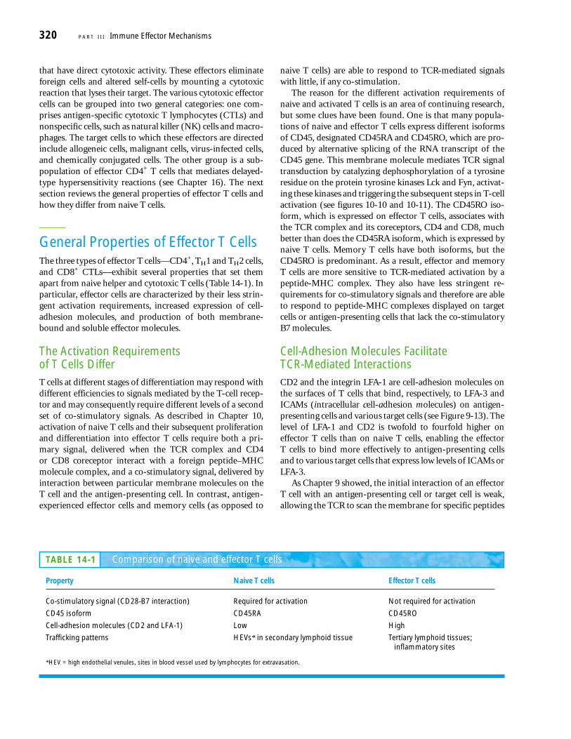
TABLE 14-1 Comparison of naive and effector T cells
Property Naive T cells Effector T cells
Co-stimulatory signal (CD28-B7 interaction) Required for activation Not required for activation
CD45 isoform CD45RA CD45RO
Cell-adhesion molecules (CD2 and LFA-1) Low High
Trafficking patterns HEVs* in secondary lymphoid tissue Tertiary lymphoid tissues;inflammatory sites
*HEV = high endothelial venules, sites in blood vessel used by lymphocytes for extravasation.
presented by self-MHC molecules. If no peptide-MHC com-plex is recognized by the effector cell, it will disengage fromthe APC or target cell. Recognition of a peptide-MHC com-plex by the TCR, however, produces a signal that increases theaffinity of LFA-1 for ICAMs on the APC or target-cell membrane, prolonging the interaction between the cells.For example, TH1 effector cells remain bound to macro-phages that display peptide–class II MHC complexes; TH2 ef-fector cells remain bound to B cells that display peptide–classII MHC complexes; and CTL effector cells bind tightly tovirus-infected target cells that display peptide–class I MHCcomplexes.
Effector T Cells Express a Variety of Effector MoleculesUnlike naive T cells, effector T cells express certain effectormolecules, which may be membrane bound or soluble (Table14-2). The membrane-bound molecules belong to the tumornecrosis factor (TNF) family of membrane proteins andinclude the Fas ligand (FASL) on CD8+ CTLs, TNF-� on TH1cells, and the CD40 ligand on TH2 cells. Each of the effectorT-cell populations also secretes distinct panels of solubleeffector molecules. CTLs secrete cytotoxins (perforins andgranzymes) as well as two cytokines, IFN-� and TNF-�. Asdescribed in Chapter 12, the TH1 and TH2 subsets secretelargely nonoverlapping sets of cytokines.
Each of these membrane-bound and secreted moleculesplays an important role in various T-cell effector functions.The Fas ligand, perforins, and granzymes, for example, medi-ate target-cell destruction by the CTL; membrane-boundTNF-� and soluble IFN-� and GM-CSF promote macro-phage activation by the TH1 cell; and the membrane-boundCD40 ligand and soluble IL-4, IL-5, and IL-6 all play a role inB-cell activation by the TH2 cell.
Cytotoxic T CellsCytotoxic T lymphocytes, or CTLs, are generated by immuneactivation of T cytotoxic (TC) cells. These effector cells havelytic capability and are critical in the recognition and elimi-nation of altered self-cells (e.g., virus-infected cells and
tumor cells) and in graft-rejection reactions. In general,CTLs are CD8+ and are therefore class I MHC restricted, al-though in rare instances CD4+ class II–restricted T cells havebeen shown to function as CTLs. Since virtually all nucleatedcells in the body express class I MHC molecules, CTLs canrecognize and eliminate almost any altered body cell.
The CTL-mediated immune response can be divided intotwo phases, reflecting different aspects of the response. Thefirst phase activates and differentiates naive TC cells intofunctional effector CTLs. In the second phase, effector CTLsrecognize antigen–class I MHC complexes on specific targetcells, which leads them to destroy the target cells.
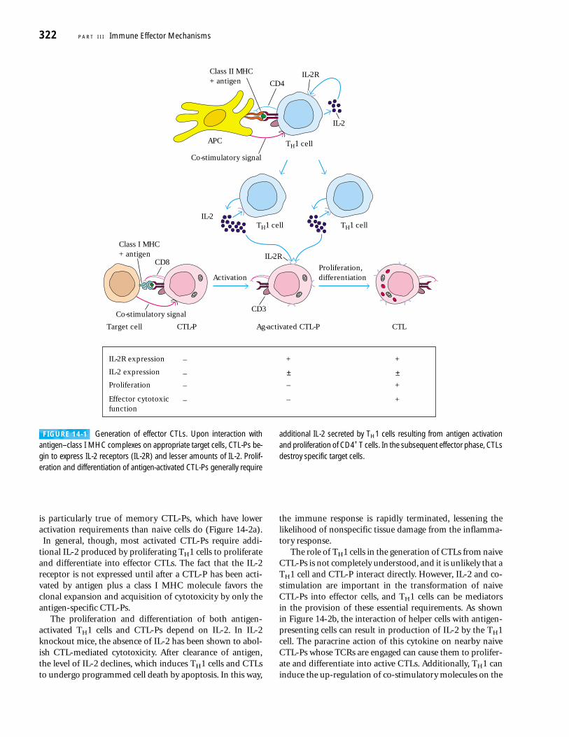
Effector CTLs Are Generated from CTL PrecursorsNaive TC cells are incapable of killing target cells and are there-fore referred to as CTL precursors (CTL-Ps) to denote theirfunctionally immature state. Only after a CTL-P has been acti-vated will the cell differentiate into a functional CTL withcytotoxic activity. Generation of CTLs from CTL-Ps appears torequire at least three sequential signals (Figure 14-1):
■ An antigen-specific signal 1 transmitted by the TCRcomplex upon recognition of a peptide–class I MHCmolecule complex
■ A co-stimulatory signal transmitted by the CD28-B7interaction of the CTL-P and the antigen-presenting cell
■ A signal induced by the interaction of IL-2 with thehigh-affinity IL-2 receptor, resulting in proliferation anddifferentiation of the antigen-activated CTL-P intoeffector CTLs
Unactivated CTL-Ps do not express IL-2 or IL-2 receptors,do not proliferate, and do not display cytotoxic activity. Anti-gen activation induces a CTL-P to begin expressing the IL-2receptor and to a lesser extent IL-2, the principal cytokinerequired for proliferation and differentiation of activatedCTL-Ps into effector CTLs. In some cases, the amount ofIL-2 secreted by an antigen-activated CTL-P may be suffi-cient to induce its own proliferation and differentiation; this
Cell-Mediated Effector Responses C H A P T E R 14 321
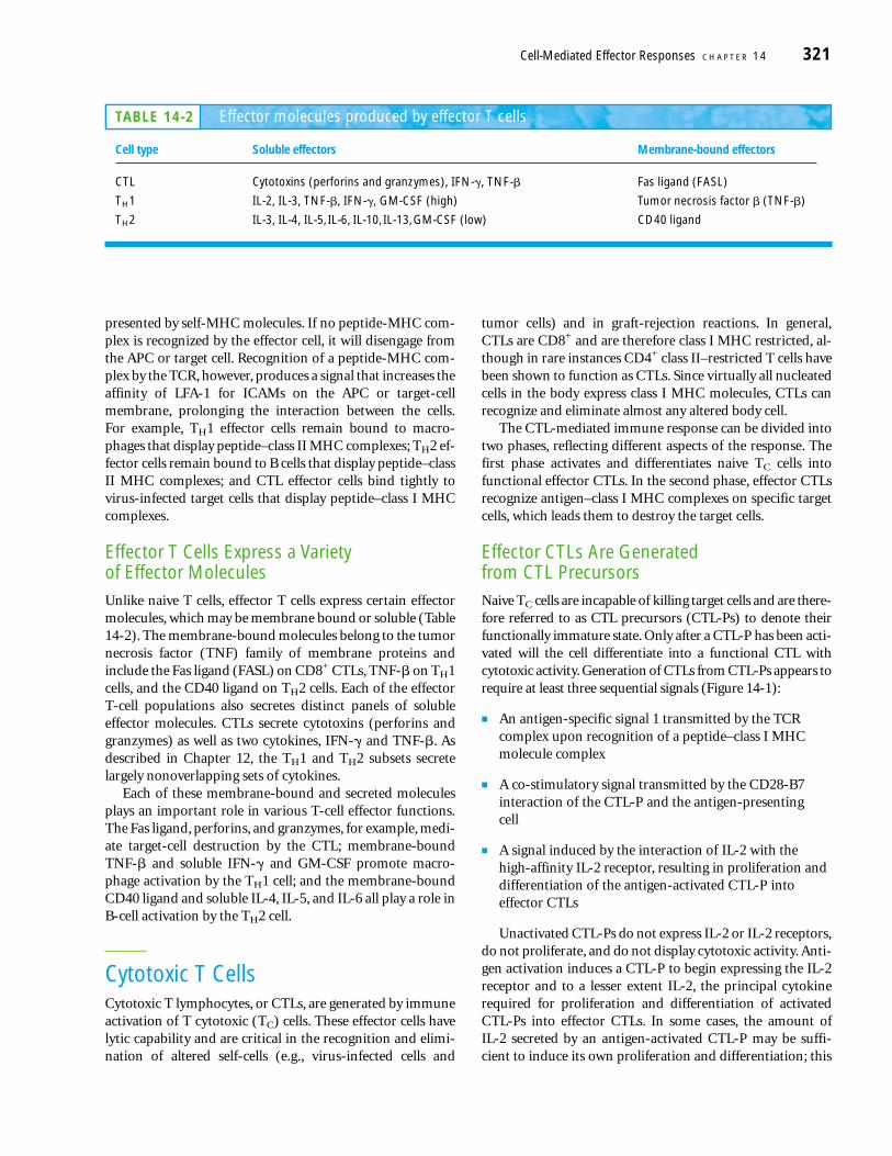
TABLE 14-2 Effector molecules produced by effector T cells
Cell type Soluble effectors Membrane-bound effectors
CTL Cytotoxins (perforins and granzymes), IFN-�, TNF-� Fas ligand (FASL)
TH1 IL-2, IL-3, TNF-�, IFN-�, GM-CSF (high) Tumor necrosis factor � (TNF-�)
TH2 IL-3, IL-4, IL-5, IL-6, IL-10, IL-13, GM-CSF (low) CD40 ligand
is particularly true of memory CTL-Ps, which have loweractivation requirements than naive cells do (Figure 14-2a).In general, though, most activated CTL-Ps require addi-
tional IL-2 produced by proliferating TH1 cells to proliferateand differentiate into effector CTLs. The fact that the IL-2receptor is not expressed until after a CTL-P has been acti-vated by antigen plus a class I MHC molecule favors theclonal expansion and acquisition of cytotoxicity by only theantigen-specific CTL-Ps.
The proliferation and differentiation of both antigen-activated TH1 cells and CTL-Ps depend on IL-2. In IL-2knockout mice, the absence of IL-2 has been shown to abol-ish CTL-mediated cytotoxicity. After clearance of antigen,the level of IL-2 declines, which induces TH1 cells and CTLsto undergo programmed cell death by apoptosis. In this way,
the immune response is rapidly terminated, lessening thelikelihood of nonspecific tissue damage from the inflamma-tory response.
The role of TH1 cells in the generation of CTLs from naiveCTL-Ps is not completely understood, and it is unlikely that aTH1 cell and CTL-P interact directly. However, IL-2 and co-stimulation are important in the transformation of naiveCTL-Ps into effector cells, and TH1 cells can be mediators in the provision of these essential requirements. As shown in Figure 14-2b, the interaction of helper cells with antigen-presenting cells can result in production of IL-2 by the TH1cell. The paracrine action of this cytokine on nearby naiveCTL-Ps whose TCRs are engaged can cause them to prolifer-ate and differentiate into active CTLs. Additionally, TH1 caninduce the up-regulation of co-stimulatory molecules on the
322 P A R T I I I Immune Effector Mechanisms
IL-2
APC
Class II MHC+ antigen CD4
IL-2R
TH1 cell TH1 cell
TH1 cell
IL-2
Co-stimulatory signal
ActivationProliferation,differentiation
Target cell CTL-P
CD8
CTL
Class I MHC+ antigen IL-2R
CD3
Ag-activated CTL-P
Co-stimulatory signal
IL-2R expression
Proliferation
Effector cytotoxicfunction
+–
–
–
+
IL-2 expression –
+
+
–
–
± ±
FIGURE 14-1 Generation of effector CTLs. Upon interaction withantigen–class I MHC complexes on appropriate target cells, CTL-Ps be-gin to express IL-2 receptors (IL-2R) and lesser amounts of IL-2. Prolif-eration and differentiation of antigen-activated CTL-Ps generally require
additional IL-2 secreted by TH1 cells resulting from antigen activationand proliferation of CD4+ T cells. In the subsequent effector phase, CTLsdestroy specific target cells.
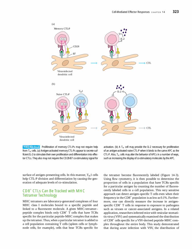
surface of antigen-presenting cells. In this manner, TH1 cellshelp CTL-P division and differentiation by causing the gen-eration of adequate levels of co-stimulation.
CD8+ CTLs Can Be Tracked with MHCTetramer TechnologyMHC tetramers are laboratory-generated complexes of fourMHC class I molecules bound to a specific peptide andlinked to a fluorescent molecule. A given MHC-tetramer–peptide complex binds only CD8+ T cells that have TCRsspecific for the particular peptide-MHC complex that makesup the tetramer. Thus, when a particular tetramer is added toa cell population containing T cells (spleen cells or lymph-node cells, for example), cells that bear TCRs specific for
the tetramer become fluorescently labeled (Figure 14-3).Using flow cytometry, it is then possible to determine theproportion of cells in a population that have TCRs specificfor a particular antigen by counting the number of fluores-cently labeled cells in a cell population. This very sensitiveapproach can detect antigen-specific T cells even when theirfrequency in the CD8+ population is as low as 0.1%. Further-more, one can directly measure the increase in antigen-specific CD8+ T cells in response to exposure to pathogenssuch as viruses or cancer-associated antigens. In a relatedapplication, researchers infected mice with vesicular stomati-tis virus (VSV) and systematically examined the distributionof CD8+ cells specific for a VSV-derived peptide-MHC com-plex throughout the entire body. This study demonstratedthat during acute infection with VSV, the distribution of
Cell-Mediated Effector Responses C H A P T E R 14 323
(a)
IL-2
Memory CTL-P
B7
CD28
CTL
(b)
Virus-infecteddendritic cell
Naive CTL-PTH1 IL-2
CTL
Virus-infecteddendritic cell
1 2
1 21 2
FIGURE 14-2 Proliferation of memory CTL-Ps may not require helpfrom TH cells. (a) Antigen-activated memory CTL-Ps appear to secrete suf-ficient IL-2 to stimulate their own proliferation and differentiation into effec-tor CTLs. They also may not require the CD28-B7 co-stimulatory signal for
activation. (b) A TH cell may provide the IL-2 necessary for proliferation of an antigen-activated naive CTL-P when it binds to the same APC as theCTL-P. Also, TH cells may alter the behavior of APCs in a number of ways,such as increasing the display of co-stimulatory molecules by the APC.
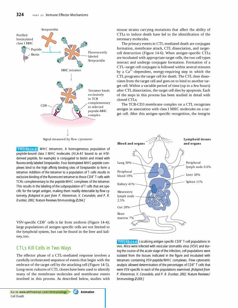
VSV-specific CD8+ cells is far from uniform (Figure 14-4);large populations of antigen-specific cells are not limited tothe lymphoid system, but can be found in the liver and kid-ney, too.
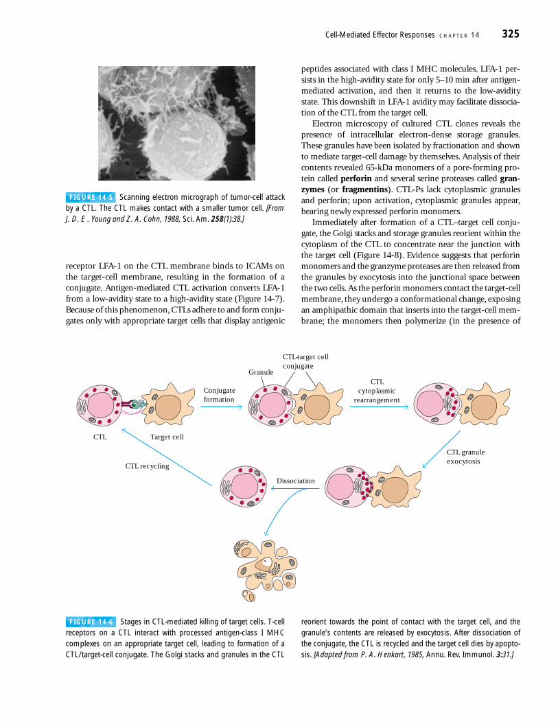
CTLs Kill Cells in Two WaysThe effector phase of a CTL-mediated response involves acarefully orchestrated sequence of events that begin with theembrace of the target cell by the attacking cell (Figure 14-5).Long-term cultures of CTL clones have been used to identifymany of the membrane molecules and membrane eventsinvolved in this process. As described below, studies with
mouse strains carrying mutations that affect the ability ofCTLs to induce death have led to the identification of thenecessary molecules.
The primary events in CTL-mediated death are conjugateformation, membrane attack, CTL dissociation, and target-cell destruction (Figure 14-6). When antigen-specific CTLsare incubated with appropriate target cells, the two cell typesinteract and undergo conjugate formation. Formation of aCTL–target cell conjugate is followed within several minutesby a Ca2+-dependent, energy-requiring step in which theCTL programs the target cell for death. The CTL then disso-ciates from the target cell and goes on to bind to another tar-get cell. Within a variable period of time (up to a few hours)after CTL dissociation, the target cell dies by apoptosis. Eachof the steps in this process has been studied in detail withcloned CTLs.
The TCR-CD3 membrane complex on a CTL recognizesantigen in association with class I MHC molecules on a tar-get cell. After this antigen-specific recognition, the integrin
324 P A R T I I I Immune Effector Mechanisms
MHC tetramer
Signal measured by flow cytometer
Purifiedbiotinylatedclass I MHC
FluorescentlylabeledStreptavidin
Streptavidin
Tetramer bindsexclusivelyto TCRcomplementaryto selectedpeptide-MHCcomplex
Peptide
Biotin
CD8
Peripherallymph node 0.6%
Spleen 11%
Lung 30%
Blood and organsLymphoid tissuesand organs
Peripheralblood 19% Liver 30%
Kidney 41%
Gut 28%
Mesentericlymph node2.5%
Bonemarrow 7%
FIGURE 14-3 MHC tetramers. A homogeneous population of peptide-bound class I MHC molecules (HLA-A1 bound to an HIV-derived peptide, for example) is conjugated to biotin and mixed withfluorescently labeled Streptavidin. Four biotinylated MHC-peptide com-plexes bind to the high affinity binding sites of Streptavidin to form atetramer. Addition of the tetramer to a population of T cells results inexclusive binding of the fluorescent tetramer to those CD8+ T cells withTCRs complementary to the peptide-MHC complexes of the tetramer.This results in the labeling of the subpopulation of T cells that are spe-cific for the target antigen, making them readily detectable by flow cy-tometry. [Adapted in part from P. Klenerman, V. Cerundolo, and P. R.Dunbar, 2002, Nature Reviews/Immunology 2:264.]
FIGURE 14-4 Localizing antigen specific CD8+ T-cell populations invivo. Mice were infected with vesicular stomatitis virus (VSV) and dur-ing the course of the acute stage of the infection, cell populations wereisolated from the tissues indicated in the figure and incubated withtetramers containing VSV-peptide/MHC complexes. Flow cytometricanalysis allowed determination of the percentages of CD8+ T cells thatwere VSV-specific in each of the populations examined. [Adapted fromP. Klenerman, V. Cerundolo, and P. R. Dunbar, 2002, Nature Reviews/Immunology 2:269.]
Go to www.whfreeman.com/immunology AnimationCell Death
receptor LFA-1 on the CTL membrane binds to ICAMs onthe target-cell membrane, resulting in the formation of aconjugate. Antigen-mediated CTL activation converts LFA-1from a low-avidity state to a high-avidity state (Figure 14-7).Because of this phenomenon, CTLs adhere to and form conju-gates only with appropriate target cells that display antigenic
peptides associated with class I MHC molecules. LFA-1 per-sists in the high-avidity state for only 5–10 min after antigen-mediated activation, and then it returns to the low-aviditystate. This downshift in LFA-1 avidity may facilitate dissocia-tion of the CTL from the target cell.
Electron microscopy of cultured CTL clones reveals thepresence of intracellular electron-dense storage granules.These granules have been isolated by fractionation and shownto mediate target-cell damage by themselves. Analysis of theircontents revealed 65-kDa monomers of a pore-forming pro-tein called perforin and several serine proteases called gran-zymes (or fragmentins). CTL-Ps lack cytoplasmic granulesand perforin; upon activation, cytoplasmic granules appear,bearing newly expressed perforin monomers.
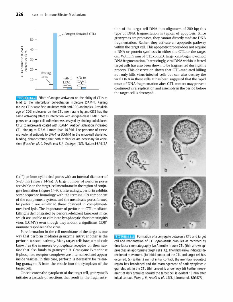
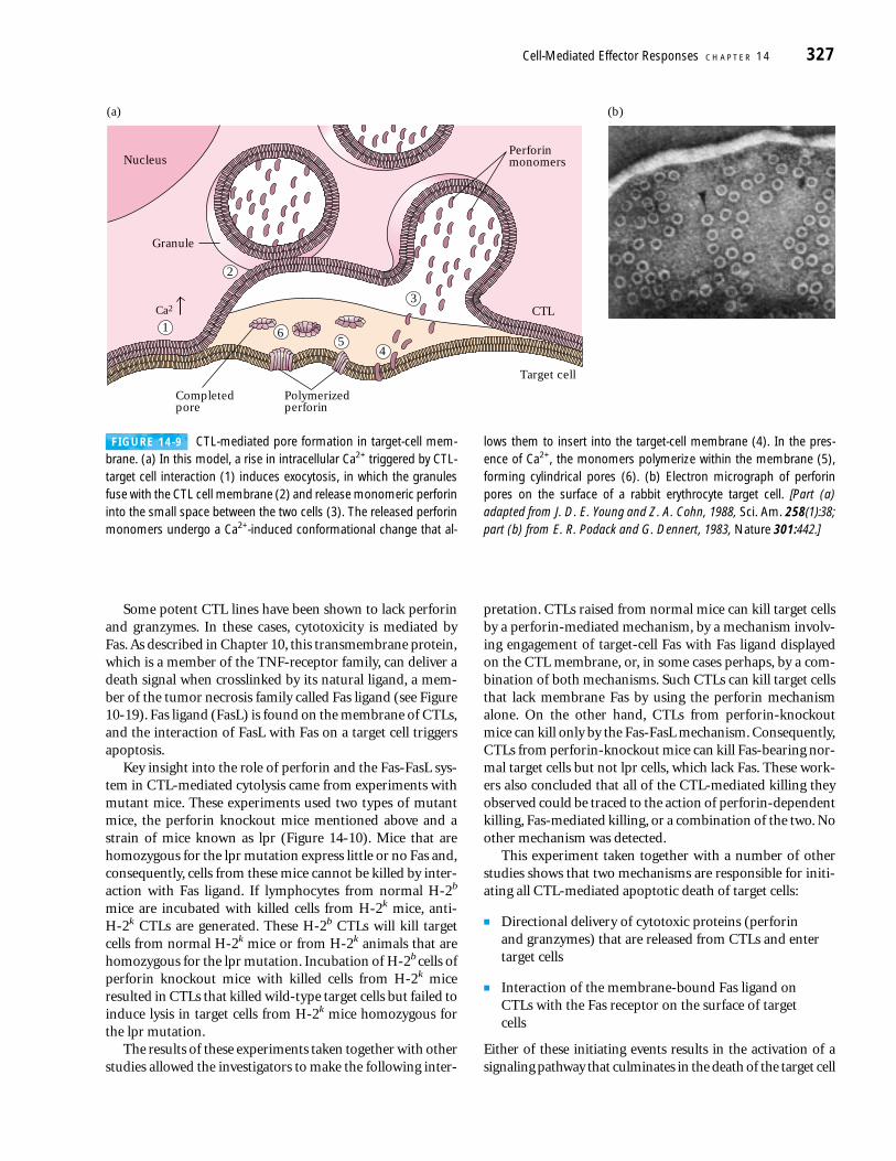
Immediately after formation of a CTL–target cell conju-gate, the Golgi stacks and storage granules reorient within thecytoplasm of the CTL to concentrate near the junction withthe target cell (Figure 14-8). Evidence suggests that perforinmonomers and the granzyme proteases are then released fromthe granules by exocytosis into the junctional space betweenthe two cells. As the perforin monomers contact the target-cellmembrane, they undergo a conformational change, exposingan amphipathic domain that inserts into the target-cell mem-brane; the monomers then polymerize (in the presence of
Cell-Mediated Effector Responses C H A P T E R 14 325
FIGURE 14-5 Scanning electron micrograph of tumor-cell attackby a CTL. The CTL makes contact with a smaller tumor cell. [From J. D. E . Young and Z. A. Cohn, 1988, Sci. Am. 258(1):38.]
CTL
CTL granuleexocytosis
Target cell
Conjugateformation
CTL-target cellconjugate
CTLcytoplasmic
rearrangement
CTL recycling
Dissociation
Granule
FIGURE 14-6 Stages in CTL-mediated killing of target cells. T-cellreceptors on a CTL interact with processed antigen-class I MHCcomplexes on an appropriate target cell, leading to formation of aCTL/target-cell conjugate. The Golgi stacks and granules in the CTL
reorient towards the point of contact with the target cell, and thegranule’s contents are released by exocytosis. After dissociation ofthe conjugate, the CTL is recycled and the target cell dies by apopto-sis. [Adapted from P. A. Henkart, 1985, Annu. Rev. Immunol. 3:31.]
Ca2+) to form cylindrical pores with an internal diameter of5–20 nm (Figure 14-9a). A large number of perforin poresare visible on the target-cell membrane in the region of conju-gate formation (Figure 14-9b). Interestingly, perforin exhibitssome sequence homology with the terminal C9 componentof the complement system, and the membrane pores formedby perforin are similar to those observed in complement-mediated lysis. The importance of perforin to CTL-mediatedkilling is demonstrated by perforin-deficient knockout mice,which are unable to eliminate lymphocytic choriomeningitisvirus (LCMV) even though they mount a significant CD8+
immune response to the virus.Pore formation in the cell membrane of the target is one
way that perforin mediates granzyme entry; another is theperforin-assisted pathway. Many target cells have a moleculeknown as the mannose 6-phosphate receptor on their sur-face that also binds to granzyme B. Granzyme B/mannose 6-phosphate receptor complexes are internalized and appearinside vesicles. In this case, perforin is necessary for releas-ing granzyme B from the vesicle into the cytoplasm of thetarget cell.
Once it enters the cytoplasm of the target cell, granzyme Binitiates a cascade of reactions that result in the fragmenta-
tion of the target-cell DNA into oligomers of 200 bp; thistype of DNA fragmentation is typical of apoptosis. Sincegranzymes are proteases, they cannot directly mediate DNAfragmentation. Rather, they activate an apoptotic pathwaywithin the target cell. This apoptotic process does not requiremRNA or protein synthesis in either the CTL or the targetcell. Within 5 min of CTL contact, target cells begin to exhibitDNA fragmentation. Interestingly, viral DNA within infectedtarget cells has also been shown to be fragmented during thisprocess. This observation shows that CTL-mediated killingnot only kills virus-infected cells but can also destroy theviral DNA in those cells. It has been suggested that the rapidonset of DNA fragmentation after CTL contact may preventcontinued viral replication and assembly in the period beforethe target cell is destroyed.
326 P A R T I I I Immune Effector Mechanisms
50
CT
Ls b
ou
nd
to
IC
AM
-1
coat
ed w
ells
, %
40
30
20
10
Antigen-activated CTLs
+Ab toLFA-1
RestingCTLs +Ab to
ICAM-1
FIGURE 14-7 Effect of antigen activation on the ability of CTLs tobind to the intercellular cell-adhesion molecule ICAM-1. Restingmouse CTLs were first incubated with anti-CD3 antibodies. Crosslink-age of CD3 molecules on the CTL membrane by anti-CD3 has thesame activating effect as interaction with antigen–class I MHC com-plexes on a target cell. Adhesion was assayed by binding radiolabeledCTLs to microwells coated with ICAM-1. Antigen activation increasedCTL binding to ICAM-1 more than 10-fold. The presence of excessmonoclonal antibody to LFA-1 or ICAM-1 in the microwell abolishedbinding, demonstrating that both molecules are necessary for adhe-sion. [Based on M. L. Dustin and T. A. Springer, 1989, Nature 341:619.]
FIGURE 14-8 Formation of a conjugate between a CTL and targetcell and reorientation of CTL cytoplasmic granules as recorded bytime-lapse cinematography. (a) A motile mouse CTL (thin arrow) ap-proaches an appropriate target cell (TC). The thick arrow indicates di-rection of movement. (b) Initial contact of the CTL and target cell hasoccurred. (c) Within 2 min of initial contact, the membrane-contactregion has broadened and the rearrangement of dark cytoplasmicgranules within the CTL (thin arrow) is under way. (d) Further move-ment of dark granules toward the target cell is evident 10 min afterinitial contact. [From J. R. Yanelli et al., 1986, J. Immunol. 136:377].
Some potent CTL lines have been shown to lack perforinand granzymes. In these cases, cytotoxicity is mediated byFas. As described in Chapter 10, this transmembrane protein,which is a member of the TNF-receptor family, can deliver adeath signal when crosslinked by its natural ligand, a mem-ber of the tumor necrosis family called Fas ligand (see Figure10-19). Fas ligand (FasL) is found on the membrane of CTLs,and the interaction of FasL with Fas on a target cell triggersapoptosis.
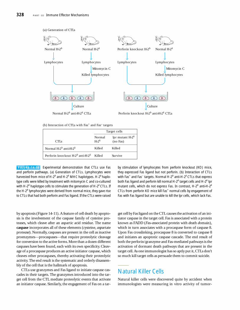
Key insight into the role of perforin and the Fas-FasL sys-tem in CTL-mediated cytolysis came from experiments withmutant mice. These experiments used two types of mutantmice, the perforin knockout mice mentioned above and astrain of mice known as lpr (Figure 14-10). Mice that arehomozygous for the lpr mutation express little or no Fas and,consequently, cells from these mice cannot be killed by inter-action with Fas ligand. If lymphocytes from normal H-2b
mice are incubated with killed cells from H-2k mice, anti-H-2k CTLs are generated. These H-2b CTLs will kill targetcells from normal H-2k mice or from H-2k animals that arehomozygous for the lpr mutation. Incubation of H-2b cells ofperforin knockout mice with killed cells from H-2k miceresulted in CTLs that killed wild-type target cells but failed toinduce lysis in target cells from H-2k mice homozygous forthe lpr mutation.
The results of these experiments taken together with otherstudies allowed the investigators to make the following inter-
pretation. CTLs raised from normal mice can kill target cellsby a perforin-mediated mechanism, by a mechanism involv-ing engagement of target-cell Fas with Fas ligand displayedon the CTL membrane, or, in some cases perhaps, by a com-bination of both mechanisms. Such CTLs can kill target cellsthat lack membrane Fas by using the perforin mechanismalone. On the other hand, CTLs from perforin-knockoutmice can kill only by the Fas-FasL mechanism. Consequently,CTLs from perforin-knockout mice can kill Fas-bearing nor-mal target cells but not lpr cells, which lack Fas. These work-ers also concluded that all of the CTL-mediated killing theyobserved could be traced to the action of perforin-dependentkilling, Fas-mediated killing, or a combination of the two. Noother mechanism was detected.
This experiment taken together with a number of otherstudies shows that two mechanisms are responsible for initi-ating all CTL-mediated apoptotic death of target cells:
■ Directional delivery of cytotoxic proteins (perforin and granzymes) that are released from CTLs and entertarget cells
■ Interaction of the membrane-bound Fas ligand on CTLs with the Fas receptor on the surface of target cells
Either of these initiating events results in the activation of asignaling pathway that culminates in the death of the target cell
Cell-Mediated Effector Responses C H A P T E R 14 327
(a) (b)
Nucleus
Granule
2
65
4
3
1
Perforin monomers
CTL
Target cell
Polymerizedperforin
Completedpore
Ca2
FIGURE 14-9 CTL-mediated pore formation in target-cell mem-brane. (a) In this model, a rise in intracellular Ca2+ triggered by CTL-target cell interaction (1) induces exocytosis, in which the granulesfuse with the CTL cell membrane (2) and release monomeric perforininto the small space between the two cells (3). The released perforinmonomers undergo a Ca2+-induced conformational change that al-
lows them to insert into the target-cell membrane (4). In the pres-ence of Ca2+, the monomers polymerize within the membrane (5),forming cylindrical pores (6). (b) Electron micrograph of perforinpores on the surface of a rabbit erythrocyte target cell. [Part (a)adapted from J. D. E. Young and Z. A. Cohn, 1988, Sci. Am. 258(1):38;part (b) from E. R. Podack and G. Dennert, 1983, Nature 301:442.]
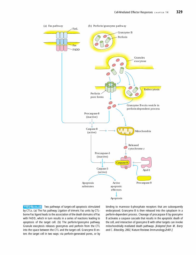
by apoptosis (Figure 14-11). A feature of cell death by apopto-sis is the involvement of the caspase family of cysteine pro-teases, which cleave after an aspartic acid residue. The namecaspase incorporates all of these elements (cysteine, aspartateprotease). Normally, caspases are present in the cell as inactiveproenzymes—procaspases—that require proteolytic cleavagefor conversion to the active forms. More than a dozen differentcaspases have been found, each with its own specificity. Cleav-age of a procaspase produces an active initiator caspase, whichcleaves other procaspases, thereby activating their proteolyticactivity. The end result is the systematic and orderly disassem-bly of the cell that is the hallmark of apoptosis.
CTLs use granzymes and Fas ligand to initiate caspase cas-cades in their targets. The granzymes introduced into the tar-get cell from the CTL mediate proteolytic events that activatean initiator caspase. Similarly, the engagement of Fas on a tar-
get cell by Fas ligand on the CTL causes the activation of an ini-tiator caspase in the target cell. Fas is associated with a proteinknown as FADD (Fas-associated protein with death domain),which in turn associates with a procaspase form of caspase 8.Upon Fas crosslinking, procaspase 8 is converted to caspase 8and initiates an apoptotic caspase cascade. The end result ofboth the perforin/granzyme and Fas-mediated pathways is theactivation of dormant death pathways that are present in thetarget cell. As one immunologist has so aptly put it, CTLs don’tso much kill target cells as persuade them to commit suicide.
Natural Killer CellsNatural killer cells were discovered quite by accident whenimmunologists were measuring in vitro activity of tumor-
328 P A R T I I I Immune Effector Mechanisms
Normal H-2b
Normal H-2b anti-H-2k CTLs
Normal H-2b anti-H-2k
Perforin knockout H-2b anti-H-2k
Normal H-2k
Killed Killed
Killed Survive
CTLslpr mutant H-2k
(no Fas)
Lymphocytes
Normal H-2k
Lymphocytes
Mitomycin C
Killed lymphocytes Killed lymphocytes
Culture
(a) Generation of CTLs
(b) Interaction of CTLs with Fas+ and Fas– targets
b k b bk
Perforin knockout H-2b
Perforin knockout H-2b anti-H-2k CTLs
Lymphocytes
Normal H-2k
Lymphocytes
Mitomycin C
Culture
b k b bk
Target cells
FIGURE 14-10 Experimental demonstration that CTLs use Fasand perforin pathways. (a) Generation of CTLs. Lymphocytes wereharvested from mice of H-2b and H-2k MHC haplotypes. H-2k haplo-type cells were killed by treatment with mitomycin C and co-culturedwith H-2b haplotype cells to stimulate the generation of H-2k CTLs. Ifthe H-2b lymphocytes were derived from normal mice, they gave riseto CTLs that had both perforin and Fas ligand. If the CTLs were raised
by stimulation of lymphocytes from perforin knockout (KO) mice,they expressed Fas ligand but not perforin. (b) Interaction of CTLswith Fas+ and Fas– targets. Normal H-2b anti-H-2k CTLs that expressboth Fas ligand and perforin kill normal H-2k target cells and H-2k lprmutant cells, which do not express Fas. In contrast, H-2b anti-H-2k
CTLs from perforin KO mice kill Fas+ normal cells by engagement ofFas with Fas ligand but are unable to kill the lpr cells, which lack Fas.
Cell-Mediated Effector Responses C H A P T E R 14 329
(a) Fas pathway (b) Perforin/granzyme pathway
Fas
FADD
Granzyme B exits vesicle inperforin-dependent process
Caspase-8(active)
FasL
Mitochondria
Procaspase-3(inactive)
Caspase-3(active)
Procaspase-8(inactive)
Apoptosissubstrates
Apoptosis
Activeapoptoticeffectors
Caspase-9
Procaspase-9
Releasedcytochrome c
Apaf-1
Endoccytosis
Perforinpore forms
Granulesexocytose
Granzyme B
Perforin
FIGURE 14-11 Two pathways of target-cell apoptosis stimulatedby CTLs. (a) The Fas pathway. Ligation of trimeric Fas units by CTL-borne Fas ligand leads to the association of the death domains of Faswith FADD, which in turn results in a series of reactions leading toapoptosis of the target cell. (b) The perforin/granzyme pathway.Granule exocytosis releases granzymes and perforin from the CTLinto the space between the CTL and the target cell. Granzyme B en-ters the target cell in two ways: via perforin-generated pores, or by
binding to mannose 6-phosphate receptors that are subsequently endocytosed. Granzyme B is then released into the cytoplasm in aperforin-dependent process. Cleavage of procaspase 8 by granzymeB activates a caspase cascade that results in the apoptotic death ofthe cell, and interaction of granzyme B with other targets can invokemitochondrially mediated death pathways. [Adapted from M. Barryand C. Bleackley, 2002, Nature Reviews Immunology 2:401.]
specific cells taken from mice with tumors. Normal unim-munized mice and mice with unrelated tumors served asnegative controls. Much to the consternation of the investi-gators, the controls showed significant lysis of the tumorcells, too. Characterization of this nonspecific tumor-cellkilling revealed that a population of large granular lympho-cytes was responsible. The cells, which were named naturalkiller (NK) cells for their nonspecific cytotoxicity, make up5%–10% of the recirculating lymphocyte population. Thesecells are involved in immune defenses against viruses andtumors. Because NK cells produce a number of immunolog-ically important cytokines, they play important roles in im-mune regulation and influence both innate and adaptiveimmunity. In particular, IFN-� production by NK cells canaffect the participation of macrophages in innate immunityby activation of the phagocytic and microbicidal activities.IFN-� derived from NK cells can influence the TH1 versusTH2 commitment of helper T cell populations by its in-hibitory effects on TH2 expansion, and stimulate TH1 devel-opment via induction of IL-12 by macrophages and den-dritic cells. The Chediak-Higashi syndrome described in theClinical Focus illustrates the disastrous consequences of alack of NK cells.
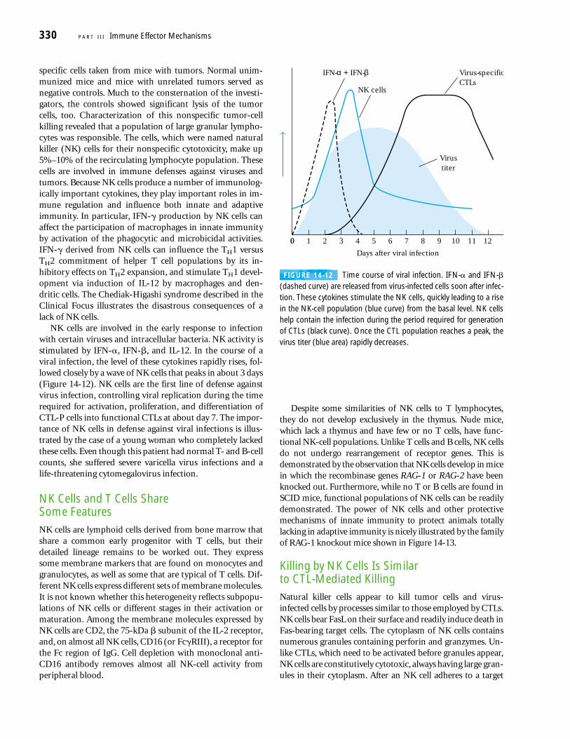
NK cells are involved in the early response to infectionwith certain viruses and intracellular bacteria. NK activity isstimulated by IFN-�, IFN-�, and IL-12. In the course of aviral infection, the level of these cytokines rapidly rises, fol-lowed closely by a wave of NK cells that peaks in about 3 days(Figure 14-12). NK cells are the first line of defense againstvirus infection, controlling viral replication during the timerequired for activation, proliferation, and differentiation ofCTL-P cells into functional CTLs at about day 7. The impor-tance of NK cells in defense against viral infections is illus-trated by the case of a young woman who completely lackedthese cells. Even though this patient had normal T- and B-cellcounts, she suffered severe varicella virus infections and alife-threatening cytomegalovirus infection.
NK Cells and T Cells Share Some Features NK cells are lymphoid cells derived from bone marrow thatshare a common early progenitor with T cells, but theirdetailed lineage remains to be worked out. They expresssome membrane markers that are found on monocytes andgranulocytes, as well as some that are typical of T cells. Dif-ferent NK cells express different sets of membrane molecules.It is not known whether this heterogeneity reflects subpopu-lations of NK cells or different stages in their activation ormaturation. Among the membrane molecules expressed byNK cells are CD2, the 75-kDa � subunit of the IL-2 receptor,and, on almost all NK cells, CD16 (or Fc�RIII), a receptor forthe Fc region of IgG. Cell depletion with monoclonal anti-CD16 antibody removes almost all NK-cell activity fromperipheral blood.
Despite some similarities of NK cells to T lymphocytes,they do not develop exclusively in the thymus. Nude mice,which lack a thymus and have few or no T cells, have func-tional NK-cell populations. Unlike T cells and B cells, NK cellsdo not undergo rearrangement of receptor genes. This isdemonstrated by the observation that NK cells develop in micein which the recombinase genes RAG-1 or RAG-2 have beenknocked out. Furthermore, while no T or B cells are found inSCID mice, functional populations of NK cells can be readilydemonstrated. The power of NK cells and other protectivemechanisms of innate immunity to protect animals totallylacking in adaptive immunity is nicely illustrated by the familyof RAG-1 knockout mice shown in Figure 14-13.
Killing by NK Cells Is Similar to CTL-Mediated KillingNatural killer cells appear to kill tumor cells and virus-infected cells by processes similar to those employed by CTLs.NK cells bear FasL on their surface and readily induce death inFas-bearing target cells. The cytoplasm of NK cells containsnumerous granules containing perforin and granzymes. Un-like CTLs, which need to be activated before granules appear,NK cells are constitutively cytotoxic, always having large gran-ules in their cytoplasm. After an NK cell adheres to a target
330 P A R T I I I Immune Effector Mechanisms
0
Days after viral infection
12
NK cells
Virustiter
IFN-α + IFN-β Virus-specificCTLs
0 1 2 3 4 5 6 7 8 9 10 11
FIGURE 14-12 Time course of viral infection. IFN-� and IFN-�(dashed curve) are released from virus-infected cells soon after infec-tion. These cytokines stimulate the NK cells, quickly leading to a risein the NK-cell population (blue curve) from the basal level. NK cellshelp contain the infection during the period required for generationof CTLs (black curve). Once the CTL population reaches a peak, thevirus titer (blue area) rapidly decreases.
cell, degranulation occurs with release of perforin and gran-zymes at the junction of the interacting cells. The roles of per-forin and granzymes in NK-mediated killing of target cells byapoptosis are believed to be similar to their roles in the CTL-mediated process.
Despite these similarities, NK cells differ from CTLs in sev-eral significant ways. First, NK cells do not express antigen-specific T-cell receptors or CD3. In addition, recognition oftarget cells by NK cells is not MHC restricted; that is, in manycases the same levels of NK-cell activity are observed withsyngeneic and allogeneic tumor cells. Moreover, althoughprior priming enhances CTL activity, NK-cell activity doesnot increase after a second injection with the same tumorcells. In other words, the NK-cell response generates no im-munologic memory.
NK Cells Have Both Activation and Inhibition ReceptorsGiven that NK cells do not express antigen-specific receptors,the mechanism by which NK cells recognize altered self-cellsand distinguish them from normal body cells baffled immu-nologists for years. The solution to the problem emergedwith the realization that NK cells employ two different cate-gories of receptors, one that delivers inhibition signals to NKcells, and another that delivers activation signals. Initially, itwas thought that there were two receptors, one that activatedand another that inhibited NK cells—the so-called two-receptor model. It is now clear that there are many different
cell-surface receptors for activation signals and a number ofdifferent kinds for inhibitory ones. Consequently, it is moreappropriate to think in terms of an opposing-signals modelrather than a two-receptor model. It is the balance betweenactivating signals and inhibitory signals that is believed toenable NK cells to distinguish healthy cells from infected orcancerous ones. It is important to be aware that additionalNK-activating signals can be delivered by soluble factors.These include cytokines such as � and � interferons, TNF-�,IL-12, and IL-15.
The exact nature of the membrane-bound receptors onNK cells that produce activation is not completely clear. Anti-body crosslinking of many molecules found on the surface ofNK cells can activate these cells artificially, but the naturalligands for many of these putative activation receptors (ARs)are not known. Some of the candidate ARs are members ofa class of carbohydrate-binding proteins known as C-typelectins, so named because they have calcium-dependent carbohydrate-recognition domains. NKR-P1 is an exampleof a C-type lectin found on NK cells that has activation prop-erties. In addition to lectins, other molecules on NK cellsmight be involved in activation, including CD2 (receptor forthe adhesion molecule LFA-3), and the Fc�III receptor,CD16. Although CD16 is responsible for antibody-mediatedrecognition and killing of target cells by NK cells, it is proba-bly not involved in non-antibody-dependent killing. In addi-tion to the molecules already mentioned, three additionalproteins, NKp30, NKp44, and NKp46, appear to play signifi-cant roles in the activation of human NK cells.
Clues to the sources of inhibitory signals came from stud-ies of the killing of tumor cells and virus-infected cells by NKcells. It was noticed that the preferential killing of mousetumor cells compared with normal cells correlated with alack of expression of MHC molecules by the tumor cells.Experiments with human cells showed that NK cells lysed aB-cell line that was MHC deficient because it had been trans-formed by Epstein-Barr virus. However, when this cell linewas transformed with human HLA genes so that it expressedhigh levels of MHC molecules, NK cells failed to lyse it. Theseobservations led to the idea that NK cells target for killingcells that have aberrant MHC expression. Since many virus-infected and tumor cells have reduced MHC expression, thismodel made good physiological sense. Vindication of thisproposal has come from the discovery of receptors on NKcells that produce inhibitory signals when they recognizeMHC molecules on potential target cells. These inhibitoryreceptors on the NK cell then prevent NK-cell killing, prolif-eration, and cytokine release.
Two major groups of inhibitory receptors have beenfound on NK cells. One of these is a family of C-type-lectin–inhibitory receptors (CLIR), and the other is a group of Ig-superfamily–inhibitory receptors (ISIR) known as the killer-cell–inhibitory receptors (KIR). Even though these groups are chemically quite different, they are together referred to as the inhibitory-receptor superfamily (IRS). In humans, the
Cell-Mediated Effector Responses C H A P T E R 14 331
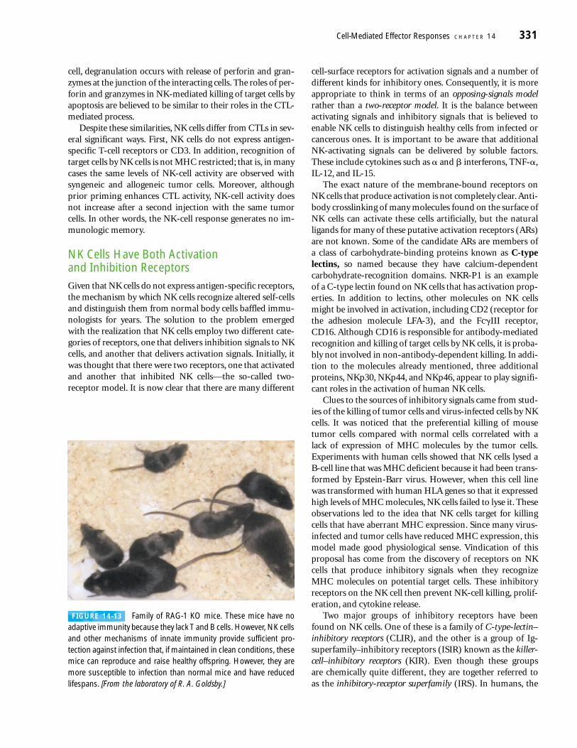
FIGURE 14-13 Family of RAG-1 KO mice. These mice have noadaptive immunity because they lack T and B cells. However, NK cellsand other mechanisms of innate immunity provide sufficient pro-tection against infection that, if maintained in clean conditions, thesemice can reproduce and raise healthy offspring. However, they aremore susceptible to infection than normal mice and have reducedlifespans. [From the laboratory of R. A. Goldsby.]
C-type-lectin–inhibitory receptor is CD94/NKG2, a disulfide-bonded heterodimer made up of two glycoproteins, one ofwhich is CD94 and the other a member of the NKG2 family.The CD94/NKG2 receptors recognize HLA-E on potentialtarget cells. Because HLA-E is not transported to the surfaceof a cell unless it has bound a peptide derived from HLA-A,
HLA-B, or HLA-C, the amount of HLA-E on the surfaceserves as indicator of the overall level of class I MHC biosyn-thesis in the cells. These inhibitory CD94/NKG2 receptorsare thus not specific for a particular HLA allele and will sendinhibitory signals to the NK cell, with the net result thatkilling of potential target cells is inhibited if they are express-
332 P A R T I I I Immune Effector Mechanisms
humans, display the huge cytoplasmicgranules that are a morphological hall-mark of the disease. Studies of the diseasein beige mice complement those in hu-mans, and have led to the conclusion thatsevere defects in the formation, fusion, ortrafficking of intracellular vesicles proba-bly underlie its devastating pathology.
Bone marrow transplantation (BMT)is the only effective therapy for the defec-tive natural killer activity, aberrant macro-phage activation, and susceptibility tobacterial infections that plague those af-flicted with Chediak-Higashi syndrome.However, this is a risky and complex ther-apy. A look at the experience of 10 CHSchildren who underwent BMT for theirdisease is informative. BMT is best done
with marrow from a donor whose HLAtype is identical to that of the recipient. Un-fortunately, it may be difficult or impossibleto obtain HLA-matched bone marrow, and3 of the patients had to settle for HLA-non-identical marrow. After a median interval of6.5 years post-transplantation, 6 of the 7patients who had received marrow fromHLA-identical donors were alive, but only 1of the 3 recipients of HLA-nonidenticalmarrow survived. The clinical picture in thesurvivors was markedly improved. Theywere no longer hypersusceptible to bacte-rial infection, displayed significant NK-cellactivity, and did not suffer from uncon-trolled and pathological macrophage acti-vation. However, the albinism and lack ofeye pigmentation were not improved byBMT. HLA-identical BMT is thus acceptedas a curative treatment for Chediak-Higashi syndrome, but reliance on HLA-nonidentical transplantation is experimen-tal and carries very high risk.
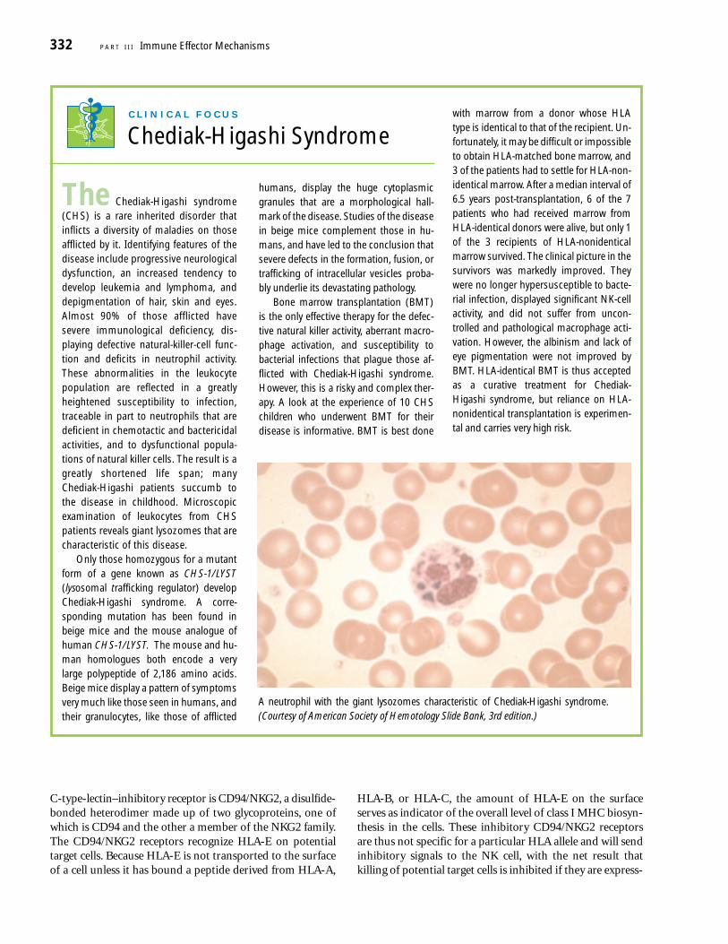
The Chediak-Higashi syndrome(CHS) is a rare inherited disorder thatinflicts a diversity of maladies on thoseafflicted by it. Identifying features of thedisease include progressive neurologicaldysfunction, an increased tendency todevelop leukemia and lymphoma, anddepigmentation of hair, skin and eyes.Almost 90% of those afflicted havesevere immunological deficiency, dis-playing defective natural-killer-cell func-tion and deficits in neutrophil activity.These abnormalities in the leukocytepopulation are reflected in a greatlyheightened susceptibility to infection,traceable in part to neutrophils that aredeficient in chemotactic and bactericidalactivities, and to dysfunctional popula-tions of natural killer cells. The result is agreatly shortened life span; manyChediak-Higashi patients succumb tothe disease in childhood. Microscopicexamination of leukocytes from CHSpatients reveals giant lysozomes that arecharacteristic of this disease.
Only those homozygous for a mutantform of a gene known as CHS-1/LYST(lysosomal trafficking regulator) developChediak-Higashi syndrome. A corre-sponding mutation has been found inbeige mice and the mouse analogue ofhuman CHS-1/LYST. The mouse and hu-man homologues both encode a verylarge polypeptide of 2,186 amino acids.Beige mice display a pattern of symptomsvery much like those seen in humans, andtheir granulocytes, like those of afflicted
C L I N I C A L F O C U S
Chediak-Higashi Syndrome
A neutrophil with the giant lysozomes characteristic of Chediak-Higashi syndrome.(Courtesy of American Society of Hemotology Slide Bank, 3rd edition.)
ing adequate levels of class I. In contrast, KIR receptors, ofwhich more than 50 family members have been found, arespecific for one or a limited number of polymorphic prod-ucts of particular HLA loci. Unlike B and T cells, NK cells arenot limited to expressing a single KIR, but may express sev-eral, each specific for a different MHC molecule or for a set ofclosely related MHC molecules. For example, individualclones of human NK cells expressing a CD94/NKG2 receptorand as many as six different KIR receptors have been found.Because signals from inhibitory receptors have veto powerover signals from activating receptors, a negative signal fromany inhibitory receptor, whether of the CD94/NKG2 or KIRtype, can block the lysis of target cells by NK cells. Thus, cellsexpressing normal levels of unaltered MHC class I moleculestend to escape all forms of NK-cell–mediated killing.
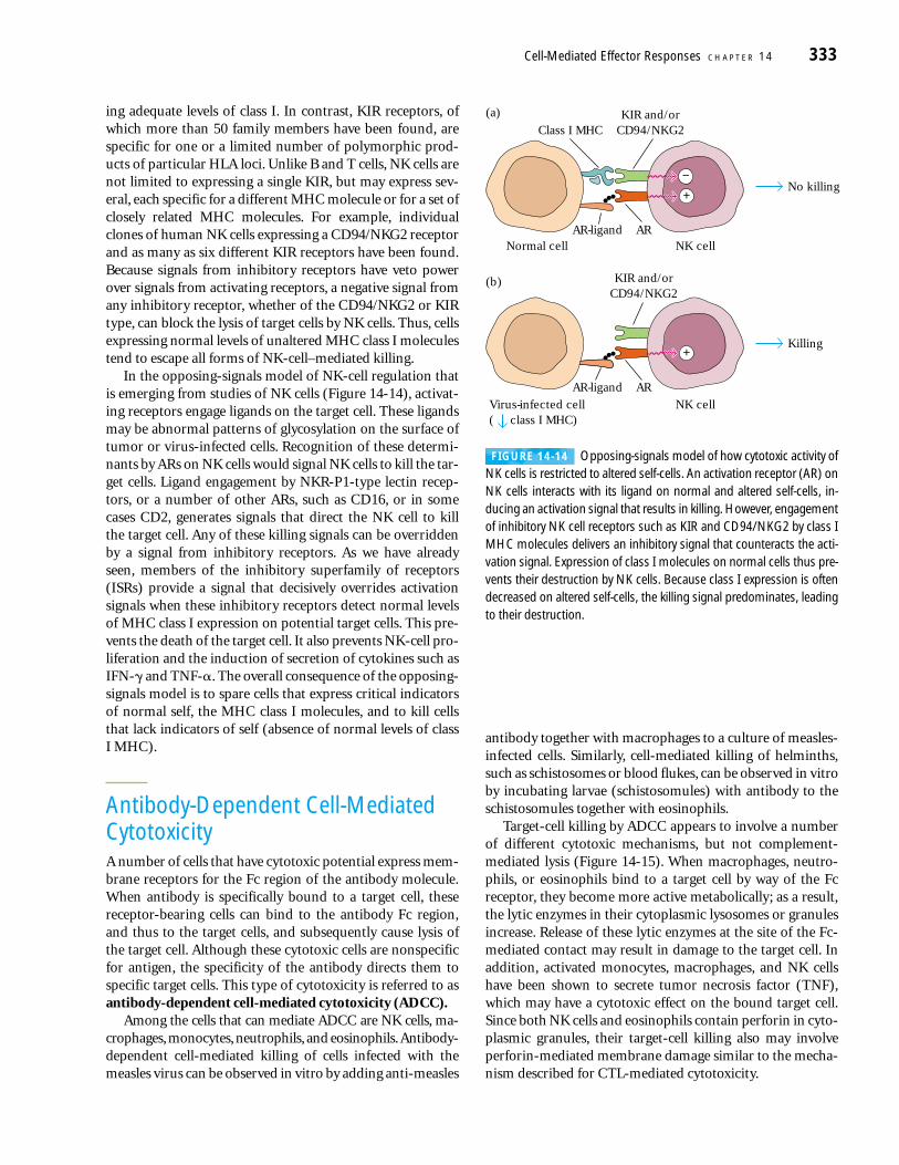
In the opposing-signals model of NK-cell regulation thatis emerging from studies of NK cells (Figure 14-14), activat-ing receptors engage ligands on the target cell. These ligandsmay be abnormal patterns of glycosylation on the surface oftumor or virus-infected cells. Recognition of these determi-nants by ARs on NK cells would signal NK cells to kill the tar-get cells. Ligand engagement by NKR-P1-type lectin recep-tors, or a number of other ARs, such as CD16, or in somecases CD2, generates signals that direct the NK cell to kill the target cell. Any of these killing signals can be overriddenby a signal from inhibitory receptors. As we have alreadyseen, members of the inhibitory superfamily of receptors(ISRs) provide a signal that decisively overrides activationsignals when these inhibitory receptors detect normal levelsof MHC class I expression on potential target cells. This pre-vents the death of the target cell. It also prevents NK-cell pro-liferation and the induction of secretion of cytokines such asIFN-� and TNF-�. The overall consequence of the opposing-signals model is to spare cells that express critical indicatorsof normal self, the MHC class I molecules, and to kill cellsthat lack indicators of self (absence of normal levels of class I MHC).
Antibody-Dependent Cell-MediatedCytotoxicityA number of cells that have cytotoxic potential express mem-brane receptors for the Fc region of the antibody molecule.When antibody is specifically bound to a target cell, thesereceptor-bearing cells can bind to the antibody Fc region,and thus to the target cells, and subsequently cause lysis ofthe target cell. Although these cytotoxic cells are nonspecificfor antigen, the specificity of the antibody directs them tospecific target cells. This type of cytotoxicity is referred to asantibody-dependent cell-mediated cytotoxicity (ADCC).
Among the cells that can mediate ADCC are NK cells, ma-crophages, monocytes, neutrophils, and eosinophils.Antibody-dependent cell-mediated killing of cells infected with themeasles virus can be observed in vitro by adding anti-measles
antibody together with macrophages to a culture of measles-infected cells. Similarly, cell-mediated killing of helminths,such as schistosomes or blood flukes, can be observed in vitroby incubating larvae (schistosomules) with antibody to theschistosomules together with eosinophils.
Target-cell killing by ADCC appears to involve a numberof different cytotoxic mechanisms, but not complement-mediated lysis (Figure 14-15). When macrophages, neutro-phils, or eosinophils bind to a target cell by way of the Fcreceptor, they become more active metabolically; as a result,the lytic enzymes in their cytoplasmic lysosomes or granulesincrease. Release of these lytic enzymes at the site of the Fc-mediated contact may result in damage to the target cell. Inaddition, activated monocytes, macrophages, and NK cellshave been shown to secrete tumor necrosis factor (TNF),which may have a cytotoxic effect on the bound target cell.Since both NK cells and eosinophils contain perforin in cyto-plasmic granules, their target-cell killing also may involveperforin-mediated membrane damage similar to the mecha-nism described for CTL-mediated cytotoxicity.
Cell-Mediated Effector Responses C H A P T E R 14 333
Normal cell NK cell
Class I MHC
No killing
(a)
(b)
+−
KIR and/orCD94/NKG2
KIR and/orCD94/NKG2
ARAR-ligand
ARAR-ligand
NK cell
Killing+
Virus-infected cell( class I MHC)
FIGURE 14-14 Opposing-signals model of how cytotoxic activity ofNK cells is restricted to altered self-cells. An activation receptor (AR) onNK cells interacts with its ligand on normal and altered self-cells, in-ducing an activation signal that results in killing. However, engagementof inhibitory NK cell receptors such as KIR and CD94/NKG2 by class IMHC molecules delivers an inhibitory signal that counteracts the acti-vation signal. Expression of class I molecules on normal cells thus pre-vents their destruction by NK cells. Because class I expression is oftendecreased on altered self-cells, the killing signal predominates, leadingto their destruction.
Experimental Assessment of Cell-Mediated CytotoxicityThree experimental systems have been particularly useful formeasuring the activation and effector phases of cell-mediatedcytotoxic responses. The mixed-lymphocyte reaction (MLR) isan in vitro system for assaying TH-cell proliferation in a cell-mediated response; cell-mediated lympholysis (CML) is an invitro assay of effector cytotoxic function; and the graft-versus-host reaction (GVH) in experimental animals provides an invivo system for studying cell-mediated cytotoxicity.
Co-Culturing T Cells with Foreign CellsStimulates MLRDuring the 1960s, early in the history of modern cellularimmunology, it was observed that when rat lymphocyteswere cultured on a monolayer of mouse fibroblast cells, therat lymphocytes proliferated and destroyed the mouse fibro-blasts. In 1970 it was discovered that functional CTLs couldalso be generated by co-culturing allogeneic spleen cells in asystem termed the mixed-lymphocyte reaction (MLR). TheT lymphocytes in an MLR undergo extensive blast transfor-mation and cell proliferation. The degree of proliferation canbe assessed by adding [3H] thymidine to the culture mediumand monitoring uptake of label into DNA in the course ofrepeated cell divisions.
Both populations of allogeneic T lymphocytes proliferatein an MLR unless one population is rendered unresponsiveby treatment with mitomycin C or lethal x-irradiation (Fig-ure 14-16). In the latter system, called a one-way MLR, theunresponsive population provides stimulator cells that ex-press alloantigens foreign to the responder T cells. Within24–48 h, the responder T cells begin dividing in response tothe alloantigens of the stimulator cells, and by 72–96 h anexpanding population of functional CTLs is generated. Withthis experimental system, functional CTLs can be generatedentirely in vitro, after which their activity can be assessedwith various effector assays.
The significant role of TH cells in the one-way MLR can bedemonstrated by use of antibodies to the TH-cell membranemarker CD4. In a one-way MLR, responder TH cells recog-nize allogeneic class II MHC molecules on the stimulatorcells and proliferate in response to these differences. Removalof the CD4+ TH cells from the responder population withanti-CD4 plus complement abolishes the MLR and preventsgeneration of CTLs. In addition to TH cells, accessory cellssuch as macrophages also are necessary for the MLR to pro-ceed. When adherent cells (mostly macrophages) are removedfrom the stimulator population, the proliferative response inthe MLR is abolished and functional CTLs are no longer gen-erated. It is now known that the function of these macro-phages is to activate the class II MHC–restricted TH cells,whose proliferation is measured in the MLR. In the absenceof TH-cell activation, there is no proliferation.
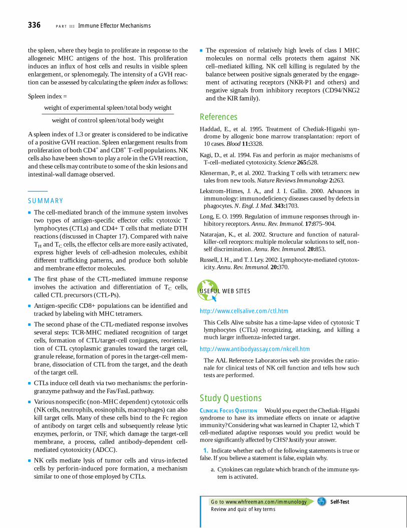
CTL Activity Can Be Demonstrated by CML Development of the cell-mediated lympholysis (CML) assaywas a major experimental advance that contributed to un-derstanding of the mechanism of target-cell killing by CTLs.In this assay, suitable target cells are labeled intracellularlywith chromium-51 (51Cr) by incubating the target cells with Na2
51 CrO4. After the 51Cr diffuses into a cell, it binds to cytoplasmic proteins, reducing passive diffusion of thelabel out of the cell. When specifically activated CTLs areincubated for 1–4 h with such labeled target cells, the cellslyse and the 51Cr is released. The amount of 51Cr releasedcorrelates directly with the number of target cells lysed by the CTLs. By means of this assay, the specificity of CTLsfor allogeneic cells, tumor cells, virus-infected cells, andchemically modified cells has been demonstrated (Figure14-17).
The T cells responsible for CML were identified by selec-tively depleting different T-cell subpopulations by means ofantibody-plus-complement lysis. In general, the activity ofCTLs exhibits class I MHC restriction. That is, they can killonly target cells that present antigen associated with syn-geneic class I MHC molecules. Occasionally, however, classII–restricted CD4+ T cells have been shown to function as CTLs.
334 P A R T I I I Immune Effector Mechanisms
Fc receptors
Neutrophil
Eosinophil
Macrophage
NK cell
Ab bound to Agand Fc receptor
Surfaceantigen (Ag)
Targetcell
Lytic enzymes
Perforin
Granzymes
TNF
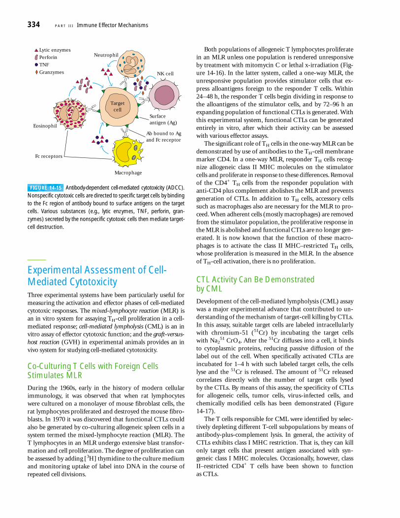
FIGURE 14-15 Antibody-dependent cell-mediated cytotoxicity (ADCC).Nonspecific cytotoxic cells are directed to specific target cells by bindingto the Fc region of antibody bound to surface antigens on the targetcells. Various substances (e.g., lytic enzymes, TNF, perforin, gran-zymes) secreted by the nonspecific cytotoxic cells then mediate target-cell destruction.
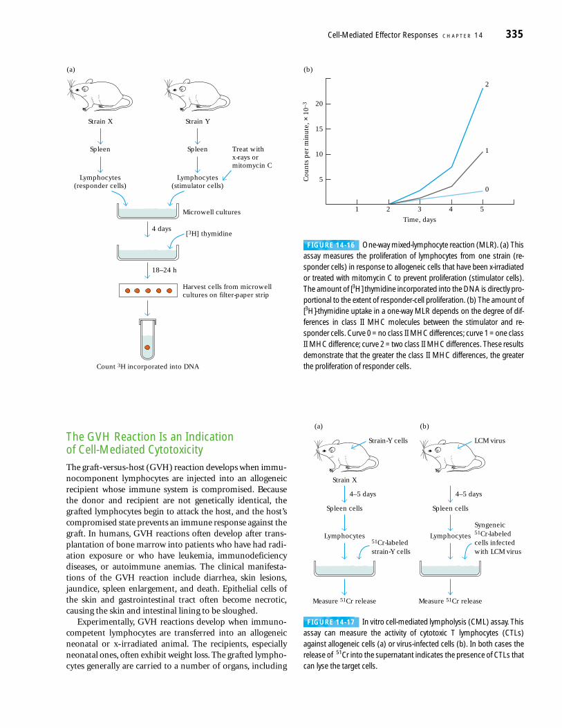
The GVH Reaction Is an Indication of Cell-Mediated CytotoxicityThe graft-versus-host (GVH) reaction develops when immu-nocomponent lymphocytes are injected into an allogeneicrecipient whose immune system is compromised. Becausethe donor and recipient are not genetically identical, thegrafted lymphocytes begin to attack the host, and the host’scompromised state prevents an immune response against thegraft. In humans, GVH reactions often develop after trans-plantation of bone marrow into patients who have had radi-ation exposure or who have leukemia, immunodeficiencydiseases, or autoimmune anemias. The clinical manifesta-tions of the GVH reaction include diarrhea, skin lesions,jaundice, spleen enlargement, and death. Epithelial cells ofthe skin and gastrointestinal tract often become necrotic,causing the skin and intestinal lining to be sloughed.
Experimentally, GVH reactions develop when immuno-competent lymphocytes are transferred into an allogeneicneonatal or x-irradiated animal. The recipients, especiallyneonatal ones, often exhibit weight loss. The grafted lympho-cytes generally are carried to a number of organs, including
Cell-Mediated Effector Responses C H A P T E R 14 335
Strain X
Spleen
Microwell cultures
Lymphocytes(responder cells)
Strain Y
Spleen
Lymphocytes(stimulator cells)
Treat withx-rays ormitomycin C
4 days
Count 3H incorporated into DNA
[3H] thymidine
18–24 h
Harvest cells from microwellcultures on filter-paper strip
(a) (b)
20
15
10
5
1 2 3
Time, days
4 5
0
1
2
Cou
nts
per
min
ute
, × 1
0–3
FIGURE 14-16 One-way mixed-lymphocyte reaction (MLR). (a) Thisassay measures the proliferation of lymphocytes from one strain (re-sponder cells) in response to allogeneic cells that have been x-irradiatedor treated with mitomycin C to prevent proliferation (stimulator cells).The amount of [3H] thymidine incorporated into the DNA is directly pro-portional to the extent of responder-cell proliferation. (b) The amount of[3H]-thymidine uptake in a one-way MLR depends on the degree of dif-ferences in class II MHC molecules between the stimulator and re-sponder cells. Curve 0 = no class II MHC differences; curve 1 = one classII MHC difference; curve 2 = two class II MHC differences. These resultsdemonstrate that the greater the class II MHC differences, the greaterthe proliferation of responder cells.
Strain-Y cells
Strain X
Spleen cells
Lymphocytes
Measure 51Cr release
4–5 days
51Cr-labeledstrain-Y cells
Syngeneic51Cr-labeledcells infectedwith LCM virus
Spleen cells
Lymphocytes
Measure 51Cr release
LCM virus
4–5 days
(a) (b)
FIGURE 14-17 In vitro cell-mediated lympholysis (CML) assay. Thisassay can measure the activity of cytotoxic T lymphocytes (CTLs)against allogeneic cells (a) or virus-infected cells (b). In both cases therelease of 51Cr into the supernatant indicates the presence of CTLs thatcan lyse the target cells.
the spleen, where they begin to proliferate in response to theallogeneic MHC antigens of the host. This proliferationinduces an influx of host cells and results in visible spleenenlargement, or splenomegaly. The intensity of a GVH reac-tion can be assessed by calculating the spleen index as follows:
Spleen index =
weight of experimental spleen/total body weight
weight of control spleen/total body weight
A spleen index of 1.3 or greater is considered to be indicativeof a positive GVH reaction. Spleen enlargement results fromproliferation of both CD4+ and CD8+ T-cell populations. NKcells also have been shown to play a role in the GVH reaction,and these cells may contribute to some of the skin lesions andintestinal-wall damage observed.
SUMMARY
■ The cell-mediated branch of the immune system involvestwo types of antigen-specific effector cells: cytotoxic Tlymphocytes (CTLs) and CD4+ T cells that mediate DTHreactions (discussed in Chapter 17). Compared with naiveTH and TC cells, the effector cells are more easily activated,express higher levels of cell-adhesion molecules, exhibitdifferent trafficking patterns, and produce both solubleand membrane effector molecules.
■ The first phase of the CTL-mediated immune responseinvolves the activation and differentiation of TC cells,called CTL precursors (CTL-Ps).
■ Antigen-specific CD8+ populations can be identified andtracked by labeling with MHC tetramers.
■ The second phase of the CTL-mediated response involvesseveral steps: TCR-MHC mediated recognition of targetcells, formation of CTL/target-cell conjugates, reorienta-tion of CTL cytoplasmic granules toward the target cell,granule release, formation of pores in the target-cell mem-brane, dissociation of CTL from the target, and the deathof the target cell.
■ CTLs induce cell death via two mechanisms: the perforin-granzyme pathway and the Fas/FasL pathway.
■ Various nonspecific (non-MHC dependent) cytotoxic cells(NK cells, neutrophils, eosinophils, macrophages) can alsokill target cells. Many of these cells bind to the Fc region of antibody on target cells and subsequently release lyticenzymes, perforin, or TNF, which damage the target-cellmembrane, a process, called antibody-dependent cell-mediated cytotoxicity (ADCC).
■ NK cells mediate lysis of tumor cells and virus-infectedcells by perforin-induced pore formation, a mechanismsimilar to one of those employed by CTLs.
■ The expression of relatively high levels of class I MHCmolecules on normal cells protects them against NKcell–mediated killing. NK cell killing is regulated by thebalance between positive signals generated by the engage-ment of activating receptors (NKR-P1 and others) andnegative signals from inhibitory receptors (CD94/NKG2and the KIR family).
ReferencesHaddad, E., et al. 1995. Treatment of Chediak-Higashi syn-
drome by allogenic bone marrow transplantation: report of10 cases. Blood 11:3328.
Kagi, D., et al. 1994. Fas and perforin as major mechanisms ofT-cell–mediated cytotoxicity. Science 265:528.
Klenerman, P., et al. 2002. Tracking T cells with tetramers: newtales from new tools. Nature Reviews Immunology 2:263.
Lekstrom-Himes, J. A., and J. I. Gallin. 2000. Advances inimmunology: immunodeficiency diseases caused by defects inphagocytes. N. Engl. J. Med. 343:1703.
Long, E. O. 1999. Regulation of immune responses through in-hibitory receptors. Annu. Rev. Immunol. 17:875–904.
Natarajan, K., et al. 2002. Structure and function of natural-killer-cell receptors: multiple molecular solutions to self, non-self discrimination. Annu. Rev. Immunol. 20:853.
Russell, J. H., and T. J. Ley. 2002. Lymphocyte-mediated cytotox-icity. Annu. Rev. Immunol. 20:370.
USEFUL WEB SITES
http://www.cellsalive.com/ctl.htm
This Cells Alive subsite has a time-lapse video of cytotoxic Tlymphocytes (CTLs) recognizing, attacking, and killing amuch larger influenza-infected target.
http://www.antibodyassay.com/nkcell.htm
The AAL Reference Laboratories web site provides the ratio-nale for clinical tests of NK cell function and tells how suchtests are performed.
Study QuestionsCLINICAL FOCUS QUESTION Would you expect the Chediak-Higashisyndrome to have its immediate effects on innate or adaptiveimmunity? Considering what was learned in Chapter 12, which Tcell-mediated adaptive responses would you predict would bemore significantly affected by CHS? Justify your answer.
1. Indicate whether each of the following statements is true orfalse. If you believe a statement is false, explain why.
a. Cytokines can regulate which branch of the immune sys-tem is activated.
336 P A R T I I I Immune Effector Mechanisms
Go to www.whfreeman.com/immunology Self-TestReview and quiz of key terms
b. Both CTLs and NK cells release perforin after interactingwith target cells.
c. Antigen activation of naive CTL-Ps requires a co-stimulatory signal delivered by interaction of CD28 and B7.
d. CTLs use a single mechanism to kill target cells.e. The secretion of certain critical cytokines is the basis of
the role played by T cells in DTH reactions.
2. You have a monoclonal antibody specific for LFA-1.You per-form CML assays of a CTL clone, using target cells for which theclone is specific, in the presence and absence of this antibody.Predict the relative amounts of 51Cr released in the two assays.Explain your answer.
3. You decide to co-culture lymphocytes from the strains listedin the table below in order to observe the mixed-lymphocytereaction (MLR). In each case, indicate which lymphocyte popu-lation(s) you would expect to proliferate.
4. In the mixed-lymphocyte reaction (MLR), the uptake of[3H]thymidine often is used to assess cell proliferation.
a. Which cell type proliferates in the MLR?b. How could you prove the identity of the proliferating
cell?c. Explain why production of IL-2 also can be used to assess
cell proliferation in the MLR.
5. Indicate whether each of the properties listed below is exhib-ited by TH cells, CTLs, both TH cells and CTLs, or neither cell type.
a. Can make IFN-�b. Can make IL-2c. Is class I MHC restrictedd. Expresses CD8e. Is required for B-cell activationf. Is cytotoxic for target cellsg. Is the main proliferating cell in an MLRh. Is the effector cell in a CML assayi. Is class II MHC restrictedj. Expresses CD4k. Expresses CD3l. Adheres to target cells by LFA-1m. Can express the IL-2 receptorn. Expresses the �� T-cell receptoro. Is the principal target of HIVp. Responds to soluble antigens alone
q. Produces perforinr. Expresses the CD40 ligand on its surface
6. Mice from several different inbred strains were infectedwith LCM virus, and several days later their spleen cells were iso-lated. The ability of the primed spleen cells to lyse LCM-infected, 51Cr-labeled target cells from various strains was deter-mined. In the accompanying table, indicate with a (+) or (–)whether the spleen cells listed in the left column would cause51Cr release from the target cells listed in the headings across thetop of the table.
7. A mouse is infected with influenza virus. How could youassess whether the mouse has TH and TC cells specific forinfluenza?
8. Explain why NK cells from a given host will kill many types of virus-infected cells but do not kill normal cells fromthat host.
9. Consider the following genetically altered mice and predictthe outcome of the indicated procedures. H-2d mice in whichboth perforin and Fas ligand have been knocked out are immu-nized with LCM virus. One week after immunization, T cellsfrom these mice are harvested and tested for cytotoxicity on thefollowing:
a. Target cells from normal LCM-infected H-2b mice
b. Target cells from normal H-2d mice
c. Target cells from H-2d mice in which both perforin andFas have been knocked out
d. Target cells from LCM-infected normal H-2d mice
e. Target cells from H-2d mice in which both perforin andFasL have been knocked out
10. You wish to determine the levels of class I–restricted T cellsin an HIV-infected individual that are specific for a peptide thatis generated from gp120, a component of the virus. Assume thatyou know the HLA type of the subject. What method would youuse and how would you perform the analysis? Please be as spe-cific as you can.
Cell-Mediated Effector Responses C H A P T E R 14 337
51Cr release from LCM-infected target cells
Source (BALB/c � B10)of primed B10.D2 B10 B10.BR F1spleen cells (H-2d) (H-2b) (H-2k) (H-2b/d)
B10.D2(H-2d)
B10(H-2b)
BALB/c(H-2d)
BALB/c � B10(H-2b/d)
Population 1 Population 2 Proliferation
C57BL/6 (H-2b) CBA (H-2k)
C57BL/6 (H-2b) CBA (H-2k)mitomycin C-treated
C57BL/6 (H-2b) (CBA � C57BL/6) F1
(H-2k/b)
C57BL/6 (H-2b) C57L (H-2b)


