Embed Size (px)
Citation preview
Biochimica et Biophysica Acta, 1153 (1993) 203-212 203 © 1993 Elsevier Science Publishers B.V. All rights reserved 0005-2736/93/$06.00
BBAMEM 76146
Static and dynamic studies of the potential-sensitive membrane probe RH421 in dimyristoylphosphatidylcholine vesicles
A t h i n a Z o u n i a R o n a l d J. C l a r k e a,, A n t o n i e J . W . G . V i s s e r b N i n a V. V i s s e r b,1
a n d J o s e f F. H o l z w a r t h a aFritz-Haber-Institut der Max-Planck-Gesellschaft, Faradayweg 4-6, D-14195 Berlin (Germany) and bDepartment of Biochemistry,
Agricultural Unicersity, Wageningen (The Netherlands)
(Received 23 April 1993)
Key words: Potential-sensitive styryl dye; Styryl dye; Lipid vesicle; Binding affinity; Temperature jump; Fluorescence lifetime; Phase transition; Quantum yield
The dynamics of the potential-sensitive styryl dye RH421 in dimyristoylphosphatidylcholine vesicles have been investigated above and below the main phase transition temperature using iodine-laser temperature-jump relaxation spectrophotometry and time-resolved fluorescence lifetime measurements. Equilibrium fluorescence titrations have shown that the affinity of the dye for the membrane is much higher in the liquid-crystalline state than in the gel state. The interaction can be described by either a partition or a binding model and a theory is presented providing a relation between these two approaches. In the liquid-crystal- line state bound dye exhibits steady-state fluorescence relaxation processes in the submicrosecond and millisecond time range following a temperature jump. Time-resolved fluorescence measurements show a variation in the fluorescence lifetime across the emission spectrum, suggesting an excited-state process occurring on the subnanosecond time scale. These processes are most likely related to dye and/or lipid reorientation following the temperature jump or excitation pulse. Temperature-dependent changes in the fluorescence excitation spectrum of bound dye suggest that the dye exists in at least two different sites within the membrane.
Introduction
Fluorescent styryl dyes are at present being widely applied to the optical recording of electrical activity in cells, cell organelles and synthetic membrane prepara- tions [1-15]. It was initially thought that these dyes responded to voltage changes across the membrane via an electrochromic mechanism (Stark effect) [16-19]. However, variation in the magnitude of the response of several dyes from one membrane preparation to an- other [7,20-22], disagreement between the observed changes in the absorbance and fluorescence excitation spectra [2,7,23] and potential-dependent shifts of the emission spectrum [10,20,23] indicate that nonelec- trochromic mechanisms involving, for example, a change in dye quantum yield, must in many cases make a significant contribution to the observed fluorescence response. Possible additional mechanisms include an electric-field-dependent modification of the dye struc-
* Corresponding author. Fax: + 49 30 8305520. t Formerly N.V. Shcherbatska.
ture or orientation in the membrane [2,12,20,23,24], a change in the degree of dye aggregation within the membrane [23] and an effect of the electric field on an excited state reaction of the dye, such as structural relaxation from the Franck-Condon excited state [23] or the formation of a twisted internal charge transfer (TICT) state [12,24].
The application of the styryl dyes to kinetic mea- surements of electrical activity in membrane prepara- tions requires that their response be significantly faster than the process being measured. For an elec- trochromic mechanism, subnanosecond response times are to be expected. However, for mechanisms involving intramembrane dye motion, much slower response times would be predicted. In a previous publication [23] the absorbance and fluorescence properties of the styryl dye RH421 (see Fig. 1) were investigated at equilibrium in aqueous solution and when bound to Na+,K+-ATPase-containing membrane fragments. Here the investigation is extended to time-resolved measurements of the dye in dimyristoylphosphatidyl- choline (DMPC) vesicles in order to identify the time scale of dynamic processes of bound dye, which may be

204
R = CH2CH2CH2CH2CH 3
Fig. 1. Structure of RH421.
a possible source of its potential sensitivity. In a future publication the effect of membrane potential on the rates of these processes is to be investigated.
Materials and Methods
N-(4-Sulphobutyl)-4-(4-(p-dipentylaminophenyl)bu- tadienyl)pyridinium inner salt (RH421) was obtained from Molecular Probes (Eugene, OR) and was used without further purification. A series of stock solutions of the dye were prepared in ethanol. For spectral measurements 5 tzl of an ethanolic dye solution was added to a quartz cuvette containing 1 ml of aqueous solvent. The final solutions measured thus contained a small and constant percentage of 0.5% ethanol.
Dimyristoylphosphatidylcholine (DMPC) was ob- tained from Avanti Polar Lipids (Alabaster, AL). DMPC unilamellar vesicles were prepared according to a modification of the ethanol injection method of Batzri and Korn [25-27]. 1 ml of a 30 mM solution of DMPC in ethanol was injected slowly over 15 min with contin- uous stirring into 10 ml of buffer solution at 30°C. The ethanol in the resulting solution was then removed by dialysis against excess buffer at 30°C. The final solution contained no detectable trace of ethanol, i.e., [ethanol] < 10/~M, according to an NADH/alcohol dehydroge- nase enzymatic assay (Boehringer, Mannheim). Dialysis tubing was purchased from Medicell International (London, UK). The phospholipid content of the vesicle suspensions was determined by the phospholipid B test from Wako (Neuss, Germany). The vesicles produced were unilamellar with external diameters in the range 50-100 nm, as determined by cryoelectronmicroscopy and confirmed by dynamic light scattering measure- ments.
All measurements with the vesicles were performed in a buffer containing 30 mM Tris, 1 mM EDTA and 150 mM NaCI. The pH of the buffer was adjusted to pH 7.2 with HC1. All solutions were prepared using triply distilled water. The origins of the various reagents used were as follows: Tris(hydroxymethyl) amino- methane (99.9%, Sigma), EDTA (99%, Sigma), NaC1
(Suprapur, Merck), HC1 (0.1 M Titrisol solution, Merck) and ethanol (analytical grade, Merck).
Absorbance measurements were performed with a Shimadzu UV-2100 u.v.-visible recording spectropho- tometer using a bandwidth of 5 nm. Steady-state fluo- rescence excitation and emission spectra were recorded with a Shimadzu RF-5000 recording spectrofluoropho- tometer using bandwidths of 5 nm for both the excita- tion and emission monochromators. For the purposes of comparison with the absorbance spectrum, the fluo- rescence excitation spectrum was recorded on a Perkin-Elmer MPF-44 fluorimeter using the ratio mode in order to compensate for the wavelength dependence of the xenon lamp intensity. In order to minimise contributions from scattering of the exciting light and higher order wavelengths, glass cut-off filters were used in front of the excitation or emission monochro- mators where appropriate. The temperature of the cuvette holders was thermostatically controlled.
The fluorescence quantum yield, q, of dye was de- termined with respect to the quantum yield of a refer- ence, qref, according to Eqn. 1 [24,28].
n 2 fF dhcm (l_10-Aref) q = qref' ~ "
nref fEte f dAem ( 1 - 1 0 - A ) (1)
F and Ere f a r e the fluorescence intensities of dye and of the reference substance. The integration of the fluorescence intensities over the emission spectrum was achieved by weighing the chart paper under the curve. A and Aref are the absorbances of dye and the reference substance, n and nre f are the refractive in- dices of the solutions. Reference substances used were rhodamine 101 (Lambda Physik, G6ttingen, Germany) with qref = 1.0 in ethanol [29] in the excitation wave- length range 490-570 nm and fluorescein (Aldrich, Steinheim, Germany) with qref = 0.85 in 0.1 M NaOH solution [28] in the excitation wavelength range 450-500 r im.
The dynamics of membrane-bound dye were investi- gated by the iodine-laser temperature-jump relaxation method. The application of an iodine laser pulse to the production of a rapid temperature jump [30-32] and the experimental optical configuration for fluorescence detection [33] have been described previously. The laser flash (wavelength 1315 nm) produces a tempera- ture jump of I°C within 3 ~s. A 200 W H g / X e arc lamp (Oriel, Darmstadt, Germany) was used as the exciting light source. The dye/vesicle suspensions were excited in the wavelength range 400-500 nm using the glass filter combination GG400 and BG12 (Schott, Mainz, Germany). The fluorescence emission was de- tected in the range 500-700 nm by using an OG515 glass cut-off filter (Schott) and a 650 nm (SWP) short
wavelength pass filter (Oriel). Nine experimental traces were averaged for each solution. For most experiments an overall bandwidth for the detection circuit of 1 MHz was used.
Time-resolved fluorescence lifetime data were recorded and analysed as described in detail elsewhere [34,35]. The excitation wavelength was set to 465 nm and the fluorescence emission was detected at either 591 nm or 688 nm.
The effect of the dye on the phase transition be- haviour of the lipid bilayer was investigated using an MC-2 ultrasensitive scanning calorimeter (Micro Cal, Northampton, MA). The effect of the small percentage of ethanol present in the suspensions was tested in separate control experiments in which pure ethanol was added to a vesicle suspension.
The calculation of the binding affinities of dye for the lipid membrane was performed using the commer- cially available non-linear least squares program ENZ- FIT-FER. The program was purchased from Biosoft (Cambridge, UK) and was run on an IBM-AT/386 compatible personal computer (mey-Soft, Berlin, Ger- many).
Theory
The association of probe molecules to lipid vesicles is generally described by either a partition model [7,36], in which one considers the membrane of the vesicles as a separate lipid phase in which the probe can dissolve, or by a binding model [20,37], in which the vesicles are considered as macromolecules to which a certain num- ber of probe molecules can bind. The partition model allows the calculation of a partition coefficient, y, of the probe between the lipid and aqueous phases. The binding model utilizes a binding constant, K, for the binding of a probe molecule to an individual binding site. Under certain conditions it will be shown that both approaches predict the same experimental be- haviour. It is the purpose of the present section, there- fore, to find a relationship between y and K, so that values calculated using the two different theoretical approaches can be directly compared.
Let us consider first the binding model. The binding reaction can be described by the following equilibrium:
K f r e e b i n d i n g s i t e + d y e ( ' o c c u p i e d b i n d i n g s i t e
The intrinsic (microscopic) binding constant, K, is de- fined by
C~L K = ( 2 )
(nc~-C~L)C D
where C~L is the total concentration of bound dye, c* is the total concentration of lipid, c D is the unbound
205
dye concentration and n is the number of binding sites per lipid molecule. Here it is assumed that each bind- ing site can only accommodate one dye molecule. It should be noted that the term binding site here does not imply a specific interaction as in the case of a substrate binding to the active site of an enzyme. Instead it is merely used as a mathematical conve- nience in order to set a limit to the number of dye molecules which can bind to a single vesicle. This is based on the observation that saturation of lipid mem- branes by probe molecules can occur [37]. In actual fact it is likely that the bound dye molecules may be distributed over a range of different positions and orientations in the bilayer.
Under conditions where there is a large excess of available binding sites over dye molecules, i.e., nc~ >> c* Eqn. 1 reduces to DL,
c * L nK = ( 3 )
C~CD
This equation can now be re-expressed by introducing the total suspension volume, Vtot, the moles of dye in the lipid, hoE, and in the aqueous solution, riD, and the moles of lipid, n L. Thus,
?IDL Vtot nK = - - ( 4 )
n D n L
Now let us assume that the lipid contributes a negligible amount to the total suspension volume, i.e., Vto t = V w, where V w is the volume of the aqueous solution. With this assumption and dividing both sides of Eqn. 4 by VL, the volume of lipid, one obtains
nK n D L V w 1
VL V L n D nL (5)
Now let us consider the partition model, in which the lipid and the aqueous solution are considered as separate phases. The partition coefficient, y, is defined as the ratio of the dye concentration in the lipid phase to that in the aqueous phase. Thus,
n D L / V L 3' - - - (6 )
nD/Vw
Comparison of Eqns. 5 and 6 shows that Eqn. 5 can be simplified as
VL nK = 3" - - (7 )
nL
VL/n L is simply the molar volume, VL, of the lipid employed. Thus, we have the following simple relation- ship between K and y,
nK = 3'- VL (8 )

206
It should be noted that this equation is only valid under conditions of great excess of lipid over dye but at sufficiently low lipid concentrations, so that the lipid itself has a negligible contribution to the total suspen- sion volume. The equation shows, however, that under these conditions the binding and partition models are mathematically equivalent and predict identical experi- mental behaviour. The two models can only be distin- guished at high dye concentrations, at which in the case of the binding model saturation of the lipid bind- ing sites would become apparent.
Let us now consider the physical significance of nK. Considering the situation where half of the dye molecules are bound and half are free, i.e., C~L = C D = C~/2, where c~ is the total dye concentration, it can easily be shown from Eqn. 3 that
1 nK = c~ (9)
Thus, assuming that there is an excess of binding sites, 1 / n K is equal to the lipid concentration at which half the dye molecules are bound. The parameters 3' and nK can, therefore, both be regarded as estimates of the dye binding affinity for the membrane. It should be noted, however, that according to the binding model, a difference in the binding affinity, nK, between differ- ent membrane preparations does not necessarily mean that there is a difference in the strength of binding. A variation in nK could be due to a difference in the strength of binding, i.e., a change in K, or a difference in the number of binding sites per lipid molecule, i.e., a change in n. In order to determine which of these possibilities is actually occurring one needs to deter- mine n and K separately, e.g., from a Scatchard dia- gram.
The value of nK can be easily determined from a titration of dye with vesicles. The free dye concentra- tion is given by
- * - * ( 1 0 ) C D - - C o CDL
Substituting this expression for c D into Eqn. 3 and rearranging yields
nK.c~ ) c~e= l+nK.c------------~L c~ (11)
If one assumes that the dye can exist in only two distinct fluorescing states, then the fluorescence, F, is given according to Beer's law by
F = fw'CD + fl'C~L (12)
where fw and fl are the molar fluorescences (arbitrary units M -1) in the aqueous solution and in the lipid, respectively. Substituting for c D and C~L from Eqns.
10 and 11, respectively, into Eqn. 12 yields upon rear- rangement
F nK.c~ = fw + ( f ' - fw) l + nK.c~. (13)
A non-linear least squares fit of the observed molar fluorescence, F / c ~ , to Eqn. 13 thus allows the deter- mination of the parameters fw, f l and nK.
R e s u l t s
Determination of binding affinities The binding affinity, nK, of RH421 to DMPC vesi-
cles above and below the main phase transition tem- perature of 23°C was determined by fluorescence titra- tion of the dye with vesicles and fitting the data to Eqn. 13 (see Fig. 2). At 26°C, i.e., above the phase transition temperature, nK -- (5.2 _+ 0.5)" 104 M -1. At 21°C, i.e., below the phase transition temperature, nK = (1.3 + 0.2). 104 M -1. The lipid phase structure, therefore, has a dramatic effect on the dye binding affinity. In the liquid-crystalline state the binding affinity is greatly enhanced in comparison to the gel state.
The observed binding affinity can be compared to the partition coefficient of 250 000 reported by Biihler et al. [7] for the association of RH421 to Na+,K +- ATPase-containing membrane fragments. The frag- ments contain a mixture of phospholipids, but with dioleoylphasphatidylcholine as one of the main compo- nents [38]. Assuming a lipid partial specific volume of 0.99 cm 3 g - 1 and a lipid molecular mass of 786 g mo l - 1, i.e., the appropriate values for dioleoylphosphatidyl-
2.5(
L .
c~ 2.0c
1.50
100
0.50 ~
0.~ - lira 2'o0 2so
Fig. 2. Molar fluorescence, F/c~, of RH421 at 26°C (o) and 21°C (©) as a function of the DMPC concentration at a constant total dye concentration of 107 nM; Aex=440 nm (eGG420 cut-off filter), Aem = 600 nm (+ OG570 cut-off filter), excitation bandwidth = 5 nm, emission bandwidth = 10 nm. The solid lines represent fits of the
data to Eqn. 13.

choline [39], one can calculate a lipid molar volume of 0.77 dm 3 mo1-1. Assuming that the dye only binds to the lipid regions of the membrane, the partition coeffi- cient of 250 000 thus corresponds according to Eqn. 8 to a binding affinity, nK, of 1.93.105 M -1. This is almost 4-times larger than the value reported above for the binding of RH421 to DMPC vesicles in the liquid- crystalline state. The lower value found for the vesicles could be partly explained when one considers the membrane permeability of the dye. The presence of the localized sulphonate group is likely to make the dye virtually membrane impermeable. In this case only approximately half of the lipid content of the vesicles is available for binding, whereas the open membrane fragments have both sides of the membrane accessible to the dye.
Differential scanning calorimetry The addition of RH421 to a DMPC vesicle suspen-
sion causes a decrease in the main gel-to-liquid-crystal phase transition temperature (Tm), a broadening of the transition profile and a slight increase in the enthalpy change (AH). For example, at a lipid/dye molar ratio of 10:1, Tm=19.8°C and A H = 3 0 kJmo1-1 These values can be compared with the corresponding ones of pure DMPC vesicles, i.e., T= 23.1°C and A H = 23 kJ mo1-1. The small concentration of ethanol (0.5% or less) present in the suspensions was found in control experiments to have a negligible effect on the phase transition behaviour in comparison to the dye.
Very similar behaviour has been previously reported by Bammel et al. [40] for a trifluoromethyl-substituted styryl probe in DMPC vesicles. Such a modification of the phase transition profile is typical of long am-
207
phiphilic molecules, which are localized with one end near the polar head groups of the lipid and the other end extending into the hydrocarbon interior [41].
Static fluorescence measurements Comparison of the fluorescence excitation spectrum
of membrane-bound dye with the spectrum of ( 1 - 10-A), where A is the absorbance of the suspension (see Fig. 3), shows that there is a disagreement be- tween the two spectra. The fluorescence excitation spectrum is red-shifted by 10-20 nm relative to the spectrum of (1 - 10 -A) and it is also not symmetrical. The excitation spectrum shows a maximum at 490 nm and a shoulder at approx. 520 nm.
The fluorescence intensity, F, is related to the ab- sorbance of a solution by the expression
F = f ' Io(1 - lO-X)p'q (14)
where f is the fraction of fluorescence light collected by the photomultiplier, I o is the intensity of the inci- dent light, p is the fraction of the total emission occurring at the chosen emission wavelength and q is the quantum yield. The disagreement between the two spectra in Fig. 3 cannot be explained by the wavelength dependence of Io, since this is compensated for by the ratio mode of the fluorimeter. The disagreement, therefore, must be due to a variation in the product p . q with the excitation wavelength. Measurements of the relative quantum yield of bound dye have shown that the value increases across the excitation spectrum, from 0.067 at 450 nm to 0.138 at 570 nm (see Fig. 4). Thus, this satisfactorily explains the red shift of the excitation spectrum relative to ( 1 - 10-A). Excitation
0.3
1_10 - A
0.1
0.0 's0 s's0 6oo
Xex/nm
Fig. 3. The fluorescence excitation spectrum (dashed line) and the spectrum of I - I0 -A (solid line) of RH421 in the presence of 500 p,M of
DMPC; [RH421] = 6.4 ~M, bandwidths = 5 nm, T = 30°C. The fluorescence excitation spectrum was recorded at an emission wavelength of 650
nm ( + RG645 cut-off filter) and its intensity was normalized to the maximum of the I - 10-A spectrum.
208
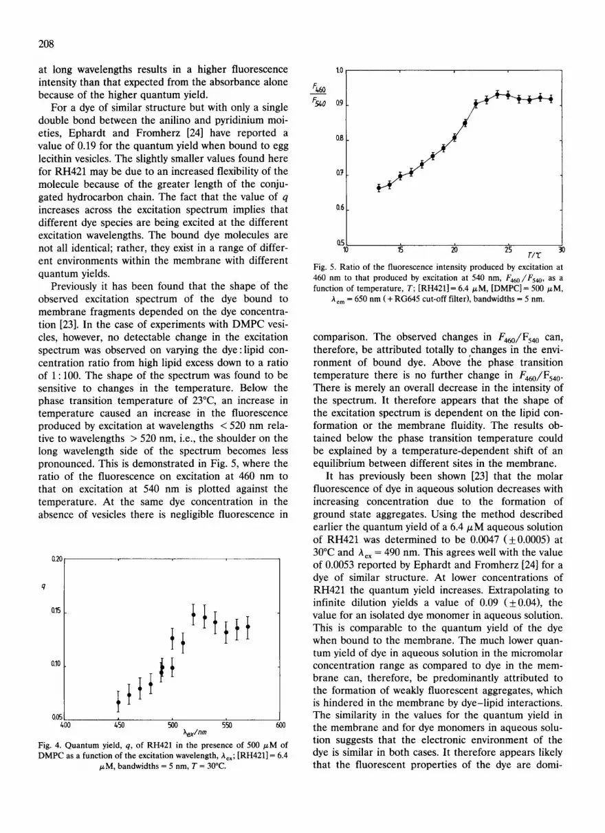
at long wavelengths results in a higher fluorescence intensity than that expected from the absorbance alone because of the higher quantum yield.
For a dye of similar structure but with only a single double bond between the anilino and pyridinium moi- eties, Ephardt and Fromherz [24] have reported a value of 0.19 for the quantum yield when bound to egg lecithin vesicles. The slightly smaller values found here for RH421 may be due to an increased flexibility of the molecule because of the greater length of the conju- gated hydrocarbon chain. The fact that the value of q increases across the excitation spectrum implies that different dye species are being excited at the different excitation wavelengths. The bound dye molecules are not all identical; rather, they exist in a range of differ- ent environments within the membrane with different quantum yields.
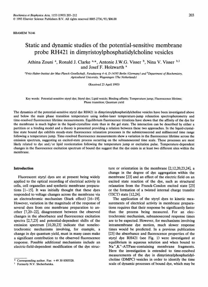
Previously it has been found that the shape of the observed excitation spectrum of the dye bound to membrane fragments depended on the dye concentra- tion [23]. In the case of experiments with DMPC vesi- cles, however, no detectable change in the excitation spectrum was observed on varying the dye:lipid con- centration ratio from high lipid excess down to a ratio of 1 : 100. The shape of the spectrum was found to be sensitive to changes in the temperature. Below the phase transition temperature of 23°C, an increase in temperature caused an increase in the fluorescence produced by excitation at wavelengths < 520 nm rela- tive to wavelengths > 520 nm, i.e., the shoulder on the long wavelength side of the spectrum becomes less pronounced. This is demonstrated in Fig. 5, where the ratio of the fluorescence on excitation at 460 nm to that on excitation at 540 nm is plotted against the temperature. At the same dye concentration in the absence of vesicles there is negligible fluorescence in
(120
q
0.15
010
405 400 ~5o ~o 5~o
Xex/nm Fig. 4. Quantum yield, q, of RH421 in the presence of 500 /zM of DMPC as a function of the excitation wavelength, kex; [RH421] = 6.4
/zM, bandwidths = 5 nm, T = 30°C.
Ft,60
1.0
0.9
0.B
0.7
Ct5 10 1~ zb 2's 30
T/' f
Fig. 5. Ratio of the fluorescence intensity produced by excitation at
460 nm to that produced by excitation at 540 nm, F46o/Fs4 o, as a function of temperature, T; [RH421] = 6.4/zM, [DMPC] = 500/zM,
~'em = 650 n m ( + RG645 cut-off filter), bandwidths = 5 nm.
comparison. The observed changes in F460//F540 can, therefore, be attributed totally to ,changes in the envi- ronment of bound dye. Above the phase transition temperature there is no further change in F460//F540 . There is merely an overall decrease in the intensity of the spectrum. It therefore appears that the shape of the excitation spectrum is dependent on the lipid con- formation or the membrane fluidity. The results ob- tained below the phase transition temperature could be explained by a temperature-dependent shift of an equilibrium between different sites in the membrane.
It has previously been shown [23] that the molar fluorescence of dye in aqueous solution decreases with increasing concentration due to the formation of ground state aggregates. Using the method described earlier the quantum yield of a 6.4/zM aqueous solution of RH421 was determined to be 0.0047 (+0.0005) at 30°C and kex = 490 nm. This agrees well with the value of 0.0053 reported by Ephardt and Fromherz [24] for a dye of similar structure. At lower concentrations of RH421 the quantum yield increases. Extrapolating to infinite dilution yields a value of 0.09 (+0.04), the value for an isolated dye monomer in aqueous solution. This is comparable to the quantum yield of the dye when bound to the membrane. The much lower quan- tum yield of dye in aqueous solution in the micromolar concentration range as compared to dye in the mem- brane can, therefore, be predominantly attributed to the formation of weakly fluorescent aggregates, which is hindered in the membrane by dye-lipid interactions. The similarity in the values for the quantum yield in the membrane and for dye monomers in aqueous solu- tion suggests that the electronic environment of the dye is similar in both cases. It therefore appears likely that the fluorescent properties of the dye are domi-

nated by the interaction of the chromophore with the polar head group region of the membrane.
Temperature jump Iodine-laser temperature-jump experiments were
performed on dye/vesicle suspensions at fixed dye and lipid concentrations ([RH421] = 6.4 ~M, [DMPC] = 500 /zM) at a range of temperatures, above and below the lipid phase transition temperature of 23°C. At low temperatures, i.e., 18-24°C, there is a nonexponential increase in fluorescence following the temperature jump. At least two and often three exponential time functions are necessary in order to fit the data. The complete relaxation is over in approx. 5 ms. Since it has been shown earlier that the binding affinity of the dye increases markedly with increasing temperature, it is likely that the observed relaxation is associated with the binding of further dye molecules to the membrane. The nonexponential nature of the relaxation could be attributed to simultaneous temperature-dependent changes in the lipid conformation [42].
At temperatures of 21°C and above, a further slower relaxation appears which is characterized by a decrease in fluorescence (see Fig. 6A). In this case the relax- ation can be adequately fitted by a single exponential time function. The rate of the relaxation was found to increase with increasing temperature. At 21°C the re- ciprocal relaxation time, ~', is 0.36 s, whereas at 30°C ~" = 0.12 s.
At temperatures of 25°C and above, the initial in- crease in fluorescence following the temperature jump is no longer apparent. At such high temperatures, the binding affinity of the dye for the membrane is so high that under the conditions of the experiment all of the dye can be considered to be bound and no further binding can occur. The vesicle membrane is also fully in the liquid-crystalline state, so that no further signifi- cant changes in the lipid conformation are expected. The combination of these effects, therefore, explains the disappearance of the fluorescence increase.
As one proceeds to higher temperatures above 25°C a very rapid relaxation develops, which is characterized by a decrease in fluorescence (see Fig. 6B). The relax- ation time of this process is faster than the time resolution of the apparatus, i.e., T < 3 /~s. This is possibly associated with a decrease in the quantum yield of the dye due to increased vibrational motion.
In terms of the application of the dye as a potential sensitive probe, the results above the phase transition temperature, where all the dye is bound, are most relevant. Under these conditions it has been found that the dye exhibits two relaxation processes, both involv- ing a decrease in fluorescence. One of the relaxations has a relaxation time of less than 3/zs and the other has a relaxation time of approx. 0.1 s. Both must be associated with a change in the state or environment of
0.020
A._E_F Fo
0.015
Q010
0.005
0.000
:• ",~'<k.,
o.o o12 oi, o16 ,J,,c o18
209
o.0o0
~F F~
- 0.005
- 0 . 0 1 0
- 0 . 0 1 5
0.0 0.2 0.t, 0.6 t/sec 0.8
Fig. 6. Temperature-jump traces. (A) RH421, 6.4 tzM, in the pres- ence of 500/zM of DMPC at 22°C, i.e., below the phase transition temperature; excitation wavelength range 400-500 nm, emission wavelength range 500-700 nm. (B) As in panel A, but at 28°C. The relative fluorescence intensity change, AF/Fo, is referred to the
initial fluorescence prior to the temperature jump, Fo.
bound dye, but the exact source of the two relaxations is still to be determined.
Time-resolved fluorescence measurements The time-resolved fluorescence decay after an exci-
tation laser pulse at 465 nm was measured for RH421 bound to the lipid membrane at a total lipid concentra- tion of 856/zM and a range of dye concentrations. The temperature was set to 30°C. Varying the dye concen- tration was found to have no significant effect on the shape of the decay profile. Typical fluorescence decay curves are shown in Fig. 7. At an emission wavelength of 591 nm the fluorescence decay turned out to be complex. First there is a very rapid growing in of excited-state species within a few picoseconds (see inset Fig. 7) which can hardly be resolved by the apparatus, and the subsequent decay could only be analysed with three components to correctly fit the experimental data to a minimum X 2 of 1.07 (for details

210
1 0 s
1 0 4 ̀
• ~ 103
c
' ~ 1 0 a
10 ~
1 0 °
0 200 400 600 800 1000 Channel number
Fig. 7. Fluorescence decay traces of RH421, 6.4 tzM, in the presence of 856 /zM of DMPC at 30°C. (a) Impulse response function mea- sured via the ultrarapid fluorescence of the dye pinacyanol ( < 10 ps lifetime) in ethanol at emission wavelength = 688 nm; (b) emission wavelength = 591 nm; (c) emission wavelength = 688 nm. The wave- length of the excitation was 465 nm. The inset shows an enlargement of the initial part of the three curves (first 60 channels). The time
equivalence per channel is 30 ps.
see Ref. 35). The lifetimes recovered (and the 66% confidence limits) were 0.35 ns (0.26-0.40 ns), 1.50 ns (1.45-1.55 ns), and 0.80 ns (0.67-0.90 ns). The lifetimes of samples at other dye concentrations were found to have similar values. The multiexponential nature of the fluorescence decay could have two possible origins. The first is ground-state heterogeneity, i.e., different dye sites within the membrane. Alternatively, a relax- ation process of the excited state may be occurring on the same time scale as fluorescence emission.
At an emission wavelength of 688 nm, it was found that the overall decay is significantly slower and a decay component of 100 ps with a negative pre-ex- ponential factor [43] is required to fit the data. The remaining decay could be reasonably fitted to a single lifetime of 1.77 ns. The presence of a negative pre-ex- ponential factor with a resolvable time constant of 100 ps (see inset Fig. 7) is definitive evidence for an ex- cited-state process in which a new fluorescent species is being produced. Fluorescence decays of samples at other concentrations were found to have similar pat- terns but with varying growing-in times between 100 and 200 ps. Similar behaviour has been observed for the fluorescent molecule 2-toluidinylnaphthalene-6-
sulphonic acid bound to egg phosphatidylcholine vesi- cles [44-47]. Easter and coworkers [44,45] attributed it to relaxation of the lipid molecules around the excited state dipole moment of the fluorophore, whereas Dem- chenko and Shcherbatska [47] proposed a translational movement of the excited fluorophore to a more polar region of the membrane.
Discussion
The equilibrium and dynamic behaviour of the po- tential-sensitive styryl dye RH421 has been investigated in dimyristoylphosphatidylcholine vesicles. In aqueous solution it has previously been found [23] that the dye readily aggregates. When bound to the vesicle mem- brane, however, no evidence for interaction between the dye molecules has been observed. In aqueous solu- tion it is likely that a dimer is formed in a head-to-tail arrangement, with the localized charges of the sulphonate group at opposite ends in order to minimise electrostatic repulsion. In the membrane such an ar- rangement appears to be hindered because of the location of the sulphonate group at the m em b rane / solution interface and the penetration of the rest of the molecule into the membrane matrix. The inhibition of dye aggregation has been found to account for the high quantum yield of membrane-bound dye compared to aqueous solutions of dye at high concentrations, i.e., _> 1 ~M. Haugland [48] has attributed the quenching of aqueous solutions of styryl dyes to conformational isomerisation, which in the membrane is hindered be- cause of the higher viscosity. At least in the case of RH421 this explanation is not sufficient. Here the major cause of quenching is the formation of ground- state aggregates in aqueous solution, which leads to self-quenching of fluorescence with concomitant short- ening of the fluorescence lifetime [49].
In a previous publication [23] two explanations were offered for concentration-dependent changes in the fluorescence excitation spectrum of dye bound to pla- nar membrane fragments: (a) dye aggregation within the membrane, and (b) dye-induced structural changes of the membrane. A mechanism of the dye's potential sensitivity was then proposed involving a perturbation of a membrane-bound monomer /aggrega te equilib- rium. In the case of lipid vesicles, however, no dye concentration dependent fluorescence changes have been observed down to an excess l ip id /dye ratio of 100:1, and such a mechanism, therefore, seems to be unlikely. The excitation spectrum of dye bound to lipid vesicles was found to exhibit changes in its shape on changing the temperature. This suggests an equilibrium between different sites within the membrane. Bammel et al. [40] observed two partially resolved resonances in the fluorine NMR spectrum of a trifluoromethyl-sub- stituted styryl dye bound to DMPC vesicles. They at-

tributed these to dye bound to sites in the inner and outer monolayer of the membrane. For RH421 this can not be the case, since the sulphonate group would render the dye virtually membrane impermeable. It seems more likely that the dye is able to bind at more than one position within one half of the bilayer, e.g., different depths within the membrane. Further evi- dence for the presence of different dye sites comes from the excitation wavelength dependence of the quantum yield of bound dye. Variation of the excita- tion wavelength causes the excitation of different dye species.
The similarity of the quantum yield of bound dye with that of dye monomers in aqueous solution sug- gests that the chromophore is in a relatively polar environment within the membrane, i.e., close to the head group region of the membrane. Loew et al. [50] have proposed a stabilization of the ground state of such styryl dyes by interaction of the positive charge on the pyridinium ring with the polar head group.
Temperature-jump measurements above the phase transition temperature have shown that bound dye exhibits two relaxation processes, one in the submi- crosecond range and the other in the millisecond range. The first is likely to be associated with a decrease in the quantum yield because of increased vibrational mobility. The origin of the second is unclear, but it is possibly related to a change of the dye's position within the membrane. This may be a possible source of volt- age sensitivity.
Time-resolved fluorescence measurements at two detection wavelengths have shown that the dye under- goes a relaxation process after excitation, possibly due to the reorientation of the surrounding lipid molecules or some motion of the dye. The relaxation occurs in the picosecond time range. This is a further process which may be influenced by the application of an external electrical field.
Now that the time scale of various dynamic pro- cesses of membrane-bound dye has been established, it is intended to examine the effect of an electric field on their rates and so determine the response time of the dye.
Acknowledgements
The authors would like to thank Herrn Jiirgen Luhm for assistance with the quantum yield measurements in aqueous solution, Herrn Joachim J~iger for cryelectron- microscopic measurements and Herrn Arie van Hoek for time-resolved fluorescence measurements. A.Z. is grateful for financial support from the Max-Planck- Gesellschaft. J.F.H. thanks the Deutsche Forschungs- gemeinschaft for a grant (DFG-696/4-2). R.J.C. ac- knowledges with gratitude financial support from the Stipendien-Fonds der Chemischen Industrie.
211
References
1 Grinvald, A., Salzberg, B.M., Lev-Ram, V. and Hildesheim, R. (1987) Biophys. J. 51,643-651.
2 Miiller, W., Windisch, H. and Tritthart, H.A. (1986) Eur. Bio- phys. J. 14, 103-111.
3 Chien, C.-B. and Pine, J. (1991) Biophys. J. 60, 697-711. 4 Ehrenberg, B., Meiri, Z. and Loew, L.M. (1984) Photochem.
Photobiol. 39, 199-205. 5 Manis, P.B. and Freeman, J.A. (1988) J. Neurosci. 8, 383-394. 6 Klodos, I. and Forbush, B., III (1988) J. Gen. Physiol. 92, 46a
(abstr.). 7 BiJhler, R., StiJrmer, W., Apell, H.-J. and L~iuger, P. (1991) J.
Membr. Biol. 121, 141-161. 8 Stiirmer, W., Biihler, R., Apell, H.-J. and L~iuger, P. (1991) J.
Membr. Biol. 121, 163-176. 9 Kn6pfel, T. and Fromherz, P. (1987) Z. Naturforschg. 42c, 986-
990. 10 Montana, V., Farkas, D.L. and Loew, L.M. (1989) Biochemistry
28, 4536-4539. 11 Gross, D., Loew, L.M., Ryan, T.A. and Webb, W.W. (1986) Prog.
Clin. Biol. Res. 210, 263-270. 12 Fromherz, P., Dambacher, K.H., Ephardt, H., Lambacher, A.,
Miiller, C.O., Neigl, R., Schaden, H., Schenk, O. and Vetter, T. (1991) Ber. Bunsenges. Phys. Chem. 95, 1333-1345.
13 Gross, D., Loew, L.M. and Webb, W.W. (1986) Biophys. J. 50, 339-348.
14 Grinvald, A., Fine, A., Farber, I.C. and Hildesheim, R. (1983) Biophys. J. 42, 195-198.
15 Grinvald, A., Frostig, R.D., Lieke, E. and Hildesheim, R. (1988) Physiol. Rev. 68, 1285-1366.
16 Loew, L.M., Bonneville, G.W. and Surow, J. (1978) J. Am. Chem. Soc. 17, 4065-4071.
17 Loew, L.M., Scully, S., Simpson, L. and Waggoner, A.S. (1979) Nature 281, 497-499.
18 Loew, L.M. and Simpson, L.L. (1981) Biophys. J. 34, 353-365. 19 Loew, L.M. (1982) J. Biochem. Biophys. Methods 6, 243-260. 20 Fluhler, E., Burnham, V.G. and Loew, L.M. (1985) Biochemistry
24, 5749-5755. 21 Grinvald, A., Hildesheim, R., Farber, I.C. and Anglister, L.
(1982) Biophys. J. 39, 301-308. 22 Loew, L.M., Cohen, L.B., Salzberg, B.M., Obaid, A.L. and
Bezanilla, F. (1985) Biophys. J. 47, 71-77. 23 Clarke, R.J., Schrimpf, P. and Sch6neich, M. (1992) Biochim.
Biophys. Acta 1112, 142-152. 24 Ephardt, H. and Fromherz, P. (1989) J. Phys. Chem. 93, 7717-
7725. 25 Batzri, S. and Korn, E.D. (1973) Biochim. Biophys. Acta 298,
1015-1019. 26 Kremer, J.M.H., Esker, M.W.J.v.d., Pathmamanoharan, C. and
Wiersema, P.H. (1977) Biochemistry 16, 3932-3935. 27 Holzwarth, J.F. (1986) Faraday Disc. 81, 74-76. 28 Parker, C.A. (1968) Photoluminescence of Solutions, pp. 262-267,
Elsevier, Amsterdam. 29 Karstens, T. and Kobs, K. (1980) J. Phys. Chem. 84, 1871-1872. 30 Holzwarth, J.F. (1979) in Techniques and Applications of Fast
Reactions in Solution (Gettins, W.J. and Wyn-Jones, E., eds.), pp. 47-59, Reidel, Dordrecht, Holland.
31 Bannister, J.J., Gormally, J., Holzwarth, J.F. and King, T.A. (1984) Chem. Br. 20, 227-233.
32 Holzwarth, J.F., Schmidt, A., Wolff, H. and Volk, R. (1977) J. Phys. Chem. 81, 2300-2301.
33 Georgalis, Y., Zouni, A., Zielenkiewicz, P., Holzwarth, J.F., Clarke, R., Hahn, U. and Saenger, W. (1992) J. Biol. Chem. 267, 10323-10330.
34 Van Hoek, A. and Visser, A.J.W.G. (1992) Proc. SPIE Int. Soc. Opt. Eng. 1640, 325-329.
212
35 Bastiaens, P.I.H., Van Hoek, A., Benen, J.A.E., Brochon, J.C. and Visser, A.J.W.G. (1992) Biophys. J. 63, 839-853.
36 Apell, H.-J. and Bersch, B. (1987) Biochim. Biophys. Acta 903, 480-494.
37 Bashford, C.L., Chance, B., Smith, J.C. and Yoshida, T. (1979) Biophys. J. 25, 63-85.
38 Peters, W.H.M., Fleuren-Jakobs, A.M.M., De Pont, J.J.H.H.M. and Bonting, S.L. (1981) Biochim. Biophys. Acta 649, 541-549.
39 Clarke, R.J. and Apell, H.-J. (1989) Biophys. Chem. 34, 225-237. 40 Bammel, B.P., Hamilton, D.D., Haugland, R.P., Hopkins, H.P.,
Schuette, J., Szalecki, W. and Smith, J.C. (1990) Biochim. Bio- phys. Acta 1024, 61-81.
41 Jain, M.K. and Wu, N.M. (1977) J. Membr. Biol. 34, 157-201. 42 Genz, A. and Holzwarth, J.F. (1986) Eur. Biophys. J. 13, 323-330. 43 Lakowicz, J.R. (1983) Principles of Fluorescence Spectroscopy,
pp. 58-59, Plenum Press, New York.
44 Easter, J.H. and Brand, L. (1973) Biochem. Biophys. Res. Com- mun. 52, 1086-1092.
45 De Toma, R.P., Easter, J.H. and Brand, L. (1976) J. Am. Chem. Soc. 98, 5001-5007.
46 Lakowicz, J.R. (1983) Principles of Fluorescence Spectroscopy, pp. 247-249, Plenum Press, New York.
47 Demchenko, A.P. and Shcherbatska, N.V. (1985) Biophys. Chem. 22, 131-143.
48 Haugland, R.P. (1992) Molecular Probes Handbook, p. 216, Molecular Probes, Eugene, OR, USA.
49 Gadella, T.W.J., Jr., Bastiaens, P.I.H., Visser, A.J.W.G. and Wirtz, K.W.A. (1991) Biochemistry 30, 5555-5564.
50 Loew, L.M., Simpson, L., Hassner, A. and Alexanian, V. (1979) J. Am. Chem. Soc. 101, 5439-5440.