Embed Size (px)
Citation preview
Copyright �9 1997 by Human Press Inc. All rights of any nature whatsoever reserved. 0273-2289/97/6201--0001511.25
Stabilization of Escherichia coli Penicillin G Acylase by Polyethylene Glycols Against
Thermal Inactivation
DILEK KAZAN 1 AND ALTAN ERARSLAN *'1'2
1Scientific and Technical Research Council of Turkey, Marmara Research Centre, Research Institute for Genetic Engineering and Biotechnology,
Laboratory of Enzyme and Fermentation Technology, P.O. Box 21, 41470 Gebze-Kocaeli, Turkey; 2Kocaeli University, Faculty of Arts and Sciences, Department of Chemistry, Section of Biochemistry, Izmit-Kocaeli, Turkey
Received December 5, 1995; Accepted January 4, 1996
ABSTRACT
The effects of five polyethylene glycol (PEG) compounds of different molecular weight on the thermal stability of penicillin G acylase (PGA) obtained from a mutant of Escherichia coli ATCC 11105 have been inves- tigated. The molecular weights of PEG compounds were 400, 4000, 6000, 10,000, and 15,000. The thermal inactivation mechanisms of both native and PEG-containing PGA were considered to obey first order inactivation kinetics during prolonged heat treatments. Optimal con- centrations of PEGs at molecular weights of 400, 4000, 6000, 10,000, and 15,000 were found to be 250, 150, 150, 100, and 50 raM, respectively. The greatest enhancement of thermostability was observed with PEG 4000 and PEG 6000, as a nearly 20-fold increase above 50~ PGA showed almost the same temperature activity profile and optimal temperature values both in the presence and absence of PEG. The addit ion of PEGs did not cause any change in the optimal temperature value of PGA, but the parameters Vm, K,n, the activation energy, and the kca t values of enzyme were markedly decreased because of the mixed inhibition by PEG compounds . The type of inhibition was found to be hyperbolic uncompetitive.
*Author to whom all correspondence and reprint requests should be addressed. E-mail: [email protected]
Applied Biochemistry and Biotechnology ] Vol. 62, 1997
2 Kazan and Erarslan
Index Entries: Escherichia coli; penicillin G acylase; polyethylene gly- cols; irreversible enzyme inactivation kinetics; hyperbolic type mixed inhibition of enzyme; enzyme thermostabilization.
INTRODUCTION
Penicillin G Acylase (PGA) (EC 3.5.1.11), a periplasmic enzyme in Escherichia coli, is capable of hydrolyzing the fermentation product to peni- cillin G (Pen G) to give 6-aminopenicillanic acid (6-APA), which is a key intermediate in the production of semisynthetic penicillins. The enzyme is also capable of catalyzing the hydrolysis of cephalosporin G (Cep G) to 7- amino-3-deacetoxy cephalosporanic acid (7-ADCA), and other reactions such as the hydrolysis of phenyl acetyl derivatives of a number of amino compounds (1). Because of its considerable industrial importance, the enzyme is of both fundamental and applied interest.
The use of thermostable biocatalysts in biotechnology has many advan- tages at high temperatures, such as the increase of reaction rate, shift of ther- modynamic equilibrium, high operational stability, increased solubility of reagents, and decreased viscosity of the reaction medium. Previously an increased catalysis rate of PGA for Pen G and Cep G hydrolysis was observed as the temperature was increased up to 60~ (2-4). However, the stability of the enzyme was considerably reduced at temperatures above 40~
Among many methods of enzyme thermostabilization, stabilization by addition of different external compounds has been extensively studied (5). Enzyme themostability is greatly influenced by the presence of water (6). Additives that strengthen the hydrophobic interactions inside protein molecules by reducing the amount of free water are described as stabiliz- ing agents (7). Since thermal unfolding of enzymes is linked to their con- formational mobility in water, it may be hindered by decreasing the water activity of enzyme solutions by deliberately adding water binding agents such as polyol compounds or PEG (8). This is often used to protect the native structure of proteins and enzyme activity from thermoinactivation.
Most of the work appearing in the literature related to the stabiliza- tion of PGA is focused on the immobilization or chemical modification of enzyme (9-13). Little work related to thermostabilization of PGA by addi- tion of polyol compounds and PEG into enzyme solutions has appeared in the literature (14-16). In this article the thermal stabilization of PGA by PEG compounds is reported.
MATERIALS AND METHODS
Chemicals Pen G and 6-APA were kindly provided by Unifar Chemical Ltd.,
Istanbul, Turkey. DEAE-Cellulose used for enzyme purification and the polyethylene glycols (PEG) at various molecular weights were obtained
Applied Biochemistry and Biotechnology Vol. 62, 1997
Stabilization of Pen G Acylase 3
from Sigma Chemical (St. Louis, MO). PEG 6000 is obtained from Merck AG (Germany). Union Carbide's PEG 400 was obtained from ORBA Biochem Ltd (Turkey). All other chemicals used were analytical grade and supplied either by Merck AG, Germany, or Sigma.
Microorganism A mutant of E. coli ATCC 11105 was obtained by chemical mutagene-
sis of parent cells treated with N-methyl-N'-nitro-N-nitrosoguanidine (NTG), as described elsewhere (2), and used throughout this work. The mutant strain shows a fourfold increase in PGA production over the par- ent strain. It is deposited in the culture collection of the Institute for Genetic Engineering and Biotechnology.
Media and Culture Collections The production of PGA by cultivation of E. coli cells in a jar fermenter
(Biostat E, Braun Melsungen GmbH, Germany) was carried out under the same medium and culture conditions described previously (17). The fer- mentation temperature, pH, and dissolved oxygen concentration were 28~ 7.0, and 10% of saturation, respectively. PGA synthesis was induced after 6 h of incubation by continuous feeding of phenyl acetic acid (PAA) into the fermenter to a final concentration of 0.3% (w/v).
Enzyme Purification Intracellular PGA was extracted from the mutant strain after cell dis-
ruption and purified by DEAE-cellulose ion exchange chromatography followed by preliminary precipitation steps as described previously (2).
Determination of Enzyme Activity The hydroxyl amine method of Batchelor et al. (18) was used to deter-
mine enzyme activity during the purification work. Since this method is not sensitive enough for kinetic investigations, the p-dimethylaminoben- zaldehyde (PDAB) method was used (19). One unit of enzyme activity is defined as the amount of enzyme required to produce 1 gmol of 6-APA per minute at 40~ and pH 8.0 from 15 mM Pen G in 50 mM phosphate buffer.
Protein Measurement Protein was measured by the Coomassie Blue binding method using
bovine serum albumin (BSA) as the standard (20,21).
Estimation of Inactivation Rate Constants at Different Temperature Values Inactivation rate constants (ki) of PGA at different temperature values,
in media containing various PEG compounds of different molecular weights were estimated according to the first order inactivation kinetics
Applied Biochemistry and Biotechnology Vol. 62, 1997
4 Kazan and Erarslan
described previously (12). A mixture of 0.05 mL PGA solution (specific activity: 35.26 U/mg and protein concentration: 0.236 mg/mL) and 0.45 mL of 50 mM phosphate buffer (pH 8.0) containing PEG compounds was incu- bated for different time intervals at various temperatures. Thereafter, 0.5 mL 30 mM Pen G solution in 50 mM phosphate buffer pH 8.0 was added and the mixture, and incubated for 3 min at 40~ The activities of enzyme solutions were measured by the PDAB method and k i values were calcu- lated from the slope of the linear equation of first order inactivation kinetics Ln[Ei/Eo] = -kit. [Ei: Initial activity of inactivated form of enzyme after heat treatment; E0: Initial activity of enzyme before heat treatment; t: time (min).]
The Expression of the Level of Thermostabilization The level of thermostabilization of PGA in PEG compounds at vari-
ous molecular weights was expressed as a stabilization factor (SF) and cal- culated using the equation:
SF = [k i ]PEG - PGA mix / [ki ]native PGA
Consequently, the SF value of native PGA is always found to be 1.0.
RESULTS AND DISCUSSION
Thermal Inactivation Kinetics of Native PGA A detailed description of the methodology used for the investigation
of the thermal inactivation kinetics of native PGA was made in previous reports (12,13). Inactivation of enzyme obeyed first order kinetics at all temperatures studied and inactivation rate constants were found to decrease with the increase of temperature.
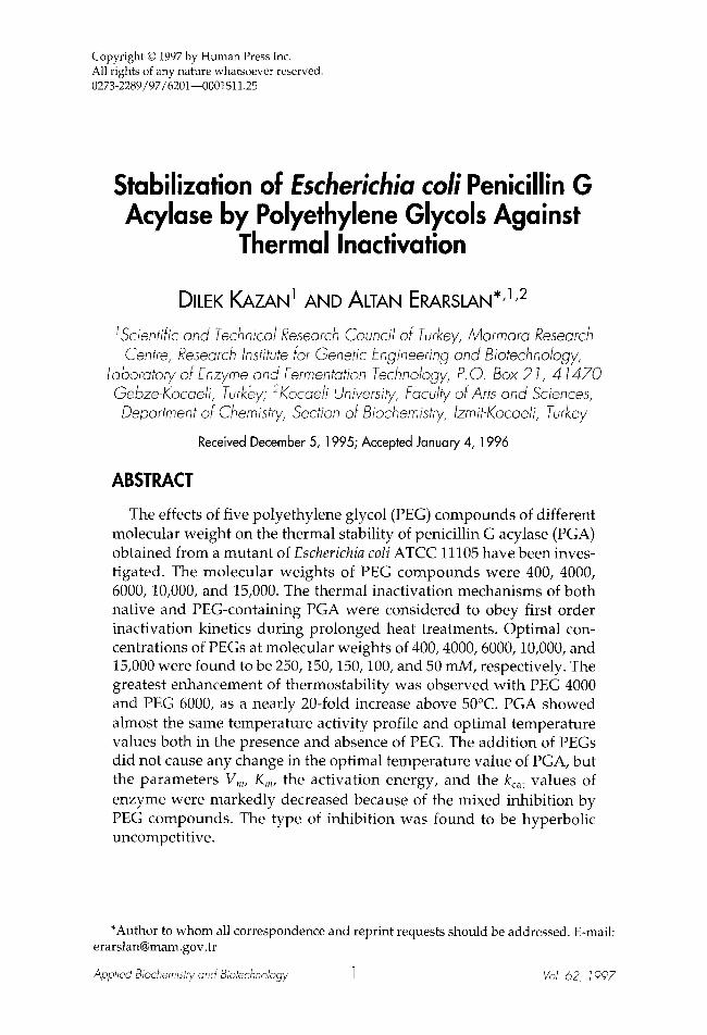
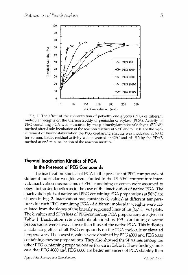
Effect of PEG Concentration on the Activity and Stability of PGA To select the optimal concentration of each PEG compound that pro-
vides the highest thermostabilization of enzyme, the effects of their dif- ferent concentrations on the stability of PGA were investigated. Thermostability was expressed as percent residual activity of PEG contain- ing PGA after 30 min incubation at 50~ Optimal concentrations of PEG compounds were estimated from the plateau values of residual activity curves and were found to be 250 mM for PEG 400, 150 mM for PEG 4000, and PEG 6000, 100 mM for PEG 10,000, and 50 mM for PEG 15,000 (Fig. 1). The protective effect of PEG compounds increased with their concentra- tions up to limiting values and it appears that there is a strong correlation between stabilization and PEG molecular weight. The higher the molecu- lar weight, the lower the concentration needed for thermostabilization. Similar results have appeared in the literature for invertase (22), Aspergillus niger glucose oxidase (23), and Bacillus licheniformis s-amylase (24).
Applied Biochemistry and Biotechnelogy VoI. 62, 1997
Stabilization of Pen G Acylase 5
100
9O
8O
.~, 7o
60
50
40
~ 30
20
10
0
0 50 100 150 200 250 300
PEG Concentration, (mM)
Fig. 1. The effect of the concentration of polyethylene glycols (PEG) of different molecular weights on the thermostability of penicillin G acylase (PGA). Activity of PEG containing PGA was measured by the p-dimethylaminobezaldehyde (PDAB) method after 3 min incubation of the reaction mixture at 40~ and pH 8.0. For the mea- surement of thermostabilization the PEG containing enzyme was incubated at 50~ for 30 min. Later, residual activity was measured at 40~ and pH 8.0 by the PDAB method after 3 min incubation of the reaction mixture.
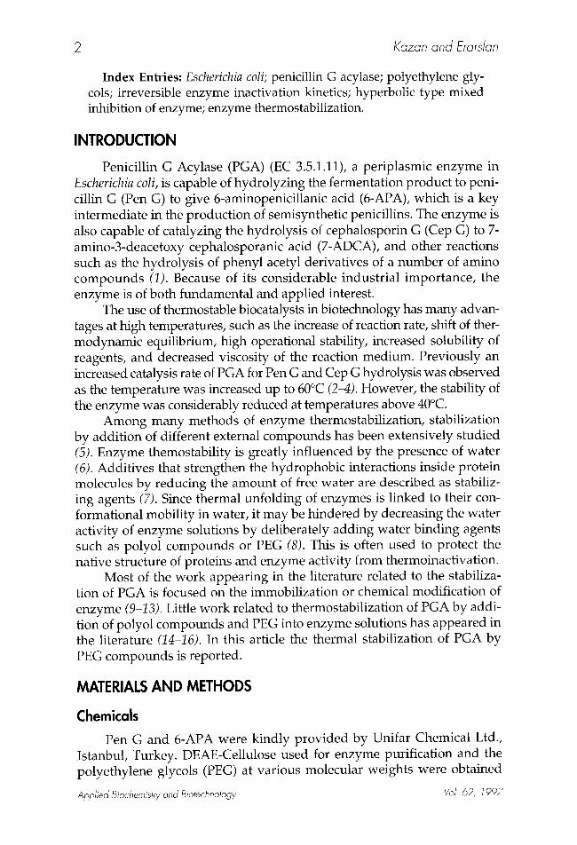
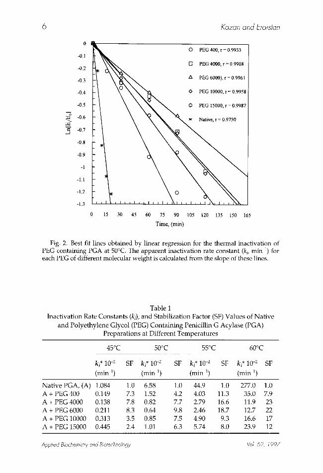
Thermal Inactivation Kinetics of PGA in the Presence of PEG Compounds The inactivation kinetics of PGA in the presence of PEG compounds of
different molecular weights were studied in the 45-60~ temperature inter- val. Inactivation mechanisms of PEG-containing enzymes were assumed to obey first-order kinetics as in the case of the inactivation of native PGA. The inactivation plots of native and PEG-containing PGA preparations at 50~ are shown in Fig. 2. Inactivation rate constants (ki values) at different tempera- tures for each PEG-containing PGA of different molecular weights were cal- culated from the slopes of the linearly regressed lines of Ln [Ei/En] v s t plots. The ki values and SF values of PEG-containing PGA preparations are given in Table 1. Inactivation rate constants obtained by PEG containing enzyme preparations were always lower than those of the native PGA. This indicates a stabilizing effect of all PEG compounds on the PGA molecule at elevated temperatures. The lowest k i values were obtained by PEG 4000 and PEG 6000 containing enzyme preparations. They also showed the SF values among the other PEG-containing preparations as shown in Table 1. These findings indi- cate that PEG 4000 and PEG 6000 are better enhancers of PGA stability than
Applied Biochemistry and Biotechnology gol. 62, 1997
6 Kazan and Erarslan
,...g
0
-OA
-0.2
-0.3
-0.4
-0.5
-0.6
-0.7
-0.8
-0.9
-1
-1.1
-1.2
-1.3
0 15 30 45 60 75 90 105 120 135 150 165
Time, (min)
Fig. 2. Best fit lines obtained by linear regression for the thermal inactivation of PEG containing PGA at 50~ The apparent inactivation rate constant (ki, min 1) for each PEG of different molecular weight is calculated from the slope of these lines.
Table 1 Inactivation Rate Constants (ki), and Stabilization Factor (SF) Values of Nat ive
and Polyethylene Glycol (PEG) Containing Penicillin G Acylase (PGA) Preparations at Different Temperatures
ki* 10 -2
(min 1)
45~ 50~ 55~ 60~
SF ki* 10 -2 SF ki* 10 -2 SF ki* 10 -2 SF
(min 1) (min 1) (min-1)
Nat ive PGA, (A) 1.084 1.0 6.58 A + PEG 400 0.149 7.3 1.52 A + PEG 4000 0.138 7.8 0.82 A + PEG 6000 0.211 8.3 0.64 A + PEG 10000 0.313 3.5 0.85 A + PEG 15000 0.445 2.4 1.01
1.0 44.9 1.0 277.0 1.0 4.2 4.03 11.3 35.0 7.9 7.7 2.79 16.6 11.9 23 9.8 2.46 18.7 12.7 22 7.5 4.90 9.3 16.6 17 6.3 5.74 8.0 23.9 12
Applied Biochemistry and Biotechnology Vol. 62, 1997
Stabilization of Pen G Acylase 7
@
240
220
200
180
160
140
120
100
80
60
40
20
0
f ~ ~ i ~ I i F I J ~ I i i I
_- - 0 - 45oc 50 c
0 0
C ~
t i I I I I I i I J i I t i
0 3000 6000 9000 12000 15000
Molecular Weight of PEG
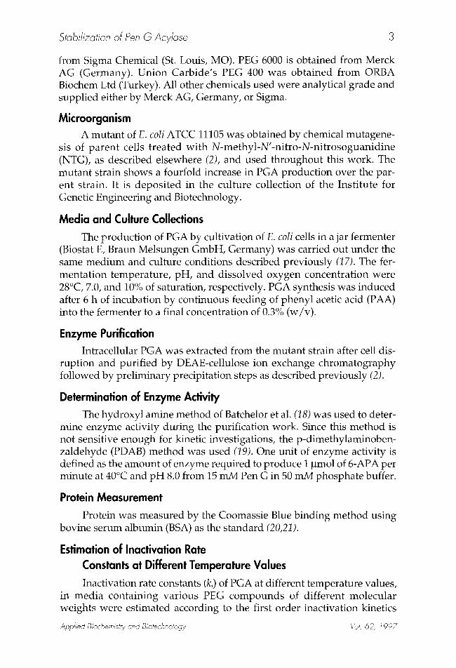
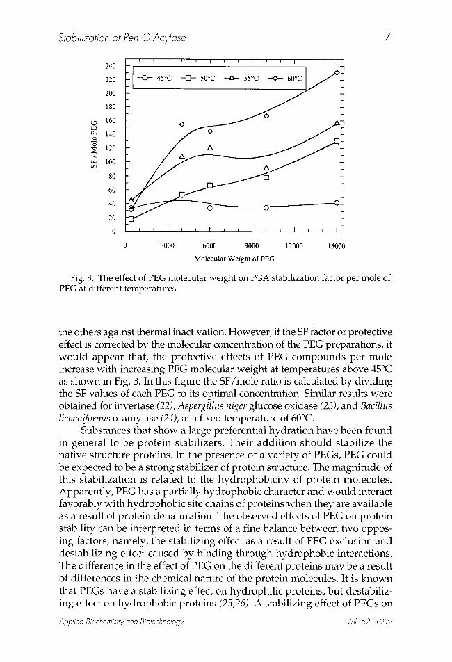
Fig. 3. The effect of PEG molecular weight on PGA stabilization factor per mole of PEG at different temperatures.
the others against thermal inactivation. However, if the SF factor or protective effect is corrected by the molecular concentration of the PEG preparations, it would appear that, the protective effects of PEG compounds per mole increase with increasing PEG molecular weight at temperatures above 45~ as shown in Fig. 3. In this figure the SF/mole ratio is calculated by dividing the SF values of each PEG to its optimal concentration. Similar results were obtained for invertase (22), Aspergillus niger glucose oxidase (23), and Bacillus licheniformis R-amylase (24), at a fixed temperature of 60~
Substances that show a large preferential hydration have been found in general to be protein stabilizers. Their addition should stabilize the native structure proteins. In the presence of a variety of PEGs, PEG could be expected to be a strong stabilizer of protein structure. The magnitude of this stabilization is related to the hydrophobicity of protein molecules. Apparently, PEG has a partially hydrophobic character and would interact favorably with hydrophobic site chains of proteins when they are available as a result of protein denaturation. The observed effects of PEG on protein stability can be interpreted in terms of a fine balance between two oppos- ing factors, namely, the stabilizing effect as a result of PEG exclusion and destabilizing effect caused by binding through hydrophobic interactions. The difference in the effect of PEG on the different proteins may be a result of differences in the chemical nature of the protein molecules. It is known that PEGs have a stabilizing effect on hydrophilic proteins, but destabiliz- ing effect on hydrophobic proteins (25,26). A stabilizing effect of PEGs on
Applied Biochemistry and Biotechnology Vol. 62, 1997
8 Kazan and Erarslan
0
-I
-2
-3
~'~ -4
-5
-6
-7
O PEG 400, r = -0.9911
O [] PEG 4000, r = -0.9899
ZX PEG 6000, r = -0.9870
O PEG 10000, r =-0.9929
XNN * PEG 15000, r=-0.9856
0.003 0.00305 0.0031 0.00315 0.0032 0.00325 0.0033
I/T, (~
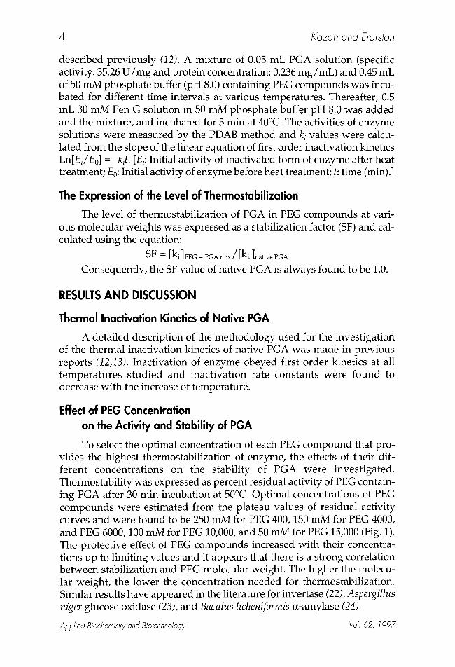
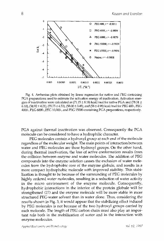
Fig. 4. Arrhenius plots obtained by linear regression for native and PEG containing PGA preparations used to estimate the activation energy of inactivation. Activation ener- gies of inactivation were calculated as (71.15 _+ 8.18) kcal/mol for native PGA and (78.51 + 6.10), (58.92 + 4.21), (55.75 • 4.53), (58.00 + 3.48), and (58 • 4.98) kcal/mol for PEG 400-, PEG 4000-, PEG 6000-, PEG 10,000-, and PEG 15000-containing PGA preparations, respectively.
PGA against thermal inactivation was observed. Consequently the PGA molecule can be considered to have a hydrophilic character.
PEG molecules contain a hydroxyl group at each end of the molecule regardless of the molecular weight. The main points of interaction between water and PEG molecules are these hydroxyl groups. On the other hand, dur ing thermal inactivation, the loss of active conformation results from the collision between enzyme and water molecules. The addit ion of PEG compounds into the enzyme solution causes the exclusion of water mole- cules from the hydrophobic core of the enzyme globule, and results in a more compact hydrophobic molecule with improved stability. This stabi- lization is thought to be because of the surrounding of PEG molecules by highly ordered water molecules, resulting in a reduction of water activity in the micro env i ronment of the enzyme molecule. Consequent ly , hydrophobic interactions in the interior of the protein globule will be strengthened (27) and the enzyme molecule will be more stable in more structured PEG-water solvent than in water alone. Thus, considering the results shown in Fig. 3, it would appear that the stabilizing effect induced by PEG molecules is not because of the two hydroxyl groups carried by each molecule. The length of PEG carbon chain must also play an impor- tant role both in the mobil ization of water and in the interaction wi th enzyme molecules.
Applied Biochemistry and Biotechnology Vol. 62, 1997
Stabilization of Pen G Acylase 9
Activation Energies (El) and Activation Free Energies (AGI) for Inactivation of Native and PEG Containing PGA
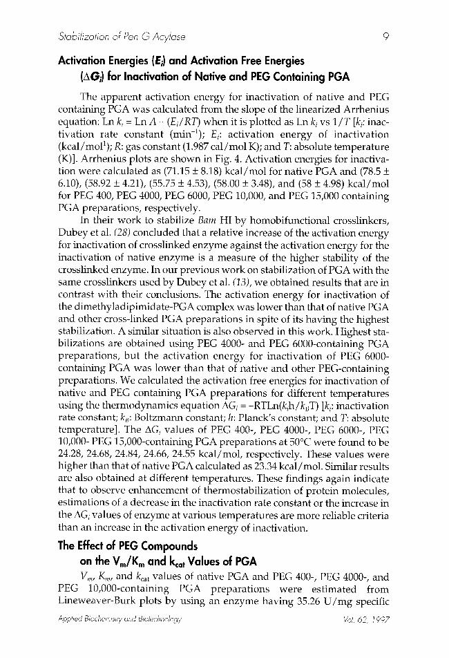
The apparent activation energy for inactivation of native and PEG containing PGA was calculated from the slope of the linearized Arrhenius equation: Ln k i = Ln A - ( E i / R T ) when it is plotted as Ln k i v s 1/T [ki: inac- tivation rate constant (min-1); • i : activation energy of inactivation (kcal/moP); R: gas constant (1.987 cal/mol K); and T: absolute temperature (K)]. Arrhenius plots are shown in Fig. 4. Activation energies for inactiva- tion were calculated as (71.15 + 8.18) kcal/mol for native PGA and (78.5 + 6.10), (58.92 + 4.21), (55.75 + 4.53), (58.00 + 3.48), and (58 + 4.98) kcal/mol for PEG 400, PEG 4000, PEG 6000, PEG 10,000, and PEG 15,000 containing PGA preparations, respectively.
In their work to stabilize Barn HI by homobifunctional crosslinkers, Dubey et al. (28) concluded that a relative increase of the activation energy for inactivation of crosslinked enzyme against the activation energy for the inactivation of native enzyme is a measure of the higher stability of the crosslinked enzyme. In our previous work on stabilization of PGA with the same crosslinkers used by Dubey et al. (13), we obtained results that are in contrast with their conclusions. The activation energy for inactivation of the dimethyladipimidate-PGA complex was lower than that of native PGA and other cross-linked PGA preparations in spite of its having the highest stabilization. A similar situation is also observed in this work. Highest sta- bilizations are obtained using PEG 4000- and PEG 6000-containing PGA preparations, but the activation energy for inactivation of PEG 6000- containing PGA was lower than that of native and other PEG-containing preparations. We calculated the activation free energies for inactivation of native and PEG containing PGA preparations for different temperatures using the thermodynamics equation AGi = -RTLn(kih/kJ) [ki: inactivation rate constant; k~: Boltzmann constant; h: Planck's constant; and T: absolute temperature]. The AGi values of PEG 400-, PEG 4000-, PEG 6000-, PEG 10,000- PEG 15,000-containing PGA preparations at 50~ were found to be 24.28, 24.68, 24.84, 24.66, 24.55 kcal/mol, respectively. These values were higher than that of native PGA calculated as 23.34 kcal/mol. Similar results are also obtained at different temperatures. These findings again indicate that to observe enhancement of thermostabilization of protein molecules, estimations of a decrease in the inactivation rate constant or the increase in the AGi values of enzyme at various temperatures are more reliable criteria than an increase in the activation energy of inactivation.
The Effect of PEG Compounds on the Vm/K m and kca t Values of PGA Vm, Kin, and kca t values of native PGA and PEG 400-, PEG 4000-, and
PEG 10,000-containing PGA preparations were estimated from Lineweaver-Burk plots by using an enzyme having 35.26 U / m g specific
Applied Biochemistry and Biotechnology Vol. 62, 1997
1 0 Kazan and Erarslan
activity and 0.236 m g / m L protein concentration. Reactions for activity measurements were carried out after 3 min incubation at 40~ and in 50 mM phosphate buffer, pH 8.0. Vm, Kin, and kca t values of PEG-containing PGA preparation were significantly lower than those of native PGA. The Vm/Km ratios were found to be 0.141 + 0.08, 0.137 + 0.013, 0.138 + 0.008, 0.137 + 0.015, 0.138 + 0.012, and 0.0137 + 0.010 for native PGA, PEG 400- (150 mM), PEG 400- (300 mM), PEG 4000- (50 mM), PEG 4000- (100 mM), PEG 10,000- (25 mM), and PEG 10,000- (50 mM) containing PGA preparations, respec- tively. These values were very close to each other and consequently Vm/Km ratio can be considered almost same for all preparations.
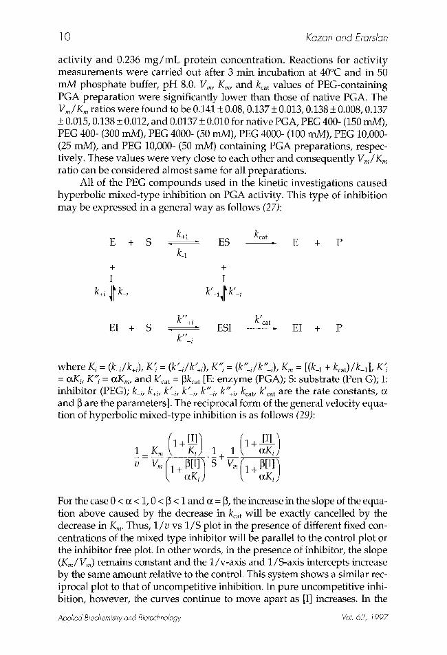
All of the PEG compounds used in the kinetic investigations caused hyperbolic mixed-type inhibition on PGA activity. This type of inhibition may be expressed in a general way as follows (27):
k+l kcat E + S ES - E + P
k_l + + I I
k+, ~I k-i k' ' ll~ -i
k"+i k'ca t EI + S ESI - EI + P
k"-i
" (k" / k " ~ . . . . . . . where K i = (k i/k+i), K i = , -i- +/,, K i = (k _i/k +i), Km= [(k-1 + kcat)/k+l], K i = otKi, K"i = OtKm, and k'ca t = ~kca t [E: enzyme (PGA); S: substrate (Pen G); I: inhibitor (PEG); k_i, k+, k ' . k" " ' _, k ' i , -i, k +i, kcat, k cat are the rate constants, c~ and [3 are the parameters]. The reciprocal form of the general velocity equa- tion of hyperbolic mixed-type inhibition is as follows (29):
( l + [ I ] ] (1+ [I]~ 1 = K~ Ki ) . 1 t- 1 o~Ki)
v V m ( l + N I l ~ S o~Ki) Vm( 1 + ~[I] ) k
For the case 0 < (z < 1, 0 < [3 < i and (z = [3, the increase in the slope of the equa- tion above caused by the decrease in kca t will be exactly cancelled by the decrease in Kin. Thus, 1 / v vs 1/S plot in the presence of different fixed con- centrations of the mixed type inhibitor will be parallel to the control plot or the inhibitor free plot. In other words, in the presence of inhibitor, the slope (Kin/Vm) remains constant and the 1/v-axis and 1/S-axis intercepts increase by the same amount relative to the control. This system shows a similar rec- iprocal plot to that of uncompetitive inhibition. In pure uncompetitive inhi- bition, however, the curves continue to move apart as [I] increases. In the
Applied Biochemistry and Biotechnology Vol. 62, 1997
Stabilization of Pen G Acylase 11
IOO
90
~ 8o
.~ 7o
< 60
'~ 50
~ 40
30
20
3
2.8
2.6
> 2.4 ,-.a
2.2
2
18
1.6
2.95
I I I l l l i l I l l l l l l l l l l l I I I I I I L I I I I I I i i l i l l l l l ] l l i 1 ~ 1 1 1 l i t i i
30 35 40 45 50 55 60 65 70 75 80 S5 90
Temperature, (~
l ' ~ l r I r l l l ' I l l l l l l ' r r l l ' ' l l l l l l l l l l l t l , ~ l , l t l l l l ~ l l l l l l l t i i l l
O PEG 400, r =-0.9657
[] PEG 4000, r =-0.9982
i PEG 6000, r =-0.9953
PEG 10000, r = -0.9906
PEG 15000, r = -0.9944 -
i Native PGA, r = -09952- 0
l l l l l l l l l I + I I I I I I I { I I I I I I l l ; "
3 3.05 3.1 3.15 3.2 3.25 3+3 3.35 3.4 3.45 3.5 3.55 3.6
[I/T]*I0 3, (~ -~
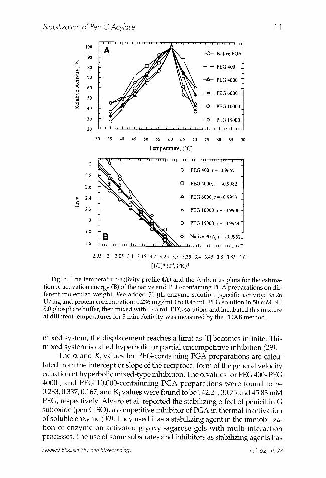
Fig. 5. The temperature-activity profile (A) and the Arrhenius plots for the estima- tion of activation energy (B) of the native and PEG-containing PGA preparat ions on dif- ferent molecular weight. We added 50 ~L enzyme solut ion (specific activity: 35.26 U / m g and protein concentration: 0.236 m g / m L ) to 0.45 mE PEG solution in 50 mM pH 8.0 phosphate buffer, then mixed with 0.45 mE PEG solution, and incubated this mixture at different temperatures for 3 min. Activity was measured by the PDAB method.
mixed system, the displacement reaches a limit as [I] becomes infinite. This mixed system is called hyperbolic or partial uncompetitive inhibition (29).
The or and K i values for PEG-containing PGA preparations are calcu- lated from the intercept or slope of the reciprocal form of the general velocity equation of hyperbolic mixed-type inhibition. The o~ values for PEG 400- PEG 4000-, and PEG 10,000-containning PGA preparations were found to be 0.283, 0.337, 0.167, and Ki values were found to be 142.21, 30.75 and 45.83 mM PEG, respectively. Alvaro et al. reported the stabilizing effect of penicillin G sulfoxide (pen G SO), a competitive inhibitor of PGA in thermal inactivation of soluble enzyme (30). They used it as a stabilizing agent in the immobiliza- tion of enzyme on activated glyoxyl-agarose gels with multi-interaction processes. The use of some substrates and inhibitors as stabilizing agents has
Applied Biochemistry and Biotechnology Vol. 62, 1997
1 2 Kazan and Erarslan
considerable importance in enzyme technology, particularly, in the chemical modification and covalent immobilization of enzymes in order to improve their properties and in preparation of industrial enzyme derivatives. Despite their hyperbolic uncompetitive inhibitory effects, PEG compounds may also be used as stabilizing agents during the modification of PGA.
Effect of PEG Compounds on the Optimal Temperature and Activation Energy of PGA The addition of PEG compounds did not cause any change on the
optimal temperature of PGA. The optimum temperature for Pen G hydrol- ysis by PGA in both cases was 60~ (Fig. 5A). However, slight decreases were observed in the activation energies of PEG containing PGA prepara- tions. The activation energies of native and PEG-PGA preparations were calculated from the Arrhenius plot (Fig. 5B). Estimated values in kcal/mol, were, (7.60 + 1.02), (8.74 + 0.27), (9.30 + 0.45), (7.46 + 0.51), and (6.58 + 0.35) for PEG 400-, PEG 4000-, PEG 6000-, PEG 10,000-, and PEG 15,000-contain- ing PGA, and (10.21 + 0.50) for the native PGA, respectively. These results indicate that the presence of PEG compounds contributes to a decrease in the activation energy of PGA.
REFERENCES 1. Shewale, J. G., Despande, B. S., Sudhakaran, V. K., and Amberkar, S. S. (1990), Process
Biochem. 25, 97. 2. Erarslan, A., Terzi, I., Gfiray, A., and Bermek, E. (1991), J. Chem. Technol. Biotechnol. 51, 27. 3. Erarslan, A. (1993), Process Biochem. 28, 311. 4. Erarslan, A. and Gfiray, A. (1991), J. Chem. Technol. BiotechnoI. 51, 181. 5. Schmid, R. D. (1979), in Advances in Biochemical Engineering, Ghose, T. K., Fiechter, A.,
and Blakebrough, N., eds., Springer Verlag, Berlin, vol. 12, pp. 41-127. 6. Klibanov, A. M. (1989), TIBS 14, 141. 7. Klibanov, A. M. (1983), in Advances in Applied Microbiology, Laskin, A. I., ed., Academic,
New York, vol. 29, pp. 1-28. 8. Larreta-Garde, V., Xu, Z. F., and Thomas, D. (1988), Enzyme Engineering, Blanch, H. W.,
and Klibanov, A. M., eds., Academic, New York, vol. 9, pp. 294-298. 9. Alvaro, G., Lafuente, R. F., Blanco, R. M., and Guis~n, J. M. (1990), Appl. Biochem.
Biotechnol. 26, 181. 10. Guis4n, J. M. (1988), Enzyme Microb. Technol. 10, 375. 11. Lafuente, R. F., Rosell, C. M., Alvaro, G., and Guis4n, J. M. (1992), Enzyme Microb.
Technol. 14, 489. 12. Erarslan, A. and Ko~er, H. (1992), Escherichia coli ATCC 11105. J. Chem. Technol.
Biotechnol. 55, 79. 13. Lafuente, R. F., Rosell, C. M., and Guis~n, J. M. (1991) Enzyme Microb. Technol. 13, 898. 14. Erarslan, A. (1995), Process Biochem. 30, 133. 15. McDougall, B., Dunnill, P., and Lilly, M. D., (1982), Enzyme Microb. Technol. 4, 114. 16. Andersson, E. and Hahn-Hagerdahl, B. (1982), Biochim. Biophys. Acta 912, 317. 17. Erarslan, A. and Gtiray, A. (1991), Do~a-Tr. J. Biology, 15, 167-174. 18. Batchelor, F. R., Chain, E. B., Hardy, T. L., Mansford, K. R. L., and Rolinson, G. N.
(1961), Proc. Roy. Soc. B 154, 498. 19. Shewale, G. J., Kumar, K. K., and Ambekar, G. R. (1987), Biotechnol. Techniques 1, 69.
Applied Biochemistry and Biotechnology Vol. 62, 1997
Stabilization of Pen G Acylase 1 3
20. Spector, T. (1978), Anal. Biochem. 86, 142. 21. Sedmak, J. J. and Grossberg, S. E. (1977), Anal. Biochem. 79, 544. 22. Monsan, P. and Combes, D. (1984), Ann. NYAcad. Sci. USA 434, 48. 23. Ye, W. N., Combes, D., and Monsan, P. (1988), Enzyme Microb. Technol. 10, 498. 24. Asther, M. and Meunier, J. C. (1990), Enzyme Microb. Technol. 12, 902. 25. Arakawa, T. and Timasheff, S. N. (1985), Biochemistry 24, 6756. 26. Lee, L. L. Y. and Lee, J. C. (1987), Biochemistry 26, 7813. 27. Mozhaev, V. V. and Martinek, K. (1984), Enzyme Microb. Technol. 6, 50. 28. Dubey, A. K., Bisaria, V. S., Mukhopadhyay, S. N., and Ghose, T. K. (1989), Biotechnol.
Bioeng. 33, 1311. 29. Segel, I. H. (1975), in Enzyme Kinetics, Behavior and Analysis of Rapid Equilibrium and
Steady-State Enzyme. Wiley, New York, pp. 178-192. 30. Alvaro, G., Lafuente, R. B., Blanco, R. M., and Guis4n, J. M. (1991), Enzyme Microb.
TechnoI. 13, 210.
Applied Biochemistry and Biotechnology Vol. 62, 1997


