Embed Size (px)
Citation preview
RESEARCH ARTICLE
Spectrofluorimetric study of the ageing of mixtionsused in the gildings of mediaeval wall paintings
Aurélie Mounier & Colette Belin & Floréal Daniel
Published online: 15 December 2010# Springer-Verlag 2010
AbstractIntroduction In the Middle Ages, we could find gildings onmural paintings. Gold, silver or tin leaves were appliedaccording to distemper or mixtion technique. For the firstone, a binder as glue is necessary, and for the second, alipidic binder is used to stick the metallic leaf. Studies ofgildings materials characterization show that the mixtiontechnique, with a mordant, is the most common. Linseed oilseems to be the binder used. It is always mixed with asiccative agent as lead. Because of bad conditions ofconservation, the gildings do not resist anymore, onlyremain traces of metal or the adhesive under-layer. Thanksto the binder fluorescence, we can nowadays detect ancientgildings.Objective The purpose of this paper is to study the degrada-tion of the linseed oil, generally mixed with lead white to givea mordant for the metallic leaf, by spectrofluorimetry.Materials and methods To understand in situ fluorescence,gildings recreations, linseed oil and lead white are aged in
hygro-thermal and ultraviolet (UV) light (313 nm)climatic rooms and under UV irradiation. Irradiationwavelengths are chosen according to the maximum ofabsorption of linseed oil and the bibliography (296, 313and 366 nm=mercury bands).Results In comparison with results (in situ UV lamp,spectrofluorimetry), excitation wavelength chosen is366 nm. Irradiations at 366 nm of linseed oil and linseedoil mixed with lead white show the most degrading effect inthe fluorescence to the big wavelength. Lead white plays animportant siccative role; it increases the intensity fluores-cence and accelerates the drying of linseed oil. This studyalso allows to show that 366 nm wavelength is good for thein situ observation.
Keywords Gildings . UV fluorescence . Gildingsrecreation . Linseed oil . Lead white . Mixtion .
Accelerated ageing
1 Introduction
Gildings are elements of decoration sometimes found onmediaeval mural paintings. The study of the gildingtechniques is particularly interesting from a historic pointof view because of the aesthetic, symbolic and prestigiouscriteria which are at the origin of the realization of thesegolden decorations. In the Middle Ages, paintings, inparticular by fresco, were decorated for the greater partwith lights, metallic decorations, glass inlays to give morevolume, relief and to add reflections and brilliance to thecolour (Pastoureau 1999). These additions were glued onthe wall, by means of an organic binder, which confersthem a bigger fragility. Of these metallic decorations oftenremain some traces because their mode of application
Responsible editor: Philippe Garrigues
A. Mounier (*) : F. DanielInstitut de Recherche sur les Archéomatériaux–Centrede Recherche en Physique Appliquée à l’Archéologie(IRAMAT–CRPAA, UMR 5060),Université Bordeaux–CNRS,Pessac, Francee-mail: [email protected]
F. Daniele-mail: [email protected]
C. BelinInstitut des Sciences Moléculaires, UMR 5255,Université de Bordeaux 1/CNRS,Talence, Francee-mail: [email protected]
Environ Sci Pollut Res (2011) 18:772–782DOI 10.1007/s11356-010-0429-5
makes them sensitive to the numerous factors of degrada-tion, in particular environmental, to which they aresubjected during centuries. Often, only remain the rests oforganic binders which served to glue the metallic leaf andthanks to their detection, it is possible for us to locate anancient gilding today.
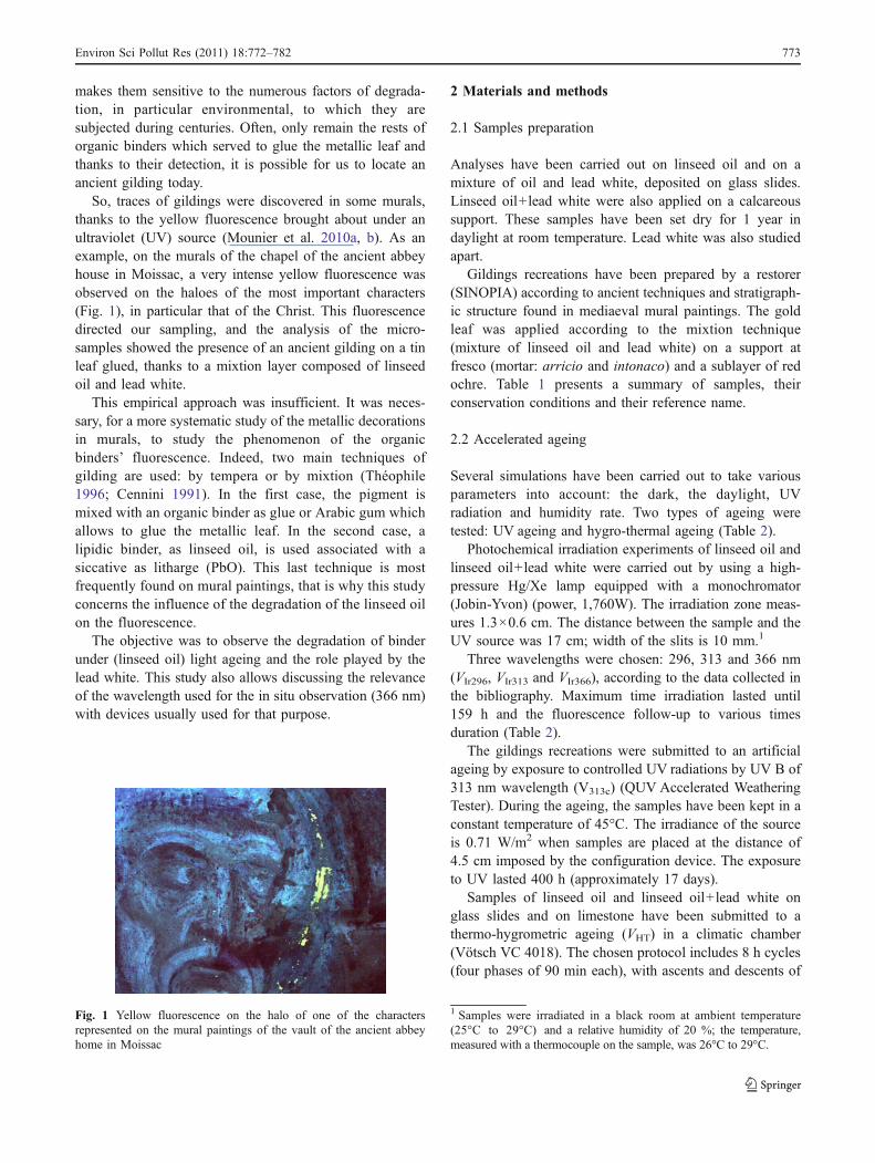
So, traces of gildings were discovered in some murals,thanks to the yellow fluorescence brought about under anultraviolet (UV) source (Mounier et al. 2010a, b). As anexample, on the murals of the chapel of the ancient abbeyhouse in Moissac, a very intense yellow fluorescence wasobserved on the haloes of the most important characters(Fig. 1), in particular that of the Christ. This fluorescencedirected our sampling, and the analysis of the micro-samples showed the presence of an ancient gilding on a tinleaf glued, thanks to a mixtion layer composed of linseedoil and lead white.
This empirical approach was insufficient. It was neces-sary, for a more systematic study of the metallic decorationsin murals, to study the phenomenon of the organicbinders’ fluorescence. Indeed, two main techniques ofgilding are used: by tempera or by mixtion (Théophile1996; Cennini 1991). In the first case, the pigment ismixed with an organic binder as glue or Arabic gum whichallows to glue the metallic leaf. In the second case, alipidic binder, as linseed oil, is used associated with asiccative as litharge (PbO). This last technique is mostfrequently found on mural paintings, that is why this studyconcerns the influence of the degradation of the linseed oilon the fluorescence.
The objective was to observe the degradation of binderunder (linseed oil) light ageing and the role played by thelead white. This study also allows discussing the relevanceof the wavelength used for the in situ observation (366 nm)with devices usually used for that purpose.
2 Materials and methods
2.1 Samples preparation
Analyses have been carried out on linseed oil and on amixture of oil and lead white, deposited on glass slides.Linseed oil+lead white were also applied on a calcareoussupport. These samples have been set dry for 1 year indaylight at room temperature. Lead white was also studiedapart.
Gildings recreations have been prepared by a restorer(SINOPIA) according to ancient techniques and stratigraph-ic structure found in mediaeval mural paintings. The goldleaf was applied according to the mixtion technique(mixture of linseed oil and lead white) on a support atfresco (mortar: arricio and intonaco) and a sublayer of redochre. Table 1 presents a summary of samples, theirconservation conditions and their reference name.
2.2 Accelerated ageing
Several simulations have been carried out to take variousparameters into account: the dark, the daylight, UVradiation and humidity rate. Two types of ageing weretested: UV ageing and hygro-thermal ageing (Table 2).
Photochemical irradiation experiments of linseed oil andlinseed oil+lead white were carried out by using a high-pressure Hg/Xe lamp equipped with a monochromator(Jobin-Yvon) (power, 1,760W). The irradiation zone meas-ures 1.3×0.6 cm. The distance between the sample and theUV source was 17 cm; width of the slits is 10 mm.1
Three wavelengths were chosen: 296, 313 and 366 nm(VIr296, VIr313 and VIr366), according to the data collected inthe bibliography. Maximum time irradiation lasted until159 h and the fluorescence follow-up to various timesduration (Table 2).
The gildings recreations were submitted to an artificialageing by exposure to controlled UV radiations by UV B of313 nm wavelength (V313c) (QUV Accelerated WeatheringTester). During the ageing, the samples have been kept in aconstant temperature of 45°C. The irradiance of the sourceis 0.71 W/m2 when samples are placed at the distance of4.5 cm imposed by the configuration device. The exposureto UV lasted 400 h (approximately 17 days).
Samples of linseed oil and linseed oil+lead white onglass slides and on limestone have been submitted to athermo-hygrometric ageing (VHT) in a climatic chamber(Vötsch VC 4018). The chosen protocol includes 8 h cycles(four phases of 90 min each), with ascents and descents of
Fig. 1 Yellow fluorescence on the halo of one of the charactersrepresented on the mural paintings of the vault of the ancient abbeyhome in Moissac
1 Samples were irradiated in a black room at ambient temperature(25°C to 29°C) and a relative humidity of 20 %; the temperature,measured with a thermocouple on the sample, was 26°C to 29°C.
Environ Sci Pollut Res (2011) 18:772–782 773
30 min between every cycle. These conditions are similar tothose studies on the alterations of pigments in muralpaintings (Aze 2005). The phases, the temperature con-ditions and the relative humidity settled in this protocol are:
– High humidity: temperature (T)=18°C, relative humid-ity (RH)=85 %
– Low temperature: T=−10°C, RH=0%– Dry heat: T=40°C, RH=25%– Wet heat: T=30°C, RH=60%
This 8 h cycle was repeated 90 times for a total duration of4 weeks.To facilitate the comparison of the organic binderswith the gildings recreations, linseed oil deposited on glassslides was also subjected to the same hygro-thermal ageing.
2.3 Analytical methods
The UV–Vis absorption spectrum was realized for linseedoil deposited on a quartz slide with a spectrophotometerHewlett Packard 8452A.
Fluorescence measurements were performed using aFluorolog SPEX 212 spectrofluorimeter, equipped withdouble monochromators on both the excitation and emissionbeams. The excitation lamp is a 450 W high-pressure xenon
lamp, and a thermoelectrically refrigerated photomultiplier(Hamamatsu R928) is used for signal detection, using thephoton counting mode. Fluorescence signals are given in thefollowing in “counts per second” (cps). Data acquisition anddata processing were computer controlled (SpectrAcq Data-max run by the Grams/32 software). Both excitation and emis-sion spectra (recorded with 4 nm bandwidth) were correctedfor instrumental factors. The measurements are made in mode“front face”. The emitted light is collected according to anangle of 22.5° with regard to the excitation beam.
3 Results
The linseed oil used in our samples focuses on anabsorption spectrum, at initial state T0, with three maximalintensities at 273, 286 and 326 nm. The spectrofluorimetricstudy was made out of different wavelengths excitations at254, 273, 286, 313, 326 and 366 nm, very often mentionedin the bibliographical data (Larson et al. 1991; Karoui et al.2006; De la Rie 1982a) and correspond to those of amercury vapour lamp. Thoury (2006) also showed that themost interesting excitation wavelengths for the study ofvarnishes are between 300 and 366 nm.
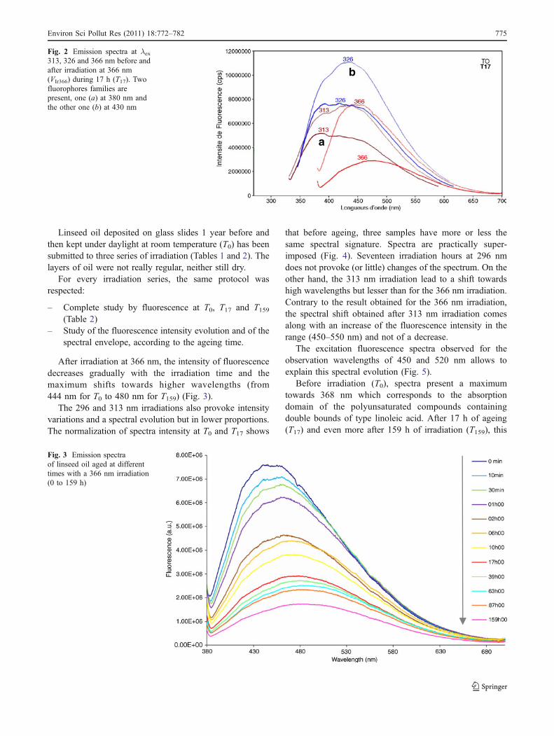
For the excitation wavelengths of 254, 273 and 286 nm, theresults are not really significant (very weak fluorescence). Forthe 313, 326 and 366 nm excitation wavelengths, thefluorescence intensity is much higher and the spectralenvelope covers the domain of 350–600 nm (Fig. 2).
The 313 and 326 nm excitation wavelengths allow to studythe evolution of two fluorophores families: the first onecentred towards 380 nm (a) and the other at about 430 nm (b).
The 366 nm excitation wavelength allows to follow thefluorescence emitted in the visible range (400–600 nm)and, by concern of comparison between the results oflaboratory and those observed in situ, with the eye as thedetector. This wavelength was chosen for the whole studyand for all the results presented below.
1. Irradiation ageing
(a) Linseed oil fluorescence before and after ageingfor an irradiation at 366 nm
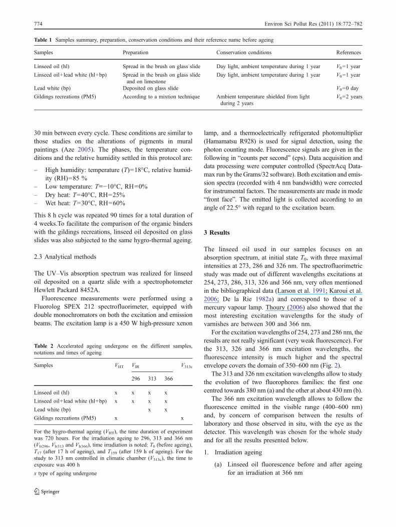
Table 1 Samples summary, preparation, conservation conditions and their reference name before ageing
Samples Preparation Conservation conditions References
Linseed oil (hl) Spread in the brush on glass slide Day light, ambient temperature during 1 year V0=1 year
Linseed oil+lead white (hl+bp) Spread in the brush on glass slideand on limestone
Day light, ambient temperature during 1 year V0=1 year
Lead white (bp) Deposited on glass slide V0=0 day
Gildings recreations (PM5) According to a mixtion technique Ambient temperature shielded from lightduring 2 years
V0=2 years
Table 2 Accelerated ageing undergone on the different samples,notations and times of ageing
Samples VHT VIR V313c
296 313 366
Linseed oil (hl) x x x x
Linseed oil+lead white (hl+bp) x x x x
Lead white (bp) x x
Gildings recreations (PM5) x x
For the hygro-thermal ageing (VHT), the time duration of experimentwas 720 hours. For the irradiation ageing to 296, 313 and 366 nm(VIr296, VIr313 and VIr366), time irradiation is noted: T0 (before ageing),T17 (after 17 h of ageing), and T159 (after 159 h of ageing). For thestudy to 313 nm controlled in climatic chamber (V313c), the time toexposure was 400 h
x type of ageing undergone
774 Environ Sci Pollut Res (2011) 18:772–782
Linseed oil deposited on glass slides 1 year before andthen kept under daylight at room temperature (T0) has beensubmitted to three series of irradiation (Tables 1 and 2). Thelayers of oil were not really regular, neither still dry.
For every irradiation series, the same protocol wasrespected:
– Complete study by fluorescence at T0, T17 and T159(Table 2)
– Study of the fluorescence intensity evolution and of thespectral envelope, according to the ageing time.
After irradiation at 366 nm, the intensity of fluorescencedecreases gradually with the irradiation time and themaximum shifts towards higher wavelengths (from444 nm for T0 to 480 nm for T159) (Fig. 3).
The 296 and 313 nm irradiations also provoke intensityvariations and a spectral evolution but in lower proportions.The normalization of spectra intensity at T0 and T17 shows
that before ageing, three samples have more or less thesame spectral signature. Spectra are practically super-imposed (Fig. 4). Seventeen irradiation hours at 296 nmdoes not provoke (or little) changes of the spectrum. On theother hand, the 313 nm irradiation lead to a shift towardshigh wavelengths but lesser than for the 366 nm irradiation.Contrary to the result obtained for the 366 nm irradiation,the spectral shift obtained after 313 nm irradiation comesalong with an increase of the fluorescence intensity in therange (450–550 nm) and not of a decrease.
The excitation fluorescence spectra observed for theobservation wavelengths of 450 and 520 nm allows toexplain this spectral evolution (Fig. 5).
Before irradiation (T0), spectra present a maximumtowards 368 nm which corresponds to the absorptiondomain of the polyunsaturated compounds containingdouble bounds of type linoleic acid. After 17 h of ageing(T17) and even more after 159 h of irradiation (T159), this
b
a
Fig. 2 Emission spectra at λex313, 326 and 366 nm before andafter irradiation at 366 nm(VIr366) during 17 h (T17). Twofluorophores families arepresent, one (a) at 380 nm andthe other one (b) at 430 nm
Fig. 3 Emission spectraof linseed oil aged at differenttimes with a 366 nm irradiation(0 to 159 h)
Environ Sci Pollut Res (2011) 18:772–782 775
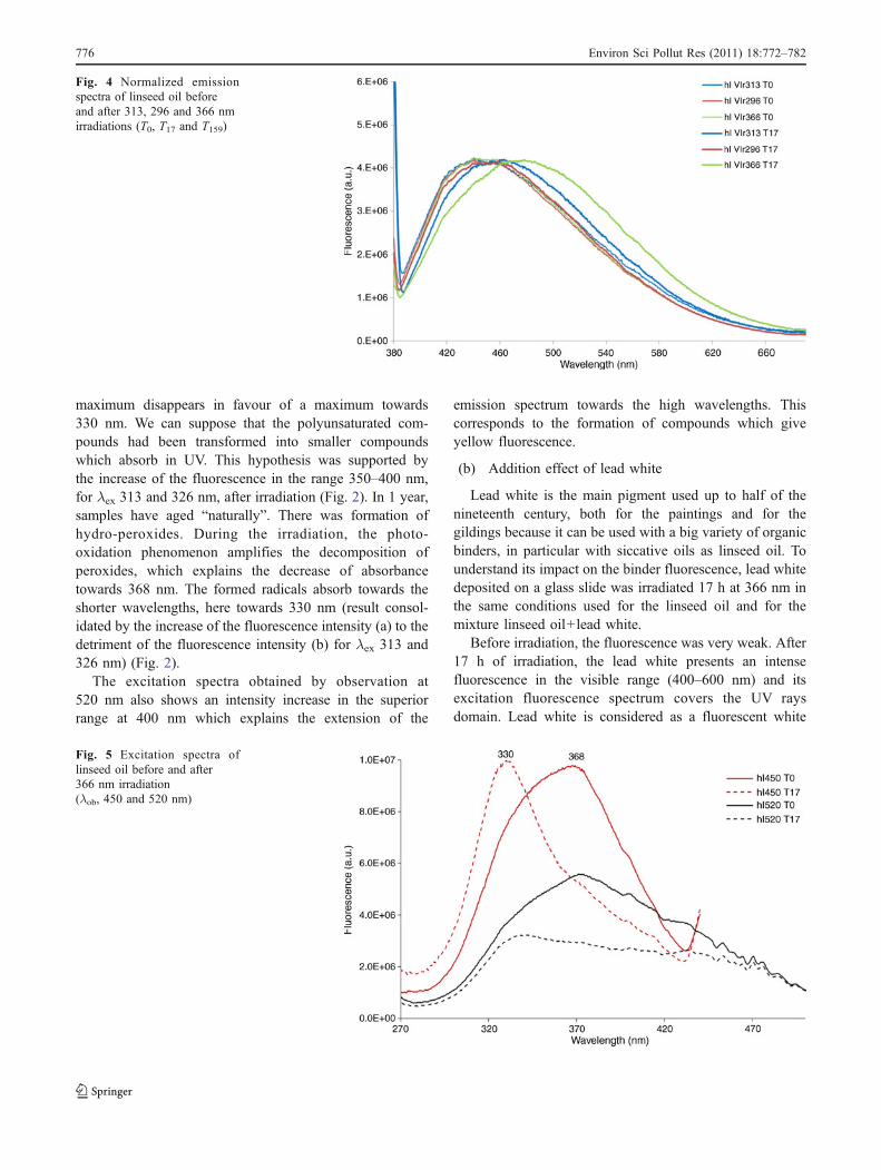
maximum disappears in favour of a maximum towards330 nm. We can suppose that the polyunsaturated com-pounds had been transformed into smaller compoundswhich absorb in UV. This hypothesis was supported bythe increase of the fluorescence in the range 350–400 nm,for λex 313 and 326 nm, after irradiation (Fig. 2). In 1 year,samples have aged “naturally”. There was formation ofhydro-peroxides. During the irradiation, the photo-oxidation phenomenon amplifies the decomposition ofperoxides, which explains the decrease of absorbancetowards 368 nm. The formed radicals absorb towards theshorter wavelengths, here towards 330 nm (result consol-idated by the increase of the fluorescence intensity (a) to thedetriment of the fluorescence intensity (b) for λex 313 and326 nm) (Fig. 2).
The excitation spectra obtained by observation at520 nm also shows an intensity increase in the superiorrange at 400 nm which explains the extension of the
emission spectrum towards the high wavelengths. Thiscorresponds to the formation of compounds which giveyellow fluorescence.
(b) Addition effect of lead white
Lead white is the main pigment used up to half of thenineteenth century, both for the paintings and for thegildings because it can be used with a big variety of organicbinders, in particular with siccative oils as linseed oil. Tounderstand its impact on the binder fluorescence, lead whitedeposited on a glass slide was irradiated 17 h at 366 nm inthe same conditions used for the linseed oil and for themixture linseed oil+lead white.
Before irradiation, the fluorescence was very weak. After17 h of irradiation, the lead white presents an intensefluorescence in the visible range (400–600 nm) and itsexcitation fluorescence spectrum covers the UV raysdomain. Lead white is considered as a fluorescent white
Fig. 4 Normalized emissionspectra of linseed oil beforeand after 313, 296 and 366 nmirradiations (T0, T17 and T159)
Fig. 5 Excitation spectra oflinseed oil before and after366 nm irradiation(λob, 450 and 520 nm)
776 Environ Sci Pollut Res (2011) 18:772–782
which absorbs UV and emits in the visible. After irradiationunder 366 nm, the sample is slightly yellow. Afterirradiation under 313 nm, it turns brown.
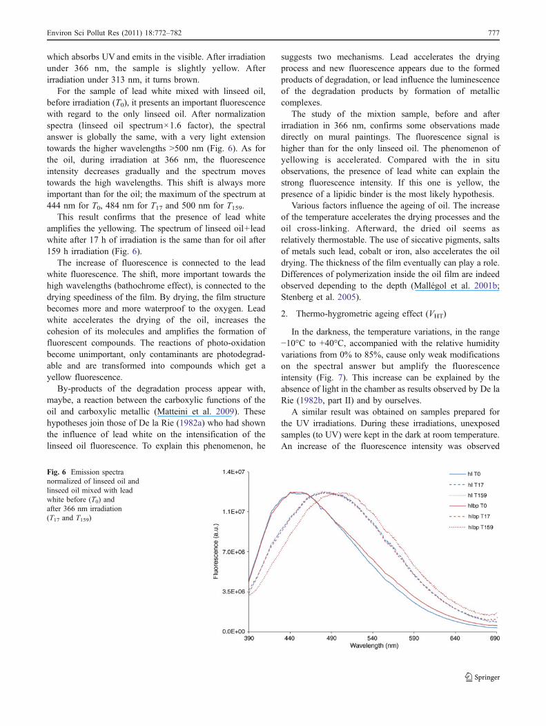
For the sample of lead white mixed with linseed oil,before irradiation (T0), it presents an important fluorescencewith regard to the only linseed oil. After normalizationspectra (linseed oil spectrum×1.6 factor), the spectralanswer is globally the same, with a very light extensiontowards the higher wavelengths >500 nm (Fig. 6). As forthe oil, during irradiation at 366 nm, the fluorescenceintensity decreases gradually and the spectrum movestowards the high wavelengths. This shift is always moreimportant than for the oil; the maximum of the spectrum at444 nm for T0, 484 nm for T17 and 500 nm for T159.
This result confirms that the presence of lead whiteamplifies the yellowing. The spectrum of linseed oil+leadwhite after 17 h of irradiation is the same than for oil after159 h irradiation (Fig. 6).
The increase of fluorescence is connected to the leadwhite fluorescence. The shift, more important towards thehigh wavelengths (bathochrome effect), is connected to thedrying speediness of the film. By drying, the film structurebecomes more and more waterproof to the oxygen. Leadwhite accelerates the drying of the oil, increases thecohesion of its molecules and amplifies the formation offluorescent compounds. The reactions of photo-oxidationbecome unimportant, only contaminants are photodegrad-able and are transformed into compounds which get ayellow fluorescence.
By-products of the degradation process appear with,maybe, a reaction between the carboxylic functions of theoil and carboxylic metallic (Matteini et al. 2009). Thesehypotheses join those of De la Rie (1982a) who had shownthe influence of lead white on the intensification of thelinseed oil fluorescence. To explain this phenomenon, he
suggests two mechanisms. Lead accelerates the dryingprocess and new fluorescence appears due to the formedproducts of degradation, or lead influence the luminescenceof the degradation products by formation of metalliccomplexes.
The study of the mixtion sample, before and afterirradiation in 366 nm, confirms some observations madedirectly on mural paintings. The fluorescence signal ishigher than for the only linseed oil. The phenomenon ofyellowing is accelerated. Compared with the in situobservations, the presence of lead white can explain thestrong fluorescence intensity. If this one is yellow, thepresence of a lipidic binder is the most likely hypothesis.
Various factors influence the ageing of oil. The increaseof the temperature accelerates the drying processes and theoil cross-linking. Afterward, the dried oil seems asrelatively thermostable. The use of siccative pigments, saltsof metals such lead, cobalt or iron, also accelerates the oildrying. The thickness of the film eventually can play a role.Differences of polymerization inside the oil film are indeedobserved depending to the depth (Mallégol et al. 2001b;Stenberg et al. 2005).
2. Thermo-hygrometric ageing effect (VHT)
In the darkness, the temperature variations, in the range−10°C to +40°C, accompanied with the relative humidityvariations from 0% to 85%, cause only weak modificationson the spectral answer but amplify the fluorescenceintensity (Fig. 7). This increase can be explained by theabsence of light in the chamber as results observed by De laRie (1982b, part II) and by ourselves.
A similar result was obtained on samples prepared forthe UV irradiations. During these irradiations, unexposedsamples (to UV) were kept in the dark at room temperature.An increase of the fluorescence intensity was observed
Fig. 6 Emission spectranormalized of linseed oil andlinseed oil mixed with leadwhite before (T0) andafter 366 nm irradiation(T17 and T159)
Environ Sci Pollut Res (2011) 18:772–782 777
according to the time between the different irradiations(VIr313, VIr296 and VIr366 nanometres).
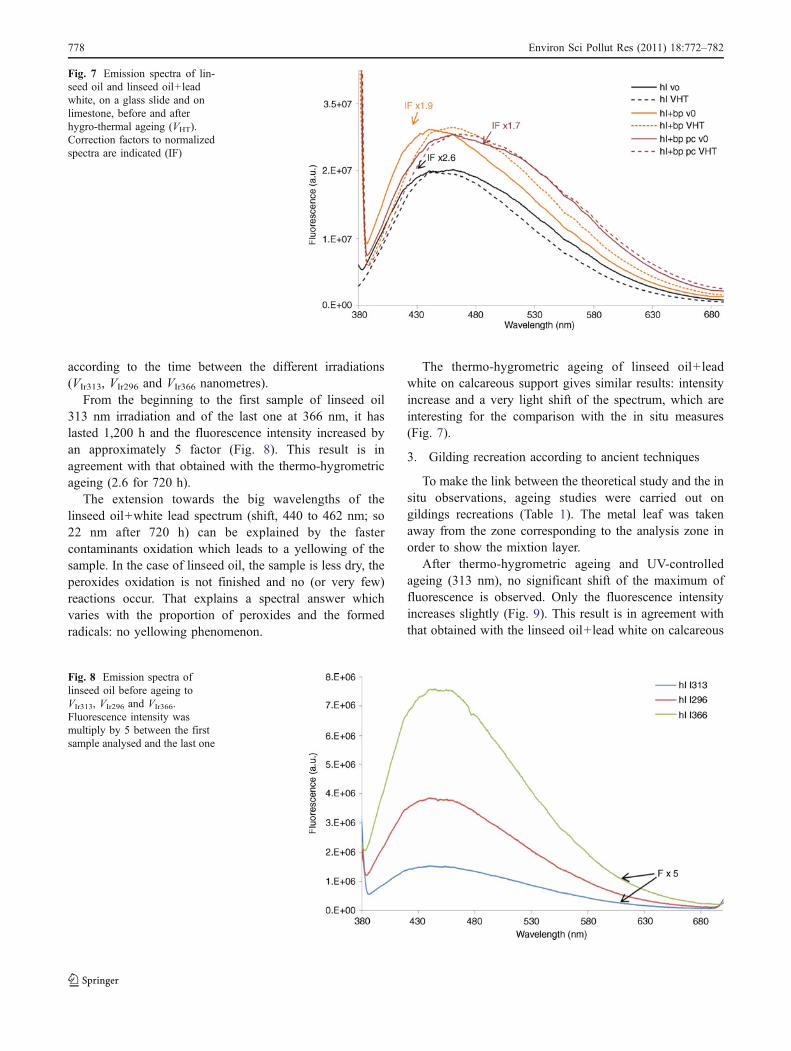
From the beginning to the first sample of linseed oil313 nm irradiation and of the last one at 366 nm, it haslasted 1,200 h and the fluorescence intensity increased byan approximately 5 factor (Fig. 8). This result is inagreement with that obtained with the thermo-hygrometricageing (2.6 for 720 h).
The extension towards the big wavelengths of thelinseed oil+white lead spectrum (shift, 440 to 462 nm; so22 nm after 720 h) can be explained by the fastercontaminants oxidation which leads to a yellowing of thesample. In the case of linseed oil, the sample is less dry, theperoxides oxidation is not finished and no (or very few)reactions occur. That explains a spectral answer whichvaries with the proportion of peroxides and the formedradicals: no yellowing phenomenon.
The thermo-hygrometric ageing of linseed oil+leadwhite on calcareous support gives similar results: intensityincrease and a very light shift of the spectrum, which areinteresting for the comparison with the in situ measures(Fig. 7).
3. Gilding recreation according to ancient techniques
To make the link between the theoretical study and the insitu observations, ageing studies were carried out ongildings recreations (Table 1). The metal leaf was takenaway from the zone corresponding to the analysis zone inorder to show the mixtion layer.
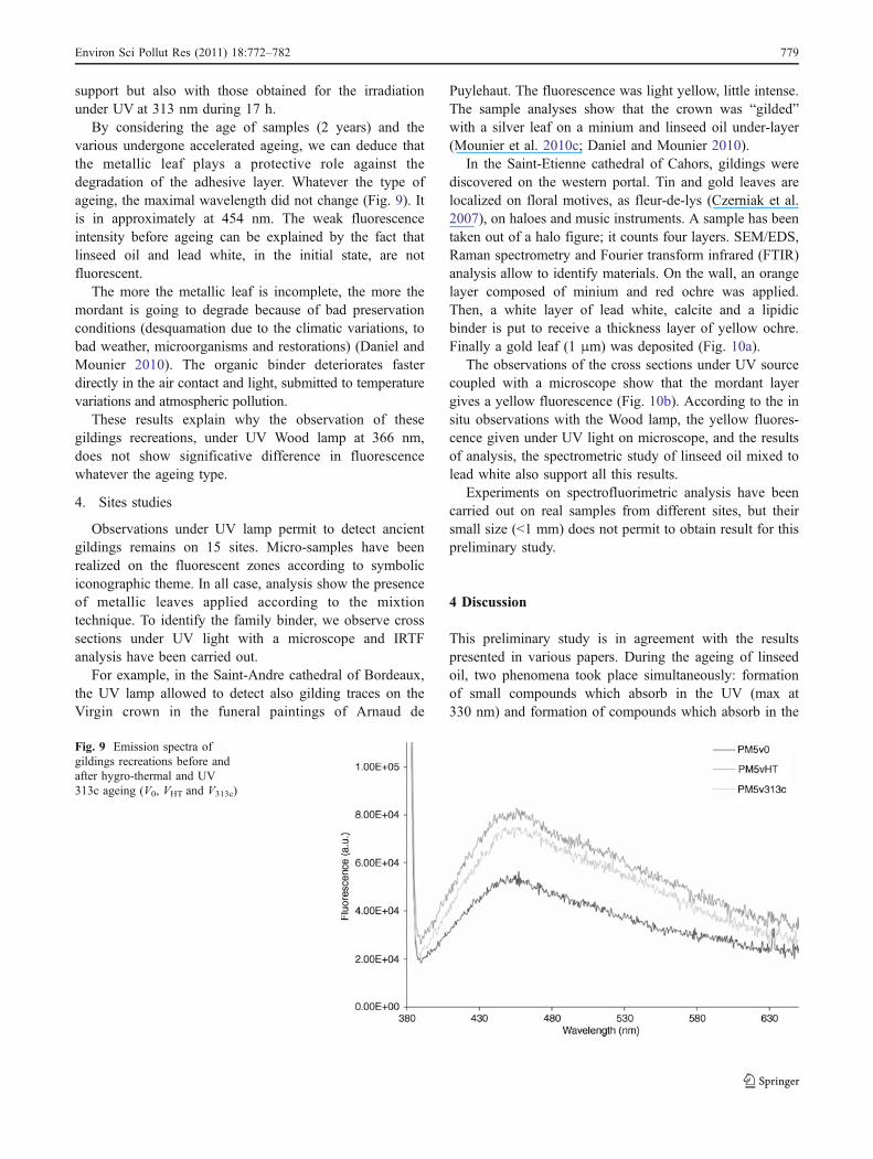
After thermo-hygrometric ageing and UV-controlledageing (313 nm), no significant shift of the maximum offluorescence is observed. Only the fluorescence intensityincreases slightly (Fig. 9). This result is in agreement withthat obtained with the linseed oil+lead white on calcareous
Fig. 7 Emission spectra of lin-seed oil and linseed oil+leadwhite, on a glass slide and onlimestone, before and afterhygro-thermal ageing (VHT).Correction factors to normalizedspectra are indicated (IF)
Fig. 8 Emission spectra oflinseed oil before ageing toVIr313, VIr296 and VIr366.Fluorescence intensity wasmultiply by 5 between the firstsample analysed and the last one
778 Environ Sci Pollut Res (2011) 18:772–782
support but also with those obtained for the irradiationunder UV at 313 nm during 17 h.
By considering the age of samples (2 years) and thevarious undergone accelerated ageing, we can deduce thatthe metallic leaf plays a protective role against thedegradation of the adhesive layer. Whatever the type ofageing, the maximal wavelength did not change (Fig. 9). Itis in approximately at 454 nm. The weak fluorescenceintensity before ageing can be explained by the fact thatlinseed oil and lead white, in the initial state, are notfluorescent.
The more the metallic leaf is incomplete, the more themordant is going to degrade because of bad preservationconditions (desquamation due to the climatic variations, tobad weather, microorganisms and restorations) (Daniel andMounier 2010). The organic binder deteriorates fasterdirectly in the air contact and light, submitted to temperaturevariations and atmospheric pollution.
These results explain why the observation of thesegildings recreations, under UV Wood lamp at 366 nm,does not show significative difference in fluorescencewhatever the ageing type.
4. Sites studies
Observations under UV lamp permit to detect ancientgildings remains on 15 sites. Micro-samples have beenrealized on the fluorescent zones according to symboliciconographic theme. In all case, analysis show the presenceof metallic leaves applied according to the mixtiontechnique. To identify the family binder, we observe crosssections under UV light with a microscope and IRTFanalysis have been carried out.
For example, in the Saint-Andre cathedral of Bordeaux,the UV lamp allowed to detect also gilding traces on theVirgin crown in the funeral paintings of Arnaud de
Puylehaut. The fluorescence was light yellow, little intense.The sample analyses show that the crown was “gilded”with a silver leaf on a minium and linseed oil under-layer(Mounier et al. 2010c; Daniel and Mounier 2010).
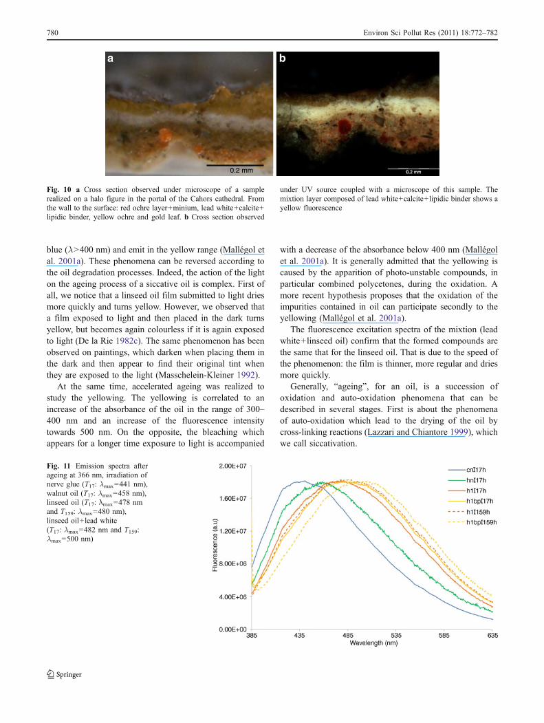
In the Saint-Etienne cathedral of Cahors, gildings werediscovered on the western portal. Tin and gold leaves arelocalized on floral motives, as fleur-de-lys (Czerniak et al.2007), on haloes and music instruments. A sample has beentaken out of a halo figure; it counts four layers. SEM/EDS,Raman spectrometry and Fourier transform infrared (FTIR)analysis allow to identify materials. On the wall, an orangelayer composed of minium and red ochre was applied.Then, a white layer of lead white, calcite and a lipidicbinder is put to receive a thickness layer of yellow ochre.Finally a gold leaf (1 μm) was deposited (Fig. 10a).
The observations of the cross sections under UV sourcecoupled with a microscope show that the mordant layergives a yellow fluorescence (Fig. 10b). According to the insitu observations with the Wood lamp, the yellow fluores-cence given under UV light on microscope, and the resultsof analysis, the spectrometric study of linseed oil mixed tolead white also support all this results.
Experiments on spectrofluorimetric analysis have beencarried out on real samples from different sites, but theirsmall size (<1 mm) does not permit to obtain result for thispreliminary study.
4 Discussion
This preliminary study is in agreement with the resultspresented in various papers. During the ageing of linseedoil, two phenomena took place simultaneously: formationof small compounds which absorb in the UV (max at330 nm) and formation of compounds which absorb in the
Fig. 9 Emission spectra ofgildings recreations before andafter hygro-thermal and UV313c ageing (V0, VHT and V313c)
Environ Sci Pollut Res (2011) 18:772–782 779
blue (λ>400 nm) and emit in the yellow range (Mallégol etal. 2001a). These phenomena can be reversed according tothe oil degradation processes. Indeed, the action of the lighton the ageing process of a siccative oil is complex. First ofall, we notice that a linseed oil film submitted to light driesmore quickly and turns yellow. However, we observed thata film exposed to light and then placed in the dark turnsyellow, but becomes again colourless if it is again exposedto light (De la Rie 1982c). The same phenomenon has beenobserved on paintings, which darken when placing them inthe dark and then appear to find their original tint whenthey are exposed to the light (Masschelein-Kleiner 1992).
At the same time, accelerated ageing was realized tostudy the yellowing. The yellowing is correlated to anincrease of the absorbance of the oil in the range of 300–400 nm and an increase of the fluorescence intensitytowards 500 nm. On the opposite, the bleaching whichappears for a longer time exposure to light is accompanied
with a decrease of the absorbance below 400 nm (Mallégolet al. 2001a). It is generally admitted that the yellowing iscaused by the apparition of photo-unstable compounds, inparticular combined polycetones, during the oxidation. Amore recent hypothesis proposes that the oxidation of theimpurities contained in oil can participate secondly to theyellowing (Mallégol et al. 2001a).
The fluorescence excitation spectra of the mixtion (leadwhite+linseed oil) confirm that the formed compounds arethe same that for the linseed oil. That is due to the speed ofthe phenomenon: the film is thinner, more regular and driesmore quickly.
Generally, “ageing”, for an oil, is a succession ofoxidation and auto-oxidation phenomena that can bedescribed in several stages. First is about the phenomenaof auto-oxidation which lead to the drying of the oil bycross-linking reactions (Lazzari and Chiantore 1999), whichwe call siccativation.
0.2 mm
a b
.
Fig. 10 a Cross section observed under microscope of a samplerealized on a halo figure in the portal of the Cahors cathedral. Fromthe wall to the surface: red ochre layer+minium, lead white+calcite+lipidic binder, yellow ochre and gold leaf. b Cross section observed
under UV source coupled with a microscope of this sample. Themixtion layer composed of lead white+calcite+lipidic binder shows ayellow fluorescence
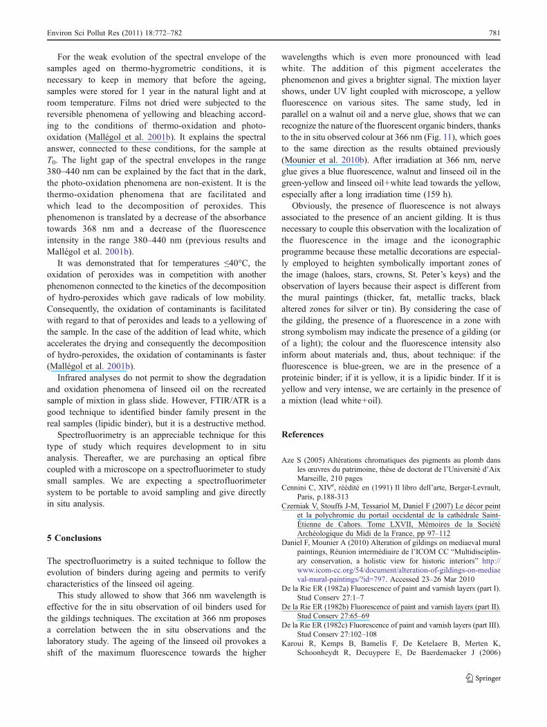
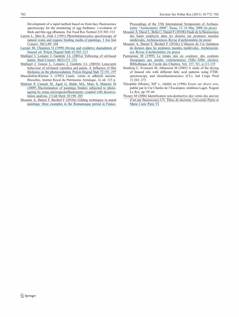
Fig. 11 Emission spectra afterageing at 366 nm, irradiation ofnerve glue (T17: λmax=441 nm),walnut oil (T17: λmax=458 nm),linseed oil (T17: λmax=478 nmand T159: λmax=480 nm),linseed oil+lead white(T17: λmax=482 nm and T159:λmax=500 nm)
780 Environ Sci Pollut Res (2011) 18:772–782
For the weak evolution of the spectral envelope of thesamples aged on thermo-hygrometric conditions, it isnecessary to keep in memory that before the ageing,samples were stored for 1 year in the natural light and atroom temperature. Films not dried were subjected to thereversible phenomena of yellowing and bleaching accord-ing to the conditions of thermo-oxidation and photo-oxidation (Mallégol et al. 2001b). It explains the spectralanswer, connected to these conditions, for the sample atT0. The light gap of the spectral envelopes in the range380–440 nm can be explained by the fact that in the dark,the photo-oxidation phenomena are non-existent. It is thethermo-oxidation phenomena that are facilitated andwhich lead to the decomposition of peroxides. Thisphenomenon is translated by a decrease of the absorbancetowards 368 nm and a decrease of the fluorescenceintensity in the range 380–440 nm (previous results andMallégol et al. 2001b).
It was demonstrated that for temperatures ≤40°C, theoxidation of peroxides was in competition with anotherphenomenon connected to the kinetics of the decompositionof hydro-peroxides which gave radicals of low mobility.Consequently, the oxidation of contaminants is facilitatedwith regard to that of peroxides and leads to a yellowing ofthe sample. In the case of the addition of lead white, whichaccelerates the drying and consequently the decompositionof hydro-peroxides, the oxidation of contaminants is faster(Mallégol et al. 2001b).
Infrared analyses do not permit to show the degradationand oxidation phenomena of linseed oil on the recreatedsample of mixtion in glass slide. However, FTIR/ATR is agood technique to identified binder family present in thereal samples (lipidic binder), but it is a destructive method.
Spectrofluorimetry is an appreciable technique for thistype of study which requires development to in situanalysis. Thereafter, we are purchasing an optical fibrecoupled with a microscope on a spectrofluorimeter to studysmall samples. We are expecting a spectrofluorimetersystem to be portable to avoid sampling and give directlyin situ analysis.
5 Conclusions
The spectrofluorimetry is a suited technique to follow theevolution of binders during ageing and permits to verifycharacteristics of the linseed oil ageing.
This study allowed to show that 366 nm wavelength iseffective for the in situ observation of oil binders used forthe gildings techniques. The excitation at 366 nm proposesa correlation between the in situ observations and thelaboratory study. The ageing of the linseed oil provokes ashift of the maximum fluorescence towards the higher
wavelengths which is even more pronounced with leadwhite. The addition of this pigment accelerates thephenomenon and gives a brighter signal. The mixtion layershows, under UV light coupled with microscope, a yellowfluorescence on various sites. The same study, led inparallel on a walnut oil and a nerve glue, shows that we canrecognize the nature of the fluorescent organic binders, thanksto the in situ observed colour at 366 nm (Fig. 11), which goesto the same direction as the results obtained previously(Mounier et al. 2010b). After irradiation at 366 nm, nerveglue gives a blue fluorescence, walnut and linseed oil in thegreen-yellow and linseed oil+white lead towards the yellow,especially after a long irradiation time (159 h).
Obviously, the presence of fluorescence is not alwaysassociated to the presence of an ancient gilding. It is thusnecessary to couple this observation with the localization ofthe fluorescence in the image and the iconographicprogramme because these metallic decorations are especial-ly employed to heighten symbolically important zones ofthe image (haloes, stars, crowns, St. Peter’s keys) and theobservation of layers because their aspect is different fromthe mural paintings (thicker, fat, metallic tracks, blackaltered zones for silver or tin). By considering the case ofthe gilding, the presence of a fluorescence in a zone withstrong symbolism may indicate the presence of a gilding (orof a light); the colour and the fluorescence intensity alsoinform about materials and, thus, about technique: if thefluorescence is blue-green, we are in the presence of aproteinic binder; if it is yellow, it is a lipidic binder. If it isyellow and very intense, we are certainly in the presence ofa mixtion (lead white+oil).
References
Aze S (2005) Altérations chromatiques des pigments au plomb dansles œuvres du patrimoine, thèse de doctorat de l’Université d’AixMarseille, 210 pages
Cennini C, XIVe, réédité en (1991) Il libro dell’arte, Berger-Levrault,Paris, p.188-313
Czerniak V, Stouffs J-M, Tessariol M, Daniel F (2007) Le décor peintet la polychromie du portail occidental de la cathédrale Saint-Étienne de Cahors. Tome LXVII, Mémoires de la SociétéArchéologique du Midi de la France, pp 97–112
Daniel F, Mounier A (2010) Alteration of gildings on mediaeval muralpaintings, Réunion intermédiaire de l’ICOM CC “Multidisciplin-ary conservation, a holistic view for historic interiors” http://www.icom-cc.org/54/document/alteration-of-gildings-on-mediaeval-mural-paintings/?id=797. Accessed 23–26 Mar 2010
De la Rie ER (1982a) Fluorescence of paint and varnish layers (part I).Stud Conserv 27:1–7
De la Rie ER (1982b) Fluorescence of paint and varnish layers (part II).Stud Conserv 27:65–69
De la Rie ER (1982c) Fluorescence of paint and varnish layers (part III).Stud Conserv 27:102–108
Karoui R, Kemps B, Bamelis F, De Ketelaere B, Merten K,Schoonheydt R, Decuypere E, De Baerdemaeker J (2006)
Environ Sci Pollut Res (2011) 18:772–782 781
Development of a rapid method based on front-face fluorescencespectroscopy for the monitoring of egg freshness: 1-evolution ofthick and thin egg albumens. Eur Food Res Technol 223:303–312
Larson L, Shin K, Zink J (1991) Photoluminescence spectroscopy ofnatural resins and organic binding media of paintings. J Am InstConserv 30(1):89–104
Lazzari M, Chiantore O (1999) Drying and oxidative degradation oflinseed oil. Polym Degrad Stab 65:303–313
Mallégol J, Lemaire J, Gardette J-L (2001a) Yellowing of oil-basedpaints. Stud Conserv 46(2):121–131
Mallégol J, Gonon L, Lemaire J, Gardette J-L (2001b) Long-termbehaviour of oil-based varnishes and paints. 4. Influence of filmthickness on the photooxidation. Polym Degrad Stab 72:191–197
Masschelein-Kleiner L (1992) Liants, vernis et adhésifs anciens.Bruxelles, Institut Royal du Patrimoine Artistique, 3e éd. 123 p
Matteini P, Camaiti M, Agati G, Baldo MA, Muto S, Matteini M(2009) Discrimination of paintings binders subjected to photo-ageing by using microspectrofluorometry coupled with deconvo-lution analysis. J Cult Herit 10:198–205
Mounier A, Daniel F, Bechtel F (2010a) Gilding techniques in muralpaintings: three examples in the Romanesque period in France.
Proceedings of the 37th International Symposium of Archaeo-metry “Archeometry 2008”, Siena, 12–16 May 2008 (in press)
Mounier A, Dayet L, Belin C, Daniel F (2010b) Etude de la fluorescencedes liants employés dans les dorures sur peintures muralesmédiévales, Archéosciences–Revue d’archéométrie (in press)
Mounier A, Daniel F, Bechtel F (2010c) L’illusion de l’or–Imitationde dorures dans les peintures murales médiévales. Archéoscien-ces–Revue d’archéométrie (in press)
Pastoureau M (1999) Le temps mis en couleurs: des couleursliturgiques aux modes vestimentaires (XIIe–XIIIe siècles),Bibliothèque de l’école des Chartres, Vol. 157, N1, p.111-135
Stenberg C, Svensson M, Johansson M (2005) A study of the dryingof linseed oils with different fatty acid patterns using FTIR-spectroscopy and chemiluminescence (CL). Ind Crops Prod21:263–272
Théophile (Moine), XIIe s., réédité en (1996) Essais sur divers arts,publié par le Cte Charles de l’Escalopier, réédition Laget, NogentLe Roi, pp 39–44
Thoury M (2006) Identification non-destructive des vernis des œuvresd’art par fluorescence UV. Thèse de doctorat, Université Pierre etMarie Curie Paris VI
782 Environ Sci Pollut Res (2011) 18:772–782


