Embed Size (px)
Citation preview
www.elsevier.com/locate/ydbio
Developmental Biology
Spatiotemporal pattern and isoforms of cadherin 23 in wild type
and waltzer mice during inner ear hair cell development
Ayala Lagziel, Zubair M. Ahmed, Julie M. Schultz, Robert J. Morell,
Inna A. Belyantseva, Thomas B. FriedmanT
Laboratory of Molecular Genetics, National Institute on Deafness and Other Communication Disorders, National Institutes of Health, 5 Research Court,
Room 2A-19, Rockville, MD 20850, USA
Received for publication 19 November 2004, revised 7 January 2005, accepted 11 January 2005
Abstract
Mutant alleles of the gene encoding cadherin 23 are associated with Usher syndrome type 1 (USH1D), isolated deafness (DFNB12) in
humans, and deafness and circling behavior in waltzer (v) mice. Stereocilia of waltzer mice are disorganized and the kinocilia misplaced,
indicating the importance of cadherin 23 for hair bundle development. Cadherin 23 was localized to developing stereocilia and proposed as a
component of the tip link. We show that, during development of the inner ear, cadherin 23 is initially detected in centrosomes at E14.5, then
along the length of emerging stereocilia, and later becomes concentrated at and subsequently disappears from the tops of stereocilia. In
mature vestibular hair bundles, cadherin 23 is present along the kinocilium and in the region of stereocilia–kinocilium bonds, a pattern
conserved in mammals, chicks, and frogs. Cadherin 23 is also present in Reissner’s membrane (RM) throughout development. In
homozygous v6J mice, a reported null allele, cadherin 23 was absent from stereocilia, but present in kinocilia, RM, and centrosomes. We
reconciled these results by identifying two novel isoforms of Cdh23 unaffected in sequence and expression by the v6J allele. Our results
suggest that Cdh23 participation in stereocilia links may be restricted to developing hair bundles.Published by Elsevier Inc.
Keywords: Cadherin 23; Stereocilia; Kinocilium; Centrosomes; Reissner’s membrane; Usher syndrome; Deafness; Waltzer mice; Adhesion proteins
Introduction
During development of the neurosensory epithelium of
the inner ear, a complex series of morphological changes
give rise to a polarized mosaic pattern of supporting cells
and hair cells (Bryant et al., 2002; Forge et al., 1997;
Nishida et al., 1998). On the apical surface of hair cells,
actin-filled stereocilia emerge and surround a single tubulin-
based kinocilium to form immature hair bundles (Frolenkov
et al., 2004). The kinocilium originates from one centro-
some/basal body of a pair (Alieva and Vorobjev, 2004;
Beisson and Wright, 2003; Hagiwara et al., 2004), and its
ultimate position on one side of the hair cell is thought to
define hair bundle polarity. In mature hair bundles, stereo-
0012-1606/$ - see front matter. Published by Elsevier Inc.
doi:10.1016/j.ydbio.2005.01.015
* Corresponding author. Fax: +1 301 402 7580.
E-mail address: [email protected] (T.B. Friedman).
cilia are organized in rows of increasing height (Tilney et
al., 1988; Denman-Johnson and Forge, 1999; Forge and
Wright, 2002) and fine filaments connect the upper part of
the kinocilium with the tallest neighboring stereocilia
(Ernstson and Smith, 1986; Goodyear and Richardson,
2003; Hillman, 1969; Tsuprun et al., 2004). A variety of link
types interconnect stereocilia to one another (Goodyear and
Richardson, 1992, 1999, 2003) and some of these may be
formed by adhesion molecules required for the formation
and maintenance of the stereocilia bundle as a cohesive
structure (Frolenkov et al., 2004).
Several adhesion proteins are essential for normal
development and maintenance of the auditory system
(Kelley, 2003) including protocadherin 15 (Ahmed et al.,
2003; Alagramam et al., 2001) and cadherin 23, members
of a family of membranous adhesion glycoproteins charac-
terized by a variable number of calcium dependent
280 (2005) 295–306
A. Lagziel et al. / Developmental Biology 280 (2005) 295–306296
extracellular (EC) domains (Hirano et al., 2003). The full-
length isoform of CDH23 comprises 69 coding exons and
is predicted to encode a protein with 27 EC domains, a
trans-membrane domain, and a unique cytoplasmic domain
(Bolz et al., 2001; Bork et al., 2001). Two splice isoforms
of the cytoplasmic domain of the human and mouse
cadherin 23 have been reported, with and without exon
68 (Bork et al., 2001; Di Palma et al., 2001b). Mutant
alleles of Cdh23 cause deafness and circling behavior in
waltzer (v) mice, which have disorganized cochlear and
vestibular hair cell stereocilia and misplaced kinocilia (Di
Palma et al., 2001a). In humans, mutant alleles of CDH23
are either associated with deaf–blindness, Usher syndrome
type 1 (Bolz et al., 2001; Bork et al., 2001), or non-
syndromic deafness (Bork et al., 2001).
In situ hybridization analyses of the mouse inner ear
revealed Cdh23 mRNA in sensory hair cells and Reissner’s
membrane (RM) (Wilson et al., 2001). Immunohistochem-
ical analysis showed cadherin 23 in the photoreceptor layer
of the retina and in stereocilia of immature mouse cochlear
and vestibular hair bundles (Boeda et al., 2002; Siemens et
al., 2002). Recently, cadherin 23 was proposed to be a
component of the tip link (Siemens et al., 2004; Sollner et
al., 2004) and was reported to be present in adult mouse
cochlear hair cell stereocilia, at odds with the previously
reported disappearance of cadherin 23 from stereocilia of
mature hair bundles (Boeda et al., 2002).
To elucidate this discrepancy, we performed a systematic
investigation of the temporal and spatial pattern of cadherin
23 expression during hair bundle development. We show
that cadherin 23 is initially expressed in centrosomes,
followed by its expression in RM, stereocilia, and kinocilia.
Furthermore, while cadherin 23 disappears from stereocilia
of mature hair bundles, it is retained in centrosomes, RM,
and along kinocilia particularly in the region of stereocilia–
kinocilium bonds. Cadherin 23 expression pattern in
centrosomes and hair bundles is conserved in mammals,
chicks, and frogs, and its expression pattern in RM is also
conserved among mammals. We present evidence for novel
isoforms of cadherin 23 that are present in late gestation and
these isoforms are unaffected by the v6J waltzer allele.
Taken together, these data support the hypothesis that
cadherin 23 has multiple functions which are evolutionally
conserved during hair bundle development of the inner ear.
Materials and methods
Expression constructs
cDNA fragments encoding the cytoplasmic domain of
mouse cadherin 23 with (GenBank accession NM_023370
nucleotides 9322–10052) and without (nucleotides 9322–
9633, 9739–10052) exon 68 were cloned into pGEX-5X-1
(Amersham-Pharmacia Biotech), pMAL-c2 (New England
Biolabs), and pEGFP-C2 vectors (Clonetech). GST-
CDH23(+68) and MBP-CDH23(+68) fusion proteins were
expressed in Rosetta E. coli (Novagen) and purified
according to the manufacturer’s instructions.
Cadherin 23 antigens and antisera
Three New Zealand white rabbits were immunized with
purified GST-CDH23(+68) fusion protein producing poly-
clonal antibodies TF7, TF8, and TF9. Purified MBP-
CDH23 protein was used to affinity-purify rabbit antisera
using an AminoLink Plus Immobilization Kit (Pierce).
Transfection of CDH23-GFP into HeLa cells
We evaluated the specificity of our antibodies raised
against the cytoplasmic domain of cadherin 23 by perform-
ing a co-localization assay. Using Lipofectamine 2000
(Invitrogen), a GFP-CDH23(+68) expression vector was
transfected into HeLa cells, which do not endogenously
express cadherin 23. After incubation for 24 h at 378C and
5% CO2 in DMEM with 10% FCS (Invitrogen), transfected
cells were fixed with 4% paraformaldehyde in PBS for 15
min and processed for immunostaining (Ahmed et al., 2003;
Belyantseva et al., 2003) using a 1:500 dilution (~0.5 mg/ml
stock) of antibodies TF7 and TF8.
Western blot and dot blot analysis
For Western blot analysis, the MBP-CDH23(+68) fusion
protein was expressed and partially purified from E. coli and
denatured in sample buffer (6.25 ml 1 M Tris–HCl pH 7.8,
1 ml 10% SDS, 0.5 ml glycerol, 0.25 ml h-mercaptoetha-
nol, 0.125 ml ddH2O, 10 mg bromophenol blue) for 3 min
at 958C, then separated on 4–20% Tris–Glycine precast
Novex gels (Invitrogen). Western blot analyses were
performed as previously described (Ahmed et al., 2003).
We used a 1:1000 dilution of antibodies TF7 and TF8, and
a 1:10,000 dilution of alkaline phosphatase-conjugated anti-
rabbit secondary antibody (Promega). For Western dot blot
analysis, 0.5 Ag of an exon 68 peptide and 0.1 Ag of MBP-
CDH23(+68) fusion protein were applied to the membrane.
For tissue Western blot analysis, cochleas of postnatal day 7
(P7) WT (B10.A-H2h4/(4R)SgDvEg � C57BL/6)F1 and
adult homozygous v6J (B10.A-H2h4/(4R)SgDvEg �C57BL/6)F1 mice were dissected and 25 Ag of protein
sample was separated on 3–8% Novex Tris-acetate gel
(Invitrogen) and processed using TF7 (1:1000 dilution, ~0.5
mg/ml stock) according to Ahmed et al. (2003).
Immunofluorescence study of mouse inner ear sensory
epithelia
(B10.A-H2h4/(4R)SgDvEg � C57BL/6)F1 mice were
mated and offspring were euthanized according to NIH
animal protocol 1126-03, and the inner ears removed by
dissection. Primary antibodies were diluted 1:1000 for TF7
A. Lagziel et al. / Developmental Biology 280 (2005) 295–306 297
and TF8, 1:200 for monoclonal anti-h-tubulin-FITC con-
jugate (SIGMA), and 1:200 for pericentrin MAb (BD
Biosciences). Rhodamine phalloidin and Alexa Fluor 633
phalloidin (Molecular Probes) were diluted 1:100. Anti-
rabbit Ig, fluorescein conjugated (Amersham Biosciences),
and Alexa Fluor 594 goat anti-rabbit (Molecular Probes)
secondary antibodies were used at 1:200 dilution as
previously described (Ahmed et al., 2003). For color
consistency, in Figs. 2–6, cadherin 23 is shown in green
and h-tubulin in red, regardless of the secondary antibody.
The control experiments were performed with substitution
of pre-immune sera for immune sera, omission of the
primary antisera, exclusion of permeabilizing detergents,
and preincubation of cadherin 23 antisera with GST-
CDH23(+68) fusion protein.
Northern blot analysis
A human multiple tissue northern blot (Poly A+ RNA,
MTN Human Blot; BD Bioscience) was hybridized with a
probe generated from the CDH23 cytoplasmic domain and
processed as previously described (Bork et al., 2001).
Novel human and mouse isoforms of cadherin 23
FirstEF software (Davuluri et al., 2001) was used to
predict candidate first exons between exons 40 and 68 of
human CDH23. Forward PCR primers were designed in the
five predicted sequences of putative exons while reverse
primers were designed from the sequence of exons 68 and
69 of CDH23. Human retina cDNA (Genemed Biotechnol-
ogies, Cambridge UK) was used as template for PCR
amplification using LA-Taq polymerase (PanVera). The
primers are listed in Supplementary Table 1.
Mouse Cdh23 isoform B was identified by BLAST
analysis of the unique sequence of human CDH23 isoform
B against the mouse genome. A forward primer designed
within one of the candidate novel Cdh23 first exon
sequences and a reverse primer from mouse Cdh23 exon
69 (Supplementary Table 1) were used to amplify isoform B
from cDNA, which was synthesized from inner ear mRNA
of P45–P50 WT and P60 homozygous v6J mice using a
Poly(A)Pure kit (Ambion). 5V RACE reactions extended the
sequence to a likely transcription start site.
EST CD675842 has 32 unique bases upstream of CDH23
exon 66. A forward primer designed within this 32 bp of
sequence and a reverse primer from exon 69 of CDH23 (F: 5VCTAGCCGCCCTCCCCACT; R: 5V CCAGTGAAATGGA-GAGAATGCTTGT) were used to PCR amplify isoform C
which was subcloned and sequenced. Human retina cDNA
was used as template. In addition, a 5V RACE primer (5VTTGGGGTGTGAGGCCCTGGTGCCTGTGGTGCA) was
designed within the unique 32 bp region of EST CD675842
which was used to amplify a product that was subcloned and
sequenced. A nested primer was designed for the second
round of 5V RACE (5V ATGTCAGTCTGGATTGCC-
TTTGGTGCAGAG). A new set of primers (Supplementary
Table 1) amplified the full-length isoform C. The mouse
ortholog of this novel human isoform C was identified and
primers were designed for cDNA amplification (Supple-
mentary Table 1) using mouse cochlea and retina cDNA.
Results
Specificity of antisera
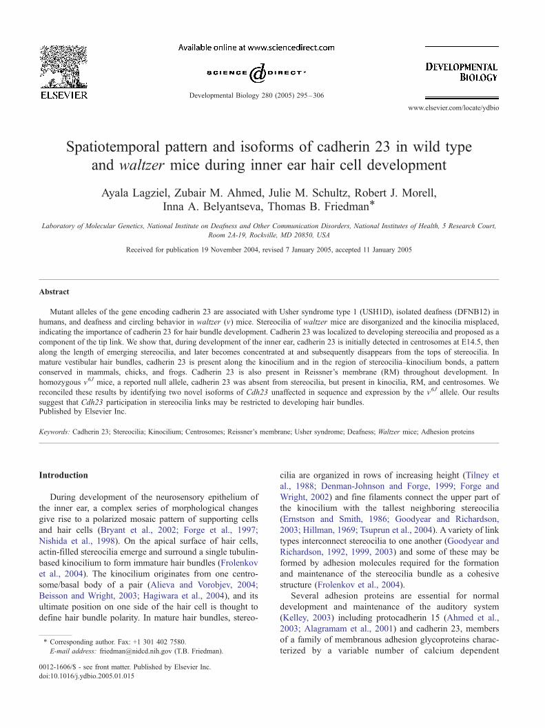
Twenty-four hours after transfection of HeLa cells with a
GFP-CDH23(+68) expression vector, the staining pattern
obtained with antibodies TF7 and TF8 overlapped with the
green fluorescence from GFP-CDH23(+68) (Figs. 1A–H)
and from GFP-CDH23(�68) (data not shown). In cells
transfected with the GFP expression vector alone, cadherin
23 immunoreactivity was not detected (Figs. 1I–L). We
also evaluated the ability of the antibodies to recognize
cadherin 23 cytoplasmic domain and the amino acid
sequence of exon 68 by Western blot analysis. TF7 and
TF8 recognized the MBP-CDH23(+68) fusion protein
expressed and purified from E. coli lysate (Fig. 1M).
Antiserum TF7 does not recognize an exon 68 only
encoded peptide while antiserum TF8 does recognize this
peptide (Fig. 1N). In the immunohistochemical studies,
antibodies TF7 and TF8 were used interchangeably and
gave identical results unless specified.
Cadherin 23 in stereocilia of immature hair cells
Consistent with the progressive development from base
to apex of the neurosensory epithelium of the cochlea (Lim
and Anniko, 1985), cadherin 23 expression was first
observed in the mouse at embryonic day 16.5 (E16.5)
along the length of stereocilia of inner and outer hair cells in
the basal but not in the apical turns of the cochlea (Figs. 2A,
B), which is in agreement with observations reported by
BoJda and coworkers (2002). By E18.5, cadherin 23 is
expressed in stereocilia of inner and outer hair cells of the
basal and apical turns of the cochlea (Figs. 2C, D). At P4–
P5 in mouse and rat cochlea (Figs. 2E–G), cadherin 23
immunoreactivity is present in the distal portion of inner and
outer hair cell stereocilia. In mouse outer hair cells,
immunoreactivity of cadherin 23 is more intense in the
region of contact between the kinocilium and the upper
portion of the adjacent tallest stereocilia (Fig. 2E). Sub-
sequently, the intensity of cadherin 23 immunofluorescence
diminished in hair cell stereocilia, and from P16 we could
not detect cadherin 23 in mouse or rat cochlear stereocilia
(Figs. 2H–J). At P16, cadherin 23 appears in the pericu-
ticular necklace (Fig. 2H).
This same pattern of cadherin 23 expression during
development was also seen in hair cell stereocilia of the
basilar papilla (BP) of the embryonic chicken inner ear and
the adult bullfrog macula sacculi. At E19, when hair bundles
Fig. 1. Validation of cadherin 23 antisera. (A–L) Arrows indicate transfected cells, arrowheads indicate non-transfected control cells. (A, E) Nomarski images
of HeLa cells 24 h after transfection with an expression vector encoding a GFP-tagged cadherin 23 cytoplasmic domain (B, F, green). Staining of the cells with
antibodies TF7 and TF8 (C, G, respectively, red) overlapped with GFP fluorescence (D, H). (I–L) HeLa cells transfected with the GFP expression vector alone
(I, K, green) revealed that subsequent immunolabeling with TF7 and TF8 antisera (J, L, respectively, red) produced no detectable levels of
immunofluorescence. (M) Western blot of partially purified lysate from E. coli expressing the MBP-CDH23 fusion protein. Lane 1, the lysate was separated
on a 4–20% Tris–Glycine gel (Novex, Invitrogen) and stained with Coomassie blue. The lysate was diluted 1:1000 in lane 2 and 1:100 in lane 3. TF7 (lane 2)
and TF8 (lane 3) antibodies recognized a single band of approximately 69 kDa (42.7 kDa MBP plus 26.7 kDa cadherin 23 cytoplasmic domain). (N) Dot blot
with 0.5 Ag of peptide 68, (CLEDYLRLKKLFAQRMVQASSCHSSISE, Princeton Biomolecules) corresponding to amino acid residues 3219 to 3246 encoded
by exon 68 (GenBank accession number AAG52817), and 0.1 Ag of MBP-CDH23(+68) fusion protein. Antibody TF7 does not recognize the exon 68 peptide,
while TF7 and TF8 recognize the expressed cytoplasmic domain encoded by exons 65–69. Scale bars, 5 Am.
A. Lagziel et al. / Developmental Biology 280 (2005) 295–306298
at the base of the BP have reached maturity (Tilney et al.,
1988), cadherin 23 is present in stereocilia of hair cells
located at the extreme apex and the apex but is not detected
in hair cells at the base of the BP (Figs. 2K–M). In the
macula sacculi of the adult bullfrog, cadherin 23 was
detected along the length of stereocilia of nascent hair
bundles, located at the periphery of this tissue (Fig. 2N).
In vestibular hair cells of the mouse, rat, and chick,
localization of cadherin 23 within stereocilia depends upon
the stage of hair bundle development. At E14.5, cadherin
23 is detected along the length of stereocilia of nascent
mouse ampullar hair bundles (Fig. 3A). At P3, when
mature bundles are present in the center of sensory
epithelia, and immature stereocilia bundles can be identi-
fied at the periphery (Denman-Johnson and Forge, 1999;
Sans and Chat, 1982), cadherin 23 is detected along the
length of the stereocilia of the immature bundles (Figs. 3B,
D). At P16 in rat ampulla, the most intense cadherin 23
staining was observed along the length of stereocilia in
immature hair bundles and in the region closer to the top
of stereocilia in maturing hair cell bundles from the
periphery of the ampulla. As hair bundles continue to
develop, the intense immunoreactivity of cadherin 23
gradually disappears from the top of stereocilia (Figs.
3C, E). Similarly, in the chicken ampulla at E10, cadherin
23 was localized along the length of nascent stereocilia
(Fig. 3F), and at E19 was no longer observed in stereocilia
of mature hair bundles (data not shown).
Cadherin 23 in the kinocilium, Reissner’s membrane, and
centrosomes
In E14.5 mouse ampullar hair cells, the kinocilium has
migrated to the side of the immature stereocilia bundle. At
this stage, cadherin 23 staining appears along the length of
stereocilia and in the kinocilium at the point of contact with
the stereocilia (Fig. 4A). By E16.5, cadherin 23 is detected
along the length of kinocilia of mature mouse ampullar hair
bundles (Fig. 4B), where it persists in the rat at P21 and is
visible both along the length and in more concentrated
patches of staining, possibly in the region of contact
between the tallest stereocilia and the kinocilium (Fig.
4C). Immunoreactivity of cadherin 23 was observed in
kinocilia of mouse utricle and saccule, and along the length
of hair cell kinocilia in the chick and guinea pig as well,
where concentrated patches of immunostaining in the
kinocilium and in the tallest stereocilia in the region of
stereocilia–kinocilium bonds were detected (data not
shown). This same pattern of cadherin 23 localization is
seen in the adult bullfrog utricle and macula sacculi, chick
basilar papilla, and in the rat ampulla (Figs. 4D–G).
In the mouse organ of Corti, cadherin 23 expression in hair
cell kinocilia is first detected at P1 in the basal turn of the
cochlea (Figs. 4H–J). The kinocilium is stained along its
entire length, with a brighter signal in the region of contact
with adjacent stereocilia. By P4, cadherin 23 expression in
hair cell kinocilia extends from the base to the apex (Figs.

Fig. 2. Temporal and spatial expression pattern of cadherin 23 (shown in green) in stereocilia of mouse and rat organ of Corti, and in stereocilia of the chick
basilar papilla and frog saccule, the homologs of the mammalian organ of Corti. (A) At E16.5 in the apical turn of the cochlea, stereocilia have not yet emerged
and cadherin 23 is not detected. (B) Cadherin 23 (green) is detected at E16.5 in stereocilia (stained for F-actin, red) of hair cells from the basal turn of the
cochlea. (C, D) At E18.5, cadherin 23 is detected along the entire length of stereocilia of hair cells from the apical and basal turns of the mouse cochlea.
Kinocilia of hair cells (arrows) and primary cilia of Dieters cells (arrowheads) are stained for h-tubulin (red). In basal cochlear hair cells of the P4 mouse (E)
and P5 rat (F), cadherin 23 (green) is localized near the top of stereocilia (stained for F-actin, red). Staining for cadherin 23 appears more intense in outer hair
cells in the region occupied by the kinocilium (arrows) both in the mouse and rat. (G) In inner hair cell stereocilia of the P5 rat, visualized using a 100� Zeiss
objective, N.A. = 1.45, cadherin 23 (green) is localized at the upper portion of stereocilia and seen as a patch between the actin core of neighboring stereocilia.
At P16 in mouse (H) and P21 in rat (I, J), cadherin 23 is no longer detected in stereocilia and is detected in the pericuticular necklace (arrow). (K–M) In E19,
chick basilar papilla cadherin 23 (green) is distributed along stereocilia of immature bundles from the extreme apex (indicated ex-apex in panel K) and apex
(arrows, panel L) but is not detected in stereocilia of basal hair cell bundles (M). (N) In adult bullfrog saccule stained for cadherin 23 (green) and h-tubulin(red), cadherin 23 is present along stereocilia of immature hair bundles located at the periphery of the macula sacculi (arrows). The base of the stereocilia
bundle is marked with white dots. Scale bars, 5 Am.
A. Lagziel et al. / Developmental Biology 280 (2005) 295–306 299
4K–M) of the cochlea. Cadherin 23 was also detected in
mouse RM as early as E16.5 (Fig. 5A) where it persists in the
adult mouse (Fig. 5B), guinea pig (Fig. 5C), and rat (data not
shown). In addition, using antisera TF7 (but not TF8, data not
shown), cadherin 23 was detected in the centrosome/basal
body where it localizes but does not overlap with pericentrin
(Fig. 5D), which labels the centrosome tube (Ou and Rattner,
2000). Cadherin 23 was also detected in the centrosomes
using antisera TF9, which was also generated against the
cytoplasmic domain of cadherin 23 and showed identical
immunoreactivity to TF7 (data not shown). In centrosomes,
cadherin 23 is initially detected at E14.5, before stereocilia
emerge on the apical surface of hair cells, and persists
throughout mouse development. Cadherin 23 is detected in
centrosomes of auditory and vestibular sensory epithelial
cells and in centrosomes of cells of the greater epithelial ridge
in the cochlea. Cadherin 23 was also detected in centrosomes
of cells of the auditory and vestibular sensory epithelia of
chick and frog (data not shown).
Cadherin 23 expression in v6J mice
The v6J allele of Cdh23 is a G to T transversion in
exon 9, encoding the third EC domain, and is predicted to
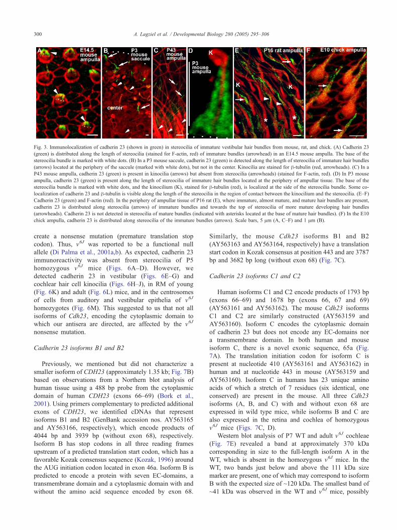
Fig. 3. Immunolocalization of cadherin 23 (shown in green) in stereocilia of immature vestibular hair bundles from mouse, rat, and chick. (A) Cadherin 23
(green) is distributed along the length of stereocilia (stained for F-actin, red) of immature bundles (arrowhead) in an E14.5 mouse ampulla. The base of the
stereocilia bundle is marked with white dots. (B) In a P3 mouse saccule, cadherin 23 (green) is detected along the length of stereocilia of immature hair bundles
(arrows) located at the periphery of the saccule (marked with white dots), but not in the center. Kinocilia are stained for h-tubulin (red, arrowheads). (C) In a
P43 mouse ampulla, cadherin 23 (green) is present in kinocilia (arrows) but absent from stereocilia (arrowheads) (stained for F-actin, red). (D) In P3 mouse
ampulla, cadherin 23 (green) is present along the length of stereocilia of immature hair bundles located at the periphery of ampullar tissue. The base of the
stereocilia bundle is marked with white dots, and the kinocilium (K), stained for h-tubulin (red), is localized at the side of the stereocilia bundle. Some co-
localization of cadherin 23 and h-tubulin is visible along the length of the stereocilia in the region of contact between the kinocilium and the stereocilia. (E–F)
Cadherin 23 (green) and F-actin (red). In the periphery of ampullar tissue of P16 rat (E), where immature, almost mature, and mature hair bundles are present,
cadherin 23 is distributed along stereocilia (arrows) of immature bundles and towards the top of stereocilia of more mature developing hair bundles
(arrowheads). Cadherin 23 is not detected in stereocilia of mature bundles (indicated with asterisks located at the base of mature hair bundles). (F) In the E10
chick ampulla, cadherin 23 is distributed along stereocilia of the immature bundles (arrows). Scale bars, 5 Am (A, C–F) and 1 Am (B).
A. Lagziel et al. / Developmental Biology 280 (2005) 295–306300
create a nonsense mutation (premature translation stop
codon). Thus, v6J was reported to be a functional null
allele (Di Palma et al., 2001a,b). As expected, cadherin 23
immunoreactivity was absent from stereocilia of P5
homozygous v6J mice (Figs. 6A–D). However, we
detected cadherin 23 in vestibular (Figs. 6E–G) and
cochlear hair cell kinocilia (Figs. 6H–J), in RM of young
(Fig. 6K) and adult (Fig. 6L) mice, and in the centrosomes
of cells from auditory and vestibular epithelia of v6J
homozygotes (Fig. 6M). This suggested to us that not all
isoforms of Cdh23, encoding the cytoplasmic domain to
which our antisera are directed, are affected by the v6J
nonsense mutation.
Cadherin 23 isoforms B1 and B2
Previously, we mentioned but did not characterize a
smaller isoform of CDH23 (approximately 1.35 kb; Fig. 7B)
based on observations from a Northern blot analysis of
human tissue using a 488 bp probe from the cytoplasmic
domain of human CDH23 (exons 66–69) (Bork et al.,
2001). Using primers complementary to predicted additional
exons of CDH23, we identified cDNAs that represent
isoforms B1 and B2 (GenBank accession nos. AY563165
and AY563166, respectively), which encode products of
4044 bp and 3939 bp (without exon 68), respectively.
Isoform B has stop codons in all three reading frames
upstream of a predicted translation start codon, which has a
favorable Kozak consensus sequence (Kozak, 1996) around
the AUG initiation codon located in exon 46a. Isoform B is
predicted to encode a protein with seven EC-domains, a
transmembrane domain and a cytoplasmic domain with and
without the amino acid sequence encoded by exon 68.
Similarly, the mouse Cdh23 isoforms B1 and B2
(AY563163 and AY563164, respectively) have a translation
start codon in Kozak consensus at position 443 and are 3787
bp and 3682 bp long (without exon 68) (Fig. 7C).
Cadherin 23 isoforms C1 and C2
Human isoforms C1 and C2 encode products of 1793 bp
(exons 66–69) and 1678 bp (exons 66, 67 and 69)
(AY563161 and AY563162). The mouse Cdh23 isoforms
C1 and C2 are similarly constructed (AY563159 and
AY563160). Isoform C encodes the cytoplasmic domain
of cadherin 23 but does not encode any EC-domains nor
a transmembrane domain. In both human and mouse
isoform C, there is a novel exonic sequence, 65a (Fig.
7A). The translation initiation codon for isoform C is
present at nucleotide 410 (AY563161 and AY563162) in
human and at nucleotide 443 in mouse (AY563159 and
AY563160). Isoform C in humans has 23 unique amino
acids of which a stretch of 7 residues (six identical, one
conserved) are present in the mouse. All three Cdh23
isoforms (A, B, and C) with and without exon 68 are
expressed in wild type mice, while isoforms B and C are
also expressed in the retina and cochlea of homozygous
v6J mice (Figs. 7C, D).
Western blot analysis of P7 WT and adult v6J cochleae
(Fig. 7E) revealed a band at approximately 370 kDa
corresponding in size to the full-length isoform A in the
WT, which is absent in the homozygous v6J mice. In the
WT, two bands just below and above the 111 kDa size
marker are present, one of which may correspond to isoform
B with the expected size of ~120 kDa. The smallest band of
~41 kDa was observed in the WT and v6J mice, possibly
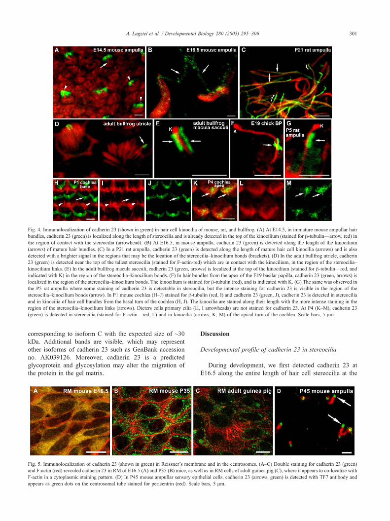
Fig. 4. Immunolocalization of cadherin 23 (shown in green) in hair cell kinocilia of mouse, rat, and bullfrog. (A) At E14.5, in immature mouse ampullar hair
bundles, cadherin 23 (green) is localized along the length of stereocilia and is already detected in the top of the kinocilium (stained for h-tubulin—arrow, red) in
the region of contact with the stereocilia (arrowhead). (B) At E16.5, in mouse ampulla, cadherin 23 (green) is detected along the length of the kinocilium
(arrows) of mature hair bundles. (C) In a P21 rat ampulla, cadherin 23 (green) is detected along the length of mature hair cell kinocilia (arrows) and is also
detected with a brighter signal in the regions that may be the location of the stereocilia–kinocilium bonds (brackets). (D) In the adult bullfrog utricle, cadherin
23 (green) is detected near the top of the tallest stereocilia (stained for F-actin-red) which are in contact with the kinocilium, in the region of the stereocilia–
kinocilium links. (E) In the adult bullfrog macula sacculi, cadherin 23 (green, arrows) is localized at the top of the kinocilium (stained for h-tubulin—red, and
indicated with K) in the region of the stereocilia–kinocilium bonds. (F) In hair bundles from the apex of the E19 basilar papilla, cadherin 23 (green, arrows) is
localized in the region of the stereocilia–kinocilium bonds. The kinocilium is stained for h-tubulin (red), and is indicated with K. (G) The same was observed in
the P5 rat ampulla where some staining of cadherin 23 is detectable in stereocilia, but the intense staining for cadherin 23 is visible in the region of the
stereocilia–kinocilium bonds (arrow). In P1 mouse cochlea (H–J) stained for h-tubulin (red, I) and cadherin 23 (green, J), cadherin 23 is detected in stereocilia
and in kinocilia of hair cell bundles from the basal turn of the cochlea (H, J). The kinocilia are stained along their length with the more intense staining in the
region of the stereocilia–kinocilium links (arrows). Dieters cells primary cilia (H, I arrowheads) are not stained for cadherin 23. At P4 (K–M), cadherin 23
(green) is detected in stereocilia (stained for F-actin—red, L) and in kinocilia (arrows, K, M) of the apical turn of the cochlea. Scale bars, 5 Am.
A. Lagziel et al. / Developmental Biology 280 (2005) 295–306 301
corresponding to isoform C with the expected size of ~30
kDa. Additional bands are visible, which may represent
other isoforms of cadherin 23 such as GenBank accession
no. AK039126. Moreover, cadherin 23 is a predicted
glycoprotein and glycosylation may alter the migration of
the protein in the gel matrix.
Fig. 5. Immunolocalization of cadherin 23 (shown in green) in Reissner’s membr
and F-actin (red) revealed cadherin 23 in RM of E16.5 (A) and P35 (B) mice, as we
F-actin in a cytoplasmic staining pattern. (D) In P45 mouse ampullar sensory epit
appears as green dots on the centrosomal tube stained for pericentrin (red). Scale
Discussion
Developmental profile of cadherin 23 in stereocilia
During development, we first detected cadherin 23 at
E16.5 along the entire length of hair cell stereocilia at the
ane and in the centrosomes. (A–C) Double staining for cadherin 23 (green)
ll as in RM cells of adult guinea pig (C), where it appears to co-localize with
helial cells, cadherin 23 (arrows, green) is detected with TF7 antibody and
bars, 5 Am.
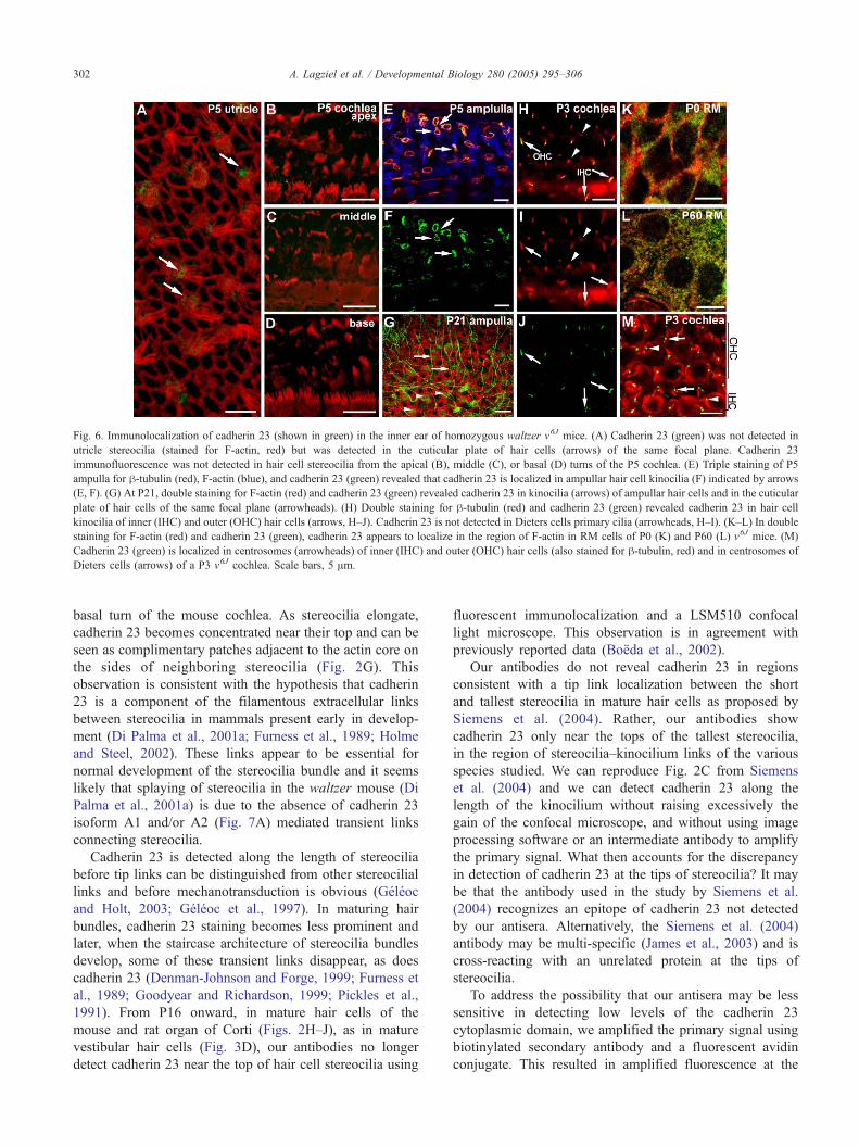
Fig. 6. Immunolocalization of cadherin 23 (shown in green) in the inner ear of homozygous waltzer v6J mice. (A) Cadherin 23 (green) was not detected in
utricle stereocilia (stained for F-actin, red) but was detected in the cuticular plate of hair cells (arrows) of the same focal plane. Cadherin 23
immunofluorescence was not detected in hair cell stereocilia from the apical (B), middle (C), or basal (D) turns of the P5 cochlea. (E) Triple staining of P5
ampulla for h-tubulin (red), F-actin (blue), and cadherin 23 (green) revealed that cadherin 23 is localized in ampullar hair cell kinocilia (F) indicated by arrows
(E, F). (G) At P21, double staining for F-actin (red) and cadherin 23 (green) revealed cadherin 23 in kinocilia (arrows) of ampullar hair cells and in the cuticular
plate of hair cells of the same focal plane (arrowheads). (H) Double staining for h-tubulin (red) and cadherin 23 (green) revealed cadherin 23 in hair cell
kinocilia of inner (IHC) and outer (OHC) hair cells (arrows, H–J). Cadherin 23 is not detected in Dieters cells primary cilia (arrowheads, H–I). (K–L) In double
staining for F-actin (red) and cadherin 23 (green), cadherin 23 appears to localize in the region of F-actin in RM cells of P0 (K) and P60 (L) v6J mice. (M)
Cadherin 23 (green) is localized in centrosomes (arrowheads) of inner (IHC) and outer (OHC) hair cells (also stained for h-tubulin, red) and in centrosomes of
Dieters cells (arrows) of a P3 v6J cochlea. Scale bars, 5 Am.
A. Lagziel et al. / Developmental Biology 280 (2005) 295–306302
basal turn of the mouse cochlea. As stereocilia elongate,
cadherin 23 becomes concentrated near their top and can be
seen as complimentary patches adjacent to the actin core on
the sides of neighboring stereocilia (Fig. 2G). This
observation is consistent with the hypothesis that cadherin
23 is a component of the filamentous extracellular links
between stereocilia in mammals present early in develop-
ment (Di Palma et al., 2001a; Furness et al., 1989; Holme
and Steel, 2002). These links appear to be essential for
normal development of the stereocilia bundle and it seems
likely that splaying of stereocilia in the waltzer mouse (Di
Palma et al., 2001a) is due to the absence of cadherin 23
isoform A1 and/or A2 (Fig. 7A) mediated transient links
connecting stereocilia.
Cadherin 23 is detected along the length of stereocilia
before tip links can be distinguished from other stereocilial
links and before mechanotransduction is obvious (Geleoc
and Holt, 2003; Geleoc et al., 1997). In maturing hair
bundles, cadherin 23 staining becomes less prominent and
later, when the staircase architecture of stereocilia bundles
develop, some of these transient links disappear, as does
cadherin 23 (Denman-Johnson and Forge, 1999; Furness et
al., 1989; Goodyear and Richardson, 1999; Pickles et al.,
1991). From P16 onward, in mature hair cells of the
mouse and rat organ of Corti (Figs. 2H–J), as in mature
vestibular hair cells (Fig. 3D), our antibodies no longer
detect cadherin 23 near the top of hair cell stereocilia using
fluorescent immunolocalization and a LSM510 confocal
light microscope. This observation is in agreement with
previously reported data (Boeda et al., 2002).
Our antibodies do not reveal cadherin 23 in regions
consistent with a tip link localization between the short
and tallest stereocilia in mature hair cells as proposed by
Siemens et al. (2004). Rather, our antibodies show
cadherin 23 only near the tops of the tallest stereocilia,
in the region of stereocilia–kinocilium links of the various
species studied. We can reproduce Fig. 2C from Siemens
et al. (2004) and we can detect cadherin 23 along the
length of the kinocilium without raising excessively the
gain of the confocal microscope, and without using image
processing software or an intermediate antibody to amplify
the primary signal. What then accounts for the discrepancy
in detection of cadherin 23 at the tips of stereocilia? It may
be that the antibody used in the study by Siemens et al.
(2004) recognizes an epitope of cadherin 23 not detected
by our antisera. Alternatively, the Siemens et al. (2004)
antibody may be multi-specific (James et al., 2003) and is
cross-reacting with an unrelated protein at the tips of
stereocilia.
To address the possibility that our antisera may be less
sensitive in detecting low levels of the cadherin 23
cytoplasmic domain, we amplified the primary signal using
biotinylated secondary antibody and a fluorescent avidin
conjugate. This resulted in amplified fluorescence at the
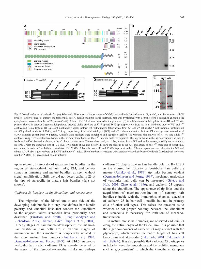
Fig. 7. Novel isoforms of cadherin 23. (A) Schematic illustration of the structure of Cdh23 and cadherin 23 isoforms A, B, and C, and the location of PCR
primers (arrows) used to amplify the transcripts. (B) A human multiple tissue Northern blot was hybridized with a probe from a sequence encoding the
cytoplasmic domain of cadherin 23 (exons 66–69). A band of ~1.35 kb was detected in the pancreas. (C) Amplification of full-length isoforms B1 and B2 with
primers shown in panel A (right and left pointing arrows) yields products of 3787 bp and 3682 bp, respectively, from the adult wild type mouse (WT) and v6J
cochlea and retina. Isoform B1 is present in all lanes whereas isoform B2 (without exon 68) is absent from WTand v6J retina. (D) Amplification of isoforms C1
and C2 yielded products of 724 bp and 619 bp, respectively, from adult wild type (WT) and v6J cochlea and retina. Isoform C1 message was detected in all
cDNA samples except from WT retina. Amplification products were subcloned and sequence verified. (E) Western blot analysis of P7 WT and adult v6J
cochleae using TF7 revealed five bands in the WT and three bands in the v6J (marked with red squares). The largest band in the WT corresponds in size to
isoform A ~370 kDa and is absent in the v6J homozygous mice. The smallest band, ~41 kDa, present in the WT and in the mutant, possibly corresponds to
isoform C with the expected size of ~30 kDa. Two bands above and below 111 kDa are present in the WT and absent in the v6J mice one of which may
correspond to isoform B with the expected size of ~120 kDa. A band between 111 and 55 kDa is present in the v6J homozygous mice and absent in the WT, and
a band of ~55 kDa is present both in the WT and in the v6J mice. These bands may represent other uncharacterized isoforms of cadherin 23 (GenBank accession
number AK039123) recognized by our antisera.
A. Lagziel et al. / Developmental Biology 280 (2005) 295–306 303
upper region of stereocilia of immature hair bundles, in the
region of stereocilia–kinocilium links, RM, and centro-
somes in immature and mature bundles, as seen without
signal amplification. Still, we did not detect cadherin 23 at
the tips of stereocilia in mature hair bundles (data not
shown).
Cadherin 23 localizes to the kinocilium and centrosomes
The migration of the kinocilium to one side of the
developing hair bundle is a step that defines hair bundle
polarity, and kinocilial links that connect the kinocilium
to the adjacent tallest stereocilia have previously been
described (Ernstson and Smith, 1986; Goodyear and
Richardson, 2003; Hillman, 1969; Tsuprun et al., 2004).
In early stages of hair bundle development, the mamma-
lian vestibular hair cells are in various stages of
maturation and the kinocilium is peripherally situated in
the more mature hair bundles (Anniko et al., 1983;
Denman-Johnson and Forge, 1999). At E14.5, in mouse
vestibular hair cells, cadherin 23 is already detected in
the region of the stereocilia–kinocilium links and perhaps
cadherin 23 plays a role in hair bundle polarity. By E18.5
in the mouse, the majority of vestibular hair cells are
mature (Anniko et al., 1983), tip links become evident
(Denman-Johnson and Forge, 1999), mechanotransduction
of vestibular hair cells can be measured (Geleoc and
Holt, 2003; Zhao et al., 1996), and cadherin 23 appears
along the kinocilium. The appearance of tip links and the
acquisition of mechanotransduction of sensory hair
bundles coincide with the immunohistochemical detection
of cadherin 23 in hair cell kinocilia but not in primary
cilia of other cell types. This raises the question as to
whether or not proper bonding between the kinocilium
and stereocilia is necessary for initiation of mechano-
transduction.
In mature mouse hair bundles, we observed cadherin 23
along the entire length of the kinocilium. It is possible that
the sugar components of cadherin 23 may interact with the
glycocalyx, which covers the entire length of hair cell
kinocilium and stereocilia (Takumida, 2001; Takumida et
al., 1988a,b). It is also possible that cadherin 23 participates
in links between the kinocilium and the otolithic membrane
(rich in glycoproteins) to which the kinocilia in its upper
A. Lagziel et al. / Developmental Biology 280 (2005) 295–306304
portion together with the tallest stereocilia are intercon-
nected (Takumida and Bagger-Sjoback, 1991; Takumida et
al., 1992; Sobkowicz et al., 1995).
Cadherin 23 was also localized in the centrosome
which together with the kinocilium is considered the
centrosomal complex (Alieva and Vorobjev, 2004). The
expression of cadherin 23 in the centrosomes precedes its
expression in stereocilia, persists throughout development,
and is evolutionally conserved in mammals, chicks, and
frogs. Cadherin 23 expression in the centrosome may
have a role not only in the development of the kinocilium
but also in the organization of microtubules (Chausovsky
et al., 2000).
Cadherin 23 in v6J mice and the identification of novel
isoforms of cadherin 23
Homozygous v6J mice have disorganized and splayed
stereocilia bundles and cadherin 23 is absent from stereo-
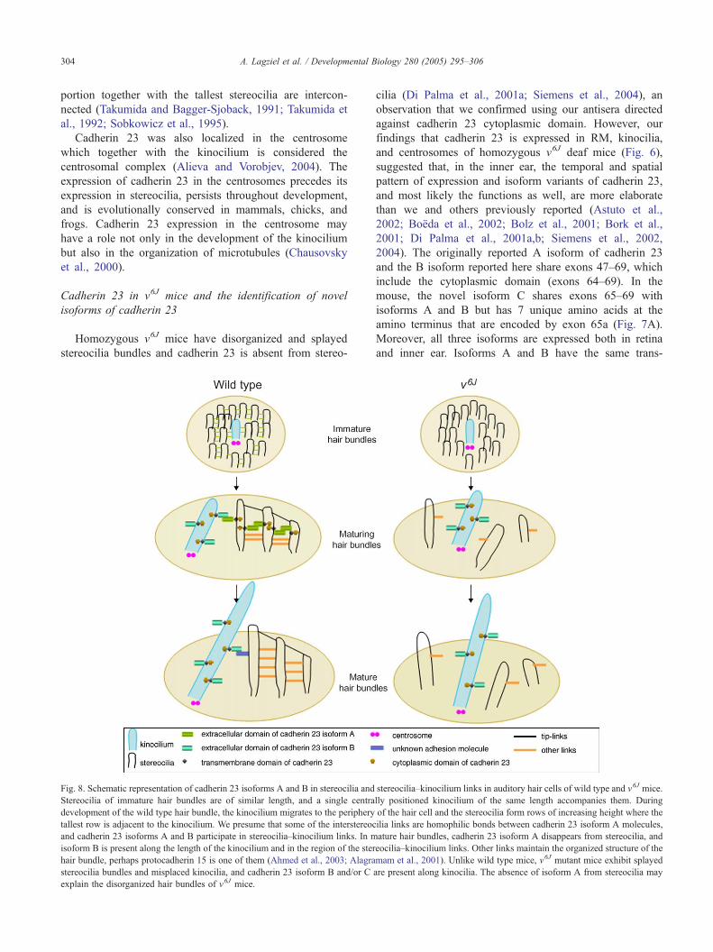
Fig. 8. Schematic representation of cadherin 23 isoforms A and B in stereocilia and
Stereocilia of immature hair bundles are of similar length, and a single centra
development of the wild type hair bundle, the kinocilium migrates to the periphery
tallest row is adjacent to the kinocilium. We presume that some of the interstereoc
and cadherin 23 isoforms A and B participate in stereocilia–kinocilium links. In m
isoform B is present along the length of the kinocilium and in the region of the ster
hair bundle, perhaps protocadherin 15 is one of them (Ahmed et al., 2003; Alagra
stereocilia bundles and misplaced kinocilia, and cadherin 23 isoform B and/or C
explain the disorganized hair bundles of v6J mice.
cilia (Di Palma et al., 2001a; Siemens et al., 2004), an
observation that we confirmed using our antisera directed
against cadherin 23 cytoplasmic domain. However, our
findings that cadherin 23 is expressed in RM, kinocilia,
and centrosomes of homozygous v6J deaf mice (Fig. 6),
suggested that, in the inner ear, the temporal and spatial
pattern of expression and isoform variants of cadherin 23,
and most likely the functions as well, are more elaborate
than we and others previously reported (Astuto et al.,
2002; Boeda et al., 2002; Bolz et al., 2001; Bork et al.,
2001; Di Palma et al., 2001a,b; Siemens et al., 2002,
2004). The originally reported A isoform of cadherin 23
and the B isoform reported here share exons 47–69, which
include the cytoplasmic domain (exons 64–69). In the
mouse, the novel isoform C shares exons 65–69 with
isoforms A and B but has 7 unique amino acids at the
amino terminus that are encoded by exon 65a (Fig. 7A).
Moreover, all three isoforms are expressed both in retina
and inner ear. Isoforms A and B have the same trans-
stereocilia–kinocilium links in auditory hair cells of wild type and v6J mice.
lly positioned kinocilium of the same length accompanies them. During
of the hair cell and the stereocilia form rows of increasing height where the
ilia links are homophilic bonds between cadherin 23 isoform A molecules,
ature hair bundles, cadherin 23 isoform A disappears from stereocilia, and
eocilia–kinocilium links. Other links maintain the organized structure of the
mam et al., 2001). Unlike wild type mice, v6J mutant mice exhibit splayed
are present along kinocilia. The absence of isoform A from stereocilia may
A. Lagziel et al. / Developmental Biology 280 (2005) 295–306 305
membrane domain while isoform C has no predicted trans-
membrane domain nor EC-domains. Therefore, based
simply on amino acid sequence, isoform C cannot function
as a classic cadherin molecule (Hirano et al., 2003), but
may play a regulatory role as a competitive surface for
proteins that interact with the cytoplasmic domain of
isoforms A and B of cadherin 23.
The exact function of all three Cdh23 isoforms remains
to be investigated, and we emphasize that the antisera
directed against the cytoplasmic domain of cadherin 23
would be expected to recognize cadherin 23 isoforms A, B,
and C. At the moment, our antisera and antisera to the
cytoplasmic domain of cadherin 23 that have been reported
(Boeda et al., 2002; Siemens et al., 2002, 2004) cannot, by
immunohistochemical localization studies alone, distinguish
between these three protein isoforms encoded by Cdh23 and
CDH23.
The loss of immunostaining in hair cell stereocilia of
v6J mice does permit conclusions about the localization
and possible function of isoform A, which coupled with
the waltzer phenotype, suggests that in the inner ear, this
isoform is important directly or indirectly at least for
hair bundle organization and perhaps positioning of the
kinocilium as illustrated in our model (Fig. 8). The
presence of cadherin 23 in RM, centrosomes, and in the
kinocilia of wild type and v6J mice probably represents
either or both isoforms B and C, which are not affected
by the v6J mutation, and possibly isoform A in the wild
type mouse.
Finally, the Cdh23753A allele is associated with age-
related hearing loss in the mouse (Noben-Trauth et al.,
2003). Since cadherin 23 disappears from stereocilia in
the mouse at P16, but is stably retained in the RM of the
adult, perhaps age-related hearing loss in the mouse is due
to a decline in some function of the RM (Lee and
Marcus, 2003) rather than or in addition to its suggested
alteration of developing stereocilia (Holme and Steel,
2004). This speculation is strengthened with our report of
additional cadherin 23 isoforms B and C and their
lifelong persistence in RM and in the kinocilia of wild
type and v6J waltzer mice. Together, these findings
suggest that the various isoforms of cadherin 23 may
perform multiple functions within the auditory system in
addition to the development of cohesive hair cell stereo-
cilia bundles.
Acknowledgments
We thank Alain Dabdoub, Dennis Drayna, Gregory
Frolenkov, Andrew Griffith, Mathew Kelley, and Doris
Wu for critically reading the manuscript, and Barbara
Plopsis for technical assistance. Work was supported by
intramural research funds DC000048-06 from the National
Institute on Deafness and Other Communication Disorders
to T.B.F.
Appendix A. Supplementary data
Supplementary data associated with this article can be
found, in the online version, at doi:10.1016/j.ydbio.2005.
01.015.
References
Ahmed, Z.M., Riazuddin, S., Ahmad, J., Bernstein, S.L., Guo, Y., Sabar,
M.F., Sieving, P., Riazuddin, S., Griffith, A.J., Friedman, T.B.,
Belyantseva, I.A., Wilcox, E.R., 2003. PCDH15 is expressed in the
neurosensory epithelium of the eye and ear and mutant alleles are
responsible for both USH1F and DFNB23. Hum. Mol. Genet. 12,
3215–3223.
Alagramam, K.N., Murcia, C.L., Kwon, H.Y., Pawlowski, K.S., Wright,
C.G., Woychick, R.P., 2001. The mouse Ames waltzer hearing-loss
mutant is caused by mutation of Pcdh15, a novel protocadherin gene.
Nat. Genet. 27, 99–102.
Alieva, I.B., Vorobjev, I.A., 2004. Vertebrate primary cilia: a sensory part of
centrosomal complex in tissue cells, but a bsleeping beautyQ in cultured
cells? Cell Biol. Int. 28, 139–150.
Anniko, M., Nordemar, H., Sobin, A., 1983. Principles in embryonic
development and differentiation of vestibular hair cells. Otolaryngol.-
Head Neck Surg. 91, 540–549.
Astuto, L.M., Bork, J.M., Weston, M.D., Askew, J.W., Fields, R.R., Orten,
D.J., Ohliger, S.J., Riazuddin, S., Morell, R.J., Khan, S., Riazuddin, S.,
Kremer, H., van Hauwe, P., Moller, C.G., Cremers, C.W., Ayuso, C.,
Heckenlively, J.R., Rohrschneider, K., Spandau, U., Greenberg, J.,
Ramesar, R., Reardon, W., Bitoun, P., Millan, J., Legge, R., Friedman,
T.B., Kimberling, W.J., 2002. CDH23 mutation and phenotype
heterogeneity: a profile of 107 diverse families with Usher syndrome
and nonsyndromic deafness. Am. J. Hum. Genet. 71, 262–275.
Belyantseva, I.A., Boger, E.T., Friedman, T.B., 2003. Myosin XVa localizes
to the tips of inner ear sensory cell stereocilia and is essential for
staircase formation of the hair bundle. Pro. Natl. Acad. Sci. U. S. A.
100, 13958–13963.
Beisson, J., Wright, M., 2003. Basal body/centriole assembly and
continuity. Curr. Opin. Cell Biol. 15, 96–104.
BoJda, B., El-Amraoui, A., Bahloul, A., Goodyear, R., Daviet, L., Blanchard,
S., Perfettini, I., Fath, K.R., Shorte, S., Reiners, J., Houdusse, A., Legrain,
P., Wolfrum, U., Richardson, G., Petit, C., 2002. Myosin VIIa, harmonin
and cadherin 23, three Usher I gene products that cooperate to shape the
sensory hair cell bundle. EMBO J. 21, 6689–6699.
Bolz, H., von Brederlow, B., Ramirez, A., Bryda, E.C., Kutsche, K.,
Nothwang, H.G., Seeliger, M., del C-Salcedo Cabrera, M., Vila, M.C.,
Molina, O.P., Gal, A., Kubisch, C., 2001. Mutation of CDH23,
encoding a new member of the cadherin gene family, causes Usher
syndrome type 1D. Nat. Genet. 27, 108–112.
Bork, J.M., Peters, L.M., Riazuddin, S., Bernstein, S.L., Ahmed, Z.M., Ness,
S.L., Polomeno, R., Ramesh, A., Schloss, M., Srisailpathy, C.R., Wayne,
S., Bellman, S., Desmukh, D., Ahmed, Z., Khan, S.N., Kaloustian, V.M.,
Li, X.C., Lalwani, A., Riazuddin, S., Bitner-Glindzicz, M., Nance, W.E.,
Liu, X.Z.,Wistow, G., Smith, R.J., Griffith, A.J.,Wilcox, E.R., Friedman,
T.B., Morell, R.J., 2001. Usher syndrome 1D and nonsyndromic
autosomal recessive deafness DFNB12 are caused by allelic mutations
of the novel cadherin-like gene CDH23. Am. J. Hum. Genet. 68, 26–37.
Bryant, J., Goodyear, R.J., Richardson, G.P., 2002. Sensory organ
development in the inner ear: molecular and cellular mechanisms. Br.
Med. Bull. 63, 39–57.
Chausovsky, A., Bershadsky, A.D., Borisy, G.G., 2000. Cadherin-mediated
regulation of microtubule dynamics. Nat. Cell Biol. 2, 797–804.
Davuluri,R.V.,Grosse, I.,Zhang,M.Q.,2001.Computational identificationof
promoters and first exons in the humangenome.Nat.Genet. 29, 412–417.
Denman-Johnson, K., Forge, A., 1999. Establishment of hair bundle
A. Lagziel et al. / Developmental Biology 280 (2005) 295–306306
polarity and orientation in the developing vestibular system of the
mouse. J. Neurocytol. 28, 821–835.
Di Palma, F., Holme, R.H., Bryda, E.C., Belyantseva, I.A., Pellegrino, R.,
Kachar, B., Steel, K.P., Noben-Trauth, K., 2001a. Mutations in Cdh23,
encoding a new type of cadherin, cause stereocilia disorganization in
waltzer, the mouse model for Usher syndrome type 1D. Nat. Genet. 27,
103–107.
Di Palma, F., Pellegrino, R., Noben-Trauth, K., 2001b. Genomic structure,
alternative splice forms and normal and mutant alleles of cadherin 23
(Cdh23). Gene 281, 31–41.
Ernstson, S., Smith, C.A., 1986. Stereo–kinociliar bonds in mammalian
vestibular organs. Acta Oto-Laryngol. 101, 395–402.
Forge, A., Wright, T., 2002. The molecular architecture of the inner ear. Br.
Med. Bull. 63, 5–24.
Forge, A., Souter, M., Denman-Johnson, K., 1997. Structural development
of sensory cells in the ear. Semin. Cell Dev. Biol. 8, 225–237.
Frolenkov, G.I., Belyantseva, I.A., Friedman, T.B., Griffith, A.J., 2004.
Genetic insights into the morphogenesis of inner ear hair cells. Nat.
Rev., Genet. 5, 489–498.
Furness, D.N., Richardson, G.P., Russel, I.J., 1989. Stereociliary bundle
morphology in organotypic cultures of the mouse cochlea. Hear. Res.
38, 95–109.
Geleoc, G.S., Holt, J.R., 2003. Developmental acquisition of sensory
transduction in hair cells of the mouse inner ear. Nat. Neurosci. 6,
1019–1020.
Geleoc, G.S., Lennan, G.W., Richardson, G.P., Kros, C.J., 1997. A
quantitative comparison of mechanoelectrical transduction in vestibular
and auditory hair cells of neonatal mice. Proc. R. Soc. Lond., Ser. B
Biol. Sci. 264, 611–621.
Goodyear, R., Richardson, G., 1992. Distribution of the 275 kD hair cell
antigen and cell surface specializations on auditory and vestibular hair
bundles in the chicken inner ear. J. Comp. Neurol. 325, 243–256.
Goodyear, R., Richardson, G., 1999. The ankle-link antigen: an epitope
sensitive to calcium chelation associated with the hair-cell surface and
the calycal processes of photoreceptors. J. Neurosci. 19, 3761–3772.
Goodyear, R.J., Richardson, G.P., 2003. A novel antigen sensitive to
calcium chelation that is associated with the tip links and kinocilial links
of sensory hair bundles. J. Neurosci. 23, 4878–4887.
Hagiwara, H., Ohwada, N., Takata, K., 2004. Cell biology of normal and
abnormal ciliogenesis in the ciliated epithelium. Int. Rev. Cytol. 234,
101–141.
Hillman, D.E., 1969. New ultrastructural findings regarding a vestibular
ciliary apparatus and its possible functional significance. Brain Res. 13,
407–412.
Hirano, S., Suzuki, S.T., Redies, C., 2003. The cadherin superfamily in
neural development: diversity, function and interaction with other
molecules. Front. Biosci. 8, d306–d355.
Holme, R.H., Steel, K.P., 2002. Stereocilia defects in waltzer (Cdh23),
shaker1 (Myo7a) and double waltzer/shaker1 mutant mice. Hear. Res.
169, 13–23.
Holme, R.H., Steel, K.P., 2004. Progressive hearing loss and increased
susceptibility to noise-induced hearing loss in mice carrying a Cdh23
but not a Myo7a mutation. J. Assoc. Res. Otolaryngol. 5, 66–79.
James, L.C., Roversi, P., Tawfik, D.S., 2003. Antibody multispecificity
mediated by conformational diversity. Science 299, 1362–1367.
Kelley, M.W., 2003. Cell adhesion molecules during inner ear and hair cell
development, including notch and its ligands. Curr. Top. Dev. Biol. 57,
321–356.
Kozak, M., 1996. Interpreting cDNA sequences: some insights from studies
on translation. Mamm. Genome 7, 563–574.
Lee, J.H., Marcus, D.C., 2003. Endolymphatic sodium homeostasis by
Reissner’s membrane. Neuroscience 119, 3–8.
Lim, D.J., Anniko, M., 1985. Developmental morphology of the mouse
inner ear. A scanning electron microscopic observation. Acta Oto-
Laryngol., Suppl. 422, 1–69.
Nishida, Y., Rivolta, M.N., Holley, M.C., 1998. Timed markers for the
differentiation of the cuticular plate and stereocilia in hair cells from the
mouse inner ear. J. Comp. Neurol. 395, 18–28.
Noben-Trauth, K., Zheng, Q.Y., Johnson, K.R., 2003. Association of
cadherin 23 with polygenic inheritance and genetic modification of
sensorineural hearing loss. Nat. Genet. 35, 21–23.
Ou, Y., Rattner, J.B., 2000. A subset of centrosomal proteins are arranged in
a tubular conformation that is reproduced during centrosome duplica-
tion. Cell Motil. Cytoskeleton 47, 13–24.
Pickles, J.O., von Perger, M., Rouse, G.W., Brix, J., 1991. The development
of links between stereocilia in hair cells of the chick basila papilla. Hear.
Res. 54, 153–163.
Sans, A., Chat, M., 1982. Analysis of temporal and spatial patterns of rat
vestibular hair cell differentiation by tritiated thymidine radioautog-
raphy. J. Comp. Neurol. 206, 1–8.
Siemens, J., Kazmierczak, P., Reynolds, A., Sticker, M., Littlewood-Evans,
A., Mqller, U., 2002. The Usher syndrome proteins cadherin 23 and
harmonin form a complex by means of PDZ–domain interactions. Proc.
Natl. Acad. Sci. U. S. A. 99, 14946–14951.
Siemens, J., Lillo, C., Dumont, R.A., Reynolds, A., Williams, D.S.,
Gillespie, P.G., Mqller, U., 2004. Cadherin 23 is a component of the tip
link in hair-cell stereocilia. Nature 428, 950–955.
Sobkowicz, H.M., Slapnick, S.M., August, B.K., 1995. The kinocilium of
auditory hair cells and evidence for its morphogenetic role during the
regeneration of stereocilia and cuticular plates. J. Neurocytol. 24,
633–653.
Sollner, C., Rauch, G.J., Siemens, J., Geisler, R., Schuster, S.C., Muller, U.,
Nicolson, T., 2004. Mutations in cadherin 23 affect tip links in zebrafish
sensory hair cells. Nature 428, 955–959.
Takumida, M., 2001. Functional morphology of the crista ampullaris: with
special interests in sensory hairs and cupula: a review. Biol. Sci. Space
15, 356–358.
Takumida, M., Bagger-Sjoback, D., 1991. Carbohydrates of the vestibular
end organs. An ultrastructural study using gold-labeled lectins. ORL. J.
Oto-Rhino-Laryngol. Relat. Spec. 53, 86–90.
Takumida, M., Harada, Y., Wersall, J., Bagger-Sjoback, D., 1988a. The
glycocalyx of inner ear sensory and supporting cells. Acta Oto-
Laryngol., Suppl. 458, 84–89.
Takumida, M., Wersall, J., Bagger-Sjoback, D., 1988b. Stereociliary
glycocalyx and interconnections in the guinea pig vestibular organs.
Acta Oto-Laryngol. 106, 130–139.
Takumida, M., Harada, Y., Bagger-Sjoback, D., 1992. The statoconial
membrane of the guinea pig utricular macula. Scanning electron
microscopic investigation combined with the freeze-fracturing techni-
que. Acta Oto-Laryngol. 112, 643–648.
Tilney, L.G., Tilney, M.S., Cotanche, D.A., 1988. Actin filaments,
stereocilia, and hair cells of the bird cochlea: V. How the staircase
pattern of stereociliary lengths is generated. J. Cell Biol. 106,
355–365.
Tsuprun, V., Goodyear, R.J., Richardson, G.P., 2004. The structure of tip
links and kinocilial links in avian sensory hair bundles. Biophys. J. 87,
4106–4112.
Wilson, S.M., Householder, D.B., Coppola, V., Tessarollo, L., Fritzsch, B.,
Lee, E.C., Goss, D., Carlson, G.A., Copeland, N.G., Jenkins, N.A.,
2001. Mutations in Cdh23 cause nonsyndromic hearing loss in waltzer
mice. Genomics 74, 228–233.
Zhao, Y., Yamoah, E.N., Gillespie, P.G., 1996. Regeneration of broken tip
links and restoration of mechanical transduction in hair cells. Proc. Natl.
Acad. Sci. U. S. A. 93, 15469–15474.


