Embed Size (px)
Citation preview

Materials Science and Engineering C 32 (2012) 494–502
Contents lists available at SciVerse ScienceDirect
Materials Science and Engineering C
j ourna l homepage: www.e lsev ie r .com/ locate /msec
Spark plasma sintering of sol–gel derived 45S5 Bioglass®-ceramics: Mechanicalproperties and biocompatibility evaluation
Q.Z. Chen a, J.L. Xu b,⁎, L.G. Yu b, X.Y. Fang a, K.A. Khor b
a Department of Materials Engineering, Monash University, Clayton, Victoria 3800, Australiab School of Mechanical & Aerospace Engineering, Nanyang Technological University, 50 Nanyang Avenue, Singapore 639798, Singapore
⁎ Corresponding author. Tel.: +65 6790 4051; fax: +E-mail address: [email protected] (J.L. Xu).
0928-4931/$ – see front matter © 2011 Elsevier B.V. Alldoi:10.1016/j.msec.2011.11.023
a b s t r a c t
a r t i c l e i n f oArticle history:Received 18 August 2011Received in revised form 4 November 2011Accepted 30 November 2011Available online 7 December 2011
Keywords:Bioglass®Sol–gelSpark plasma sinteringMechanical propertiesOsteoblast
This work aims to find an efficient sintering technique and optimal sintering conditions of a novel sol–gelderived Bioglass®-ceramic powder so as to achieve much improved mechanical properties compared toconventional Bioglass®. To this end, the spark plasma sintering (SPS) technique was for the first timeused to densify the sol–gel derived Bioglass®-ceramic powder. It was found that the sol–gel derived Bio-glass®-ceramics sintered with the SPS technique at 950 °C for 15 min had a high Young's modulus valueof ~110 GPa, which was comparable to that of compact bone and significantly higher than the maximalvalue achieved by the conventional heat treatment. Moreover, the Bioglass®-ceramic compacts sinteredwith SPS released alkaline ions slowly and as a result, these highly densified Bioglass®-ceramics exhibitedbetter cytocompatibility at the early stage of cell culture testing, compared to the conventional Bioglass®.Hence, the SPS technique is recommended to be used in the process of sol–gel derived Bioglass®-ceramicsand its tissue engineering scaffolds.
© 2011 Elsevier B.V. All rights reserved.
1. Introduction
To engineer bone tissues, which is hard and functions to supportthe body, the scaffold biomaterial must be strong and biodegradablesuch that it can temporarily replace the mechanical function of dam-aged bone until sufficient new bone tissue has formed. Unfortunately,mechanical strength and biodegradability, which are two essential re-quirements on bone tissue scaffolds, are antagonistic toward eachother [1]. Compared to crystalline calcium phosphates which are vir-tually inert, amorphous bioactive glasses are biodegradable and tendto be mechanically fragile. Clinical investigation has shown that crys-talline hydroxyapatite (HA) and related calcium phosphates are vir-tually inert, remaining within the body for as long as 6–7 years postimplantation [2]. Actually among all bioceramics, Na2O-containg bio-active glasses (e.g. 45S5 Bioglass®) are the only one that could simul-taneously achieve both mechanical strength and satisfactorybiodegradability through the formation of Na2O-containg crystallinephase, Na2Ca2Si3O9 [3]. This mechanically capable crystalline phasecan transform to a biodegradable, amorphous calcium phosphate atbody temperature and in a biological environment (in vitro). Thistransformation couples the above two irreconcilable properties inone scaffold, and the transition temporal profile can be tailored to sat-isfy the requirement of bone tissue engineering [3]. Hence, 45S5 Bio-glass® scaffolds offer a promise to go a long way towards the clinicalapplication of bone tissue engineering.
65 6792 4062.
rights reserved.
Commercially produced 45S5 Bioglass® has been made by con-ventional glass powder manufacturing methods, i.e. melting andquenching. Meanwhile, increasing research efforts are being investedin fabrication of bioactive glasses using the sol–gel technique [4–9],which has many advantages over melting–quenching processes andis an extremely versatile process. Using the sol–gel process, ceramicor glass materials can be fabricated in a variety of forms, includingultra-fine spherical powders, thin film coatings, ceramic fibers, micro-porous inorganic membranes, monolithic ceramics and glasses, orhighly porous aerogel materials [5].
Most recently, Na2O-containing glass–ceramics have been suc-cessfully synthesized by Chen et al., using a sol–gel technique in awater solution under ambient conditions [8]. The sol–gel derived45S5 glass–ceramic materials possess the major features of commer-cial Na2O-containing bioactive materials, namely, the formation ofcrystalline phase Na2Ca2Si3O9 during sintering, decrease of the crys-talline phase, and the extensive formation of amorphous bone-likeapatite in a physiological environment. However, a drawback wasalso revealed that the sintering properties of the sol–gel derived45S5 Bioglass®-ceramic powder was poor, compared to that of com-mercial 45S5 Bioglass® powder, and that the mechanical propertiesof the compact products of the sol–gel derived material were compro-mised. This forms the primary rationale of the present work: to ex-plore alternative sintering techniques to process the sol–gel derived45S5 Bioglass®-ceramic powder.
Advanced sintering methods, such as hot isostatic pressing [10]and spark plasma sintering (SPS), have proved advantageous overconventional pressureless sintering of bioceramics [11–14]. Materials

Sintering conditions
2°C/min
2°C/min
5°C/min400°C/1hr
R.T.Time
Tem
pera
ture
Fig. 1. Conventional heat treatment program designed for sintering of 45S5 Bioglass®.
Fig. 2. Shrinkage of sol–gel derived 45S5 Bioglass-ceramic pellets after spark plasmasintering at 600–950 °C for 15 min or heat treatment in a conventional furnace at600–1200 °C for 2 h.
***** **** ****
**o
***
*
**
*
*____Na2Ca2Si3O9o____CaSiO3
950oC
800oC
A.U
.
495Q.Z. Chen et al. / Materials Science and Engineering C 32 (2012) 494–502
of poor sintering properties, such as tungsten carbide (WC), havebeen successfully sintered, and a fracture toughness of 6 MPa m1/2
being achieved with the SPS route [15]. In the SPS process, a high en-ergy electric spark is discharged and the powder particle surfaces aremore easily purified and activated than in conventional sintering pro-cess. The presence of spark plasma significantly enhances rapid den-sification to high densities from powder form [16]. Therefore, theobjectives of this work are 1) to sinter sol–gel derived 45S5 Bioglass®compacts using either the SPS technique or conventional heat treat-ment, 2) to carry out a comparative investigation on the sintering ki-netics and mechanical properties of 45S5 Bioglass®-ceramicsproduced by the above two methods, and 3) finally to investigatethe biodegradation kinetics and biocompatibility of these 45S5 Bio-glass®-ceramic materials.
2. Material and methods
2.1. Material
The following chemicalswere used as precursors for the synthesis ofsol–gel 45S5 materials: tetraethyl orthosilicate (TEOS, Aldrich, 99%),triethyl phosphate (TEP, Eastman, 99.8%), sodium nitrite (Sigma-Al-drich, 99%) and calcium nitrate tetrahydrate (Sigma-Aldrich, 99%).
2.2. Methods
2.2.1. Sol–gel process and heat treatmentThe recipe and processing flowchart have been reported previous-
ly [8]. In brief, the molar ratio of TEOS, TEP, NaNO3 and Ca(NO3)24H2Owas designed according to the molar ratio of SiO2, P2O5, Na2O and CaOin 45S5 Bioglass®. To achieve a clear sol, the molar ratio betweenwater and the four precursor chemicals was set at 10. Each chemicalwas added reasonably slowly into the 0.25 M HNO3 aqueous solutionat room temperature. Each compound in the sequence was addedonly when the previous solution became clear, and was then stirredfor at least 1 h. The resulting gel was dried at 60 and 200 °C for 72and 40 h respectively, aged at 600 °C for 5 h.
Pellets, which were prepared from the above aged 45S5 Bioglass®powder (~1.5 g per pellet) under uniaxial pressure of 50 kPa, were di-vided into two groups. One group of pellets was sintered with theconventional heat treatment following the program of Fig. 1. Theother group was sintered with the SPS technique at 600–950 °C for15 min.
Table 1Element composition of the sol–gel derived 45S5 material and nominal 45S5 Bioglass®.
Sol–gel derivedBioglass®-ceramicsmol%
Nominal Bioglass®mol%
Si 47.22 46.1Na 45.96 48.8Ca 27.03 26.9P 5.54 5.0
2.2.2. CharacterisationX-ray diffraction (XRD) of the powders and compacts was carried
out using a Shimadzu 6000 Laboratory X-ray diffractometer utilizingCu Kα radiation at 40 kV and 30 mA with a graphite monochromatorto collimate the X-rays. The step size and scan rate were fixed at 0.04°and 1°/min, respectively.
The distribution of particle size of the sol–gel derived powder wasdetermined using laser diffraction analysis and analyzed by Zeta PlusParticle sizing software Ver. 4.04, Brookhaven Instruments Corp. Theparticles were dispersed into deionized water for analysis. The mor-phology and composition of the powders and pellets were studiedusing scanning electron microscopy (SEM) JEOL 7001 at 15–20 keV.The SEM samples were gold coated to improve the conductivity andminimize electron charging in SEM.
The sintered densities of compacts were obtained by Archimedes'principle using de-ionized water as immersion medium. An ADA 210/L balance (Adam Co. Ltd., USA) equipped with density kit and analysissoftware was used.
2.2.3. Mechanical propertiesThe compressive strength of sintered materials was measured
using an Instron 4204 tester at a crosshead speed of 0.5 mm/min,with a 1 kN load cell. The samples were rectangular in shape, with di-mensions: 10 mm in height and 5 mm×5 mm in cross-section. Dur-ing compression testing, the load was applied until densification ofthe porous samples started to occur.
10 20 30 40 50 60 70 80
750oC
600oC
2 theta
Fig. 3. XRD spectra of sol–gel derived 45S5 glass ceramics after spark plasma sinteringat 600–950 °C for 15 min. The peaks of the Na2Ca2Si3O9 phase and CaSiO3 are markedby * and °, respectively.

(a) (b)
(c)
20µm
20µm
5µm
(d)
Fig. 4. SEM microstructures of 45S5 Bioglass®-ceramics sintered with the conventional heat treatment at 1000 (a–b) and 1200 °C (c–d) for 1 h.
(a) (b)
(d)(c)
5µm
Fig. 5. SEM Microstructures of 45S5 Bioglass®-ceramics sintered with the spark plasma technique at 800 (a–b) and 950 °C (c–d) for 15 min.
496 Q.Z. Chen et al. / Materials Science and Engineering C 32 (2012) 494–502

Fig. 6. Mechanical properties of sol–gel derived Bioglass®-ceramics sintered with theconventional heat treatment and the SPS technique.
497Q.Z. Chen et al. / Materials Science and Engineering C 32 (2012) 494–502
2.2.4. Measurement of pH values and ion concentrationsSamples (2 g) of the materials to be tested were soaked in 10 ml of
tissue culture medium in 15-ml cone tubes under standard cell cul-ture conditions within a culture incubator (37 °C in humidified aircontaining 5% CO2). Acidity was measured at 4, 12 and 24 h by inser-tion of a pH glass electrode (Hanna Instruments, HI 1230B) attached
Fig. 7. pH as a function of material and incubation time in tissue culture medium (DMEM) atbetween any two of the following four groups: SPS_800 °C, SPS_950 °C, heat treatment at 1those of the two control groups. There was a significant increase (pb0.05) in pH value in tpared to the rest of the tested groups.
to a pH meter (Hanna Instruments, HI 98185) into the solutions viaan incubator access port, allowing the electrode to stabilize under in-cubation conditions for each reading.
The solutions used to analyze ion concentrations were preparedby soaking the materials of 2 g in deionized water of 10 ml. Solutionswere collected and transferred to new tubes at different intervals upto 2 days. All solutions were analyzed using ion chromatography.The ICS-1000 (Dionex) ion chromatograph with attached autosam-pler was used for the analysis of sodium (Na+) and calcium (Ca2+)ions. The ICS-2500 (Dionex) was used for the analysis of phosphate(PO4
3−) and silicate (SiO42−) anions. Before running a sample, the
ion chromatography system is calibrated using a standard solution.By comparing the data obtained from a sample to that obtainedfrom the known standard, sample ions can be identified and quanti-tated. The data collection system produces a chromatogram. Thechromatography software converts each peak in the chromatogramto a sample convention and produces the results.
2.2.5. Cytocompatibility in vitro (ISO 10993-5)Cytocompatibility study was performed according to the standard
cytotoxicity assessment set by the International Standardization Or-ganization (ISO 10993-5). Extracts for tissue culture were preparedby placing 0.4 g of each material in 2 ml samples of cell culture medi-um (DMEM supplemented with 10% Fetal Calf Serum (FCS), 1% L-glu-tamine and 0.5% penicillin/streptomycin) for 24 h at 37 °C/5% CO2 inculture incubator. Hydroxyapatite was used as the material control,and 2 ml of cell culture medium alone was the negative control.Prior to exposure of cells to these extracts, human osteoblast-likecells (MG63, ATCC) were seeded in standard media at a density of ap-proximately 2000 cells/well in 96 well tissue culture treated plates(Falcon, BD Bioscience, North Ryde, Australia), under standard incu-bation conditions (37 °C and 5% CO2), with medium changed everysecond day. When the cell monolayers had reached 80% confluence(at day 4), the medium in each well was entirely replaced with0.2 ml of extract media samples (medium pre-exposed to material)or control media (material control=medium pre-exposed to Pleura-Seal®; negative control=medium only). All cultures were thenallowed to proceed for 2 days.
At the end of the incubation period, culture media were collectedand the degree of cell death was determined by measurement of lac-tate dehydrogenase levels [17], as released into the culture media(“RELEASED LDH”), using a commercial kit (Sigma-Aldrich TOX-7)as we have described previously [18]. Finally, each well containing
37 °C in a 5% CO2 atmosphere. The variations in pH value were not significant (p>0.05)000 and 1200 °C. But the pH values of these four groups were significantly higher thanhe following three groups: SPS_600 °C, SPS_750 °C and heat treatment at 900 °C, com-

Fig. 8. Analysis of Na, Ca, P and Si in deionized water in which the sol–gel derived 45S5 glass ceramic was immersed for 48 h: (a)–(b) sintered with the conventional heat treatmentat 1200 and 800 °C for 2 h, respectively; (c)–(d) sintered with the spark plasma technique at 950 and 600 °C for 15 min, respectively.
498 Q.Z. Chen et al. / Materials Science and Engineering C 32 (2012) 494–502
living cells was filled with 0.2 ml fresh cell culture medium and cellswere lysed using the solution TOX-7. These lysates were then used todetermine the cellular LDH content, which equates to the number ofliving cells per well (“TOTAL LDH”). The overall LDH level was deter-mined by measuring the absorbance of the supernatant from the cen-trifuged medium at 490 nm (after subtraction for backgroundabsorbance at 690 nm) using a multiwell plate format UV–vis spec-trophotometer (Thermo Scientific). The absorbance results of LDHwere converted to the number of cells according to a linear standardcurve (not shown). Hence, cytotoxicity can be expressed as:
Percentage of dead cells ¼ RELEASED LDHRELEASED LDHþ TOTAL LDH
� 100: ð1Þ
2.2.6. Cell proliferation: AlamarBlue™Cell proliferation was assessed using a commercial AlamarBlue™
assay kit (Life Technologies). AlamarBlue™ is non-toxic to cells. Theassay does not interrupt cell culture, allowing continuous measure-ment of cell proliferation kinetics. Hence, the AlamarBlue™ assay isappropriate to evaluate the long-term cytotoxicity of biomaterialsdue to biodegradation under physiological conditions. MG63 cellswere seeded at 5000 cells/ml each well in a 48-well plate and cul-tured in the media with extracts. Incubated media in wells with nei-ther cells nor testing material extracts were used as negativecontrols. After culture for 48 h, 100 μl of AlamarBlue™ indicator wasadded to each well (except background controls) and incubatedunder culture conditions for 6 h. The media were then transferredto a new plate, followed by absorbance determination at wavelengthsof 570 and 600 nm in a spectrophotometer (Thermo Scientific, Path-tech Australia).
The above procedures were repeated every 48 h until confluencewas reached (between days 6–8). Cell proliferation was quantifiedby the percentage reduction of AlamarBlue, i.e.:
%Reduced AlamarBlue ¼ εOX λ2ð ÞA λ1ð Þ−εOX λ1ð ÞA λ2ð ÞεRED λ1ð ÞA− λ2ð Þ−εRED λ2ð ÞA− λ1ð Þ � 100; ð2Þ
where A(λ1) and A(λ2) are the values of absorbance of test wells mea-sured at a wavelength of λ1 and λ2, and A−(λ1) and A−(λ2) are thevalues of absorbance of negative control wells containing onlymedia and AlamarBlue™ without cells. All values were blanked withthe readings of background controls. The other parameters inEq. (2) are as follows: εOX(λ1)=80,586, εOX(λ2)=117,216,εRED(λ1)=155,677 and εRED(λ2)=14,652.
2.2.7. Statistical analysisAll experiments were run with five samples per experimental
group, and data are shown as mean±standard error of the mean.One-way analysis of variance (ANOVA) with Tukey's post-hoc testwas performed to analyze the statistical difference, and significancelevels were set at a p-value of less than 0.05.
3. Results and discussion
3.1. Composition of the sol–gel derived 45S5 Bioglass®-ceramic powder
The composition of the sol–gel derived glass–ceramic powderwas determined using EDX analysis in SEM JEOL 7001, as listed inTable 1, which is reasonably close to the nominal composition of45S5 Bioglass® [19]. The results from particle sizing showed that90% of the powders are in the range of 5–23 μm with a mean

(a) (b)
(c) (d)
100µm
Fig. 9. Typical morphology of MG63 cells cultured for 2 days in (a) the standard culture medium, (b) the extractant medium of hydroxyapatite, (c)–(d) the extractant medium ofsol–gel derived Bioglass®-ceramics sintered with the conventional heat treatment at 1000 and 1200 °C, respectively. Magnification of the images is the same.
499Q.Z. Chen et al. / Materials Science and Engineering C 32 (2012) 494–502
diameter ~7.6 μm. The d0.1, d0.5 and d0.9 are 5.32, 8.60 and 17.6 μm,respectively. .
3.2. Sintering kinetics of sol–gel derived 45S5 Bioglass®-ceramic pellets
A curve of relative density (i.e. ρρBG
, where ρ and ρBG=2.7 cm3 arethe densities of a sintered pellet and of a fully densified Bioglass®ma-terial [20]) against sintering conditions was determined, as shown inFig. 2. The pellets, which were prepared from ~1.5 g 45S5 Bioglass®powder under uniaxial pressure of 50 kPa, were sintered with eitherSPS technique or conventional heat treatment. It was found that amaximum relative density, ~0.92 or 0.89, was achieved at 950 and1200 °C with the SPS technique and conventional heat treatment, re-spectively. Further raising the sintering temperature did not increasethe density further. Hence, the SPS is more efficient than the conven-tional heat treatment.
3.3. Characterisation of sintered sol–gel derived 45S5 Bioglass®-ceramicpellets
It has been reported that a crystalline phase Na2Ca2Si3O9 forms inthe sol–gel derived glass–ceramic powder sintered with the conven-tional heat treatment [8]. The XRD analysis of this work (Fig. 3)showed that this phase presented in the sol–gel derived Bioglass®-ceramics after sintering with the SPS technique at 600–950 °C for15 min.
SEM examination revealed many micropores in the Bioglass-ceramics after sintering with the conventional heat treatment at allconditions (Fig. 4a and c). Fine crystalline particles of Na2Ca2Si3O9
phase formed and distributed homogenously in the glass matrixafter sintering at up to 1000 °C for 2 h (Fig. 4b). It should be men-tioned that the maximal crystallinity is about 80% in 45S5 composi-tion [3]. In most of the sintered materials, the amorphous phase
forms a matrix to host the particles of the Na2Ca2Si3O9 crystallinephase (Figs. 4b, 5b and d). The percentage of crystalline phase in sin-tered 45S5 Bioglass® is about 80%, according to previous work [1].However, sintering at excessively high temperature, 1100 and1200 °C for 2 h for example, resulted in the segregation of glassphase, leaving bared crystalline particles with virtually no glass ma-trix (Fig. 4d). This inhomogeneous microstructure indicates that anover-sintering process occurred at the 1100–1200 °C/2 h conditions.
In contrast to the materials sintered with the conventional heattreatment, much fewer micropores presented in the glass–ceramicsthat were sintered with the SPS technique (Fig. 5a and c), especiallythe one sintered at 950 °C (Fig. 5c). Fine and homogenously distribut-ed crystalline particles of Na2Ca2Si3O9 phase were populated in theglass matrix after SPS at 800 and 950 °C (Fig. 5b and d). The presenceof fewer microvoids and homogeneous microstructure of fine crystal-line particles in the glass matrix produced much improved mechani-cal properties in the SPS processed materials, compared to thosetreated with the conventional heating process, as discussed in thenext section.
3.4. Mechanical properties
Fig. 6 demonstrates the mechanical properties of the 45S5 Bio-glass®-ceramic materials after sintering with the SPS technique andconventional heat treatment. The mechanical properties increasedwith increasing sintering temperature. The maximal values ofYoung's modulus achieved by the conventional heat treatment wasabout 90 GPa on average, which was lower than the maximal Young'smodulus (~110 GPa) achieved by the SPS technique at 950 °C(Fig. 6a). Moreover, the small standard deviation of mechanical prop-erties of SPS samples indicates that their mechanical performance isreliable. In contrast, the conventional sintered compacts showed
(a) (b)
(c) (d)
100µm
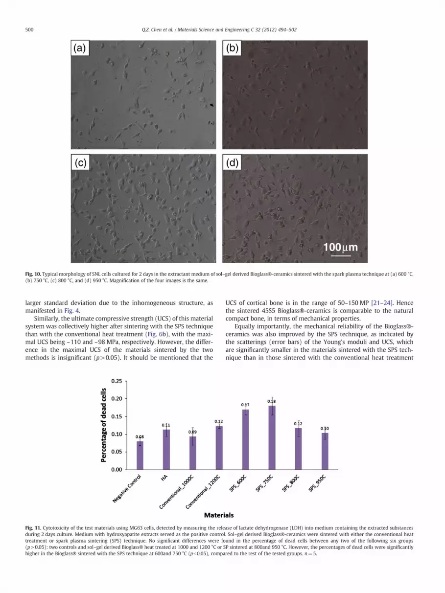
Fig. 10. Typical morphology of SNL cells cultured for 2 days in the extractant medium of sol–gel derived Bioglass®-ceramics sintered with the spark plasma technique at (a) 600 °C,(b) 750 °C, (c) 800 °C, and (d) 950 °C. Magnification of the four images is the same.
500 Q.Z. Chen et al. / Materials Science and Engineering C 32 (2012) 494–502
larger standard deviation due to the inhomogeneous structure, asmanifested in Fig. 4.
Similarly, the ultimate compressive strength (UCS) of this materialsystem was collectively higher after sintering with the SPS techniquethan with the conventional heat treatment (Fig. 6b), with the maxi-mal UCS being ~110 and ~98 MPa, respectively. However, the differ-ence in the maximal UCS of the materials sintered by the twomethods is insignificant (p>0.05). It should be mentioned that the
Fig. 11. Cytotoxicity of the test materials using MG63 cells, detected by measuring the releduring 2 days culture. Medium with hydroxyapatite extracts served as the positive controtreatment or spark plasma sintering (SPS) technique. No significant differences were fo(p>0.05): two controls and sol–gel derived Bioglass® heat treated at 1000 and 1200 °C or Shigher in the Bioglass® sintered with the SPS technique at 600and 750 °C (pb0.05), compa
UCS of cortical bone is in the range of 50–150 MP [21–24]. Hencethe sintered 45S5 Bioglass®-ceramics is comparable to the naturalcompact bone, in terms of mechanical properties.
Equally importantly, the mechanical reliability of the Bioglass®-ceramics was also improved by the SPS technique, as indicated bythe scatterings (error bars) of the Young's moduli and UCS, whichare significantly smaller in the materials sintered with the SPS tech-nique than in those sintered with the conventional heat treatment
ase of lactate dehydrogenase (LDH) into medium containing the extracted substancesl. Sol–gel derived Bioglass®-ceramics were sintered with either the conventional heatund in the percentage of dead cells between any two of the following six groupsP sintered at 800and 950 °C. However, the percentages of dead cells were significantlyred to the rest of the tested groups. n=5.

Fig. 12. MG63 cell proliferation kinetics measured by the AlamarBlue™ technique. Theinitial plating density was 5000 cells ml−1 in each well of a 48-well plate (n=5). Me-dium with the hydroxyapatite extract was the positive control. Sol–gel derived Bio-glass®-ceramics were sintered with either the conventional heat treatment or sparkplasma sintering (SPS) technique. The differences between any two groups were notsignificant (p>0.05) on days 4–6. The cell growth was apparently lower on day 2 inthe extractant media of materials sintered with SPS technique at 600 and 750 °C, com-pared to the rest of groups.
501Q.Z. Chen et al. / Materials Science and Engineering C 32 (2012) 494–502
(Fig. 6a and b). The mechanical properties shown in Fig. 6 are consis-tent with their microstructures shown in Figs. 4 and 5. The higher in-cidence of microvoids in the materials sintered with the conventionalheat treatment is most likely responsible for the poor mechanical per-formance (i.e. lower mean values of mechanical properties and largerscatterings of raw data), compared to those sintered with the SPStechnique. Hence, we conclude that the SPS technique is an efficientsintering method, which can densify the sol–gel derived Bioglass-ceramics quickly, cheaply and reliably.
3.5. pH values of culture medium exposed to the Bioglass®-ceramics
The negative control was the media which was not in contact withany tested materials, and positive control was the media exposed toHA. Compared with the two control groups, the pH values of themedia were increased when in contact with the Bioglass®-ceramicmaterials (Fig. 7), indicating that alkaline ions had been released.The change in pH values was considerably slower in the media soakedwith relatively well sintered pellets, i.e. SPS at 800 and 950 °C andconventional heat treatment at 1000–1200 °C. It is apparent that thechange in pH value, as well as the ion release (Fig. 8), was primarilycontrolled by the level of densification in these materials, as signifi-cant densification was observed with the materials sintered at theseconditions (SPS at 800 and 950 °C and conventional heat treatmentat 1000–1200 °C), as indicated by the values of relative density inFig. 2, SEM examination (Figs. 4 and 5) and the mechanical propertiesin Fig. 6. It is likely that the surface area exposed to the medium wassignificantly reduced in these densified materials, because a poroussample has a larger surface area exposed to the surrounding liquid,resulting in faster ion release.
3.6. Ion release into the deionized water from the Bioglass®-ceramics
Therewas no significant difference in the sodium ion concentrationsafter 24 h soaking (p>0.05) (Fig. 8). However, considerably higher so-dium concentrations were detected in the first 24 h in the solutionssoaked with the materials sintered at lower temperature, e.g. conven-tional heat treatment at 800 °C (Fig. 8b) or SPS at 600 °C (Fig. 8d), com-pared to those soaked with relatively well densified materials (Fig. 8aand c). This result is reasonably in agreement with the data of pHmea-surement (Fig. 7). The difference in the release of other ions, i.e. calcium(Ca2+) ions, phosphate (PO4
3−) and silicon (SiO42−) anions, was insig-
nificant between any two of the tested groups. The different time-release patterns among the samples were primarily due to the degree
of densification of samples. Densified samples showed slow ion releasekinetics at the early stage of soaking. Hence, it was concluded that thepHvalue of themediumwas primarily affected by the release of sodiumions when exposed to the Bioglass®-ceramic materials.
3.7. Evaluation of cytocompatibility
The biocompatibility of melt-derived 45S5 Bioglass® has beenevaluated previously, both in vitro and in vivo [25–27]. Given thatthe current 45S5 Bioglass®-ceramics was produced by a new process,it was necessary to evaluate the biocompatibility of the materials,using the cytocompatibility standard described by ISO 10993-5which is required by the FDA. Hydroxyapatite was used as a positivecontrol in this work. Medium containing the extracts of sintered pow-ders was found to support proliferation of MG63 cells, and visual mi-croscopic observation could identify no gross differences in cellproliferation at day 2 in the following six groups of samples: two con-trols, Bioglass®-ceramics sintered at 1000 and 1200 °C with the con-ventional heat treatment (Fig. 9) and materials sintered at 800 and950 °C with the SPS technique (Fig. 10c and d). The proportions ofdead cells observed in MG63 cultures exposed to these sintered mate-rials or HA were not significantly different (p>0.05) (Fig. 11). How-ever, the cell growth was apparently slow at day 2 in the mediacontaining the extracts of materials sintered with the SP techniqueat low temperature, i.e. 600 and 750 °C (Fig. 10a and b), which wasconfirmed by the quantitative assessment (Fig. 11).
However, there was no significant difference in cell proliferationafter day 2, as indicated by the quantitative assessment using the Ala-marBlue™ technique (Fig. 12). It was revealed that the growth kinet-ics of MG63 cells were generally similar between media tested, withno significant difference seen between any two of the cellular growthkinetic curves (p>0.05) at days 4 and 6. Further evaluation at extend-ed time points is needed to verify the long-term safety and efficacy.
4. Conclusions
This work has explored the spark plasma sintering (SPS) of sol–gelderived 45S5 Bioglass®-ceramics. The SPS has been proved to be muchmore efficient than the conventional heat treatment for sintering thepresent sol–gel derived 45S5 Bioglass®-ceramics. The optimal SPS con-ditions were found to be around 950 °C for 15 min, which can improvemechanical properties of the sol–gel derived 45S5 Bioglass®-ceramics,compared to those sintered with the conventional heat treatment atabove 1000 °C for 2 h. This investigation also revealed that the Bio-glass®-ceramics sintered at the optimal conditions released alkalineions slowly, especially at the early stage of incubating, such that thechange of pH value in the surrounding medium was mild. As a result,the well densified Bioglass®-ceramics exhibited better cytocompatibil-ity at the early stage of cell culture, compared to the counterparts sin-tered at other conditions, although there was no significant differencefrom a long-term point of view. Hence, we recommend that SPS tech-nique be used in the process of sol–gel derived Bioglass®-ceramicsand its tissue engineering scaffolds.
References
[1] Q.Z. Chen, A.R. Boccaccini, Adv. Eng. Mater. 8 (4) (2006 Apr) 285–289.[2] M. Marcacci, E. Kon, V. Moukhachev, A. Lavroukov, S. Kutepov, R. Quarto, et al.,
Tissue Eng. 13 (5) (2007) 947–955.[3] Q.Z. Chen, I.D. Thompson, A.R. Boccaccini, Biomaterials 27 (11) (2006 Apr)
2414–2425.[4] R. Li, A.E. Clark, L.L. Hench, J. Appl. Biomater. 2 (4) (1991) 231–239 Win.[5] L.L. Hench, Curr. Opin. Solid State Mater. Sci. 2 (5) (1997) 604–610.[6] A. Ramila, F. Balas, M. Vallet-Regi, Chem. Mater. 14 (2) (2002) 542–548.[7] J.R. Jones, J. Eur. Ceram. Soc. 29 (7) (2009) 1275–1281.[8] Q.Z. Chen, Y.A. Li, L.Y. Jin, J.M.W. Quinn, P.A. Komesaroff, Acta Biomater. 6 (10)
(2010 Oct) 4143–4153.[9] D. Carta, D.M. Pickup, J.C. Knowles, M.E. Smith, R.J. Newport, J. Mater. Chem.
15 (21) (2005) 2134–2140.

502 Q.Z. Chen et al. / Materials Science and Engineering C 32 (2012) 494–502
[10] C. Wiegand, U.C. Hipler, in: T. Heinze, M. Janura, A. Koschella (Eds.), Utilization ofLignocellulosic Materials, 2010, pp. 1–13.
[11] M. Takagi, M. Mochida, N. Uchida, K. Saito, K. Uematsu, J. Mater. Sci. Mater. Med. 3(3) (1992 May) 199–203.
[12] Y.W. Gu, N.H. Loh, K.A. Khor, S.B. Tor, P. Cheang, Biomaterials 23 (1) (2002 Jan)37–43.
[13] J. Takahashi, S. Kawano, S. Shimada, K. Kageyama, Jpn. J. Appl. Phys., Part 1 38 (9B)(1999) 5493–5496 Sep.
[14] C.T. Wu, Expert Rev. Med. Devices 6 (3) (2009 May) 237–241.[15] M. Omori, Mater. Sci. Eng., A 287 (2) (2000 Aug) 183–188.[16] J.L. Xu, K.A. Khor, J. Inorg. Biochem. 101 (2007) 187–195.[17] T.A. Schildhauer, C.J.E. Gekle, G. Muhr, Chirurg 70 (8) (1999 Aug) 888–896.[18] M.W. Berchtold, H. Brinkmeier, M. Muntener, Physiol. Rev. 80 (3) (2000)
1215–1265.[19] D.C. Clupper, J.E. Gough, P.M. Embanga, L. Notingher, L.L. Hench, M.M. Hall,
J. Mater. Sci. - Mater. Med. 15 (2004) 803–808.
[20] L.L. Hench, J. Wilson, An Introduction to Bioceramics, 2 ed. Word Scientific,London, 1999.
[21] R.K. Nalla, J.H. Kinney, R.O. Ritchie, Nat. Mater. 2 (3) (2003 Mar) 164–168.[22] P. Zioupos, J.D. Currey, Bone 22 (1) (1998 Jan) 57–66.[23] T.M. Keaveny, W.C. Hayes, in: B.K. Hall (Ed.), Bone A Treatise, Vol 7:Bone Growth,
CRC Press, Boca Raton, FL, 1993, pp. 285–344.[24] W.R. Moore, S.E. Graves, G.I. Bain, Aust. N. Z. J. Surg. 71 (6) (2001 Jun) 354–361.[25] P. Saravanapavan, J. Selvakumaran, L.L. Hench, Bioceramics 16 (2004) 254-2;
785–8.[26] P. Saravanapavan, J.R. Jones, S. Verrier, R. Beilby, V.J. Shirtliff, L.L. Hench, et al.,
Bio-Med. Mater. Eng. 14 (4) (2004) 467–486.[27] H.R. Stanley, M.B. Hall, A.E. Clark, C.J. King, L.L. Hench, J.J. Berte, Int. J. Oral
Maxillofac. Implants 12 (1) (1997) 95–105.


