Embed Size (px)
Citation preview
Silicate and borate glasses as composite fillers: a bioactivityand biocompatibility study
P. P. Lopes • B. J. M. Leite Ferreira •
P. S. Gomes • R. N. Correia • M. H. Fernandes •
M. H. V. Fernandes
Received: 16 December 2010 / Accepted: 27 April 2011 / Published online: 10 May 2011
� Springer Science+Business Media, LLC 2011
Abstract Composites filled with a silicate glass (CSi) and
a new borate glass (CB) were developed and compared in
terms of their in vitro behaviour both in acellular
and cellular media. Acellular tests were carried out in SBF
and the composites were characterized by SEM-EDS, XRD
and ICP. Biocompatibility studies were investigated by in
vitro cell culture with MG-63 osteoblast-like and human
bone marrow cells. The growth of spherical calcium phos-
phate aggregates was observed in acellular medium on all
composite surfaces indicating that these materials became
potentially bioactive. The biological assessment resulted in
a dissimilar behavior of the composites. The CSi demon-
strated an inductive effect on the proliferation of cells. The
cells showed a normal morphology and high growth rate
when compared to standard culture plates. Contrarily,
inhibition of cell proliferation occurred in the CB probably
due to its high degradation rate, leading to high B and Mg
ionic concentration in the cell culture medium.
1 Introduction
Acrylic polymers have been extensively used in ortho-
paedic and dental applications as filling and fixing agents
[1]. However, a long-term problem associated with this
material is the formation of fibrous tissue at the bone-
cement interface which may compromise the fixation of the
prosthesis [2]. Thus, over the years, a lot of research effort
has been put into optimization of the properties of these
materials [1, 3, 4].
The main condition for a synthetic material to form a
stable bond with the bone is the precipitation of an apatite
layer on its surface, which is responsible for its bioactivity
[5]. Several compositions of glasses may promote this
behaviour and they can be used to improve the bone
bonding capability of inert matrices like PMMA.
According to previous work, the formation of
hydroxyapatite (HA) seems to be induced by functional
groups existing on material surface, such as Si–OH [6]. So,
it is accepted and well characterized that glasses like 45S5
(Bioglass�) form a silica-rich gel layer (Si–OH), through
ion exchange reactions with the physiological medium
followed by precipitation of calcium and phosphate ions,
resulting in a bioactive material, which enhances bonding
capability with bone and soft tissues [7–9].
Recent researches demonstrated that borate-based glas-
ses derived from the 45S5 glass by fully or partially
replacing the SiO2 with B2O3, can be converted into
hydroxyapatite when placed in dilute phosphate solution,
being the conversion rate to HA more rapid for the glasses
with higher B2O3 content [10, 11]. It is believed that the
formation of apatite on B-based glasses follows a set of
dissolution-precipitation reactions similar to those of a
Si-based glass, but the silica gel layer is absent [10].
The biological performance of the B-based glasses has
been addressed in previous studies. In vivo, particles of a
boron-modified 45S5 glass containing 2 wt% of boron
oxide, implanted in rat tibia bone marrow, promoted new
bone formation and a significant increase of the thickness
P. P. Lopes � B. J. M. L. Ferreira � R. N. Correia �M. H. V. Fernandes (&)
CICECO and Department of Ceramics and Glass Engineering,
University of Aveiro, Campus Universitario de Santiago,
3810-193 Aveiro, Portugal
e-mail: [email protected]
P. S. Gomes � M. H. Fernandes
Laboratory of Pharmacology and Cellular Biocompatibility,
Faculty of Dental Medicine, University of Porto, Rua Dr Manuel
Pereira da Silva, 4200-393 Porto, Portugal
123
J Mater Sci: Mater Med (2011) 22:1501–1510
DOI 10.1007/s10856-011-4331-6
of osseointegrated tissue when compared with control 45S5
glass [12]. Also, in vitro, boron-modified 45S5 glass con-
taining varying amounts of B2O3, tested as-prepared and
partially converted to HA (by soaking in a K2HPO4),
allowed the proliferation of osteoblast cells, although
inhibition of cell growth was observed for the glasses with
higher B2O3 content, especially for the as-prepared sam-
ples and tested in static conditions [13]. In this context, it
seems useful to investigate new materials and expand the
range of glass compositions available for use in biological
applications and the B-based glasses might be a promising
material for biological applications.
In terms of filler, innovative studies published by the
present group showed that some glasses of the
3CaO�P2O5–MgO–SiO2 system have the potential to be
used as biomaterial [14] and the high MgO content in the
composition of these glasses does not hinder their apatite
forming ability [15, 16]. Therefore further investigation
into its biological behaviour will be of great interest,
since the effect of this filler on the proliferation of cells
has not been disclosed.
In this context, the main purpose of the present work is
to analyse the behaviour of PMMA-co-EHA composites
filled with silicate (CSi) and new borate (CB) glasses, in
acellular and in cellular media, regarding their capability
for calcium phosphate formation and osteoblast cell pro-
liferation and differentiation. The proposed borate glass
composition was prepared by replacing all the silicate of
the silica based glass by borate, thus becoming to our
knowledge the only B-based glass with addition of MgO,
for biomedical application, found in the literature. Bioac-
tivity of the composites was assessed in SBF and cyto-
compatibility studies were performed firstly with the
osteoblast-like MG63 cell line for a rapid screening assay
and, afterwards, with human bone marrow cells to assess
the performance of the material regarding osteoblastic
proliferation and differentiation events.
2 Materials and methods
2.1 Preparation of the glasses
Two different glass compositions were used in the exper-
iments. A silicate glass composed of (mol%) 38% CaO,
12.7% P2O5, 24.8% MgO, 24.5% SiO2 and a borate glass,
which consists of a similar composition where SiO2 was
entirely replaced by B2O3. The glasses were prepared
through the classic melt-quenching method and the resul-
tant glass frit was dry-milled (Retsch, RM100 Mortar
Grinder Mill) to a powder with mean particle size of
10 lm, measured with a Coulter LS Particle Size Analyzer.
The amorphous character of the glasses was confirmed by
X-ray diffraction (XRD, Rigaku Geigerflex Dmax-C with
CuKa radiation).
2.2 Preparation of the composites
PMMA/EHA/Glass composites in a ratio (wt%) of
25:25:50 respectively, were synthesized and compared
according to the glass composition [16]. Methylmethacry-
late (MMA) and ethylhexylacrylate (EHA) were obtained
from Aldrich Chemical Company and Merck supplied
benzoyl peroxide (BPO). All the reagents were used as
received. The composites were prepared by free radical
polymerization at 80�C for 24 h and no activating agent
was used. The monomers were first mixed in a glass reci-
pient, afterwards BPO (polymerization initiator) was dis-
solved in this liquid mixture and finally the glass was
incorporated into the mix. It was poured into a Teflon
mould where polymerization took place. Samples of
5 9 5 9 3 mm3 were prepared for the bioactivity and
biocompatibility studies, and sterilized by 70% alcohol.
2.3 In vitro bioactivity
In order to evaluate the in vitro bioactivity and compare the
degree of apatite formation on the composites, specimens
were mounted vertically and soaked in simulated body
fluid (SBF) at physiological conditions of temperature and
pH, respectively, 37�C and 7.4. The SBF solution was
prepared according to the formulation of Kokubo and
Takadama [17], with ion concentrations nearly equal to
those of human blood plasma (Table 1). This solution was
previously filtered through a Milipore 0.22 lm system and
it was used a constant specimen surface area to solution
volume ratio of 0.1 cm-1. The materials were soaked for
periods of 1, 3, 7, 14 and 21 days. After immersion the
samples were removed from the fluid and their crystallin-
ity, morphology and surface modification were followed by
X-ray diffraction (XRD) and scanning electron microscopy
coupled with X-ray energy dispersive spectroscopy (SEM-
EDS, Hitachi S-4100, Japan) at an acceleration voltage of
25 keV and beam current of 10 lA. The solution was
characterized by inductively coupled plasma spectroscopy
(ICP, Jobin Yvon, JY70 Plus) to measure ionic concen-
tration and pH was evaluated at the different times.
Table 1 Ionic concentrations (mM) of SBF and human blood plasma
Na? K? Mg2? Ca2? Cl- HCO3- HPO4
2- SO42-
Plasma 142.0 5.0 1.5 2.5 103.0 27.0 1.0 0.5
SBF 142.0 5.0 1.5 2.5 147.8 4.2 1.0 0.5
1502 J Mater Sci: Mater Med (2011) 22:1501–1510
123
2.4 Biocompatibility studies
2.4.1 MG63 osteoblast-like cells
The MG63 cell line, originally derived from a human
osteosarcoma, has shown numerous osteoblastic features,
being largely used in biocompatibility tests [18]. These
cells were cultured at 37�C in a humidified atmosphere of
5% CO2 in air, in a-Minimal Essential Medium (a-MEM)
containing 10% fetal bovine serum, 50 lg/ml ascorbic
acid, 50 lg/ml gentamicin and 2.5 lg/ml fungizone. For
subculture, PBS (phosphate-buffered saline) was used to
wash the cell monolayer twice, which was then incubated
with trypsin—EDTA solution (0.05% trypsin, 0.25%
EDTA) for 5 min at 37�C to detach the cells. Cells were
resuspended in culture medium and cultured (2 9 104 cell
cm-2) for 7 days in standard polystyrene culture plates
(control) and on the surface of the composites. The med-
ium was changed every 2–3 days. Control cultures and
seeded material samples were evaluated at days 1, 3, and 7
for cell viability/proliferation and observed by confocal
laser scanning microscopy (CLSM; Leica SP2 AOBS).
2.4.2 Human bone marrow cells
Human bone marrow, obtained from orthopaedic surgery
procedures (after patient informed consent), was cultured in
the same experimental conditions as those used in the culture
of MG63 cells. Primary cultures were maintained until near
confluence (10–15 days) and, at this stage, adherent cells
were enzymatically released (trypsin–EDTA solution). The
cells were seeded at a density of 2 9 104 cell/cm2 in control
conditions (standard plastic culture plates) and on the sur-
face of the composites. Control cultures and seeded material
samples were cultured for 21 days in the presence of 50 lg/
ml ascorbic acid, 10 mM b-glycerophosphate and 10 nM
dexamethasone, experimental conditions reported to allow
the osteoblast differentiation in this culture system [19].
All the experiments were performed in the first subculture,
since the sequential passage of bone marrow cells results
in a progressive loss of the osteoblastic phenotype [20].
Control cultures and colonized composites were evaluated
throughout the culture time for cell morphology, cell via-
bility/proliferation, alkaline phosphatase (ALP) activity and
ability to form calcium phosphate deposits, as follows.
2.4.3 Cell viability/proliferation
The MTT (3-(4,5-dimethylthiazol-2-yl)-2,5-diphenyltet-
razolium bromide) assay is a simple colorimetric method to
measure cytotoxicity and viability/proliferation, first devel-
oped by Mosmann [21]. This method is based on the capacity
of viable cells to metabolize tetrazolium salt by forming
purple formazan crystals, which can be dissolved and
quantified by measuring the absorbance of the solution
at 600 nm. Cultures were incubated with 0.5 mg/ml of MTT
in the last 4 h of the tested culture period; the medium was
then decanted, formazan salts were dissolved with 200 ll of
dimethylsulphoxide and the absorbance was measured in an
ELISA reader. Results were compared in terms of macro-
scopic surface area and expressed as Acm-2.
2.4.4 Alkaline phosphatase activity
Alkaline phosphatase is a glycoprotein that participates in
processes leading to mineral formation in tissues like bone
[22]. ALP activity was determined in cell lysates (obtained
by treatment of the cultures with 0.1% triton in water) and
assayed by the hydrolysis of p-nitrophenyl phosphate in
alkaline buffer solution, pH 10.3, and colorimetric deter-
mination of the product (p-nitrophenol) at k = 405 nm:
Hydrolysis was carried out for 30 min at 37�C. Results are
expressed in nanomoles of p-nitrophenol produced per min
per lg of protein (nmol min-1/lg protein).
2.4.5 SEM and CLSM microscopy
The samples for SEM observation were fixed in 1.5%
glutaraldehyde in 0.14 M sodium cacodylate buffer (pH
7.3), then dehydrated in graded alcohols, critical-point
dried, sputter-coated with gold and analysed in a JEOL
JSM 6301F scanning electron microscope equipped with a
X-ray energy dispersive spectroscopy (EDS) microanalysis
capability (voyager XRMA System, Noran Instruments).
For CLSM assessment, the samples were fixed in 3.7%
paraformaldehyde (10 min). Cell cytoskeleton filamentous
actin (F-actin) was visualized by treating the cells with
Alexa Fluor90 488 Phalloidin (1:20 dilution in PBS, 1 h)
and counterstaining with propidium iodide (1 lg ml-1,
10 min) for cell nuclei labelling. Labelled cultures were
mounted in Vectashield� and examined with a Leica SP2
AOBS (Leica Microsystems) microscope.
2.5 Statistical analysis
Values are expressed as mean ± standard deviation (SD)
of three replicates and were compared using the student’s
t-test, with a significance level of p \ 0.05.
3 Results and discussion
3.1 In vitro bioactivity
Bioactivity of CSi and CB composites was assessed in
SBF, which is widely employed as synthetic plasma, unlike
J Mater Sci: Mater Med (2011) 22:1501–1510 1503
123
most of the reported studies regarding the bioactivity of the
B-based glasses [10, 11, 13, 23], that used a 0.02 M
K2HPO4 solution (with a phosphate ions concentration
approximately 20 times that of the human blood plasma).
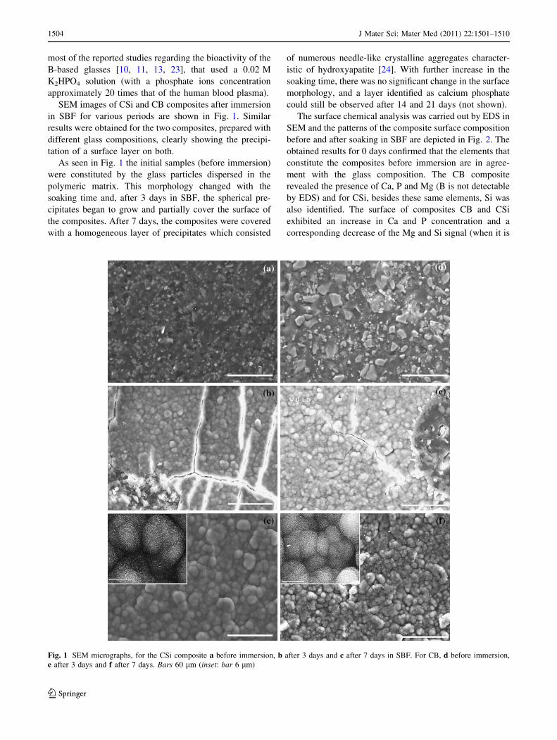
SEM images of CSi and CB composites after immersion
in SBF for various periods are shown in Fig. 1. Similar
results were obtained for the two composites, prepared with
different glass compositions, clearly showing the precipi-
tation of a surface layer on both.
As seen in Fig. 1 the initial samples (before immersion)
were constituted by the glass particles dispersed in the
polymeric matrix. This morphology changed with the
soaking time and, after 3 days in SBF, the spherical pre-
cipitates began to grow and partially cover the surface of
the composites. After 7 days, the composites were covered
with a homogeneous layer of precipitates which consisted
of numerous needle-like crystalline aggregates character-
istic of hydroxyapatite [24]. With further increase in the
soaking time, there was no significant change in the surface
morphology, and a layer identified as calcium phosphate
could still be observed after 14 and 21 days (not shown).
The surface chemical analysis was carried out by EDS in
SEM and the patterns of the composite surface composition
before and after soaking in SBF are depicted in Fig. 2. The
obtained results for 0 days confirmed that the elements that
constitute the composites before immersion are in agree-
ment with the glass composition. The CB composite
revealed the presence of Ca, P and Mg (B is not detectable
by EDS) and for CSi, besides these same elements, Si was
also identified. The surface of composites CB and CSi
exhibited an increase in Ca and P concentration and a
corresponding decrease of the Mg and Si signal (when it is
Fig. 1 SEM micrographs, for the CSi composite a before immersion, b after 3 days and c after 7 days in SBF. For CB, d before immersion,
e after 3 days and f after 7 days. Bars 60 lm (inset: bar 6 lm)
1504 J Mater Sci: Mater Med (2011) 22:1501–1510
123
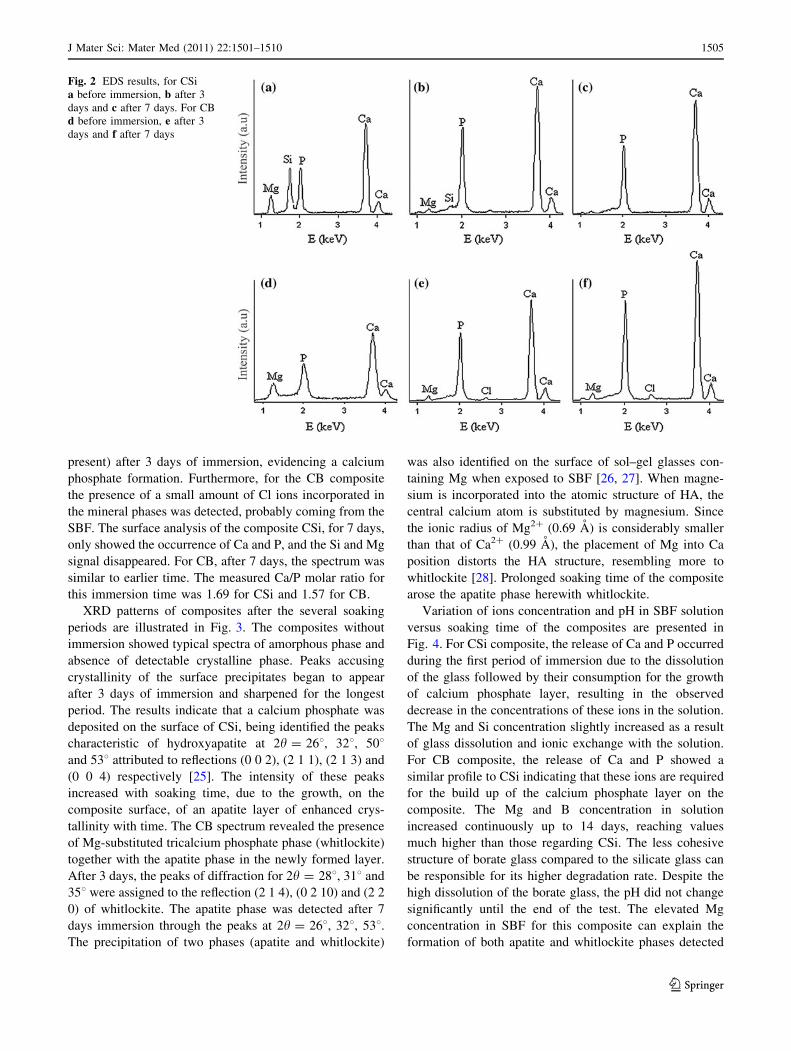
present) after 3 days of immersion, evidencing a calcium
phosphate formation. Furthermore, for the CB composite
the presence of a small amount of Cl ions incorporated in
the mineral phases was detected, probably coming from the
SBF. The surface analysis of the composite CSi, for 7 days,
only showed the occurrence of Ca and P, and the Si and Mg
signal disappeared. For CB, after 7 days, the spectrum was
similar to earlier time. The measured Ca/P molar ratio for
this immersion time was 1.69 for CSi and 1.57 for CB.
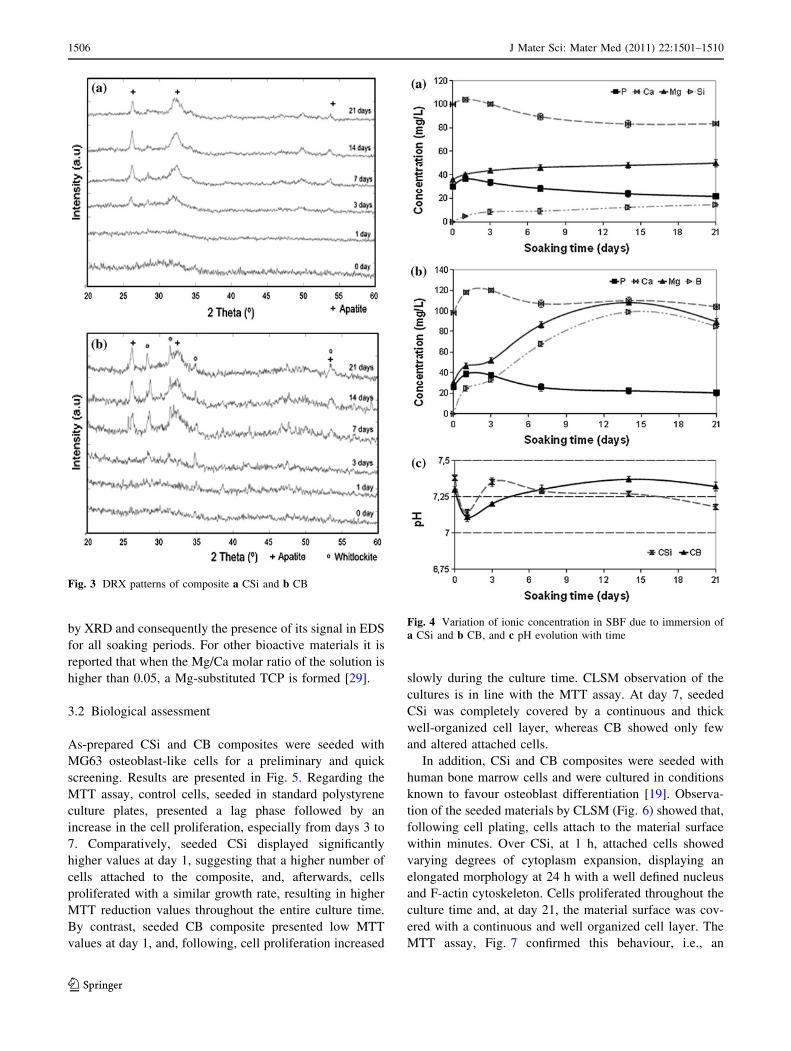
XRD patterns of composites after the several soaking
periods are illustrated in Fig. 3. The composites without
immersion showed typical spectra of amorphous phase and
absence of detectable crystalline phase. Peaks accusing
crystallinity of the surface precipitates began to appear
after 3 days of immersion and sharpened for the longest
period. The results indicate that a calcium phosphate was
deposited on the surface of CSi, being identified the peaks
characteristic of hydroxyapatite at 2h = 268, 328, 508and 538 attributed to reflections (0 0 2), (2 1 1), (2 1 3) and
(0 0 4) respectively [25]. The intensity of these peaks
increased with soaking time, due to the growth, on the
composite surface, of an apatite layer of enhanced crys-
tallinity with time. The CB spectrum revealed the presence
of Mg-substituted tricalcium phosphate phase (whitlockite)
together with the apatite phase in the newly formed layer.
After 3 days, the peaks of diffraction for 2h = 288, 318 and
358 were assigned to the reflection (2 1 4), (0 2 10) and (2 2
0) of whitlockite. The apatite phase was detected after 7
days immersion through the peaks at 2h = 268, 328, 538.The precipitation of two phases (apatite and whitlockite)
was also identified on the surface of sol–gel glasses con-
taining Mg when exposed to SBF [26, 27]. When magne-
sium is incorporated into the atomic structure of HA, the
central calcium atom is substituted by magnesium. Since
the ionic radius of Mg2? (0.69 A) is considerably smaller
than that of Ca2? (0.99 A), the placement of Mg into Ca
position distorts the HA structure, resembling more to
whitlockite [28]. Prolonged soaking time of the composite
arose the apatite phase herewith whitlockite.
Variation of ions concentration and pH in SBF solution
versus soaking time of the composites are presented in
Fig. 4. For CSi composite, the release of Ca and P occurred
during the first period of immersion due to the dissolution
of the glass followed by their consumption for the growth
of calcium phosphate layer, resulting in the observed
decrease in the concentrations of these ions in the solution.
The Mg and Si concentration slightly increased as a result
of glass dissolution and ionic exchange with the solution.
For CB composite, the release of Ca and P showed a
similar profile to CSi indicating that these ions are required
for the build up of the calcium phosphate layer on the
composite. The Mg and B concentration in solution
increased continuously up to 14 days, reaching values
much higher than those regarding CSi. The less cohesive
structure of borate glass compared to the silicate glass can
be responsible for its higher degradation rate. Despite the
high dissolution of the borate glass, the pH did not change
significantly until the end of the test. The elevated Mg
concentration in SBF for this composite can explain the
formation of both apatite and whitlockite phases detected
Fig. 2 EDS results, for CSi
a before immersion, b after 3
days and c after 7 days. For CB
d before immersion, e after 3
days and f after 7 days
J Mater Sci: Mater Med (2011) 22:1501–1510 1505
123
by XRD and consequently the presence of its signal in EDS
for all soaking periods. For other bioactive materials it is
reported that when the Mg/Ca molar ratio of the solution is
higher than 0.05, a Mg-substituted TCP is formed [29].
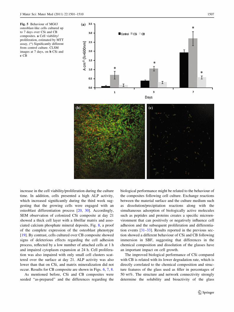
3.2 Biological assessment
As-prepared CSi and CB composites were seeded with
MG63 osteoblast-like cells for a preliminary and quick
screening. Results are presented in Fig. 5. Regarding the
MTT assay, control cells, seeded in standard polystyrene
culture plates, presented a lag phase followed by an
increase in the cell proliferation, especially from days 3 to
7. Comparatively, seeded CSi displayed significantly
higher values at day 1, suggesting that a higher number of
cells attached to the composite, and, afterwards, cells
proliferated with a similar growth rate, resulting in higher
MTT reduction values throughout the entire culture time.
By contrast, seeded CB composite presented low MTT
values at day 1, and, following, cell proliferation increased
slowly during the culture time. CLSM observation of the
cultures is in line with the MTT assay. At day 7, seeded
CSi was completely covered by a continuous and thick
well-organized cell layer, whereas CB showed only few
and altered attached cells.
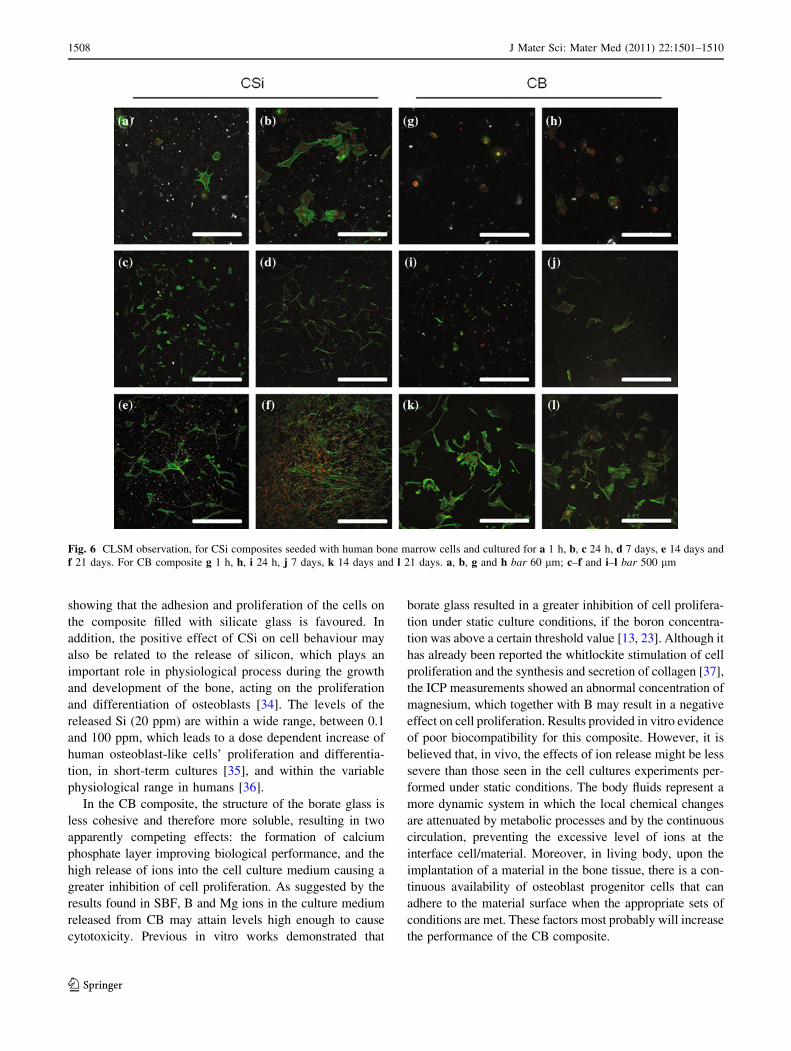
In addition, CSi and CB composites were seeded with
human bone marrow cells and were cultured in conditions
known to favour osteoblast differentiation [19]. Observa-
tion of the seeded materials by CLSM (Fig. 6) showed that,
following cell plating, cells attach to the material surface
within minutes. Over CSi, at 1 h, attached cells showed
varying degrees of cytoplasm expansion, displaying an
elongated morphology at 24 h with a well defined nucleus
and F-actin cytoskeleton. Cells proliferated throughout the
culture time and, at day 21, the material surface was cov-
ered with a continuous and well organized cell layer. The
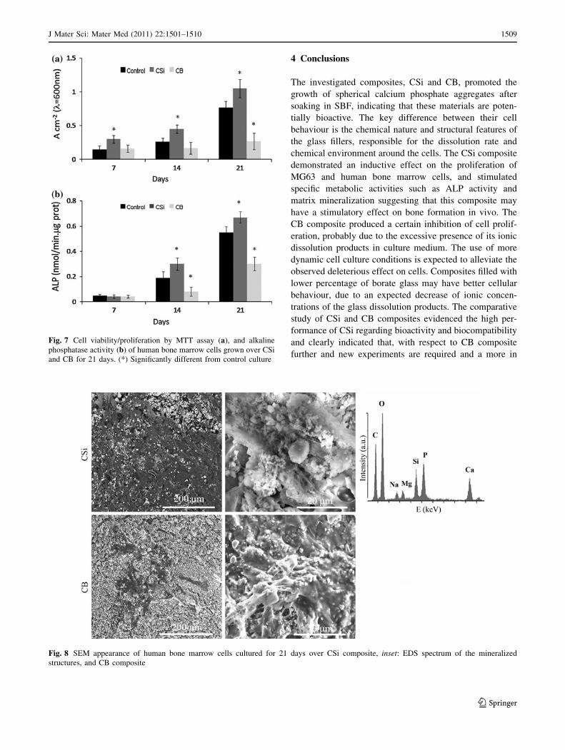
MTT assay, Fig. 7 confirmed this behaviour, i.e., an
Fig. 3 DRX patterns of composite a CSi and b CB
Fig. 4 Variation of ionic concentration in SBF due to immersion of
a CSi and b CB, and c pH evolution with time
1506 J Mater Sci: Mater Med (2011) 22:1501–1510
123
increase in the cell viability/proliferation during the culture
time. In addition, cells presented a high ALP activity,
which increased significantly during the third week sug-
gesting that the growing cells were engaged with an
osteoblast differentiation process [20, 30]. Accordingly,
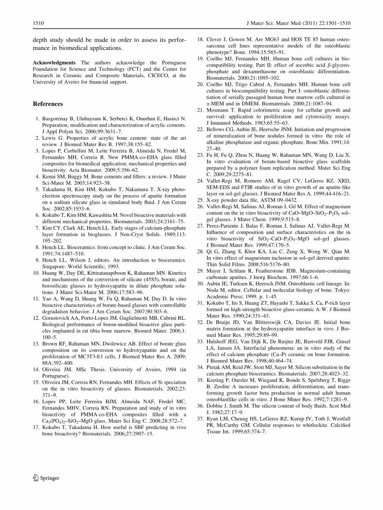
SEM observation of colonized CSi composite at day 21
showed a thick cell layer with a fibrillar matrix and asso-
ciated calcium phosphate mineral deposits, Fig. 8, a proof
of the complete expression of the osteoblast phenotype
[19]. By contrast, cells cultured over CB composite showed
signs of deleterious effects regarding the cell adhesion
process, reflected by a low number of attached cells at 1 h
and impaired cytoplasm expansion at 24 h. Cell prolifera-
tion was also impaired with only small cell clusters scat-
tered over the surface at day 21. ALP activity was also
lower than that on CSi, and matrix mineralization did not
occur. Results for CB composite are shown in Figs. 6, 7, 8.
As mentioned before, CSi and CB composites were
seeded ‘‘as-prepared’’ and the differences regarding the
biological performance might be related to the behaviour of
the composites following cell culture. Exchange reactions
between the material surface and the culture medium such
as dissolution/precipitation reactions along with the
simultaneous adsorption of biologically active molecules
such as peptides and proteins creates a specific microen-
vironment that can positively or negatively influence cell
adhesion and the subsequent proliferation and differentia-
tion events [31–33]. Results reported in the previous sec-
tion showed a different behaviour of CSi and CB following
immersion in SBF, suggesting that differences in the
chemical composition and dissolution of the glasses have
an important impact on cell growth.
The improved biological performance of CSi compared
with CB is related with its lower degradation rate, which is
directly correlated to the chemical composition and struc-
ture features of the glass used as filler in percentages of
50 wt%. The structure and network connectivity strongly
determine the solubility and bioactivity of the glass
Fig. 5 Behaviour of MG63
osteoblast-like cells cultured up
to 7 days over CSi and CB
composites. a Cell viability/
proliferation, estimated by MTT
assay, (*) Significantly different
from control culture. CLSM
images at 7 days, on b CSi and
c CB
J Mater Sci: Mater Med (2011) 22:1501–1510 1507
123
showing that the adhesion and proliferation of the cells on
the composite filled with silicate glass is favoured. In
addition, the positive effect of CSi on cell behaviour may
also be related to the release of silicon, which plays an
important role in physiological process during the growth
and development of the bone, acting on the proliferation
and differentiation of osteoblasts [34]. The levels of the
released Si (20 ppm) are within a wide range, between 0.1
and 100 ppm, which leads to a dose dependent increase of
human osteoblast-like cells’ proliferation and differentia-
tion, in short-term cultures [35], and within the variable
physiological range in humans [36].
In the CB composite, the structure of the borate glass is
less cohesive and therefore more soluble, resulting in two
apparently competing effects: the formation of calcium
phosphate layer improving biological performance, and the
high release of ions into the cell culture medium causing a
greater inhibition of cell proliferation. As suggested by the
results found in SBF, B and Mg ions in the culture medium
released from CB may attain levels high enough to cause
cytotoxicity. Previous in vitro works demonstrated that
borate glass resulted in a greater inhibition of cell prolifera-
tion under static culture conditions, if the boron concentra-
tion was above a certain threshold value [13, 23]. Although it
has already been reported the whitlockite stimulation of cell
proliferation and the synthesis and secretion of collagen [37],
the ICP measurements showed an abnormal concentration of
magnesium, which together with B may result in a negative
effect on cell proliferation. Results provided in vitro evidence
of poor biocompatibility for this composite. However, it is
believed that, in vivo, the effects of ion release might be less
severe than those seen in the cell cultures experiments per-
formed under static conditions. The body fluids represent a
more dynamic system in which the local chemical changes
are attenuated by metabolic processes and by the continuous
circulation, preventing the excessive level of ions at the
interface cell/material. Moreover, in living body, upon the
implantation of a material in the bone tissue, there is a con-
tinuous availability of osteoblast progenitor cells that can
adhere to the material surface when the appropriate sets of
conditions are met. These factors most probably will increase
the performance of the CB composite.
Fig. 6 CLSM observation, for CSi composites seeded with human bone marrow cells and cultured for a 1 h, b, c 24 h, d 7 days, e 14 days and
f 21 days. For CB composite g 1 h, h, i 24 h, j 7 days, k 14 days and l 21 days. a, b, g and h bar 60 lm; c–f and i–l bar 500 lm
1508 J Mater Sci: Mater Med (2011) 22:1501–1510
123
4 Conclusions
The investigated composites, CSi and CB, promoted the
growth of spherical calcium phosphate aggregates after
soaking in SBF, indicating that these materials are poten-
tially bioactive. The key difference between their cell
behaviour is the chemical nature and structural features of
the glass fillers, responsible for the dissolution rate and
chemical environment around the cells. The CSi composite
demonstrated an inductive effect on the proliferation of
MG63 and human bone marrow cells, and stimulated
specific metabolic activities such as ALP activity and
matrix mineralization suggesting that this composite may
have a stimulatory effect on bone formation in vivo. The
CB composite produced a certain inhibition of cell prolif-
eration, probably due to the excessive presence of its ionic
dissolution products in culture medium. The use of more
dynamic cell culture conditions is expected to alleviate the
observed deleterious effect on cells. Composites filled with
lower percentage of borate glass may have better cellular
behaviour, due to an expected decrease of ionic concen-
trations of the glass dissolution products. The comparative
study of CSi and CB composites evidenced the high per-
formance of CSi regarding bioactivity and biocompatibility
and clearly indicated that, with respect to CB composite
further and new experiments are required and a more in
Fig. 7 Cell viability/proliferation by MTT assay (a), and alkaline
phosphatase activity (b) of human bone marrow cells grown over CSi
and CB for 21 days. (*) Significantly different from control culture
Fig. 8 SEM appearance of human bone marrow cells cultured for 21 days over CSi composite, inset: EDS spectrum of the mineralized
structures, and CB composite
J Mater Sci: Mater Med (2011) 22:1501–1510 1509
123
depth study should be made in order to assess its perfor-
mance in biomedical applications.
Acknowledgments The authors acknowledge the Portuguese
Foundation for Science and Technology (FCT) and the Center for
Research in Ceramic and Composite Materials, CICECO, at the
University of Aveiro for financial support.
References
1. Basgorenay B, Ulubayram K, Serbetci K, Onurhan E, Hasirci N.
Preparation, modification and characterization of acrylic cements.
J Appl Polym Sci. 2006;99:3631–7.
2. Lewis G. Properties of acrylic bone cement: state of the art
review. J Biomed Mater Res B. 1997;38:155–82.
3. Lopes P, Corbellini M, Leite Ferreira B, Almeida N, Fredel M,
Fernandes MH, Correia R. New PMMA-co-EHA glass filled
composites for biomedical application: mechanical properties and
bioactivity. Acta Biomater. 2009;5:356–62.
4. Kenni SM, Buggy M. Bone cements and fillers: a review. J Mater
Sci-Mater M. 2003;14:923–38.
5. Takadama H, Kim HM, Kokubo T, Nakamura T. X-ray photo-
electron spectroscopy study on the process of apatite formation
on a sodium silicate glass in simulated body fluid. J Am Ceram
Soc. 2002;85:1933–6.
6. Kokubo T, Kim HM, Kawashita M. Novel bioactive materials with
different mechanical properties. Biomaterials. 2003;24:2161–75.
7. Kim CY, Clark AE, Hench LL. Early stages of calcium-phosphate
layer formation in bioglasses. J Non-Cryst Solids. 1989;113:
195–202.
8. Hench LL. Bioceramics: from concept to clinic. J Am Ceram Soc.
1991;74:1487–510.
9. Hench LL, Wilson J, editors. An introduction to bioceramics.
Singapore: World Scientific; 1993.
10. Huang W, Day DE, Kittiratanapiboon K, Rahaman MN. Kinetics
and mechanisms of the conversion of silicate (45S5), borate, and
borosilicate glasses to hydroxyapatite in dilute phosphate solu-
tions. J Mater Sci-Mater M. 2006;17:583–96.
11. Yao A, Wang D, Huang W, Fu Q, Rahaman M, Day D. In vitro
bioactive characteristics of borate-based glasses with controllable
degradation behavior. J Am Ceram Soc. 2007;90:303–6.
12. Gorustovich AA, Porto-Lopez JM, Guglielmotti MB, Cabrini RL.
Biological performance of boron-modified bioactive glass parti-
cles implanted in rat tibia bone marrow. Biomed Mater. 2006;1:
100–5.
13. Brown RF, Rahaman MN, Dwilewicz AB. Effect of borate glass
composition on its conversion to hydroxyapatite and on the
proliferation of MC3T3-E1 cells. J Biomed Mater Res A. 2009;
88A:392–400.
14. Oliveira JM. MSc Thesis. University of Aveiro, 1994 (in
Portuguese).
15. Oliveira JM, Correia RN, Fernandes MH. Effects of Si speciation
on the in vitro bioactivity of glasses. Biomaterials. 2002;23:
371–9.
16. Lopes PP, Leite Ferreira BJM, Almeida NAF, Fredel MC,
Fernandes MHV, Correia RN. Preparation and study of in vitro
bioactivity of PMMA-co-EHA composites filled with a
Ca3(PO4)2–SiO2–MgO glass. Mater Sci Eng C. 2008;28:572–7.
17. Kokubo T, Takadama H. How useful is SBF predicting in vivo
bone bioactivity? Biomaterials. 2006;27:2907–15.
18. Clover J, Gowen M. Are MG63 and HOS TE 85 human osteo-
sarcoma cell lines representative models of the osteoblastic
phenotype? Bone. 1994;15:585–91.
19. Coelho MJ, Fernandes MH. Human bone cell cultures in bio-
compatibility testing. Part II: effect of ascorbic acid b-glycero-
phosphate and dexamethasone on osteoblastic differentiation.
Biomaterials. 2000;21:1095–102.
20. Coelho MJ, Trigo Cabral A, Fernandes MH. Human bone cell
cultures in biocompatibility testing. Part I: osteoblastic differen-
tiation of serially passaged human bone marrow cells cultured in
a-MEM and in DMEM. Biomaterials. 2000;21:1087–94.
21. Mosmann T. Rapid colorimetric assay for cellular growth and
survival: application to proliferation and cytotoxicity assays.
J Immunol Methods. 1983;65:55–63.
22. Bellows CG, Aubin JE, Heersche JNM. Initiation and progression
of mineralization of bone nodules formed in vitro: the role of
alkaline phosphatase and organic phosphate. Bone Min. 1991;14:
27–40.
23. Fu H, Fu Q, Zhou N, Huang W, Rahaman MN, Wang D, Liu X.
In vitro evaluation of borate-based bioactive glass scaffolds
prepared by a polymer foam replication method. Mater Sci Eng
C. 2009;29:2275–81.
24. Vallet-Regi M, Romero AM, Ragel CV, LeGeros RZ. XRD,
SEM-EDS and FTIR studies of in vitro growth of an apatite-like
layer on sol-gel glasses. J Biomed Mater Res A. 1999;44:416–21.
25. X-ray powder data file, ASTM 09–0432.
26. Vallet-Regi M, Salinas AJ, Roman J, Gil M. Effect of magnesium
content on the in vitro bioactivity of CaO–MgO–SiO2–P2O5 sol–
gel glasses. J Mater Chem. 1999;9:515–8.
27. Perez-Pariente J, Balas F, Roman J, Salinas AJ, Vallet-Regi M.
Influence of composition and surface characteristics on the in
vitro bioactivity of SiO2–CaO–P2O5–MgO sol–gel glasses.
J Biomed Mater Res. 1999;47:170–5.
28. Qi G, Zhang S, Khor KA, Liu C, Zeng X, Weng W, Qian M.
In vitro effect of magnesium inclusion in sol–gel derived apatite.
Thin Solid Films. 2008;516:5176–80.
29. Mayer I, Schlam R, Featherstone JDB. Magnesium-containing
carbonate apatites. J Inorg Biochem. 1997;66:1–6.
30. Aubin JE, Turksen K, Heersch JNM. Osteoblastic cell lineage. In:
Noda M, editor. Cellular and molecular biology of bone. Tokyo:
Academic Press; 1999. p. 1–45.
31. Kokubo T, Ito S, Huang ZT, Hayashi T, Sakka S. Ca, P-rich layer
formed on high-strength bioactive glass–ceramic A-W. J Biomed
Mater Res. 1990;24:331–43.
32. De Bruijn JD, Van Blitterswijk CA, Davies JE. Initial bone
matrix formation at the hydroxyapatite interface in vivo. J Bio-
med Mater Res. 1995;29:89–99.
33. Hulshoff JEG, Van Dijk K, De Ruijter JE, Rietveld FJR, Ginsel
LA, Jansen JA. Interfacial phenomena: an in vitro study of the
effect of calcium phosphate (Ca–P) ceramic on bone formation.
J Biomed Mater Res. 1998;40:464–74.
34. Pietak AM, Reid JW, Stott MJ, Sayer M. Silicon substitution in the
calcium phosphate bioceramics. Biomaterials. 2007;28:4023–32.
35. Keeting P, Oursler M, Wiegand K, Bonde S, Spelsberg T, Riggs
B. Zeolite A increases proliferation, differentiation, and trans-
forming growth factor beta production in normal adult human
osteoblastlike cells in vitro. J Bone Miner Res. 1992;7:1281–9.
36. Dobbie J, Smith M. The silicon content of body fluids. Scot Med
J. 1982;27:17–9.
37. Ryan LM, Cheung HS, LeGeros RZ, Kurup IV, Toth J, Westfall
PR, McCarthy GM. Cellular responses to whitlockite. Calcified
Tissue Int. 1999;65:374–7.
1510 J Mater Sci: Mater Med (2011) 22:1501–1510
123


![[Biocompatibility studies of periodontal dressings]](https://img.dokumen.tips/doc/110x75/6354ce5b60cfc5aa0404e054/biocompatibility-studies-of-periodontal-dressings.jpg)


