Embed Size (px)
Citation preview
The American Journal of Pathology, Vol. 178, No. 6, June 2011
Copyright © 2011 American Society for Investigative Pathology.
Published by Elsevier Inc. All rights reserved.
DOI: 10.1016/j.ajpath.2011.02.034
Short Communication
Skp2 Is Necessary for Myc-Induced KeratinocyteProliferation but Dispensable for Myc Oncogenic
Activity in the Oral EpitheliumChristopher Sistrunk,* Everardo Macias,†
Keiichi Nakayama,‡ Yongbaek Kim,§
and Marcelo L. Rodriguez-Puebla*¶
From the Center for Comparative Medicine and Translational
Research,* and the Departments of Population Health and
Pathobiology,§ and Molecular Biomedical Sciences,¶ North Carolina
State University, Raleigh, North Carolina; and the Medical Institute
of Bioregulation,‡ Kyushu University, Fukuoka, Japan
The proto-oncogene c-Myc encodes a transcription fac-tor that is implicated in the regulation of cellular pro-liferation, differentiation, and apoptosis. Myc acceler-ates the rate of cell proliferation, at least in part,through its ability to down-regulate the expression ofthe cell cycle inhibitor p27Kip1. Moreover, p27Kip1 pro-tein levels are regulated by ubiquitin-mediated turn-over, leading to destruction by the E3 ubiquitin ligaseSCFSkp2. Therefore, we hypothesize that a lack of Skp2expression should lead to increased p27Kip1 levels andfurther inhibition of Myc-mediated proliferation and tu-morigenesis. Myc expression in epithelial tissues oftransgenic mice (K5-Myc) led to increased keratinocyteproliferation and the development of spontaneous tu-mors within the oral cavity. We generated K5-Myc–transgenic mice in an Skp2-null background. Consistentwith our hypothesis, we found that Myc-mediated kera-tinocyte hyperproliferation was abolished by the loss ofSkp2. However, Skp2 ablation did not affect Myc-driventumorigenesis because the incidence, latency, and de-gree of differentiation of oral tumors were identical be-tween K5-Myc/Skp2�/� and K5-Myc/Skp2�/� mice. Alto-gether, these findings suggest that Skp2 and p27Kip1 arecritical for Myc-driven keratinocyte proliferation; how-ever, Myc-mediated tumorigenesis in the oral epitheliumis independent of the Skp2-p27Kip1 axis. (Am J Pathol
2011, 178:2470–2477; DOI: 10.1016/j.ajpath.2011.02.034)
The proteasome pathway involves ubiquitin modification
and degradation of substrates by the proteasome com-2470
plex. Ubiquitin-mediated protein turnover is regulated byE3 ubiquitin ligases, such as SCF and anaphase-promot-ing complexes. The E3 SCFSkp2 complex is composed offour subunits: S-phase kinase-associated protein 1(Skp1), cullin, a ring-finger protein, and Skp2, which is amember of a large family of F-box adaptor proteins.1–3
Skp2 targets several cell cycle regulators for ubiquitina-tion, including the Cip/Kip family of cyclin-dependent ki-nase inhibitors (p21Cip1, p27Kip1, and p57Kip2), cyclin D1,p130, and Myc (alias c-myc).4–6 Skp2 contributes toG2/M progression by mediating the degradation ofp27Kip1, which is responsible for most of the phenotypesexhibited by Skp2-null mice.7 In fact, ablation of p27Kip1
overrides the Skp2�/� phenotypes, such as body size,polyploid nuclei, and multiple centrosome duplications.8
In addition, Skp2 also targets other important regulatorsof survival and/or apoptosis for degradation. For in-stance, Skp2 suppresses p53-dependent apoptosis byantagonizing the interaction between CREB-binding pro-tein/p300 and p53.9 Consistent with its role in tumor de-velopment, Skp2 is overexpressed in many experimentaland human tumors and has a transforming capacity;therefore, it is classified as an oncogene.10,11 Notably,elevated levels of Skp2 in human tumors correlate withlow p27Kip1 levels; overexpression of Skp2 in prostateepithelium decreases p27Kip1 levels and induces prolif-eration.12,13
The Myc proto-oncogenes are members of short-livedtranscription factors, a family that plays an important rolein cell proliferation, apoptosis, and cancer develop-
Supported by a grant from the National Cancer Institute/NIH (RO1CA116328).
Accepted for publication February 17, 2011.
Current address of C.S., Department of Molecular Biomedical Sci-ences, North Carolina State University, Raleigh, North Carolina; of E.M.,Dell Pediatric Research Institute, College of Pharmacy, The University ofTexas, Austin, Texas.
Address reprint request to Marcelo L. Rodriguez-Puebla, Ph.D., NorthCarolina State University, College of Veterinary Medicine, Molecular Bio-medical Sciences, 4700 Hillsborough St, Raleigh, NC 27606. E-mail:
[email protected].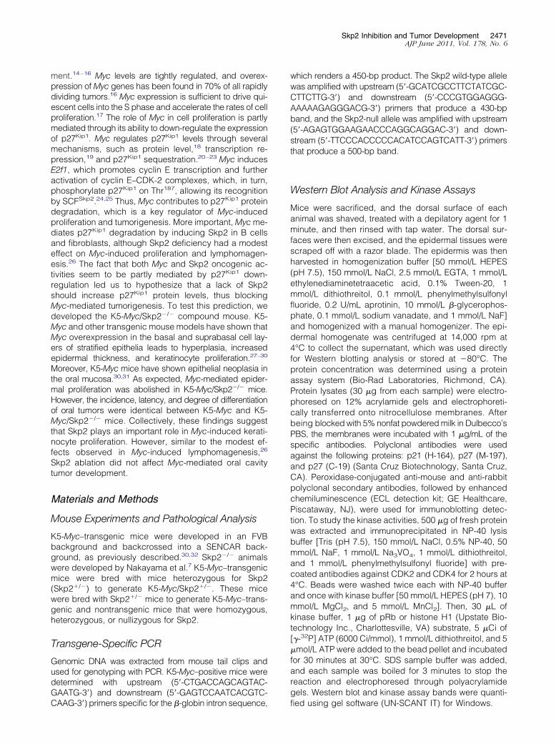
Skp2 Inhibition and Tumor Development 2471AJP June 2011, Vol. 178, No. 6
ment.14–16 Myc levels are tightly regulated, and overex-pression of Myc genes has been found in 70% of all rapidlydividing tumors.16 Myc expression is sufficient to drive qui-escent cells into the S phase and accelerate the rates of cellproliferation.17 The role of Myc in cell proliferation is partlymediated through its ability to down-regulate the expressionof p27Kip1. Myc regulates p27Kip1 levels through severalmechanisms, such as protein level,18 transcription re-pression,19 and p27Kip1 sequestration.20–23 Myc inducesE2f1, which promotes cyclin E transcription and furtheractivation of cyclin E–CDK-2 complexes, which, in turn,phosphorylate p27Kip1 on Thr187, allowing its recognitionby SCFSkp2.24,25 Thus, Myc contributes to p27Kip1 proteindegradation, which is a key regulator of Myc-inducedproliferation and tumorigenesis. More important, Myc me-diates p27Kip1 degradation by inducing Skp2 in B cellsand fibroblasts, although Skp2 deficiency had a modesteffect on Myc-induced proliferation and lymphomagen-esis.26 The fact that both Myc and Skp2 oncogenic ac-tivities seem to be partly mediated by p27Kip1 down-regulation led us to hypothesize that a lack of Skp2should increase p27Kip1 protein levels, thus blockingMyc-mediated tumorigenesis. To test this prediction, wedeveloped the K5-Myc/Skp2�/� compound mouse. K5-Myc and other transgenic mouse models have shown thatMyc overexpression in the basal and suprabasal cell lay-ers of stratified epithelia leads to hyperplasia, increasedepidermal thickness, and keratinocyte proliferation.27–30
Moreover, K5-Myc mice have shown epithelial neoplasia inthe oral mucosa.30,31 As expected, Myc-mediated epider-mal proliferation was abolished in K5-Myc/Skp2�/� mice.However, the incidence, latency, and degree of differentiationof oral tumors were identical between K5-Myc and K5-Myc/Skp2�/� mice. Collectively, these findings suggestthat Skp2 plays an important role in Myc-induced kerati-nocyte proliferation. However, similar to the modest ef-fects observed in Myc-induced lymphomagenesis,26
Skp2 ablation did not affect Myc-mediated oral cavitytumor development.
Materials and Methods
Mouse Experiments and Pathological Analysis
K5-Myc–transgenic mice were developed in an FVBbackground and backcrossed into a SENCAR back-ground, as previously described.30,32 Skp2�/� animalswere developed by Nakayama et al.7 K5-Myc–transgenicmice were bred with mice heterozygous for Skp2(Skp2�/�) to generate K5-Myc/Skp2�/�. These micewere bred with Skp2�/� mice to generate K5-Myc–trans-genic and nontransgenic mice that were homozygous,heterozygous, or nullizygous for Skp2.
Transgene-Specific PCR
Genomic DNA was extracted from mouse tail clips andused for genotyping with PCR. K5-Myc–positive mice weredetermined with upstream (5=-CTGACCAGCAGTAC-GAATG-3=) and downstream (5=-GAGTCCAATCACGTC-
CAAG-3=) primers specific for the �-globin intron sequence,which renders a 450-bp product. The Skp2 wild-type allelewas amplified with upstream (5=-GCATCGCCTTCTATCGC-CTTCTTG-3=) and downstream (5=-CCCGTGGAGGG-AAAAAGAGGGACG-3=) primers that produce a 430-bpband, and the Skp2-null allele was amplified with upstream(5=-AGAGTGGAAGAACCCAGGCAGGAC-3=) and down-stream (5=-TTCCCACCCCCACATCCAGTCATT-3=) primersthat produce a 500-bp band.
Western Blot Analysis and Kinase Assays
Mice were sacrificed, and the dorsal surface of eachanimal was shaved, treated with a depilatory agent for 1minute, and then rinsed with tap water. The dorsal sur-faces were then excised, and the epidermal tissues werescraped off with a razor blade. The epidermis was thenharvested in homogenization buffer [50 mmol/L HEPES(pH 7.5), 150 mmol/L NaCl, 2.5 mmol/L EGTA, 1 mmol/Lethylenediaminetetraacetic acid, 0.1% Tween-20, 1mmol/L dithiothreitol, 0.1 mmol/L phenylmethylsulfonylfluoride, 0.2 U/mL aprotinin, 10 mmol/L �-glycerophos-phate, 0.1 mmol/L sodium vanadate, and 1 mmol/L NaF]and homogenized with a manual homogenizer. The epi-dermal homogenate was centrifuged at 14,000 rpm at4°C to collect the supernatant, which was used directlyfor Western blotting analysis or stored at �80°C. Theprotein concentration was determined using a proteinassay system (Bio-Rad Laboratories, Richmond, CA).Protein lysates (30 �g from each sample) were electro-phoresed on 12% acrylamide gels and electrophoreti-cally transferred onto nitrocellulose membranes. Afterbeing blocked with 5% nonfat powdered milk in Dulbecco’sPBS, the membranes were incubated with 1 �g/mL of thespecific antibodies. Polyclonal antibodies were usedagainst the following proteins: p21 (H-164), p27 (M-197),and p27 (C-19) (Santa Cruz Biotechnology, Santa Cruz,CA). Peroxidase-conjugated anti-mouse and anti-rabbitpolyclonal secondary antibodies, followed by enhancedchemiluminescence (ECL detection kit; GE Healthcare,Piscataway, NJ), were used for immunoblotting detec-tion. To study the kinase activities, 500 �g of fresh proteinwas extracted and immunoprecipitated in NP-40 lysisbuffer [Tris (pH 7.5), 150 mmol/L NaCl, 0.5% NP-40, 50mmol/L NaF, 1 mmol/L Na3VO4, 1 mmol/L dithiothreitol,and 1 mmol/L phenylmethylsulfonyl fluoride] with pre-coated antibodies against CDK2 and CDK4 for 2 hours at4°C. Beads were washed twice each with NP-40 bufferand once with kinase buffer [50 mmol/L HEPES (pH 7), 10mmol/L MgCl2, and 5 mmol/L MnCl2]. Then, 30 �L ofkinase buffer, 1 �g of pRb or histone H1 (Upstate Bio-technology Inc., Charlottesville, VA) substrate, 5 �Ci of[�-32P] ATP (6000 Ci/mmol), 1 mmol/L dithiothreitol, and 5�mol/L ATP were added to the bead pellet and incubatedfor 30 minutes at 30°C. SDS sample buffer was added,and each sample was boiled for 3 minutes to stop thereaction and electrophoresed through polyacrylamidegels. Western blot and kinase assay bands were quanti-
fied using gel software (UN-SCANT IT) for Windows.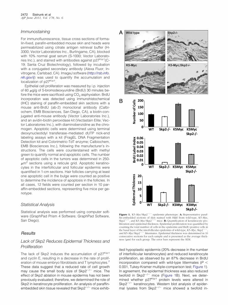
2472 Sistrunk et alAJP June 2011, Vol. 178, No. 6
Immunostaining
For immunofluorescence, tissue cross sections of forma-lin-fixed, parafin-embedded mouse skin and heads werepermeabilized using citrate antigen retrieval buffer (H-3300; Vector Laboratories Inc., Burlingame, CA), blockedwith 10% normal goat serum (S-1000; Vector Laborato-ries Inc.), and stained with antibodies against p27Kip1(C-19; Santa Cruz Biotechnology), followed by incubationwith a conjugated secondary antibody (Alexa Fluor; In-vitrogene, Carlsbad, CA). ImageJ software (http://rsb.info.nih.gov/ij) was used to quantify the accumulation andlocalization of p27Kip1.
Epithelial cell proliferation was measured by i.p. injectionof 60 �g/g of 5-bromodeoxyuridine (BrdU) 30 minutes be-fore the mice were sacrificed using CO2 asphyxiation. BrdUincorporation was detected using immunohistochemical(IHC) staining of paraffin-embedded skin sections with amouse anti-BrdU (ab-2) monoclonal antibody (Calbi-ochem, EMB Biosciences, San Diego, CA), a biotin-con-jugated anti-mouse antibody (Vector Laboratories Inc.),and an avidin-biotin peroxidase kit (Vectastain Elite; Vec-tor Laboratories Inc.), with diaminobenzidine as the chro-mogen. Apoptotic cells were determined using terminaldeoxynucleotidyl transferase–mediated dUTP nick-endlabeling assays with a kit (FragEL DNA FragmentationDetection kit and Colorimetric-TdT enzyme; Calbiochem,EMB Biosciences Inc.), following the manufacturer’s in-structions. The cells were counterstained with methylgreen to quantify normal and apoptotic cells. The numberof apoptotic cells in the tumors was determined in 250-�m2 sections using a reticule grid. Apoptotic keratino-cytes in the interfollicular and follicular epidermis werequantified in 1-cm sections. Hair follicles carrying at leastone apoptotic cell in the bulge were counted as positiveto determine the incidence of apoptosis in the follicles. Inall cases, 12 fields were counted per section in 10 par-affin-embedded sections, representing five mice per ge-notype.
Statistical Analysis
Statistical analysis was performed using computer soft-ware (GraphPad Prism 4 Software; GraphPad Software,San Diego).
Results
Lack of Skp2 Reduces Epidermal Thickness andProliferation
The lack of Skp2 induces the accumulation of p27Kip1
and cyclin E, resulting in a decrease in the rate of prolif-eration of mouse embryo fibroblasts and T lymphocytes.7
These data suggest that a reduced rate of cell growthmay cause the small body size of Skp2�/� mice. Theeffect of Skp2 ablation in mouse epidermis has not beenpreviously evaluated; therefore, we determined the role ofSkp2 in keratinocyte proliferation. An analysis of paraffin-
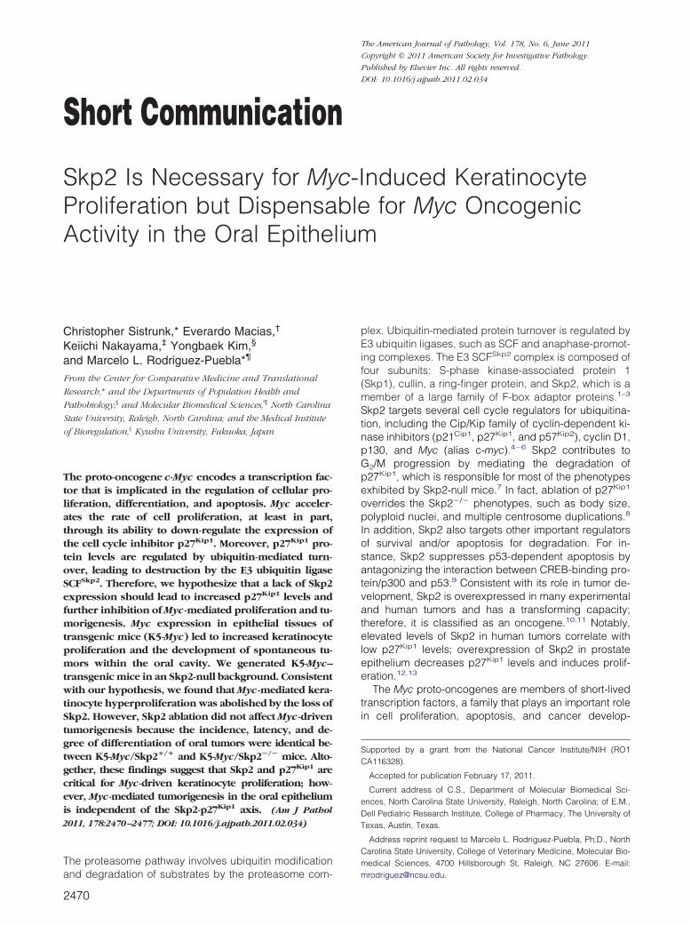
embedded skin tissue revealed that Skp2�/� mice exhib-ited hypoplastic epidermis (20% decrease in the numberof interfollicular keratinocytes) and reduced keratinocyteproliferation, as observed by an 87% decrease in BrdUincorporation compared with wild-type littermates (P �0.001, Tukey-Kramer multiple-comparison test; Figure 1).In agreement, the epidermal thickness was also reducedtwofold in Skp2�/� mice (Figure 1B). Next, we deter-mined whether p27Kip1 protein levels were altered inSkp2�/� keratinocytes. Western blot analysis of epider-
Figure 1. K5-Myc/Skp2�/� epidermis phenotype. A: Representative paraf-fin-embedded sections of skin stained with H&E from wild-type, K5-Myc,Skp2�/�, and K5-Myc/Skp2�/� mice. B: Quantification of keratinocyte pro-liferation and epidermal thickness. Epidermal proliferation was quantified bycounting the total number of cells in the epidermis and BrdU-positive cells inthe basal layer of the interfollicular epidermis of wild-type, K5-Myc, Skp2�/�,and K5-Myc/Skp2�/� littermates. Epidermal thickness was determined in 10consecutive sections for each sample and is presented as the average thick-ness (�m) for each group. The error bars represent the SEM.
mal lysates from Skp2�/� mice showed a twofold in-
Skp2 Inhibition and Tumor Development 2473AJP June 2011, Vol. 178, No. 6
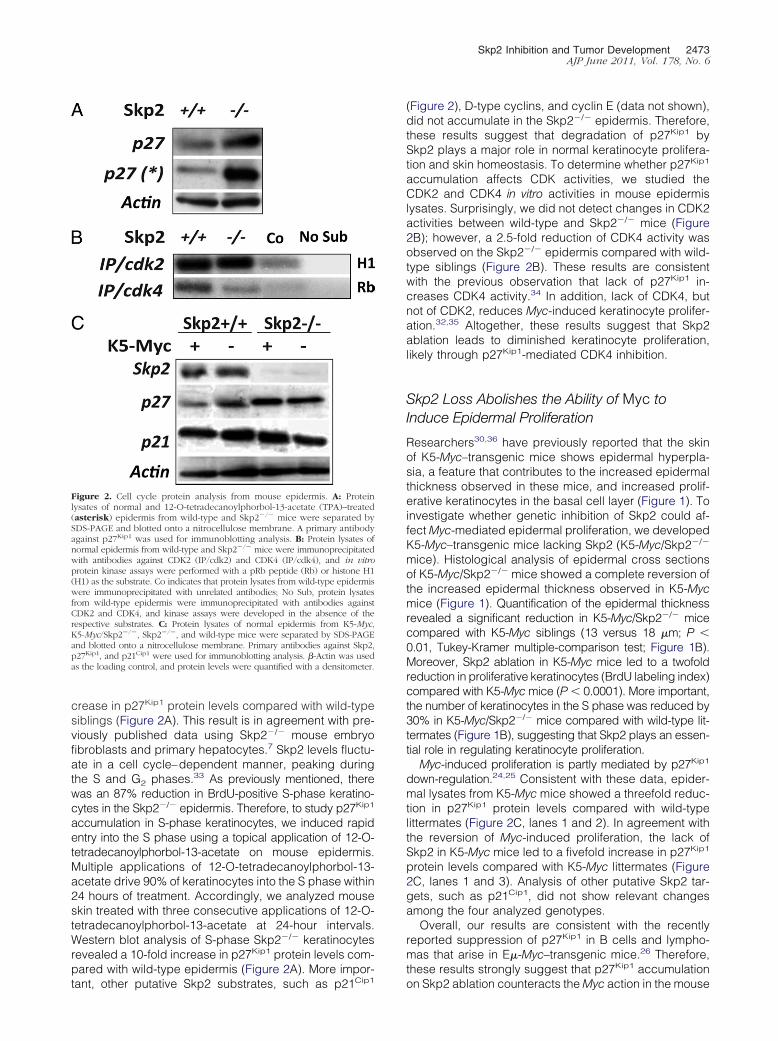
crease in p27Kip1 protein levels compared with wild-typesiblings (Figure 2A). This result is in agreement with pre-viously published data using Skp2�/� mouse embryofibroblasts and primary hepatocytes.7 Skp2 levels fluctu-ate in a cell cycle–dependent manner, peaking duringthe S and G2 phases.33 As previously mentioned, therewas an 87% reduction in BrdU-positive S-phase keratino-cytes in the Skp2�/� epidermis. Therefore, to study p27Kip1
accumulation in S-phase keratinocytes, we induced rapidentry into the S phase using a topical application of 12-O-tetradecanoylphorbol-13-acetate on mouse epidermis.Multiple applications of 12-O-tetradecanoylphorbol-13-acetate drive 90% of keratinocytes into the S phase within24 hours of treatment. Accordingly, we analyzed mouseskin treated with three consecutive applications of 12-O-tetradecanoylphorbol-13-acetate at 24-hour intervals.Western blot analysis of S-phase Skp2�/� keratinocytesrevealed a 10-fold increase in p27Kip1 protein levels com-pared with wild-type epidermis (Figure 2A). More impor-
Figure 2. Cell cycle protein analysis from mouse epidermis. A: Proteinlysates of normal and 12-O-tetradecanoylphorbol-13-acetate (TPA)–treated(asterisk) epidermis from wild-type and Skp2�/� mice were separated bySDS-PAGE and blotted onto a nitrocellulose membrane. A primary antibodyagainst p27Kip1 was used for immunoblotting analysis. B: Protein lysates ofnormal epidermis from wild-type and Skp2�/� mice were immunoprecipitatedwith antibodies against CDK2 (IP/cdk2) and CDK4 (IP/cdk4), and in vitroprotein kinase assays were performed with a pRb peptide (Rb) or histone H1(H1) as the substrate. Co indicates that protein lysates from wild-type epidermiswere immunoprecipitated with unrelated antibodies; No Sub, protein lysatesfrom wild-type epidermis were immunoprecipitated with antibodies againstCDK2 and CDK4, and kinase assays were developed in the absence of therespective substrates. C: Protein lysates of normal epidermis from K5-Myc,K5-Myc/Skp2�/�, Skp2�/�, and wild-type mice were separated by SDS-PAGEand blotted onto a nitrocellulose membrane. Primary antibodies against Skp2,p27Kip1, and p21Cip1 were used for immunoblotting analysis. �-Actin was usedas the loading control, and protein levels were quantified with a densitometer.
tant, other putative Skp2 substrates, such as p21Cip1
(Figure 2), D-type cyclins, and cyclin E (data not shown),did not accumulate in the Skp2�/� epidermis. Therefore,these results suggest that degradation of p27Kip1 bySkp2 plays a major role in normal keratinocyte prolifera-tion and skin homeostasis. To determine whether p27Kip1
accumulation affects CDK activities, we studied theCDK2 and CDK4 in vitro activities in mouse epidermislysates. Surprisingly, we did not detect changes in CDK2activities between wild-type and Skp2�/� mice (Figure2B); however, a 2.5-fold reduction of CDK4 activity wasobserved on the Skp2�/� epidermis compared with wild-type siblings (Figure 2B). These results are consistentwith the previous observation that lack of p27Kip1 in-creases CDK4 activity.34 In addition, lack of CDK4, butnot of CDK2, reduces Myc-induced keratinocyte prolifer-ation.32,35 Altogether, these results suggest that Skp2ablation leads to diminished keratinocyte proliferation,likely through p27Kip1-mediated CDK4 inhibition.
Skp2 Loss Abolishes the Ability of Myc toInduce Epidermal Proliferation
Researchers30,36 have previously reported that the skinof K5-Myc–transgenic mice shows epidermal hyperpla-sia, a feature that contributes to the increased epidermalthickness observed in these mice, and increased prolif-erative keratinocytes in the basal cell layer (Figure 1). Toinvestigate whether genetic inhibition of Skp2 could af-fect Myc-mediated epidermal proliferation, we developedK5-Myc–transgenic mice lacking Skp2 (K5-Myc/Skp2�/�
mice). Histological analysis of epidermal cross sectionsof K5-Myc/Skp2�/� mice showed a complete reversion ofthe increased epidermal thickness observed in K5-Mycmice (Figure 1). Quantification of the epidermal thicknessrevealed a significant reduction in K5-Myc/Skp2�/� micecompared with K5-Myc siblings (13 versus 18 �m; P �0.01, Tukey-Kramer multiple-comparison test; Figure 1B).Moreover, Skp2 ablation in K5-Myc mice led to a twofoldreduction in proliferative keratinocytes (BrdU labeling index)compared with K5-Myc mice (P � 0.0001). More important,the number of keratinocytes in the S phase was reduced by30% in K5-Myc/Skp2�/� mice compared with wild-type lit-termates (Figure 1B), suggesting that Skp2 plays an essen-tial role in regulating keratinocyte proliferation.
Myc-induced proliferation is partly mediated by p27Kip1
down-regulation.24,25 Consistent with these data, epider-mal lysates from K5-Myc mice showed a threefold reduc-tion in p27Kip1 protein levels compared with wild-typelittermates (Figure 2C, lanes 1 and 2). In agreement withthe reversion of Myc-induced proliferation, the lack ofSkp2 in K5-Myc mice led to a fivefold increase in p27Kip1
protein levels compared with K5-Myc littermates (Figure2C, lanes 1 and 3). Analysis of other putative Skp2 tar-gets, such as p21Cip1, did not show relevant changesamong the four analyzed genotypes.
Overall, our results are consistent with the recentlyreported suppression of p27Kip1 in B cells and lympho-mas that arise in E�-Myc–transgenic mice.26 Therefore,these results strongly suggest that p27Kip1 accumulation
on Skp2 ablation counteracts the Myc action in the mouse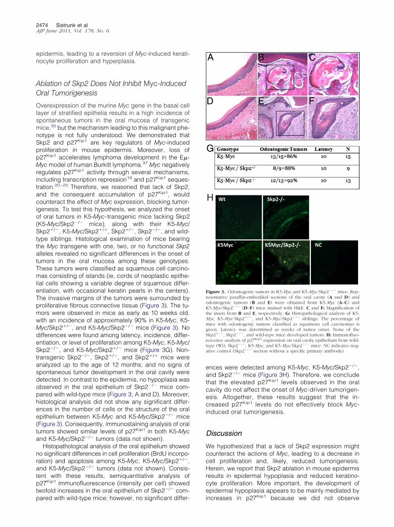
2474 Sistrunk et alAJP June 2011, Vol. 178, No. 6
epidermis, leading to a reversion of Myc-induced kerati-nocyte proliferation and hyperplasia.
Ablation of Skp2 Does Not Inhibit Myc-InducedOral Tumorigenesis
Overexpression of the murine Myc gene in the basal celllayer of stratified epithelia results in a high incidence ofspontaneous tumors in the oral mucosa of transgenicmice,30 but the mechanism leading to this malignant phe-notype is not fully understood. We demonstrated thatSkp2 and p27Kip1 are key regulators of Myc-inducedproliferation in mouse epidermis. Moreover, loss ofp27Kip1 accelerates lymphoma development in the E�-Myc model of human Burkitt lymphoma.37 Myc negativelyregulates p27Kip1 activity through several mechanisms,including transcription repression19 and p27Kip1 seques-tration.20–23 Therefore, we reasoned that lack of Skp2,and the consequent accumulation of p27Kip1, wouldcounteract the effect of Myc expression, blocking tumor-igenesis. To test this hypothesis, we analyzed the onsetof oral tumors in K5-Myc–transgenic mice lacking Skp2(K5-Myc/Skp2�/� mice), along with their K5-Myc/Skp2�/�, K5-Myc/Skp2�/�, Skp2�/�, Skp2�/�, and wild-type siblings. Histological examination of mice bearingthe Myc transgene with one, two, or no functional Skp2alleles revealed no significant differences in the onset oftumors in the oral mucosa among these genotypes.These tumors were classified as squamous cell carcino-mas consisting of islands (ie, cords of neoplastic epithe-lial cells showing a variable degree of squamous differ-entiation, with occasional keratin pearls in the centers).The invasive margins of the tumors were surrounded byproliferative fibrous connective tissue (Figure 3). The tu-mors were observed in mice as early as 10 weeks old,with an incidence of approximately 90% in K5-Myc, K5-Myc/Skp2�/�, and K5-Myc/Skp2�/� mice (Figure 3). Nodifferences were found among latency, incidence, differ-entiation, or level of proliferation among K5-Myc, K5-Myc/Skp2�/�, and K5-Myc/Skp2�/� mice (Figure 3G). Non-transgenic Skp2�/�, Skp2�/�, and Skp2�/� mice wereanalyzed up to the age of 12 months; and no signs ofspontaneous tumor development in the oral cavity weredetected. In contrast to the epidermis, no hypoplasia wasobserved in the oral epithelium of Skp2�/� mice com-pared with wild-type mice (Figure 3, A and D). Moreover,histological analysis did not show any significant differ-ences in the number of cells or the structure of the oralepithelium between K5-Myc and K5-Myc/Skp2�/� mice(Figure 3). Consequently, immunostaining analysis of oraltumors showed similar levels of p27Kip1 in both K5-Mycand K5-Myc/Skp2�/� tumors (data not shown).
Histopathological analysis of the oral epithelium showedno significant differences in cell proliferation (BrdU incorpo-ration) and apoptosis among K5-Myc, K5-Myc/Skp2�/�,and K5-Myc/Skp2�/� tumors (data not shown). Consis-tent with these results, semiquantitative analysis ofp27Kip1 immunofluorescence (intensity per cell) showedtwofold increases in the oral epithelium of Skp2�/� com-
pared with wild-type mice; however, no significant differ-ences were detected among K5-Myc, K5-Myc/Skp2�/�,and Skp2�/� mice (Figure 3H). Therefore, we concludethat the elevated p27Kip1 levels observed in the oralcavity do not affect the onset of Myc-driven tumorigen-esis. Altogether, these results suggest that the in-creased p27Kip1 levels do not effectively block Myc-induced oral tumorigenesis.
Discussion
We hypothesized that a lack of Skp2 expression mightcounteract the actions of Myc, leading to a decrease incell proliferation and, likely, reduced tumorigenesis.Herein, we report that Skp2 ablation in mouse epidermisresults in epidermal hypoplasia and reduced keratino-cyte proliferation. More important, the development ofepidermal hypoplasia appears to be mainly mediated by
Figure 3. Odontogenic tumors in K5-Myc and K5-Myc/Skp2�/� mice. Rep-resentative paraffin-embedded sections of the oral cavity (A and D) andodontogenic tumors (B and E) were obtained from K5-Myc (A–C) andK5-Myc/Skp2�/� (D–F) mice stained with H&E. C and F: Magnification ofthe insets from B and E, respectively. G: Histopathological analysis of K5-Myc, K5-Myc/Skp2�/�, and K5-Myc/Skp2�/� siblings. The percentage ofmice with odontogenic tumors classified as squamous cell carcinomas isgiven. Latency was determined as weeks of tumor onset. None of theSkp2�/�, Skp2�/�, and wild-type mice developed tumors. H: Immunofluo-rescence analysis of p27Kip1 expression on oral cavity epithelium from wild-type (Wt), Skp2�/�, K5-Myc, and K5-Myc/Skp2�/� mice. NC indicates neg-ative control (Skp2�/� section without a specific primary antibody).
increases in p27Kip1 because we did not observe
Skp2 Inhibition and Tumor Development 2475AJP June 2011, Vol. 178, No. 6
changes in other putative substrates of Skp2, such ascyclin D1, p21Cip1, or p57Kip.24,38–40 Moreover, mousekeratinocytes did not show accumulation of cyclin E, aspreviously reported in Skp2�/� mouse embryo fibroblastsand hepatocytes.7 Thus, our findings indicate thatp27Kip1 is a main player in Skp2-mediated keratinocyteregulation.8 More important, in vitro analysis of CDK ac-tivities shows that CDK4, but not CDK2, inhibits in theSkp2�/� epidermis. The Cip/Kip family members wereinitially described as general CDK inhibitors; however,several groups41,42 have shown that p21Cip1 and p27Kip1
bind, but do not interfere, with CDK4/CDK6 activities.There is no clear consent about the role of Cip/Kip mem-bers on CDK4 activity. Ray et al43 recently reported twoindependent models by which p27Kip1 might inhibitCDK4. According to these models, the absence of thespecific tyrosine phosphorylation of p27Kip1 results in thebinding and inhibition of CDK4 by p27Kip1. Consistentwith this model, lack of CDK4, but not of CDK2, hasreduced Myc-induced keratinocyte proliferation32,35 andp27Kip1 ablation, resulting in activation of both CDK4 andCDK2 in mouse epidermis.34 Recent observations44,45
have shown that the ablation of CDK2 in a p27Kip1-nullbackground did not mitigate the phenotypes of p27Kip1
deficiency, clearly showing that p27Kip1 can act indepen-dently of CDK2. Thus, whether CDK4 is the main targetfor p27Kip1 inhibition in mouse epidermis warrants futureinvestigations.
Transgenic mice overexpressing Myc in the basal celllayer of the stratified epithelium experienced severe epi-dermal hyperplasia, hypertrophy, and, in some cases,tumor development.27,30–32 We found that forced expres-sion of the Myc oncogene led to decreased p27Kip1 pro-tein levels in mouse epidermis. Supporting our initial hy-pothesis, genetic inhibition of Skp2, and the consequentp27Kip1 accumulation, severely crippled the ability of theMyc oncogene to drive keratinocyte proliferation and fur-ther induce epidermal hyperplasia. Consistent with thesedata, biochemical analysis of K5-Myc/Skp2�/� mouseepidermis showed that lack of Skp2 expression reversedthe reduced levels of p27Kip1 observed in the K5-Mycepidermis. The Myc oncogene also affects p27Kip1 activ-ity by inducing CDK4 expression, leading to p27Kip1 se-questration by the CDK4/D-type cyclin complexes.32,46
Therefore, changes in p27Kip1 protein levels play an im-portant role in Myc-induced hyperplasia and Skp2–/– hy-poplasia. Interesting, Myc-mediated p27Kip1 down–regu-lation was dependent on the elevated levels of Skp2 in Bcells from the transgenic E�-Myc mouse.26 However, thiseffect seems to be tissue specific because we did notobserve changes in Skp2 levels in the K5-Myc epidermis.Researchers22,23,32,47 have shown that low levels ofp27Kip1 and/or p27Kip1 sequestration, on CDK4 expres-sion in the K5-Myc epidermis, results in CDK2 activation.However, CDK2 ablation does not reverse the hyperpro-liferative phenotype of the K5-Myc epidermis.35 Thus, it istempting to hypothesize that Myc activity deregulateskeratinocyte proliferation through mechanisms other thanCDK2 activation. Altogether, these results suggest thatSkp2 plays a central role in the deregulation of keratino-
cyte proliferation triggered by Myc overexpression.Deregulation of Myc expression plays a causal role inthe genesis of several types of human and experimentalmalignancies.48–53 Herein, we showed that Skp2 ablationdoes not inhibit Myc-induced oral tumorigenesis. In fact,we did not find any differences in the latency, incidence,differentiation, or level of proliferation among K5-Myc,K5-Myc/Skp2�/�, and K5-Myc/Skp2�/� mice. As ex-pected, we observed accumulation of the p27Kip1 proteinin the Skp2�/� oral epithelium, although high p27Kip1
levels were also observed in K5-Myc and K5-Myc/Skp2�/� mice. It is not clear why Myc expression doesnot reduce p27Kip1 levels in the oral cavity, but this ob-servation warrants further investigation. The analysis ofp27Kip1 in oral tumors from K5-Myc and K5-Myc/Skp2�/�
mice showed no significant differences between bothgenotypes (data not shown). Thus, we speculate that lackof Skp2 results in a tissue-specific effect in mouse epi-dermis and oral epithelium, blocking keratinocyte prolif-eration, likely through CDK4 inhibition but not oral epithe-lium cell proliferation. In support of a tissue-specific roleof p27Kip1 levels, a clear discrepancy was observed be-tween the phenotype of epidermis and the oral epitheliumof these mouse models. The Skp2�/� mice showed nosigns of developing epithelial hypoplasia in the oral cav-ity, as observed in their epidermis. Also, the oral epithe-lium of K5-Myc mice showed no development of the hy-perplasia observed in the epidermis. Therefore, wehypothesized that Myc-induced oral tumorigenesis doesnot depend on p27Kip1 levels, resulting in the unsuccess-ful blocking of tumor development in K5-Myc/Skp2�/�
mice. In support of our finding, Skp2 deficiency had amodest or no effect on Myc-induced lymphomagenesis inE�-Myc–transgenic mice.26
Skp2 may play additional roles by regulating tumordevelopment; Lin et al54 showed that Skp2 inactivation onits own does not induce cellular senescence. However,aberrant oncogenic signals and/or inactivation of tumorsuppressor genes triggers a potent suppressive senes-cence response in mice devoid of Skp2.54 In addition,Skp2 targets regulators of survival and/or apoptosis fordegradation. For instance, Skp2 suppresses p53-depen-dent apoptosis by regulating CBP/p300 protein levelsand antagonizing the interaction between CBP/p300 andp53.9 We did not observe changes in apoptosis levels inK5-Myc/Skp2�/� and K5-Myc tumors; thus, our studiespredict that p27Kip1 accumulation, senescence, and/orapoptosis does not play an important role in Myc-inducedoral tumorigenesis. Skp2 is also a strong stimulator ofMyc’s transcriptional activities,55,56 suggesting that abla-tion of Skp2 may reduce cell proliferation and/or tumordevelopment. Conversely, Skp2 participates in Myc pro-teasomal degradation, predicting that ablation of Skp2would result in Myc stabilization and increase tumor de-velopment. Our results show that the stimulation of Myctranscriptional activity by Skp255,56 is dispensable forMyc oncogenic activities. In this scenario, ablation ofSkp2 stabilizes p27Kip1 and c-myc protein levels, allow-ing K5-Myc to behave as a potent oncogene, even in thepresence of elevated levels of p27Kip1 protein.
Collectively, the data presented in this study and re-
cent reports from other researchers26,54 suggest that
2476 Sistrunk et alAJP June 2011, Vol. 178, No. 6
Skp2 is an efficient regulator of normal proliferationthrough the regulation of p27Kip1 protein levels, but it isinefficient in alleviating Myc-induced tumorigenesis.Therefore, the suitability of Skp2 as a target for therapeu-tic intervention must be considered in a tissue-dependentmanner and in the context of the particular oncogenicpathway affected.
Acknowledgments
We thank Paula Miliani de Marval for her critical review ofthe manuscript and the Laboratory Animal Resources(College of Veterinary Medicine, North Carolina State Uni-versity, Raleigh).
References
1. Gregory MA, Hann SR: c-Myc proteolysis by the ubiquitin-protea-some pathway: stabilization of c-Myc in Burkitt’s lymphoma cells. MolCell Biol 2000, 20:2423–2435
2. Bahram F, von der Lehr N, Cetinkaya C, Larsson LG: c-Myc hot spotmutations in lymphomas result in inefficient ubiquitination and de-creased proteasome-mediated turnover. Blood 2000, 95:2104–2110
3. Weissman AM: Themes and variations on ubiquitylation. Nat Rev MolCell Biol 2001, 2:169–178
4. Carrano AC, Eytan E, Hershko A, Pagano M: SKP2 is required forubiquitin-mediated degradation of the CDK inhibitor p27. Nat Cell Biol1999, 1:193–199
5. Nakayama KI, Nakayama K: Ubiquitin ligases: cell-cycle control andcancer. Nat Rev Cancer 2006, 6:369–381
6. DeSalle LM, Pagano M: Regulation of the G1 to S transition by theubiquitin pathway. FEBS Lett 2001, 490:179–189
7. Nakayama K, Nagahama H, Minamishima Y, Matsumoto M, Naka-michi I, Kitagawa K, Shirane M, Tsunematsu R, Tsukiyama T, IshidaN, Kitagawa M, Nakayama K-I, Hatayama S: Targeted disruption ofSkp2 results in accumulation of cyclin E and p27Kip1, polyploidyand centrosome overeduplication. EMBO J 2000, 19:2069 –2081
8. Nakayama K, Nagahama H, Minamishima YA, Miyake S, Ishida N,Hatakeyama S, Kitagawa M, Iemura S, Natsume T, Nakayama KI:Skp2-mediated degradation of p27 regulates progression into mito-sis. Dev Cell 2004, 6:661–672
9. Kitagawa M, Lee SH, McCormick F: Skp2 suppresses p53-dependentapoptosis by inhibiting p300. Mol Cell 2008, 29:217–231
10. Latres E, Chiarle R, Schulman BA, Pavletich NP, Pellicer A, InghiramiG, Pagano M: Role of the F-box protein Skp2 in lymphomagenesis.Proc Natl Acad Sci U S A 2001, 98:2515–2520
11. Gstaiger M, Jordan R, Lim M, Catzavelos C, Mestan J, Slingerland J,Krek W: Skp2 is oncogenic and overexpressed in human cancers.Proc Natl Acad Sci U S A 2001, 98:5043–5048
12. Kudo Y, Kitajima S, Sato S, Miyauchi M, Ogawa I, Takata T: Highexpression of S-phase kinase-interacting protein 2, human F-boxprotein, correlates with poor prognosis in oral squamous cell carci-nomas. Cancer Res 2001, 61:7044–7047
13. Shim EH, Johnson L, Noh HL, Kim YJ, Sun H, Zeiss C, Zhang H:Expression of the F-box protein SKP2 induces hyperplasia, dysplasia,and low-grade carcinoma in the mouse prostate. Cancer Res 2003,63:1583–1588
14. Grandori C, Cowley SM, James LP, Eisenman RN: The Myc/Max/Madnetwork and the transcriptional control of cell behavior. Annu Rev CellDev Biol 2000, 16:653–699
15. Nilsson JA, Cleveland JL: Myc pathways provoking cell suicide andcancer. Oncogene 2003, 22:9007–9021
16. Boxer LM, Dang CV: Translocations involving c-myc and c-myc func-tion. Oncogene 2001, 20:5595–5610
17. Cavalieri F, Goldfarb M: Growth factor-deprived BALB/c 3T3 murine
fibroblasts can enter the S phase after induction of c-myc geneexpression. Mol Cell Biol 1987, 7:3554–356018. Keller UB, Old JB, Dorsey FC, Nilsson JA, Nilsson L, MacLean KH,Chung L, Yang C, Spruck C, Boyd K, Reed SI, Cleveland JL: Myctargets Cks1 to provoke the suppression of p27Kip1, proliferation andlymphomagenesis. EMBO J 2007, 26:2562–2574
19. Yang W, Shen J, Wu M, Arsura M, FitzGerald M, Suldan Z, Kim DW,Hofmann CS, Pianetti S, Romieu-Mourez R, Freedman LP, Sonen-shein GE: Repression of transcription of the p27(Kip1) cyclin-dependent kinase inhibitor gene by c-Myc. Oncogene 2001, 20:1688 –1702
20. Vlach J, Hennecke S, Alevizopoulos K, Conti D, Amati B: Growtharrest by the cyclin-dependent kinase inhibitor p27Kip1 is abrogatedby c-Myc. EMBO J 1996, 15:6595–6604
21. Muller D, Bouchard C, Rudolph B, Steiner P, Stuckmann I, Saffrich R,Ansorge W, Huttner W, Eilers M: Cdk2-dependent phosphorylation ofp27 facilitates its Myc-induced release from cyclin E/cdk2 com-plexes. Oncogene 1997, 15:2561–2576
22. Perez-Roger I, Kim SH, Griffiths B, Sewing A, Land H: Cyclins D1 andD2 mediate myc-induced proliferation via sequestration of p27(Kip1)and p21(Cip1). EMBO J 1999, 18:5310–5320
23. Bouchard C, Thieke K, Maier A, Saffrich R, Hanley-Hyde J, AnsorgeW, Reed S, Sicinski P, Bartek J, Eilers M: Direct induction of cyclin D2by Myc contributes to cell cycle progression and sequestration ofp27. EMBO J 1999, 18:5321–5333
24. Sherr CJ, Roberts JM: CDK inhibitors: positive and negative regula-tors of G1-phase progression. Genes Dev 1999, 10:1491–1502
25. Philipp-Staheli J, Payne SR, Kemp CJ: p27(Kip1): regulation andfunction of a haploinsufficient tumor suppressor and its misregulationin cancer. Exp Cell Res 2001, 264:148–168
26. Old JB, Kratzat S, Hoellein A, Graf S, Nilsson JA, Nilsson L, Na-kayama KI, Peschel C, Cleveland JL, Keller UB: Skp2 directs Myc-mediated suppression of p27Kip1 yet has modest effects on Myc-driven lymphomagenesis. Mol Cancer Res 2010, 8:353–362
27. Waikel RL, Wang XJ, Roop DR: Targeted expression of c-Myc in theepidermis alters normal proliferation, differentiation and UV-B in-duced apoptosis. Oncogene 1999, 18:4870–4878
28. Waikel RL, Kawachi Y, Waikel PA, Wang XJ, Roop DR: Deregulatedexpression of c-Myc depletes epidermal stem cells. Nat Genet 2001,28:165–168
29. Pelengaris S, Littlewood T, Khan M, Elia G, Evan G: Reversibleactivation of c-Myc in skin: induction of a complex neoplastic pheno-type by a single oncogenic lesion. Mol Cell 1999, 3:565–577
30. Rounbehler RJ, Schneider-Broussard R, Conti CJ, Johnson DG: Myclacks E2F1’s ability to suppress skin carcinogenesis. Oncogene2001, 20:5341–5349
31. Rounbehler RJ, Rogers PM, Conti CJ, Johnson DG: Inactivation ofE2f1 enhances tumorigenesis in a Myc transgenic model. Cancer Res2002, 62:3276–3281
32. Miliani de Marval PL, Macias E, Rounbehler R, Sicinski P, Kiyokawa H,Johnson DG, Conti CJ, Rodriguez-Puebla ML: Lack of cyclin-depen-dent kinase 4 inhibits c-myc tumorigenic activities in epithelial tis-sues. Mol Cell Biol 2004, 24:7538–7547
33. Nakayama KI, Nakayama K: Regulation of the cell cycle by SCF-typeubiquitin ligases. Semin Cell Dev Biol 2005, 16:323–333
34. Macias E, de Marval PL, Senderowicz A, Cullen J, Rodriguez-PueblaML: Expression of CDK4 or CDK2 in mouse oral cavity is retained inadult pituitary with distinct effects on tumorigenesis. Cancer Res2008, 68:162–171
35. Macias E, Kim Y, Miliani de Marval PL, Klein-Szanto A, Rodriguez-Puebla ML: Cdk2 deficiency decreases ras/CDK4-dependent malig-nant progression, but not myc-induced tumorigenesis. Cancer Res2007, 67:9713–9720
36. Miliani de Marval PL, Macias E, Conti CJ, Rodriguez-Puebla ML:Enhanced malignant tumorigenesis in Cdk4 transgenic mice. Onco-gene 2004, 23:1863–1873
37. Martins CP, Berns A: Loss of p27(Kip1) but not p21(Cip1) decreasessurvival and synergizes with MYC in murine lymphomagenesis.EMBO J 2002, 21:3739–3748
38. Tsvetkov LM, Yeh KH, Lee SJ, Sun H, Zhang H: p27(Kip1) ubiq-uitination and degradation is regulated by the SCF(Skp2) complexthrough phosphorylated Thr187 in p27. Curr Biol 1999, 9:661– 664
39. Marti A, Wirbelauer C, Scheffner M, Krek W: Interaction between
ubiquitin-protein ligase SCFSKP2 and E2F-1 underlies the regulationof E2F-1 degradation. Nat Cell Biol 1999, 1:14–19
Skp2 Inhibition and Tumor Development 2477AJP June 2011, Vol. 178, No. 6
40. Kamura T, Hara T, Kotoshiba S, Yada M, Ishida N, Imaki H, Hat-akeyama S, Nakayama K, Nakayama KI: Degradation of p57Kip2mediated by SCFSkp2-dependent ubiquitylation. Proc Natl Acad SciU S A 2003, 100:10231–10236
41. Blain SW, Montalvo E, Massague J: Differential interaction of thecyclin-dependent kinase (Cdk) inhibitor p27Kip1 with cyclin A-Cdk2and cyclin D2-Cdk4. J Biol Chem 1997, 272:25863–25872
42. Labaer J, Garret MD, Stevenson LF, Slingerland JM, Sandhu C, ChouHS, Fattaey A, Harlow E: New functional activities for the p21 family ofCDK inhibitors. Genes Dev 1997, 11:847–862
43. Ray A, James MK, Larochelle S, Fisher RP, Blain SW: p27Kip1 inhibitscyclin D-cyclin-dependent kinase 4 by two independent modes. MolCell Biol 2009, 29:986–999
44. Aleem E, Kiyokawa H, Kaldis P: Cdc2-cyclin E complexes regulatethe G1/S phase transition. Nat Cell Biol 2005, 7:831–836
45. Martin A, Odajima J, Hunt SL, Dubus P, Ortega S, Malumbres M,Barbacid M: Cdk2 is dispensable for cell cycle inhibition and tumorsuppression mediated by p27(Kip1) and p21(Cip1). Cancer Cell2005, 7:591–598
46. Hermeking H, Rago C, Schuhmacher M, Li Q, Barrett JF, Obaya AJ,O’Connell BC, Mateyak MK, Tam W, Kohlhuber F, Dang CV, SedivyJM, Eick D, Vogelstein B, Kinzler KW: Identification of CDK4 as atarget of c-MYC. Proc Natl Acad Sci U S A 2000, 97:2229–2234
47. Miliani de Marval PL, Gimenez-Conti IB, LaCava M, Martinez LA,Conti CJ, Rodriguez-Puebla ML: Transgenic expression of cyclin-dependent kinase 4 results in epidermal hyperplasia, hypertrophy,and severe dermal fibrosis. Am J Pathol 2001, 159:369–379
48. Askew DS, Ashmun RA, Simmons BC, Cleveland JL: Constitutive c-myc
expression in an IL-3-dependent myeloid cell line suppresses cell cyclearrest and accelerates apoptosis. Oncogene 1991, 6:1915–192249. Facchini LM, Penn L: The molecular role of Myc in growth andtransformation: recent discoveries lead to new insights. FASEB J1998, 12:633–651
50. Waters CM, Littlewood TD, Hancock DC, Moore JP, Evan GI: c-mycprotein expression in untransformed fibroblasts. Oncogene 1991,6:797–805
51. Akervall J, Bockmuhl U, Petersen I, Yang K, Carey TE, Kurnit DM: Thegene ratios c-MYC: cyclin-dependent kinase (CDK)N2A and CCND1:CDKN2A correlate with poor prognosis in squamous cell carcinomaof the head and neck. Clin Cancer Res 2003, 9:1750–1755
52. Bitzer M, Stahl M, Arjumand J, Rees M, Klump B, Heep H, GabbertHE, Sarbia M: C-myc gene amplification in different stages of oesoph-ageal squamous cell carcinoma: prognostic value in relation to treat-ment modality. Anticancer Res 2003, 23:1489–1493
53. Field JK, Spandidos DA, Stell PM, Vaughan ED, Evan GI, Moore JP:Elevated expression of the c-myc oncoprotein correlates with poorprognosis in head and neck squamous cell carcinoma. Oncogene1989, 4:1463–1468
54. Lin HK, Chen Z, Wang G, Nardella C, Lee SW, Chan CH, Yang WL,Wang J, Egia A, Nakayama KI, Cordon-Cardo C, Teruya-Feldstein J,Pandolfi PP: Skp2 targeting suppresses tumorigenesis by Arf-p53-independent cellular senescence. Nature 2010, 464:374–379
55. Kim SY, Herbst A, Tworkowski KA, Salghetti SE, Tansey WP: Skp2regulates Myc protein stability and activity. Mol Cell 2003, 11:1177–1188
56. von der Lehr N, Johansson S, Wu S, Bahram F, Castell A, CetinkayaC, Hydbring P, Weidung I, Nakayama K, Nakayama KI, Soderberg O,Kerppola TK, Larsson LG: The F-box protein Skp2 participates in
c-Myc proteosomal degradation and acts as a cofactor for c-Myc-regulated transcription. Mol Cell 2003, 11:1189–1200


