Embed Size (px)
Citation preview

Molecular and Cellular Neuroscience 40 (2009) 39–49
Contents lists available at ScienceDirect
Molecular and Cellular Neuroscience
j ourna l homepage: www.e lsev ie r.com/ locate /ymcne
SK2 channels are required for function and long-term survival of efferent synapses onmammalian outer hair cells
Vidya Murthy a, Stéphane F. Maison b,c, Julián Taranda a,d, Nadeem Haque e,1, Chris T. Bond f,A. Belén Elgoyhen d, John P. Adelman f, M. Charles Liberman b,c, Douglas E. Vetter a,⁎a Dept. of Neuroscience, Tufts Univ. School of Medicine, 136 Harrison Ave. Boston, MA 02111, USAb Massachusetts Eye and Ear Infirmary, Eaton-Peabody Laboratory, USAc Harvard Medical School, Dept. of Otology and Laryngology, USAd Instituto de Investigaciones en Ingeniería Genética y Biología Molecular (INGEBI), Consejo Nacional de Investigaciones Científicas y Técnicas (CONICET), Argentinae Univ. of Notre Dame, USAf Vollum Institute, Oregon Health Sciences University, USA
⁎ Corresponding author.E-mail address: [email protected] (D.E. Vette
1 Work performed as a visiting undergraduate sumNeuroscience, Tufts Univ. School of Medicine.
1044-7431/$ – see front matter © 2008 Elsevier Inc. Aldoi:10.1016/j.mcn.2008.08.011
a b s t r a c t
a r t i c l e i n f oArticle history:
Cochlear hair cells use SK2 c Received 8 August 2008Accepted 29 August 2008Available online 18 September 2008Keywords:CochleaSmall conductance potassium channelsNicotinic receptorsSynaptic degenerationSynaptogenesis
urrents to shape responses to cholinergic efferent feedback from the brain. UsingSK2−/− mice, we demonstrate that, in addition to their previously defined role in modulating hair cellmembrane potentials, SK2 channels are necessary for long-term survival of olivocochlear fibers and synapses.Loss of the SK2 gene also results in loss of electrically driven olivocochlear effects in vivo, and downregulation of ryanodine receptors involved in calcium-induced calcium release, the main inducer of nAChRevoked SK2 activity. Generation of double-null mice lacking both the α10 nAChR gene, loss of which resultsin hypertrophied olivocochlear terminals, and the SK2 gene, recapitulates the SK2−/− synaptic phenotype andgene expression, and also leads to down regulation of α9 nAChR gene expression. The data suggest ahierarchy of activity necessary to maintain early olivocochlear synapses at their targets, with SK2 serving anepistatic, upstream, role to the nAChRs.
© 2008 Elsevier Inc. All rights reserved.
Introduction
Neuronal firing patterns are shaped by the characteristics of theafter-hyperpolarization (AHP) following action potentials (Köhleret al., 1996; Hallworth et al., 2003; Bond et al., 2004). The AHP iscomposed of fast and slow components, mediated by the largeconductance voltage- and calcium-activated potassium (BK) channels,and the small conductance, slowly activating (SK) potassium channels,respectively. Control over the computational state of excitable cells canbe exerted via activation of SK channels alone, as for example inhippocampal LTP, whereby adding or removing SK channels from theplasma membrane modulates EPSP strength (Lin et al., 2008).
The inner ear houses specialized epithelia containing hair cells, thesensory cells that transduce mechanical energy into the neural signalstransmitted to the central nervous system. Cochlear hair cells can befunctionally and spatially divided into inner and outer hair cells. Innerhair cells are responsible for conveying the detection of soundstimulation events to the brain, while outer hair cells are largelyresponsible for amplifying and modulating cochlear mechanics, and
r).mer scholar in the Dept. of
l rights reserved.
thus indirectly also impacting inner hair cell function. Unlike othercells responsible for sensory transduction outside the cochlea, outerhair cells receive direct efferent (descending) synaptic input though-out life. Inner hair cells receive only a transient direct efferentinnervation between 3 and 10 days after birth. This efferent systemarises from the superior olivary region of the brain (Rasmussen, 1942),and is thus termed the olivocochlear (OC) system. The OC system islargely cholinergic, and the receptors mediating cholinergic neuro-transmission in the cochlea are composed of α9 and α10 nAChRsubunits (Elgoyhen et al., 1994, 2001; Vetter et al., 1999, 2007). Thesecells also express both BK (Hafidi et al., 2005) and SK type potassiumchannels (Dulon et al., 1998). While the apamin-sensitive SK channelfamily is composed of three family members (Köhler et al., 1996), onlythe SK2 form is expressed bymammalian hair cells (Dulon et al., 1998).While expression of the nAChRs establishes the cholinoceptiveproperties of hair cells, cholinergic OC effects stem from a combinednicotinic and SK2 response whereby an ACh-induced calcium fluxultimately activates SK2 channels, thus generating a hyperpolarizingresponse to efferent stimulation. The hair cell response therefore is abiphasic cellular response composed of a short latency depolarizationfollowed by a significant hyperpolarization (Fuchs andMurrow,1992a,b; Blanchet et al., 1996; Dulon and Lenoir, 1996; Oliver et al., 2000).The functional coupling between SK2 channels and the nAChRsdefines the postsynaptic unit by which efferent feedback onto hair
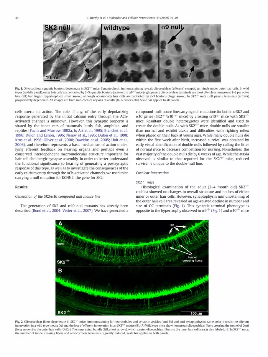
Fig. 1. Olivocochlear synaptic boutons degenerate in SK2−/− mice. Synaptophysin immunostaining reveals olivocochlear (efferent) synaptic terminals under outer hair cells. In wildtypes (middle panel), outer hair cells are contacted by 2–5 synaptic boutons (arrows). Inα9−/−mice (right panel), olivocochlear terminals aremost often less numerous (1–2 per outerhair cell) but larger (hypertrophied, small arrow), although occasionally hair cells are contacted by 2–3 boutons (large arrow). In SK2−/− mice (left panel), terminals (arrows)progressively degenerate. All images are from mid-cochlea regions of adults (8–12 weeks old). Scale bar applies to all panels.
40 V. Murthy et al. / Molecular and Cellular Neuroscience 40 (2009) 39–49
cells exerts its action. The role, if any, of the early depolarizingresponse generated by the initial calcium entry through the ACh-activated channel is unknown. However, this synaptic property isshared by the inner ears of mammals, birds, fish, amphibia, andreptiles (Fuchs and Murrow, 1992a, b; Art et al., 1995; Blanchet et al.,1996; Dulon and Lenoir, 1996; Nenov et al., 1996; Dulon et al., 1998;Kros et al., 1998; Oliver et al., 2000; Dawkins et al., 2005; Holt et al.,2006), and therefore represents a basic mechanism of action under-lying efferent feedback on hearing organs and perhaps even aconserved interdependent macromolecular structure important forhair cell cholinergic synapse assembly. In order to better understandthe functional significance to hearing of generating a postsynapticresponse of this type, as well as to investigate the consequences of theearly calcium entry through the ACh-activated channels, we usedmicecarrying a null mutation for KCNN2, the gene for SK2.
Results
Generation of the SK2/α10 compound null mouse line
The generation of SK2 and α10 null mutants has already beendescribed (Bond et al., 2004; Vetter et al., 2007). We have generated a
Fig. 2. Olivocochlear fibers degenerate in SK2−/− mice. Immunostaining for neurotubules aninnervation in a wild type mouse (A) and the loss of efferent innervation in an SK2−/− mouse(long arrows) to the outer hair cells (OHCs). The inner spiral bundle (ISB, short arrows), whichthe number of tunnel crossing fibers and olivocochlear terminals is greatly reduced. Scale b
compound-nullmouse line carrying nullmutations for both the SK2 andα10 genes (SK2−/−/α10 −/− mice) by crossing α10−/− mice with SK2+/−
mice. Resultant double heterozygotes were identified and used tocreate the double nulls. As with SK2−/− mice, double nulls are smallerthan normal and exhibit ataxia and difficulties with righting reflexwhen placed on their back at young ages. While many double nulls diewithin the first week after birth, increased survival was obtained byearly visual identification of double nulls followed by culling the litterof normal mice to decrease competition for nursing. Nonetheless, thevast majority of the double nulls die by 6 weeks of age.While the ataxiaobserved is similar to that reported for the SK2−/− mice, reducedsurvival is unique to the double-null line.
Cochlear innervation
SK2−/− miceHistological examination of the adult (2–4 month old) SK2−/−
cochlea showed no changes in overall structure and no loss of eitherinner or outer hair cells. However, synaptophysin immunostaining ofthe outer hair cell area revealed an age-related decline in number andsize of OC terminals (Fig. 1). This synaptic terminal phenotype isopposite to the hypertrophy observed in α9−/− (Fig. 1) and α10−/− mice
d synaptic vesicles (anti-TuJ and anti-synaptophysin; same color) reveals the efferent(B). (A) Wild type mice show numerous olivocochlear fibers crossing the tunnel of Corticarries olivocochlear fibers to the inner hair cell area, is also labeled. (B) In SK2−/− mice,ar applies to both panels.

Fig. 3. Olivocochlear degeneration is incomplete at 3weeks in both inner and outer hair cell areas. Synaptophysin immunostaining shows the reduction of olivocochlear terminals in SK2−/−
and SK2−/−/α10 −/− mice at 3 weeks of age. (A) In wild types, there are 2–4 OC terminals per OHC (large arrows), and the inner spiral bundle (ISB) is dense with terminal and en passantswellings surrounding the inner hair cells (position marked by asterisks). Additionally, numerous tunnel crossing fibers (small arrows) are observed. (B) In SK2−/− mice, olivocochlearterminals in theOHC region are fewer andmore varied in size (large arrows). The ISB ismore disorganized, and nodistinct cupping occurs around the inner hair cells. Fewer tunnel crossingfibers are present, and those that remain often times possess varicosities along their length (arrowheads). (C) The double-null mice resemble the SK2−/− mice, with scattered, smallerolivocochlear terminals in the OHC region (large arrows) and few tunnel crossing fibers. Scale bar in B is for A–B. All images are from the middle turn of the cochlea.
41V. Murthy et al. / Molecular and Cellular Neuroscience 40 (2009) 39–49
(Vetter et al., 1999, 2007). At P18, OC terminals in SK2−/− mice showedsmall changes in size and number, and tunnel crossing fibers werebeginning to show signs of degeneration. By 5–6 weeks, terminalnumber and sizewere clearly reduced (Fig. 2), and by 6months, few orno efferent boutons remained on OHCs of the basal turn. In the apicalturn, terminals sometimes persisted (data not shown). No synapticterminal degenerationwas observed in SK2+/− mice. In the inner spiralbundle (ISB), few synaptophysin labeled terminals were organizedbeneath the inner hair cell (Fig. 3B), although there was an apparentincrease in the number of fibers that left the main fiber tract andtraversed toward the IHCs (Fig. 5B). The main longitudinal body of theISB fiber tract, however, was not significantly different fromwild type.
Immunostaining for tubulin (TuJ1) reveals OC fibers as they crossthe tunnel of Corti (tunnel crossing fibers, Figs. 2, 3) to the outer haircell region. Tunnel-crossing fibers of SK2−/− mice at all ages examinedwere thin, with varicosities along their length (Fig. 3B, SupplementaryFig. 1). Such features were not observed in wild types and suggestongoing degeneration. Counts of tunnel-crossing OC fibers in the mid-basal turn revealed loss in SK2−/− mice beginning as early as P18-21, atime soon after the fibers initially cross the tunnel to encounter theirtargets (compare Figs. 3A, B; Fig. 4). Counts in wild type mice varied
from 65 to 76 fibers/500 μm, without significant age-related changefrom 2 to 10 weeks (Fig. 4) and out to 6 months (data not shown). InSK2−/−mice, fiber counts were normal at 2 weeks of age, but fell with aroughly exponential time course to about 17.5% of the wild types by10 weeks (Fig. 4). A two-way ANOVA (genotype×age) coupled with aBonferroni post-hoc test for multiple comparisons t-test revealedstatistically significant differences for all ages compared betweengenotype (3 weeks, pb0.05; 4–10 weeks, pb0.001).
TuJ1 immunostaining revealed abnormalities in the afferentinnervation to OHCs as well. The long, spiraling dendrites of thissystem normally form compact fascicles in the outer spiral bundles(OSBs) underneath the OHCs. However, in the SK2−/− mice the OSBswere more loosely organized, with some fibers completely defascicu-lated from the bundle (Fig. 5).
Synaptophysin and TuJ immunostaining also revealed loss of OCterminals in the inner hair cell area of SK2−/− mice (Fig. 2, Figs. 3A, B,Fig. 5). The inner spiral bundle (ISB) normally consists of a compactspiral track of fibers giving rise to nests of terminals surrounding thebase of IHCs (Fig. 3A, arrows, Fig. 5A). While the ISB was wider andmore diffuse in SK2−/− mice, the number of synaptophysin-positiveterminals was dramatically reduced (Figs. 3B, 5B).

Fig. 4. Quantification of olivocochlear degeneration over postnatal time. Countingolivocochlear tunnel-crossing fibers reveals a progressive degeneration in SK2−/− (opencircles) and SK2−/−/α10 −/− (open squares) compared to wild type (filled circle). SK2−/−
mice beginwith approximately the same number of fibers as wild typemice (2 weeks ofage), but steadily decline in numbers over 10 weeks. The double SK2−/−/α10−/− miceexhibit a similar degeneration profile, but the rate of degeneration is increased. Meansand SEMs are shown. Data are from the middle turn of the cochlea in 4–7 cochleae ofeach genotype at each age.
Fig. 5. Afferent fibers under outer hair cells and efferent fibers in the inner hair cellregion are de-fasciculated in SK2−/−mice. Immunostaining of afferent and efferent fiberswith TuJ1 reveals alterations in fiber fasciculation. (A) Afferent fibers under the outerhair cells, running as the outer spiral bundle (OSB) and efferent fibers in the inner spiralbundle (ISB), run as tight bundles in wild type mice. The inner spiral bundle (ISB) iscompact and outlines the region occupied by unstained inner hair cells (asteriskidentifies one such region). (B) In the SK2−/− mice, OSB fibers are de-fasciculated(arrowhead) from the main bundle, and run as individual fibers (arrows). The ISB islikewise disorganized, and IHC pockets are seldom observed.
42 V. Murthy et al. / Molecular and Cellular Neuroscience 40 (2009) 39–49
SK2−/−/ α10 −/− (double null) miceLoss of the ACh receptors involved in OC function does not result in
loss of OC terminals or fibers (Vetter et al., 1999, 2007). This is unlikethe case following loss of the functionally coupled SK2 potassiumchannel as described above. To better define the role of nAChRs andthe SK2 channel in OC synapse development and/or survival, wegenerated SK2−/−/α10 −/− mice. This compound null leaves theα9 geneunaltered, and thus potentially retains nAChR activity mediated viahomomericα9 receptors, as previously described (see Fig. 2D of Vetteret al., 2007).
The compound SK2−/−/α10 −/− mice exhibited the same OCdegeneration (Fig. 3B) observed in the SK2−/− mice (Fig. 3B), i.e.boutons in the OHC area were abnormally small by 3 weeks, andtunnel crossing fibers showed signs of degeneration as early as P18,with decreased fiber counts at all ages examined between 3 and10 weeks (Fig. 4). A two-way ANOVA of fiber counts in SK2−/−/α10 −/−
mice versus wild type mice revealed statistically significant effects ofgenotype and age (3 weeks, pb0.01, 4–10 weeks, pb0.001). Exponen-tial curve fitting suggested an accelerated loss in the SK2−/−/α10−/−
compared to the SK2−/− mice, with time constants of approximately3 weeks and 5 weeks, respectively. Similar to SK2−/− mice, thecompound nulls did not exhibit loss of hair cells.
Cochlear expression of NCAM in SK2−/− mice
To better understand the mechanisms behind the degeneration ofthe OC fibers, western blots were produced to assess the expressionlevel of Neural Cell Adhesion Molecule (NCAM), a cell adhesionmolecule normally expressed by cochlear hair cells and OC fibersduring the early postnatal synaptogenic phase of cochlear develop-ment. Pan-NCAM antibody recognized two well-described NCAMisoforms, migrating at 140 kDa and 120 kDa. A third isoform, normallydescribed as a 180 kDa band was not observed in these cochlearlysates. Transfer was normal in this region of the gel as indicated by
dye transfer of the molecular weight ladder, suggesting that thesemice may not express the 180 kDa isoform of NCAM, at least atappreciable levels, at these ages. Interestingly, NCAM protein expres-sion of SK2−/− mice at 6 weeks of age, a time at which activedegeneration of OC fibers is still underway, is approximately 50%higher than wild type (Fig. 6). The 15 week old SK2−/− mice, in whichOC fiber degeneration is essentially complete, demonstrated asignificant down regulation of NCAM expression (50% less than thesix week old wild type levels) that was indistinguishable from agematched wild type mice (Fig. 6).
In vivo cochlear function
Neonatal elimination of the OC bundle can lead to auditorythreshold elevation in the adult ear (Walsh et al., 1998). We thereforeinvestigated baseline cochlear sensitivity in SK2−/− mice by examiningauditory brainstem responses (ABRs), the summed activity of auditoryneurons evoked by short tone pips, and distortion product otoacousticemissions (DPOAEs), acoustic responses measured in the ear canalthat are created and amplified by normal OHC function. No differencesin threshold responses between adult (2 month old) SK2−/− and wildtype mice were detected at any of the frequencies tested, by eithermetric of cochlear function (Figs. 7A, B). Similar results werepreviously obtained in both α9−/− and α10 −/− mice (Vetter et al.,1999, 2007).
To evaluate the role of the SK2 gene in OC function in the adultcochlea, we measured effects of OC electrical activation on DPOAE

Fig. 6. Protein expression levels of the cell adhesion molecule NCAM is upregulated in SK2−/− mice. A: Western blot analysis reveals that at 6 weeks of age, a time at whichapproximately 40% of OC fibers remain and are still actively degenerating, NCAM expression is significantly upregulated by approximately 50% over the level of the wild type age-matched control (p=0.0171). At 15 weeks, a time at which degenerative processes are complete, NCAM expression appears slightly lower that wild type age-matched samples, but nostatistically significant difference was detected. Bar graph is relevant for the 140 kDa NCAM isoform (the major isoform expressed in the cochlear lysate).
43V. Murthy et al. / Molecular and Cellular Neuroscience 40 (2009) 39–49
amplitudes in SK2−/− ears using 8 week old mice. OC activationnormally decreases the OHC contribution to cochlear amplification,thereby eliciting a fast suppression of DPOAE amplitudes afterefferent activation onset (Fig. 8). The suppression is normally greatestfor mid-frequency tones, mirroring the peak of OHC efferent terminaldensity in the middle of the cochlear spiral (Maison et al., 2003). In8 week old SK2−/− mice, an age when roughly 35% of OC innervationremains, efferent-evoked suppression of DPOAEs was never observed(n=8 ears tested at 6 different DPOAE-evoking frequencies). In mostears, the fast suppression was replaced by a small, slow-onsetenhancement evoked by the shock train to the OC bundle, similar toprevious reports on the α9−/− and α10−/− mice or in wild type micetreated with strychnine, a potent blocker of the α9 nAChR (Maisonet al., 2007a). In the group-mean data, these small effects werestatistically significant at all frequencies (comparing pre-shockpoints to during shock points by t-test: pb0.001). The small size of
Fig. 7. Baseline cochlear sensitivity is unaffected in SK2−/−mice. Mean thresholds (±SEM) for a(B) are shown for 8 wildtype vs. SK2−/− ears at 8 weeks of age. No statistical differences are
the OC-evoked enhancements in the SK2−/− ears (compared to thatseen in α10−/− mice) is consistent with the significant loss of OCfibers and terminals at 8 weeks (Figs. 1–4). Given the potentialconfounding variable of fiber degeneration on the results, 26 day oldSK2−/− mice, which still possess approximately 65% of the tunnelcrossing fibers of wild types (Fig. 4) were examined. While thesemice also showed no DP suppression, they did exhibit a stronger OC-evoked enhancement in response to efferent fiber activation (Fig. 8,triangles), likely resulting from the greater number of surviving OCfibers. While it would be best to use mice with no degenerativechanges at all, mice younger than those used cannot reliably be usedfor this analysis. Expression of prestin (the molecular motorunderlying OHC motility) matures at approximately 3 weeks ofage (Abe et al., 2007), and distortion product generation continuesto mature through the fourth postnatal week (Henley et al., 1990),thus confounding any results obtained at earlier ages.
uditory brainstem responses (A) and distortion product otoacoustic emissions (DPOAEs)found between SK2−/− and wild type mice.

Fig. 8. Olivocochlear-mediated suppression of cochlear responses is absent in SK2−/− mice. (A) The normal suppression of cochlear responses seen during shock trains to theolivocochlear bundle is absent in SK2−/− ears, and is replaced by a small response enhancement. DPOAE amplitudes are normalized to the average pre-shock value. Test frequency was16 kHz. (B) Mean (±SEM) efferent effect in 7 wild type and 8 SK2−/− ears, defined as the average change in the DPOAE amplitude (in dB) for the first three measurements after shock-train onset.
Fig. 9. Expression of other genes is altered by loss of SK2. Quantitative RT-PCR oncochleas from wild type, SK2−/−, and SK2−/−/α10−/− ears showed that ryanodinereceptors were down regulated in both null lines. The α9 nAChR gene was downregulated, but only when both SK2 and α10 nAChR were deleted. Whereas BKα wasslightly up regulated following loss of the SK2 gene, BKβ was significantly downregulated. The beta subunit is known to confer calcium and voltage sensitivity to themain, alpha, subunit, and thus this change may have significant but until nowunrecognized physiological consequences.ΔΔCt analysis was carried out as described inExperimental methods. Expression levels were averaged among all replicates betweenqPCR runs, and plotted with standard deviations.
44 V. Murthy et al. / Molecular and Cellular Neuroscience 40 (2009) 39–49
Cochlear gene expression
qRT-PCR was performed on cochleae from adult wild type, SK2−/−,and SK2−/−/α10 −/− mice to examine expression levels of genesinvolved in synaptic transmission between OC terminals and theircochlear targets, as well as in general hair cell physiology. Equalamplification efficiency between all primers was verified (Supple-mental Data Fig. 2). All results were first normalized against myosinVIIa expression and plotted with respect to wild type expressionlevels.
The genes assayed include Chrna9 and Chrna10, encoding the α9and α10 subunits comprising the nicotinic ACh receptor (nAChR)expressed by OHCs at the OC synaptic complex (Elgoyhen et al.,1994, 2001; Vetter et al., 1999, 2007), and the ryanodine receptorgenes RyR1, RyR2, and RyR3, which encode proteins expressed inthe subsynaptic cistern, an intracellular organelle likely responsiblefor amplifying calcium signals via calcium-induced calcium release(Lioudyno et al., 2004). Delta-delta CT analysis (Livak and Schmitt-gen, 2001) indicated no change in the steady-state mRNA levels ofChrna9, and small changes in Chrna10 (up 33%), and RyR3 (down16%) mRNA in the SK2−/− mice (Fig. 9). However, RyR1 and RyR2,expression levels decreased in the SK2−/− mice at 7 weeks of age(74% and 53%, respectively, Fig. 8), coinciding with a time at whichroughly 70% of the tunnel crossing fibers had degenerated (Fig. 4).While only moderate changes in RyR2, and RyR3 transcripts weredetected in the SK2−/−/α10 −/− mice (down regulated 22% and 28%respectively), Chrna9 and RyR1 expression level were moredramatically decreased (44% and 60% respectively) in the doublenulls at 5–6 weeks of age (Fig. 9). Because most double-null micedied by 6–8 weeks of age, qRT-PCR was not carried out beyond thispoint.
Other ion channels also play a role in maturation of auditoryfunction, including the BK channel (Rüttiger et al., 2004) and the L-type voltage activated calcium channel CaV1.3 (Brandt et al., 2003;Michna et al., 2003; Nemzou et al., 2006). Therefore expression ofKcnma1 and Kcnmb1, encoding the BK α and β1 subunits respectively,and Cacnd1, encoding CaV1.3, was also examined in the SK2−/− mice toassess the maturational state of the auditory process (due to the low
survival of double nulls and therefore less sample RNA, these geneswere not investigated in the double null line). BKα was upregulatedslightly (32%), while BKβ1 involved in modulation of the BK channelgating properties and also its trafficking (Kim et al., 2007) was downregulated (37%) while CaV1.3 remained unchanged (Fig. 9).
Stabilization of the efferent postsynaptic complex
The postsynaptic complex apposing olivocochlear terminals mini-mally contains the nACh receptor subunits and the SK2 channel. These

Fig. 10. SK2 expression during early synaptic refinement periods and in the adult cochlea in mice lacking the nAChR α9 subunit. A: SK2 immunostaining of P7 wild type micedemonstrates no detectable expression in the OHC region. Inner and outer pillar cells (IPC, OPC) are immunopositive, but no distinct localization is apparent. B: Inα9−/−mice, distinctlocalization of SK2 along the lateral walls of OHCs can be observed at P7 as indicated by the line segments. Scale bar in A applies also to B. C: At higher magnification, the distinctlocalization of SK2 along the lateral walls of the OHC is more readily apparent. A few of the SK2 puncta are indicated by arrows. The nucleus of one OHC is indicated by an asterisk forpurposes of orientation. D: In the matureα9−/− cochlea, SK2 puncta (red, one of which is indicated by an arrow) are lost from the lateral walls, and have collapsed to take up a positionapposed to the incoming OC terminals, stained by synaptophysin antibodies (green). The lateral olivocochlear system (LOCS) is indicated in the inner hair cell region for orientationpurposes.
45V. Murthy et al. / Molecular and Cellular Neuroscience 40 (2009) 39–49
proteins must be associated with cytoskeletal elements to anchorthem at the synaptic pole. Given the combined observations of apersistent nAChR gene expression observed in the SK2−/− mice, alongwith our inability to elicit electrically evoked OC activity, these datastrongly suggest that an additional defect in nAChR targeting orassembly also exists in the SK2−/− mice. While attempts at producingnAChR α9 subunit antibodies that meet high standards of specificityhave not been successful, one may still assess to what extent the SK2channel participates in stabilization of the postsynaptic complex, andthereby imply mechanisms to explain the loss of nAChR function, byexamining the behavior of SK2 in the absence of one of the nAChRsubunits. Loss of SK2 protein from the synaptic pole in the absence ofnAChR subunit expression may indicate that SK2 and the nAChRs arelinked together via adapter proteins and then further linked to anunderlying cytomatrix. Thus, we examined the immunolocalization ofSK2 in OHCs of α9−/− mice.
Immunostaining for SK2 in the α9−/− mouse revealed precociousexpression of SK2 in the OHCs (compare Figs. 10A and B). Additionally,at these early postnatal stages of olivocochlear fiber invasion, SK2 wasnot localized to the base of the OHCs, but rather decorated the lateralwalls of the OHCs (Figs. 10B, C). However, with maturation, SK2expression ceased along the basolateral surfaces of the hair cells, andwas found only at the synaptic pole of the OHCs apposed to OCterminals (Fig. 10D).
Discussion
Expression of the SK2 channel and the nAChR α10 subunit ceasesin inner hair cells concomitant with the loss of ACh-inducible currents(Elgoyhen et al., 1994, 2001; Katz et al., 2004), a rise in BK channelexpression (Hafidi et al., 2005), and onset of adult-like hearing. Outerhair cells, however, express both nAChR subunits and the SK2 channelthroughout life, and functional coupling is maintained between them(Oliver et al., 2000; Katz et al., 2004). At a cellular level, the initialconsequence of nAChR activity in hair cells is depolarization (Fuchs
and Murrow, 1992a, b) due to calcium influx via the nAChRs, followedby rapid calcium-induced calcium release that activates SK2-mediatedhyperpolarization (Lioudyno et al., 2004). Normal function of the OCsynapse on OHCs decreases sound-evoked cochlear vibrations (Russelland Murugasu, 1997), and elevates cochlear thresholds by decreasingthe normal amplification of motion mediated by OHCs (Vetter et al.,1999). These OC effects are lost following genetic manipulations of thenAChR complex (Vetter et al., 1999, 2007) that eliminate ACh-inducedcalcium influx, andwhen release of calcium from intracellular stores isblocked by drugs acting on the ryanodine receptor (Russell andMurugasu, 1997; Lioudyno et al., 2004). Thus, the calcium-activatedSK2 channel occupies a central position in these important cochlearfunctions.
Our data strongly suggest that hair cell responses induced and/ormodulated by OC activation are necessary for the survival of OCinnervation and that these responses must involve SK2-mediatedhyperpolarization. Interestingly, our data examining the youngestSK2−/− mice also indicate that without SK2 expression, the nAChRdoes not attain functional status, at least at the “systems” leveldefined by our inability to elicit the classic OC-mediated decrease ofdistortion product amplitudes. While one may wish to examine theSK2−/− mice without any hint of OC fiber degeneration (for example,the one week old mice), the youngest age examined here (P26)represents among the earliest ages that can possibly be used for thisanalysis given the maturation time frame of prestin expression thatunderlies the mechanism of distortion product generation. Loss offunctional nAChR function in the SK2−/− mice results in a similar lackof in vivo effects of OC activation compared with the α9 and α10nulls, which also show replacement of the normally rapid suppres-sion of DP amplitude following OC fiber activation by a slow OC-evoked enhancement. Such enhancements mirror that elicitedduring strychnine block of α9/α10 function in wild type mice(Maison, et al., 2007a). Given that the enhancements decrease withage in the SK2−/− mice, hence correlated with a decrease in OCinnervation, the data suggest that whatever the mechanism of the OC

46 V. Murthy et al. / Molecular and Cellular Neuroscience 40 (2009) 39–49
enhancement, it does require an intact OC fiber innervation to occur.Electrophysiological measures of ACh-induced activity in SK2−/−
mice have also revealed a lack of response at the channel level(P. Fuchs, pers. comm.).
Given the dual observations of unchanged α9 gene transcriptlevels and yet loss of ACh-mediated function in the SK2−/− mice, onecould interpret the loss of physiological function of SK2−/− mice simplyas a defect in nAChR subunits assembling into the cholinergic receptormacromolecular complex. However, it is unclear what feedbackmechanism may be in place to alter nAChR assembly in the face ofloss of SK2 unless the entire nAChR/SK2 channel complex isassembled prior to targeting/insertion into the membrane, andadditionally that SK2 were to act in a dominant manner importantfor transport and/or targeting of the entire protein complex. BecauseSK2 binds directly to α-actinin 2 in heart muscle (Lu et al., 2007), andgiven that some neurotransmitter receptors are similarly bound andtargeted to the membrane by cytoskeletal elements (Cabello et al.,2007), it may be that SK2 represents a component around which theremainder of the postsynaptic SK2/nAChR multiprotein complexcoalesces via linker proteins into a functional core complex readyfor membrane insertion, but that is stabilized to the cytomatrix viacomponents unique to the SK2 channel. Such scenarios are notunprecedented. For example, numerous proteins involved in synapticvesicle dynamics make up a large portion of presynaptic active zonesand are assembled into Piccolo–Bassoon transport vesicles (PTVs)prior to membrane insertion (Shapira et al., 2003), demonstrating alack of apparent upper limits on the size of such macromolecularassemblies.
In support of such a scenario, our data demonstrate the release ofSK2 from the synaptic pole of OHCs in the absence of α9 nAChRsubunits during early postnatal stages of synaptic refinement in theinner ear. Ultimately SK2 is localized normally to the synaptic polein the absence of nAChR expression, yet, in the absence of SK2expression, no ACh-inducible activity occurs. Together, the findingsusing the SK2−/− and nAChR α9−/− mice suggest that one plausibleexplanation of the loss of ACh-mediated currents in the SK2−/− miceis that SK2 stabilizes and anchors the hair cell nAChRs, but that thenAChRs do not play an appreciable role in stabilizing and/oranchoring SK2 (since SK2 is still found apposed to mature OCterminals in the α9−/− mice despite early mis-localization at a timeprior to OC synaptic innervation). This would imply that the nAChR/SK2 postsynaptic complex is anchored via components native to theSK2 protein. Final resolution of whether this scenario is true awaitsgood nAChR antibodies, a general problem plaguing the field (Moseret al., 2007).
The postnatal degeneration of OC terminals in SK2−/− micecontrasts with the developmental hypertrophy and postnatalstability of OC terminals found in both α9 and α10 nulls (Vetteret al., 1999, 2007). While far from a complete mechanistic answer toreasons for the degeneration observed, our data indicate that thehair cells actually seem to be trying to hold onto the degeneratingOC terminals, insofar as one may interpret the upregulation of thecell adhesion molecule NCAM during the degenerative process.NCAM is normally involved in target recognition and stabilization ofthe OC fibers at their target as OC synapses are generated (Whitlonand Rutishauser, 1990; Whitlon et al., 1999). Thus, loss of OHCadhesion seems not to be a mechanism by which these fibers areshed from OHCs in the SK2−/− mice. Further analysis will be requiredto better understand the mechanism behind the fiber degenerationobserved in SK2−/− mice.
Finally, we addressed the question of whether the SK2 or α10 (ifeither) gene dominates OC synapse formation and survival at OHCs bygenerating and studying a compound-null mouse line lacking bothSK2 and α10 genes. Since the compound SK2−/− /α10 −/− mice showedsynaptic degeneration similar to that following ablation of SK2 alone,it can be surmised that minimally presence of the SK2 channel, if not
actually its activity, plays a dominant role in shaping, or even drivingthe initial development of OC fiber-hair cell contacts, and appears keyto the survival of these synapses. If our proposed scenario of SK2/nAChR subunit protein interactions is correct as described above, thisevent may not be surprising, as with the loss of SK2 would by defaultalso come the loss of membrane insertion/stabilization of nAChRs,automatically nullifying the possibility that the nAChR synapticphenotype can express itself. The normal efferent innervationobserved in SK2 heterozygotes and overexpressers (Maison et al.,2007b) suggests that a minimal SK2 response (the presumptive stateof the heterozygotes) is sufficient for maintenance of normal OCsynapses, and overexpression of the gene has no deleterious effectson synaptic structure. Therefore, SK2 channel activity is clearlyupstream of, and epistatic to, the role of nAChRs in synapsemaintenance, since the previously observed synaptic hypertrophyof the α10−/− mice is masked (or altered) by the phenotype expressedfollowing ablation of the SK2 gene. The fact that OC fiberdegeneration occurs only when the SK2 gene is ablated furthersuggests a recessive epistatic state.
Taken together, our gene and protein expression data, coupledwith the systems level functional data presented, indicate thatexpression of SK2 establishes (whether it be via activity or structure)an environment uponwhich nAChR subunit gene expression can thenexert a further effect (not necessarily limited to nAChR expressionalone) to facilitate OC synaptic maintenance programs. Such a statedemonstrates for the first time a distinct, two-step process in OCnicotinic synaptogenesis, and clearly demonstrates a previouslyunknown requirement for SK2 expression in nAChR expression andfunction. It is important to recognize, however, that this data do notnecessarily predict a direct protein:protein interaction between SK2and α9/10, but rather simply point to an interaction along a signalingcascade, an intersection of signaling networks, or, more likely, anindirect interaction via linker proteins during macromolecularassembly.
Control over the conductance of SK channels represents amechanism by which firing reliability and temporal precision can becontrolled (Hallworth et al., 2003), thereby modulating signalintegration. Such a control mechanism can have profound implica-tions for auditory system function, given the spontaneous activity ofhair cells during development, and the importance of temporalcharacteristics of auditory activity along the central neural pathways.By inducing SK2 activity, the efferent system modulates thesedischarge patterns, and may thereby also shape central auditorydevelopment. Expression of SK2 seems to ensure proper expressionand function of the nAChRs as well, further demonstrating the key roleplayed by SK2 in cochlear efferent function, and defining the OCpostsynaptic complex as an indivisible nAChR/SK2 macromolecularentity.
Experimental methods
Null mutant mice
Generation and initial characterization of both the SK2−/− andα10 −/− mice have been previously described (Bond et al., 2004);(Vetter et al., 2007). The SK2−/− strain background is C57Bl/6J, andthe α10−/− strain background is B6.CAST-Cdh23AHL+/Kjn (stock#2756). The B6.CAST line is a congenic line in which one of theprominent age-related hearing loss loci of the C57Bl/6J was replacedwith the Cast.Ei locus. Other than this manipulation, the strains areidentical. Compound-null mice carrying deletions for both SK2 andα10 genes (SK2−/−/α10 −/−) were generated by first breeding α10 −/−
to SK2+/−. Following genotyping to identify double heterozygotes,breeding was continued to generate double nulls by first generatinga population of mice that were SK2+/− on an α10 −/− background.Upon production of the SK2+/−/α10 −/− line, pairs from this line were

47V. Murthy et al. / Molecular and Cellular Neuroscience 40 (2009) 39–49
used to establish the SK2−/−/α10 −/− line. Controls were generatedfrom the appropriate crosses of SK2 and α10 colonies to furthergenerate the SK2+/+ /α10 +/+ and SK2−/−/ α10 +/− mice.
Immunostaining
Cochleae were exposed, perfused through the round and ovalwindows with 4% paraformaldehyde in 0.1 M sodium phosphatebuffer at room temperature, isolated, and post-fixed in the samefixative for 1 h. Following post-fixation, the cochleae were decalcified(8% EDTA in PBS) overnight at room temperature, or two to three daysat 4 °C on a rotator. Cochleae were subsequently stripped of the bonyouter capsule, and individual turns of the cochlea were hand cut forwhole-mount processing. Immunostaining of the efferent terminalswas performed using a mouse anti-synaptophysin antibody (Milli-pore, Billerica, MA, MAB5258). Immunostaining of efferent fiberscrossing the tunnel of Corti was performed with antibodies to SNAP25(Sigma, St. Louis, MO; cat. # S9684) and processed with biotinylatedsecondaries, ABC reagent (Vector Labs, Burlingame, CA), and diami-nobenzidine (DAB) for standard light microscopy, or TuJ-1 (class III β-tubulin, Neuromics, Northfield, MN; cat. # MO15013) and processedwith Oregon Green labeled secondary antibodies (Molecular Probes/InVitrogen) for confocal microscopy. Tissue for confocal observationwas mounted onto slides and coverslipped with SlowFade Gold(Molecular Probes/InVitrogen, Carlsbad, CA).
Tunnel-crossing fibers were counted in both DAB- or fluorescence-reacted portions of the mid-basal turn. Fibers were counted in threerandomly selected fields with a 40× objective and expressed as fibersper 500 μm. Statistical analysis was performed as a two way ANOVA(genotype×age) in Prism (version 4.0b).
Immunostaining for SK2 was similar to processing for wholemount cochlear sections through the decalcification step, after which,the cochleaswere cryoprotected in 30% sucrose/0.9% NaCl overnight at4 °C, and subsequently embedded in a peel away mold in OCT.Cryosectioning was performed on a Leica cryostat, and sections weredirectly mounted onto Superfrost Plus slides (Fisher), dried on a slidewarmer at 35 °C, and then stored until use in a slide box at −30 °C.Anti-SK2 antibody (Alomone Labs) was used at a final concentration of1:1000.
Quantitative RT-PCR
Primers for genes of interest were purchased from Qiagen(QuantiTect PCR primer sets, Qiagen, Inc., Valencia, CA). Cochleaewere harvested from adult (6–8 week old) mice composed of mixedsexes. During harvest, and prior to freezing, all cerebellum tissue wasremoved from the temporal bone. Cochleae were immediately frozenby immersion in liquid nitrogen and stored in a liquid nitrogen dewar.Sample processing was performed using 6 cochlea for each biologicalPCR replicate as follows: 1) cochleae were first weighed to assess thevolume of Trizol (In Vitrogen, Carlsbad, CA) needed in subsequentsteps; 2) transferred to the appropriate volume of Trizol andhomogenized using disposable plastic generators (cat# 1426126,Fisher Scientific, Waltham, MA) and a hand held polytron; and 3)RNA was prepared according to the manufacturers instructions, withtwo minor modifications. A first spin was performed at 2500 ×g tosediment debris, and the supernatant was transferred to a fresh tube.For precipitation of the RNA, 0.8 M sodium citrate, 1.2 M sodiumchloride was used in addition to the isopropanol step in themanufacturer's instructions. RNA concentration was assessed using aNanoDrop system, and integrity was assessed via analysis ofelectropherograms generated on an Agilent Technologies Bioanalyzerlabchip (RNA 6000, Nano Assay mode).
Quantitative reverse transcription-PCR (qRT-PCR) analysis wasperformed using a one step RT-PCR procedure employing theQuantiTect SYBR Green RT-PCR kit (Qiagen, Inc.), HotStarTaq DNA
polymerase (Qiagen, Inc.), and the QuantiTect primers of choicefollowing manufacturer's recommendations. Triplicate reactions (i.e.technical replicates) using pooled cochleae of 3 mice wereperformed at least twice (the second and any subsequent replica-tions with a new pool of 3 mice, i.e. biological replicates) for eachprimer set as separate reactions. Cycle-by-cycle and dissociation(melt curves) fluorescence data were collected as a SYBR Greenfluorescence assay. PCR reactions were performed on a StratageneMX4000 instrument (Stratagene, Inc., La Jolla, California). Analysis ofrelative fold change in gene expression was calculated via the ΔΔCT
method (Livak and Schmittgen, 2001) using myosin VIIa as anormalizing standard. Amplification efficiency of primers wasinitially monitored prior to performing the experiment to ensureequality between primer sets (see Supplemental Data Fig. 2).Because of limited sample, a post-hoc statistical power score wasalso generated to ensure sufficient power (large enough sample size)of the study for interpretation of results. A power score greater than80% was used as threshold for sufficient power, and if met, a greaternumber of samples was deemed unnecessary.
Western blotting for protein expression levels
Cochlear lysates were produced using the entire cochlear capsule.Cerebellar pieces, when present within the bounds of the semi-circular canals, were manually extracted prior to homogenization.Cochleae were pooled from at least three mice for each lysate. As eachcochlear capsule was dissected free of the temporal bone, it wasimmersed in 1 ml of ice cold Pierce T-PER Tissue Extraction reagentsupplemented with protease inhibitor with EDTA (Pierce), andmanually ground in a Kontes Duvall #21 ground glass homogenizer.The lysate was then transferred to an eppendorf tube and centrifugedat 14 K rpm for 20 min to sediment debris. The supernatant wasrecovered and used to determine protein concentration using a Piercemicro-BCA protein estimation kit. Lysate was then aliquoted andstored at −35 °C until use.
Triplicate western (immuno-) blots, minimally using two differentlysates per genotype pool (biological replicates), were carried outusing standard SDS-PAGE procedures to determine expression levels.Gels were wet transferred to PVDF membrane (Bio-Rad) using wettransfer techniques. Blots were incubated at RT for 1 h in 5% non-fatmilk, followed by an overnight incubation at 4 °C in primary antibody(mouse anti-NCAM, BD Bioscience (clone NCAM13), 1:1000). Washeswere performed using PBS-Tween20 for 30 min, followed byincubation in species appropriate peroxidase labeled secondary(1:2000). Bands were visualized using Pierce DuraWest ECL on aKodak ImageStation 440. Quantification of all bandswas performed bygenerating a densitometric ratio (Un-Scan-It gel, Silk Scientific) toGAPDH, used as a loading control. Six week old SK2+/+ values werethen set to 100%, and all values of other bands were normalized to thiscondition.
Cochlear physiology
Cochlear sensitivityAll physiological experiments were conducted in a temperature-
controlled, soundproof chamber maintained at ∼32 °C. Acousticstimuli were presented via a custom acoustic assembly consisting oftwo electrostatic drivers (Tucker Davis Technologies, EC-1) to generateacoustic stimuli and a Knowles miniature microphone (EK3103) torecord ear canal sound pressure. Stimuli were generated, andresponses measured, with locked digital I–O boards at 4-μs sampling(National Instruments, AO-6052E). Mice were anesthetized withxylazine (20 mg/kg i.p.) and ketamine (100 mg/kg i.p.). To measureauditory brainstem responses (ABRs), needle electrodes were insertedat vertex and pinna, with a ground near the tail. ABR potentials wereevoked with 5-ms tone pips delivered at 35/s. The response was

48 V. Murthy et al. / Molecular and Cellular Neuroscience 40 (2009) 39–49
amplified (10,000 X), filtered (100 Hz–3 kHz), and averaged with anA–D board in a LabVIEW-driven data-acquisition system. Sound levelwas raised in 5 dB steps from 10 dB below threshold up to 80 dB SPL.Upon visual inspection of stacked waveforms, ‘threshold’ was definedas the lowest SPL level at which any wave could be detected. Tomeasure distortion product otoacoustic emissions (DPOAEs), twoprimary tones f1 and f2 (with f2/f1=1.2 and f2 level 10 dB b f1 level)were presented continuously to the ear canal. Ear canal soundpressure was amplified and digitally sampled at 20 μs (NationalInstruments, A-2000). FFTs were computed and averaged over 5consecutive waveform traces, and 2f1–f2 DPOAE amplitude andsurrounding noise floor were extracted, a procedure requiring ∼4 sof data acquisition and processing time.
Olivocochlear assayAdult (2 months old) and juvenile (P26) mice were anesthetized
with urethane (1.20 g/kg i.p.) and xylazine (20 mg/kg i.p.). A posteriorcraniotomy and partial cerebellar aspiration were performed toexpose the floor of the IVth ventricle. To stimulate the OC bundle,shocks (monophasic pulses of 150 μs duration presented at 200/s)were applied through fine silver wires (0.4 mm spacing) placed alongthe midline, spanning the OC decussation. Shock threshold for facialtwitches was determined, muscle paralysis induced with α-d-tubocurarine (1.25 mg/kg i.p.), and the animal connected to arespirator via a tracheal cannula. Shock levels were raised to 6 dBabove twitch threshold. During the OC suppression assay, f2 level wastypically set to produce a DPOAE ∼10–15 dB Nnoise floor. To measureOC effects, repeatedmeasures of baseline DPOAE amplitudewere firstobtained (n=12), followed by a series of 17 continuous periods inwhich DPOAE amplitudes were measured with simultaneous shocksto the OC bundle. In some experiments, DPOAE measures and CAPmeasures were interleaved before during and after the OC shocktrain.
Acknowledgments
This work was supported by: NIH R01 DC006258 (DEV), R01DC0188 (MCL), P30 DC 05209 (MCL), and The Tufts Center forNeuroscience Research Imaging Core and Genomics Core P30NS047243.
Appendix A. Supplementary data
Supplementary data associated with this article can be found, inthe online version, at doi:10.1016/j.mcn.2008.08.011.
References
Abe, T., Kakehata, S., Kitani, R., Maruya, S., Navaratnam, D., Santos-Sacchi, J., Shinkawa,H., 2007. Developmental expression of the outer hair cell motor prestin in themouse. J. Membr. Biol. 215, 49–56.
Art, J., Wu, Y., Fettiplace, R., 1995. The calcium-activated potassium channels of turtlehair cells. J. Gen. Physiol. 105, 49–72.
Blanchet, C., Eróstegui, C., Sugasawa, M., Dulon, D., 1996. Acetylcholine-inducedpotassium current of guinea pig outer hair cells: its dependence on a calciuminflux through nicotinic-like receptors. J. Neurosci. 16, 2574–2584.
Bond, C., Herson, P., Strassmaier, T., Hammond, R., Stackman, R., Maylie, J., Adelman, J.,2004. Small conductance Ca2+-activated K+ channel knock-out mice reveal theidentity of calcium-dependent afterhyperpolarization currents. J. Neurosci. 24,5301–5306.
Brandt, A., Striessnig, J., Moser, T., 2003. CaV1.3 channels are essential fordevelopment and presynaptic activity of cochlear inner hair cells. J. Neurosci.23, 10832–10840.
Cabello, N., Remelli, R., Canela, L., Soriguera, A., Mallol, J., Canela, E.I., Robbins, M.J., Lluis,C., Franco, R., McIlhinney, R.A., Ciruela, F., 2007. Actin-binding protein alpha-actinin-1 interacts with the metabotropic glutamate receptor type 5b andmodulates the cell surface expression and function of the receptor. J. Biol. Chem.282, 12143–12153.
Dawkins, R., Keller, S., Sewell, W., 2005. Pharmacology of acetylcholine-mediated cellsignaling in the lateral line organ following efferent stimulation. J. Neurophysiol. 93,2541–2551.
Dulon, D., Lenoir, M.,1996. Cholinergic responses in developing outer hair cells of the ratcochlea. Eur. J. Neurosci. 8, 1945–1952.
Dulon, D., Luo, L., Zhang, C., Ryan, A., 1998. Expression of small-conductance calcium-activated potassium channels (SK) in outer hair cells of the rat cochlea. Eur. J.Neurosci. 10, 907–915.
Elgoyhen, A.B., Johnson, D.S., Boulter, J., Vetter, D.E., Heinemann, S., 1994. Alpha 9: anacetylcholine receptor with novel pharmacological properties expressed in ratcochlear hair cells. Cell 79, 705–715.
Elgoyhen, A., Vetter, D., Katz, E., Rothlin, C., Heinemann, S., Boulter, J., 2001. alpha10: adeterminant of nicotinic cholinergic receptor function in mammalian vestibularand cochlear mechanosensory hair cells. Proc. Natl. Acad. Sci. U. S. A. 98, 3501–3506.
Fuchs, P., Murrow, B., 1992a. A novel cholinergic receptor mediates inhibition of chickcochlear hair cells. Proc. Biol. Sci. 248, 35–40.
Fuchs, P., Murrow, B., 1992b. Cholinergic inhibition of short (outer) hair cells of thechick's cochlea. J. Neurosci. 12, 800–809.
Hafidi, A., Beurg, M., Dulon, D., 2005. Localization and developmental expression of BKchannels in mammalian cochlear hair cells. Neuroscience 130, 475–484.
Hallworth, N., Wilson, C., Bevan, M., 2003. Apamin-sensitive small conductancecalcium-activated potassium channels, through their selective coupling tovoltage-gated calcium channels, are critical determinants of the precision, pace,and pattern of action potential generation in rat subthalamic nucleus neurons invitro. J. Neurosci. 23, 7525–7542.
Henley III, C.M., Owings, M.H., Stagner, B.B., Martin, G.K., Lonsbury-Martin, B.L., 1990.Postnatal development of 2f1–f2 otoacoustic emissions in pigmented rat. Hear. Res.43, 141–148.
Holt, J., Lysakowski, A., Goldberg, J., 2006. Mechanisms of efferent-mediated responsesin the turtle posterior crista. J. Neurosci. 26, 13180–13193.
Katz, E., Elgoyhen, A., Gómez-Casati, M., Knipper, M., Vetter, D., Fuchs, P., Glowatzki, E.,2004. Developmental regulation of nicotinic synapses on cochlear inner hair cells.J. Neurosci. 24, 7814–7820.
Kim, E.Y., Zou, S., Ridgway, L.D., Dryer, S.E., 2007. Beta1-subunits increase surfaceexpression of a large-conductance Ca2+-activated K+ channel isoform. J. Neuro-physiol. 97, 3508–3516.
Köhler, M., Hirschberg, B., Bond, C., Kinzie, J., Marrion, N., Maylie, J., Adelman, J., 1996.Small-conductance, calcium-activated potassium channels frommammalian brain.Science 273, 1709–1714.
Kros, C., Ruppersberg, J., Rüsch, A., 1998. Expression of a potassium current in inner haircells during development of hearing in mice. Nature 394, 281–284.
Lin, M.T., Lujan, R., Watanabe, M., Adelman, J.P., Maylie, J., 2008. SK2 channel plasticitycontributes to LTP at Schaffer collateral-CA1 synapses. Nat. Neurosci. 11, 170–177.
Lioudyno, M., Hiel, H., Kong, J., Katz, E., Waldman, E., Parameshwaran-Iyer, S., Glowatzki,E., Fuchs, P., 2004. A “synaptoplasmic cistern”mediates rapid inhibition of cochlearhair cells. J. Neurosci. 24, 11160–11164.
Livak, K., Schmittgen, T., 2001. Analysis of relative gene expression data using real-timequantitative PCR and the 2(-Delta Delta C(T)) method. Methods 25, 402–408.
Lu, L., Zhang, Q., Timofeyev, V., Zhang, Z., Young, J.N., Shin, H.S., Knowlton, A.A.,Chiamvimonvat, N., 2007. Molecular coupling of a Ca2+-activated K+ channel to L-type Ca2+ channels via alpha-actinin2. Circ. Res. 100, 112–120.
Maison, S., Adams, J., Liberman, M., 2003. Olivocochlear innervation in the mouse:immunocytochemical maps, crossed versus uncrossed contributions, and trans-mitter colocalization. J. Comp. Neurol. 455, 406–416.
Maison, S., Vetter, D., Liberman, M., 2007a. A novel effect of cochlear efferents: in vivoresponse enhancement does not require alpha9 cholinergic receptors. J. Neuro-physiol. 97, 3269–3278.
Maison, S., Parker, L., Young, L., Adelman, J., Zuo, J., Liberman, M., 2007b. Overexpressionof SK2 channels enhances efferent suppression of cochlear responses withoutenhancing noise resistance. J. Neurophysiol. 97, 2930–2936.
Michna, M., Knirsch, M., Hoda, J., Muenkner, S., Langer, P., Platzer, J., Striessnig, J.,Engel, J., 2003. Cav1.3 (alpha1D) Ca2+ currents in neonatal outer hair cells of mice.J. Physiol. 553, 747–758.
Moser, N., Mechawar, N., Jones, I., Gochberg-Sarver, A., Orr-Urtreger, A., Plomann,M., Salas,R., Molles, B., Marubio, L., Roth, U., Maskos, U., Winzer-Serhan, U., Bourgeois, J.P., LeSourd, A.M., De Biasi, M., Schroder, H., Lindstrom, J., Maelicke, A., Changeux, J.P.,Wevers, A., 2007. Evaluating the suitability of nicotinic acetylcholine receptorantibodies for standard immunodetection procedures. J. Neurochem. 102, 479–492.
Nemzou, N.R.M., Bulankina, A., Khimich, D., Giese, A., Moser, T., 2006. Synapticorganization in cochlear inner hair cells deficient for the CaV1.3 (alpha1D) subunitof L-type Ca2+ channels. Neuroscience 141, 1849–1860.
Nenov, A., Norris, C., Bobbin, R., 1996. Acetylcholine response in guinea pig outer haircells. II. Activation of a small conductance Ca(2+)-activated K+ channel. Hear Res.101, 149–172.
Oliver, D., Klöcker, N., Schuck, J., Baukrowitz, T., Ruppersberg, J., Fakler, B., 2000. Gatingof Ca2+-activated K+ channels controls fast inhibitory synaptic transmission atauditory outer hair cells. Neuron 26, 595–601.
Rasmussen, G.L., 1942. An efferent cochlear bundle. Anat. Rec. 82, 441.Russell, I.J., Murugasu, E., 1997. Medial efferent inhibition suppresses basilar membrane
responses to near characteristic frequency tones of moderate to high intensities.J. Acoust. Soc. Am. 102, 1734–1738.
Rüttiger, L., Sausbier, M., Zimmermann, U., Winter, H., Braig, C., Engel, J., Knirsch, M.,Arntz, C., Langer, P., Hirt, B., Müller, M., Köpschall, I., Pfister, M., Münkner, S.,Rohbock, K., Pfaff, I., Rüsch, A., Ruth, P., Knipper, M., 2004. Deletion of the Ca2+-activated potassium (BK) alpha-subunit but not the BKbeta1-subunit leads toprogressive hearing loss. Proc. Natl. Acad. Sci. U. S. A. 101, 12922–12927.
Shapira, M., Zhai, R.G., Dresbach, T., Bresler, T., Torres, V.I., Gundelfinger, E.D., Ziv, N.E.,Garner, C.C., 2003. Unitary assembly of presynaptic active zones from Piccolo–Bassoon transport vesicles. Neuron 38, 237–252.

49V. Murthy et al. / Molecular and Cellular Neuroscience 40 (2009) 39–49
Vetter, D.E., Liberman, M.C., Mann, J., Barhanin, J., Boulter, J., Brown, M.C., Saffiote-Kolman,J., Heinemann, S.F., Elgoyhen, A.B.,1999. Role of alpha9 nicotinic ACh receptor subunitsin the development and function of cochlear efferent innervation. Neuron 23, 93–103.
Vetter, D.E., Katz, E., Maison, S.F., Taranda, J., Turcan, S., Ballestero, J., Liberman, M.C.,Elgoyhen, A.B., Boulter, J., 2007. The α10 nicotinic acetylcholine receptor subunit isrequired for normal synaptic function and integrity of the olivocochlear system.Proc. Natl. Acad. Sci. U. S. A. 104, 20594–20599.
Walsh, E., McGee, J., McFadden, S., Liberman, M., 1998. Long-term effects of sectioningthe olivocochlear bundle in neonatal cats. J. Neurosci. 18, 3859–3869.
Whitlon, D.S., Rutishauser, U.S., 1990. NCAM in the organ of Corti of the developingmouse. J. Neurocytol. 19, 970–977.
Whitlon, D., Zhang, X., Pecelunas, K., Greiner, M., 1999. A temporospatial map ofadhesive molecules in the organ of Corti of the mouse cochlea. J. Neurocytol. 28,955–968.


