Embed Size (px)
Citation preview

Plastic and Reconstructive Surgery Advance Online Article DOI: 10.1097/PRS.0b013e318230c7d1
Shock Wave Therapy in Wound Healing
Ali A. Qureshi1, Kimberly M. Ross2, Rei Ogawa, M.D.,Ph.D. 3, Dennis P. Orgill,
M.D., Ph.D. 4
1Harvard Medical School, Boston, MA
2Division of Plastic Surgery, Brigham and Women’s Hospital, Boston, MA
3Department of Plastic, Reconstructive and Aesthetic Surgery, Nippon Medical
School, Tokyo, Japan
4Division of Plastic Surgery, Brigham and Women’s Hospital, Harvard Medical
School, Boston, MA
Copyright @ American Society of Plastic Surgeons. All rights reserved.
ACCEPTED
Corresponding Author:
Dennis P. Orgill, M.D., Ph.D.
Division of Plastic and Reconstructive Surgery
Brigham and Women's Hospital
75 Francis Street. Boston, MA 02115
Tel: (617) 732-5456
Fax: (617) 732-7387
E-mail: [email protected]
Financial Disclosure: Dr. Orgill has served as an advisor for Sanuwave Corporation. Dr. Orgill has been a principal investigator on research grants to the Brigham and Women's Hospital by Kinetic Concepts Incorporated (KCI), manufacturers of the VAC device. He has also appeared as an expert witness for litigation proceedings for KCI. The remaining authors have no financial interest in any of the products, devices, or drugs mentioned in this manuscript. Currently, none of the authors have any financial interest in any of the products, devices, or drugs mentioned in the article."
Copyright @ American Society of Plastic Surgeons. All rights reserved.
ACCEPTED
Abstract
Background
Recently, shock wave therapy (SWT) has been investigated as an adjuvant therapy in
the treatment of acute and chronic wounds. There are several devices with focused and
unfocused shock waves that have been administered to a heterogenous group of
wounds. Encouraging preclinical and clinical studies suggest that SWT may promote
wound healing with little or no adverse events prompting investigations into the
mechanism of action as well as additional clinical trials.
Methods
The peer-reviewed literature within the last 10 years was studied using an evidence-
based approach.
Results
Preclinical studies demonstrate that SWT affects cellular function and leads to the
expression of several genes and elaboration of growth factors known to promote wound
healing. Limited clinical trials are encouraging for the use of SWT in the treatment of
acute and chronic wounds. Serious complications including wound infections, bleeding,
hematomas, seromas or petechiae have not been reported in the largest of these
studies.
Conclusions
SWT is an intriguing physical modality that may play an important role as an adjuvant
therapy in wound healing. To date, there is no consensus on which wounds are most
likely to benefit from SWT and what the optimal power, degree of focus, and frequency
or number of cycles should be. Well-designed pre-clinical and clinical studies are
necessary to better understand SWT in wound healing.
Introduction
Extracorporeal shock wave therapy has revolutionized the treatment of urolithiasis
Copyright @ American Society of Plastic Surgeons. All rights reserved.
ACCEPTED

allowing fragmentation of stones at a distance, avoiding invasive surgery in most cases.
Variants of this technology have been used to treat fractures1,2,3,4, osteonecrosis of the
femoral head5, plantar fasciitis 6,7 and critical myocardial and limb ischemia8. Most
recently shock wave therapy (SWT) has been used in the treatment of acute and chronic
wounds, burns and skin flaps.
Shock waves are biphasic high-energy acoustic waves that can be generated by
electrohydraulics. A high voltage spark is discharged under water, causing vaporization
and the release of acoustic waves with high peak pressures that rapidly decline over 10
µs9,10. As the shock wave propagates over distance, energy is absorbed by the tissue.
The degree of focus can be modulated by parabolic reflectors, resulting in a variable
concentration energy at a desired location (Figure 1). Shock waves are defined by their
waveform, the number of impulses, the frequency of impulses and energy flux density
(mJ/mm2).
The mechanisms of biological changes that result from shock waves are not entirely
clear. One hypothesis is that shock waves act as transient micromechanical forces that
induce perturbations at the cell structural level, thereby altering biologic activity.
Mechanotransduction results from geometrical changes in the cellular cytoskeleton,
which is analogous to design concepts of tensegrity articulated by architect Buckminster
Fuller and sculptor Kenneth Snelson11 and applied to biological systems by Ingber.
Briefly, external deformations can be transduced to an already “prestressed” or internally
balanced cytoskeleton through tensile linkages or cell surface receptors that would
initiate a cascade of intracellular events leading to changes in cell activity12. Such an
explanation for SWT would parallel our existing understanding of soft-tissue expanders
in reconstructive surgery, distraction osteogenesis, and most recently, wound healing
Copyright @ American Society of Plastic Surgeons. All rights reserved.
ACCEPTED

with the vacuum-assisted closure(VAC) device, in which micromechanical forces
promotes wound healing through increased cell division, angiogenesis and release of
growth factors in the wound bed13.
Pre-clinical experience using SWT in suggests a potentially important role in promoting
healing in diabetic wounds, flap necrosis and burns. There have been clinical studies
with low levels of evidence based on the criteria of the Center for Evidence Based
Medicine14. Among these studies, only a few have been prospective, randomized,
controlled studies that fail to meet several key CONSORT criteria15. The limited clinical
evidence and lack of rigorous study design have made it difficult for clinicians and
regulators to fully support SWT in wound healing at this time. Several questions,
including optimal SWT parameters, timing of treatments and types of wounds most
suited for SWT remain unanswered and warrant further clinical studies.
Methods
Literature search
We searched Medline, the Cochrane Database of Systematic Reviews and Cochrane
Controlled Trials Register. The search strategy we used included MESH terms
“WOUNDS AND INJURIES/THERAPY”, “WOUNDS AND INJURIES/PATHOLOGY”,
“SOFT TISSUE INJURIES/PATHOLOGY”, “SOFT TISSUE INJURIES/THERAPY”,
“UTRASONIC THERAPY/METHODS” and “HIGH-ENERGY SHOCK
WAVES/THERAPEUTIC USE” along with text words. No other limits were applied to any
of the searches. Additionally, reference lists of full-text papers obtained through these
searches were searched.
Selection
We included SWT preclinical studies in animals, in-vitro studies and clinical studies
Copyright @ American Society of Plastic Surgeons. All rights reserved.
ACCEPTED
including prospective and retrospective trials that included wound healing, flap necrosis,
and burns. Because of the scarcity of SWT clinical trials, we did not exclude non-
randomized or poorly controlled trials. Outcomes of interest included: improved wound
healing, flap necrosis, and reepithelization of burns.
Data abstraction
Review of randomized controlled trials was carried out based on the recommendations
of the PRISMA statement16.
Data analysis
The Centre for Evidence Based Medicine Levels of Evidence14 were applied to the
clinical studies reviewed. Additionally, the CONSORT checklist of information was
applied to those that were randomized controlled studies15.
Results
Wound Healing
Two preclinical studies have looked at SWT in diabetic wounds. Kuo et al17 administered
unfocused SWT (800 impulses at 0.09 mJ/mm2) to STZ-induced diabetic rats with a
dorsal skin defect. SWT significantly reduced wound size in diabetic rats with greater
reductions seen with more treatment (Table I). There was increased blood perfusion,
decreased pro-inflammatory activity, and increased VEGF, eNOS and PCNA
expression17.
A more recent study18 in db+/db+ mice with a full thickness dorsal skin defect found that
unfocused SWT (200 impulses at 0.1 mJ/mm2) led to a prolonged and elevated
expression of gene subsets. SWT had no effect on wound closure in diabetic or control
mice. Multiple treatments with unfocused SWT further delayed wound healing after
Copyright @ American Society of Plastic Surgeons. All rights reserved.
ACCEPTED
initially increasing the size of the wound18.
Schaden et al19 found a 75% treatment response (as defined by 100% wound
epithelization) in a Level 2b study of 208 patients with heterogeneous wounds treated
with debridement and unfocused SWT (100 to 1000 impulses at 0.1 mJ/mm2) with a
mean of 3 treatments. One third of wounds were acute and nearly 40% of wounds had
either partial or complete failure to heal after primary surgical closure, an important
confounder unaccounted for in the analysis. Excluding venous stasis and arterial
insufficiency ulcers, wound etiology did not affect treatment success, but statistical
analyses to justify this conclusion were not performed. Analyses based on wound size
and duration revealed small wounds (<10cm2) of short duration (<1 month old) were
most likely to rapidly completely re-epithelialize.
Saggini et al 20 led a Level 3b study with 30 consecutive patients treated with focused
SWT (100 impulses at 0.037 mJ/mm2) every 2 weeks (range of 4-10 sessions) until
complete healing was achieved. Unlike others, this study used focused SWT and a lower
energy flux density. A 50% complete healing response (parameters not defined) with no
adverse events was reported. This conclusion was obtained by grouping a
heterogeneous patient population and their individual responses: posttraumatic ulcers
(69% complete healing), venous ulcers (36% complete healing) and diabetic ulcers (25%
complete healing). No subset analysis based on wound etiology was done. In the
remaining ulcers without complete healing, increased wound bed blood supply was
observed (data not provided). A significant decrease in pain based on the pain self-
assessment numeric box scale in treated patients was also reported.
Wang et al21 found complete healing (parameters not defined) in 31% of patients in a
Copyright @ American Society of Plastic Surgeons. All rights reserved.
ACCEPTED
Level 2b study with 72 patients with chronic diabetic foot ulcers treated with focused
SWT (300 + 100 impulses/cm2 at 0.11 mJ/cm2) every 2 weeks for 6 weeks. Increased
perfusion, cell concentration and activity were noted. Notably, the control arm received
hyperbaric oxygen therapy (HBO) instead of standard therapy. The wounds studied were
relatively large (SWT: 11.2 +/- 20 cm2, HBO: 10.5 +/- 20 cm2, mean size +/- SD). SWT
was found to be superior to HBO. Rationale for treatment parameters and details of the
clinical assessment are lacking. Because the study did not have long term follow up, the
natural history of diabetic ulcers treated with SWT remains unknown.
Morretti et al22 conducted a Level 2b study of 30 diabetic patients with neuropathic foot
ulcers treated with debridement followed by unfocused SWT (100 pulses of 0.03
mJ/mm2) for 3 sessions every 72 hours and wound care. The control arm was treated
with debridement, pressure relief and treatment of infection. The wounds studied were
small (SWT 300 +/- 130 mm2, control 250 +/- 100 mm2, mean size +/- SD). SWT
parameters were based on the authors’ clinical experience with SWT in orthopedics. In
20 weeks, the treatment arm had a healing rate of 53% versus 33% in the control.
Though randomized, the random allocation sequence, its mechanism and
implementation were not explained. The study excluded chronic diabetic ulcers greater
than 5 cm to avoid selection bias.
Dumfarth et al23 carried out a Level 2b study with 100 patients undergoing vein
harvesting for coronary artery bypass graft (CABG) surgery, half of whom received
unfocused SWT (25 impulses at 0.1 mJ/mm2) at the wound closure site of the vein graft.
Treated patients had lower ASEPSIS scores (serous discharge, erythema, purulent
exudates, separation of the deep tissue, isolation of bacteria, and duration of inpatient
stay) on postoperative days 3 and 7 with no reported complications from treatment,
Copyright @ American Society of Plastic Surgeons. All rights reserved.
ACCEPTED

suggesting better wound healing. Treated patients had a statistically significant lower
use of antibiotics for leg wounds. However, the study was not powered for its primary
outcome. The long term effects of SWT in these surgical wounds were not assessed.
Recently, Ottomann et al24 conducted a Level 1b study with 28 patients with acute
traumatic wounds and burns requiring skin grafting treated with unfocused SWT (100
impulses at 0.1 mJ/mm2) to the skin graft donor site immediately after skin harvest. A
significantly decreased time for reepithelization of skin graft donor sites in the SWT arm
(13.9 +/- 2.0 days) versus control (16.7 +/- 2.0) was reported. The study was powered to
detect a difference in time to epithelization with adequate randomization and blinding.
However, the sample size was too small to study other outcomes including pain and the
cosmesis of donor sites and did not have long-term follow up.
Flap Necrosis
Several preclinical studies examined the role of SWT in preventing necrosis of skin flaps
in animal models, after the orthopedic and trauma literature suggested SWT could
induce neovascularization and increase VEGF expression among other proangiogenic
genes25,26 (Table 2). Meirer et al27 applied SWT (2500 impulses at 0.15 mJ/mm2) to the
random portion of an epigastric skin flap model immediately after surgery. There was
significantly less necrotic surface area in SWT treated rats (2.2 +/- 1.9%) at one-week
follow-up versus control (17.4 +/- 4.4%)27. In a later study, SWT was hypothesized to
decrease flap necrosis through reciprocal increase in VEGF expression in adjacent skin
but the detected difference in expression failed to reach statistical significance at a p-
value of 0.0528.
The same group compared SWT to gene therapy with VEGF and found SWT treated
Copyright @ American Society of Plastic Surgeons. All rights reserved.
ACCEPTED
rats to have significantly smaller necrotic zones of the flap29, in a study where surgical
procedures were performed by three different plastic surgeons and analyses were not
blinded. SWT was found to be superior to gene therapy with transforming growth factor-
b in a study of SWT (750 impulses at 0.15 mJ/mm2) administered immediately after
raising an epigastric skin flap in rats30. However, there was no significant difference in
flap vascularization assessed by CD31 staining between SWT and gene therapy rats.
Rationale for SWT parameters was also lacking.
Yan et al31 administered SWT (750 impulses at 0.09 mJ/mm2) to the mid and distal
portions of a cranially based random pattern flap model in rats and found increased
blood perfusion and expression of nitric oxide and VEGF. SWT parameters were based
on pilot studies, though it is unclear whether focused or unfocused SWT was used.
There was increased vasodilation of pre-existing vessels in the early post-operative
period with neovascularization apparent on post-operative days 3 and 1031. This study
suggested that SWT administered immediately postoperatively starts a series of discrete
events that could explain when certain changes in the flap are seen.
Two studies have looked at the immunologic changes induced by focused SWT in flap
necrosis models. Kuo et al32 applied focused SWT (500 impulses at 0.15 mJ/mm2) to five
areas of a rat dorsal random flap model. Increased VEGF and PCNA expression,
reduced leukocyte infiltration and decreased TNF-alpha expression in flap tissue
ischemic zones were found, suggesting that SWT may dampen the inflammatory
response in ischemic tissue32. Kuo et al33 repeated the same experiment and found
decreased leukocyte infiltration and tissue apoptosis, increased recruitment of skin
fibroblasts, down-regulation of oxygen radical burst, and increased eNOS expression33.
Copyright @ American Society of Plastic Surgeons. All rights reserved.
ACCEPTED
One study compared preoperative SWT to no treatment in an epigastric skin flap model
and noted a significant reduction in necrotic flap area34. However, a head-to-head
comparison of preoperative versus postoperative SWT to determine optimal timing of
SWT has not been done. Kamelger et al35 assessed a dose-dependent effect of SWT in
a murine epigastric skin flap model by varying impulses (200, 500, 1500, 2500, 5000 and
0) at 0.11 mJ/mm2. Optimum enhancement of skin flap survival was at 500 with no
significant increase at 1500 and 2500 impulses and increased necrosis observed at
5000 impulses. Changes in expression of growth factors or neovascularization with
different impulses were not assessed.
No clinical studies of SWT for the prevention of flap necrosis have been conducted.
Burns
The application of SWT was examined in a murine model with full thickness cutaneous
burns36 (Table 3). Gene expression studies showed more than fivefold increased in
chemokine and proinflammatory cytokine genes 4 hours postburn that were not seen in
SWT-treated wounds. Davis et al36 found that administration of unfocused SWT (200
impulses at 0.1 mJ/mm2) one hour postburn led to a significant reduction in neutrophil
infiltration at the wound margin and central wound bed at 4 and 24 hours postburn. No
significant difference in macroscopic wound closure contraction, degree of subeschar
keratinocyte migration, rate of wound reepithelization and granulation development were
found. The study was neither randomized nor powered for its primary outcomes.
Meirer et al37 described a Level 4 case report of a patient with deep partial thickness
burns of the forearm who refused skin grafting for cosmetic reasons and instead
received SWT (1500 impulses at 0.11 mJ/mm2) on days 3 and 7 post-burn. The patient
Copyright @ American Society of Plastic Surgeons. All rights reserved.
ACCEPTED
had near complete reepithelization on day 15 and a well-healed wound without scarring
at 6 months follow-up. Recently, Arno et al38 conducted a Level 4 case-series study of
15 patients with <5% TBSA deep partial/full thickness skin burns who received
unfocused SWT (500 impulses at mJ/mm2) on days 3 and 5 post-burn. Patients
underwent debridement and STSG in the absence of burn reepithelization 2.5 weeks or
more after SWT therapy. 80% of the patients healed before 3 weeks; 15% of patients
required surgical debridement and STSG and 5% developed hypertrophic scarring.
Increase in perfusion based on laser Doppler imaging (LDI) images was also observed.
Discussion
The advent of SWT provides a potential new therapeutic modality for acute and chronic
wounds which likely acts through mechanotransduction and immunomodulatory
mechanisms. SWT promotes expression of macromolecules in wound healing, including
VEGF, eNOS and PCNA. Because of the large experience using this technology to treat
urolithiasis and other conditions in humans, it appears to be a safe technology. The
clinical efficacy of this technology in specific wound types as well as the precise
mechanisms of action is now beginning to be understood.
SWT may be perceived by cell surface receptors through extracellular matrix and fluid
effects. Mechanoreceptors including integrins, ion channels, connexins and/or the lipid
component of the plasma membrane activation could all possibly be affected by SWT.
Akt-mediated mechanotransduction in fibroblasts has been show to play a role in
hypertrophic scar formation in response to mechanical forces, suggesting that Akt and
other upstream components like Focal Adhesion Kinase (FAK) would be important
candidates to study in the future for SWT39. Future studies may further elucidate the
mechanotranduction effects of SWT. Shock waves may also stimulate sensory nerve
Copyright @ American Society of Plastic Surgeons. All rights reserved.
ACCEPTED
fibers including nociceptors that produce the somatic sensation of mechanical force,
which may explain why some patients treated with SWT report decreased pain.
Clinical studies of SWT in wound healing suggest that wound etiology, size, and
chronicity may impact response to SWT. However, the actual administration of SWT in
current clinical studies varies in type (unfocused versus focused), total number of
impulses, energy flux density, and frequency. While the physics of SWT and preclinical
studies suggest that unfocused SWT is superior for the treatment of superficial soft
tissue defects, there has been no direct comparison of unfocused and focused SWT in
clinical trials to date. Therefore, whether there is a clinically relevant difference in
unfocused versus focused SWT remains unknown. Many authors who studied SWT in
other clinical settings used the same devices in their studies of wound healing. To our
knowledge, there have been no preclinical or clinical studies that have published data to
suggest that there are experimental limitations that did not permit use of either type of
SWT. Similarly, we do not have a complete understanding of the optimal SWT settings.
Additional basic science studies along with RCTs and registry studies powered to detect
clinically relevant outcomes will be necessary to increase our understanding of this
technology. Specifically, better characterization of the effects of SWT in homogenous
groups of wounds would lead to identification of subsets of patients that are ideal
candidates for SWT. In order to achieve this, thoughtful investigations to determine the
type and specific parameters of SWT suited for different wounds must be determined
first.
Currently, the FDA has approved devices that administer SWT for the treatment of
plantar fasciitis and lateral epicondylitis but has not approved its use to treat acute and
Copyright @ American Society of Plastic Surgeons. All rights reserved.
ACCEPTED
chronic wounds. SWT shows promise in improving our ability to enhance wound healing
through mechanotransduction or immunomodulatory mechanisms. We look forward to
future innovation in this field to understand more fully the mechanisms of action as well
as optimal treatment of specific wound types.
Acknowledgements: We would like to thank Britlyn Orgill for her assistance with the
figure.
References
1. Schaden W, Fischer A, Sailler A. Extracorporeal shock wave therapy of nonunion
or delayed osseous union: Clinical Orthop and Related Res. 2001;387:90-94.
2. Wang CJ, Liu HC, Fu TH. The effects of extracorporeal shockwave on acute
high-energy long bone fractures of the lower extremity: Arch Orthop Trauma
Surg. 2007;127:137-42.
3. Wang CJ, Chen HS, Chen CE, Yang KD. Treatment of nonunions of long bone
fractures with shock waves: Clinical Orthopaedics and Related Research.
2001;387:95-101.
4. Elster EA, Stojadinovic A, Forsberg J, Shawen S, Andersen R, Schaden W.
Extracorporeal shock wave therapy for nonunion of the tibia: J Orthop Trauma.
2010;24:133-141.
5. Wang CJ, Wang FS, Huang CC, Yang KD, Weng LH, Huang HY. Treatment for
osteonecrosis of the femoral head: Comparison of extracorporeal shock waves
with core decompression and bone-grafting: J Bone Joint Surg Am.
2005;87:2380-2387.
6. Ogden JA, Alvarez R, Levitt R, Cross GL, Marlow M. Shock wave therapy for
Copyright @ American Society of Plastic Surgeons. All rights reserved.
ACCEPTED
chronic proximal plantar fasciitis: Clinical Orthop and Related Res. 2001;387:47-
59.
7. Wang CJ, Chen HS, Huang TW. Shockwave therapy for patients with plantar
fasciitis: a one year follow up study: Foot & Ankle International. 2002;23:204-07.
8. Ito K, Fukumoto Y, Shimokawa H. Extracorporeal shock wave therapy as a new
and non-invasive angiogenic strategy: Tohoku J Exp Med. 2009;219:1-9.
9. Haupt, G, Haupt A, Ekkernkamp A, Gerety B, Chvapil M. Influence of shock
waves on fracture healing: Urology. 1992;38:529-532.
10. Wang CJ. An overview of shock wave therapy in musculoskeletal disorders:
Chang Gung Med J. 2003;26:220-232.
11. Fuller B. Tensegrity: Portfolio Artnews Annu. 1961;4:112-127.
12. Ingber D. Tensegrity-based mechanosensing from macro to micro: Prog Biophys
Mol Biol. 2008;97:163-179.
13. Saxena V, Hwang CW, Huang S, Eichbaum Q, Ingber D, Orgill DP. Vacuum-
assisted closure: microdeformations of wounds and cell proliferation: Plast
Reconstr Surg. 2004;114:1086-96; discussion 1097-98.
14. Centre for Evidence Based Medicine, University of Oxford. March 2009 Levels of
Evidence. Available at: www.cebm.net. Accessed December 5, 2010.
15. CONSORT (Consilidated Standards of Reporting Trials). 2010 CONSORT
Checklist. Available at: www.consort-statement.org. Accessed December 5,
2010.
16. Moher D, Liberati A, Tetzlaff J, Altman DG, and the PRISMA Group. Preferred
reporting items for systematic reviews and meta-anaylses: the PRISMA
statement: Ann Inter Med. 2009;151:264-70.
17. Kuo RK, Wang CT, Wang FS, Yang KD, Chiang YC, Wang CJ. Extracorporeal
shock-wave therapy enhanced wound healing via increasing topical blood
Copyright @ American Society of Plastic Surgeons. All rights reserved.
ACCEPTED

perfusion and tissue regeneration in a rat model of STZ-induced diabetes:
Wound Rep & Regen. 2009;17:522-30.
18. Zins SR, Amare MF, Tadaki DK, Elster EA, Davis TA. Comparative analysis of
angiogenic gene expression in normal and impaired wound healing in diabetic
mice: effects of extracorporeal shock wave therapy: Angiogenesis. 2010;13:293-
304.
19. Schaden W, Thiele R, Kolp C, Pusch M, Nissan A, Attinger C, Maniscalco-
Theberge M, Peoples G, Elster EA, Stojadinovic A. Shock wave therapy for acute
and chronic soft tissue wounds: a feasibility study: J Surg Res. 2007;143:1-12.
20. Saggini R, Figus A, Troccola A, Cocco V, Saggini A, Scuderi N. Extracorporeal
shock wave therapy for management of chronic ulcers in the lower extremities:
Ultrasound Med & Biol. 2008;34:1261-71.
21. Wang CJ, Kuo YR, Wu RW, Liu RT, Hsu CS, Wang FS, Yang KD. Extracorporeal
shockwave treatment for chronic diabetic foot ulcers: J Surg Res. 2009;152:96-
103.
22. Moretti B, Notarnicola A, Maggio G, Moretti L, Pascone M, Tafuri S, Patella V.
The management of neuropathic ulcers of the foot in diabetes by shock wave
therapy: BMC Musculoskelet Disord. 2009;10:54.
23. Dumfarth J, Zimpfer D, Voggele-Kadletz M, Holfeld J, Sihorsch F, Schaden W,
Czerny M, Aharinejad S, Wlner E, Grimm M. Prophylactic low-energy shock
wave therapy improves wound healing after vein harvesting for coronary artery
bypass surgery: a prospective, randomized trial: J Thorac Surg. 2008;86:1909-
13.
24. Ottomann C, Hartmann B, Tyler J, Maier H, Thiele R, Schaden W, Stojadinovic
A. Prospective randomized trial of accelerated re-epithelization of skin graft
donor sites using extracorporeal shock wave therapy: J Am Coll Surg.
Copyright @ American Society of Plastic Surgeons. All rights reserved.
ACCEPTED
2010;211:361-67.
25. Wang CJ, Wang FS, Yang KD, Weng LH, Hsu CC, Huang CS, Yang LC. Shock
wave therapy induced neovascularization at the tendon-bone junction: J Orthop
Res. 2003;21:984-89.
26. Krokowicz L, Cwykiel J, Klimczak A, Mielniczuk M, Siemionow M. Pulsed
acoustic cellular treatment induces expression of proangiogenic factors and
chemokines in muscle flaps: J Trauma. 2010;69:1448-56.
27. Meirer R, Kamelger FS, Huemer GM, Wanner S, Piza-Katzer H. Extracorporal
shock wave may enhance skin flap survival in an animal model: British Assoc of
Plast Surg. 2005;58:53-57.
28. Meirer R, Brunner A, Deibl M, Oehlbauer M, Piza-Katzer H, Kamelger FS. Shock
wave therapy reduces necrotic flap zones and induces VEGF expression in
animal epigastric skin flap model: J Reconstr Microsurg. 2007;23:231-6.
29. Meirer R, Huemer GM, Oehlbauer M, Wanner S, Piza-Katzer H, Kamelger FS.
Comparison of the effectiveness of gene therapy with vascular endothelial growth
factor or shock wave therapy to reduce ischaemic necrosis in an epigastric skin
flap model in rats: J Plast & Reconstr & Aesth Surg. 2007;60:266-71.
30. Huemer G, Meirer R, Gurunluoglu R, Kamelger FS, Dunst KM, Wanner S, Piza-
Katzer H. Comparison of the effectiveness of gene therapy with transforming
growth factor-b or extracorporal shock wave therapy to reduce ischemic necrosis
in an epigastric skin flap model in rats: Wound Repair and Regen. 2005;13:262-
68.
31. Yan X, Zeng B, Chai Y, Luo C, Li X. Improvement of blood flow, expression of
nitric oxide, and vascular endothelial growth factor by low-energy shockwave
therapy in random-pattern skin flap model: Ann Surg. 2008;61:646-53.
32. Kuo YR, Wu WS, Hsieh YL, Wang FS, Wang CT, Chiang YC, Wang CJ.
Copyright @ American Society of Plastic Surgeons. All rights reserved.
ACCEPTED
Extracorporeal shock wave enhanced extended skin flap tissue survival via
increase of topical blood perfusion and associated with suppression of tissue pro-
inflammation: J Surg Res. 2007;143:385-92.
33. Kuo YR, Wang CT, Wang FS, Yang KD, Chiang YC, Wang CJ. Extracorporeal
shock wave treatment modulates skin fibroblast recruitment and leukocyte
infiltration for enhancing extended skin-flap survival: Wound Repair and Regen.
2009;17:80-87.
34. Reichenberger M, Germann G, Roth HJ, Meirer R, Engel H. Preoperative shock
wave therapy redues ischemic necrosis in an epigastric skin flap model: Ann
Surg. 2009;63:682-84.
35. Kamelger F, Oehlbauer M, Piza-Katzer H, Meirer R. Extracorporeal shock wave
treatment in ischemic tissues: what is the appropriate number of shock wave
impulses?: J Reconst Microsurg. 2010;26:117-21.
36. Davis T, Stojadinovic A, Anam K, Amare M, Naik S, Peoples GE, Tadaki D,
Elster EA. Extracorporeal shock wave therapy suppresses the early
proinflammatory immune response to a severe cutaneous burn injury: Int Wound
J. 2009;6:11-21.
37. Meirer R, Kamelger FS, Piza-Katzer H. Shock wave therapy: an innovative
treatment method for partial thickness burns: Burns. 2005;31:921-2.
38. Arno A, Garcia O, Sancho H, Barret A. Extracorporeal shock waves, a new non-
surgical method to treat severe burns: Burns. 2010; 36:844-9.
39. Paterno, J, Vial IN, Wong VW, Rustad KC, Sorkin M, Shi Y, Bhatt KA,
Thangarajah H, Glotzback JP, Gurtner GC. Akt-mediated mechanotransduction
in murine fibroblasts during hypertrophic scar formation: Wound Rep Regen.
2011;19:49-58.
Copyright @ American Society of Plastic Surgeons. All rights reserved.
ACCEPTED

Figure Legends
Figure 1. Schematic diagram of SWT for wounds. A shock wave is produced by a
sparkplug in a conductive device and can be focused with a parabolic reflector and
conductive gel. The waveform shows peak pressures of 100 MPa after around 10 μsec,
followed by a brief period of subatmospheric pressure. The wave is attenuated as it
traverses the tissue
Copyright @ American Society of Plastic Surgeons. All rights reserved.
ACCEPTED
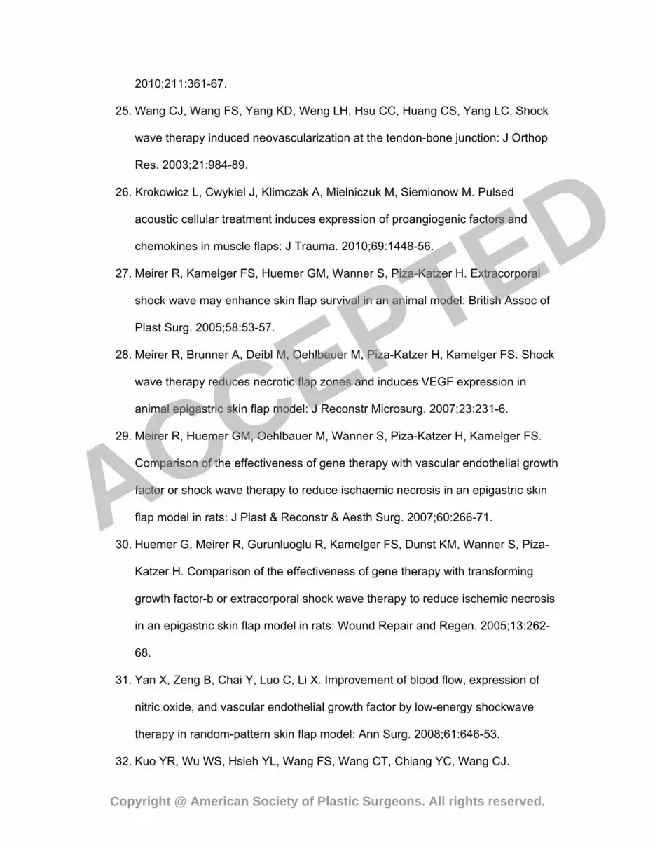
Table 1. Pre-clinical (*) and clinical studies using SWT in wounds
Wound Healing
Author Year Published
Number of Subjects
Size of Injury Number of Pulses
Density of Energy (mJ/mm2)
Focused or Unfocused
Kuo et al. [17]
2009 30 ESW, 20 control*
6 x 5cm 800 0.09 unfocused
Zins et al. [18]
2010 15 ESW, 15 control*
Circular 19 mm diameter (280 mm2)
200 0.1 unfocused
Schaden et al. [19]
2007 208 differing per patient
100 0.1 unfocused
Saggini et al. [20]
2008 30 ESW, 10 control
differing per patient
100 0.037 focused
Wang et al. [21]
2009 40 ESW, 42 HBO
11.2 +/- 20 cm2 500 0.11 focused
Moretti et al. [22]
2009 15 ESW, 15 Control
300 +/- 130 mm2
100 0.03 unfocused
Dumfarth et al. [23]
2008 50 ESW, 50 control
differing per patient
25 0.1 unfocused
Ottoman et al. [24]
2010 28 differing per patient
100 0.1 unfocused
*Pre-clinical
Copyright @ American Society of Plastic Surgeons. All rights reserved.
ACCEPTED

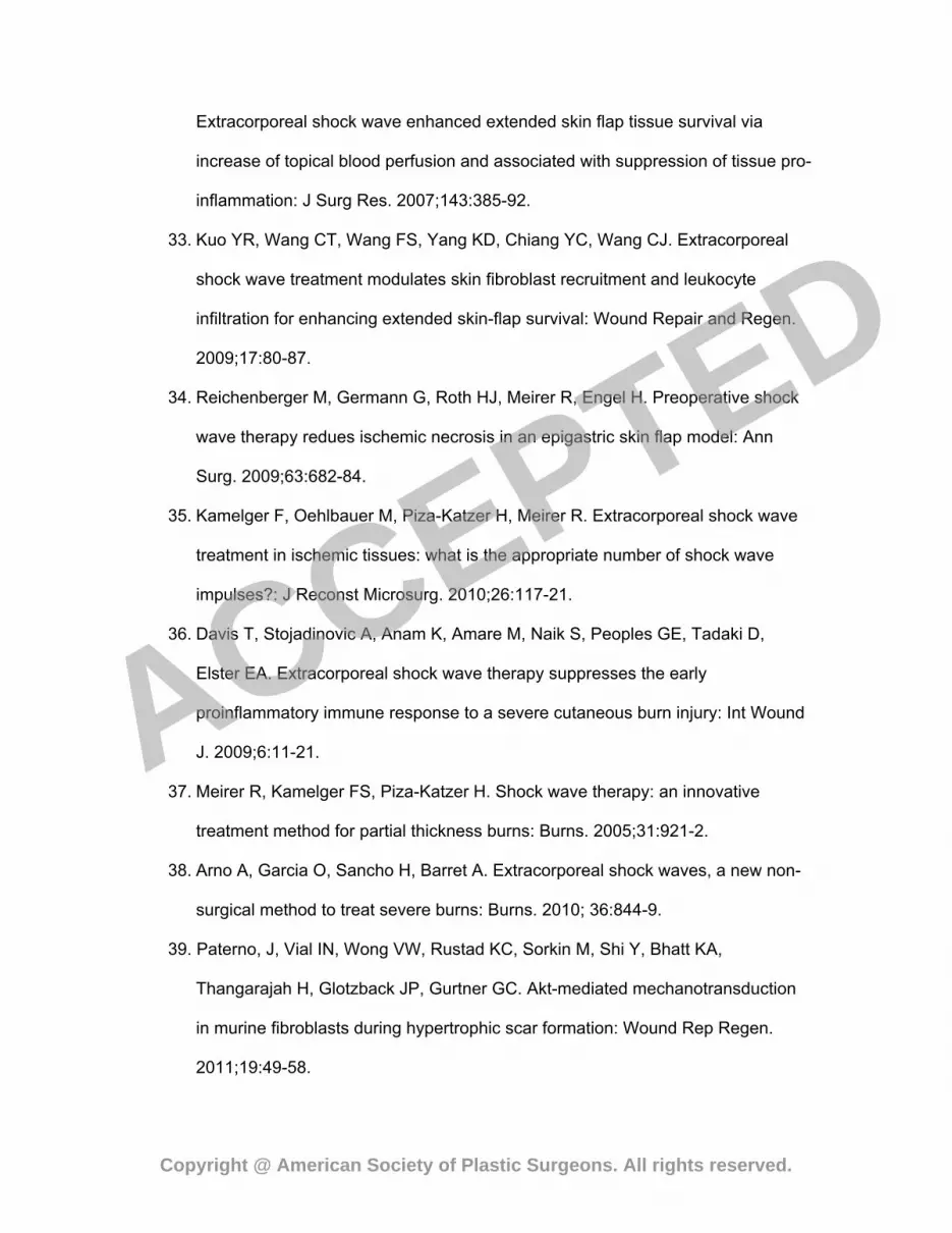
Table 2. Pre-clinical (*) and clinical studies using SWT in flaps.
Flap Necrosis
Author Year
Published
Number
of
subjects
Size
of
Injury
Number
or
Pulses
Density of
Energy
(mJ/mm2)
Focused or
Unfocused
Meirer et al.
27
2005 10
ESW,
10
control
8 x
8cm
2500 0.15 Focused
Meirer et al.
[28
2007 20
ESW,
20
control
8 x
8cm
500 0.11 Focused
Meirer et al.
29
2007 10
ESW,
10
control
8 x
8cm
500 0.11 Focused
Huemer et al.
30
2005 10
ESW,
10
control,
10
TGF-β
8 x
8cm
750 0.15 Focused
Yan et al. 31 2008 42
study,
3 x
10cm
750 0.09 Focused
Copyright @ American Society of Plastic Surgeons. All rights reserved.
ACCEPTED
42
control
Kuo et al. 32 2007 36 10 x
3cm
500 0.15 focused
Kuo et al. 33 2009 36 10 x
3cm
500 0.15 focused
Reichenberger
et al. 34
2009 10
ESW,
10
control
6 x
10cm
500 0.11 Focused
Kamelger et
al. [35
2010 36 8 x
8cm
200,
500,
1500,
2500,
5000
and 0
0.11 Focused
Copyright @ American Society of Plastic Surgeons. All rights reserved.
ACCEPTED
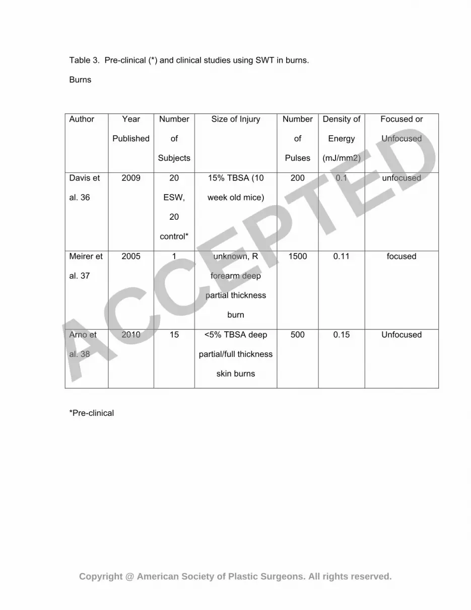
Table 3. Pre-clinical (*) and clinical studies using SWT in burns.
Burns
Author Year
Published
Number
of
Subjects
Size of Injury Number
of
Pulses
Density of
Energy
(mJ/mm2)
Focused or
Unfocused
Davis et
al. 36
2009 20
ESW,
20
control*
15% TBSA (10
week old mice)
200 0.1 unfocused
Meirer et
al. 37
2005 1 unknown, R
forearm deep
partial thickness
burn
1500 0.11 focused
Arno et
al. 38
2010 15 <5% TBSA deep
partial/full thickness
skin burns
500 0.15 Unfocused
*Pre-clinical
Copyright @ American Society of Plastic Surgeons. All rights reserved.
ACCEPTED

Figure 1
Copyright @ American Society of Plastic Surgeons. All rights reserved.
ACCEPTED