Embed Size (px)
Citation preview
Pharmacognosy Research [Phcog Res.]C
on
ten
ts..
....
March - April 2010 | Volume 2 | Issue 2
Original articless
Wound healing properties and kill kinetics of Clerodendron splendens G. Don, a Ghanaian wound healing plantStephen Y. Gbedema, Kisseih Emelia, Adu Francis, Annan Kofi, Woode Eric .................. 63
Crystal and molecular structure of lancerodiol–p–hydroxybenzoateMohamed H. Abd El–Razek, Mohamed–Elamir F. Hegazy, Abou El–Hamd H. Mohamed .................. 69
Chemical constituents of Tephrosia purpureaAli K. Khalafalah, Afifi H. Yousef, Abeer M. Esmail, Mohamed H. Abdelrazik, Mohamed E. F. Hegazy, Abou-El-Hamd H. Mohamed .................. 72
Analgesic activity, toxicity study and phytochemical screening of standardized Cinnomomum iners leaves methanolic extractF. Mustaffa, J. Indurkar, S. Ismail, M. N. Mordi, S. Ramanathan, S. M. Mansor .................. 76
Colon targeted curcumin delivery using guar gumA. K. Singhal, N. Nalwaya, E. Edwin Jarald, Showkat Ahmed .................. 82
Diuretic activity of leaves of Plectranthus amboinicus (Lour) Spreng in male albino ratsRoshan Patel, Naveen K. Mahobia, Ravindra Gendle, Basant Kaushik, Sudarshan K. Singh .................. 86
Antiarthritic and antioxidant effects of the leaf extract of Ficus exasperata P. Beauv. (Moraceae)Wonder M. K. Abotsi, Eric Woode, George K. Ainooson, Ama K. Amo-Barimah, Eric Boakye-Gyasi .................. 89
Standardization of Ajmodadi churna, a polyherbal formulationNeeraj K. Sriwastava, C. S. Shreedhara, H. N. Aswatha Ram .................. 98
Bronchodilator activity of aqueous extract of stem bark of Ailanthus excelsa Roxb.Dinesh Kumar, S. S. Bhujbal, R. S. Deoda, S. C. Mudgade .................. 102
Antimicrobial properties, antioxidant activity and bioactive compounds from six wild edible mushrooms of western ghats of Karnataka, IndiaCh. Ramesh, Manohar G. Pattar .................. 107
Antiproliferative activity and induction of apoptosis in estrogen receptor-positive and negative human breast carcinoma cell lines by Gmelina asiatica rootsMadhu Katyayani Balijepalli, Satyanarayana Tandra, Mallikarjuna Rao Pichika .................. 113
AUTHOR INSTITUTION MAP FOR THIS ISSUE
Please note that not all the institutions may get mapped due to non-availability of requisite information in Google Map. For AIM of other issues, please check Archives/Back Issues page on the journal’s website.
Pharmacognosy Research | March 2010 | Vol 2 | Issue 2 63
Address for correspondence:Gbedema Y. Stephen, Department of Pharmaceutics, Faculty of Pharmacy and Pharmaceutical Sciences, Kwame Nkrumah University of Science and Technology, Kumasi, Ghana, West Africa. E-mail: [email protected]
DOI: 10.4103/0974-8490.62948
Wound healing properties and kill kinetics of Clerodendron splendens G. Don, a Ghanaian wound healing plantStephen Y. Gbedema, Kisseih Emelia, Adu Francis, Annan Kofi1, Woode Eric2
Departments of Pharmaceutics, 1Pharmacognosy and 2Pharmacology, Faculty of Pharmacy and Pharmaceutical Sciences, Kwame Nkrumah University of Science and Technology, Kumasi, Ghana, West Africa
Submitted: 17-02-2010 Revised: 22-03-2010 Published: 04-05-2010
O R I G I N A L A R T I C L E
INTRODUCTION
More than 80% of the world’s population depends upon traditional medicines for the management of various diseases including skin infections.[1] Recently, the traditional use of plants for wound healing has received attention by the scientific community.[2] Wound healing is a complex process characterized by homeostasis, re-epithelization, granulation tissue formation and remodeling of the extracellular matrix.[3] Although the healing process progresses naturally, an infection can seriously delay this healing process by prolonging the inflammatory phase, disrupting the normal clotting mechanisms, promoting disordered leukocyte function and ultimately delaying
angiogenesis.[4] Therefore, a variety of activities such as tensile strength and collagen estimation of healing tissue, antioxidant and antimicrobial effects can explain the traditional use of a plant for helping the wounds to heal.[2]
Clerodendron splendens G. Don (family: Verbenaceae) is a climbing shrub, mostly found growing on cultivated lands between food crops in Ghana and other West African countries. Carbohydrates, glycosides, unsaturated sterols, triterpenoids and flavonoids are reported to be present in the leaves,[5] in addition to volatile oil in the flowers.[6] The plant is used ethnomedicinally in Ghana for the treatment of vaginal thrush, bruises, wounds and various skin infections.[7] In this study, we provide scientific evidence for the use of this plant in the traditional treatment of some infectious conditions and in wound healing.
MATERIALS AND METHODS
Collection of plant materialThe plant was collected from Kumasi in the Ashanti
As part of our general objective of investigating indigenous plants used in wound healing in Ghana, we hereby report our findings from some in vitro and in vivo studies related to wound healing activities of Clerodendron splendens G. Don (Verbanaceae). Methanolic extract of the aerial parts of the plant was tested for antimicrobial activity against Gram positive bacteria (Bacillus subtilis, Staphylococcus aureus, Streptococcus faecalis, Micrococcus flavus, as well as resistant strains of Staph. aureus SA1199B, RN4220 and XU212), Gram negative bacteria (Escherichia coli, Pseudomonas aeruginosa, Proteous mirabilis, Klebsiella pneumoniae) and Candida albicans using the micro-well dilution method. Survivor–time studies of the microorganisms, radical scavenging activity using 2,2′-diphenylpicrylhydrazyl (DPPH) and various in vivo wound healing activity studies were also conducted on the extract. The extract exhibited biostatic action against all the test microorganisms with a Minimum Inhibition Concentration (MIC) ranging between 64 and 512 µg/ml and a free radical scavenging property with an IC50 value of 103.2 µg/ml. The results of the in vivo wound healing tests showed that upon application of C. splendens ointment, there was a reduction in the epithelization period from 26.7 days (control) to 13.6 days along with a marked decrease in the scar area from 54.2 mm2 (control) to 25.2 mm2. Significant increase in the tensile strength and hydroxyproline content were also observed as compared to the control and was comparable to nitrofurazone. The above results appear to justify the traditional use of C. splendens in wound healing and treatment of skin infections in Ghana.
Key words: Clerodendron splendens, antimicrobial, antioxidant, biostatic, wound healing
A B S T R A C T
P H C O G R E S .
64 Pharmacognosy Research | March 2010 | Vol 2 | Issue 2
Region of Ghana in July 2007 and was authenticated at the Department of Pharmacognosy, Kwame Nkrumah University of Science and Technology, Ghana. A voucher specimen (FP/07/0032) has been kept in the herbarium of the Department.
Extraction and phytochemical screeningThe plant material was air-dried at room temperature for 7 days and milled into coarse powder using a Laboratory Mill Machine (Type 8, Christy & Norris, UK). Two hundred grams of the powder was cold-macerated with one litre methanol (Sigma-Aldrich, USA) over 48 h. The extract was concentrated under reduced pressure using the rotary evaporator and dried over nitrogen to give a yield of 12.3 g.
The methanolic extract was phytochemically screened for carbohydrates, tannins, saponin, anthraquinones, cardiac and cyanogenic glycosides, flavonoids and alkaloids using the procedures outlined by Wall et al[8] and Harbon.[9]
Test organismsThe bacteria used for the tests were obtained from the National Collection of Type Cultures (NCTC), UK. The Gram positive bacteria used were Bacillus subtilis (NCTC 10073), Staphylococcus aureus (NCTC 4163) Streptococcus faecalis (NCTC 775), Micrococcus flavus (NCTC 7743), as well as resistant strains of S. aureus SA1199B, RN4220 and XU212. The Gram negative bacteria used were Escherichia coli (NCTC 9002), Pseudomonas aeruginosa (NCTC 10662), Proteous mirabilis (NCTC 10975) and Klebsiella pneumoniae (NCTC 418). Candida albicans (NCPF 3179) was also used.
Minimum inhibitory concentration determinationInocula of the microorganisms were prepared from a 24 h Mueller-Hinton Broth (Sigma-Aldrich, St. Louis, MO, USA) cultures and suspensions were adjusted to 105
colony forming units per ml (cfu/ml). Minimum inhibition concentration (MIC) values of the extract were determined based on a micro-well dilution method.[10] The 96-well sterile plates were prepared by dispensing 180 µl of the inoculated broth plus a 20-µl aliquot of the plant extract made up in broth or 20 µl broth in the case of negative control in each well. Neomycin and tetracycline (Sigma-Aldrich, USA) were included as positive controls for bacteria and clotrimazole for C. albicans. Plates were covered and incubated for 24 h at 37oC. Bacterial growth was determined after the addition of 50 µl p-iodonitrotetrazolium violet (0.2 mg/ml, Sigma).
Survival–time studiesMid-logarithmic phase cultures of the organisms were transferred into 50-ml portions of prewarmed Mueller-Hinton broth containing MIC, 2× MIC and 4× MIC concentrations of the extract, to yield final concentrations of 105 cfu/ml. The broths were maintained, with agitation, on a water bath at 37oC.
Aliquots (1 ml) were withdrawn after 0, 1, 2, 3, 4, 5, 6, 12 and 24 h and appropriately diluted in Mueller-Hinton broth to neutralize the effect of the extract and viable counts determined by pour plate techniques. Each of the diluted culture suspensions (1 ml) was added to sterile petri dishes and approximately 20 ml, melted and cooled (45oC), Mueller-Hinton agar was added and mixed. Control broths containing 1% v/v Dimethyl Sulfoxide (DMSO) were also set up for each test organism. All experiments were performed in triplicates. Colonies were counted and the number of survivors was calculated after incubation at 37°C for 48 h. Survivor–time curves were drawn for the test organisms, exposed to the various concentrations, as well as the control.
Antioxidant activityThe method of Yoshida et al.[11] as outlined in detail[12] was used to determine the free radical scavenging activity of the C. splendens extract. Quantities of 0.5 ml of various concentrations of the extract in methanol were added to 5 ml of a 0.004% methanol solution of 2,2′-diphenylpicrylhydrazyl (DPPH). This was incubated at room temperature for 30 min after which absorbance (Ai) was read against a blank (A0) at 517 nm on a UV Spectrophotometer (Cecil, 2000 series). l-ascorbic acid was employed as a control and the inhibition of free radical DPPH, in percentage was calculated as: Scavenging activity (%) = (A0 – Ai / A0) × 100.
The IC50 value was calculated through extrapolation from linear analysis, using the Prism Software (version 4, Santiago California). This denoted the concentration of extract required to scavenge 50% of DPPH radicals.
AnimalsMale Sprague–Dawley rats (170–190 g) were purchased from Noguchi Memorial Research Institute, Accra, Ghana. They were kept at 26 ± 2oC and a relative humidity of 44–55%, light and dark cycles of 10 and 14 h, respectively, for 1 week before the experiment. Animals were given the rodent diet and water ad libitum. All studies were conducted in accordance with the National Institute of Health’s guideline for Survival Rodent Surgery (1985) after approval from the Institutional Ethics Committee. All surgical procedures were carried out under thiopentone sodium (25 mg/kg, i.p.) anesthesia. Animals were allowed to recover and were housed individually in metallic cages containing sterilized paper cuttings.
In the experiment, the rats were divided into three groups (n = 6). Group 1 was the control group which received simple ointment BP base. Group 2 was treated with the reference drug (0.2% w/w nitrofurazone, a standard antimicrobial agent used in topical wound dressings). Group 3 received C.
Gbedema, et al.: Healing properties and kill kinetics of Clerodendron splendens G. Don
Pharmacognosy Research | March 2010 | Vol 2 | Issue 2 65
splendens ointment (33.3% w/w C. splendens extract in Simple Ointment BP) topically on wounds that were created on the dorsal back of rats daily until the wounds completely healed.[13,14] One hundred milligrams of ointment was spread over 500 mm2 area.
Excision wound modelAn impression was applied on the dorsal thoracic region 1 cm away from the vertebral column and 5 cm away from the ear using a biopsy punch (Acuderm, Florida, USA) of 2.5 cm diameter, on the anesthetized rat. The skin of the impressed area was excised to its full thickness to obtain a wound area of about 500 mm2. Hemostasis was achieved by blotting the wound with a cotton swab soaked in normal saline.
Wound areaContractions, which contribute to wound closure in the first 2 weeks, were studied by tracing the raw wound. The wound area was measured after specific time intervals by retracing the wound on a millimetre-scale graph paper. The difference in the area of the wound indicated the degree of wound healing.[15]
Collagen estimationHydroxyproline constituent of collagen was measured.[16] Healed tissues of the wounds were cut and dried in hot air oven at 60–70oC to constant weights and hydrolyzed in 6 M HCl at 130oC for 4 h in sealed tubes. The hydrolyzate was neutralized (to pH 7.0) and subjected to chloramine-T oxidation for 20 min. The reaction was terminated by addition of 0.4 M perchloric acid and color was developed with the help of Ehrlich reagent at 60oC and measured at 557 nm using the spectrophotometer (Cecil, 2000 series).
Incision wound modelRats were anesthetized and two paravertebral long incisions made through the skin and cutaneous muscles at a distance of about 1.5 cm from the midline on each side of their depilated back. Aseptic techniques were not applied and no local or systemic antimicrobial was used throughout the experiment.[17] Each of the three groups of animals was treated in the same manner as for the excision wound model. The parted skin was kept together by stitching with a black silk surgical thread (No. 000) and a curved needle (No. 11). Continuous threads on both wound edges were tightened for good wound closure. Group 1 animals (controls) were treated topically with simple ointment base BP whilst groups 2 and 3 received C. splendens extract [CS] and nitrofurazone ointments respectively twice a day for 9 days.
Tensile strengthThe tensile strength of a wound represents the degree of wound healing, so wound healing agents usually provide
a gain in tensile strength.[18] The sutures were removed on the ninth day of the wound and the tensile strength measured on the 10th day. The animals were anesthetized and strips of healing tissue (8 mm width and 20 mm long) along with normal skin at the two ends were excised. The tensile strength was measured by loading a strip between the upper and lower holders of the Tensile Testing Machine TKG-20 (Fine Testing Machines. India) in such a way that the effective load bearing size was 8 × 8 mm with the wound remaining in the centre.[19] The total breaking load was measured in Newtons and the tensile strength was calculated from the following equation:Tensile strength = total breaking load/cross-sectional area
The mean tensile strength of the two paravertebral incisions on both sides of the animals was taken as the measure of the tensile strength of the wound for an individual animal. The tensile strength of C. splendens ointment-treated wounds was compared with the control and nitrofurazone ointment as the standard. Further, epithelization period and scar area were measured daily for 25 days after tensile strength determination.[15]
RESULTS AND DISCUSSION
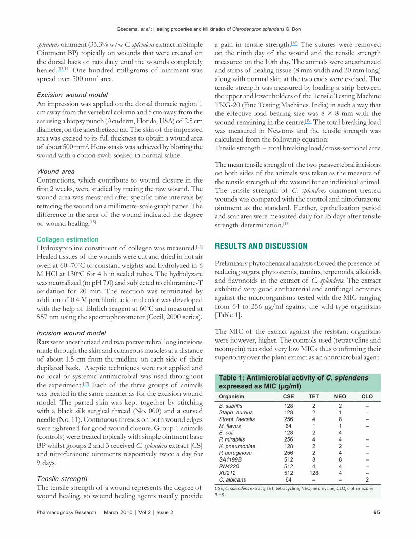
Preliminary phytochemical analysis showed the presence of reducing sugars, phytosterols, tannins, terpenoids, alkaloids and flavonoids in the extract of C. splendens. The extract exhibited very good antibacterial and antifungal activities against the microorganisms tested with the MIC ranging from 64 to 256 µg/ml against the wild-type organisms [Table 1].
The MIC of the extract against the resistant organisms were however, higher. The controls used (tetracycline and neomycin) recorded very low MICs thus confirming their superiority over the plant extract as an antimicrobial agent.
Table 1: Antimicrobial activity of C. splendens expressed as MIC (µg/ml)Organism CSE TET NEO CLOB. subtilis 128 2 2 –Staph. aureus 128 2 1 –Strept. faecalis 256 4 8 –M. flavus 64 1 1 –E. coli 128 2 4 –P. mirabilis 256 4 4 –K. pneumoniae 128 2 2 –P. aeruginosa 256 2 4 –SA1199B 512 8 8 –RN4220 512 4 4 –XU212 512 128 4 –C. albicans 64 – – 2
CSE, C. splendens extract; TET, tetracycline; NEO, neomycine; CLO, clotrimazole; n = 5
Gbedema, et al.: Healing properties and kill kinetics of Clerodendron splendens G. Don
66 Pharmacognosy Research | March 2010 | Vol 2 | Issue 2
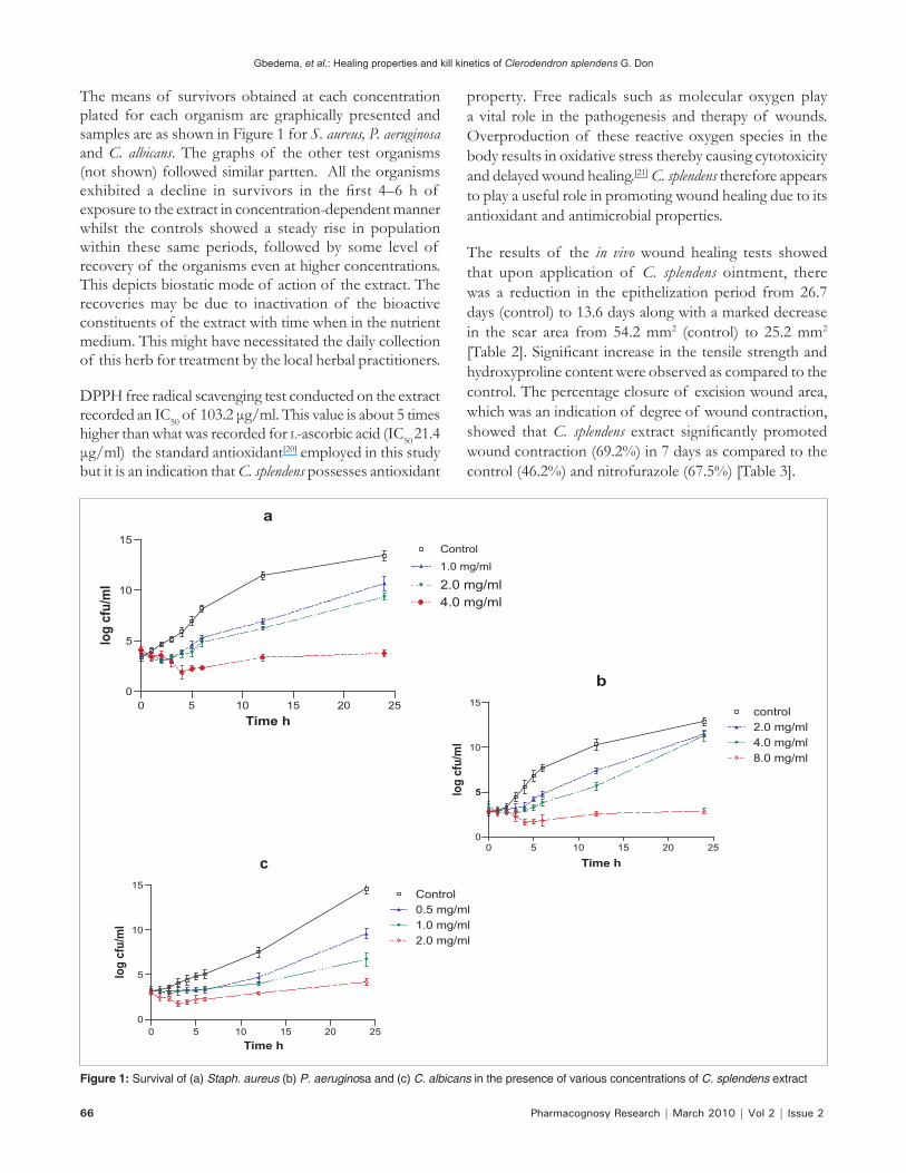
The means of survivors obtained at each concentration plated for each organism are graphically presented and samples are as shown in Figure 1 for S. aureus, P. aeruginosa and C. albicans. The graphs of the other test organisms (not shown) followed similar partten. All the organisms exhibited a decline in survivors in the first 4–6 h of exposure to the extract in concentration-dependent manner whilst the controls showed a steady rise in population within these same periods, followed by some level of recovery of the organisms even at higher concentrations. This depicts biostatic mode of action of the extract. The recoveries may be due to inactivation of the bioactive constituents of the extract with time when in the nutrient medium. This might have necessitated the daily collection of this herb for treatment by the local herbal practitioners.
DPPH free radical scavenging test conducted on the extract recorded an IC50 of 103.2 μg/ml. This value is about 5 times higher than what was recorded for l-ascorbic acid (IC50 21.4 μg/ml) the standard antioxidant[20] employed in this study but it is an indication that C. splendens possesses antioxidant
property. Free radicals such as molecular oxygen play a vital role in the pathogenesis and therapy of wounds. Overproduction of these reactive oxygen species in the body results in oxidative stress thereby causing cytotoxicity and delayed wound healing.[21] C. splendens therefore appears to play a useful role in promoting wound healing due to its antioxidant and antimicrobial properties.
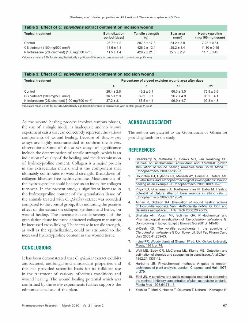
The results of the in vivo wound healing tests showed that upon application of C. splendens ointment, there was a reduction in the epithelization period from 26.7 days (control) to 13.6 days along with a marked decrease in the scar area from 54.2 mm2 (control) to 25.2 mm2 [Table 2]. Significant increase in the tensile strength and hydroxyproline content were observed as compared to the control. The percentage closure of excision wound area, which was an indication of degree of wound contraction, showed that C. splendens extract significantly promoted wound contraction (69.2%) in 7 days as compared to the control (46.2%) and nitrofurazole (67.5%) [Table 3].
Figure 1: Survival of (a) Staph. aureus (b) P. aeruginosa and (c) C. albicans in the presence of various concentrations of C. splendens extract
Gbedema, et al.: Healing properties and kill kinetics of Clerodendron splendens G. Don
Pharmacognosy Research | March 2010 | Vol 2 | Issue 2 67
As the wound healing process involves various phases, the use of a single model is inadequate and no in vitro experiment exists that can collectively represent the various components of wound healing. Because of this, in vivo assays are highly recommended to confirm the in vitro observations. Some of the in vivo assays of significance include the determination of tensile strength, which is an indication of quality of the healing, and the determination of hydroxyproline content. Collagen is a major protein in the extracellular matrix and is the component that ultimately contributes to wound strength. Breakdown of collagen liberates free hydroxyproline. Measurement of the hydroxyproline could be used as an index for collagen turnover. In the present study, a significant increase in the hydroxyproline content of the granulation tissue of the animals treated with C. splendens extract was recorded compared to the control group, thus indicating the positive effect of the extract on collagen synthesis and hence, on wound healing. The increase in tensile strength of the granulation tissue indicated enhanced collagen maturation by increased cross-linking. The increase in tensile strength, as well as the epithelization, could be attributed to the increased hydroxyproline content in the wound tissue.
CONCLUSIONS
It has been demonstrated that C. splendens extract exhibits antibacterial, antifungal and antioxidant properties and this has provided scientific basis for its folkloric use in the treatment of various infectious conditions and wound healing. The wound healing potential which was confirmed by the in vivo experiments further supports the ethnomedicinal use of the plant.
ACKNOWLEDGEMENT
The authors are grateful to the Government of Ghana for providing funds for the study.
REFERENCES
1. Steenkamp V, Mathivha E, Gouws MC, van Rensburg CE. Studies on antibacterial, antioxidant and fibroblast growth stimulation of wound healing remedies from South Africa. J Ethnopharmacol 2004;95:353-7.
2. Houghton PJ, Hylands PJ, Mensah AY, Hensel A, Deters AM. In vitro tests and ethnopharmacological investigations: Wound healing as an example. J Ethnopharmacol 2005;100:100–7.
3. Priya KS, Gnanamani A, Radhakrishnan N, Babu M. Healing potential of Datura alba on burn wounds in albino rats. J Ethnopharmacol 2002;83:193–9.
4. Annan K, Dickson RA. Evaluation of wound healing actions of Hoslundia opposita Vahl, Anthocleista nobilis G. Don and Balanites aegyptiaca L. J Sci Tech 2008;28:26-35.
5. Shehata AH, Yousif MF, Soliman GA. Phytochemical and Pharmacological investigation of Clerodendron splendens G. Don growing in Egypt. Egypt J Biomed Sci 2001;7:145-63.
6. el-Deeb KS. The volatile constituents in the absolute of Clerodendron splendens G Don flower oil. Bull Fac Pharm Cairo Univ 2003;41:259-63.
7. Irvine FR. Woody plants of Ghana. 1st ed. UK: Oxford University Press; 1961. p. 74.
8. Wall ME, Eddy CR, McClenna ML, Klump ME. Detection and estimation of steroids and sapogenins in plant tissue. Anal Chem 1952;24:1337-42.
9. Harborne JB. Phytochemical methods: A guide to modern techniques of plant analysis. London: Chapman and Hall; 1973. p. 279.
10. Eloff JN. A sensitive and quick microplate method to determine the minimal inhibitory concentration of plant extracts for bacteria. Planta Med 1998;64:711–3.
11. Yoshida T, Mori K, Hatano T, Okumura T, Uehara I, Komagoe K,
Table 2: Effect of C. splendens extract ointment on incision woundTopical treatment Epithelization
period (days)Tensile strength
(g)Scar area
(mm2)Hydroxyproline
(mg/100 mg tissue)Control 26.7 ± 1.2 287.5 ± 17.3 54.2 ± 3.8 7.28 ± 0.34CS ointment (100 mg/500 mm2) 13.6 ± 1.1 426.2 ± 12.4 25.2 ± 3.4 11.10 ± 0.45Nitrofurazone (2% ointment) [100 mg/500 mm2] 11.5 ± 1.4 428.2 ± 21.3 27.9 ± 2.9c 11.7 ± 0.45
Values are mean ± SEM for six rats; Statistically significant difference in comparison with control group: P < 0.05
Table 3: Effect of C. splendens extract ointment on excision woundTopical treatment Percentage of closed excision wound area after days
4 7 15 21Control 26.4 ± 2.6 46.2 ± 3.1 68.3 ± 3.5 75.8 ± 3.6CS ointment (100 mg/500 mm2) 36.5 ± 2.9 69.2 ± 3.7 96.7 ± 4.8 98.2 ± 4.1Nitrofurazone (2% ointment) [100 mg/500 mm2] 37.2 ± 3.1 67.5 ± 4.1 96.9 ± 4.7 99.3 ± 4.8
Values are mean ± SEM for six rats. Statistically significant difference in comparison with control group: P < 0.05
Gbedema, et al.: Healing properties and kill kinetics of Clerodendron splendens G. Don
68 Pharmacognosy Research | March 2010 | Vol 2 | Issue 2
et al. Studies on inhibition mechanism of autoxidatoin by tannins and flavonoids: Radical scavenging effects of tannins and related polyphenols on 1,1-diphenyl-2-picrylhydrazyl radical. Chem Pharm Bull 1989;37:19-21.
12. Annan K, Houghton PJ. Antibacterial, antioxidant and fibroblast growth stimulation of aqueous extracts of Ficus asperifolia Miq. and Gossypium arboreum L., wound-healing plants of Ghana. J Ethnopharmacol 2008;119:141-4.
13. Singh M, Govindarajan R, Nath V, Rawat AK, Mehrotra S. Antimicrobial, wound healing and antioxidant activity of Plagiochasma appendiculatum Lehm. et Lind. J Ethnopharmacol 2006;107:67-72.
14. Chattergee K, Chakravorty A. Wound healing properties of the new antibiotics (MT81) in mice. Indian Drugs 1993;30:450-2.
15. Werner S, Breeden M, Hübner G, Greenhalgh DG, Longaker MT. Induction of keratinocyte growth factor expression is reduced and delayed during wound healing in the genetically diabetic mouse. J Invest Dermatol 1994;103:469-73.
16. Shukla A, Rasik AM, Jain GK, Shankar R, Kulshrestha DK, Dhawan BN. In vitro and in vivo wound healing activity of asiaticoside isolated from Centella asiatica. J Ethnopharmacol
1999;65:1-11.17. Udupa AI, Kurkani DR, Udupa SL. Effect of Tridax procumbens
extracts on wound healing. Int J Pharmacol 1995;33:37-40.18. Govindarajan R, Vijayakumar M, Rao CV, Shirwaikar A, Mehrotra
S, Pushpangadan P. Healing potential of Anogeissus latifolia for dermal wounds in rats. Acta Pharm 2004;54:331-8.
19. Perez Gutierrez RM, Vargas SR. Evaluation of the wound healing properties of Acalypha langiana in diabetic rats. Fitoterapia 2006;77:286-9.
20. Bizimenyera ES, Aderogba MA, Eloff JN, Swan GE. Potential of neuroprotective antioxidant- based therapeutics from Peltophorum africanum Sond (Fabaceae). Afr J Trad Comp Alt Med 2007;4:99-106.
21. Somashekar S, Saraswati U, Laxminarayana U. Evaluation of antioxidant and wound healing effects of alcoholic and aqueous extracts of Ocimum sanctum L. in rats. eCAM 2008;5:95-101.
Source of Support: Government of Ghana, Conflict of Interest: None declared.
Gbedema, et al.: Healing properties and kill kinetics of Clerodendron splendens G. Don
Author Help: Online submission of the manuscripts
Articles can be submitted online from http://www.journalonweb.com. For online submission, the articles should be prepared in two files (first page file and article file). Images should be submitted separately.
1) First Page File: Prepare the title page, covering letter, acknowledgement etc. using a word processor program. All information related to your identity
should be included here. Use text/rtf/doc/pdf files. Do not zip the files.2) Article File: The main text of the article, beginning with the Abstract to References (including tables) should be in this file. Do not include any information
(such as acknowledgement, your names in page headers etc.) in this file. Use text/rtf/doc/pdf files. Do not zip the files. Limit the file size to 1 MB. Do not incorporate images in the file. If file size is large, graphs can be submitted separately as images, without their being incorporated in the article file. This will reduce the size of the file.
3) Images: Submit good quality color images. Each image should be less than 2048 kb (2 MB) in size. The size of the image can be reduced by
decreasing the actual height and width of the images (keep up to about 6 inches and up to about 1200 pixels) or by reducing the quality of image. JPEG is the most suitable file format. The image quality should be good enough to judge the scientific value of the image. For the purpose of printing, always retain a good quality, high resolution image. This high resolution image should be sent to the editorial office at the time of sending a revised article.
4) Legends: Legends for the figures/images should be included at the end of the article file.


