Embed Size (px)
Citation preview
Sequence Analysis of Bacterial DNA in the Colon and Stomach of the Tyrolean Iceman Raul J. Cano,1 Friedrich Tiefenbrunner,2 Massimo Ubaldi,3 Clarissa Del Cueto,4 Stefania Luciani,3 Tobe Cox,1 Paula Orkand,4 Karl H. Kunzel,5 and Franco Rollo3* 1Environmental Biotechnology Institute,California Polytechnic State University, San Luis Obispo, California 93407 2Hygiene Institute, University of Innsbruck, A-6020 Innsbruck, Austria 3Dipartimento di Biologia Molecolare, Cellulare e Animale, Universita` di Camerino, 62032 Camerino, Italy 4Instituto de Neurobiologı´a, Universidad de Puerto Rico, San Juan, Puerto Rico 00901 5Institute of Anatomy, University of Innsbruck, A-6010 Innsbruck, Austria
ABSTRACT The male human body found in an Alpine glacier on September 19,
1991 (“Tyrolean Iceman”) has, for the first time in history, given scientists a
chance to perform detailed anatomical, histological, and molecular investigations
on the organs of a person from the Neolithic Age (5350– 5100 B.P.). In the
present study, tissue samples aseptically taken from the stomach and the colon of
the mummy were utilized for DNA extraction, and the DNA was PCR-amplified,
using primer pairs designed to bind to fragments of the 16s ribosomal RNA gene
(16s rDNA) of a broad range of bacteria. The PCR products were cloned in
plasmid vectors, and the recombinant clones (amplicons) were sequenced. The
sequence data were finally used for scanning data libraries containing the
corresponding sequences of present-day bacteria, to infer the putative
ecophysiology of the ancient ones. The same procedure was repeated on some
fragments of grass from the clothing found near the corpse. These fragments
were taken as a control of the microbiological situation of the glacier. The
results show that the flora of the Iceman’s stomach is entirely composed of
Burkholderia pickettii, an organism commonly found in aquatic habitats. The
colon, on the other hand, contains several members of the fecal flora of humans,
such as Clostridium perfringens, C. ghonii, C. sordellii, Eubacterium tenue, and
Bacteroides sp. The Iceman’s colon, however, was found to contain, rather
unexpectedly, also some members of the genus Vibrio. The results are discussed
in light of what is known about the preservation of microbial DNA at the
Iceman’s site and of previous parasitological studies performed on the Iceman
himself and on human coprolites. Am J Phys Anthropol 112:297–309, 2000.
One of the most exciting archaeological discoveries of the century has been the mummified
corpse of a prehistoric man, known as the “Tyrolean Iceman” or “Otzi” on September 19,
1991. It was found at 3,213 m above sea level near the Similaun glacier, in the Otztaler
Alps, close to the Austro-Italian border. Radiocarbon datings indicated an age between
5,350 and 5,100 years (Bonani et al., 1992) corresponding to the Late Neolithic. After a
long stay at the Innsbruck University Anatomy Institute (Austria), lasting about 7 years, the
mummy is now preserved and exhibited at the Archaeological Museum of Bolzano (Italy).
Together with the body, a full set of surviving tools and weapons were found, including
an ax made of a wooden haft and copper blade, a bow-stave with arrows, and a small
dagger made of flint. A whole Neolithic garment was also recovered at the Alpine
archaeological site. It included a cloak made of knotted tufts of grass, and footwear made
of leather, which employed hay as stuffing (Spindler, 1995). The major recovery of
remains was performed a few days after the discovery and without special precaution. One
year later, a second archaeological expedition allowed a further acquisition of interesting
finds comprising uncontaminated grass and leather fragments from the clothing and
uncontaminated skin fragments from the body (Bagolini et al., 1995).
In a previous investigation (Ubaldi et al., 1998), we analyzed the distribution of bacterial
DNA in different organs and tissues of a Peruvian mummy and showed that it was possible
to identify the major bacterial taxa in the intestinal microflora of a person who lived in the
Andes about 1,000 years ago. The bacterial flora of the intestinal ecosystem is known to be
influenced by diet, interaction among microorganisms, drug assumption, toxins, and
carcinogenic substances (Blaut et al., 1998).
The present study describes the analysis of bacterial DNA in tissue samples obtained
aseptically from the stomach and colon of the glacier mummy. The same analysis was performed
on some fragments of grass clothing taken as a control of the microbiological situation of the
glacier where the mummy was found.
MATERIALS AND METHODS
Collection of samples
Grass samples. Small fragments of grass (each about 4 cm in length, equivalent to
approximately 16 mg dry weight) were taken from three samples (T27, T44, and T182)
collected during the second archaeological expedition, carried out in August 1992 (Rollo
et al., 1994). Soon after their recovery from the ice, the samples were put in a refrigerated
container and transferred to Bolzano, where they were kept in a freezer at —5°C to 2°C.
The following month, the samples of grass were transferred to the Department of
Molecular and Cell Biology of the University of Camerino in a thermally insulated
container and stored at —25°C. Details of the recovery and description of such finds are
recorded by Bagolini et al. (1995). Subsequently, the samples were radiocarbon-dated
using accelerator mass spectrometry (Rollo et al., 1994) to check the contemporaneity be-
tween the mummy and these finds.
Biopsic samples. Colon and stomach tissue samples from the Iceman were collected by
endoscopic surgery under strict aseptic conditions, using thoroughly sterilized titanium-
made, pointed, and blunt trocars, forceps, nippers, and scissors. To avoid any kind of
surface contamination, a rectangular portion of the skin was aseptically cut and lifted to
insert the titanium trocar tube. The specimens were placed in toxin-free plastic tubes and
kept frozen at —70°C until processed for DNA extraction or for electron microscopic
studies.
Electron microscopic studies
Representative samples of biopsies from the colon were fixed in 2% glutaraldehyde, pH
7.0, for 24 hr and washed in 0.1 M cacodylate buffer. Post-fixation was carried out with
1% OsO4 in 0.1 M cacodylate buffer for 24 hr, dehydrated in an alcohol series, stained en
bloc with 3% uranyl acetate, and mounted in epoxy. After the resin hardened, 1-µm
sections were cut, and microscopic examination was performed.
Aspartic acid racemization
Aspartic acid (Asp) racemization studies were performed on small representative
fragments of the colon and stomach (about 10–15 mg each) as described by Poinar et al.
(1996). Briefly, the specimens were washed in HCl 0.01 N and hydrolyzed in twice-dis-
tilled 6 N HCl for 24 hr at 100°C. The hydrolysates were desiccated under vacuum and the
residues were reconstituted in twice-distilled water. The amino acids were derivatized with
O-phthaldialdehyde/Nacetyl-L-cysteine (OPA/NAC) and analyzed by a high-pressure
liquid chromatography (HPLC) system (HP 1100, Hewlett Packard, Palo Alto, CA), using
fluorescent detection (Zaho and Bada, 1995)
DNA analysis
Extraction and PCR amplification of DNA from grass samples. The details of the DNA
extraction procedure from grass samples (T27, T44, and T182) were reported in Rollo et al.
(1995a). The DNA, finally precipitated using ethanol at —20°C, was further purified by gel
electrophoresis on 1.5% low melting temperature agarose to eliminate PCR inhibitors. The
electrophoretic fractionation showed that the DNA separates in a main band corresponding
to approximately 20 kb in length, preceded by a long smear of fragments down to 100–200
bp in length (Rollo et al., 1995a). For PCR amplification, the agarose was cut in corre-
spondence with the 100–5,000-bp fraction of the fluorescent smear and melted at 65°C, and
1 µl of the agarose-DNA suspension was directly added to the reaction mixture.
Enzymatic amplifications were performed in 50 µl of a reaction mixture containing 10 mM
Tris-HCl, pH 8.3, 1.5 mM MgCl2, 50 mM KCl, 0.1 mg/ml gelatin, 200 mM of each dNTP, 1
mM of each oligonucleotide primer, and 2.5 units of Taq polymerase (AmpliTaqTM, Perkin
Elmer Cetus). Combinations of universal oligonucleotide primers designed to bind to
relatively long (500–800 bp) portions of the 16s rDNA (see below) were unsuccessful;
therefore, the following universal oligonucleotide primers of Lane (1991) were employed:
27F, 5'-AGAGTTTGATCCTGGCTCAG-3'; and 342R, 5'-CT-GCTGCCTCCCGTAG-3'.
These primers bind to an approximately 350-bp-long portion of the bacterial gene for 16s
ribosomal RNA (16s rDNA). The short length of the fragment ensures a high
amplification efficiency. Following an initial step at 94°C for 5 min to denature genomic
DNA, the thermal cycler was set at 94°C for 1 min, 58°C for 30 sec, and 72°C for 1 min.
Forty-five amplification cycles were performed.
Extraction and PCR amplification of DNA from biopsic samples. Endoscopic biopsy
specimens from the colon or stomach were pulverized in a sterile Braun glass homogenizer.
DNA extraction was performed as described by Cano (1997) on the basis of Boom et al.
(1990). Amplifications were performed as described by Cano (1997), using 5 µl of
extracted DNA, 2 units of low DNA AmpliTaq DNA polymerase (Perkin-Elmer, Norwalk,
CT), 2 µg/ml bovine serum albumin, 0.5 µM each of a pair of 16s rDNA forward and
reverse primers, 2.0 mM MgCl2, and 200 mM of each dNTP in a total volume of 50 µl.
The following primers were employed: 8F, 5'-AGCGTCAAACTTTTAAATTGAA-3';
564R, 5'-CCTGCGTGCGCTTTACGCCC-3'; 584R, 5'-ACATCTGACTTAACAAACCG-3';
and 805R, 5'-TCGACATCGTTTACGGCGTG-3'. Each primer was coded with a number
indicating the position of the 5' base using E. coli 16s rRNA nucleotide position numbering.
The following primer combinations were used: 8F-564R, 8F-584R, and 8F-805R, spanning,
respectively, DNA segments of 556, 576, and 797 bp of length.
For the primer pair 8F-805R, PCR consisted of an initial denaturation step at 94°C for 2
min followed by 40 cycles of denaturation at 94°C for 30 sec, annealing at 58°C for 30 sec,
and elongation at 72°C for 1 min.
For the primer pairs 8F-584R and 8F-564R, the amplifications were carried out for 35
cycles of 94°C for 30 sec, 57°C for 30 sec, and 72°C for 45 sec. The amplifications were
initiated by 2 min at 94°C to denature genomic DNA.
To assess the presence of inhibitors of the PCR in DNA samples from extracted tissues,
aliquots of extracted DNA were seeded with approximately 1 pg of plasmid DNA
containing a 440-bp fragment of E. coli and enzymatically amplified using the appropri-
ate primers and amplification conditions. Amplified DNA was evaluated by gel elec-
trophoresis.
In the amplifications both of purified grass DNA and of DNA extracted from biopsic
samples, to eliminate small amounts of E. coli DNA possibly present in the commercial
enzyme preparations and, in general, any types of contaminant DNA, the Taq polymerase
and reaction components were pretreated with 3 units of DNase I (from bovine pancreas) for
30 min at room temperature. After incubation, the DNase I was inactivated by boiling for 10
min at 94°C.
DNA cloning and sequencing. Amplification products were cloned into pMOS-Blue T-vector
(pMOS-Blue T-vector kit, Amer-sham, UK) according to the manufacturer’s instructions.
Recombinant plasmids were isolated by silica gel procedure (Plasmix, Talent, Trieste, Italy).
The purified products were cycle sequenced using dye-deoxy terminators and M13 lacZ
forward and reverse primers. We utilized either an ABI PRISM 310 or an ABI PRISM 377
automated sequencer (dRhodamine Terminator Cycle Sequencing Ready Reaction, PE
Applied Biosystems, Foster City, CA). The nucleotide sequence of the shortest (350 bp) grass
inserts was read in one direction, while that of the longest inserts from the stomach and colon
was read in two directions.
Phylogenetic analyses. Sequences were initially compared with reference sequences
available in the databases of Genbank and EMBL by using ungapped BLAST (Altschul et
al., 1990) and FASTA (Pearson and Lipman, 1988) search programs to determine their
approximate phylogenetic affiliation. In addition, the nucleotide sequences of the
amplicons were evaluated for the presence of DNA chimeras by using the CHECK-
CHIMERA program (Maidak et al., 1997) available from the Ribosomal Database Project.
The 16s rDNA sequences were aligned by using the Clustal V function (Higgins et al.,
1992) of the GDE 2.0 software package. Distance matrices were constructed from the
aligned sequences and corrected for the superimposed mutations by the method of Tajima
and Nei (1984). Phylogenetic trees were constructed using the TREECON program (Van
de Peer and De Wachter, 1993) with a neighbor-joining algorithm (Saitou and Nei, 1987).
The robustness of inferred topologies was tested by bootstrap resampling of trees (Felsen-
stein, 1985).
Precautions to avoid contamination.
All the extraction and amplification operations were carried out taking into account the
precautions required for the analysis of DNA in ancient specimens (Herrmann and
Hummel, 1994). These precautions included the wearing of sterile gloves, the pretreat-
ment of mortars, pestles, and homogenizers with HCl, the use of an ultraviolet-irradiated
safety cabinet, and dedicated gel tray and tanks. Additionally, mock extraction and blank
amplification reactions were always performed. Moreover, it should be pointed out that
the use of cloning to identify the composition of PCR products is an efficient strategy to
recognize contaminant sequences as well.
The effectiveness of the precautions adopted is shown by the fact that despite all the
cloning work in our laboratory being performed using E. coli as a recipient organism, not
a single sequence attributable to this bacterium was found in several hundreds of 16s
rDNA amplicons screened in the course of the present and other ancient DNA
investigations.
RESULTS
Analysis of bacterial DNA from the
Iceman’s gastrointestinal tract
Transmission electron photomicrographs of the colon tissue show the presence of a still
identifiable muscularis-mucosa (Fig. 1a) and various types of rod-shaped and filamentous
bacteria colonizing the surfaces of the colon epithelium (Fig. 1b). In addition, estimates of
aspartic acid racemization performed on colon and stomach samples indicated D/LAsp ratios
ranging from 0.06–0.08. These values are consistent with a satisfactory degree of DNA
preservation (Poinar et al., 1996).
[Insert Figure 1]
Four out of seven stomach and colon biopsy samples yielded amplifiable DNA. The
DNA fragments were successfully cloned into a plasmid vector. A total of 33 clones was
analyzed by sequencing: 16 from the stomach library, and the remaining 17 clones from the
colon library. The screening of the 16s rDNA library from stomach showed that it was
entirely composed of Burkholderia pickettii. Fourteen out of 16 clones analyzed had high
percent base identity (= 98%) with either Burkholderia pickettii or its synonym,
Pseudomonas pickettii. This organism is commonly found in aquatic habitats and has been
isolated from wet hospital equipment, disinfecting solutions, cosmetics, paper mill
effluents, teething rings, and a variety of patient sources (McNeil et al., 1985).
On the other hand, the sequence analysis of the colon amplicons showed a mixture of
bacterial species (Table 1). The most representative species is C. perfringens (clones
O8SB, O8SC, O8SG, O8SH, 1A, Oim29, and Oim26), an anaerobic, Gram-positive, spore-
forming rod, widely distributed in the environment. This species of Clostridium is fre-
quently part of the normal human endogenous flora and is commonly isolated from
infections in humans (Lewis et al., 1980). The molecular analysis also led to the
identification of some more clostridia: C. ghonii (clone O8SA) that can be isolated from
soil, marine sediments, soft-tissue infections in humans (Gorbach and Thadepalli, 1975),
and human feces (Finegold et al., 1983), and C. sordelli (O8SD). The latter is an organism
normally isolated from soil, normal human feces, and human clinical specimens (Gorbach
and Thadepalli, 1975).
The 16s rDNA sequence of the clones O8SE and O815 show a correlation with
Eubacterium tenue, which can be isolated from abscesses following abortion, knee sy-
novial fluid, blood, and fecal samples.
Bacteroides sp. (clone OI1675) was also detected in the Iceman’s gut. This bacterium is
a prominent member of the human gut flora (Hill, 1995) and is commonly encountered in
clinical specimens from the gastrointestinal tract (Isenberg and D’Amato, 1991).
A member of the genus Bacillus (clone I110) was also identified. The natural habitat of
Bacillus spp. is the soil, but strains of this genus have also been isolated from samples in
extreme environments such as deserts and Antarctica. This sporing aerobe is also a
member of the normal fecal flora of healthy adult humans, with a reported concentration
of 104–106 cells per gram of feces (Hill, 1995).
It is noteworthy that members of the genus Vibrio (clones Oim25, OIm30, and OIm31)
were detected in relatively high numbers. These are facultative anaerobic, asporigenous,
Gram-negative rods that are natural inhabitants of aquatic environments. Infections of the
colon are contracted by the fecal-oral route (Kelly et al., 1991).
Two clones (Oim 25 and Oim30) most closely resemble Vibrio metschnikovii. This
organism is one of the species of Vibrio that can be found in human clinical specimens.
Although not a bona fide human pathogen, V. metschnikovii has been reported to cause
human diarrhea (Dalsgaard et al., 1996; Buck, 1992; Magalhaes et al., 1996). Furthermore,
V. metschnikovii has been isolated from lakes and streams with evidence of fecal
contamination (Dalsgaard et al., 1996; Bitto et al., 1992).
[Insert Table 1]
Analysis of bacterial DNA from the
Iceman’s grass clothing
Of the three specimens tested, only one (T27) produced a weak band of amplified
DNA. The amplification product was subsequently cloned into a plasmid vector to pro-
duce a library of amplicons. Thirty amplicons from the library were analyzed by
sequencing. The results of the comparison with the reference sequences in the database
are shown in Table 2.
The bacterial DNA associated with the Neolithic grass shows a considerable degree of
heterogeneity in species composition. The nucleotide sequences obtained from five clones
(T2721a, T2702a, T2705b, T2778c, and T2784c) appear to be closely related to the
organisms of the genus Duganella, formerly called Zoogloea (Hiraishi et al., 1997). Such
microrganisms (P-proteobacteria) include Gram-negative aerobic rods, and as porigenous
and noncyst-forming eubacteria. The species of the genus Duganella are found
principally in organically polluted fresh waters, wastewaters, and aerobic biological
wastewater treatment systems. They are also commonly found in drinking-water biofilms
(Kalmbach et al., 1997). A recent work describes the organisms of this genus as recurring
contaminants during PCR analysis (Tanner et al., 1998).
Clostridium spp. (clones T2707a, T2703c, T2703b, and T27118c) are relatively abun-
dant among the Iceman’s grass microflora, reflecting their wide distribution over the
earth’s environments. They are commonly found in soil, sewage, marine sediments, de-
caying vegetation, and animal and plant products. In addition, they can be isolated from

the intestinal tract of mammals, other vertebrates, and insects, and from wounds or soft-
tissue infections of humans and other animals.
The clone T2789c shows considerable sequence similarity with an unidentified a-
proteobacterium found during the investigation of an indigenous bacterial community in
marine Antarctic ice characterized by psycrophilic and gas-vacuolated members (Gosink
and Staley, 1995). Other clones (T2704b, T2715a, and T2760c) show sequence similarity
with bacteria of the genus Arthrobacter. Bacteria of this genus are important members of
soil communities. Although they do not produce spores, they are remarkably resistant to
desiccation (Stolp, 1988). Psychrophilic strains have been found in cave and glacier
muds in the Arctic, Lapland, the Pyrenees, and the Alps (Gounot, 1967; Cameron et al.,
1973) and in lacustrine environments.
[Insert Table 2]
Among the sequences of organisms at present available in the nucleotide databases, the
group of clones T2702b, T2719c, and T2767c shows a certain grade of similarity with an
unidentified Cytophagales bacterium (D84607) isolated from environmental samples
(Mitsui et al., 1997). This organism is also considered a laboratory contaminant of PCR
processes (Tanner et al., 1998).
Agrococcus jenensis (clone T2704c) is a Gram-positive coryneform bacterium isolated
from soil and from sandstone layers.
The nucleotide sequence of clone T2727a shows some similarity with the corresponding
sequence of Deinococcus radiodurans. Several observations suggest that Deinococcus
species may be widely distributed in nature, but in modest number. They can be isolated
from many sites, including the air of clean rooms and laboratories (Murray and Brooks,
1986). On the other hand, T2703a and T2727c sequences seem to be related to that of
Curtobacterium citreum. The Curtobacteria are irregular, nonsporing, Gram-positive rods
and are obligately aerobic; most members of the genus have been isolated from plants
(Komagata and Suzuki, 1986).
The clones T2704c, T2757c, and T2730c show a moderate similarity with the corre-
sponding sequences of species of the genus Pseudomonas isolated from Arctic soil polluted
with fossil fuel. Psycrophilic members of this genus have been reported in caves of
Lapponia, the Pyrenees, and the Alps (Baross and Morita, 1978). Finally, the nucleotide
sequences of a group of amplicons (T2772c, T2705a, T2715c, T2785c, T2724c, T2794c,
and T27107c) prove to be correlated with the corresponding sequences of microorganisms
identified in very different environmental samples by a culture-independent molecular
phylogenetic approach (Suzuki et al., 1997; Dojka et al., 1998).
DISCUSSION AND CONCLUSIONS
The first analyses of the mummy’s endogenous DNA performed by Handt et al. (1994)
showed that the degradation of the mitochondrial DNA (mtDNA) did not allow the
amplification by PCR of molecules longer than 200 base pairs (bp). Besides, the titration
of residual human mtDNA molecules, by competitive PCR tests, revealed that the copy
number was more than six orders of magnitude less than would be expected in a fresh
human tissue. These results led to the conclusion that most of the endogenous DNA was
degraded and that single-copy genes could not be amplified from the sample analyzed.
Molecular analyses performed on radiocarbon-dated samples of grass coming from the
cloak and the stuffing of the footwear, on the other hand, demonstrated that the original
DNA of the grass (Rollo et al., 1994) was also maintained. In this case, however,
fragments of the chloroplast’s large subunit of the ribulose bisphosphate carboxylase
(rbcL) gene, as long as 530 bp, could be retrieved and their nucleotide sequence read. The
phylogenetic analysis allowed the plant species utilized by Neolithic man to manufacture
his equipment to be identified. In addition, scanning electron microscope observations
revealed that the ancient grass was covered by fungal hyphae and algal cysts. The
observations also showed very rare bacterial cells. These microorganisms were probably
associated with the grass since the Iceman’s time (Rollo et al., 1994, 1995a,b).
A more detailed picture, focused on the fungal component of the Iceman’s grass clothes,
was subsequently produced by Rollo et al. (1995c). The DNA extracted from two
radiocarbon-dated specimens was submitted to PCR amplification, employing two different
primer pairs. While the first encompassed the nuclear gene for 5.8s ribosomal RNA (rRNA)
and flanked transcribed spacers ITS1 and ITS2 (approximately 540 bp in length), the
second bound to an approximately 600-bp-long fragment within the reading frame of the
nuclear small subunit (SSU) rRNA gene. Amplified DNA was cloned and sequenced.
Phylogenetic analyses based on the nucleotide sequences showed that DNAs should be
ascribed to a psychrophilic basidiomycetous yeast and two ascomycetes.
The present analyses confirm that bacteria are poorly represented among the grass-
clothing microflora. Only one out of three DNA preparations from the grass was suc-
cessfully amplified by PCR, producing only a weak band of DNA. This picture of low
bacterial biomass is further confirmed by the recovering of nucleotide sequences cor-
responding to Duganella sp. (formerly Zoogloea) and an unidentified Cytophagales
(D84607). These species have been detected in clone libraries from very different low-
biomass habitats. Because the extraction from low-level DNA samples is particularly
sensitive to potentially contaminating DNA during the PCR process, it has been supposed
that these rDNA sequences may be intrinsic contaminants of PCR reactions (Tanner et al.,
1998 ). On the other hand, some of the 16s rDNA amplicons from the grass are indicative
of a microbial flora (psycrophilic (x-proteobacterium, Pseudomonas, and Arthrobacter),
consistent with the environmental characteristics of the Alpine site and with the microflora
composition of the Iceman’s skin (Rollo et al., 2000).
The situation inside the Iceman’s body is completely different. With regard to the ul-
trastructural preservation of the mummy tissues, our results basically fit with those reported
by Hess et al. (1998). These authors, in particular, noted the presence of bacteria in the
digestive tract of the Iceman. Our DNA analyses, on the other hand, clearly show that the
microorganisms of the stomach and colon are different. In the colon, Gram-positive,
anaerobic rods are largely represented, while in the stomach, with the absolute
predominance of Burkholderia, only Gram-negative rods are present. This result
demonstrates that no appreciable postmortem translocation of bacteria has taken place from
organ to organ.
The finding of Burkholderia pickettii in the stomach, however, deserves a few com-
ments. With the exception of Helicobacter pylori, the normal stomach defenses are suf-
ficiently effective so as to render it free of either pathogenic or saprophytic bacteria. Nor
is B. pickettii a known common constituent of the normal oral cavity flora. The most
plausible explanation is that this organism may have entered the stomach after death as a
passenger in postmortem water aspiration. The Iceman body is known to have been at
least partially submerged in a pool of meltwater between the time of his discovery and the
removal of the body. In addition, there is increasing evidence that the corpse, in the days
and weeks following the Iceman’s death, was submerged in meltwater (Bereuter et al.,
1997; Rollo et al., 2000).
[Insert Figure 2]
[Insert Figure 3]
When some (Oim29, Oim26, O8SH, O8S G, O8SB, O8SC, O8SE, O815, O8SD, and
O8SA) of the amplicon sequences of the colon microflora are inserted in the phylogenetic
tree of Clostridia, they cluster into two main groups (Fig. 2): one is closely related to C.
perfringens, while the other has a closer relationship with C. difficile, C. ghoni, C. sordelli,
and E. tenue. It is remarkable that C. perfringens is the predominant species among the
organisms identified in the colon microflora of the mummy, and that this bacterium is a
member of the human normal endogenous flora. C. sordelli, C. ghoni, and E. tenue are
also meaningful representatives of the human gut microflora, and are often isolated from
human fecal samples. Also, Bacteroides spp. (clone OI 1675) are considered a prominent
member of the human gut flora (Hill, 1995).
Finally, of particular interest is the detection of DNA sequences (clones Oim25, Oim30,
and Oim31) corresponding to species of the genus Vibrio. As shown in the phylogenetic
tree of Figure 3, these sequences fall in the branching of V. aestuarianus and V.
metschnikovii, and at the same time they occupy a phylogenetically distant position from
V. cholerae, the organism responsible for a severe form of diarrhea in the cholera
epidemic. As mentioned in Results, V. metschnikovii has been frequently isolated from
fresh, brackish, and marine waters, and humans occasionally become colonized or infected
by these sources.
It is well-known that there is a correlation between the composition of gut microflora
and the dietary habits of an individual, and his well-being. The intestinal microflora
plays an important role in preventing colonization by pathogens and takes part in nu-
merous metabolic processes useful for maintaining human health (Blaut et al., 1998).
Therefore, the analysis of gut bacterial composition may represent an alternative ap-
proach to classical ethno-anthropological methods to infer information about the food
habits, lifestyle, and health conditions of ancient civilizations, as well as the structure of
human microbial ecosystems of the past.
The Neolithic period was characterized by great innovations and changes that deeply
influenced human life. First of all, there was a transition from a nomadic way of life to
agriculture, breeding domesticated animals, and a higher level of social interaction among
the members of the community than in the Paleolithic. Secondly, the life in larger human
aggregates increased the spread of diseases by the fecal-oral route, with the concomitant
reduction in survival curves of the early Neolithic human popula tion (Cournede et al.,
1996). In view of this, the finding of V. metschnikovii DNA may be connected with that of
an egg of Trichuris trichura (whipworm) in the sacro-gluteal region of the mummy and the
detection of antibodies against this intestinal parasite (Aspo¨ck et al., 1996). This, in turn,
fits with the report of Bouchet et al. (1995) describing the identification of many well-
preserved helminth eggs, including Trichuris, Capillaria, Fasciola hepatica, and
Diphyllobotrium sp. in 12 human coprolites and in 10 sediment samples rich in organic
matter form the Neolithic site of Chalain, Jura.
LITERATURE CITED
Altschul SF, Gish W, Miller W, Myers EW, Lipman DJ. 1990. Basic local alignment search
tool. J Mol Biol 215:403–410.
Aspo¨ck H, Auer H, Picher O. 1996. Trichuris trichiura eggs in the Neolithic glacier mummy
from the Alps. Parasitol Today 12:255–256.
Bagolini B, Dal Ri L, Lippert A, Nothdurfter H. 1995. Der Mann im Eis: die Fundbergung
1992 am Tisenjoch, Gem. Schnals, Su¨dtirol. In: Spindler K, Rastbichler-Zissernig E,
Wilfing H, zur Nedden D, Nothdurfter H, editors. Der Mann im Eis: neue Funde und
Ergebnisse. Wien, New York: Springer-Verlag. p 3–22.
Baross JA, Morita RY. 1978. Microbial life at low temperature: ecological aspects. In: Kushner
DJ, editor. Microbial life in extreme environments. London, New York, San Francisco:
Academic Press. p 10–57.
Bereuter TL, Mikenda W, Reiter C. 1997. Iceman’s mummification-implications from
infrared spectroscopical and histological studies. Chem Eur J 7:1032– 1038.
Bitto AO, Kale OO, Oduntan SO. 1992. Epidemiological survey of an outbreak of
gastroenteritis in a rural community in Oyo State. West Afr J Med 11:34–38.
Blaut M, Griggs C, Collins MD, Welling G, Dore´ J, van Loo J, de Vos W. 1998.
Quantitative assessment of the human gut flora using a panel of rRNA targeted hy-
bridization probes. In: Gaukel V, Spieß W, editors. Third Karlsruhe Nutrition Symposium.
European research towards safer and better food (proceedings part 1: lectures). October
18–20, 1998. Bundesforschungsanstalt fu¨r Ema¨hrung, Karlsruhe. p 102–109.
Bonani G, Ivy SD, Niklaus TR, Suter M, Housley RA, Bronj CR, van Klinken GJ, Edges REM.
1992. Altersbestimmung von Lilligrammproben der O¨ tzaler Gletscherleiche mit der
Beschleunigermassenapektrometrie methode (AMS). In: Ho¨pfel F, Platzer W, Spindler K,
editors. Der Mann im Eis, volume 1. Innsbruck: Vero¨ffentlichungen der Universita¨t Inns-
bruck. p. 108–116.
Boom R, Sol C, Salimans M, Jansen C, Wertheim-van Dillen P, van der Noordaa J. 1990. Rapid
and simple method for the purification of nucleic acids. J Clin Microbiol 28:495–503.
Bouchet F, Petrequin P, Paicheler JC, Dommelier S. 1995. Premiere approche
paleoparasitologique du site neolithique de Chalain Jura, France. Bull Soc Pathol Exot
88:265–268.
Buck JD. 1992. Recovery of Vibrio metschnikovii from Market Seafood. J Food Safety 12:73–
78.
Cameron RE, Morelli FA, Honour RC. 1973. Aerobiological monitoring of Dry Valley drilling
sites. Antarctic J US 8:211–214.
Cano RJ. 1997. DNA amplification techniques in fossilized samples. In: Toranzos GA, editor.
Environmental applications of nucleic acid amplification techniques. Lancaster, PA:
Technomic Publishing Co. p 183–210.
Cournede N, Designere C, Picouleau D. 1996. Du ne´olithique au 20e`me sie`cle. De la
naissance de l’agriculture a` la nutrition the´rapeutique. Soins 604: 85–89.
Dalsgaard A, Alarcon A, Lanata CF, Jensen T, Hansen HJ, Delgado F, Gil AI, Penny ME,
Taylor D. 1996. Clinical manifestations and molecular epidemiology of five cases of
diarrhoea in children associated with Vibrio metschnikovii in Arequipa, Peru. J Med Micro-
biol 45:494–500.
Dojka MA, Hugenholtz P, Haack SK, Pace NR. 1998. Microbial diversity in a hydrocarbon- and
chlorinated-solvent-contaminated aquifer undergoing intrinsic bioremediation. Appl Environ
Microbiol 64:3869–3877.
Felsenstein J. 1985. Confidence limits on phylogenies: an approach using the bootstrap.
Evolution 39:783–791.
Finegold SM, Sutter VL, Mathisen GE. 1983. Normal indigenous intestinal flora. In: Hentges E,
editor. Human intestinal microflora in health and disease. New York: Academic Press. p 3–
31.
Gorbach SL, Thadepalli H. 1975. Isolation of Clostridium in human infections: evaluation
of 114 cases. J Infect Dis 131:81–85.
Gosink JJ, Staley JT. 1995. Biodiversity of gas vacuolate bacteria from Antarctic sea ice and
water. Appl Environ Microbiol 61:3486–3489.
Gounot AM. 1967. Roˆle biologique des Arthrobacters dans les limons souterrains. Ann Inst
Pasteur Paris 113:923–945.
Handt O, Richards M, Trommsdorff M, Kilger C, Simanainen J, Oleg G, Bauer K, Stone A,
Hedges R, Schaffner W, Utermann G, Sykes B, Pa¨a¨bo S. 1994. Molecular genetic analyses
of the Tyrolean Ice Man. Science 264:1775–1778.
Herrmann B, Hummel S. 1994. General aspects of sample preparation. In: Hummel S,
Herrmann B, editors. Ancient DNA. New York: Springer-Verlag. p 59–68.
Hess MW, Klima G, Pfaller K, Ku¨nzel KH, Gaber O. 1998. Histological investigation of
the Tyrolean Ice Man. Am J Phys Anthopol 106:521–532.
Higgins DG, Bleasby AJ, Fuchs R. 1992. CLUSTAL V, improved software for multiple
sequence alignment. Comput Appl Biosci 8:189–191.
Hill MJ. 1995. The normal gut bacterial flora. In: Hill MJ editor. Role of gut bacteria in
human toxicology and pharmacology. London: Taylor and Francis. p 3–17.
Hiraishi A, Shin YK, Sugiyama J. 1997. Proposal to reclassify Zoogloea ramigera IAM 12670
(P. R. Dugan 115) as Duganella zoogloeoides gen. Nov., sp. nov. Int J Syst Bacteriol
47:1249–1252.
Isenberg HD, D’Amato RF. 1991. Indigenous and pathogenic microorganisms of humans. In:
Balows A, Hausler WJ Jr, Herrmann K, Isemberg HD, Shadomy HJ, editors. Manual of
clinical microbiology. Washington, DC: ASM Press. p 2–15.
Kalmbach S, Manz W, Szewzyk U. 1997. Isolation of new bacterial species from drinking
water biofilms and proof of their in situ dominance with highly specific 16s rRNA
probes. Appl Environ Microbiol 63: 4164–4170.
Kelly MT, Hickman-Brenner FW, Farmer JJ III. 1991. Vibrio. In: Balows A, Hausler WJ Jr,
Herrmann K, Isemberg HD, Shadomy HJ, editors. Manual of clini-
cal microbiology. Washington, DC: ASM Press. p 384–395.
Komagata K, Suzuki K. 1986. Genus Curtobacterium. In: Holt JG, editor. Bergey’s manual of
systematic bacteriology. Baltimore: Williams & Wilkins. p 1313– 1317.
Lane DJ. 1991. 16s/23s rRNA sequencing. In: Stackebrandt E, Goodfellow M, editors.
Nucleic acid techniques in bacterial systematics. Chichester: John Wiley and Sons, Ltd. p
115–175.
Lewis JF, Mullins N, Johnson P. 1980. Isolation and evaluation of Clostridia from clinical
sources. South Med J 73:427–432.
Magalhaes V, Branco A, de Andrade Lima R, Magalhaes M. 1996. Vibrio metschnikovii
among diarrheal patients during cholera epidemic in Recife, Brazil. Rev Inst Med Trop Sao
Paulo 38:1–3.
Maidak BL, Olsen GJ, Larsen N, Overbeek R, McCaughey M, Woese CR. 1997. The RDP
Ribosomal Database Project. Nucleic Acids Res 25:109–111.
McNeil MM, Solomon SL, Anderson RL, Davis BJ, Spengler RF, Reisberg BE, Thornsberry
C, Martone WJ. 1985. Nosocomial Pseudomonas pickettii colonization associated with a
contaminated respiratory therapy solution in a special care nursery. J Clin Microbiol
22:903–907.
Mitsui H, Gorlach K, Lee H, Hattori R, Hattori T. 1997. Incubation time and media
requirements of culturable bacteria from different phylogenetic groups. J Microbiol
Methods 30:103–110.
Murray RGE, Brooks BW. 1986. Genus I. Deinococcus. In: Holt JG, editor. Bergey’s manual of
systematic bacteriology. Baltimore: Williams & Wilkins. p 1035– 1043.
Pearson WR, Lipman DJ. 1988. Improved tools for biological sequence comparison. Proc Natl
Acad Sci USA 85:2444–2448.
Poinar HN, Ho¨ss M, Bada JL, Pa¨a¨bo S. 1996. Amino acid racemization and the preservation
of ancient DNA. Science 272:864–866.
Rollo F, Asci W, Antonini S, Marota I, Ubaldi M. 1994. Molecular ecology of a Neolithic
meadow: the DNA of the grass remains from the archaeological site of the Tyrolean Iceman.
Experientia 50:576–584.
Rollo F, Asci W, Marota I, Sassaroli S. 1995a. DNA analysis of grass remains found at the
Iceman’s archaeological site. In: Spindler K, Rastbichler-Zissernig E, Wilfing H, zur
Nedden D, Nothdurfter H, editors. Der Mann im Eis: neue Funde und Ergebnisse. Wien,
New York: Springer-Verlag. p 91–105.
Rollo F, Asci W, Antonini S, Ubaldi M. 1995b. The “Neolithic” microbial flora of the
Iceman’s grass: morphological description and DNA analysis. In: Spindler K, Rastbichler-
Zissernig E, Wilfing H, zur Nedden D, Nothdurfter H, editors. Der Mann im Eis: neue
Funde und Ergebnisse. Wien, New York: Springer-Verlag. p 107–114.
Rollo F, Sassaroli S, Ubaldi M. 1995c. Molecular phylogeny of the fungi of the Iceman’s
grass clothing. Curr Genet 28:289–297.
Rollo F, Luciani S, Canapa A, Marota I. 2000. Analysis of bacterial DNA in skin and
muscle of the Tyrolean Iceman offers new insight into the mummification process. Am J
Phys Anthropol 111:211–219.
Saitou N, Nei M. 1987. The neighbor-joining method, a new method for reconstructing
phylogenetic trees. Mol Biol Evol 4:406–425.
Spindler K. 1995. The man in the ice. Phoenix, London: Orion Books, Ltd.
Stolp H. 1988. Microbial ecology: organisms, habitats, activities. Cambridge studies in ecology.
Cambridge: Cambridge University Press.
Suzuki MT, Rappe MS, Haimberger ZW, Winfield H, Adair N, Strobel J, Giovannoni SJ. 1997.
Bacterial diversity among small-subunit rRNA gene clones and cellular isolates from the same
seawater sample. Appl Environ Microbiol 63:983–989.
Tajima F, Nei M. 1984. Estimation of evolutionary distance between nucleotide sequences.
Mol Biol Evol 1:269–285.
Tanner MA, Goebel BM, Dojka MA, Pace NR. 1998. Specific ribosomal DNA sequences from
diverse environmental settings correlate with experimental contaminants. Appl Environ
Microbiol 64:3110–3113.
Ubaldi M, Luciani S, Marota I, Fornaciari G, Cano RJ, Rollo F. 1998. Sequence analysis of
bacterial DNA in the colon of an Andean mummy. Am J Phys Anthropol 107:285–295.
Van de Peer Y, De Wachter R. 1993. TREECON, a software package for the construction
and drawing of evolutionary trees. Comput Appl Biosci 9:177–182.
Zaho M, Bada JL. 1995. Determination of a-dialkylamino acids and their enantiomers in

geological samples by high-performance liquid chromatography after derivatization with a
chiral adduct of o-phthaldaildehyde. J Chromatogr 690:55–63.
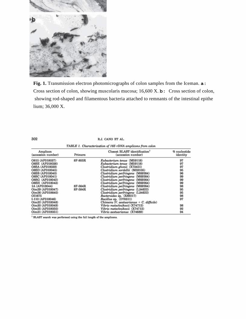
Fig. 1. Transmission electron photomicrographs of colon samples from the Iceman. a: Cross section of colon, showing muscolaris mucosa; 16,600 X. b: Cross section of colon, showing rod-shaped and filamentous bacteria attached to remnants of the intestinal epithe lium; 36,000 X.
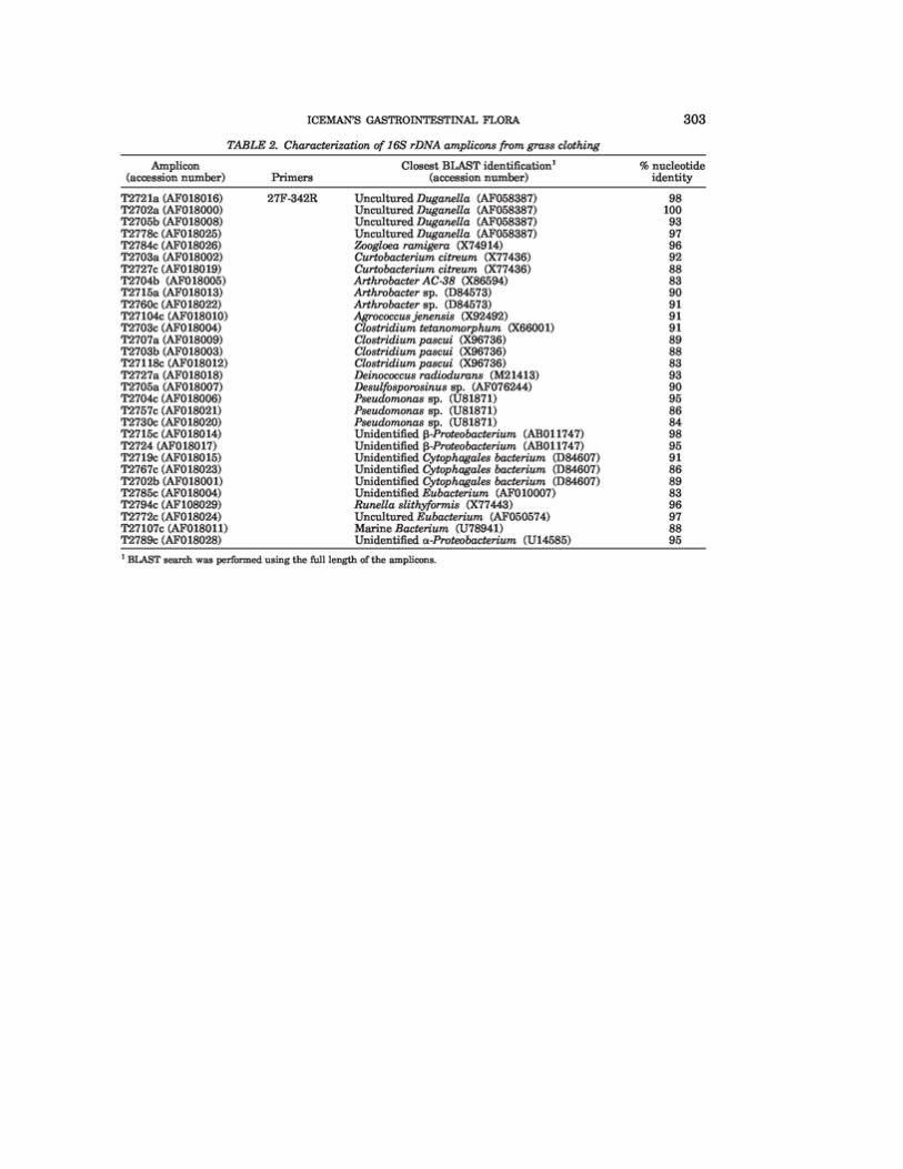
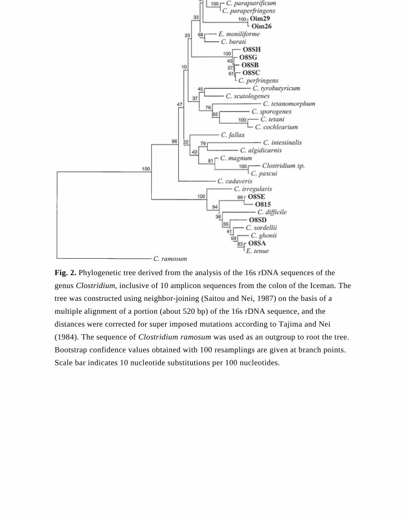
Fig. 2. Phylogenetic tree derived from the analysis of the 16s rDNA sequences of the genus Clostridium, inclusive of 10 amplicon sequences from the colon of the Iceman. The tree was constructed using neighbor-joining (Saitou and Nei, 1987) on the basis of a multiple alignment of a portion (about 520 bp) of the 16s rDNA sequence, and the distances were corrected for super imposed mutations according to Tajima and Nei (1984). The sequence of Clostridium ramosum was used as an outgroup to root the tree. Bootstrap confidence values obtained with 100 resamplings are given at branch points. Scale bar indicates 10 nucleotide substitutions per 100 nucleotides.
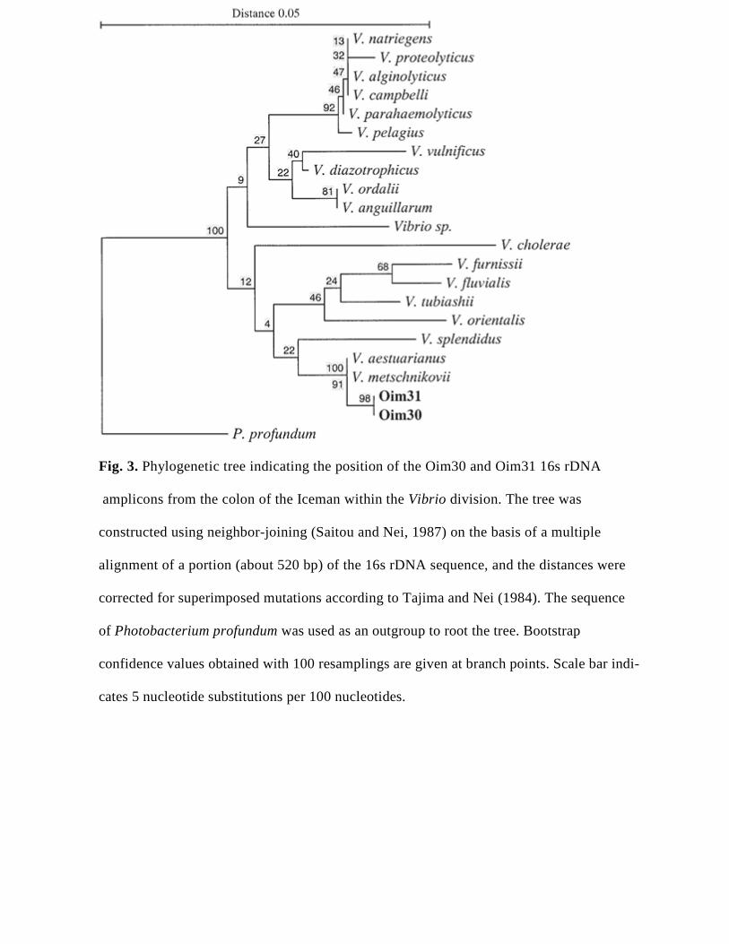
Fig. 3. Phylogenetic tree indicating the position of the Oim30 and Oim31 16s rDNA amplicons from the colon of the Iceman within the Vibrio division. The tree was constructed using neighbor-joining (Saitou and Nei, 1987) on the basis of a multiple alignment of a portion (about 520 bp) of the 16s rDNA sequence, and the distances were corrected for superimposed mutations according to Tajima and Nei (1984). The sequence of Photobacterium profundum was used as an outgroup to root the tree. Bootstrap confidence values obtained with 100 resamplings are given at branch points. Scale bar indi- cates 5 nucleotide substitutions per 100 nucleotides.


