Embed Size (px)
Citation preview
TH
EJ
OU
RN
AL
OF
CE
LL
BIO
LO
GY
©
The Rockefeller University Press $8.00The Journal of Cell Biology, Vol. 169, No. 6, June 20, 2005 897–908http://www.jcb.org/cgi/doi/10.1083/jcb.200412143
JCB: ARTICLE
JCB 897
Septin-dependent compartmentalization of the endoplasmic reticulum during yeast polarized growth
Cosima Luedeke,
1
Stéphanie Buvelot Frei,
1
Ivo Sbalzarini,
2
Heinz Schwarz,
3
Anne Spang,
4
and Yves Barral
1
1
Biology Department, Institute of Biochemistry, Swiss Federal Institute of Technology (ETH), ETH-Hönggerberg, 8093 Zürich, Switzerland
2
Institute of Computational Science, ETH, ETH-Zentrum, 8092 Zürich, Switzerland
3
Max Planck Institute of Developmental Biology and
4
Friedrich Miescher Laboratory of the Max Planck Society, D-72076 Tübingen, Germany
olarized cells frequently use diffusion barriers toseparate plasma membrane domains. It is unknownwhether diffusion barriers also compartmentalize
intracellular organelles. We used photobleaching tech-niques to characterize protein diffusion in the yeast endo-plasmic reticulum (ER). Although a soluble protein diffusedrapidly throughout the ER lumen, diffusion of ER membraneproteins was restricted at the bud neck. Ultrastructuralstudies and fluorescence microscopy revealed the presenceof a ring of smooth ER at the bud neck. This ER domain
P
and the restriction of diffusion for ER membrane proteinsthrough the bud neck depended on septin function. Themembrane-associated protein Bud6 localized to the budneck in a septin-dependent manner and was required torestrict the diffusion of ER membrane proteins. Our resultsindicate that Bud6 acts downstream of septins to assemblea fence in the ER membrane at the bud neck. Thus, inpolarized yeast cells, diffusion barriers compartmentalizethe ER and the plasma membrane along parallel lines.
Introduction
The ER, the major intracellular membrane system of eukary-otes, ensures the biosynthesis of all lipid precursors, as well asthe membrane insertion and the translocation through the lipidbilayer of most membrane and secreted proteins (for reviewssee Matlack et al., 1998; Meldolesi and Pozzan, 1998; McMaster,2001; Ma and Hendershot, 2001). As a calcium-storing organelle,the ER plays crucial roles in signal transduction and the regulationof calcium-dependent processes, such as the control of myosinII activity during muscle contraction (Meldolesi and Pozzan,1998). However, ER function reaches beyond the metabolismand impacts the structural organization of the cell, at least throughthe formation of the nuclear envelope (Baumann and Walz,2001). As such, it is “the” eukaryotic organelle par excellence.
The ER is formed of an oxidizing environment envelopedby a single lipid bilayer. It assembles into sheets and reticulatedtubules that appear continuous with each other by electronmicroscopy (Baumann and Walz, 2001). Furthermore, photo-bleaching experiments showed that ER components freelydiffuse throughout the entire ER of fibroblasts (Dayel et al.,1999; Nikonov et al., 2002). Thus, the consensus has emerged
that eukaryotic cells contain a single ER. In turn, ultrastructuralstudies established that the continuous ER membrane is highlyorganized and forms differentiated domains, such as the nuclearenvelope and the rough and smooth ER (Baumann and Walz,2001). However, we still know little about how these structuresdifferentiate from each other. We also know little about theinvolvement of the ER in complex cellular processes such ascell polarization and cell division. Particularly, we do not knowhow the ER is cleaved at or before cytokinesis.
In most cells, the ER is tightly associated with the cyto-skeleton, and it colocalizes extensively with microtubules inanimal cells (Barr, 2002; Du et al., 2004). This tight associationof ER and cytoskeleton suggests that cell polarization mightstrongly impact on ER organization. Over the last decades, cellpolarization has been mainly apprehended as the asymmetricdistribution of plasma membrane markers. In epithelial cells,neurons, and yeast, this asymmetry takes the form of functionallyand structurally distinct plasma membrane domains that areseparated by diffusion barriers (Faty et al., 2002; Boiko andWinckler, 2003). Whether and how the compartmentalizationof the plasma membrane affects the internal organization ofthe cell has not been studied much, and little is known abouthow cell polarity impinges on ER organization.
The budding yeast
Saccharomyces cerevisiae
has providedan excellent model to study cell polarity and its molecularmechanism (Pruyne and Bretscher, 2000a,b). This unicellular
C. Luedeke and S. Buvelot Frei contributed equally to this paper.Correspondence to Yves Barral: [email protected] used in this paper: FLIP, fluorescence loss in photobleaching;SDK, septin-dependent kinase; WT, wild type.The online version of this article includes supplemental material.
JCB • VOLUME 169 • NUMBER 6 • 2005898
organism divides by budding; i.e., it polarizes its growth to pro-duce a daughter cell de novo. The restriction of cell growth to thedeveloping bud depends on the polarization of exocytosis and theactin cytoskeleton. Actin cables are nucleated at the bud cortex ina formin-dependent manner and align along the mother-bud axis.These cables serve as tracks for the myosin-dependent deliveryof exocytic vesicle to the bud. Thereby, they ensure the polarizeddelivery of new membrane, cell wall remodeling enzymes, andcell wall material during bud growth. During this process, theyeast plasma membrane is compartmentalized into a bud and amother domain that are separated by a septin-dependent diffusionbarrier (Barral et al., 2000; Takizawa et al., 2000). Septins areGTPases that assemble into membrane-associated filaments. Inyeast, these filaments form a ring at the cortex of the bud neck(for review see Faty et al., 2002). Among other functions, thisring establishes the lateral diffusion barrier that helps maintainthe compartmentalization of the yeast plasma membrane.
Recent studies suggest that cell polarity not only affectsthe plasma membrane but also deeper functions of the cell suchas protein synthesis. In yeast, at least 24 mRNAs are specifi-cally translated in the bud (Shepard et al., 2003). Localizationof these transcripts depends on actin and the type V myosinMyo4 and its binding partner She3 (Jansen, 2001). Little isknown about how these mRNAs are maintained in the budupon transport. However, the fact that at least 18 of them en-code membrane proteins (Shepard et al., 2003) suggests thatthe ER in the bud might play some role in RNA anchoring. Inthis study, we focused our attention on the impact of cell polar-ity on the organization of the yeast ER.
Results
The bud and mother ERs form separate diffusion domains
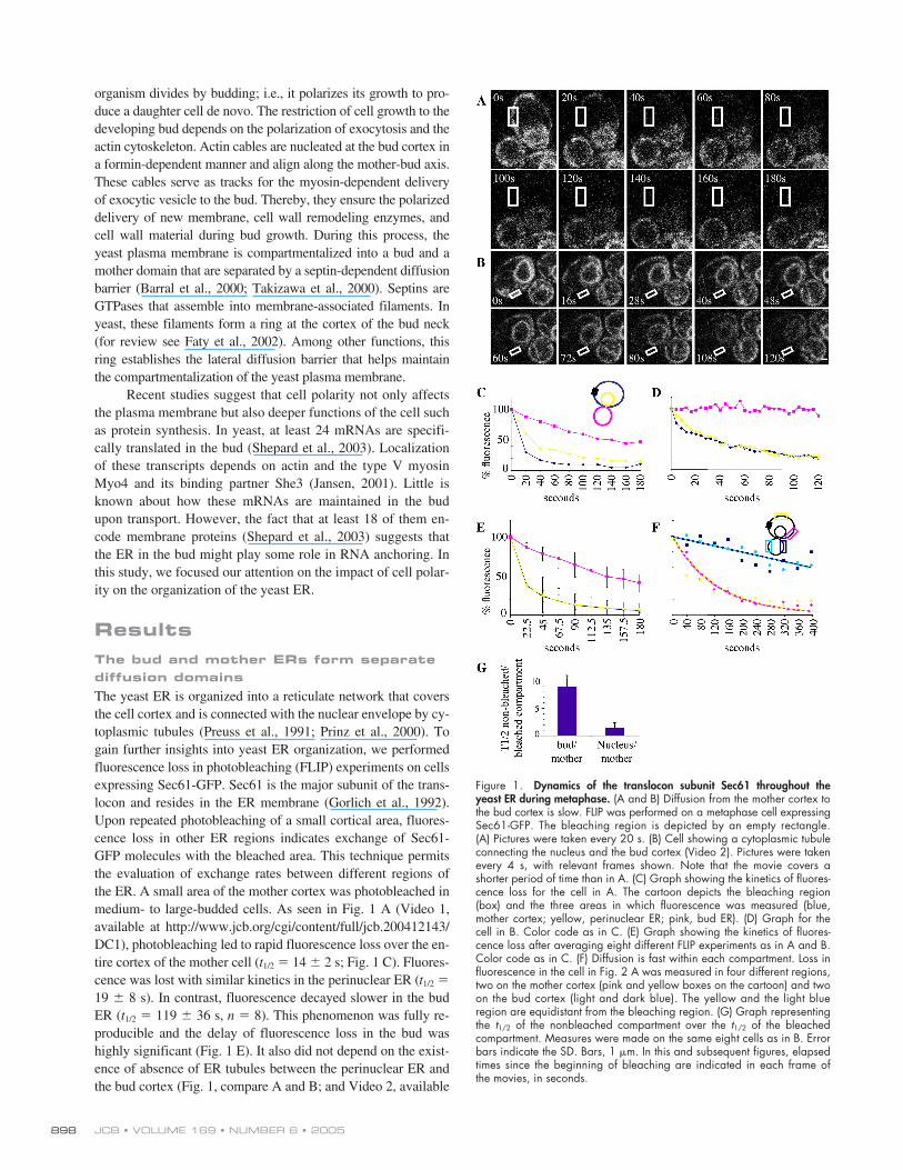
The yeast ER is organized into a reticulate network that coversthe cell cortex and is connected with the nuclear envelope by cy-toplasmic tubules (Preuss et al., 1991; Prinz et al., 2000). Togain further insights into yeast ER organization, we performedfluorescence loss in photobleaching (FLIP) experiments on cellsexpressing Sec61-GFP. Sec61 is the major subunit of the trans-locon and resides in the ER membrane (Gorlich et al., 1992).Upon repeated photobleaching of a small cortical area, fluores-cence loss in other ER regions indicates exchange of Sec61-GFP molecules with the bleached area. This technique permitsthe evaluation of exchange rates between different regions ofthe ER. A small area of the mother cortex was photobleached inmedium- to large-budded cells. As seen in Fig. 1 A (Video 1,available at http://www.jcb.org/cgi/content/full/jcb.200412143/DC1), photobleaching led to rapid fluorescence loss over the en-tire cortex of the mother cell (
t
1/2
�
14
�
2 s; Fig. 1 C). Fluores-cence was lost with similar kinetics in the perinuclear ER (
t
1/2
�
19
�
8 s). In contrast, fluorescence decayed slower in the budER (
t
1/2
�
119
�
36 s,
n
�
8). This phenomenon was fully re-producible and the delay of fluorescence loss in the bud washighly significant (Fig. 1 E). It also did not depend on the exist-ence of absence of ER tubules between the perinuclear ER andthe bud cortex (Fig. 1, compare A and B; and Video 2, available
Figure 1. Dynamics of the translocon subunit Sec61 throughout theyeast ER during metaphase. (A and B) Diffusion from the mother cortex tothe bud cortex is slow. FLIP was performed on a metaphase cell expressingSec61-GFP. The bleaching region is depicted by an empty rectangle.(A) Pictures were taken every 20 s. (B) Cell showing a cytoplasmic tubuleconnecting the nucleus and the bud cortex (Video 2). Pictures were takenevery 4 s, with relevant frames shown. Note that the movie covers ashorter period of time than in A. (C) Graph showing the kinetics of fluores-cence loss for the cell in A. The cartoon depicts the bleaching region(box) and the three areas in which fluorescence was measured (blue,mother cortex; yellow, perinuclear ER; pink, bud ER). (D) Graph for thecell in B. Color code as in C. (E) Graph showing the kinetics of fluores-cence loss after averaging eight different FLIP experiments as in A and B.Color code as in C. (F) Diffusion is fast within each compartment. Loss influorescence in the cell in Fig. 2 A was measured in four different regions,two on the mother cortex (pink and yellow boxes on the cartoon) and twoon the bud cortex (light and dark blue). The yellow and the light blueregion are equidistant from the bleaching region. (G) Graph representingthe t1/2 of the nonbleached compartment over the t1/2 of the bleachedcompartment. Measures were made on the same eight cells as in B. Errorbars indicate the SD. Bars, 1 �m. In this and subsequent figures, elapsedtimes since the beginning of bleaching are indicated in each frame ofthe movies, in seconds.
ER COMPARTMENTALIZATION IN YEAST • LUEDEKE ET AL.
899
at http://www.jcb.org/cgi/content/full/jcb.200412143/DC1).The ratio of
t
1/2
of the nonbleached compartment over the
t
1/2
ofthe bleached compartment is close to 1 for the perinuclear ER(1.4
�
0.8), but nine times higher (9
�
1.7; Fig. 1 G) for the budcortex. Although the
t
1/2
values observed in the different experi-ments highly depended on the photobleaching protocols used,this ratio remained fairly independent of the size of the bleachedarea and the duration and intensity of bleaching pulses. There-fore, it is used in the rest of the text to compare experiments andstrains. Altogether, these results indicate that Sec61 diffusion israpid within the mother cortical ER and between the mother cor-tex and the perinuclear ER. In contrast, Sec61 molecules ex-change slowly between mother and bud ERs.
To determine whether the slow loss of fluorescence in thebud is due to its distance to the bleached area, we compared therate of fluorescence loss between ER regions of the mother andbud that lied at different distances from the bleached area.Fluorescence decayed with similar kinetics in the different do-mains of the mother cortex (Fig. 1 F, yellow and pink traces).Thus, Sec61-GFP diffusion was instantaneous at the mothercortex, which can be treated as a single compartment. Simi-larly, the bud cortex also behaved as a single compartment(Fig. 1 F, light and dark blue traces). The region of the bud cor-tex marked in light blue and the region of the mother cortexmarked in yellow (Fig. 1 F) are equidistant from the bleachedarea. Thus, the fact that these two regions lose fluorescencewith distinct kinetics indicated that the difference observed be-tween mother and bud cortices was not due to their differentdistance to the bleached area.
Translation dramatically slows down the diffusion ofSec61 (Nikonov et al., 2002). Thus, differences in translational
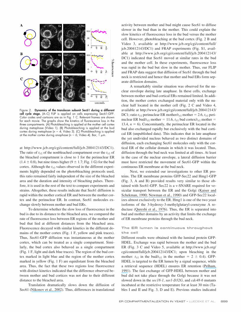
activity between mother and bud might cause Sec61 to diffuseslower in the bud than in the mother. This could explain theslow kinetics of fluorescence loss in the bud versus the mothercell. However, photobleaching at the bud cortex (Fig. 2 B andVideo 3, available at http://www.jcb.org/cgi/content/full/jcb.200412143/DC1) and FRAP experiments (Fig. S1, avail-able at http://www.jcb.org/cgi/content/full/jcb.200412143/DC1) indicated that Sec61 moved at similar rates in the budand the mother cell. In these experiments, fluorescence losswas rapid in the bud but slow in the mother. Thus, our FLIPand FRAP data suggest that diffusion of Sec61 through the budneck is restricted and hence that mother and bud ERs form sep-arate diffusion domains.
A remarkably similar situation was observed for the nu-clear envelope during late anaphase. In these cells, exchangebetween mother and bud cortical ERs remained limited. In addi-tion, the mother cortex exchanged material only with the nu-clear half located in the mother cell (Fig. 2 C and Video 4,available at http://www.jcb.org/cgi/content/full/jcb.200412143/DC1; ratio
t
1/2
perinuclear ER mother/
t
1/2
mother
�
2.6,
t
1/2
peri-nuclear ER bud/
t
1/2
mother
�
11.6,
t
1/2
bud cortex/
t
1/2
mother
�
13.4,
n
�
6). Concomitantly, the perinuclear ER located in thebud also exchanged rapidly but exclusively with the bud corti-cal ER (unpublished data). This indicates that in late anaphasethe yet undivided nucleus behaved as two distinct domains ofdiffusion, each exchanging Sec61 molecules only with the cor-tical ER of the cellular domain in which it was located. Thus,diffusion through the bud neck was limited at all times. At leastin the case of the nuclear envelope, a lateral diffusion barriermust have restricted the movement of Sec61-GFP within thecontinuous ER membrane at the bud neck.
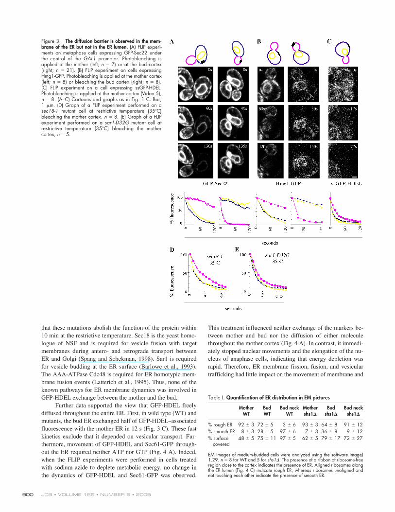
Next, we extended our investigations to other ER pro-teins. The ER membrane proteins GFP-Sec22 and Hmg1-GFP(Fig. 3, A and B) provided results very similar to those ob-tained with Sec61-GFP. Sec22 is a v-SNARE required for ve-sicular transport between the ER and the Golgi (Kaiser andSchekman, 1990; Newman et al., 1990) and GFP-Sec22 local-izes almost exclusively to the ER. Hmg1 is one of the two yeastisoforms of the 3-hydroxy-3-methylglutaryl-coenzyme A re-ductase (Qureshi et al., 1976). Thus, the ER is separated intobud and mother domains by an activity that limits the exchangeof ER membrane proteins through the bud neck.
The ER lumen is continuous throughout the cell
Different results were obtained with the luminal protein GFP-HDEL. Exchange was rapid between the mother and the budER (Fig. 3 C and Video 5, available at http://www.jcb.org/cgi/content/full/jcb.200412143/DC1; upon bleaching in themother:
t
1/2
in the bud/
t
1/2
in the mother
�
2
�
0.4). GFP-HDEL is targeted to the ER lumen by a signal sequence, whilea retrieval sequence (HDEL) ensures ER retention (Pelham,1991). The fast exchange of GFP-HDEL between mother andbud did not take place through the Golgi because it was notslowed down in the
sec18-1
,
sar1-D32G
, and
cdc48-6
mutantsincubated at the restrictive temperature for at least 30 min (Ta-bles I and II and Fig. 3, D and E). Previous studies indicated
Figure 2. Dynamics of the translocon subunit Sec61 during a differentcell cycle stage. (A–C) FLIP is applied on cells expressing Sec61-GFP.Color codes and cartoons are as in Fig. 1 C. Relevant frames are shownfor each movie. The graphs show the kinetics of fluorescence loss in thethree compartments. (A) Photobleaching is applied at the mother cell cortexduring metaphase (Video 1). (B) Photobleaching is applied at the budcortex during metaphase (n � 4; Video 3). (C) Photobleaching is appliedat the mother cortex during anaphase (n � 6; Video 4). Bar, 1 �m.
JCB • VOLUME 169 • NUMBER 6 • 2005900
that these mutations abolish the function of the protein within10 min at the restrictive temperature. Sec18 is the yeast homo-logue of NSF and is required for vesicle fusion with targetmembranes during antero- and retrograde transport betweenER and Golgi (Spang and Schekman, 1998). Sar1 is requiredfor vesicle budding at the ER surface (Barlowe et al., 1993).The AAA-ATPase Cdc48 is required for ER homotypic mem-brane fusion events (Latterich et al., 1995). Thus, none of theknown pathways for ER membrane dynamics was involved inGFP-HDEL exchange between the mother and the bud.
Further data supported the view that GFP-HDEL freelydiffused throughout the entire ER. First, in wild type (WT) andmutants, the bud ER exchanged half of GFP-HDEL–associatedfluorescence with the mother ER in 12 s (Fig. 3 C). These fastkinetics exclude that it depended on vesicular transport. Fur-thermore, movement of GFP-HDEL and Sec61-GFP through-out the ER required neither ATP nor GTP (Fig. 4 A). Indeed,when the FLIP experiments were performed in cells treatedwith sodium azide to deplete metabolic energy, no change inthe dynamics of GFP-HDEL and Sec61-GFP was observed.
This treatment influenced neither exchange of the markers be-tween mother and bud nor the diffusion of either moleculethroughout the mother cortex (Fig. 4 A). In contrast, it immedi-ately stopped nuclear movements and the elongation of the nu-cleus of anaphase cells, indicating that energy depletion wasrapid. Therefore, ER membrane fission, fusion, and vesiculartrafficking had little impact on the movement of membrane and
Figure 3. The diffusion barrier is observed in the mem-brane of the ER but not in the ER lumen. (A) FLIP experi-ments on metaphase cells expressing GFP-Sec22 underthe control of the GAL1 promotor. Photobleaching isapplied at the mother (left; n � 7) or at the bud cortex(right; n � 21). (B) FLIP experiment on cells expressingHmg1-GFP. Photobleaching is applied at the mother cortex(left; n � 8) or bleaching the bud cortex (right; n � 8).(C) FLIP experiment on a cell expressing ssGFP-HDEL.Photobleaching is applied at the mother cortex (Video 5),n � 8. (A–C) Cartoons and graphs as in Fig. 1 C. Bar,1 �m. (D) Graph of a FLIP experiment performed on asec18-1 mutant cell at restrictive temperature (35�C)bleaching the mother cortex. n � 8. (E) Graph of a FLIPexperiment performed on a sar1-D32G mutant cell atrestrictive temperature (35�C) bleaching the mothercortex, n � 5.
Table I.
Quantification of ER distribution in EM pictures
MotherWT
BudWT
Bud neckWT
Mothershs1
�
Budshs1
�
Bud neckshs1
�
% rough ER 92
�
3 72
�
5 3
�
6 93
�
3 64
�
8 91
�
12% smooth ER 8
�
3 28
�
5 97
�
6 7
�
3 36
�
8 9
�
12% surface
covered48
�
5 75
�
11 97
�
5 62
�
5 79
�
17 72
�
27
EM images of medium-budded cells were analyzed using the software ImageJ1.29.
n
�
8 for WT and 5 for
shs1
�
. The presence of a ribbon of ribosome-freeregion close to the cortex indicates the presence of ER. Aligned ribosomes alongthe ER lumen (Fig. 4 C) indicate rough ER, whereas ribosomes unaligned andnot touching each other indicate the presence of smooth ER.
ER COMPARTMENTALIZATION IN YEAST • LUEDEKE ET AL.
901
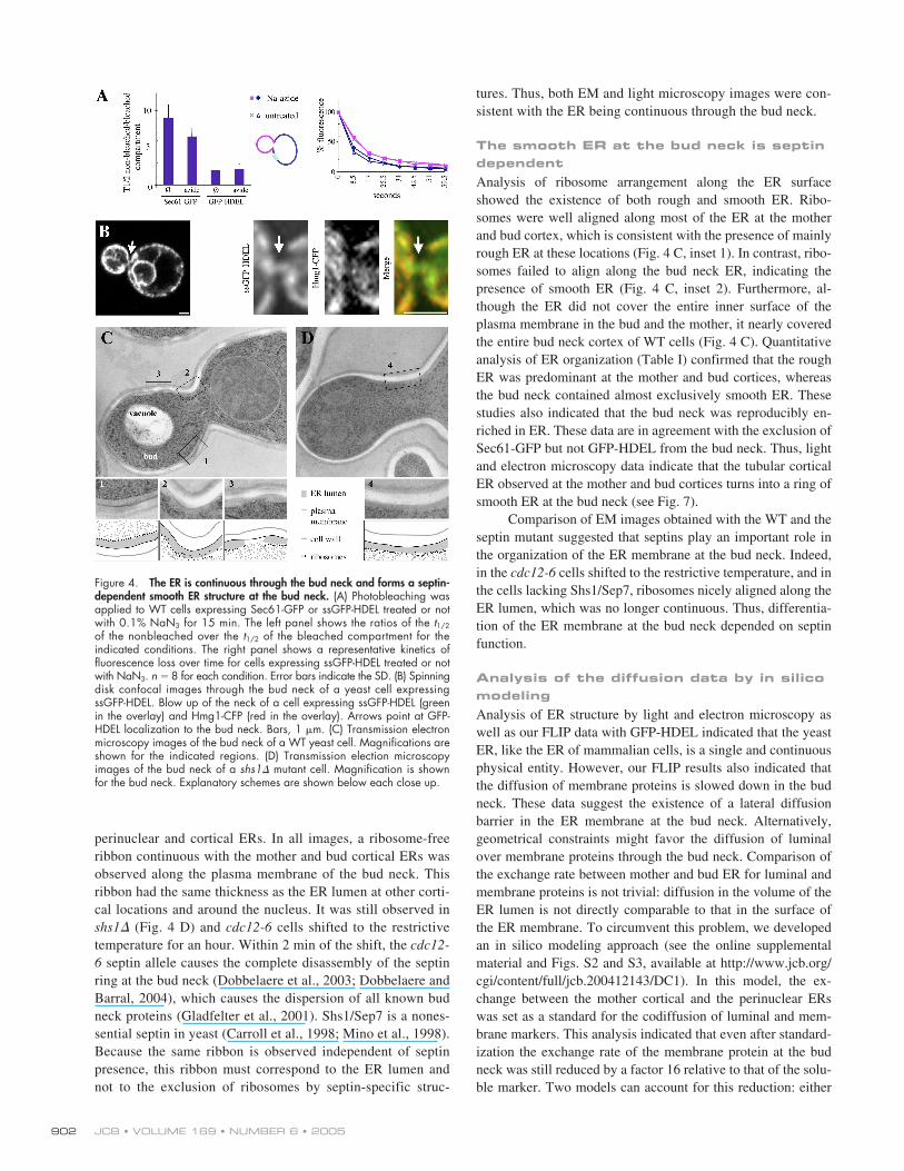
luminal proteins within the ER. Thus, the ER must be physi-cally continuous throughout the cell, including the bud neck.
The ER is continuous through the bud neck
To investigate this possibility, we characterized the morphol-ogy of the ER by light and electron microscopy. Using confo-cal microscopy and GFP-HDEL as a marker, the ER lumen wasobserved to be continuous through the bud neck (Fig. 4 B) ofmost cells. The ER membrane appeared also continuous at thebud neck when C
6
-BODIPY ceramide was used to visualize theER bilayer (unpublished data). In contrast, both ER membranemarkers Sec61-GFP (see Fig. 6 B) and Hmg1-GFP (not de-picted) were seen only very rarely and in those cases only tran-siently at the bud neck (e.g., Fig. 1 B, Sec61-GFP at 28 s). The
difference between the luminal and the ER membrane markerswas obvious when GFP-HDEL was coexpressed with Hmg1-CFP, in which case an ER domain labeled with GFP and de-void of CFP was observed at the bud neck (Fig. 4 B). Thus, ourdata indicate that the ER is continuous through the bud neckbut that the ER membrane at this location is different from therest of the ER membrane because it extensively lacks classicalER membrane proteins such as Sec61 and Hmg1.
Analysis of ER organization by high pressure freezingEM led to similar conclusions (Fig. 4 C). In these images, theER lumen is apparent as a ribosome-free ribbon around thenucleus and at the cell cortex. ER tubules are also observed inthe cytoplasm, some of which emerge from the cortical or theperinuclear ER. In rare instances, these ER tubules linked the
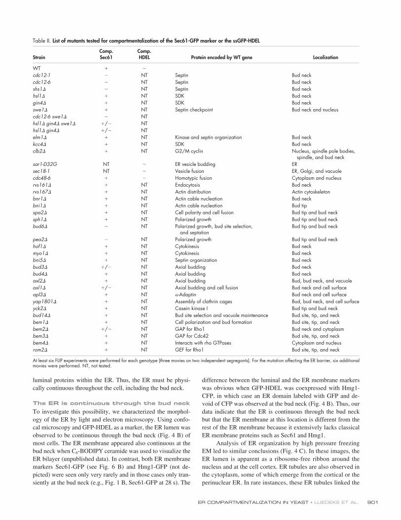
Table II.
List of mutants tested for compartmentalization of the Sec61-GFP marker or the ssGFP-HDEL
StrainComp.Sec61
Comp.HDEL Protein encoded by WT gene Localization
WT � �
cdc12-1 � NT Septin Bud neckcdc12-6 � NT Septin Bud neckshs1� � NT Septin Bud neckhsl1� � NT SDK Bud neckgin4� � NT SDK Bud neckswe1� � NT Septin checkpoint Bud neck and nucleuscdc12-6 swe1� � NThsl1� gin4� swe1� �/� NThsl1� gin4� �/� NTelm1� � NT Kinase and septin organization Bud neckkcc4� � NT SDK Bud neckclb2� � NT G2/M cyclin Nucleus, spindle pole bodies,
spindle, and bud necksar1-D32G NT � ER vesicle budding ERsec18-1 NT � Vesicle fusion ER, Golgi, and vacuolecdc48-6 � � Homotypic fusion Cytoplasm and nucleusrvs161� � NT Endocytosis Bud neckrvs167� � NT Actin distribution Actin cytoskeletonbnr1� � NT Actin cable nucleation Bud neckbni1� � NT Actin cable nucleation Bud tipspa2� � NT Cell polarity and cell fusion Bud tip and bud necksph1� � NT Polarized growth Bud tip and bud neckbud6� � NT Polarized growth, bud site selection,
and septationBud tip and bud neck
pea2� � NT Polarized growth Bud tip and bud neckhof1� � NT Cytokinesis Bud neckmyo1� � NT Cytokinesis Bud neckbni5� � NT Septin organization Bud neckbud3� �/� NT Axial budding Bud neckbud4� � NT Axial budding Bud neckaxl2� � NT Axial budding Bud, bud neck, and vacuoleaxl1� �/� NT Axial budding and cell fusion Bud neck and cell surfaceapl3� � NT �-Adaptin Bud neck and cell surfaceyap1801� � NT Assembly of clathrin cages Bud, bud neck, and cell surfaceyck2� � NT Casein kinase I Bud tip and bud neckbud14� � NT Bud site selection and vacuole maintenance Bud site, tip, and neckbem1� � NT Cell polarization and bud formation Bud site, tip, and neckbem2� �/� NT GAP for Rho1 Bud neck and cytoplasmbem3� � NT GAP for Cdc42 Bud site, tip, and neckbem4� � NT Interacts with rho GTPases Cytoplasm and nucleusrom2� � NT GEF for Rho1 Bud site, tip, and neck
At least six FLIP experiments were performed for each genotype (three movies on two independent segregants). For the mutation affecting the ER barrier, six additionalmovies were performed. NT, not tested.
JCB • VOLUME 169 • NUMBER 6 • 2005902
perinuclear and cortical ERs. In all images, a ribosome-freeribbon continuous with the mother and bud cortical ERs wasobserved along the plasma membrane of the bud neck. Thisribbon had the same thickness as the ER lumen at other corti-cal locations and around the nucleus. It was still observed inshs1� (Fig. 4 D) and cdc12-6 cells shifted to the restrictivetemperature for an hour. Within 2 min of the shift, the cdc12-6 septin allele causes the complete disassembly of the septinring at the bud neck (Dobbelaere et al., 2003; Dobbelaere andBarral, 2004), which causes the dispersion of all known budneck proteins (Gladfelter et al., 2001). Shs1/Sep7 is a nones-sential septin in yeast (Carroll et al., 1998; Mino et al., 1998).Because the same ribbon is observed independent of septinpresence, this ribbon must correspond to the ER lumen andnot to the exclusion of ribosomes by septin-specific struc-
tures. Thus, both EM and light microscopy images were con-sistent with the ER being continuous through the bud neck.
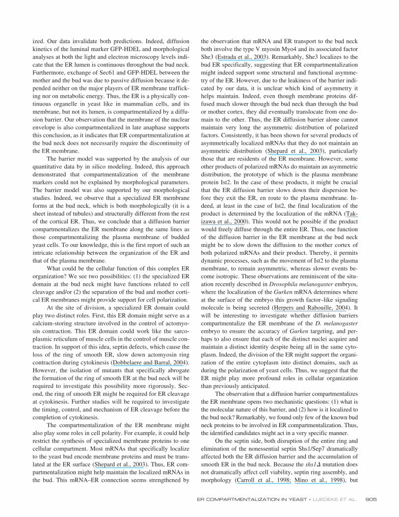
The smooth ER at the bud neck is septin dependentAnalysis of ribosome arrangement along the ER surfaceshowed the existence of both rough and smooth ER. Ribo-somes were well aligned along most of the ER at the motherand bud cortex, which is consistent with the presence of mainlyrough ER at these locations (Fig. 4 C, inset 1). In contrast, ribo-somes failed to align along the bud neck ER, indicating thepresence of smooth ER (Fig. 4 C, inset 2). Furthermore, al-though the ER did not cover the entire inner surface of theplasma membrane in the bud and the mother, it nearly coveredthe entire bud neck cortex of WT cells (Fig. 4 C). Quantitativeanalysis of ER organization (Table I) confirmed that the roughER was predominant at the mother and bud cortices, whereasthe bud neck contained almost exclusively smooth ER. Thesestudies also indicated that the bud neck was reproducibly en-riched in ER. These data are in agreement with the exclusion ofSec61-GFP but not GFP-HDEL from the bud neck. Thus, lightand electron microscopy data indicate that the tubular corticalER observed at the mother and bud cortices turns into a ring ofsmooth ER at the bud neck (see Fig. 7).
Comparison of EM images obtained with the WT and theseptin mutant suggested that septins play an important role inthe organization of the ER membrane at the bud neck. Indeed,in the cdc12-6 cells shifted to the restrictive temperature, and inthe cells lacking Shs1/Sep7, ribosomes nicely aligned along theER lumen, which was no longer continuous. Thus, differentia-tion of the ER membrane at the bud neck depended on septinfunction.
Analysis of the diffusion data by in silico modelingAnalysis of ER structure by light and electron microscopy aswell as our FLIP data with GFP-HDEL indicated that the yeastER, like the ER of mammalian cells, is a single and continuousphysical entity. However, our FLIP results also indicated thatthe diffusion of membrane proteins is slowed down in the budneck. These data suggest the existence of a lateral diffusionbarrier in the ER membrane at the bud neck. Alternatively,geometrical constraints might favor the diffusion of luminalover membrane proteins through the bud neck. Comparison ofthe exchange rate between mother and bud ER for luminal andmembrane proteins is not trivial: diffusion in the volume of theER lumen is not directly comparable to that in the surface ofthe ER membrane. To circumvent this problem, we developedan in silico modeling approach (see the online supplementalmaterial and Figs. S2 and S3, available at http://www.jcb.org/cgi/content/full/jcb.200412143/DC1). In this model, the ex-change between the mother cortical and the perinuclear ERswas set as a standard for the codiffusion of luminal and mem-brane markers. This analysis indicated that even after standard-ization the exchange rate of the membrane protein at the budneck was still reduced by a factor 16 relative to that of the solu-ble marker. Two models can account for this reduction: either
Figure 4. The ER is continuous through the bud neck and forms a septin-dependent smooth ER structure at the bud neck. (A) Photobleaching wasapplied to WT cells expressing Sec61-GFP or ssGFP-HDEL treated or notwith 0.1% NaN3 for 15 min. The left panel shows the ratios of the t1/2
of the nonbleached over the t1/2 of the bleached compartment for theindicated conditions. The right panel shows a representative kinetics offluorescence loss over time for cells expressing ssGFP-HDEL treated or notwith NaN3. n � 8 for each condition. Error bars indicate the SD. (B) Spinningdisk confocal images through the bud neck of a yeast cell expressingssGFP-HDEL. Blow up of the neck of a cell expressing ssGFP-HDEL (greenin the overlay) and Hmg1-CFP (red in the overlay). Arrows point at GFP-HDEL localization to the bud neck. Bars, 1 �m. (C) Transmission electronmicroscopy images of the bud neck of a WT yeast cell. Magnifications areshown for the indicated regions. (D) Transmission election microscopyimages of the bud neck of a shs1� mutant cell. Magnification is shownfor the bud neck. Explanatory schemes are shown below each close up.
ER COMPARTMENTALIZATION IN YEAST • LUEDEKE ET AL. 903
the morphology of the connections offers a larger volume/sur-face ratio at the bud neck and thereby favors the exchange ofthe soluble protein over that of the membrane bound, or the dif-fusion constant is specifically reduced in the membrane. Thefirst model predicts that the connections between mother andbud cortical ERs should be 16 times thicker than those betweencortical and perinuclear ER, which is in contradiction to ourEM data. Thus, the second model is most likely correct. Ourdata indicate the existence of a diffusion barrier slowing downthe diffusion of proteins in the ER membrane at the bud neck.This conclusion fits with the existence of a differentiated ERmembrane at the same location. Thus, we concluded that a dif-fusion barrier compartmentalized the ER membrane intomother and bud diffusion domains.
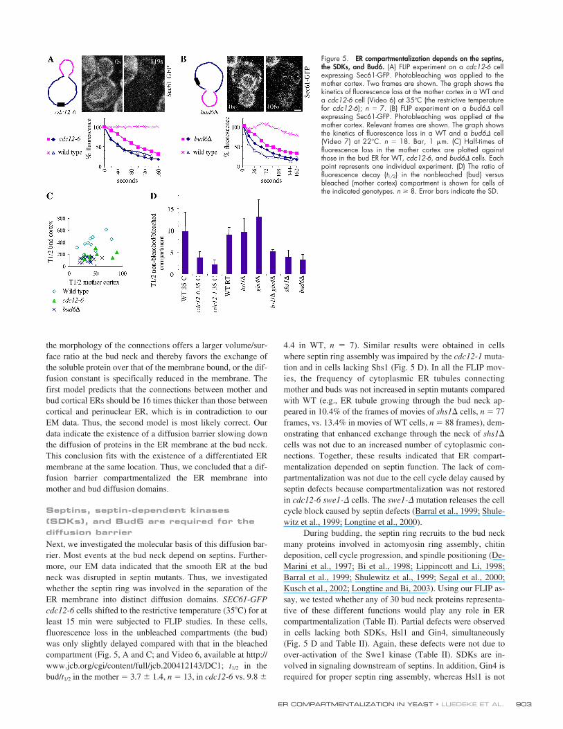
Septins, septin-dependent kinases (SDKs), and Bud6 are required for the diffusion barrierNext, we investigated the molecular basis of this diffusion bar-rier. Most events at the bud neck depend on septins. Further-more, our EM data indicated that the smooth ER at the budneck was disrupted in septin mutants. Thus, we investigatedwhether the septin ring was involved in the separation of theER membrane into distinct diffusion domains. SEC61-GFPcdc12-6 cells shifted to the restrictive temperature (35�C) for atleast 15 min were subjected to FLIP studies. In these cells,fluorescence loss in the unbleached compartments (the bud)was only slightly delayed compared with that in the bleachedcompartment (Fig. 5, A and C; and Video 6, available at http://www.jcb.org/cgi/content/full/jcb.200412143/DC1; t1/2 in thebud/t1/2 in the mother � 3.7 � 1.4, n � 13, in cdc12-6 vs. 9.8 �
4.4 in WT, n � 7). Similar results were obtained in cellswhere septin ring assembly was impaired by the cdc12-1 muta-tion and in cells lacking Shs1 (Fig. 5 D). In all the FLIP mov-ies, the frequency of cytoplasmic ER tubules connectingmother and buds was not increased in septin mutants comparedwith WT (e.g., ER tubule growing through the bud neck ap-peared in 10.4% of the frames of movies of shs1� cells, n � 77frames, vs. 13.4% in movies of WT cells, n � 88 frames), dem-onstrating that enhanced exchange through the neck of shs1�
cells was not due to an increased number of cytoplasmic con-nections. Together, these results indicated that ER compart-mentalization depended on septin function. The lack of com-partmentalization was not due to the cell cycle delay caused byseptin defects because compartmentalization was not restoredin cdc12-6 swe1-� cells. The swe1-� mutation releases the cellcycle block caused by septin defects (Barral et al., 1999; Shule-witz et al., 1999; Longtine et al., 2000).
During budding, the septin ring recruits to the bud neckmany proteins involved in actomyosin ring assembly, chitindeposition, cell cycle progression, and spindle positioning (De-Marini et al., 1997; Bi et al., 1998; Lippincott and Li, 1998;Barral et al., 1999; Shulewitz et al., 1999; Segal et al., 2000;Kusch et al., 2002; Longtine and Bi, 2003). Using our FLIP as-say, we tested whether any of 30 bud neck proteins representa-tive of these different functions would play any role in ERcompartmentalization (Table II). Partial defects were observedin cells lacking both SDKs, Hsl1 and Gin4, simultaneously(Fig. 5 D and Table II). Again, these defects were not due toover-activation of the Swe1 kinase (Table II). SDKs are in-volved in signaling downstream of septins. In addition, Gin4 isrequired for proper septin ring assembly, whereas Hsl1 is not
Figure 5. ER compartmentalization depends on the septins,the SDKs, and Bud6. (A) FLIP experiment on a cdc12-6 cellexpressing Sec61-GFP. Photobleaching was applied to themother cortex. Two frames are shown. The graph shows thekinetics of fluorescence loss at the mother cortex in a WT anda cdc12-6 cell (Video 6) at 35�C (the restrictive temperaturefor cdc12-6); n � 7. (B) FLIP experiment on a bud6� cellexpressing Sec61-GFP. Photobleaching was applied at themother cortex. Relevant frames are shown. The graph showsthe kinetics of fluorescence loss in a WT and a bud6� cell(Video 7) at 22�C. n � 18. Bar, 1 �m. (C) Half-times offluorescence loss in the mother cortex are plotted againstthose in the bud ER for WT, cdc12-6, and bud6� cells. Eachpoint represents one individual experiment. (D) The ratio offluorescence decay (t1/2) in the nonbleached (bud) versusbleached (mother cortex) compartment is shown for cells ofthe indicated genotypes. n 8. Error bars indicate the SD.
JCB • VOLUME 169 • NUMBER 6 • 2005904
(Longtine et al., 2000; Dobbelaere et al., 2003). Because cellslacking Gin4 alone are not defective in ER compartmentaliza-tion, septin organization defects do not account for the com-partmentalization defect observed in the hsl1� gin4� doublemutant. These results suggest that SDKs act redundantly anddownstream of septins on ER organization. They might modifyER proteins at the bud neck.
In addition to SDKs, only Bud6 and Pea2 were involvedin ER compartmentalization. In bud6� and pea2� cells the lossof compartmentalization was similar to that caused by septinmutations (Table II; Fig. 5, B–D; and Video 7, available athttp://www.jcb.org/cgi/content/full/jcb.200412143/DC1). Bud6(Amberg et al., 1997), which is a peripheral membrane protein,has been implicated in actin polarization, microtubule interac-tion with the cell cortex (Segal et al., 2000), and bud site selec-tion in diploid cells. Together with Pea2, it is a component ofthe polarizome (Sheu et al., 2000), a complex that also containseither of the formins Bni1 and Bnr1, and the polarity factorsSpa2 and Sph1. Analysis of ER compartmentalization inbni1�, bnr1�, spa2�, and sph1� cells showed no defect. Thus,the role of Bud6 and Pea2 in ER compartmentalization ap-peared not to be shared by the other components of the polari-zome. Furthermore, like shs1�, the bud6� mutation did not in-crease the number of cytoplasmic ER tubules connectingmother and bud (tubules were present in 11.4% of the frames ofbud6� movies, n � 138 frames, vs. 13.4% in WT movies).Thus, the stimulation of exchange between the mother and thebud in bud6� cells was due to a compartmentalization defectand not to an increased number of ER tubules.
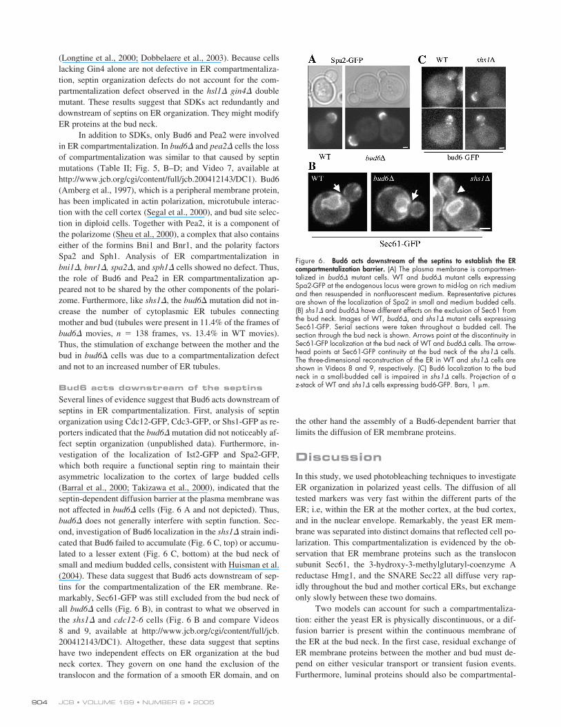
Bud6 acts downstream of the septinsSeveral lines of evidence suggest that Bud6 acts downstream ofseptins in ER compartmentalization. First, analysis of septinorganization using Cdc12-GFP, Cdc3-GFP, or Shs1-GFP as re-porters indicated that the bud6� mutation did not noticeably af-fect septin organization (unpublished data). Furthermore, in-vestigation of the localization of Ist2-GFP and Spa2-GFP,which both require a functional septin ring to maintain theirasymmetric localization to the cortex of large budded cells(Barral et al., 2000; Takizawa et al., 2000), indicated that theseptin-dependent diffusion barrier at the plasma membrane wasnot affected in bud6� cells (Fig. 6 A and not depicted). Thus,bud6� does not generally interfere with septin function. Sec-ond, investigation of Bud6 localization in the shs1� strain indi-cated that Bud6 failed to accumulate (Fig. 6 C, top) or accumu-lated to a lesser extent (Fig. 6 C, bottom) at the bud neck ofsmall and medium budded cells, consistent with Huisman et al.(2004). These data suggest that Bud6 acts downstream of sep-tins for the compartmentalization of the ER membrane. Re-markably, Sec61-GFP was still excluded from the bud neck ofall bud6� cells (Fig. 6 B), in contrast to what we observed inthe shs1� and cdc12-6 cells (Fig. 6 B and compare Videos8 and 9, available at http://www.jcb.org/cgi/content/full/jcb.200412143/DC1). Altogether, these data suggest that septinshave two independent effects on ER organization at the budneck cortex. They govern on one hand the exclusion of thetranslocon and the formation of a smooth ER domain, and on
the other hand the assembly of a Bud6-dependent barrier thatlimits the diffusion of ER membrane proteins.
DiscussionIn this study, we used photobleaching techniques to investigateER organization in polarized yeast cells. The diffusion of alltested markers was very fast within the different parts of theER; i.e, within the ER at the mother cortex, at the bud cortex,and in the nuclear envelope. Remarkably, the yeast ER mem-brane was separated into distinct domains that reflected cell po-larization. This compartmentalization is evidenced by the ob-servation that ER membrane proteins such as the transloconsubunit Sec61, the 3-hydroxy-3-methylglutaryl-coenzyme Areductase Hmg1, and the SNARE Sec22 all diffuse very rap-idly throughout the bud and mother cortical ERs, but exchangeonly slowly between these two domains.
Two models can account for such a compartmentaliza-tion: either the yeast ER is physically discontinuous, or a dif-fusion barrier is present within the continuous membrane ofthe ER at the bud neck. In the first case, residual exchange ofER membrane proteins between the mother and bud must de-pend on either vesicular transport or transient fusion events.Furthermore, luminal proteins should also be compartmental-
Figure 6. Bud6 acts downstream of the septins to establish the ERcompartmentalization barrier. (A) The plasma membrane is compartmen-talized in bud6� mutant cells. WT and bud6� mutant cells expressingSpa2-GFP at the endogenous locus were grown to mid-log on rich mediumand then resuspended in nonfluorescent medium. Representative picturesare shown of the localization of Spa2 in small and medium budded cells.(B) shs1� and bud6� have different effects on the exclusion of Sec61 fromthe bud neck. Images of WT, bud6�, and shs1� mutant cells expressingSec61-GFP. Serial sections were taken throughout a budded cell. Thesection through the bud neck is shown. Arrows point at the discontinuity inSec61-GFP localization at the bud neck of WT and bud6� cells. The arrow-head points at Sec61-GFP continuity at the bud neck of the shs1� cells.The three-dimensional reconstruction of the ER in WT and shs1� cells areshown in Videos 8 and 9, respectively. (C) Bud6 localization to the budneck in a small-budded cell is impaired in shs1� cells. Projection of az-stack of WT and shs1� cells expressing bud6-GFP. Bars, 1 �m.
ER COMPARTMENTALIZATION IN YEAST • LUEDEKE ET AL. 905
ized. Our data invalidate both predictions. Indeed, diffusionkinetics of the luminal marker GFP-HDEL and morphologicalanalyses at both the light and electron microscopy levels indi-cate that the ER lumen is continuous throughout the bud neck.Furthermore, exchange of Sec61 and GFP-HDEL between themother and the bud was due to passive diffusion because it de-pended neither on the major players of ER membrane traffick-ing nor on metabolic energy. Thus, the ER is a physically con-tinuous organelle in yeast like in mammalian cells, and itsmembrane, but not its lumen, is compartmentalized by a diffu-sion barrier. Our observation that the membrane of the nuclearenvelope is also compartmentalized in late anaphase supportsthis conclusion, as it indicates that ER compartmentalization atthe bud neck does not necessarily require the discontinuity ofthe ER membrane.
The barrier model was supported by the analysis of ourquantitative data by in silico modeling. Indeed, this approachdemonstrated that compartmentalization of the membranemarkers could not be explained by morphological parameters.The barrier model was also supported by our morphologicalstudies. Indeed, we observe that a specialized ER membraneforms at the bud neck, which is both morphologically (it is asheet instead of tubules) and structurally different from the restof the cortical ER. Thus, we conclude that a diffusion barriercompartmentalizes the ER membrane along the same lines asthose compartmentalizing the plasma membrane of buddedyeast cells. To our knowledge, this is the first report of such anintricate relationship between the organization of the ER andthat of the plasma membrane.
What could be the cellular function of this complex ERorganization? We see two possibilities: (1) the specialized ERdomain at the bud neck might have functions related to cellcleavage and/or (2) the separation of the bud and mother corti-cal ER membranes might provide support for cell polarization.
At the site of division, a specialized ER domain couldplay two distinct roles. First, this ER domain might serve as acalcium-storing structure involved in the control of actomyo-sin contraction. This ER domain could work like the sarco-plasmic reticulum of muscle cells in the control of muscle con-traction. In support of this idea, septin defects, which cause theloss of the ring of smooth ER, slow down actomyosin ringcontraction during cytokinesis (Dobbelaere and Barral, 2004).However, the isolation of mutants that specifically abrogatethe formation of the ring of smooth ER at the bud neck will berequired to investigate this possibility more rigorously. Sec-ond, the ring of smooth ER might be required for ER cleavageat cytokinesis. Further studies will be required to investigatethe timing, control, and mechanism of ER cleavage before thecompletion of cytokinesis.
The compartmentalization of the ER membrane mightalso play some roles in cell polarity. For example, it could helprestrict the synthesis of specialized membrane proteins to onecellular compartment. Most mRNAs that specifically localizeto the yeast bud encode membrane proteins and must be trans-lated at the ER surface (Shepard et al., 2003). Thus, ER com-partmentalization might help maintain the localized mRNAs inthe bud. This mRNA–ER connection seems strengthened by
the observation that mRNA and ER transport to the bud neckboth involve the type V myosin Myo4 and its associated factorShe3 (Estrada et al., 2003). Remarkably, She3 localizes to thebud ER specifically, suggesting that ER compartmentalizationmight indeed support some structural and functional asymme-try of the ER. However, due to the leakiness of the barrier indi-cated by our data, it is unclear which kind of asymmetry ithelps maintain. Indeed, even though membrane proteins dif-fused much slower through the bud neck than through the budor mother cortex, they did eventually translocate from one do-main to the other. Thus, the ER diffusion barrier alone cannotmaintain very long the asymmetric distribution of polarizedfactors. Consistently, it has been shown for several products ofasymmetrically localized mRNAs that they do not maintain anasymmetric distribution (Shepard et al., 2003), particularlythose that are residents of the ER membrane. However, someother products of polarized mRNAs do maintain an asymmetricdistribution, the prototype of which is the plasma membraneprotein Ist2. In the case of these products, it might be crucialthat the ER diffusion barrier slows down their dispersion be-fore they exit the ER, en route to the plasma membrane. In-deed, at least in the case of Ist2, the final localization of theproduct is determined by the localization of the mRNA (Tak-izawa et al., 2000). This would not be possible if the productwould freely diffuse through the entire ER. Thus, one functionof the diffusion barrier in the ER membrane at the bud neckmight be to slow down the diffusion to the mother cortex ofboth polarized mRNAs and their product. Thereby, it permitsdynamic processes, such as the movement of Ist2 to the plasmamembrane, to remain asymmetric, whereas slower events be-come isotropic. These observations are reminiscent of the situ-ation recently described in Drosophila melanogaster embryos,where the localization of the Gurken mRNA determines whereat the surface of the embryo this growth factor–like signalingmolecule is being secreted (Herpers and Rabouille, 2004). Itwill be interesting to investigate whether diffusion barrierscompartmentalize the ER membrane of the D. melanogasterembryo to ensure the accuracy of Gurken targeting, and per-haps to also ensure that each of the distinct nuclei acquire andmaintain a distinct identity despite being all in the same cyto-plasm. Indeed, the division of the ER might support the organi-zation of the entire cytoplasm into distinct domains, such asduring the polarization of yeast cells. Thus, we suggest that theER might play more profound roles in cellular organizationthan previously anticipated.
The observation that a diffusion barrier compartmentalizesthe ER membrane opens two mechanistic questions: (1) what isthe molecular nature of this barrier, and (2) how is it localized tothe bud neck? Remarkably, we found only few of the known budneck proteins to be involved in ER compartmentalization. Thus,the identified candidates might act in a very specific manner.
On the septin side, both disruption of the entire ring andelimination of the nonessential septin Shs1/Sep7 dramaticallyaffected both the ER diffusion barrier and the accumulation ofsmooth ER in the bud neck. Because the shs1� mutation doesnot dramatically affect cell viability, septin ring assembly, andmorphology (Carroll et al., 1998; Mino et al., 1998), but
JCB • VOLUME 169 • NUMBER 6 • 2005906
strongly impacts on ER organization, Shs1 may be specializedfor this process. In turn, Bud6 affected only the diffusion bar-rier and not ER morphology. Thus, the smooth ER at the budneck does not depend on the presence of the barrier and is notsufficient to form a barrier. Previous data have established thatBud6 is associated with internal membranes (Jin and Amberg,2000). We propose that Bud6 is a smooth ER protein (Fig. 7),where it might influence protein diffusion.
The role of septins in the recruitment or regulation of ERproteins such as Bud6 might be indirect and mediated at leastin part through signaling. Indeed, we found that the SDKs Gin4and Hsl1 are also involved in the formation of the diffusionbarrier. Because SDKs did not seem to function in smooth ERorganization, their substrates must be specifically involved inER compartmentalization. It will be helpful to identify thesesubstrates and to investigate whether Bud6 is one of them. Theview that septins act on the ER via signaling mechanismsmight explain how they could potentially act at a distance dur-ing, for example, the compartmentalization of the nuclear en-velope at the bud neck during anaphase.
It is unclear to which extent the observations that we de-scribe here are specific to ER of fungi or relevant for other eu-karyotes. At first sight, the fact that the ER of animal cells ismainly cytoplasmic, and not cortical as in fungi, suggests thatER compartmentalization is less likely in metazoans. It will beinteresting to perform FLIP experiments on polarized and di-viding cells and to investigate whether the role that we ascribeto the cleavage apparatus in the compartmentalization of theyeast ER is conserved during the division of animal cells.
Materials and methodsStrain constructionYeast strains were constructed by standard genetic techniques. Diploidswere isolated on selective medium and subsequently sporulated at 23�C.The background is, unless specified otherwise, S288c. ssGFP-HDEL wasexpressed from the 2-�m vector pG14 (Lesser and Guthrie, 1993); ssGFP-HDEL has the signal peptide of CTS1 and the HDEL retrieval sequence at itsCOOH terminus (a gift from E. Bertrand, Centre National de la RechercheScientifique, Montpellier, France). Strains containing Sec61-GFP (providedby D. Liakopoulos, Swiss Federal Institute of Technology [ETH], Zurich,Switzerland), Spa2-GFP, Bud6-GFP, Hmg1-GFP, or pGAL-GFP-Sec22 weremade using the PCR-based integration system (Longtine et al., 1998). The
different deletions shown in Table I were obtained from the EUROSCARFdeletion collection (S288c) and provided to us by M. Peter (ETH, Zurich,Switzerland). Each time that an effect was observed, the mutation wasbackcrossed several times into our background. The hsl1�, gin4�, shs1�,and swe1� strains are isogenic with S288c. Cdc12-GFP was expressedfrom a centromeric plasmid (Dobbelaere et al., 2003). The sec18-1 andcdc48-6 strains were gifts of R. Collins (Cornell University, Ithaca, NY) andS. Jentsch (Max Planck Institute, Münich, Germany), respectively.
FLIP and FRAP experimentsCells were grown on YPD plates, resuspended in liquid nonfluorescent me-dium, and immobilized on nonfluorescent medium (Waddle et al., 1996)containing 1.6% agarose. Photobleaching was applied on the areashown on the figures, using a microscope (model LSM510; Carl Zeiss Mi-croImaging, Inc.) and a Plan-Apochromat 100 objective (NA 1.4). ForFRAP, scans were collected at 5-s intervals for a minimum of 120 s usingthe acquisition software LSM510 (Carl Zeiss MicroImaging, Inc.). Bleach-ing regions were irradiated with 250 iterations of 50% laser intensity at30% output of an argon laser (488 nm) and scans were collected with typ-ically 1% laser intensity at the same conditions. All pictures of FLIP experi-ments shown in the figures were treated to account for the bleaching dueto image acquisition, whereas the movies were left untreated.
Pictures shown in Fig. 4 B were taken on a spinning-disc confocal sys-tem (Axiovert 200M; Carl Zeiss MicroImaging, Inc.) with a Plan-Apochro-mat 100 objective. The overlay picture was taken on a DeltaVision micro-scope (Applied Precision) and deconvolved using the softwox software.
FLIP analysis of septin mutants (cdc12-1 and cdc12-6)cdc12-1 mutant cells were grown to early log phase at permissive temper-ature (24�C) in liquid YPD medium and arrested in G1 with 5 �g/ml�-factor for 2.5 h. After removal of �-factor from the medium by washingtwice with fresh YPD, cells were mounted on nonfluorescent agarose bedsas described in the previous section and immediately shifted to 35�C on aheated stage. FLIP analysis was performed on buds formed after the tem-perature shift, starting after 30 min. cdc12-6 cells were grown to mid-logphase on YPD plates at 22�C (permissive), mounted on agarose beds, andshifted to 35�C on the heated stage. FLIP analysis was performed in me-dium- to large-budded preanaphase cells 30 min after temperature shift.
Quantification of FLIP experimentsAnalysis of the FLIP experiments was performed using the ImageJ 1.29software (http://rsb.info.nih.gov/ij). The loss of fluorescence over timewas measured in different regions of interest (usually the mother cortex,bud cortex, and perinuclear ER). In addition, we measured the loss of fluo-rescence on neighboring control cells to account for the loss due to visual-ization. Finally, we measured the intensity of the background. The intensityin the region of interest was calculated as (region � background)/(controlcell � background) and then put in fractions.
BODIPY staining of ER membranes, light and electron microscopy, and image processingFor BODIPY staining, cells were grown overnight to early to mid-logphase in YPD, harvested, and resuspended in SC media to 5 OD600/ml.The culture was incubated for 10 min at 30�C under agitation. DefattedBSA was added to a final concentration of 5 mg/ml and supplementedwith 2.5 �l C6 BODIPY ceramide (4 mM stock in DMSO; MolecularProbes). The cells were incubated for 20 min at 30�C under agitationand mounted for direct inspection. Alternatively, the cells were fixed af-ter the incubation period with 4% formaldehyde. No difference in thestaining was detected between life and fixed cells. Light microscopy wasperformed using either a DeltaVision microscope (DeltaVision SpectrisSystem; Applied Precision) equipped with a Coolsnap HQ camera(Roper Scientific) or an Olympus BX50 equipped with a camera Imago(TiLL Photonics). In all cases, we used 100 objectives of NA 1.4. De-convolution was performed by a constrained iterative method using thesoftwox software (Applied Precision). Further image processing was per-formed using the Photoshop software (Adobe) and was reduced to the op-timization of the levels. No gamma adjustments were applied. Unless oth-erwise indicated, light microscopy was performed at RT (22�C) asdescribed previously (Dobbelaere et al., 2003). EM was performed as de-scribed previously (Sandmann et al., 2003).
ModelThe quantitative transport model was formulated on the basis of standardphysical principles, leading to a system of coupled differential equations.Photobleaching and scanning cycles were modeled as algebraic equations.
Figure 7. Model of rough and smooth ER in the bud neck of yeast cells.Sections through the septin filaments are shown in green. Bud6 localizationto the smooth ER is symbolized by red dots.
ER COMPARTMENTALIZATION IN YEAST • LUEDEKE ET AL. 907
Online supplemental materialThe quantitative transport model is fully described and assessed in the on-line supplemental material. Online supplemental material is available athttp://www.jcb.org/cgi/content/full/jcb.200412143/DC1.
We are grateful to Markus Aebi and Ari Helenius for helpful discussions. Wethank E. Bertrand, R. Collins, S. Jentsch, and M. Peter for strains and con-structs; D. Liakopoulos for the Sec61-GFP construct; and J. Dobbelaere fortechnical help.
This work was supported by the Swiss Federal Institute of Technology(ETH), the Swiss National Science Foundation (Y. Barral), and the Max PlanckSociety (A. Spang). S. Buvelot Frei was supported by a postdoctoral fellow-ship from the Roche Foundation and I. Sbalzarini by the ETH Strategic Excel-lence Project in Computational Science. Y. Barral and A. Spang are Euro-pean Molecular Biology Organization Young Investigators.
Submitted: 22 December 2004Accepted: 12 May 2005
ReferencesAmberg, D.C., J.E. Zahner, J.W. Mulholland, J.R. Pringle, and D. Botstein.
1997. Aip3p/Bud6p, a yeast actin-interacting protein that is involved inmorphogenesis and the selection of bipolar budding sites. Mol. Biol.Cell. 8:729–753.
Barlowe, C., C. d’Enfert, and R. Schekman. 1993. Purification and characteriza-tion of SAR1p, a small GTP-binding protein required for transport vesicleformation from the endoplasmic reticulum. J. Biol. Chem. 268:873–879.
Barr, F.A. 2002. Inheritance of the endoplasmic reticulum and Golgi apparatus.Curr. Opin. Cell Biol. 14:496–499.
Barral, Y., M. Parra, S. Bidlingmaier, and M. Snyder. 1999. Nim1-related ki-nases coordinate cell cycle progression with the organization of the pe-ripheral cytoskeleton in yeast. Genes Dev. 13:176–187.
Barral, Y., V. Mermall, M.S. Mooseker, and M. Snyder. 2000. Compartmental-ization of the cell cortex by septins is required for maintenance of cellpolarity in yeast. Mol. Cell. 5:841–851.
Baumann, O., and B. Walz. 2001. Endoplasmic reticulum of animal cells andits organization into structural and functional domains. Int. Rev. Cytol.205:149–214.
Bi, E., P. Maddox, D.J. Lew, E.D. Salmon, J.N. McMillan, E. Yeh, and J.R.Pringle. 1998. Involvement of an actomyosin contractile ring in Saccha-romyces cerevisiae cytokinesis. J. Cell Biol. 142:1301–1312.
Boiko, T., and B. Winckler. 2003. Picket and other fences in biological mem-branes. Dev. Cell. 5:191–192.
Carroll, C.W., R. Altman, D. Schieltz, J.R. Yates, and D. Kellogg. 1998. Theseptins are required for the mitosis-specific activation of the Gin4 kinase.J. Cell Biol. 143:709–717.
Dayel, M.J., E.F. Hom, and A.S. Verkman. 1999. Diffusion of green fluorescentprotein in the aqueous-phase lumen of endoplasmic reticulum. Biophys. J.76:2843–2851.
DeMarini, D.J., A.E. Adams, H. Fares, C. De Virgilio, G. Valle, J.S. Chuang,and J.R. Pringle. 1997. A septin-based hierarchy of proteins required forlocalized deposition of chitin in the Saccharomyces cerevisiae cell wall.J. Cell Biol. 139:75–93.
Dobbelaere, J., and Y. Barral. 2004. Spatial coordination of cytokinetic eventsby compartmentalization of the cell cortex. Science. 305:393–396.
Dobbelaere, J., M.S. Gentry, R.L. Hallberg, and Y. Barral. 2003. Phosphoryla-tion-dependent regulation of septin dynamics during the cell cycle. Dev.Cell. 4:345–357.
Du, Y., S. Ferro-Novick, and P. Novick. 2004. Dynamics and inheritance of theendoplasmic reticulum. J. Cell Sci. 117:2871–2878.
Estrada, P., J. Kim, J. Coleman, L. Walker, B. Dunn, P. Takizawa, P. Novick, andS. Ferro-Novick. 2003. Myo4p and She3p are required for cortical ER in-heritance in Saccharomyces cerevisiae. J. Cell Biol. 163:1255–1266.
Faty, M., M. Fink, and Y. Barral. 2002. Septins: a ring to part mother anddaughter. Curr. Genet. 41:123–131.
Gladfelter, A.S., J.R. Pringle, and D.J. Lew. 2001. The septin cortex at the yeastmother-bud neck. Curr. Opin. Microbiol. 4:681–689.
Gorlich, D., S. Prehn, E. Hartmann, K.U. Kalies, and T.A. Rapoport. 1992. Amammalian homolog of SEC61p and SECYp is associated with ribo-somes and nascent polypeptides during translocation. Cell. 71:489–503.
Herpers, B., and C. Rabouille. 2004. mRNA localization and ER-based proteinsorting mechanisms dictate the use of transitional endoplasmic reticu-lum-golgi units involved in gurken transport in Drosophila oocytes. Mol.Biol. Cell. 15:5306–5317.
Huisman, S.M., O.A. Bales, M. Bertrand, M.F. Smeets, S.I. Reed, and M. Segal.2004. Differential contribution of Bud6p and Kar9p to microtubule cap-ture and spindle orientation in S. cerevisiae. J. Cell Biol. 167:231–244.
Jansen, R.P. 2001. mRNA localization: message on the move. Nat. Rev. Mol.Cell Biol. 2:247–256.
Jin, H., and D.C. Amberg. 2000. The secretory pathway mediates localization ofthe cell polarity regulator Aip3p/Bud6p. Mol. Biol. Cell. 11:647–661.
Kaiser, C.A., and R. Schekman. 1990. Distinct sets of SEC genes govern trans-port vesicle formation and fusion early in the secretory pathway. Cell.61:723–733.
Kusch, J., A. Meyer, M.P. Snyder, and Y. Barral. 2002. Microtubule capture bythe cleavage apparatus is required for proper spindle positioning in yeast.Genes Dev. 16:1627–1639.
Latterich, M., K.U. Frohlich, and R. Schekman. 1995. Membrane fusion andthe cell cycle: Cdc48p participates in the fusion of ER membranes.Cell. 82:885–893.
Lesser, C.F., and C. Guthrie. 1993. Mutational analysis of pre-mRNA splicingin Saccharomyces cerevisiae using a sensitive new reporter gene, CUP1.Genetics. 133:851–863.
Lippincott, J., and R. Li. 1998. Sequential assembly of myosin II, an IQGAP-like protein, and filamentous actin to a ring structure involved in buddingyeast cytokinesis. J. Cell Biol. 140:355–366.
Longtine, M.S., and E. Bi. 2003. Regulation of septin organization and functionin yeast. Trends Cell Biol. 13:403–409.
Longtine, M.S., A. McKenzie III, D.J. Demarini, N.G. Shah, A. Wach, A.Brachat, P. Philippsen, and J.R. Pringle. 1998. Additional modules forversatile and economical PCR-based gene deletion and modification inSaccharomyces cerevisiae. Yeast. 14:953–961.
Longtine, M.S., C.L. Theesfeld, J.N. McMillan, E. Weaver, J.R. Pringle, andD.J. Lew. 2000. Septin-dependent assembly of a cell cycle-regulatorymodule in Saccharomyces cerevisiae. Mol. Cell. Biol. 20:4049–4061.
Ma, Y., and L.M. Hendershot. 2001. The unfolding tale of the unfolded proteinresponse. Cell. 107:827–830.
Matlack, K.E., W. Mothes, and T.A. Rapoport. 1998. Protein translocation: tun-nel vision. Cell. 92:381–390.
McMaster, C.R. 2001. Lipid metabolism and vesicle trafficking: more than justgreasing the transport machinery. Biochem. Cell Biol. 79:681–692.
Meldolesi, J., and T. Pozzan. 1998. The endoplasmic reticulum Ca2� store: aview from the lumen. Trends Biochem. Sci. 23:10–14.
Mino, A., K. Tanaka, T. Kamei, M. Umikawa, T. Fujiwara, and Y. Takai. 1998.Shs1p: a novel member of septin that interacts with spa2p, involved inpolarized growth in Saccharomyces cerevisiae. Biochem. Biophys. Res.Commun. 251:732–736.
Newman, A.P., J. Shim, and S. Ferro-Novick. 1990. BET1, BOS1, and SEC22are members of a group of interacting yeast genes required for transportfrom the endoplasmic reticulum to the Golgi complex. Mol. Cell. Biol.10:3405–3414.
Nikonov, A.V., E. Snapp, J. Lippincott-Schwartz, and G. Kreibich. 2002. Activetranslocon complexes labeled with GFP–Dad1 diffuse slowly as largepolysome arrays in the endoplasmic reticulum. J. Cell Biol. 158:497–506.
Pelham, H.R. 1991. Recycling of proteins between the endoplasmic reticulumand Golgi complex. Curr. Opin. Cell Biol. 3:585–591.
Preuss, D., J. Mulholland, C.A. Kaiser, P. Orlean, C. Albright, M.D. Rose, P.W.Robbins, and D. Botstein. 1991. Structure of the yeast endoplasmic retic-ulum: localization of ER proteins using immunofluorescence and immu-noelectron microscopy. Yeast. 7:891–911.
Prinz, W.A., L. Grzyb, M. Veenhuis, J.A. Kahana, P.A. Silver, and T.A. Rapo-port. 2000. Mutants affecting the structure of the cortical endoplasmicreticulum in Saccharomyces cerevisiae. J. Cell Biol. 150:461–474.
Pruyne, D., and A. Bretscher. 2000a. Polarization of cell growth in yeast. J. CellSci. 113:571–585.
Pruyne, D., and A. Bretscher. 2000b. Polarization of cell growth in yeast. I. Es-tablishment and maintenance of polarity states. J. Cell Sci. 113:365–375.
Qureshi, N., R.E. Dugan, W.W. Cleland, and J.W. Porter. 1976. Kinetic analy-sis of the individual reductive steps catalyzed by beta-hydroxy-beta-methylglutaryl-coenzyme A reductase obtained from yeast. Biochemis-try. 15:4191–4207.
Sandmann, T., J.M. Herrmann, J. Dengjel, H. Schwarz, and A. Spang. 2003.Suppression of coatomer mutants by a new protein family with COPIand COPII binding motifs in Saccharomyces cerevisiae. Mol. Biol. Cell.14:3097–3113.
Segal, M., K. Bloom, and S.I. Reed. 2000. Bud6 directs sequential microtubuleinteractions with the bud tip and bud neck during spindle morphogenesisin Saccharomyces cerevisiae. Mol. Biol. Cell. 11:3689–3702.
Shepard, K.A., A.P. Gerber, A. Jambhekar, P.A. Takizawa, P.O. Brown, D. Her-schlag, J.L. DeRisi, and R.D. Vale. 2003. Widespread cytoplasmic mRNA
JCB • VOLUME 169 • NUMBER 6 • 2005908
transport in yeast: identification of 22 bud-localized transcripts usingDNA microarray analysis. Proc. Natl. Acad. Sci. USA. 100:11429–11434.
Sheu, Y.J., Y. Barral, and M. Snyder. 2000. Polarized growth controls cell shapeand bipolar bud site selection in Saccharomyces cerevisiae. Mol. Cell.Biol. 20:5235–5247.
Shulewitz, M.J., C.J. Inouye, and J. Thorner. 1999. Hsl7 localizes to a septin ringand serves as an adapter in a regulatory pathway that relieves tyrosinephosphorylation of Cdc28 protein kinase in Saccharomyces cerevisiae.Mol. Cell. Biol. 19:7123–7137.
Spang, A., and R. Schekman. 1998. Reconstitution of retrograde transport fromthe Golgi to the ER in vitro. J. Cell Biol. 143:589–599.
Takizawa, P.A., J.L. DeRisi, J.E. Wilhelm, and R.D. Vale. 2000. Plasma mem-brane compartmentalization in yeast by messenger RNA transport and aseptin diffusion barrier. Science. 290:341–344.
Waddle, J.A., T.S. Karpova, R.H. Waterston, and J.A. Cooper. 1996. Movementof cortical actin patches in yeast. J. Cell Biol. 132:861–870.


