Embed Size (px)
Citation preview
all ceramic
all you need
SCIENTIFIC REPORTVol. 03 / 2001 – 2017English
IPS e.max® | SCIENTIFIC REPORT | Vol. 03 / 2001 – 2017
2
Foreword 3
Introduction 4
Clinical Performance 7
Studies Lithium Disilicate IPS e.max® Press 11
• in vivo studies
• in vitro studies
IPS e.max® CAD 26 • in vivo studies
• In vitro studies
Zirconium Oxide IPS e.max® ZirCAD 47 • in vivo studies
• in vitro studies
Biocompatibility 68
Definition of Terms 71
Literature 75
TABLE OF CONTENTS
IPS e.max® | SCIENTIFIC REPORT | Vol. 03 / 2001 – 2017
3
FOREWORD
Dr Thomas Hirt Dr Arnd Peschke
IPS e.max comprises highly esthetic lithium disilicate and high-strength zirconium oxide products for use with both the Press and CAD/CAM techniques. Through linking material and processing technologies, the IPS e.max system has undeniably changed the dental market and enabled the breakthrough of full contour, metal-free restorations. A combination of outstanding esthetics, excellent mechanical properties and impressive processing-tolerance has led to exceptional clinical results, and in turn highly satisfied customers.
Achieving this success, required excellence in several areas, along with a clear vision of how the market could evolve with this product system. The various technical hurdles that occurred were overcome with intensive developmental work over many years. Broad-based patenting and technically sophisticated production processes ensured Ivoclar Vivadent’s unique market position. Together with the specially developed Programat oven range, luting materials and corresponding technical equipment, a comprehensive, robust system was created, covering a wide range of applications. IPS e.max set new standards regarding efficient, esthetic, minimally invasive all-ceramic restorations.
The IPS e.max system underwent systematic expansion, with new clinical indications added according to customer demand. Along with an ever-expanding choice of colours and translucencies, product improvements include the IPS e.max Press Multi (the world’s first polychromatic press ingot), the IPS e.max Abutment Solutions - for fabricating individual abutments and abutment crowns and the IPS e.max ZirCAD MT Multi disc that combines high strength with a shade and translucency gradient.
Long-standing support from dental technicians, dentists, opinion leaders and university professors from all over the world was essential for the success of these product developments and market-penetration. With this in mind, we would like to thank everyone who has had an enthusiastic hand in improving and distributing IPS e.max.
The IPS e.max system has had a lasting impact on the dental market and its components will serve as reference materials in dentistry for long to come. Hardly any other dental material is as clinically well- documented as IPS e.max. This Scientific Report presents the most important results from these studies.
Best regards
Dr Thomas Hirt Dr Arnd Peschke Chief Technology Officer Director R&D Clinic
IPS e.max® | SCIENTIFIC REPORT | Vol. 03 / 2001 – 2017
4
INTRODUCTION
The IPS e.max® system is an innovative all-ceramic system that covers all indications ranging from thin veneers to multi-unit bridges. It comprises lithium disilicate glass-ceramic materials for both Press and CAD/CAM techniques (IPS e.max Press and IPS e.max CAD), an innovative zirconium oxide ceramic in disc and block form (IPS e.max ZirCAD), a coordinated veneering ceramic (IPS e.max Ceram) and a press-on fluorapatite ceramic (IPS e.max ZirPress).
• IPS e.max Press is a highly esthetic, reliable and versatile lithium disilicate glass-ceramic for the Press technique. It is used to fabricate single restorations, hybrid-abutments and 3-unit bridges (premolar region).
• IPS e.max CAD: is a highly esthetic, reliable and versatile lithium disilicate glass-ceramic for the CAD/CAM technique. It is used to fabricate single- tooth restorations, hybrid abutments and 3-unit bridges (premolar region).
• IPS e.max ZirCAD comprises materials for the universal creation of zirconium oxide restorations. A coordinated product portfolio utilizing modern CAD/CAM techniques leads to efficient fabrication processes and reproducible, esthetic results.
• IPS e.max ZirPress is a fluorapatite glass- ceramic for the rapid and efficient PRESS-ON technique onto zirconium oxide frameworks (IPS e.max ZirCAD).
• IPS e.max Ceram is a highly esthetic fluor-apatite layering ceramic, which is used to characterize and veneer substructures made of lithium disilicate and zirconium oxide.
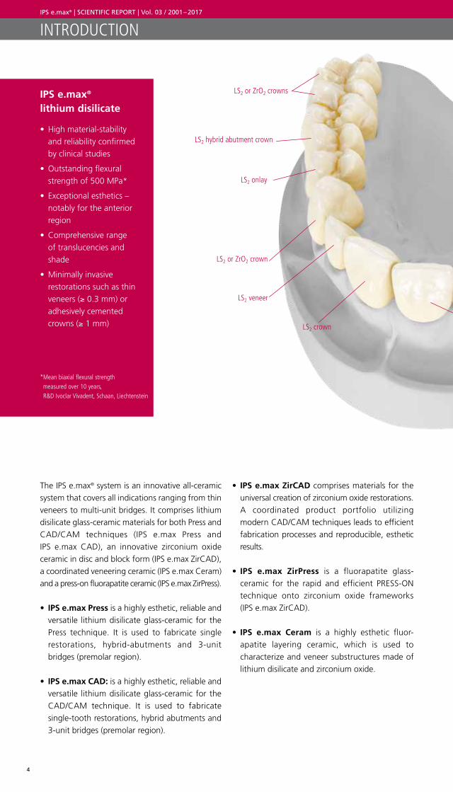
IPS e.max® lithium disilicate
• High material-stability and reliability confirmed by clinical studies
• Outstanding flexural strength of 500 MPa*
• Exceptional esthetics – notably for the anterior region
• Comprehensive range of translucencies and shade
• Minimally invasive restorations such as thin veneers (≥ 0.3 mm) or adhesively cemented crowns (≥ 1 mm)
LS2 or ZrO2 crowns
LS2 or ZrO2 crown
LS2 hybrid abutment crown
LS2 onlay
LS2 veneer
LS2 crown
* Mean biaxial flexural strength measured over 10 years, R&D Ivoclar Vivadent, Schaan, Liechtenstein
IPS e.max® | SCIENTIFIC REPORT | Vol. 03 / 2001 – 2017
5
Introduction
From the development of the IPS e.max materials to the present, their use has been investigated extensively by the scientific community and many renowned experts have contributed to the expanding body of literature evaluating their clinical performance. This plus the ever-growing
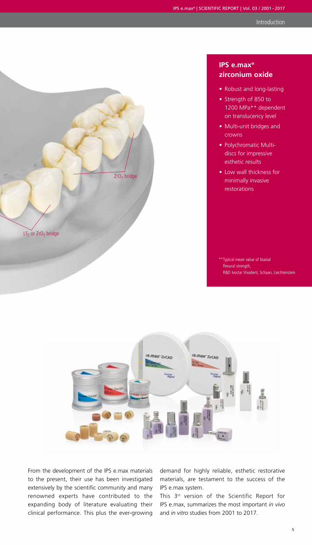
IPS e.max® zirconium oxide
• Robust and long-lasting
• Strength of 850 to 1200 MPa** dependent on translucency level
• Multi-unit bridges and crowns
• Polychromatic Multi- discs for impressive esthetic results
• Low wall thickness for minimally invasive restorations
LS2 or ZrO2 bridge
ZrO2 bridge
** Typical mean value of biaxial flexural strength, R&D Ivoclar Vivadent, Schaan, Liechtenstein
demand for highly reliable, esthetic restorative materials, are testament to the success of the IPS e.max system.This 3rd version of the Scientific Report for IPS e.max, summarizes the most important in vivo and in vitro studies from 2001 to 2017.
IPS e.max® | SCIENTIFIC REPORT | Vol. 03 / 2001 – 2017
6
IPS e.max® | SCIENTIFIC REPORT | Vol. 03 / 2001 – 2017
7
IPS e.max® Materials – Clinical Performance
IPS e.max® | SCIENTIFIC REPORT | Vol. 03 / 2001 – 2017
8
Summary survival statistics
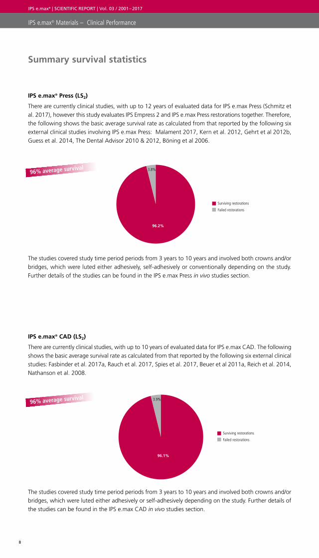
IPS e.max® Press (LS2)
There are currently clinical studies, with up to 12 years of evaluated data for IPS e.max Press (Schmitz et al. 2017), however this study evaluates IPS Empress 2 and IPS e.max Press restorations together. Therefore, the following shows the basic average survival rate as calculated from that reported by the following six external clinical studies involving IPS e.max Press: Malament 2017, Kern et al. 2012, Gehrt et al 2012b, Guess et al. 2014, The Dental Advisor 2010 & 2012, Böning et al 2006.
IPS e.max® CAD (LS2)
There are currently clinical studies, with up to 10 years of evaluated data for IPS e.max CAD. The following shows the basic average survival rate as calculated from that reported by the following six external clinical studies: Fasbinder et al. 2017a, Rauch et al. 2017, Spies et al. 2017, Beuer et al 2011a, Reich et al. 2014, Nathanson et al. 2008.
The studies covered study time period periods from 3 years to 10 years and involved both crowns and/or bridges, which were luted either adhesively, self-adhesively or conventionally depending on the study. Further details of the studies can be found in the IPS e.max Press in vivo studies section.
The studies covered study time period periods from 3 years to 10 years and involved both crowns and/or bridges, which were luted either adhesively or self-adhesively depending on the study. Further details of the studies can be found in the IPS e.max CAD in vivo studies section.
IPS e.max® Materials – Clinical Performance
96% average survival
96% average survival
Failed restorations
Surviving restorations
96.2%
3.8%
Failed restorations
Surviving restorations
96.1%
3.9%
IPS e.max® | SCIENTIFIC REPORT | Vol. 03 / 2001 – 2017
9
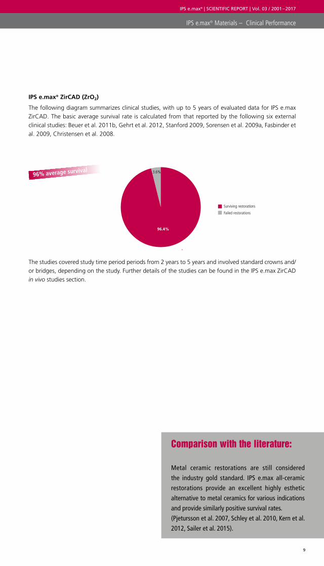
IPS e.max® ZirCAD (ZrO2)
The following diagram summarizes clinical studies, with up to 5 years of evaluated data for IPS e.max ZirCAD. The basic average survival rate is calculated from that reported by the following six external clinical studies: Beuer et al. 2011b, Gehrt et al. 2012, Stanford 2009, Sorensen et al. 2009a, Fasbinder et al. 2009, Christensen et al. 2008.
The studies covered study time period periods from 2 years to 5 years and involved standard crowns and/or bridges, depending on the study. Further details of the studies can be found in the IPS e.max ZirCAD in vivo studies section.
IPS e.max® Materials – Clinical Performance
96% average survival
Failed restorations
Surviving restorations
96.4%
3.6%
Comparison with the literature:
Metal ceramic restorations are still considered
the industry gold standard. IPS e.max all-ceramic
restorations provide an excellent highly esthetic
alternative to metal ceramics for various indications
and provide similarly positive survival rates.
(Pjetursson et al. 2007, Schley et al. 2010, Kern et al.
2012, Sailer et al. 2015).
IPS e.max® | SCIENTIFIC REPORT | Vol. 03 / 2001 – 2017
10
IPS e.max® Lithium Disilicate
(LS2)
IPS e.max® | SCIENTIFIC REPORT | Vol. 03 / 2001 – 2017
11
in vivo studies in vitro studies
IPS e.max® Lithium Disilicate
(LS2)
IPS e.max® | SCIENTIFIC REPORT | Vol. 03 / 2001 – 2017
12
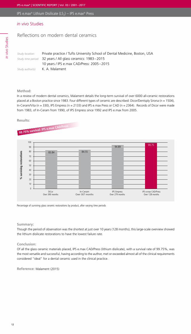
Reflections on modern dental ceramics
Study location: Private practice / Tufts University School of Dental Medicine, Boston, USAStudy time period: 32 years / All glass ceramics: 1983 – 2015 10 years / IPS e.max CAD/Press: 2005 – 2015Study author(s): K. A. Malament
Method: In a review of modern dental ceramics, Malament details the long-term survival of over 6000 all-ceramic restorations
placed at a Boston practice since 1983. Four different types of ceramic are described: Dicor/Dentsply Sirona (n = 1504),
In-Ceram/Vita (n = 330), IPS Empress (n = 2133) and IPS e.max Press or CAD (n = 2364). Records of Dicor were made
from 1983, of In-Ceram from 1990, of IPS Empress since 1992 and IPS e.max from 2005.
Results:
Summary: Though the period of observation was the shortest at just over 10 years (128 months), this large-scale overview showed
the lithium disilicate restorations to have the lowest failure rate.
Conclusion: Of all the glass ceramic materials placed, IPS e.max CAD/Press (lithium disilicate), with a survival rate of 99.75%, was
the most versatile and successful, having according to the author, met or exceeded almost all of the clinical requirements
considered “ideal” for a dental ceramic used in the clinical practice.
Reference: Malament (2015)
Percentage of surviving glass ceramic restorations by product, after varying time periods
IPS e.max® Lithium Disilicate (LS2) – IPS e.max® Press
% s
urvi
ving
res
tora
tion
s
100
70
80
90
50
60
0
10
20
30
40
DiCor Over 390 months
In-Ceram Over 307 months
IPS Empress Over 279 months
IPS e.max CAD/Press Over 128 months
83.84 84.55
94.8999.75
99.75% survival: IPS e.max CAD/Press
in v
ivo
Stud
ies
in vivo Studies
IPS e.max® | SCIENTIFIC REPORT | Vol. 03 / 2001 – 2017
13
IPS e.max® Lithium Disilicate (LS2) – IPS e.max® Press
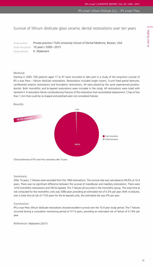
Survival of lithium disilicate glass ceramic dental restorations over ten years
Study location: Private practice / Tufts University School of Dental Medicine, Boston, USAStudy time period: 10 years / 2005 – 2015Study author(s): K. Malament
Method: Starting in 2005, 556 patients aged 17 to 97 were recruited to take part in a study of the long-term survival of
IPS e.max Press – lithium disilicate restorations. Restorations included single crowns, 3-unit fixed partial dentures,
cantilevered anterior restorations and foundation restorations. All were placed by the same experienced prostho-
dontist. Both monolithic and bi-layered restorations were included in the study. All restorations were luted with
Variolink II. A restorative failure constituted any fracture of the restoration that necessitated replacement. Chips of less
than 1 mm that could be re-shaped and polished were not considered failures.
Results:
Summary: After 10 years, 7 failures were recorded from the 1960 restorations. The survival rate was calculated as 99.6% at 10.4
years. There was no significant difference between the survival of mandibular and maxillary restorations. There were
1410 monolithic restorations and 550 bi-layered. The 7 failures all occurred in the monolithic group. The total time at
risk computed for the monolithic units was 3380 years providing an estimated risk of 0.2% per year. With no failures,
over a total time at risk of 1733 years for the bi-layered units, the estimated risk was 0% per year.
Conclusion: IPS e.max Press lithium disilicate restorations showed excellent survival over the 10.4 year study period. The 7 failures
occurred during a cumulative monitoring period of 5113 years, providing an estimated risk of failure of 0.14% per
year.
Reference: Malament (2017)
Clinical performance of IPS e.max Press restorations after 10 years
99.6% survival
Failed restorations
Intact restorations
99.6%
0.4%
in v
ivo
Stud
ies
IPS e.max® | SCIENTIFIC REPORT | Vol. 03 / 2001 – 2017
14
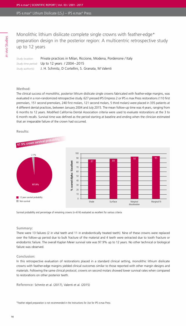
Monolithic lithium disilicate complete single crowns with feather-edge* preparation design in the posterior region: A multicentric retrospective study up to 12 years
Study location: Private practices in Milan, Riccione, Modena, Pordenone / ItalyStudy time period: Up to 12 years / 2004 – 2015Study author(s): J. H. Schmitz, D Cortellini, S. Granata, M Valenti
Method: The clinical success of monolithic, posterior lithium disilicate single crowns fabricated with feather-edge margins, was
evaluated in a non-randomized retrospective study. 627 pressed IPS Empress 2 or IPS e.max Press restorations (110 first
premolars, 151 second premolars, 240 first molars, 121 second molars, 5 third molars) were placed in 335 patients at
4 different dental practices, between January 2004 and July 2015. The mean follow-up time was 4 years, ranging from
6 months to 12 years. Modified California Dental Association criteria were used to evaluate restorations at the 3 to
6 month recalls. Survival time was defined as the period starting at baseline and ending when the clinician estimated
that an irreparable failure of the crown had occurred.
Results:
Summary: There were 13 failures (2 in vital teeth and 11 in endodontically treated teeth). Nine of these crowns were replaced
over the follow-up period due to bulk fracture of the material and 4 teeth were extracted due to tooth fracture or
endodontic failure. The overall Kaplan Meier survival rate was 97.9% up to 12 years. No other technical or biological
failure was observed.
Conclusion: In this retrospective evaluation of restorations placed in a standard clinical setting, monolithic lithium disilicate
crowns with feather-edge margins yielded clinical outcomes similar to those reported with other margin designs and
materials. Following the same clinical protocol, crowns on second molars showed lower survival rates when compared
to restorations on other posterior teeth.
Reference: Schmitz et al. (2017), Valenti et al. (2015)
*Feather edged preparation is not recommended in the Instructions for Use for IPS e.max Press
Survival probability and percentage of remaining crowns (n=614) evaluated as excellent for various criteria
97.9% crown survival probability
Non-survival
12 year survival probability
2.1%
97.9%
% s
core
d A
lpha
/ Ex
celle
nt
100
70
80
90
50
60
0
10
20
30
40
Shade Surface Marginal discoloration
Marginal fit
95938987
IPS e.max® Lithium Disilicate (LS2) – IPS e.max® Press
in v
ivo
Stud
ies
IPS e.max® | SCIENTIFIC REPORT | Vol. 03 / 2001 – 2017
15
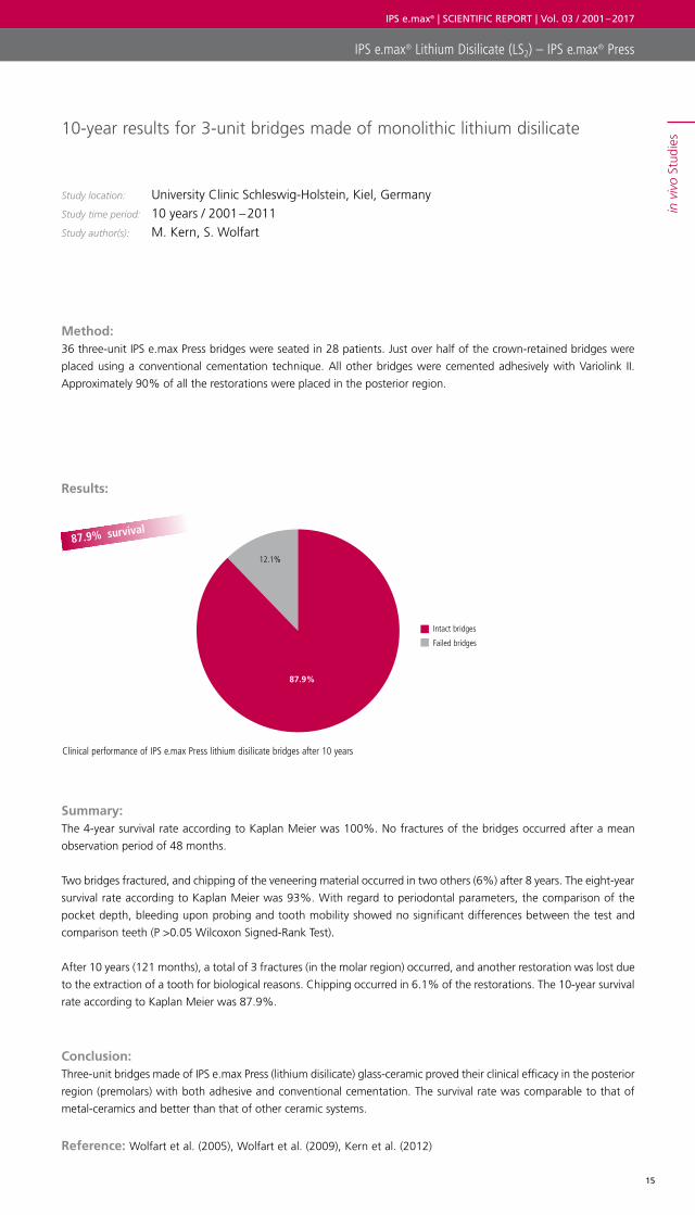
10-year results for 3-unit bridges made of monolithic lithium disilicate
Study location: University Clinic Schleswig-Holstein, Kiel, GermanyStudy time period: 10 years / 2001 – 2011Study author(s): M. Kern, S. Wolfart
Method: 36 three-unit IPS e.max Press bridges were seated in 28 patients. Just over half of the crown-retained bridges were
placed using a conventional cementation technique. All other bridges were cemented adhesively with Variolink II.
Approximately 90% of all the restorations were placed in the posterior region.
Results:
Summary: The 4-year survival rate according to Kaplan Meier was 100%. No fractures of the bridges occurred after a mean
observation period of 48 months.
Two bridges fractured, and chipping of the veneering material occurred in two others (6%) after 8 years. The eight-year
survival rate according to Kaplan Meier was 93%. With regard to periodontal parameters, the comparison of the
pocket depth, bleeding upon probing and tooth mobility showed no significant differences between the test and
comparison teeth (P >0.05 Wilcoxon Signed-Rank Test).
After 10 years (121 months), a total of 3 fractures (in the molar region) occurred, and another restoration was lost due
to the extraction of a tooth for biological reasons. Chipping occurred in 6.1% of the restorations. The 10-year survival
rate according to Kaplan Meier was 87.9%.
Conclusion: Three-unit bridges made of IPS e.max Press (lithium disilicate) glass-ceramic proved their clinical efficacy in the posterior
region (premolars) with both adhesive and conventional cementation. The survival rate was comparable to that of
metal-ceramics and better than that of other ceramic systems.
Reference: Wolfart et al. (2005), Wolfart et al. (2009), Kern et al. (2012)
Clinical performance of IPS e.max Press lithium disilicate bridges after 10 years
87.9% survival
Failed bridges
Intact bridges
12.1%
87.9%
IPS e.max® Lithium Disilicate (LS2) – IPS e.max® Press
in v
ivo
Stud
ies
IPS e.max® | SCIENTIFIC REPORT | Vol. 03 / 2001 – 2017
16
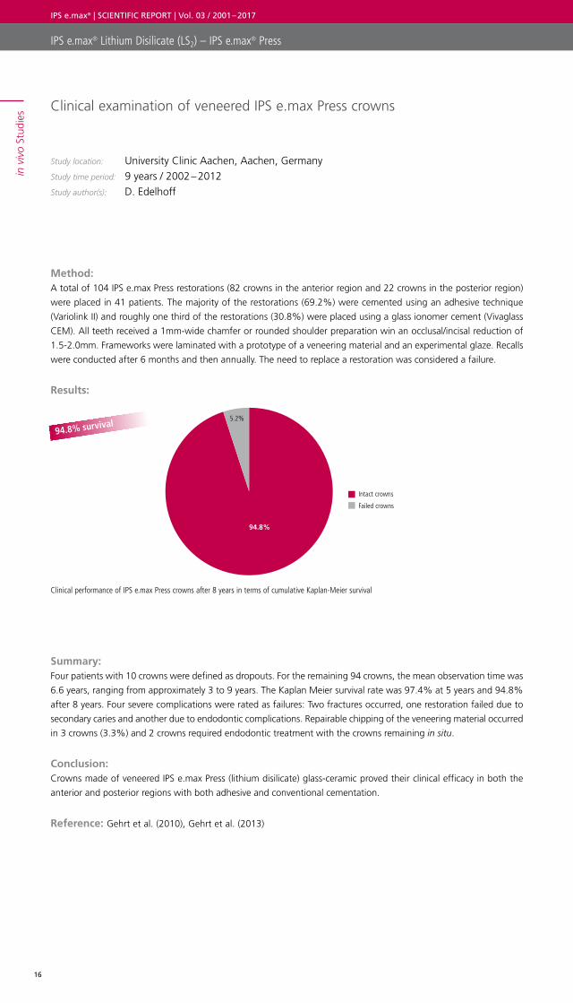
Clinical examination of veneered IPS e.max Press crowns
Study location: University Clinic Aachen, Aachen, GermanyStudy time period: 9 years / 2002 – 2012Study author(s): D. Edelhoff
Method: A total of 104 IPS e.max Press restorations (82 crowns in the anterior region and 22 crowns in the posterior region)
were placed in 41 patients. The majority of the restorations (69.2%) were cemented using an adhesive technique
(Variolink II) and roughly one third of the restorations (30.8%) were placed using a glass ionomer cement (Vivaglass
CEM). All teeth received a 1mm-wide chamfer or rounded shoulder preparation win an occlusal/incisal reduction of
1.5-2.0mm. Frameworks were laminated with a prototype of a veneering material and an experimental glaze. Recalls
were conducted after 6 months and then annually. The need to replace a restoration was considered a failure.
Results:
Summary: Four patients with 10 crowns were defined as dropouts. For the remaining 94 crowns, the mean observation time was
6.6 years, ranging from approximately 3 to 9 years. The Kaplan Meier survival rate was 97.4% at 5 years and 94.8%
after 8 years. Four severe complications were rated as failures: Two fractures occurred, one restoration failed due to
secondary caries and another due to endodontic complications. Repairable chipping of the veneering material occurred
in 3 crowns (3.3%) and 2 crowns required endodontic treatment with the crowns remaining in situ.
Conclusion: Crowns made of veneered IPS e.max Press (lithium disilicate) glass-ceramic proved their clinical efficacy in both the
anterior and posterior regions with both adhesive and conventional cementation.
Reference: Gehrt et al. (2010), Gehrt et al. (2013)
Clinical performance of IPS e.max Press crowns after 8 years in terms of cumulative Kaplan-Meier survival
Failed crowns
Intact crowns
5.2%
94.8%
94.8% survival
IPS e.max® Lithium Disilicate (LS2) – IPS e.max® Press
in v
ivo
Stud
ies
IPS e.max® | SCIENTIFIC REPORT | Vol. 03 / 2001 – 2017
17
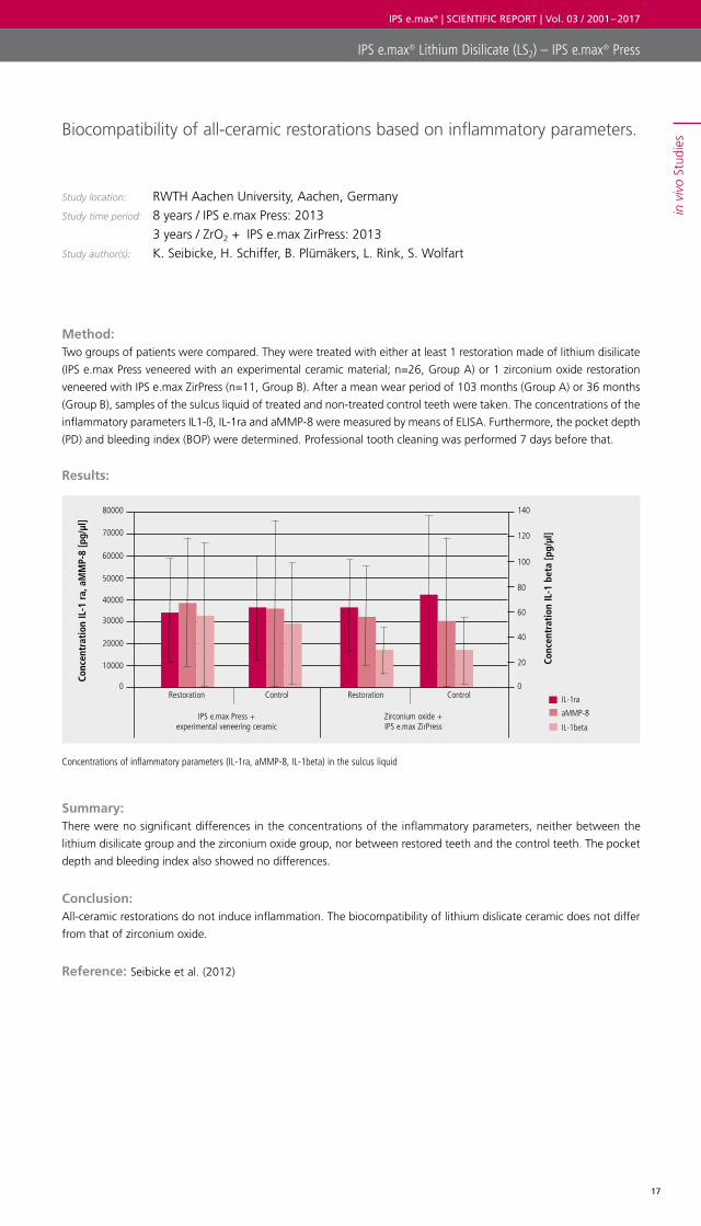
Biocompatibility of all-ceramic restorations based on inflammatory parameters.
Study location: RWTH Aachen University, Aachen, GermanyStudy time period: 8 years / IPS e.max Press: 2013 3 years / ZrO2 + IPS e.max ZirPress: 2013Study author(s): K. Seibicke, H. Schiffer, B. Plümäkers, L. Rink, S. Wolfart
Method: Two groups of patients were compared. They were treated with either at least 1 restoration made of lithium disilicate
(IPS e.max Press veneered with an experimental ceramic material; n=26, Group A) or 1 zirconium oxide restoration
veneered with IPS e.max ZirPress (n=11, Group B). After a mean wear period of 103 months (Group A) or 36 months
(Group B), samples of the sulcus liquid of treated and non-treated control teeth were taken. The concentrations of the
inflammatory parameters IL1-ß, IL-1ra and aMMP-8 were measured by means of ELISA. Furthermore, the pocket depth
(PD) and bleeding index (BOP) were determined. Professional tooth cleaning was performed 7 days before that.
Results:
Summary: There were no significant differences in the concentrations of the inflammatory parameters, neither between the
lithium disilicate group and the zirconium oxide group, nor between restored teeth and the control teeth. The pocket
depth and bleeding index also showed no differences.
Conclusion: All-ceramic restorations do not induce inflammation. The biocompatibility of lithium dislicate ceramic does not differ
from that of zirconium oxide.
Reference: Seibicke et al. (2012)
Concentrations of inflammatory parameters (IL-1ra, aMMP-8, IL-1beta) in the sulcus liquid
Conc
entr
atio
n IL
-1 r
a, a
MM
P-8
[pg/
µl]
IL-1ra
aMMP-8
IL-1beta
Conc
entr
atio
n IL
-1 b
eta
[pg/
µl]
80000 140
70000 120
60000
50000
100
40000
20000
30000
80
60
40
10000 20
0 0Restoration
IPS e.max Press + experimental veneering ceramic
Zirconium oxide + IPS e.max ZirPress
Restoration Control Control
IPS e.max® Lithium Disilicate (LS2) – IPS e.max® Press
in v
ivo
Stud
ies
IPS e.max® | SCIENTIFIC REPORT | Vol. 03 / 2001 – 2017
18
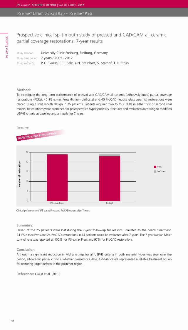
Prospective clinical split-mouth study of pressed and CAD/CAM all-ceramic partial coverage restorations: 7-year results
Study location: University Clinic Freiburg, Freiburg, Germany Study time period: 7 years / 2005 – 2012Study author(s): P. C. Guess, C. F. Selz, Y-N. Steinhart, S. Stampf, J. R. Strub
Method: To investigate the long term performance of pressed and CAD/CAM all ceramic (adhesively luted) partial coverage
restorations (PCRs), 40 IPS e.max Press (lithium disilicate) and 40 ProCAD (leucite glass ceramic) restorations were
placed using a split mouth design in 25 patients. Patients required two to four PCRs in either first or second vital
molars. Restorations were examined for postoperative hypersensitivity, fractures and evaluated according to modified
USPHS criteria at baseline and annually for 7 years.
Results:
Summary: Eleven of the 25 patients were lost during the 7-year follow-up for reasons unrelated to the dental treatment.
24 IPS e.max Press and 24 ProCAD restorations in 14 patients could be evaluated after 7 years. The 7-year Kaplan Meier
survival rate was reported as 100% for IPS e.max Press and 97% for ProCAD restorations.
Conclusion: Although a significant reduction in Alpha ratings for all USPHS criteria in both material types was seen over the
period, all-ceramic partial crowns, whether pressed or CAD/CAM-fabricated, represented a reliable treatment option
for restoring larger defects in the posterior region.
Reference: Guess et al. (2013)
Clinical performance of IPS e.max Press and ProCAD crowns after 7 years
Num
ber
of r
esto
rati
ons
0
5
10
15
20
25
IPS e.max Press ProCAD
Intact
Fractured
100% IPS e.max Press survival
IPS e.max® Lithium Disilicate (LS2) – IPS e.max® Press
in v
ivo
Stud
ies
IPS e.max® | SCIENTIFIC REPORT | Vol. 03 / 2001 – 2017
19
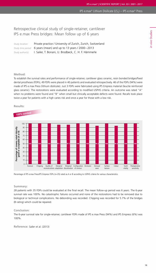
Retrospective clinical study of single-retainer, cantilever IPS e.max Press bridges: Mean follow up of 6 years
Study location: Private practice / University of Zurich, Zurich, Switzerland Study time period: 6 years (mean) and up to 13 years / 2000 – 2013Study author(s): I. Sailer, T. Bonani, U. Brodbeck, C. H. F. Hämmerle
Method: To establish the survival rates and performance of single-retainer, cantilever glass ceramic, resin bonded bridges/fixed
dental prostheses (FDPs), 49 FDPs were placed in 40 patients and evaluated retrospectively. 46 of the FDPs (94%) were
made of IPS e.max Press (lithium disilicate). Just 3 FDPs were fabricated using IPS Empress material (leucite reinforced
glass ceramic). The restorations were evaluated according to modified USPHS criteria. An outcome was rated “A”
when no problems were found and “B” when small but clinically acceptable defects were found. Recalls took place
twice a year for patients with a high caries risk and once a year for those with a low risk.
Results:
Summary: 28 patients with 35 FDPs could be evaluated at the final recall. The mean follow-up period was 6 years. The 6-year
survival rate was 100%. No catastrophic failures occurred and none of the restorations had to be removed due to
biological or technical complications. No debonding was recorded. Chipping was recorded for 5.7% of the bridges
(B rating) which could be repaired.
Conclusion: The 6-year survival rate for single-retainer, cantilever FDPs made of IPS e.max Press (94%) and IPS Empress (6%) was
100%.
Reference: Sailer et al. (2013)
Percentage of IPS e.max Press/IPS Empress FDPs (n=35) rated as A or B according to USPHS criteria for various characteristics
Perc
enta
ge o
f cr
owns
rat
ed c
lnic
ally
go
od o
r ac
cept
able
(A o
r B)
0
30
20
10
50
40
60
70
90
80
100
Fracture Chipping Marginal adaptation
Marginal discoloration
Quality of reconstructions
Configuration of contour
Occlusion Occlusal wear
Surface texture
Colour Tooth vitality
Postoperative sensitivity
100 100 100 100 100 100 100 10097.1 97.1 94.3 94.3
100% survival
IPS e.max® Lithium Disilicate (LS2) – IPS e.max® Press
in v
ivo
Stud
ies
IPS e.max® | SCIENTIFIC REPORT | Vol. 03 / 2001 – 2017
20
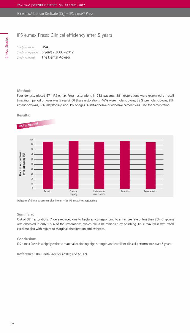
IPS e.max Press: Clinical efficiency after 5 years
Study location: USAStudy time period: 5 years / 2006 – 2012Study author(s): The Dental Advisor
Method: Four dentists placed 671 IPS e.max Press restorations in 282 patients. 381 restorations were examined at recall
(maximum period of wear was 5 years). Of these restorations, 46% were molar crowns, 38% premolar crowns, 8%
anterior crowns, 5% inlays/onlays and 3% bridges. A self-adhesive or adhesive cement was used for cementation.
Results:
Summary: Out of 381 restorations, 7 were replaced due to fractures, corresponding to a fracture rate of less than 2%. Chipping
was observed in only 1.5% of the restorations, which could be remedied by polishing. IPS e.max Press was rated
excellent also with regard to marginal discoloration and esthetics.
Conclusion: IPS e.max Press is a highly esthetic material exhibiting high strength and excellent clinical performance over 5 years.
Reference: The Dental Advisor (2010) and (2012)
Evaluation of clinical parameters after 5 years – for IPS e.max Press restorations
98.1% survival
Shar
e of
res
tora
tion
s w
ith
top
rati
ng [%
]
100
90
80
70
60
50
40
30
20
10
0
Esthetics Fracture, chipping
Resistance to discolouration
Sensitivity Decementation
IPS e.max® Lithium Disilicate (LS2) – IPS e.max® Press
in v
ivo
Stud
ies
IPS e.max® | SCIENTIFIC REPORT | Vol. 03 / 2001 – 2017
21
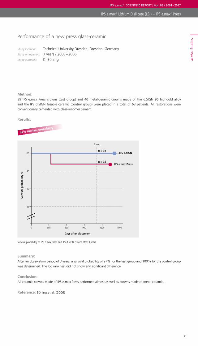
Performance of a new press glass-ceramic
Study location: Technical University Dresden, Dresden, GermanyStudy time period: 3 years / 2003 – 2006Study author(s): K. Böning
Method: 39 IPS e.max Press crowns (test group) and 40 metal-ceramic crowns made of the d.SIGN 96 highgold alloy
and the IPS d.SIGN fusable ceramic (control group) were placed in a total of 63 patients. All restorations were
con ventionally cemented with glass-ionomer cement.
Results:
Summary: After an observation period of 3 years, a survival probability of 97% for the test group and 100% for the control group
was determined. The log rank test did not show any significant difference.
Conclusion: All-ceramic crowns made of IPS e.max Press performed almost as well as crowns made of metal-ceramic.
Reference: Böning et al. (2006)
Survival probability of IPS e.max Press and IPS d.SIGN crowns after 3 years
97% survival probability
Surv
ival
pro
babi
lity
%
Days after placement
n = 34
IPS e.max Press
IPS d.SIGN
n = 32
100
95
90
85
3 years
0 300 600 900 1200 1500
IPS e.max® Lithium Disilicate (LS2) – IPS e.max® Press
in v
ivo
Stud
ies
IPS e.max® | SCIENTIFIC REPORT | Vol. 03 / 2001 – 2017
22
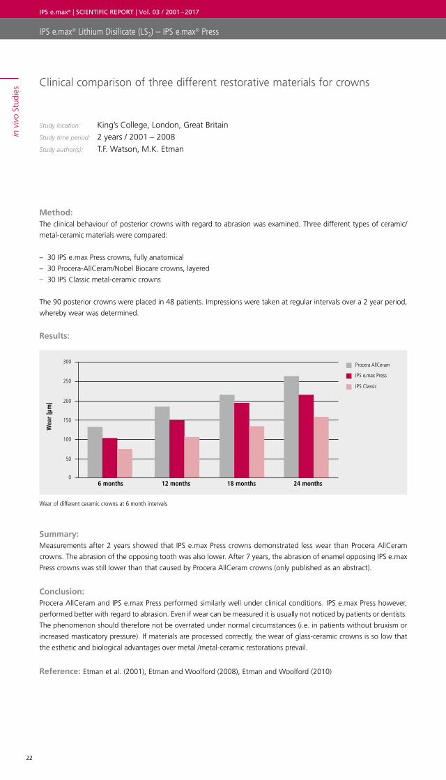
Clinical comparison of three different restorative materials for crowns
Study location: King’s College, London, Great BritainStudy time period: 2 years / 2001 – 2008Study author(s): T.F. Watson, M.K. Etman
Method: The clinical behaviour of posterior crowns with regard to abrasion was examined. Three different types of ceramic/
metal-ceramic materials were compared:
– 30 IPS e.max Press crowns, fully anatomical
– 30 Procera-AllCeram/Nobel Biocare crowns, layered
– 30 IPS Classic metal-ceramic crowns
The 90 posterior crowns were placed in 48 patients. Impressions were taken at regular intervals over a 2 year period,
whereby wear was determined.
Results:
Summary: Measurements after 2 years showed that IPS e.max Press crowns demonstrated less wear than Procera AllCeram
crowns. The abrasion of the opposing tooth was also lower. After 7 years, the abrasion of enamel opposing IPS e.max
Press crowns was still lower than that caused by Procera AllCeram crowns (only published as an abstract).
Conclusion: Procera AllCeram and IPS e.max Press performed similarly well under clinical conditions. IPS e.max Press however,
performed better with regard to abrasion. Even if wear can be measured it is usually not noticed by patients or dentists.
The phenomenon should therefore not be overrated under normal circumstances (i.e. in patients without bruxism or
increased masticatory pressure). If materials are processed correctly, the wear of glass-ceramic crowns is so low that
the esthetic and biological advantages over metal /metal-ceramic restorations prevail.
Reference: Etman et al. (2001), Etman and Woolford (2008), Etman and Woolford (2010)
Wear of different ceramic crowns at 6 month intervals
Wea
r [µ
m]
6 months 12 months 18 months 24 months
Procera AllCeram
IPS e.max Press
IPS Classic250
300
200
150
100
50
0
IPS e.max® Lithium Disilicate (LS2) – IPS e.max® Press
in v
ivo
Stud
ies
IPS e.max® | SCIENTIFIC REPORT | Vol. 03 / 2001 – 2017
23
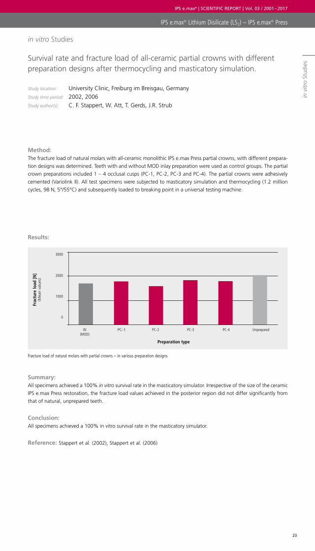
Survival rate and fracture load of all-ceramic partial crowns with different preparation designs after thermocycling and masticatory simulation.
Study location: University Clinic, Freiburg im Breisgau, GermanyStudy time period: 2002, 2006Study author(s): C. F. Stappert, W. Att, T. Gerds, J.R. Strub
Method: The fracture load of natural molars with all-ceramic monolithic IPS e.max Press partial crowns, with different prepara-
tion designs was determined. Teeth with and without MOD inlay preparation were used as control groups. The partial
crown preparations included 1 – 4 occlusal cusps (PC-1, PC-2, PC-3 and PC-4). The partial crowns were adhesively
cemented (Variolink II). All test specimens were subjected to masticatory simulation and thermocycling (1.2 million
cycles, 98 N, 5°/55°C) and subsequently loaded to breaking point in a universal testing machine.
Results:
Summary: All specimens achieved a 100% in vitro survival rate in the masticatory simulator. Irrespective of the size of the ceramic
IPS e.max Press restoration, the fracture load values achieved in the posterior region did not differ significantly from
that of natural, unprepared teeth.
Conclusion: All specimens achieved a 100% in vitro survival rate in the masticatory simulator.
Reference: Stappert et al. (2002), Stappert et al. (2006)
Fracture load of natural molars with partial crowns – in various preparation designs
Frac
ture
load
[N]
(Mea
n va
lues
)
Preparation type
3000
2000
1000
0
IN(MOD)
PC-1 PC-2 PC-3 PC-4 Unprepared
IPS e.max® Lithium Disilicate (LS2) – IPS e.max® Press
in v
itro
Stud
ies
in vitro Studies
IPS e.max® | SCIENTIFIC REPORT | Vol. 03 / 2001 – 2017
24
All-ceramic partial crowns on premolars. Cavity-preparation design, reliability and fracture load upon fatigue.
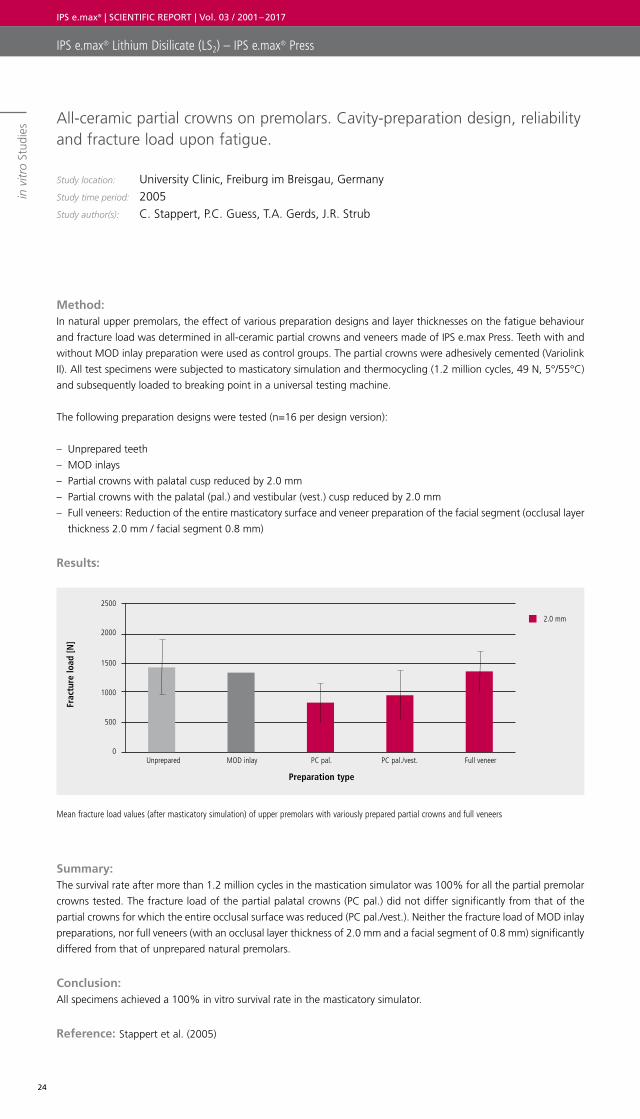
Study location: University Clinic, Freiburg im Breisgau, GermanyStudy time period: 2005Study author(s): C. Stappert, P.C. Guess, T.A. Gerds, J.R. Strub
Method: In natural upper premolars, the effect of various preparation designs and layer thicknesses on the fatigue behaviour
and fracture load was determined in all-ceramic partial crowns and veneers made of IPS e.max Press. Teeth with and
without MOD inlay preparation were used as control groups. The partial crowns were adhesively cemented (Variolink
II). All test specimens were subjected to masticatory simulation and thermocycling (1.2 million cycles, 49 N, 5°/55°C)
and subsequently loaded to breaking point in a universal testing machine.
The following preparation designs were tested (n=16 per design version):
– Unprepared teeth
– MOD inlays
– Partial crowns with palatal cusp reduced by 2.0 mm
– Partial crowns with the palatal (pal.) and vestibular (vest.) cusp reduced by 2.0 mm
– Full veneers: Reduction of the entire masticatory surface and veneer preparation of the facial segment (occlusal layer
thickness 2.0 mm / facial segment 0.8 mm)
Results:
Summary: The survival rate after more than 1.2 million cycles in the mastication simulator was 100% for all the partial premolar
crowns tested. The fracture load of the partial palatal crowns (PC pal.) did not differ significantly from that of the
partial crowns for which the entire occlusal surface was reduced (PC pal./vest.). Neither the fracture load of MOD inlay
preparations, nor full veneers (with an occlusal layer thickness of 2.0 mm and a facial segment of 0.8 mm) significantly
differed from that of unprepared natural premolars.
Conclusion: All specimens achieved a 100% in vitro survival rate in the masticatory simulator.
Reference: Stappert et al. (2005)
Mean fracture load values (after masticatory simulation) of upper premolars with variously prepared partial crowns and full veneers
Frac
ture
load
[N]
Preparation type
2500
2.0 mm
2000
1500
1000
500
0Unprepared MOD inlay PC pal. PC pal./vest. Full veneer
IPS e.max® Lithium Disilicate (LS2) – IPS e.max® Press
in v
itro
Stud
ies
IPS e.max® | SCIENTIFIC REPORT | Vol. 03 / 2001 – 2017
25
Weibull probability curve for implants with abutments made of IPS e.max Press at a load of 200N
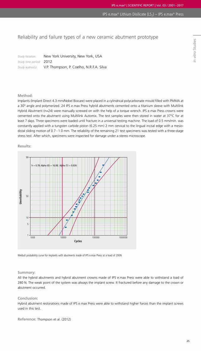
Reliability and failure types of a new ceramic abutment prototype
Study location: New York University, New York, USA Study time period: 2012Study author(s): V.P. Thompson, P. Coelho, N.R.F.A. Silva
Method: Implants (Implant Direct 4.3 mm/Nobel Biocare) were placed in a cylindrical polycarbonate mould filled with PMMA at
a 30° angle and polymerized. 24 IPS e.max Press hybrid abutments cemented onto a titanium sleeve with Multilink
Hybrid Abutment (n=24) were manually screwed on with the help of a torque wrench. IPS e.max Press crowns were
cemented onto the abutment using Multilink Automix. The test samples were then stored in water at 37°C for at
least 7 days. Three specimens were loaded until fracture in a universal testing machine. The load of 0.5 mm/min. was
constantly applied with a tungsten carbide piston (6.25 mm) 2 mm cervical to the lingual incisal edge with a mesio-
distal sliding motion of 0.7 – 1.0 mm. The reliability of the remaining 21 test specimens was tested with a three-stage
stress test. After which, specimens were inspected for damage under a stereo microscope.
Results:
Summary: All the hybrid abutments and hybrid abutment crowns made of IPS e.max Press were able to withstand a load of
280 N. The weak point of the system was always the implant screw. It fractured before any damage to the crown or
abutment occurred.
Conclusion: Hybrid abutment restorations made of IPS e.max Press were able to withstand higher forces than the implant screws
used in this test.
Reference: Thompson et al. (2012)
Unr
elia
bilit
y
h = 0.78; Alpha (0) = 16.08; Alpha (1) = 0.026
Cycles
99
50
10
5
11000 10000 100000 1000000
IPS e.max® Lithium Disilicate (LS2) – IPS e.max® Press
in v
itro
Stud
ies
IPS e.max® | SCIENTIFIC REPORT | Vol. 03 / 2001 – 2017
26
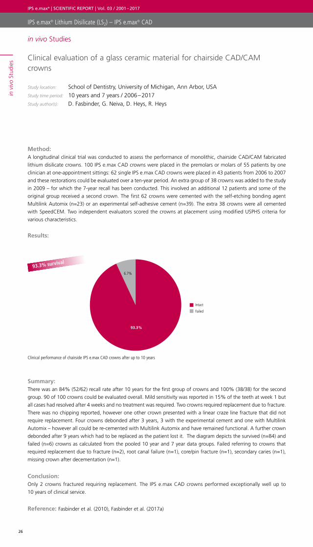
Clinical evaluation of a glass ceramic material for chairside CAD/CAM crowns
Study location: School of Dentistry, University of Michigan, Ann Arbor, USA Study time period: 10 years and 7 years / 2006 – 2017Study author(s): D. Fasbinder, G. Neiva, D. Heys, R. Heys
Method: A longitudinal clinical trial was conducted to assess the performance of monolithic, chairside CAD/CAM fabricated
lithium disilicate crowns. 100 IPS e.max CAD crowns were placed in the premolars or molars of 55 patients by one
clinician at one-appointment sittings: 62 single IPS e.max CAD crowns were placed in 43 patients from 2006 to 2007
and these restorations could be evaluated over a ten-year period. An extra group of 38 crowns was added to the study
in 2009 – for which the 7-year recall has been conducted. This involved an additional 12 patients and some of the
original group received a second crown. The first 62 crowns were cemented with the self-etching bonding agent
Multilink Automix (n=23) or an experimental self-adhesive cement (n=39). The extra 38 crowns were all cemented
with SpeedCEM. Two independent evaluators scored the crowns at placement using modified USPHS criteria for
various characteristics.
Results:
Summary: There was an 84% (52/62) recall rate after 10 years for the first group of crowns and 100% (38/38) for the second
group. 90 of 100 crowns could be evaluated overall. Mild sensitivity was reported in 15% of the teeth at week 1 but
all cases had resolved after 4 weeks and no treatment was required. Two crowns required replacement due to fracture.
There was no chipping reported, however one other crown presented with a linear craze line fracture that did not
require replacement. Four crowns debonded after 3 years, 3 with the experimental cement and one with Multilink
Automix – however all could be re-cemented with Multilink Automix and have remained functional. A further crown
debonded after 9 years which had to be replaced as the patient lost it. The diagram depicts the survived (n=84) and
failed (n=6) crowns as calculated from the pooled 10 year and 7 year data groups. Failed referring to crowns that
required replacement due to fracture (n=2), root canal failure (n=1), core/pin fracture (n=1), secondary caries (n=1),
missing crown after decementation (n=1).
Conclusion: Only 2 crowns fractured requiring replacement. The IPS e.max CAD crowns performed exceptionally well up to
10 years of clinical service.
Reference: Fasbinder et al. (2010), Fasbinder et al. (2017a)
Clinical performance of chairside IPS e.max CAD crowns after up to 10 years
93.3% survival
Failed
Intact
6.7%
93.3%
IPS e.max® Lithium Disilicate (LS2) – IPS e.max® CAD
in v
ivo
Stud
ies
in vivo Studies
IPS e.max® | SCIENTIFIC REPORT | Vol. 03 / 2001 – 2017
27
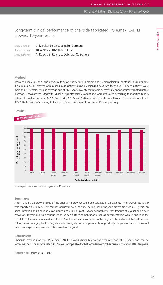
Long-term clinical performance of chairside fabricated IPS e.max CAD LT crowns: 10-year results
Study location: Universität Leipzig, Leipzig, GermanyStudy time period: 10 years / 2006/2007 – 2017Study author(s): A. Rauch, S. Reich, L. Dalchau, O. Schierz
Method: Between June 2006 and February 2007 forty-one posterior (31 molars and 10 premolars) full contour lithium disilicate
(IPS e.max CAD LT) crowns were placed in 34 patients using a chairside CAD/CAM technique. Thirteen patients were
male and 21 female, with an average age of 46.5 years. Twenty teeth were successfully endodontically treated before
insertion. Crowns were luted with Multilink Sprint/Ivoclar Vivadent and were evaluated according to modified USPHS
criteria at baseline and after 6, 12, 24, 36, 48, 60, 72 and 120 months. Clinical characteristics were rated from A1=1,
A2=2, B=3, C=4, D=5 relating to Excellent, Good, Sufficient, Insufficient, Poor respectively.
Results:
Summary: After 10 years, 33 crowns (80% of the original 41 crowns) could be evaluated in 26 patients. The survival rate in situ
was reported as 86.6%. Five failures occurred over the time period, involving one crown-fracture at 2 years, an
apical infection and a carious lesion under a core build up at 6 years, a lengthwise root fracture at 7 years and a new
crown at 10 years due to a carious lesion. When further complications such as decementation were included in the
calculation, the survival rate reduced to 76.3% after ten years. As shown in the diagram, the surface of the restorations,
colour, crown margin, tooth integrity, crown integrity and compliance (how positively the patient rated the overall
treatment experience), were all rated excellent or good.
Conclusion: Chairside crowns made of IPS e.max CAD LT proved clinically efficient over a period of 10 years and can be
recommended. The survival rate (86.6%) was comparable to that recorded with other ceramic materials after ten years.
Reference: Rauch et al. (2017)
Percentage of crowns rated excellent or good after 10 years in situ
86.6% survival in situ
Perc
enta
ge o
f cr
owns
rat
ed
Exce
llent
or
Goo
d
Evaluated characteristic
0
30
20
10
50
40
60
70
90
80
100
Surface Colour Adhesive gap
Tooth integrity
Crown margin
Crown integrity
Approximal contact
Sensitivity Complaints Compliance Occlusion
100 100 100 100 88 97 9791 100 85 100
IPS e.max® Lithium Disilicate (LS2) – IPS e.max® CAD
in v
ivo
Stud
ies
IPS e.max® | SCIENTIFIC REPORT | Vol. 03 / 2001 – 2017
28
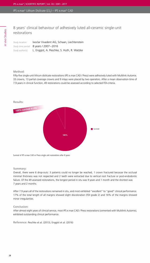
8 years’ clinical behaviour of adhesively luted all-ceramic single-unit restorations
Study location: Ivoclar Vivadent AG, Schaan, Liechtenstein Study time period: 8 years / 2007 – 2016Study author(s): L. Enggist, A. Peschke, S. Huth, R. Watzke
Method: Fifty-five single-unit lithium disilicate restorations (IPS e.max CAD / Press) were adhesively luted with Multilink Automix.
33 crowns, 13 partial coverage crowns and 9 inlays were placed by two operators. After a mean observation-time of
7.9 years in clinical function, 49 restorations could be assessed according to selected FDI-criteria.
Results:
Summary: Overall, there were 6 drop-outs: 3 patients could no longer be reached, 1 crown fractured because the occlusal
minimal thickness was not respected and 2 teeth were extracted due to vertical root fracture or post-endodontic
failure. Of the 49 assessed restorations, the longest period in situ was 9 years and 1 month and the shortest was
7 years and 2 months.
After 7.9 years all of the restorations remained in situ, and most exhibited “excellent” to “good” clinical performance.
17% of the total length of all margins showed slight discoloration (FDI grade 2) and 16% of the margins showed
minor irregularities.
Conclusion: After almost eight years of clinical service, most IPS e.max CAD / Press restorations (cemented with Multilink Automix),
exhibited outstanding clinical performance.
Reference: Peschke et al. (2013), Enggist et al. (2016)
Survival of IPS e.max CAD or Press single unit restorations after 8 years
100% survival in situ
Survived
100%
IPS e.max® Lithium Disilicate (LS2) – IPS e.max® CAD
in v
ivo
Stud
ies
IPS e.max® | SCIENTIFIC REPORT | Vol. 03 / 2001 – 2017
29
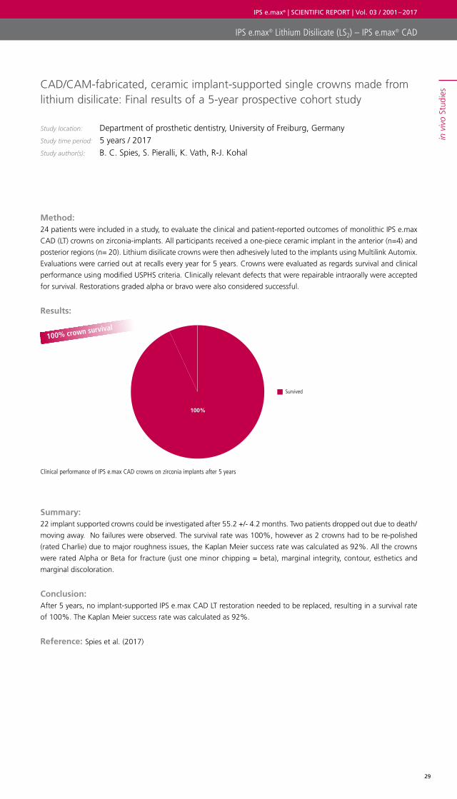
CAD/CAM-fabricated, ceramic implant-supported single crowns made from lithium disilicate: Final results of a 5-year prospective cohort study
Study location: Department of prosthetic dentistry, University of Freiburg, GermanyStudy time period: 5 years / 2017Study author(s): B. C. Spies, S. Pieralli, K. Vath, R-J. Kohal
Method: 24 patients were included in a study, to evaluate the clinical and patient-reported outcomes of monolithic IPS e.max
CAD (LT) crowns on zirconia-implants. All participants received a one-piece ceramic implant in the anterior (n=4) and
posterior regions (n= 20). Lithium disilicate crowns were then adhesively luted to the implants using Multilink Automix.
Evaluations were carried out at recalls every year for 5 years. Crowns were evaluated as regards survival and clinical
performance using modified USPHS criteria. Clinically relevant defects that were repairable intraorally were accepted
for survival. Restorations graded alpha or bravo were also considered successful.
Results:
Summary: 22 implant supported crowns could be investigated after 55.2 +/- 4.2 months. Two patients dropped out due to death/
moving away. No failures were observed. The survival rate was 100%, however as 2 crowns had to be re-polished
(rated Charlie) due to major roughness issues, the Kaplan Meier success rate was calculated as 92%. All the crowns
were rated Alpha or Beta for fracture (just one minor chipping = beta), marginal integrity, contour, esthetics and
marginal discoloration.
Conclusion: After 5 years, no implant-supported IPS e.max CAD LT restoration needed to be replaced, resulting in a survival rate
of 100%. The Kaplan Meier success rate was calculated as 92%.
Reference: Spies et al. (2017)
Clinical performance of IPS e.max CAD crowns on zirconia implants after 5 years
100% crown survival
Survived
100%
IPS e.max® Lithium Disilicate (LS2) – IPS e.max® CAD
in v
ivo
Stud
ies
IPS e.max® | SCIENTIFIC REPORT | Vol. 03 / 2001 – 2017
30
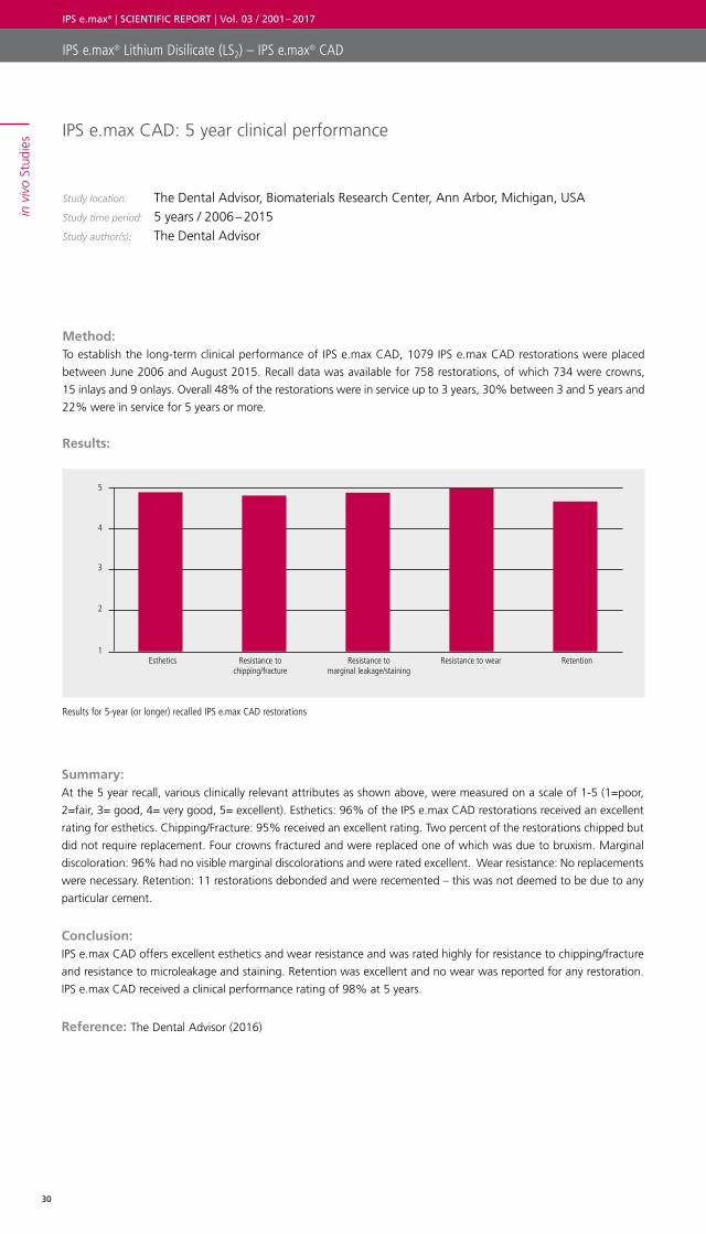
IPS e.max CAD: 5 year clinical performance
Study location: The Dental Advisor, Biomaterials Research Center, Ann Arbor, Michigan, USAStudy time period: 5 years / 2006 – 2015Study author(s): The Dental Advisor
Method: To establish the long-term clinical performance of IPS e.max CAD, 1079 IPS e.max CAD restorations were placed
between June 2006 and August 2015. Recall data was available for 758 restorations, of which 734 were crowns,
15 inlays and 9 onlays. Overall 48% of the restorations were in service up to 3 years, 30% between 3 and 5 years and
22% were in service for 5 years or more.
Results:
Summary: At the 5 year recall, various clinically relevant attributes as shown above, were measured on a scale of 1-5 (1=poor,
2=fair, 3= good, 4= very good, 5= excellent). Esthetics: 96% of the IPS e.max CAD restorations received an excellent
rating for esthetics. Chipping/Fracture: 95% received an excellent rating. Two percent of the restorations chipped but
did not require replacement. Four crowns fractured and were replaced one of which was due to bruxism. Marginal
discoloration: 96% had no visible marginal discolorations and were rated excellent. Wear resistance: No replacements
were necessary. Retention: 11 restorations debonded and were recemented – this was not deemed to be due to any
particular cement.
Conclusion: IPS e.max CAD offers excellent esthetics and wear resistance and was rated highly for resistance to chipping/fracture
and resistance to microleakage and staining. Retention was excellent and no wear was reported for any restoration.
IPS e.max CAD received a clinical performance rating of 98% at 5 years.
Reference: The Dental Advisor (2016)
Results for 5-year (or longer) recalled IPS e.max CAD restorations
5
4
3
2
1Esthetics Resistance to
chipping/fractureResistance to
marginal leakage/stainingResistance to wear Retention
IPS e.max® Lithium Disilicate (LS2) – IPS e.max® CAD
in v
ivo
Stud
ies
IPS e.max® | SCIENTIFIC REPORT | Vol. 03 / 2001 – 2017
31
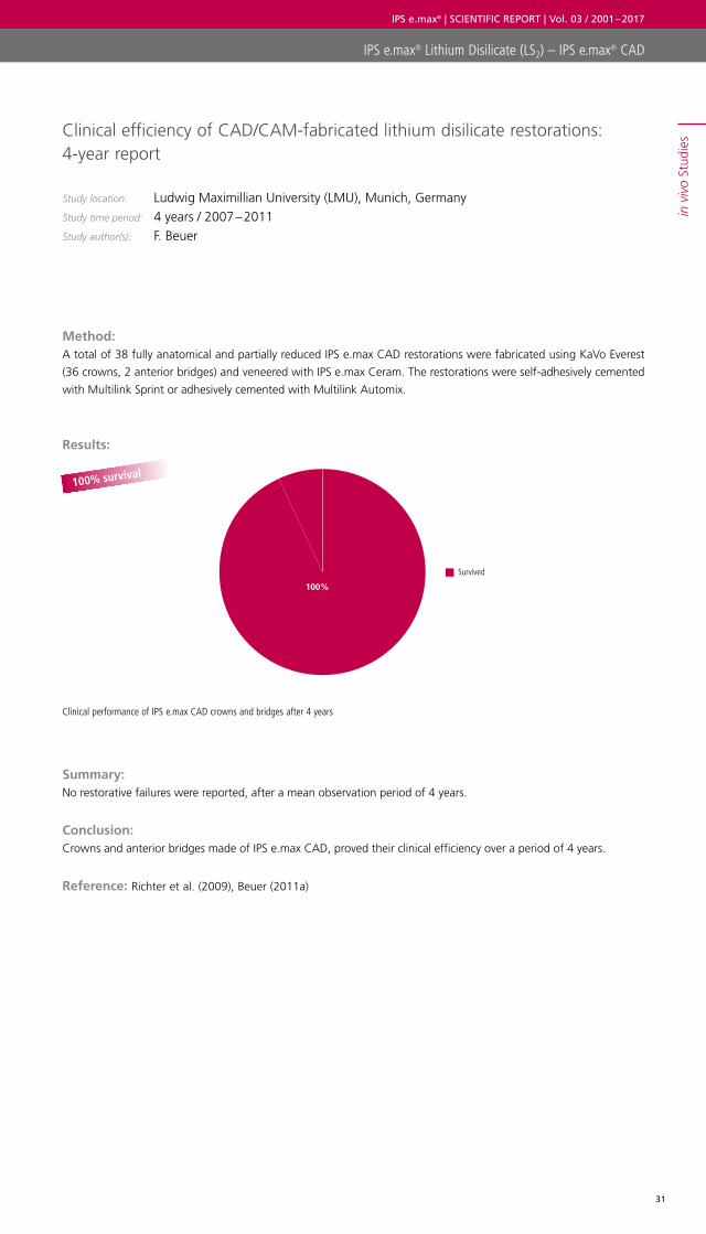
Clinical efficiency of CAD/CAM-fabricated lithium disilicate restorations: 4-year report
Study location: Ludwig Maximillian University (LMU), Munich, GermanyStudy time period: 4 years / 2007 – 2011Study author(s): F. Beuer
Method: A total of 38 fully anatomical and partially reduced IPS e.max CAD restorations were fabricated using KaVo Everest
(36 crowns, 2 anterior bridges) and veneered with IPS e.max Ceram. The restorations were self-adhesively cemented
with Multilink Sprint or adhesively cemented with Multilink Automix.
Results:
Summary: No restorative failures were reported, after a mean observation period of 4 years.
Conclusion: Crowns and anterior bridges made of IPS e.max CAD, proved their clinical efficiency over a period of 4 years.
Reference: Richter et al. (2009), Beuer (2011a)
Clinical performance of IPS e.max CAD crowns and bridges after 4 years
100% survival
IPS e.max® Lithium Disilicate (LS2) – IPS e.max® CAD
in v
ivo
Stud
ies
Survived
100%
IPS e.max® | SCIENTIFIC REPORT | Vol. 03 / 2001 – 2017
32
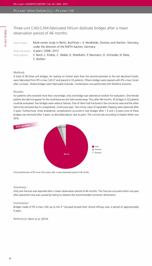
Three-unit CAD-CAM-fabricated lithium disilicate bridges after a mean observation period of 46 months
Study location: Multi-center study in Berlin, Buchholz i. d. Nordheide, Zwickau and Aachen, Germany, under the direction of the RWTH Aachen, Germany
Study time period: 4 years / 2008 – 2012 Study author(s): S. Reich, L. Endres, C. Weber, K. Wiedhahn, P. Neumann, O. Schneider, N. Rafai,
S. Wolfart
Method: A total of 38 three-unit bridges, for seating no further back than the second premolar as the last abutment tooth,
were fabricated from IPS e.max CAD LT and placed in 33 patients. Fifteen bridges were layered with IPS e.max Ceram
after cut-back. Twelve bridges were fabricated chairside. Cementation was performed with Multilink Automix.
Results: For patients who received more than one bridge, only one bridge was selected at random for evaluation. One female
patient also did not appear for the recall because she had moved away. Thus after 48 months, 32 bridges in 32 patients
could be evaluated. Two bridges were rated as failures. One of them had fractured in the connector area and the other
had to be removed due to unexplained, continuous pain. Two minor cases of repairable chipping were observed after
3 years. Furthermore, three endodontic complications occurred in two bridges after 1.3 and 1.6 years (one of these
bridges was removed after 3 years, as described above, due to pain). The survival rate according to Kaplan-Meier was
93%.
Summary: Only one fracture was reported after a mean observation period of 46 months. This fracture occurred within one year
after placement and was caused by failing to observe the recommended connector dimensions.
Conclusion: Bridges made of IPS e.max CAD up to the 2nd bicuspid proved their clinical efficacy over a period of approximately
4 years.
Reference: Reich et al. (2014)
Clinical performance of IPS e.max CAD crowns after a mean observation period of 46 months
93% survival
Intact
Fractured
Removed 93.7%
3.2% 3.2%
IPS e.max® Lithium Disilicate (LS2) – IPS e.max® CAD
in v
ivo
Stud
ies
IPS e.max® | SCIENTIFIC REPORT | Vol. 03 / 2001 – 2017
33
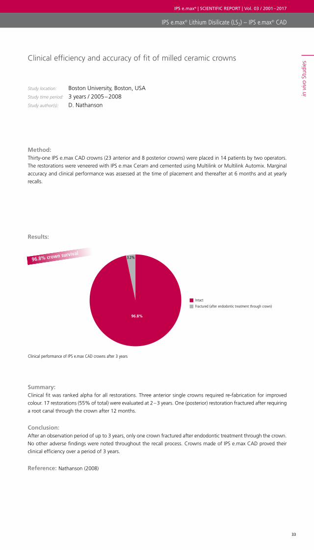
Clinical efficiency and accuracy of fit of milled ceramic crowns
Study location: Boston University, Boston, USA Study time period: 3 years / 2005 – 2008Study author(s): D. Nathanson
Method: Thirty-one IPS e.max CAD crowns (23 anterior and 8 posterior crowns) were placed in 14 patients by two operators.
The restorations were veneered with IPS e.max Ceram and cemented using Multilink or Multilink Automix. Marginal
accuracy and clinical performance was assessed at the time of placement and thereafter at 6 months and at yearly
recalls.
Results:
Summary: Clinical fit was ranked alpha for all restorations. Three anterior single crowns required re-fabrication for improved
colour. 17 restorations (55% of total) were evaluated at 2 – 3 years. One (posterior) restoration fractured after requiring
a root canal through the crown after 12 months.
Conclusion: After an observation period of up to 3 years, only one crown fractured after endodontic treatment through the crown.
No other adverse findings were noted throughout the recall process. Crowns made of IPS e.max CAD proved their
clinical efficiency over a period of 3 years.
Reference: Nathanson (2008)
Clinical performance of IPS e.max CAD crowns after 3 years
96.8% crown survival
Intact
Fractured (after endodontic treatment through crown)
96.8%
3.2%
IPS e.max® Lithium Disilicate (LS2) – IPS e.max® CAD
in v
ivo
Stud
ies
IPS e.max® | SCIENTIFIC REPORT | Vol. 03 / 2001 – 2017
34
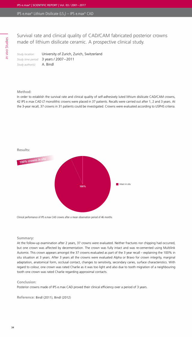
Survival rate and clinical quality of CAD/CAM fabricated posterior crowns made of lithium disilicate ceramic. A prospective clinical study.
Study location: University of Zurich, Zurich, SwitzerlandStudy time period: 3 years / 2007 – 2011 Study author(s): A. Bindl
Method: In order to establish the survival rate and clinical quality of self-adhesively luted lithium disilicate CAD/CAM crowns,
42 IPS e.max CAD LT monolithic crowns were placed in 37 patients. Recalls were carried out after 1, 2 and 3 years. At
the 3-year recall, 37 crowns in 31 patients could be investigated. Crowns were evaluated according to USPHS criteria.
Results:
Summary: At the follow-up examination after 2 years, 37 crowns were evaluated. Neither fractures nor chipping had occurred,
but one crown was affected by decementation. The crown was fully intact and was re-cemented using Multilink
Automix. This crown appears amongst the 37 crowns evaluated as part of the 3 year recall – explaining the 100% in
situ situation at 3 years. After 3 years all the crowns were evaluated Alpha or Bravo for crown integrity, marginal
adaptation, anatomical form, occlusal contact, changes to sensitivity, secondary caries, surface characteristics. With
regard to colour, one crown was rated Charlie as it was too light and also due to tooth migration of a neighbouring
tooth one crown was rated Charlie regarding approximal contacts.
Conclusion: Posterior crowns made of IPS e.max CAD proved their clinical efficiency over a period of 3 years.
Reference: Bindl (2011), Bindl (2012)
Clinical performance of IPS e.max CAD crowns after a mean observation period of 46 months
100% crowns in situ
Intact in situ 100%
IPS e.max® Lithium Disilicate (LS2) – IPS e.max® CAD
in v
ivo
Stud
ies
IPS e.max® | SCIENTIFIC REPORT | Vol. 03 / 2001 – 2017
35
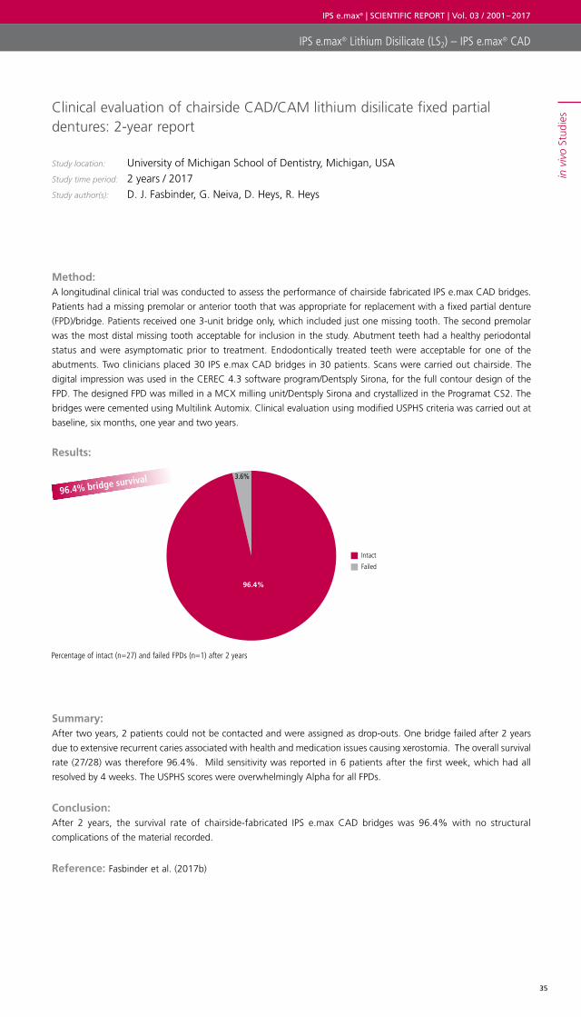
Clinical evaluation of chairside CAD/CAM lithium disilicate fixed partial dentures: 2-year report
Study location: University of Michigan School of Dentistry, Michigan, USAStudy time period: 2 years / 2017 Study author(s): D. J. Fasbinder, G. Neiva, D. Heys, R. Heys
Method: A longitudinal clinical trial was conducted to assess the performance of chairside fabricated IPS e.max CAD bridges.
Patients had a missing premolar or anterior tooth that was appropriate for replacement with a fixed partial denture
(FPD)/bridge. Patients received one 3-unit bridge only, which included just one missing tooth. The second premolar
was the most distal missing tooth acceptable for inclusion in the study. Abutment teeth had a healthy periodontal
status and were asymptomatic prior to treatment. Endodontically treated teeth were acceptable for one of the
abutments. Two clinicians placed 30 IPS e.max CAD bridges in 30 patients. Scans were carried out chairside. The
digital impression was used in the CEREC 4.3 software program/Dentsply Sirona, for the full contour design of the
FPD. The designed FPD was milled in a MCX milling unit/Dentsply Sirona and crystallized in the Programat CS2. The
bridges were cemented using Multilink Automix. Clinical evaluation using modified USPHS criteria was carried out at
baseline, six months, one year and two years.
Results:
Summary: After two years, 2 patients could not be contacted and were assigned as drop-outs. One bridge failed after 2 years
due to extensive recurrent caries associated with health and medication issues causing xerostomia. The overall survival
rate (27/28) was therefore 96.4%. Mild sensitivity was reported in 6 patients after the first week, which had all
resolved by 4 weeks. The USPHS scores were overwhelmingly Alpha for all FPDs.
Conclusion: After 2 years, the survival rate of chairside-fabricated IPS e.max CAD bridges was 96.4% with no structural
complications of the material recorded.
Reference: Fasbinder et al. (2017b)
96.4% bridge survival
Percentage of intact (n=27) and failed FPDs (n=1) after 2 years
Intact
Failed
96.4%
3.6%
IPS e.max® Lithium Disilicate (LS2) – IPS e.max® CAD
in v
ivo
Stud
ies
IPS e.max® | SCIENTIFIC REPORT | Vol. 03 / 2001 – 2017
36
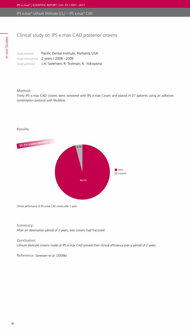
Clinical study on IPS e.max CAD posterior crowns
Study location: Pacific Dental Institute, Portland, USAStudy time period: 2 years / 2006 – 2009 Study author(s): J.A. Sorensen, R. Trotman, K. Yokoyama
Method: Thirty IPS e.max CAD crowns were veneered with IPS e.max Ceram and placed in 27 patients using an adhesive
cementation protocol with Multilink.
Results:
Summary: After an observation period of 2 years, two crowns had fractured.
Conclusion: Lithium disilicate crowns made of IPS e.max CAD proved their clinical efficiency over a period of 2 years.
Reference: Sorensen et al. (2009b)
93.3% crown survival
Clinical performance of IPS e.max CAD crowns after 2 years
Intact
Fractured
93.3%
6.7%
IPS e.max® Lithium Disilicate (LS2) – IPS e.max® CAD
in v
ivo
Stud
ies
IPS e.max® | SCIENTIFIC REPORT | Vol. 03 / 2001 – 2017
37
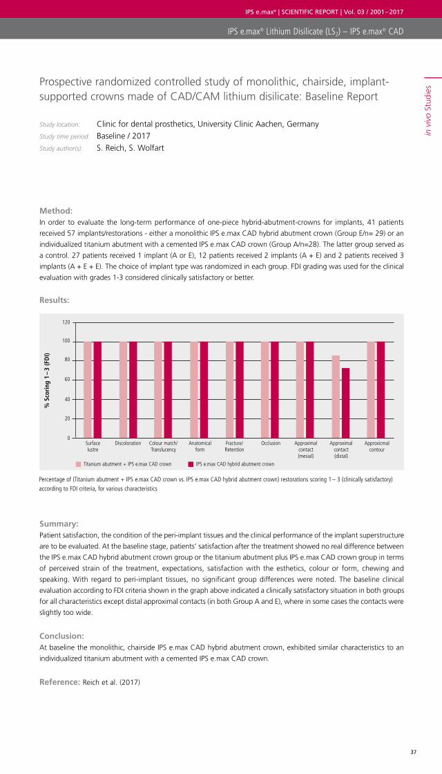
Prospective randomized controlled study of monolithic, chairside, implant- supported crowns made of CAD/CAM lithium disilicate: Baseline Report
Study location: Clinic for dental prosthetics, University Clinic Aachen, GermanyStudy time period: Baseline / 2017 Study author(s): S. Reich, S. Wolfart
Method: In order to evaluate the long-term performance of one-piece hybrid-abutment-crowns for implants, 41 patients
received 57 implants/restorations - either a monolithic IPS e.max CAD hybrid abutment crown (Group E/n= 29) or an
individualized titanium abutment with a cemented IPS e.max CAD crown (Group A/n=28). The latter group served as
a control. 27 patients received 1 implant (A or E), 12 patients received 2 implants (A + E) and 2 patients received 3
implants (A + E + E). The choice of implant type was randomized in each group. FDI grading was used for the clinical
evaluation with grades 1-3 considered clinically satisfactory or better.
Results:
Summary: Patient satisfaction, the condition of the peri-implant tissues and the clinical performance of the implant superstructure
are to be evaluated. At the baseline stage, patients’ satisfaction after the treatment showed no real difference between
the IPS e.max CAD hybrid abutment crown group or the titanium abutment plus IPS e.max CAD crown group in terms
of perceived strain of the treatment, expectations, satisfaction with the esthetics, colour or form, chewing and
speaking. With regard to peri-implant tissues, no significant group differences were noted. The baseline clinical
evaluation according to FDI criteria shown in the graph above indicated a clinically satisfactory situation in both groups
for all characteristics except distal approximal contacts (in both Group A and E), where in some cases the contacts were
slightly too wide.
Conclusion: At baseline the monolithic, chairside IPS e.max CAD hybrid abutment crown, exhibited similar characteristics to an
individualized titanium abutment with a cemented IPS e.max CAD crown.
Reference: Reich et al. (2017)
Percentage of (Titanium abutment + IPS e.max CAD crown vs. IPS e.max CAD hybrid abutment crown) restorations scoring 1 – 3 (clinically satisfactory) according to FDI criteria, for various characteristics
% S
cori
ng 1
– 3
(FD
I)
0
20
40
60
80
100
120
Surface lustre
Discoloration Anatomical form
Fracture/Retention
Colour match/Translucency
Occlusion Approximal contact(mesial)
Approximal contact(distal)
Approxicmal contour
Titanium abutment + IPS e.max CAD crown IPS e.max CAD hybrid abutment crown
IPS e.max® Lithium Disilicate (LS2) – IPS e.max® CAD
in v
ivo
Stud
ies
IPS e.max® | SCIENTIFIC REPORT | Vol. 03 / 2001 – 2017
38
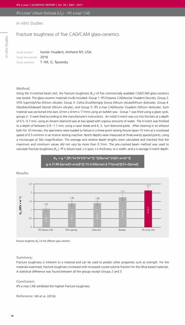
Fracture toughness of five CAD/CAM glass-ceramics
Study location: Ivoclar Vivadent, Amherst NY, USA.Study time period: 2016 Study author(s): T. Hill, G. Tysowsky
Method: Using the V-notched beam test, the fracture toughness (KIC) of five commercially available CAD/CAM glass-ceramics
was tested. The glass-ceramic materials (n=8) included: Group 1: IPS Empress CAD/Ivoclar Vivadent (leucite), Group 2:
VITA Suprinity/Vita (lithium silicate), Group 3: Celtra Duo/Dentsply Sirona (lithium silicate/lithium disilicate), Group 4:
Obsidian/Glidewell Dental (lithium silicate), and Group 5: IPS e.max CAD/Ivoclar Vivadent (lithium disilicate). Each
material was sectioned into bars (3 mm x 4 mm x 17 mm) using an IsoMet saw. Group 1 was fired using a glaze cycle,
groups 2 – 5 were fired according to the manufacturer’s instructions. An initial V-notch was cut into the bars at a depth
of 0.5 – 0.7 mm, using an Amann diamond saw at low speed with copious amounts of water. The V-notch was finished
to a depth of between 0.9 – 1.1 mm, using a razor blade and 6, 3, 1µm diamond paste. After cleaning in an ethanol
bath for 10 minutes, the specimens were loaded to failure in a three-point testing fixture (span-15 mm) at a crosshead
speed of 0.5 mm/min in an Instron testing machine. Notch depths were measured at three evenly spaced points, using
a microscope at 50x magnification. The average and relative depth lengths were calculated and checked that the
maximum and minimum values did not vary by more than 0.1mm. The pre-cracked beam method was used to
calculate fracture toughness (KIC). Pf is failure load; s is span; t is thickness; w is width; and a is average V-notch depth:
KIC = g * [(Pf*Sx10-6)/(t*w3/2)] *[(3(a/w)1/2)/(2(1-a/w)3/2)]
g is {1.99-[(a/w)(1-a/w)]*[2.15-3.93(a/w)+2.7*(a/w)2]}/[1+2(a/w)]
Results:
Summary: Fracture toughness is inherent to a material and can be used to predict other properties such as strength. For the
materials examined, fracture toughness increased with increased crystal volume fraction for the lithia based materials.
A statistical difference was found between all the groups except Groups 2 and 3.
Conclusion: IPS e.max CAD exhibited the highest fracture toughness.
Reference: Hill et al. (2016)
Fracture toughness (KIC) of five different glass ceramics
IPS e.max® Lithium Disilicate (LS2) – IPS e.max® CAD
in v
itro
Stud
ies
K IC
(MPa
• m
0.5 )
2.5
1.07
1.36 1.38
1.71
2.11
2
1.5
1
0.5
0IPS Empress CAD VITA Suprinity Celtra Duo Obsidian IPS e.max CAD
Gro
up 1
Gro
up 2
Gro
up 3
Gro
up 4
Gro
up 5
in vitro Studies
IPS e.max® | SCIENTIFIC REPORT | Vol. 03 / 2001 – 2017
39
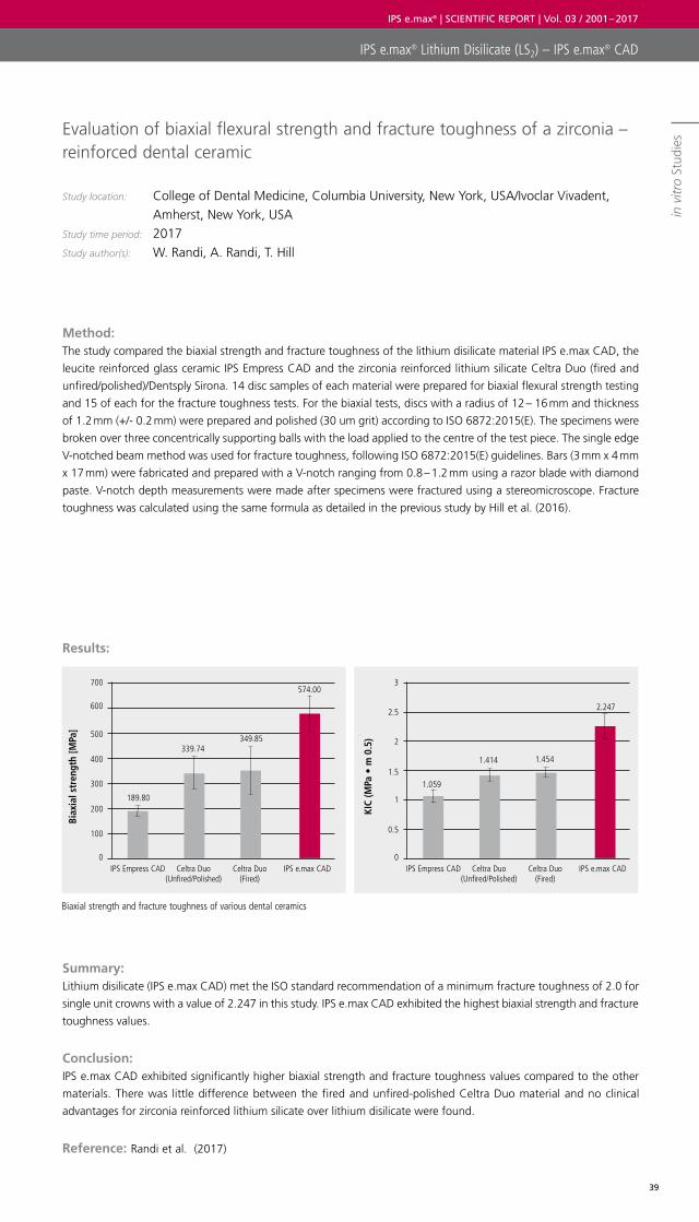
Evaluation of biaxial flexural strength and fracture toughness of a zirconia –reinforced dental ceramic
Study location: College of Dental Medicine, Columbia University, New York, USA/Ivoclar Vivadent, Amherst, New York, USA
Study time period: 2017 Study author(s): W. Randi, A. Randi, T. Hill
Method: The study compared the biaxial strength and fracture toughness of the lithium disilicate material IPS e.max CAD, the
leucite reinforced glass ceramic IPS Empress CAD and the zirconia reinforced lithium silicate Celtra Duo (fired and
unfired/polished)/Dentsply Sirona. 14 disc samples of each material were prepared for biaxial flexural strength testing
and 15 of each for the fracture toughness tests. For the biaxial tests, discs with a radius of 12 – 16 mm and thickness
of 1.2 mm (+/- 0.2 mm) were prepared and polished (30 um grit) according to ISO 6872:2015(E). The specimens were
broken over three concentrically supporting balls with the load applied to the centre of the test piece. The single edge
V-notched beam method was used for fracture toughness, following ISO 6872:2015(E) guidelines. Bars (3 mm x 4 mm
x 17 mm) were fabricated and prepared with a V-notch ranging from 0.8 – 1.2 mm using a razor blade with diamond
paste. V-notch depth measurements were made after specimens were fractured using a stereomicroscope. Fracture
toughness was calculated using the same formula as detailed in the previous study by Hill et al. (2016).
Results:
Summary: Lithium disilicate (IPS e.max CAD) met the ISO standard recommendation of a minimum fracture toughness of 2.0 for
single unit crowns with a value of 2.247 in this study. IPS e.max CAD exhibited the highest biaxial strength and fracture
toughness values.
Conclusion: IPS e.max CAD exhibited significantly higher biaxial strength and fracture toughness values compared to the other
materials. There was little difference between the fired and unfired-polished Celtra Duo material and no clinical
advantages for zirconia reinforced lithium silicate over lithium disilicate were found.
Reference: Randi et al. (2017)
Biaxial strength and fracture toughness of various dental ceramics
IPS e.max® Lithium Disilicate (LS2) – IPS e.max® CAD
in v
itro
Stud
ies
Biax
ial s
tren
gth
[MPa
]
KIC
(MPa
• m
0.5
)
700 3
6002.5
500
400
2
3001.5
2001
100 0.5
0 0IPS Empress CAD IPS Empress CADCeltra Duo
(Unfired/Polished)Celtra Duo
(Unfired/Polished)Celtra Duo
(Fired)Celtra Duo
(Fired)IPS e.max CAD IPS e.max CAD
339.741.414
574.00
2.247
189.80
1.059
349.85
1.454
IPS e.max® | SCIENTIFIC REPORT | Vol. 03 / 2001 – 2017
40
IPS e.max® Lithium Disilicate (LS2) – IPS e.max® CAD
in v
itro
Stud
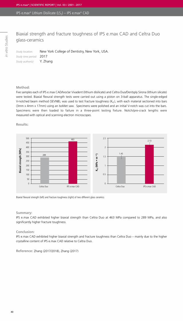
ies Biaxial strength and fracture toughness of IPS e.max CAD and Celtra Duo
glass-ceramics
Study location: New York College of Dentistry, New York, USA. Study time period: 2017 Study author(s): Y. Zhang
Method: Five samples each of IPS e.max CAD/Ivoclar Vivadent (lithium disilicate) and Celtra Duo/Dentsply Sirona (lithium silicate)
were tested. Biaxial flexural strength tests were carried out using a piston on 3-ball apparatus. The single-edged
V-notched beam method (SEVNB), was used to test fracture toughness (KIC), with each material sectioned into bars
(3mm x 4mm x 17mm) using an IsoMet saw. Specimens were polished and an initial V-notch was cut into the bars.
Specimens were then loaded to failure in a three-point testing fixture. Notch/pre-crack lengths were
measured with optical and scanning electron microscopes.
Results:
Summary: IPS e.max CAD exhibited higher biaxial strength than Celtra Duo at 463 MPa compared to 289 MPa, and also
significantly higher fracture toughness.
Conclusion: IPS e.max CAD exhibited higher biaxial strength and fracture toughness than Celtra Duo – mainly due to the higher
crystalline content of IPS e.max CAD relative to Celtra Duo.
Reference: Zhang (2017/2018), Zhang (2017)
Biaxial flexural strength (left) and fracture toughness (right) of two different glass ceramics
K IC
(MPa
• m
0.5 )
2.5
1.45
2
1.5
1
0.5
0Celtra Duo
2.13
IPS e.max CAD
Biax
ial s
tren
gth
[MPa
]
500
450
289
400
350
300
250
200
150
100
50
0Celtra Duo
463
IPS e.max CAD
IPS e.max® | SCIENTIFIC REPORT | Vol. 03 / 2001 – 2017
41
IPS e.max® Lithium Disilicate (LS2) – IPS e.max® CAD
in v
itro
Stud
iesMechanical characteristics of a zirconia-reinforced lithium silicate CAD/CAM
restorative material
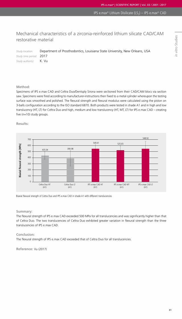
Study location: Department of Prosthodontics, Louisiana State University, New Orleans, USA Study time period: 2017 Study author(s): K. Vu
Method: Specimens of IPS e.max CAD and Celtra Duo/Dentsply Sirona were sectioned from their CAD/CAM blocs via section
saw. Specimens were fired according to manufacturer-instructions then fixed to a metal cylinder whereupon the testing
surface was smoothed and polished. The flexural strength and flexural modulus were calculated using the piston on
3-balls configuration according to the ISO standard 6870. Both products were tested in shade A1 and in high and low
translucency (HT, LT) for Celtra Duo and high, medium and low translucency (HT, MT, LT) for IPS e.max CAD – creating
five (n=10) study groups.
Results:
Summary: The flexural strength of IPS e.max CAD exceeded 500 MPa for all translucencies and was significantly higher than that
of Celtra Duo. The two translucencies of Celtra Duo exhibited greater variation in flexural strength than the three
translucencies of IPS e.max CAD.
Conclusion: The flexural strength of IPS e.max CAD exceeded that of Celtra Duo for all translucencies.
Reference: Vu (2017)
Biaxial flexural strength of Celtra Duo and IPS e.max CAD in shade A1 with different translucencies
Biax
ial f
lexu
ral s
tren
gth
[MPa
]
700
600
500
400
300
200
100
0Celtra Duo HT
(A1)Celtra Duo LT
(A1)IPS e.max CAD HT
(A1)IPS e.max CAD MT
(A1)IPS e.max CAD LT
(A1)
435.04 390.98
549.41 525.65
548.92
IPS e.max® | SCIENTIFIC REPORT | Vol. 03 / 2001 – 2017
42
IPS e.max® Lithium Disilicate (LS2) – IPS e.max® CAD
in v
itro
Stud
ies Monolithic and veneered CAD/CAM lithium disilicate bridges vs. metal-
ceramic: Comparison of the fracture load values and failure modes upon fatigue
Study location: University Clinic, Freiburg im Breisgau, Germany Study time period: 2012 Study author(s): S. Schultheis, J.R. Strub, T.A. Gerds, P.C. Guess
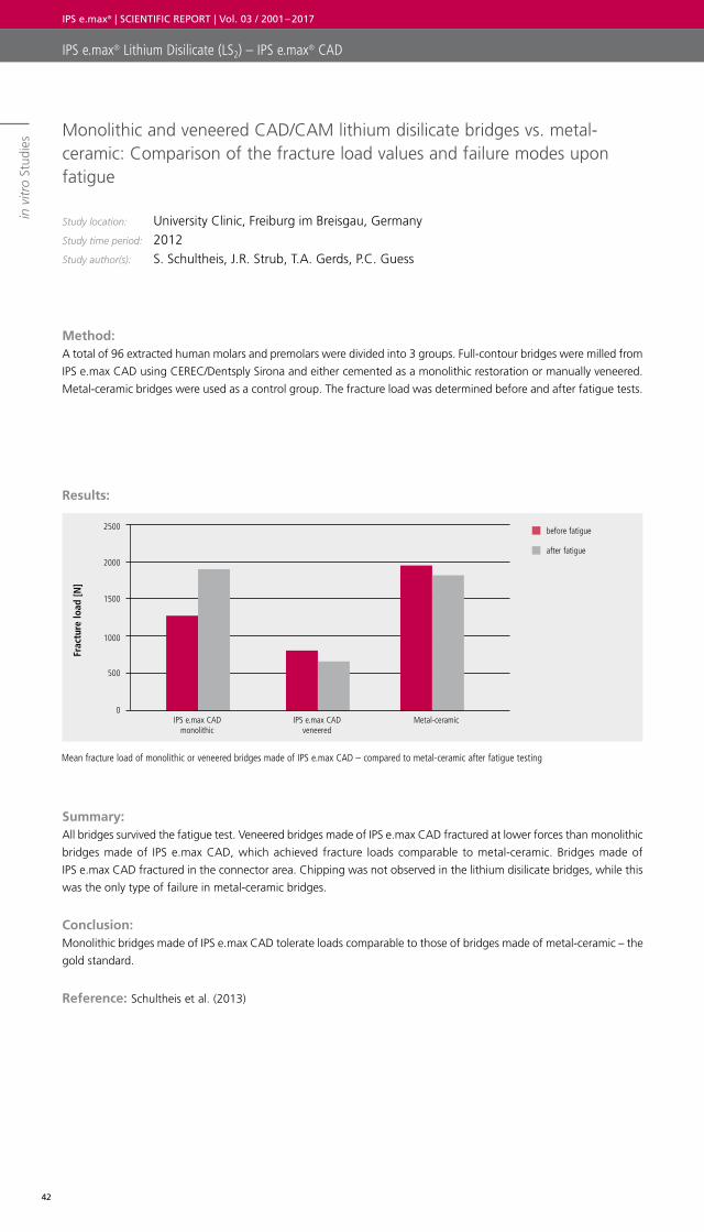
Method: A total of 96 extracted human molars and premolars were divided into 3 groups. Full-contour bridges were milled from
IPS e.max CAD using CEREC/Dentsply Sirona and either cemented as a monolithic restoration or manually veneered.
Metal-ceramic bridges were used as a control group. The fracture load was determined before and after fatigue tests.
Results:
Summary: All bridges survived the fatigue test. Veneered bridges made of IPS e.max CAD fractured at lower forces than monolithic
bridges made of IPS e.max CAD, which achieved fracture loads comparable to metal-ceramic. Bridges made of
IPS e.max CAD fractured in the connector area. Chipping was not observed in the lithium disilicate bridges, while this
was the only type of failure in metal-ceramic bridges.
Conclusion: Monolithic bridges made of IPS e.max CAD tolerate loads comparable to those of bridges made of metal-ceramic – the
gold standard.
Reference: Schultheis et al. (2013)
Mean fracture load of monolithic or veneered bridges made of IPS e.max CAD – compared to metal-ceramic after fatigue testing
Frac
ture
load
[N]
2500
2000
1500
1000
500
0IPS e.max CAD
monolithic IPS e.max CAD
veneeredMetal-ceramic
before fatigue
after fatigue
IPS e.max® | SCIENTIFIC REPORT | Vol. 03 / 2001 – 2017
43
Monolithic CAD/CAM lithium disilicate compared to veneered Y-TZP crowns: Comparison of the failure types and reliability after fatigue
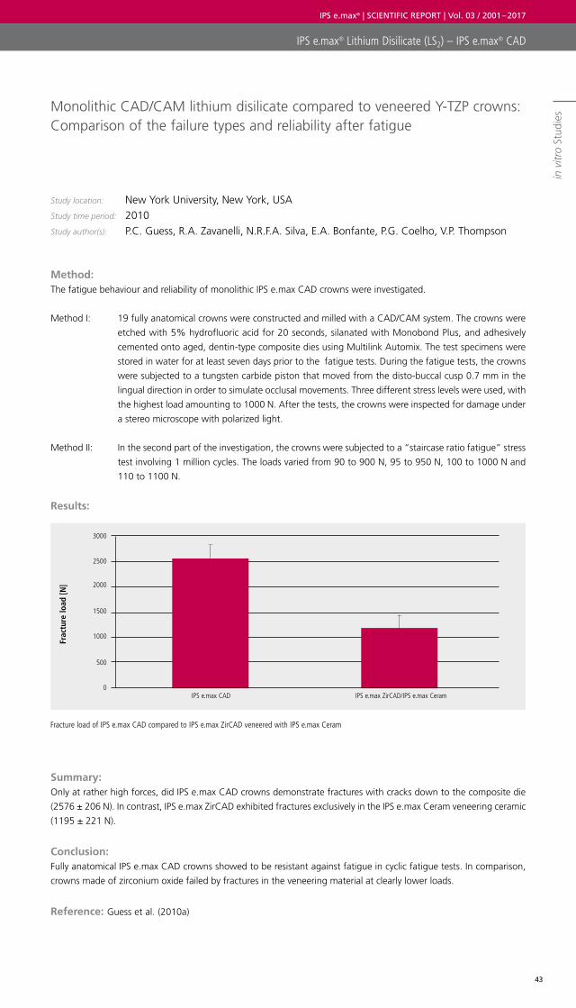
Study location: New York University, New York, USA Study time period: 2010 Study author(s): P.C. Guess, R.A. Zavanelli, N.R.F.A. Silva, E.A. Bonfante, P.G. Coelho, V.P. Thompson
Method: The fatigue behaviour and reliability of monolithic IPS e.max CAD crowns were investigated.
Method I: 19 fully anatomical crowns were constructed and milled with a CAD/CAM system. The crowns were
etched with 5% hydrofluoric acid for 20 seconds, silanated with Monobond Plus, and adhesively
cemented onto aged, dentin-type composite dies using Multilink Automix. The test specimens were
stored in water for at least seven days prior to the fatigue tests. During the fatigue tests, the crowns
were subjected to a tungsten carbide piston that moved from the disto-buccal cusp 0.7 mm in the
lingual direction in order to simulate occlusal movements. Three different stress levels were used, with
the highest load amounting to 1000 N. After the tests, the crowns were inspected for damage under
a stereo microscope with polarized light.
Method II: In the second part of the investigation, the crowns were subjected to a “staircase ratio fatigue” stress
test involving 1 million cycles. The loads varied from 90 to 900 N, 95 to 950 N, 100 to 1000 N and
110 to 1100 N.
Results:
Summary: Only at rather high forces, did IPS e.max CAD crowns demonstrate fractures with cracks down to the composite die
(2576 ± 206 N). In contrast, IPS e.max ZirCAD exhibited fractures exclusively in the IPS e.max Ceram veneering ceramic
(1195 ± 221 N).
Conclusion: Fully anatomical IPS e.max CAD crowns showed to be resistant against fatigue in cyclic fatigue tests. In comparison,
crowns made of zirconium oxide failed by fractures in the veneering material at clearly lower loads.
Reference: Guess et al. (2010a)
Fracture load of IPS e.max CAD compared to IPS e.max ZirCAD veneered with IPS e.max Ceram
Frac
ture
load
[N]
3000
2500
2000
1500
1000
500
0IPS e.max CAD IPS e.max ZirCAD/IPS e.max Ceram
IPS e.max® Lithium Disilicate (LS2) – IPS e.max® CAD
in v
itro
Stud
ies
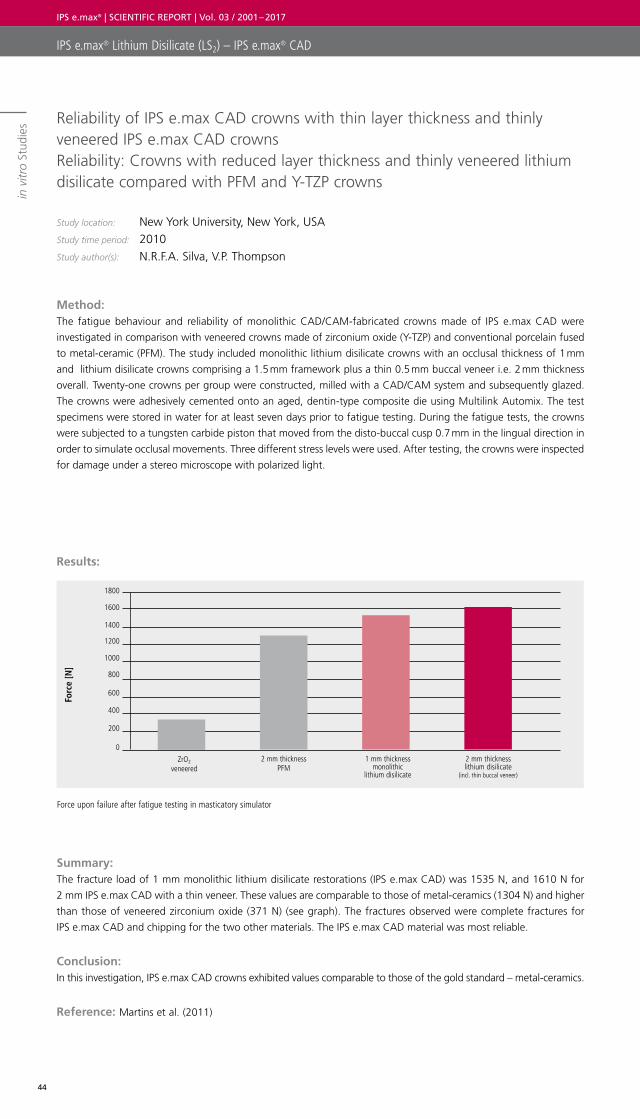
IPS e.max® | SCIENTIFIC REPORT | Vol. 03 / 2001 – 2017
44
IPS e.max® Lithium Disilicate (LS2) – IPS e.max® CAD
in v
itro
Stud
ies Reliability of IPS e.max CAD crowns with thin layer thickness and thinly
veneered IPS e.max CAD crowns Reliability: Crowns with reduced layer thickness and thinly veneered lithium disilicate compared with PFM and Y-TZP crowns
Study location: New York University, New York, USA Study time period: 2010 Study author(s): N.R.F.A. Silva, V.P. Thompson
Method: The fatigue behaviour and reliability of monolithic CAD/CAM-fabricated crowns made of IPS e.max CAD were
investigated in comparison with veneered crowns made of zirconium oxide (Y-TZP) and conventional porcelain fused
to metal-ceramic (PFM). The study included monolithic lithium disilicate crowns with an occlusal thickness of 1 mm
and lithium disilicate crowns comprising a 1.5 mm framework plus a thin 0.5 mm buccal veneer i.e. 2 mm thickness
overall. Twenty-one crowns per group were constructed, milled with a CAD/CAM system and subsequently glazed.
The crowns were adhesively cemented onto an aged, dentin-type composite die using Multilink Automix. The test
specimens were stored in water for at least seven days prior to fatigue testing. During the fatigue tests, the crowns
were subjected to a tungsten carbide piston that moved from the disto-buccal cusp 0.7 mm in the lingual direction in
order to simulate occlusal movements. Three different stress levels were used. After testing, the crowns were inspected
for damage under a stereo microscope with polarized light.
Results:
Summary: The fracture load of 1 mm monolithic lithium disilicate restorations (IPS e.max CAD) was 1535 N, and 1610 N for
2 mm IPS e.max CAD with a thin veneer. These values are comparable to those of metal-ceramics (1304 N) and higher
than those of veneered zirconium oxide (371 N) (see graph). The fractures observed were complete fractures for
IPS e.max CAD and chipping for the two other materials. The IPS e.max CAD material was most reliable.
Conclusion: In this investigation, IPS e.max CAD crowns exhibited values comparable to those of the gold standard – metal-ceramics.
Reference: Martins et al. (2011)
Force upon failure after fatigue testing in masticatory simulator
Forc
e [N
]
1800
1600
1400
1200
1000
800
600
400
200
02 mm thickness
PFMZrO2
veneered1 mm thickness
monolithic lithium disilicate
2 mm thicknesslithium disilicate
(incl. thin buccal veneer)
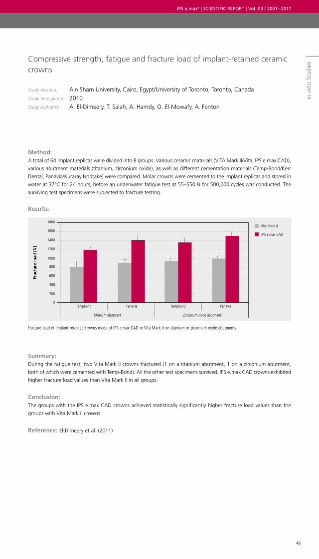
IPS e.max® | SCIENTIFIC REPORT | Vol. 03 / 2001 – 2017
45
Compressive strength, fatigue and fracture load of implant-retained ceramic crowns
Study location: Ain Sham University, Cairo, Egypt/University of Toronto, Toronto, Canada Study time period: 2010 Study author(s): A. El-Dimeery, T. Salah, A. Hamdy, O. El-Mowafy, A. Fenton
Method: A total of 64 implant replicas were divided into 8 groups. Various ceramic materials (VITA Mark II/Vita, IPS e.max CAD),
various abutment materials (titanium, zirconium oxide), as well as different cementation materials (Temp-Bond/Kerr
Dental, Panavia/Kuraray Noritake) were compared. Molar crowns were cemented to the implant replicas and stored in
water at 37°C for 24 hours, before an underwater fatigue test at 55–550 N for 500,000 cycles was conducted. The
surviving test specimens were subjected to fracture testing.
Results:
Summary: During the fatigue test, two Vita Mark II crowns fractured (1 on a titanium abutment, 1 on a zirconium abutment,
both of which were cemented with Temp-Bond). All the other test specimens survived. IPS e.max CAD crowns exhibited
higher fracture load values than Vita Mark II in all groups.
Conclusion: The groups with the IPS e.max CAD crowns achieved statistically significantly higher fracture load values than the
groups with Vita Mark II crowns.
Reference: El-Dimeery et al. (2011)
Fracture load of implant-retained crowns made of IPS e.max CAD or Vita Mark II on titanium or zirconium oxide abutments
Frac
ture
load
[N]
Vita Mark II
IPS e.max CAD
1800
1600
1400
1200
1000
800
600
400
200
0
Titanium abutment Zirconium oxide abutment
Tempbond TempbondPanavia Panaviain
vitr
o St
udie
s
IPS e.max® | SCIENTIFIC REPORT | Vol. 03 / 2001 – 2017
46
IPS e.max® Lithium Disilicate (LS2) – In Vivo Studies
IPS e.max® | SCIENTIFIC REPORT | Vol. 03 / 2001 – 2017
47
in vivo studies in vitro studies
IPS e.max® Zirconium Oxide
(ZrO2)
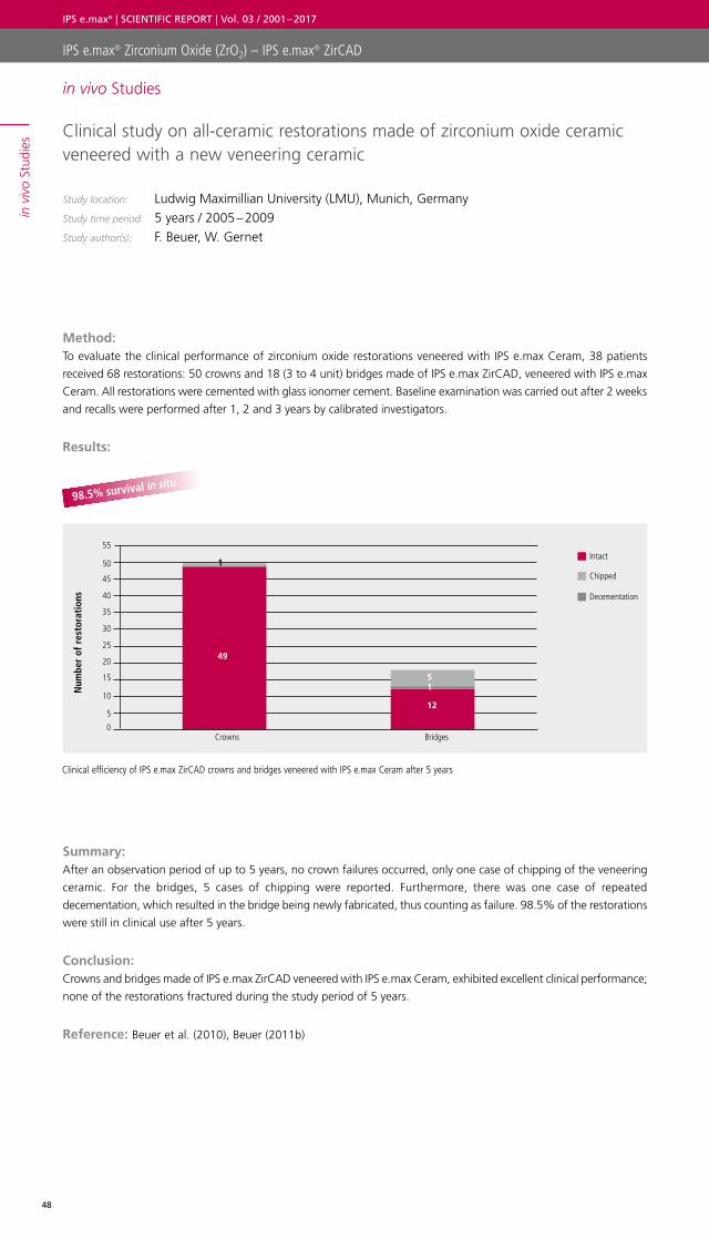
IPS e.max® | SCIENTIFIC REPORT | Vol. 03 / 2001 – 2017
48
Clinical study on all-ceramic restorations made of zirconium oxide ceramic veneered with a new veneering ceramic
Study location: Ludwig Maximillian University (LMU), Munich, Germany Study time period: 5 years / 2005 – 2009Study author(s): F. Beuer, W. Gernet
Method: To evaluate the clinical performance of zirconium oxide restorations veneered with IPS e.max Ceram, 38 patients
received 68 restorations: 50 crowns and 18 (3 to 4 unit) bridges made of IPS e.max ZirCAD, veneered with IPS e.max
Ceram. All restorations were cemented with glass ionomer cement. Baseline examination was carried out after 2 weeks
and recalls were performed after 1, 2 and 3 years by calibrated investigators.
Results:
Summary: After an observation period of up to 5 years, no crown failures occurred, only one case of chipping of the veneering
ceramic. For the bridges, 5 cases of chipping were reported. Furthermore, there was one case of repeated
decementation, which resulted in the bridge being newly fabricated, thus counting as failure. 98.5% of the restorations
were still in clinical use after 5 years.
Conclusion: Crowns and bridges made of IPS e.max ZirCAD veneered with IPS e.max Ceram, exhibited excellent clinical performance;
none of the restorations fractured during the study period of 5 years.
Reference: Beuer et al. (2010), Beuer (2011b)
Clinical efficiency of IPS e.max ZirCAD crowns and bridges veneered with IPS e.max Ceram after 5 years
98.5% survival in situ
Num
ber
of r
esto
rati
ons
0
10
5
25
20
15
30
40
35
45
50
55
Crowns Bridges
Intact
Chipped
Decementation
49
1
12
51
IPS e.max® Zirconium Oxide (ZrO2) – IPS e.max® ZirCAD
in v
ivo
Stud
ies
in vivo Studies
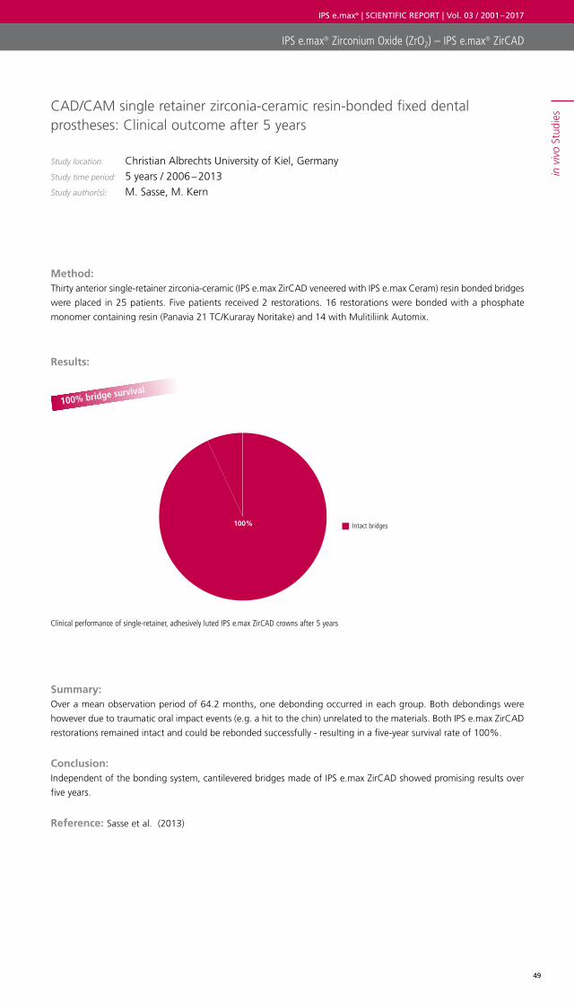
IPS e.max® | SCIENTIFIC REPORT | Vol. 03 / 2001 – 2017
49
IPS e.max® Zirconium Oxide (ZrO2) – IPS e.max® ZirCAD
in v
ivo
Stud
iesCAD/CAM single retainer zirconia-ceramic resin-bonded fixed dental
prostheses: Clinical outcome after 5 years
Study location: Christian Albrechts University of Kiel, Germany Study time period: 5 years / 2006 – 2013 Study author(s): M. Sasse, M. Kern
Method: Thirty anterior single-retainer zirconia-ceramic (IPS e.max ZirCAD veneered with IPS e.max Ceram) resin bonded bridges
were placed in 25 patients. Five patients received 2 restorations. 16 restorations were bonded with a phosphate
monomer containing resin (Panavia 21 TC/Kuraray Noritake) and 14 with Mulitiliink Automix.
Results:
Summary: Over a mean observation period of 64.2 months, one debonding occurred in each group. Both debondings were
however due to traumatic oral impact events (e.g. a hit to the chin) unrelated to the materials. Both IPS e.max ZirCAD
restorations remained intact and could be rebonded successfully - resulting in a five-year survival rate of 100%.
Conclusion: Independent of the bonding system, cantilevered bridges made of IPS e.max ZirCAD showed promising results over
five years.
Reference: Sasse et al. (2013)
100% bridge survival
Clinical performance of single-retainer, adhesively luted IPS e.max ZirCAD crowns after 5 years
Intact bridges100%
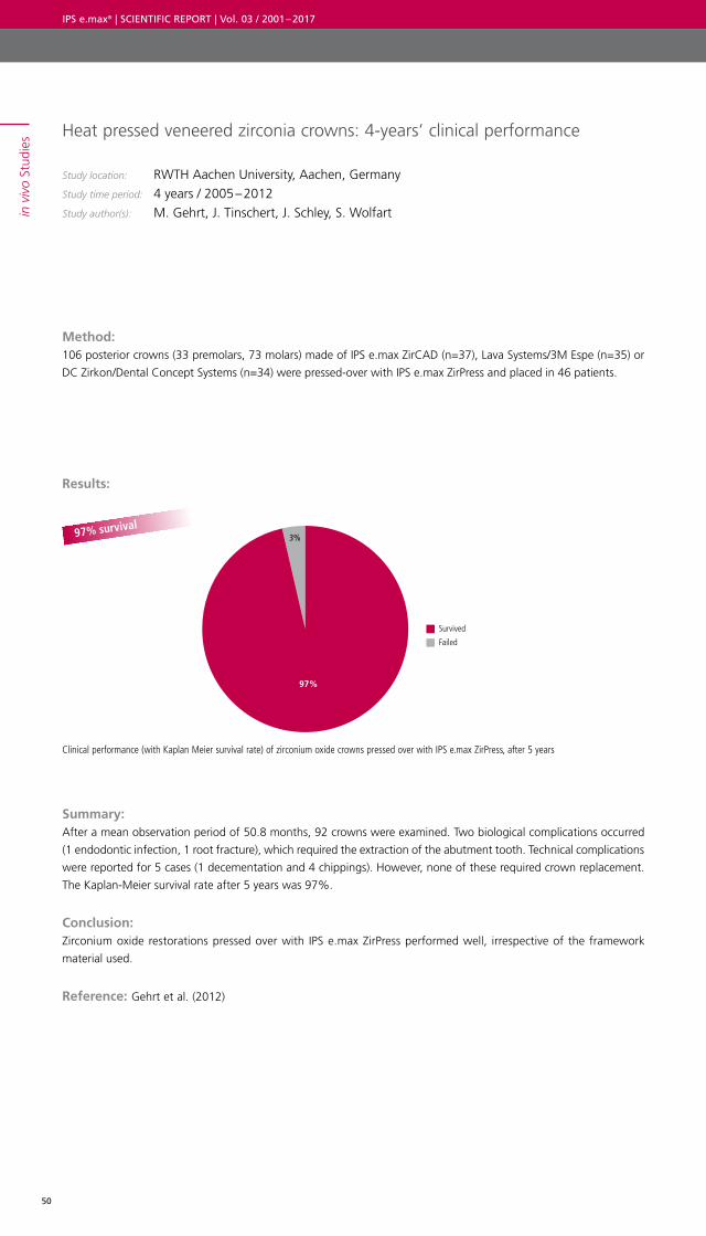
IPS e.max® | SCIENTIFIC REPORT | Vol. 03 / 2001 – 2017
50
in v
ivo
Stud
ies Heat pressed veneered zirconia crowns: 4-years’ clinical performance
Study location: RWTH Aachen University, Aachen, Germany Study time period: 4 years / 2005 – 2012 Study author(s): M. Gehrt, J. Tinschert, J. Schley, S. Wolfart
Method: 106 posterior crowns (33 premolars, 73 molars) made of IPS e.max ZirCAD (n=37), Lava Systems/3M Espe (n=35) or
DC Zirkon/Dental Concept Systems (n=34) were pressed-over with IPS e.max ZirPress and placed in 46 patients.
Results:
Summary: After a mean observation period of 50.8 months, 92 crowns were examined. Two biological complications occurred
(1 endodontic infection, 1 root fracture), which required the extraction of the abutment tooth. Technical complications
were reported for 5 cases (1 decementation and 4 chippings). However, none of these required crown replacement.
The Kaplan-Meier survival rate after 5 years was 97%.
Conclusion: Zirconium oxide restorations pressed over with IPS e.max ZirPress performed well, irrespective of the framework
material used.
Reference: Gehrt et al. (2012)
97% survival
Clinical performance (with Kaplan Meier survival rate) of zirconium oxide crowns pressed over with IPS e.max ZirPress, after 5 years
Survived
Failed
97%
3%
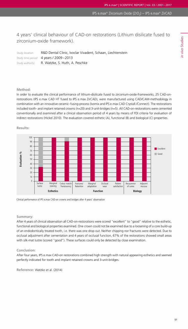
IPS e.max® | SCIENTIFIC REPORT | Vol. 03 / 2001 – 2017
51
IPS e.max® Zirconium Oxide (ZrO2) – IPS e.max® ZirCAD
in v
ivo
Stud
ies4 years’ clinical behaviour of CAD-on restorations (Lithium disilicate fused to
zirconium-oxide framework).
Study location: R&D Dental Clinic, Ivoclar Vivadent, Schaan, Liechtenstein Study time period: 4 years / 2009 – 2013 Study author(s): R. Watzke, S. Huth, A. Peschke
Method: In order to evaluate the clinical performance of lithium-disilicate fused to zirconium-oxide-frameworks, 25 CAD-on-
restorations (IPS e.max CAD HT fused to IPS e.max ZirCAD), were manufactured using CAD/CAM-methodology in
combination with an innovative ceramic–fusing-process (Ivomix and IPS e.max CAD Crystall./Connect). The restorations
included tooth- and implant retained crowns (n=20) and 3-unit-bridges (n=5). All CAD-on-restorations were cemented
conventionally and examined after a clinical observation period of 4 years by means of FDI criteria for evaluation of
indirect restorations (Hickel 2010). The evaluation covered esthetic (A), functional (B) and biological (C) properties.
Results:
Summary: After 4 years of clinical observation all CAD-on-restorations were scored “excellent” to “good” relative to the esthetic,
functional and biological properties examined. One crown could not be examined due to a loosening of a core build-up
of an endodontically treated tooth, i.e. there was one drop out. Neither chipping nor fractures were detected. Due to
occlusal adjustment after cementation and 4 years of occlusal function, 67% of the restorations showed small areas
with silk-mat lustre (scored “good”). These surfaces could only be detected by close examination.
Conclusion: After four years, IPS e.max CAD-on restorations combined high strength with natural appearing esthetics and seemed
perfectly indicated for tooth and implant retained crowns and 3-unit-bridges.
Reference: Watzke et al. (2014)
Clinical performance of IPS e.max CAD-on crowns and bridges after 4 years’ observation
Eval
uati
on %
100
90
80
70
60
50
40
30
20
10
0Surfacelustre
Colour match/Translucency
Marginal adaptation
Marginal staining
Fractures/Retention
Occlusal wear
Patient satisfaction
Recuurence of caries
Adjacent mucosa
Esthetics Function Biology
Excellent
Good

IPS e.max® | SCIENTIFIC REPORT | Vol. 03 / 2001 – 2017
52
in v
ivo
Stud
ies
IPS e.max® Zirconium Oxide (ZrO2) – IPS e.max® ZirCAD
Clinical performance of IPS e.max Ceram on IPS e.max ZirCAD
Study location: Pacific Dental Institute, Portland, USAStudy time period: 4 years / 2004 – 2009Study author(s): J.A. Sorensen
Method: Incorporation of 20 IPS e.max ZirCAD bridges, veneered with IPS e.max Ceram.
Results:
Summary: No absolute failures were reported in an observation period of 46.7 ± 5 months. The survival rate was 100%. Two
small (cohesive) chippings within the veneering ceramic were reported.
Conclusion: With a survival rate of 100%, the clinical performance of IPS e.max ZirCAD bridges veneered with IPS e.max Ceram
was excellent.
Reference: Sorensen et al. (2009a)
Clinical performance of bridges made of IPS e.max ZirCAD veneered with IPS e.max Ceram
100% survival
Survived 100%
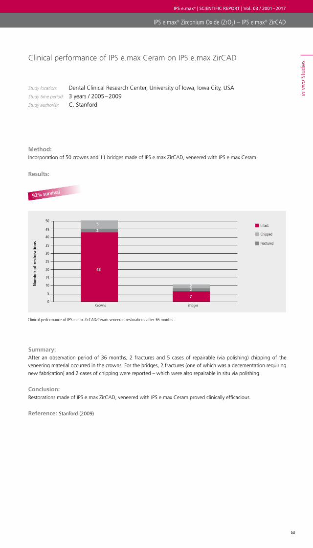
IPS e.max® | SCIENTIFIC REPORT | Vol. 03 / 2001 – 2017
53
in v
ivo
Stud
ies
IPS e.max® Zirconium Oxide (ZrO2) – IPS e.max® ZirCAD
Clinical performance of IPS e.max Ceram on IPS e.max ZirCAD
Study location: Dental Clinical Research Center, University of Iowa, Iowa City, USA Study time period: 3 years / 2005 – 2009Study author(s): C. Stanford
Method: Incorporation of 50 crowns and 11 bridges made of IPS e.max ZirCAD, veneered with IPS e.max Ceram.
Results:
Summary: After an observation period of 36 months, 2 fractures and 5 cases of repairable (via polishing) chipping of the
veneering material occurred in the crowns. For the bridges, 2 fractures (one of which was a decementation requiring
new fabrication) and 2 cases of chipping were reported – which were also repairable in situ via polishing.
Conclusion: Restorations made of IPS e.max ZirCAD, veneered with IPS e.max Ceram proved clinically efficacious.
Reference: Stanford (2009)
Clinical performance of IPS e.max ZirCAD/Ceram-veneered restorations after 36 months
92% survival
Num
ber
of r
esto
rati
ons
0
10
5
25
20
15
30
40
35
45
50
Crowns Bridges
Intact
Chipped
Fractured
12
2
5
43
22
7
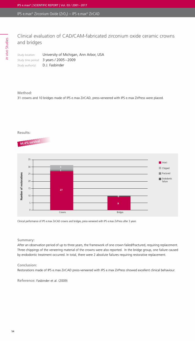
IPS e.max® | SCIENTIFIC REPORT | Vol. 03 / 2001 – 2017
54
in v
ivo
Stud
ies
IPS e.max® Zirconium Oxide (ZrO2) – IPS e.max® ZirCAD
Clinical evaluation of CAD/CAM-fabricated zirconium oxide ceramic crowns and bridges
Study location: University of Michigan, Ann Arbor, USAStudy time period: 3 years / 2005 – 2009Study author(s): D.J. Fasbinder
Method: 31 crowns and 10 bridges made of IPS e.max ZirCAD, press-veneered with IPS e.max ZirPress were placed.
Results:
Summary: After an observation period of up to three years, the framework of one crown failed/fractured, requiring replacement.
Three chippings of the veneering material of the crowns were also reported. In the bridge group, one failure caused
by endodontic treatment occurred. In total, there were 2 absolute failures requiring restorative replacement.
Conclusion: Restorations made of IPS e.max ZirCAD press-veneered with IPS e.max ZirPress showed excellent clinical behaviour.
Reference: Fasbinder et al. (2009)
Clinical performance of IPS e.max ZirCAD crowns and bridges, press-veneered with IPS e.max ZirPress after 3 years
94.4% survival
Num
ber
of r
esto
rati
ons
0
10
5
25
20
15
30
35
Crowns Bridges
Intact
Chipped
Fractured
Endodontic failure
2
7
27
13
9
0
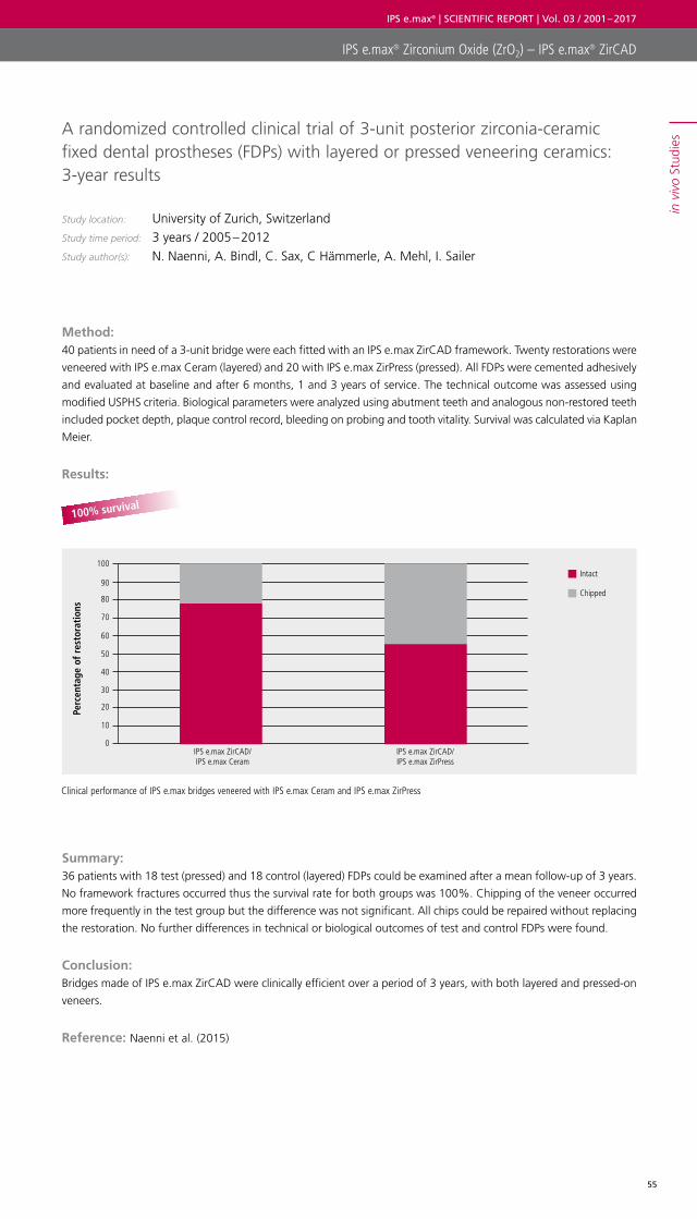
IPS e.max® | SCIENTIFIC REPORT | Vol. 03 / 2001 – 2017
55
in v
ivo
Stud
ies
IPS e.max® Zirconium Oxide (ZrO2) – IPS e.max® ZirCAD
A randomized controlled clinical trial of 3-unit posterior zirconia-ceramic fixed dental prostheses (FDPs) with layered or pressed veneering ceramics: 3-year results
Study location: University of Zurich, SwitzerlandStudy time period: 3 years / 2005 – 2012Study author(s): N. Naenni, A. Bindl, C. Sax, C Hämmerle, A. Mehl, I. Sailer
Method: 40 patients in need of a 3-unit bridge were each fitted with an IPS e.max ZirCAD framework. Twenty restorations were
veneered with IPS e.max Ceram (layered) and 20 with IPS e.max ZirPress (pressed). All FDPs were cemented adhesively
and evaluated at baseline and after 6 months, 1 and 3 years of service. The technical outcome was assessed using
modified USPHS criteria. Biological parameters were analyzed using abutment teeth and analogous non-restored teeth
included pocket depth, plaque control record, bleeding on probing and tooth vitality. Survival was calculated via Kaplan
Meier.
Results:
Summary: 36 patients with 18 test (pressed) and 18 control (layered) FDPs could be examined after a mean follow-up of 3 years.
No framework fractures occurred thus the survival rate for both groups was 100%. Chipping of the veneer occurred
more frequently in the test group but the difference was not significant. All chips could be repaired without replacing
the restoration. No further differences in technical or biological outcomes of test and control FDPs were found.
Conclusion: Bridges made of IPS e.max ZirCAD were clinically efficient over a period of 3 years, with both layered and pressed-on
veneers.
Reference: Naenni et al. (2015)
Clinical performance of IPS e.max bridges veneered with IPS e.max Ceram and IPS e.max ZirPress
100% survival
Perc
enta
ge o
f re
stor
atio
ns
0
20
10
50
40
30
60
80
70
90
100
IPS e.max ZirCAD/IPS e.max Ceram
Intact
Chipped
IPS e.max ZirCAD/IPS e.max ZirPress
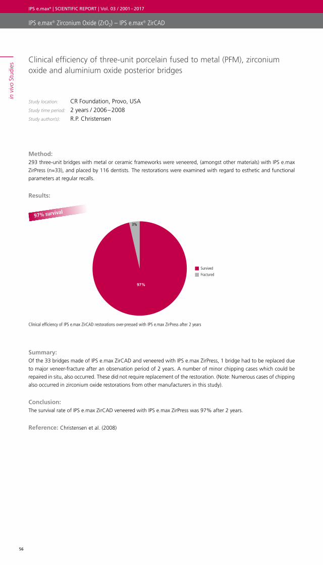
IPS e.max® | SCIENTIFIC REPORT | Vol. 03 / 2001 – 2017
56
in v
ivo
Stud
ies
IPS e.max® Zirconium Oxide (ZrO2) – IPS e.max® ZirCAD
Clinical efficiency of three-unit porcelain fused to metal (PFM), zirconium oxide and aluminium oxide posterior bridges
Study location: CR Foundation, Provo‚ USA Study time period: 2 years / 2006 – 2008 Study author(s): R.P. Christensen
Method: 293 three-unit bridges with metal or ceramic frameworks were veneered, (amongst other materials) with IPS e.max
ZirPress (n=33), and placed by 116 dentists. The restorations were examined with regard to esthetic and functional
parameters at regular recalls.
Results:
Summary: Of the 33 bridges made of IPS e.max ZirCAD and veneered with IPS e.max ZirPress, 1 bridge had to be replaced due
to major veneer-fracture after an observation period of 2 years. A number of minor chipping cases which could be
repaired in situ, also occurred. These did not require replacement of the restoration. (Note: Numerous cases of chipping
also occurred in zirconium oxide restorations from other manufacturers in this study).
Conclusion: The survival rate of IPS e.max ZirCAD veneered with IPS e.max ZirPress was 97% after 2 years.
Reference: Christensen et al. (2008)
97% survival
Clinical efficiency of IPS e.max ZirCAD restorations over-pressed with IPS e.max ZirPress after 2 years
Survived
Fractured
97%
3%
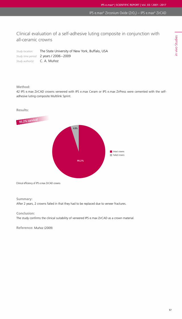
IPS e.max® | SCIENTIFIC REPORT | Vol. 03 / 2001 – 2017
57
in v
ivo
Stud
ies
IPS e.max® Zirconium Oxide (ZrO2) – IPS e.max® ZirCAD
Clinical evaluation of a self-adhesive luting composite in conjunction with all-ceramic crowns
Study location: The State University of New York‚ Buffalo, USAStudy time period: 2 years / 2006 – 2009Study author(s): C. A. Muñoz
Method: 42 IPS e.max ZirCAD crowns veneered with IPS e.max Ceram or IPS e.max ZirPress were cemented with the self-
adhesive luting composite Multilink Sprint.
Results:
Summary: After 2 years, 2 crowns failed in that they had to be replaced due to veneer fractures.
Conclusion: The study confirms the clinical suitability of veneered IPS e.max ZirCAD as a crown material.
Reference: Muñoz (2009)
95.2% survival
Clinical efficiency of IPS e.max ZirCAD crowns
Intact crowns
Failed crowns
95.2%
4.8%
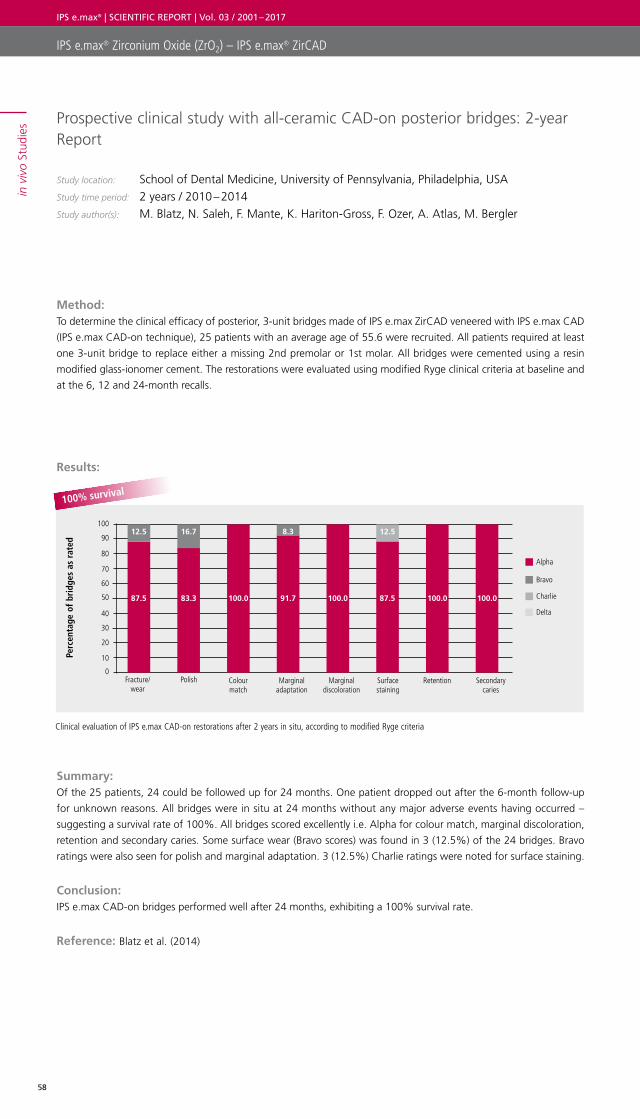
IPS e.max® | SCIENTIFIC REPORT | Vol. 03 / 2001 – 2017
58
Perc
enta
ge o
f br
idge
s as
rat
ed
100
90
80
70
60
50
40
30
20
10
0Fracture/
wearColour match
Marginal discoloration
Polish Marginal adaptation
Surface staining
Retention Secondary caries
Alpha
Bravo
Charlie
Delta
87.5 83.3 91.7 87.5100.0 100.0 100.0 100.0
12.5 16.7 8.3 12.5
in v
ivo
Stud
ies
IPS e.max® Zirconium Oxide (ZrO2) – IPS e.max® ZirCAD
Prospective clinical study with all-ceramic CAD-on posterior bridges: 2-year Report
Study location: School of Dental Medicine, University of Pennsylvania, Philadelphia, USA Study time period: 2 years / 2010 – 2014Study author(s): M. Blatz, N. Saleh, F. Mante, K. Hariton-Gross, F. Ozer, A. Atlas, M. Bergler
Method: To determine the clinical efficacy of posterior, 3-unit bridges made of IPS e.max ZirCAD veneered with IPS e.max CAD
(IPS e.max CAD-on technique), 25 patients with an average age of 55.6 were recruited. All patients required at least
one 3-unit bridge to replace either a missing 2nd premolar or 1st molar. All bridges were cemented using a resin
modified glass-ionomer cement. The restorations were evaluated using modified Ryge clinical criteria at baseline and
at the 6, 12 and 24-month recalls.
Results:
Summary: Of the 25 patients, 24 could be followed up for 24 months. One patient dropped out after the 6-month follow-up
for unknown reasons. All bridges were in situ at 24 months without any major adverse events having occurred –
suggesting a survival rate of 100%. All bridges scored excellently i.e. Alpha for colour match, marginal discoloration,
retention and secondary caries. Some surface wear (Bravo scores) was found in 3 (12.5%) of the 24 bridges. Bravo
ratings were also seen for polish and marginal adaptation. 3 (12.5%) Charlie ratings were noted for surface staining.
Conclusion: IPS e.max CAD-on bridges performed well after 24 months, exhibiting a 100% survival rate.
Reference: Blatz et al. (2014)
Clinical evaluation of IPS e.max CAD-on restorations after 2 years in situ, according to modified Ryge criteria
100% survival

IPS e.max® | SCIENTIFIC REPORT | Vol. 03 / 2001 – 2017
59
in v
ivo
Stud
ies
IPS e.max® Zirconium Oxide (ZrO2) – IPS e.max® ZirCAD
Three-unit posterior zirconia-ceramic fixed dental prostheses (FDPs) veneered with layered and milled (CAD-on) veneering ceramics: 1-year follow-up of a randomized controlled clinical trial.
Study location: University of Zurich, SwitzerlandStudy time period: 1 year / 2015Study author(s): P. Grohmann, A. Bindl, C. Hämmerle, A. Mehl, I. Sailer
Method: To compare zirconia-ceramic fixed dental prostheses (FDPs) veneered with either a CAD/CAM lithium disilicate
veneering ceramic (CAD-on) or a manually layered veneering ceramic with respect to survival, technical and biological
outcomes – 60 patients in need of one posterior three-unit bridge (FDP) were included in the study. Thirty IPS e.max
ZirCAD FDPs were veneered with a CAD/CAM lithium disilicate veneering ceramic (IPS e.max CAD HT) using the
CAD-on technique (test group). The other thirty were veneered with a layered veneering ceramic (IPS e.max Ceram)
(control group). For the clinical evaluation at baseline, 6, and 12 months, United States Public Health Service (USPHS)
criteria were used. The biological outcome was judged by comparing the plaque control record (PCR), bleeding on
probing (BOP), and probing pocket depth (PPD).
Results:
Summary: Fifty-six patients were examined at a mean follow-up of 13.9 months. At the 1-year follow-up the survival rate was
100% in both the test and control group. No significant differences of the technical outcomes occurred. Major
chipping occurred in the control group (n = 3) and predominantly minor chipping in the test group (minor n = 2, major
n = 1). No biological problems or differences were found.
Conclusion: Both types of zirconia-ceramic FDPs exhibited very good clinical outcomes without differences between groups.
Chipping occurred in both types of FDPs in small amounts, yet the extension of the chippings differed.
Reference: Grohmann et al. (2015)
Survival rate after 1 year in both test and control groups
100% survival
Survived 100%
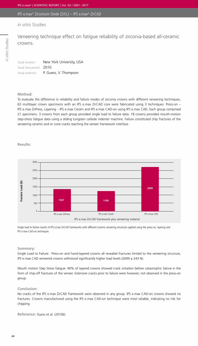
IPS e.max® | SCIENTIFIC REPORT | Vol. 03 / 2001 – 2017
60
in v
itro
Stud
ies
IPS e.max® Zirconium Oxide (ZrO2) – IPS e.max® ZirCAD
Veneering technique effect on fatigue reliability of zirconia-based all-ceramic crowns.
Study location: New York University, USAStudy time period: 2010Study author(s): P. Guess, V. Thompson
Method: To evaluate the difference in reliability and failure modes of zirconia crowns with different veneering techniques,
63 multilayer crown specimens with an IPS e.max ZirCAD core were fabricated using 3 techniques: Press-on –
IPS e.max ZirPress, Layering – IPS e.max Ceram and IPS e.max CAD-on using IPS e.max CAD. Each group comprised
21 specimens. 3 crowns from each group provided single load to failure data. 18 crowns provided mouth-motion
step-stress fatigue data using a sliding tungsten carbide indenter machine. Failure constituted chip fractures of the
veneering ceramic and or cone cracks reaching the veneer framework interface.
Results:
Summary: Single Load to Failure: Press-on and hand-layered crowns all revealed fractures limited to the veneering structure,
IPS e.max CAD veneered crowns withstood significantly higher load levels (2699 ± 243 N).
Mouth motion Step Stress fatigue: 49% of layered crowns showed crack initiation before catastrophic failure in the
form of chip-off fractures of the veneer. Extensive cracks prior to failure were however, not observed in the press-on
group.
Conclusion: No cracks of the IPS e.max ZirCAD framework were observed in any group. IPS e.max CAD-on crowns showed no
fractures. Crowns manufactured using the IPS e.max CAD-on technique were most reliable, indicating no risk for
chipping.
Reference: Guess et al. (2010b)
Single load to failure results of IPS e.max ZirCAD frameworks with different ceramic veneering structures applied using the press-on, layering and IPS e.max CAD-on techniques
Frac
ture
Loa
d (N
)
IPS e.max ZirCAD framework plus veneering material
3000
2500
2000
1500
1000
500
0IPS e.max ZirPress IPS e.max Ceram IPS e.max CAD
1367 1195
2699
in vitro Studies
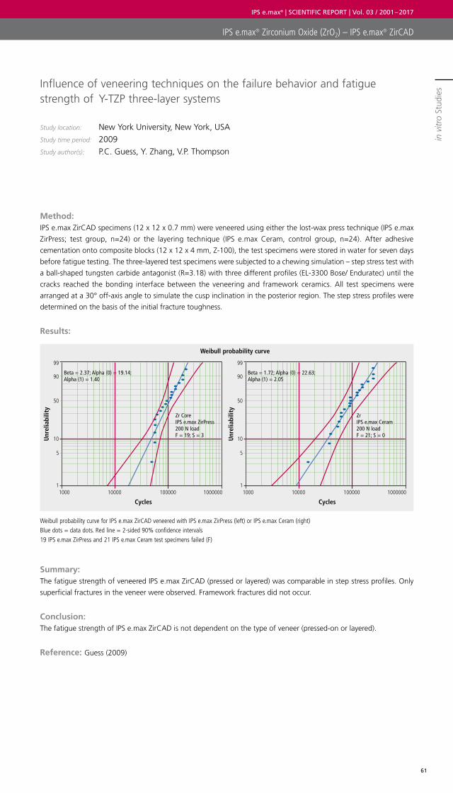
IPS e.max® | SCIENTIFIC REPORT | Vol. 03 / 2001 – 2017
61
in v
itro
Stud
ies
IPS e.max® Zirconium Oxide (ZrO2) – IPS e.max® ZirCAD
Influence of veneering techniques on the failure behavior and fatigue strength of Y-TZP three-layer systems
Study location: New York University, New York, USAStudy time period: 2009Study author(s): P.C. Guess, Y. Zhang, V.P. Thompson
Method: IPS e.max ZirCAD specimens (12 x 12 x 0.7 mm) were veneered using either the lost-wax press technique (IPS e.max
ZirPress; test group, n=24) or the layering technique (IPS e.max Ceram, control group, n=24). After adhesive
cementation onto composite blocks (12 x 12 x 4 mm, Z-100), the test specimens were stored in water for seven days
before fatigue testing. The three-layered test specimens were subjected to a chewing simulation – step stress test with
a ball-shaped tungsten carbide antagonist (R=3.18) with three different profiles (EL-3300 Bose/ Enduratec) until the
cracks reached the bonding interface between the veneering and framework ceramics. All test specimens were
arranged at a 30° off-axis angle to simulate the cusp inclination in the posterior region. The step stress profiles were
determined on the basis of the initial fracture toughness.
Results:
Summary: The fatigue strength of veneered IPS e.max ZirCAD (pressed or layered) was comparable in step stress profiles. Only
superficial fractures in the veneer were observed. Framework fractures did not occur.
Conclusion: The fatigue strength of IPS e.max ZirCAD is not dependent on the type of veneer (pressed-on or layered).
Reference: Guess (2009)
Weibull probability curve for IPS e.max ZirCAD veneered with IPS e.max ZirPress (left) or IPS e.max Ceram (right) Blue dots = data dots. Red line = 2-sided 90% confidence intervals 19 IPS e.max ZirPress and 21 IPS e.max Ceram test specimens failed (F)
Unr
elia
bilit
y
Unr
elia
bilit
y
Weibull probability curve
Beta = 2.37; Alpha (0) = 19.14; Alpha (1) = 1.40
Beta = 1.72; Alpha (0) = 22.63; Alpha (1) = 2.05
Zr Core IPS e.max ZirPress200 N load F = 19; S = 3
Zr IPS e.max Ceram200 N load F = 21; S = 0
Cycles
99
90
50
10
5
11000 10000 100000 1000000
Cycles
99
90
50
10
5
11000 10000 100000 1000000
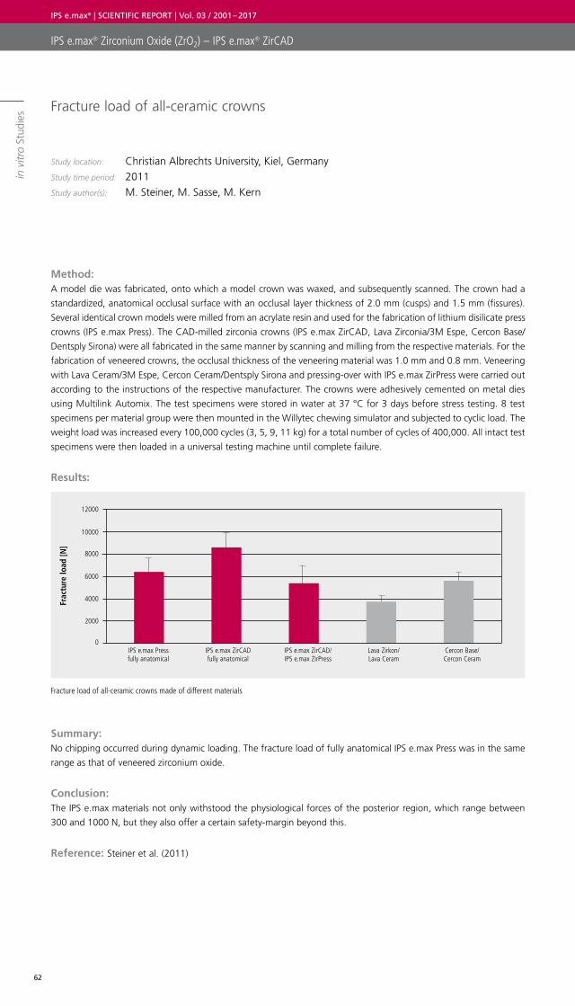
IPS e.max® | SCIENTIFIC REPORT | Vol. 03 / 2001 – 2017
62
in v
itro
Stud
ies
IPS e.max® Zirconium Oxide (ZrO2) – IPS e.max® ZirCAD
Fracture load of all-ceramic crowns
Study location: Christian Albrechts University, Kiel, Germany Study time period: 2011Study author(s): M. Steiner, M. Sasse, M. Kern
Method: A model die was fabricated, onto which a model crown was waxed, and subsequently scanned. The crown had a
standardized, anatomical occlusal surface with an occlusal layer thickness of 2.0 mm (cusps) and 1.5 mm (fissures).
Several identical crown models were milled from an acrylate resin and used for the fabrication of lithium disilicate press
crowns (IPS e.max Press). The CAD-milled zirconia crowns (IPS e.max ZirCAD, Lava Zirconia/3M Espe, Cercon Base/
Dentsply Sirona) were all fabricated in the same manner by scanning and milling from the respective materials. For the
fabrication of veneered crowns, the occlusal thickness of the veneering material was 1.0 mm and 0.8 mm. Veneering
with Lava Ceram/3M Espe, Cercon Ceram/Dentsply Sirona and pressing-over with IPS e.max ZirPress were carried out
according to the instructions of the respective manufacturer. The crowns were adhesively cemented on metal dies
using Multilink Automix. The test specimens were stored in water at 37 °C for 3 days before stress testing. 8 test
specimens per material group were then mounted in the Willytec chewing simulator and subjected to cyclic load. The
weight load was increased every 100,000 cycles (3, 5, 9, 11 kg) for a total number of cycles of 400,000. All intact test
specimens were then loaded in a universal testing machine until complete failure.
Results:
Summary: No chipping occurred during dynamic loading. The fracture load of fully anatomical IPS e.max Press was in the same
range as that of veneered zirconium oxide.
Conclusion: The IPS e.max materials not only withstood the physiological forces of the posterior region, which range between
300 and 1000 N, but they also offer a certain safety-margin beyond this.
Reference: Steiner et al. (2011)
Fracture load of all-ceramic crowns made of different materials
Frac
ture
load
[N]
12000
10000
8000
6000
4000
2000
0IPS e.max Pressfully anatomical
IPS e.max ZirCADfully anatomical
IPS e.max ZirCAD/IPS e.max ZirPress
Lava Zirkon/Lava Ceram
Cercon Base/Cercon Ceram
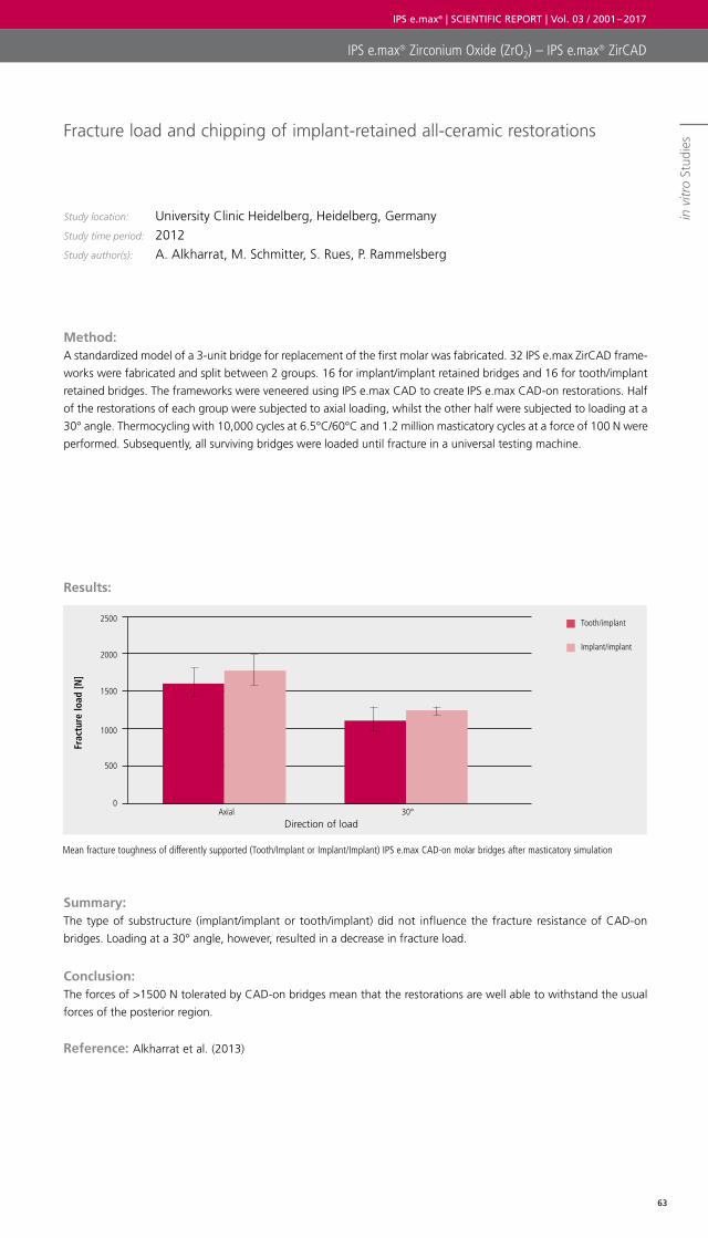
IPS e.max® | SCIENTIFIC REPORT | Vol. 03 / 2001 – 2017
63
in v
itro
Stud
ies
IPS e.max® Zirconium Oxide (ZrO2) – IPS e.max® ZirCAD
Mean fracture toughness of differently supported (Tooth/Implant or Implant/Implant) IPS e.max CAD-on molar bridges after masticatory simulation
Fracture load and chipping of implant-retained all-ceramic restorations
Study location: University Clinic Heidelberg, Heidelberg, Germany Study time period: 2012Study author(s): A. Alkharrat, M. Schmitter, S. Rues, P. Rammelsberg
Method: A standardized model of a 3-unit bridge for replacement of the first molar was fabricated. 32 IPS e.max ZirCAD frame-
works were fabricated and split between 2 groups. 16 for implant/implant retained bridges and 16 for tooth/implant
retained bridges. The frameworks were veneered using IPS e.max CAD to create IPS e.max CAD-on restorations. Half
of the restorations of each group were subjected to axial loading, whilst the other half were subjected to loading at a
30° angle. Thermocycling with 10,000 cycles at 6.5°C/60°C and 1.2 million masticatory cycles at a force of 100 N were
performed. Subsequently, all surviving bridges were loaded until fracture in a universal testing machine.
Results:
Summary: The type of substructure (implant/implant or tooth/implant) did not influence the fracture resistance of CAD-on
bridges. Loading at a 30° angle, however, resulted in a decrease in fracture load.
Conclusion: The forces of >1500 N tolerated by CAD-on bridges mean that the restorations are well able to withstand the usual
forces of the posterior region.
Reference: Alkharrat et al. (2013)
Frac
ture
load
[N]
2500
2000
1500
1000
500
0Axial 30°
Direction of load
Tooth/implant
Implant/implant
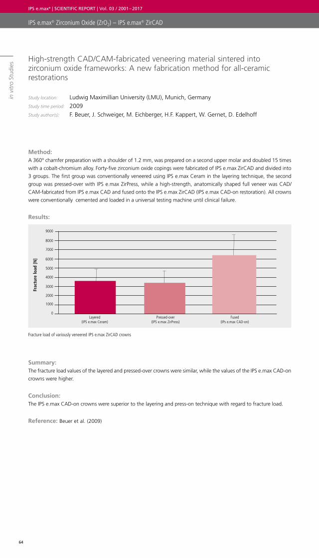
IPS e.max® | SCIENTIFIC REPORT | Vol. 03 / 2001 – 2017
64
in v
itro
Stud
ies
IPS e.max® Zirconium Oxide (ZrO2) – IPS e.max® ZirCAD
Fracture load of variously veneered IPS e.max ZirCAD crowns
High-strength CAD/CAM-fabricated veneering material sintered into zirconium oxide frameworks: A new fabrication method for all-ceramic restorations
Study location: Ludwig Maximillian University (LMU), Munich, Germany Study time period: 2009Study author(s): F. Beuer, J. Schweiger, M. Eichberger, H.F. Kappert, W. Gernet, D. Edelhoff
Method: A 360° chamfer preparation with a shoulder of 1.2 mm, was prepared on a second upper molar and doubled 15 times
with a cobalt-chromium alloy. Forty-five zirconium oxide copings were fabricated of IPS e.max ZirCAD and divided into
3 groups. The first group was conventionally veneered using IPS e.max Ceram in the layering technique, the second
group was pressed-over with IPS e.max ZirPress, while a high-strength, anatomically shaped full veneer was CAD/
CAM-fabricated from IPS e.max CAD and fused onto the IPS e.max ZirCAD (IPS e.max CAD-on restoration). All crowns
were conventionally cemented and loaded in a universal testing machine until clinical failure.
Results:
Summary: The fracture load values of the layered and pressed-over crowns were similar, while the values of the IPS e.max CAD-on
crowns were higher.
Conclusion: The IPS e.max CAD-on crowns were superior to the layering and press-on technique with regard to fracture load.
Reference: Beuer et al. (2009)
Frac
ture
load
[N]
0
1000
2000
3000
4000
6000
5000
7000
8000
9000
Layered (IPS e.max Ceram)
Pressed-over (IPS e.max ZirPress)
Fused (IPs e.max CAD-on)

IPS e.max® | SCIENTIFIC REPORT | Vol. 03 / 2001 – 2017
65

IPS e.max® | SCIENTIFIC REPORT | Vol. 03 / 2001 – 2017
66
Biocompatibility Definition of Terms
Literature
IPS e.max® | SCIENTIFIC REPORT | Vol. 03 / 2001 – 2017
67
Biocompatibility Definition of Terms
Literature
IPS e.max® | SCIENTIFIC REPORT | Vol. 03 / 2001 – 2017
68
Biocompatibility can be defined as the ability of a substance/material to be in contact with a living system without
producing an adverse effect. Tests indicate the reactivity or tolerance of cells to soluble compounds of a material.
Biocompatibility tests may include in vitro investigations (conducted in artificial environments such as petri/cell culture
dishes) such as cytotoxicity, mutagenicity, irritation and sensitivity tests. These tests are useful but have limited
significance. Only in vivo investigations (performed in the living organism) i.e. clinical experience, can provide a final
and definitive evaluation of biocompatibility.
In order to minimize biocompatibility risks from the outset, Ivoclar Vivadent strives to use well-established raw materials
that have already proven safe in vivo – in the development of new products.
The biocompatibility of lithium disilicate glass-ceramic and zirconium oxide has been assessed on the basis of toxicity
data from various institutes, plus data found in literature. In these tests, neither lithium disilicate nor zirconium oxide
showed excessive solubility, cytotoxicity, genotoxicity or any significant radioactivity.
Chemical durability/solubility
Ceramic materials are highly resistant to acid and corrosion attacks and are therefore regarded as exceptionally bio-
compatible. The conditions found in the oral cavity (pH and temperature changes) are also not extreme enough to
dissolve components from dental ceramics. The standard ISO 6872 prescribes guidelines for chemical solubility testing.
Lithium disilicate
The chemical solubility of IPS e.max lithium disilicate (IPS e.max Press and IPS e.max CAD) was evaluated according to
ISO 6872. The values found were clearly below the limit of 100 µg/cm2. An analysis of ions (dissolved in artificial saliva
and acetic acid) from IPS e.max Press and IPS e.max CAD specimens demonstrated a low ion content. Concentrations
were in the same range as those of other dental ceramics.
Zirconium oxide
IPS e.max ZirCAD blocks, discs and colouring liquids were similarly tested for chemical solubility according to ISO 6872.
All values were also well below the limit of 100 µg/cm2.
Cytotoxicity
Cytotoxicity refers to the capability of a substance to damage cells. The XTT assay is used to determine whether or not
the substance being investigated inhibits cell proliferation or even causes cell death. The resulting XTT50 value refers
to the concentration of a substance sufficient to reduce the cell viability by half. The lower the XTT50 concentration,
the more cytoxic. Numerous tests were carried out on both lithium disilicate and zirconium oxide and neither showed
cytotoxic potential.
Lithium disilicate
– RCC Report In vitro cytotoxicity test evaluation of materials for medical devices (direct cell contact assay) CCR Project
571100 (28 October 1996)*
– RCC Report In vitro cytotoxicity test evaluation of materials for medical devices (direct cell contact assay) CCR Project
590001 (24 June 1997) *
– RCC Report In vitro cytotoxicity test evaluation of materials for medical devices (direct cell contact assay) CCR Project
590002 (24 June 1997) *
– RCC Report Cytotoxicity Assay in vitro: Evaluation of materials for Medical Devices) RCC-devices with e.max Press
(XTT Test) RCC-CCR study number 1165602 (March 2008) *
– NIOM; Test Rep.; #012/04 (4 March 2004) *
– NIOM; Test Rep.; #004/04 (4 February 2004) *
– Grall, F. Toxicon Final GLP Report: 10-1251-G1. Agar Diffusion Test – ISO. April 2010.*
Zirconium oxide
In a “worst case” testing scenario, the in vitro cytotoxicity of the MT O (bleach) discs (immersed in various colouring
liquids) was evaluated. None of the samples possessed any cytotoxic potential:
– Roth M. Cytotoxicity assay in vitro (XTT-Test). Envigo Report No. 1716001. 2015.*
– Roth M. Cytotoxicity assay in vitro (XTT-Test). Envigo Report No. 1716007. 2015.*
– Roth M. Cytotoxicity assay in vitro (XTT-Test). Envigo Report No. 1716005. 2015.*
– Roth M. Cytotoxicity assay in vitro (XTT-Test). Envigo Report No. 1716003. 2015.*
BIOCOMPATIBILITY
IPS e.max® | SCIENTIFIC REPORT | Vol. 03 / 2001 – 2017
69
Biocompatibility
– Roth M. Cytotoxicity assay in vitro (XTT-Test). Envigo Report No. 1734305. 2016.*
– Roth M. Cytotoxicity assay in vitro (XTT-Test). Envigo Report No. 1734303. 2016.*
– Roth M. Cytotoxicity assay in vitro (XTT-Test). Envigo Report No. 1734301. 2016.*
The in-vitro cytotoxicity of the pre-shaded discs: IPS e.max ZirCAD MO4 and IPS e.max ZirCAD MO2, was also examined
via XTT test. No cytotoxic potential was determined:
– Meurer K. Cytotoxicity assay in vitro: Evaluation of materials for medical devices (XTT-test). RCC-CCR Report No.
1015500. 2006. *
– Heppenheimer A. Cytotoxicity assay in vitro: Evaluation of materials for medical devices (XTT-Test). RCC-CCR Report
No. 1120101. 2007. *
Genotoxicity
Genotoxicity refers to the capability of substances or external influences to damage or alter the genetic materials of
cells. Ames tests were carried out with lithium disilicate and (deeply coloured) zirconium oxide samples. Neither
material showed mutagenicity.
Lithium disilicate
– RCC Report Salmonella Typhimurium and Escherichia Coli Reverse Mutation Assay with e.max Press (Ames Test)
RCC – CCR study number 1165601 (May 2008)
– Devaki S, Toxikon Final GLP Report: 10-1251-G3: Salmonella typhimurium and Escherichia coli reverse mutation
assay – ISO. April 2010.
Zirconium oxide
– Sokolowski A. Salmonella typhimurium and Escherichia coli reverse mutation assay. Envigo Report No. 1716009.
2015. *
– Sokolowski A. Salmonella typhimurium and Escherichia coli reverse mutation assay. Envigo Report No. 1716015.
2015. *
– Sokolowski A. Salmonella typhimurium and Escherichia coli reverse mutation assay. Envigo Report No. 1716013.
2015. *
– Sokolowski A. Salmonella typhimurium and Escherichia coli reverse mutation assay. Envigo Report No. 1716011.
2015. *
– Sokolowski A. Salmonella typhimurium and Escherichia coli reverse mutation assay. Envigo Report No. 1734313.
2016. *
– Sokolowski A. Salmonella typhimurium and Escherichia coli reverse mutation assay. Envigo Report No. 1734315.
2016. *
– Sokolowski A. Salmonella typhimurium and Escherichia coli reverse mutation assay. Envigo Report No. 1734317.
2016. *
Radioactivity
The standards EN ISO 6872, EN ISO 9693 and IS13356 forbid the use of radioactive additives and stipulate the
maximum level of radioactivity permissible in ceramic materials. Tests are made for minute levels of thorium or uranium
which may be present in raw materials or pigments. Radioactivity levels in lithium dislocate and zirconium oxide were
all far below the allowable threshold of 1Bq/g (ISO 6872).
Lithium disilicate
– Laugs O. Activity measurement of the nuclides 232Th and 238U in dental ceramic with Pulver e.max Press Multi
A3.5. Forschungszentrum Jülich. 2014. *
– Küppers G. Activity measurement of the nuclides 232Th and 238U in dental ceramic with IPS e.max CAD MO4.
Forschungszentrum Jülich. 2013. *
– Küppers G. Activity measurement of the nuclides 232Th and 238U in dental ceramic with IPS e.max CAD HT C4.
Forschungszentrum Jülich. 2013. *
– Küppers G. Activity measurement of the nuclides 232Th and 238U in dental ceramic with IPS e.max CAD LT D4.
Forschungszentrum Jülich. 2013. *
IPS e.max® | SCIENTIFIC REPORT | Vol. 03 / 2001 – 2017
70
Zirconium oxide
– Küppers G. Activity measurement of the nuclides 232Th and 238U in dental ceramic with EAM591. Forschungs-
zentrum Jülich. 2006. *
– Laugs O. Activity measurement of the nuclides 232Th and 238U in dental ceramic with Probe 1298-1 PU ZirCAD LT.
Forschungszentrum Jülich Report No. 17-10064. 2017. *
– Laugs O. Activity measurement of the nuclides 232Th and 238U in dental ceramic with Probe 1298-2 PU ZirCAD
Schneide. Forschungszentrum Jülich Report No. 17-10065. 2017. *
Conclusion
The IPS e.max lithium disilicate and zirconium oxide ceramics were examined for their toxicological
potential with regard to their use as medical products. Dental ceramics are generally known and accepted as highly
biocompatible, numerous studies were conducted which confirm this. In addition, the scientific literature and a decade
plus of clinical use are testament to the safety of these materials.
It can be concluded that the IPS e.max ceramics pose no health hazard if used correctly, and the benefits of their use
outweigh any residual risk.
* Reports of investigations commissioned by Ivoclar Vivadent AG are not published or for distribution
Biocompatibility
IPS e.max® | SCIENTIFIC REPORT | Vol. 03 / 2001 – 2017
71
DEFINITION OF TERMS
Studies
Studies are conducted to forecast or examine the behaviour of materials when used for the intended application.
Aspects of functionality, reliability, safety, compatibility or user-friendliness are often of most interest.
• In vitro studies
In vitro means “in glass”. These examinations are conducted in a laboratory outside of their normal biological con-
text. Many materials science or toxicological tests are carried out in vitro, since they cannot be conducted on human
beings for practical or ethical reasons. Moreover in vitro studies have the advantage that researchers can work under
standardized conditions – plus they are often quicker and less expensive than in vivo studies
• In vivo studies
In vivo means “in the living object”. Such studies are carried out within the biological context i.e. in human beings.
The advantage is that results are more meaningful as the investigations are conducted under real conditions. They
are however complex due to a wealth of possible influencing factors. They require exact planning, systematic
methods and statistically correct evaluation. Randomized controlled studies are considered the gold standard.
• Prospective study
A study planned to be conducted in the future in order to test a certain hypothesis, such as material A is as good
as material B. After preparation of a test plan, the patients are recruited and the material used. The test subjects are
observed over a certain period of time and the results are subsequently evaluated.
• Retrospective study
Analysis of data collected in the past. For example - all cases of bridge fractures that occurred in a dental office are
examined to find out if the fractures happen more frequently with one material than with another.
Clinical Evaluation Techniques for Restorations
Cvar and Ryge/USPHS Criteria
(Cvar & Ryge 1971 and 2005)
Cvar and Ryge developed their much used measurement scale over 40 years ago. This method of evaluation is i
nterchangeably referred to as Cvar & Ryge criteria, Ryge criteria or USPHS criteria. The criteria were drawn up for
evaluating amalgam or resin based direct restorations. Various authors modified the criteria as restoratives improved
over time in terms of longevity. These are referred to as modified Ryge or modified USPHS criteria. The criteria used
the Alpha, Bravo, Charlie, Delta evaluation scale. These scores have different meanings depending on the criteria being
assessed however in general: Alpha = excellent/optimal, Bravo = acceptable, Charlie = unacceptable/insufficient and
Delta = needs replacing.
Hickel/FDI Criteria
(Hickel et al, 2007 and 2010)
Hickel et al as part of the FDI World Dental Federation Science Committee, published a paper in 2007 outlining a
proposal for a more modern clinical evaluation of both direct and indirect restorations. They present evaluation
criteria related to the original Ryge criteria. These are evaluated as follows: Score 1 = Excellent, Score 2 = Very good
but not ideal, Score 3 = Sufficient with minor shortcomings, Score 4 = Unacceptable but repairable, Score 5 =
Unacceptable and needs replacing. Hickel et al compare their scoring system with Cvar and Ryge as follows:
Cvar & Ryge Hickel/FDI
Alpha Scores 1 & 2
Bravo Score 3
Charlie Score 4
Delta Score 5
In 2010 a number of changes and improvements to the 2007 guidelines were added.
IPS e.max® | SCIENTIFIC REPORT | Vol. 03 / 2001 – 2017
72
Definition of Terms
Mechanical properties and in vitro tests
In materials science, there are numerous test methods to determine the mechanical properties of materials. The object
of mechanical testing in dentistry, is to make estimates about the clinical efficacy of a material. However, standard
test methods frequently test isolated stress conditions, whereas the effects on a material are much more complex in
clinical reality. Nevertheless materials science examinations in the laboratory do permit the comparison of different
materials when tested in exactly the same way.
Fracture Load
The fracture load indicates the value at which a component fractures. Values are mostly indicated in N (Newton).
Flexural Strength
The flexural strength indicates the flexural stress value that, when exceeded, causes the test specimen to fracture.
There are several different methods to determine the flexural strength. Examples of frequently used methods are the
biaxial strength (disc-shaped test specimens), 3-point flexural strength, 4-point flexural strength (bar-shaped test
specimens). Flexural strength is highly dependent on the measuring method used and the surface texture e.g. polished
or ground. Data can only be compared if the methodology is the same. The strength is indicated in MPa (megapascal).
Fracture Toughness
Fracture toughness (KIC) is a unit of measure for the ability of a material to resist crack propagation. KIC, which is also
called stress intensity factor or crack toughness, is the critical value at which a catastrophic failure of the component
occurs and the stored energy is released in the form of new surfaces, heat and kinetic energy. Various methods can
be used to determine the fracture toughness of a material. Similarly to flexural strength values, results of individual
measurements can only be compared if the same methods of measurement are used. Typical methods are described
briefly below.
IF (Indentation Fracture) method
After the samples have been prepared, different loads are applied to them with a Vickers hardness tester to produce
indentation patterns on the surfaces of the samples. The cracks that have formed at the corners of the indentations
are measured in an optical microscope. The fracture toughness is calculated as a function of the length of the cracks
measured, the indentation load applied and characteristic values of the material (modulus of elasticity, hardness). The
material may appear anisotropic under the microscope, depending on the size, shape and orientation of the crystals.
IS (Indentation Strength) method
After the samples have been prepared, different loads are applied to them with a Vickers hardness tester to produce
indentation patterns on the surfaces of the samples. Subsequently, the samples are subjected to a strength test (3-
point, 4-point or biaxial flexural strength). The fracture toughness is calculated as a function of the strength value
measured, the indentation load applied and the characteristic values of the material (modulus of elasticity, hardness).
SEVNB (Single Edge V-Notched Beam) method
Once the specimens are prepared, a defined notch is placed by means of a diamond bur, razor blade and polishing
paste. The test specimens are then subjected to a strength test. The KIC value is calculated in accordance with ISO
6872:2008.
Hardness
The hardness of a material is the resistance of a material to the penetration by another body. Various methods can be
used to determine hardness, such as Vickers, Knoop, Brinell and Rockwell. In the Vickers method, for example, the
surface of a material is loaded with a fine point in the form of a pyramid. The deeper the point penetrates, the less
hard the material is considered to be. When indicating hardness, the corresponding method and ideally the load and
duration of the load application, should be indicated. Values can only be compared when the method is identical.
IPS e.max® | SCIENTIFIC REPORT | Vol. 03 / 2001 – 2017
73
Definition of Terms
Modulus of elasticity
The modulus of elasticity describes the stiffness of the material, that is, its resistance against elastic (temporary)
deformation when a stress is applied. The stiffer the material the higher the elastic modulus.
Thermocycling / Chewing simulation / Fatigue
During the development of new materials, it is important to determine how susceptible they are to fracture under the
expected stress conditions in the oral cavity. In vitro chewing simulation / fatigue tests are often used, as results are
available quickly and materials can be tested and compared under standardized conditions. Test specimens are usually
adhesively cemented to standardized PMMA dies and subjected to cyclic, eccentric loading with a pointed steel
antagonist in a water bath. The load is increased in steps, e.g. 100,000 cycles with approximately 80 N, then 100,000
cycles with approximately 150 N, followed by 100,000 cycles with approximately 220 N (0.8Hz). Test specimens
are simultaneously subject to thermocycling of 105 s each at 5°C and 105 s at 55°C. The number of cycles before
fracturing or chipping occurs is measured.
Dynamic stress test
In a dynamic fatigue test, the fatigue behaviour of test specimens is tested in a force- or distance-controlled testing
machine. In a test of implants and implant superstructures according to ISO 14801, the test specimens are typically
subject to 2 million cycles (2 Hz, water at 37°C).
Cohesive/adhesive delamination
Delamination such as chipping is cohesive if the fracture surface is within a material, e.g. within a veneer. In contrast,
a fracture is adhesive, if it occurs between two materials, e.g. at the interface between the framework material and
veneer.
Weibull theory / Weibull statistics
Compared to other materials, ceramics exhibit special strength behaviour. Ceramic fractures originate from
imperfections in the component. The number of imperfections therefore greatly influences strength values, and can
cause relatively wide scattering of the measured data. Strength values also depend on the size of the component, i.e.
the smaller the component, the fewer imperfections that are present and consequently - the higher the strength.
Weibull statistics take these aspects into consideration.
The Weibull modulus “m” makes a statement about the reliability of a material; the higher “m” is, the more reliable
the measured strength values (more narrow scattering).
Weibull strength
Strength measurements in ceramic materials tend to yield results that scatter widely. Consequently, the Weibull
strength value is often utilized. This indicates the load at which 63.21% of all samples of a test series fail .
Other terms used for Weibull strength are “characteristic strength” or “mean strength”.
Survival Rates
Kaplan-Meier survival rate
The Kaplan-Meier survival rate is used in studies to present and calculate the probability that a certain (mostly un-
desired) incident does not occur for a test specimen. In studies involving dental ceramics, the incident is most
frequently the failure of the restoration. A special characteristic of these survival curves is that they take dropouts into
account which depending on the study may mean patients and/or restorations. These are then represented on the
Kaplan Meier curve as a sudden drop.
IPS e.max® | SCIENTIFIC REPORT | Vol. 03 / 2001 – 2017
74
Definition of Terms
Cementation / Luting
Dental cements or luting agents are materials used for cementing/luting indirect restorations to the remaining tooth
structure/core. Both adhesive and non-adhesive materials are available.
Conventional cementation
Zinc phosphate, carboxlate and glass ionomer cements are all conventional materials. Most consist of a powder plus
a liquid component, which are manually mixed. Some are available in mixing capsules. The chemical setting process
starts immediately after mixing and does not involve additional initiation. No special pre-treatment of the prepared
tooth is needed in conjunction with these materials. Usually, the restoration is simply placed as delivered by the dental
laboratory. Complete isolation of the prepared tooth is not required. However, a retentive preparation design is
necessary which may entail considerable loss of healthy tooth structure. Conventional cements usually have a grey-
opaque appearance and, are therefore visible if the cement joint is exposed. Glass-ionomer cements have been further
developed to produce a new group of materials known as hybrid cements. In addition to glass-ionomer components,
hybrid cements contain monomers, so that both a cement setting reaction and polymer cross-linking occurs to ensure
a complete cure. These luting materials feature better mechanical properties but also lack an adhesive bond to the
tooth structure.
Adhesive luting composites
Adhesive composite-based luting materials are resins, composed of monomers and inorganic fillers. These materials
can establish a sound chemical bond with the dental hard tissues and allow minimally invasive techniques. They are
classified into self-curing, light-curing and dual- curing materials. By carefully selecting the pigments and colour
additives, tooth-coloured luting composites are not visible if the cement joint is exposed. Enamel and dentin are pre-
treated as prescribed by the adhesive luting protocol and the glass-ceramic material to be luted is usually etched with
hydrofluoric acid and treated with a silane coupling agent. The clinical success of glass-ceramic restorations would
have been unthinkable without composite luting materials.
Self-adhesive luting composites
These combine the advantages of conventional and adhesive luting materials. Although adhesive luting composites
have many advantages, their application involves effort (isolation, application of additional steps and products such as
dentin adhesives and primers), whereas conventional cements are simpler to use. Self-adhesive luting composites
bond, both to the tooth structure and the restorative material, reducing the number of steps involved in their
application and so also eliminating potential sources of error.
IPS e.max® | SCIENTIFIC REPORT | Vol. 03 / 2001 – 2017
75
LITERATURE
Beuer F, Schweiger J, Eichberger M, Kappert H F, Gernet W, Edelhoff D. (2009). High-strength CAD/CAM-fabricated veneering material sintered to zirconia copings – a new fabrication mode for all-ceramic restorations. Dent Mater 25: 121-128
Beuer F, Stimmelmayr M, Gernet W, Edelhoff D, Guh J F, Naumann M. (2010). Prospective study of zirconia-based restorations: 3-year clinical results. Quintessence Int 41: 631-637
Beuer F. (2011a). Bericht zur klinischen Eignung von Kronen und Brücken aus Lithium-Disilikat-Ergebnisse nach 4 Jahren. Study report for Ivoclar Vivadent. Data on file
Beuer F. (2011b). Bericht zur klinischen Eignung von e.max Ceram- Verblendkeramik auf Zirkoniumoxid-Gerüstrestaurationen – Ergebnisse nach 5 Jahren. Study report for Ivoclar Vivadent. Data on file
Beuer F. (2012). Bericht zur klinischen Eignung von CAD/CAM- gefertigten Verblendungen aus Lithium-Disilikat auf Zirkoniumoxid- Einzelkronengerüsten. Study report for Ivoclar Vivadent. Data on file
Bindl A. (2011). Überlebensrate und klinische Qualität von CAD/CAM-gefertigten Seitenzahnkronen aus Lithium-Disilikat- Keramik. Eine prospektive klinische Studie (2-Jahresbericht). Study report for Ivoclar Vivadent. Data on file
Bindl A. (2012). Überlebensrate und klinische Qualität von CAD/CAM- gefertigten Seitenzahnkronen aus Lithium-Disilikat- Keramik. Eine prospektive klinische Studie (3-Jahresbericht). Study report for Ivoclar Vivadent. Data on file
Blatz M, Saleh N, Mante F, Hariton-Gross K, Ozer F, Atlas A, Bergler M. (2014). Prospective clinical evaluation of CAD-on posterior all-ceramic fixed partial dentures. 24 months recall report. Study report for Ivoclar Vivadent. Data on file
Böning K, Ullmann U, Wolf A, Lazarek K, Walter M. (2006). Drei-jährige klinische Bewährung konventionell zementierter Einzel-kronen aus Lithium-Disilikat-Keramik. Dtsch Zahnärztl Z 61: 604-611
Christensen R P, Eriksson K A, Ploeger B J. (2008). Clinical per formance of PFM, zirconia and alumina three-unit posterior prostheses. IADR Abstract #1566, Toronto
Cvar J, Ryge G. (1971/2005). Criteria for the clinical evaluation of dental restorative materials. US DHEW Document, US Public Health Service 790244, Printing Office San Francisco 1971:1-42 and reprinted as Cvar J, Ryge G. Reprint of Criteria for the clinical evaluation of dental restorative materials. Clin Oral Invest 2005; 9: 215-252
El-Dimeery A, Salah T, Hamdy A, El-Mowafy O, Fenton A. (2011). Compressive fatigue-resistance and fracture strength of implant-supported ceramic crowns. IADR Abstract #142172, San Diego
Enggist L, Peschke A, Huth S, Watzke R. (2016). 8-Year Clinical behavior of adhesively luted all-ceramic single-unit restorations. IADR abstract #0113, Jerusalem
Etman M K, Watson T F, Woolford M. (2001). Early clinical wear of Procera-Allceram restorations and an experimental ceramic. J Dent Res 80: 642
Etman M K, Woolford M J (2008). Seven-year clinical evaluation of all-ceramic crowns: wear and crack analysis. IADR Abstract #308, Toronto
Etman M K, Woolford M J. (2010). Three-year clinical evaluation of two ceramic crown systems: a preliminary study. J Prosthet Dent 103: 80-90
Fasbinder D J, Dennison J B. (2009). Clinical Evaluation of CAD/ CAM zirconium ceramic crowns and fixed partial dentures. Study report for Ivoclar Vivadent. Data on file
Fasbinder D J, Dennison J B, Heys D, Neiva G. (2010). A clinical evaluation of chairside lithium disilicate CAD/CAM crowns: a two- year report. J Am Dent Assoc 141 Suppl 2:10S-14S
Fasbinder D J, Neiva G, Heys D, Heys R. (2017a). Clinical evaluation of glass ceramic material for chairside CAD/CAM crowns: 10-year report. Study report for Ivoclar Vivadent. Data on file
Fasbinder D J, Neiva G, Heys D, Heys R. (2017b). Clinical evaluation of chairside CAD/CAM lithium disilicate fixed partial dentures: 2-year report. Study report for Ivoclar Vivadent. Data on file
Gehrt M A, Rafai N, Reich S, Wolfart S, Edelhoff D. (2010). Outcome of Lithium-disilicate crowns after 8 years. IADR Abstract #656, Barcelona
Gehrt M, Tinschert J, Schley J, Wolfart S. (2012). Heat-pressed veneered zirconia crowns: 4 years of clinical performance. IADR Abstract #150, Helsinki
Gehrt M, Wolfart S, Rafai N, Reich S, Edelhoff D. (2013). Clinical results of lithium-disilicate crowns after up to 9 years of service. Clin Oral Invest. 17: 275 – 284
Grohmann P, Bindl A, Hämmerle C, Mehl A, Sailer I. Three-unit posterior zirconia-ceramic fixed dental prostheses (FDPs) veneered with layered and milled (CAD-on) veneering ceramics: 1 year follow-up of a randomized controlled clinical trial. Quintessence Int 2015:816-870
Guess P C. (2009). Einfluss von Verblendtechniken auf das Ver-sagensverhalten und die Dauerbelastbarkeit von Y-TZP-Drei- Schichtsystemen. The European Journal of Esthetic Dentistry 4: 284-299
Guess P C, Zavanelli R A, Silva N R F A, Bonfante E A, Coelho P G, Thompson V P. (2010a). Monolithic CAD/CAM lithium disilicate versus veneered Y-TZP crowns: Comparison of failure modes and reliability after fatigue. Int J Prosthodont, 2010; 23 (5) 434-42
Guess P C, Silva N R, Bonfante E A, Coelho P G, Zavanelli R, Thompson V P. (2010b). Veneering technique effect on fatigue reliability of zirconia-based all-ceramic crowns. J Dent Res (Spec Iss B) 2010; 89
Guess P C, Selz C F, Steinhart Y-N, Stampf S, Strub J R. (2013). Prospective clinical split-mouth study of pressed and CAD/CAM all-ceramic partial coverage restorations: 7-year results. Int J Prosthodont. Jan-Feb; 26 (1): 21-5
Heintze S D, Rousson V. (2010a). Fracture rates of IPS Empress all-ceramic crowns - a systematic review. Int J Prosthodont 23: 129-133
Heintze S D, Rousson V. (2010b). Survival of zirconia- and metal- supported fixed dental prostheses: a systematic review. Int J Prosthodont 23: 493-502
Hickel R, Roulet JF, Bayne S, Heintze SD, Mjor IA, Peters M, et al. (2007) Recommendations for conducting controlled clinical studies of dental restorative materials. Science Committee Project 2/98--FDI World Dental Federation study design (Part I) and criteria for evaluation (Part II) of direct and indirect restorations including onlays and partial crowns. J Adhes Dent 2007; 9 Suppl. 1: 121-147
IPS e.max® | SCIENTIFIC REPORT | Vol. 03 / 2001 – 2017
76
Literature
Hickel R, Peschke A, Tyas M, Mjor I, Bayne S, Peters M, et al. (2010). FDI World Dental Federation: clinical criteria for the evaluation of direct and indirect restorations-update and clinical examples. J Adhes Dent 2010; 12:259-272
Hill T, Tysowsky G. (2016). Fracture toughness of five CAD/CAM glass ceramics. Ivoclar Vivadent Inc., Amherst NY, USA. AADR Abstract #1672 Los Angeles
Kern M, Sasse M, Wolfart S. (2012). Ten-year outcome of three-unit fixed dental prostheses made from monolithic lithium disilicate ceramic. J Am Dent Assoc. 143(3): 234-40
Malament K A. (2015). Reflections on modern dental ceramics. Dentistry Today, Viewpoint, November 2015 34 (11): 10-12
Malament K A. (2017). Survival of lithium disilicate glass ceramic dental restorations over ten years. Study Report. Publication pending. Data on file
Martins L D M, Coelho P G, Valverde G B, Bonfante E A, Bonfante G, Rekow E D, Thompson V P, Silva N R F A. (2011). Reliability: reduced-thickness and thinly-veneered lithium disilicate vs. MCR and Y-TZP crowns. IADR Abstract #149736, San Diego
Muñoz C A. (2009). Clinical evaluation of a self-adhesive resin cement on all-ceramic crowns – 2 year report. Study report for Ivoclar Vivadent. Data on file
Naenni N, Bindl A, Sax C, Hämmerle C, Sailer I. (2015). A randomized controlled clinical trial of 3-unit posterior zirconia- ceramic fixed dental prostheses (FDP) with layered or pressed veneering ceramics: 3-year results. J Dent Nov 43 (11) 1365-70
Nathanson D. (2008). Clinical performance and fit of a milled ceramic crown system. IADR Abstract #303, Toronto
Peschke A, Enggist L, Watzke R. (2013). 2-Year Clinical behavior of adhesively cemented all-ceramic single-unit restorations. IADR abstract #181, Seattle
Pjetursson B E, Sailer I, Zwahlen M, Hämmerle C H F. (2007). A systematic review of the survival and complication rates of all- ceramic and metal-ceramic reconstructions after an observation period of at least 3 years. Part I: single crowns. Clin Oral Implants Res. 18 Suppl 3: 73-85.
Rauch A, Schierz O. (2017). Klinische Studie zur Bewertung einer maschinenbearbeitbaren Keramik (IPS e.max CAD LT) zur chair-side Herstellung von Einzelzahnrestaurationen. 120-Monats- Bericht. Leipzig University. Study report for Ivoclar Vivadent. Data on file
Randi W, Randi A, Hill T. (2017). Evaluation of biaxial flexural strength and fracture toughness of a zirconia-reinforced dental ceramic. 47th Annual Session of the American College of Prostho-dontists. Digital poster session #102, San Francisco
Reich S, Endres L, Weber C, Wiedhahn K, Neumann P, Schneider O, Rafai N, Wolfart S. (2014). Three-unit CAD/CAM generated lithium-disilicate FDPs after a mean observation time of 46 months. Clin Oral Investig. Dec;18 (9):2171-8. doi: 10.1007/s00784-014-1191-8. Epub
Reich S, Wolfart S. (2017). Prospektive randomisierte kontrollierte Interventionsstudie von chairside gefertigten monolithischen Einzelzahnimplantatsuprakonstruktion aus einer CAD/CAM LithiumDisilikat-Keramik: Baselinebericht. Study report for Ivoclar Vivadent. Data on file
Richter J, Schweiger J, Gernet W, Beuer F. (2009). Clinical Performance of CAD/CAM-fabricated lithium disilicate restorations. IADR Abstract #82, Munich
Sailer I, Grohmann P, Hämmerle C H F, Mehl A, Bindl A. (2012). A randomized clinical trial of 3-unit posterior zirconia-ceramic fixed dental prostheses (FDPs) veneered with layered and milled (CAD-on) veneering ceramics. Baseline report. Study report for Ivoclar Vivadent. Data on file
Sailer I, Bonani T, Brodbeck U, Hämmerle C H F. (2013) Retro-spective clinical study of single-retainer cantilever anterior and posterior glass-ceramic resin-bonded fixed dental prostheses at a mean follow-up of 6 years. Int J Prosthodont. Sep-Oct; 26 (5): 443-50.
Sailer I, Makarov N A, Thoma D S, Zwahlen M, Pjetursson B E. (2015) All-ceramic or metal-ceramic tooth-supported fixed dental prostheses (FDPs)? A systematic review of the survival and complication rates. Part I: Single crowns SCs). Dent Mater; 31: 603–623
Sasse M, Kern M. (2013). CAD/CAM single retainer zirconia- ceramic resin-bonded fixed dental prostheses: Clinical outcome after 5 years. Int J of Comp Dent. 16: 109-118
Schley J S, Heussen N, Reich S, Fischer J, Haselhuhn K, Wolfart S. (2010). Survival probability of zirconia-based fixed dental prostheses up to 5 yr: a systematic review of the literature. Eur J Oral Sci 118: 443-450
Schmitz J, Cortellini D, Granata S, Valenti M. (2017). Monolithic lithium disilicate complete single crowns with feather-edge preparation design in the posterior region: A multicentric retrospective study up to 12 years. Quintessence Int. 2017 Jul 20:601-608. doi: 10.3290/j.qi.a38678. Epub ahead of print
Schultheis S, Strub J R, Gerds T A, Guess P C (2013). Monolithic and bi-layer CAD/CAM lithium-disilicate versus metal-ceramic fixed dental prostheses: comparison of fracture loads and failure modes after fatigue. Clin Oral Investig. Jun; 17 (5): 1407-13. doi: 10.1007/s00784-012-0830-1. Epub 2012 Sep 22
Seibicke K, Schiffer H, Plümäkers B, Rink L, Wolfart S. (2012). Biocompatibility of All-Ceramic-Restorations on the basis of inflammatory parameters. IADR Abstract #1676, Seattle
Sorensen J A, Trotman R, Yokojama K. (2009a). Clinical Longevity of e.max ZirCAD posterior fixed partial dentures. Study report for Ivoclar Vivadent. Data on file
Sorensen J A, Trotman R, Yokoyama K. (2009b). IPS e.max CAD Posterior Crown Clinical Study. Study report for Ivoclar Vivadent. Data on file
Spies B C, Pieralli S, Vach K, Kohal R-J. (2017). CAD/CAM- fabricated ceramic implant-supported single crowns made from lithium disilicate: Final results of a 5-year prospective cohort study. Clin Implant Dent Relat Res. Sep 21. doi: 10.1111/clr.13072. Epub ahead of print
Stanford C. (2009). Outcome evaluation of all ceramic crowns and fixed partial dentures bonded with two different dental cements. Study report for Ivoclar Vivadent. Data on file
Stappert C F, Att W, Strub J R. (2002). Überlebensrate und Bruch-festigkeit von vollkeramischen Teilkronen unterschiedlicher Präparation nach thermozyklischer Kausimulation. Eine in-vitro-Studie. Abstracts. Study report for Ivoclar Vivadent. Data on file
Stappert C F, Guess P C, Gerds T, Strub J R. (2005). All-ceramic partial coverage premolar restorations. Cavity preparation design, reliability and fracture resistance after fatigue. Am J Dent 18: 275-280
IPS e.max® | SCIENTIFIC REPORT | Vol. 03 / 2001 – 2017
77
Literature
The information is provided free of charge. Neither we, nor any party associated with us
are liable for any incidental, direct, indirect, specific, special or punitive damages
(including but not limited to lost data, loss of use, or any costs of procuring substitute
information) arising from your or another’s use/non-use of the information, even if we
or our representatives are informed of the possibility thereof.
We take no responsibility for the accuracy, validity or reliability of information provided
by third parties. We accept no liability regarding the use of the information, even if we
have been advised to the contrary. Use of the information is entirely at your own risk. It
is provided “as-is” or “as received” without any explicit or implicit warranty, including
(without limitation) merchantability or fitness for a particular purpose, or regarding
(without limitation) usability or suitability for a particular purpose.
Stappert C F, Att W, Gerds T, Strub J R. (2006). Fracture resistance of different partial-coverage ceramic molar restorations: An in vitro investigation. J Am Dent Assoc 137: 514-522
Steiner M, Sasse M, Kern M. (2011). Fracture resistance of all- ceramic crown systems. IADR Abstract #2999, San Diego
The Dental Advisor (2010). IPS e.max 4-year Clinical Performance Report. 2010 Vol. 27, No. 05
The Dental Advisor (2012). IPS e.max 5-year Clinical Performance Report. 2012 Vol. 29, No. 06
The Dental Advisor (2016). IPS e.max CAD 5-year Clinical Performance Report. 2016 Vol. 33, No. 10
Thompson V P, Coelho P, Silva N R F A. (2012). Reliability and failure modes of a new ceramic abutment prototype. Research report. Study report for Ivoclar Vivadent. Data on file
Tinschert J. (2008). Klinisch prospektive Studie zur Haltbarkeit von überpressten Seitenzahnkronen aus Zirkoniumoxid. Study report for Ivoclar Vivadent. Data on file
Valenti M, Valenti A. (2015). Retrospective survival analysis of 110 lithium disilicate crowns with feather-edge marginal preparation. The International Journal of Esthetic Dentistry Summer: 10 (2): 246-57
Vu K. (2017). Mechanical and optical characteristics of zirconia- reinforced lithium silicate CAD/CAM restorative material. Study report for Ivoclar Vivadent. Data on file
Watzke R, Huth S, Enggist L, Peschke A. (2014). 4 years clinical behavior of CAD-on restorations. J Dent Res (Spec Iss B): 93
Wendler M, Belli R, Petschelt A, Mevec D, Harrer W, Lube T, Danzer R, Lohbauer U. (2016). Chairside CAD/CAM materials. Part 2: Flexural strength testing. Dental Materials 33: 99-109
Wolfart S, Bohlsen F, Wegner S M, Kern M. (2005). A preliminary prospective evaluation of all-ceramic crown-retained and inlay- retained fixed partial dentures. Int J Prosthodont 18: 497-505
Wolfart S, Eschbach S, Scherrer S, Kern M. (2009). Clinical out-come of three-unit lithium-disilicate glass-ceramic fixed dental prostheses: up to 8 years results. Dent Mater 25: 63-71
Zhang Y. (2017). Short beam toughness and biaxial flexural strength of IPS e.max CAD and Celtra Duo glass ceramics. Study report for Ivoclar Vivadent. Data on file
Zhang Y. (2017/2018). Pre-submission report for AADR 2018. Study report for Ivoclar Vivadent. Data on file
697327/en/2018-09-12
Ivoclar Vivadent AGBendererstrasse 29494 SchaanLiechtensteinTel. +423 235 35 35Fax +423 235 33 60www.ivoclarvivadent.com
Ivoclar Vivadent Pty. Ltd. 1 – 5 Overseas DriveP.O. Box 367Noble Park, Vic. 3174AustraliaTel. +61 3 9795 9599Fax +61 3 9795 9645www.ivoclarvivadent.com.au
Ivoclar Vivadent GmbHTech Gate ViennaDonau-City-Strasse 11220 WienAustria Tel. +43 1 263 191 10Fax: +43 1 263 191 111www.ivoclarvivadent.at
Ivoclar Vivadent Ltda.Alameda Caiapós, 723Centro Empresarial TamboréCEP 06460-110 Barueri – SPBrazilTel. +55 11 2424 7400www.ivoclarvivadent.com.br
Ivoclar Vivadent Inc.1-6600 Dixie RoadMississauga, OntarioL5T 2Y2CanadaTel. +1 905 670 8499Fax +1 905 670 3102www.ivoclarvivadent.us
Ivoclar Vivadent Shanghai Trading Co., Ltd.2/F Building 1, 881 Wuding Road, Jing An District 200040 Shanghai ChinaTel. +86 21 6032 1657Fax +86 21 6176 0968www.ivoclarvivadent.com
Ivoclar Vivadent Marketing Ltd.Calle 134 No. 7-B-83, Of. 520BogotáColombiaTel. +57 1 627 3399Fax +57 1 633 1663www.ivoclarvivadent.co
Ivoclar Vivadent SASB.P. 11874410 Saint-JoriozFranceTel. +33 4 50 88 64 00Fax +33 4 50 68 91 52www.ivoclarvivadent.fr
Ivoclar Vivadent GmbH Dr. Adolf-Schneider-Str. 273479 Ellwangen, JagstGermanyTel. +49 7961 889 0Fax +49 7961 6326www.ivoclarvivadent.de
Ivoclar Vivadent Marketing (India) Pvt. Ltd. 503/504 Raheja Plaza 15 B Shah Industrial Estate Veera Desai Road, Andheri (West) Mumbai, 400 053 IndiaTel. +91 22 2673 0302 Fax +91 22 2673 0301www.ivoclarvivadent.in
Ivoclar Vivadent Marketing Ltd.The IconHorizon Broadway BSDBlock M5 No. 1Kecamatan Cisauk Kelurahan Sampora15345 Tangerang Selatan – BantenIndonesiaTel. +62 21 3003 2932Fax +62 21 3003 2934www.ivoclarvivadent.com
Ivoclar Vivadent s.r.l. Via del Lavoro, 4740033 Casalecchio di Reno (BO)ItalyTel. +39 051 6113555Fax +39 051 6113565www.ivoclarvivadent.it
Ivoclar Vivadent K.K.1-28-24-4F HongoBunkyo-ku Tokyo 113-0033JapanTel. +81 3 6903 3535Fax +81 3 5844 3657www.ivoclarvivadent.jp
Ivoclar Vivadent Ltd.4F TAMIYA Bldg.215 Baumoe-roSeocho-guSeoul, 06740Republic of KoreaTel. +82 2 536 0714Fax +82 2 6499 0744www.ivoclarvivadent.co.kr
Ivoclar Vivadent S.A. de C.V.Calzada de Tlalpan 564,Col Moderna, Del Benito Juárez03810 México, D.F.MéxicoTel. +52 (55) 50 62 10 00Fax +52 (55) 50 62 10 29www.ivoclarvivadent.com.mx
Ivoclar Vivadent BVDe Fruittuinen 322132 NZ HoofddorpNetherlandsTel. +31 23 529 3791Fax +31 23 555 4504www.ivoclarvivadent.com
Ivoclar Vivadent Ltd.12 Omega St, RosedalePO Box 303011 North HarbourAuckland 0751New ZealandTel. +64 9 914 9999Fax +64 9 914 9990www.ivoclarvivadent.co.nz
Ivoclar Vivadent Polska Sp. z o.o.Al. Jana Pawła II 7800-175 WarszawaPolandTel. +48 22 635 5496Fax +48 22 635 5469www.ivoclarvivadent.pl
Ivoclar Vivadent LLC Prospekt Andropova 18 korp. 6/ office 10-06 115432 Moscow Russia Tel. +7 499 418 0300 Fax +7 499 418 0310 www.ivoclarvivadent.ru
Ivoclar Vivadent Marketing Ltd.Qlaya Main St.Siricon Building No.14, 2nd FloorOffice No. 204P.O. Box 300146Riyadh 11372Saudi ArabiaTel. +966 11 293 8345Fax +966 11 293 8344www.ivoclarvivadent.com
Ivoclar Vivadent S.L.U.Carretera de Fuencarral nº24Portal 1 – Planta Baja28108-Alcobendas (Madrid)SpainTel. +34 91 375 78 20Fax +34 91 375 78 38www.ivoclarvivadent.es
Ivoclar Vivadent ABDalvägen 14169 56 SolnaSwedenTel. +46 8 514 939 30Fax +46 8 514 939 40www.ivoclarvivadent.se
Ivoclar Vivadent Liaison Office: Tesvikiye Mahallesi Sakayik SokakNisantas’ Plaza No:38/2Kat:5 Daire:24 34021 Sisli – Istanbul Turkey Tel. +90 212 343 0802 Fax +90 212 343 0842www.ivoclarvivadent.com
Ivoclar Vivadent LimitedCompass BuildingFeldspar CloseWarrens Business ParkEnderbyLeicester LE19 4SDUnited KingdomTel. +44 116 284 7880Fax +44 116 284 7881www.ivoclarvivadent.co.uk
Ivoclar Vivadent, Inc. 175 Pineview DriveAmherst, N.Y. 14228USATel. +1 800 533 6825Fax +1 716 691 2285www.ivoclarvivadent.us
Ivoclar Vivadent – worldwide
This product forms a part of our Fixed Prosthetics category. All the products of this category are optimally coordinated with each other.