Embed Size (px)
Citation preview
FS
dtpclwmaeneCc3tlc
K
fcd
d
GASTROENTEROLOGY 2011;140:2009–2018
S-Nitrosylation of the Death Receptor Fas Promotes FasLigand–Mediated Apoptosis in Cancer Cells
LISSBETH LEON-BOLLOTTE,*,‡,§ SELVAKUMAR SUBRAMANIAM,*,‡,§ OLIVIER CAUVARD,*,‡,§
STÉPHANIE PLENCHETTE–COLAS,*,‡,§ CATHERINE PAUL,*,‡,§ CINDY GODARD,*,‡,§ ANTONIO MARTINEZ–RUIZ,�
PATRICK LEGEMBRE,¶ JEAN–FRANÇOIS JEANNIN,*,‡,§ and ALI BETTAIEB*,‡,§
*Ecole Pratique des Hautes Etudes, Tumor Immunology and Immunotherapy Laboratory, Dijon, France; ‡Inserm U866, Dijon, France; §University of Burgundy, Dijon,�
rance; Servicio de Immunologia, Hospital Universitario de La Princesa, Instituto de Investigacion Sanitaria Princesa (IP), c/Diego de Leon 62, E-28006, Madrid,pain; ¶Universite de Rennes-1, IRSET/EA SERAIC, 2 avenue du Prof Leon Bernard, 35043 Rennes, France
cpepaictBsc
Cmeaca
gctm
BA
SIC–
ALI
MEN
TARY
TRA
CT
BACKGROUND & AIMS: Fas belongs to the family oftumor necrosis factor receptors which induce apoptosis.Many cancer cells express Fas but do not undergo Fas-mediated apoptosis. Nitric oxide reverses this resistanceby increasing levels of Fas at the plasma membrane. Westudied the mechanisms by which NO affects Fas func-tion. METHODS: Colon and mammary cancer cell lineswere incubated with the NO donor glyceryl trinitrate orlipid A; S-nitrosylation of Fas was monitored using thebiotin switch assay. Fas constructs that contained muta-tions at cysteine residues that prevent S-nitrosylationwere used to investigate the involvement of S-nitrosyla-tion in Fas-mediated cell death. Apoptosis was monitoredaccording to morphologic criteria. RESULTS: NO in-
uced S-nitrosylation of cysteine residues 199 and 304 inhe cytoplasmic part of Fas. In cancer cells that overex-ressed wild-type Fas, S-nitrosylation induced Fas re-ruitment to lipid rafts and sensitized the cells to Fasigand. In cells that expressed a mutant form of Fas inhich cysteine 304 was replaced by valine residue, NO-ediated translocation of Fas to lipid rafts was affected
nd the death-inducing signal complex and synergisticffect of glyceryl trinitrate–Fas ligand were inhibited sig-ificantly. These effects were not observed in cells thatxpressed Fas with a mutation at cysteine 199. CON-LUSIONS: We identified post-translational modifi-
ations (S-nitrosylation of cysteine residues 199 and04) in the cytoplasmic domain of Fas. S-nitrosyla-ion at cysteine 304 promotes redistribution of Fas toipid rafts, formation of the death-inducing signalomplex, and induction of cell death.
eywords: Colon Cancer; Tumor; Signaling; Localization.
Fas (APO-1/CD95) is a transmembrane receptor thatbelongs to the tumor necrosis factor–receptor super-
amily. Fas plays a physiological role in immune responseontrol (eg, through mediating activation-induced T-celleath).1,2 Interaction of the Fas receptor with its ligand
(FasL) initiates assembly of an intracellular death-inducingsignaling complex (DISC)3 that contains the Fas-associated
eath domain (FADD) adaptor protein and either pro-
aspase-8 or procaspase-10.4,5 Autoprocessing of theseroenzymes within the DISC generates proteolytically activenzymes.6 In type I cells, in which Fas is localized mainly inlasma membrane lipid rafts,7–10 active caspase-8 directlyctivates a cascade of effector caspases in a mitochondria-ndependent manner. In type II cells, whose rafts do notontain Fas, limited amounts of caspase-8 are activated athe DISC and cleave the proapoptotic Bcl-2 family memberid. In turn, truncated Bid moves to the mitochondria totimulate the release of soluble factors that activate theaspase cascade in the cytosol.11,12
Engagement of Fas can trigger apoptosis of some can-cer cells13,14 but not all cancer cells respond to FasL.
hanges of Fas expression frequently are found in cancerutations in the Fas gene,15,16 down-regulation of Fas
xpression at the cell surface,17 and high levels of anti-poptotic proteins such as FADD-like interleukin-1-beta-onverting enzyme (FLICE)-like inhibitory protein; Bcl-2nd inhibitors of apoptosis18 –21 can decrease the potency
of the Fas-mediated apoptotic signal. A series of post-translational modifications also modulate Fas-dependentcell death. The membrane-proximal region of the cyto-plasmic domain of Fas can be phosphorylated by anunidentified kinase.22,23 The receptor also may be palmi-toylated at cysteine 199 (Cys199) in human beings andCys194 in mice, and this facilitates Fas redistributioninto lipid rafts and its association with ezrin and theactin cytoskeleton.24,25 Cys294 of murine Fas can be S-
lutathionylated after FasL engagement, which involvesaspase-dependent degradation of glutaredoxin 1, andhis promotes aggregation of the receptor, its recruit-
ent into lipid rafts, and propagation of Fas-dependent
Abbreviations used in this paper: ALPS, autoimmune lymphoprolif-erative syndrome; Cys, cysteine; DISC, death-inducing signaling com-plex; FADD, Fas-associated death domain; FasL, Fas-ligand; FLICE,FADD-like interleukin-1-beta-converting enzyme; GFP, green fluores-cent protein; GTN, glyceryl trinitrate; NO, nitric oxide; NOS, nitric oxidesynthase; SNO, nitrosothiol; SNO-proteins, S-nitrosylated proteins; 3D,3-dimensional; WT, wild-type.
© 2011 by the AGA Institute0016-5085/$36.00
doi:10.1053/j.gastro.2011.02.053
cFt
dFtFsc
E
AectttH
BA
SIC–
ALIM
ENTA
RY
TRA
CT
2010 LEON ET AL GASTROENTEROLOGY Vol. 140, No. 7
apoptosis.26,27 Less Fas accumulates at the surface ofancer cells, which have a reduced apoptotic response toasL.28 –30 Fas expression is positively regulated by theranscription factor nuclear factor-�B,31 whereas other
transcription factors such as activator protein-1, signaltransducer and activator of transcription 3,32 Yin Yang1,33 and the Fas-associated phosphatase-134,35 have theopposite effect.
The down-regulation or mutation of Fas has beenproposed as a mechanism by which cancer cells avoid
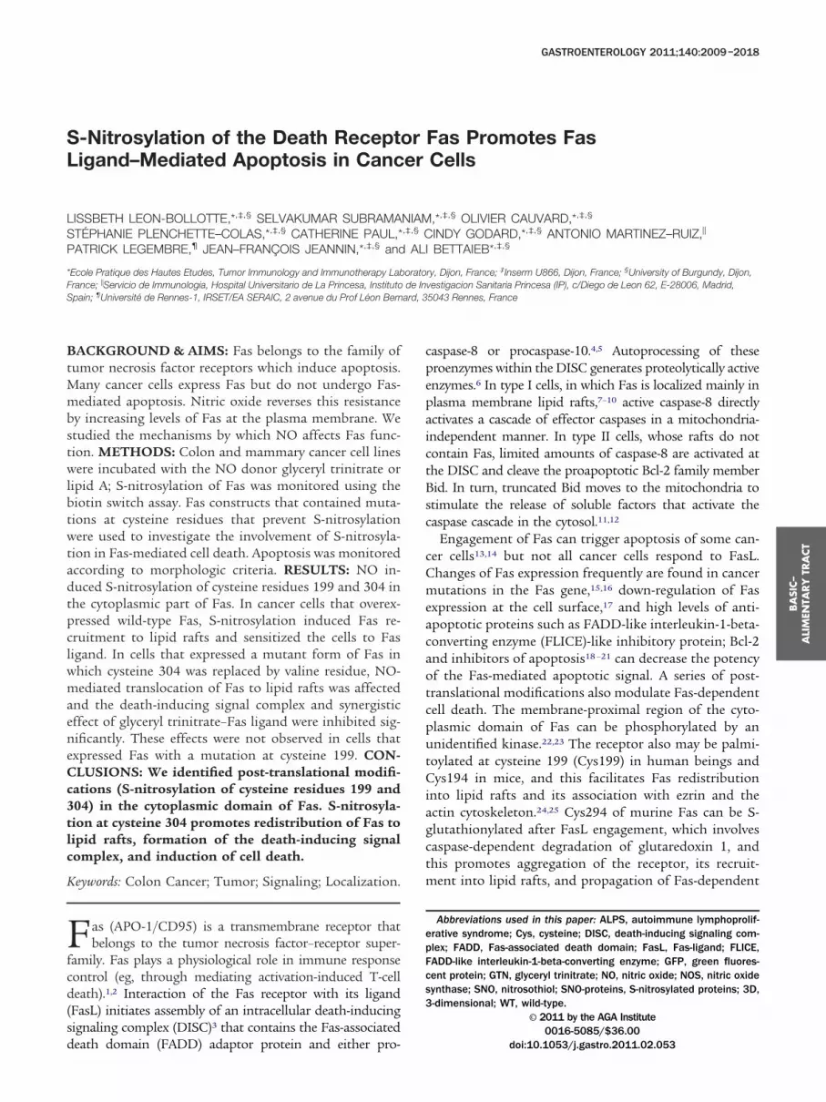
Figure 1. NO increases Fas expression and sensitizes cells to FasL-mediMT6-H were left nontreated (NT) or treated with 500 mmol/L GTN or wit
Fas expression, cells were lysed and 50 �g of each lysate was subjectedusing the anti-Fas antibody. Constitutive heat shock protein 70 (Hsc70) wa
–treated EMT6-H cells involved inducible NOS activity and hence NO,xpression of Fas was evaluated using a mouse anti-Fas antibody by (B) coells. Data are representative of at least 3 independent experiments. (E) Aphen with or without soluble FasL (sFasL) for 24 hours. (F) To verify that aphe NO scavenger 2-(4-Carboxyphenyl)-dihydro-tetramethyl-imidazol-oxide
oechst 33342 and then counted. Data are the � standard error of the mean o
destruction by the immune system through reduced ap-optosis sensitivity.36 Breaking such resistance was ren-
ered possible with some anticancer drugs that enhanceas-receptor expression and aggregation at the surface ofumor cells, thereby increasing the apoptotic response toasL.36 –38 We have shown that nitric oxide also has aensitizing effect by increasing Fas expression on cancerells,39 which raises the possibility that Fas-receptor ex-
pression or function could be regulated by S-nitrosyla-tion, especially because Fas harbors a 3-dimensional (3D)
poptosis. Colon cancer cells SW480 and C51 and mammary cancer cellsmg/mL lipid A for the indicated times. (A and C) For analysis of whole-cellium dodecyl sulfate–polyacrylamide gel electrophoresis and immunoblotas loading control. (C) To verify that any increase in Fas expression in lipidwere treated with the NOS inhibitor aminoguanidine (AG). Cell surfacel microscopy in SW480 cells or by (D) flow cytometry in EMT6-H and C51
is was induced in SW480 by treatment with 500 �mol/L GTN for 24 hoursis induced by GTN and FasL involved NO, SW480 cells were treated withIO) and GTN for 24 hours then with or without sFasL for 6 hours. This short
ated ah 0.5to sods usedcellsnfoca
optosoptos
(cPTreatment with sFasL (6 vs 24 h) is owing to the high toxicity of cPTIO. Apoptotic cells were identified by fluorescence microscopy after staining nuclei with
f at least 3 independent experiments. *P � .05 and ***P � .001.
nammfwatatddiPSo
ofb
Sal
BA
SIC–
ALI
MEN
TARY
TRA
CT
June 2011 S–NITROSYLATION OF THE DEATH RECEPTOR FAS 2011
structure of the proposed consensus motif of S-nitrosy-lation. We show here that both exogenous and endoge-nous NO induce S-nitrosylation of Cys199 and Cys304 ofthe Fas receptor, and this correlates with enhanced ex-pression of Fas at the plasma membrane and its translo-cation into lipid rafts. Expression of a non-nitrosylablemutant Fas receptor prevents NO-mediated sensitizationto Fas-induced apoptosis. Thus, S-nitrosylation of Fasreceptor is an additional posttranslational mechanismthat regulates Fas-mediated cell death.
Materials and MethodsCell LinesHuman SW480 and murine C51 colon cancer cell
lines were obtained from the American Tissue Culture Col-lection (Manassas, VA). EMT6-H cells are clones selectedfrom the murine mammary EMT6 cancer cell line.40 All celllines were cultured in a 1:1 (vol/vol) mixture of Dulbecco’smodified Eagle medium and HAMF-F10 (Biowittaker, Fon-tenay-Sous-Bois, France) supplemented with 10% fetal bo-vine serum and 2 mmol/L L-glutamine at 37°C in a dryatmosphere. All cell lines were tested and found to be free ofMycoplasma species infection.
Biotin Switch AssayThe biotin switch assay was performed as de-
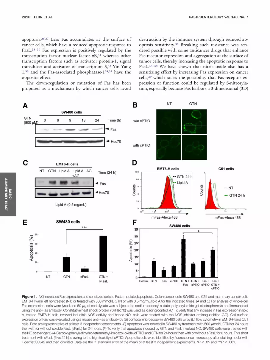
scribed41 with some modifications.42 Briefly, cells (1 �108) were washed in phosphate-buffered saline (PBS) andhomogenized in nondenaturing nitrosothiol (SNO) lysisbuffer (50 mmol/L Tris-HCl, pH 7.4, 300 mmol/L NaCl,5 mmol/L ethylenediaminetetraacetic acid [EDTA], 0.1
Figure 2. NO induces S-nitrosylation of Fas. SW480 cells were left nNO-proteins were detected using an anti-Cys NO antibody by immunond the biotinylated proteins were purified with streptavidin-agarose, foll
atter fraction was subjected to immunoblotting for Fas (C-20 antibody).was detected after purification of biotinylated proteins as indicated befo
heat shock protein 70 (Hsc70) was used as loading control. Data are represmmol/L neocuproine, 1% Triton X-100, and proteaseinhibitors), then 4 volumes of blocking buffer (225mmol/L HEPES, pH 7.7, 0.9 mmol/L EDTA, 90 �mol/L
eocuproine, and 2.5% sodium dodecyl sulfate) weredded. Free thiols were blocked by methylation with 20mol/L methyl methane thiosulfonate at 50°C for 20inutes. After removing excess methyl methane thiosul-
onate by precipitating twice with acetone, nitrosothiolsere reduced to thiols with 1 mmol/L ascorbate withoutltering the methylated thiols. The newly formed thiolshen were linked with sulfydryl-specific biotinylating re-gent biotin-N-[6-(biotinamido)-hexyl]-1=-(2=-pyridyldi-hio) propionamide (HPDP). Biotinylated proteins wereetected by immunoblotting using horseradish peroxi-ase– conjugated streptavidin or were purified by precip-
tation with Ultralink Immobilized Neutravidin Proteinlus and eluted from the beads with Laemmli buffer.amples were analyzed by immunoblotting with anti-Fasr anti– green fluorescent protein (GFP) antibodies.
StatisticsData are presented as means � standard deviation
f the indicated number of experiments. Significant dif-erences were evaluated by analysis of variance followedy the Student t test.
ResultsNO-Mediated Increase in Fas ExpressionSensitizes Cancer Cells to FasLWhen SW480 human colon cancer cells are ex-
posed to the NO donor glyceryl trinitrate (GTN) an
ated (NT) or treated with 500 �mol/L GTN for the indicated times. (A)scence. Protein extracts were subjected to the biotin switch technique,by elution with 2-mercaptoethanol (the SNO-enriched fraction). (B) ThisT6-H cells were treated with 0.5 �g/mL lipid A and Fas S-nitrosylation
d immunoblotting with an anti-Fas antibody (from Abcam). Constitutive
ontrefluoreowed(C) EMre an
entative of at least 3 independent experiments.
inwaadcFvTCah�(FGwds
mcta(Gap
Fvara
ei
StS
BA
SIC–
ALIM
ENTA
RY
TRA
CT
2012 LEON ET AL GASTROENTEROLOGY Vol. 140, No. 7
increase in Fas expression is observed,39 here shown bymmunoblotting (Figure 1A) and by fluorescent immu-ostaining (Figure 1B, upper panel). Similar responses alsoere observed in the EMT6-H murine mammary cancernd the C51 murine colon cancer cell lines (Figure 1Cnd D). When EMT6-H cells were exposed to the lipid Aerivative OM-174, which similar to most lipid A mole-ules induces inducible NO synthase (NOS) expression,as expression increased, an effect that was partially re-ersed by the NOS inhibitor aminoguanidine (Figure 1C).his suggests that NO was involved in up-regulating Fas.ell lines were exposed to 500 �mol/L GTN for 24 hoursnd then treated with soluble FasL for an additional 24ours. Apoptotic cell death was induced synergistically (P
.01) in SW480 cells (Figure 1E) as well as in C51Supplementary Materials and Methods, Supplementaryigure 1A) and EMT6-H cells (Supplementary Figure 1B).TN-mediated sensitization of FasL-induced apoptosisas dependent of NO because 2-(4-Carboxyphenyl)-ihydro-tetramethyl-imidazol-oxide, a scavenger of NO,ignificantly affected cell death, suggesting the involve-
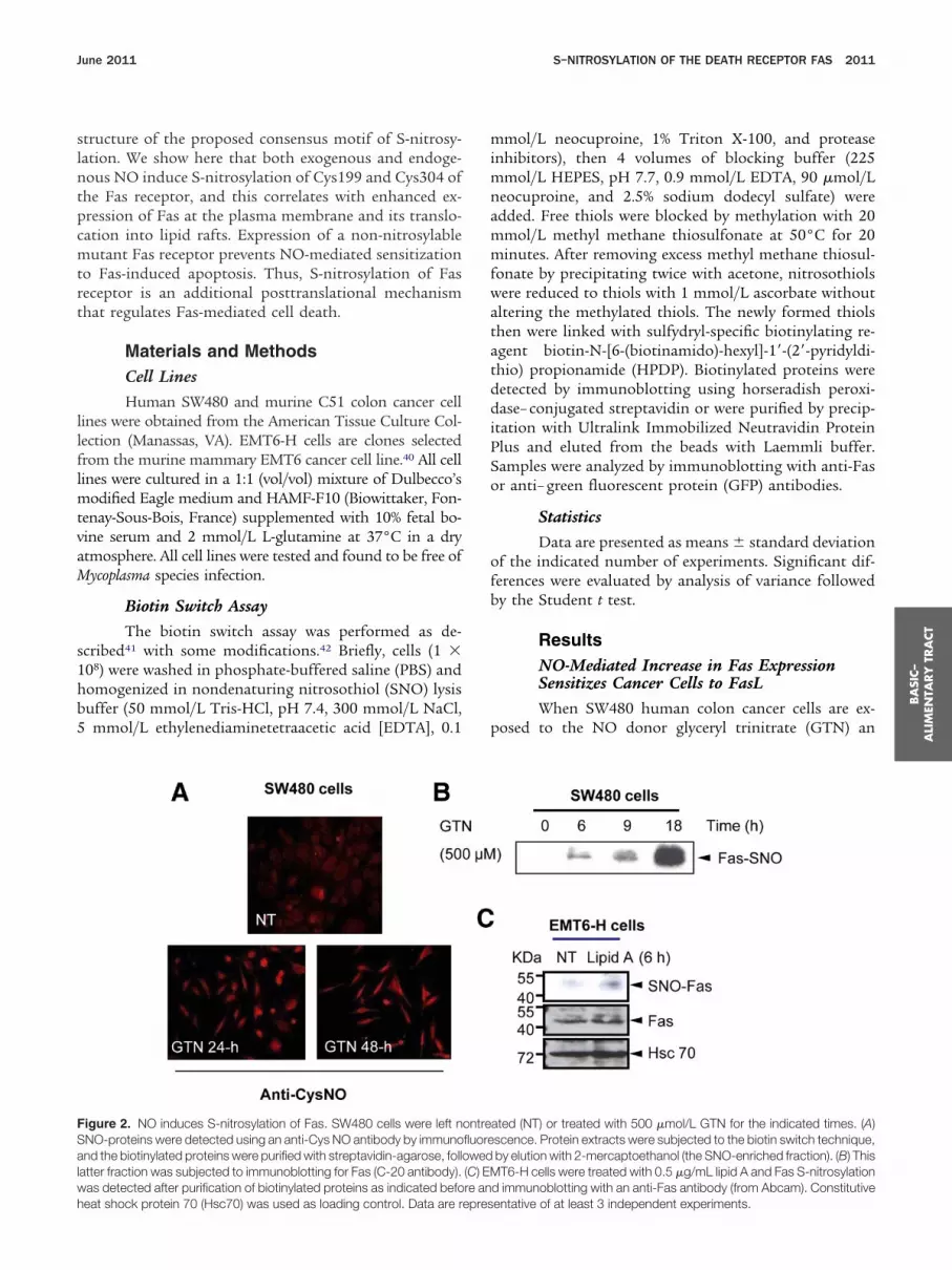
Figure 3. Human Fas is S-nitrosylated at cysteines 199 and 304. SW48xpression of these different forms of Fas was determined by flow cytom
mmunoblot using an anti-GFP antibody. Cells from these different clones wassay. Constitutive heat shock protein 70 (Hsc 70) was used ad loadinganti-GFP antibodies. (E) To verify that S-nitrosylation is not affected in cell
NO-protein profiles are assessed by immunoblotting with horseradish preated with or without the NO donor S-nitroso-cysteine (Cys-NO) at 1 mm
NO-protein levels using densitometric analyses of total protein levels (lower paent of NO in this synergistic increase in sensitivity ofells to apoptosis (Figure 1F). It was noted that therimeric recombinant FasL was more efficient to inducepoptosis in C51 cells than the monomeric soluble FasLSupplementary Figure 1A), and the synergistic effect ofTN and FasL was not caused by an increase in NO levels
s attested by nitrite accumulation measurement (Sup-lementary Figure 2).
NO Induces S-Nitrosylation of FasTo determine whether NO could induce S-nitrosy-
lation of Fas, we first used a biotin switch assay tomeasure the amount of S-nitrosylated proteins (SNO-proteins) present in cells. The level of SNO-protein inSW480 cells increased significantly when cells were ex-posed to 500 �mol/L GTN for 18 hours (Supplementary
igure 3A) or for 48 hours (Supplementary Figure 3B). Toerify the biotin labeling of SNO, reactive methyl meth-ne thiosulfonate (which blocks free thiols on cysteineesidues) or ascorbate (which converts SNO to thiols andllows labeling of nascent thiols with biotin-HPDP) were
were stably transfected with Fas-WT, Fas-C199V, and Fas-C304V. Theither at the (A) whole Fas level or the (B) cell surface Fas level, and by (C)ated with or without 500 �mol/L GTN for 18 hours before the biotin switch
rol. (D) Streptavidin-purified proteins were analyzed by immunoblot withnontransfected (NT) or expressing the different mutated forms of Fas, fulldase (HRP)-conjugated streptavidin (streptavidin-HRP) lysates from cellsor the indicated time (upper panel). The histogram shows quantification of
0 cellsetry eere trecont
s lefteroxiol/L f
nel). Data are representative of at least 3 independent experiments.
csChi3b3
w(dS
nc(ai
BA
SIC–
ALI
MEN
TARY
TRA
CT
June 2011 S–NITROSYLATION OF THE DEATH RECEPTOR FAS 2013
omitted. As expected, SNO-proteins were more abundantin the absence of methyl methane thiosulfonate andbiotinylated proteins were less abundant in the absenceof ascorbate (Supplementary Figure 3A). The GTN-in-duced increase in total SNO-proteins also was detectedusing immunofluorescence up to 48 hours after the startof the treatment (Figure 2A). In cell lysates enriched forSNO-proteins (SNO-purified proteins) (SupplementaryFigure 3B), Fas was found to accumulate in the SNO-enriched fraction of GTN-treated SW480 cells (Figure2B). A similar partitioning was observed in fractions fromEMT6-H cells exposed to the synthetic lipid A, OM-174(Figure 2C). It is noteworthy that the total amount ofSNO-protein significantly increased in OM-174 –treatedcells (Supplementary Figure 3C). Thus, both exogenousand endogenous NO triggered S-nitrosylation of the Fasreceptor.
Human Fas Is S-Nitrosylated at Cysteines 199and 304The cytoplasmic part of the Fas receptor includes
a cysteine residue close to its transmembrane domain(Cys199) and another one near its death domain(Cys304) (Supplementary Figure 4A). Analysis of the 3Dstructure of the Fas intracellular domain indicated thatCys304 is within a consensus sequence (K/R/H)-C-(D/E)for S-nitrosylation43 (Supplementary Figure 4B). SW480ells were stably transfected with GFP-tagged Fas con-tructs mutated, at these cysteine residues (C199V or304V)24 and the resultant GFP-positive clones had aigh expression of Fas, with comparable intensities either
n whole cells (Figure 3A) or at the cell surface (FigureB). Expression of the constructs in cells was confirmedy immunoblotting using an anti-GFP antibody (FigureC). When SW480 cells were exposed to 500 �mol/L
GTN and subjected to the biotin switch assay, a strongincrease in SNO-Fas was detected in SW480 cells express-ing wild-type Fas (Fas-WT) whereas only a weak increasein SNO-Fas was detected in cells expressing the C199V orC304V Fas mutants (Figure 3D). The defective S-nitrosy-lation of the mutant forms of Fas does not seem to berelated to a defect in S-nitrosylation because the profilesof S-nitrosylation in total cell lysates were similar in thedifferent clones (Figure 3E).
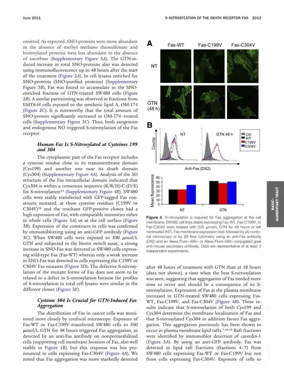
Cysteine 304 Is Crucial for GTN-Induced FasAggregationThe distribution of Fas in cancer cells was moni-
tored more closely by confocal microscopy. Exposure ofFas-WT or Fas-C199V–transfected SW480 cells to 500�mol/L GTN for 48 hours triggered Fas aggregation, asdetected by an anti-Fas antibody on nonpermeabilizedcells (supporting cell membrane location of Fas, also wellvisible in Figure 1B), but this response was less pro-nounced in cells expressing Fas-C304V (Figure 4A). We
noted that Fas aggregation was more markedly detected fafter 48 hours of treatment with GTN than at 18 hours(data not shown), a time when the best S-nitrosylationwas seen, suggesting that aggregation of Fas needed moretime to occur and should be a consequence of its S-nitrosylation. Expression of Fas at the plasma membraneincreased in GTN-treated SW480 cells expressing Fas-WT, Fas-C199V, and Fas-C304V (Figure 4B). These re-sults indicate that S-nitrosylation of both Cys199 andCys304 determine the membrane localization of Fas andthat S-nitrosylated Cys304 in addition favors Fas aggre-gation. This aggregation previously has been shown tooccur in plasma membrane lipid rafts.7,44,45 Raft fractions
ere identified by immunoblot detection of caveolin-1Figure 5A). By using an anti-GFP antibody, Fas wasetected in lipid raft fractions (fractions 4 –7) fromW480 cells expressing Fas-WT or Fas-C199V but not
Figure 4. S-nitrosylation is required for Fas aggregation at the cellmembrane. SW480 cell lines stably expressing Fas-WT, Fas-C199V, orFas-C304V were treated with 500 �mol/L GTN for 48 hours or leftontreated (NT). Fas membrane expression was followed by (A) confo-al microscopy or by (B) flow cytometry using an anti-Fas antibodyDX2) and an Alexa Fluor–488– or Alexa Fluor–568–conjugated goatnti-mouse secondary antibody. Data are representative of at least 3
ndependent experiments.
rom cells expressing Fas-C304V. Exposure of cells to
SatGcbbw(apEbFso
um
BA
SIC–
ALIM
ENTA
RY
TRA
CT
2014 LEON ET AL GASTROENTEROLOGY Vol. 140, No. 7
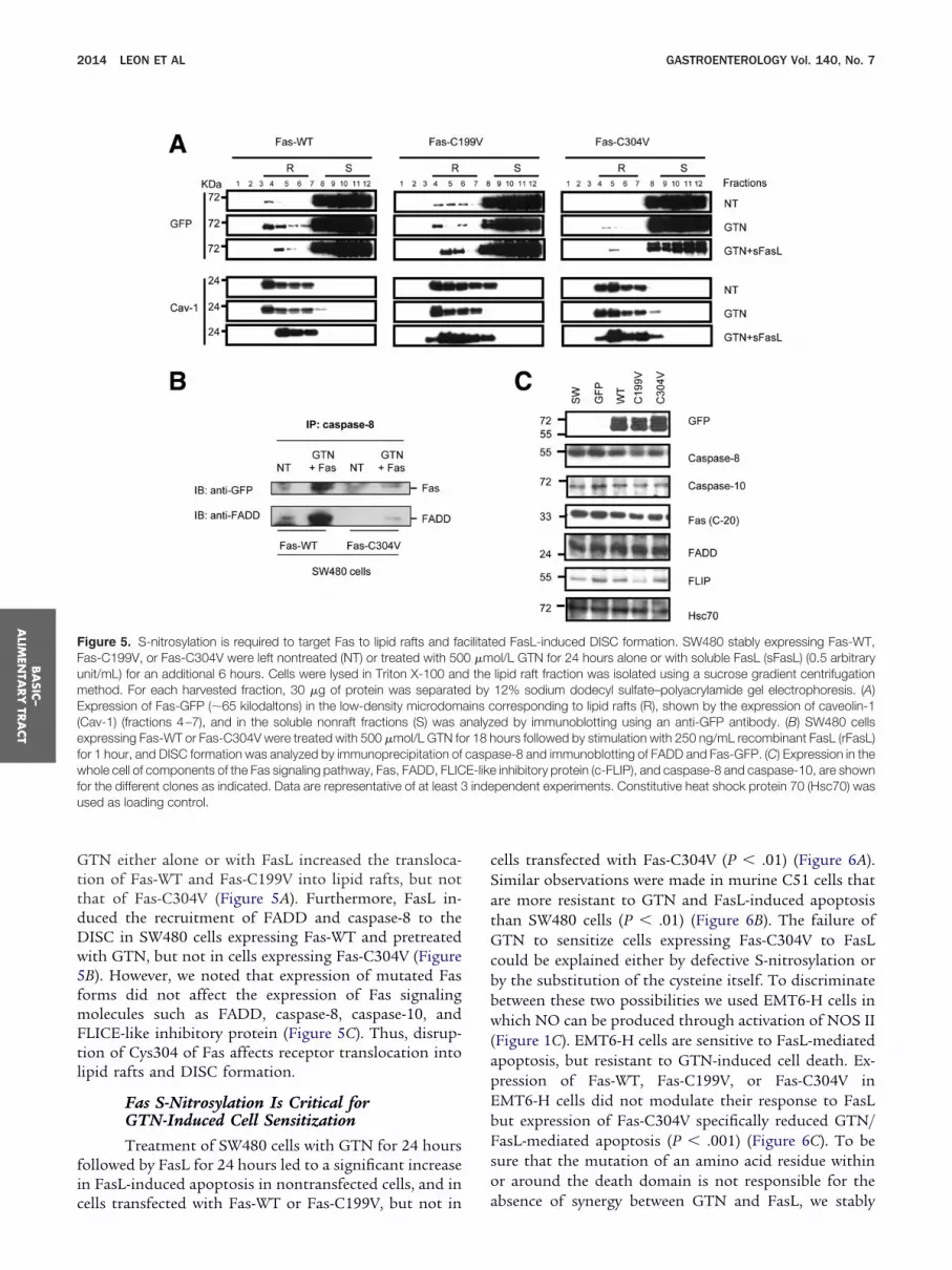
GTN either alone or with FasL increased the transloca-tion of Fas-WT and Fas-C199V into lipid rafts, but notthat of Fas-C304V (Figure 5A). Furthermore, FasL in-duced the recruitment of FADD and caspase-8 to theDISC in SW480 cells expressing Fas-WT and pretreatedwith GTN, but not in cells expressing Fas-C304V (Figure5B). However, we noted that expression of mutated Fasforms did not affect the expression of Fas signalingmolecules such as FADD, caspase-8, caspase-10, andFLICE-like inhibitory protein (Figure 5C). Thus, disrup-tion of Cys304 of Fas affects receptor translocation intolipid rafts and DISC formation.
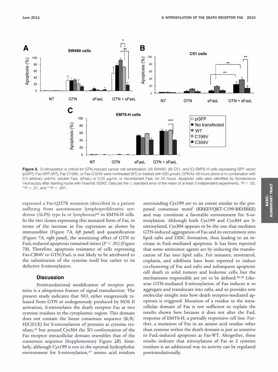
Fas S-Nitrosylation Is Critical forGTN-Induced Cell SensitizationTreatment of SW480 cells with GTN for 24 hours
followed by FasL for 24 hours led to a significant increasein FasL-induced apoptosis in nontransfected cells, and in
Figure 5. S-nitrosylation is required to target Fas to lipid rafts and faFas-C199V, or Fas-C304V were left nontreated (NT) or treated with 50nit/mL) for an additional 6 hours. Cells were lysed in Triton X-100 anethod. For each harvested fraction, 30 �g of protein was separate
Expression of Fas-GFP (�65 kilodaltons) in the low-density microdom(Cav-1) (fractions 4–7), and in the soluble nonraft fractions (S) was aexpressing Fas-WT or Fas-C304V were treated with 500 �mol/L GTN fofor 1 hour, and DISC formation was analyzed by immunoprecipitation ofwhole cell of components of the Fas signaling pathway, Fas, FADD, FLICfor the different clones as indicated. Data are representative of at least 3used as loading control.
cells transfected with Fas-WT or Fas-C199V, but not in a
cells transfected with Fas-C304V (P � .01) (Figure 6A).imilar observations were made in murine C51 cells thatre more resistant to GTN and FasL-induced apoptosishan SW480 cells (P � .01) (Figure 6B). The failure ofTN to sensitize cells expressing Fas-C304V to FasL
ould be explained either by defective S-nitrosylation ory the substitution of the cysteine itself. To discriminateetween these two possibilities we used EMT6-H cells inhich NO can be produced through activation of NOS II
Figure 1C). EMT6-H cells are sensitive to FasL-mediatedpoptosis, but resistant to GTN-induced cell death. Ex-ression of Fas-WT, Fas-C199V, or Fas-C304V inMT6-H cells did not modulate their response to FasLut expression of Fas-C304V specifically reduced GTN/asL-mediated apoptosis (P � .001) (Figure 6C). To beure that the mutation of an amino acid residue withinr around the death domain is not responsible for the
ed FasL-induced DISC formation. SW480 stably expressing Fas-WT,ol/L GTN for 24 hours alone or with soluble FasL (sFasL) (0.5 arbitrarylipid raft fraction was isolated using a sucrose gradient centrifugation12% sodium dodecyl sulfate–polyacrylamide gel electrophoresis. (A)orresponding to lipid rafts (R), shown by the expression of caveolin-1ed by immunoblotting using an anti-GFP antibody. (B) SW480 cellsours followed by stimulation with 250 ng/mL recombinant FasL (rFasL)
ase-8 and immunoblotting of FADD and Fas-GFP. (C) Expression in theinhibitory protein (c-FLIP), and caspase-8 and caspase-10, are shown
pendent experiments. Constitutive heat shock protein 70 (Hsc70) was
cilitat0 �md thed byains cnalyzr 18 hcaspE-likeinde
bsence of synergy between GTN and FasL, we stably
7Ftd
BA
SIC–
ALI
MEN
TARY
TRA
CT
June 2011 S–NITROSYLATION OF THE DEATH RECEPTOR FAS 2015
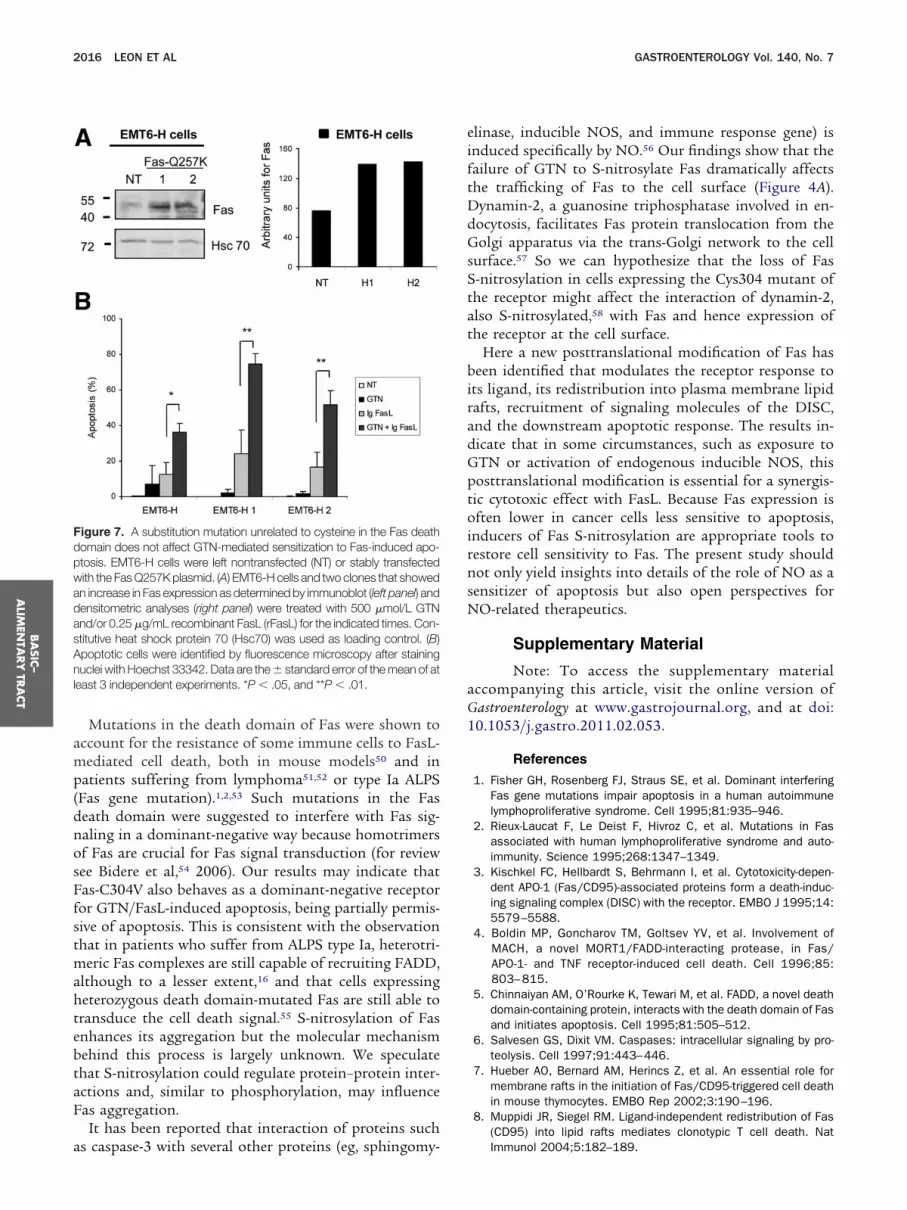
expressed a Fas-Q257K mutation (described in a patientsuffering from autoimmune lymphoproliferative syn-drome (ALPS) type Ia or lymphoma)46 in EMT6-H cells.In the two clones expressing this mutated form of Fas, interms of the increase in Fas expression as shown byimmunoblot (Figure 7A, left panel) and quantification(Figure 7A, right panel), the sensitizing effect of GTN toFasL-induced apoptosis remained intact (P � .01) (Figure
B). Therefore, apoptosis resistance of cells expressingas-C304V to GTN/FasL is not likely to be attributed tohe substitution of the cysteine itself but rather to itsefective S-nitrosylation.
DiscussionPosttranslational modification of receptor pro-
teins is a ubiquitous feature of signal transduction. Thepresent study indicates that NO, either exogenously re-leased from GTN or endogenously produced by NOS IIactivation, S-nitrosylates the death receptor Fas at twocysteine residues in the cytoplasmic region. This domaindoes not contain the linear consensus sequence (K/R/H)C(D/E) for S-nitrosylation of proteins at cysteine res-idues,43 but around Cys304 the 3D conformation of theFas receptor intracellular domain resembles that of theconsensus sequence (Supplementary Figure 2B). Simi-larly, although Cys199 is not in the optimal hydrophobic
Figure 6. S-nitrosylation is critical for GTN-induced cancer cell sensi(pGFP), Fas-GFP (WT), Fas-C199V, or Fas-C304V were nontreated (NT0.5 arbitrary unit/mL soluble FasL (sFasL) or 0.25 �g/mL or recombmicroscopy after staining nuclei with Hoechst 33342. Data are the � s**P � .01, and ***P � .001.
environment for S-nitrosylation,47 amino acid residues
surrounding Cys199 are to an extent similar to the pro-posed consensus motif (KRKEVQKT-C199-RKHRKE)and may constitute a favorable environment for S-ni-trosylation. Although both Cys199 and Cys304 are S-nitrosylated, Cys304 appears to be the one that mediatesGTN-induced aggregation of Fas and its recruitment intolipid rafts and DISC formation, thus leading to an in-crease in FasL-mediated apoptosis. It has been reportedthat some antitumor agents act by inducing the translo-cation of Fas into lipid rafts. For instance, resveratrol,cisplatin, and edelfosin have been reported to induceco-clustering of Fas and rafts and subsequent apoptoticcell death in solid tumors and leukemic cells, but themechanisms responsible are yet to be defined.48,49 Like-wise GTN-mediated S-nitrosylation of Fas induces it toaggregate and translocate into rafts, and so provides newmolecular insight into how death receptor-mediated ap-optosis is triggered. Mutation of a residue in the intra-cellular domain of Fas is not sufficient to explain theresults shown here because it does not alter the FasLresponse of EMT6-H, a partially responsive cell line. Fur-ther, a mutation of Fas in an amino acid residue otherthan cysteine within the death domain is just as sensitiveto FasL-induced apoptosis as Fas-WT. Altogether, theseresults indicate that nitrosylation of Fas at 2 cysteineresidues is an additional way its activity can be regulated
n. (A) SW480, (B) C51, and (C) EMT6-H cells expressing GFP vectorreated with 500 �mol/L GTN for 48 hours alone or in combination with
FasL for 24 hours. Apoptotic cells were identified by fluorescencerd error of the mean of at least 3 independent experiments. *P � .05,
tizatio) or tinanttanda
posttranslationally.
p
dnos
ftDdGsSta
1
Anl
BA
SIC–
ALIM
ENTA
RY
TRA
CT
2016 LEON ET AL GASTROENTEROLOGY Vol. 140, No. 7
Mutations in the death domain of Fas were shown toaccount for the resistance of some immune cells to FasL-mediated cell death, both in mouse models50 and in
atients suffering from lymphoma51,52 or type Ia ALPS(Fas gene mutation).1,2,53 Such mutations in the Fas
eath domain were suggested to interfere with Fas sig-aling in a dominant-negative way because homotrimersf Fas are crucial for Fas signal transduction (for reviewee Bidere et al,54 2006). Our results may indicate that
Fas-C304V also behaves as a dominant-negative receptorfor GTN/FasL-induced apoptosis, being partially permis-sive of apoptosis. This is consistent with the observationthat in patients who suffer from ALPS type Ia, heterotri-meric Fas complexes are still capable of recruiting FADD,although to a lesser extent,16 and that cells expressingheterozygous death domain-mutated Fas are still able totransduce the cell death signal.55 S-nitrosylation of Fasenhances its aggregation but the molecular mechanismbehind this process is largely unknown. We speculatethat S-nitrosylation could regulate protein–protein inter-actions and, similar to phosphorylation, may influenceFas aggregation.
It has been reported that interaction of proteins such
Figure 7. A substitution mutation unrelated to cysteine in the Fas deathdomain does not affect GTN-mediated sensitization to Fas-induced apo-ptosis. EMT6-H cells were left nontransfected (NT) or stably transfectedwith the Fas Q257K plasmid. (A) EMT6-H cells and two clones that showedan increase in Fas expression as determined by immunoblot (left panel) anddensitometric analyses (right panel) were treated with 500 �mol/L GTNand/or 0.25 �g/mL recombinant FasL (rFasL) for the indicated times. Con-stitutive heat shock protein 70 (Hsc70) was used as loading control. (B)
poptotic cells were identified by fluorescence microscopy after staininguclei with Hoechst 33342. Data are the � standard error of the mean of at
east 3 independent experiments. *P � .05, and **P � .01.
as caspase-3 with several other proteins (eg, sphingomy-
elinase, inducible NOS, and immune response gene) isinduced specifically by NO.56 Our findings show that theailure of GTN to S-nitrosylate Fas dramatically affectshe trafficking of Fas to the cell surface (Figure 4A).ynamin-2, a guanosine triphosphatase involved in en-ocytosis, facilitates Fas protein translocation from theolgi apparatus via the trans-Golgi network to the cell
urface.57 So we can hypothesize that the loss of Fas-nitrosylation in cells expressing the Cys304 mutant ofhe receptor might affect the interaction of dynamin-2,lso S-nitrosylated,58 with Fas and hence expression of
the receptor at the cell surface.Here a new posttranslational modification of Fas has
been identified that modulates the receptor response toits ligand, its redistribution into plasma membrane lipidrafts, recruitment of signaling molecules of the DISC,and the downstream apoptotic response. The results in-dicate that in some circumstances, such as exposure toGTN or activation of endogenous inducible NOS, thisposttranslational modification is essential for a synergis-tic cytotoxic effect with FasL. Because Fas expression isoften lower in cancer cells less sensitive to apoptosis,inducers of Fas S-nitrosylation are appropriate tools torestore cell sensitivity to Fas. The present study shouldnot only yield insights into details of the role of NO as asensitizer of apoptosis but also open perspectives forNO-related therapeutics.
Supplementary Material
Note: To access the supplementary materialaccompanying this article, visit the online version ofGastroenterology at www.gastrojournal.org, and at doi:
0.1053/j.gastro.2011.02.053.
References
1. Fisher GH, Rosenberg FJ, Straus SE, et al. Dominant interferingFas gene mutations impair apoptosis in a human autoimmunelymphoproliferative syndrome. Cell 1995;81:935–946.
2. Rieux-Laucat F, Le Deist F, Hivroz C, et al. Mutations in Fasassociated with human lymphoproliferative syndrome and auto-immunity. Science 1995;268:1347–1349.
3. Kischkel FC, Hellbardt S, Behrmann I, et al. Cytotoxicity-depen-dent APO-1 (Fas/CD95)-associated proteins form a death-induc-ing signaling complex (DISC) with the receptor. EMBO J 1995;14:5579–5588.
4. Boldin MP, Goncharov TM, Goltsev YV, et al. Involvement ofMACH, a novel MORT1/FADD-interacting protease, in Fas/APO-1- and TNF receptor-induced cell death. Cell 1996;85:803–815.
5. Chinnaiyan AM, O’Rourke K, Tewari M, et al. FADD, a novel deathdomain-containing protein, interacts with the death domain of Fasand initiates apoptosis. Cell 1995;81:505–512.
6. Salvesen GS, Dixit VM. Caspases: intracellular signaling by pro-teolysis. Cell 1997;91:443–446.
7. Hueber AO, Bernard AM, Herincs Z, et al. An essential role formembrane rafts in the initiation of Fas/CD95-triggered cell deathin mouse thymocytes. EMBO Rep 2002;3:190–196.
8. Muppidi JR, Siegel RM. Ligand-independent redistribution of Fas(CD95) into lipid rafts mediates clonotypic T cell death. Nat
Immunol 2004;5:182–189.
1
1
1
1
1
1
1
1
1
1
2
2
2
2
2
2
2
2
2
2
3
3
3
3
3
3
3
3
3
3
4
4
4
4
4
4
4
4
4
4
BA
SIC–
ALI
MEN
TARY
TRA
CT
June 2011 S–NITROSYLATION OF THE DEATH RECEPTOR FAS 2017
9. Eramo A, Sargiacomo M, Ricci-Vitiani L, et al. CD95 death-induc-ing signaling complex formation and internalization occur in lipidrafts of type I and type II cells. Eur J Immunol 2004;34:1930–1940.
0. Legembre P, Daburon S, Moreau P, et al. Modulation of Fas-mediated apoptosis by lipid rafts in T lymphocytes. J Immunol2006;176:716–720.
1. Li H, Zhu H, Xu CJ, et al. Cleavage of BID by caspase 8 mediatesthe mitochondrial damage in the Fas pathway of apoptosis. Cell1998;94:491–501.
2. Luo X, Budihardjo I, Zou H, et al. Bid, a Bcl2 interacting protein,mediates cytochrome c release from mitochondria in response toactivation of cell surface death receptors. Cell 1998;94:481–490.
3. Trauth BC, Klas C, Peters AM, et al. Monoclonal antibody-medi-ated tumor regression by induction of apoptosis. Science 1989;245:301–305.
4. Itoh N, Yonehara S, Ishii A, et al. The polypeptide encoded by thecDNA for human cell surface antigen Fas can mediate apoptosis.Cell 1991;66:233–243.
5. Maeda T, Yamada Y, Moriuchi R, et al. Fas gene mutation in theprogression of adult T cell leukemia. J Exp Med 1999;189:1063–1071.
6. Martin DA, Zheng L, Siegel RM, et al. Defective CD95/APO-1/Fassignal complex formation in the human autoimmune lymphopro-liferative syndrome, type Ia. Proc Natl Acad Sci U S A 1999;96:4552–4557.
7. Bullani RR, Wehrli P, Viard-Leveugle I, et al. Frequent downregu-lation of Fas (CD95) expression and function in melanoma. Mel-anoma Res 2002;12:263–270.
8. Scaffidi C, Fulda S, Srinivasan A, et al. Two CD95 (APO-1/Fas)signaling pathways. EMBO J 1998;17:1675–1687.
9. Yang BF, Xiao C, Roa WH, et al. Calcium/calmodulin-dependentprotein kinase II regulation of c-FLIP expression and phosphory-lation in modulation of Fas-mediated signaling in malignant gli-oma cells. J Biol Chem 2003;278:7043–7050.
0. Mandruzzato S, Brasseur F, Andry G, et al. A CASP-8 mutationrecognized by cytolytic T lymphocytes on a human head and neckcarcinoma. J Exp Med 1997;186:785–793.
1. Teitz T, Wei T, Valentine MB, et al. Caspase 8 is deleted orsilenced preferentially in childhood neuroblastomas with amplifi-cation of MYCN. Nat Med 2000;6:529–535.
2. Eberle A, Reinehr R, Becker S, et al. CD95 tyrosine phosphory-lation is required for CD95 oligomerization. Apoptosis 2007;12:719–729.
3. Kennedy NJ, Budd RC. Phosphorylation of FADD/MORT1 and Fasby kinases that associate with the membrane-proximal cytoplas-mic domain of Fas. J Immunol 1998;160:4881–4888.
4. Chakrabandhu K, Herincs Z, Huault S, et al. Palmitoylation isrequired for efficient Fas cell death signaling. EMBO J 2007;26:209–220.
5. Feig C, Tchikov V, Schutze S, et al. Palmitoylation of CD95facilitates formation of SDS-stable receptor aggregates that ini-tiate apoptosis signaling. EMBO J 2007;26:221–231.
6. Anathy V, Aesif SW, Guala AS, et al. Redox amplification ofapoptosis by caspase-dependent cleavage of glutaredoxin 1 andS-glutathionylation of Fas. J Cell Biol 2009;184:241–252.
7. Fernandes AP, Holmgren A. Glutaredoxins: glutathione-depen-dent redox enzymes with functions far beyond a simple thiore-doxin backup system. Antioxid Redox Signal 2004;6:63–74.
8. Algeciras-Schimnich A, Pietras EM, Barnhart BC, et al. Two CD95tumor classes with different sensitivities to antitumor drugs. ProcNatl Acad Sci U S A 2003;100:11445–11450.
9. Botti C, Buglioni S, Benevolo M, et al. Altered expression of FASsystem is related to adverse clinical outcome in stage I-II breastcancer patients treated with adjuvant anthracycline-based che-
motherapy. Clin Cancer Res 2004;10:1360–1365.0. Sikora J, Dworacki G, Zeromski J. Expression of Fas and Fasligand and apoptosis in tumor-associated lymphocytes and intumor cells from malignant pleural effusions. Nat Immunol 1998;16:244–255.
1. Chan H, Bartos DP, Owen-Schaub LB. Activation-dependent tran-scriptional regulation of the human Fas promoter requires NF-kappaB p50-p65 recruitment. Mol Cell Biol 1999;19:2098–2108.
2. Ivanov VN, Krasilnikov M, Ronai Z. Regulation of Fas expressionby STAT3 and c-Jun is mediated by phosphatidylinositol 3-kinase-AKT signaling. J Biol Chem 2002;277:4932–4944.
3. Garban HJ, Bonavida B. Nitric oxide inhibits the transcriptionrepressor Yin-Yang 1 binding activity at the silencer region of theFas promoter: a pivotal role for nitric oxide in the up-regulation ofFas gene expression in human tumor cells. J Immunol 2001;167:75–81.
4. Ivanov VN, Lopez Bergami P, Maulit G, et al. FAP-1 associationwith Fas (Apo-1) inhibits Fas expression on the cell surface. MolCell Biol 2003;23:3623–3635.
5. Ungefroren H, Kruse ML, Trauzold A, et al. FAP-1 in pancreaticcancer cells: functional and mechanistic studies on its inhibitoryrole in CD95-mediated apoptosis. J Cell Sci 2001;114:2735–2746.
6. Peter ME, Legembre P, Barnhart BC. Does CD95 have tumorpromoting activities? Biochim Biophys Acta 2005;1755:25–36.
7. Micheau O, Solary E, Hammann A, et al. Fas ligand-independent,FADD-mediated activation of the Fas death pathway by anticancerdrugs. J Biol Chem 1999;274:7987–7992.
8. Garban HJ, Bonavida B. Nitric oxide sensitizes ovarian tumorcells to Fas-induced apoptosis. Gynecol Oncol 1999;73:257–264.
9. Millet A, Bettaieb A, Renaud F, et al. Influence of the nitric oxidedonor glyceryl trinitrate on apoptotic pathways in human coloncancer cells. Gastroenterology 2002;123:235–246.
0. Gauthier N, Lohm S, Touzery C, et al. Tumour-derived and host-derived nitric oxide differentially regulate breast carcinoma me-tastasis to the lungs. Carcinogenesis 2004;25:1559–1565.
1. Jaffrey SR, Snyder SH. The biotin switch method for the detectionof S-nitrosylated proteins. Sci STKE 2001;2001:PL1.
2. Martinez-Ruiz A, Lamas S. Detection and identification of S-nitrosylated proteins in endothelial cells. Methods Enzymol2005;396:131–139.
3. Stamler JS, Toone EJ, Lipton SA, et al. (S)NO signals: transloca-tion, regulation, and a consensus motif. Neuron 1997;18:691–696.
4. Gajate C, Mollinedo F. The antitumor ether lipid ET-18-OCH(3)induces apoptosis through translocation and capping of Fas/CD95 into membrane rafts in human leukemic cells. Blood 2001;98:3860–3863.
5. Algeciras-Schimnich A, Shen L, Barnhart BC, et al. Molecularordering of the initial signaling events of CD95. Mol Cell Biol2002;22:207–220.
6. Beneteau M, Daburon S, Moreau JF, et al. Dominant-negative Fasmutation is reversed by down-expression of c-FLIP. Cancer Res2007;67:108–115.
7. Hess DT, Matsumoto A, Kim SO, et al. Protein S-nitrosylation:purview and parameters. Nat Rev Mol Cell Biol 2005;6:150–166.
8. Delmas D, Rebe C, Lacour S, et al. Resveratrol-induced apopto-sis is associated with Fas redistribution in the rafts and theformation of a death-inducing signaling complex in colon cancercells. J Biol Chem 2003;278:41482–41490.
9. Lacour S, Hammann A, Grazide S, et al. Cisplatin-induced CD95redistribution into membrane lipid rafts of HT29 human colon
cancer cells. Cancer Res 2004;64:3593–3598.
5
5
BA
SIC–
ALIM
ENTA
RY
TRA
CT
2018 LEON ET AL GASTROENTEROLOGY Vol. 140, No. 7
50. Kimura K, Wakatsuki T, Yamamoto M. A variant mRNA speciesencoding a truncated form of Fas antigen in the rat liver. BiochemBiophys Res Commun 1994;198:666–674.
1. Gronbaek K, Straten PT, Ralfkiaer E, et al. Somatic Fas mutationsin non-Hodgkin’s lymphoma: association with extranodal diseaseand autoimmunity. Blood 1998;92:3018–3024.
2. Landowski TH, Moscinski L, Burke R, et al. CD95 antigen mutations inhematopoietic malignancies. Leuk Lymphoma 2001;42:835–846.
53. Drappa J, Vaishnaw AK, Sullivan KE, et al. Fas gene mutations inthe Canale-Smith syndrome, an inherited lymphoproliferative dis-order associated with autoimmunity. N Engl J Med 1996;335:1643–1649.
54. Bidere N, Su HC, Lenardo MJ. Genetic disorders of programmedcell death in the immune system. Annu Rev Immunol 2006;24:321–352.
55. Legembre P, Barnhart BC, Zheng L, et al. Induction of apoptosisand activation of NF-kappaB by CD95 require different signallingthresholds. EMBO Rep 2004;5:1084–1089.
56. Matsumoto A, Comatas KE, Liu L, et al. Screening for nitricoxide-dependent protein-protein interactions. Science 2003;301:657–661.
57. Ivanov VN, Ronai Z, Hei TK. Opposite roles of FAP-1 and dynaminin the regulation of Fas (CD95) translocation to the cell surfaceand susceptibility to Fas ligand-mediated apoptosis. J Biol Chem2006;281:1840–1852.
58. Wang G, Moniri NH, Ozawa K, et al. Nitric oxide regulates endo-cytosis by S-nitrosylation of dynamin. Proc Natl Acad Sci U S A
2006;103:1295–1300.Received June 30, 2010. Accepted February 14, 2011.
Reprint requestsAddress requests for reprints to: Ali Bettaieb, PhD, CRI U866,
Inserm-EPHE-Université de Bourgogne, Faculté de Médecine, 7Boulevard Jeanne d’Arc, 21079 Dijon Cedex, France. e-mail:[email protected]; fax: (33) 3-80-39-34-34.
AcknowledgmentsThe authors are grateful to Dr A. O. Hueber (CNRS UMR 6543,
Nice, France) for the invaluable Fas vector constructs. The authorsthank A. Hammann (Plateau de Cytométrie, IFR 100, Dijon, France)and C. Arnould (Dimacell, Dijon, France) for technical assistance.The authors also thank E. Solary (Inserm UMR 1009, IGR, Villejuif,France) for critical discussion of the manuscript.
Conflicts of interestThe authors disclose no conflicts.
FundingThis work was supported by grants from the Ligue Nationale
Contre le Cancer (Comités de Sâone-et-Loire, de Nevers, de Côted’Or, et le Cancéropole Grand Est), and from the SpanishGovernment (grant CSD007-00020); by a doctoral fellowship fromEPHE; the Association pour la Recherche Contre le Cancer (L.L-B.);and by a postdoctoral fellowship from Institut National du Cancer
(L.L-B.) and from la Région de Bourgogne (S.S.).
ubD
ucp
wpamtc
stda1st
June 2011 S–NITROSYLATION OF THE DEATH RECEPTOR FAS 2018.e1
Supplementary Materials and Methods
Drugs and ReagentsGTN, a NO donor that requires bioactivation by
the enzyme aldehyde dehydrogenase to produce ni-trite,1,2,3 was purchased from Merck (Lyon, France), OM-174 was purchased from OM-Pharma (Meyrin, Switzer-land),4 and Ultralink Immobilized Neutravidin ProteinPlus, EZ-Link Biotin-HPDP, and streptavidin-conjugatedhorseradish peroxidase were purchased from Perbio Sci-ence (Brebières, France). The antibodies used includedthe following: rabbit anti-S-nitroso-cysteine (SNO-Cys)from Sigma-Aldrich (St. Quentin Fallavier, France); rab-bit anti-Fas (C-20), mouse anti-GFP, anti– constitutiveheat shock protein 70 (Hsc 70) monoclonal antibodies,and horseradish-peroxidase– conjugated monkey anti-goat antibodies from Santa Cruz Biotechnology (Le Per-ray en Yvelines, France); anti-CD95 (DX2) and anti-FADD from BD Pharmingen; purified mouse anti–caveolin 1 from BD Biosciences (Le Pont de Claix,France); anti–FLICE-like inhibitory protein and anti–caspase-8 monoclonal antibodies from Axxora (Villeur-banne, France); goat anti– caspase-8 from Santa Cruz(San Diego, CA); anti– caspase 10 from Medical and Bi-ological Laboratories (Nagoya, Japan); goat anti-Fas fromR&D Systems (Lille, France); and rabbit anti-Fas fromAbcam (Paris, France). Bovine serum albumin was fromID Bio (Limoges, France). Protease inhibitor cocktail wasfrom Roche Diagnostics (Meylan, France). Soluble FasLwas collected from the supernatant of FasL-transfectedNeuro2A cells (provided by Dr Fontana, Lausanne, Swit-zerland). One arbitrary unit of soluble FasL contained in100 �L of supernatant of Neuro2A cells that had beenconfluent for 48 hours.5 The recombinant immunoglob-
lin-FasL was kindly provided by Drs Taupin and Legem-re (Composantes Innees de la Reponse Immunitaire etifférenciation(CIRID), Bordeaux, France).
Apoptosis DetectionCancer cells (3 � 105 cells/mL of culture me-
dium) were treated at 37°C with the indicated reagents.After treatment, the whole population of cells (attachedand loose cells) was collected and apoptosis was assessedby quantifying the percentage of cells with condensednuclei and fragmented chromatin using Hoechst 33342staining and fluorescence microscopy as previously de-scribed.1
Flow CytometryThe presence of Fas receptor at the plasma mem-
brane or in the cell as a whole was determined by stainingcells with phycoerythrin-conjugated anti-Fas monoclonalantibodies (DX2 or goat anti-mFas) and analyzing themby flow cytometry. For plasma membrane staining, 1 �106 cells were washed with cold PBS and resuspended in100 �L of staining buffer (1% bovine serum albumin,
0.1% sodium azide in PBS) before incubation with anti- uFas (1:100 [vol/vol] goat anti-Fas or 1:500 [vol/vol] DX2)or isotype immunoglobulin G control antibody for 1hour. Cells were washed twice in the buffer, followed byincubation with a fluorescein isothiocyanate or AlexaFluor–568 – conjugated anti-mouse or anti-goat antibodydiluted 1:1000 (vol/vol) for 30 minutes. Cells then werewashed twice in staining buffer and analyzed using aFACSCalibur system (BD Biosciences) with the CellQuestprogram (BD Biosciences, Le Pont de Claix, France).
Generation of Cell Clones Stably ExpressingDifferent Forms of Fas-GFPWT Fas, Fas mutated at Cys199 (Fas-C199V), and
Fas mutated at Cys304 (Fas-C304V) were kindly providedby Dr A. O. Hueber (CNRS UMR 6543, Nice, France). TheFas Q257K mutation was generated using the PBJ1-neo-Fas WT vector.6 The calcium phosphate technique was
sed for stable transfection of SW480, C51, and EMT6-Hancer cells with the different plasmids. Briefly, 5 �g oflasmid was mixed with 440 �L of H20 and 50 �L of 2.5
mol/L CaCl2 and incubated with cells for 6 hours, afterhich cells were washed with PBS and returned to com-lete culture medium. Thirty-six hours later, G418 wasdded to the cultures to a final concentration of 0.8g/mL. G418-resistant cells were selected, cloned, and
ested for GFP or Fas expression by fluorescence activatedell sorting or immunoblot.
ImmunofluorescencePermeabilized (for intracellular staining) and non-
permeabilized (for plasma membrane staining) cells (3 �106) were incubated with primary antibodies (diluted1:100) in 1% bovine serum albumin in PBS for 45 min-utes at room temperature. After washing with PBS, cellswere incubated for 30 minutes with appropriate AlexaFluor– 488 – conjugated or Texas Red– conjugated anti-bodies (diluted 1:1000). Samples were imaged by epiflu-orescence and confocal microscopy. Unrelated isotype-matching antibodies were used as negative controls.
Immunoblot AnalysisCells were lysed for 30 minutes at 4°C in SNO or
immunoprecipitation (IP) lysis buffers. Lysates were cen-trifuged at 12,000 rpm for 15 minutes and protein con-centrations were determined using a Bio-Rad (Marnes-La-Coquette, France) kit according to the manufacturer’sprotocol. Proteins (50 –100 �g) were resolved by 10%odium dodecyl sulfate (SDS) –polyacrylamide gel elec-rophoresis and analyzed by immunoblotting. Optimalilutions of primary antibodies, including a polyclonalnti-Fas/CD95 and a monoclonal anti–Hsc 70, were:1000 (vol/vol). The horseradish-peroxidase– conjugatedecondary antibodies were used at 1:5000 (vol/vol) dilu-ion and the enhanced chemiluminescence system was
sed for detection (Santa Cruz Biotechnology).
wh
1
2018.e2 LEON ET AL GASTROENTEROLOGY Vol. 140, No. 7
DISC ImmunoprecipitationImmunoprecipitation was performed as de-
scribed.7 Cells (�108) were stimulated and then werelysed on ice in 1 mL lysis buffer (20 mmol/L Tris [ph 7.5],150 mmol/L NaCl, 1% Nonidet-P40, 10% glycerol, com-plete protease inhibitor (Roche, Indianapolis, IN) for 30minutes on ice. After centrifugation at 14,000 � g at 4°Cfor 30 minutes, supernatants were precleared during 2hours at 4°C in the presence of protein G sepharose(Dutscher, Brumath, France). After centrifugation at1000 � g for 3 minutes the supernatant was incubated
ith anti– caspase-8 antibody (0.2 mg/mL) at 4°C for 20ours in the presence of 50 �L protein G sepharose. The
precipitates were washed 4 times in lysis buffer andresuspended in loading buffer containing 2-mercapto-ethanol and subjected to SDS–polyacrylamide gel elec-trophoresis and immunoblotting.
Lipid Raft IsolationLipid rafts were isolated by sucrose density gradi-
ent centrifugation as previously described.8 Cells (1 �108) were washed in PBS, lysed on ice for 20 minutes in1 mL of 1% Triton X-100 in 25 mmol/L morpho-lineethanesulfonic acid, 150 mmol/L NaCl, pH 6.5 bufferwith a protease inhibitor cocktail, and homogenized (10strokes) with a loose-fitting glass Dounce homogenizer(Polylabo, Illkirch, France). Homogenates were mixedwith 2 mL of 80% sucrose in 1% Triton X-100 in 25mmol/L morpholineethanesulfonic acid, 150 mmol/LNaCl, pH 6.5 buffer and placed in a centrifuge tube, thenoverlaid with 4 mL of 30% sucrose and 4 mL of 5%sucrose and centrifuged at 175,000 � g for 20 hours at4°C. Fractions (1 mL) were collected from the top of thegradient and fraction 3–7 were precipitated with 2 vol-umes of acetone and analyzed by immunoblotting.
Detection of NO ProductionNO production was determined by measuring the
accumulation of nitrites in cell culture media using the
Griess microassay as described.9Analysis of the 3D Structure of FasThe structure of the human death domain of Fas
was obtained from the NCBI protein structure database(www.ncbi.nlm.nih.gov). The 3D structure of Fas deathdomain was resolved experimentally using nuclear mag-netic resonance spectroscopy by Huang et al.10 A ribbonrepresentation and consensus motif for S-nitrosylationwas obtained using the open-source software packageWebLab Viewer Active X version 4.0.
References
1. Millet A, Bettaieb A, Renaud F, et al. Influence of the nitric oxidedonor glyceryl trinitrate on apoptotic pathways in human coloncancer cells. Gastroenterology 2002;123:235–246.
2. Chen Z, Zhang J, Stamler JS. Identification of the enzymaticmechanism of nitroglycerin bioactivation. Proc Natl Acad Sci U SA 2002;99:8306–8311.
3. Beretta M, Gruber K, Kollau A, et al. Bioactivation of nitroglycerinby purified mitochondrial and cytosolic aldehyde dehydroge-nases. J Biol Chem 2008;283:17873–17880.
4. Onier N, Hilpert S, Reveneau S, et al. Expression of induciblenitric oxide synthase in tumors in relation with their regressioninduced by lipid A in rats. Int J Cancer 1999;81:755–760.
5. Rensing-Ehl A, Frei K, Flury R, et al. Local Fas/APO-1 (CD95)ligand-mediated tumor cell killing in vivo. Eur J Immunol 1995;25:2253–2258.
6. Beneteau M, Daburon S, Moreau JF, et al. Dominant-negative Fasmutation is reversed by down-expression of c-FLIP. Cancer Res2007;67:108–115.
7. Rebe C, Cathelin S, Launay S, et al. Caspase-8 prevents sus-tained activation of NF-kappaB in monocytes undergoing mac-rophagic differentiation. Blood 2007;109:1442–1450.
8. Delmas D, Rebe C, Lacour S, et al. Resveratrol-induced apoptosisis associated with Fas redistribution in the rafts and the forma-tion of a death-inducing signaling complex in colon cancer cells.J Biol Chem 2003;278:41482–41490.9.
9. Green LC, Wagner DA, Glogowski J, et al. Analysis of nitrate,nitrite, and [15N] nitrate in biological fluids. Anal Biochem 1982;126:131–138.
0. Huangn B, Eberstadt M, Olejniczak ET, et al. NMR structure andmutagenesis of the Fas (APO-1/CD95) death domain. Nature1996;384:638–641.
(t
June 2011 S–NITROSYLATION OF THE DEATH RECEPTOR FAS 2018.e3
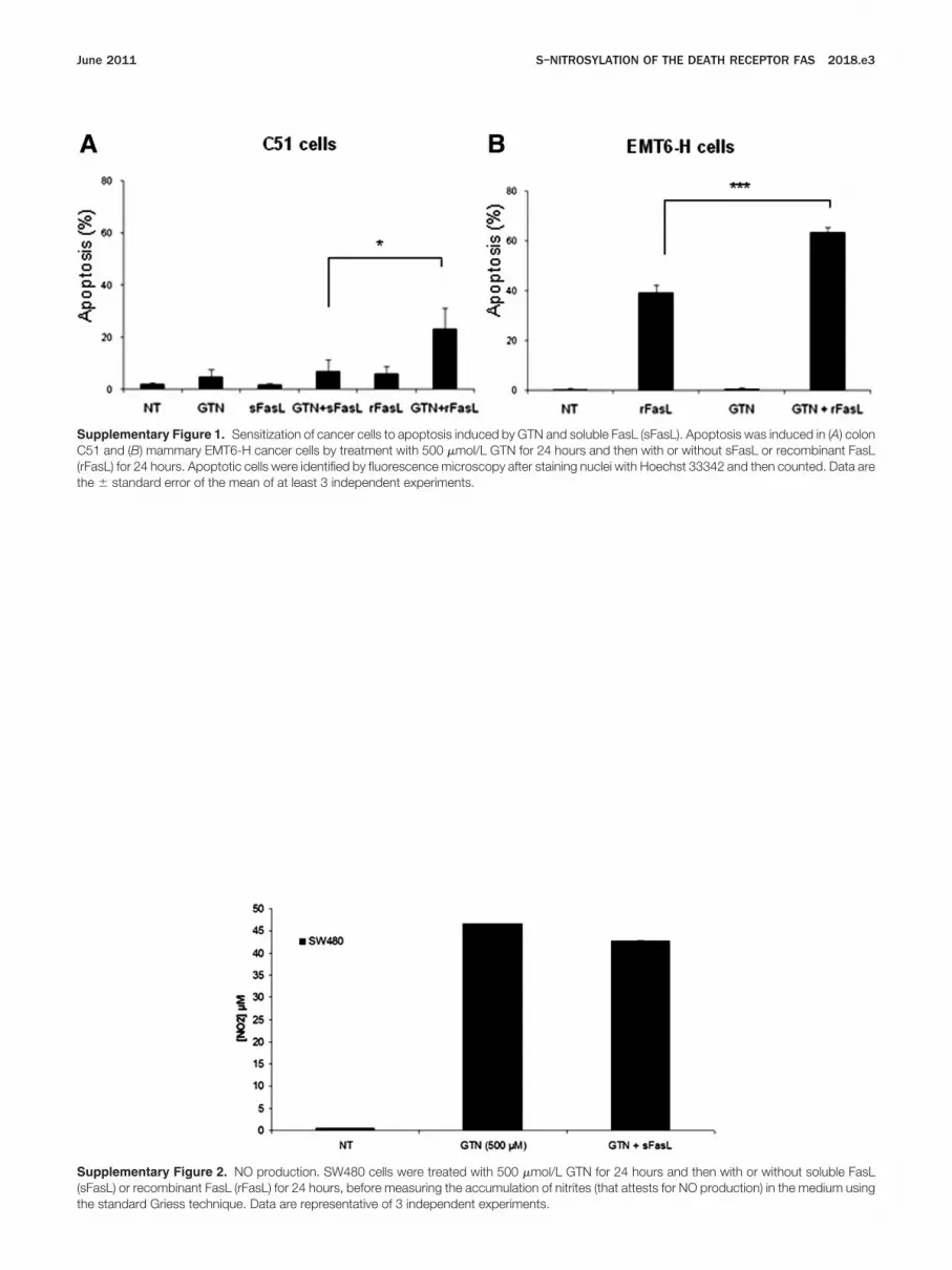
Supplementary Figure 1. Sensitization of cancer cells to apoptosis induced by GTN and soluble FasL (sFasL). Apoptosis was induced in (A) colonC51 and (B) mammary EMT6-H cancer cells by treatment with 500 �mol/L GTN for 24 hours and then with or without sFasL or recombinant FasLrFasL) for 24 hours. Apoptotic cells were identified by fluorescence microscopy after staining nuclei with Hoechst 33342 and then counted. Data are
he � standard error of the mean of at least 3 independent experiments.Supplementary Figure 2. NO production. SW480 cells were treated with 500 �mol/L GTN for 24 hours and then with or without soluble FasL(sFasL) or recombinant FasL (rFasL) for 24 hours, before measuring the accumulation of nitrites (that attests for NO production) in the medium using
the standard Griess technique. Data are representative of 3 independent experiments.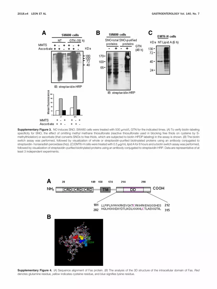
fl
d
2018.e4 LEON ET AL GASTROENTEROLOGY Vol. 140, No. 7
Supplementary Figure 3. NO induces SNO. SW480 cells were treated with 500 �mol/L GTN for the indicated times. (A) To verify biotin-labelingspecificity for SNO, the effect of omitting methyl methane thiosulfonate (reactive thiosulfonate used in blocking free thiols on cysteine by S-methylthiolation) or ascorbate (that converts SNOs to free thiols, which are subjected to biotin-HPDP labeling) in the assay is shown. (B) The biotinswitch assay was performed, followed by visualization of whole or streptavidin-purified biotinylated proteins using an antibody conjugated tostreptavidin- horseradish peroxidase (hrp). (C) EMT6-H cells were treated with 0.5 �g/mL lipid A for 6 hours and a biotin switch assay was performed,ollowed by visualization of streptavidin-purified biotinylated proteins using an antibody conjugated to streptavidin HRP. Data are representative of at
east 3 independent experiments.Supplementary Figure 4. (A) Sequence alignment of Fas protein. (B) The analysis of the 3D structure of the intracellular domain of Fas. Red
enotes glutamine residue, yellow indicates cysteine residue, and blue signifies lysine residue.


