Embed Size (px)
Citation preview
pubs.acs.org/jmcPublished on Web 10/27/2009r 2009 American Chemical Society
J. Med. Chem. 2009, 52, 7029–7043 7029
DOI: 10.1021/jm901133z
rvβ3 Integrin-Targeting Arg-Gly-Asp (RGD) Peptidomimetics Containing Oligoethylene Glycol
(OEG) Spacers
Vincent Rerat,† Georges Dive,‡ Alex A. Cordi,§ Gordon C. Tucker,§ Reine Bareille, ) Jo€elle Am�ed�ee, ) Laurence Bordenave, ),^
and Jacqueline Marchand-Brynaert*,†
†Unit�e de Chimie Organique et M�edicinale, Universit�e Catholique de Louvain, Batiment Lavoisier, Place L. Pasteur 1, 1348 Louvain-la-Neuve,Belgium, ‡Centre d’Ing�enierie des Prot�eines, Universit�e de Li�ege, Batiment B6, All�ee de la Chimie, 4000 Sart-Tilman, Belgium, §Institut deRecherches Servier, Rue des Moulineaux 11, 92150 Suresnes, France, )INSERM, U577, Universit�e Victor Segalen Bordeaux 2, Rue L�eo Saignat146, 33076 Bordeaux Cedex, France, and ^CIC-IT Biomat�eriaux, INSERM, Pessac, F-33604 France; CHUBordeaux, Hopital Xavier Arnozan,Pessac, 33604, France
Received June 9, 2009
RGD peptides are used in biomaterials science for surface modifications with a view to elicit selectivecellular responses. Our objective is to replace peptides by small peptidomimetics acting similarly. Wedesigned novel molecules targetingRvβ3 integrin and featuring spacer-arms (for surface grafting), whichdo not disturb the biological activity, from (L) N-(3-(trifluoromethyl)benzenesulfonyl) tyrosine used asscaffold. Various Arg-mimics were fixed on the phenol function, and the ortho position was used for thecoupling of OEG spacers. All peptidomimetics were active in the nM range in a binding test towardhuman Rvβ3 integrin (IC50 = 0.1 to 1.7 nM) and selective versus platelet integrin RIIbβ3. Selectedcompounds revealed excellent ability to inhibit bone cells adhesion on vitronectin. Modeling anddocking studies were performed for comparing the most active RGD peptidomimetic to cilengitide, i.e.,cyclo-[RGDfN(Me)V]-. Lastly, the adhesion of endothelial cells on a cultivation support grafted withRGD peptidomimetics was significantly improved.
Introduction
RGD (Arg-Gly-Asp)-based ligands of Rvβ3 integrin areintensively investigated in topics related to pathology, phar-macology, and materials science. The RGD peptide sequencehas been recognized 25 years ago as the cell attachment site invarious extracellular matrix (ECMa) proteins.1,2 At the sametime, cellular receptors of ECM proteins have been identifiedand most have been classified in the integrin family. Thesereceptors are heterodimeric transmembrane glycoproteinsformed by the association of R and β subunits.3-6 Theymediate cell adhesion and migration phenomena.
The Rvβ3 integrin (also called vitronectin receptor) is mainlyinvolved in adhesion of osteoclasts and osteoblasts to bonematrix, migration of vascular smoothmuscle cells, angiogenesis
of proliferating endothelium, and tumor invasiveness.7-10
Therefore, antagonists of Rvβ3 receptor, namely RGD cyclicpeptides11 and RGD peptidomimetics,12,13 are currently devel-oped as potential drugs for the treatment of osteoporosis,restenosis, ocular disease, and cancer.14-16 This is exemplifiedwith cilengitide, i.e., cyclo-[RGDfN(Me)V]-, actually underclinical trials.11 RGD-based strategies are also considered forthe selective delivery of therapeutics and imaging agents totumor cells.17-19 Last, biomaterials for stimulated cell adhesionarepreparedby surfacemodificationwithRGDmolecules.20-22
Controlling the interface between cells and solid substrates isof major concern in tissue engineering and regenerative med-icine.23 In this context, we are interested in the covalentattachment of RGD peptidomimetics on the surface of inertpolymer materials such as poly(ethylene terephthalate) (PET),with a view to promote cellular adhesion in the absence ofserum or ECM proteins.24,25 The development of such astrategy requires peptidomimetic molecules featuring an an-chorage arm having a position on the molecular scaffold thatdoes not disturb the biological activity. In this article, wedescribe the synthesis and the validation of novel RGDpeptidomimetics, containing OEG (oligoethylene glycol)spacers, and constructed on the (L)-tyrosine scaffold, fortargeting Rvβ3 integrin. Our aim is the development of molec-ular devices useful in biomaterials research principally, but alsoin drug or imaging agent delivery and immunotherapy.26,27
Results and Discussion
Design. The promotion of cellular adhesion on biocompa-tible polymer substrates is generally achieved either by coating
*To whom correspondence should be addressed. Phone: þ32 (0)10 4727 40. Fax:þ32 (0)10 47 41 68. E-mail: [email protected].
aAbbreviations: ACN, acetonitrile; Boc, tert-butyloxylcarbonyl;DMEM, Dulbecco’s Modified Eagle Medium; DMF, dimethylforma-mide; DMSO, dimethylsulfoxide; EC, endothelial cell; ECM, extracel-lular matrix; EDTA, ethylenediaminetetraacetic acid; EG, ethyleneglycol; FCS, fetal calf serum; FtN, phthalimide; GRGDS, glycine-arginine-glycine-aspartic acid-serine; HOP, human osteoprogenitor;HSV, human saphenous vein; IMDM, Iscove Modified Dulbecco’sMedium; IC50, half maximal inhibitory concentration; MIDAS, metalion dependent adhesion site; OEG, olygoethylene glycol; PB, phosphatebuffer; PBS, phosphate buffered saline; PET, poly(ethylene tereph-thalate); PS, polystyrene; PyBop,: benzotriazol-1-yl-oxy-tris-(dime-thylamino)phosphonium hexafluorophosphate; RGD, arginine-gly-cine-aspartic acid; RHF, restricted Hartree-Fock; TCPS, tissue culturepolystyrene; TFA, trifluoroacetic acid; THF, tetrahydrofuran; XPS,X-ray photoelectron spectroscopy.
7030 Journal of Medicinal Chemistry, 2009, Vol. 52, No. 22 Rerat et al.
the materials with ECM proteins and RGD-containing largepeptides20 or by surface derivatization with small (cyclic)RGD peptides28,29 and peptidomimetics (i.e., nonpeptidemimics of the RGD sequence constrained in the bioactiveconformation). This latter approach is quite underemployedfrom the literature,25 most probably because it requiresexpertise and consistent efforts in organic synthesis. As faras inorganic (bio)materials are concerned, the coating oftitanium with thiol-terminated RGD peptidomimetics hasbeen reported by Kessler et al.30
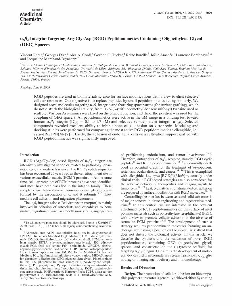
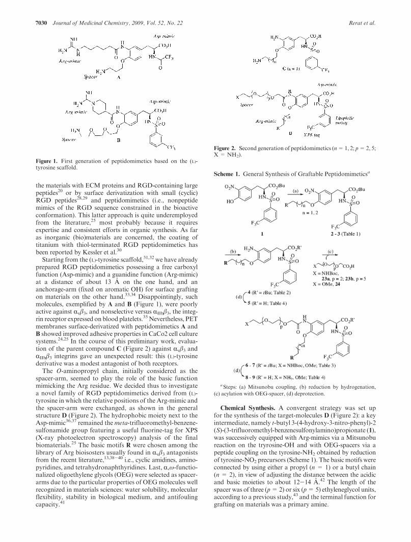
Starting from the (L)-tyrosine scaffold,31,32 we have alreadyprepared RGD peptidomimetics possessing a free carboxylfunction (Asp-mimic) and a guanidine function (Arg-mimic)at a distance of about 13 A on the one hand, and ananchorage-arm (fixed on aromatic OH) for surface graftingon materials on the other hand.33,34 Disappointingly, suchmolecules, exemplified by A and B (Figure 1), were poorlyactive against Rvβ3, and nonselective versus RIIbβ3, the integ-rin receptor expressed on blood platelets.35Nevertheless, PETmembranes surface-derivatized with peptidomimetics A andB showed improved adhesive properties in CaCo2 cell culturesystems.24,25 In the course of this preliminary work, evalua-tion of the parent compound C (Figure 2) against Rvβ3 andRIIbβ3 integrins gave an unexpected result: this (L)-tyrosinederivative was a modest antagonist of both receptors.
The O-aminopropyl chain, initially considered as thespacer-arm, seemed to play the role of the basic functionmimicking the Arg residue. We decided thus to investigatea novel family of RGD peptidomimetics derived from (L)-tyrosine in which the relative positions of the Arg-mimic andthe spacer-arm were exchanged, as shown in the generalstructure D (Figure 2). The hydrophobic moiety next to theAsp-mimic36,37 remained themeta-trifluoromethyl-benzene-sulfonamide group featuring a useful fluorine-tag for XPS(X-ray photoelectron spectroscopy) analysis of the finalbiomaterials.25 The basic motifs R were chosen among thelibrary of Arg bioisosters usually found in Rvβ3 antagonistsfrom the recent literature,13,38-40 i.e., cyclic amidines, amino-pyridines, and tetrahydronaphthyridines. Last, R,ω-functio-nalized oligoethylene glycols (OEG) were selected as spacer-arms due to the particular properties of OEGmolecules wellrecognized in materials sciences: water solubility, molecularflexibility, stability in biological medium, and antifoulingcapacity.41
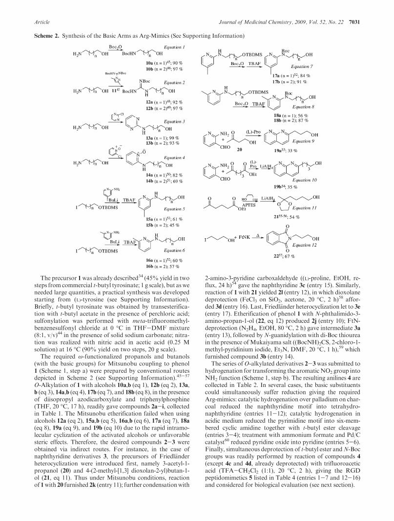
Chemical Synthesis. A convergent strategy was set upfor the synthesis of the target-molecules D (Figure 2): a keyintermediate, namely t-butyl 3-(4-hydroxy-3-nitro-phenyl)-2(S)-(3-trifluoromethyl-benzenesulfonylamino)propionate (1),was successively equipped with Arg-mimics via a Mitsunobureaction on the tryrosine-OH and with OEG-spacers via apeptide coupling on the tyrosine-NH2 obtained by reductionof tyrosine-NO2 precursors (Scheme 1). The basic motifs wereconnected by using either a propyl (n = 1) or a butyl chain(n = 2), in view of adjusting the distance between the acidicand basic moieties to about 12-14 A.42 The length of thespacer was of three (p=2) or six (p=5) ethyleneglycol units,according to a previous study,43 and the terminal function forgrafting on materials was a primary amine.
Figure 1. First generation of peptidomimetics based on the (L)-tyrosine scaffold.
Figure 2. Second generation of peptidomimetics (n=1, 2; p=2, 5;X = NH2).
Scheme 1. General Synthesis of Graftable Peptidomimeticsa
a Steps: (a) Mitsunobu coupling, (b) reduction by hydrogenation,
(c) acylation with OEG-spacer, (d) deprotection.
Article Journal of Medicinal Chemistry, 2009, Vol. 52, No. 22 7031
The precursor 1was already described34 (45% yield in twosteps from commercial t-butyl tyrosinate; 1 g scale), but asweneeded large quantities, a practical synthesis was developedstarting from (L)-tyrosine (see Supporting Information).Briefly, t-butyl tyrosinate was obtained by transesterifica-tion with t-butyl acetate in the presence of perchloric acid;sulfonylation was performed with meta-trifluoromethyl-benzenesulfonyl chloride at 0 �C in THF-DMF mixture(8:1, v/v)44 in the presence of solid sodium carbonate; nitra-tion was realized with nitric acid in acetic acid (0.25 Msolution) at 16 �C (90% yield on two steps, 20 g scale).
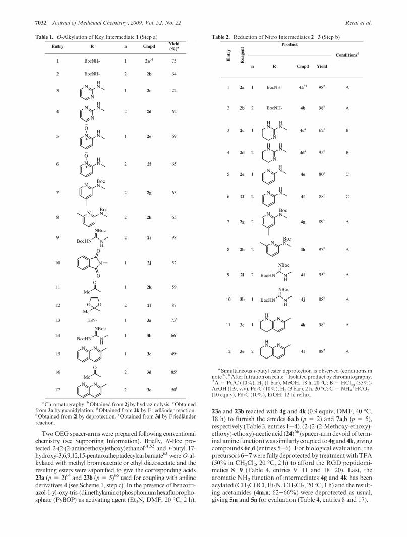
The required ω-functionalized propanols and butanols(with the basic groups) for Mitsunobu coupling to phenol1 (Scheme 1, step a) were prepared by conventional routesdepicted in Scheme 2 (see Supporting Information).45-57
O-Alkylation of 1 with alcohols 10a,b (eq 1), 12b (eq 2), 13a,b (eq 3), 14a,b (eq 4), 17b (eq 7), and 18b (eq 8), in the presenceof diisopropyl azodicarboxylate and triphenylphosphine(THF, 20 �C, 17 h), readily gave compounds 2a-i, collectedin Table 1. The Mitsunobu etherification failed when usingalcohols 12a (eq 2), 15a,b (eq 5), 16a,b (eq 6), 17a (eq 7), 18a(eq 8), 19a (eq 9), and 19b (eq 10) due to the rapid intramo-lecular cyclization of the activated alcohols or unfavorablesteric effects. Therefore, the desired compounds 2-3 wereobtained via indirect routes. For instance, in the case ofnaphthyridine derivatives 3, the precursors of Friedl€anderheterocyclization were introduced first, namely 3-acetyl-1-propanol (20) and 4-(2-methyl-[1,3] dioxolan-2-yl)butan-1-ol (21, eq 11). Thus under Mitsunobu conditions, reactionof 1with 20 furnished 2k (entry 11); further condensationwith
2-amino-3-pyridine carboxaldehyde ((L)-proline, EtOH, re-flux, 24 h)54 gave the naphthyridine 3c (entry 15). Similarly,reaction of 1 with 21 yielded 2l (entry 12), in which dioxolanedeprotection (FeCl3 on SiO2, acetone, 20 �C, 2 h)58 affor-ded 3d (entry 16). Last, Friedl€ander heterocyclization let to 3e(entry 17). Etherification of phenol 1 with N-phthalimido-3-amino-propan-1-ol (22, eq 12) produced 2j (entry 10); FtN-deprotection (N2H4, EtOH, 80 �C, 2 h) gave intermediate 3a(entry 13), followed byN-guanidylation with di-Boc thioureain the presence ofMukaiyama salt ((BocNH)2CS, 2-chloro-1-methyl-pyridinium iodide, Et3N, DMF, 20 �C, 1 h),59 whichfurnished compound 3b (entry 14).
The series ofO-alkylated derivatives 2-3was submitted tohydrogenation for transforming the aromaticNO2 group intoNH2 function (Scheme 1, step b). The resulting anilines 4 arecollected in Table 2. In several cases, the basic substituentscould simultaneously suffer reduction giving the requiredArg-mimics: catalytic hydrogenation over palladium on char-coal reduced the naphthyridine motif into tetrahydro-naphthyridine (entries 11-12); catalytic hydrogenation inacidic medium reduced the pyrimidine motif into six-mem-bered cyclic amidine together with t-butyl ester cleavage(entries 3-4); treatment with ammonium formate and Pd/Ccatalyst60 reduced pyridine oxide into pyridine (entries 5-6).Finally, simultaneous deprotection of t-butyl ester andN-Bocgroups was readily performed by reaction of compounds 4(except 4c and 4d, already deprotected) with trifluoroaceticacid (TFA-CH2Cl2 (1:1), 20 �C, 2 h), giving the RGDpeptidomimetics 5 listed in Table 4 (entries 1-7 and 12-16)and considered for biological evaluation (see next section).
Scheme 2. Synthesis of the Basic Arms as Arg-Mimics (See Supporting Information)
7032 Journal of Medicinal Chemistry, 2009, Vol. 52, No. 22 Rerat et al.
TwoOEG spacer-arms were prepared following conventionalchemistry (see Supporting Information). Briefly, N-Boc pro-tected 2-(2-(2-aminoethoxy)ethoxy)ethanol61,62 and t-butyl 17-hydroxy-3,6,9,12,15-pentaoxaheptadecylcarbamate63 were O-al-kylated with methyl bromoacetate or ethyl diazoacetate and theresulting esters were saponified to give the corresponding acids23a (p = 2)64 and 23b (p = 5)65 used for coupling with anilinederivatives 4 (see Scheme 1, step c). In the presence of benzotri-azol-1-yl-oxy-tris-(dimethylamino)phosphoniumhexafluoropho-sphate (PyBOP) as activating agent (Et3N, DMF, 20 �C, 2 h),
23a and 23b reacted with 4g and 4k (0.9 equiv, DMF, 40 �C,18 h) to furnish the amides 6a,b (p = 2) and 7a,b (p = 5),respectively (Table 3, entries 1-4). (2-(2-(2-Methoxy-ethoxy)-ethoxy)-ethoxy)-acetic acid (24)66 (spacer-armdevoid of term-inal amine function)was similarly coupled to4gand 4k, givingcompounds 6c,d (entries 5-6). For biological evaluation, theprecursors 6-7were fully deprotected by treatmentwithTFA(50% in CH2Cl2, 20 �C, 2 h) to afford the RGD peptidomi-metics 8-9 (Table 4, entries 9-11 and 18-20). Last, thearomatic NH2 function of intermediates 4g and 4k has beenacylated (CH3COCl, Et3N,CH2Cl2, 20 �C, 1 h) and the result-ing acetamides (4m,n; 62-66%) were deprotected as usual,giving 5m and 5n for evaluation (Table 4, entries 8 and 17).
Table 2. Reduction of Nitro Intermediates 2-3 (Step b)
a Simultaneous t-butyl ester deprotection is observed (conditions innoted). bAfter filtrationon celite. c Isolated product by chromatography.dA = Pd/C (10%), H2 (1 bar), MeOH, 18 h, 20 �C; B = HClaq (35%)-AcOH (1:9, v/v), Pd/C (10%), H2 (3 bar), 2 h, 20 �C; C=NH4
þHCO2-
(10 equiv), Pd/C (10%), EtOH, 12 h, reflux.
Table 1. O-Alkylation of Key Intermediate 1 (Step a)
aChromatography. bObtained from 2j by hydrazinolysis. cObtainedfrom 3a by guanidylation. dObtained from 2k by Friedl€ander reaction.eObtained from 2l by deprotection. fObtained from 3d by Friedl€anderreaction.
Article Journal of Medicinal Chemistry, 2009, Vol. 52, No. 22 7033
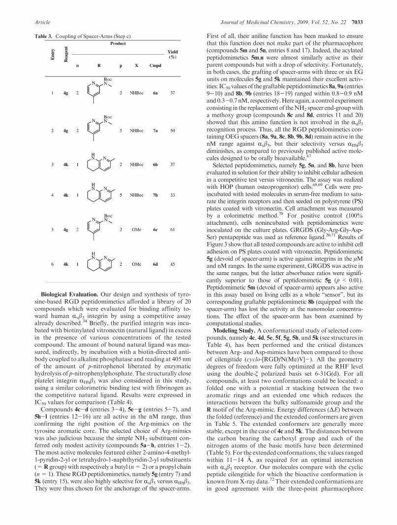
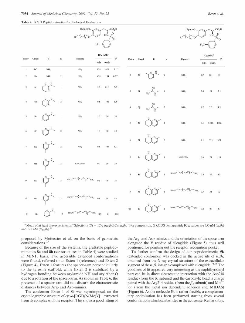
Biological Evaluation. Our design and synthesis of tyro-sine-based RGD peptidomimetics afforded a library of 20compounds which were evaluated for binding affinity to-ward human Rvβ3 integrin by using a competitive assayalready described.34 Briefly, the purified integrin was incu-bated with biotinylated vitronectin (natural ligand) in excessin the presence of various concentrations of the testedcompound. The amount of bound natural ligand was mea-sured, indirectly, by incubation with a biotin-directed anti-body coupled to alkaline phosphatase and reading at 405 nmof the amount of p-nitrophenol liberated by enzymatichydrolysis of p-nitrophenylphosphate. The structurally closeplatelet integrin RIIbβ3 was also considered in this study,using a similar colorimetric binding test with fibrinogen asthe competitive natural ligand. Results were expressed inIC50 values for comparison (Table 4).
Compounds 4c-d (entries 3-4), 5e-g (entries 5-7), and5h-l (entries 12-16) are all active in the nM range, thusconfirming the right position of the Arg-mimics on thetyrosine aromatic core. The selected choice of Arg-mimicswas also judicious because the simple NH2 substituent con-ferred only modest activity (compounds 5a-b, entries 1-2).The most active molecules featured either 2-amino-4-methyl-1-pyridin-2-yl or tetrahydro-1-naphthyridin-2-yl substituents(=R group) with respectively a butyl (n=2) or a propyl chain(n=1). TheseRGDpeptidomimetics, namely 5g (entry 7) and5k (entry 15), were also highly selective for Rvβ3 versus RIIbβ3.They were thus chosen for the anchorage of the spacer-arms.
First of all, their aniline function has been masked to ensurethat this function does not make part of the pharmacophore(compounds 5m and 5n, entries 8 and 17). Indeed, the acylatedpeptidomimetics 5m,n were almost similarly active as theirparent compounds but with a drop of selectivity. Fortunately,in both cases, the grafting of spacer-arms with three or six EGunits on molecules 5g and 5k maintained their excellent activ-ities: IC50 valuesof thegraftablepeptidomimetics8a,9a (entries9-10) and 8b, 9b (entries 18-19) ranged within 0.8-0.9 nMand0.3-0.7 nM, respectively.Here again, a control experimentconsisting in the replacement of theNH2 spacer end-groupwitha methoxy group (compounds 8c and 8d, entries 11 and 20)showed that this amino function is not involved in the Rvβ3recognition process. Thus, all the RGD peptidomimetics con-tainingOEG spacers (8a, 9a, 8c, 8b, 9b, 8d) remain active in thenM range against Rvβ3, but their selectivity versus RIIbβ3diminishes, as compared to previously published active mole-cules designed to be orally bioavailable.67
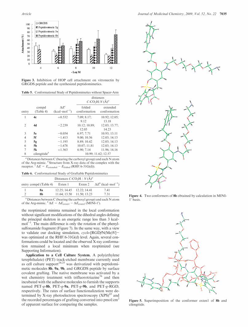
Selected peptidomimetics, namely 5g, 5n, and 8b, have beenevaluated in solution for their ability to inhibit cellular adhesionin a competitive test versus vitronectin. The assay was realizedwith HOP (human osteoprogenitor) cells.68,69 Cells were pre-incubated with tested molecules in serum-free medium to satu-rate the integrin receptors and then seeded on polystyrene (PS)plates coated with vitronectin. Cell attachment was measuredby a colorimetric method.70 For positive control (100%attachment), cells nonincubated with peptidomimetics wereinoculated on the culture plates. GRGDS (Gly-Arg-Gly-Asp-Ser) pentapeptide was used as reference ligand.20,71 Results ofFigure 3 show that all tested compounds are active to inhibit celladhesion on PS plates coated with vitronectin. Peptidomimetic5g (devoid of spacer-arm) is active against integrins in the μMand nM ranges. In the same experiment, GRGDS was active inthe same ranges, but the latter absorbance ratios were signifi-cantly superior to those of peptidomimetic 5g (p < 0.01).Peptidomimetic 5m (devoid of spacer-arm) appears also activein this assay based on living cells as a whole “sensor”, but itscorresponding graftable peptidomimetic 8b (equipped with thespacer-arm) has lost the activity at the nanomolar concentra-tions. The effect of the spacer-arm has been examined bycomputational studies.
Modeling Study.A conformational study of selected com-pounds, namely 4c, 4d, 5e, 5f, 5g, 5h, and 5k (see structures inTable 4), has been performed and the critical distancesbetween Arg- and Asp-mimics have been compared to thoseof cilengitide (cyclo-[RGDfN(Me)V]-). All the geometrydegrees of freedom were fully optimized at the RHF levelusing the double-ζ polarized basis set 6-31G(d). For allcompounds, at least two conformations could be located: afolded one with a potential π stacking between the twoaromatic rings and an extended one which reduces theinteractions between the bulky sulfonamide group and theR motif of the Arg-mimic. Energy differences (ΔE) betweenthe folded (reference) and the extended conformers are givenin Table 5. The extended conformers are generally morestable, except in the case of 4c and 5k. The distances betweenthe carbon bearing the carboxyl group and each of thenitrogen atoms of the basic motifs have been determined(Table 5). For the extended conformations, the values rangedwithin 11-14 A, as required for an optimal interactionwith Rvβ3 receptor. Our molecules compare with the cyclicpeptide cilengitide for which the bioactive conformation isknown fromX-ray data.72 Their extended conformations arein good agreement with the three-point pharmacophore
Table 3. Coupling of Spacer-Arms (Step c)
7034 Journal of Medicinal Chemistry, 2009, Vol. 52, No. 22 Rerat et al.
proposed by Moitessier et al. on the basis of geometricconsiderations.73
Because of the size of the systems, the graftable peptido-mimetics 8a and 8b (see structures in Table 4) were studiedin MINI1 basis. Two accessible extended conformationswere found, referred to as Exten 1 (reference) and Exten 2(Figure 4). Exten 1 features the spacer-arm perpendicularlyto the tyrosine scaffold, while Exten 2 is stabilized by ahydrogen bonding between arylamide NH and arylether Odue to a rotation of the spacer-arm. As shown in Table 6, thepresence of a spacer-arm did not disturb the characteristicdistances between Arg- and Asp-mimics.
The conformer Exten 1 of 8b was superimposed on thecrystallographic structure of cyclo-[RGDfN(Me)V]- extractedfrom its complex with the receptor. This shows a good fitting of
the Arg- and Asp-mimics and the orientation of the space-armalongside the V residue of cilengitide (Figure 5), thus wellpositioned for pointing out the receptor recognition pocket.
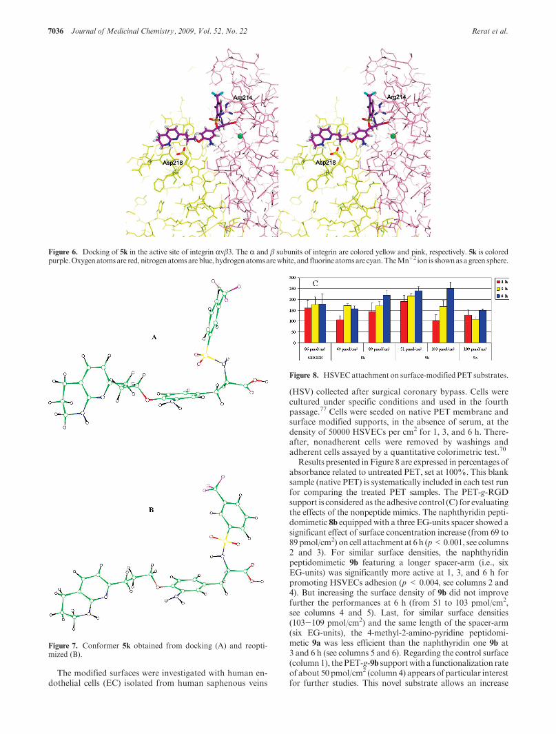
To further confirm the design of our peptidomimetic, 5k(extended conformer) was docked in the active site of Rvβ3,obtained from the X-ray crystal structure of the extracellularsegment of theRvβ3 integrin complexedwith cilengitide.74,75 Thegoodness of fit appeared very interesting as the naphthyridinylpart can be in direct electrostatic interaction with the Asp218residue (from the Rv subunit) and the carboxylic head is chargepaired with the Arg214 residue (from the β3 subunit) andMn2þ
ion (from the metal ion dependent adhesion site, MIDAS)(Figure 6). As the molecule 5k is rather flexible, a complemen-tary optimization has been performed starting from severalconformationswhichcanbe fitted in theactive site.Remarkably,
Table 4. RGD Peptidomimetics for Biological Evaluation
aMean of at least two experiments. b Selectivity (S) = IC50 RIIbβ3/IC50 Rvβ3.cFor comparison, GRGDS pentapeptide IC50 values are 750 nM (Rvβ3)
and 120 nM (RIIbβ3).71
Article Journal of Medicinal Chemistry, 2009, Vol. 52, No. 22 7035
the reoptimized minima remained in the local conformationwithout significantmodifications of the dihedral angles definingthe principal skeleton in an energetic range less than 3 kcal 3mol-1. The main difference is only the rotation of the phenyl-sulfonamide fragment (Figure 7). In the same way, with a viewto validate our docking simulation, cyclo-[RGDfN(Me)V]-was optimized at the RHF/6-31G(d) level. Again, several con-formations could be located and the observed X-ray conforma-tion remained a local minimum when reoptimized (seeSupporting Information).
Application to a Cell Culture System. A poly(ethyleneterephthalate) (PET) track-etched membrane currently usedas cell culture support24,25 was derivatized with peptidomi-metic molecules 8b, 9a, 9b, and GRGDS peptide by surfacecovalent grafting. The native membrane was activated by awet chemistry treatment with trifluorotriazine76 and thenincubated with the adhesive molecules to furnish the supportsnamed PET-g-8b, PET-g-9a, PET-g-9b, and PET-g-RGD,respectively. The rates of surface functionalization were de-termined by X-ray photoelectron spectroscopy (XPS)43 andthe recorded percentages of grafting converted into pmol/cm2
of apparent surface for comparing the samples.
Table 5. Conformational Study of Peptidomimetics without Spacer-Arm
distances
C-CO2H/N (A)a
entry
compd
(Table 4)
ΔEc
(kcal 3mol-1)
folded
conformation
extended
conformation
1 4c þ0.532 7.09; 8.17;
9.12
10.92; 12.05;
13.18
2 4d -2.239 10.12; 10.89;
12.05
12.03; 13.77;
14.23
3 5e -0.054 6.97; 7.71 10.93; 13.11
4 5f -1.413 9.00; 10.56 12.03; 14.13
5 5g -1.195 8.89; 10.42 12.03; 14.13
6 5h -1.678 10.07; 11.81 12.03; 14.13
7 5k þ1.563 6.90; 7.14 11.96; 14.16
8 cilengitideb 10.99; 11.62; 12.57aDistances between C (bearing the carboxyl group) and each N atom
of the Arg-mimic. b Structure from X-ray data of the complex with thereceptor. cΔE = Eextended - Efolded (RHF/6-31G(d)).
Table 6. Conformational Study of Graftable Peptidomimetics
Distances C-CO2H / N (A)a
entry compd (Table 4) Exten 1 Exten 2 ΔEb (kcal 3mol-1)
1 8a 12.25; 14.45 12.22; 14.41 7.41
2 8b 11.64; 13.50 11.50; 13.23 7.31aDistances between C (bearing the carboxyl group) and each N atom
of the Arg-mimic. bΔE = ΔEexten1 - ΔEexten2 (MINI-10).
Figure 4. Two conformers of 8b obtained by calculation in MINI-10 basis.
Figure 3. Inhibition of HOP cell attachment on vitronectin byGRGDS peptide and the synthesized peptidomimetics.
Figure 5. Superimposition of the conformer exten1 of 8b andcilengitide.
7036 Journal of Medicinal Chemistry, 2009, Vol. 52, No. 22 Rerat et al.
The modified surfaces were investigated with human en-dothelial cells (EC) isolated from human saphenous veins
(HSV) collected after surgical coronary bypass. Cells werecultured under specific conditions and used in the fourthpassage.77 Cells were seeded on native PET membrane andsurface modified supports, in the absence of serum, at thedensity of 50000 HSVECs per cm2 for 1, 3, and 6 h. There-after, nonadherent cells were removed by washings andadherent cells assayed by a quantitative colorimetric test.70
Results presented inFigure 8 are expressed in percentages ofabsorbance related to untreated PET, set at 100%. This blanksample (native PET) is systematically included in each test runfor comparing the treated PET samples. The PET-g-RGDsupport is considered as the adhesive control (C) for evaluatingthe effects of the nonpeptide mimics. The naphthyridin pepti-domimetic 8b equippedwith a three EG-units spacer showed asignificant effect of surface concentration increase (from 69 to89 pmol/cm2) on cell attachment at 6 h (p<0.001, see columns2 and 3). For similar surface densities, the naphthyridinpeptidomimetic 9b featuring a longer spacer-arm (i.e., sixEG-units) was significantly more active at 1, 3, and 6 h forpromoting HSVECs adhesion (p < 0.004, see columns 2 and4). But increasing the surface density of 9b did not improvefurther the performances at 6 h (from 51 to 103 pmol/cm2,see columns 4 and 5). Last, for similar surface densities(103-109 pmol/cm2) and the same length of the spacer-arm(six EG-units), the 4-methyl-2-amino-pyridine peptidomi-metic 9a was less efficient than the naphthyridin one 9b at3 and 6 h (see columns 5 and 6). Regarding the control surface(column1), thePET-g-9b supportwith a functionalization rateof about 50 pmol/cm2 (column 4) appears of particular interestfor further studies. This novel substrate allows an increase
Figure 6. Docking of 5k in the active site of integrin Rvβ3. The R and β subunits of integrin are colored yellow and pink, respectively. 5k is coloredpurple.Oxygenatomsare red,nitrogenatomsareblue,hydrogenatomsarewhite, and fluorineatomsare cyan.TheMnþ2 ion is shownasagreensphere.
Figure 7. Conformer 5k obtained from docking (A) and reopti-mized (B).
Figure 8. HSVECattachment on surface-modified PET substrates.
Article Journal of Medicinal Chemistry, 2009, Vol. 52, No. 22 7037
of cellular attachment of 200-250% in a serum-free culturemedium, in comparison with the commercial (native) PETmembrane, while the performance of PET-g-RGD (about85 pmol/cm2) is lower (150-180%).
Conclusion
The idea that RGD-peptidomimetic molecules could beequipped with a spacer-arm without disturbing their capacityto bind the Rvβ3 integrin has been successfully exploited in thecase of ligands based on the tyrosine scaffold. This scaffoldwas initially considered by theMerck group for the research ofplatelet receptor RIIbβ3 antagonists,78 giving tirofibane ascardiovascular drug, and more recently used by Kessleret al. for the design of selective R5β1 ligands,
42,52 potentiallyuseful in the field of antiangiogenic cancer therapy. Bothreceptors, RIIbβ3 and R5β1 feature high sequence similaritywith Rvβ3 integrin.
Practically, we have introduced the spacer-arm on thetyrosine aromatic ring thanks to a simple sequence of reac-tions involving nitration/reduction/acylation. It is worth not-ing that this position of functionalization is unusual. Otherstudies devoted to bifunctional ligands targeting tumor cells,in the fields of drug delivery and imaging contrast agents,made use of the lipophilic moiety (i.e., the sulfonamide groupof piperazine-based RGD peptidomimetics) to couple thespacer.79-81
All the synthesized compounds showed natural liganddisplacement competence in the nM range for Rvβ3, with avariable selectivity versus RIIbβ3 (S= 1.4 103 for 5k and 0.83102 for 8b; S=1.8 102 for 5g and 0.3 102 for 8a, see Table 4).The drop of selectivity related to the presence of OEG spacerscould not be presently explained. Although Rvβ3/RIIbβ3 selec-tivity is an important parameter for therapeutic applica-tions, this aspect is not a limiting factorwhenpeptidomimeticsare devoted to ex vivo surface modification of biomaterials.Similarly to the reference peptideGRGDS, peptidomimetics 5and 8were able to inhibit HOP cell adhesion on a vitronectin-coated support.
Computational studies have confirmed the good fitting ofour graftable RGD-peptidomimetics into the target receptorand the possible positioning of the spacer-arm outside thebinding site. This is exactly the requirement for the develop-ment of molecules 8a,b and 9a,b in the field cell adhesivematerials with a view of endothelialisation of vasculargrafts.77 Preliminary results of HSV (human saphenous vein)cell adhesion on poly(ethylene terephthalate) (PET) mem-branes grafted with peptidomimetics demonstrated the goodperformance of one substrate, namely PET-g-9b. Furtherworks are in progress for studying EC proliferation andresponse under physiological shear stress conditions in flowchambers.
Experimental Section
Chemistry. Reagents were purchased from Aldrich or AcrosOrganics and used as received. Anhydrous solvents were pur-chased from Fluka. Reactions needing anhydrous conditionswere performed in flame-dried glassware, placed under an argonatmosphere. 1H and 13CNMR spectra were recorded on Bruker300 Ultra Shield and Bruker AM 500 spectrometers, usingdeuterated solvents from Rocc SA (Belgium) and TMS(tetramethylsilane) as internal standard. Patterns are designedas s (singlet), br s (broad singlet), d (doublet), dd (doublet ofdoublet), t (triplet), q (quartet), m (multiplet). Coupling con-stants (J) are given in hertz (Hz). IR spectra were recorded on a
Shimatzu FTIR 8400S equipment; products were deposited asthin films on NaCl crystal. Low resolution mass spectra wereobtained with a TSQ 7000 spectrometer from Finnigan in CI(chemical ionization) mode and with a LCQ or quantumspectrometer from Finnigan in ESI (electronspray ionization)mode at 70 eV and APCI (atmospheric pressure chemicalionization) mode at 100 eV. High resolution mass spectra(HRMS) were recorded at the University of Mons-Hainaut,laboratory of professor R. Flammang. Melting points weremeasured on a Buchi B-540 apparatus calibrated with benzoicacid and are uncorrected. Rotations weremeasured on a Perkin-Elmer 241 MC apparatus using sodium D ray, at 25 �C; con-centrations are given in g/100 mL. Elemental analyses weremade in the Christopher Ingold laboratories (Department ofChemistry, Imperial College, London). Analytical thin layerchromatography (TLC) was carried out on Merck TLC platescoated with silica gel 60 F254 (0.25 mm layer thickness); visua-lization was carried out using either a UV lamp (254 nm), orKMnO4, phosphomolybdic acid (10% in ethanol), and ninhy-drin (10% in ethanol) as indicators. Flash chromatography wasperformed with Merck 60 silica gel (230-400 mesh ASTM).Solvent mixtures used for TLC and flash chromatography arereported in v/v total. Lyophilization of aqueous solutions wasrealized on a Alpha 2-4LD-plus equipment from Christ(Osterode, Germany). The purity of tested compounds (at least95%) was established by analytical HPLC using a Waters(Belgium) equipment (Waters 600 pump, Waters 600 multi-solvent controller, 996 photodiode array detector, injectionsystem of Mistral, Spark Holland and EMPOWER software),and a chiralcel OD-H column (5 μm) from Daicel ChemicalIndustries (Illkirch, France) of 250 mm � 4.6 mm (internaldiameter). Elution was made with a mixture of hexane (HPLCgrade Chromasolv, Sigma-Aldrich) and i-propyl alcohol(HPLC grade HiPerSolv Chromanorm, VWR), in linear gradi-ent mode (from Hex/i-PrOH 75:25 to Hex/i-PrOH 40:60 over30 min) at a flow rate of 1 mL/min and temperature of 22 �C(detection at 254 nm).
Selected compounds are fully described below. For the others,namely 2b, 2c, 2d, 2e, 2f, 2h, 2i, 2j, 2l, 3a, 3b, 3d, 3e, 4b, 4c, 4d, 4e,4f, 4h, 4i, 4j, 4l, 6c, 6d, 5b, 5e, 5f, 5h, 5i, 5j, and 5k, see SupportingInformation.
General Procedure for Coupling Alcohols to Key Intermediate
1 (Table 1). Tyrosine scaffold 1 (0.5 g, 1.02 mmol, 1 equiv) andalcohol (from Scheme 2, 1.12 mmol, 1.1 equiv) were dissolved indry THF (4 mL) under argon atmosphere, and cooled at 0 �C.Ph3P (0.4 g, 1.53 mmol, 1.5 equiv) and then DIAD (0.3 mL,1.43 mmol, 1.4 equiv) were added dropwise. The stirred mixturewas allowed to reach slowly room temperature and further leftfor 1-12 h at 20 �C. Concentration under vacuum and flashchromatography on silica gel gave the coupled product 2 ofTable 1.
(S)-t-Butyl 3-(4-(4-(t-Butoxycarbonyl(4-methylpyridin-2-yl)-amino)butoxy)-3-nitrophenyl)-2-(3-(trifluoromethyl)phenylsulfon-amido)propanoate (2g). The title compound was obtained from17b (0.250 g, 0.89 mmol) as a pale-yellow oil (0.424 g, 63%). Rf
(nHex/EtOAc 6:4) = 0.6. IR 2978, 1701, 1533, 1327, 1161 cm-1.1HNMR (500MHz, CDCl3) δ 1.26 (s, 9 H), 1.5 (s, 9 H), 1.83 (m,4H), 2.33 (s, 3H), 2.99 (m, 1H), 3.07 (m, 1H), 3.98 (t, J=6.4Hz,2 H), 4.06 (m, 3 H), 5.26 (d, J = 6.5 Hz, SO2NH, 1 H), 6.84 (d,J=5.1Hz, 1 H), 6.93 (d, J=8.6Hz, 1 H), 7.35 (dd, J=8.6, 2.2Hz, 1 H), 7.4 (s, 1 H), 7.55 (s, 1 H), 7.61 (t, J=7.8 Hz, 1 H), 7.79(d, J=7.8 Hz, 1 H), 7.96 (d, J=7.9Hz, 1 H), 8.04 (s, 1 H), 8.21(d, J = 5.1 Hz, 1 H). 13C NMR (125 MHz, CDCl3) δ 21, 25.25,26.25, 27.55, 28.18, 37.98, 46.15, 56.68, 69.23, 80.8, 83.63, 114.38,120.57, 120.88, 124.08, 124.7 (CF3), 126.41, 127.18, 129.24,129.76, 130.34, 131.55, 135.44, 139.08, 140.95, 147.18, 148.14,151.54, 154.18, 154.46, 169.23. MS (APCI)m/z 697, 641. HRMSC35H43F3N4O9S calcd for [MþNa]þ, 775.2601; found, 775.2628.
(S)-t-Butyl 3-(3-Nitro-4-(4-oxo-pentyloxy)phenyl)-2-(3-(trifluoro-methyl)phenylsulfonamido)propanoate (2k).The title compoundwas

7038 Journal of Medicinal Chemistry, 2009, Vol. 52, No. 22 Rerat et al.
obtained from 20 (0.082mL, 0.861mmol) as apale-yellowoil (0.277g, 59%). Rf (ether/nHex 85:15) = 0.46. IR 2978, 1716, 1531, 1327,1163 cm-1. 1H NMR (300 MHz, CDCl3) δ 1.27 (s, 9 H), 2.09 (m,2H), 2.19 (s, 3H), 2.73 (t, J=6.8Hz, 2H), 2.99 (m, 1H), 3.07 (m,1H), 4.06 (m, 3H), 5.45 (d, J=8.9Hz, SO2NH, 1H), 6.96 (d, J=8.6Hz, 1H), 7.37 (dd,J=8.6, 2.2Hz, 1H), 7.6 (d,J=2.2Hz, 1H),7.63 (t, J = 7.8 Hz, 1 H), 7.8 (d, J = 7.8 Hz, 1 H), 7.98 (d, J =7.8 Hz, 1 H), 8.04 (s, 1 H). 13C NMR (75 MHz, CDCl3) δ 23.06,27.86, 30.29, 38.24, 39.45, 57.08, 68.47, 84, 114.73, 124.35, 126.69,127.68, 129.66, 130.17, 130.68, 131.55, 135.81, 139.11, 141.07,151.73, 169.38, 208.47, (CF3 not visible). MS (APCI) m/z 573[M - H]-, 517. HRMS C25H29F3N2O8S calcd for [M þ Na]þ,597.1494; found, 597.1481.
(S)-t-Butyl 3-(4-(3-[1,8]Naphthyridin-2-yl-propoxy)-3-nitro-phenyl)-2-(3-(trifluoromethyl)phenylsulfonamido)propanoate (3c). Asolution of 2-amino-3-pyridin-carboxaldehyde (0.064 g, 0.517mmol), precursor 2k (0.270 g, 0.470 mmol) and (L)-proline (0.027g, 0.235 mmol) in EtOH (5 mL) was refluxed for 48 h under Aratmosphere. After concentration under vacuum, the residue waspurified by chromatography to afford the title compound (0.167 g,49%) as a white solid. Rf (DCM/i-PrOH 98:2) = 0.4. mp =143-144 �C. IR 1731, 1610, 1533, 1327, 1161 cm-1. 1H NMR(500 MHz, CDCl3) δ 1.26 (s, 9 H), 2.51 (quint, J= 6.3 Hz, 2 H),2.97 (m, 1H), 3.05 (m, 1H), 3.3 (t, J=6.3Hz, 2H), 4.06 (m, 1H),4.2 (t, J=6.3 Hz, 2 H), 5.38 (d, J=9Hz, SO2NH, 1 H), 6.97 (d,J=8.6Hz,1H), 7.33 (dd,J=8.6, 2.2Hz, 1H), 7.46 (d,J=8.4Hz,1H), 7.46 (dd,J=7.9, 4.3Hz, 1H), 7.57 (d, J=2.2Hz, 1H), 7.6 (t,J=7.8Hz,1H), 7.77 (d,J=7.8Hz,1H), 7.96 (d,J=7.8Hz,1H),8.03 (s, 1H), 8.11 (d, J=8.4Hz, 1H), 8.17 (dd, J=7.9, 2Hz, 1H),9.08 (dd, J=4.3, 2Hz, 1H). 13CNMR(125MHz,CDCl3) δ 27.45,27.55, 34.58, 37.95, 56.73, 68.52, 83.69, 114.49, 121.06, 121.44,123.02, 124.07, 126.33, 127.12, 129.3, 129.81, 130.34, 131.55,135.38, 136.74, 137.03, 139.07, 140.78, 151.55, 153.2, 155.83,165.18, 169.03, (CF3 not visible). MS (ESI) m/z 1318 [M- H]- �2, 659 [M-H]-, 209.HRMSC31H31F3N4O7S calcd for [MþH]þ,661.1944; found, 661.1949.
General Procedure for Reduction of 2-3 (Table 2). MethodA.
A solution of 2 or 3 in MeOH or EtOH (0.1 mmol/3 mL)containing Pd/C (10%) as catalyst (0.01 g/0.1mmol product 2 or3) was placed under H2 atmosphere (1 atm) and stirred for 18 hat 20 �C. The mixture was filtered over a short celite pad, usingMeOH (EtOH); filtrate concentration under vacuum gavequantitatively crude 4.
Method B. A solution of 2 (0.2 mmol) in HOAc (5 mL) and37% HClaq (0.5 mL), containing Pd/C (10%) as catalyst (0.1 g)was introduced in a Parr flask. The mixture was hydrogenated(Parr apparatus) for 2 h at 20 �C, under a pressure of 45 psi. Themixture was filtered on a celite pad, using MeOH-H2O. Con-centration under vacuum and chromatography gave crude 4,recovered by lyophilization.
Method C. A solution of 2 (0.5 mmol) and ammoniumformate (5 mmol) in EtOH (5 mL), containing Pd/C (10%) ascatalyst (0.05 g), was refluxed for 12 h under Ar atmosphere andvigorous stirring. The mixture was filtered on a celite pad, usingEtOH. After concentration, the residue was dissolved in EtOAc(10 mL), washed with brine (2 � 5 mL), and dried (MgSO4).Solvent evaporation gave crude compound 4.
(S)-t-Butyl 3-(3-Amino-4-(4-(t-butoxycarbonyl(4-methylpyri-din-2-yl)amino)butoxy)phenyl)-2-(3-(trifluoromethyl)phenylsulfon-amido)propanoate (4g).The title compound was obtained from 2g(0.420 g, 0.560mmol), according tomethodA, as a pale-yellow oil(0.361 g, 89%). Rf (ether/EtOAc 9:1) = 0.9. [R]D20 -11 (c = 1.3,CHCl3). IR 2975, 1703, 1325, 1159 cm-1. 1H NMR (500 MHz,CDCl3) δ 1.26 (s, 9 H), 1.49 (s, 9 H), 1.79 (m, 4 H), 2.33 (s, 3 H),2.87 (m, 2H), 3.71 (br s,NH2, 2H), 3.93 (t, J=6Hz, 2H), 3.99 (t,J=7Hz, 2 H), 4.06 (m, 1 H), 5.16 (d, J=8.9 Hz, SO2NH, 1 H),6.39 (dd,J=8, 2.1Hz, 1H), 6.43 (d,J=2.1Hz, 1H), 6.57 (d, J=8.2Hz, 1H), 6.84 (d,J=5.3Hz, 1H), 7.39 (s, 1H), 7.57 (t, J=7.8Hz, 1H), 7.75 (d, J=7.8Hz, 1H), 7.91 (d, J=7.8Hz, 1H), 8.05(s, 1H), 8.21 (d, J=5.3Hz, 1H). 13CNMR (125MHz, CDCl3) δ
21, 25.44, 26.56, 27.57, 28.22, 38.76, 46.42, 56.96, 67.86, 80.64,82.6, 111.07, 115.78, 119.16, 119.51, 120.99, 124.1, 124.7 (CF3),127.07, 129.04, 129.6, 130.37, 131.35, 136.12, 141.13, 145.72,147.18, 148.14, 154.18, 154.46, 169.53. MS (APCI) m/z 721 [M- H]-, 621, 209. HRMS C35H45F3N4O7S calcd for [M þ H]þ,723.3039; found, 723.3057. Anal. calcd (%) C, 58.17; H, 6.23; N,7.76; S, 4.45. Found C, 58.13; H, 6.37; N, 7.75; S, 4.98.
(S)-t-Butyl 3-(3-Amino-4-(3-(5,6,7,8-tetrahydro-1,8-naphthy-ridin-2-yl)propoxy)phenyl)-2-(3-(trifluoromethyl)phenylsulfon-amido)propanoate (4k). The title compound was obtainedfrom 3c (0.150 g, 0.227 mmol), according to method A, as apale-brown foam (0.143 g, 98%). Rf (EtOAc/acetone 8:2) =0.2. [R]D20-11 (c=1, CHCl3). IR 2955, 1731, 1651, 1518, 1327,1159 cm-1. 1HNMR (500MHz, CDCl3) δ 1.21 (s, 9 H), 1.9 (m,2 H), 2.25 (m, 2 H), 2.68 (br s, NH2, 2 H), 2.71 (t, J=6.1 Hz, 2H), 2.85 (m, 2 H), 2.91 (t, J= 7.4 Hz, 2 H), 3.47 (m, 2 H), 3.93(t, J = 5.8 Hz, 2 H), 4.02 (m, 1 H), 5.5 (m, SO2NH, 1 H), 6.35(m, 2H), 6.43 (s, 1H), 6.57 (d, J=8.4Hz, 1H), 7.26 (d, J=7.3Hz, 1H), 7.56 (t, J=7.8Hz, 1H), 7.73 (d, J=7.8Hz, 1H), 7.9(d, J = 7.8 Hz, 1 H), 8.01 (br s, NH, 1 H), 8.1 (br s, 1 H). 13CNMR (125 MHz, CDCl3) δ 19.44, 25.42, 27.56, 28.51, 30.17,38.59, 40.91, 57.16, 66.39, 82.55, 109.98, 111.03, 115.92,117.92, 118.92, 124, 124.7 (CF3), 127.5, 129, 129.64, 130.38,131.35, 136.26, 139.96, 141.25, 145.33, 148.69, 152.46, 169.71.MS (ESI) m/z 635 [M þ H]þ, 579. HRMS C31H37F3N4O5Scalcd for [M þ H]þ, 635.2515; found, 635.2525. Anal. CalcdforM 3 0.5H2O (%) C, 57.85; H, 5.90. Found C, 58.02; H, 5.82.
General Procedure for Acylation of 4. Precursor 4 (0.1 mmol)dissolved in DCM (2 mL) was treated successively with Et3N(0.11 mmol) and acetyl chloride (0.11 mmol) at 0 �C under Aratmosphere. Themixturewas stirred for 15min at 0 �Cand 1 h at20 �C. Dilution with EtOAc, washing with water, drying(MgSO4) and chromatography gave the acetanilide.
(S)-t-Butyl 3-(3-Acetamido-4-(4-(t-butoxycarbonyl(4-methyl-pyridin-2-yl)amino)butoxy)phenyl)-2-(3-(trifluoromethyl)phenyl-sulfonamido)propanoate (4m). The title compound was obtainedfrom 4g (0.05 g) as a white foam (0.036 g, 62%).Rf (ether/n-Hex9:1) = 0.44. 1H NMR (300 MHz, CDCl3) δ 1.27 (s, 9 H), 1.5 (s,9 H), 1.83 (m, 4H), 2.13 (s, 3 H), 2.34 (s, 3 H), 2.87 (m, 1H), 3.02(m, 1H), 4.02 (m, 5H), 5.2 (d, J=9.4Hz, SO2NH, 1H), 6.68 (d,J=8.4Hz, 1H), 6.79 (dd, J=8.4, 2Hz, 1H), 6.85 (d, J=5Hz,1 H), 7.39 (s, 1 H), 7.56 (t, J=7.9 Hz, 1 H), 7.74 (d, J=7.9 Hz,1 H), 7.8 (br s, NHCO, 1 H), 7.94 (d, J = 7.9 Hz, 1 H), 8.02 (s,1 H), 8.09 (d, J= 2Hz, 1 H), 8.2 (d, J= 5Hz, 1 H). 13C NMR(75 MHz, CDCl3) δ 21.34, 25.03, 25.86, 26.57, 27.9, 28.54,38.96, 46.67, 57.42, 68.53, 81.2, 83.22, 110.82, 120.87, 121.04121.23, 124.4, 124.88, 127.66, 127.82, 129.1, 129.87, 130.81,131.81, 141.45, 146.45, 147.47, 148.5, 154.5, 154.7, 167.19,169.86, (CF3 not visible). MS (ESI) m/z 787 [M þ Na]þ, 765[M þ H]þ. HRMS C37H47F3N4O8S calcd for [M þ Na]þ,787.2964; found, 787.2935.
(S)-t-Butyl 3-(3-Acetamido-4-(3-(5,6,7,8-tetrahydro-1,8-naphthy-ridin-2-yl)propoxy)phenyl)-2-(3-(trifluoromethyl)phenylsulfon-amido)propanoate (4n).The title compoundwas obtained from4k (0.1 g) as a white solid (0.07 g, 66%). Rf (acetone/n-Hex8:2) = 0.56. IR 2955, 1731, 1651, 1327, 1159 cm-1. 1H NMR(300 MHz, CDCl3) δ 1.26 (s, 9 H), 1.91 (m, 2 H), 2.17 (m þ s,5 H), 2.72 (m, 4 H), 2.86 (m, 1 H), 3.02 (m, 1 H), 3.39 (m, 2 H),4.01 (t, J=6.4Hz, 2H), 4.07 (m, 1H), 4.85 (br s, SO2NH, 1H),5.3 (br s, NH, 1H), 6.36 (d, J=7.3Hz, 1H), 6.7 (d, J=8.3Hz,1 H), 6.79 (dd, J = 8.3, 2 Hz, 1 H), 7.08 (d, J = 7.3 Hz, 1 H),7.54 (t, J = 7.9 Hz, 1 H), 7.73 (d, J = 7.9 Hz, 1 H), 7.85 (br s,CONH, 1 H), 7.93 (d, J = 7.9 Hz, 1 H), 8.02 (s, 1 H), 8.09 (d,J = 2 Hz, 1 H). 13C NMR (75 MHz, CDCl3) δ 19.44, 25.14,26.55, 27.92, 29.22, 34.1, 39.01, 41.81, 57.41, 68.29, 83.24,111.35, 111.52, 114.04, 120.99, 124.43, 124.85, 127.71, 128.05,129.27, 129.89, 130.82, 131.9, 137.14, 141.46, 146.49, 155.94,156.73, 168.37, 170.05 (CF3 not visible). MS (ESI) m/z 675[M - H]-, 209. HRMS C33H39F3N4O6S calcd for [M þ H]þ,677.2621; found, 677.2606.
Article Journal of Medicinal Chemistry, 2009, Vol. 52, No. 22 7039
General Procedure for the Coupling of Spacer-Arms (Table 3).Acid 23or 24 (see Scheme 1, 0.2mmol) dissolved inDMF (1mL)was treated successively with PyBOP (0.011 g, 0.2 mmol) andEt3N (0.027mL, 0.2mmol) at 20 �CunderAr atmosphere. After2 h, precursor 4 (0.18 mmol) dissolved in DMF (0.3 mL) wasadded dropwise with a syringe, and the mixture was stirred for18 h at 20 �C. After dilution with brine (5 mL), the solution wasextracted with ether (3 � 5 mL) and EtOAc (1 � 5 mL). Thecombined organic phases were washed with brine (5 mL), driedover MgSO4, and concentrated under vacuum. The residue(brown oil) was purified by chromatography on silica gel.
(S)-t-Butyl 3-(3-(2-(2-(2-(2-(t-Butoxycarbonyl)aminoethoxy)-ethoxy)ethoxy)acetamido)-4-(4-(4-methylpyridin-2-yl-t-butoxy-carbonylamino)butoxy)phenyl-2-(3-(trifluoromethyl)phenylsulfon-anmido)propanoate (6a). The title compound was obtained from23a (0.046 g) and 4g (0.1 g) as a white foam (0.052 g, 37%). Rf
(EtOAc/acetone 8:2)= 0.53. IR 2936, 1701, 1327, 1163 cm-1. 1HNMR (500 MHz, CDCl3) δ 1.27 (s, 9 H), 1.44 (s, 9 H), 1.49 (s,9 H), 1.84 (m, 4H), 2.34 (s, 3 H), 2.88 (m, 1H), 3.01(m, 1H), 3.28(m, 2 H), 3.5 (t, J = 5.8 Hz, 2 H), 3.61-3.75 (m, 8 H), 4.02 (m,4H), 4.07 (m, 1H), 4.12 (s, 2H), 5.07 (br s, BocNH, 1H), 5.32 (d,J=10Hz, SO2NH, 1H), 6.72 (d, J=8.3Hz, 1H), 6.85 (dd, J=8.3, 2Hz, 1H), 6.87 (d, J=5.4Hz, 1H), 7.4 (s, 1 H), 7.57 (t, J=7.8 Hz, 1 H), 7.75 (d, J=7.8Hz, 1 H), 7.95 (d, J=7.8 Hz, 1 H),8.02 (s, 1 H), 8.13 (d, J=2Hz, 1 H), 8.2 (d, J=5.4Hz, 1 H), 8.9(br s, CONH, 1 H). 13CNMR (125MHz, CDCl3) δ 21.42, 25.53,26.79, 27.91, 28.51, 28.64, 38.95, 40.52, 46.54, 57.39, 68.36,70.37-71.2, 71.35, 81.43, 83.25, 111.12, 120.6, 120.98, 121.18,124.41, 125.32, 127.13, 127.76, 129.31, 129.91, 130.81, 131.78,141.47, 146.93, 147.12, 148.38, 154.38, 154.42, 156.22, 167.78,169.94, (CF3 not visible). MS (ESI) m/z 1010 [M - H]-, 910.HRMSC48H68F3N5O13S calcd for [MþNa]þ, 1034.4384; found,1034.4384.
(S)-t-Butyl 3-(3-(2-(2-(2-(2-(2-(2-(2-(t-Butoxycarbonyl) amino-ethoxy)ethoxy)ethoxy)ethoxy)ethoxy)ethoxy)acetamido)-4-(4-(4-methylpyridin-2-yl-t-butoxycarbonylamino)butoxy)phenyl-2-(3-(tri-fluoromethyl)phenylsulfonanmido)propanoate (7a). The title com-pound was obtained from 23b (0.171 g) and 4g (0.219 g) as a pale-yellow foam (0.165 g, 50%). Rf (EtOAc/acetone 9:1) = 0.57. IR2932, 1700, 1541, 1327, 1163 cm-1. 1HNMR (500MHz,CDCl3) δ1.25 (s, 9H), 1.43 (s, 9H), 1.49 (s, 9H), 1.79 (m, 2H), 1.85 (m, 2H),2.34 (s, 3H), 2.88 (m, 1H), 3.01 (m, 1H), 3.3 (m, 2H), 3.54 (t, J=5.1Hz, 2H), 3.61-3.75 (m, 20H), 4 (m, 4H), 4.07 (m, 1H), 4.17 (s,2H), 5.14 (br s,BocNH,1H), 5.44 (br s, SO2NH,1H), 6.71 (d,J=8.3Hz, 1H), 6.81 (dd,J=8.3, 2Hz, 1H), 6.86 (d,J=5.4Hz, 1H),7.37 (s, 1 H), 7.58 (t, J= 7.8 Hz, 1 H), 7.75 (d, J= 7.8 Hz, 1 H),7.92 (d, J=7.8Hz, 1H), 8 (s, 1H), 8.13 (s, 1H), 8.2 (d, J=5.4Hz,1 H), 8.7 (s, CONH, 1 H). 13C NMR (125 MHz, CDCl3) δ 21.27,25.56, 26.47, 27.82, 28.47, 28.53, 38.98, 40.29, 46.57, 57.48, 68.52,69.7-70.78, 71.3, 79.53, 81.14, 83.21, 111.1, 120.94, 121.28, 122,124.24, 125.39, 126.89, 127.55, 129.33, 130.02, 130.73, 131.76,141.32, 147.03, 147.47, 148.51, 154.4, 154.7, 155, 167.19, 169.86,(CF3 not visible). MS (ESI) m/z 1142 [M - H]-. HRMSC54H80F3N5O16S calcd for [M þ Na]þ, 1166.5171; found,1166.5159.
(S)-t-Butyl 3-(3-(2-(2-(2-(2-(t-Butoxycarbonyl)aminoethoxy)-ethoxy)ethoxy)acetamido)-4-(3-(5,6,7,8-tetrahydro-1,8-naphthyr-idin-2-yl)propoxy)phenyl-2-(3(trifluoromethyl)phenylsulfonamido)-propanoate (6b). The title compound was obtained from 23a
(0.058 g) and 4k (0.110 g) as a white foam (0.059 g, 37%). Rf
(EtOAc/acetone 6:4)= 0.57. IR 2931, 1684, 1327, 1161 cm-1. 1HNMR (500 MHz, CDCl3) δ 1.26 (s, 9 H), 1.42 (s, 9 H), 1.91 (m,2 H), 2.19 (m, 2 H), 2.71 (t, J=6.2Hz, 2 H), 2.75 (t, J=7.4Hz,2 H), 2.85-3.04 (m, 2 H), 3.28 (m, 2 H), 3.41 (m, 2 H), 3.46-3.8(m, 10 H), 3.98 (t, J= 6.4 Hz, 2 H), 4.01 (m, 1 H), 4.13 (s, 2 H),5.05-5.4 (m, SO2NHþ BocNH, 2 H), 6.35 (d, J=7.2Hz, 1 H),6.71 (d, J=8.3 Hz, 1 H), 6.82 (d, J=8.3 Hz, 1 H), 7.12 (d, J=7.2 Hz, 1 H), 7.56 (t, J=7.9 Hz, 1 H), 7.74 (d, J=7.9 Hz, 1 H),7.94 (d, J = 7.9 Hz, 1 H), 8.03 (s, 1 H), 8.14 (s, 1 H), 8.9 (br s,CONHþHN-CdN, 2 H). 13CNMR (75MHz, CDCl3) δ 21.23,
26.4, 27.88, 28.6, 29.12, 33.3, 38.83, 40.46, 41.76, 57.49, 67.83,70.1-71, 83.23, 111.39, 111.56, 114.85, 121.71, 124.36, 125.53,126.85, 127.8, 129.33, 129.98, 130.82, 131.72, 137.86, 141.46,147.23, 155.27, 155.96, 156.61, 168.26, 169.65, (CF3 not visible).MS (ESI)m/z 922 [M-H]-. HRMS C44H60F3N5O11S calcd for[M þ Na]þ, 964.3860; found, 964.3831.
(S)-t-Butyl 3-(3-(2-(2-(2-(2-(2-(2-(2-(t-Butoxycarbonyl) amino-
ethoxy)ethoxy)ethoxy)ethoxy)ethoxy)ethoxy)acetamido)-4-(3-(5,6,7,8-tetrahydro-1,8,naphthyridin-2-yl)propoxy)phenyl)-2-(3-(trifluoromethyl)phenylsulfonamido)propanoate (7b). The titlecompound was obtained from 23b (0.171 g) and 4k (0.344 g)as a very viscous colorless oil (0.187 g, 33%). Rf (EtOAc/acetone 1:1) = 0.6. IR 2926, 1704, 1327, 1161 cm-1. 1HNMR (500 MHz, CDCl3) δ 1.26 (s, 9 H), 1.45 (s, 9 H), 1.89(m, 2 H), 2.19 (m, 2 H), 2.69 (t, J = 6.2 Hz, 2 H), 2.74 (t, J =7.4 Hz, 2 H), 2.86-3.02 (m, 2 H), 3.31 (m, 2 H), 3.39 (m, 2 H),3.51-3.8 (m, 22H), 3.99 (t, J=6.4Hz, 2H), 4.07 (m, 1 H), 4.13(s, 2 H), 4.85 (br s, BocNH, 1 H), 5.35 (br s, SO2NH, 1 H), 6.36(d, J=7.3 Hz, 1 H), 6.7 (d, J=8.3 Hz, 1 H), 6.79 (dd, J=8.3,2 Hz, 1 H), 7.08 (d, J= 7.3 Hz, 1 H), 7.54 (t, J= 7.9 Hz, 1 H),7.73 (d, J= 7.9 Hz, 1 H), 7.85 (br s, CONH, 1 H), 7.93 (d, J=7.9 Hz, 1 H), 8.02 (s, 1 H), 8.13 (d, J = 2 Hz, 1 H), 8.99 (br s,NH-CdN, 1 H). 13C NMR (75 MHz, CDCl3) δ 21.61, 26.53,27.86, 28.6, 29.22, 33.98, 38.88, 40.51, 41.74, 57.45, 67.91,70.3-71.3, 83.12, 111.09, 111.55, 113.74, 120.74, 124.33,125.18, 127.08, 127.67, 129.18, 129.83, 130.73, 131.72, 136.88,141.49, 146.88, 156.06, 156.91, 156.93, 167.71, 170.03, (CF3 notvisible). MS (ESI) m/z 1054 [M - H]-. HRMS C50H72F3-N5O14S calcd for [M þ H]þ, 1056.4827; found, 1056.4845.
General Procedure for Boc Deprotection (Table 4). Boc-pro-tected compound (0.1 g) dissolved in DCM (1 mL) was treatedwith TFA (1 mL) during 2 h at 20 �C. Concentration undervacuum quantitatively gave the peptidomimetic (for testing) asTFA salt. Compounds are stored in the fridge (-18 �C) as TFAsalt. Neutralization could be performed by dissolution in DCM(0.1 g/5 mL DCM), washing with phosphate buffer (pH 8, 2 �1 mL), drying (MgSO4), and concentration (yellow oils).
(S)-3-(3-Amino-4-(4-(4-methylpyridin-2-ylamino)butoxy)phenyl)-2-(3-(trifluoromethyl)phenylsulfonamido)propanoic Acid (5g). 1HNMR (500 MHz, CD3OD) δ 1.96 (m, 4 H), 2.4 (s, 3 H), 2.86 (m,1H), 3.12 (m, 1H), 3.44 (t, J=6.6Hz, 2H), 4.11 (m, 1H), 4.16 (t,J=7.8Hz, 2H), 6.73 (dd, J=6.7, 1.2Hz, 1H), 6.88 (s, 1H), 7.04(d,J=8.4Hz, 1H), 7.22 (dd,J=8.4, 2Hz, 1H), 7.25 (d,J=2Hz,1 H), 7.69 (m, 2 H), 7.86 (d, J=7.9 Hz, 1 H), 7.96 (d, J=7.9 Hz,1 H), 8.02 (s, 1 H). 13C NMR (125 MHz, CD3OD) δ 22.81, 26.74,28.24, 39.69, 43.71, 59.66, 70.35, 113.65, 114.66, 116.33, 121.28,125.68, 126.78, 130.9, 131.93, 132.02, 132.4, 132.86, 133.35, 136.39,144.49, 144.5, 153.12, 155.07, 174.34.HRMSC26H29F3N4O5Scalcdfor [M þ H]þ, 567.1889; found, 567.1882.
(S)-3-(3-Amino-4-(4-(5,6,7,8-tetrahydro-1,8-naphthyridin-2-yl)-butoxy)phenyl)-2-(3-(trifluoromethyl)phenylsulfonamido)propanoicAcid (5l). 1HNMR (500MHz, CD3OD) δ 1.93 (m, 6 H), 2.83 (m,5 H), 3.1 (m, 1 H), 3.49 (t, J=5.6Hz, 2 H), 4.1 (m, 1 H), 4.15 (m,2 H), 6.63 (d, J= 7.3 Hz, 1 H), 7.05 (d, J= 9Hz, 1 H), 7.25 (m,2 H), 7.57 (d, J=7.3 Hz, 1 H), 7.69 (t, J=7.9Hz, 1 H), 7.86 (d,J=7.9Hz, 1H), 7.98 (d,J=7.9Hz, 1H), 8.03 (s, 1H). 13CNMR(125 MHz, CD3OD) δ 21.38, 27.19, 27.34, 30.26, 34.1, 39.84,43.06, 59.66, 70.36, 112.51, 114.62, 121.52, 124.64, 125.76, 126.59,130.92, 132.04, 132.44, 132.96, 133.27, 143.63, 144.58, 150.58,153.04, 153.76, 174.43, (C-CF3 not visible). HRMS C28H31F3-N4O5S calcd for [M þ H]þ, 593.2046; found, 593.2022.
(S)-3-(3-Acetamido-4-(4-(4-methylpyridin-2-ylamino)butoxy)-phenyl)-2-(3-(trifluoromethyl)phenylsulfonamido)propanoic Acid
(5m). 1H NMR (500 MHz, CD3OD) δ 1.93 (m, 4 H), 2.14 (s,3 H), 2.38 (s, 3 H), 2.75 (m, 1 H), 3.02 (m, 1 H), 3.4 (t, J=7Hz,2 H), 4.07 (m, 3 H), 6.73 (d, J=6.6Hz, 1 H), 6.8 (d, J=8.5Hz,1 H), 6.82 (s, 1 H), 6.88 (dd, J=8.5, 2.1 Hz, 1 H), 7.58 (m, 2 H),7.67 (d, J= 6.6 Hz, 1 H), 7.8 (d, J= 7.9 Hz, 1 H), 7.85 (d, J=7.9 Hz, 1 H), 7.95 (s, 1 H). 13C NMR (125 MHz, CD3OD) δ22.82, 24.55, 26.87, 28.31, 40, 43.72, 60, 69.83, 113.66, 113.67,
7040 Journal of Medicinal Chemistry, 2009, Vol. 52, No. 22 Rerat et al.
116.4, 125.54, 126.31, 128.38, 128.56, 130.58, 130.83, 131.9,132.36, 133, 133.5, 136.26, 144.69, 151.07, 154.96, 172.63,174.94, (CF3 not visible). HRMS C28H31F3N4O6S calcd for[M þ H]þ, 609.1995; found, 609.1998.
(S)-3-(3-Acetamido-4-(3-(5,6,7,8-tetrahydro-1,8-naphthyridin-2-yl)propoxy)phenyl)-2-(3-(trifluoromethyl)phenylsulfonamido)-propanoic Acid (5n). 1H NMR (500 MHz, CD3OD) δ 1.92 (m,2H), 2.17 (s, 3H), 2.21 (m, 2H), 2.78 (m, 3H), 2.91 (t, J=7.4Hz,2 H), 3.02 (m, 1 H), 3.45 (t, J=5.4Hz, 2 H), 4.03 (t, J=5.8Hz,2H), 4.09 (m, 1H), 6.63 (d, J=7.4Hz, 1H), 6.77 (d, J=8.4Hz,1 H), 6.9 (dd, J = 8.4, 1.8 Hz, 1 H), 7.56 (m, 2 H), 7.61 (t, J =7.8 Hz, 1 H), 7.8 (d, J= 7.8 Hz, 1 H), 7.87 (d, J= 7.8 Hz, 1 H),7.96 (s, 1 H). 13C NMR (125 MHz, CD3OD) δ 21.3, 24.6, 27.25,30.03, 31.2, 40.02, 43.14, 60, 68.57, 112.85, 113.58, 121.7, 125.63,126.67, 128.43, 128.67, 130.64, 131.1, 131.9, 132.41, 133.05,143.69, 144.71, 149.79, 151.08, 153.57, 172.8, 174.84, (CF3 notvisible). HRMS C29H31F3N4O6S calcd for [M þ H]þ, 621.1995;found, 621.1985.
(S)-3-(3-(2-(2-(2-(2-Aminoethoxy)ethoxy)ethoxy)acetamido)-4-(4-(4-methylpyridin-2-ylamino)butoxy)phenyl)-2-(3-(trifluoro-methyl)phenylsulfonamido)propanoic Acid (8a). 1H NMR (500MHz, CD3OD) δ 1.93 (m, 2 H), 1.98 (m, 2 H), 2.38 (s, 3 H), 2.74(m, 1 H), 3.05 (m, 3 H), 3.43 (t, J=6.8 Hz, 2 H), 3.60-3.85 (m,10H), 4.08 (m, 3H), 4.18 (s, 2H), 6.73 (dd, J=6.6, 1.3Hz, 1H),6.84 (m, 2 H), 6.91 (dd, J=8.3, 2 Hz, 1 H), 7.61 (t, J=7.8 Hz,1 H), 7.7 (d, J=6.6 Hz, 1 H), 7.81 (d, J=7.8 Hz, 1 H), 7.89 (d,J=7.8Hz, 1H), 7.9 (bs, 2 H). 13CNMR (125MHz, CD3OD) δ22.85, 26.84, 28.45, 40.13, 41.44, 43.66, 60.08, 68.79, 69.84,71.8-72.9, 113.4, 113.41, 116.42, 123.65, 125.5, 127.97, 128.26,130.61, 131.24, 131.89, 132.38, 133, 136.43, 144.6, 149.57, 155,155.1, 171.3, 174.96, (CF3 not visible). HRMS C34H45F3N5O9Scalcd for [M þ H]þ, 756.2890; found, 756.2886.
(S)-3-(3-(2-(2-(2-(2-Aminoethoxy)ethoxy)ethoxy)acetamido)-4-(3-(5,6,7,8-tetrahydro-1,8-naphthyridin-2-yl)propoxy)phenyl)-2-(3-(trifluoromethyl)phenylsulfonamido)propanoic Acid (8b).1H NMR (500 MHz, CD3OD) δ 1.92 (m, 2 H), 2.14 (m, 2 H),2.75 (t, J=6.2Hz, 2H), 2.81 (m, 3H), 2.96 (m, 1H), 3.03 (t, J=5.2 Hz, 2 H), 3.4 (t, J = 5.1 Hz, 2 H), 3.6-3.79 (m, 10 H), 3.88(m, 1 H), 4.03 (t, J = 5.8 Hz, 2 H), 4.15 (s, 2 H), 6.59 (d, J =7.3 Hz, 1 H), 6.78 (d, J = 8.3 Hz, 1 H), 6.9 (dd, J = 8.3, 2 Hz,1 H), 7.44 (d, J=7.4 Hz, 1 H), 7.62 (t, J=7.8 Hz, 1 H), 7.8 (d,J = 7.8 Hz, 1 H), 7.89 (d, J = 2 Hz, 1 H), 7.96 (d, J= 7.8 Hz,1 H), 8 (s, 1 H). 13C NMR (125 MHz, CD3OD) δ 21.46, 27.36,30.36, 31.49, 40.79, 41.38, 42.75, 61.6, 68.8, 69.19, 72.09-72.84,112.23, 113.28, 120.93, 124.05, 125.62, 128.1, 128.17, 130.63,131.7, 131.99, 132.59, 142.79, 144.47, 149.23, 150.61, 155.06,171.14, 177.99, (C-CF3 not visible). HRMS C35H44F3N5O19Scalcd for [M þ H]þ, 768.2890; found, 768.2883.
(S)-3-(3-(20-Amino-3,6,9,12,15,18-hexaoxaicosanamido)-4-(4-(4-methylpyridin-2-ylamino)butoxy)phenyl)-2-(3-(trifluoromethyl)-phenylsulfonamido)propanoic Acid (9a). 1H NMR (500 MHz,CD3OD) δ 1.93-1.99 (m, 4 H), 2.38 (s, 3 H), 2.74 (m, 1 H), 3.05(m, 3 H), 3.13 (t, J = 5 Hz, 2 H), 3.43 (m, 2 H), 3.59-3.81 (m,20H), 4.08 (m, 3 H), 4.18 (s, 2 H), 6.73 (dd, J=6.5, 1.3 Hz, 1 H),6.83 (m, 2H), 6.91 (m, 1H), 7.61 (m, 1H), 7.7 (m, 1H), 7.8 (d, J=7.8 Hz, 1 H), 7.86 (d, J = 7.8 Hz, 1 H), 7.9 (m, 2 H). 13C NMR(125 MHz, CD3OD) δ 22.85, 26.87, 28.45, 40.13, 41.52, 43.68,60.11, 68.76, 69.84, 71.8-72.88, 113.4, 113.42, 116.42, 123.57,125.5, 127.93, 128.26, 130.61, 131.18, 131.9, 132.37, 132.95, 136.43,144.7, 149.54, 154.9, 154.94, 171.28, 174.94, (CF3 not visible).HRMS C40H56F3N5O12S calcd for [M þ H]þ, 888.3677; found,888.3674.
(S)-3-(3-(20-Amino-3,6,9,12,15,18-hexaoxaicosanamido)-4-(3-(5,6,7,8-tetrahydro-1,8-naphthyridin-2-yl)propoxy)phenyl)-2-(3-(trifluoromethyl)phenylsulfonamido)propanoic Acid (9b). 1HNMR (300 MHz, CD3OD) δ 1.87 (m, 2 H), 2.15 (m, 2 H),2.73 (m, 5H), 3.01 (m, 1H), 3.11 (t, J=5.1Hz, 2H), 3.38 (t, J=5.3 Hz, 2 H), 3.54-3.87 (m, 22 H), 4.03 (m, 3 H), 4.18 (s, 2 H),6.45 (d, J=7.2 Hz, 1 H), 6.76 (d, J=8.4 Hz, 1 H), 7.26 (d, J=8.4 Hz, 1 H), 7.27 (d, J=7.2 Hz, 1 H), 7.6 (t, J= 7.5 Hz, 1 H),
7.77 (d, J = 7.5 Hz, 1 H), 7.93 (m, 3 H). 13C NMR (75 MHz,CD3OD) δ 22.32, 27.81, 30.79, 33.35, 41.06, 41.51, 42.99, 61.99,68.78, 69.48, 72.76-72.81, 112.57, 113.28, 118.78, 123.47,125.63, 128.02, 128.06, 130.6, 131.64, 131.99, 132.24, 132.92,141.11, 144.54, 149.23, 154.25, 156.16, 171.14, 177.99, (CF3 notvisible). HRMS C41H56F3N5O12S calcd for [M þ H]þ,900.3677; found, 900.3699.
Evaluations. Binding to Isolated Integrins. The human Rvβ3integrins (purified from placenta) were purchased from Chemi-con International Inc. (Temecula, CA), and RIIbβ3 integrinswere purified from platelets by affinity chromatography, asdescribed.82 The integrins were diluted to 500 ng/mL (Rvβ3) or1 μg/mL (RIIbβ3) in a Tris binding buffer (2 mM CaCl2, 1 mMMgCl2 and MnCl2, pH 7.5), and absorbed onto 96-well Costar(Bethesda, MD) binding plates (100 μL per well). After a one-hour incubation at room temperature, the plates were incubatedfor another hour with a blocking solution of 1% albumin toprevent nonspecific binding. The plates were then washed threetimes in binding buffer containing 0.1% albumin. The physio-logical ligands vitronectin (BD Biosciences, Le Pont de Claix,France) for Rvβ3 and fibrinogen (Sigma) for RIIbβ3 were labeledwith N-hydroxysuccinimido-biotin. Stock solutions of com-pounds were solubilized at 10 mM in dimethylsulfoxide(DMSO). All compounds were aliquoted and stored at-20 �C. Adsorbed integrins were then incubated for 30 minwith their corresponding soluble biotinylated ligand in excess, inthe binding buffer containing 0.1% albumin and in the presenceof various concentrations of tested compounds. After severalwashings, the amount of bound ligands was indirectly estimatedafter the sequential incubation with an antibody against biotincoupled to alkaline phosphatase and para-nitrophenyl phos-phate for readout at 405 nm in a colorimetric assay. Theconcentration of compound corresponding to 50% inhibition(IC50) was inferred from the dose-response curves of at leasttwo pooled experiments and used to compare the products.
Human OsteoProgenitor (HOP) Cell Cultures. HOP cellswere isolated from human bone marrow stromal cells(HBMSC) according to Vilamitjana et al.,83 with some mod-ifications.84 Briefly, human bone marrow was obtained byaspiration from the iliac crest of healthy donors (30-70 y/o)undergoing hip prosthesis surgery after traumatic shock. Cellswere separated into single suspension by sequentially passingthe suspension through syringes fitted with 16, 18, and 21 gaugeneedles. After centrifugation for 15 min at 800g, the pellet wasresuspended in Iscove Modified Dulbecco’s Medium (IMDM,Gibco) supplemented with 10% (v/v) fetal calf serum (FCS,Gibco) and 10-8 M dexamethasone (Sigma). Cells were thenplated into 75 cm2 cell culture flasks (Nunc) and incubated in ahumidified atmosphere of 95% air and 5% CO2 at 37 �C. Fortwo weeks, the complete medium was supplemented with 10-8
dexamethasone and then every 3 days with IMDM containing10% FCS (v/v). Subculturing was performed using 0.2% (w/v)trypsin, 5 mM EDTA. Cells arising from the second subculturewere used in these experiments to get a homogeneous cellpopulation in terms of cell phenotype. Alkaline phosphataseand Cbfa1/runx2 gene expression were assessed by real timequantitative PCR to control the osteoblastic phenotype.
Competition and Cell Adhesion Assay. The 96-well polystyr-ene plates (Nunc) were coated with vitronectin from humanplasma (Sigma) dissolved at 5 μg/mL in IMDM 1 mM CaCl2.The coated plates (30 μL per well) were conditioned 2 h at 37 �Cand then rinsed in PBS before use. Peptidomimetics wereprepared at the following final concentrations: 10-4, 10-6,10-8, 10-10, 10-12 M. HOP (32000 cells/mL) were incubatedwith peptidomimetics 5g, 5n, and 8b in serum-free IMDM tosaturate the integrin receptors on the cell surface for 1 h at 37 �Cunder gentle shaking before seeding. For each competitionassay, 100 μL of the cell suspension were inoculated ontothe vitronectin-coated well, and the competitive adhesion wasallowed to take place for 30 min at 37 �C.
Article Journal of Medicinal Chemistry, 2009, Vol. 52, No. 22 7041
Cell attachment was measured by a modified colorimetricmethod according to Landegren et al.70 After the nonadherentcells were removed by washings with PBS 0.1 M pH 7.4, 500 μLof chromogenic substrate solution (7.5 mM substrate (p-nitro-phenylN-acetyl β-D-glucosaminide); 0.1 MNa citrate; pH= 5;5% (v/v) Triton-X 100)were added for 2 h at 37 �C in humidifiedatmosphere. The reaction was stopped with 5 mM EDTA/ 50mM glycine pH 10.4. The resulting chromophore was measuredspectrophotometrically at 405 nm. The controls of cell adhesionwere the followings: cells nonincubated in the presence ofpeptidomimetics then inoculated on culture plates coated withvitronectin (positive controls) and cells incubated in the pre-sence of GRGDS (Sigma) at the same final concentrations thenseeded on coated plates. Experiments were performed at leasttwice (n = 6 wells per concentration). Results are expressed asabsorbance ratios of positive controls.
The U Mann-Whitney nonparametric test was used to testsignificancy (p < 0.05).
Calculations. The geometry of the molecules has been fullyoptimized at theRHF level using the polarized double ζ basis set6-31G(d)85 and theminimal basisMINI-10 for the compound 8aand 8b.86 All the calculations have been performed with theGaussian 03 program.87
Cell Culture System. Substrate Functionalization. Track-etched microporous membrane of PET (cyclopore) used as cellculture support was purchased from Whatman SA (Louvain-la-Neuve, Belgium). Samples for surface modification were cutin disks of 13 mm diameter and placed in a 24-well PS plate.Samples were washed with acetonitrile (ACN) (1mL/sample, 10min shaking) then activated by immersion in a freshly prepared1 M solution of trifluorotriazine (1 mL/sample) for 2 or 3 h at30 or 40 �C. After washing with ACN, the samples wereincubated in a 10-3 M solution of adhesive molecules(graftable peptidomimetics and pentapeptideGRGDS) in phos-phate buffer pH 8 (PB)-ACN (1:1, v/v; 1 mL/sample) for 17 h at20 �C. The samples were washed successively (1 mL/sample)with PB-ACN,H2O-ACN, 1NHCl-ACN, andH2O-ACN. ForXPS analysis, samples were dried under high vacuum. Blanksamples were prepared as above, but with omitting either theactivation step, or the incubation step. The analyses wereperformed on a Kratos Axis Ultra spectrometer (Kratos Ana-lytical, Manchester, UK) as previously described.88 The deriva-tization rates were calculated from the experimental F/C atomicratios (for peptidomimetics) and the N/C atomic ratio (forpentapeptide).43 Results of covalent grafting were validated bycomparison with the blank samples showing less than 0.01% ofcontamination by adsorbed peptidomimetic/peptide.
Endothelial Cell Culture. Mature endothelial cells were iso-lated from human saphenous veins (HSVEC) collected aftersurgical coronary bypass according to the French legislation.The samples were kept in Hanks’ balanced salt solution supple-mented with heparin (50 UI/mL; Sanofi Synthelabo, Paris,France) and transferred to the laboratory for isolation ofendothelial cells. Cells were harvested as described by Golledgeet al.89 The isolated cells were seeded on culture flasks coatedwith 0.2% gelatin (w/v) and grown in a complete culturemedium composed of M199 medium with glutamax (Invitro-gen Corp, Gergy Pontoise, France) supplemented with 20%FCS (Eurobio, les Ulis, France), heparin 50 IU/mL (SanofiSynthelabo, Paris, France), gentamycin 50 μg/mL (Biomedica,Boussens, France), 10 ng/mL bFGF (SigmaAldrich, St QuentinFallavier, France). Media were changed three times a week.Cells were used for attachment experiments at passage 4.
Endothelial Cell Attachment. A pool of HSV cells from threedonors was used. Disks of native and functionalized PETmembranes were sterilized with ethanol (70%, 10 min), rinsedwith phosphate buffered saline (PBS) three times (0.1 M, pH7.4), and placed in 24-well tissue culture plates (Corning);samples were immobilized by the mean of glass rings. HSVECswere seeded on the surface of PET disks at the density of 5� 104
cells/cm2 in serum-free medium DMEM (Dulbecco’s ModifiedEagleMediumwithout phenol red), for 1, 3, and 6 h (n=3). Foreach incubation time, cell attachment was measured by acolorimetric method according to Landegren et al.70 with somemodifications. The nonadherent cells were removed by washingwith PBS 0.1 M pH 7.4. Chromogenic substrate solution of500 μL (see above, HOP assay) was added for 2 h at 37 �C inhumidified atmosphere. Reaction was stopped (see above, HOPassay), and the resulting chromophore was measured spectro-photometrically at 405 nm. The positive and negative controlsfor cell adhesion were the native tissue culture PET and thetissue culture polystyrene (TCPS) coated with agarose (2%)respectively. Results of Figure 8 are expressed as % of absor-bance considering native PET as 100%. The UMann-Whitneynonparametric test was used to test significance.
Acknowledgment. This work was supported by the FRS-FNRS (Belgium) and FRIA-FNRS (Belgium), and by UCL(Louvain-la-Neuve) and ULg (Sart-Tilman) for computa-tional facilities. V.R. is FRIA fellow, and G.D. and J.M.-B.are senior research associates of FRS-FNRS. Sabrina De-vouge (UCL) and Annie Genton (Servier) are acknowledgedfor technical assistance.
Supporting Information Available: Large scale synthesis ofprecursor 1 (t-butyl 3-(4-hydroxy-3-nitro-phenyl)-2 (S)-(3-tri-fluoromethyl-benzene sulfonylamino)propionate) and HPLCcontrol of its enantiomeric purity, synthesis ofω-functionalizedpropanols and butanols bearing the basic groups (or precursorsof Arg-mimics) and their spectroscopic data (10a, 10b, 12a, 12b,13a, 13b, 14a, 14b, 15a, 15b, 16a, 16b, 17a, 17b, 18a, 18b, 19a,19b, 21, 22; see Scheme 2), the synthesis and characterization ofthe OEG spacer-arms (23a, 23b, 24) and the complete descrip-tions of compounds cited in Tables 1-4 (except those includedin the Experimental Section-Chemistry) are provided. Detailsof computational studies on cilengitide are also given). Thismaterial is available free of charge via the Internet at http://pubs.acs.org.
References
(1) Pierschbacher, M. D.; Ruoslahti, E. Cell attachment activity offibronectin can be duplicated by small synthetic fragments of themolecule. Nature 1984, 309, 30–33.
(2) Ruoslahti, E. The RGD story: a personnal account. Matrix Biol.2003, 22, 459–465.
(3) Tamkun, J. W.; DeSimone, D. W.; Fonda, D.; Patel, R. S.; Buck,C.; Horwitz, A. F.; Hynes, R. O. Structure of integrin, a glycopro-tein involved in the transmembrane linkage between fibronectinand actin. Cell 1986, 46, 271–282.
(4) Humphries,M. J. Integrin structure.Biochem. Soc. Trans. 2000, 28,311–340.
(5) Takagi, J.; Petre, B. M.; Walz, T.; Springer, T. A. Global con-formational rearrangements in integrin extracellular domains inoutside-in and outside-out signaling. Cell 2002, 110, 599–611.
(6) Hynes, R.O. Integrins: bidirectional, allosteric signalingmachines.Cell 2002, 110, 673–687.
(7) Brooks, P. C.; Clark, R. A. F.; Cheresh, D. A. Requirement ofvascular integrinRvβ3 for angiogenesis.Science 1994, 264, 569–571.
(8) Duong, L. T.; Rodan, G. A. Integrin-mediated signaling in theregulation of osteoclast adhesion and activation. Front. Biosci.1998, 3, d757–d768.
(9) Petitclerc, E.; Stromblad, S.; Von Schakscha, T. L.; Mitjans, F.;Piutlats, J.; Montgommery, A. M.; Cheresh, D. A.; Brooks, P. C.Integrin Rvβ3 promotes M21 melanoma growth in human skin byregulating tumor cell survival. Cancer Res. 1999, 59, 2724–2730.
(10) Kumar, C. C. Integrin Rvβ3 as a therapeutic target for blockingtumor-induced angiogenesis. Curr. Drug Targets 2003, 4, 123–131.
(11) Dechantstreiter, M. A.; Planker, E.; Mathae, B.; Lohof, E.; Hoel-zemann, G.; Jonczyk, A.; Goodman, S. L.; Kessler, H. N-Methy-lated cyclic RGD peptides as highly active and selective Rvβ3integrin antagonists. J. Med. Chem. 1999, 42, 3033–3040.
(12) Keenan,R.M.;Miller,W.H.;Kwon, C.; Ali, F. E.; Callahan, J. F.;Calvo, R. R.; Hwang, S.-M.; Kopple, K. D.; Peishoff, C. E.;
7042 Journal of Medicinal Chemistry, 2009, Vol. 52, No. 22 Rerat et al.
Samanen, J. M.; Wong, A. S.; Yuan, C.-K.; Huffman, W. F.Discovery of potent nonpeptide vitronectin receptor (Rvβ3) an-tagonists. J. Med. Chem. 1997, 40, 2289–2292.
(13) Miller, W. H.; Keenan, R. M.; Willette, R. N.; Lark, M. W.Identification and in vivo efficacy of small-molecule antagonistsof integrin Rvβ3 (the vitronectin receptor). Drug Discovery Today2000, 5, 397–408.
(14) Kerr, J. S.; Slee, A.M.;Mousa, S. A. TheRv integrin antagonists asnovel anticancer agents: an update. Expert Opin. Investig. Drugs2002, 11, 1765–1774.
(15) Coleman, P. J.; Brashear, K.M.; Askew, B. C.; Hutchinson, J. H.;McVean, C. A.; Duong, L. T.; Feuston, B. P.; Fernandez-Metzler, C.; Gentile, M. A.; Hartman, G. D.; Kimmel, D. B.;Leu,C.-T.;Lipfert,L.;Merkle,K.;Pennypacker,B.; Prueksaritanont,T.; Rodan, G. A.; Wesolowski, G. A.; Rodan, S. B.; Duggan, M.E. Nonpeptide Rvβ3 antagonists. Part 11: Discovery and precli-nical evaluation of potent Rvβ3 antagonists for the preventionand treatment of osteoporosis. J. Med. Chem. 2004, 47, 4829–4837.
(16) Dayam, R.; Aiello, F.; Deng, J.; Wu, Y.; Garofalo, A.; Chen, X.;Neamati, N. Discovery of small molecule integrinRvβ3 antagonistsas novel anticancer agents. J. Med. Chem. 2006, 49, 4526–4534.
(17) Thumshirn, G.; Hersel, U.; Goodman, S. L.; Kessler, H. Multi-meric cyclic RGD peptides as potential tools for tumor targeting:solid-phase peptide synthesis and chemoselective oxime ligation.Chem.;Eur. J. 2003, 9, 2717–2725.
(18) Nasongkla, N.; Shuai, X.; Ai, H.; Weinberg, B. D.; Pink, J.;Boothman, D. A.; Gao, J. cRGD-functionalized polymer micellesfor targeted doxorubicin delivery.Angew. Chem., Int. Ed. 2004, 43,6323–6327.
(19) Temming, K.; Schiffelers, R. M.; Molema, G.; Kok, R. J. RGD-based strategies for selective delivery of therapeutics and imagingagents to the tumour vasculature. Drug Resist. Updates 2005, 8,381–402.
(20) Hersel, U.; Dahmen, C.; Kessler, H. RGD modified polymers:biomaterials for stimulated cell adhesion and beyond.Biomaterials2003, 24, 4385–4415.
(21) Siebers, M. C.; ter Brugge, P. J.; Walboomers, X. F.; Jansen, J. A.Integrins as liker proteins between osteoblasts and bone replacingmaterials. A critical review. Biomaterials 2005, 26, 137–146.
(22) Lieb, E.; Hacker,M.; Tessmar, J.; Kunz-Schughart, L. A.; Fiedler,J.; Dahmen, C.; Hersel, U.; Kessler, H.; Schulz, M. B.; G€opferich,A. Mediating specific cell adhesion to low-adhesive diblock copo-lymers by instant modification with cyclic RGD peptides. Bioma-terials 2005, 26, 2333–2341.
(23) Ma, P. X. Biomimetic materials for tissue engineering. Adv. DrugDelivery Rev. 2008, 60, 184–198.
(24) Marchand-Brynaert, J.; Detrait, E.;Noiset, O.; Boxus, T.; Schneider,Y.-J.; Remacle, C. Biological evaluation of RGD peptidomimetics,designed for the covalent derivatization of cell culture substrata, aspotential promotors of cellular adhesion. Biomaterials 1999, 20,1773–1782.
(25) Biltresse, S.; Attolini, M.; Marchand-Bryanert, J. Cell adhesivePET membranes by surface grafting of RGD peptidomimetics.Biomaterials 2005, 26, 4576–4587.
(26) Li, L.-S.; Rader, C.;Matsushita,M.;Das, S.; Barbas, C. F.; Lerner,R. A.; Sinha, S. C. Chemical adaptor immunotherapy: design,synthesis, and evaluation of novel integrin-targeting devices.J. Med. Chem. 2004, 47, 5630–5640.
(27) Meyer, A.; Auernheimer, J.; Modlinger, A.; Kessler, H. TargetingRGD recognizing integrins: drug development, biomaterial re-search, tumor imaging and targeting. Curr. Pharm. Des. 2006, 12,2723–2747.
(28) Patel, S.; Thakar, R.G.;Wong, J.;McLeod, S. D.; Li, S. Control ofcell adhesion on poly(methyl methacrylate). Biomaterials 2006, 27,2890–2897.
(29) Auernheimer, J.; Zukowski, D.; Dahmen, C.; Kantlehner, M.;Enderle, A.; Goodman, S. L.; Kessler, H. Titanium implantmaterials with improved biocompatibity through coating withphosphonate-anchored cyclic RGD peptides. ChemBioChem2005, 6, 2034–2040.
(30) Dahmen, C.; Auernheimer, J.; Meyer, A.; Enderle, A.; Goodman,S. L.; Kessler, H. Improving implant materials by coating withnonpeptidic, highly specific integrin ligands. Angew. Chem., Int.Ed. 2004, 43, 6649–6652.
(31) Curley, G. P.; Blum, H.; Humphries, M. J. Integrin antagonists.Cell. Mol. Life, Sci. 1999, 56, 427–441.
(32) Hartman,G.D.; Egbertson,M. S.; Halczenko,W.; Laswell,W. L.;Duggan,M. E.; Smith, R. L.; Naylor, A.M.;Manno, P.D.; Lynch,R. J.; Zhang, G.; Chang, C. T.-C.; Gould, R. J. Non-peptidefibrinogen receptor antagonists. 1. Discovery and design of exositeinhibitors. J. Med. Chem. 1992, 35, 4640–4642.
(33) Boxus, T.; Touillaux, R.; Dive, G.; Marchand-Bryanert, J. Synth-esis and evaluation of RGD peptidomimetics aimed at surfacebioderivatization of polymer substrates. Bioorg.Med. Chem. 1998,6, 1577–1595.
(34) Biltresse, S.; Attolini, M.; Dive, G.; Cordi, A.; Tucker, G. C.;Marchand-Brynaert, J. Novel RDG-like molecules based on thetyrosine template: design, synthesis, and biological evaluation onisolated integrins Rvβ3/RIIbβ3 and in cellular adhesion tests.Bioorg.Med. Chem. 2004, 12, 5379–5393.
(35) Yamanaka,T.;Ohkudo,M.;Kuroda, S.;Nakamura,H.;Takahashi,F.; Aoki, T.; Mihara, K.; Seki, J.; Kato, M. Design, synthesis, andstructure-activity relationships of potent GPIIb/IIIa antagonists:discovery of FK419. Bioorg. Med. Chem. 2005, 13, 4343–4352.
(36) Duggan,M. E.; Duong, L. T.; Fisher, J. E.; Hamill, T. G.; Hoffman;Huff,W. F. J. R.; Ihle, N. C.; Leu, C.-T.; Nagy, R.M.; Perkins, J. J.;Rodan, S. B.; Wesolowski, G.; Whitman, D. B.; Zartman, A. E.;Rodan, G. A.; Hartman, G. D. Nonpeptide Rvβ3 Antagonists. 1.Transformation of a Potent, Integrin-Selective RIIbβ3 Antagonistinto a Potent Rvβ3 Antagonist. J. Med. Chem. 2000, 43, 3736–3745.
(37) Rockwell, A. L.; Rafalski, M.; Pitts, W. J.; Batt, D. G.; Petraitis,J. J.; DeGrado,W. F.; Mousa, S.; Jadhav, P. K. Rapid synthesis ofRGD mimetics with isoxazoline scaffolds on solid phase: identifi-cation of Rvβ3 antagonists lead compounds. Bioorg. Med. Chem.Lett. 1999, 9, 937–942.
(38) Duggan, M. E.; Hutchinson, J. H. Ligands to the integrin receptorRvβ3. Expert Opin. Ther. Pat. 2000, 10, 1367–1383.
(39) Coleman, P. J.; Duong, L. T. Ligands to the integrin receptorRvβ3.Expert Opin. Ther. Pat. 2002, 12, 1009–1021.
(40) Cacciari, B.; Spalluto, G. Non peptidic Rvβ3 antagonists: Recentdevelopments. Curr. Med. Chem. 2005, 12, 51–70.
(41) Pilkington-Miksa, M. A.; Sarkar, S.; Writer, M. J.; Barker, S. E.;Shamlou, P.A.;Hart, S. L.; Hailes,H.C.; Tabor,A. B. Synthesis ofbifonctional integrin-binding peptides containing PEG spacers ofdefined length for nonviral gene delivery. Eur. J. Org. Chem. 2008,2900–2914.
(42) Heckmann, D.; Meyer, A.; Laufer, B.; Zahn, G.; Stragies, R.;Kessler, H. Rational design of highly active and selective ligandsfor the R5β1 integrin receptor. ChemBioChem 2008, 9, 1397–1407.
(43) Biltresse, S.;Descamps,D.;Henneuse-Boxus,C.;Marchand-Brynaert,J. Effect of the chemical nature and the length of spacer-arms on thecovalent grafting of molecular probes at the surface of PET mem-branes. J. Polym. Sci., Part A: Polym. Chem. 2002, 40, 770–781.
(44) Penso, M.; Albanese, D.; Landini, D.; Lupi, V.; Tricarico, G.Specific solvation as a tool for theN-chemoselective arylsulfonyla-tion of tyrosine and (4-hydroxyphenyl)glycine methyl esters. Eur.J. Org. Chem. 2003, 4513–4517.
(45) Brennauer, A.; Keller, M.; Freund, M.; Bernhardt, G.; Buschauer,A. Decomposition of 1-(ω-aminoalkanoyl)guanidines under alka-line conditions. Tetrahedron Lett. 2007, 48, 6996–6999.
(46) Nguyen, C.; Ruda, G. F.; Schipani, A.; Kasinathan, G.; Leal, I.;Musso-Buendia, A.; Kaiser, H.; Brun, R.; Ruiz-Perez, L. M.;Sahlberg, B.-L.; Johanson, N. G.; Gonzalez-Pacanowska, D.;Gilbert, I. Acyclic Nucleoside Analogues as Inhibitors of Plasmo-dium falciparum dUTPase. J. Med. Chem. 2006, 49, 4183–4195.
(47) Kim, K. S.; Qian, L. Fire-retardant and intumescent compositionsfor cellulosic material. Tetrahedron Lett. 1993, 34, 7677–7680.
(48) Frederick, R.; Charlier, C.; Robert, S.; Wouters, J.; Masereel, B.;Pochet, L. Investigation of mechanism-based thrombin inhibitors:Implications of a highly conserved water molecule for the bindingof coumarins within the S pocket. Bioorg. Med. Chem. Lett. 2006,16, 2017–2021.
(49) Botta, M.; Corelli, F.; Maga, G.; Manetti, F.; Renzulli, M.;Spadari, S. Research on anti-HIV-1 agents. Part 2: Solid-phasesynthesis, biological evaluation and molecular modeling studies of2,5,6- trisubstituted-4(3H)-pyrimidinones targeting HIV-1 reversetranscriptase. Tetrahedron 2001, 57, 8357–8367.
(50) Marugan, J. J.; Manthey, C.; Anaclerio, B.; Lafrance, L.; Lu, T.;Markotan, T.; Leonard, K. A.; Crysler, C.; Eisennagel, S.;Dasgupta, M.; Tomczuk, B. Design, Synthesis, and BiologicalEvaluation of Novel Potent and Selective Rvβ3/RIIbβ3 IntegrinDual Inhibitors with Improved Bioavailability. Selection of theMolecular Core. J. Med. Chem. 2005, 48, 926–934.
(51) Geneste, H.; Kling, A.; Lauterbach, A.; Graef, C. I.; Subkowski,T.; Hornberger, W. Preparation of dibenzo[b,e]azepines as Rvβ3integrin ligands. Int. Patent WO 2002014320, 2002.
(52) Heckmann, D.; Meyer, A.; Marinelli, L.; Zahn, G.; Stragies, R.;Kessler, H. Probing integrin selectivity: rational design of highlyactive and selective ligands for the R5β1 and Rvβ3 integrin receptor.Ang. Chem., Int. Ed. 2007, 46, 3571–3574.
(53) Lefrancois, J. M.; Heckmann, B. Preparation of pyrimidine aminoacid derivatives as antagonists of the vitronectin receptor. FrenchPatent FR 2870541, 2005.
Article Journal of Medicinal Chemistry, 2009, Vol. 52, No. 22 7043
(54) Dormer, P. G.; Eng, K. K.; Farr, R. N.; Humphrey, G. R.;McWilliams, J. C.; Reider, P. J.; Sager, J. W.; Volante, R. P.Highly Regioselective Friedlaender Annulations with UnmodifiedKetones Employing Novel Amine Catalysts: Syntheses of 2-Sub-stituted Quinolines, 1,8-Naphthyridines, and Related Hetero-cycles. J. Org. Chem. 2003, 68, 467–477.
(55) Ameer, F.; Giles, R. G. F.; Green, I. R.; Nagabhushana, K. S. TheDDQ mediated cyclization products of some 2-hydroxy-3-(1’-alkenyl)-1,4-naphthoquinones.Synth.Commun. 2002, 32, 369–380.
(56) Backes, J. R.; Koert, U. Stereoselective synthesis of themonomericunit of SCH 351448. Eur. J. Org. Chem. 2006, 2777–2785.
(57) Prabhakaran, P. C.; Gould, S. J.; Orr, G. R.; Coward, J. K.Synthesis of chirally deuteriated phthalimido-propanols and eva-luation of their absolute stereochemistry. J. Am. Chem. Soc. 1988,110, 5779–5784.
(58) Kim, K. S.; Song, Y. H.; Lee, B. H.; Hahn, C. S. Efficient andselective cleavage of acetals and ketals using ferric chloride ad-sorbed on silica gel. J. Org. Chem. 1986, 51, 404–406.
(59) Yong, Y. F.; Kowalski, J. A.; Lipton, M. A. Facile and EfficientGuanylation of Amines Using Thioureas and Mukaiyama’s Re-agent. J. Org. Chem. 1997, 62, 1540–1542.
(60) Balicki, R.;Maciejewski, G. Amild and selective deoxygenation ofN-oxides with ammonium formate as a catalytic hydrogen transferagent. Synth. Commun. 2002, 32, 1681–1683.
(61) Salvagnini, C.; Gharbi, S.; Boxus, T.; Marchand-Brynaert, J.Synthesis and evaluation of a small library of graftable thrombininhibitors derived from (L)-arginine. Eur. J. Med. Chem. 2007, 42,37–53.
(62) Momtaz, M.; Rerat, V.; Gharbi, S.; Gerard, E.; Pourcelle, V.;Marchand-Brynaert, J. A graftable LDV peptidomimetic: design,synthesis and application to a blood filtration membrane. Bioorg.Med. Chem. Lett. 2008, 18, 1084–1090.
(63) Takahashi, T.; Shiyama, T.; Hosoya, K.; Tanaka, A. Developmentof chemically stable solid phases for the target isolation withreduced nonspecific binding proteins. Bioorg. Med. Chem. Lett.2006, 16, 447–450.
(64) Adamczyk,M.; Fishpaugh, J.R.; Thiruvazhi,M.Concise synthesisof N-protected carboxyalkyl ether amines. Org. Prep. Proc. Int.2002, 34, 326–331.
(65) Chapman, R. G.; Ostuni, E.; Liang, M. N.; Yan, L.; Whitesides,G.M. Self-assembledmonolayer surfaces that resist the adsorptionof biological species. Int. Patent WO 2002006407, 2002.
(66) Martinez, V.; Mecking, S.; Tassaing, T.; Besnard, M.; Moisan, S.;Moisan, S.; Cansell, F.; Aymonier, C. Dendritic core-shell macro-molecules soluble in supercritical carbon dioxide.Macromolecules2006, 39, 3978–3979.
(67) Sulyok, G. A. G.; Gibson, C.; Goodman, S. L.; H€olzemann, G.;Wiesner, M.; Kessler, H. Solid-phase synthesis of a nonpeptideRGD mimetic library: new selective Rvβ3 integrin antagonists.J. Med. Chem. 2001, 44, 1938–1950.
(68) Port�e-Durrieu, M. C.; Guillemot, F.; Pallu, S.; Labrug�ere, C.;Brouillaud, B.; Bareille, R.; Am�ed�ee, J.; Barthe, N.; Dard, M.;Baquey, Ch. Cyclo-(DfKRG) peptide grafting onto Ti-6Al-4V:physical characterization and interest towards human osteopro-genitor cells adhesion. Biomaterials 2004, 25, 4837–4846.
(69) Pallu, S.; Bourget, C.; Bareille, R.; Labrugere, C.; Dard, M.;Sewing, A.; Jonczyk, A.; Vernizeau, M.; Durrieu, M. C.;Am�ed�ee-Vilamitjana, J. The effect of cyclo-DfKRG peptide im-mobilization on titanium on the adhesion and differentiation ofhuman osteoprogenitor cells. Biomaterials 2005, 26, 6932–6940.
(70) Landegren, U. Measurement of cell numbers by means of theendogenous enzyme hexosaminidase. Applications to detectionof lymphokines and cell surface antigens. J. Immunol. Methods1984, 67, 379–388.
(71) Pfaff, M.; Tangemann, K.; M€uller, B.; Gurrath, M.; M€uller, G.;Kessler, H.; Timpl, R.; Engel, J. Selective recognition of cyclicRGDpeptides of NMRdefined conformation by RIIbβ3, Rvβ3, andR5β1 integrins. J. Biol. Chem. 1994, 269, 20233–20238.
(72) Xiong, J.-P.; Stehle, T.; Zhang, R.; Joachimiak, A.; Frech, M.;Goodman, S. L.; Arnaout,M. Crystal structure of the extracellularsegment of integrin Rvβ3 in complex with an Arg-Gly-Asp ligand.Science 2002, 296, 151–155.
(73) Moitessier, N.; Henry, C.; Maigret, B.; Chapleur, Y. Combiningpharmacophore search, automated docking, and molecular dy-namics simulations as a novel strategy for flexible docking. Proofof concept: docking of arginine-glycine-aspartic acid-like com-pounds into the Rvβ3 binding site. J. Med. Chem. 2004, 47, 4178–4187.
(74) Manzoni, L.; Bassanini, M.; Belvisi, L.; Motto, I.; Scolastico, C.;Castorina, M.; Pisano, C. Nonpeptide integrin antagonists: RGDmimetics incorporating substituted azabicycloalkanes as aminoacid replacements. Eur. J. Org. Chem. 2007, 1309–1317.
(75) Zanardi, F.; Burreddu,P.;Rassu,G.;Auzzas,L.; Battistini, L.;Curti,C.; Sartori, A.; Nicastro, G.; Menchi, G.; Cini, N.; Bottonocetti, A.;Raspanti, S.; Casiraghi, G. Discovery of subnanomolar arginine-glycine-aspartate-based Rvβ3/Rvβ5 integrin binders embedding4-aminoproline residues. J. Med. Chem. 2008, 51, 1771–1782.
(76) Momtaz, M.; Dewez, J.-L.; Marchand-Brynaert, J. Chemicalreactivity assay and surface characterization of a poly(vinylidenefluoride microfiltration membrane (Durapore DVPP). J. Membr.Sci. 2005, 250, 29–37.
(77) Chollet, C.; Chanseau, C.; Remy, M.; Guignandon, A.; Bareille,R.; Labrug�ere, C.; Bordenave, L.; Durrieu, M.-C. The effect ofRGDdensity on osteoblast and endothelial cell behavior onRGD-grafted polyethylene terephthalate surfaces. Biomaterials 2009, 30,711–720.
(78) Hartman,G. D.; Egbertson,M. S.; Halczenko,W.; Laswell,W. L.;Duggan,M. E.; Smith, R. L.; Naylor, A.M.;Manno, P. D.; Lynch,R. J.; Zhang, G.; Chang, C. T.-C.; Gould, R. J. Non-peptidefibrinogen receptor antagonists. 1. Discovery and design of exositeinhibitors. J. Med. Chem. 1992, 35, 4640–4642.
(79) Owen, R. M.; Carlson, C. B.; Xu, J.; Mowery, P.; Fasella, E.;Kiessling, L. L. Bifunctional ligands that target cells displaying theRvβ3 integrin. ChemBioChem 2007, 8, 68–82.
(80) Carlson, C. B.; Mowery, P.; Owen, R. M.; Dykhuizen, E. C.;Kiessling, L. L. Selective tumor cell targeting using low-affinity,multivalent interactions. ACS Chem. Biol. 2007, 2, 119–127.
(81) Dijkgraaf, I.; Kruijtzer, J. A. W.; Frielink, C.; Soede, A. C.;Hilbers, H. W.; Oyen, W. J. G.; Corstens, F. H. M.; Liskamp, R.M. J.; Boerman, O. C. Synthesis and biological evaluation ofpotent Rvβ3 integrin receptor antagonists. Nucl. Med. Biol. 2006,33, 953–961.
(82) M€uller, B.; Zerwes, H. G.; Tangemann, K.; Peter, J.; Engel, J.2-Step binding mechanism of fribrinogen to RIIbβ3 integrin recon-stituted into planar lipid bilayers. J. Biol. Chem. 1993, 268, 6800–6808.
(83) Vilamitjana-Am�ed�ee, J.; Bareille, R.; Rouais, F.; Caplan, A. E.;Harmand, M. F. Human bone marrow stromal cells expressingosteoblastic phenotype in vitro. Cell. Dev. Biol. 1993, 29, 699–707.
(84) Villars, F.; Guillotin, B.; Am�ed�ee, T.; Dutoya, S.; Bordenave, L.;Bareille, R.; Am�ed�ee, J. Effect of HUVEC on human osteopro-genitor cell differentiation needs heterotypic gap junction commu-nication. Am. J. Physiol. Cell. Physiol. 2002, 282, C775–C785.
(85) Hariharan, P. C.; Pople, J. A. The influence of polarizationfunctions onmolecular hydrogenation energies.Theor. Chim. Acta1973, 28, 213–217.
(86) Dive,G.;Dehareng,D.;Ghuysen, J.M. Energy analysis of small tomedium sized H-bonded complexes. Theor. Chim. Acta 1993, 85,409–421.
(87) Frisch,M. J.; Trucks,G.W.; Schlegel,H. B.; Scuseria,G. E.; Robb,M. A.; Cheeseman, J. R.; Montgomery, J. A.; Jr., Vreven, T.;Kudin, K. N.; Burant, J. C.; Millam, J. M.; Iyengar, S. S.; Tomasi,J.; Barone, V.; Mennucci, B.; Cossi, M.; Scalmani, G.; Rega, N.;Petersson, G. A.; Nakatsuji, H.; Hada,M.; Ehara,M.; Toyota, K.;Fukuda, R.; Hasegawa, J.; Ishida, M.; Nakajima, T.; Honda, Y.;Kitao, O.; Nakai, H.; Klene, M.; Li, X.; Knox, J. E.; Hratchian,H. P.; Cross, J. B.; Adamo, C.; Jaramillo, J.; Gomperts, R.;Stratmann, R. E.; Yazyev, O.; Austin, A. J.; Cammi, R.; Pomelli,C.; Ochterski, J. W.; Ayala, P. Y.; Morokuma, K.; Voth, G. A.;Salvador, P.; Dannenberg, J. J.; Zakrzewski, V. G.; Dapprich, S.;Daniels, A. D.; Strain, M. C.; Farkas, O.; Malick, D. K.; Rabuck,A. D.; Raghavachari, K.; Foresman, J. B.; Ortiz, J. V.; Cui, Q.;Baboul, A. G.; Clifford, S.; Cioslowski, J.; Stefanov, B. B.; Liu, G.;Liashenko, A.; Piskorz, P.; Komaromi, I.; Martin, R. L.; Fox, D.J.; Keith, T.;Al-Laham, M. A.; Peng, C. Y.; Nanayakkara, A.;Challacombe, M.; Gill, P. M. W.; Johnson, B.; Chen, W.; Wong,M. W.; Gonzalez, C.; Pople, J. A. Gaussian 03, revision C.02;Gaussian, Inc.: Wallingford, CT, 2004.
(88) Pourcelle, V.; Devouge, S.; Garinot, M.; Pr�eat, V.; Marchand-Brynaert, J. PCL-PEG-based nanoparticles grafted with GRGDSpeptide: preparation and surface analysis by XPS. Biomacromole-cules 2007, 8, 3977–3983.
(89) Golledge, J.; Tumer, R. J.; Harley, S. L.; Springall, D. R.; Powell,J. T. Development of an in vitro model to study the response ofsaphenous vein endothelium to pulsatile arterial flow and circumfer-ential deformation. Eur. J. Vasc. Endovasc. Surg. 1997, 13, 605–612.


![Synthesis, Structure and Conformation of Partially-Modified Retro- and Retro-Inversoψ[NHCH(CF3)]Gly Peptides](https://img.dokumen.tips/doc/110x75/6324233703238a9ff60ac2e4/synthesis-structure-and-conformation-of-partially-modified-retro-and-retro-inversopsnhchcf3gly.jpg)


