Embed Size (px)
Citation preview

Autonomic Neuroscience: Basic and Clinical 238 (2022) 102929
Available online 23 December 20211566-0702/© 2021 The Authors. Published by Elsevier B.V. This is an open access article under the CC BY license (http://creativecommons.org/licenses/by/4.0/).
Review
Risk stratification of syncope: Current syncope guidelines and beyond
Richard Sutton a, Fabrizio Ricci b,c, Artur Fedorowski d,*
a National Heart & Lung Institute, Imperial College, Dept. of Cardiology, Hammersmith Hospital, Du Cane Road, London W12 0HS, United Kingdom b Department of Neuroscience, Imaging and Clinical Sciences, “G.d'Annunzio” University of Chieti-Pescara, Via Luigi Polacchi, 11, 66100 Chieti, Italy c Casa di Cura Villa Serena, Citta Sant'Angelo, Italy d Dept. of Cardiology, Karolinska University Hospital, and Department of Medicine, Karolinska Institute, Stockholm, Sweden
A R T I C L E I N F O
Keywords: Syncope Risk stratification Cardiac syncope Noncardiac syncope Outcomes
A B S T R A C T
Syncope is an alarming event carrying the possibility of serious outcomes, including sudden cardiac death (SCD). Therefore, immediate risk stratification should be applied whenever syncope occurs, especially in the Emergency Department, where most dramatic presentations occur. It has long been known that short- and long-term syncope prognosis is affected not only by its mechanism but also by presence of concomitant conditions, especially cardiovascular disease. Over the last two decades, several syncope prediction tools have been developed to refine patient stratification and triage patients who need expert in-hospital care from those who may receive nonurgent expert care in the community. However, despite promising results, prognostic tools for syncope remain chal-lenging and often poorly effective.
Current European Society of Cardiology syncope guidelines recommend an initial syncope workup based on detailed patient's history, physical examination supine and standing blood pressure, resting ECG, and laboratory tests, including cardiac biomarkers, where appropriate. Subsequent risk stratification based on screening of features aims to identify three groups: high-, intermediate- and low-risk. The first should immediately be hos-pitalized and appropriately investigated; intermediate group, with recurrent or medium-risk events, requires systematic evaluation by syncope experts; low-risk group, sporadic reflex syncope, merits education about its benign nature, and discharge. Thus, initial syncope risk stratification is crucial as it determines how and by whom syncope patients are managed. This review summarizes the crucial elements of syncope risk stratification, pros and cons of proposed risk evaluation scores, major challenges in initial syncope management, and how risk stratification impacts management of high-risk/recurrent syncope.
1. Introduction
Syncope is defined as transient loss of consciousness (TLOC) due to cerebral hypoperfusion, characterized by swift onset, short duration, and spontaneous complete recovery. A correct diagnosis of syncope re-quires an understanding of key clinical clues, pathophysiology and un-derlying epidemiological patterns, and a multidisciplinary cooperative effort. Syncope is indeed only one of the many causes of TLOC, and before determination of the cause can be achieved, it is crucial to ascertain whether syncope had indeed occurred, or whether the spell of unconsciousness was due to one of the many other conditions within the larger spectrum of TLOC such as epilepsy, head trauma or psychogenic attacks (van Dijk et al., 2009; Brignole et al., 2018).
Syncope is a symptom that carries the possibility of very serious outcomes, including cardiovascular complications and mortality (Yasa
et al., 2018; Ricci et al., 2018; Koene et al., 2017), the latter typically as a consequence of cardiac disease. For this reason, a risk stratification approach is required whenever and wherever it is encountered. Syncope presents throughout the medical profession from general practitioner to Internal medicine (Cardiology, Neurology, Geriatrics, and Psychiatry) and, also, to Orthopedics via Emergency Departments (ED).
Physicians seeing patients with suspected syncope should be able to undertake the initial investigation and perform risk stratification, if appropriate. The European Society of Cardiology (ESC) and Heart Rhythm Society (HRS) guideline documents (Brignole et al., 2018; Shen et al., 2017) have addressed the approach to the patient, with the ESC defining the initial evaluation as history, physical examination, lying and standing blood pressure (BP) as essential (van Dijk et al., 2009).
The most dramatic presentations are likely to be at ED prompting focus in this area. Nevertheless, the ED approach has pertinence for all
* Corresponding author at: Department of Cardiology, Karolinska University Hospital, Eugeniavagen 3, 171 64 Solna, Stockholm, Sweden. E-mail address: [email protected] (A. Fedorowski).
Contents lists available at ScienceDirect
Autonomic Neuroscience: Basic and Clinical
journal homepage: www.elsevier.com/locate/autneu
https://doi.org/10.1016/j.autneu.2021.102929 Received 26 July 2021; Received in revised form 27 November 2021; Accepted 8 December 2021

Autonomic Neuroscience: Basic and Clinical 238 (2022) 102929
2
other venues of presentation. The early attempts at distinction between patients who have cardiovascular syncope and others who have reflex syncope is an appropriate place to start. Work commenced in the late 1980s and 1990s, which has been reviewed (Sutton et al., 2012). Both European and North American efforts were diligent but were divided into those from ED physicians whose emphasis is on the making of a quick and accurate diagnosis in order to place the patient in the best available care setting, either as an inpatient or an outpatient, while cardiologists later seek to make a complete diagnosis and treat the pa-tient. These two approaches are potentially compatible but in practice not so easy to coordinate. The result was that none of the syncope evaluation rules and scores was satisfactory, and some failed when put into practice.
Some consensus is being achieved most notably from Canada with a risk scoring system (CRSS) (Sheldon et al., 2011; Thir-uganasambandamoorthy et al., 2020a) and a short form-completion exercise from Basel, Switzerland (du Fay de Lavallaz et al., 2021). Both of these approaches have shown efficacy in large ED-based studies (Sutton, 2021). When applied, either of these methods should speed the assessment time and make it much more accurate. Notably, in the Basel case, blood tests (troponin or brain natriuretic peptide) are included, and today these are quite widely available and add to precision (Thir-uganasambandamoorthy et al., 2020a; du Fay de Lavallaz et al., 2021; Sutton, 2021).
In this review, we will cover the development of risk stratification and its current status, including the methodology and role of syncope clinics culminating in future adaptions to achieve the widest application worldwide.
2. Why and how to identify high-risk syncope?
Cardiac syncope is a TLOC either primarily due to an arrhythmic event, related to myocardial ischemia, or secondary to structural car-diopulmonary disorders (e.g. atrial myxoma, left atrial thrombus, aortic stenosis, pulmonary embolus, or acute aortic dissection), and can be a harbinger of sudden death (Koene et al., 2017; Olshansky et al., 2008). Thus, when assessing a patient with syncope, the primary objective of the evaluation is to capture the short-term risk of death and the likeli-hood of an underlying cardiovascular disorder (Brignole et al., 2018). High-risk patients are dominantly those with cardiovascular conditions mentioned above that must be identified by careful physical examina-tion and 12-lead ECG plus appropriate investigations. If this risk can be confidently ruled out, the focus moves to identifying causes and possible triggers of syncope with the aim of improving quality of life and pre-venting short/long-term adverse events associated with related trauma (Furtan et al., 2020) and associated medical conditions (Ricci et al., 2018). Low-risk and younger patients with reflex syncope have an excellent prognosis (Soteriades et al., 2002), while noncardiac/unex-plained syncope combined with orthostatic hypotension (OH) in middle- aged and older patients have been linked to a worse long-term outcome, mainly driven by age, severity of comorbidities, and may need hospital admission (Yasa et al., 2018; Ricci et al., 2018; Ruwald et al., 2013; Ricci et al., 2015; Ricci et al., 2017).
The initial diagnostic workup of patients with transient loss of con-sciousness (T-LOC) suspected to be syncope consists of careful history taking (Wieling et al., 2015), physical examination (including supine and standing blood pressure (BP), heart rate (HR) and basic neurological examination), and 12-lead ECG. When a diagnosis is highly likely or confirmed, no further evaluation is needed, and appropriate treatment can be planned (Fig. 1).
In many cases, there are clinical features that suggest a diagnosis on initial evaluation (Table 1). In patients without obvious cause of syncope after initial assessment, evaluation shifts to stratification into high-, intermediate-, or low-risk categories. Risk stratification for potential short-term adverse events is necessary for immediate decision making in the acute setting and triage into hospitalization versus outpatient
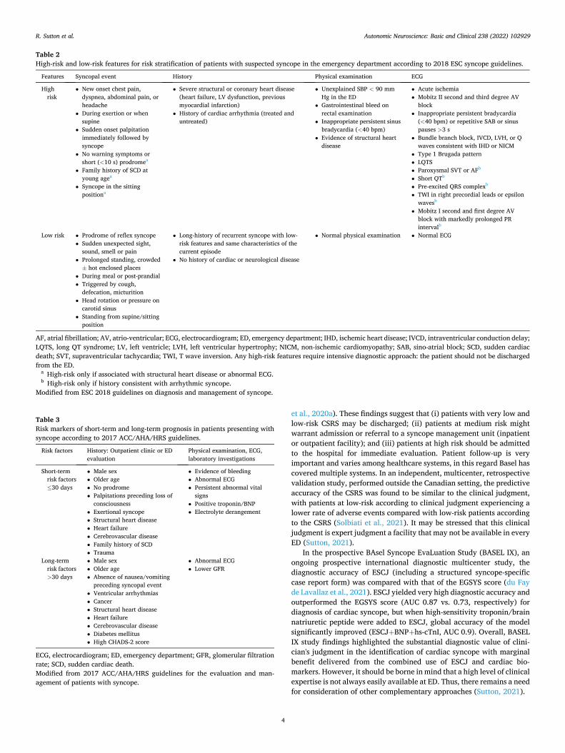
management (Wieling et al., 2016). The 2018 ESC guidelines for the diagnosis and management of syn-
cope delivered a list of low- and high-risk features (Table 2) aimed to provide quick separation of patients with a likely low-risk condition, who may benefit from reassurance and early discharge, from patients with clinical findings or previous history suggestive of cardiovascular disease, who may need intensive diagnostic approach, urgent treatment and hospital admission (Brignole et al., 2018). Intermediate risk patients (i.e. neither high- nor low-risk patients) require a strategy that hinges on the availability of specialist syncope care, more formal syncope outpa-tient clinic/unit or syncope expertise. Admission to an observational unit when the syncope presentation and patient's risk profile suggest increased likelihood of early complications (Fig. 1). Guidelines (Brignole et al., 2018; Shen et al., 2017), the SEEDS study (Shen et al., 2004) and work of Sun et al. (2014a) are available to guide duration of monitoring, 6–24 h covers present practice. To summarize the European syncope guidelines, patients with traditional clinical markers of worse prognosis such as multiple comorbidities, hypotension, and anemia, and those with underlying cardiovascular conditions should be treated as high-risk patients, which favors admission for rapid evaluation.
In contrast, the 2017 ACC (American College of Cardiology)/AHA (American Heart Association)/HRS guidelines for the evaluation and management of patients with syncope focus on short-term (<30 days) and long-term (>30 days) high-risk predictors based on patient's history and initial examination. These clinical high-risk predictors overlap be-tween the two groups, in particular regarding presence of heart disease, abnormal ECG, and absence of prodrome (Table 3) (Shen et al., 2017), and are similar to those found in European guidelines.
Additional criteria requiring inpatient evaluation for actionable diagnostic and therapeutic pathways are listed in Table 4 (Shen et al., 2017).
After initial history, physical examination, and baseline ECG, the selection of appropriate diagnostic tests is also determined by risk stratification. Diagnostic imaging (such as computer tomography
Fig. 1. Risk stratification and clinical management of patients with suspected syncope in the Emergency Department. High-risk features are detailed in Tables 2–3. Patients who do not meet criteria of either high- or low-risk are classified as intermediate risk and should be further investigated by specialized units or experts. Low-risk patients should be instructed to contact their healthcare providers in case or syncope recurrence for further assistance. BP, blood pressure; ECG, electrocardiogram; ED, emergency department; OH, orthostatic hypotension; T-LOC, transient loss of consciousness.
R. Sutton et al.

Autonomic Neuroscience: Basic and Clinical 238 (2022) 102929
3
angiography or echocardiography) and laboratory tests (such as routine blood hematology, biochemistry) have low diagnostic yield and should not be routinely used for syncope assessment. Cardiac damage bio-markers such as troponin and brain natriuretic peptide (BNP) have proven valuable in emergency presentations offering help to busy emergency physicians to select those patients requiring hospitalization (Clark et al., 2019; du Fay de Lavallaz et al., 2019). Some still frequently employed testing should now be considered inappropriate due to lack of diagnostic yield in syncope patients, notably EEG and brain imaging. Further diagnostic tests are usually not recommended in patients with uncertain diagnosis but low-risk profile and single or rare recurrences.
In patients that have intermediate-risk, or presenting with low-risk features, but recurrent syncopal episodes, cardiovascular autonomic tests (including carotid sinus massage, orthostatic challenge, head-up tilt testing, Valsalva maneuver, deep breathing and 24-hour arterial blood pressure monitoring) and prolonged ECG monitoring (external or implantable) should be considered.
For high-risk patients, in whom an underlying cardiac cause of syncope is likely, the primary aim is to establish a diagnosis, especially those associated with potential for rapid clinical deterioration, where there is evidence of structural cardiovascular disease. In this patient population, immediate in-hospital ECG monitoring, echocardiography, computed tomography, cardiac magnetic resonance, exercise stress test, electrophysiological study, or angiography can be indicated according to the specific clinical scenario.
3. Syncope prediction tools
Several syncope prediction tools - including the Martin-Kapoor score (Martin et al., 1997), the San Francesco Syncope Rule (SFSR) (Quinn et al., 2006), the Osservatorio Epidemiologico sulla Sincope nel Lazio (OESIL) score (Colivicchi et al., 2003), the Risk Stratification of Syncope in the Emergency Department (ROSE) score (Reed et al., 2010), the Evaluation of Guidelines in Syncope Study (EGSYS) score (Del Rosso et al., 2008), the Boston Syncope Criteria (Grossman et al., 2007), the Short-Term Prognosis of Syncope (STePS) score (Costantino et al., 2008), the FAINT score (Probst et al., 2020) and the Basel IX ECG ALERT-CS tool (Zimmermann et al., 2021) - using various combinations of different clinical parameters derived from ECG, history, physical ex-amination and serum biomarkers, have been developed over the past 20 years to refine patient stratification, estimate prognosis, determine pa-tient disposition and distinguish who needs expert in-hospital care from those who may receive nonurgent expert care in the community (Table 5) (Sutton, 2021; Sarasin et al., 2003; Sun et al., 2009).
Regrettably, lack of reproducibility and remarkable heterogeneity in study design, variables, and outcome definitions of primary studies prevented widespread use of these tools in clinical practice (Sheldon et al., 2011; Costantino et al., 2014). Current European and American guidelines delivered weak recommendation (class of recommendation IIb, level of evidence B) for routine use of risk stratification scores in ED. Prediction tools have suffered limited external validity, performed no better than good clinical judgment, have not reduced inappropriate admission, and should not be used alone to perform risk stratification in the ED (Brignole et al., 2018; Shen et al., 2017; Costantino et al., 2014). Overall, the proposed schemes have not been sufficiently discriminating, and there has been a strong consensus that risk stratification scores yield poor prognostic value compared with unstructured clinical judgment in predicting short-term and long-term serious adverse outcomes after syncope (Shen et al., 2017; Sutton et al., 2012).
More recently, the Canadian Syncope Risk Score (CSRS) system and a structured early standardized clinical judgment (ESCJ) have shown ef-ficacy in large studies ED-based.
The CSRS is a scoring system obtained after disposition from ED, assigning positive values where clinical data indicate a serious condition and negative in cases where less serious outcomes are expected, which has been shown to predict 30-day composite outcome (including death, arrhythmia, myocardial infarction, serious structural heart disease, aortic dissection, pulmonary embolism, severe pulmonary hypertension, severe hemorrhage, any other serious condition causing syncope and procedural interventions for the treatment of syncope) with a high de-gree of discrimination, calibration, and accuracy (Thir-uganasambandamoorthy et al., 2016) (Table 6). The performance of the original prediction tool was later successfully validated in a prospective multicenter external Canadian cohort, confirming a robust risk classifi-cation with less than 1% very low or low-risk patients, approximately 20% of high-risk patients, and 50% of very-high-risk CSRS patients experiencing 30-day serious outcomes (Thiruganasambandamoorthy
Table 1 Clinical characteristics associated with increased probability of cardiac and noncardiac causes of syncope
More often associated with cardiac causes of syncope
More often associated with noncardiac causes of syncope
2017 ACC/ AHA/ HRS US
• Older age (>60 years) • Male sex • Presence of known ischemic
heart disease, structural heart disease, previous arrhythmias, or reduced ventricular function
• Brief prodrome, such as palpitations, or sudden loss of consciousness without prodrome
• Syncope during exertion • Syncope in the supine position • Low number of syncope
episodes (1 or 2) • Abnormal cardiac examination • Family history of inheritable
conditions or premature SCD (<50 years of age)
• Known congenital heart disease
• Younger age • No known cardiac disease • Syncope only in the standing
position • Positional change from supine
or sitting to standing • Presence of prodrome: nausea,
vomiting, feeling warmth • Presence of specific triggers:
dehydration, pain, distressful stimulus, medical environment
• Situational triggers: cough, laugh, micturition, defecation, deglutition
• Frequent recurrence and prolonged history of syncope with similar characteristics
• Many episodes over long period
2018 ESC EU
• During exertion or when supine • Sudden onset palpitation
immediately followed by syncope
• Family history of unexplained sudden death at young age
• Presence of structural heart disease or coronary artery disease
ECG findings suggesting arrhythmic syncope
• Bifascicular block (defined as either left or right BBB combine with left anterior or left posterior fascicular block)
• Other intraventricular conduction abnormalities (QRS duration ≥0.12 s)
• Mobitz I second-degree ann first degree AV block with markedly prolonged PR interval
• Asymptomatic mild inappropriate sinus bradycardia (40–50 bpm) or slow atrial fibrillation (40–50 bpm) in the absence of negatively chronotropic medications
• Non-sustained VT • Pre-excited QRS complexes • Long or short QT intervals • Early repolarization • ST-segment elevation with type
1 morphology in lead V1-V3 • Negative T waves in right
precordial leads, epsilon wave • Left ventricular hypertrophy
suggesting hypertrophic cardiomyopathy
Reflex syncope
• Long history of recurrent syncope, in particular occurring before the age of 40 years
• After unpleasant sight, sound, smell, or pain
• Prolonged standing • During meal • Being in crowded and/or hot
places • Autonomic activation before
syncope: pallor, sweating, and/ or nausea/vomiting
• With head rotation or pressure on carotid sinus (as in tumours, shaving, tight collars)
• Absence of heart disease Syncope due to OH
• While or after standing • Prolonged standing • Standing after exertion • Post-prandial hypotension • Temporal relationship with
start or titration of vasodepressive drugs or diuretics leading to hypotension
• Presence of autonomic neuropathy or parkinsonism
R. Sutton et al.
Autonomic Neuroscience: Basic and Clinical 238 (2022) 102929
4
et al., 2020a). These findings suggest that (i) patients with very low and low-risk CSRS may be discharged; (ii) patients at medium risk might warrant admission or referral to a syncope management unit (inpatient or outpatient facility); and (iii) patients at high risk should be admitted to the hospital for immediate evaluation. Patient follow-up is very important and varies among healthcare systems, in this regard Basel has covered multiple systems. In an independent, multicenter, retrospective validation study, performed outside the Canadian setting, the predictive accuracy of the CSRS was found to be similar to the clinical judgment, with patients at low-risk according to clinical judgment experiencing a lower rate of adverse events compared with low-risk patients according to the CSRS (Solbiati et al., 2021). It may be stressed that this clinical judgment is expert judgment a facility that may not be available in every ED (Sutton, 2021).
In the prospective BAsel Syncope EvaLuation Study (BASEL IX), an ongoing prospective international diagnostic multicenter study, the diagnostic accuracy of ESCJ (including a structured syncope-specific case report form) was compared with that of the EGSYS score (du Fay de Lavallaz et al., 2021). ESCJ yielded very high diagnostic accuracy and outperformed the EGSYS score (AUC 0.87 vs. 0.73, respectively) for diagnosis of cardiac syncope, but when high-sensitivity troponin/brain natriuretic peptide were added to ESCJ, global accuracy of the model significantly improved (ESCJ+BNP+hs-cTnI, AUC 0.9). Overall, BASEL IX study findings highlighted the substantial diagnostic value of clini-cian's judgment in the identification of cardiac syncope with marginal benefit delivered from the combined use of ESCJ and cardiac bio-markers. However, it should be borne in mind that a high level of clinical expertise is not always easily available at ED. Thus, there remains a need for consideration of other complementary approaches (Sutton, 2021).
Table 2 High-risk and low-risk features for risk stratification of patients with suspected syncope in the emergency department according to 2018 ESC syncope guidelines.
Features Syncopal event History Physical examination ECG
High risk
• New onset chest pain, dyspnea, abdominal pain, or headache
• During exertion or when supine
• Sudden onset palpitation immediately followed by syncope
• No warning symptoms or short (<10 s) prodromea
• Family history of SCD at young agea
• Syncope in the sitting positiona
• Severe structural or coronary heart disease (heart failure, LV dysfunction, previous myocardial infarction)
• History of cardiac arrhythmia (treated and untreated)
• Unexplained SBP < 90 mm Hg in the ED
• Gastrointestinal bleed on rectal examination
• Inappropriate persistent sinus bradycardia (<40 bpm)
• Evidence of structural heart disease
• Acute ischemia • Mobitz II second and third degree AV
block • Inappropriate persistent bradycardia
(<40 bpm) or repetitive SAB or sinus pauses >3 s
• Bundle branch block, IVCD, LVH, or Q waves consistent with IHD or NICM
• Type 1 Brugada pattern • LQTS • Paroxysmal SVT or AFb
• Short QTb
• Pre-excited QRS complexb
• TWI in right precordial leads or epsilon wavesb
• Mobitz I second and first degree AV block with markedly prolonged PR intervalb
Low risk • Prodrome of reflex syncope • Sudden unexpected sight,
sound, smell or pain • Prolonged standing, crowded
± hot enclosed places • During meal or post-prandial • Triggered by cough,
defecation, micturition • Head rotation or pressure on
carotid sinus • Standing from supine/sitting
position
• Long-history of recurrent syncope with low- risk features and same characteristics of the current episode
• No history of cardiac or neurological disease
• Normal physical examination • Normal ECG
AF, atrial fibrillation; AV, atrio-ventricular; ECG, electrocardiogram; ED, emergency department; IHD, ischemic heart disease; IVCD, intraventricular conduction delay; LQTS, long QT syndrome; LV, left ventricle; LVH, left ventricular hypertrophy; NICM, non-ischemic cardiomyopathy; SAB, sino-atrial block; SCD, sudden cardiac death; SVT, supraventricular tachycardia; TWI, T wave inversion. Any high-risk features require intensive diagnostic approach: the patient should not be discharged from the ED.
a High-risk only if associated with structural heart disease or abnormal ECG. b High-risk only if history consistent with arrhythmic syncope.
Modified from ESC 2018 guidelines on diagnosis and management of syncope.
Table 3 Risk markers of short-term and long-term prognosis in patients presenting with syncope according to 2017 ACC/AHA/HRS guidelines.
Risk factors History: Outpatient clinic or ED evaluation
Physical examination, ECG, laboratory investigations
Short-term risk factors ≤30 days
• Male sex • Older age • No prodrome • Palpitations preceding loss of
consciousness • Exertional syncope • Structural heart disease • Heart failure • Cerebrovascular disease • Family history of SCD • Trauma
• Evidence of bleeding • Abnormal ECG • Persistent abnormal vital
signs • Positive troponin/BNP • Electrolyte derangement
Long-term risk factors >30 days
• Male sex • Older age • Absence of nausea/vomiting
preceding syncopal event • Ventricular arrhythmias • Cancer • Structural heart disease • Heart failure • Cerebrovascular disease • Diabetes mellitus • High CHADS-2 score
• Abnormal ECG • Lower GFR
ECG, electrocardiogram; ED, emergency department; GFR, glomerular filtration rate; SCD, sudden cardiac death. Modified from 2017 ACC/AHA/HRS guidelines for the evaluation and man-agement of patients with syncope.
R. Sutton et al.

Autonomic Neuroscience: Basic and Clinical 238 (2022) 102929
5
Despite promising results, prognostic tools for syncope remain challenging and can be inefficient. Firstly, the prognosis of patients presenting with syncope is related to the underlying disease, its severity and progression, and the effectiveness of specific therapy. Secondly, available clinical decision rules are based on mean values obtained from heterogeneous patient populations, whereas, in clinical practice, indi-vidually tailored decisions are most needed (Sun et al., 2014b). Thirdly, previous studies focused on a wide range of composite endpoints, but the inconsistent selection of outcomes limits the biological plausibility of identified predictors. For example, predictors of cardiac arrhythmia, pulmonary embolism, stroke, and occult gastrointestinal bleeding are likely to be different and difficult to fit into one scale. Therefore, future research should pertinently target clinically relevant and coherent out-comes (Sun et al., 2014b). Finally, as fatal and severe syncope-related adverse events are rare, enrollment of large patient cohorts is key to ensure powered, adequately calibrated, and stable risk-prediction models.
4. The role of syncope clinics
While discussing the initial risk stratification in syncope, we must also consider the organization of post-stratification workup, an inevi-table part of the process. The follow-up of syncope patients strongly depends on the identification of underlying pathology. If the patient has been diagnosed at initial presentation, specific treatment should be offered under supervision of an appropriate expert, e.g. an electro-physiologist for arrhythmias. Unexplained syncope or syncope with a difficult to treat manifestation - e.g. recurrent vasovagal attacks or se-vere orthostatic intolerance - should be referred to an expert with necessary syncope management skills. In this context, physical or virtual syncope units have been promoted by guidelines, only briefly summa-rized here (Brignole et al., 2018). A syncope unit (SU) is a facility featuring a standardized approach to diagnosis and management of
TLOC and related symptoms, with dedicated staff and access to appro-priate diagnostics and therapeutic pathways (Kenny et al., 2015).
Over the last two decades, several models of SUs have been tested to (i) standardize the approach to syncope patients: (ii) improve diagnostic yield and both clinical and cost-effectiveness of syncope management; (iii) reduce the length of stay and number of hospital admissions; (iv) ensure optimal allocation of diagnostic resources, reducing the number of expensive, inconsistent and unnecessary tests (Sun et al., 2014a; Colivicchi et al., 2003; Kenny et al., 2015; Walsh et al., 2015; Kenny et al., 2002; Brignole et al., 2003; Brignole et al., 2010; Sanders et al., 2013; Brignole et al., 2006a; Brignole et al., 2006b; Petkar et al., 2011; Rodriguez-Entem et al., 2008; Fedorowski et al., 2010).
Randomized data from the Syncope Evaluation in the Emergency Department Study (SEEDS) suggested that designated ED-based syncope units for evaluation of patients with intermediate-risk profiles with standardized protocols for ruling-out cardiac causes of syncope can reduce hospital admission rate, length of stay, and costs without compromising the quality of care (Shen et al., 2004). In the prospective, multicenter, EGSYS-2, a standardized method of syncope management based on a decision-making approach (standardized-care group), rather than a strategy based on generic implementation of guidelines (usual- care group), significantly improved overall diagnostic yield, reduced hospital admissions, resource consumption, and overall associated costs (Brignole et al., 2006a).
Despite established benefits yielded from syncope clinics (Kenny et al., 2015) and endorsement of guidelines for their implementation (Brignole et al., 2018), SUs are yet not widely established due to important barriers including limited awareness of benefit, lack of syn-cope specialists and formal syncope training programs, need for multi-disciplinary expertise, necessity to engage multiple stakeholders, inadequate reimbursement, fear of increasing costs associated with development of a new structure and lack of large clinical studies demonstrating their superiority vs. conventional management (Kenny et al., 2015).
This issue is discussed in detail in the next paper in the current article collection.
Here, we would like to emphasize that SUs offer education on pre-vention of reflex syncope, management of OH, falls and traumatic in-juries related to both these conditions, antihypertensive medication optimization, prescription of blood pressure elevating drugs, as well as rapid access to more advanced procedures such as cardiac implantable devices, echocardiography, cardiac imaging, electrophysiological study, stress test, and specialist consultations (e.g. neurological, psychiatric/ psychological, pediatric, or geriatric), if appropriate.
5. Risk stratification for tailored treatment of syncope
The framework of treatment is risk stratification and identification of specific syncope mechanisms - rather than etiology or clinical presen-tation - to select therapy. Reflex syncope and orthostatic hypotension are the most frequent causes of T-LOC when cardiac syncope is ruled-out. In this setting, age, prodrome, BP, use of hypotensive drugs, contribution of vasodepression and cardioinhibition are helpful in tailoring treatment (Brignole and Rivasi, 2021). All patients require education, lifestyle measures - avoidance of triggers and counter-pressure maneuvers - and expansion of intravascular volume - adequate fluid and salt intake are important and effective (Brignole et al., 2002; van Dijk et al., 2006; Krediet et al., 2002). In hypertensive patients, reduction or cessation of antihypertensive medication can be safely recommended (Moonen et al., 2016; van der Wardt et al., 2017), with systolic BP targets tailored ac-cording to age, cardiovascular and hypotensive risk, frailty, and disability status (Rivasi et al., 2020). In patients with recurrent noncardiac syncope and important vasodepression, treatment with flu-drocortisone, midodrine, or droxidopa can be considered. In cases of cardioinhibition atomoxetine may be helpful in the future (Sheldon et al., 2019a).
Table 4 Medical conditions requiring hospital admission for further evaluation and therapy.
Medical conditions requiring hospital admission for diagnosis or treatment
Arrhythmias • Sustained or symptomatic ventricular arrhythmias • Symptomatic conduction system disease • Symptomatic bradycardia or sinus pauses not related to
neurally mediated syncope • Symptomatic SVT • Pacemaker/ICD malfunctiona
• Inheritable arrhythmogenic cardiovascular conditions Cardiovascular
disorders • Myocardial ischemia • Severe aortic stenosis • Pericardium tamponade • HCM • Prosthetic valve dysfunction • Pulmonary embolism • Aortic dissection • Acute heart failure • Moderate-to-severe LV dysfunction • Left atrial myxoma/thrombus • Need for urgent evaluation and treatment if it cannot be
achieved in another way (i.e. observation unit), e.g. ECG monitoring, echocardiography, stress test, electrophysiological study, cardiac magnetic resonance imaging, CT or invasive coronary angiography
Non-cardiac conditions
• Severe anemia/gastrointestinal bleeding • Major traumatic injury due to syncope • Persistent vital sign abnormalities
CT, computed tomography; ECG, electrocardiogram; ED, emergency depart-ment; HCM, hypertrophic cardiomyopathy; ICD, implantable cardioverter defi-brillator; LV, left ventricle; SVT, supraventricular tachycardia. Modified from 2017 ACC/AHA/HRS guidelines for the evaluation and management of patients with syncope.
a To reduce inappropriate admissions, patients who have a cardiac device and syncope should undergo prompt device interrogation.
R. Sutton et al.

Autonomic Neuroscience: Basic and Clinical 238 (2022) 102929
6
Noncardiac syncope with cardioinhibition including asystole (>3 s pause or >6 s asymptomatic pause) is recorded during a spontaneous event, induced by carotid sinus massage or during head-up tilt testing points to consideration of cardiac pacing, which is the only therapy of proven efficacy for the dominant bradycardic phenotype and may be expected to reduce syncopal recurrences in patients aged >40 years (Brignole et al., 2016; Brignole et al., 2015; Brignole et al., 2012). In paced patients with recurrent syncope and a positive tilt test, specific treatment for hypotensive susceptibility should be offered in addition to cardiac pacing (Brignole et al., 2014; Yasa et al., 2019a; Yasa et al., 2019b). Recently, in patients aged ≥40 years with severe recurrent re-flex syncope and tilt-induced asystole, the BioSync trial showed that a dual-chamber pacemaker with closed-loop system is highly effective in reducing syncopal recurrence (Brignole et al., 2021a). Thus, pacing must now be held a tenable option for cardioinhibitory vasovagal syncope (Baron-Esquivias et al., 2019) and tilt testing has to be recognized as a means of candidate selection for cardiac pacing (Sutton et al., 2021; Brignole et al., 2021b), possibly without need for ILR confirmation of diagnosis. Given the very high spontaneous remission rate over the medium term in patients presenting frequent reflex syncope, either due to regression to the mean or placebo effect, close surveillance for a period is reasonable before any major management decision (Pournazari et al., 2017; Sutton, 2017).
In structural heart disease and primary electrical disorders, syncope is usually of great concern, and there is consensus on the therapeutic goal being not only to prevent syncopal recurrence but also to treat the underlying disease and reduce mortality (Probst and Gourraud, 2018). In patients at high risk of sudden cardiac death when the mechanism of syncope is non-arrhythmic, management is the same as for patients without syncope. In the presence of clinical features of structural car-diovascular disorders or primary electrical diseases, unexplained syn-cope - defined as syncope that does not meet any class I diagnostic criterion of ESC Guidelines - should always be considered as suspected arrhythmic syncope. It is therefore particularly important to focus on determining whether the syncope is related to life-threatening arrhythmia or it is noncardiac syncope will define the patient's man-agement, with an ICD or completion of investigation with prolonged ECG monitoring or genetic testing (e.g. for long QT syndrome) in selected cases (Priori et al., 2015; Appignani et al., 2021).
6. Future of syncope evaluation and risk stratification
6.1. Biomarkers
The role of cardiac biomarkers for triage of cardiac syncope is still under investigation. Biomarkers reflecting myocardial injury, such as
Table 5 Syncope prediction tools.
Risk score, author, year Sample size
Risk factors Outcome Validation
Risk stratification of syncope (Martin et al., 1997)
252 Age > 45 years, ECG, history of VAs, HF 1-year severe arrhythmias or arrhythmic death
No external validation
OESIL risk score (Colivicchi et al., 2003) 270 Abnormal ECG, age >65 years, history of CVD, no prodromes
1-year mortality Prognostic yield no better than clinical judgment (IPD meta- analysis)a
Risk score to predict arrhythmias (Sarasin et al., 2003)
175 Age >65 years, history of HF, abnormal ECG Arrhythmias No external validation
San Francisco Syncope Rule (Quinn et al., 2006)
684 Abnormal ECG, heart failure, shortness of breath, hematocrit <30%, triage SBP < 90 mm Hg
7-day serious events Limited generalizability in external validation studies
Boston Syncope Rule (Grossman et al., 2007)
293 ACS, conduction disease, cardiac history, VHD, family history of SD, abnormal vital signs, volume depletion, primary CNS event
30-day critical intervention or adverse outcome
EGSYS score (Del Rosso et al., 2008) 260 ECG, Hx of heart disease, palpitations preceding syncope, syncope during effort o while supine, precipitating and/or predisposing factors, autonomic prodromes
2-year mortality Cardiac syncope probability
Externally validated, but prognostic yield no better than clinical judgment (IPD meta- analysis)a
Short-Term Prognosis of Syncope (STePS) (Costantino et al., 2008)
670 Short term: ECG, trauma, no prodromes, male sex Long term: age >65 years, neoplasms, stroke, VAs, SHD
Death, need for major procedures, and early readmission to the hospital, at 10 days and 1 year
No external validation
Syncope Risk Score (Sun et al., 2009) 2584 Age >90 years, male sex, history of arrhythmia, triage SBP > 160 mm Hg, abnormal ECG, abnormal troponin I, near-syncope
30-day serious events Prognostic yield no better than clinical judgment (IPD meta- analysis)a
ROSE (Reed et al., 2010) 550 ECG, BNP ≥300, HR ≤50 bpm, Hb ≤ 9 g/dL, chest pain, O2sat ≤94%, stool positivity for occult blood test
30-day serious events No external validation
Canadian Syncope Risk Score (Thiruganasambandamoorthy et al., 2016)
4030 Predisposition VVS symptoms, history of heart disease SBP, elevated troponin, ECG features, ED diagnosis of VVS or cardiac syncope
30-day serious events Multicenter external validation but limited generalizability outside Canada
Basel IX ECG ALERT-CS tool (Zimmermann et al., 2021)
2007 HR and QTc-interval (continuous predictors), rhythm, atrioventricular block, ST-segment depression, bundle branch block and ventricular extrasystole/non-sustained ventricular tachycardia
Cardiac syncope probability Independent external validation
FAINT score (Probst et al., 2020) 3177 History of HF, history of cardiac arrhythmia, abnormal ECG, elevated pro BNP, and elevated hscTn T
30-day serious events No external validation
Early Standardized Clinical Judgement (ESCJ) (du Fay de Lavallaz et al., 2021)
1494 Compilation of standardized syncope-specific CRF ± troponin ± BNP
1-year MACEs Cardiac syncope probability
Diagnostic accuracy for cardiac syncope higher than EGSYS score, but no external validation
BNP, brain natriuretic peptide; CVD, cardiovascular disease; ECG, electrocardiogram; ED, emergency department; Hb, hemoglobin; HF, heart failure; HR, heart rate; IPD, individual patient data; MACEs, major adverse cardiovascular events; SBP, systolic blood pressure; SD, sudden death; SHD, structural heart disease; VAs, ven-tricular arrhythmias; VVS, vasovagal syncope.
a Costantino et al., 2014.
R. Sutton et al.

Autonomic Neuroscience: Basic and Clinical 238 (2022) 102929
7
cardiac troponin, heart failure, such as B-type natriuretic peptides, and other different neuroendocrine pathways possibly involved in the pathophysiology of cardiac syncope, such as midregional–pro-A-type natriuretic peptide (MRproANP), C-terminal proendothelin 1, copeptin, and midregional-proadrenomedullin, have been proposed for under-standing underlying mechanisms, diagnosis and tailoring therapy of cardiac syncope, but with controversial results (Badertscher et al., 2017; Thiruganasambandamoorthy et al., 2015; Pfister et al., 2009; Fedor-owski et al., 2013).
In the multicenter BASEL IX study, the diagnostic accuracy of MRproANP for cardiac syncope was high and provided significantly incremental diagnostic value over clinical judgment and EGSYS score among unselected patients presenting to ED with syncope. An algorithm based on the combination of MRproANP and clinical judgment yielded a sensitivity of 99% and a negative predictive value of 99% for early rule- out of cardiac syncope (Badertscher et al., 2017).
Further data from the BASEL IX study showed that serum BNP, NT- proBNP, hs-cTnT, and hs-cTnI concentrations were significantly higher in cardiac syncope and yielded moderate-to-high accuracy for the diagnosis of cardiac syncope (du Fay de Lavallaz et al., 2019). Notably, the prognostic accuracy of BNP, NT-proBNP, hs-cTnI, and hs-cTnT for MACE was moderate-to-good (AUC, 0.75–0.79), superior to ROSE, OESIL, and SFSR scores, but significantly lower than CSRS. The latter outperformed cardiac biomarkers alone and combined with other outcome prediction tools.
In another ED-based multicenter cohort from Canada, serum NT- proBNP levels - although generally much higher among ED patients with syncope who had a 30-day adverse serious event - did not improve the prognostic accuracy of CSRS (Thiruganasambandamoorthy et al., 2020b). Consequently, further research is needed to determine how cardiac and other biomarkers should be incorporated into a risk strati-fication algorithm and to understand whether this will lead to more efficient and cost-effective syncope healthcare delivery (Sandhu and Sheldon, 2019). It is important to keep in mind that physicians in a busy ED setting will prefer tests that are quickly performed, easy to interpret, and have important incremental value for risk stratification and clinical decision-making.
6.2. Genetic testing
The genetic basis of vasovagal syncope is discussed in detail in another article in the current collection. Briefly, family pedigree studies, twin studies, genome-wide association studies, and gene duplicate studies indicate loci in the genome that associate with reflex syncope, although precise genes and proteins remain undetermined (Sheldon et al., 2019b; Fedorowski et al., 2021). Our understanding of the ge-netics underpinning hypotensive susceptibility, reflex syncope (Sheldon and Sandhu, 2019) and orthostatic intolerance syndromes (Fedorowski et al., 2012) is in an early phase, with much yet to be explored and, thus, cannot be applied in risk stratification.
Concerning inheritable arrhythmogenic diseases, the availability of genetic information can be used for diagnostic purposes and for guiding risk stratification in a few diseases, such as long QT syndrome (Schwartz et al., 2020), lamin A/C cardiomyopathy (Priori et al., 2015), cate-cholaminergic polymorphic ventricular tachycardia (Ackerman et al., 2011; Priori et al., 2013), progressive cardiac conduction system disease (Priori et al., 2013; Gray and Behr, 2016) and arrhythmogenic cardio-myopathy (Corrado et al., 2020). The next-generation sequencing era has evolved rapidly, providing challenges including the definition of pathogenicity, identification of background genetic noise, increased detection of variants of uncertain significance but new opportunities, including the discovery of novel entities such as calmodulinopathies or Triadin knockout syndrome (Gray and Behr, 2016).
6.3. Harnessing artificial intelligence for syncope management
Artificial intelligence (AI) research is rapidly increasing in clinical medicine. AI-based interventions can match or even outperform physi-cians' skills in predictive modeling because of the ability to process multiple variables simultaneously across large datasets by adequately trained and validated machine learning (ML) algorithms (D'Ascenzo et al., 2021). AI solutions have shown potential to improve patient care, reduce the frequency of adverse events (Bates et al., 2021), decrease the rate of hospital admission and costs of inappropriate treatments and hospitalization, overall ensuring more effective and equitable use of resources (Romero-Brufau et al., 2020). There are good opportunities to implement successfully alternative prediction tools based on artificial neural networks that can be leveraged to improve the risk stratification of syncope patients in the ED. Neural networks can outperform the classic models since they adapt to the initial data sample, showing greater generalizability. At present, only very few studies have analyzed the application of AI tools to syncope detection and risk prediction, despite preliminary encouraging results. Falavigna et al. realized an innovative ANN model predicting hospitalization with a sensitivity of 100% and a specificity of 79% and outperforming OESIL and SFSR scores in a cohort of 1844 subjects presenting with syncope to the ED, eventually opening a new era for technology-enabled innovative solu-tions to customize and improve risk stratification of syncope patients (Falavigna et al., 2019). Based on the same data used to derive and validate the CSRS, Grant et al. produced four competing ML models to predict 30-day serious adverse events after ED disposition and observed that the ML modeling matched the predictive performance of the CSRS, while using fewer predictors (Grant et al., 2021).
Natural language processing (NLP) techniques have also been recently used to identify syncopal episodes in ED medical records with high sensitivity and acceptable positive predictive value, resulting in a 96% reduction of working time needed for manual identification (Dipaola et al., 2019). NLP techniques can be leveraged in the future to collect a huge amount of data suitable for large-scale analysis and to build AI-based robust and accurate predictive models to support clinical decision-making.
Table 6 Canadian Syncope Risk Score.
Category Risk factors Points Total score (− 3 to 11)
Risk
Clinical assessment Predisposition to VVSa
Prevalent CVD SBP < 90 or >180 mm Hg
− 1 1 2
− 3 − 2
Very low
− 1 0
Low
Investigations (troponin, ECG)
Elevated troponin QRS axis ≤30 or >100◦
QRS duration >130 ms QTc interval > 480 ms
2 1 1 2
1 2 3
Medium
ED diagnosis Vasovagal syncope Cardiac syncope
− 2 2
4 5
High
≥6 Very high
The Canadian Syncope Risk Score is used to identify patients with syncope at risk of serious adverse events within 30 days after disposition from the emergency department. The score is obtained by adding the points of each risk factor. SBP: systolic blood pressure; VVS: vasovagal syncope, ED: emergency department, CVD: history of cardiovascular disease.
a Triggered by warm, crowded place, prolonged standing, fear, emotion, or pain. Modified from Thiruganasambandamoorthy et al. (2016).
R. Sutton et al.

Autonomic Neuroscience: Basic and Clinical 238 (2022) 102929
8
AI-enabled ECG acquired during normal sinus rhythm has been shown to identify individuals with a high likelihood of atrial fibrillation using a convolutional neural network (Attia et al., 2019) and hidden disease state signatures by unsupervised deep neural networks algo-rithms (Siontis et al., 2021). Similarly, QRS complex shape features predicted the occurrence of ventricular fibrillation with high accuracy using an artificial neural network (Taye et al., 2019). This evidence supports the hypothesis that subtle patterns on the normal sinus rhythm ECG can be captured with AI techniques and predict risk of future arrhythmic events or structural heart disease (Attia et al., 2021; Cohen- Shelly et al., 2021). Further research is needed to test the ability of AI- enhanced ECG interpretation to identify early markers of arrhythmic vulnerability or heart disease. Overall, AI and ML methods appear promising tools for risk-stratification of syncope, but the added benefit over traditional statistical methods remains unproven.
7. Conclusions
Syncope is a diagnostic challenge. Initial risk stratification aims to identify high-risk patients that require hospitalization, intermediate-risk patients that should be further investigated and treated to prevent syncope recurrences, and low-risk patients that need reassurance, edu-cation on avoidance of triggers and employment of physical counter- measures, improved hydration and salt supplementation. In the Emer-gency Department, progress has already been made regarding stratifi-cation algorithms and risk scores, but there is no consensus for the latter, and clinical judgment is still the cornerstone of syncope management. In diagnosis and therapy, Syncope Units add refinement of diagnosis and management benefits. There is evidence of improved management of all syncope patients offering optimism for the future of syncope care.
References
Ackerman, M.J., Priori, S.G., Willems, S., Berul, C., Brugada, R., Calkins, H., et al., 2011. HRS/EHRA expert consensus statement on the state of genetic testing for the channelopathies and cardiomyopathies: this document was developed as a partnership between the Heart Rhythm Society (HRS) and the european heart rhythm association (EHRA). Europace 13 (8), 1077–1109.
Appignani, M., Khanji, M.Y., Arbustini, E., Stuppia, L., Ceriello, L., Girolamo, E.D., et al., 2021. Is occult genetic substrate the missing link between arrhythmic mitral annular disjunction syndrome and sudden cardiac death? Can. J. Cardiol. 37 (10), 1651–1653. https://doi.org/10.1016/j.cjca.2021.04.014.
Attia, Z.I., Noseworthy, P.A., Lopez-Jimenez, F., Asirvatham, S.J., Deshmukh, A.J., Gersh, B.J., et al., 2019. An artificial intelligence-enabled ECG algorithm for the identification of patients with atrial fibrillation during sinus rhythm: a retrospective analysis of outcome prediction. Lancet 394 (10201), 861–867.
Attia, Z.I., Harmon, D.M., Behr, E.R., Friedman, P.A., 2021. Application of artificial intelligence to the electrocardiogram. Eur. Heart J. 42 (46), 4717–4730. https://doi. org/10.1093/eurheartj/ehab649.
Badertscher, P., Nestelberger, T., de Lavallaz, J.D.F., Than, M., Morawiec, B., Kawecki, D., et al., 2017. Prohormones in the early diagnosis of cardiac syncope. J. Am. Heart Assoc. 6 (12).
Baron-Esquivias, G., Baron-Solis, C., Ordonez, A., 2019. Pacing for patients suffering from cardioinhibitory vasovagal syncope using the closed-loop system. Front. Cardiovasc. Med. 6, 192.
Bates, D.W., Levine, D., Syrowatka, A., Kuznetsova, M., Craig, K.J.T., Rui, A., et al., 2021. The potential of artificial intelligence to improve patient safety: a scoping review. NPJ Digit Med. 4 (1), 54.
Brignole, M., Rivasi, G., 2021. New insights in diagnostics and therapies in syncope: a novel approach to non-cardiac syncope. Heart 107 (11), 864–873.
Brignole, M., Croci, F., Menozzi, C., Solano, A., Donateo, P., Oddone, D., et al., 2002. Isometric arm counter-pressure maneuvers to abort impending vasovagal syncope. J. Am. Coll. Cardiol. 40 (11), 2053–2059.
Brignole, M., Disertori, M., Menozzi, C., Raviele, A., Alboni, P., Pitzalis, M.V., et al., 2003. Management of syncope referred urgently to general hospitals with and without syncope units. Europace 5 (3), 293–298.
Brignole, M., Ungar, A., Bartoletti, A., Ponassi, I., Lagi, A., Mussi, C., et al., 2006. Standardized-care pathway vs. Usual management of syncope patients presenting as emergencies at general hospitals. Europace 8 (8), 644–650.
Brignole, M., Menozzi, C., Bartoletti, A., Giada, F., Lagi, A., Ungar, A., et al., 2006. A new management of syncope: prospective systematic guideline-based evaluation of patients referred urgently to general hospitals. Eur. Heart J. 27 (1), 76–82.
Brignole, M., Ungar, A., Casagranda, I., Gulizia, M., Lunati, M., Ammirati, F., et al., 2010. Prospective multicentre systematic guideline-based management of patients referred to the syncope units of general hospitals. Europace 12 (1), 109–118.
Brignole, M., Menozzi, C., Moya, A., Andresen, D., Blanc, J.J., Krahn, A.D., et al., 2012. Pacemaker therapy in patients with neurally mediated syncope and documented asystole: third international study on syncope of uncertain etiology (ISSUE-3): a randomized trial. Circulation 125 (21), 2566–2571.
Brignole, M., Donateo, P., Tomaino, M., Massa, R., Iori, M., Beiras, X., et al., 2014. Benefit of pacemaker therapy in patients with presumed neurally mediated syncope and documented asystole is greater when tilt test is negative: an analysis from the third international study on syncope of uncertain etiology (ISSUE-3). Circ. Arrhythm. Electrophysiol. 7 (1), 10–16.
Brignole, M., Ammirati, F., Arabia, F., Quartieri, F., Tomaino, M., Ungar, A., et al., 2015. Assessment of a standardized algorithm for cardiac pacing in older patients affected by severe unpredictable reflex syncopes. Eur. Heart J. 36 (24), 1529–1535.
Brignole, M., Arabia, F., Ammirati, F., Tomaino, M., Quartieri, F., Rafanelli, M., et al., 2016. Standardized algorithm for cardiac pacing in older patients affected by severe unpredictable reflex syncope: 3-year insights from the syncope unit project 2 (SUP 2) study. Europace 18 (9), 1427–1433.
Brignole, M., Moya, A., de Lange, F.J., Deharo, J.C., Elliott, P.M., Fanciulli, A., et al., 2018. 2018 ESC guidelines for the diagnosis and management of syncope. Eur. Heart J. 39 (21), 1883–1948.
Brignole, M., Russo, V., Arabia, F., Oliveira, M., Pedrote, A., Aerts, A., et al., 2021. Cardiac pacing in severe recurrent reflex syncope and tilt-induced asystole. Eur. Heart J. 42 (5), 508–516.
Brignole, M., Sutton, R., Fedorowski, A., 2021. Are convictions more dangerous enemies of truth than lies? Eur. Heart J. 42 (17), 1711–1712.
Clark, C.L., Gibson, T.A., Weiss, R.E., Yagapen, A.N., Malveau, S.E., Adler, D.H., et al., 2019. Do high-sensitivity troponin and natriuretic peptide predict death or serious cardiac outcomes after Syncope? Acad. Emerg. Med. 26 (5), 528–538.
Cohen-Shelly, M., Attia, Z.I., Friedman, P.A., Ito, S., Essayagh, B.A., Ko, W.Y., et al., 2021. Electrocardiogram screening for aortic valve stenosis using artificial intelligence. Eur. Heart J. 42 (30), 2885–2896.
Colivicchi, F., Ammirati, F., Melina, D., Guido, V., Imperoli, G., Santini, M., et al., 2003. Development and prospective validation of a risk stratification system for patients with syncope in the emergency department: the OESIL risk score. Eur. Heart J. 24 (9), 811–819.
Corrado, D., Perazzolo Marra, M., Zorzi, A., Beffagna, G., Cipriani, A., Lazzari, M., et al., 2020. Diagnosis of arrhythmogenic cardiomyopathy: the Padua criteria. Int. J. Cardiol. 319, 106–114.
Costantino, G., Perego, F., Dipaola, F., Borella, M., Galli, A., Cantoni, G., et al., 2008. Short- and long-term prognosis of syncope, risk factors, and role of hospital admission: results from the STePS (Short-term prognosis of Syncope) study. J. Am. Coll. Cardiol. 51 (3), 276–283.
Costantino, G., Casazza, G., Reed, M., Bossi, I., Sun, B., Del Rosso, A., et al., 2014. Syncope risk stratification tools vs clinical judgment: an individual patient data meta-analysis. Am J Med. 127 (11), 1126 e13- e25.
D'Ascenzo, F., De Filippo, O., Gallone, G., Mittone, G., Deriu, M.A., Iannaccone, M., et al., 2021. Machine learning-based prediction of adverse events following an acute coronary syndrome (PRAISE): a modelling study of pooled datasets. Lancet 397 (10270), 199–207.
Del Rosso, A., Ungar, A., Maggi, R., Giada, F., Petix, N.R., De Santo, T., et al., 2008. Clinical predictors of cardiac syncope at initial evaluation in patients referred urgently to a general hospital: the EGSYS score. Heart 94 (12), 1620–1626.
van Dijk, N., Quartieri, F., Blanc, J.J., Garcia-Civera, R., Brignole, M., Moya, A., et al., 2006. Effectiveness of physical counterpressure maneuvers in preventing vasovagal syncope: the physical counterpressure manoeuvres trial (PC-Trial). J. Am. Coll. Cardiol. 48 (8), 1652–1657.
van Dijk, J.G., Thijs, R.D., Benditt, D.G., Wieling, W., 2009. A guide to disorders causing transient loss of consciousness: focus on syncope. Nat Rev Neurol. 5 (8), 438–448.
Dipaola, F., Gatti, M., Pacetti, V., Bottaccioli, A.G., Shiffer, D., Minonzio, M., et al., 2019. Artificial intelligence algorithms and natural language processing for the recognition of syncope patients on emergency department medical records. J. Clin. Med. 8 (10).
Falavigna, G., Costantino, G., Furlan, R., Quinn, J.V., Ungar, A., Ippoliti, R., 2019. Artificial neural networks and risk stratification in emergency departments. Intern. Emerg. Med. 14 (2), 291–299.
du Fay de Lavallaz, J., Badertscher, P., Nestelberger, T., Zimmermann, T., Miro, O., Salgado, E., et al., 2019. B-Type natriuretic peptides and cardiac troponins for diagnosis and risk-stratification of syncope. Circulation 139, 2403–2418. https:// doi.org/10.1161/CIRCULATIONAHA.118.038358.
du Fay de Lavallaz, J., Badertscher, P., Zimmermann, T., Nestelberger, T., Walter, J., Strebel, I., et al., 2021. Early standardized clinical judgement for syncope diagnosis in the emergency department. J Intern Med. 290 (3), 728–739. https://doi.org/ 10.1111/joim.13269.
Fedorowski, A., Burri, P., Juul-Moller, S., Melander, O., 2010. A dedicated investigation unit improves management of syncopal attacks (Syncope study of unselected population in Malmo–SYSTEMA I). Europace 12 (9), 1322–1328.
Fedorowski, A., Franceschini, N., Brody, J., Liu, C., Verwoert, G.C., Boerwinkle, E., et al., 2012. Orthostatic hypotension and novel blood pressure-associated gene variants: genetics of postural hemodynamics (GPH) consortium. Eur. Heart J. 33 (18), 2331–2341.
Fedorowski, A., Burri, P., Struck, J., Juul-Moller, S., Melander, O., 2013. Novel cardiovascular biomarkers in unexplained syncopal attacks: the SYSTEMA cohort. J. Intern. Med. 273 (4), 359–367.
Fedorowski, A., Pirouzifard, M., Sundquist, J., Sundquist, K., Sutton, R., Zoller, B., 2021. Risk factors for syncope associated with multigenerational relatives with a history of syncope. JAMA Netw. Open 4 (3), e212521.
R. Sutton et al.

Autonomic Neuroscience: Basic and Clinical 238 (2022) 102929
9
Furtan, S., Pochcial, P., Timler, D., Ricci, F., Sutton, R., Fedorowski, A., et al., 2020. Prognosis of syncope with head injury: a tertiary center perspective. Front. Cardiovasc. Med. 7, 125.
Grant, L., Joo, P., Nemnom, M.J., Thiruganasambandamoorthy, V., 2021. Machine learning versus traditional methods for the development of risk stratification scores: a case study using original Canadian syncope risk score data. Intern. Emerg. Med. https://doi.org/10.1007/s11739-021-02873-y.
Gray, B., Behr, E.R., 2016. New insights into the genetic basis of inherited arrhythmia syndromes. Circ. Cardiovasc. Genet. 9 (6), 569–577.
Grossman, S.A., Fischer, C., Lipsitz, L.A., Mottley, L., Sands, K., Thompson, S., et al., 2007. Predicting adverse outcomes in syncope. J. Emerg. Med. 33 (3), 233–239.
Kenny, R.A., O'Shea, D., Walker, H.F., 2002. Impact of a dedicated syncope and falls facility for older adults on emergency beds. Age Ageing 31 (4), 272–275.
Kenny, R.A., Brignole, M., Dan, G.A., Deharo, J.C., van Dijk, J.G., Doherty, C., et al., 2015. Syncope unit: rationale and requirement–the european heart rhythm association position statement endorsed by the Heart Rhythm Society. Europace 17 (9), 1325–1340.
Koene, R.J., Adkisson, W.O., Benditt, D.G., 2017. Syncope and the risk of sudden cardiac death: evaluation, management, and prevention. J. Arrhythm. 33 (6), 533–544.
Krediet, C.T., van Dijk, N., Linzer, M., van Lieshout, J.J., Wieling, W., 2002. Management of vasovagal syncope: controlling or aborting faints by leg crossing and muscle tensing. Circulation 106 (13), 1684–1689.
Martin, T.P., Hanusa, B.H., Kapoor, W.N., 1997. Risk stratification of patients with syncope. Ann. Emerg. Med. 29 (4), 459–466.
Moonen, J.E., Foster-Dingley, J.C., de Ruijter, W., van der Grond, J., de Craen, A.J., van der Mast, R.C., 2016. Effect of discontinuation of antihypertensive medication on orthostatic hypotension in older persons with mild cognitive impairment: the DANTE study Leiden. Age Ageing 45 (2), 249–255.
Olshansky, B., Poole, J.E., Johnson, G., Anderson, J., Hellkamp, A.S., Packer, D., et al., 2008. Syncope predicts the outcome of cardiomyopathy patients: analysis of the SCD-HeFT study. J. Am. Coll. Cardiol. 51 (13), 1277–1282.
Petkar, S., Bell, W., Rice, N., Iddon, P., Cooper, P., McKee, D., et al., 2011. Initial experience with a rapid access blackouts triage clinic. Clin. Med. (Lond). 11 (1), 11–16.
Pfister, R., Diedrichs, H., Larbig, R., Erdmann, E., Schneider, C.A., 2009. NT-pro-BNP for differential diagnosis in patients with syncope. Int. J. Cardiol. 133 (1), 51–54.
Pournazari, P., Sahota, I., Sheldon, R., 2017. High remission rates in vasovagal syncope: systematic review and meta-analysis of observational and randomized studies. JACC Clin. Electrophysiol. 3 (4), 384–392.
Priori, S.G., Wilde, A.A., Horie, M., Cho, Y., Behr, E.R., Berul, C., et al., 2013. HRS/ EHRA/APHRS expert consensus statement on the diagnosis and management of patients with inherited primary arrhythmia syndromes: document endorsed by HRS, EHRA, and APHRS in may 2013 and by ACCF, AHA, PACES, and AEPC in june 2013. Heart Rhythm. 10 (12), 1932–1963.
Priori, S.G., Blomstrom-Lundqvist, C., Mazzanti, A., Blom, N., Borggrefe, M., Camm, J., et al., 2015. 2015 ESC guidelines for the management of patients with ventricular arrhythmias and the prevention of sudden cardiac death: the task force for the Management of Patients with ventricular arrhythmias and the prevention of sudden cardiac death of the european Society of Cardiology (ESC). Endorsed by: Association for European Paediatric and Congenital Cardiology (AEPC). Eur. Heart J. 36 (41), 2793–2867.
Probst, V., Gourraud, J., 2018. Unexplained syncope in patients with high risk of sudden cardiac death. Oxford University Press, ESC CardioMed - Third Edition.
Probst, M.A., Gibson, T., Weiss, R.E., Yagapen, A.N., Malveau, S.E., Adler, D.H., et al., 2020. Risk stratification of older adults who present to the emergency department with syncope: the FAINT score. Ann. Emerg. Med. 75 (2), 147–158.
Quinn, J., McDermott, D., Stiell, I., Kohn, M., Wells, G., 2006. Prospective validation of the San Francisco syncope rule to predict patients with serious outcomes. Ann. Emerg. Med. 47 (5), 448–454.
Reed, M.J., Newby, D.E., Coull, A.J., Prescott, R.J., Jacques, K.G., Gray, A.J., 2010. The ROSE (risk stratification of syncope in the emergency department) study. J. Am. Coll. Cardiol. 55 (8), 713–721.
Ricci, F., Fedorowski, A., Radico, F., Romanello, M., Tatasciore, A., Di Nicola, M., et al., 2015. Cardiovascular morbidity and mortality related to orthostatic hypotension: a meta-analysis of prospective observational studies. Eur. Heart J. 36 (25), 1609–1617.
Ricci, F., Manzoli, L., Sutton, R., Melander, O., Flacco, M.E., Gallina, S., et al., 2017. Hospital admissions for orthostatic hypotension and syncope in later life: insights from the malmo preventive project. J. Hypertens. 35 (4), 776–783.
Ricci, F., Sutton, R., Palermi, S., Tana, C., Renda, G., Gallina, S., et al., 2018. Prognostic significance of noncardiac syncope in the general population: a systematic review and meta-analysis. J. Cardiovasc. Electrophysiol. 29 (12), 1641–1647.
Rivasi, G., Brignole, M., Rafanelli, M., Bilo, G., Pengo, M.F., Ungar, A., et al., 2020. Blood pressure management in hypertensive patients with syncope: how to balance hypotensive and cardiovascular risk. J. Hypertens. 38 (12), 2356–2362.
Rodriguez-Entem, F., Gonzalez-Enriquez, S., Olalla-Antolin, J.J., Cobo-Belaustegui, M., Exposito-Garcia, V., Llano-Cardenal, M., et al., 2008. Management of syncope in the emergency department without hospital admission: usefulness of an arrhythmia unit coordinated protocol. Rev. Esp. Cardiol. 61 (1), 22–28.
Romero-Brufau, S., Wyatt, K.D., Boyum, P., Mickelson, M., Moore, M., Cognetta- Rieke, C., 2020. Implementation of artificial intelligence-based clinical decision support to reduce hospital readmissions at a regional hospital. Appl Clin Inform. 11 (4), 570–577.
Ruwald, M.H., Hansen, M.L., Lamberts, M., Hansen, C.M., Vinther, M., Kober, L., et al., 2013. Prognosis among healthy individuals discharged with a primary diagnosis of syncope. J. Am. Coll. Cardiol. 61 (3), 325–332.
Sanders, N.A., Jetter, T.L., Brignole, M., Hamdan, M.H., 2013. Standardized care pathway versus conventional approach in the management of patients presenting with faint at the University of Utah. Pacing Clin. Electrophysiol. 36 (2), 152–162.
Sandhu, R.K., Sheldon, R.S., 2019. Are cardiac biomarkers the key to solving the syncope mystery? Circulation 139 (21), 2419–2421.
Sarasin, F.P., Hanusa, B.H., Perneger, T., Louis-Simonet, M., Rajeswaran, A., Kapoor, W. N., 2003. A risk score to predict arrhythmias in patients with unexplained syncope. Acad. Emerg. Med. 10 (12), 1312–1317.
Schwartz, P.J., Ackerman, M.J., Antzelevitch, C., Bezzina, C.R., Borggrefe, M., Cuneo, B. F., et al., 2020. Inherited cardiac arrhythmias. Nat Rev Dis Primers. 6 (1), 58.
Sheldon, R.S., Sandhu, R.K., 2019. The search for the genes of vasovagal syncope. Front. Cardiovasc. Med. 6, 175.
Sheldon, R.S., Morillo, C.A., Krahn, A.D., O'Neill, B., Thiruganasambandamoorthy, V., Parkash, R., et al., 2011. Standardized approaches to the investigation of syncope: Canadian cardiovascular society position paper. Can. J Cardiol. 27 (2), 246–253.
Sheldon, R.S., Lei, L., Guzman, J.C., Kus, T., Ayala-Paredes, F.A., Angihan, J., et al., 2019. A proof of principle study of atomoxetine for the prevention of vasovagal syncope: the prevention of syncope trial VI. Europace 21 (11), 1733–1741.
Sheldon, R., Rose, M.S., Ritchie, D., Martens, K., Maxey, C., Jagers, J., et al., 2019. Genetic association study in multigenerational kindreds with vasovagal syncope: evidence for involvement of sex-specific serotonin signaling. Circ. Arrhythm. Electrophysiol. 12 (1), e006884.
Shen, W.K., Decker, W.W., Smars, P.A., Goyal, D.G., Walker, A.E., Hodge, D.O., et al., 2004. Syncope evaluation in the emergency department study (SEEDS): a multidisciplinary approach to syncope management. Circulation 110 (24), 3636–3645.
Shen, W.K., Sheldon, R.S., Benditt, D.G., Cohen, M.I., Forman, D.E., Goldberger, Z.D., et al., 2017. 2017 ACC/AHA/HRS guideline for the evaluation and management of patients with syncope: a report of the American College of Cardiology/American Heart Association task force on clinical practice guidelines and the Heart Rhythm Society. Circulation 136 (5), e60–e122.
Siontis, K.C., Noseworthy, P.A., Attia, Z.I., Friedman, P.A., 2021. Artificial intelligence- enhanced electrocardiography in cardiovascular disease management. Nat. Rev. Cardiol. 18 (7), 465–478.
Solbiati, M., Talerico, G., Villa, P., Dipaola, F., Furlan, R., Furlan, L., et al., 2021. Multicentre external validation of the Canadian syncope risk score to predict adverse events and comparison with clinical judgement. Emerg. Med. J. 38 (9), 701–706.
Soteriades, E.S., Evans, J.C., Larson, M.G., Chen, M.H., Chen, L., Benjamin, E.J., et al., 2002. Incidence and prognosis of syncope. N. Engl. J. Med. 347 (12), 878–885.
Sun, B.C., Derose, S.F., Liang, L.J., Gabayan, G.Z., Hoffman, J.R., Moore, A.A., et al., 2009. Predictors of 30-day serious events in older patients with syncope. Ann. Emerg. Med. 54 (6), 769–778 e1–5.
Sun, B.C., McCreath, H., Liang, L.J., Bohan, S., Baugh, C., Ragsdale, L., et al., 2014. Randomized clinical trial of an emergency department observation syncope protocol versus routine inpatient admission. Ann. Emerg. Med. 64 (2), 167–175.
Sun, B.C., Costantino, G., Barbic, F., Bossi, I., Casazza, G., Dipaola, F., et al., 2014. Priorities for emergency department syncope research. Ann. Emerg. Med. 64 (6), 649–55 e2.
Sutton, R., 2017. Reflex syncope: diagnosis and treatment. J. Arrhythm. 33 (6), 545–552. Sutton, R., 2021. Syncope presenting to the emergency department. J. Intern. Med. 290
(3), 755–756. https://doi.org/10.1111/joim.13258. Sutton, R., Brignole, M., Benditt, D.G., 2012. Key challenges in the current management
of syncope. Nat. Rev. Cardiol. 9 (10), 590–598. Sutton, R., Fedorowski, A., Olshansky, B., Gert van Dijk, J., Abe, H., Brignole, M., et al.,
2021. Tilt testing remains a valuable asset. Eur. Heart J. 42 (17), 1654–1660. Taye, G.T., Shim, E.B., Hwang, H.J., Lim, K.M., 2019. Machine learning approach to
predict ventricular fibrillation based on QRS complex shape. Front. Physiol. 10, 1193.
Thiruganasambandamoorthy, V., Ramaekers, R., Rahman, M.O., Stiell, I.G., Sikora, L., Kelly, S.L., et al., 2015. Prognostic value of cardiac biomarkers in the risk stratification of syncope: a systematic review. Intern. Emerg. Med. 10 (8), 1003–1014.
Thiruganasambandamoorthy, V., Kwong, K., Wells, G.A., Sivilotti, M.L.A., Mukarram, M., Rowe, B.H., et al., 2016. Development of the Canadian syncope risk score to predict serious adverse events after emergency department assessment of syncope. CMAJ 188 (12), E289–E298.
Thiruganasambandamoorthy, V., Sivilotti, M.L.A., Le Sage, N., Yan, J.W., Huang, P., Hegdekar, M., et al., 2020. Multicenter emergency department validation of the Canadian syncope risk score. JAMA Intern. Med. 180 (5), 737–744.
Thiruganasambandamoorthy, V., McRae, A.D., Rowe, B.H., Sivilotti, M.L.A., Mukarram, M., Nemnom, M.J., et al., 2020. Does N-terminal pro-B-type natriuretic peptide improve the risk stratification of emergency department patients with syncope? Ann. Intern. Med. 172 (10), 648–655.
Walsh, K., Hoffmayer, K., Hamdan, M.H., 2015. Syncope: diagnosis and management. Curr. Probl. Cardiol. 40 (2), 51–86.
van der Wardt, V., Harrison, J.K., Welsh, T., Conroy, S., Gladman, J., 2017. Withdrawal of antihypertensive medication: a systematic review. J. Hypertens. 35 (9), 1742–1749.
Wieling, W., van Dijk, N., de Lange, F.J., Olde Nordkamp, L.R., Thijs, R.D., van Dijk, J.G., et al., 2015. History taking as a diagnostic test in patients with syncope: developing expertise in syncope. Eur. Heart J. 36 (5), 277–280.
Wieling, W., Thijs, R.D., Linzer, M., de Lange, F.J., Ross, A., van Dijk, J.G., et al., 2016. Great expectations: what patients with unexplained syncope desire. J. Intern. Med. 279 (3), 259–264.
R. Sutton et al.

Autonomic Neuroscience: Basic and Clinical 238 (2022) 102929
10
Yasa, E., Ricci, F., Magnusson, M., Sutton, R., Gallina, S., Caterina, R., et al., 2018. Cardiovascular risk after hospitalisation for unexplained syncope and orthostatic hypotension. Heart 104 (6), 487–493.
Yasa, E., Ricci, F., Holm, H., Persson, T., Melander, O., Sutton, R., et al., 2019. Pacing therapy in the management of unexplained syncope: a tertiary care Centre prospective study. Open Heart. 6 (1), e001015.
Yasa, E., Ricci, F., Holm, H., Persson, T., Melander, O., Sutton, R., et al., 2019. Cardiovascular autonomic dysfunction is the Most common cause of syncope in paced patients. Front. Cardiovasc. Med. 6, 154.
Zimmermann, T., du Fay de Lavallaz, J., Walter, J.E., Strebel, I., Nestelberger, T., Joray, L., et al., 2021. Development of an electrocardiogram-based risk calculator for a cardiac cause of syncope. Heart 107 (22), 1796–1804. https://doi.org/10.1136/ heartjnl-2020-318430.
R. Sutton et al.


