Embed Size (px)
Citation preview
Neuropsychologia 39 (2001) 1037–1046
Right medial thalamic lesion causes isolated retrograde amnesia
Laurie A. Miller a,d,*, Diana Caine a,e, Antony Harding f, Elizabeth J. Thompson b,Matthew Large c, John D.G. Watson a,d
a Neuropsychology Unit, Royal Prince Alfred Hospital, Missenden Road, Camperdown, Sydney, NSW 2050, Australiab Department of Radiology, Royal Prince Alfred Hospital, Sydney, NSW 2050, Australiac Department of Psychiatry, Royal Prince Alfred Hospital, Sydney, NSW 2050, Australia
d Department of Medicine, Uni�ersity of Sydney, Sydney, NSW 2006, Australiae Department of Psychology, Uni�ersity of Sydney, Sydney, NSW 2006, Australia
f Prince of Wales Medical Research Institute, Sydney, NSW 2031, Australia
Received 10 November 2000; received in revised form 22 February 2001; accepted 23 February 2001
Abstract
Pervasive retrograde amnesia without anterograde memory impairment has rarely been described as a consequence ofcircumscribed brain damage. We report this phenomenon in a 33 yr-old, right-handed man (JG) in association with the extensionin the right thalamus of a previously small, bilateral thalamic lesion. JG presented with a dense amnesia for autobiographicalmaterial more than a few years old, with some sparing of recent memories. Furthermore, he was completely unable to recognisefamous people or world events. Many other aspects of semantic knowledge were intact and there was no evidence of generalintellectual impairment, executive dysfunction or loss of visual imagery. Magnetic resonance imaging revealed an acute lesion inthe right thalamus and two small, symmetrical, bilateral non-acute thalamic lesions. Follow-up neuropsychological assessmentindicated a stable pattern of impaired retrograde and spared anterograde memory over 18 months and psychiatric assessmentsyielded no evidence of confabulation, malingering or other symptoms to suggest psychogenic amnesia. JG’s profile indicates thatthe division of declarative memory into just two categories – episodic and semantic – is inadequate. Rather, his case adds to thegrowing body evidence to suggest that world knowledge pertaining to people and events is stored or accessed similarly toautobiographical information and differently from other types of more general factual knowledge. We hypothesize that the rightmediodorsal thalamic nucleus and immediately surrounding regions comprise the central processing mechanism referred to byMcClelland (Revue Neurologique, 150 (1994) 570) and Markowitsch (Brain Research Review, 21 (1995) 117) as responsible forinducing and co-ordinating the recall of these sorts of cortically stored memory engrams. © 2001 Elsevier Science Ltd. All rightsreserved.
Keywords: Brain; Semantic memory; Autobiographical memory; Famous faces
www.elsevier.com/locate/neuropsychologia
1. Introduction
Retrograde amnesia occurring together with antero-grade amnesia has been noted after thalamic injury [88],infarction [4,16,31,36,42,71,90,97], in Wernicke–Kor-sakoff syndrome, which involves the thalamus[11,38,59,96], and in transient global amesia, whereright thalamic hypometabolism has been noted on PETscanning [5]. Because, in previously reported cases, the
loss of past memories has always occurred togetherwith an anterograde memory deficit, a precise role forthe thalamus in retrograde memory has yet to bedefined. Here we present, for the first time, a case ofisolated retrograde amnesia associated with a well-defined thalamic lesion. This case has significant impli-cations both for the organisation of memory and forthe neural circuitry involved in mnestic function.
Significant and persistent retrograde amnesia withoutan anterograde memory impairment has been reportedin a small number of patients with circumscribed dam-age, either to the frontal lobes [19,21] or to the poste-rior cingulate gyrus and/or retrosplenial region[30,44,57,73]. In other cases where this phenomenon
* Corresponding author. Tel.: +1-612-95157816; fax: +1-612-95157474.
E-mail address: [email protected] (L.A. Miller).
0028-3932/01/$ - see front matter © 2001 Elsevier Science Ltd. All rights reserved.PII: S0028-3932(01)00041-0
L.A. Miller et al. / Neuropsychologia 39 (2001) 1037–10461038
has been observed, there has been more diffuse damageresulting from trauma or encephalitis [12,14,20,48,55,62,89,91,100]. In most of these cases, there hasbeen additional cognitive compromise resulting in attri-bution of the retrograde memory disorder to the sec-ondary effects of disorganized retrieval strategies,impaired visual imaging, and/or a tendency to con-fabulate.
In the context of organic brain damage, retrogradeamnesia is usually time-limited with a temporal gradi-ent, such that the patient is better at recalling the moredistant past than the more recent ([1,19,82,89]). Thefinding that retrograde amnesia after mesial temporallobe damage extends backward in time for several years([18,66,81]) gave rise to the proposal that memories areconsolidated via the hippocampal region over a periodof years [86]. This consolidation period is thought to befollowed by storage of memories in diffuse corticalnetworks, independent of the hippocampal system([68]). The fact that there are a number of patients withboth anterograde and extensive retrograde memory im-pairment, but without other cognitive deficits to indi-cate widespread cortical dysfunction [20,23,31,72,98],suggests that in some cases, retrograde amnesia mayresult from blocked access to stored memories ratherthan from a destruction of diffusely stored representa-tions. A role for the thalamus in coordinating andcontrolling the large scale cortical networks involved inconscious recollection has been put forward [9,39,61].
In patients with retrograde memory loss, the degreeof impairment may differ according to type of to-be-re-membered material. In some patients who were unableto recall autobiographical information, there wasrelative sparing of factual world knowledge[27,41,42,55,57,94], whereas the reverse pattern has alsobeen documented [22,32,35,49]. These dissociations sup-port cognitive theories that divide declarative memoryprocesses into those involving semantic knowledge ver-sus those involving episodic information ([11,78,86,93]).Markowitsch [61] has proposed that left sided lesionsare more likely to cause deficient recall from the seman-tic knowledge system, whereas right sided brain lesionsare particularly likely to disrupt autobiographical orepisodic memory. There have also been some cases toindicate that dissociations in the ability to access differ-ent types of semantic knowledge can occur ([40,42]). Itis not yet clear in what these differences consist, orwhether such dissociations depend on lesion location.
In most patients with retrograde amnesia, proceduralknowledge is thought to be preserved [48,62]. Thisaspect of memory for the past is often underexplored inamnesic patients, however, and there is at least onedescription of procedural knowledge being lost alongwith other types of memories by a patient with retro-grade amnesia [64].
Although rarely seen in association with a focal brainlesion, a profound and isolated inability to recall pastevents is one of the most common presentations ofsimulated and psychogenic amnesia [56]. Psychogenic(or dissociative) amnesia is conceptualised as an amne-sia of unconscious but psychological origin and isdefined in the Diagnostic and statistical manual ofmental disorders, fourth edition (p. 478) [24] as ‘‘aninability to recall important personal information, usu-ally of a traumatic or stressful nature, that is tooextensive to be explained by ordinary forgetfulness.’’The disorder ‘‘is not due to the direct physiologicaleffects of... a neurological or other general medicalcondition.’’ Recently, however, Kapur [47] has sug-gested that psychogenic amnesia can sometimes accom-pany a neurological disorder and he proposed anumber of features to help distinguish psychogenicfrom organic retrograde amnesia.
Here we report a case of isolated retrograde amnesiafollowing the extension of a previously asymptomatic,small bilateral thalamic lesion into the right medialthalamic region. In this patient, the amnesia affectedmemory for autobiographical information, and someaspects of procedural memory, as well as knowledge offamous people and famous events. Other aspects ofsemantic knowledge were spared. Strikingly, other areasof cognition including anterograde memory, frontallobe functioning and visual imagery were intact. Therewere no indications from the patient’s background tosuggest a psychogenic cause to the disorder and, 18months after its onset, the condition remains stable,with no secondary gains having emerged. The case thusprovides a unique opportunity to examine the role ofthe thalamus in the retrieval of stored memories.
2. Case presentation
JG was a 33 yr-old, right handed tradesman whopresented with amnesia and disorientation. Ten daysprior to his admission, he had developed a sore throatassociated with coughing, sneezing and rhinorrhoea.Two days prior to admission, he became somnolent anddifficult to rouse. On 29 July, 1998, JG woke complain-ing of a headache but showered and readied himself forwork as usual. As he parted company with his wife, hestopped to ask her where he was working, and what hewas supposed to be doing there. When she replied heappeared quite confused and was briefly tearful, saying‘‘I’m so sorry, I don’t remember anything!’’ He wastaken to his general practitioner who noted that heprofessed almost no knowledge of his past life. Thedoctor arranged hospital admission that same day.
On admission, JG was alert and had few complaints.He was neither depressed nor anxious although heseemed mildly perplexed by his memory loss. He knew
L.A. Miller et al. / Neuropsychologia 39 (2001) 1037–1046 1039
his name and date of birth, but did not know a numberof other things about himself (e.g., whether he smoked).He did not know the year or the day. He had norecollection of two overseas trips taken during theprevious year and was unable to say how they hadcelebrated his own or his wife’s last birthdays. Onfurther questioning, it became clear that he was unableto remember anything of his life in England (he hadimmigrated to Australia in 1993). For example, hecould not give his parents’ names, describe any aspectof his childhood or remember any details of his school-ing. His last memory was of going to bed five daysearlier and, when questioned, he thought that his cur-rent contract tradesman job was at a site where he hadactually been working months earlier. In contrast to hispoor retrograde memory, JG showed normal day-to-day anterograde memory. For example, a few daysafter admission, he was able to describe in detail thecerebral angiogram that he had undergone the previousday.
This profound and pervasive retrograde amnesia as-sociated with very good day-to-day memory suggestedpsychogenic or simulated amnesia, so a formal psychi-atric review was conducted. This revealed no evidenceof a disturbance of mood, thought form or thoughtcontent. JG’s general health was good. According to hiswife, he smoked ten cigarettes per day and took modestamounts of alcohol. He used no illicit drugs. There wasno personal or family history of migraine or psychiatricillness. His wife knew of no significant personal orfinancial stress. Formal psychiatric review was repeatedon two occasions over the 18 months post-discharge.JG became mildly depressed and was treated success-fully with Zoloft. At no time did he show evidence ofmalingering, confabulation or other significantpsychopathology.
On neurological examination at the time of admis-sion, JG’s fine finger movements on the left were foundto be clumsy. Laboratory investigations revealed a mildlymphopaenia (1.16×10⁹/l; N=1.5–4.0×10⁹/l), con-sistent with viral infection and which returned to nor-mal within two days. All other routine haematologicaland biochemical investigations were normal. The resultsof extensive investigations for the causes of stroke in ayoung person were negative.
JG’s wife told us that over a few days in 1996 he hadmanifested similar symptoms. At that time he had beenirritable, tearful (which was very out of character),forgetful and difficult to rouse, in association withflu-like symptoms. When we asked about JG’s memoryover the intervening 2 yr, his wife felt that there hadnever been any problem that compared to the one thathe had developed recently. For example, no retrogradememory problems were noticed when he went home toEngland in 1997.
3. Neuroradiological findings
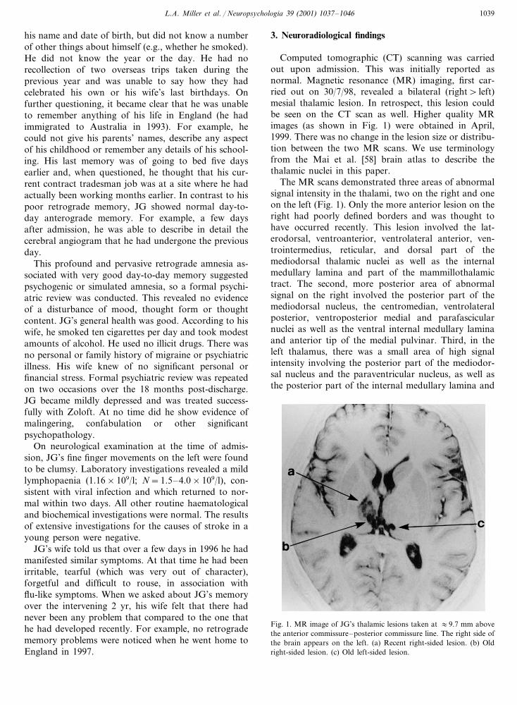
Computed tomographic (CT) scanning was carriedout upon admission. This was initially reported asnormal. Magnetic resonance (MR) imaging, first car-ried out on 30/7/98, revealed a bilateral (right� left)mesial thalamic lesion. In retrospect, this lesion couldbe seen on the CT scan as well. Higher quality MRimages (as shown in Fig. 1) were obtained in April,1999. There was no change in the lesion size or distribu-tion between the two MR scans. We use terminologyfrom the Mai et al. [58] brain atlas to describe thethalamic nuclei in this paper.
The MR scans demonstrated three areas of abnormalsignal intensity in the thalami, two on the right and oneon the left (Fig. 1). Only the more anterior lesion on theright had poorly defined borders and was thought tohave occurred recently. This lesion involved the lat-erodorsal, ventroanterior, ventrolateral anterior, ven-trointermedius, reticular, and dorsal part of themediodorsal thalamic nuclei as well as the internalmedullary lamina and part of the mammillothalamictract. The second, more posterior area of abnormalsignal on the right involved the posterior part of themediodorsal nucleus, the centromedian, ventrolateralposterior, ventroposterior medial and parafascicularnuclei as well as the ventral internal medullary laminaand anterior tip of the medial pulvinar. Third, in theleft thalamus, there was a small area of high signalintensity involving the posterior part of the mediodor-sal nucleus and the paraventricular nucleus, as well asthe posterior part of the internal medullary lamina and
Fig. 1. MR image of JG’s thalamic lesions taken at �9.7 mm abovethe anterior commissure–posterior commissure line. The right side ofthe brain appears on the left. (a) Recent right-sided lesion. (b) Oldright-sided lesion. (c) Old left-sided lesion.
L.A. Miller et al. / Neuropsychologia 39 (2001) 1037–10461040
anterior tip of the medial pulvinar. The latter tworegions were quite well defined and appeared to be ofslightly higher signal intensity than the more anteriorcomponent on the right. Their appearance was judgedto be nonacute. The estimated volume of the totallesion in the right thalamus was 772 mm3 and on theleft it was 72 mm3. The mammillary bodies, anteriorprincipal nuclei and fornices appeared normal bilater-ally and showed no evidence of atrophy. Selective cere-bral angiography was performed and was found to benormal.
The thalamic lesions were thought most likely to bethe result of infarction. The findings suggested at leasttwo episodes. The major radiological differential diag-nosis was demyelination, but the distribution of thelesions was considered to be quite atypical and therewas no evidence of lesions involving the corpus callo-sum. Recently, however, a case of encephalomyelitisaffecting the thalamus has been described [74] and thisaetiology must be considered a possibility.
4. Neuropsychological assessments
4.1. Initial assessment (1 day, 5 days and 3 monthsfollowing admission)
On formal testing, JG’s autobiographical memorywas exceptionally poor (Table 1). On the autobiograph-ical memory interview [53], his recall (both of semanticdetails and autobiographical incidents) was significantlyimpaired for every period of his life with the exceptionof the last 5 yr, for which he showed some patchyrecall. For example, he could remember the date of hiswedding 5 yr earlier and some details of the occasion.He was also able to recall a number of details of gamesplayed by England in the Football World Cup in June,1998.
To assess JG’s ability to recognise autobiographicalmaterial, three multiple choice tests were created frominformation and photographs supplied by his mother,sister and wife. JG gave his informed consent to partic-ipate in these experiments. In the first test (Major LifeEvents), there were 18 questions about his previous life(e.g., name of former fiancee, place where he had hisfirst job, school sport in which he had won prizes), eachwith four possible responses. In a second, two-step testinvolving personally familiar faces from the past, 29photographs of faces of friends and relatives fromAustralia (9 ‘‘recent’’) and England (20 ‘‘remote’’ from7–25 yr ago) were first presented one at a time amongstthe photographs of three strangers. JG had to pick theface that was familiar. In the second part of this test, hewas asked to match each photograph of his friends orfamily members to one of four Christian names. As canbe seen in Table 1, with the exception of his perfor-
Table 1Autobiographical retrograde memory tests: initial and 12-month fol-low-up scores
Follow-upInitialMeasure
Standardised measureAutobiographical memory interview
Personal semantic information2.5/21a 0/21aChildhood7.5/21aEarly adult 11.5/21a
Recent 9/21a 21/2119/63aTotal 32.5/63a
Autobiographical incidents0/9aChildhood 0/9a
0/9a3/9aEarly adult6/9 6/9Recent
6/27a9/27aTotal
Multiple-choice autobiographical memory testsMajor life events 3/18b 5/18b
Recognition of friends and relativesChoosing familiar faces
9/99/9Recent5/20bRemote 8/20
Matching name to face9/9 9/9Recent4/20b 8/20Remote
a Denotes impaired performance �2 SD from the published meanfor normal control subjects.
b Denotes performance�chance.
mance on recently known faces, he was unable torecognise the correct answers on these multiple-choicetests.
JG’s retrograde memory for some aspects of non-au-tobiographical information was also profoundly im-paired. During his hospital admission, JG could notrecognise ten famous people (e.g., Margaret Thatcher)either from their names or their pictures. Three monthsfollowing his admission, JG’s knowledge of famouspeople and world events was tested in more detail usingnew tests of famous people and famous events designedto be appropriate to his cultural background and age.For the Famous People test, there were three parts, oneinvolving 30 famous faces, one involving 60 famousnames and one in which the original 30 famous faceswere re-presented with choices of famous names. Forthe most part, the famous people were alive and verymuch a part of current popular culture (e.g., Bill Clin-ton, Nicole Kidman), but some were well-known figuresfrom the past (e.g., Adolph Hitler, Albert Einstein). Onthe Famous Events test, there were 32 items (e.g., Whathappened at Lockerbie, Scotland?) pertaining to eventsfrom the 1970s, 1980s and 1990s. For each of thesetests, the correct answer was presented amongst threedistractors and a forced choice procedure was used.
JG’s scores on these new experimental measures werecompared with those of three men with no known
L.A. Miller et al. / Neuropsychologia 39 (2001) 1037–1046 1041
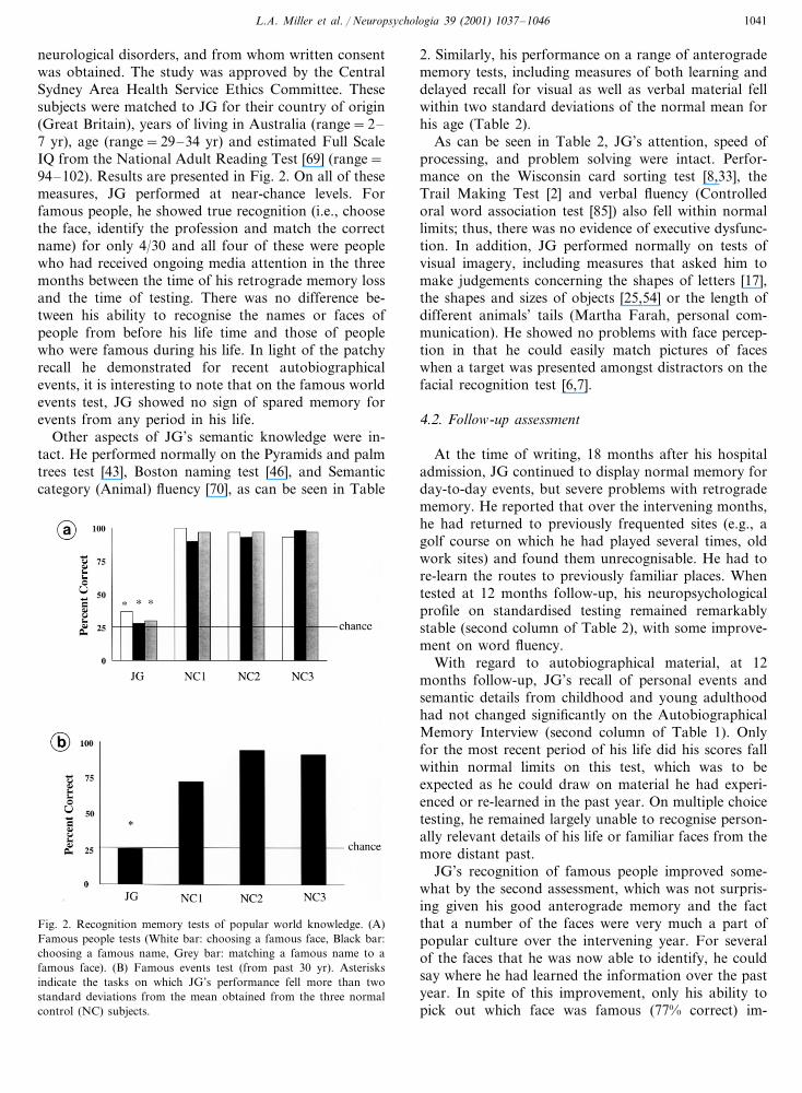
neurological disorders, and from whom written consentwas obtained. The study was approved by the CentralSydney Area Health Service Ethics Committee. Thesesubjects were matched to JG for their country of origin(Great Britain), years of living in Australia (range=2–7 yr), age (range=29–34 yr) and estimated Full ScaleIQ from the National Adult Reading Test [69] (range=94–102). Results are presented in Fig. 2. On all of thesemeasures, JG performed at near-chance levels. Forfamous people, he showed true recognition (i.e., choosethe face, identify the profession and match the correctname) for only 4/30 and all four of these were peoplewho had received ongoing media attention in the threemonths between the time of his retrograde memory lossand the time of testing. There was no difference be-tween his ability to recognise the names or faces ofpeople from before his life time and those of peoplewho were famous during his life. In light of the patchyrecall he demonstrated for recent autobiographicalevents, it is interesting to note that on the famous worldevents test, JG showed no sign of spared memory forevents from any period in his life.
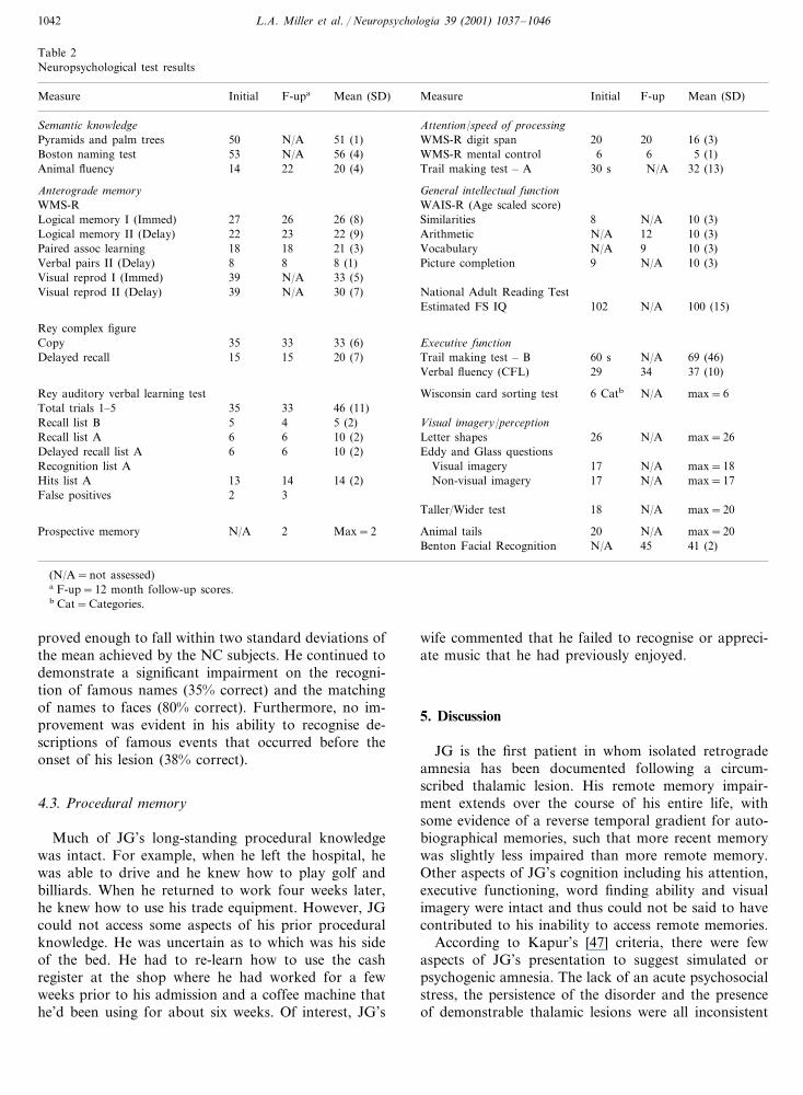
Other aspects of JG’s semantic knowledge were in-tact. He performed normally on the Pyramids and palmtrees test [43], Boston naming test [46], and Semanticcategory (Animal) fluency [70], as can be seen in Table
2. Similarly, his performance on a range of anterogradememory tests, including measures of both learning anddelayed recall for visual as well as verbal material fellwithin two standard deviations of the normal mean forhis age (Table 2).
As can be seen in Table 2, JG’s attention, speed ofprocessing, and problem solving were intact. Perfor-mance on the Wisconsin card sorting test [8,33], theTrail Making Test [2] and verbal fluency (Controlledoral word association test [85]) also fell within normallimits; thus, there was no evidence of executive dysfunc-tion. In addition, JG performed normally on tests ofvisual imagery, including measures that asked him tomake judgements concerning the shapes of letters [17],the shapes and sizes of objects [25,54] or the length ofdifferent animals’ tails (Martha Farah, personal com-munication). He showed no problems with face percep-tion in that he could easily match pictures of faceswhen a target was presented amongst distractors on thefacial recognition test [6,7].
4.2. Follow-up assessment
At the time of writing, 18 months after his hospitaladmission, JG continued to display normal memory forday-to-day events, but severe problems with retrogradememory. He reported that over the intervening months,he had returned to previously frequented sites (e.g., agolf course on which he had played several times, oldwork sites) and found them unrecognisable. He had tore-learn the routes to previously familiar places. Whentested at 12 months follow-up, his neuropsychologicalprofile on standardised testing remained remarkablystable (second column of Table 2), with some improve-ment on word fluency.
With regard to autobiographical material, at 12months follow-up, JG’s recall of personal events andsemantic details from childhood and young adulthoodhad not changed significantly on the AutobiographicalMemory Interview (second column of Table 1). Onlyfor the most recent period of his life did his scores fallwithin normal limits on this test, which was to beexpected as he could draw on material he had experi-enced or re-learned in the past year. On multiple choicetesting, he remained largely unable to recognise person-ally relevant details of his life or familiar faces from themore distant past.
JG’s recognition of famous people improved some-what by the second assessment, which was not surpris-ing given his good anterograde memory and the factthat a number of the faces were very much a part ofpopular culture over the intervening year. For severalof the faces that he was now able to identify, he couldsay where he had learned the information over the pastyear. In spite of this improvement, only his ability topick out which face was famous (77% correct) im-
Fig. 2. Recognition memory tests of popular world knowledge. (A)Famous people tests (White bar: choosing a famous face, Black bar:choosing a famous name, Grey bar: matching a famous name to afamous face). (B) Famous events test (from past 30 yr). Asterisksindicate the tasks on which JG’s performance fell more than twostandard deviations from the mean obtained from the three normalcontrol (NC) subjects.
L.A. Miller et al. / Neuropsychologia 39 (2001) 1037–10461042
Table 2Neuropsychological test results
F-upa Mean (SD) MeasureMeasure InitialInitial F-up Mean (SD)
Attention/speed of processingSemantic knowledgeN/A 51 (1) WMS-R digit span 20 20 16 (3)Pyramids and palm trees 50N/A 56 (4) WMS-R mental control53 6Boston naming test 6 5 (1)
14Animal fluency 22 20 (4) Trail making test – A 30 s N/A 32 (13)
Anterograde memory General intellectual functionWAIS-R (Age scaled score)WMS-R
Logical memory I (Immed) 27 26 26 (8) Similarities 8 N/A 10 (3)23 22 (9) Arithmetic22 N/ALogical memory II (Delay) 12 10 (3)
18Paired assoc learning 18 21 (3) Vocabulary N/A 9 10 (3)8Verbal pairs II (Delay) 8 8 (1) Picture completion 9 N/A 10 (3)
N/A 33 (5)39Visual reprod I (Immed)39Visual reprod II (Delay) N/A 30 (7) National Adult Reading Test
Estimated FS IQ 102 N/A 100 (15)
Rey complex figure33 33 (6) Executi�e function35Copy15Delayed recall 20 (7)15 Trail making test – B 60 s N/A 69 (46)
Verbal fluency (CFL) 29 34 37 (10)
Wisconsin card sorting test 6 CatbRey auditory verbal learning test N/A max=633 46 (11)35Total trials 1–54 5 (2) Visual imagery/perceptionRecall list B 56 10 (2) Letter shapes6 26Recall list A N/A max=26
6Delayed recall list A 6 10 (2) Eddy and Glass questionsRecognition list A Visual imagery 17 N/A max=18
14 14 (2) Non-visual imagery13 17Hits list A N/A max=173False positives 2
Taller/Wider test 18 N/A max=20
Prospective memory N/A 2 Max=2 Animal tails 20 N/A max=20Benton Facial Recognition N/A 45 41 (2)
(N/A=not assessed)a F-up=12 month follow-up scores.b Cat=Categories.
proved enough to fall within two standard deviations ofthe mean achieved by the NC subjects. He continued todemonstrate a significant impairment on the recogni-tion of famous names (35% correct) and the matchingof names to faces (80% correct). Furthermore, no im-provement was evident in his ability to recognise de-scriptions of famous events that occurred before theonset of his lesion (38% correct).
4.3. Procedural memory
Much of JG’s long-standing procedural knowledgewas intact. For example, when he left the hospital, hewas able to drive and he knew how to play golf andbilliards. When he returned to work four weeks later,he knew how to use his trade equipment. However, JGcould not access some aspects of his prior proceduralknowledge. He was uncertain as to which was his sideof the bed. He had to re-learn how to use the cashregister at the shop where he had worked for a fewweeks prior to his admission and a coffee machine thathe’d been using for about six weeks. Of interest, JG’s
wife commented that he failed to recognise or appreci-ate music that he had previously enjoyed.
5. Discussion
JG is the first patient in whom isolated retrogradeamnesia has been documented following a circum-scribed thalamic lesion. His remote memory impair-ment extends over the course of his entire life, withsome evidence of a reverse temporal gradient for auto-biographical memories, such that more recent memorywas slightly less impaired than more remote memory.Other aspects of JG’s cognition including his attention,executive functioning, word finding ability and visualimagery were intact and thus could not be said to havecontributed to his inability to access remote memories.
According to Kapur’s [47] criteria, there were fewaspects of JG’s presentation to suggest simulated orpsychogenic amnesia. The lack of an acute psychosocialstress, the persistence of the disorder and the presenceof demonstrable thalamic lesions were all inconsistent
L.A. Miller et al. / Neuropsychologia 39 (2001) 1037–1046 1043
with the diagnosis of psychogenic amnesia. Further-more, there was no apparent benefit to him from thepatient role and neither extensive questioning of hisfamily nor the passage of time has revealed circum-stances that could be associated with malingering.
The MR images yielded evidence of at least twoneurological events in the thalamus, with the morerecent involving the medial region on the right side andthe older incident(s) involving posteromedial regionsbilaterally. The lesions lie in the territory of the thalam-operforate (also known as the paramedian) arteries.JG’s wife gave a history of a short period of similarmild mood changes, somnolence, and memory prob-lems occurring 2 yr prior to the present event. In lightof the radiological findings, we believe that those symp-toms were probably associated with an earlier episodeof damage to the thalamus. It is important to note thatthe onset of JG’s dense and persistent retrograde amne-sia occurred only after the second episode, which in-volved extension of the lesion into more anterodorsalthalamic regions on the right, including the mammillo-thalamic tract, internal medullary lamina andanterodorsal part of the dorsomedial nucleus.
The anteromesial portion of the thalamus has richprojections to the frontal and temporal lobes and exten-sive hypometabolism has been found in these corticalareas as a consequence of a mediodorsal thalamic le-sion [76]. A number of investigators [3,45,61,67,83,99]have proposed that these neocortical regions are crucialto remote memory retrieval. Thus, one might argue thatJG’s memory deficit is the result not of thalamic dys-function per se, but of diminished functioning in theserelated cortical areas. This interpretation is difficult tosustain in view of JG’s normal performance on neu-ropsychological measures sensitive to frontal and tem-poral lobe dysfunction.
A few previous patients have been reported with anextensive and persistent inability to access remote mem-ories (along with anterograde amnesia) following bilat-eral thalamic infarction [42,71,90,97]. In addition, apatient described by Peru and Fabbro [77] initiallyshowed both a retrograde and anterograde memoryimpairment after bilateral, anteromedial, thalamicvenous infarction. Interestingly, their patient’s retro-grade memory impairment resolved in conjunction withshrinkage of the right-sided ischaemic region. Consider-ing the overlap in site of lesion in these cases and in JG,we conclude that the thalamic regions most likely to beimportant to retrograde memory include the rightmediodorsal nucleus, right mammillothalamic tract,postero-medial region of the left mediodorsal nucleusand internal medullary laminae bilaterally.
The lesion locus in JG differs from that of patientswith isolated anterograde amnesia following thalamicdamage [50,60,65,75,84,97]. In the latter cases, the com-mon area of lesion involves the left mammillothalamic
tract and, in all but one [60], the left mediodorsalnucleus. The importance of the mammillo-thalamictract in anterograde memory was emphasised in a re-cent review of neuropsychological deficits caused bythalamic infarction [95]. JG has no anterograde mem-ory deficit and, in his case, there is relative sparing ofthe left mediodorsal nucleus and complete sparing ofthe left mammillothalamic tract.
JG was able to remember some details about the lastfew years of his life. In contrast, his more remotememories were completely lost to him. It has beenpostulated that transition from a predominantlyhippocampus-dependent to a neocortex-dependentmnemonic representation takes years [79,86]. JG’s pat-tern of memory loss in conjunction with the absence ofany temporal lobe abnormalities indicates that he hassome continued access to memories from the past 5 yr(i.e., according to this model, those presumably stillbeing processed in the hippocampal network), but haslost access to more remote memories (i.e., those thathave presumably been stored in the neocortex). Thus,his case provides support both for the view that thethalamus plays a key role in providing access to neocor-tically stored memories [9,39,61] and, indirectly, for atime-limited contribution of the hippocampus to mem-ory consolidation (cf. [68,86]).
The pattern of spared versus inaccessible memory inJG adds to the growing body of evidence that thedivision of declarative memory into just two categories-episodic and semantic [13,87,93] is inadequate. JG wasunable to remember famous people by their faces ornames but displayed a good ability to access otheraspects of semantic knowledge (e.g., names of objects,exemplars of categories, conceptual relationships be-tween objects). Dissociation’s in the ability to recalldifferent types of world knowledge has been notedpreviously [26,28,34,37,40,42,51]. Findings from thesecases, and now also from JG, indicate that knowledgepertaining to people, events, and possibly to other typesof unique material (e.g., famous buildings [26,72]) dif-fers in some critical way from other types of factualknowledge such as words and meanings. The nature ofthis difference has not been much explored but mayhave something to do with the extent to which informa-tion about the world is linked or tagged to autobio-graphical events or to particular points in time [10,52].Furthermore, JG’s surprisingly incomplete recall of pre-viously learned procedural information indicates theneed to study the ways in which this knowledge may besubdivided, and differentially stored or accessed. Weare currently exporing these issues.
The lack of concern demonstrated by JG over hisloss of past memories has been noted in other patientswith retrograde amnesia caused by thalamic infarction[15,80,92]. If this attitude is prevalent in patients withisolated retrograde amnesia, it may help to explain why
L.A. Miller et al. / Neuropsychologia 39 (2001) 1037–10461044
the diagnosis is so rare. In JG’s case, the initial CT scanwas reported as normal and within a few weeks of hishospitalization, he had returned to work and to someof his previous sporting interests. His wife said he hadno significant problems in carrying out his activities ofdaily living. Had he had not complained of loss ofmemory for his past life and had MR scanning notbeen conducted, the remarkable features of his casecould easily have been missed. It may also be importantthat much of his past life had occurred in a differentphysical context involving different people than thosewith whom he was presently familiar. Because of therelative preservation of JG’s recent memory and hisability to recognise those with whom he had had recentcontact, this pattern of retrograde amnesia might havebeen less apparent in someone who had remained in thesame place all his life.
Overall, the findings from JG provide unique evi-dence that the right mesiodorsal region of the thalamusplays a crucial role in the ability to access remotememories, including those pertaining to autobiographi-cal semantic details, autobiographical incidents and tosome aspects of world knowledge. These findings areconsistent with the proposals put forward by Markow-itsch and colleagues [29,61] that the right hemispherehas a special role in autobiographical memory. Wehypothesize that the right mediodorsal nucleus, rightmammillothalamic tract and right internal medullarylamina (and possibly parts of the left mediodorsalnucleus and left internal medullary lamina) comprisethe central processing mechanism referred to by Mc-Clelland [63] and Markowitsch [61] as responsible forinducing and coordinating the recall of cortically storedmemory engrams.
Acknowledgements
We appreciate the help of Dr Ron Shnier who ob-tained the high quality MR images and Mr RogerFulton and Ms Irina Harris, who translated the imagesand assisted in the production of the prints. We wouldalso like to acknowledge the work of Ms Nora Breenand Ms Karen Wallace in helping to assemble theFamous People Test and the assistance of Mr KenSaunders in preparing the questions for the FamousEvents Test.
References
[1] Albert MS, Butters N, Levin J. Temporal gradients in theretrograde amnesia of patients with alcoholic Korsakoff’s dis-ease. Archives of Neurology 1979;36:211–6.
[2] Army individual test battery manual of directions and scoring.Washington, DC: War Department, Adjutant General’s Office,1944.
[3] Baddeley A, Wilson B. Amnesia, autobiographical memory,and confabulation. In: Rubin DC, editor. Autobiographicalmemory. Cambridge: Cambridge University Press, 1986:225–51.
[4] Barbizet J, Degos JD, Louarn F, Nguyen JP, Mas JL. Amnesiepar lesion ischemique bi-thalamique. Revue Neurologique1981;137:415–24.
[5] Baron JC, Petit-Taboue MC, Le Doze F, Desgranges B,Ravenel N, Marchal G. Right frontal cortex hypmetabolism intransient global amnesia: a PET study. Brain 1994;117:545–52.
[6] Benton AL, Hamsher KdeS, Varney NR, Spreen O. Contribu-tions to neuropsychological assessment. New York: OxfordUniversity Press, 1983.
[7] Benton A, VanAllen MW. Impairment in facial recognition inpatients with cerebral disease. Cortex 1968;4:344–58.
[8] Berg EA. A simple objective technique for measuring flexibilityin thinking. Journal of General Psychology 1948;39:15–22.
[9] Bressler SL. Large-scale cortical networks and cognition. BrainResearch Review 1995;20:288–304.
[10] Brown NR, Shevell SK, Rips LJ. Public memories and theirpersonal context. In: Rubin DC, editor. Autobiographicalmemory. Cambridge: Cambridge University Press, 1986:149–58.
[11] Butters N, Cermak LS. Alcoholic Korsakoffs syndrome: aninformation-processing approach to amnesia. New York: Aca-demic Press, 1980.
[12] Calabrese P, Markowitsch HJ, Durwen HF, Widlitzek H,Haupts M, Holinka B, et al. Right temporofrontal cortex ascritical locus for the ecphory of old episodic memories. Journalof Neurology, Neurosurgery and Psychiatry 1996;61:304–10.
[13] Cermak L. The episodic/semantic distinction in amnesia. In:Butters N, Squire LR, editors. The neuropsychology of mem-ory. New York: Guilford Press, 1984:55–62.
[14] Cermak LS, O’Connor M. The anterograde and retrograderetrieval ability of a patient with amnesia due to encephalitis.Neuropsychologia 1983;21:213–34.
[15] Choi D, Sudarsky L, Schachter S, Biber M, Burke P. Medialthalamic hemorrhage with amnesia. Archives of Neurology1983;40:611–3.
[16] Clarke S, Assal G, Bogousslavsky J, Regli F, Townsend DW,Leenders KL, et al. Pure amnesia after unilateral left polarthalamic infarct: topographic and sequential neuropsychologi-cal and metabolic (PET) correlations. Journal of NeurologyNeurosurgery and Psychiatry 1994;57:27–34.
[17] Coltheart M, Hull E, Slater D. Sex differences in imagery andreading. Nature 1975;253:438–40.
[18] Corkin S. Lasting consequences of bilateral medial temporallobectomy: clinical course and experimental findings in H.M.Seminars in Neurology 1984;4:249–59.
[19] Costello A, Fletcher PC, Dolan RJ, Frith CD, Shallice T. Theorigins of forgetting in a case of isolated retrograde amnesiafollowing a haemorrhage: evidence from functional imaging.Neurocase 1998;4:437–46.
[20] Damasio AR, Eslinger PJ, Damasio H, Van Hoesen GW,Cornell S. Multimodal amnesic syndrome following bilateraltemporal and basal forebrain damage. Archives of Neurology1985;42:252–9.
[21] Della Sala S, Laiacona M, Spinnler H, Trivelli C. Autobio-graphical recollection and frontal damage. Neuropsychologia1993;31:823–39.
[22] DeRenzi E, Liotti M, Nichelli P. Semantic amnesia with preser-vation of autobiographic memory: a case report. Cortex1987;23:575–97.
[23] DeRenzi E, Lucchelli F. Dense retrograde amnesia, intactlearning capability and abnormal forgetting rate: a consolida-tion deficit? Cortex 1993;29:449–66.
L.A. Miller et al. / Neuropsychologia 39 (2001) 1037–1046 1045
[24] Diagnostic and statistical manual of mental disorders fourthedition: DSM-IV. Washington, DC: American Psychiatric As-sociation, 1994.
[25] Eddy JK, Glass AL. Reading and listening to high and lowimagery sentences. Journal of Verbal Learning and VerbalBehavior 1981;20:333–45.
[26] Ellis AW, Young AW, Critchley EMR. Loss of memory forpeople following temporal lobe damage. Brain 1989;112:1469–83.
[27] Eslinger PJ. Autobiographical memory after temporal lobelesions. Neurocase 1998;4:481–95.
[28] Evans JJ, Heggs AJ, Antoun N, Hodges JR. Progressive proso-pagnosia associated with selective right temporal atrophy: anew syndrome? Brain 1995;118:1–13.
[29] Fink GR, Markowitsch HJ, Reinkemeier M, Bruckbauer T,Kessler J, Heiss W-D. Cerebral representation of one’s ownpast: neural networks involved in autobiographical memory.Journal of Neuroscience 1996;16:4275–82.
[30] Gainotti G, Almonti S, Di Betta AM, Silveri MC. Retrogradeamnesia in a patient with retrosplenial tumour. Neurocase1998;4:519–26.
[31] Graff-Radford N, Damasio H, Yamada T, Eslinger PJ, Dama-sio AR. Nonhaemorrhagic thalamic infarction: clinical neu-ropsychological and electrophysiological findings in fouranatomical groups defined by computerized tomography. Brain1985;108:485–516.
[32] Graham DS, Pratt KH, Hodges JR. A reverse temporal gradi-ent for public events in a single case of semantic dementia.Neurocase 1998;4:461–70.
[33] Grant DA, Berg EA. A behavioral analysis of degree of impair-ment and ease of shifting to new responses in a Weigl-type cardsorting problem. Journal of Experimental Psychology1948;39:404–11.
[34] Greene JDW, Hodges JR. The fractionation of remote memory:evidence from a longitudinal study of dementia of Alzheimertype. Brain 1996;119:129–42.
[35] Grossi D, Trojano L, Grasso A, Orsini A. Selective ‘‘semanticamnesia’’ after closed-head injury; a case report. Cortex1988;24:457–64.
[36] Guberman A, Stuss D. The syndrome of bilateral paramedianthalamic infarction. Neurology 1983;33:540–6.
[37] Hanley JR, Young AW, Pearson NA. Defective recognition offamiliar people. Cognitive Neuropsychology 1989;6:179–210.
[38] Harding A, Halliday G, Caine D, Kril J. Degeneration ofanterior thalamic nuclei differentiates alcoholics with amnesia.Brain 2000;123:141–54.
[39] Harth E. The sketchpad model. A theory of consciousness,perception and imagery. Consciousness and Cognition1995;4:346–68.
[40] Hodges JR, Graham KS. A reversal of the temporal gradientfor famous person knowledge in semantic dementia: implica-tions for the neural organisation of long-term memory. Neu-ropsychologia 1998;36:803–25.
[41] Hodges JR, Gurd JM. Remote memory and lexical retrieval ina case of frontal Pick’s disease. Archives of Neurology1994;51:821–7.
[42] Hodges JR, McCarthy RA. Autobiographical amnesia resultingfrom bilateral paramedian thalamic infarction: a case study incognitive neurobiology. Brain 1993;116:921–40.
[43] Howard D, Patterson K. The pyramids and palm trees test.Bury St. Edmunds, UK: Thames Valley Test Company, 1992.
[44] Hunkin N, Parkin AJ, Bradley VA, Burrows EH, Aldrich FK,Jansari A, et al. Focal retrograde amnesia following closedhead injury: a case study and theoretical account. Neuropsy-chologia 1995;33:509–23.
[45] Jones RD, Grabowski TJ, Tranel D. The neural basis ofretrograde memory: evidence from positron emission tomogra-
phy for the role of non-mesial temporal lobe structures. Neuro-case 1998;4:471–9.
[46] Kaplan EF, Goodglass H, Weintraub S. The Boston namingtest. Boston: E. Kaplan & H. Goodglass, 1978.
[47] Kapur N. Syndromes of retrograde amnesia: a conceptual andempirical synthesis. Psychological Bulletin 1999;125:800–25.
[48] Kapur N, Ellison D, Smith MP, McLellan DL, Burrows EH.Focal retrograde amnesia following bilateral temporal lobepathology. Brain 1992;115:73–85.
[49] Kapur N, Ellison D, Parkin AJ, Hunkin NM, Burrows E,Sampson SA, et al. Bilateral temporal lobe pathology withsparing of medial temporal lobe structures: lesion profile andpattern of memory disorder. Neuropsychologia 1994;32:23–38.
[50] Kapur N, Thompson S, Cook P, Lang D, Brice J. Anterogradebut not retrograde memory loss following combined mamillarybody and medial thalamic lesions. Neuropsychologia1996;34:1–8.
[51] Kitchener EG, Hodges JR. Impaired knowledge of famouspeople and events with intact autobiographical memory in acase of progressive right temporal lobe degeneration: implica-tions for the organisation of remote memory. Cognitive Neu-ropsychology 1999;16:589–607.
[52] Kopelman MD, Stanhope N, Kingsley D. Retrograde amnesiain patients with diencephalic, temporal lobe or frontal lesions.Neuropsychologia 1999;37:939–58.
[53] Kopelman M, Wilson B, Baddeley A. The autobiographicalmemory interview. Bury St. Edmunds, UK: Thames Valley TestCompany, 1990.
[54] Kosslyn SM, Reiser BJ, Farah MJ, Fliegel SL. Generatingvisual images: units and relations. Journal of ExperimentalPsychology: General 1983;112:278–303.
[55] Kroll NEA, Markowitsch HJ, Knight RT, von Cramon DY.Retrieval of old memories: the temporofrontal hypothesis.Brain 1997;120:1377–99.
[56] Lishman WA. Organic psychiatry, 2nd ed. Oxford: Blackwell,1987.
[57] Lucchelli F, Muggia S, Spinnler H. The ‘Petites Madeleines’phenomenon in two amnesic patients: sudden recovery of for-gotten memories. Brain 1995;118:167–83.
[58] Mai JK, Assheuer J, Paxinos G. Atlas of the human brain. NewYork: Academic Press, 1997.
[59] Mair WGP, Warrington EK, Weiskrantz L. Memory disorderin Korsakoff’s psychosis: a neuropathological and neuropsy-chological investigation of two cases. Brain 1979;102:749–83.
[60] Malamut BL, Graff-Radford N, Chawluk J, Grossman RI, GurRC. Memory in a case of bilateral thalamic infarction. Neurol-ogy 1992;42:163–9.
[61] Markowitsch HJ. Which brain regions are critically involved inthe retrieval of old episodic memory? Brain Research Review1995;21:117–27.
[62] Markowitsch HJ, von Cramon DY, Schuri U. Mnestic perfor-mance profile of a bilateral diencephalic infarct patient withpreserved intelligence and severe amnesic disturbances. Journalof Clinical and Experimental Neuropsychology 1993;15:627–52.
[63] McClelland JL. The organization of memory: a parallel dis-tributed processing perspective. Revue Neurologique1994;150:570–9.
[64] Meltzer M. Poor memory: a case report. In: Kapur N, editor.Injured brains of medical minds: views from within. Oxford:Oxford University Press, 1997:8–15.
[65] Michel D, Laurent B, Foyatier N, Blanc A, Portafaix M.Infarctus thalamique paramedian gauche: etude de la memoireet du langage. Revue Neurologique 1982;138:533–50.
[66] Milner B, Corkin S, Teuber H-L. Further analysis of thehippocampal amnesic syndrome: 14-year follow-up study ofH.M. Neuropsychologia 1968;6:215–34.
L.A. Miller et al. / Neuropsychologia 39 (2001) 1037–10461046
[67] Moscovitch M. Confabulation and the frontal systems: strategicversus associative retrieval in neuropsychological theories ofmemory. In: Roediger HL, Craik FIM, editors. Varieties ofmemory and consciousness. Hillsdale: Lawrence Erlbaum Asso-ciates, 1989:133–60.
[68] Nadel L, Moscovitch M. Memory consolidation, retrogradeamnesia and the hippocampal complex. Current Opinion inNeurobiology 1997;7:217–27.
[69] Nelson HE. The national adult reading test (NART) test man-ual. Windsor, Berks, UK: NFER-Nelson, 1982.
[70] Newcombe F. Missile wounds of the brain: a study of psycho-logical deficits. London: Oxford University Press, 1969.
[71] Nichelli P, Bahmanian-Behbahani G, Gentilini M, Vecchi A.Preserved memory abilities in thalamic amnesia. Brain1988;111:1337–53.
[72] O’Connor M, Butters N, Miliotis P, Eslinger P, Cermak LS.The dissociation of anterograde and retrograde amnesia in apatient with herpes encephalitis. Journal of Clinical and Exper-imental Neuropsychology 1992;14:159–87.
[73] Ogden JA. Visual object agnosia, prosopagnosia, achromatop-sia, loss of visual imagery, and autobiographical amnesia fol-lowing recovery from cortical blindness: case M.H.Neuropsychologia 1993;31:571–89.
[74] Olivero WC, Deshmukh P, Gujrati M. Bilateral enhancingthalamic lesion in a 10 year old boy: case report. Journal ofNeurology, Neurosurgery and Psychiatry 1999;66:633–5.
[75] Parkin AJ, Rees JE, Hunkin NM, Rose PE. Impairment ofmemory following discrete thalamic infarction. Neuropsycholo-gia 1994;32:39–51.
[76] Pepin EP, Auray-Pepin L. Selective laterodorsal frontal lobedysfunction associated with diencephalic amnesia. Neurology1993;43:733–41.
[77] Peru A, Fabbro F. Thalamic amnesia following venous infarc-tion: evidence from a single case study. Brain and Cognition1997;33:278–94.
[78] Piercy MF. Experimental studies of the organic amnesic syn-drome. In: Whitty CWM, Zangwill OL, editors. Amnesia, 2nded. London: Butterworth, 1977:1–51.
[79] Rempel-Clower NL, Zola SM, Squire LR, Amaral DG. Threecases of enduring memory impairment after bilateral damagelimited to the hippocampal formation. Journal of Neuroscience1996;16:5233–55.
[80] Schott B, Mauguiere B, Laurent O, Serclerat O, Fischer C.L’amnesie thalamique. Revue Neurologique 1980;136:117–30.
[81] Scoville WB, Milner B. Loss of recent memory after bilateralhippocampal lesions. Journal of Neurology, Neurosurgery andPsychiatry 1957;20:11–21.
[82] Seltzer B, Benson DF. The temporal pattern of retrogradeamnesia in Korsakoff’s disease. Neurology 1974;24:527–30.
[83] Shallice T. From neuropsychology to mental structure. Cam-bridge: Cambridge University Press, 1988.
[84] Speedie LJ, Heilman KM. Amnestic disturbance following in-farction of the left dorsomedial nucleus of the thalamus. Neu-ropsychologia 1982;20:597–604.
[85] Spreen O, Benton AL. Neurosensory center comprehensiveexamination for aphasia (NCCEA). Victoria: University ofVictoria, Neuropsychological Laboratory, 1969.
[86] Squire LR. Mechanisms of memory. Science 1986;232:1612–9.[87] Squire LR. Memory and brain. Oxford: Oxford University
Press, 1987.[88] Squire LR, Moore RY. Dorsal thalamic lesion in a noted case
of human memory dysfunction. Annals of Neurology1979;6:503–6.
[89] Stracciari A, Ghidoni E, Guarino M, Poletti M, Pazzaglia P.Post-traumatic retrograde amnesia with selective impairment ofautobiographical memory. Cortex 1994;30:459–68.
[90] Stuss DT, Guberman A, Nelson R, Larochelle S. The neu-ropsychology of paramedian thalamic infarction. Brain andCognition 1988;8:348–78.
[91] Stuss DT, Guzman DA. Severe remote memory loss withminimal anterograde amnesia: a clinical note. Brain and Cogni-tion 1988;8:21–30.
[92] Swanson RA, Schmidley MD. Amnestic syndrome and verticalgaze palsy: early detection of bilateral thalamic infarction byCT and NMR. Stroke 1985;16:823–7.
[93] Tulving E. Episodic and semantic memory. In: Tulving E,Donaldson W, editors. Organization of memory. New York:Academic Press, 1972:381–403.
[94] Tulving E, Schacter DL, McLachlan DR, Moscovitch M. Prim-ing of semantic autobiographical knowledge: a case study ofretrograde amnesia. Brain and Cognition 1988;8:3–20.
[95] Van der Werf YD, Witter MP, Uylins HBM, Jolles J. Neu-ropsychology of infarctions in the thalamus: a review. Neu-ropsychologia 2000;38:613–27.
[96] Victor M, Adams RD, Collins GH. The Wernicke-Korsakoffsyndrome and related neurologic disorders due to alcoholismand malnutrition, 2nd ed. Philadelphia, PA: FA Davis, 1989.
[97] von Cramon DY, Hebel N, Schuri U. A contribution to theanatomical basis of thalamic amnesia. Brain 1985;108:993–1008.
[98] Warrington EK, McCarthy RA. The fractionation of retro-grade amnesia. Brain and Cognition 1988;7:184–200.
[99] Worthington A. Dysexecutive paramnesia: strategic retrievaldeficits in retrospective and prospective remembering. Neuro-case 1999;5:47–57.
[100] Yoneda Y, Yamadori A, Mori E, Yamashita H. Isolatedprolonged retrograde amnesia. European Neurology1992;32:340–2.


