Embed Size (px)
Citation preview
Abstract - A new species of Rhinochelys, R. nammourensis n. sp. (Chelonioidea: Protostegidae),is described on the basis of beautifully preserved complete and articulated skeletons from theNammoura locality, a Cenomanian (Late Cretaceous) Lagerstätte in Lebanon. Our study providesontogenetic and front flipper characters for the family Protostegidae. Despite a very large ulnare, thefront flipper of protostegids appears to retain the primitive digit configuration of chelonioids, in therelative length of the digits and phalanges and in the absence of reduction of the number of phalanges.The relative abundance of hatchling turtles suggests that Nammoura was near a nesting site during theCenomanian.
Key words: Testudines, Protostegidae, Cretaceous, Cenomanian, Lebanon.
Riassunto - Rhinochelys (Chelonioidea: Protostegidae), del Cretacico superiore (Cenomaniano)di Nammoura, Libano.
Una nuova specie di Rhinochelys, R. nammourensis n. sp. (Chelonioidea: Protostegidae) vienedescritta sulla base di scheletri completi e articolati in ottimo stato di conservazione provenienti dallalocalità di Nammoura, Lagerstätte cenomaniano (Cretacico superiore) del Libano. Lo studio forniscecaratteri ontogenetici e della natatoia anteriore per la famiglia Protostegidae. Nonostante un ulnaremolto sviluppato, la natatoia anteriore dei protostegidi conserva apparentemente la configurazione pri-mitiva delle dita nella relativa lunghezza delle dita stesse e delle falangi e nell’assenza di riduzione delnumero di falangi. La relativa abbondanza di neonati suggerisce che nel Cenomaniano Nammoura eranelle vicinanze di un sito di nidificazione.
Parole chiave: Testudines, Protostegidae, Cretacico, Cenomaniano, Libano.
IntroductionThe Nammoura locality is situated about 25 km northeast of Beirut, near the
village of Nammoura, Kesrouâune Caza, in the region of Mont-Liban. The siteconsists of two quarries opened in the Al Gabour valley. The narrow and deep val-
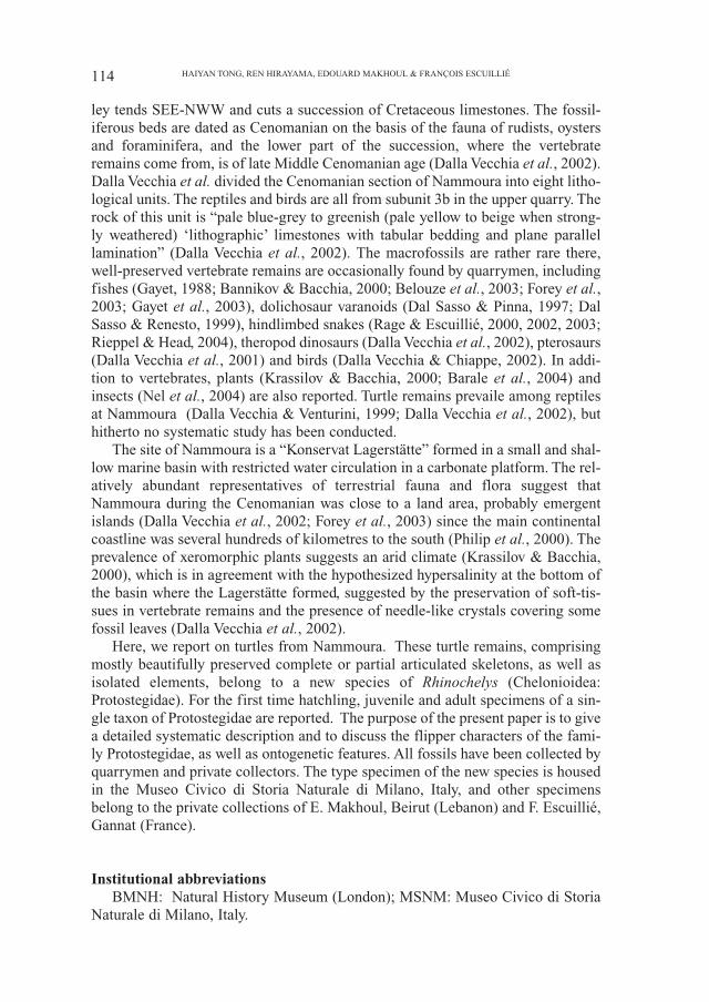
Atti Soc. it. Sci. nat. Museo civ. Stor. nat. Milano, 147 (I): 113-138, Gennaio 2006
Haiyan Tong*, Ren Hirayama**, Edouard Makhoul***&
François Escuillié****
Rhinochelys (Chelonioidea: Protostegidae)from the Late Cretaceous (Cenomanian)
of Nammoura, Lebanon
*16 cour du Liégat, 75013 Paris, France, e-mail: [email protected]**School of International Liberal Studies, Waseda University, Nishiwaseda 1-7-14, Shinjuku-ku,Tokyo 169-0051, Japan, e-mail: [email protected] ***Faculté de Médecine, Université St. Joseph, Beyrouth, Liban, e-mail: [email protected]****Eldonia, 28 rue Hettier de Boislambert, 03800 Gannat, France, e-mail: [email protected]
114 HAIYAN TONG, REN HIRAYAMA, EDOUARD MAKHOUL & FRANÇOIS ESCUILLIÉ
ley tends SEE-NWW and cuts a succession of Cretaceous limestones. The fossil-iferous beds are dated as Cenomanian on the basis of the fauna of rudists, oystersand foraminifera, and the lower part of the succession, where the vertebrateremains come from, is of late Middle Cenomanian age (Dalla Vecchia et al., 2002).Dalla Vecchia et al. divided the Cenomanian section of Nammoura into eight litho-logical units. The reptiles and birds are all from subunit 3b in the upper quarry. Therock of this unit is “pale blue-grey to greenish (pale yellow to beige when strong-ly weathered) ‘lithographic’ limestones with tabular bedding and plane parallellamination” (Dalla Vecchia et al., 2002). The macrofossils are rather rare there,well-preserved vertebrate remains are occasionally found by quarrymen, includingfishes (Gayet, 1988; Bannikov & Bacchia, 2000; Belouze et al., 2003; Forey et al.,2003; Gayet et al., 2003), dolichosaur varanoids (Dal Sasso & Pinna, 1997; DalSasso & Renesto, 1999), hindlimbed snakes (Rage & Escuillié, 2000, 2002, 2003;Rieppel & Head, 2004), theropod dinosaurs (Dalla Vecchia et al., 2002), pterosaurs(Dalla Vecchia et al., 2001) and birds (Dalla Vecchia & Chiappe, 2002). In addi-tion to vertebrates, plants (Krassilov & Bacchia, 2000; Barale et al., 2004) andinsects (Nel et al., 2004) are also reported. Turtle remains prevaile among reptilesat Nammoura (Dalla Vecchia & Venturini, 1999; Dalla Vecchia et al., 2002), buthitherto no systematic study has been conducted.
The site of Nammoura is a “Konservat Lagerstätte” formed in a small and shal-low marine basin with restricted water circulation in a carbonate platform. The rel-atively abundant representatives of terrestrial fauna and flora suggest thatNammoura during the Cenomanian was close to a land area, probably emergentislands (Dalla Vecchia et al., 2002; Forey et al., 2003) since the main continentalcoastline was several hundreds of kilometres to the south (Philip et al., 2000). Theprevalence of xeromorphic plants suggests an arid climate (Krassilov & Bacchia,2000), which is in agreement with the hypothesized hypersalinity at the bottom ofthe basin where the Lagerstätte formed, suggested by the preservation of soft-tis-sues in vertebrate remains and the presence of needle-like crystals covering somefossil leaves (Dalla Vecchia et al., 2002).
Here, we report on turtles from Nammoura. These turtle remains, comprisingmostly beautifully preserved complete or partial articulated skeletons, as well asisolated elements, belong to a new species of Rhinochelys (Chelonioidea:Protostegidae). For the first time hatchling, juvenile and adult specimens of a sin-gle taxon of Protostegidae are reported. The purpose of the present paper is to givea detailed systematic description and to discuss the flipper characters of the fami-ly Protostegidae, as well as ontogenetic features. All fossils have been collected byquarrymen and private collectors. The type specimen of the new species is housedin the Museo Civico di Storia Naturale di Milano, Italy, and other specimensbelong to the private collections of E. Makhoul, Beirut (Lebanon) and F. Escuillié,Gannat (France).
Institutional abbreviationsBMNH: Natural History Museum (London); MSNM: Museo Civico di Storia
Naturale di Milano, Italy.
Systematic Paleontology
Chelonioidea Agassiz, 1857 Protostegidae Cope, 1872Rhinochelys Seeley, 1869
Type species: Rhinochelys pulchriceps (Owen, 1842)Revised diagnosis: protostegid with a large nasal bone and a laterally expandedand swollen skull roof at the prefrontal level.
Rhinochelys nammourensis n. sp.(Figs. 1 – 8, 10)
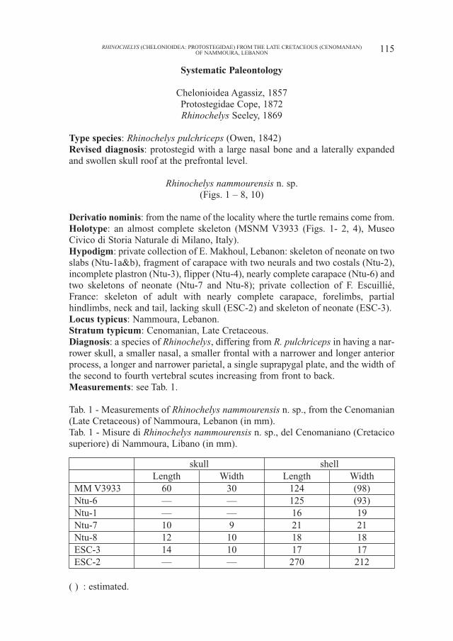
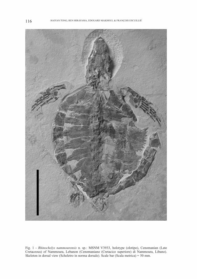
Derivatio nominis: from the name of the locality where the turtle remains come from.Holotype: an almost complete skeleton (MSNM V3933 (Figs. 1- 2, 4), MuseoCivico di Storia Naturale di Milano, Italy).Hypodigm: private collection of E. Makhoul, Lebanon: skeleton of neonate on twoslabs (Ntu-1a&b), fragment of carapace with two neurals and two costals (Ntu-2),incomplete plastron (Ntu-3), flipper (Ntu-4), nearly complete carapace (Ntu-6) andtwo skeletons of neonate (Ntu-7 and Ntu-8); private collection of F. Escuillié,France: skeleton of adult with nearly complete carapace, forelimbs, partialhindlimbs, neck and tail, lacking skull (ESC-2) and skeleton of neonate (ESC-3). Locus typicus: Nammoura, Lebanon.Stratum typicum: Cenomanian, Late Cretaceous. Diagnosis: a species of Rhinochelys, differing from R. pulchriceps in having a nar-rower skull, a smaller nasal, a smaller frontal with a narrower and longer anteriorprocess, a longer and narrower parietal, a single suprapygal plate, and the width ofthe second to fourth vertebral scutes increasing from front to back.Measurements: see Tab. 1.
Tab. 1 - Measurements of Rhinochelys nammourensis n. sp., from the Cenomanian(Late Cretaceous) of Nammoura, Lebanon (in mm).Tab. 1 - Misure di Rhinochelys nammourensis n. sp., del Cenomaniano (Cretacicosuperiore) di Nammoura, Libano (in mm).
skull shellLength Width Length Width
MM V3933 60 30 124 (98)Ntu-6 — — 125 (93)Ntu-1 — — 16 19Ntu-7 10 9 21 21Ntu-8 12 10 18 18ESC-3 14 10 17 17ESC-2 — — 270 212
( ) : estimated.
115RHINOCHELYS (CHELONIOIDEA: PROTOSTEGIDAE) FROM THE LATE CRETACEOUS (CENOMANIAN)OF NAMMOURA, LEBANON
116 HAIYAN TONG, REN HIRAYAMA, EDOUARD MAKHOUL & FRANÇOIS ESCUILLIÉ
Fig. 1 - Rhinochelys nammourensis n. sp.: MSNM V3933, holotype (olotipo), Cenomanian (LateCretaceous) of Nammoura, Lebanon (Cenomaniano (Cretacico superiore) di Nammoura, Libano).Skeleton in dorsal view (Scheletro in norma dorsale). Scale bar (Scala metrica) = 50 mm.
Description and comparisons
Skull (Figs. 1-3)
PreservationThe skull is preserved in MSNM V3933, Ntu-7, Ntu-8 and ESC-3. In MSNM
V3933, it is nearly complete and visible in dorsal view (Fig. 2). The surface of theskull roof between the orbits is damaged, and the skull is crushed dorsoventrally, andslightly laterally as well. The skull of Ntu-7 is incomplete. It is exposed in dorsal viewand lacks the skull roof (Fig. 3D). Ntu-8 is damaged and exposed in ventral view (Fig.3C). The skull of ESC-3 is almost complete and exposed in dorsal view (Fig. 3A-B).The detailed description is based on MSNM V3933 (Figs. 1 and 2). The hatchlingspecimens are compared to MSNM V3933.
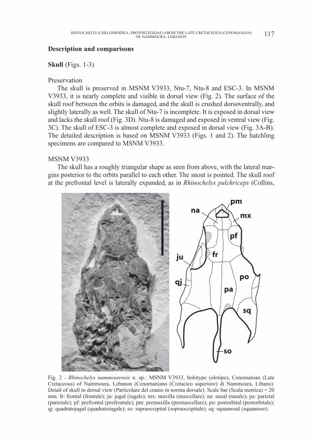
MSNM V3933The skull has a roughly triangular shape as seen from above, with the lateral mar-
gins posterior to the orbits parallel to each other. The snout is pointed. The skull roofat the prefrontal level is laterally expanded, as in Rhinochelys pulchriceps (Collins,
117RHINOCHELYS (CHELONIOIDEA: PROTOSTEGIDAE) FROM THE LATE CRETACEOUS (CENOMANIAN)OF NAMMOURA, LEBANON
Fig. 2 - Rhinochelys nammourensis n. sp.: MSNM V3933, holotype (olotipo), Cenomanian (LateCretaceous) of Nammoura, Lebanon (Cenomaniano (Cretacico superiore) di Nammoura, Libano).Detail of skull in dorsal view (Particolare del cranio in norma dorsale). Scale bar (Scala metrica) = 20mm. fr: frontal (frontale); ju: jugal (iugale); mx: maxilla (mascellare); na: nasal (nasale); pa: parietal(parietale); pf: prefrontal (prefrontale); pm: premaxilla (premascellare); po: postorbital (postorbitale);qj: quadratojugal (quadratoiugale); so: supraoccipital (sopraoccipitale); sq: squamosal (squamoso).
pm
mx
pf
na
fr
pa
po
sq
so
ju
qj
1970; Hirayama, 1994). The orbit is large and mainly directed laterally. The posteri-or part of the skull, although crushed, shows a deep temporal emargination, which ismuch deeper than that in the reconstruction of Rhinochelys pulchriceps (Hirayama,1994). The crista supraoccipitalis is very long.
Nasal: the anterior portion of both nasals is complete, but the surface of theposterior portion is damaged. The nasal is a large bone, which is about half thesize of the prefrontal. It is an anteroposteriorly elongate element forming theupper rim of the apertura narium externa. Anterolaterally, the nasal/maxilla sutureis well preserved. Posterolaterally, the nasal contacts the prefrontal. Posteriorly,the nasal meets the frontal, preventing the prefrontals from meeting one anotheron the midline.
The nasal is absent in derived Cheloniidae and in Dermochelyidae, but stillpresent in primitive cheloniids, such as Toxochelys latiremis and Porthochelys lat-iceps, in which a pair of small nasals is present (Zangerl, 1953b). AmongProtostegidae, a relatively large nasal, at least half the size of the prefrontal, is pres-ent only in Rhinochelys pulchriceps and Desmatochelys lowi (Williston, 1898;Collins, 1970; Hirayama, 1994; Elliot et al., 1997), while Santanachelys gaffneyi(Hirayama, 1998), Notochelone costata (Hirayama, unpublished data), andChelosphargis advesa (Hirayama, 1994) have a much smaller nasal, less than onethird the size of the prefrontal. The nasal is absent in advanced protostegids, suchas Protostega gigas (Wieland, 1906) and Archelon ischyros (Wieland, 1900a,1909). The nasal of Rhinochelys pulchriceps, however, is as large as the prefrontal,larger than in MSNM V3933 and Desmatochelys lowi (Hirayama, 1994).
Prefrontal: the left prefrontal is damaged, and although the outline of the right oneis complete, parts of its surface are damaged. The prefrontal forms the anterior part ofthe upper margin of the orbit, along which it is laterally expanded and swollen, as inRhinochelys pulchriceps (Collins, 1970; Hirayama, 1994). The prefrontal contacts themaxilla anteriorly by a nearly transverse suture. Anteromedially, it contacts the nasaland posteromedially the frontal. The prefrontals do not contact one another along themidline, as in most protostegids, including Santanachelys gaffneyi, Rhinochelys pul-chriceps and Desmatochelys lowi. In advanced protostegids (Archelon ischyros andProtostega gigas), the prefrontals meet one another along the midline.
Premaxilla: only the dorsal side and part of the anterior surface of both pre-maxillae are visible. The premaxilla forms the lower rim of the apertura nariumexterna and contacts the maxilla posterolaterally.
Maxilla: the maxilla is visible only dorsally and laterally. It forms the lateral rimof the apertura narium externa, the anterior rim, and the anterior portion of thelower rim of the orbit. On the skull roof, the maxilla contacts the nasal mediallyand the prefrontal posteriorly. The maxilla/jugal contact is visible on the left side,on the floor of the orbit.
Frontal: the surface of both frontals is damaged, but the right frontal is morecomplete. The morphology and contacts of the frontal in MSNM V3933 are simi-lar to those of Santanachelys gaffneyi and Rhinochelys pulchriceps. The frontalforms a small portion of the upper rim of the orbit between the prefrontal and thepostorbital, as in Santanachelys gaffneyi, Rhinochely pulchriceps andDesmatochelys lowi. In Archelon ischyros and Protostega gigas, the frontal does
118 HAIYAN TONG, REN HIRAYAMA, EDOUARD MAKHOUL & FRANÇOIS ESCUILLIÉ
not reach the orbital margin. The frontal contacts the nasal anteriorly by a shortsuture, the prefrontal anterolaterally, the parietal posteriorly and the postorbitalposterolaterally. The frontal in MSNM V3933 appears to be smaller than that ofRhinochelys pulchriceps and its anterior portion is narrower than in the latter.
Postorbital: both postorbitals are complete but crushed. The postorbital is ananteroposteriorly elongate element forming the posterior rim of the orbit. It con-tacts the frontal anteromedially by a short suture, the parietal medially by a longsuture, the jugal anterolaterally, the quadratojugal posterolaterally and thesquamosal posteriorly. Posteriorly, the postorbital forms a small portion of the tem-poral emargination, separating the parietal from the squamosal, as inSantanachelys gaffneyi, Rhinochelys pulchriceps and Desmatochelys lowi. InArchelon ischyros, Protostega gigas, dermochelyids and most cheloniids, the pos-torbital does not reach the temporal emargination.
Parietal: both parietals are complete but crushed. The parietal is an anteropos-teriorly elongate bone, which appears to be longer and narrower than that of R. pul-chriceps. It forms the medial portion of the temporal emargination and contacts thefrontal anteriorly, the postorbital laterally and the supraoccipital posteriorly by ashort suture. The parietal/squamosal contact is absent as in Santanachelys gaffneyi,Rhinochelys pulchriceps and Desmatochelys lowi, while this contact is present inmost Cheloniidae, Dermochelyidae, Archelon ischyros and Protostega gigas.
Quadratojugal: only the upper part of the quadratojugal is visible on both sidesbetween the jugal and the squamosal. It contacts the postorbital medially.
Jugal: the jugal is preserved on both sides but only the upper part is visible. Itforms the posteroventral margin of the orbit and contacts the postorbital medially,the quadratojugal posteriorly and the maxilla anteriorly. The jugal has no medialprocess, as in the other protostegids (Hirayama, 1998).
Squamosal: of the two squamosals, the right one is better preserved. This is atriangular element as seen from above. It forms the lateral portion of the temporalemargination, contacting the postorbital anteromedially and the quadratojugalanterolaterally. The squamosal of MSNM V3933 is similar to that of Santanachelysgaffneyi in shape, but it is clearly larger than in the latter. Desmatochelys lowi,Protostega gigas and Archelon ischyros also have a larger squamosal, like MSNMV3933, this bone is however wider in dorsal view in these forms than in MSNMV3933. The squamosal is not preserved in Rhinochelys pulchriceps; the recon-struction of the skull by Hirayama (1994) shows a small squamosal, which is sim-ilar to that of Santanachelys gaffneyi in size.
Supraoccipital: the supraoccipital is complete but somewhat deformed. It is ananteroposteriorly elongate element as seen from above. It forms the crista supraoc-cipitalis and contacts the parietals anteriorly. The crista supraoccipitalis is verylong, being longer than that of Santanachelys gaffneyi. This structure is not pre-served in Rhinochelys pulchriceps.
The skull scute sulci are not distinguishable because of the crushing of theskull roof. Other elements of the skull are not visible at the present state of thepreparation.
Hyoid bone: the posterior portion of the cornu branchiale I is visible on theright side of MSNM V3933. It is curved with the posterior end flattened.
119RHINOCHELYS (CHELONIOIDEA: PROTOSTEGIDAE) FROM THE LATE CRETACEOUS (CENOMANIAN)OF NAMMOURA, LEBANON
120 HAIYAN TONG, REN HIRAYAMA, EDOUARD MAKHOUL & FRANÇOIS ESCUILLIÉ
mxna
fr
pf
pa po
sqqj
ju
AB
DC
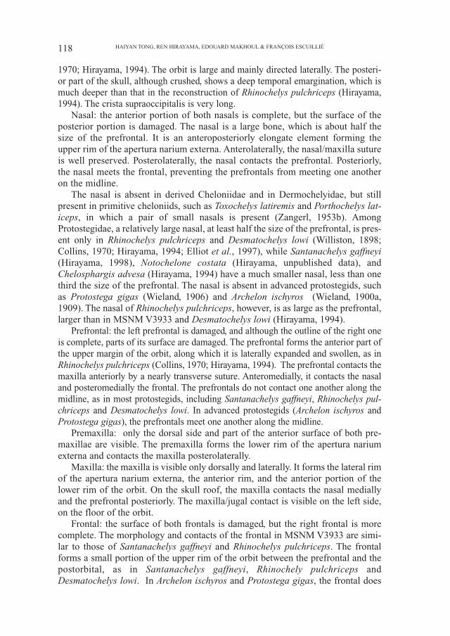
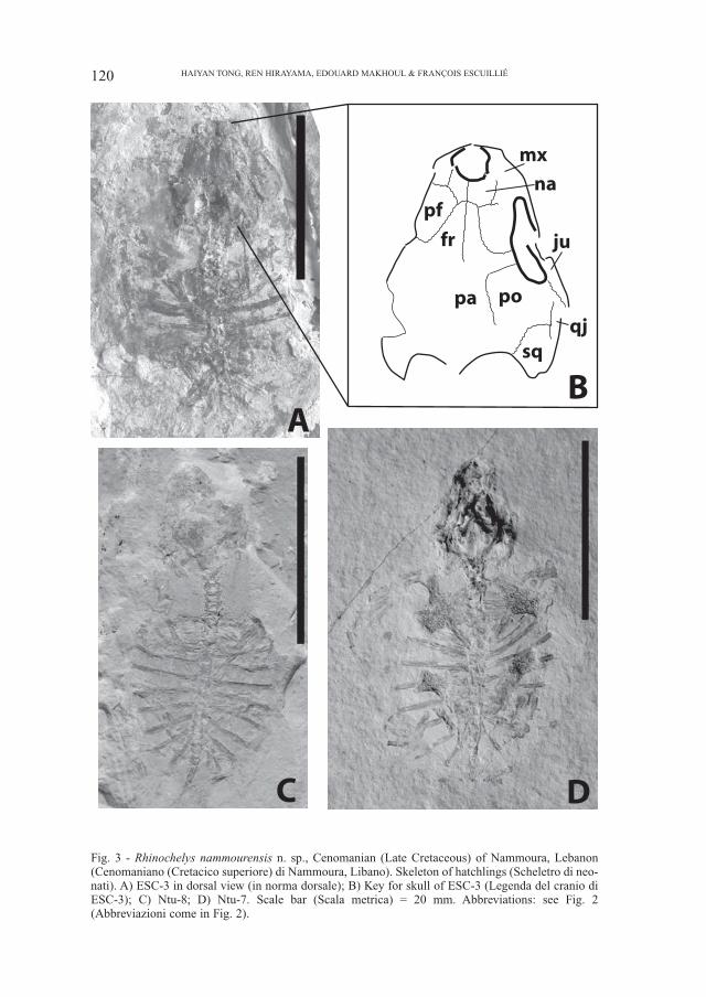
Fig. 3 - Rhinochelys nammourensis n. sp., Cenomanian (Late Cretaceous) of Nammoura, Lebanon(Cenomaniano (Cretacico superiore) di Nammoura, Libano). Skeleton of hatchlings (Scheletro di neo-nati). A) ESC-3 in dorsal view (in norma dorsale); B) Key for skull of ESC-3 (Legenda del cranio diESC-3); C) Ntu-8; D) Ntu-7. Scale bar (Scala metrica) = 20 mm. Abbreviations: see Fig. 2(Abbreviazioni come in Fig. 2).
Hatchling specimensThe skull of hatchling specimens Ntu-7, Ntu-8 and ESC-3 is large relative to the
shell and rather wide as seen in other neonate turtles. Ntu-7 and ESC-3 are visible indorsal view, ESC-3 has the skull roof partly preserved while in Ntu-7 the skull roofis damaged. The overall shape of the skull of these hatchlings, as seen from above,is similar to that of MSNM V3933, the skull margins posterior to the orbit being par-allel to each another and the anterior part of the skull roof being laterally expanded.The morphology and the relationships of the skull elements, including the maxilla,nasal, prefrontal, frontal, postorbital, squamosal, jugal and quadratojugal in ESC-3are similar to those of MSNM V3933 (Fig. 3).
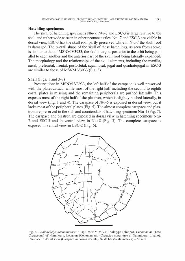
Shell (Figs. 1 and 3-7)Preservation: in MSNM V3933, the left half of the carapace is well preserved
with the plates in situ, while most of the right half including the second to eighthcostal plates is missing and the remaining peripherals are pushed laterally. Thisexposes most of the right half of the plastron, which is slightly pushed laterally, indorsal view (Fig. 1 and 4). The carapace of Ntu-6 is exposed in dorsal view, but itlacks most of the peripheral plates (Fig. 5). The almost complete carapace and plas-tron are preserved in the slab and counterslab of hatchling specimen Ntu-1 (Fig. 7).The carapace and plastron are exposed in dorsal view in hatchling specimens Ntu-7 and ESC-3 and in ventral view in Ntu-8 (Fig. 3). The complete carapace isexposed in ventral view in ESC-2 (Fig. 6).
121RHINOCHELYS (CHELONIOIDEA: PROTOSTEGIDAE) FROM THE LATE CRETACEOUS (CENOMANIAN)OF NAMMOURA, LEBANON
Fig. 4 - Rhinochelys nammourensis n. sp.: MSNM V3933, holotype (olotipo), Cenomanian (LateCretaceous) of Nammoura, Lebanon (Cenomaniano (Cretacico superiore) di Nammoura, Libano).Carapace in dorsal view (Carapace in norma dorsale). Scale bar (Scala metrica) = 50 mm.
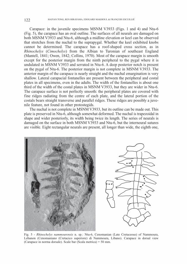
Carapace: in the juvenile specimens MSNM V3933 (Figs. 1 and 4) and Ntu-6(Fig. 5), the carapace has an oval outline. The surfaces of all neurals are damaged onboth MSNM V3933 and Ntu-6, although a midline elevation or keel can be observedthat stretches from the nuchal to the suprapygal. Whether the keel exhibited knobscannot be determined. The carapace has a roof-shaped cross section, as inRhinochelys (Cimochelys) from the Albian to Turonian of southeast England(Mantell, 1841; Owen, 1842; Collins, 1970). Most of the carapace margin is smoothexcept for the posterior margin from the ninth peripheral to the pygal where it isundulated in MSNM V3933 and serrated in Ntu-6. A deep posterior notch is presenton the pygal of Ntu-6. The posterior margin is not complete in MSNM V3933. Theanterior margin of the carapace is nearly straight and the nuchal emargination is veryshallow. Lateral carapacial fontanelles are present between the peripheral and costalplates in all specimens, even in the adults. The width of the fontanelles is about onethird of the width of the costal plates in MSNM V3933, but they are wider in Ntu-6.The carapace surface is not perfectly smooth: the peripheral plates are covered withfine ridges radiating from the centre of each plate, and the lateral portion of thecostals bears straight transverse and parallel ridges. These ridges are possibly a juve-nile feature, not found in other protostegids.
The nuchal is not complete in MSNM V3933, but its outline can be made out. Thisplate is preserved in Ntu-6, although somewhat deformed. The nuchal is trapezoidal inshape and wider posteriorly, its width being twice its length. The series of neurals isdamaged on the surface in both MSNM V3933 and Ntu-6, but the interneural suturesare visible. Eight rectangular neurals are present, all longer than wide, the eighth one,
122 HAIYAN TONG, REN HIRAYAMA, EDOUARD MAKHOUL & FRANÇOIS ESCUILLIÉ
Fig. 5 - Rhinochelys nammourensis n. sp.: Ntu-6, Cenomanian (Late Cretaceous) of Nammoura,Lebanon (Cenomaniano (Cretacico superiore) di Nammoura, Libano). Carapace in dorsal view(Carapace in norma dorsale). Scale bar (Scala metrica) = 50 mm.
however, is shorter than the other neurals. There is one suprapygal as in most protoste-gids except for Rhinochelys (Cimochleys) and Desmatochelys lowi (Zangerl & Sloan,1960; Collins, 1970; Hirayama, 1994, 1997). The suprapygal is large, roughly semi-circular in shape and slightly wider than long. In Rhinochelys (Cimochelys), twosuprapygals are present, the first one is trapezoidal or triangular and the second one isrectangular, wider than long and larger than the first suprapygal. Eight costal plates arepresent as in Santanachelys gaffneyi and Rhinochelys (Cimochelys), with the lateralportion narrowed to form a peg fitting into a socket on the peripheral. Protostega gigasand Archelon ischyros have nine more blade-like costals. The complete series of elevenperipherals is preserved on the left side of MSNM V3933. The first peripheral isroughly triangular and wider than long. The pygal plate is not complete in MSNMV3933, it is nearly complete in Ntu-6, but with the surface damaged on both. Thepygal has a deep and wide notch on the posterior margin in Ntu-6. This is differentfrom Rhinochelys (Cimochelys) in which such a notch is absent and the posterior endof the carapace is pointed (BM 28706 and BM 36751) as in all other protostegids,except for Santanachelys gaffneyi (Hirayama, 1994, 1997, 1998).
The scute sulci are weakly developed but partly discernable on MSNM V3933and Ntu-6. The first, third and fourth vertebrals are wider than long while the sec-ond one is much longer than wide. The intervertebral sulci cross the first neuraland costal, the third neural and costal, the fifth neural and costal and the eighthneural and costal plates respectively. The width of the second to fourth vertebralsincreases strongly from front to back, with the lateral margins strongly angled. InRhinochelys (Cimochelys), the scute sulci are more deeply printed than in theNammoura turtles; the vertebral scutes extend to at least half the width of the costalplates and their width remains unchanged or becomes slightly smaller from frontto back (Owen, 1842; Collins, 1970).
The carapace of adult specimen ESC-2 is slightly more elongate than in thejuveniles and it is almost flat in ventral view. The carapace margin is smooththroughout. Although the posterior margin of the carapace is a little damaged, aposterior notch is apparently present on the pygal. This notch is shallower in ESC-2 than in the juveniles. The carapacial fontanelles are more closed than in the juve-niles, being about one fourth of the width of the costal plates. The morphology ofplates in ESC-2, including the nuchal, the neural series, the pygal and the periph-erals is similar to those of the juveniles.
The hatchling specimens Ntu-1, Ntu-7, Ntu-8 and ESC-3 have a more roundedcarapace than the juveniles. Their costals are blade-like and completely separatedfrom one another by large fontanelles due to poor dermal ossification. No periph-erals are preserved in Ntu-1, Ntu-8 and ESC-3. Some yellow spots are presentaround the carapace of Ntu-7 (Fig. 3D), however they seem not to be osseous.
Plastron: in MSNM V3933, the right part of the plastron, including the righthyoplastron, hypoplastron and xiphiplastron, is visible in dorsal view, through thedamaged carapace. However, the anterior part of the plastron, including the epi-plastron and entoplastron, is not visible. The hyoplastron and hypoplastron arelarge and star-shaped plates with short finger-like prongs which are well visible onthe medial and lateral margins. The hypoplastron is smaller than the hyoplastron,as in Archelon ischyros and Protostega gigas. The hyoplastron contacts thehypoplastron by a long and strongly interdigitated suture. There is a large centralfontanelle between the hyoplastra and hypoplastra. The central plastral fontanelle
123RHINOCHELYS (CHELONIOIDEA: PROTOSTEGIDAE) FROM THE LATE CRETACEOUS (CENOMANIAN)OF NAMMOURA, LEBANON
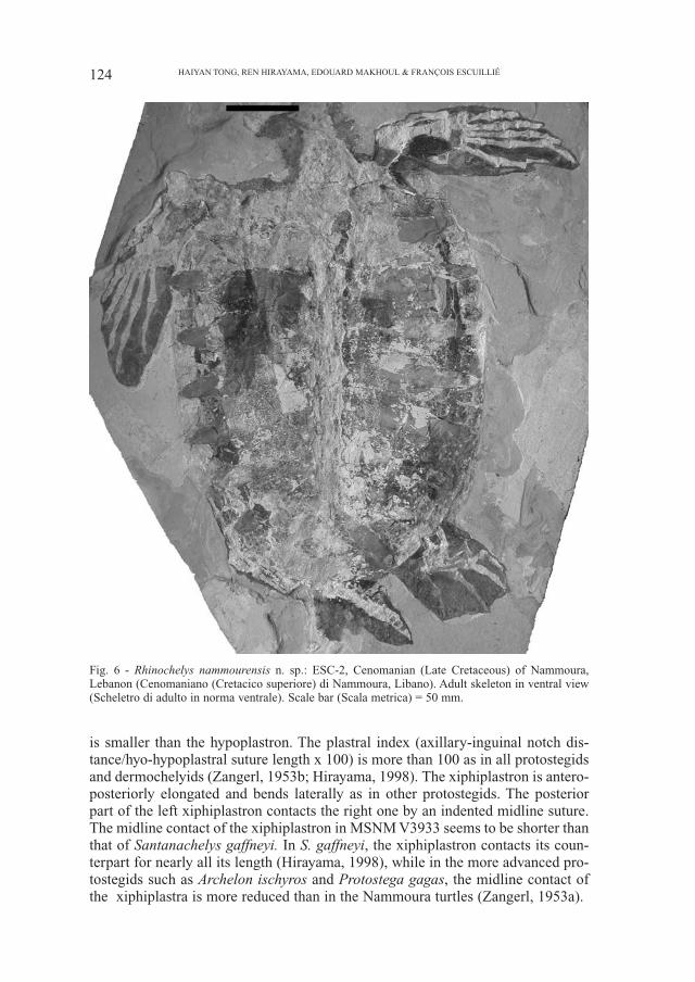
is smaller than the hypoplastron. The plastral index (axillary-inguinal notch dis-tance/hyo-hypoplastral suture length x 100) is more than 100 as in all protostegidsand dermochelyids (Zangerl, 1953b; Hirayama, 1998). The xiphiplastron is antero-posteriorly elongated and bends laterally as in other protostegids. The posteriorpart of the left xiphiplastron contacts the right one by an indented midline suture.The midline contact of the xiphiplastron in MSNM V3933 seems to be shorter thanthat of Santanachelys gaffneyi. In S. gaffneyi, the xiphiplastron contacts its coun-terpart for nearly all its length (Hirayama, 1998), while in the more advanced pro-tostegids such as Archelon ischyros and Protostega gagas, the midline contact ofthe xiphiplastra is more reduced than in the Nammoura turtles (Zangerl, 1953a).
124 HAIYAN TONG, REN HIRAYAMA, EDOUARD MAKHOUL & FRANÇOIS ESCUILLIÉ
Fig. 6 - Rhinochelys nammourensis n. sp.: ESC-2, Cenomanian (Late Cretaceous) of Nammoura,Lebanon (Cenomaniano (Cretacico superiore) di Nammoura, Libano). Adult skeleton in ventral view(Scheletro di adulto in norma ventrale). Scale bar (Scala metrica) = 50 mm.
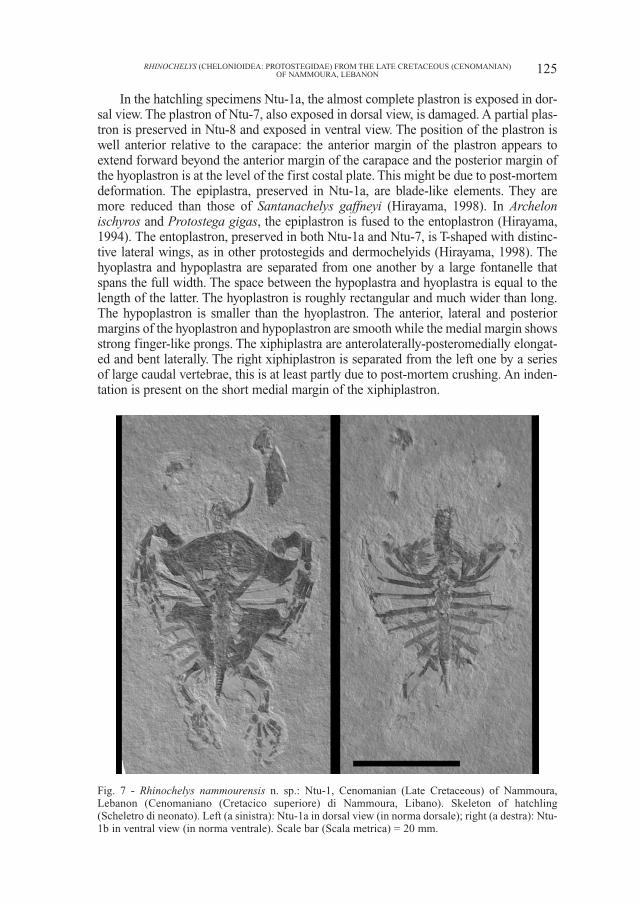
In the hatchling specimens Ntu-1a, the almost complete plastron is exposed in dor-sal view. The plastron of Ntu-7, also exposed in dorsal view, is damaged. A partial plas-tron is preserved in Ntu-8 and exposed in ventral view. The position of the plastron iswell anterior relative to the carapace: the anterior margin of the plastron appears toextend forward beyond the anterior margin of the carapace and the posterior margin ofthe hyoplastron is at the level of the first costal plate. This might be due to post-mortemdeformation. The epiplastra, preserved in Ntu-1a, are blade-like elements. They aremore reduced than those of Santanachelys gaffneyi (Hirayama, 1998). In Archelonischyros and Protostega gigas, the epiplastron is fused to the entoplastron (Hirayama,1994). The entoplastron, preserved in both Ntu-1a and Ntu-7, is T-shaped with distinc-tive lateral wings, as in other protostegids and dermochelyids (Hirayama, 1998). Thehyoplastra and hypoplastra are separated from one another by a large fontanelle thatspans the full width. The space between the hypoplastra and hyoplastra is equal to thelength of the latter. The hyoplastron is roughly rectangular and much wider than long.The hypoplastron is smaller than the hyoplastron. The anterior, lateral and posteriormargins of the hyoplastron and hypoplastron are smooth while the medial margin showsstrong finger-like prongs. The xiphiplastra are anterolaterally-posteromedially elongat-ed and bent laterally. The right xiphiplastron is separated from the left one by a seriesof large caudal vertebrae, this is at least partly due to post-mortem crushing. An inden-tation is present on the short medial margin of the xiphiplastron.
125RHINOCHELYS (CHELONIOIDEA: PROTOSTEGIDAE) FROM THE LATE CRETACEOUS (CENOMANIAN)OF NAMMOURA, LEBANON
Fig. 7 - Rhinochelys nammourensis n. sp.: Ntu-1, Cenomanian (Late Cretaceous) of Nammoura,Lebanon (Cenomaniano (Cretacico superiore) di Nammoura, Libano). Skeleton of hatchling(Scheletro di neonato). Left (a sinistra): Ntu-1a in dorsal view (in norma dorsale); right (a destra): Ntu-1b in ventral view (in norma ventrale). Scale bar (Scala metrica) = 20 mm.
Vertebral columnCervical vertebrae: four cervical vertebrae are visible in MSNM V3933,
exposed in lateral view. Only the neural arches are visible, showing strongzygapophyses. A series of articulated cervicals is preserved in Ntu-1, mostly inNtu-1b, exposed in ventrolateral view. As in other chelonioid sea turtles, the cen-tra are stout and short. The best preserved vertebral centra, the 5th to 7th ones,show a slight ventral keel. In Ntu-7, Ntu-8, ESC-3 and ESC-2, the cervical verte-brae are preserved but they are either crushed or damaged. There is no articularsurface exposed.
Caudal vertebrae: no caudal vertebrae are visible in MSNM V3933. A series offourteen articulated caudal vertebrae is preserved in ESC-2 and there are sixteencaudal vertebrae in Ntu-1. Caudal vertebrae are also preserved in Ntu-7, Ntu-8 andESC-3, but they are damaged. The tail of ESC-2 is long, extending well beyond theposterior margin of the carapace, while that of hatchling Ntu-1 is short, extendingbarely beyond the posterior carapace margin. In both specimens, the centra arestout and short, their size decreases from front to back.
Pectoral girdle The pectoral girdle is preserved only in the hatchlings, and is best preserved in
Ntu-1. In this specimen, the blade-like coracoid is very long as in other advancedchelonioids. It is much longer than the humerus and extends posteromedially to theanterior margin of the hypoplastron. The scapular angle, formed by the acromionand the scapular prong, is about 120°.
Pelvic girdle The pelvic girdle is visible only in hatchlings, but in a very bad condition. An
accurate description is impossible.
Forelimb (Figs. 1, 6-8) The forelimbs are preserved on both sides of MSNM V3933 and ESC-2. Ntu-4
is an isolated right front flipper, but most bones are missing, although their imprintis well preserved. Both forelimbs are preserved in Ntu-1. An incomplete left fore-limb is preserved in Ntu-7. In Ntu-8, only the imprint of the right humerus is pre-served.
Humerus: in the adult ESC-2, both humeri are damaged and exposed in ventralview. The right humerus is the better-preserved element and although its surface isdamaged, the outline is preserved. The humerus is robust, with a straight shaft. Thelarge medial process is taller than the caput humeri. The small lateral process is sit-uated mid-way along the shaft of the humerus and is restricted to the anterior sur-face of the humeral shaft as in other protostegids (Hirayama, 1992). The posteriormargin of the humeral shaft is strongly concave as in Archelon ischyros andProtostega gigas (Wieland, 1900b).
In the juvenile MSNM V3933, only the distal portion of the right humerus isvisible. The distal end is expanded and the posterior margin of the humeral shaft isstrongly concave as in ESC-2.
In the hatchling specimens, the humerus is nearly complete in Ntu-1a, but lack-
126 HAIYAN TONG, REN HIRAYAMA, EDOUARD MAKHOUL & FRANÇOIS ESCUILLIÉ
127RHINOCHELYS (CHELONIOIDEA: PROTOSTEGIDAE) FROM THE LATE CRETACEOUS (CENOMANIAN)OF NAMMOURA, LEBANON
humerus
radius
ulna
ulnare
pisiform
distal carpals
IM
I1
I2
intermedium
centrale
VM
V1
V2
V3
II3
III3
IV3A B C
D E
F G
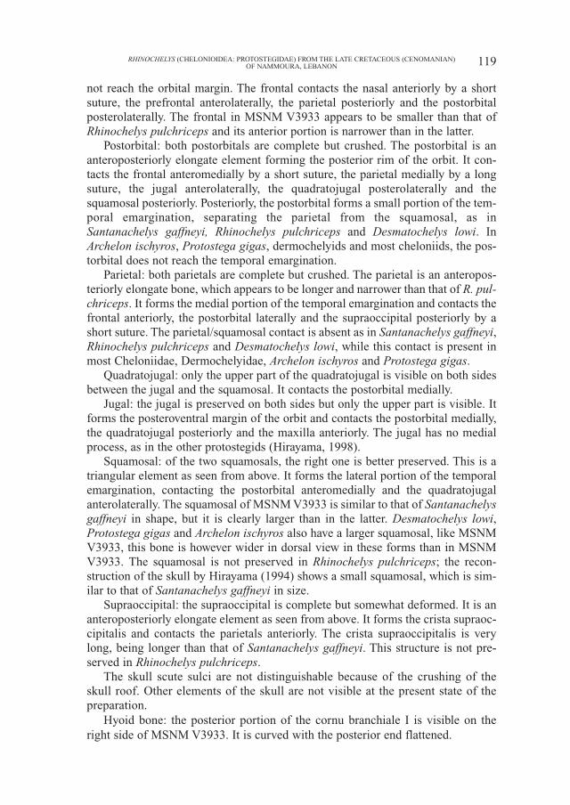
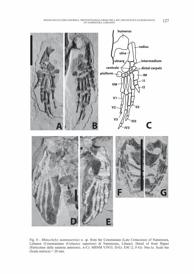
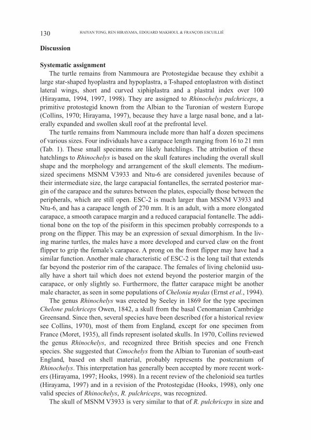
Fig. 8 - Rhinochelys nammourensis n. sp. from the Cenomanian (Late Cretaceous) of Nammoura,Lebanon (Cenomaniano (Cretacico superiore) di Nammoura, Libano). Detail of front flipper(Particolare della natatoia anteriore). A-C): MSNM V3933; D-E): ESC-2; F-G): Ntu-1a. Scale bar(Scala metrica) = 20 mm.
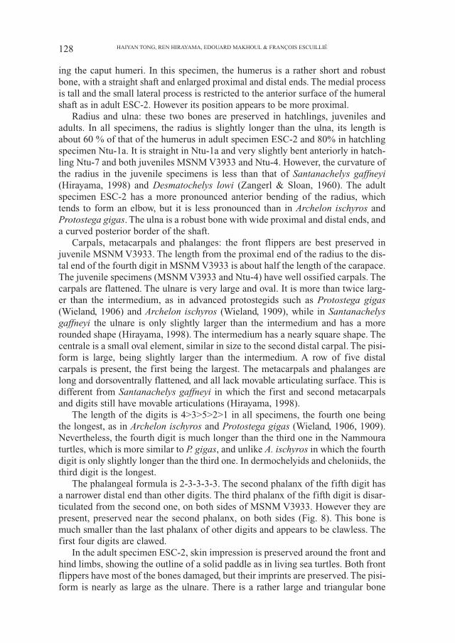
ing the caput humeri. In this specimen, the humerus is a rather short and robustbone, with a straight shaft and enlarged proximal and distal ends. The medial processis tall and the small lateral process is restricted to the anterior surface of the humeralshaft as in adult ESC-2. However its position appears to be more proximal.
Radius and ulna: these two bones are preserved in hatchlings, juveniles andadults. In all specimens, the radius is slightly longer than the ulna, its length isabout 60 % of that of the humerus in adult specimen ESC-2 and 80% in hatchlingspecimen Ntu-1a. It is straight in Ntu-1a and very slightly bent anteriorly in hatch-ling Ntu-7 and both juveniles MSNM V3933 and Ntu-4. However, the curvature ofthe radius in the juvenile specimens is less than that of Santanachelys gaffneyi(Hirayama, 1998) and Desmatochelys lowi (Zangerl & Sloan, 1960). The adultspecimen ESC-2 has a more pronounced anterior bending of the radius, whichtends to form an elbow, but it is less pronounced than in Archelon ischyros andProtostega gigas. The ulna is a robust bone with wide proximal and distal ends, anda curved posterior border of the shaft.
Carpals, metacarpals and phalanges: the front flippers are best preserved injuvenile MSNM V3933. The length from the proximal end of the radius to the dis-tal end of the fourth digit in MSNM V3933 is about half the length of the carapace.The juvenile specimens (MSNM V3933 and Ntu-4) have well ossified carpals. Thecarpals are flattened. The ulnare is very large and oval. It is more than twice larg-er than the intermedium, as in advanced protostegids such as Protostega gigas(Wieland, 1906) and Archelon ischyros (Wieland, 1909), while in Santanachelysgaffneyi the ulnare is only slightly larger than the intermedium and has a morerounded shape (Hirayama, 1998). The intermedium has a nearly square shape. Thecentrale is a small oval element, similar in size to the second distal carpal. The pisi-form is large, being slightly larger than the intermedium. A row of five distalcarpals is present, the first being the largest. The metacarpals and phalanges arelong and dorsoventrally flattened, and all lack movable articulating surface. This isdifferent from Santanachelys gaffneyi in which the first and second metacarpalsand digits still have movable articulations (Hirayama, 1998).
The length of the digits is 4>3>5>2>1 in all specimens, the fourth one beingthe longest, as in Archelon ischyros and Protostega gigas (Wieland, 1906, 1909).Nevertheless, the fourth digit is much longer than the third one in the Nammouraturtles, which is more similar to P. gigas, and unlike A. ischyros in which the fourthdigit is only slightly longer than the third one. In dermochelyids and cheloniids, thethird digit is the longest.
The phalangeal formula is 2-3-3-3-3. The second phalanx of the fifth digit hasa narrower distal end than other digits. The third phalanx of the fifth digit is disar-ticulated from the second one, on both sides of MSNM V3933. However they arepresent, preserved near the second phalanx, on both sides (Fig. 8). This bone ismuch smaller than the last phalanx of other digits and appears to be clawless. Thefirst four digits are clawed.
In the adult specimen ESC-2, skin impression is preserved around the front andhind limbs, showing the outline of a solid paddle as in living sea turtles. Both frontflippers have most of the bones damaged, but their imprints are preserved. The pisi-form is nearly as large as the ulnare. There is a rather large and triangular bone
128 HAIYAN TONG, REN HIRAYAMA, EDOUARD MAKHOUL & FRANÇOIS ESCUILLIÉ
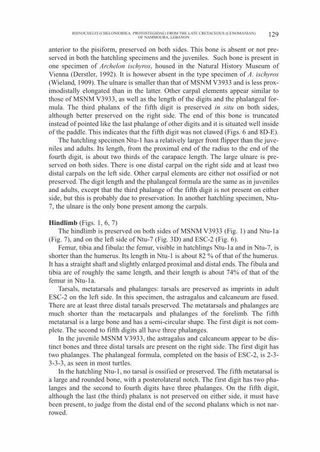
anterior to the pisiform, preserved on both sides. This bone is absent or not pre-served in both the hatchling specimens and the juveniles. Such bone is present inone specimen of Archelon ischyros, housed in the Natural History Museum ofVienna (Derstler, 1992). It is however absent in the type specimen of A. ischyros(Wieland, 1909). The ulnare is smaller than that of MSNM V3933 and is less prox-imodistally elongated than in the latter. Other carpal elements appear similar tothose of MSNM V3933, as well as the length of the digits and the phalangeal for-mula. The third phalanx of the fifth digit is preserved in situ on both sides,although better preserved on the right side. The end of this bone is truncatedinstead of pointed like the last phalange of other digits and it is situated well insideof the paddle. This indicates that the fifth digit was not clawed (Figs. 6 and 8D-E).
The hatchling specimen Ntu-1 has a relatively larger front flipper than the juve-niles and adults. Its length, from the proximal end of the radius to the end of thefourth digit, is about two thirds of the carapace length. The large ulnare is pre-served on both sides. There is one distal carpal on the right side and at least twodistal carpals on the left side. Other carpal elements are either not ossified or notpreserved. The digit length and the phalangeal formula are the same as in juvenilesand adults, except that the third phalange of the fifth digit is not present on eitherside, but this is probably due to preservation. In another hatchling specimen, Ntu-7, the ulnare is the only bone present among the carpals.
Hindlimb (Figs. 1, 6, 7)The hindlimb is preserved on both sides of MSNM V3933 (Fig. 1) and Ntu-1a
(Fig. 7), and on the left side of Ntu-7 (Fig. 3D) and ESC-2 (Fig. 6).Femur, tibia and fibula: the femur, visible in hatchlings Ntu-1a and in Ntu-7, is
shorter than the humerus. Its length in Ntu-1 is about 82 % of that of the humerus.It has a straight shaft and slightly enlarged proximal and distal ends. The fibula andtibia are of roughly the same length, and their length is about 74% of that of thefemur in Ntu-1a.
Tarsals, metatarsals and phalanges: tarsals are preserved as imprints in adultESC-2 on the left side. In this specimen, the astragalus and calcaneum are fused.There are at least three distal tarsals preserved. The metatarsals and phalanges aremuch shorter than the metacarpals and phalanges of the forelimb. The fifthmetatarsal is a large bone and has a semi-circular shape. The first digit is not com-plete. The second to fifth digits all have three phalanges.
In the juvenile MSNM V3933, the astragalus and calcaneum appear to be dis-tinct bones and three distal tarsals are present on the right side. The first digit hastwo phalanges. The phalangeal formula, completed on the basis of ESC-2, is 2-3-3-3-3, as seen in most turtles.
In the hatchling Ntu-1, no tarsal is ossified or preserved. The fifth metatarsal isa large and rounded bone, with a posterolateral notch. The first digit has two pha-langes and the second to fourth digits have three phalanges. On the fifth digit,although the last (the third) phalanx is not preserved on either side, it must havebeen present, to judge from the distal end of the second phalanx which is not nar-rowed.
129RHINOCHELYS (CHELONIOIDEA: PROTOSTEGIDAE) FROM THE LATE CRETACEOUS (CENOMANIAN)OF NAMMOURA, LEBANON
Discussion
Systematic assignmentThe turtle remains from Nammoura are Protostegidae because they exhibit a
large star-shaped hyoplastra and hypoplastra, a T-shaped entoplastron with distinctlateral wings, short and curved xiphiplastra and a plastral index over 100(Hirayama, 1994, 1997, 1998). They are assigned to Rhinochelys pulchriceps, aprimitive protostegid known from the Albian to the Turonian of western Europe(Collins, 1970; Hirayama, 1997), because they have a large nasal bone, and a lat-erally expanded and swollen skull roof at the prefrontal level.
The turtle remains from Nammoura include more than half a dozen specimensof various sizes. Four individuals have a carapace length ranging from 16 to 21 mm(Tab. 1). These small specimens are likely hatchlings. The attribution of thesehatchlings to Rhinochelys is based on the skull features including the overall skullshape and the morphology and arrangement of the skull elements. The medium-sized specimens MSNM V3933 and Ntu-6 are considered juveniles because oftheir intermediate size, the large carapacial fontanelles, the serrated posterior mar-gin of the carapace and the sutures between the plates, especially those between theperipherals, which are still open. ESC-2 is much larger than MSNM V3933 andNtu-6, and has a carapace length of 270 mm. It is an adult, with a more elongatedcarapace, a smooth carapace margin and a reduced carapacial fontanelle. The addi-tional bone on the top of the pisiform in this specimen probably corresponds to aprong on the flipper. This may be an expression of sexual dimorphism. In the liv-ing marine turtles, the males have a more developed and curved claw on the frontflipper to grip the female’s carapace. A prong on the front flipper may have had asimilar function. Another male characteristic of ESC-2 is the long tail that extendsfar beyond the posterior rim of the carapace. The females of living cheloniid usu-ally have a short tail which does not extend beyond the posterior margin of thecarapace, or only slightly so. Furthermore, the flatter carapace might be anothermale character, as seen in some populations of Chelonia mydas (Ernst et al., 1994).
The genus Rhinochelys was erected by Seeley in 1869 for the type specimenChelone pulchriceps Owen, 1842, a skull from the basal Cenomanian CambridgeGreensand. Since then, several species have been described (for a historical reviewsee Collins, 1970), most of them from England, except for one specimen fromFrance (Moret, 1935), all finds represent isolated skulls. In 1970, Collins reviewedthe genus Rhinochelys, and recognized three British species and one Frenchspecies. She suggested that Cimochelys from the Albian to Turonian of south-eastEngland, based on shell material, probably represents the postcranium ofRhinochelys. This interpretation has generally been accepted by more recent work-ers (Hirayama, 1997; Hooks, 1998). In a recent review of the chelonioid sea turtles(Hirayama, 1997) and in a revision of the Protostegidae (Hooks, 1998), only onevalid species of Rhinochelys, R. pulchriceps, was recognized.
The skull of MSNM V3933 is very similar to that of R. pulchriceps in size and
130 HAIYAN TONG, REN HIRAYAMA, EDOUARD MAKHOUL & FRANÇOIS ESCUILLIÉ
morphology. However some differences can be observed: in comparison with R.pulchriceps, the skull of MSNM V3933 is more elongate, with a smaller nasal, thefrontal is smaller with a narrower anterior portion and the parietal is more elongate;the temporal emargination is deeper, being more than half the length of the pari-etal. In R. pulchriceps, the temporal emargination is ‘about a third of the length ofthe parietal’ (Collins, 1970).
The shell of the Nammoura turtles is that of primitive protostegid and similarto that of Cimochelys in its general morphology, including the carapace outline,roof-shaped cross section, size of carapacial fontanelles, shape of hyoplastron andhypoplastron. This supports the hypothesis that Rhinochelys and Cimochelys rep-resents the same taxon. However the carapace of the Nammoura turtles slopes lesssharply toward the sides and therefore is more flattened. This seems not to be dueto post-mortem deformation since both MSNM V3933 and Ntu-6 have a similarshell curvature. Other differences between the Nammoura turtles and Rhinochelys(Cimochelys) are found in the suprapygal and pygal. The Nammoura turtles haveone suprapygal while Rhinochelys (Cimochelys) has two suprapygals.Furthermore, the width of the vertebral scutes of the Nammoura turtles increasefrom the second to the fourth, while these scutes have a similar width inRhinochelys (Cimochelys). These differences justify the erection of a new speciesof Rhinochelys: R. nammourensis.
Front flipper characters (Figs. 8-9)Our knowledge on the complete front flipper of protostegids was limited to date
to advanced members such as Protostega gigas (Wieland, 1902, 1906) andArchelon ischyros (Wieland, 1909), in addition to the incomplete but articulatedflippers of Santanachelys gaffneyi (Hirayama, 1998), Desmatochelys lowi (Zangerl& Sloan, 1960) and Terlinguachelys fischbecki (Lehman & Tomlinson, 2004), anda few elements in Calcarichelys gemma (Hooks, 1998). Wieland (1902, 1906)studied the flippers of Toxochelys latiremis, Archelon ischyros and Protostegagigas and noticed some evolutionary trends in chelonioids, among them the elon-gation of some digits, and a great and persistent increase in size of the pisiform.
The complete and articulated front flippers of the Nammoura turtles provideimportant information on their structure and evolution, especially on the propor-tion between the different elements. The flippers of the Nammoura turtles are morederived than those of Santanachelys gaffneyi because the ulnare is more proxi-modistally elongate and much larger than the intermedium and because allmetacarpals and phalanges lack movable articulations. In this respect, the flipperof the Nammoura turtles more closely resembles those of advanced protostegids,such as Archelon ischyros and Protostega gigas. However, the second digit in theNammoura turtles remains short, a primitive feature found also in Santanachelysgaffneyi.
The comparisons made with other chelonioid sea turtles reveal some interest-ing features of the family Protostegidae (Tab. 2):
131RHINOCHELYS (CHELONIOIDEA: PROTOSTEGIDAE) FROM THE LATE CRETACEOUS (CENOMANIAN)OF NAMMOURA, LEBANON
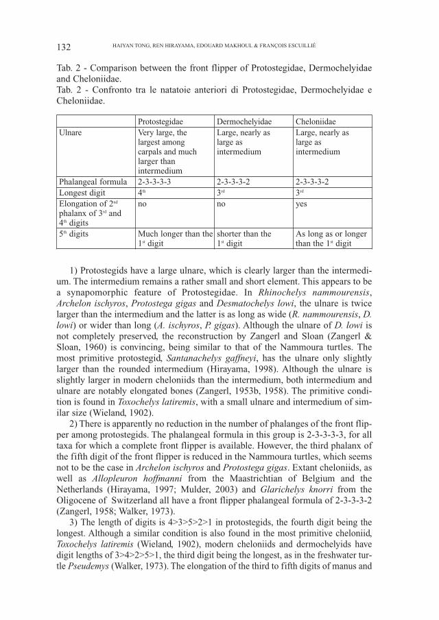
Tab. 2 - Comparison between the front flipper of Protostegidae, Dermochelyidaeand Cheloniidae.Tab. 2 - Confronto tra le natatoie anteriori di Protostegidae, Dermochelyidae eCheloniidae.
Protostegidae Dermochelyidae CheloniidaeUlnare Very large, the Large, nearly as Large, nearly as
largest among large as large ascarpals and much intermedium intermediumlarger thanintermedium
Phalangeal formula 2-3-3-3-3 2-3-3-3-2 2-3-3-3-2Longest digit 4th 3rd 3rd
Elongation of 2nd no no yesphalanx of 3rd and4th digits5th digits Much longer than the shorter than the As long as or longer
1st digit 1st digit than the 1st digit
1) Protostegids have a large ulnare, which is clearly larger than the intermedi-um. The intermedium remains a rather small and short element. This appears to bea synapomorphic feature of Protostegidae. In Rhinochelys nammourensis,Archelon ischyros, Protostega gigas and Desmatochelys lowi, the ulnare is twicelarger than the intermedium and the latter is as long as wide (R. nammourensis, D.lowi) or wider than long (A. ischyros, P. gigas). Although the ulnare of D. lowi isnot completely preserved, the reconstruction by Zangerl and Sloan (Zangerl &Sloan, 1960) is convincing, being similar to that of the Nammoura turtles. Themost primitive protostegid, Santanachelys gaffneyi, has the ulnare only slightlylarger than the rounded intermedium (Hirayama, 1998). Although the ulnare isslightly larger in modern cheloniids than the intermedium, both intermedium andulnare are notably elongated bones (Zangerl, 1953b, 1958). The primitive condi-tion is found in Toxochelys latiremis, with a small ulnare and intermedium of sim-ilar size (Wieland, 1902).
2) There is apparently no reduction in the number of phalanges of the front flip-per among protostegids. The phalangeal formula in this group is 2-3-3-3-3, for alltaxa for which a complete front flipper is available. However, the third phalanx ofthe fifth digit of the front flipper is reduced in the Nammoura turtles, which seemsnot to be the case in Archelon ischyros and Protostega gigas. Extant cheloniids, aswell as Allopleuron hoffmanni from the Maastrichtian of Belgium and theNetherlands (Hirayama, 1997; Mulder, 2003) and Glarichelys knorri from theOligocene of Switzerland all have a front flipper phalangeal formula of 2-3-3-3-2(Zangerl, 1958; Walker, 1973).
3) The length of digits is 4>3>5>2>1 in protostegids, the fourth digit being thelongest. Although a similar condition is also found in the most primitive cheloniid,Toxochelys latiremis (Wieland, 1902), modern cheloniids and dermochelyids havedigit lengths of 3>4>2>5>1, the third digit being the longest, as in the freshwater tur-tle Pseudemys (Walker, 1973). The elongation of the third to fifth digits of manus and
132 HAIYAN TONG, REN HIRAYAMA, EDOUARD MAKHOUL & FRANÇOIS ESCUILLIÉ
pes is considered as a synapomorphic feature of Chelonioidea (Hirayama, 1994), acharacter already recognized by (Wieland, 1909). Zangerl (1953b) pointed out thatthe second to fourth digits of the front flipper are greatly elongated in cheloniids. Thefifth digit of cheloniids is generally short, being shorter than the second one and onlyslightly longer than the first digit. In protostegids, the fifth digit is greatly elongatedand is much longer than the second digit.
133RHINOCHELYS (CHELONIOIDEA: PROTOSTEGIDAE) FROM THE LATE CRETACEOUS (CENOMANIAN)OF NAMMOURA, LEBANON
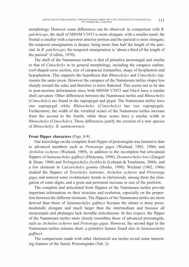
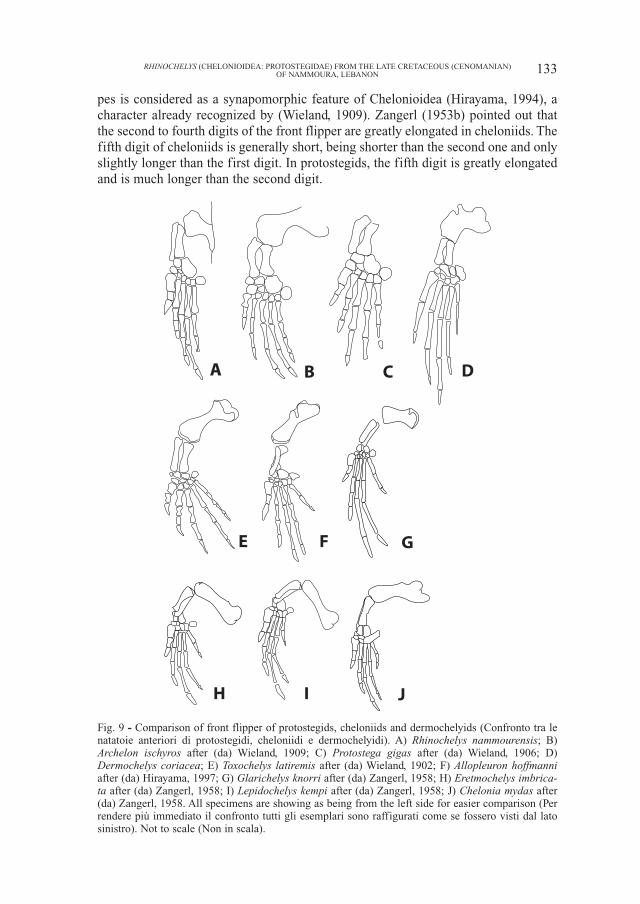
Fig. 9 - Comparison of front flipper of protostegids, cheloniids and dermochelyids (Confronto tra lenatatoie anteriori di protostegidi, cheloniidi e dermochelyidi). A) Rhinochelys nammourensis; B)Archelon ischyros after (da) Wieland, 1909; C) Protostega gigas after (da) Wieland, 1906; D)Dermochelys coriacea; E) Toxochelys latiremis after (da) Wieland, 1902; F) Allopleuron hoffmanniafter (da) Hirayama, 1997; G) Glarichelys knorri after (da) Zangerl, 1958; H) Eretmochelys imbrica-ta after (da) Zangerl, 1958; I) Lepidochelys kempi after (da) Zangerl, 1958; J) Chelonia mydas after(da) Zangerl, 1958. All specimens are showing as being from the left side for easier comparison (Perrendere più immediato il confronto tutti gli esemplari sono raffigurati come se fossero visti dal latosinistro). Not to scale (Non in scala).
A B C D
E F G
H I J
The great elongation of the third and the fourth digits in advanced cheloniids,including all extant species, as well as Allopleuron hoffmanni and Glarichelysknorri, is accomplished by a significant elongation of the second phalanx, espe-cially that of the third digit. This results in the second phalanx of these digits beingclearly longer than the first phalanx of the same digit. In dermochelyids and pro-tostegids, the second phalanx of the third and fourth digits is roughly as long as thefirst one. However the primitive cheloniid Toxochelys has a digit configurationsimilar to that of protostegids (Wieland, 1902). This suggests that the protostegidsmight retain the primitive digit configuration of Chelonioidea.
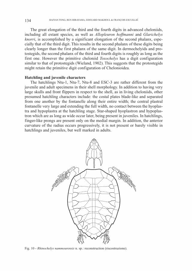
Hatchling and juvenile charactersThe hatchlings Ntu-1, Ntu-7, Ntu-8 and ESC-3 are rather different from the
juvenile and adult specimens in their shell morphology. In addition to having verylarge skulls and front flippers in respect to the shell, as in living cheloniids, otherpresumed hatchling characters include: the costal plates blade-like and separatedfrom one another by the fontanelle along their entire width; the central plastralfontanelle very large and extending the full width, no contact between the hyoplas-tra and hypoplastra at the hatchling stage. Star-shaped hyoplastron and hypoplas-tron which are as long as wide occur later, being present in juveniles. In hatchlings,finger-like prongs are present only on the medial margin. In addition, the anteriorcurvature of the radius occurs progressively, it is not present or barely visible inhatchlings and juveniles, but well marked in adults.
134 HAIYAN TONG, REN HIRAYAMA, EDOUARD MAKHOUL & FRANÇOIS ESCUILLIÉ
Fig. 10 - Rhinochelys nammourensis n. sp.: reconstruction (riscostruzione).
ConclusionThe Cenomanian Nammoura locality has yielded the most complete known
Rhinochelys specimens, which belong to a new species, Rhinochelys nammourensis.Almost complete and articulated skeletons, hitherto unknown for this genus, supportthe hypothesis that Rhinochelys, based on skulls, and Cimochelys, based on shells,belong to the same taxon. The hatchling, juvenile and adult specimens of the sametaxon, reported for the first time among protostegids, reveal important ontogeneticcharacters. The complete and articulated front flippers provide additional plesiomor-phic and synapomorphic features for the family Protostegidae relative toDermochelyidae and Cheloniidae. In addition to the synapomorphic features of thehumerus and radius already mentioned by Hirayama (1992, 1994, 1997), the protoste-gids have a extraordinarily large ulnare. However, the front flipper of protostegidsappears to retain the primitive digit configuration of chelonioids, in the relative lengthof the digits and phalanges and in the absence of reduction of the number of phalanges,which closely resembles the condition of the primitive cheloniid Toxochelys latiremis.
During the Cenomanian, Lebanon and the whole Arabic peninsula were part ofthe African continent, on the northern part of Gondwana. The Nammoura turtlesare therefore the first Protostegidae to be recorded from the African-Arabian con-tinent. The distribution of the genus Rhinochelys is therefore extended from theAnglo-Parisian Basin and northern Tethys southward to the eastern part of theTethys. The discovery of relatively abundant neonate turtles probably indicates thatthe locality was not far from the nesting site.
AcknowledgmentsThe authors would like to thank C. Dal Sasso (MSNM) for permission to
study the specimen in his care and E. Buffetaut for improving the manuscript.
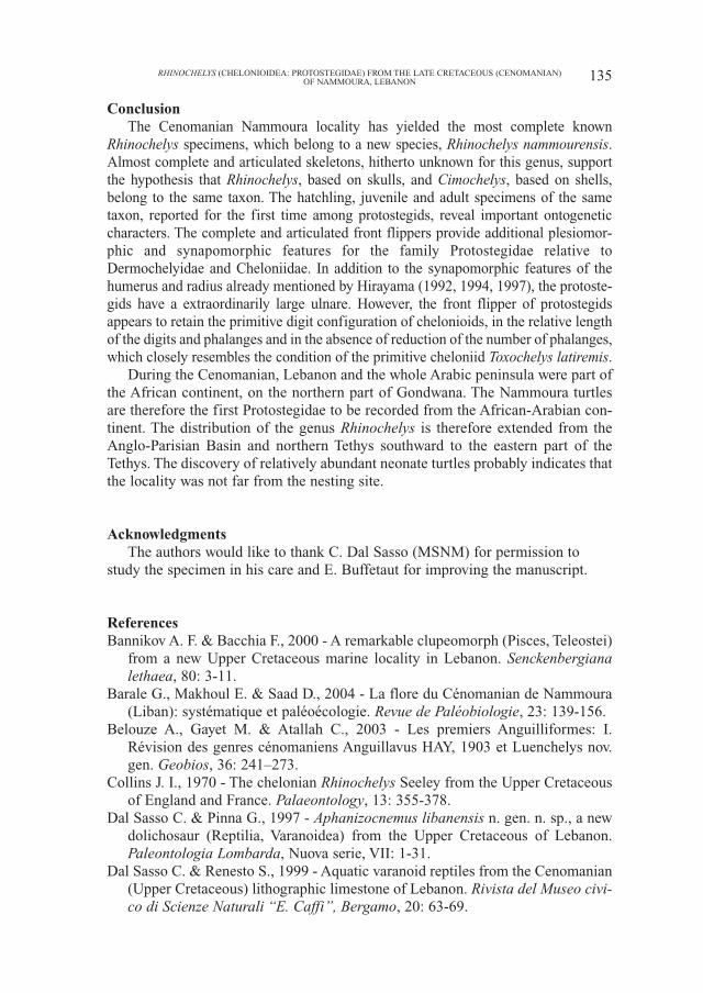
ReferencesBannikov A. F. & Bacchia F., 2000 - A remarkable clupeomorph (Pisces, Teleostei)
from a new Upper Cretaceous marine locality in Lebanon. Senckenbergianalethaea, 80: 3-11.
Barale G., Makhoul E. & Saad D., 2004 - La flore du Cénomanian de Nammoura(Liban): systématique et paléoécologie. Revue de Paléobiologie, 23: 139-156.
Belouze A., Gayet M. & Atallah C., 2003 - Les premiers Anguilliformes: I.Révision des genres cénomaniens Anguillavus HAY, 1903 et Luenchelys nov.gen. Geobios, 36: 241–273.
Collins J. I., 1970 - The chelonian Rhinochelys Seeley from the Upper Cretaceousof England and France. Palaeontology, 13: 355-378.
Dal Sasso C. & Pinna G., 1997 - Aphanizocnemus libanensis n. gen. n. sp., a newdolichosaur (Reptilia, Varanoidea) from the Upper Cretaceous of Lebanon.Paleontologia Lombarda, Nuova serie, VII: 1-31.
Dal Sasso C. & Renesto S., 1999 - Aquatic varanoid reptiles from the Cenomanian(Upper Cretaceous) lithographic limestone of Lebanon. Rivista del Museo civi-co di Scienze Naturali “E. Caffi”, Bergamo, 20: 63-69.
135RHINOCHELYS (CHELONIOIDEA: PROTOSTEGIDAE) FROM THE LATE CRETACEOUS (CENOMANIAN)OF NAMMOURA, LEBANON
Dalla Vecchia F. M., Arduini P. & Kellner A. W. A., 2001 - The first pterosaur fromthe Cenomanian (Late Cretaceous) Lagerstätten of Lebanon. CretaceousResearch, 22: 219-225.
Dalla Vecchia F. M. & Chiappe L. M., 2002 - First Avian Skeleton from the Mesozoicof Northern Gondwana. Journal of Vertebrate Paleontology, 22: 856-860.
Dalla Vecchia F. M. & Venturini S., 1999 - The Middle Cenomanian Lagerstätte ofAl Nammoura (Kesrouâne Caza, N. Lebanon). Rivista del Museo civico diScienze Naturali “E. Caffi”, Bergamo, 20: 75-77.
Dalla Vecchia F. M., Venturini S. & Tentor M., 2002 - The Cenomanian (LateCretaceous) Konservat-Lagerstätte of en Nammoûra (Kesrouâne Province),northern Lebanon. Bollettino della Società Paleontologica Italiana, 41: 51-68.
Derstler K., 1992 - Preliminary report on Brigitta, the Vienna specimen ofArchelon. University of New Orleans, 1-39.
Elliot D. K., Irby G. V. & Hutchison J. H., 1997 - Desmatochelys lowi, a marine tur-tle from the Upper Cretaceous. In: Ancient Marine Reptiles. Callaway J. M.and Nicholls E. L. (eds.). Academic Press: 243-258.
Ernst C. H., Lovich J. E. & Barbour R. W., 1994 - Turtles of the United States andCanada. Smithsonian Institution Press.
Forey P. L., Lu Y., Patterson C. & Davis C. E., 2003 - Fossil fishes from theCenomanian (Upper Cretaceous) of Namoura, Lebanon. Journal of SystematicPalaeontology, 1: 227-330.
Gayet M., 1988 - Gharbouria libanica nov. gen., nov. sp., “Salmoniforme” nou-veau en provenance d’Aïn-el-Ghârboûr, nouveau gisement cénomanien duLiban. Bulletin du Muséum d’Histoire naturelle, Paris, Section C, 10: 199-225.
Gayet M., Belouze A. & Abi Saad P., 2003 - Liban - Mémoire du temps. Les pois-sons fossiles. Editions DésIris.
Hirayama R., 1992 - Humeral morphology of chelonioid sea-turtles; its functionalanalysis and phylogenetic implications. The Bulletin of the Hobetsu Museum, 8:17-57.
Hirayama R., 1994 - Phylogenetic systematics of chelonioid sea turtles. The IslandArc, 3: 270-284.
Hirayama R., 1997 - Distribution and diversity of Cretaceous chelonioids. In:Ancient marine reptiles. Callaway J. M. and Nicholls E. L. (eds.). AcademicPress: 225-241.
Hirayama R., 1998 - Oldest known sea turtle. Nature, 392: 705-708.Hooks G. E. I., 1998 - Systematic revision of the Protostegidae, with a redescrip-
tion of Calcarichelys gemma Zangerl, 1953. Journal of VertebratePaleontology, 18: 85-98.
Krassilov V. & Bacchia F., 2000 - Cenomanian florule of Nammoura, Lebanon.Cretaceous Research, 21: 785-799.
Lehman T. M. & Tomlinson S. L., 2004 - Terlinguachelys fischbecki, a new genusand species of sea turtle (Chelonioidea: Protostegidae) from the UppeCretaceous of Texas. Journal of Paleontology, 78: 1163-1178.
Mantell G. A., 1841 - On the fossil remains of turtles discoveried in the Chalk-for-mation of the South-east England. Philosophical Transactions of the RoyalSociety, London, 1841.
136 HAIYAN TONG, REN HIRAYAMA, EDOUARD MAKHOUL & FRANÇOIS ESCUILLIÉ
Moret L., 1935 - Rhinochelys amaberti. Nouvelle espèce de tortue marine duVraconien de la Fauge, près du Villard-de-Lans (Isère). Bulletin de la SociétéGéologique de France, ser. 5, 5: 605-620.
Mulder E. W. A., 2003 - On latest Cretaceous tetrapods from the Maastrichtian typearea. Publicaties van het Natuurhistorisch Genootschap in Limburg, ReeksXLIV, aflevering 1, Stichting Natuurpublicaties Limburg.
Nel A., Azar D., Martinez-Delclos X. & Makhoul E., 2004 - A new Upper Cretaceousspecies of Chresmoda from Lebanon – a latest representative of Chresmodidae(Insecta: Polyneoptera inc. sed.): first record of homeotic mutations in the fossilrecord of insects. European Journal of Entomology, 101: 145–151.
Owen R., 1842 - Report on the British reptiles. Report of the British Associationfor the Advancement of Science, 1841: 60-204.
Philip J., Floquet M., Platel J. P., Bergerat F., Sandulescu M., Baraboshkin E.,Amon E. O., Guiraud R., Vaslet D., Le Nindre Y., Ziegler M., Poisson A. &Bouaziz S., 2000 - Map 14 -Late Cenomanian (94.7 to 93.5 Ma). In: Atlas Peri-Tethys, Palaeogeographical Maps. Dercourt J. et al. (eds.). CCGM/CGMW.
Rage J.-C. & Escuillié F., 2000 - Un nouveau serpent bipède du Cénomanien(Crétacé). Implications phylétiques. Comptes Rendus de l’Academie desSciences de Paris, IIa, t. 330: 513-520.
Rage J.-C. & Escuillié F., 2002 - Eupodophis, new name for the genus PodophisRage and Escuillié, 2000, an extinct bipedal snake, preoccupied by PodophisWiegmann, 1843 (Lacertilia, Scincidae). Amphibia-Reptilia, 23: 232-233.
Rage J.-C. & Escuillié F., 2003 - The Cenomanian: stage of hindlimbed snakes.Carnets de Géologie, 2003/01: 1-11.
Rieppel O. & Head J., 2004 - New specimens of the fossil snake genus EupodophisRage & Escuillié, from Cenomanian (Late Cretaceous) of Lebanon. Memoriedella Società Italiana di Scienze Naturali e del Museo Civico di Storia Naturaledi Milano, 32: 1-26.
Seeley H. G., 1869 - Index to the fossil remains of Aves, Ornithosauria, andReptilia from the Secondary System of Strata arranged in the WoodwardianMuseum of the University of Cambridge. Cambridge University Press.
Walker W. F., 1973 - The locomotion apparatus of Testudines. In: Biology of theReptilia. Gans C. and Parsons T. S. (eds.). Academic Press, 1-100.
Wieland G. R., 1900a - The skull, pelvis, and probable relationships of the hugeturtles of the genus Archelon from the Fort Pierre Cretaceous of South Dakoda.The American Journal of Science, 9: 237-251.
Wieland G. R., 1900b - Some observations on certain well-marked stages in theevolution of the Testudinate humerus. The American Journal of Science, ser. 4,9: 413-423.
Wieland G. R., 1902 - Notes on the Cretaceous turtles, Toxochelys and Archelon,with a classification of the marine Testudinata. The American Journal ofScience, ser. 4: 95-108.
Wieland G. R., 1906 - The osteology of Protostega. Memoirs of the CarnegieMuseum, II: 279-304.
Wieland G. R., 1909 - Revision of the Protostegidae. The American Journal ofScience, ser. 4, 27: 101-130.
137RHINOCHELYS (CHELONIOIDEA: PROTOSTEGIDAE) FROM THE LATE CRETACEOUS (CENOMANIAN)OF NAMMOURA, LEBANON
Williston S. W., 1898 - Part VI. Turtles. Introduction and Desmatochelys. TheUniversity Geological Survey of Kansas, IV: 351-387.
Zangerl R., 1953a - The vertebrate fauna of the Selma Formation of Alabama. PartIII: The turtles of the family Protostegidae. Fieldiana: Geology Memoirs, 3: 61-132.
Zangerl R., 1953b - The vertebrate fauna of the Selma Formation of Alabama. PartVI: The turtles of the Family Toxochelyidae. Fieldiana: Geology Memoirs, 3:137-277.
Zangerl R., 1958 - Die oilgozänen Meerschildkröten von Glarus. SchweizerischePalaeontologische Abhandlungen, 73: 1-56.
Zangerl R. & Sloan R. S., 1960 - A new specimen of Desmatochelys lowi Williston,a primitive cheloniid sea turtle from the Cretaceous of South Dakota. FieldianaGeology, 14: 7-40.
Ricevuto: 10 giugno 2005Approvato: 6 dicembre 2005
138 HAIYAN TONG, REN HIRAYAMA, EDOUARD MAKHOUL & FRANÇOIS ESCUILLIÉ