Embed Size (px)
Citation preview
The FASEB Journal • Research Communication
Retinoic acid expands the evolutionarily reduceddentition of zebrafish
Pawat Seritrakul,* Eric Samarut,† Tenzing T. S. Lama,* Yann Gibert,‡ Vincent Laudet,†and William R. Jackman*,1
*Department of Biology, Bowdoin College, Brunswick, Maine, USA; †Molecular Zoology Group,Institut de Génomique Fonctionnelle de Lyon, Université de Lyon, Centre National de la RechercheScientifique, Institut National de la Recherche Agronomique, Université Claude Bernard Lyon 1,Ecole Normale Supérieure de Lyon, Lyon, France; and ‡Deakin University School of Medicine,Waurn Ponds, Victoria, Australia
ABSTRACT Zebrafish lost anterior teeth during evo-lution but retain a posterior pharyngeal dentition thatrequires retinoic acid (RA) cell-cell signaling for itsdevelopment. The purposes of this study were to testthe sufficiency of RA to induce tooth development andto assess its role in evolution. We found that exposureof embryos to exogenous RA induces a dramatic ante-rior expansion of the number of pharyngeal teeth thatlater form and shifts anteriorly the expression patternsof genes normally expressed in the posterior tooth-forming region, such as pitx2 and dlx2b. After RAexposure, we also observed a correlation between car-tilage malformations and ectopic tooth induction, aswell as abnormal cranial neural crest marker geneexpression. Additionally, we observed that the RA-induced zebrafish anterior teeth resemble in patternand number the dentition of fish species that retainanterior pharyngeal teeth such as medaka but thatmedaka do not express the aldh1a2 RA-synthesizingenzyme in tooth-forming regions. We conclude that RAis sufficient to induce anterior ectopic tooth develop-ment in zebrafish where teeth were lost in evolution,potentially by altering neural crest cell development,and that changes in the location of RA synthesis corre-late with evolutionary changes in vertebrate denti-tions.—Seritrakul, P., Samarut, E., Lama, T. T. S.,Gibert, Y., Laudet, V., Jackman, W. R. Retinoic acidexpands the evolutionarily reduced dentition of ze-brafish. FASEB J. 26, 000–000 (2012). www.fasebj.org
Key Words: teeth ! neural crest cells ! retinaldehyde dehydrogenase
Primitive ray-finned fish possessed numerous teethin their pharyngeal regions, and the generally reduceddentitions of modern species are thought to be largelythe result of evolutionary tooth loss (1, 2). The ze-brafish (Danio rerio) lineage, as a member of the orderCypriniformes, lost anterodorsal pharyngeal teeth 65 or
more million years ago (3, 4), and only retain part ofthe original posteroventral pharyngeal dentition. Amore extensive dentition has not reevolved in any ofthe nearly 3000 described cypriniform species, despitediverse feeding modes, including piscivory (5). If thereis indeed selective pressure to increase the dentitionbut it is no longer possible due to developmentalconstraints, this may represent a modern example ofDollo’s law of the irreversibility of evolution (6–8).Such irreversibility may be the result of pleiotropicinteractions during development (9, 10).
Retinoic acid (RA), an oxidized form of vitamin A, isa cell-cell signaling molecule with numerous functionsin developing vertebrate embryos. Evidence of thecrucial roles RA plays in development was establishedover half a century ago, when the offspring of femalerodents fed a vitamin A-deficient diet during pregnancyshowed congenital defects in many organ systems (11).Synthesized from carotenoids, the active all-trans formof RA acts as a ligand for nuclear RA receptors, whichin turn function as transcription factors to control theexpression of numerous targets, including Hox genes(12). The amphiphilic nature of RA, with both polarand nonpolar moieties, allows this molecule to passrelatively easily through cell membranes and act as apositional cue for cells during embryogenesis (13).
In vertebrates, RA is particularly important in antero-posterior (AP) patterning and in the development ofcranial neural crest (CNC) cells. During the develop-mental specification of the AP axis, RA can directlyregulate Hox gene expression, and alterations in RAlevels result in homeotic changes in AP identity (14–16). Such RA-induced homeotic transformations in-clude the duplication of skeletal elements, such asvertebrae (17, 18). RA is also required for the properspecification of CNC cells, a class of vertebrate pluripo-tent migratory cells that give rise to cephalic structures,including cartilage, bone, and teeth (19, 20). Disrup-
1 Correspondence: Bowdoin College, 6500 College Station,Brunswick, ME 04011, USA. E-mail: [email protected]
doi: 10.1096/fj.12-209304
Abbreviations: AP, anteroposterior; CB5, 5th ceratobran-chial element; CNC, cranial neural crest; dpf, days postfertil-ization; hpf, hours postfertilization; RA, retinoic acid
10892-6638/12/0026-0001 © FASEB
AQ: 1
tapraid4/z38-faseb/z38-faseb/z3801212/z389008d12z xppws S!1 8/24/12 5:41 Ms. No.: 12-209304 Input-smk
tions in RA signaling cause malformations of CNC-derived tissues in many vertebrates (21, 22).
Teeth exhibit conservation in their early developmentacross vertebrate species and begin their formation inboth mammals and teleost fishes as an epithelial placode(23, 24). Subsequent stages of tooth morphogenesis anddifferentiation are coordinated by reciprocal cell signal-ing between this dental epithelium and an underlyingmesenchyme, which employs members of several evolu-tionarily conserved vertebrate gene families, including thefibroblast growth factor and hedgehog signaling pathways(25–27). The mammalian dental mesenchyme is knownto be largely derived from CNC cells (28), and the dentalmesenchyme of zebrafish pharyngeal teeth also expressCNC cell markers (25).
RA signaling has been studied previously in relationto tooth development. In mice, mutants lacking RAreceptors develop deformed skulls without teeth (29).Conversely, exposing embryonic mouse mandibles totissue culture medium containing excess RA altersdental epithelial morphology, increases tooth bud sizein the diastema region, and possibly switches toothidentity between molars and incisors (30). These stud-ies provide some clues to the potential roles of RA inmammalian tooth development, although there hasbeen no gene expression data or direct functional testsat a molecular level to explain the mechanisms under-lying these phenotypes. In zebrafish, it has been shownthat tooth induction depends on an endogenoussource of RA from retinaldehyde dehydrogenase-ex-pressing cells and that chemically blocking RA synthesisresults in the complete absence of teeth (31). However,effects of RA overexpression on zebrafish tooth devel-opment have not yet been described.
Here we report that exposure of zebrafish embryos toexogenous RA induces the formation of an extensivesymmetrical pattern of supernumerary teeth. Corre-sponding with this induction, we find up-regulation ofgenes normally expressed in developing teeth and inthe nearby posterior pharyngeal region such as pitx2,dlx2b, and hoxb5a. After RA treatment, CNC cells arepresent but with disrupted arrangements and abnormalgene expression, and cartilage development is corre-spondingly disrupted. We compare the expanded RA-induced zebrafish pharyngeal dentition with the wild-type dentitions of other teleost species and findsimilarities especially with the teeth of medaka. How-ever, comparisons of retinaldehyde dehydrogenase ex-pression between these species suggest that evolution-ary changes in RA signaling were not likely responsiblefor the reduction of the zebrafish pharyngeal dentition.
MATERIALS AND METHODS
Animal husbandry
Embryos of wild-type zebrafish (Danio rerio, Hamilton 1822)were obtained from in-house stocks derived originally from acommercial supplier (LiveAquaria.com, Rhinelander, WI,
USA). Embryos were raised at 28.5°C in an embryo mediumconsisting of 30% Danieu’s medium and 0.003% phenylthio-urea to prevent pigmentation (25). Phenylthiourea use isimportant to note, as it has recently been reported toenhance certain RA-related craniofacial phenotypes in ze-brafish (32). Zebrafish staging was done as described previ-ously (33), with developmental time reported in hours post-fertilization (hpf) or days postfertilization (dpf). The fli1:GFPreporter fish were of the Tg(fli1:EGFP)y1 line (34), and thedlx2b:GFP reporters were from the Tg(dlx2b:EGFP)cs1 line(35). Astyanax mexicanus (De Filippi 1853) stages are reportedin dpf, and medaka (Oryzias latipes, Temminck and Schlegel1846) stages are reported as dpf or stage numbers (36).Animal use was in accordance with protocols approved by theInstitutional Animal Care and Use Committee at BowdoinCollege and the Université de Lyon.
Exogenous RA treatment and bead implantation
Embryos were manually dechorionated and added to embryomedium containing either 0.05% DMSO alone as a control orDMSO plus 6 " 10#7 M all-trans RA (Biomol International,Plymouth, PA, USA). RA treatments were from 24 hpf onwardunless indicated otherwise. Embryos were kept in the dark orwith minimal lighting to avoid RA degradation (31). Toinactivate RA, embryos were washed into new medium andexposed to bright light for 5 min. AG 1-X8 beads (Bio-Rad,Hercules, CA, USA) were incubated in fresh solution of 6 "10#4 M RA and implanted as described previously (37).
Histology and in situ hybridization
Whole-mount mRNA in situ hybridization was performed asdescribed previously (25). Riboprobe sources: zebrafish dlx2b,dlx2a, and pitx2 (25); crestin (38); hoxb5a (39); sox9a (40);foxd3 (41); mitfa (42), lef1 (43); prdm1a (44); aldh1a2 (31);and medaka aldh1a2 (sequences 1367–1830 of Genbankaccession number NM_001104821). Optical confocal micros-copy sections of in situ hybridization specimens were obtainedusing 633-nm fluorescence of the BM Purple (Roche, India-napolis, IN, USA) staining product (45). Double fluorescencein situ hybridization was performed as described previously(46). Alizarin red and alcian blue skeletal single and doublestaining were performed as described previously (47).
Microscopy and statistical analysis
Embryos were visualized and photographed on a LeicaDMI3000B inverted microscope with DFC420C camera (LeicaMicrosystems, Solms, Germany), a Leica MZ16F stereomicro-scope with DFC300FX camera (Leica Microsystems), or aZeiss 510 Meta confocal laser-scanning microscope (CarlZeiss, Jena, Germany). Three-dimensional images of teethwere generated from confocal data using Volocity (PerkinElmer, San Jose, CA, USA). Photograph files were processedwith Adobe Photoshop (Adobe Systems, San Jose, CA, USA)and minimally adjusted as a whole for brightness, contrast,and/or color balance. Statistical significance of collected datawas calculated using a 2-tailed Fisher’s exact test of indepen-dence (48).
RESULTS
Exogenous RA expands zebrafish tooth developmentanteriorly and dorsally
Wild-type zebrafish larvae normally develop a singlepair of fully formed teeth on each side of the ventral,
2 Vol. 26 December 2012 SERITRAKUL ET AL.The FASEB Journal ! www.fasebj.org
tapraid4/z38-faseb/z38-faseb/z3801212/z389008d12z xppws S!1 8/24/12 5:41 Ms. No.: 12-209304 Input-smk

posterior pharynx by 4 dpf (Fig. 1A), with 2–3 youngerpairs of tooth germs also present nearby but not aseasily visualized (49). We found that embryos treatedwith 6 " 10#7 M exogenous RA from 24 hpf onward orin a window from 24 to 36 hpf exhibited a dramaticsupernumerary tooth phenotype by 4 dpf, with toothformation extending into ectopic anterior and dorsalregions (Fig. 1B; n!119/119 relative to 0/66 controls;P$0.01, 2-tailed Fisher’s exact test). These RA-treatedlarvae possessed %10–16 supernumerary teeth arranged in
a bilaterally symmetrical pattern. Most of the ectopicteeth were oriented with the tip toward the midline;however, we often also observed 2–4 teeth at theanterior end of the pattern with tips pointing caudally.The apparent fusion of adjacent teeth was also occa-sionally observed (n!23). We also employed a dlx2b:GFP reporter transgenic line that marks developingdental epithelium and mesenchyme (25, 35). DMSO-treated control fish showed 2–3 closely associated spotsof bright GFP expression on each side of the posteriorpharynx (Fig. 1C; n!20), while RA-treated embryosexhibited 8–12 supernumerary spots of GFP expres-sion, correlating to the number and location of devel-oping supernumerary teeth (Fig. 1D; n!20; P$0.01,2-tailed Fisher’s exact test). Confocal microscopy addi-tionally revealed the presence of teeth forming inpositions dorsal to the pharynx (Fig. 1E–H). Embryostreated with lower RA concentrations or beginning atlater stages did not exhibit this supernumerary toothphenotype as has previously been reported (50), andthose treated with higher concentrations or at earlier timepoints did not survive long enough to develop teeth.
RA expands expression of tooth-associated genes
We characterized gene expression changes associatedwith RA exposure beginning at 24 hpf at several tissueand spatial levels. Expression of the homeodomaintranscription factor pitx2 has been reported in both thedental epithelium and in nearby nondental pharyngealepithelium and represents the earliest marker of toothgerms in both mammals and fish (25, 51). We foundthat relative to control embryos that express pitx2mRNA strongly in the posterior pharyngeal regionwhere teeth are developing by 48 hpf (Fig. 2A, C;n!10), in RA-treated embryos pitx2 expression wasexpanded anteriorly (Fig. 2B, D; n!17; P$0.01, 2-tailedFisher’s exact test). To apply RA in a more localizedmanner, we implanted RA-coated beads at 24-25 hpfadjacent to the posterior pharynx. In RA bead-im-planted larvae, we also observed an expansion of pha-ryngeal pitx2 expression near the site of implantation by48 hpf (Fig. 2E; n!5/10), a result significantly differentfrom the normal pitx2 expression observed in controlbead-implanted embryos (Fig. 2F; n!0/15; P$0.01,2-tailed Fisher’s exact test).
The homeobox transcription factor hoxb5a exhibitssomewhat broader posterior pharyngeal expressionthan does pitx2, being strongly expressed in the regionwhere teeth are forming by 56 hpf (Fig. 2G, I; n!9). Wefound that in RA-treated embryos, pharyngeal hoxb5aexpression was expanded both anteriorly and dorsally,correlating to the regions where we find supernumer-ary teeth (Fig. 2H, J; n!16; P$0.01, 2-tailed Fisher’sexact test). We also examined early dlx2b expression asa more restricted marker of the dental epithelium andmesenchyme (25). We found in RA-treated embryosthat dlx2b mRNA expression patterns appeared normalat 48 and 60 hpf but at 72 hpf were expanded anteriorly(Fig. 2H; n!11) relative to controls (Fig. 2G; n!8;
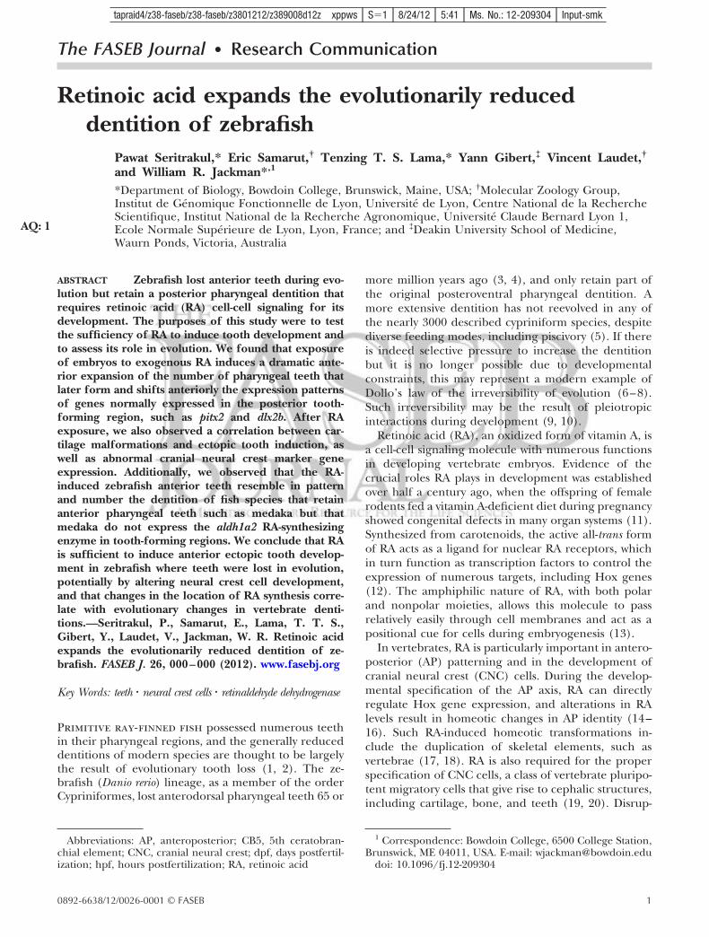
Figure 1. Exogenous RA treatment expands tooth develop-ment. A, B) Ventral view of the pharyngeal region of 4-dpfalizarin red-stained zebrafish larvae, anterior to the left. A)DMSO control exhibiting a pair of well-formed teeth on eachside of the ventral, posterior pharynx. B) Specimen treatedexogenously with 6 " 10#7 M RA between 24 and 36 hpf, withectopic teeth positioned more anteriorly than in wild type.Additional teeth located more dorsally are not visible in thisfocal plane. C, D) Oblique ventrolateral view of the head of4-dpf dlx2b:GFP reporter larvae. C) Control embryo with GFPexpression in developing tooth germs. D) RA-treated larvawith anteriorly expanded supernumerary foci of GFP expres-sion. E, F) Confocal micrographs oriented as in A and B, witha single plane of labeled cell nuclei (gray) in the ventralpharynx superimposed on an extended-focus composite ofdlx2b:GFP reporter expression (green). G, H) Transversesections in planes indicated in E and F. G) Control larva withdlx2b:GFP expression in tooth germs developing normally,ventral to the lumen of the pharynx (dotted line). H)RA-treated specimen with tooth germs located both dorsaland ventral to the pharyngeal lumen. Arrows indicate se-lected teeth or tooth germs. Scale bars ! 50 &m.
3RETINOIC ACID INDUCES ZEBRAFISH TEETH
F1
COLOR
F2
tapraid4/z38-faseb/z38-faseb/z3801212/z389008d12z xppws S!1 8/24/12 5:41 Ms. No.: 12-209304 Input-smk

P$0.01, 2-tailed Fisher’s exact test). Simultaneouslylabeling dlx2b and hoxb5a mRNA at 96 hpf revealed thatthe ectopic anterior expansion of dlx2b correlated withectopic anterior hoxb5a expression (Fig. 2N; n!6).
RA tooth expansion correlates with cartilagedisruption
We observed that the occurrence of supernumeraryteeth in RA-treated embryos was often associated withcartilage malformation or loss. Zebrafish teeth do notrequire skeletal support for their initial development(52), but because dental mesenchyme cells and carti-lage cells both derive from CNC cell precursors, wefurther examined this correlation. By 4 dpf, the firstpair of pharyngeal teeth has attached to the fifthceratobranchial cartilage and younger teeth are devel-oping nearby (Fig. 3A). In embryos treated with exog-enous RA starting at 24 hpf, cartilage morphology wasseverely disrupted in the region of supernumerarytooth formation (Fig. 3B). In localized treatments usingRA-coated beads implanted at 24 hpf, at 4 dpf weobserved cartilage malformation accompanied by su-pernumerary tooth formation nearby to where thebeads were implanted (Fig. 3D–F; n!15/23), relative tocontrols (Fig. 3C; n!0/13; P$0.01, 2-tailed Fisher’sexact test).
CNC cells are present but disorganized after RAexposure
Because CNC cells contribute to both tooth and carti-lage development, and because the specification ofCNC cells has been shown to be regulated by RA (19),
Figure 2. RA expands pitx2, dlx2b, and hoxb5a expression.Lateral (A, B, E–H, K, L) or ventral (C, D, I, J, M, N) view ofmRNA in situ hybridizations, anterior to the left, with normalsite of tooth formation indicated (arrow). A) Control embryoexpressing pitx2 in the posterior pharyngeal region at 48 hpf.B) Embryo treated with exogenous RA exhibits an anteriorexpansion of pitx2 (arrowhead). C) Horizontal confocal fluo-rescence section of pharyngeal epithelial and tooth germpitx2 expression in control. D) Identical section after RAtreatment with pitx2 expression expanded anteriorly in thepharyngeal epithelium (arrowhead). E) Embryo implantedwith a control bead (asterisk) and a wild-type pattern of pitx2expression. F) RA bead-implanted embryo with pitx2 expres-sion extended more anteriorly (arrowhead). G) Expression ofhoxb5a mRNA in the posterior brain and pharyngeal region(arrow) at 56 hpf. H) RA-treated embryo with anteriorexpansion of pharyngeal hoxb5a expression (arrowhead). I)Horizontal section of hoxb5a expression surrounding controltooth germs (arrow). J) RA-exposed embryo with hoxb5aexpression expanded anteriorly (arrowhead). K) dlx2b ex-pression in a control embryo at 72 hpf. L) RA-treated embryoshowing dlx2b expression expanded anteriorly (arrowhead).M) 96 hpf larva with dlx2b (green) and hoxb5a (red) expres-sion in the posterior pharynx in and around developingteeth. N) Larva after RA treatment with both dlx2b and hoxb5aexpression expanded anteriorly (arrowheads). Scale bars !100 &m.
4 Vol. 26 December 2012 SERITRAKUL ET AL.The FASEB Journal ! www.fasebj.org
F3
COLOR
tapraid4/z38-faseb/z38-faseb/z3801212/z389008d12z xppws S!1 8/24/12 5:41 Ms. No.: 12-209304 Input-smk

we investigated CNC-cell-associated gene expressionafter RA exposure. After RA exposure starting at 24 hpf,we surveyed early postmigratory CNC marker expres-sion from 27 to 36 hpf and focused on 30 hpf as havingmaximal effects. We first employed a fli1:GFP reportertransgenic line that labels CNC cells (34, 53). Relativeto controls (Fig. 4A, C; n!12), we found that after24–30 hpf RA exposure, fli1:GFP reporter expressionin CNC cells was present at similar levels but that CNCcell arrangements, especially in the most posteriorpharyngeal arch stream near where teeth later arise,appeared less compactly organized (Fig. 4B, D; n!12;P$0.01, 2-tailed Fisher’s exact test). dlx2a marks CNCcells during their migration into the pharyngeal arches(54). Similarly to what has previously been reported(55), we found that RA-treated embryos exhibiteddown-regulation of dlx2a (n!25/25) relative to con-trols (Fig. 4C, D; n!4/26; P$0.01, 2-tailed Fisher’sexact test). crestin marks premigratory and migratingCNC cells (38). Relative to controls (Fig. 4E; n!9),after RA treatment, embryos at 30 hpf showed general
up-regulation of crestin (Fig. 4F; n!10; P$0.01, 2-tailedFisher’s exact test). Together, these results suggest thatpharyngeal CNC cells are present after RA treatment,but that they are disorganized in arrangement andexhibit abnormal gene expression.
To further assess RA effects on CNC cell fate near thetooth-forming region, we examined later mRNA ex-pression of markers of specific differentiating CNC celltypes. sox9a is expressed in differentiating CNC-derivedskeleton and has been shown to directly regulate type-IIcollagen during cartilage development (40, 56). Wefound that relative to controls (Fig. 5A; n!4), after24–56 hpf RA exposure sox9a expression was upregu-lated in the posterior pharyngeal region (Fig. 5B, n!5).Similarly, we examined foxd3, as at later stages its
Figure 4. RA alters late migratory/early postmigratory cranialneural crest organization and gene expression. Lateral viewsof 30-hpf embryos, anterior to the left (arrows indicatenormal tooth-forming region). A) Control fli1:GFP reporterexpression. B) RA-treated embryos at with disorganized cellarrangements at 24–30 hpf. C, D) Magnified views fromembryos in A and B. E) Control dlx2a mRNA expression. F)RA-treated embryo with down-regulated dlx2a. G) Controlcrestin mRNA expression. H) RA-treated embryo with crestinexpression upregulated in the anterior pharyngeal region(arrowhead). Scale bar ! 100 &m.
Figure 3. RA tooth expansion correlates with cartilage disrup-tion. Ventral views of the 4-dpf pharyngeal region in alcianblue, alizarin red double-stained larvae, anterior to the left(arrows indicate selected teeth). A) Control zebrafish larvawith a pair of well-formed teeth attached to the ossifying fifthceratobranchial cartilages. B) Larva treated with RA from 24hpf exhibiting supernumerary teeth and severe cartilage loss.C) Larva showing normal tooth development with a controlbead positioned nearby. D–F) Larvae with RA-coated beadimplantation at 24 hpf exhibit a range of cartilage deforma-tion and supernumerary tooth phenotypes ranging fromrelatively severe (D) to relatively mild (E, arrow indicatesectopic midline tooth). F) Closeup view showing supernumer-ary teeth in proximity to an RA bead. Scale bars ! 50 &m.
5RETINOIC ACID INDUCES ZEBRAFISH TEETH
F4
COLOR
F5
COLOR
tapraid4/z38-faseb/z38-faseb/z3801212/z389008d12z xppws S!1 8/24/12 5:41 Ms. No.: 12-209304 Input-smk

expression pattern includes developing CNC-derivedglial cells (57). Similarly we found that compared withcontrols (Fig. 5C; n!6), foxd3 mRNA was present in amore extensive area of the pharyngeal region after RAexposure (Fig. 5D; n!6). In contrast, the expression ofmitfa, a marker of differentiating melanocytes (42),appeared similar between control (Fig. 5E; n!6) and
RA-exposed embryos (Fig. 5F; n!6), although thepattern of differentiated melanocytes appeared abnor-mal. Expression of these markers suggests that 24 hpfRA exposure is affecting the differentiation and/ordistribution of both ectomesenchymal and nonecto-mesenchymal CNC cell types.
We also examined RA effects on the mRNA expres-sion of the canonical wingless pathway transcriptionfactor lef1 and the zinc-finger protein prdm1a, as both ofthese genes are required for zebrafish tooth develop-ment (58, 59) and both have been characterized to liedownstream of an RA signal during fin development(60). We found lef1 mRNA to be expressed throughoutthe pharyngeal region at 56 hpf (Fig. 5G; n!8) and thatafter 24–56 hpf RA exposure, expression levels ap-peared to increase (Fig. 5H; n!6/7). Pharyngeal ex-pression of prdm1a mRNA was more limited in controlembryos, mostly to the developing tooth germs (Fig. 5I;n!11) but appeared greatly expanded after RA treat-ment (Fig. 5J; n!22). Taken together, these expressiondata suggest that RA may regulate tooth developmentwith a similar mechanism to its role in fin formation.
Phylogenetic comparisons
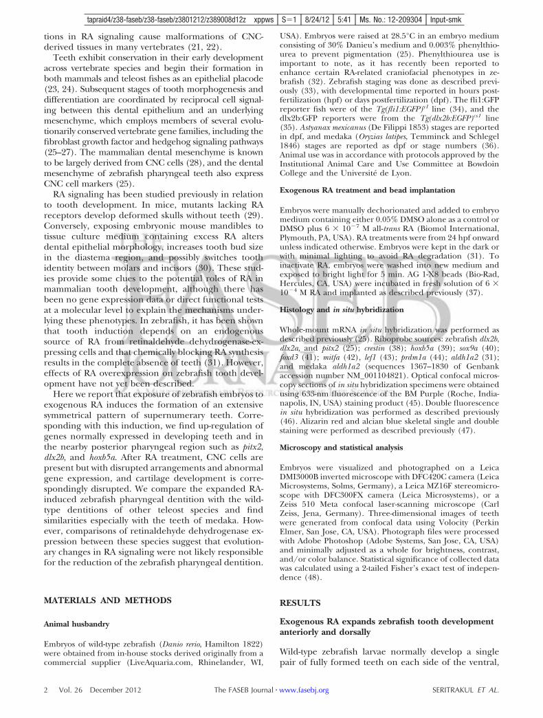
To visualize the overall arrangement of teeth in RA-treated zebrafish embryos, and make comparisons withother species, we generated three-dimensional, confo-cal microscope images of alizarin-red-stained speci-mens (Fig. 6). Simultaneously viewing teeth from allfocal planes revealed a bilaterally symmetrical patternof zebrafish RA-induced teeth (Fig. 6C). Teeth atparticular anteroposterior levels also typically sharedthe same relative dorsal/ventral positioning with theircontralateral counterparts (Fig. 6D). We next exam-ined similarly staged Mexican tetra (Astyanax mexica-nus) and medaka (Oryzias latipes) larvae (Fig. 6E–H).Mexican tetras (order Characiformes) are relativelyclosely related to zebrafish (order Cypriniformes), withmedaka (order Beloniformes) as a more distant relativeof both (61). To match the developmental stage in allthree species as closely as possible, we chose stageswhen pharyngeal tooth development had commencedbut nearby bones were not extensively calcified. At thisstage, Mexican tetra possess 2 bilateral pairs of dorsalpharyngeal teeth located rostrally to a pair of ventralteeth, the latter of which correspond to the teeth inwild-type zebrafish (Fig. 6E, F). Medaka at this stageshare a similar posteroventral dentition with 2 pairs ofteeth but have a much more extensive dorsal pharyn-geal dentition with five bilateral pairs of teeth extend-ing further anteriorly (Fig. 6G). The supernumerary RAinduced teeth in zebrafish appeared similar to thepharyngeal dentition in medaka in tooth number,anteroposterior location, and dorsoventral arrange-ment, but the patterns are not identical.
Because of the similarities between the medaka wild-type and zebrafish RA-induced pharyngeal dentitions,we wondered whether the medaka condition may bethe result of more extensive RA signaling relative to
Figure 5. Gene expression analysis of CNC cell subtype markersand potential RA target genes. Lateral views of 52- to 56-hpfembryos, anterior to the left (arrows indicate normal tooth-forming region). A) Control sox9a mRNA expression at 56 hpf.B) RA-treated embryo with up-regulation of sox9a expression inthe pharyngeal region (arrowhead) at 24–56 hpf. C) Controlfoxd3 mRNA expression. D) RA-treated embryo with morewidespread pharyngeal foxd3 expression (arrowhead). E) Con-trol mitfa mRNA expression marking developing melanocytes(double arrow). Differentiated melanocytes are also visible (ar-rowhead). F) RA-treated embryo with similar levels of mitfaexpression but a disorganized-appearing melanocyte pattern(arrowhead). G) Control lef1 mRNA expression. H) RA-treatedembryo with up-regulation of pharyngeal lef1 expression (arrow-head). I) Control prdm1a mRNA expression, restricted primarilyto the developing tooth germ. J) RA-treated embryo showingstrong prdm1a up-regulation (arrowhead). Scale bar ! 100 &m.
6 Vol. 26 December 2012 SERITRAKUL ET AL.The FASEB Journal ! www.fasebj.org
COLOR
F6
tapraid4/z38-faseb/z38-faseb/z3801212/z389008d12z xppws S!1 8/24/12 5:41 Ms. No.: 12-209304 Input-smk
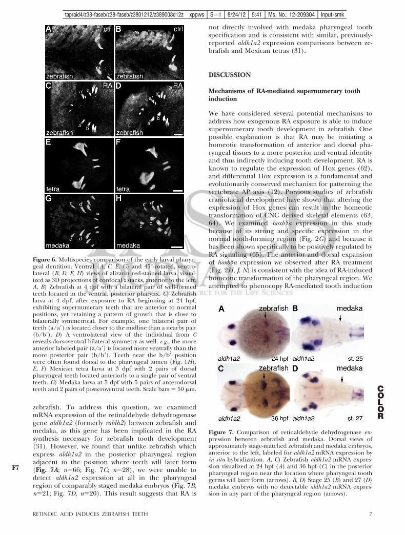
zebrafish. To address this question, we examinedmRNA expression of the retinaldehyde dehydrogenasegene aldh1a2 (formerly raldh2) between zebrafish andmedaka, as this gene has been implicated in the RAsynthesis necessary for zebrafish tooth development(31). However, we found that unlike zebrafish whichexpress aldh1a2 in the posterior pharyngeal regionadjacent to the position where teeth will later form(Fig. 7A; n!66; Fig. 7C; n!28), we were unable todetect aldh1a2 expression at all in the pharyngealregion of comparably staged medaka embryos (Fig. 7B,n!21; Fig. 7D, n!20). This result suggests that RA is
not directly involved with medaka pharyngeal toothspecification and is consistent with similar, previously-reported aldh1a2 expression comparisons between ze-brafish and Mexican tetras (31).
DISCUSSION
Mechanisms of RA-mediated supernumerary toothinduction
We have considered several potential mechanisms toaddress how exogenous RA exposure is able to inducesupernumerary tooth development in zebrafish. Onepossible explanation is that RA may be initiating ahomeotic transformation of anterior and dorsal pha-ryngeal tissues to a more posterior and ventral identityand thus indirectly inducing tooth development. RA isknown to regulate the expression of Hox genes (62),and differential Hox expression is a fundamental andevolutionarily conserved mechanism for patterning thevertebrate AP axis (12). Previous studies of zebrafishcraniofacial development have shown that altering theexpression of Hox genes can result in the homeotictransformation of CNC derived skeletal elements (63,64). We examined hoxb5a expression in this studybecause of its strong and specific expression in thenormal tooth-forming region (Fig. 2G) and because ithas been shown specifically to be positively regulated byRA signaling (65). The anterior and dorsal expansionof hoxb5a expression we observed after RA treatment(Fig. 2H, J, N) is consistent with the idea of RA-inducedhomeotic transformation of the pharyngeal region. Weattempted to phenocopy RA-mediated tooth induction
Figure 7. Comparison of retinaldehyde dehydrogenase ex-pression between zebrafish and medaka. Dorsal views ofapproximately stage-matched zebrafish and medaka embryos,anterior to the left, labeled for aldh1a2 mRNA expression byin situ hybridization. A, C) Zebrafish aldh1a2 mRNA expres-sion visualized at 24 hpf (A) and 36 hpf (C) in the posteriorpharyngeal region near the location where pharyngeal toothgerms will later form (arrows). B, D) Stage 25 (B) and 27 (D)medaka embryos with no detectable aldh1a2 mRNA expres-sion in any part of the pharyngeal region (arrows).
Figure 6. Multispecies comparison of the early larval pharyn-geal dentition. Ventral (A, C, E, G) and 45°-rotated, ventro-lateral (B, D, F, H) views of alizarin red-stained larva, visual-ized as 3D projections of confocal z stacks, anterior to the left.A, B) Zebrafish at 4 dpf with a bilateral pair of well-formedteeth located in the ventral, posterior pharynx. C) Zebrafishlarva at 4 dpf, after exposure to RA beginning at 24 hpf,exhibiting supernumerary teeth that are anterior to normalpositions, yet retaining a pattern of growth that is close tobilaterally symmetrical. For example, one bilateral pair ofteeth (a/a=) is located closer to the midline than a nearby pair(b/b=). D) A ventrolateral view of the individual from Creveals dorsoventral bilateral symmetry as well: e.g., the moreanterior labeled pair (a/a=) is located more ventrally than themore posterior pair (b/b=). Teeth near the b/b= positionwere often found dorsal to the pharyngeal lumen (Fig. 1H).E, F) Mexican tetra larva at 3 dpf with 2 pairs of dorsalpharyngeal teeth located anteriorly to a single pair of ventralteeth. G) Medaka larva at 5 dpf with 5 pairs of anterodorsalteeth and 2 pairs of posteroventral teeth. Scale bars ! 50 &m.
7RETINOIC ACID INDUCES ZEBRAFISH TEETH
F7
COLOR
tapraid4/z38-faseb/z38-faseb/z3801212/z389008d12z xppws S!1 8/24/12 5:41 Ms. No.: 12-209304 Input-smk
with hoxb5a overexpression by mRNA injection but didnot observe a tooth phenotype (unpublished results).However, there are many potential explanations forthis lack of result, including that homeotic transforma-tion is not responsible for RA-induced ectopic toothformation, that RA is acting through more than oneHox gene and overexpressing a single gene has noeffect, or that RA is acting through other factors to alterAP identity such as Cdx genes (17).
An alternative, although not necessarily mutuallyexclusive, way RA could be influencing tooth develop-ment is through CNC cell specification. One of thedefining features of CNC cells is high developmentalplasticity: CNC cell fate is largely determined by molec-ular signals in the microenvironment and generally notpreprogrammed (66). CNC cells have long been knownto contribute to the development of both cranial carti-lage and teeth (20), raising the possibility that a switchin CNC cell fate could lead to the overproduction ofone organ type at the expense of the other. CNC cellsin the zebrafish pharyngeal region express the home-odomain transcription factor dlx2a (54), and inhibitionof dlx2a function results in reduced and abnormaldevelopment of pharyngeal cartilages (67). In addition,in chickens, misexpression of Dlx2 leads to ectopiccranial cartilage formation (68), and exogenous RAexposure in zebrafish suppresses Dlx gene expression,resulting in pharyngeal cartilage malformation and loss(55). Together, these data suggest that RA may beinvolved in directing CNC cells toward a cartilage fateby influencing the expression of Dlx genes. The pres-ence of fli1:GFP-expressing cells in the posterior pha-ryngeal region suggests that CNC cells are present atlate migration stages after RA exposure (Fig. 4D), butdown-regulation of dlx2a and up-regulation of crestinsuggest that the remaining CNC cells are already notdeveloping normally (Fig. 4F, H). However, inhibitionof dlx2a function alone does not result in supernumer-ary tooth formation (35), suggesting that if RA isaltering CNC cell fate specification, it is doing so bychanging the expression of multiple downstream tar-gets. In addition, our analysis of later CNC markerexpression suggests that multiple CNC cell types may beoverrepresented after 24 hpf RA exposure, includingchondrocytes (sox9a; Fig. 5B) and glia (foxd3; Fig. 5D),but examining melanocytes suggest that not all CNCcell types are overrepresented after RA treatment(mitfa; Fig. 5F). Together, these data suggest that RAmay be promoting the development of certain CNC celltypes, such as both cartilage and tooth odontoblasts,rather than directing a switch in fate between CNC cellsubtypes.
It is also possible that RA induction of tooth devel-opment may be relatively direct. A study using chroma-tin immunoprecipitation and gel mobility shift assays inmice has shown that RA nuclear receptor complexesdirectly bind to cis-regulatory regions of the Pitx2 gene(69), the earliest marker of tooth formation (51). Astudy using similar methods has, in turn, demonstrateddirect regulation of Dlx2 by Pitx2 (70). Our observation
of expansion of pitx2 expression after RA exposure(Fig. 2B, D, F) is consistent with the direct up-regulationof pitx2 by RA. We also do not observe ectopic dlx2bexpression until many hours after the time of initial RAexposure (Fig. 2L), consistent with RA having an indi-rect effect on dlx2b expression. However, our data fromlef1 and prdm1a (Fig. 5G–J), genes known to be down-stream of RA signaling in limb development and re-quired for zebrafish tooth development (58–60), areconsistent with RA exhibiting a more indirect controlof tooth development than by direct up-regulation ofpitx2. Direct tests of pitx2 function during tooth devel-opment could help establish whether this gene mayindeed be acting as an intermediary between RA signal-ing and tooth morphogenesis.
RA in the evolution of teleost dentitions
The order Cypriniformes is thought to have greatlyreduced its dentition early in its evolution such thatteeth remained only associated with the posterior andventral 5th ceratobranchial element (CB5), possibly asan adaptation for suction feeding (1, 71). It is interest-ing to consider whether the evolutionary reduction orloss of RA signaling in the anterior and dorsal pharynxmay have been part of a developmental mechanism bywhich the Cypriniform ancestor reduced its dentition.If this were the case, one might expect to find similar-ities between the zebrafish RA-induced dental patternswith the normal tooth arrangements of other teleostfishes that have retained a less modified dentition, aswell as more extensive RA signaling in the anteriorpharyngeal regions of such species.
The bowfin Amia clava, a species immediately basal toteleosts phylogenetically (61), possesses an extensivelarval dentition associated with both anterior and pos-terior pharyngeal skeletal elements (72). As the teleostsister group, the dentition of Amia may represent theancestral condition for teleosts. Pharyngeal tooth re-duction is common in other orders of the teleostradiation, including the belaniform medaka (Oryziaslatipes) and the characiform Mexican tetra (Astyanaxmexicanus), but is typically less severe than what is foundin the cypriniformes (2, 73). The medaka and zebrafishlineages diverged early in the teleost radiation (61);thus, the teeth of Amia clava may represent the closestproxy for the ancestral pharyngeal dentition of thesemodel species. By this logic, the more extensive pha-ryngeal dentition of medaka is more similar to theancestral condition than is the more reduced dentitionof zebrafish.
Of the species we examined, we interpret the RA-induced zebrafish tooth pattern as most resembling thewild-type pharyngeal dentition of medaka (Fig. 6). Inmedaka, pharyngeal teeth attach to 2 dorsal parabran-chial bones and ventrally to CB5 (73). Similaritiesbetween RA-induced zebrafish teeth and wild-typemedaka dentitions include tooth number, orientation,the anteroposterior extent of tooth formation, andsymmetry in the left-right and dorsoventral axes. It is
8 Vol. 26 December 2012 SERITRAKUL ET AL.The FASEB Journal ! www.fasebj.org
tapraid4/z38-faseb/z38-faseb/z3801212/z389008d12z xppws S!1 8/24/12 5:41 Ms. No.: 12-209304 Input-smk
also notable that RA-exposed zebrafish often developteeth dorsal to the pharyngeal lumen, a position whereteeth never form in zebrafish or any other cypriniformspecies (5). These similarities prompted us to wonderwhether RA signaling may have been ancestrally moreprevalent in the pharyngeal region, and whether theevolutionary reduction of the cypriniform dentitionmay have been the consequence of reduction in ante-rior pharyngeal RA signaling. This scenario wouldpredict that a species with a more extensive pharyngealdentition, such as medaka, would have more RA signal-ing associated with the development of the anteriorpharynx. To test this idea, we compared the zebrafishand medaka mRNA expression of aldh1a2, as this genehas been implicated as a key regulator of RA levelsrequired for zebrafish tooth and craniofacial develop-ment (21, 31). However, in contrast to the abovescenario, our data suggest instead that medaka have noretinaldehyde dehydrogenase expression associatedwith pharyngeal tooth development (Fig. 7). This resultis consistent with previous work where RA inhibitionhad a much lesser effect on tooth development inmedaka than it had in zebrafish (31). We thereforespeculate that the RA-induced ectopic dentition inzebrafish may not be recapitulating an ancestral condi-tion; however, phylogenetic comparisons of RA expres-sion and function during the tooth development ofother species will shed further light on this question.
However, even if raising RA signaling levels in ze-brafish does not actually restore a more ancestral-likedentition, the number of teeth and area in which theyreside is clearly increased by increasing RA, so why hasthis not occurred during cypriniform evolution to bringback teeth in species that would benefit from a moreextensive dentition? Examples of cypriniforms whereadaptive pressure may be present to produce moreteeth include piscivorous pikeminnows (74) and Dani-onella dracula, which has evolved fang-like jawboneprojections used in male displays (75). The answer tothis question may lie with the corresponding cartilagedisruptions we have observed to accompany RA-in-duced tooth expansion. It is possible to induce a moreextensive dentition by gross RA overexpression, but toincrease tooth number without adversely affectingnearby skeletal elements may require more sophisti-cated control. Such fine control over RA levels, distri-bution pattern, or the simultaneous coexpression ofmodulatory cofactors may be difficult to evolve; thusconstraining how cypriniforms could possibly reacquirea more extensive dentition. Further comparative studyof the mechanisms of RA action on tooth and skeletaldevelopment in zebrafish and medaka and other non-cypriniform species, as well as similar comparativeanalyses of other developmental signaling pathways,will help to explain this apparent case of evolutionarydevelopmental constraint.
The authors are very grateful to Kristin Artinger (Univer-sity of Colorado, Aurora, CO, USA) for sharing ideas, pre-publication data, and reagents; Vicki Prince (University of
Chicago, Chicago, IL, USA) and Paul Henion (Ohio StateUniversity, Columbus, OH, USA) for reagents; and MikePalopoli, Andrea Jowdry, and David Rivers for helpful sugges-tions. This work was supported by U.S. National Institutes ofHealth grants 5P20-RR-016463-12 and 8P20-GM-103423-12(to W.R.J.) and Agence Nationale de la Recherche grantANR-09-BLAN-0127-01 (to V.L.).
REFERENCES
1. Gosline, W. (1973) Considerations regarding the phylogeny ofcypriniform fishes, with special reference to structures associ-ated with feeding. Copeia 1973, 761–776
2. Stock, D. W. (2001) The genetic basis of modularity in thedevelopment and evolution of the vertebrate dentition. Philos.Trans. R Soc. Lond. B Biol. Sci. 356, 1633–1653
3. Patterson, C. (1993) Osteichthyes: teleostei. In The Fossil Record2 (Benton, M., ed.) pp. 621–656, Chapman & Hall, London
4. Sibbing, F. (1991) Food capture and oral processing. In CyprinidFishes: Systematics, Biology, and Exploitation (Winfield, I., andNelson, J., eds.) pp. 377–412, Chapman & Hall, New York
5. Stock, D. W. (2007) Zebrafish dentition in comparative context.J. Exp. Zoolog. B Mol. Dev. Evol. 308B, 523–549
6. Dollo, L. (1893) Les lois de l’évolution. Bull. Soc. Bel. Géol. Pal.Hydr. 7, 164–166
7. Goldberg, E. E., and Igic, B. (2008) On phylogenetic tests ofirreversible evolution. Evolution 62, 2727–2741
8. Marshall, C. R., Raff, E. C., and Raff, R. A. (1994) Dollo’s lawand the death and resurrection of genes. Proc. Natl. Acad. Sci.U. S. A. 91, 12283–12287
9. Galis, F., Arntzen, J. W., and Lande, R. (2010) Dollo’s law andthe irreversibility of digit loss in Bachia. Evolution 64, 2466–2485
10. Lande, R. (1978) Evolutionary mechanisms of limb loss intetrapods. Evolution 32, 73–92
11. Wilson, J., Roth, C., and Warkany, J. (1953) An analysis of thesyndrome of malformations induced by maternal vitamin Adeficiency. Effects of restoration of vitamin A at various timesduring gestation. Am. J. Anat. 92, 189–217
12. Mallo, M., Wellik, D. M., and Deschamps, J. (2010) Hox genesand regional patterning of the vertebrate body plan. Dev. Biol.344, 7–15
13. Niederreither, K., and Dollé, P. (2008) Retinoic acid in devel-opment: towards an integrated view. Nat. Rev. Genet. 9, 541–553
14. Allan, D., Houle, M., Bouchard, N., Meyer, B. I., Gruss, P., andLohnes, D. (2001) RAR' and Cdx1 interactions in vertebralpatterning. Dev. Biol. 240, 46–60
15. Manzanares, M., Wada, H., Itasaki, N., Trainor, P. A., Krumlauf,R., and Holland, P. W. (2000) Conservation and elaboration ofHox gene regulation during evolution of the vertebrate head.Nature 408, 854–857
16. Kessel, M., and Gruss, P. (1991) Homeotic transformations ofmurine vertebrae and concomitant alteration of Hox codesinduced by retinoic acid. Cell 67, 89–104
17. Houle, M., Sylvestre, J.-R., and Lohnes, D. (2003) Retinoic acidregulates a subset of Cdx1 function in vivo. Development 130,6555–6567
18. Yamaguchi, M., Nakamoto, M., Honda, H., Nakagawa, T., Fujita,H., Nakamura, T., Hirai, H., Narumiya, S., and Kakizuka, A.(1998) Retardation of skeletal development and cervical abnor-malities in transgenic mice expressing a dominant-negativeretinoic acid receptor in chondrogenic cells. Proc. Natl. Acad. Sci.U. S. A. 95, 7491–7496
19. Minoux, M., and Rijli, F. M. (2010) Molecular mechanisms ofcranial neural crest cell migration and patterning in craniofacialdevelopment. Development 137, 2605–2621
20. De Beer, G. (1947) The differentiation of neural crest cells intovisceral cartilages and odontoblasts in Amblystoma, and a re-examination of the germ-layer theory. Proc. R Soc. Lond. B Biol.Sci. 134, 377–398
21. Begemann, G., Schilling, T. F., Rauch, G. J., Geisler, R., andIngham, P. W. (2001) The zebrafish neckless mutation reveals arequirement for raldh2 in mesodermal signals that pattern thehindbrain. Development 128, 3081–3094
9RETINOIC ACID INDUCES ZEBRAFISH TEETH
AQ: 2
tapraid4/z38-faseb/z38-faseb/z3801212/z389008d12z xppws S!1 8/24/12 5:41 Ms. No.: 12-209304 Input-smk

22. Lohnes, D., Mark, M., Mendelsohn, C., Dollé, P., Dierich, A.,Gorry, P., Gansmuller, A., and Chambon, P. (1994) Function ofthe retinoic acid receptors (RARs) during development (I).Craniofacial and skeletal abnormalities in RAR double mutants.Development 120, 2723–2748
23. Huysseune, A., Van der heyden, C., and Sire, J.-Y. (1998) Earlydevelopment of the zebrafish (Danio rerio) pharyngeal dentition(Teleostei, Cyprinidae). Anat. Embryol. 198, 289–305
24. Peters, H., and Balling, R. (1999) Teeth. Where and how tomake them. Trends Genet. 15, 59–65
25. Jackman, W. R., Draper, B. W., and Stock, D. W. (2004) Fgfsignaling is required for zebrafish tooth development. Dev. Biol.274, 139–157
26. Jackman, W. R., Yoo, J. J., and Stock, D. W. (2010) Hedgehogsignaling is required at multiple stages of zebrafish toothdevelopment. BMC Dev. Biol. 10, 119
27. Klein, O. D., Lyons, D. B., Balooch, G., Marshall, G. W., Basson,M. A., Peterka, M., Boran, T., Peterkova, R., and Martin, G. R.(2008) An FGF signaling loop sustains the generation of differ-entiated progeny from stem cells in mouse incisors. Development135, 377–385
28. Chai, Y., Jiang, X., Ito, Y., Bringas, P., Han, J., Rowitch, D. H.,Soriano, P., McMahon, A. P., and Sucov, H. M. (2000) Fate ofthe mammalian cranial neural crest during tooth and mandib-ular morphogenesis. Development 127, 1671–1679
29. Mark, M., Lohnes, D., Mendelsohn, C., Dupé, V., Vonesch, J. L.,Kastner, P., Rijli, F., Bloch-Zupan, A., and Chambon, P. (1995)Roles of retinoic acid receptors and of Hox genes in thepatterning of the teeth and of the jaw skeleton. Int. J. Dev. Biol.39, 111–121
30. Kronmiller, J. E., Nguyen, T., and Berndt, W. (1995) Instructionby retinoic acid of incisor morphology in the mouse embryonicmandible. Arch. Oral Biol. 40, 589–595
31. Gibert, Y., Bernard, L., Debiais-Thibaud, M., Bourrat, F., Joly,J.-S., Pottin, K., Meyer, A., Rétaux, S., Stock, D. W., Jackman,W. R., Seritrakul, P., Begemann, G., and Laudet, V. (2010)Formation of oral and pharyngeal dentition in teleosts dependson differential recruitment of retinoic acid signaling. FASEB J.24, 3298–3309
32. Bohnsack, B. L., Gallina, D., and Kahana, A. (2011) Phenothio-urea sensitizes zebrafish cranial neural crest and extraocularmuscle development to changes in retinoic acid and IGFsignaling. PLoS ONE 6, e22991
33. Kimmel, C. B., Ballard, W. W., Kimmel, S. R., Ullmann, B., andSchilling, T. F. (1995) Stages of embryonic development of thezebrafish. Dev. Dyn. 203, 253–310
34. Lawson, N. D., and Weinstein, B. M. (2002) In vivo imaging ofembryonic vascular development using transgenic zebrafish.Dev. Biol. 248, 307–318
35. Jackman, W. R., and Stock, D. W. (2006) Transgenic analysis ofDlx regulation in fish tooth development reveals evolutionaryretention of enhancer function despite organ loss. Proc. Natl.Acad. Sci. U. S. A. 103, 19390–19395
36. Iwamatsu, T. (2004) Stages of normal development in themedaka Oryzias latipes. Mech. Dev. 121, 605–618
37. White, R. J., Nie, Q., Lander, A. D., and Schilling, T. F. (2007)Complex regulation of cyp26a1 creates a robust retinoic acidgradient in the zebrafish embryo. PLoS Biol. 5, e304
38. Luo, R., An, M., Arduini, B. L., and Henion, P. D. (2001)Specific pan-neural crest expression of zebrafish Crestinthroughout embryonic development. Dev. Dyn. 220, 169–174
39. Prince, V. E., Joly, L., Ekker, M., and Ho, R. K. (1998) Zebrafishhox genes: genomic organization and modified colinear expres-sion patterns in the trunk. Development 125, 407–420
40. Chiang, E. F., Pai, C. I., Wyatt, M., Yan, Y. L., Postlethwait, J., andChung, B. (2001) Two sox9 genes on duplicated zebrafishchromosomes: expression of similar transcription activators indistinct sites. Dev. Biol. 231, 149–163
41. Odenthal, J., and Nüsslein-Volhard, C. (1998) Fork head do-main genes in zebrafish. Dev. Genes. Evol. 208, 245–258
42. Lister, J. A., Robertson, C. P., Lepage, T., Johnson, S. L., andRaible, D. W. (1999) nacre encodes a zebrafish microphthalmia-related protein that regulates neural-crest-derived pigment cellfate. Development 126, 3757–3767
43. Dorsky, R. I., Snyder, A., Cretekos, C. J., Grunwald, D. J., Geisler,R., Haffter, P., Moon, R. T., and Raible, D. W. (1999) Maternal
and embryonic expression of zebrafish lef1. Mech. Dev. 86,147–150
44. Hernandez-Lagunas, L., Choi, I. F., Kaji, T., Simpson, P.,Hershey, C., Zhou, Y., Zon, L., Mercola, M., and Artinger, K. B.(2005) Zebrafish narrowminded disrupts the transcription factorprdm1 and is required for neural crest and sensory neuronspecification. Dev. Biol. 278, 347–357
45. Trinh, L. A., McCutchen, M. D., Bonner-Fraser, M., Fraser, S. E.,Bumm, L. A., and McCauley, D. W. (2007) Fluorescent in situhybridization employing the conventional NBT/BCIP chromo-genic stain. Biotechniques 42, 756–759
46. Talbot, J. C., Johnson, S. L., and Kimmel, C. B. (2010) Hand andDlx genes specify dorsal, intermediate and ventral domainswithin zebrafish pharyngeal arches. Development 137, 2507–2517
47. Walker, M. B., and Kimmel, C. B. (2007) A two-color acid-freecartilage and bone stain for zebrafish larvae. Biotech. Histochem.82, 23–28
48. McDonald, J. (2009) Fisher’s exact test of independence. InHandbook of Biological Statistics, pp. 70–75, Sparky House, Balti-more
49. Van der Heyden, C., and Huysseune, A. (2000) Dynamics oftooth formation and replacement in the zebrafish (Danio rerio)(Teleostei, Cyprinidae). Dev. Dyn. 219, 486–496
50. Laue, K., Jänicke, M., Plaster, N., Sonntag, C., and Hammer-schmidt, M. (2008) Restriction of retinoic acid activity byCyp26b1 is required for proper timing and patterning ofosteogenesis during zebrafish development. Development 135,3775–3787
51. Mucchielli, M. L., Mitsiadis, T. A., Raffo, S., Brunet, J. F., Proust,J. P., and Goridis, C. (1997) Mouse Otlx2/RIEG expression in theodontogenic epithelium precedes tooth initiation and requiresmesenchyme-derived signals for its maintenance. Dev. Biol. 189,275–284
52. Schilling, T. F., Piotrowski, T., Grandel, H., Brand, M., Heisen-berg, C. P., Jiang, Y. J., Beuchle, D., Hammerschmidt, M., Kane,D. A., Mullins, M. C., van Eeden, F. J., Kelsh, R. N., Furutani-Seiki, M., Granato, M., Haffter, P., Odenthal, J. Warga, R. M.,Trowe, T., and Nusslein-Volhard, C. (1996) Jaw and branchialarch mutants in zebrafish I: branchial arches. Development 123,329–344
53. Crump, J. G., Maves, L., Lawson, N. D., Weinstein, B. M., andKimmel, C. B. (2004) An essential role for Fgfs in endodermalpouch formation influences later craniofacial skeletal pattern-ing. Development 131, 5703–5716
54. Akimenko, M. A., Ekker, M., Wegner, J., Lin, W., and Wester-field, M. (1994) Combinatorial expression of three zebrafishgenes related to distal-less: part of a homeobox gene code forthe head. J. Neurosci. 14, 3475–3486
55. Ellies, D. L., Langille, R. M., Martin, C. C., Akimenko, M. A., andEkker, M. (1997) Specific craniofacial cartilage dysmorphogen-esis coincides with a loss of dlx gene expression in retinoicacid-treated zebrafish embryos. Mech. Dev. 61, 23–36
56. Bell, D. M., Leung, K. K., Wheatley, S. C., Ng, L. J., Zhou, S.,Ling, K. W., Sham, M. H., Koopman, P., Tam, P. P., and Cheah,K. S. (1997) SOX9 directly regulates the type-II collagen gene.Nat. Genet. 16, 174–178
57. Ignatius, M.S., Moose, H. E., El-Hodiri, H. M., and Henion, P. D.(2008) Colgate/hdac1 repression of foxd3 expression is required topermit mitfa-dependent melanogenesis. Dev. Biol. 313, 568–583
58. Birkholz, D. A., Olesnicky Killian, E. C., George, K. M., andArtinger, K. B. (2009) Prdm1a is necessary for posterior pha-ryngeal arch development in zebrafish. Dev. Dyn. 238, 2575–2587
59. McGraw, H. F., Drerup, C. M., Culbertson, M. D., Linbo, T.,Raible, D. W., and Nechiporuk, A. V. (2011) Lef1 is required forprogenitor cell identity in the zebrafish lateral line primordium.Development 138, 3921–3930
60. Mercader, N., Fischer, S., and Neumann, C. J. (2006) Prdm1acts downstream of a sequential RA, Wnt and Fgf signalingcascade during zebrafish forelimb induction. Development 133,2805–2815
61. Nelson, J. (2006). Fishes of the World, 4th Ed., John Wiley & Sons,Hoboken, NJ, USA
62. Marshall, H., Morrison, A., Studer, M., Pöpperl, H., and Krum-lauf, R. (1996) Retinoids and Hox genes. FASEB J. 10, 969–978
10 Vol. 26 December 2012 SERITRAKUL ET AL.The FASEB Journal ! www.fasebj.org
tapraid4/z38-faseb/z38-faseb/z3801212/z389008d12z xppws S!1 8/24/12 5:41 Ms. No.: 12-209304 Input-smk

63. Hunter, M. P., and Prince, V. E. (2002) Zebrafish hox paraloguegroup 2 genes function redundantly as selector genes to patternthe second pharyngeal arch. Dev. Biol. 247, 367–389
64. Alexandre, D., Clarke, J. D., Oxtoby, E., Yan, Y. L., Jowett, T.,and Holder, N. (1996) Ectopic expression of Hoxa-1 in thezebrafish alters the fate of the mandibular arch neural crest andphenocopies a retinoic acid-induced phenotype. Development122, 735–746
65. Feng, L., Hernandez, R. E., Waxman, J. S., Yelon, D., andMoens, C. B. (2010) Dhrs3a regulates retinoic acid biosynthesisthrough a feedback inhibition mechanism. Dev. Biol. 338, 1–14
66. Baker, C. V., and Bronner-Fraser, M. (1997) The origins of theneural crest. Part II: an evolutionary perspective. Mech. Dev. 69,13–29
67. Sperber, S. M., Saxena, V., Hatch, G., and Ekker, M. (2008)Zebrafish dlx2a contributes to hindbrain neural crest survival, isnecessary for differentiation of sensory ganglia and functionswith dlx1a in maturation of the arch cartilage elements. Dev.Biol. 314, 59–70
68. Gordon, C. T., Brinas, I. M. L., Rodda, F. A., Bendall, A. J., andFarlie, P. G. (2010) Role of Dlx genes in craniofacial morpho-genesis: Dlx2 influences skeletal patterning by inducing ecto-mesenchymal aggregation in ovo. Evol. Dev. 12, 459–473
69. Kumar, S., and Duester, G. (2010) Retinoic acid signaling inperioptic mesenchyme represses Wnt signaling via induction ofPitx2 and Dkk2. Dev. Biol. 340, 67–74
70. Venugopalan, S. R., Li, X., Amen, M. A., Florez, S., Gutierrez,D., Cao, H., Wang, J., and Amendt, B. A. (2011) Hierarchicalinteractions of homeodomain and forkhead transcription fac-tors in regulating odontogenic gene expression. J. Biol. Chem.286, 21372–21383
71. Nichols, J. T. (1930) Speculation on the history of the ostario-physi. Copeia 1930, 148–151
72. Grande, L., and Bemis, W. E. (1998) A comprehensive phylo-genetic study of amiid fishes (Amiidae) based on comparativeskeletal anatomy. An empirical search for interconnected pat-terns of natural history. J. Vertebr. Paleontol. Mem. 4, 1–690
73. Debiais-Thibaud, M., Borday-Birraux, V., Germon, I., Bourrat,F., Metcalfe, C. J., Casane, D., and Laurenti, P. (2007) Develop-ment of oral and pharyngeal teeth in the medaka (Oryziaslatipes): comparison of morphology and expression of eve1 gene.J. Exp. Zool. 308, 693–708
74. Portz, D., and Tyus, H. (2004) Fish humps in two ColoradoRiver fishes: a morphological response to cyprinid predation?Env. Biol. Fishes 71, 233–245
75. Britz, R., Conway, K. W., and Rüber, L. (2009) Spectacularmorphological novelty in a miniature cyprinid fish, Danionelladracula n. sp. Proc. R. Soc. Lond. B Biol. Sci. 276, 2179–2186
Received for publication April 3, 2012.Accepted for publication August 20, 2012.
11RETINOIC ACID INDUCES ZEBRAFISH TEETH
tapraid4/z38-faseb/z38-faseb/z3801212/z389008d12z xppws S!1 8/24/12 5:41 Ms. No.: 12-209304 Input-smk

JOBNAME: AUTHOR QUERIES PAGE: 1 SESS: 3 OUTPUT: Fri Aug 24 05:44:53 2012/tapraid4/z38-faseb/z38-faseb/z3801212/z389008d12z
AQ1— Per journal style, affiliations are grouped by institution, not by author or by city. Do all ofthe units listed in the second affiliation indicate a single hierarchy for the Molecular ZoologyGroup within the Ecole Normale Supérieure de Lyon? If not, please indicate the separateinstitutions.
AQ2— Please confirm or revise affiliations for individuals who provided research materials(Artinger, Prince, Henion).
AUTHOR QUERIES
AUTHOR PLEASE ANSWER ALL QUERIES 1