Embed Size (px)
Citation preview
Newcastle University ePrints
Zegeye A, Yahaya S, Fialips CI, White ML, Gray ND, Manning DAC. Refinement
of industrial kaolin by microbial removal of iron-bearing impurities.
Applied Clay Science 2013, 86, 47-53.
Copyright:
© 2013 The Authors. Published by Elsevier B.V.
This is an open-access article distributed under the terms of the Creative Commons Attribution-
NonCommercial-No Derivative Works License, which permits non-commercial use, distribution, and
reproduction in any medium, provided the original author and source are credited.
DOI link to article:
http://dx.doi.org/10.1016/j.clay.2013.08.041
Date deposited: 16th January 2014
This work is licensed under a Creative Commons Attribution-NonCommercial-No Derivative Works
2.0 License
ePrints – Newcastle University ePrints
http://eprint.ncl.ac.uk
Applied Clay Science 86 (2013) 47–53
Contents lists available at ScienceDirect
Applied Clay Science
j ourna l homepage: www.e lsev ie r .com/ locate /c lay
Research paper
Refinement of industrial kaolin by microbial removal ofiron-bearing impurities☆
Asfaw Zegeye ⁎, Sani Yahaya, Claire I. Fialips, Maggie L. White, Neil D. Gray, David A.C. ManningSchool of Civil Engineering and Geosciences, Drummond Building, Newcastle University, Newcastle upon Tyne, NE1 7RU, UK
☆ This is an open-access article distributed under the tAttribution-NonCommercial-No Derivative Works Licommercial use, distribution, and reproduction in anyauthor and source are credited.⁎ Corresponding author at: Laboratoire de Chimie P
l'Environnement, UMR 7564, 405 rue de Vandoeuvre, 546E-mail address: [email protected] (A. Zeg
0169-1317/$ – see front matter © 2013 The Authors. Pubhttp://dx.doi.org/10.1016/j.clay.2013.08.041
a b s t r a c t
a r t i c l e i n f oArticle history:Received 22 November 2012Received in revised form 19 August 2013Accepted 28 August 2013Available online 31 October 2013
Keywords:KaolinBioleachingIron-respiring bacteriaShewanella spBrightnessWhiteness
The commercial value of kaolin raw materials is greatly affected by the presence and content of iron-bearingimpurities, which can have a detrimental effect on the whiteness and refractoriness of manufactured products.Because of the high cost and environmental impact of techniques currently used to remove these impurities,some effort is now targeted toward the development of alternative methods, such as biological processes. Thispaper reports a series of anaerobic microcosm experiments conducted to evaluate the suitability of iron-respiring bacteria (IRB) of the Shewanella species (S. alga BrY, S. oneidensis MR-1, S. putrefaciens CN32,and S. putrefaciens CIP 8040), in bioleaching iron-bearing impurities from raw kaolin. All tested bacterial strainswere able to reduce and leach ferric iron present in the kaolin, thereby substantially improving its colorproperties. Among the tested bacteria, S. putrefaciens CIP8040 produced the greatest improvements, withincreases in ISO brightness and whiteness from 74% to 79% and from 54% to 66%, respectively, in 5 days at30°C. Neither secondarymineral nor crystal-chemical alteration of the kaolinitewas observedbyX-ray diffractionand infrared spectroscopy. Observations of the biotreated kaolins by scanning electron microscopy showed thatthe original hexagonal shape of the clay particles became less regular. Further research and development shouldnow focus on optimising the rate and extent of the bioleaching process before its application at a larger pilot orindustrial scale. In particular, further studies should evaluate the environmental and economical benefitscompared to currently used approaches, such as the chemical bleaching with sodium hydrosulfite.
© 2013 The Authors. Published by Elsevier B.V. All rights reserved.
1. Introduction
Clayminerals are among themost important structural materials onthe Earth's surface, beingmajor components in soils and sediments, andthe rocks formed from these (Moore and Reynolds, 1997). Clays play animportant role in environmental, agricultural and industrial processessuch as nutrient cycling, plant growth, petroleum production, andcontaminant migration (Stucki, 2006; Stucki et al., 2002). Clay rawmaterials include kaolins, which are mined materials rich inkaolinite (Al2Si2O5(OH)4), a clay mineral generally formed by theintense weathering or hydrothermal alteration of aluminosilicateminerals, such as feldspars and mica. Apart from kaolinite, kaolinsas mined contain other mineral phases, such as quartz, micas andunaltered feldspars. They also contain minerals that act as pigments,including iron oxides or oxyhydroxides (lepidocrocite, goethite, and/or hematite), which give a brown colour, and the TiO2 polymorphsanatase and/or rutile, which give a pink colour. Similarly to kaolinite,
erms of the Creative Commonscense, which permits non-medium, provided the original
hysique et Microbiologie pour00 Villers-Les-Nancy, France.eye).
lished by Elsevier B.V. All rights reser
iron oxides and oxyhydroxides are common products of intenseweathering; their presence in kaolins is frequent but in low quantity(generally less than 5m%).
Kaolins are exploited for a wide range of industrial applications, suchas the production of paper (as afiller and/or a coatingmaterial), ceramics(to add strength, abrasion resistance, and rigidity), plastics (as a filler)and paints (as a filler and thickening agent). Before kaolin products canbe used by manufacturing industry, the raw kaolins have to be refinedto meet commercial specifications (Hosseini et al., 2007; Styriakovaand Styriak, 2000). One of the key criteria for the industrial use ofprocessed kaolins for paper manufacture and ceramics is a low contentin iron-bearing minerals as such impurities substantially affect thewhiteness and refractoriness of the product, affecting especially paperand ceramic applications. In particular, because of their particularlyintense red or brownish color, it is essential that very low contents ofthe iron oxides and oxyhydroxides are achieved by processing followingmining, either by blending or treatment duringmineral production. Evena very low iron concentration in a kaolin may result in a significantreduction in the whiteness of a ceramic product as any coloration isintensified by the firing process (Lee et al., 2002).
Ferric iron (FeIII) is only soluble at an acidic pH of 3 or below. Ferrousiron (FeII) is soluble over a wider range of pH but, at circum-neutral pHor above, it is only stable under reducing conditions. In the presence ofoxygen, it is rapidly oxidised to the trivalent form and precipitated as an
ved.
Table 1XRF and specific surface area data of the kaolin used in this study.
Composition Kaolin
SiO2 (%) 48.7TiO2 (%) 0.08Al2O3(%) 35.8Fe2O3 (%) 1.05CaO (%) 0.06MgO (%) 0.25K2O (%) 1.94Na2O (%) 0.07Specific surface area (m2/g) 8.19N 53 μm b 0.1%N 10 μm 10%b 2 μm 39%
48 A. Zegeye et al. / Applied Clay Science 86 (2013) 47–53
Fe(III)-bearing solid (Schwertmann and Taylor, 1989). The removal ofFe(III)-bearing impurities from industrial kaolins is generally achievedby the combinationof physical techniques (magnetic separation, selectiveflocculation) with chemical treatments under acidic or reducingconditions. The reductive leaching of Fe from kaolins with sodiumhydrosulfite (Na2S2O4), alternatively known as sodium dithionite, isparticularly efficient and is currently employed by the kaolin industry,in some cases giving very low iron concentrations (below 0.3% Fe2O3)and very high brightness values (above 94%) required for highspecification kaolin products (Thurlow, 2001). However, these chemicaltreatments have economical, technological, and environmental dis-advantages. In particular, sodium hydrosulfite is an expensive anddangerous chemical requiring specific and costly storage and transportarrangements. Iron leaching with this chemical is also fairly complex,requiring careful monitoring of the pH, the density of the kaolin slurry,the oxygen level, and the amount of added sodium hydrosulfite as thereaction of reduction of Fe(III) may be impaired by concurrent reactions(Conley and Lloyds, 1970). Its use also produces large amounts ofeffluents that have high concentrations of dissolved sulfates, requiringchemical treatment, often in large ponds, before disposal.
Over the last decade or so, some effort has been targeted on thedevelopment of alternative refining methods, such as biologicalprocesses, to remove iron impurities from raw kaolins (Arslan andBayat, 2009; Camselle et al., 2003; Guo et al., 2010a,b; Hosseini et al.,2007; Lee et al., 2002; Musial et al., 2011; Stucki, 1988). The capital andenergy costs of bacterial leaching are likely to be low and no or minimalenvironmental impact is expected. Such an approach is thereforeconsidered to be of great potential for future mineral treatments(Bosecker, 1997; Hosseini et al., 2007). Most studies on the biologicalleaching of metal impurities from mineral materials have focused onacid-producing microorganisms, such as Bacillus sp., (Guo et al., 2010a;He et al., 2011) and Aspergillus niger (Arslan and Bayat, 2009; Guo et al.,2010b; Hosseini et al., 2007; Musial et al., 2011). The organic acidsproduced by their metabolism abiotically dissolve metals from solidmaterials, improving to some extent the quality of the raw material. Inparticular, the organic acids produced by the filamentous fungusA. niger (principally oxalic but also citric and gluconic acids) resultedin an increase in whiteness of another kaolin from 56.5% to 80% in40 h (Camselle et al., 2003). Several technological problems werehowever identified, such as the use of two separate stages, one for theculture of the A. niger and the second for kaolin acid-leaching, andthe need to maintain the pH around 3 through the addition ofacid. Experiments using direct addition of oxalic acid (notbiologically produced) required heating to 84 °C for more than5 h to remove 44 wt.% of Fe from a raw kaolin (Terrazas Calderonet al., 2005). In the UK, unpublished work in the 1990s indicatedcosts of around £30/kg for treating kaolin with oxalic acid in heatedreactors, compare to around £7/kg using sodium hydrosulfite. Toevaluate the effectiveness of microbiological treatments ineconomically improving the quality and value of raw kaolin, newstudies have to focus on different bacterial metabolisms.Biogeochemical evidence supports the potential importance ofcrystalline or amorphous Fe-bearing minerals as electron acceptorsfor Fe-reducing bacteria in soils and subsurface sediments (Lovley,2000; Lovley and Phillips, 1988). A phylogenetically and physiologicallydiverse group of bacteria has been isolated that is capable of iron-respiring reduction. Iron-respiring bacteria (IRB) gain energy by couplingthe oxidation of organic compounds or hydrogen to the reduction of ferricFe oxides (Nealson and Myers, 1992; Nealson and Saffarini, 1994). Thesebacteria have been shown to reduce Fe(III) within the structure of clayminerals and associated phases (Jaisi et al., 2007; Kostka et al., 1996,2002; Vorhies and Gaines, 2009). Moreover, some IRB were isolatedfrom subsurface kaolin lenses (sedimentary kaolins, Georgia, USA)indicating their presence associated with raw kaolins in natural settings(Sheilbolina et al., 2007). Thus, the use of IRBmay be of industrial interestfor the removal of iron impurities from kaolins.
In the present study, the Fe(III) removal efficiency from a kaolinfrom SW England has been evaluated using different ShewanellaIRB species (S. alga BrY, S. oneidensis MR-1, S. putrefaciens CN32,and S. putrefaciens CIP 8040). The microbial reduction of Fe(III) wasmonitored in batch cultures under non-growth conditions. The rateand extent of Fe(III) reduction were examined as a function of theShewanella species and cell/kaolin ratio. The bio-treated materialswere analysed byX-ray diffraction (XRD), scanning electronmicroscopy(SEM), and Fourier transform infrared spectroscopy (FTIR) toinvestigate any mineralogical transformation. The whiteness andbrightness indices of the kaolins were also assessed by spectrometry.
2. Materials and methods
2.1. Kaolin materials
The kaolin sample used in this study was provided by ImerysMinerals Ltd. (St. Austell, Cornwall, UK). It is a processed product(‘Remblend’) derived ultimately from a large open pit in kaolinisedgranite near St. Austell, Cornwall, UK (Psyrillos et al., 1999). In thiscase, raw kaolin slurry was generated by washing the altered granitewith a high-pressure water jet. The coarser grained and heavier sand,mica, feldspar, and other associated minerals were removed using avariety of processes combining gravity settling and subsequentmechanical separation to obtain a kaolinite-rich clay slurry. Thekaolin slurry was then passed through hydrocyclones to collect theless than 53 μm fraction, consisting almost exclusively of kaoliniteparticles (Psyrillos et al., 1999). The final kaolin slurries have amineral content of around 250 g L−1 and chemical analysis using X-ray fluorescence spectroscopy (XRF) revealed a low iron content(~ 1.05 m% Fe2O3) and the presence of few other impurities, asTiO2, K2O, and MgO (Table 1).
2.2. Preparation of iron respiring bacteria inocula
Pure lyophilised cultures of S. oneidensis strain MR-1, S. algae strainBrY, S. putrefaciens strain CN 32 (S. p. CN 32), and S. putrefaciens strainCIP 8040 were obtained from NCIMB Ltd. (Aberdeen, Scotland) andDSMZ (Braunschweig, Germany). The cells were cultured as describedin (Zegeye et al., 2007). Briefly, frozen cells from a stock (20% glycerolat −80 °C) were revived under aerobic conditions on tryptic soy agar(TSA). They were sub-cultured twice, and then the colonies were usedto prepare a suspension with a target optical density of 0.55 ± 0.01(λ=600 nm). Twenty millilitres of this suspension was inoculated in200mL of trypcase soy broth (TSB) in order to initiate the liquid culture.Cells were grown to a stationary growth phase (24h) and harvested bycentrifugation, washed twicewith sterile NaCl 0.9% and concentrated inthe same medium. Cells were purged for 30 minutes by bubblingwith N2 to reach anoxic conditions, sterilised by filtration through amembrane of pore size 0.2 μm (Millex FG50, Millipore) and used toinoculate batches with kaolin.
0 1 2 3 4 5Time (d)
0
1E-02
2E-02
3E-02
4E-02
5E-02
6E-02
7E-02
8E-02
mm
ol F
e(II
) / g
kao
lin
S. putrefaciens CIP8040S. putrefaciens CN 32S. oneidensis
S. algae
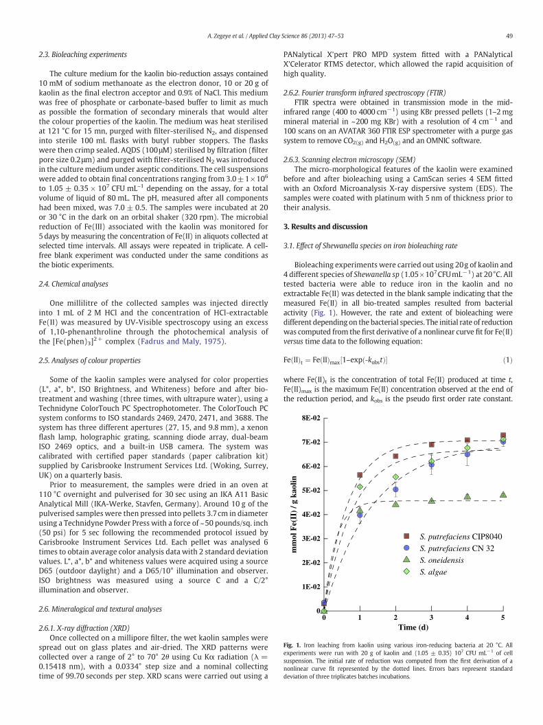
Fig. 1. Iron leaching from kaolin using various iron-reducing bacteria at 20 °C. Allexperiments were run with 20 g of kaolin and (1.05 ± 0.35) 107 CFU mL−1 of cellsuspension. The initial rate of reduction was computed from the first derivation of anonlinear curve fit represented by the dotted lines. Errors bars represent standarddeviation of three triplicates batches incubations.
49A. Zegeye et al. / Applied Clay Science 86 (2013) 47–53
2.3. Bioleaching experiments
The culture medium for the kaolin bio-reduction assays contained10 mM of sodium methanoate as the electron donor, 10 or 20 g ofkaolin as the final electron acceptor and 0.9% of NaCl. This mediumwas free of phosphate or carbonate-based buffer to limit as muchas possible the formation of secondary minerals that would alterthe colour properties of the kaolin. The medium was heat sterilisedat 121 °C for 15 mn, purged with filter-sterilised N2, and dispensedinto sterile 100 mL flasks with butyl rubber stoppers. The flaskswere then crimp sealed. AQDS (100μM) sterilised by filtration (filterpore size 0.2μm) and purged with filter-sterilised N2 was introducedin the culturemedium under aseptic conditions. The cell suspensionswere added to obtain final concentrations ranging from 3.0±1×10⁶
to 1.05 ± 0.35 × 10⁷ CFU mL–1 depending on the assay, for a totalvolume of liquid of 80 mL. The pH, measured after all componentshad been mixed, was 7.0 ± 0.5. The samples were incubated at 20or 30 °C in the dark on an orbital shaker (320 rpm). The microbialreduction of Fe(III) associated with the kaolin was monitored for5 days by measuring the concentration of Fe(II) in aliquots collected atselected time intervals. All assays were repeated in triplicate. A cell-free blank experiment was conducted under the same conditions asthe biotic experiments.
2.4. Chemical analyses
One millilitre of the collected samples was injected directlyinto 1 mL of 2 M HCl and the concentration of HCl-extractableFe(II) was measured by UV-Visible spectroscopy using an excessof 1,10-phenanthroline through the photochemical analysis ofthe [Fe(phen)3]2+ complex (Fadrus and Maly, 1975).
2.5. Analyses of colour properties
Some of the kaolin samples were analysed for color properties(L*, a*, b*, ISO Brightness, and Whiteness) before and after bio-treatment and washing (three times, with ultrapure water), using aTechnidyne ColorTouch PC Spectrophotometer. The ColorTouch PCsystem conforms to ISO standards 2469, 2470, 2471, and 3688. Thesystem has three different apertures (27, 15, and 9.8 mm), a xenonflash lamp, holographic grating, scanning diode array, dual-beamISO 2469 optics, and a built-in USB camera. The system wascalibrated with certified paper standards (paper calibration kit)supplied by Carisbrooke Instrument Services Ltd. (Woking, Surrey,UK) on a quarterly basis.
Prior to measurement, the samples were dried in an oven at110 °C overnight and pulverised for 30 sec using an IKA A11 BasicAnalytical Mill (IKA-Werke, Stavfen, Germany). Around 10 g of thepulverised samples were then pressed into pellets 3.7cm in diameterusing a Technidyne Powder Press with a force of ~50 pounds/sq. inch(50 psi) for 5 sec following the recommended protocol issued byCarisbrooke Instrument Services Ltd. Each pellet was analysed 6times to obtain average color analysis data with 2 standard deviationvalues. L*, a*, b* and whiteness values were acquired using a sourceD65 (outdoor daylight) and a D65/10° illumination and observer.ISO brightness was measured using a source C and a C/2°illumination and observer.
2.6. Mineralogical and textural analyses
2.6.1. X-ray diffraction (XRD)Once collected on a millipore filter, the wet kaolin samples were
spread out on glass plates and air-dried. The XRD patterns werecollected over a range of 2° to 70° 2θ using Cu Kα radiation (λ =0.15418 nm), with a 0.0334° step size and a nominal collectingtime of 99.70 seconds per step. XRD scans were carried out using a
PANalytical X'pert PRO MPD system fitted with a PANalyticalX'Celerator RTMS detector, which allowed the rapid acquisition ofhigh quality.
2.6.2. Fourier transform infrared spectroscopy (FTIR)FTIR spectra were obtained in transmission mode in the mid-
infrared range (400 to 4000 cm−1) using KBr pressed pellets (1–2mgmineral material in ~200 mg KBr) with a resolution of 4 cm−1 and100 scans on an AVATAR 360 FTIR ESP spectrometer with a purge gassystem to remove CO2(g) and H2O(g) and an OMNIC software.
2.6.3. Scanning electron microscopy (SEM)The micro-morphological features of the kaolin were examined
before and after bioleaching using a CamScan series 4 SEM fittedwith an Oxford Microanalysis X-ray dispersive system (EDS). Thesamples were coated with platinum with 5 nm of thickness prior totheir analysis.
3. Results and discussion
3.1. Effect of Shewanella species on iron bioleaching rate
Bioleaching experiments were carried out using 20g of kaolin and4 different species of Shewanella sp (1.05×10⁷CFUmL−1) at 20°C. Alltested bacteria were able to reduce iron in the kaolin and noextractable Fe(II) was detected in the blank sample indicating that themeasured Fe(II) in all bio-treated samples resulted from bacterialactivity (Fig. 1). However, the rate and extent of bioleaching weredifferent depending on the bacterial species. The initial rate of reductionwas computed from the first derivative of a nonlinear curve fit for Fe(II)versus time data to the following equation:
Fe IIð Þt ¼ Fe IIð Þmax 1–exp ‐kobstð Þ½ � ð1Þ
where Fe(II)t is the concentration of total Fe(II) produced at time t,Fe(II)max is the maximum Fe(II) concentration observed at the end ofthe reduction period, and kobs is the pseudo first order rate constant.
0 1 2 3 4 5
Time (d)
0
2E-02
4E-02
6E-02
8E-02
1E-01
1E-01
1E-01
Fe(
II)
mM
/ g
kaol
in
K:10g; B: (1,05 ± 0,35) 10E7K:10g; B: (3 ± 1) 10E6K:10g; B: (6 ± 2) 10E6K:20g; B: (3 ± 1) 10E6K:20g; B: (1,05 ± 0,35) 10E7
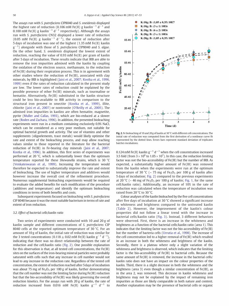
Fig. 2. Fe bioleaching of 10 and 20g of kaolin at 30°Cwith different cell concentrations. Theinitial rate of reduction was computed from the first derivation of a nonlinear curve fitrepresented by the dotted lines. Errors bars represent standard deviation of triplicatesbatches incubations.
50 A. Zegeye et al. / Applied Clay Science 86 (2013) 47–53
The assays run with S. putrefaciens CIP8040 and S. oneidensis displayedthe highest rate of reduction (0.106 mM Fe(II) g kaolin−1 d−1 and0.100 mM Fe(II) g kaolin−1 d−1 respectively). Although the assaysrun with S. putrefaciens CN32 displayed a lower rate of reduction(0.05 mM Fe(II) g kaolin−1 d−1), the extent of reduction after5 days of incubation was one of the highest (1.35 mM Fe(II) kaoling−1) alongside with those of S. putrefaciens CIP8040 and S. algae.On the other hand, S. oneidensis displayed the lowest extent ofreduction, reaching the value of 0.93 mM Fe(II) per gram of kaolinafter 5 days of incubation. These results indicate that IRB are able toremove the iron impurities admixed with the kaolin by couplingthe oxidation of the electron source, methanoate, to the reductionof Fe(III) during their respiration process. This is in agreement withother studies where the reduction of Fe(III), associated with clayminerals, by IRB is highlighted (Jaisi et al., 2007; Kostka et al., 1996,1999) even if the rates of reduction calculated in the present studyare low. The lower rates of reduction could be explained by thepossible presence of other Fe(III) minerals, such as tourmaline orhematite. Alternatively, Fe(III) substituted in the kaolin structurecould be less bio-available to IRB activity in comparison to thestructural iron present in smectite (Kostka et al., 1999), illite,chlorite (Jaisi et al., 2007) or nontronite (O'Reilly et al., 2005). Theadmixed iron impurities in kaolins are often hematite, magnetite,pyrite (Muller and Callas, 1993), which are bio-reduced at a slowerrate (Roden and Zachara, 1996). In addition, the presented bioleachingexperiments were run in a medium containing exclusively 0.9% NaCl,which can be considered as a very poor medium, not suitable foroptimal bacterial growth and activity. The use of vitamins and othersupplements (oligoelements, trace metals) would likely optimise therate and extent of the bioleaching process, and may allow reachingvalues similar to those reported in the literature for the bacterialreduction of Fe(III) in Fe-bearing clay minerals (Jaisi et al., 2007;Kostka et al., 1996). In addition, this first series of experiments wasperformed at 20 °C, which is substantially lower than the optimumtemperature reported for these Shewanella strains, which is 30 °C(Venkateswaran et al., 1999). Increasing the temperature wouldtherefore be expected to substantially improve the rate and extentof bioleaching. The use of higher temperature and additives wouldhowever increase the overall cost of the refinement procedure.Numerous supplemental bioleaching experiments would be requiredto evaluate the added benefits for each modification of the procedure(additives and temperature) and identify the optimum bioleachingconditions in terms of both benefits and costs.
Subsequent experiments focused on bioleaching with S. putrefaciensCIP 8040 because itwas themost suitable bacterium in terms of rate andextent of iron reduction.
3.2. Effect of bacterial cells/kaolin ratio
Two series of experiments were conducted with 10 and 20 g ofkaolin sample and different concentrations of S. putrefaciens CIP8040 cells at the reported optimum temperature of 30 °C. For anamount of 10 g of kaolin, the initial rate of reduction was similar forthe 3 tested concentrations (0.118± 0.02mM Fe(II) kaolin g−1 d−1),indicating that there was no direct relationship between the rate ofreduction and the cell/kaolin ratio (Fig. 2). One possible explanationfor this observation is that, at all tested cell concentrations, the Fe(III)sites on the surfaces of the iron-bearingmineral particleswere probablysaturated with cells such that any increase in cell number would notlead to any increase in the reduction rate. Regardless of the tested cellconcentration, the extent of reduction reached after 5days of incubationwas about 75 mg of Fe2O3 per 100 g of kaolin, further demonstratingthat the cell number was not the limiting factor during Fe(III) reductionbut that the bio-accessibility of Fe(III) was likely governing the overallreduction kinetics. For the assays run with 20 g of kaolin, the rate ofreduction increased from 0.016 mM Fe(II) kaolin g−1 d−1 to
0.124mM Fe(II) kaolin g−1 d−1 when the cell concentration increased3.5 fold (from 3×10⁶ to 1.05×10⁷). In this case, the reduction limitingfactor was not the bio-accessibility of Fe(III) but the number of IRB. Asexpected, a substantially higher amount of Fe(III) was removedfrom the kaolin when the experiments were run at the optimumtemperature of 30 °C (~ 75 mg of Fe2O3 per 100 g of kaolin after5 days of incubation; Fig. 2) compared to the previous experimentsat 20 °C (~ 46 mg of Fe2O3 per 100 g of kaolin; Fig. 1, for the samecell/kaolin ratio). Additionally, an increase of 10% in the rate ofreduction was calculated when the temperature of incubation wasraised from 20 °C to 30 °C.
Colour analyses of the kaolin bioleachedby thefive cell concentrationsafter five days of incubation at 30 °C showed a significant increasein whiteness and brightness compared to the untreated kaolin(Table 2). However, the improvement of the kaolin's colourproperties did not follow a linear trend with the increase inbacterial cells/kaolin ratio (Fig. 3). Instead, 3 different behaviorswere observed. First, there is an increase in the whiteness andbrightness as a function of the bacterial cells/kaolin ratio (area 1). Thisindicates that the limiting factor was not the bio-accessibility of Fe(III)but the number of bacteria cells (Urrutia et al., 1998). The increase ofthe cell concentration led to a higher removal of Fe(III) which resultedin an increase in both the whiteness and brightness of the kaolin.Secondly, there is a plateau where only a slight variation of thewhiteness and brightness is observed, which indicates that the limitingfactor is the bio-accessibility of Fe(III) to bacterial cells. Because thesame amount of Fe(III) is removed, the increase in the bacterial cells/kaolin ratio does not have an impact on the colour properties of thekaolin. Third, there is a slight decrease in both the whiteness and thebrightness (area 3) even though a similar concentration of Fe(III), asin the area 2, was removed. This decrease in kaolin whiteness andbrightness may not be explained by the impact of remaining ironimpurities as those are likely comparable in both nature and content.Another explanation may be the presence of bacterial cells or organic
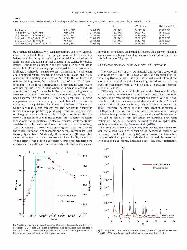
Table 2Colour analyses data of kaolin before and after bioleaching with different Shewanella putrefaciens CIP8040 concentration after 5 days of incubation at 30 °C.
L* A* B* %Whiteness %Brightness (ISO)
Blank kaolin 91.81(0.012) −0.08 (0.02) 5.56 (0.02) 54.31(0.08) 73.98 (0.02)10 g kaolin/(3± 1) 106 CFUmL−1 92.86 (0.02) −0.41 (0.02) 3.53 (0.02) 66.30 (0.10) 78.63 (0.06)10 g kaolin/(6± 2) 106 CFUmL−1 92.78 (0.01) −0.41 (0.02) 3.58 (0.02) 65.86 (0.08) 78.42 (0.04)10 g kaolin/(1.05±0.35) 107 CFUmL−1 92.14(0.01) −0.35 (0.02) 3.80 (0.02) 63.32 (0.10) 76.78 (0.02)20 g kaolin/(3± 1) 106 CFUmL−1 92.40 (0.01) −0.23 (0.02) 5.27 (0.02) 57.10 (0.06) 75.60 (0.02)20 g kaolin/(1.05±0.35) 107 CFUmL−1 93.00 (0.01) −0.39 (0.02) 3.73 (0.01) 65.73 (0.06) 78.77(0.02)
51A. Zegeye et al. / Applied Clay Science 86 (2013) 47–53
by-products of bacterial activity, such as organic polymers, which couldcolour the material. Though the samples were washed extensivelybefore the colour analyses, such organic materials may bind to thekaolin particles and remain in small amount in the washed bioleachedkaolins. Being more abundant in this last sample (higher cell/kaolinratio), their effect on colour properties would be more pronouncedleading to a slight reduction in the colourmeasurements. Thewhitenessand brightness values reached their maximum (66.3% and 79.0%,respectively), indicating an increase of 22.07% for the whiteness and6.5% for the brightness, for a cell/kaolin ratio of 24 × 106 CFU per gof kaolin. The whiteness improvement is comparable with resultsobtained by Guo et al. (2010b) where an increase of around 34%was observed using fermentative indigenous iron-reducing bacteria.However, although higher increases in whiteness, up to 70%, havebeen observed in other studies (Arslan and Bayat, 2009), a directcomparison of the whiteness improvement obtained in the presentstudy with other published data is not straightforward. This is dueto the fact that microorganisms can enhance kaolin quality, interms of colour properties, by producing organic or inorganic acidsor changing redox conditions to leach Fe(III) iron impurities. Thebacterial metabolism used in the present study to refine the kaolinis anaerobic iron respiration (e.g. electron transfer) while the studiesavailable in the literature emphasise fermentative metabolism (e.g.acid production) or mixed metabolism (e.g. soil consortium) wherethe relative importance of anaerobic and aerobic metabolism is notthoroughly identified. Additionally, the amount of Fe(III) impurities(admixed or structural) can vary from kaolin to kaolin, dependingon the origin of the deposit and geological processes, hampering thecomparison. Nevertheless, our study highlights that a metabolism
0 2.0*105 4.0*105 6.0*105 8.0*105 1.0*106 1.2*106
Ratio
50.0
55.0
60.0
65.0
70.0
Whi
tnes
s
73.0
74.0
75.0
76.0
77.0
78.0
79.0
Bri
ghtn
ess
Whitness
Brightness
I II III
Fig. 3.Whiteness and brightness evolution after 5days of incubation as the function of cell/kaolin ratio (CFU /g kaolin). The blue box represents the best cell/kaolin ratio identified inthis study, to achieve a noticeable improvement of the kaolin colour properties. The errorbars represent standard deviation of six separate analyses.
other than fermentative can be used to improve the quality of industrialkaolin even though supplementary research is needed to exploit thismetabolism to its full potential.
3.3. Mineralogical analyses of the kaolin after Fe(III) bioleaching
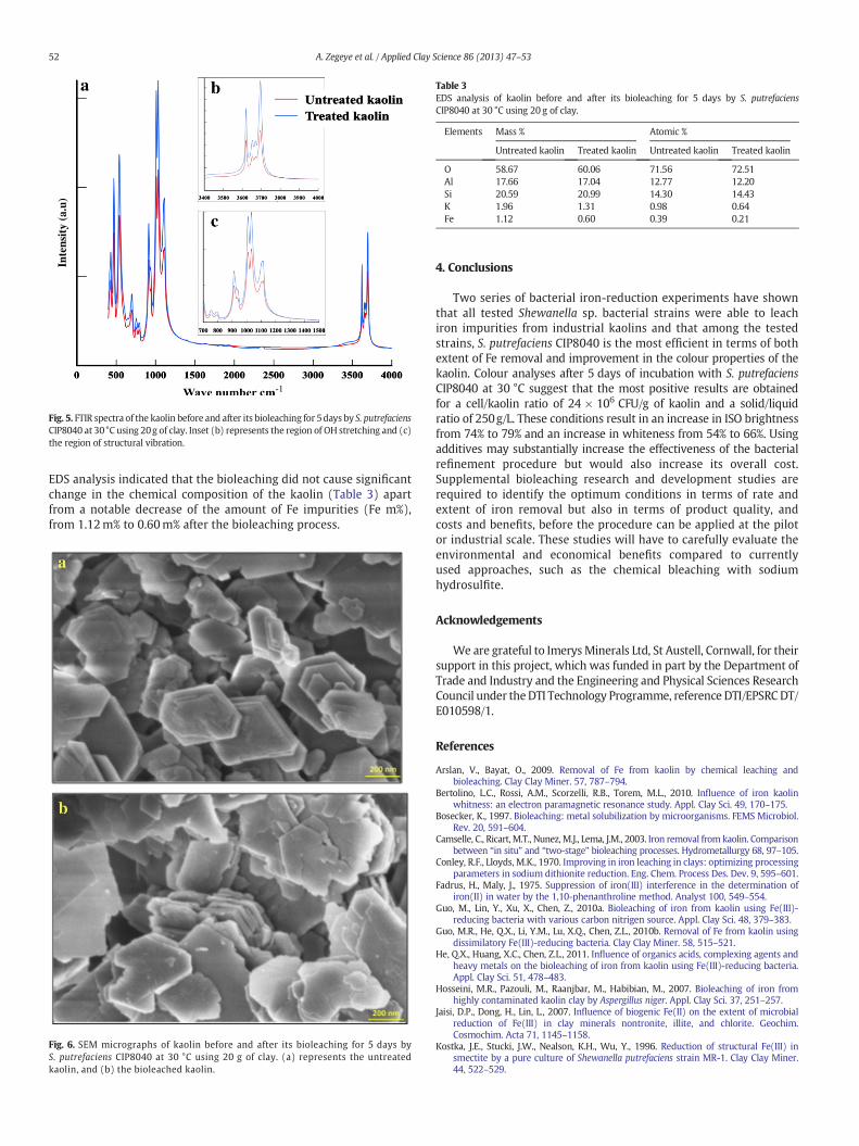
The XRD patterns of the raw material and kaolin treated withS. putrefaciens CIP 8040 for 5 days at 30 °C are identical (Fig. 4),indicating that very little — if any — structural modification of thekaolinite occurred during the bioleaching procedure, and that nocrystalline secondary material was formed, as elsewhere reported(Guo et al., 2010a).
FTIR analyses of the initial kaolin and of the blank samples after5 days at 30 °C are very similar and characteristic of kaolinite withno measurable trace of organic material or bacterial cells (Fig. 5a).In addition, all spectra show a small shoulder at 3598 cm−1, whichis characteristic of AlFeOH vibrators (Fig. 5b), (Petit and Decarreau,1990), therefore indicating that the small amount of structuralFe(III) present in the kaolinite crystal lattice was not removed duringthe bioleaching procedure. In fact, only a small part of this structuraliron can be removed from the kaolin by industrial processingtechniques (magnetic separation followed by sodium hydrosulfiteleaching) as evidenced by Bertolino et al., (2010).
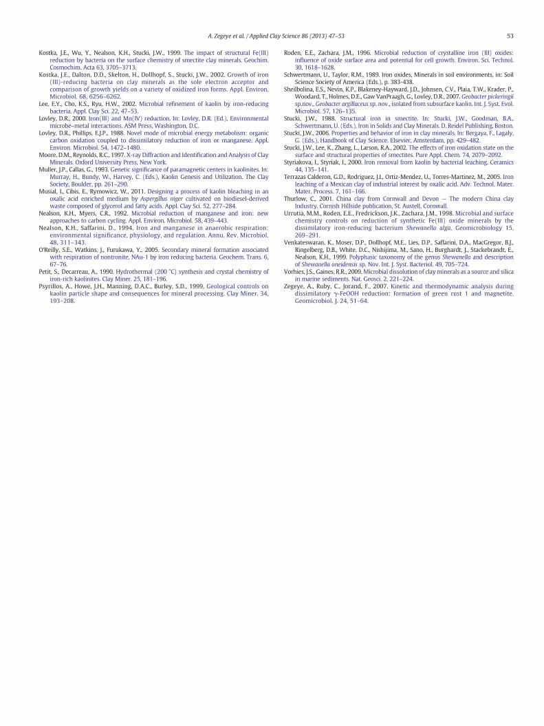
Observations of the initial kaolin by SEM revealed the presence ofwell-crystallised kaolinite consisting of hexagonal particles ofdifferent size and thickness (Fig. 6a). In comparison, the bioleachedsamples exhibited particles with similar size and thickness butwith rounded and slightly damaged edges (Fig. 6b). Additionally,
0 10 20 30 40 50 60 70
2θ
0
7*104
6*104
5*104
4*104
3*104
2*104
1*104
8*104
9*104
Inte
nsit
y (a
.u)
Untreated kaolinTreated kaolin
K
K
K
K
K
K
K K K
Fig. 4. XRD patterns of kaolin before and after its bioleaching for 5 days by S. putrefaciensCIP8040 at 30 °C using 20 g of clay, K= kaolinite peaks a.u= arbitrary units.
Inte
nsit
y (a
.u)
Fig. 5. FTIR spectra of the kaolin before and after its bioleaching for 5days by S. putrefaciensCIP8040 at 30°C using 20g of clay. Inset (b) represents the region of OH stretching and (c)the region of structural vibration.
Table 3EDS analysis of kaolin before and after its bioleaching for 5 days by S. putrefaciensCIP8040 at 30 °C using 20 g of clay.
Elements Mass % Atomic %
Untreated kaolin Treated kaolin Untreated kaolin Treated kaolin
O 58.67 60.06 71.56 72.51Al 17.66 17.04 12.77 12.20Si 20.59 20.99 14.30 14.43K 1.96 1.31 0.98 0.64Fe 1.12 0.60 0.39 0.21
52 A. Zegeye et al. / Applied Clay Science 86 (2013) 47–53
EDS analysis indicated that the bioleaching did not cause significantchange in the chemical composition of the kaolin (Table 3) apartfrom a notable decrease of the amount of Fe impurities (Fe m%),from 1.12m% to 0.60m% after the bioleaching process.
Fig. 6. SEM micrographs of kaolin before and after its bioleaching for 5 days byS. putrefaciens CIP8040 at 30 °C using 20 g of clay. (a) represents the untreatedkaolin, and (b) the bioleached kaolin.
4. Conclusions
Two series of bacterial iron-reduction experiments have shownthat all tested Shewanella sp. bacterial strains were able to leachiron impurities from industrial kaolins and that among the testedstrains, S. putrefaciens CIP8040 is the most efficient in terms of bothextent of Fe removal and improvement in the colour properties of thekaolin. Colour analyses after 5 days of incubation with S. putrefaciensCIP8040 at 30 °C suggest that the most positive results are obtainedfor a cell/kaolin ratio of 24 × 106 CFU/g of kaolin and a solid/liquidratio of 250g/L. These conditions result in an increase in ISO brightnessfrom 74% to 79% and an increase in whiteness from 54% to 66%. Usingadditives may substantially increase the effectiveness of the bacterialrefinement procedure but would also increase its overall cost.Supplemental bioleaching research and development studies arerequired to identify the optimum conditions in terms of rate andextent of iron removal but also in terms of product quality, andcosts and benefits, before the procedure can be applied at the pilotor industrial scale. These studies will have to carefully evaluate theenvironmental and economical benefits compared to currentlyused approaches, such as the chemical bleaching with sodiumhydrosulfite.
Acknowledgements
We are grateful to ImerysMinerals Ltd, St Austell, Cornwall, for theirsupport in this project, which was funded in part by the Department ofTrade and Industry and the Engineering and Physical Sciences ResearchCouncil under theDTI Technology Programme, reference DTI/EPSRCDT/E010598/1.
References
Arslan, V., Bayat, O., 2009. Removal of Fe from kaolin by chemical leaching andbioleaching. Clay Clay Miner. 57, 787–794.
Bertolino, L.C., Rossi, A.M., Scorzelli, R.B., Torem, M.L., 2010. Influence of iron kaolinwhitness: an electron paramagnetic resonance study. Appl. Clay Sci. 49, 170–175.
Bosecker, K., 1997. Bioleaching: metal solubilization by microorganisms. FEMS Microbiol.Rev. 20, 591–604.
Camselle, C., Ricart, M.T., Nunez,M.J., Lema, J.M., 2003. Iron removal from kaolin. Comparisonbetween “in situ” and “two-stage” bioleaching processes. Hydrometallurgy 68, 97–105.
Conley, R.F., Lloyds, M.K., 1970. Improving in iron leaching in clays: optimizing processingparameters in sodium dithionite reduction. Eng. Chem. Process Des. Dev. 9, 595–601.
Fadrus, H., Maly, J., 1975. Suppression of iron(III) interference in the determination ofiron(II) in water by the 1,10-phenanthroline method. Analyst 100, 549–554.
Guo, M., Lin, Y., Xu, X., Chen, Z., 2010a. Bioleaching of iron from kaolin using Fe(III)-reducing bacteria with various carbon nitrigen source. Appl. Clay Sci. 48, 379–383.
Guo, M.R., He, Q.X., Li, Y.M., Lu, X.Q., Chen, Z.L., 2010b. Removal of Fe from kaolin usingdissimilatory Fe(III)-reducing bacteria. Clay Clay Miner. 58, 515–521.
He, Q.X., Huang, X.C., Chen, Z.L., 2011. Influence of organics acids, complexing agents andheavy metals on the bioleaching of iron from kaolin using Fe(III)-reducing bacteria.Appl. Clay Sci. 51, 478–483.
Hosseini, M.R., Pazouli, M., Raanjbar, M., Habibian, M., 2007. Bioleaching of iron fromhighly contaminated kaolin clay by Aspergillus niger. Appl. Clay Sci. 37, 251–257.
Jaisi, D.P., Dong, H., Lin, L., 2007. Influence of biogenic Fe(II) on the extent of microbialreduction of Fe(III) in clay minerals nontronite, illite, and chlorite. Geochim.Cosmochim. Acta 71, 1145–1158.
Kostka, J.E., Stucki, J.W., Nealson, K.H., Wu, Y., 1996. Reduction of structural Fe(III) insmectite by a pure culture of Shewanella putrefaciens strain MR-1. Clay Clay Miner.44, 522–529.
53A. Zegeye et al. / Applied Clay Science 86 (2013) 47–53
Kostka, J.E., Wu, Y., Nealson, K.H., Stucki, J.W., 1999. The impact of structural Fe(III)reduction by bacteria on the surface chemistry of smectite clay minerals. Geochim.Cosmochim. Acta 63, 3705–3713.
Kostka, J.E., Dalton, D.D., Skelton, H., Dollhopf, S., Stucki, J.W., 2002. Growth of iron(III)-reducing bacteria on clay minerals as the sole electron acceptor andcomparison of growth yields on a variety of oxidized iron forms. Appl. Environ.Microbiol. 68, 6256–6262.
Lee, E.Y., Cho, K.S., Ryu, H.W., 2002. Microbial refinement of kaolin by iron-reducingbacteria. Appl. Clay Sci. 22, 47–53.
Lovley, D.R., 2000. Iron(III) and Mn(IV) reduction. In: Lovley, D.R. (Ed.), Environmentalmicrobe–metal interactions. ASM Press, Washington, D.C.
Lovley, D.R., Phillips, E.J.P., 1988. Novel mode of microbial energy metabolism: organiccarbon oxidation coupled to dissimilatory reduction of iron or manganese. Appl.Environ. Microbiol. 54, 1472–1480.
Moore, D.M., Reynolds, R.C., 1997. X-ray Diffraction and Identification and Analysis of ClayMinerals. Oxford University Press, New York.
Muller, J.P., Callas, G., 1993. Genetic significance of paramagnetic centers in kaolinites. In:Murray, H., Bundy, W., Harvey, C. (Eds.), Kaolin Genesis and Utilization. The ClaySociety, Boulder, pp. 261–290.
Musial, I., Cibis, E., Rymowicz, W., 2011. Designing a process of kaolin bleaching in anoxalic acid enriched medium by Aspergillus niger cultivated on biodiesel-derivedwaste composed of glycerol and fatty acids. Appl. Clay Sci. 52, 277–284.
Nealson, K.H., Myers, C.R., 1992. Microbial reduction of manganese and iron: newapproaches to carbon cycling. Appl. Environ. Microbiol. 58, 439–443.
Nealson, K.H., Saffarini, D., 1994. Iron and manganese in anaerobic respiration:environmental significance, physiology, and regulation. Annu. Rev. Microbiol.48, 311–343.
O'Reilly, S.E., Watkins, J., Furukawa, Y., 2005. Secondary mineral formation associatedwith respiration of nontronite, NAu-1 by iron reducing bacteria. Geochem. Trans. 6,67–76.
Petit, S., Decarreau, A., 1990. Hydrothermal (200 °C) synthesis and crystal chemistry ofiron-rich kaolinites. Clay Miner. 25, 181–196.
Psyrillos, A., Howe, J.H., Manning, D.A.C., Burley, S.D., 1999. Geological controls onkaolin particle shape and consequences for mineral processing. Clay Miner. 34,193–208.
Roden, E.E., Zachara, J.M., 1996. Microbial reduction of crystalline iron (III) oxides:influence of oxide surface area and potential for cell growth. Environ. Sci. Technol.30, 1618–1628.
Schwertmann, U., Taylor, R.M., 1989. Iron oxides, Minerals in soil environments, in: SoilScience Society of America (Eds.), p. 383-438.
Sheilbolina, E.S., Nevin, K.P., Blakeney-Hayward, J.D., Johnsen, C.V., Plaia, T.W., Krader, P.,Woodard, T., Holmes, D.E., Gaw VanPraagh, G., Lovley, D.R., 2007.Geobacter pickeringiisp.nov., Geobacter argillaceus sp. nov., isolated from subsurface kaolin. Int. J. Syst. Evol.Microbiol. 57, 126–135.
Stucki, J.W., 1988. Structural iron in smectite. In: Stucki, J.W., Goodman, B.A.,Schwertmann, U. (Eds.), Iron in Solids and ClayMinerals. D. Reidel Publishing, Boston.
Stucki, J.W., 2006. Properties and behavior of iron in clay minerals. In: Bergaya, F., Lagaly,G. (Eds.), Handbook of Clay Science. Elsevier, Amsterdam, pp. 429–482.
Stucki, J.W., Lee, K., Zhang, L., Larson, R.A., 2002. The effects of iron oxidation state on thesurface and structural properties of smectites. Pure Appl. Chem. 74, 2079–2092.
Styriakova, I., Styriak, I., 2000. Iron removal from kaolin by bacterial leaching. Ceramics44, 135–141.
Terrazas Calderon, G.D., Rodriguez, J.I., Ortiz-Mendez, U., Torres-Martinez, M., 2005. Ironleaching of a Mexican clay of industrial interest by oxalic acid. Adv. Technol. Mater.Mater. Process. 7, 161–166.
Thurlow, C., 2001. China clay from Cornwall and Devon — The modern China clayIndustry. Cornish Hillside publication, St. Austell, Cornwall.
Urrutia, M.M., Roden, E.E., Fredrickson, J.K., Zachara, J.M., 1998. Microbial and surfacechemistry controls on reduction of synthetic Fe(III) oxide minerals by thedissimilatory iron-reducing bacterium Shewanella alga. Geomicrobiology 15,269–291.
Venkateswaran, K., Moser, D.P., Dollhopf, M.E., Lies, D.P., Saffarini, D.A., MacGregor, B.J.,Ringelberg, D.B., White, D.C., Nishijima, M., Sano, H., Burghardt, J., Stackebrandt, E.,Nealson, K.H., 1999. Polyphasic taxonomy of the genus Shewanella and descriptionof Shewanella oneidensis sp. Nov. Int. J. Syst. Bacteriol. 49, 705–724.
Vorhies, J.S., Gaines, R.R., 2009. Microbial dissolution of clayminerals as a source and silicain marine sediments. Nat. Geosci. 2, 221–224.
Zegeye, A., Ruby, C., Jorand, F., 2007. Kinetic and thermodynamic analysis duringdissimilatory γ-FeOOH reduction: formation of green rust 1 and magnetite.Geomicrobiol. J. 24, 51–64.