Embed Size (px)
Citation preview

G l y c o c o n j u g a t e J o u r n a l (1996) 1 3 : 2 6 3 - 2 7 1
Recognition of the blood group H type 2 trisaccharide epitope by 28 monoclonai antibodies and three lectins
R O S E L L A M O L L I C O N E 1, A N N E C A I L L E A U 1, A N N E I M B E R T Y 2, P I E R R E G A N E 3, S E R G E P E R E Z 4 and R A F A E L O R I O L 1 .
ZlNSERM U.178 and Universitd Paris-Sud, 16 Avenue Paul VailIant-Couturier, F94807 Villejuif cedex. France 2Laboratoire de Synth~se Organique-CNRS, Facultd des Sciences et Techniques, 2 rue de la Houssini~re, F44072 Nantes eedex 03, France 3Institut National de la Transfusion Sanguine, 75915 Paris, France 41ng~nierie Moldeulaire, INRA BP 1627, F44316 Nantes cedex 03, France
Received 7 July 1995, revised 27 September 1995
The patterns of cross-reaction of 30 monoclonal antibodies and three lectins were determined by ELISA with 21 ABH, Ii or Lewis related synthetic oligosaccharides coupled to bovine serum albumin. At least seven main groups of cross-reactive patterns were identified among the antibodies, plus several isolated antibodies which had intermediate patterns between two of the main antibody groups. The three lectins had different cross-reaction patterns, Galactia tenuiflora was different from all the antibodies, Ulex europaeus lectin 1 and Lotus tetragonolobus were similar, but not identical to groups III and V of antibodies respectively. The anti-H antibodies cross-reacting with A type 2 gave similar agglutination scores with all the normal ABO erythrocytes, while the anti-H antibodies not cross-reacting with A type 2 reacted with different scores: O > A2 > A2B > B > A1 > A1B > Oh, suggesting that these antibodies react better with the free H epitopes and do not recognize the H in A or B epitopes. Based on the ELISA and agglutin/ttion results and the lowest energy conformations of each oligosaccharide obtained by computer modelling, the most probable oligosaccharide surface areas recognized by each antibody main group are illustrated.
Keywords: ELISA, synthetic oligosaccharides, monoclonal antibodies, blood group H, lectins, ABO
Introduction
The major blood group anti-A and anti-B monoclonal antibody specificities have been thoroughly studied [1] and the molecular basis of the ABO genetic polymorphism has been elucidated [2M]. The molecular basis of the H [5], Se [6,7] and Lewis [8-11] related blood group polymorphisms have been also elucidated. However the serological cross-reaction patterns of monoclonal anti-H are not very well known.
As early as 1982, a mouse monoclonal antibody anti-H type 2 was described [12] and oligosaccharide probes for this blood group specificity were synthesized [13]. Ten anti-H antibodies submitted to the First International Workshop on Monoclonal Antibodies Against Human Red Blood Cell and Related Antigens (Paris, 1987) were
*To whom correspondence should be addressed.
0282-0080 © 1996 Chapman & Hall
analysed with these synthetic oligosaccharide epitopes and showed a main cross-reaction with Le y [14]. Similar results were obtained with ten anti-H submitted to the Second International Workshop (Lund, 1990) with the same set of synthetic oligosaccharide epitopes [15], and independently with eleven other anti-H monoclonals tested with glycolipids by another group [16]. Since then we have continued collecting anti-H reagents and have increased the number of chemically related oligosacchar- ide epitopes used for the analysis, in order to get further insights into this antibody specificity.
During the last 5 years we have been able to analyse 28 monoclonal antibodies and three lectins, all reacting with the major red cell H type 2 epitope. We have increased the number of chemically related synthetic oligosaccharides to 21 and have made a computer simulation of the lowest energy three-dimensional structures for ABH and Lewis related oligosaccharides

264 Mollicone et al.
[17]. Using these tools we now present a review of the H type 2 antibody specificities, their cross-reactions with related chemical structures, and the corresponding carbo- hydrate surfaces of the most favoured conformations, expected to react with the different groups of anti-H type 2 reagents.
Materials and methods
Synthetic oligosaccharides Twenty-one ABH, Ii or Lewis related oligosaccharides covalently coupled to bovine serum albumin (BSA) by a carbonyl-octyl linking arm, with an average of 15 hapten groups per molecule [18], were obtained from Chem- biomed Ltd (Alberta Research Council, Edmonton, Canada) (Table 1).
Monoclonal antibodies Human anti-I Ma was a gift from E. Kabat [19, 20]. Ten mouse monoclonal antibodies were obtained from the Second International Workshop [21]: 056 clone ML27 (IgM), Centre Regional de Transfusion Sanguine de Rennes, France; 057 clone 109.34Cll (IgM), Centre Regional de Transfusion Sanguine de Lille, France; 058 clone H-86/44 (IgM) and 059 clone H-86/50 (IgM), National Hematological Scientific Center of Moscow,
Russia; 060 clone H005 (IgM), 066 clone 672/7E3 (IgM) and 067 clone 822/9E3 (IgM), BioCarb AB, Lund Sweden; 063 clone OSK16 (IgM), Osaka Red Cross Center, Osaka, Japan; 064 clone MS2.110A1A4 (IgM) and 065 clone MS3.318A1A1 (IgM), Institut National de la Transfusion Sanguine, Paris, France. Six mouse anti- bodies: NaMS-4F9 (4F9), NaM10-3C10 (3C10), NaM31- 5G9 (5G9), NaM19-7Ell (7El 1)(IgM), NaM9-1F9 (1F9) and NaM26-8B9 (8B9) were obtained from D. Blanchard, Centre Regional de Transfusion Sanguine de Nantes, France. Mouse clone BRIC41 was obtained from D. Anstee, International Blood Group reference Laboratory, Bristol UK. Two mouse antibodies: MS131 and MRT517 were obtained from E Rouger, Institut National de la Transfusion Sanguine, Paris, France. Two mouse anti- bodies: 19-OLE and 7-LE (named 34W6 in the first Workshop [14]) were obtained from J. Bara, INSERM U.55, Saint Antoine Hospital, Paris, France. Two mouse antibodies: BNH9 [22] and BNF13 [23] were obtained from A. Blancher, Centre Regional de Transfusion Sanguine de Toulouse, France. Mouse clone 91,C-2 was obtained from J. Kolberg, National Institute of Public Health, Oslo, Norway. Two mouse antibodies: 1BE12 [24, 25] and 1BD6 were obtained from A. Roseto, DICA, Universit6 Technologique de Compi6gne, France. Mouse clone 92FRA2.1 was obtained from Chembiomed Ltd, Edmonton, Canada. Mouse clone 64/4D8 [26,27] was
Table 1. Structures of the ABH, Ii and Lewis related synthetic oligosaccharides used for coating the ELISA microtitre plates.
Name Chemical structure
B trisaccharide A trisaccharide A type 3 A Le b I trisaccharide LacNAc i trisaccharide
I pentasaccharide
A t y p e 2 H type 2 H type 6 Le y Le x H t y p e 5 H type 1 Le b
Le a Type 1 precursor H type 3 H type 4 Le disaccharide
GalM --~3 (Fuccd---~2)Gal/31--~R GalNAcM-+3(Fuccd---~2)Gal/31---)R GalNAcat--+3(Fucc~l--+2)Gal/31-~4GalNAcc~l---~R GalNAc~l--->3(Fucal---~2)Gal/31-+3(Fucc~l-M)GlcNAc/31--+R
Gal/31--*4GlcNAc/31 ~6Gal/31 --~R GalI31-+4GlcNAc/31-~R Gal/31--+4GlcNAc/31--~3Gal/3 t--~R Gal/31--~4GlcNAc/31--+3
Gal/31---~R Gal/31--~4GIcNAc/31--+6
GalNAcc~ l--~3(Fucc~l--~2)Gal/31---~4GlcNAc/31---~R Fuc~ 1--~2Gal/31-+4GlcNAc/31---~R Fuc~ 1-+2Gal/31--)4Glc/31---~R Fucc~l---~2Gal/31---~4(Fuce~l-+3)GlcNAc/31--+R
Gal/31--~4(Fucc~l ~3)GlcNAc/31--+R Fucc~ 1--~2Gal/31--->3 Gal/31---~R Fucc~ 1--)2Gal/31--,3GlcNAc/31---~R Fucc~ 1---~2Gal/31---~3(Fucc~ 1--~4)GlcNAc/31---~R
Gal/31--+3(Fuca 1-+4)GlcNAc/31---~R Gal/31-+3GlcNAcI31-+R
Fucc~ 1---~2Gal/31-+4GalNAcc~ 1---~R Fuc~ 1-+2Gal/31--)4GalNAc/31-+R
Fucc~ 1 ---~4GlcNAc/31--~R
R = (CH2)8-CO-NH-BSA.

Recognition of the blood group H type 2 trisaccharide epitope
bought from BioCarb AB, Lund, Sweden (this antibody has been commercialized as clone H001).
Lectins
Affinity purified, biotin labelled Ulex europaeus lectin I (UEAI) and Lotus tetragonolobus lectin (LTL) were obtained from Vector Laboratories (Burlingame, CA, USA), Galactia tenuiflora lectin was prepared by affinity chromatography on H type 2 Synsorb (Chembiomed, Alberta Research Council, Edmonton, Canada) and labelled with biotin (Amersham, UK) as described [28].
ELISA tests with synthetic oligosaccharide structures
ELISA tests were performed with a biotin-streptavidin system (Amersham, UK). Microtitre plates with 96 flat bottom wells (Nunc, Danemark) were coated overnight at room temperature, with 50/xl per well of a solution at 1/xgm1-1 of the synthetic oligosaccharide hapten-BSA in phosphate buffered saline pH 8 (PBS). The antigen solution was removed and the plates were incubated 1 h at 37 °C with 200/xl per well of gelatin 3% in PBS. After washing, 50/xl per well of two-fold serial dilutions of monoclonal antibodies in PBS containing gelatin (0.5%) and Yween 20 (0.02%) were added and incubated overnight. The plates were washed and 50/xl of biotin labelled secondary antibodies (anti-mouse Ig, anti-mouse /x chain or anti-human Ig) diluted 1:1000 in PBS were added and incubated for 2 h, washed again and perox- idase-labelled streptavidin diluted 1:1000 was added for 3 h. After a final wash the coloured reaction was developed with 100/xl per well of o-phenylene diamine (OPD) at 1 mgm1-1 in citrate buffer 0.02M, pH 5, containing 3/zlm1-1 of H202 at 30%. After 3 min the reaction was stopped with 200/xl of HC1 4 N and the optical density at 540 ,/m was read in a Microplate Reader (MR 600, Dynatech). Four antibodies (057, 066, 7El 1 and
265
MS 131) reacted better with the anti-/x than with the anti- Ig biotin labelled secondary antibody.
Direct tests with biotin-labelled lectins were performed in the same way without the primary and secondary antibodies. Negative controls were performed with BSA coated plates and the results for BSA coated wells were subtracted from the results of the hapten-BSA coated wells for each lectin and antibody.
Cluster analysis
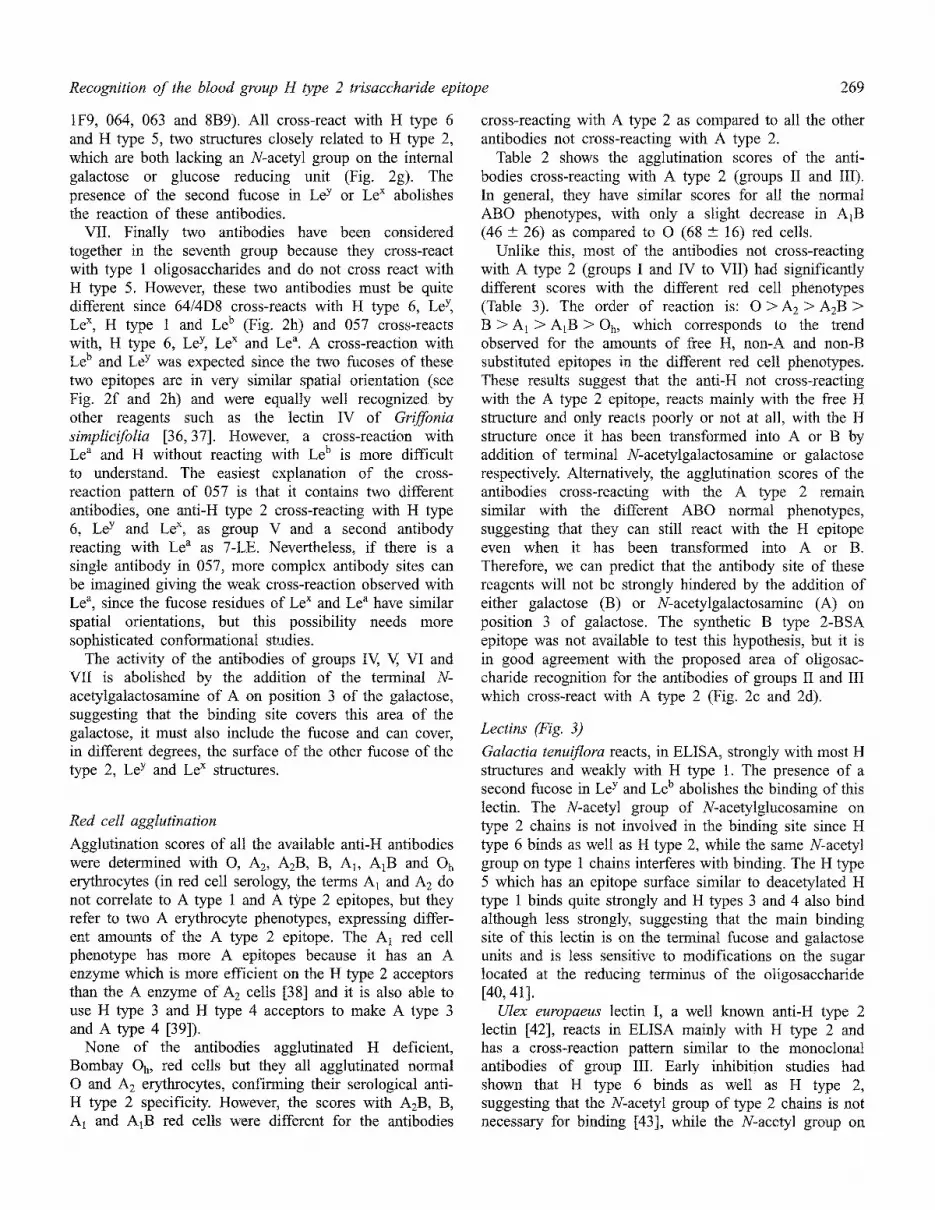
The ELISA results of antibodies and lectins were ordered in both horizontal and vertical directions, in order to maximize the formation of positive clusters around the H type 2 epitope. The antibodies reacting with H type 2 were ordered in seven main groups of cross-reacting patterns. Within each group, the order of columns was further adjusted to obtain decreasing OD values from left to right as described [1, 29].
Red cell agglutination
Serial two-fold dilutions in PBS were prepared for each antibody and 100/xl of each dilution was incubated for 30 rain with 100/xl of 2% red cell suspensions of each phenotype (O, A2, AzB, B, A1, A1B and Oh). Red cells were spun down for 15 s at 1000 × g and agglutination scores were determined [30]. Antibodies 7-LE, 063, 067, 1BE12, 1BD6, 92FRA2.1 and I-Ma could not be tested by agglutination (Tables 2 and 3).
Computer modelling
The lowest energy conformation of oligosaccharides H type 2, A type 2, Le a, Le b and Le y were taken from a previous study [17]. The conformations of oligosacchar- ides H type 5 and H type 6 were characterized using the same approach. Starting geometries were built from MONOBANK, a database of 3D structures of mono-
Table 2. Agglutination scores with different red cell phenotypes of the monoclonal anti-H type 2 antibodies (groups II to III of Fig. 1), reacting also with the A type 2 tetrasaccharide in ELISA (column on the right). None of them agglutinated the Oh red cells (Bombay), and all agglutinated normal ABO red cells with similar scores, confirming their anti-H specificity. Only the A1B red cells have scores significantly lower than O and A 2 reed cells (p < 0.05).
Antibodies Agglutination scores of different red cell phenotypes A ~pe 2 ELISA
0 A2 A2B B A 1 AIB O h
061 51 47 31 25 24 0 0 0.6 058 79 86 68 82 76 51 0 0.4 BNH9 83 81 70 63 61 61 0 0.3 19OLE 69 69 62 56 57 44 0 0.9 MS 131 43 50 41 40 36 20 0 1.0 C10 90 79 63 83 93 63 0 1.4 G9 63 73 84 51 69 84 0 1.5 BRIC41 65 53 55 5 50 42 0 1.5
Mean + SD 68 --+ 16 67 --+ 15 59 --+ 17 57 --+ 20 58 --+ 22 46 + 26 0 --+ 0 l --+ 0.5
266 Mollicone et al.
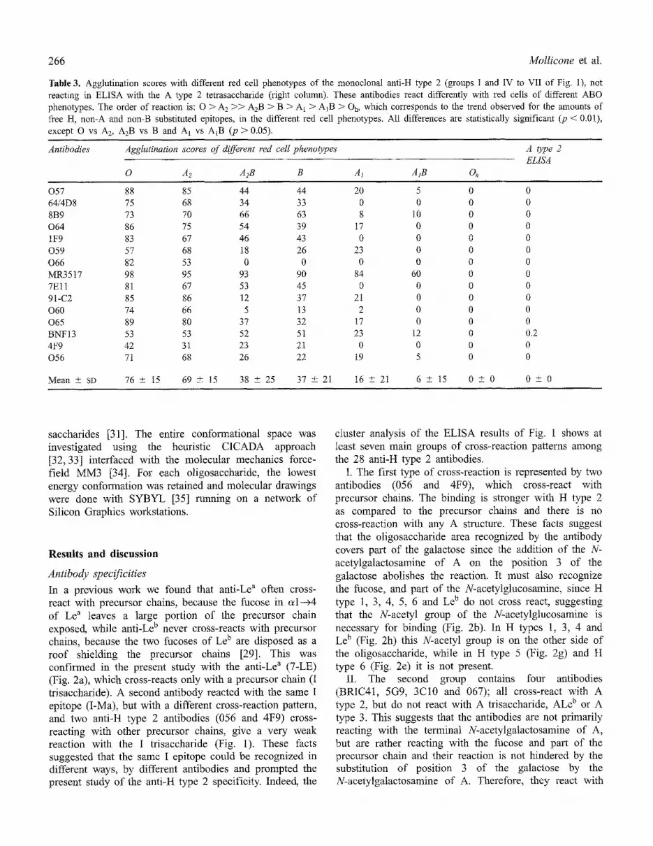
Table 3. Agglutination scores with different red cell phenotypes of the monoclonal anti-H type 2 (groups I and IV to VII of Fig. 1), not reacting in ELISA with the A type 2 tetrasaccharide (right column). These antibodies react differently with red cells of different ABO phenotypes. The order of reaction is: O > A 2 >> A2B > B > A1 > A1B > Oh, which corresponds to the trend observed for the amounts of free H, non-A and non-B substituted epitopes, in the different red cell phenotypes. All differences are statistically significant (p < 0.01), except O vs A2, A2B vs B and AI vs A1B (p > 0.05).
Antibodies Agglutination scores of different red cell phenoO~pes A type 2 ELtSA
0 A2 A2B B A1 AtB Oh
057 88 85 44 44 20 5 0 0 64/4D8 75 68 34 33 0 0 0 0 8B9 73 70 66 63 8 10 0 0 064 86 75 54 39 17 0 0 0 1F9 83 67 46 43 0 0 0 0 059 57 68 18 26 23 0 0 0 066 82 53 0 0 0 0 0 0 MR3517 98 95 93 90 84 60 0 0 7Ell 81 67 53 45 0 0 0 0 91-C2 85 86 12 37 21 0 0 0 060 74 66 5 13 2 0 0 0 065 89 80 37 32 17 0 0 0 BNF13 53 53 52 51 23 12 0 0.2 4F9 42 31 23 21 0 0 0 0 056 71 68 26 22 19 5 0 0
Mean +_ SD 76 + 15 69 --+ 15 38 4-_ 25 37 + 21 16 +- 21 6 +- 15 0 + 0 0 --+ 0
saccharides [31]. The entire conformational space was investigated using the heuristic CICADA approach [32, 33] interfaced with the molecular mechanics force- field MM3 [34]. For each oligosaccharide, the lowest energy conformation was retained and molecular drawings were done with SYBYL [35] running on a network of Silicon Graphics workstations.
Results and discussion
Antibody specificities
In a previous work we found that anti-Le a often cross- react with precursor chains, because the fucose in oH-->4 of Le a leaves a large portion of the precursor chain exposed, while anti-Le b never cross-reacts with precursor chains, because the two fucoses of Le b are disposed as a roof shielding the precursor chains [29]. This was confirmed in the present study with the anti-Le a (7-LE) (Fig. 2a), which cross-reacts only with a precursor chain (I trisaccharide). A second antibody reacted with the same I epitope (I-Ma), but with a different cross-reaction pattern, and two anti-H type 2 antibodies (056 and 4F9) cross- reacting with other precursor chains, give a very weak reaction with the I trisaccharide (Fig. 1). These facts suggested that the same I epitope could be recognized in different ways, by different antibodies and prompted the present study of the anti-H type 2 specificity. Indeed, the
cluster analysis of the ELISA results of Fig. 1 shows at least seven main groups of cross-reaction patterns among the 28 anti-H type 2 antibodies.
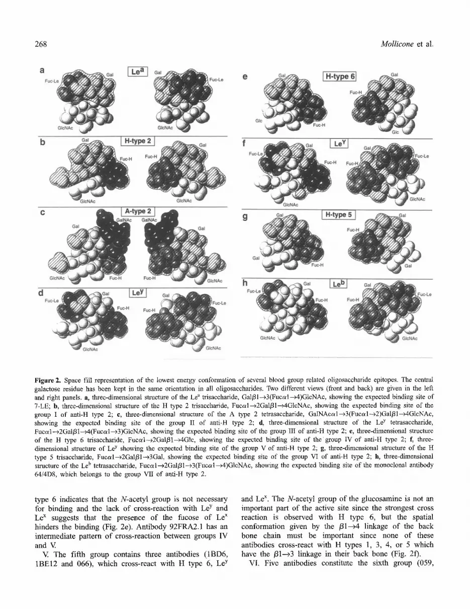
I. The first type of cross-reaction is represented by two antibodies (056 and 4F9), which cross-react with precursor chains. The binding is stronger with H type 2 as compared to the precursor chains and there is no cross-reaction with any A structure. These facts suggest that the oligosaccharide area recognized by the antibody covers part of the galactose since the addition of the N- acetylgalactosamine of A on the position 3 of the gatactose abolishes the reaction. It must also recognize the fucose, and part o f the N-acetylglucosamine, since H type 1, 3, 4, 5, 6 and Le b do not cross react, suggesting that the N-acetyl group of the N-acetylglucosamine is necessary for binding (Fig. 2b). In H types 1, 3, 4 and Le b (Fig. 2h) this N-acetyl group is on the other side of the oligosaccharide, while in H type 5 (Fig. 2g) and H type 6 (Fig. 2e) it is not present.
II. The second group contains four antibodies (BRIC4t , 5G9, 3C10 and 067); all cross-react with A type 2, but do not react with A trisaccharide, ALe b or A type 3. This suggests that the antibodies are not primarily reacting with the terminal N-acetylgalactosamine of A, but are rather reacting with the fucose and part of the precursor chain and their reaction is not hindered by the substitution of position 3 of the galactose by the N-acetylgatactosamine of A. Therefore, they react with

Recognition of the blood group H type 2 trisaccharide epitope 267
ctrl VII Vl
Oligosaccharide 64/
epitopes 7-LE 057 4D8 8B9 063 064 1F9 059
B trisaccharide O O 0 0 0 0 O 0
A trisaccharide 0 0 0 0 0 0 0 0
A type 3 0 0 0 0 0 0 O 0
ALe b 0 0 0 0 0 0 0 0
trisaccharide 0.3 0 0 0 0.2 0 0 0,I
LacNac 0.1 0 0 0 0 0 0 0
i trisaccharide 0 0 0 0 0. t 0 0 0
pentasaccaride 0,1 0 O 0 0.2 0 0 O
V IV III II I ctrl
1BE 1BD )2FR MR3 91, BNF BNH 19-0 MS1 BRI£ 4urn
066 12 6 A2.1 517 7Ell C-2 060,,,,, 065 13 061 058 9 LE 31 067,.,3C,1,05G9 41 4F9 056 -Ma
0 O O 0 O O O O O O O 0 0 0 0 O 0 O O 0 0 0
O 0 0 O 0 0 0 0 O 0 0 0 0 0 0 0 O 0 0 0 0 0
0 0 0 0 0 0 0 0 0 0 0 0 0 0 0 0 O 0 0 0 0 0
0 0 0 0 0 0 0 0 0 0 0 0 0 0 0 0 0 0 0 O 0 0
0 0 O 0 0 0 0 0 0 0 0 0 0 0 0 0 0 0 0 0,2 0,2 1,0
0 0 0 0 O 0 0 0 0 0 0 0 0 0 0 0 0.2 0 0 1,1 0.9 1,3
0 0 0 0 0 0 0 0 0 0 0 0 0 0 0 0 0 0 0 0,9 0,9 0
O O O 0 O 0 0 O 0 0 0 0.2 0 O 0 0 O 0 0 0.8 0.8 O
A type 2 0 0 0 O 0 O 0 0
Htype2 0 1;4 0,7 1.2 1.4 1,4 114 0,9
Htype6 0 1.4 0.6 1.4 0.5 1.4 1.5 1,1
Le y 0 1.5 0.9 0.2 0 0.2 0,2 0.1
Le x 0 1.4 0.5 0 0 0 0 0
Htype5 0 0 0 019 0.9 0.4 0.8 0.3
H type 1 0 0 0.7 0 0 0 0 0
I Le b 0 0 1.0 0 0 0 0 O
Le a 0,6 0.5 0 0 0 0 0 0
Type 1 disac. 0 0 0 o 0 0 0 0
H type 3 0 0 0 0 0 0 O 0
H type 4 0 O 0 0 0 0 O 0
Le disaccharide 0 0 0 0 0 0 0 0
0 O O 0 0 0 0 O 0 0.2 046 0;4 0 , 3 0:9 1 ,0 1A, 1,4 i .5 1.5 0 0 0
0,7 0,3 0 : 6 1:5 1.1 1.5 0A 0.4 0,4 :i.1 1,5 1.2 1.2 0.9 1.5J t.1 1.1 1,2 0.8 1.4 1.3 0
1.5 0.9 0.6 1.5 1.1 1.0 0.9 0.7 0,3 0 t.4 1.2 1.1 0.7 1.5 0.1 O O 0 O O 0
1.5 1.3 1.2 1,5 02 0 0 0 0 0 1.3 t.2 0.9 0.8 0 0.1 0 O O 0 0 O
1A 0.8 0:8 0 0 0 0 0 0 0 0,9 & 5 0,4 0i6 0 0,1 0 O 0 0 O 0
0 0 0 0 0 0 0 0,1 0 0 0.4 121 t,2 O 0 0,1 0 0 0 0 0.1 0
0 0 0 0 0 0 0 0,1 0 0 0.2 0.2 0 0 0 0.1 0 0 0 0 0 0
O 0 0 0 0 0 0 O 0 0 0 0 0 0 0 0 0 0 0 0 O 0
0 0 0 O 0 0 0 O O 0 0 0 0 0 0 O O 0 O 0 0 0
0 0 O 0 O 0 O 0 0 O 0 0 0 0 0 0 O 0 O 0 0 O
O O 0 0 O 0 O O 0 0.1 0,1 0.1 O 0 0 0 O 0 O 0 0 0
O 0 0 O 0 0 O 0 O 0 0 0 0 O O O O O 0 O 0 0
0 0 O 0 0 0 O 0 0 0 0 0 0.1 0 0 0 O O 0 0 0 0
Figure 1. ELISA binding of 30 monoclonal antibodies to 21 immobilized oligosaccharide epitopes. Two antibodies, not reacting with H type 2, were added as negative controls (ctrt): the human antibody I-Ma (right), which reacts with type 2 precursor chains, N- acetyllactosamine (LacNAc) and I trisaccharide and the mouse monoclonal 7-LE (left), which reacts with Le a and I trisaccharides. Among the 28 mouse monoclonals reacting with H type 2, seven main patterns of cross-reactions can be distinguished: [. Cross-reaction with type 2 precursor oligosaccharides; II. Cross-reaction with A type 2; III. Cross-reaction with A type 2, H type 6, Le y, Le x and H type 5; IV Cross-reaction with H type 6; V. Cross-reaction with H type 6, Le y and LeX; VI. Cross-reaction with H type 6 and H type 5; VII. Cross- reaction with H type 6, Le y, Le x and type 1 oligosaccharide structures.
both H type 2 and A type 2 (Fig. 2c). The N-acetyl group of the N-acetylglucosamine is necessary for the binding of these antibodies since H type 6, which is identical to H type 2, but has glucose instead of N- acetylgklcosamine at the reducing end (Fig. 2e), does not bind.
III. Three antibodies constitute the third group (BNH9, 058 and 061), they cross-react with A type 2, H type 5, H type 6, Le y and Le x. They do not react with the A trisaccharide nor any other A epitope, suggesting that the binding is not hindered by the presence of A, as in group II. In addition, the N-acetyl group of N-acetylglucosa- mine is not indispensable for the binding of these antibodies, since they cross-react with H type 6. The presence of a second fucose in Le y, slightly decreases the binding of two o f these antibodies and the lack of the fucose o f H in Le x strongly diminishes the binding,
suggesting that the fucose of H is a main anchoring point for the binding of these antibodies, but the fucose of the Le x can contribute some binding energy (Fig. 2d).
Two antibodies MS131 and 19-OLE represent two intermediary steps between groups II and IlL Similarly, the antibody BNF13 can be considered as an intermediate form between groups II and III, since it reacts very weakly, with A type 2 (for the sake of clarity, ELISA values of 0.2 have been excluded from the shaded clusters in Fig. 1; however, in some antibodies such as BNF13 they suggest specific weak binding), or alter- natively as an intermediary between groups lII and IV if we disregard its weak binding to A type 2 (Fig. 1).
IV The fourth group cross-reacts only with H type 6 and does not cross-react with any of the A or H types 1, 3, 4, or 5. it is constituted by five antibodies (065, 060, 9tC-2, 7Ell and MR3517). The cross-reaction with H
268 Mollicone et al.
Figure 2. Space fill representation of the lowest energy conformation of several blood group related oligosaccharide epitopes. The central galactose residue has been kept in the same orientation in all oligosaccharides. Two different views (front and back) are given in the left and right panels, a, three-dimensional structure of the Le a trisaccharide, Gal/31--+3(Fucal--~4)GlcNAc, showing the expected binding site of 7-LE; b, three-dimensional structure of the H type 2 trisaccharide, Fucal--+2Gal/31--+4GlcNAc, showing the expected binding site of the group I of anti-H type 2; e, three-dimensional structure of the A type 2 tetrasaccharide, GalNAcal--~3(Fucal---~2)Gal/31---~4GlcNAc, showing the expected binding site of the group II of anti-H type 2; d, three-dimensional structure of the Le y tetrasaccharide, Fuco~l---~2Gal/31--~4(Fucal-~3)GtcNAc, showing the expected binding site of the group Ill of anti-H type 2; e, three-dimensional structure of the H type 6 trisaccharide, Fuccd--~2Gal/31--~4Glc, showing the expected binding site of the group IV of anti-It type 2; f, three- dimensional structure of Le y showing the expected binding site of the group V of anti-H type 2; g, three-dimensional structure of the H type 5 trisaccharide, Fucal---~2Gal/31--~3Gal, showing the expected binding site of the group VI of anti-H type 2; h, three-dimensional structure of the Le b tetrasaccharide, Fucal--~2Gal/31--~3(Fucal--~4)GlcNAc, showing the expected binding site of the monoclonal antibody 64/4D8, which belongs to the group VII of anti-H type 2.
type 6 indicates that the N-acetyl group is not necessary for binding and the lack of cross-reaction with Le y and Le x suggests that the presence of the fucose of Le x hinders the binding (Fig. 2e). Antibody 92FRA2.1 has an intermediate pattern of cross-reaction between groups IV and V
V The fifth group contains three antibodies (1BD6, 1BE12 and 066), which cross-react with H type 6, Le y
and Le x. The N-acetyl group of the glucosamine is not an important part o f the active site since the strongest cross reaction is observed with H type 6, but the spatial conformation given by the /31---~4 linkage of the back bone chain must be important since none of these antibodies cross-react with H types 1, 3, 4, or 5 which have the /31--~3 linkage in their back bone (Fig. 2f).
VI. Five antibodies constitute the sixth group (059,
Recognition of the blood group H type 2 trisaccharide epitope 269
iF9, 064, 063 and 8B9). All cross-react with H type 6 and H type 5, two structures closely related to H type 2, which are both lacking an N:acetyl group on the internal galactose or glucose reducing unit (Fig. 2g). The presence of the second fucose in Le y or Le ~ abolishes the reaction of these antibodies.
VII. Finally two antibodies have been considered together in the seventh group because they cross-react with type 1 oligosaccharides and do not cross react with H type 5. However, these two antibodies must be quite different since 64/4D8 cross-reacts with H type 6, Le y Le ~, H type 1 and Le b (Fig. 2h) and 057 cross-reacts with, H type 6, Le y Le x and Le ~. A cross-reaction with Le b and Le y was expected since the two fucoses of these two epitopes are in very similar spatial orientation (see Fig. 2f and 2h) and were equally well recognized by other reagents such as the lectin IV of Griffonia simpliciJolia [36,37]. However, a cross-reaction with Lea and H without reacting with Le b is more difficult to understand. The easiest explanation of the cross- reaction pattern of 057 is that it contains two different antibodies, one anti-H type 2 cross-reacting with H type 6, Le y and Le x, as group V and a second antibody reacting with Lea as 7-LE. Nevertheless, if there is a single antibody in 057, more complex antibody sites can be imagined giving the weak cross-reaction observed with Le ~, since the fucose residues of Le x and Le a have similar spatial orientations, but this possibility needs more sophisticated conformational studies.
The activity of the antibodies of groups IV, V, VI and VII is abolished by the addition of the terminal N- acetytgalactosamine of A on position 3 of the galactose, suggesting that the binding site covers this area of the galactose, it must also include the fucose and can cover, in different degrees, the surface of the other fucose of the type 2, Le y and Le ~ structures.
Red cell agglutination
Agglutination scores of all the available anti-H antibodies were determined with O, A2, AzB, B, Aa, A1B and Oh erythrocytes (in red cell serology, the terms A I and A2 do not correlate to A type 1 and A type 2 epitopes, but they refer to two A erythrocyte phenotypes, expressing differ- ent amounts of the A type 2 epitope. The A1 red cell phenotype has more A epitopes because it has an A enzyme which is more efficient on the H type 2 acceptors than the A enzyme o f A 2 cells [38] and it is also able to use H type 3 and H type 4 acceptors to make A type 3 and A type 4 [39]).
None of the antibodies agglutinated H deficient, Bombay Oh, red cells but they all agglutinated normal O and A2 erythrocytes, confirming their serological anti- H type 2 specificity. However, the scores with AzB , B, A1 and A1B red cells were different for the antibodies
cross-reacting with A type 2 as compared to all the other antibodies not cross-reacting with A type 2.
Table 2 shows the agglutination scores of the anti- bodies cross-reacting with A type 2 (groups II and III). In general, they have similar scores for all the normal ABO phenotypes, with only a slight decrease in AIB (46 _+ 26) as compared to O (68 + 16) red cells.
Unlike this, most of the antibodies not cross-reacting with A type 2 (groups ! and IV to VII) had significantly different scores with the different red cell phenotypes (Table 3). The order of reaction is: O > A2 > A2B > B > A 1 > A1B > Oh, which corresponds to the trend observed for the amounts of free H, non-A and non-B substituted epitopes in the different red cell phenotypes. These results suggest that the anti-H not cross=reacting with the A type 2 epitope, reacts mainly with the free H structure and only reacts poorly or not at all, with the H structure once it has been transformed into A or B by addition of terminal N-acetylgalactosamine or galactose respectively. Alternatively, the agglutination scores of the antibodies cross-reacting with the A type 2 remain similar with the different ABO normal phenotypes, suggesting that they can still react with the H epitope even when it has been transformed into A or B. Therefore, we can predict that the antibody site of these reagents will not be strongly hindered by the addition of either galactose (B) or N-acetylgalactosamine (A) on position 3 of galactose. The synthetic B type 2-BSA epitope was not available to test this hypothesis, but it is in good agreement with the proposed area of oligosac- charide recognition for the antibodies of groups II and III which cross-react with A type 2 (Fig. 2c and 2d).
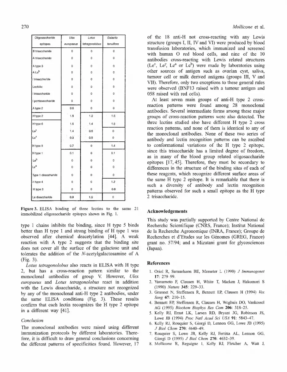
Lectins (b~g. 3)
Galactia tenuiflora reacts, in ELISA, strongly with most H structures and weakly with H type 1. The presence of a second fucose in Le y and Le b abolishes the binding of this Iectin. The N-acetyl group of N-acetylglucosamine on type 2 chains is not involved in the binding site since H type 6 binds as well as H type 2, while the same N-acetyl group on type 1 chains interferes with binding. The H type 5 which has an epitope surface similar to deacetylated H type 1 binds quite strongly and H types 3 and 4 also bind although less strongly, suggesting that the main binding site of this tectin is on the terminal fucose and galactose units and is less sensitive to modifications on the sugar located at the reducing terminus of the oligosaccharide [40,41].
Ulex europaeus lectin I, a well known anti-H type 2 lectin [42], reacts in ELISA mainly with H type 2 and has a cross-reaction pattern similar to the monoclonal antibodies of group III. Early inhibition studies had shown that H type 6 binds as well as H type 2, suggesting that the N-acetyl group of type 2 chains is not necessary for binding [43], while the N-acetyl group on
270 Mollicone et al.
Oligosaccharide
epitopes
B trisacchafide
A trisaccharide
A type 3
A Le b
trisacchafide
LacNAc
i trisaccharide
pentasaccharide
A type 2
H type 2
H type 6
Le y
Le x
H type 5
H type 1
Le b
Le a
Type 1 disaccharide
H type 4
H type 3
Le disaccharide
Ulex
europaeus
Lotus Galactia
tetragonolobus tenuiflora
0
0
0
0
0
0
0
0
0.6
1.5 1.2
1,5 1.4
1,4 0.6
0,3 0.5
0.7 0
0.1 0
0 0
0 0
0 0
0 0
0 0
0 , 9 1.5
1.5
1,5
1.4
0.1
0
0
0
1.2
0.6
Figure3. ELISA binding of three lectins to the same 21 immobilized oligosaccharide epitopes shown in Fig. 1.
type 1 chains inhibits the binding, since H type 5 binds better than H type 1 and strong binding of H type 1 was observed after chemical deacetylation [44]. A weak reaction with A type 2 suggests that the binding site does not cover all the surface of the galactose unit and tolerates the addition of the N-acetylgalactosamine of A (Fig. 3).
Lotus tetragonolobus also reacts in ELISA with H type 2, but has a cross-reaction pattern similar to the monoclonal antibodies of group V. However, Ulex europaeus and Lotus tetragonolobus react in addition with the Lewis disaccharide, a structure not recognized by any of the monoclonal anti-H type 2 antibodies, under the same ELISA conditions (Fig. 3). These results confirm that each lectin recognizes the H type 2 epitope in a different way [41].
Conclusion
The monoclonal antibodies were raised using different immunization protocols by different laboratories. There- fore, it is difficult to draw general conclusions concerning the different patterns of specificities found. However, 17
of the 18 anti-H not cross-reacting with any Lewis structure (groups I, II, IV and VI) were produced by blood transfusion laboratories, which immunized and screened with human O red blood cells, and nine of the 10 antibodies cross-reacting with Lewis related structures (Le x, Le y Le a or Le b) were made by laboratories using other sources of antigen such as ovarian cyst, saliva, tumour cell or milk derived antigens (groups III, V and VII). Therefore, only two exceptions to these general rules were observed (BNF13 raised with a tumour antigen and 058 raised with red cells).
At least seven main groups of anti-H type 2 cross- reaction patterns were found among 28 monoclonal antibodies. Several intermediate forms among these major groups of cross-reaction patterns were also detected. The three lectins studied also have different H type 2 cross reaction patterns, and none of them is identical to any of the monoclonal antibodies. None of these two series of antibody and lectin recognition patterns can be ascribed to conformational variations of the H type 2 epitope, since this trisaccharide has a limited degree of freedom, as in many of the blood group related oligosaccharide epitopes [17,45]. Therefore, they must be secondary to differences in the structure of the binding sites of each of these reagents, which recognize different surface areas of the same H type 2 epitope. It is remarkable that there is such a diversity of antibody and lectin recognition patterns observed for such a small epitope as the H type 2 trisaccharide.
Acknowledgements
This study was partially supported by Centre National de Recherche Scientifique (CNRS, France); Institut National de la Recherche Agronomique (INRA, France); Groupe de Recherches et d'Etudes sur les G6nomes (GREG, France) grant no. 57/94; and a Mizutani grant for glycosciences (Japan).
References
1. Oriol R, Samuelsson BE, Messeter L (1990) J lmmunogenet 17:279 99.
2. Yamamoto F, Clausen H, White I", Marken J, Hakomori S (1990) Nature 345: 229-33.
3. Grunnet N, Steffensen R, Bennett EP, Clausen H (1994) r4)x Sang 67: 210-15.
4. Bennett EP, Steffensen R, Ctausen H, Weghuis DO, Vankessel AG (1995) Biochem Biophys Res Corn 206: 318-25.
5. Kelly RJ, Ernst LK, Larsen RD, Bryant JG, Robinson JS, Lowe JB (1994) Proc Natl Acad Sci USA 91: 5843-47.
6. Kelly RJ, Rouquier S, Giorgi D, Lennon GG, Lowe JB (1995) J Biol Chem 270: 4640-49.
7. Rouquier S, Lowe JB, Kelly RJ, Fertitta AL, Lennon GG, Giorgi D (1995) J BioI Chem 270: 4632-39.
8. Molticone R, Reguigne I, Kelly R J, Fletcher A, Watt J,

Recognition of the blood group H type 2 trisaccharide epitope
Chatfield S, Aziz A, Cameron HS, Weston BW, Lowe JB, Oriol R (1994) J Biol Chem 269: 20987-94.
9. Elmgren A, Rydberg L, Larson G (t993) Biochem Biophys Res Corn 196: 515-20.
10. Koda Y, Kimura H, Mekada E (1993) Blood 82: 2915-19. 1 i. Nishihara S, Narimatsu H, Iwasaki It, Yazawa S, Akamatsu S,
Ando T, Seno rl, Narimatsu I (1994) J Biol Chem 269: 29271- 78.
12. Knowles RW, Bai Y, Daniels GL, Watkins W (11982) J immunogenet 9: 69-76.
13. Hindsgaul O, Norberg T, Le Pendu J, Lemieux RU (1982) Carbohydr Res 109: 109-42.
14. Oriol R, Gane P, Rouger P, Mollicone R (1987) Rev Franf Transf Immunoh~matol 30: 671-79.
15. Mollicone R, Dalix AM, Gane P, Nemec M, Oriol R (1990) In Second International Workshop on Monoctonal Antibodies against Human Red Blood Cells and Related Antigens (Chester MA, Johnson U, Lundblad A, L6w B, Messeter L, Samuelsson Beds) Lund, Sweden, p. 85.
16. Furukawa K, Welt S, Yin BWT, Feickert HJ, Takahashi T, Ueda R, Lloyd KO (1990) Mol Immunol 27: 723-32.
!7. imberty A, Mikros E, Koca J, Mollicone R, Oriol R, Perez S (1995) Glycoconjugate J 12: 331-49.
18. Lemieux RU (1978) Chem Soc Rev 7: 423-52. t9. Kabat EA, Liao J, Lemieux RU (1978) Immunochemist~ 15:
727-31. 20. Lemieux RU, Wong TC, Liao J, Kabat EA (1984) Mot
Immunol 21: 751-59. 21. Chester MA, Johnson U, Lundblad A, L6w B, Messeter L,
Samuelsson B, Eds (1990) Proceedings of the Second ~nternational Workshop on Monoctonal Antibodies against Human Red Blood Cells and Related Antigens. Land, Sweden, p. 209.
22. Delsol G, Blancher A, A1 Saati T, Ralfkiaer E, Lamitzen A, Bruigeres L, Brousset P, Rigal-Huguet F, Mazerolles C, Robert A, Chittal SM (1991) Br J Cancer 64: 321-26.
23. Blancher A, AI Saati T, Marty Y, Delsol G (1988) In INTS Symposium on Monoclonal Antibodies against Human Red Blood Cell and Related Antigens (Rouger P, Salmon C, eds) Arnette, Paris, France, p. 220-23.
24. Pancino G, Charpin C, Osinaga E, Betaille B, Le Roy M, Calvo F, Roseto A (1990) Cancer Res 50: 7333-42.
271
25. Pancino G, Toubert ME, Osinaga E, Chatelet F, Leroy M, Schlageter MH, Desroys du Roure F, Catvo F, Teillac P, Najean Y (1991) Int J Cancer 47: 221-26.
26. Enblad E Grimelius B, Busch C, Pghlman L, Pont6n L Chester MA, Lundblad A (1986) Anticancer Res 6: 13946.
27. Brodin T, Chester MA, Karlsson KA, Messeter L, Zopf D, Lundblad A (1987) Glycoconjugate J 4: 399-406.
28. Le Pendu J, Gerard G, Lambert F, Mollicone R, Oriol R (1986) Glycoconjugate J 3: 203-16.
29. Good AH, Yan O, Lamontagne LR, Oriol R (1992) Vox Sang 62: t80-89.
30. Marsh WL (1972) Transfusion 12: 352-53. 31. Perez S, Delage MM (1991) C~rbohydr Res 212: 253-59. 32. Koca J (1994) J Molec Struct (Theochen 0 308: 13-24. 33. Koca J, Perez S, Imberty A (1995) J Computat Chem 16: 296-
310. 34. MM3 (1992) QCPE, Creative Arts Building 181, Indiana
University, Bloomington, IN 47405 USA. 35. Tripos Associates (1988) S. Hanley Road, Suite 303, St Louis,
MO 63144, USA. 36. Spohr U, Hindsgaul O, Lemieux RU (1985) Can J Chem 63:
2644-52. 37. Vandonselaar M, Delbaere LTJ, Lemieux RU (1987) J Biol
Chem 262: 10848-49. 38. Yamamoto E McNeill PD, Hakomori S (1992) Biochem
Biophys Res Comm 187: 366-.74. 39. Clausen It, Levery SB, Nudelman E, Tsuchiya S, Hakomori S
(1985) Plvc Natl Acad Sci USA 82: 1199-203. 40. Cromer R, Spohr U, Khare DP, Le Pendu J, Lemieux RU
(1992) Can J Chem 70: 1511-30. 41. Du M-H, Spohr U, Lemieux RU (1994) Glyconjugate J 11:
443-61. 42. Hindsgaul O, Khare DP, Bach M, Lemieux RU (1985) Can J
Chem 63: 2653-58. 43. Pereira MEA, Kisailius EC, Gruezo F, Kabat EA (1978) Arch
Biochem Biophys 185: 108-15. 44. Lemieux RU, Le Pendu J, Itindsgaul O (1979) Japanese J
Antibiot 32 suppl: s21-31. 45. Lemieux RU (1994) The chemical mapping of protein
carbohydrate complexes. In Complex Carbohydrates in Drug Research (Bock K, Clausen H, eds) Munksgard: Copenhagen p. 188-97.


