Embed Size (px)
Citation preview

This article appeared in a journal published by Elsevier. The attachedcopy is furnished to the author for internal non-commercial researchand education use, including for instruction at the authors institution
and sharing with colleagues.
Other uses, including reproduction and distribution, or selling orlicensing copies, or posting to personal, institutional or third party
websites are prohibited.
In most cases authors are permitted to post their version of thearticle (e.g. in Word or Tex form) to their personal website orinstitutional repository. Authors requiring further information
regarding Elsevier’s archiving and manuscript policies areencouraged to visit:
http://www.elsevier.com/authorsrights
Author's personal copy
Two antibacterial C-type lectins from crustacean, Eriocheir sinensis,stimulated cellular encapsulation in vitro
Xing-Kun Jin 1, Shuang Li 1, Xiao-Nv Guo, Lin Cheng, Min-Hao Wu, Shang-Jian Tan, You-Ting Zhu,Ai-Qing Yu, Wei-Wei Li ⇑, Qun Wang ⇑School of Life Science, East China Normal University, Shanghai, PR China
a r t i c l e i n f o
Article history:Received 30 May 2013Revised 24 July 2013Accepted 24 July 2013Available online 30 July 2013
Keywords:Antibacterial activityChinese mitten crabEncapsulationHemocytes
a b s t r a c t
The first step of host fighting against pathogens is that pattern recognition receptors recognized patho-gen-associated molecular patterns. However, the specificity of recognition within the innate immunemolecular of invertebrates remains largely unknown. In the present study, we investigated how inverte-brate pattern recognition receptor (PRR) C-type lectins might be involved in the antimicrobial response incrustacean. Based on our previously obtained completed coding regions of EsLecA and EsLecG in Eriocheirsinensis, the recombinant EsLectin proteins were produced via prokaryotic expression system and affinitychromatography. Subsequently, both rEsLecA and rEsLecG were discovered to have wide spectrum bind-ing activities towards microorganisms, and their microbial-binding was calcium-independent. Moreover,the binding activities of both rEsLecA and rEsLecG induced the aggregation against microbial pathogens.Both microorganism growth inhibitory activities assays and antibacterial activities assays revealed theircapabilities of suppressing microorganisms growth and directly killing microorganisms respectively. Fur-thermore, the encapsulation assays signified that both rEsLecA and rEsLecG could stimulate the cellularencapsulation in vitro. Collectively, data presented here demonstrated the successful expression andpurification of two C-type lectins proteins in the Chinese mitten crab, and their critical role in the innateimmune system of an invertebrate.
� 2013 Elsevier Ltd. All rights reserved.
1. Introduction
Invertebrate animals do not truely have a adaptive immunitythat is generated by memory and targeted immunoglobulin pro-duction, as in vertebrates (Du Pasquier, 2001; Watthanasurorotet al., 2011). Nonetheless invertebrate, such as crustaceans, arecapable of motivating effective innate immune responses to pro-tection against microbial infections (Li and Xiang, 2013; Söderhälland Smith, 1986). In the event of pathogens intruding, their con-served pathogen-associated molecular patterns (PAMPs), such aslipopolysaccharides (LPS), peptidoglycans and b-1, 3-glucans, i.e.,which are essential and unique components of virtually all micro-organisms, but absent in higher organisms (Janeway, 1989) couldbe discriminated by a wide range of pattern recognition receptors(PRRs) that are highly conserved in biological evolution (Janewayand Medzhitov, 2002), then encountered a variety of defensemechanisms.
As an important member of non-TLR PRRs, lectins exist as eithertransmembrane receptors or soluble proteins in circulating fluids(Christophides et al., 2002; Janeway and Medzhitov, 2002). Theyplay crucial roles in innate immunity such as nonself-recognitionand clearance of invading microorganisms (Söderhäll and Cerenius,1992; Yu and Kanost, 2004), via recognizing and non-covalentlybinding to specific sugar moieties and agglutinate pathogens bybinding to cell surface glycoproteins and glycoconjugates (Lis andSharon, 1998). C-type lectins are the most diverse and best studiedamong the lectin superfamily. The term C-type lectin was originallyused to distinguish a group of Ca2+-dependent (C-type) carbohy-drate-binding proteins from the other types of lectins (Drickamer,1988). This big gene family mediate sugar binding with diversearchitecture contained homologous carbohydrate recognition do-mains (CRDs) by which discriminate specific oligosaccharides atcell surfaces, attach to circulating proteins and in the extracellularmatrix (Drickamer and Taylor, 1993; Weis et al., 1991; Zelenskyand Gready, 2005). Comparing with the well-studied lectins in ver-tebrates, the knowledge of entire innate immune responses medi-ated by different lectins in invertebrates is still far from beingunderstood (Cerenius et al., 2010a; Wang and Wang, 2013).
In the present study, we investigate how invertebrate patternrecognition receptor (PRR) C-type lectins might be involved in
0145-305X/$ - see front matter � 2013 Elsevier Ltd. All rights reserved.http://dx.doi.org/10.1016/j.dci.2013.07.016
⇑ Corresponding authors. Address: School of Life Science, East China NormalUniversity, No. 500 Dong-Chuan Road, Shanghai 200241, PR China. Fax: +86 2124206934.
E-mail addresses: [email protected] (W.-W. Li), [email protected] (Q.Wang).
1 These authors contributed equally to this work.
Developmental and Comparative Immunology 41 (2013) 544–552
Contents lists available at ScienceDirect
Developmental and Comparative Immunology
journal homepage: www.elsevier .com/locate /dc i

Author's personal copy
the antimicrobial response in crab. The completed coding regionsof EsLecA and EsLecG were obtained from our previously work onEsLectins in Eriocheir sinensis (Jin et al., 2012), and the recombinantEsLectin proteins were got via prokaryotic expression system andaffinity chromatography. Subsequently, the binding activities andantibacterial activities of rEsLectins towards different microorgan-isms were tested, and their microorganism agglutination inductionand cellular encapsulation stimulation were observed. Our resultssuggested that both rEsLecA and rEsLecG proteins might act aspowerful effectors in crustacean innate immunity, which couldpotentially provide a vehicle for further invertebrate immunologyresearch.
2. Materials and methods
2.1. Sample preparation
Healthy adult Chinese mitten crabs were collected from theTongchuan aquatic products market in Shanghai, China (n = 50;100 ± 12 g wet weight). Prior to dissection, crabs were placed inan ice bath (1–2 min) until each was lightly anesthetized. Thehepatopancreas was harvested, snap frozen in liquid nitrogen,and stored at �80 �C prior to nucleic acid analysis. For EsLectin cod-ing regions cloning, prepared hepatopancreas from 10 crabs werepooled, and ground with a pestle and mortar prior to nucleic acidextraction. Hemolymph was draw from the hemocoel in arthrodialmembrane of the last pair of walking legs using a syringe (approx-imately 2.0 ml per crab) with an equal volume of anticoagulantsolution (0.1 M glucose, 30 mM citrate, 26 mM citric acid, 0.14 MNaCl, 10 mM EDTA) added, and centrifuged at 500g at 4 �C to iso-late hemocytes.
2.2. Total RNA isolation
Total RNA was extracted from E. sinensis hepatopancreas usingTrizol� reagent (RNA Extraction Kit, Invitrogen, CA, USA) accordingto the manufacturer’s protocol. The total RNA concentration andquality were estimated using spectrophotometry at an absorbanceat 260 nm and agarose-gel electrophoresis, respectively. Total RNA(4 lg) was reverse transcribed using the PrimeScript™ II 1st StrandcDNA Synthesis Kit (TaKaRa, Japan).
2.3. Construction of the recombinant expression plasmids
The coding regions of EsLecA and EsLecG were amplified fromhepatopancreas cDNA template by specific primer pairs designedwith BamHI and XhoI endonuclease sites included at the 50 end ofthe forward and reverse primers, respectively (Table 1). The PCRproducts were double digested with endonucleases BamHI andXhoI (New England Biolabs, USA), then purified by Wizard� SVGel and PCR Clean-Up System (Promega, USA), ligated to the corre-sponding cohesive ends of pET32a plasmid (Novagen, Germany)
with T4 ligase (New England Biolabs, USA), and confirmed bytwo-way DNA sequencing with T7 primer pairs (Table 1). The po-sitive transformants of Escherichia coli BL21-DE3 (Tiangen, China)was selected to express recombinant proteins. The pET32a plasmidwithout any insert fragment was selected as blank control, whichcould express the recombinant thioredoxin protein (rTrx).
2.4. Expression and purification of recombinant EsLectins
The positive transformants were extensively cultured until log-arithmic phase (OD600 value reached 0.6) in ampicillin-containingLuria-Bertani (LB) broth. Afterwards, the isopropyl-b-D-1-thioga-lactopyranoside (IPTG) was added into the medium (1 mM finalconcentration) to induce the rEsLecA and rEsLecG proteins expres-sion. After aerobic culture overnight at 37 �C, the bacteria were col-lected by centrifugation (6000g). The pellets were suspended in8 ml of Guanidine Lysis Buffer and ultrasonicated. The 8 ml lysatewas then added to a prepared Purification Column and purifiedby affinity chromatography using His-binding resin chromatogra-phy (Invitrogen, Carlsbad, USA) following the manufacturer’sinstructions under hybrid conditions. The prepared resin was sus-pended in the lysate solution for 30–60 min, and the target proteinwith His-tag was bound to the resin of Purification Column. Subse-quently, the resin was centrifuged in low speed (800g) and washedwith 8 ml Native Wash Buffer for four times. Finally the proteinwas eluted with 8–12 ml Native Elution Buffer. Purified rEsLectinswere buffer exchanged to TBS (50 mM Tris–HCl, 100 mM NaCl, pH7.5) by Amico� Ultra Filters (Millipores, Billerica, USA). Routineprotein estimation was conducted using the Bradford method,using bovine serum albumin (BSA, Bio-Rad) as the standard.
2.5. Western blotting
A set of Western blotting assays were designed to determinethe binding activity of rEsLectin proteins towards different micro-organisms, and the necessity of calcium for microbial-bindingactivity of rEsLectin proteins. Gram-positive bacteria Staphylocco-cus aureus, Bacillus subtilis and Microbacterium lactium, Gram-nega-tive bacteria Vibrio parahemolyticus, Aeromonas hydrophila andE. coli, and one yeast strain Pichia pastoris were extensively cul-tured in corresponding medium (LB for bacteria, Yeast PeptoneDextrose YPD for yeast) and then pelleted by centrifugation(6000g), at last suspended in 2 ml TBS (OD600 � 1.0). Purified rEsL-ecA and rEsLecG proteins (0.1 mg/ml; 500 ll) were incubated with500 ll microorganisms (2 � 107 cells/ml) in TBS for 1 h at 37 �C.The mixed microorganisms were pelleted (6000g, 5 min), washedfour times with TBS, and then eluted with 10% SDS. Eluents weresuspended in SDS–PAGE loading buffer and separated by 12%SDS–PAGE and transferred to PVDF membrane. The membranewas blocked with 5% dry skim milk in TBS (10 mM Tris–HCl,150 mM NaCl, pH 7.5) at room temperature for 2 h, and then sub-jected to immunoblot assay using anti-His-tag mouse antibody(Cwbio, China) (1:500) diluted by 2% dry skim milk in TBS incubat-ing in 4 �C overnight. Positive reactivity was detected using HRP-conjugated goat anti-mouse IgG (1:2000) (Cwbio, China), andwas visualized through the ChemiDocXRS instrument (Bio-Rad,USA) detection.
2.6. Microorganisms agglutination activities assays
The microorganisms agglutination activities of recombinantEsLectin proteins were assessed as previously described methods(Cheng et al., 2013). Gram-positive bacteria S. aureus, Gram-nega-tive bacteria E. coli and the yeast P. pastoris were cultivated over-night and collected by centrifugation (6000g). The pellets werewashed and suspended in TBS (50 mM Tris–HCl, 100 mM NaCl,
Table 1Primer sequences.
Primers name Sequences (50–30)
Recombinant expressionpET32a-EsLecA-sense CGCGGATCCACCGAAGTGTCCTGTAACGCCpET32a-EsLecA-antisense CCGCTCGAGTTAAGGCGTATACTGGCAGATGpET32a-EsLecG-sense CGCGGATCCCAGACCAGAGCTTGTCAGAACGpET32a-EsLecG-antisense CCGCTCGAGTTAAAAGATCTGACAGATGGCGT
SequencingT7-Promotor TAATACGACTCACTATAGT7-Terminator GCTAGTTATTGCTCAGCGG
X.-K. Jin et al. / Developmental and Comparative Immunology 41 (2013) 544–552 545

Author's personal copy
pH 7.5) and adjusted to an OD600 value of approximately 1.0.Microbial cells (10 ll) were incubated with 50 ll rEsLecA and rEs-LecG proteins respectively, in the presence or absence of 10 mMCaCl2. The 50 ll TBS buffer with CaCl2 or rTrx or blank were usedas control respectively. The mixtures were incubated at room tem-perature (1–2 h). The reactions were observed by bright lightmicroscopy (Leica, DM4000B, Germany).
2.7. Microorganisms growth inhibitory activities assays
Gram-positive bacteria S. aureus, Gram-negative bacteria E. coliand the yeast P. pastoris were chose to test the microorganismsgrowth inhibitory activities of rEsLectin proteins. Single colony ofchosen strains was selected and cultured at 30 �C in 1 ml LB orYPD broth respectively, and the rEsLectins proteins were added atthree levels of final concentration (0, 30 and 150 lg/ml). Each sam-ple was incubated with aeration at 200 rpm and the OD600 wasmeasured every 2 h. The microorganisms growth curves weredrawn based on the data from three independent experimentalrepeats.
2.8. Antibacterial activity assays
Antimicrobial activities of rEsLectin proteins against E. coli andS. aureus were performed on petri dishes respectively. The selectedsingle colonies were intensively cultured in the LB medium, subse-quently collected by centrifugation (6000g) for 10 min, and sus-pended in TBS (50 mM Tris–HCl, 100 mM NaCl, pH 7.5). Theprepared microbes were smeared into 100 mm � 20 mm petridishes respectively. Perforex was used to produce 10 mm in diam-eter pores. Equal concentration of either rTrx or rEsLectin proteins(50 lg/100 ll TBS) was added into the pores respectively. Then, theplates were incubated at 37 �C for 16 h (E. coli) or 30 �C for 16 h (S.aureus). The antibiotic ampicillin (12.5 lg/100 ll TBS) and 100 llTBS was set as control.
2.9. Hemocytes encapsulation assays
Hemocytes encapsulation assays of rEsLectin proteins were per-formed as previously described (Cheng et al., 2013). In this study,Ni-NTA agarose beads (Novagen, Germany) were washed threetimes and equilibrated in TBS. subsequently incubated with His-tagged protein rEsLecA and rEsLecG at room temperature for 1 h.Protein-coated beads were washed with TBS four times, and sus-pended in TBS. Hemocytes were isolated from Section 2.1, and pre-pared as the same way. Purified hemocytes were added to eachgroup of the protein-coated beads, and incubated at room temper-ature for 6 h and 24 h. The final reactions were observed by lightmicroscopy (Leica, DM4000B, Germany). The recombinant Thiore-doxin protein (rTrx) was set as a control.
2.10. Bioinformatics analysis
Similarity analysis was performed with BLASTX (http://www.ncbi.nlm.nih.gov/) and multiple sequence alignments wereconducted using ClustalW2. Signal sequence and motif predictionwere performed using SMART (http://smart.embl-heidelberg.de/).Protein molecular weights were predicted by ProtScale (http://web.expasy.org/protscale/).
3. Results
3.1. Sequences compare between EsLecA and EsLecG
Sequences alignment results showed that the identities be-tween EsLecA and EsLecG were 29%. Both EsLecA and EsLecG hadthe conserved four cysteine residues, which are crucial to formthe two disulphide bridges of characteristic double-loop (Zelenskyand Gready, 2005). In the carbohydrate recognition domains(CRDs), EsLecA was found to have QPD (Gln-Pro-Asp) motif whichhad been predicted to be functional as galactose binding, mean-while, the EsLecG exhibited EPE (Glu-Pro-Glu) motif instead of typ-ical EPN (Glu-Pro-Asn) that had been predicted to be specific formannose binding (Zelensky and Gready, 2005) (Fig. 1).
3.2. Expression and purification recombinant EsLectins
The deduced proteins of EsLecA and EsLecG had been predictedto have molecular weights of approximately 15.6 and 15.0 kDawithout any signal-peptide. In the prokaryotic expression system,both rEsLecA and rEsLecG were expressed as fusion proteins withextra His-tag and Trx-tag at the N-terminal, by which increasedthe molecular mass of the expressed target proteins to approxi-mately 33.0 and 32.3 kDa. The coding frames were confirmed bytwo-way sequencing of recombinant plasmids, and the purified re-combinant proteins were confirmed by SDS–PAGE electrophoresisvisualizing by Coomassie brilliant blue stain (Fig. 2).
3.3. Microorganisms binding activities
Recombinant EsLecA protein was capable of binding with al-most all tested microorganisms, especially towards V. parahemolyt-icus and P. pastoris. Comparatively, rEsLecG exhibited weakerbinding capabilities than rEsLecA towards Gram-positive bacteria,and even none with M. lactium (Fig. 3A).
In the absence of calcium (10 mM EDTA added), both rEsLecAand rEsLecG could still bind to the Gram-positive bacteria S. aureusand Gram-negative bacteria V. parahemolyticus, suggesting that themicrobial-binding activities of rEsLectin protiens were calcium-independent. In addition, the presence of calcium could dramati-cally increase the affinities of rEsLectin proteins binding towardsmicroorganisms (Fig. 3B).
3.4. Microorganisms agglutination
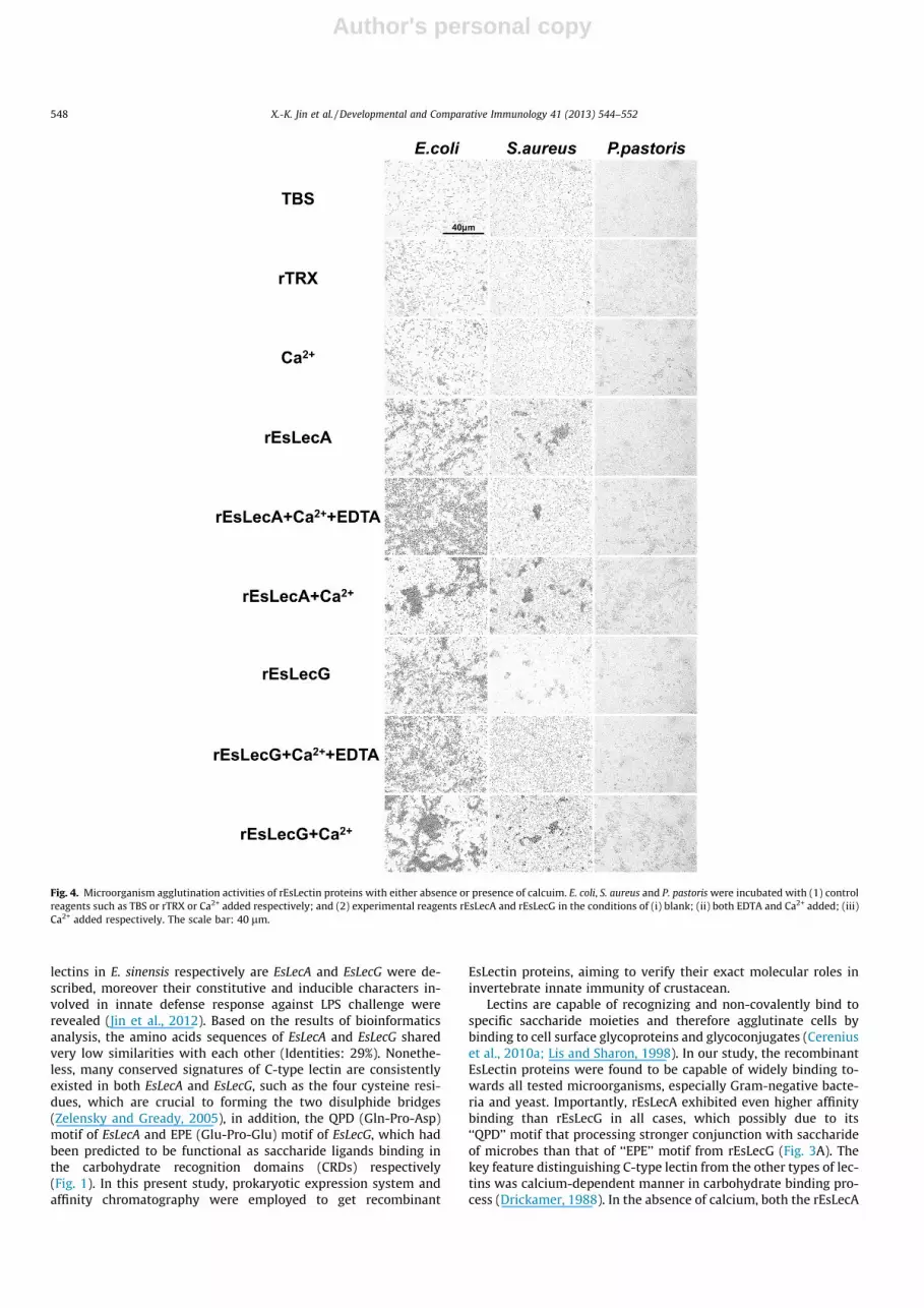
To determine whether the binding activities of rEsLectin pro-teins could induce the aggregation of microbial pathogens, weincubated Gram-positive bacteria S. aureus, Gram-negative bacte-ria E. coli and the yeast P. pastoris with rEsLectin proteins and as-sessed microbial aggregation by light microscopy. Both therEsLecA and rEsLecG were capable of inducing aggregation ofE. coli, S. aureus and P. pastoris. In the absence of calcium, the S. aur-eus aggregations induced by rEsLectin proteins were severelyinhibited, but the aggregations of E. coli and P. pastoris were notcompletely inhibited. In the presence of calcium, all the microbialaggregations induced by rEsLectin proteins were consistently en-hanced (Fig. 4).
3.5. Microorganisms growth inhibition and antibacterial effect
To determine the antimicrobial activities of rEsLectin proteins,their inhibition effect on the growth of microorganisms wereexamined. Both rEsLecA and rEsLecG could significantly inhibitall tested microbe stains comparing with the TBS (rEsLectin 0 lg/ml). Furthermore, the microbial growth were slightly suppressed
546 X.-K. Jin et al. / Developmental and Comparative Immunology 41 (2013) 544–552

Author's personal copy
with 30 lg/ml rEsLectin proteins, and severely inhibited when theconcentration of rEsLectin proteins were increased to 150 lg/ml(Fig. 5).
The antibacterial activities of rEsLectin proteins were also de-tected on petri dishes. The transparent rings were found to bearound the pores with rEsLectin proteins and ampicillin added,which signified their analogous microbe killing effects as ampicil-lin against E. coli and S. aureus, comparing with the rTrx and TBS(Fig. 6).
3.6. In vitro cellular encapsulation
To test whether the rEsLectin proteins could promote the cellu-lar encapsulation in vitro, the encapsulation assays were carriedout. Nickel agarose beads coated with either rEsLectin proteins(rEsLecA or rEsLecG) or thioredoxin protein (rTrx) were incubatedwith hemocytes from E. sinensis, and encapsulation phenomenonwas observed. The agarose beads coated with either rEsLecA or rEs-LecG could equally stimulated the encapsulation of hemocytes un-til 24 h incubation, comparing with that coated with thioredoxinprotein (rTrx)(Fig. 7).
4. Discussion
Lectins have been reported to contribute in invertebrate innateimmune response, such as prophenoloxidase activation (Cereniuset al., 2010b; Söderhäll and Cerenius, 1998; Yu et al., 1999),enhancement of encapsulation (Cerenius et al., 2010a; Ling andYu, 2006; Yu et al., 2005), nodulation of hemocytes (Koizumiet al., 1999; Söderhäll and Cerenius, 1992), opsonization(Jiravanichpaisal et al., 2006; Jomori and Natori, 1992), and antimi-crobial activity (Tunkijjanukij and Olafsen, 1998; Yu et al., 2007). Inour previous works, the cloning, sequence analysis, tissue-specificdistribution, and immune responsiveness of two novel C-type
Fig. 1. Multiple amino acid sequence alignments between EsLecA and EsLecG by ClustalW. Identical (⁄) and similar (. or :) residues are indicated. The conserved four cysteineresidues were boxed. In the carbohydrate recognition domains (CRDs), the QPD (Gln-Pro-Asp) motif of EsLecA and the EPE (Glu-Pro-Glu) motif of EsLecG were shown as redletters. (For interpretation of the references to colour in this figure legend, the reader is referred to the web version of this article.)
Fig. 2. Expressed and purified recombinant proteins of EsLecA and EsLecG stainingwith Coomassie Brilliant Blue. Lanes 1 and 4, lysate of E. coli with pET32a-EsLecAand pET32a-EsLecG without induction respectively; Lanes 2 and 5, lysate of E. coliwith pET32a-EsLecA and pET32a-EsLecG induced by IPTG respectively; Lanes 3 and6, purified recombinant EsLecA and EsLecG by His-tag binding resin chromatogra-phy. Lanes M represented the protein marker.
Fig. 3. (A) Microorganisms binding activities of the recombinant EsLectin proteins.Logarithmic growth phase microbial strains were incubated with rEsLecA andrEsLecG proteins and ultrasonicated. Subsequently the washed pellets weresubjected to SDS–PAGE and detected by Western blot with anti-His-tag antibody.(B) The microbial-binding activities of rEsLectin proteins were calcium-indepen-dent. The presence of calcium could dramatically increase the affinities of rEsLectinproteins binding towards microorganisms.
X.-K. Jin et al. / Developmental and Comparative Immunology 41 (2013) 544–552 547
Author's personal copy
lectins in E. sinensis respectively are EsLecA and EsLecG were de-scribed, moreover their constitutive and inducible characters in-volved in innate defense response against LPS challenge wererevealed (Jin et al., 2012). Based on the results of bioinformaticsanalysis, the amino acids sequences of EsLecA and EsLecG sharedvery low similarities with each other (Identities: 29%). Nonethe-less, many conserved signatures of C-type lectin are consistentlyexisted in both EsLecA and EsLecG, such as the four cysteine resi-dues, which are crucial to forming the two disulphide bridges(Zelensky and Gready, 2005), in addition, the QPD (Gln-Pro-Asp)motif of EsLecA and EPE (Glu-Pro-Glu) motif of EsLecG, which hadbeen predicted to be functional as saccharide ligands binding inthe carbohydrate recognition domains (CRDs) respectively(Fig. 1). In this present study, prokaryotic expression system andaffinity chromatography were employed to get recombinant
EsLectin proteins, aiming to verify their exact molecular roles ininvertebrate innate immunity of crustacean.
Lectins are capable of recognizing and non-covalently bind tospecific saccharide moieties and therefore agglutinate cells bybinding to cell surface glycoproteins and glycoconjugates (Cereniuset al., 2010a; Lis and Sharon, 1998). In our study, the recombinantEsLectin proteins were found to be capable of widely binding to-wards all tested microorganisms, especially Gram-negative bacte-ria and yeast. Importantly, rEsLecA exhibited even higher affinitybinding than rEsLecG in all cases, which possibly due to its‘‘QPD’’ motif that processing stronger conjunction with saccharideof microbes than that of ‘‘EPE’’ motif from rEsLecG (Fig. 3A). Thekey feature distinguishing C-type lectin from the other types of lec-tins was calcium-dependent manner in carbohydrate binding pro-cess (Drickamer, 1988). In the absence of calcium, both the rEsLecA
Fig. 4. Microorganism agglutination activities of rEsLectin proteins with either absence or presence of calcuim. E. coli, S. aureus and P. pastoris were incubated with (1) controlreagents such as TBS or rTRX or Ca2+ added respectively; and (2) experimental reagents rEsLecA and rEsLecG in the conditions of (i) blank; (ii) both EDTA and Ca2+ added; (iii)Ca2+ added respectively. The scale bar: 40 lm.
548 X.-K. Jin et al. / Developmental and Comparative Immunology 41 (2013) 544–552

Author's personal copy
and rEsLecG could still bind with Gram-positive bacteria S. aureusand Gram-negative bacteria V. parahemolyticus, indicating that themicrobial-binding for rEsLectin proteins was calcium-independent.Importantly, the presence of calcium could significantly enhancethe binding activities of rEsLectin proteins towards microorgan-isms (Fig. 3B). In the microbial aggregation assays, both the rEsLe-cA and rEsLecG were capable of inducing aggregation of E. coli, S.aureus and P. pastoris. In the absence of calcium, the aggregationstriggered by rEsLectin proteins were severely but not completelyinhibited towards S. aureus, E. coli and P. pastoris. On the contrary,in the presence of calcium, all the microbial aggregations inducedby rEsLectin proteins were dramatically enhanced (Fig. 4).Although the C-type lectin family members were initially discov-ered for their calcium-dependent manners in ligands binding,many other C-type lectins exhibited the same calcium-dependentor -independent features. In the worm, Manduca sexta, Immulectin
was able to induce the agglutination of S. aureus in a calcium-dependent pattern (Yu et al., 1999), however, the Immulectin-2did not require calcium for its binding activity (Yu and Ma,2006). In the shrimp Fenneropenaeus chinensis, the mature Fc-hsLrequired calcium for its agglutinating activity, but not for microor-ganism binding or antimicrobial activity (Sun et al., 2008). In theamphioxus Branchiostoma belcheri, the calcium was essential inthe hemagglutination and microbial aggregation of AmphiCTL1,but not in the microbial binding and growth suppression, becauseit was not a direct ligand or essential factor for binding, but itmight affect the formation of dimers or oligomers, which are re-quired for agglutinating activities (Yu et al., 2007). Based on theseresults, it seems like that both the microbial-binding activities andthe microbial-aggregating activities of rEsLectin proteins are cal-cium-independent, in addition, they could be intensively enhancedby the calcium.
Fig. 5. Microorganisms growth inhibitory activities of rEsLectin proteins. S. aureus, E. coli and P. pastoris mixed with 0 lg, 30 lg or 150 lg rEsLectins proteins, and OD600 weremeasured at different growth time interval. (a) and (b), bacterial growth curves of S. aureus added by rEsLecA and rEsLecG; (c) and (d) bacterial growth curves of E. coli addedby rEsLecA and rEsLecG; (e) and (f) bacterial growth curves of P. pastoris added by rEsLecA and rEsLecG.
X.-K. Jin et al. / Developmental and Comparative Immunology 41 (2013) 544–552 549

Author's personal copy
Fig. 6. Antibacterial activities against E. coli and S. aureus of rEsLectin proteins performed on petri dishes. 100 ll TBS solution with 50 lg rTrx proteins or rEsLectins proteinswere added to every single pore of the agar plates mixed with microbes respectively. Then, the plates were incubated at 37 �C for 16 h (E. coli) or 30 �C for 16 h (S. aureus). Thetransparent ring around the pores signifies antibacterial activity. 100 ll TBS solution with blank or 12.5 lg Ampicillin (Amp) were set as control.
Fig. 7. Recombinant EsLectin proteins could promote hemocytes encapsulation. Nickel agarose beads coated with thioredoxin protein (rTrx) or rEsLectin proteins (rEsLecA orrEsLecG) were incubated with hemocytes from E. sinensis. The beads were observed by microscopy at 0, 6 and 24 h after incubation. The scale bar: 50 lm.
550 X.-K. Jin et al. / Developmental and Comparative Immunology 41 (2013) 544–552

Author's personal copy
Innate immunity is constituted by a series of pattern recogni-tion receptors (PRRs) which have antimicrobial activity in additionto immune recognition. These PRRs play crucial roles in nonspecifichost defenses by preventing or inhibiting pathogen intruding viaspecifically recognizing potential pathogen-associated molecularpatterns (PAMPs) (Janeway, 1989; Janeway and Medzhitov,2002). As an important PRRs, a plenty of C-type lectins have beenreported to have antibacterial activity. For instance, in the tunicatePolyandrocarpa misakiensis, a calcium-dependent galactose-bind-ing lectin exhibited extremely strong antibacterial activity (Suzukiet al., 1990). In the amphioxus B. belcheri, AmphiCTL1 possessedwide-range microorganism recognition, and acted as a directmicrobial killing protein via interaction with peptidoglycan andglucan, representing a new function for invertebrate lectin-medi-ated immunity (Yu et al., 2007). In the shrimp F. chinensis, one C-type lectin Fc-hsL displayed remarkable antimicrobial activity,especially have high activity against Gram-positive bacteria andsome fungi, and moderate activity against Gram-negative bacteria(Sun et al., 2008). Moreover, another C-type lectin FcLec4 wasdemonstrated to have direct activity of facilitating the clearanceof V. anguillarum in vivo (Wang et al., 2009). In our study, bothrEsLecA and rEsLecG proteins were determined to have obviouslyinhibitory effect on the growth of all tested microbe stains suchas S. aureus, E. coli and P. pastoris comparing with the TBS (rEsLectin0 lg/ml). Furthermore, the inhibitory effects of rEsLectin proteinsagainst microbial growth were found to be dose-dependent, sincethe microorganisms growth were almost inhibited in existence of150 lg/ml rEsLectins, but still proceeded in existence of 30 lg/mlrEsLectins (Fig. 5). Besides, the antibacterial activities of rEsLectinsperformed on petri dishes signified their analogous bacterial killingeffects as ampicillin against E. coli and S. aureus, comparing withthe TBS and rTrx (Fig. 6). Consequently, it seems like that boththe rEsLecA and rEsLecG are probably involved in the immobiliza-tion of bacteria, binding and destruction of bacteria cells walls, andhence resulted in microorganism growth inhibition or termination(Tunkijjanukij and Olafsen, 1998; Yu et al., 2007). Nonetheless, fur-ther investigation should be performed in order to explain themolecular mechanisms involving in antibacterial activity.
The crustacean has an incompletely closed vascular system,where hemocytes participating in immunity towards intrudingpathogens in addition to processing oxygen transportation (Linand Söderhäll, 2011; Söderhäll et al., 2003). In crustacean innateimmune system, hemocytes could not only synthesize and exocy-tose a battery of bioactive molecules, but also mediate rapid im-mune reactions such as coagulation and encapsulation (Cereniuset al., 2010a; Smith and Chisholm, 1992). Encapsulation is definedas a cellular immune response that is solely existed in inverte-brates fighting against foreign particles too large for phagocytosisby individual hemocytes. Unlike phagocytosis, the formation ofencapsulation results in a mutilayered, overlapping sheath, ofhemocytes around the invader, and finally the invader is elimi-nated within the encapsulated capsules (Jiravanichpaisal et al.,2006). With a broad range of biological functions, C-type lectinshave been reported to contribute in promoting cellular encapsula-tion in invertebrates (Cerenius et al., 2010a; Lis and Sharon, 1998).In the worm, M. sexta, both IML-1 and IML-3 could initiate attach-ment of hemocytes to foreign objects, hence stimulated encapsula-tion, but not melanization (Ling and Yu, 2006; Yu et al., 2005),whereas, IML-2 could not only enhanced encapsulation but alsoleaded to melanization (Ling and Yu, 2006; Yu and Kanost, 2004).In the fly Drosophila melanogaster, coating of agarose beads with re-combinant DL2 and DL3 enhanced their encapsulation and melan-ization by hemocytes in vitro (Ao et al., 2007). In the shrimpLitopenaeus vannamei, Sepharose 4B beads coated with rLvCTLDwere encapsulated by shrimp hemocytes and that melanizationfollowed by 24 h post-encapsulation (Junkunlo et al., 2012). In
the crab E. sinensis, recombinant EsCTL could promote hemocyteencapsulation in vitro (Wang et al., 2013). In this study, the Ni-NTA agarose beads coated with either rEsLecA or rEsLecG couldequally give rise to the encapsulation of hemocytes until 24 h incu-bation, comparing with that coated with thioredoxin protein (rTrx)(Fig. 7). These results signified that both the rEsLecA and EsLecGfacilitated the hemocytes recognition toward the invading foreignparticles, subsequently initiated the encapsulation of hemocyteswhich were consistent with other C-type lectins reported ininvertebrates.
5. Conclusions
Taken together, the results from this work characterized boththe rEsLecA and rEsLecG proteins with microbial-binding activitiesand even directly microbial killing activities. Furthermore, both therEsLecA and rEsLecG could stimulate the cellular encapsulationin vitro. More investigation should be directed to understand thespecific interaction mechanisms of C-type lectins with variouspathogen associated molecular patterns, and also their antibacte-rial activity in vivo.
Acknowledgments
This research was supported by grants from the National Natu-ral Science Foundation of China (No. 31172393), the National Sci-ence and Technology Support Program of China (2012BAD26B04),the National Research Foundation for the Doctoral Program ofHigher Education of China (20110076110016) and the InnovationProgram of Shanghai Municipal Education Commission (13zz031).
References
Ao, J., Ling, E., Yu, X.Q., 2007. Drosophila C-type lectins enhance cellularencapsulation. Molecular Immunology 44, 2541–2548.
Cerenius, L., Jiravanichpaisal, P., Liu, H.-p., Söderhäll, I., 2010a. Crustaceanimmunity. In: Söderhäll, K. (Ed.), Invertebrate Immunity. Springer, US, pp.239–259.
Cerenius, L., Kawabata, S.-i., Lee, B.L., Nonaka, M., Söderhäll, K., 2010b. Proteolyticcascades and their involvement in invertebrate immunity. Trends inBiochemical Sciences 35, 575–583.
Cheng, L., Jin, X.K., Li, W.W., Li, S., Guo, X.N., Wang, J., Gong, Y.N., He, L., Wang, Q.,2013. Fatty acid binding proteins FABP9 and FABP10 participate in antibacterialresponses in Chinese mitten crab, Eriocheir sinensis. PLoS ONE 8, e54053.
Christophides, G.K., Zdobnov, E., Barillas-Mury, C., Birney, E., Blandin, S., Blass, C.,Brey, P.T., Collins, F.H., Danielli, A., Dimopoulos, G., Hetru, C., Hoa, N.T.,Hoffmann, J.A., Kanzok, S.M., Letunic, I., Levashina, E.A., Loukeris, T.G., Lycett, G.,Meister, S., Michel, K., Moita, L.F., Müller, H.-M., Osta, M.A., Paskewitz, S.M.,Reichhart, J.-M., Rzhetsky, A., Troxler, L., Vernick, K.D., Vlachou, D., Volz, J., vonMering, C., Xu, J., Zheng, L., Bork, P., Kafatos, F.C., 2002. Immunity-related genesand gene families in Anopheles gambiae. Science 298, 159–165.
Drickamer, K., 1988. Two distinct classes of carbohydrate-recognition domains inanimal lectins. The Journal of Biological Chemistry 263, 9557–9560.
Drickamer, K., Taylor, M.E., 1993. Biology of animal lectins. Annual Review of CellBiology 9, 237–264.
Du Pasquier, L., 2001. The immune system of invertebrates and vertebrates.Comparative Biochemistry and Physiology Part B: Biochemistry and MolecularBiology 129, 1–15.
Janeway, C.A., 1989. Approaching the asymptote? Evolution and revolution inimmunology. Cold Spring Harbor Symposia on Quantitative Biology 54, 1–13.
Janeway Jr., C.A., Medzhitov, R., 2002. Innate immune recognition. Annual Review ofImmunology 20, 197–216.
Jin, X.-K., Li, W.-W., Cheng, L., Li, S., Guo, X.-N., Yu, A.-Q., Wu, M.-H., He, L., Wang, Q.,2012. Two novel short C-type lectin from Chinese mitten crab, Eriocheir sinensis,are induced in response to LPS challenged. Fish & Shellfish Immunology 33,1149–1158.
Jiravanichpaisal, P., Lee, B.L., Söderhäll, K., 2006. Cell-mediated immunity inarthropods: hematopoiesis, coagulation, melanization and opsonization.Immunobiology 211, 213–236.
Jomori, T., Natori, S., 1992. Function of the lipopolysaccharide-binding protein ofPeriplaneta americana as an opsonin. FEBS Letters 296, 283–286.
Junkunlo, K., Prachumwat, A., Tangprasittipap, A., Senapin, S., Borwornpinyo, S.,Flegel, T.W., Sritunyalucksana, K., 2012. A novel lectin domain-containingprotein (LvCTLD) associated with response of the whiteleg shrimp Penaeus(Litopenaeus) vannamei to yellow head virus (YHV). Developmental andComparative Immunology 37, 334–341.
X.-K. Jin et al. / Developmental and Comparative Immunology 41 (2013) 544–552 551

Author's personal copy
Koizumi, N., Imamura, M., Kadotani, T., Yaoi, K., Iwahana, H., Sato, R., 1999. Thelipopolysaccharide-binding protein participating in hemocyte nodule formationin the silkworm Bombyx mori is a novel member of the C-type lectinsuperfamily with two different tandem carbohydrate-recognition domains.FEBS Letters 443, 139–143.
Li, F., Xiang, J., 2013. Recent advances in researches on the innate immunity ofshrimp in China. Developmental and Comparative Immunology 39, 11–26.
Lin, X., Söderhäll, I., 2011. Crustacean hematopoiesis and the astakine cytokines.Blood 117, 6417–6424.
Ling, E., Yu, X.Q., 2006. Cellular encapsulation and melanization are enhanced byimmulectins, pattern recognition receptors from the tobacco hornwormManduca sexta. Developmental and Comparative Immunology 30, 289–299.
Lis, H., Sharon, N., 1998. Lectins: carbohydrate-specific proteins that mediatecellular recognition. Chemical Reviews 98, 637–674.
Söderhäll, I., Bangyeekhun, E., Mayo, S., Söderhäll, K., 2003. Hemocyte productionand maturation in an invertebrate animal; proliferation and gene expression inhematopoietic stem cells of Pacifastacus leniusculus. Developmental andComparative Immunology 27, 661–672.
Söderhäll, K., Cerenius, L., 1992. Crustacean immunity. Annual Review of FishDiseases 2, 3–23.
Söderhäll, K., Cerenius, L., 1998. Role of the prophenoloxidase-activating system ininvertebrate immunity. Current Opinion in Immunology 10, 23–28.
Söderhäll, K., Smith, V.J., 1986. The prophenoloxidase activating system: thebiochemistry of its activation and role in arthropod cellular immunity, withspecial reference to crustaceans. In: Brehélin, M. (Ed.), Immunity inInvertebrates. Springer Berlin, Heidelberg, pp. 208–223.
Smith, V.J., Chisholm, J.R.S., 1992. Non-cellular immunity in crustaceans. Fish &Shellfish Immunology 2, 1–31.
Sun, Y.D., Fu, L.D., Jia, Y.P., Du, X.J., Wang, Q., Wang, Y.H., Zhao, X.F., Yu, X.Q., Wang,J.X., 2008. A hepatopancreas-specific C-type lectin from the Chinese shrimpFenneropenaeus chinensis exhibits antimicrobial activity. MolecularImmunology 45, 348–361.
Suzuki, T., Takagi, T., Furukohri, T., Kawamura, K., Nakauchi, M., 1990. A calcium-dependent galactose-binding lectin from the tunicate Polyandrocarpamisakiensis. Isolation, characterization, and amino acid sequence. Journal ofBiological Chemistry 265, 1274–1281.
Tunkijjanukij, S., Olafsen, J.A., 1998. Sialic acid-binding lectin with antibacterialactivity from the horse mussel: further characterization and
immunolocalization. Developmental and Comparative Immunology 22, 139–150.
Wang, L., Wang, L., Zhang, D., Li, F., Wang, M., Huang, M., Zhang, H., Song, L., 2013. Anovel C-type lectin from crab Eriocheir sinensis functions as pattern recognitionreceptor enhancing cellular encapsulation. Fish & Shellfish Immunology 34,832–842.
Wang, X.W., Wang, J.X., 2013. Diversity and multiple functions of lectins in shrimpimmunity. Developmental and Comparative Immunology 39, 27–38.
Wang, X.W., Zhang, X.W., Xu, W.T., Zhao, X.F., Wang, J.X., 2009. A novel C-type lectin(FcLec4) facilitates the clearance of Vibrio anguillarum in vivo in Chinese whiteshrimp. Developmental and Comparative Immunology 33, 1039–1047.
Watthanasurorot, A., Jiravanichpaisal, P., Liu, H., Söderhäll, I., Söderhäll, K., 2011.Bacteria-induced Dscam isoforms of the crustacean, Pacifastacus leniusculus.PLoS Pathogens 7, e1002062.
Weis, W.I., Kahn, R., Fourme, R., Drickamer, K., Hendrickson, W.A., 1991. Structure ofthe calcium-dependent lectin domain from a rat mannose-binding proteindetermined by MAD phasing. Science 254, 1608–1615.
Yu, X.Q., Gan, H., Kanost, M.R., 1999. Immulectin, an inducible C-type lectin from aninsect, Manduca sexta, stimulates activation of plasma prophenol oxidase. InsectBiochemistry and Molecular Biology 29, 585–597.
Yu, X.Q., Kanost, M.R., 2004. Immulectin-2, a pattern recognition receptor thatstimulates hemocyte encapsulation and melanization in the tobacco hornworm,Manduca sexta. Developmental and Comparative Immunology 28, 891–900.
Yu, X.Q., Ma, Y., 2006. Calcium is not required for immulectin-2 binding, butprotects the protein from proteinase digestion. Insect Biochemistry andMolecular Biology 36, 505–516.
Yu, X.Q., Tracy, M.E., Ling, E., Scholz, F.R., Trenczek, T., 2005. A novel C-typeimmulectin-3 from Manduca sexta is translocated from hemolymph into thecytoplasm of hemocytes. Insect Biochemistry and Molecular Biology 35, 285–295.
Yu, Y., Yu, Y., Huang, H., Feng, K., Pan, M., Yuan, S., Huang, S., Wu, T., Guo, L., Dong,M., Chen, S., Xu, A., 2007. A short-form C-type lectin from amphioxus acts as adirect microbial killing protein via interaction with peptidoglycan and glucan. J.Immunol. 179, 8425–8434.
Zelensky, A.N., Gready, J.E., 2005. The C-type lectin-like domain superfamily. TheFEBS Journal 272, 6179–6217.
552 X.-K. Jin et al. / Developmental and Comparative Immunology 41 (2013) 544–552


