Embed Size (px)
Citation preview

CODEN IEJMAT Internet Electronic Journal of Molecular Design 2007, 6, 280–301 ISSN 1538–6414 BioChem Press http://www.biochempress.com
Internet Electronic Journal of Molecular DesignSeptember 2007, Volume 6, Number 9, Pages 280–301
Editor: Ovidiu Ivanciuc
QSAR for Analogs of 1,5–N,N'–Disubstituted–2–(substitutedbenzenesulphonyl) Glutamamides as Antitumor Agents
Parthasarathi Panda,1,2 Soma Samanta,1 Sk. Mahasin Alam,1
Soumya Basu,1 and Tarun Jha 1
1 Division of Medicinal and Pharmaceutical Chemistry, Department of Pharmaceutical Technology, P.O. Box 17020, Jadavpur University, Kolkata 700 032, India
2 School of Chemical & Biomolecular Engineering, Nanyang Technological University, 42 Nanyang Avenue, Student Services Centre, Level 3, Singapore 639815
Received: May 29, 2007; Accepted: September 7, 2007; Published: September 30, 2007
Citation of the article: P. Panda, S. Samanta, Sk. M. Alam, S. Basu, and T. Jha, QSAR for Analogs of 1,5–N,N'–Disubstituted–2–(substituted benzenesulphonyl) Glutamamides as Antitumor Agents, InternetElectron. J. Mol. Des. 2007, 6, 280–301, http://www.biochempress.com.
Copyright © 2007 BioChem Press

P. Panda, S. Samanta, Sk. M. Alam, S. Basu, and T. Jha Internet Electronic Journal of Molecular Design 2007, 6, 280–301
Internet Electronic Journalof Molecular Design
BioChem Presshttp://www.biochempress.com
QSAR for Analogs of 1,5–N,N'–Disubstituted–2–(substitutedbenzenesulphonyl) Glutamamides as Antitumor Agents
Parthasarathi Panda,1,2 Soma Samanta,1 Sk. Mahasin Alam,1
Soumya Basu,1 and Tarun Jha 1,*1 Division of Medicinal and Pharmaceutical Chemistry, Department of Pharmaceutical Technology,
P.O. Box 17020, Jadavpur University, Kolkata 700 032, India 2 School of Chemical & Biomolecular Engineering, Nanyang Technological University, 42
Nanyang Avenue, Student Services Centre, Level 3, Singapore 639815
Received: May 29, 2007; Accepted: September 7, 2007; Published: September 30, 2007
Internet Electron. J. Mol. Des. 2007, 6 (9), 280–301 Abstract
Motivation. Cancer is a widespread and life threatening disease for which new and improved drugs are needed.It is well established that the transformation of the normal cells to a cancerous phenotype is often associated with cognate changes in the transport and metabolism of nutrients such as glucose and glutamine. Many tumor cellsare particularly avid glutamine consumers. Glutamine also plays a key role in tumor cell energetics, and severaltumor cell lines use glutamine as their major respiratory fuel. Method. Based on our composite program of development of new potential anticancer agents through rationaldesign, 32 analogs of 1,5–N,N'–disubstituted–2–(substituted benzenesulphonyl) glutamamide were synthesized, characterized and biologically evaluated against Ehrlich Ascites Carcinoma (EAC) cells in Swiss Albino mice.Tumor cell inhibition was considered as the biological activity parameter. A QSAR study was performed on thisdata set, showing the importance of ETSA and RTSA indices of several atoms, the energy of HOMO, the energy gap between HOMO and LUMO, as well as the approximate surface area. Results. The QSAR study highlights the atomic features and molecular descriptors that determine the antitumoractivity of these glutamamides analogs. These computational models also illustrate the importance of atomiccharge, energy of HOMO, and energy of LUMO for the biological activity.Keywords. Glutamamides; quantitative structure–activity relationships; QSAR; antitumor activity; topological indices; quantum–chemical descriptors.
Abbreviations and notations EAC, Ehrlich ascites carcinoma RTSA, Refractotopological state atomQSAR, Quantitative structure–activity relationships EHOMO, Energy of highest occupied molecular orbitalAM1, Austin model 1 ELUMO, Energy of lowest unoccupied molecular orbitalETSA, Electrotopological state atom MR, Molar Refractivity
280BioChem Press http://www.biochempress.com
* Correspondence author; phone; +91 33 24146666 ext 2495 (o), +91 33 24383814 (r), 09433187443 (m), fax: +91 3324146927; e–mail: [email protected].

QSAR for Analogs of 1,5–N,N'–Disubstituted–2–(substituted benzenesulphonyl) Glutamamides as Antitumor Agents Internet Electronic Journal of Molecular Design 2007, 6, 280–301
1 INTRODUCTION
Drugs used in cancer treatment have significant limitations. Neoplastic transformation is accompanied by adaptive increases in nucleotide and protein synthesis. The high rates of protein synthesis in rapidly growing tumors require a continuous supply of both the essential and the nonessential amino acids. Tumors assimilate not only nitrogen from the diet but also nitrogen fromthe host proteins raising the concept of tumors as the “nitrogen traps” actively competing with the host for nitrogenous compounds. Tumors use the incorporated amino acids for both oxidation and protein synthesis [1]. As glutamine (1a), a neutral non–essential amino acid, is the most abundant free –amino acid in the body. It is the main vehicle for circulation of ammonia in a nontoxic form.It is assumed that tumors behave indeed as “glutamine traps” rather than “nitrogen trap” [2,3]. Glutamine (1a) may play a major role in supporting the tumor cell growth and metabolism, in part, through its regulation of key biosynthetic pathways. It also provides multiple contributions to the cellular growth by participating in protein, purine and pyrimidine metabolisms with extensive uses of both its nitrogen and carbon skeleton [4,5] and may be used as a principle respiratory substrate [6]. Studies on experimental and human tumors, looking at changes in enzymic activity and relative mRNA levels of both glutamine synthetase (GS) and glutaminase (GA) revealed a similar pattern in many cases: a knock–down or repression of GS expression along with an over–expression of GA [7–9]. Glutamine (1a) uptake of different types of solid tumors (breast, ovarian, colon, and liver) was characterized [10] and the rates of DNA and protein synthesis correlated directly with the concentration of glutamine (1a) in the culture media [11]. Neutrophils, lymphocytes, fibroblasts and macrophages use glutamine (1a) as a primary metabolic fuel as well as using it for nucleotide synthesis which regulates cellular proliferation. Patients of cancer often develop muscle glutamine(1a) depletion due to uptake by tumors and chronic protein catabolism [10]. Cell growth is a function of glutamine (1a) influx and suggests that it is used to supply glutamate and cystine perhaps for glutathione synthesis [11].
NO
Cl
COO
NH3
OO
N NH3COO
H
N
NH2
CORHOOC
O
N NH3COO
H
N+ +
(3)(1)
+
(4)(2)
__
a: R = NH2
b: R = OHFigure 1. Structure of glutamine (1), azaserine (2), DON (3) and acivicin (4).
The reported antagonists of glutamine, e.g., azaserine (2), 6–diaza–5–oxo–L–norleucin (DON) (3) and acivicin (4) (Figure 1) are potent inhibitors of glutamine dependent amidotransferases [12]. On the basis of this, we have already synthesized and reported anticancer activities of some 5–N–substituted–2–(substituted benzenesulphonyl) glutamamides earlier [13–17]. Here, we reported
281BioChem Press http://www.biochempress.com

P. Panda, S. Samanta, Sk. M. Alam, S. Basu, and T. Jha Internet Electronic Journal of Molecular Design 2007, 6, 280–301
syntheses of some QSAR analogs of these glutamamides, which are the structural variants of glutamine (1a) as well as glutamic acid (1b), based on our earlier work [13–17]. Glutamine (1a) and glutamic acid (1b) are biologically interconvertible. Enzymes responsible for this interconvertability are glutaminase and glutamine synthetase. Glutamine (1a) played an importantrole in the tumor cell growth by supplying its amide nitrogen. A glutamamide also contained amide‘N’ and may inhibit enzymes glutaminase or glutamine synthetase or both. In keeping all these points in view, it was assumed that the structural variants of glutamine (1a) and that of the biotransformation product glutamic acid (1b) might possess antitumor activity. Thus, compoundswhich are analogs of both glutamine (1a) and glutamic acid (1b) were selected, designed by the QSAR studies of our earlier work [13–17].
R1
R2
R3
SO2Cl
R1
R2
R3
NH2
COOHHOOC
SO2NH
R1
R2
R3
R2 SO2NH
R1
R3 ClOC COCl
6b - 6e 7
8a-8e 9a-9e
HOOC COOH
5b-e
Figure 2. Intermediates and reacting compounds.
In continuation of our composite program of rational drug design [13–43], thirty–two QSAR analogs of 1,5–N,N'–disubstituted–2–(substituted benzenesulphonyl)glutamamides were synthesized by preparing substituted benzenesulphonyl chlorides (6b–e) from substituted benzenes (5b–e), followed by the condensation with glutamic acid (7) to 2–(substituted benzenesulphonyl)glutamic acids (8a–e), subsequent acid chloride formation to 2–(substituted benzenesulphonyl)glutamoyl dichlorides (9a–e). The intermediate and reacting compounds are shown in Figure 2.
ONH O NH
S
O
NH
O
R1
R2
R4
R3
1
23
4
56
R4'
7
8
910
1112
13
14
15
19
16
17
18
Figure 3. General structure of 1,5–N,N'–disustituted–2–(substituted benzenesulphonyl) glutamamides (10–41) witharbitrary numbers.
282BioChem Press http://www.biochempress.com

QSAR for Analogs of 1,5–N,N'–Disubstituted–2–(substituted benzenesulphonyl) Glutamamides as Antitumor Agents Internet Electronic Journal of Molecular Design 2007, 6, 280–301
The general structure of 1,5–N,N'–disubstituted–2–(substituted benzenesulphonyl)glutamamidesis shown in Figure 3. The synthesized compounds (10–41) were biologically evaluated for their possible anticancer activity against Ehrlich Ascites Carcinoma (EAC) cells in Swiss Albino mice.Percentage inhibition of ascites cells was considered as the biological activity parameter. QSAR studies of these 32 glutamamide were performed using different parameters like physicochemical,topological (ETSA and RTSA indices), geometrical, constitutional and semi–empirical quantumchemical descriptors.
2 MATERIALS AND METHODS
2.1 Chemistry Thirty two QSAR analogs of 1,5–N,N'–disubstituted–2–(substituted benzenesulphonyl)
glutamamides (10–41) were synthesized using 2–(substituted benzenesulphonyl) glutamoyldichlorides (9a–9e) according to the method described earlier [13–17]. Compounds 9a–9e were prepared by treating 2–(substituted benzenesulphonyl)glutamic acids (8a–8e) with thionyl chloride. Chlorosulphonylation of substituted benzenes (5b–5e) gave the corresponding sulphonyl chlorides (6b–6e) which when condensed with L–glutamic acid (7) yielded 2–(substituted benzenesulphonyl)–glutamic acids 8a–8e (Figure 2) [44–49].
All these compounds were characterized qualitatively and quantitatively by performing both analytical and spectrophotometric analysis. Melting points of all the synthesized compounds were measured on Mel–Temp, a capillary melting point apparatus, and are uncorrected. Elementalanalysis (C, H, N) of the final compounds was performed on 2400 Series–II CHN analyzer of Perkin–Elmer. The IR spectra were recorded on FTIR – 8400S Model of SHIMADZU using KBr pellets. Frequencies were expressed in cm–1. 1H NMR spectra were collected at 25°C in the pulsed Fourier Transformation mode on Bruker DRX 300 MHz spectrophotometers using solvents described and was consistent with the proposed structures. Chemical shifts are reported in ppm(parts per million) relative to tetramethyl silane (Me4Si) as an internal standard for solutions in deuteriorated dimethylsulfoxide (DMSO–d6). Splitting patterns have been designated as s (singlet), d (doublet), t (triplet), dd (doublet of doublet) and m (multiplet). The position of hydrogen atomsdescribed in the 1H NMR interpretation are as per the general structure (Figure 2) and substitution at the R4 position represent by superscript ‘ ’ (double dash) and substitution at R4
' position by superscript ‘'''’ (triple dash). The mass spectra (FAB/EI) were recorded on JEOL JMS–SX–102 massspectrophotometer. m–nitrobenzyl alcohol (MNBA) was used as the matrix (M+) which showed the M + 1 peak at 154, 2M + 1 peak at 307. Reactions were monitored by the analytical thin layer chromatography performed on silica gel G plates. The spots were located keeping the TLC plates in iodine chamber. Physical data of the intermediates and that of the final compounds are summarizedin Table 1 and 2 respectively.
283BioChem Press http://www.biochempress.com

P. Panda, S. Samanta, Sk. M. Alam, S. Basu, and T. Jha Internet Electronic Journal of Molecular Design 2007, 6, 280–301
Table 1. Physical Data for the Intermediate CompoundsCpd. R1 R2 R3 Mp (°C) a % Yield Molecular formula MW b
6b CH3 H CH3 Liquid 69.85 C8H9ClO2S 204.686c H F H Liquid 74.60 C6H4ClFO2S 194.616d H t–C4H9 H 56–58 71.61 C10H13ClO2S 232.736e CH3 CH3 H Liquid 70.20 C8 9ClO2S 204.688a H H H 145–147 53.08 C11H13NO6S 287.298b CH3 H CH3 102–104 40.61 C13H17NO6S 315.348c H F H 125–127 45.65 C11H12FNO6S 305.288d H t– C4H9 H 162–164 43.84 C15H21NO6S 343.408e CH3 CH3 H 109–111 67.11 C13H17NO6S 315.34
a Melting point; b Molecular weight
Table 2. Physical Data of Synthesized 1,5–N,N'–Disustituted–2–(Substituted Benzenesulphonyl) Glutamamides (10–41)Cpd. R1 R2 R3 R4/ R4’ Mp ( C) a %Yield Molecular Formula MW b
10 H H H n–C5H11 110–112 67.52 C21H35N3O4S 425.5911 H H H n–C6H13 135–137 57.00 C23H39N3O4S 453.6412 H H H c–C6H11 203–205 70.29 C23H35N3O4S 449.6113 H H H C6H5CH2 188–190 77.13 C25H27N3O4S 465.5714 CH3 H CH3 H 141–143 40.25 C13H19N3O4S 313.3715 CH3 H CH3 C2H5 170–172 21.34 C17H27N3O4S 369.4816 CH3 H CH3 n–C3H7 124–126 11.90 C19H31N3O4S 397.5317 CH3 H CH3 n–C4H9 92–94 62.98 C21H35N3O4S 425.5918 CH3 H CH3 n–C5H11 88–90 62.56 C23H39N3O4S 453.6419 CH3 H CH3 n–C6H13 93–95 65.47 C25H43N3O4S 481.6920 CH3 H CH3 C6H5 120–122 50.80 C25H27N3O4S 465.5721 CH3 H CH3 C6H5CH2 202–204 31.94 C27H31N3O4S 493.6222 CH3 H CH3 i– C3H7 165–167 47.59 C19H31N3O4S 397.5323 CH3 H CH3 c–C6H11 185–187 66.02 C25H39N3O4S 477.6624 H F H H 91–93 20.13 C11H14FN3O4S 303.3125 H F H n–C3H7 218–220 51.21 C17H26FN3O4S 387.4726 H F H n–C4H9 198–200 51.43 C19H30FN3O4S 415.5227 H F H n–C5H11 170–172 61.94 C21H34FN3O4S 443.5828 H F H n–C6H13 186–188 58.26 C23H38FN3O4S 471.6329 H F H C6H5 188–190 33.57 C23H22FN3O4S 455.5030 H F H C6H5CH2 222–224 28.41 C25H26FN3O4S 483.5631 H F H i–C3H7 215–217 43.33 C17H26FN3O4S 387.4732 H F H c–C6H11 235–237 42.44 C23H34FN3O4S 467.6033 H t–C4H9 H i–C4H9 75–77 71.91 C23H39N3O4S 453.6434 H t–C4H9 H c–C6H11 163–165 81.48 C27H43N3O4S 505.7135 CH3 CH3 H C2H5 164–166 55.48 C17H27N3O4S 369.4836 CH3 CH3 H n–C3H7 153–155 51.56 C19H31N3O4S 397.5337 CH3 CH3 H C6H5 226–228 57.57 C25H27N3O4S 465.5738 CH3 CH3 H C6H5CH2 201–203 44.72 C27H31N3O4S 493.6239 CH3 CH3 H i–C3H7 200–202 63.46 C19H31N3O4S 397.5340 CH3 CH3 H i–C4H9 158–160 55.57 C21H35N3O4S 425.5941 CH3 CH3 H c–C6H11 240–242 56.12 C25H39N3O4S 477.66
a Melting point; b Molecular weight
Mass–, IR–, and Proton NMR spectroscopic as well as microanalysis data of the finalcompounds are shown in Table 3.
284BioChem Press http://www.biochempress.com
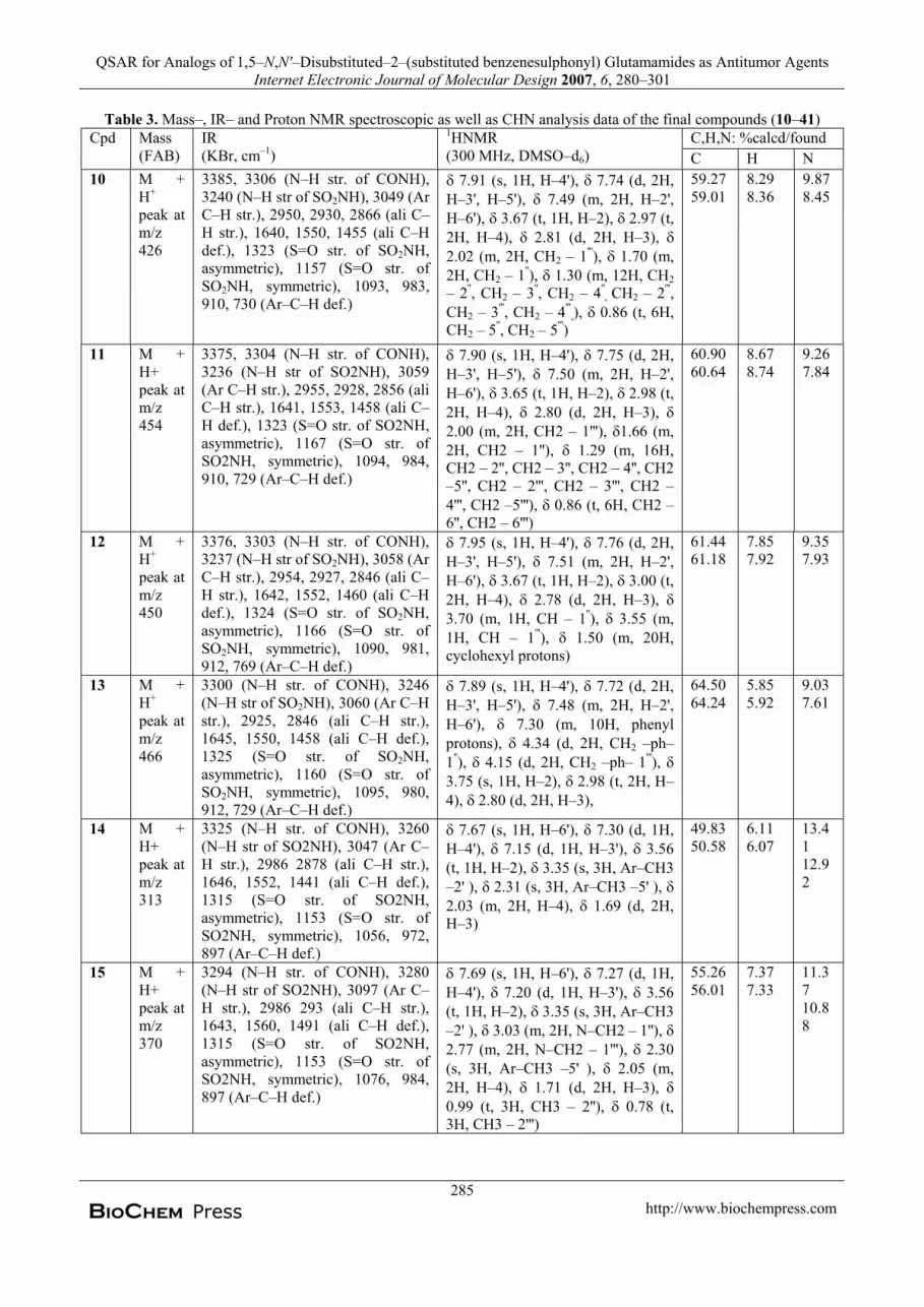
QSAR for Analogs of 1,5–N,N'–Disubstituted–2–(substituted benzenesulphonyl) Glutamamides as Antitumor Agents Internet Electronic Journal of Molecular Design 2007, 6, 280–301
Table 3. Mass–, IR– and Proton NMR spectroscopic as well as CHN analysis data of the final compounds (10–41)C,H,N: %calcd/found Cpd Mass
(FAB)IR(KBr, cm–1)
1HNMR(300 MHz, DMSO–d6) C H N
10 M +H+
peak at m/z426
3385, 3306 (N–H str. of CONH),3240 (N–H str of SO2NH), 3049 (Ar C–H str.), 2950, 2930, 2866 (ali C–H str.), 1640, 1550, 1455 (ali C–Hdef.), 1323 (S=O str. of SO2NH,asymmetric), 1157 (S=O str. of SO2NH, symmetric), 1093, 983, 910, 730 (Ar–C–H def.)
7.91 (s, 1H, H–4'), 7.74 (d, 2H, H–3', H–5'), 7.49 (m, 2H, H–2',H–6'), 3.67 (t, 1H, H–2), 2.97 (t,2H, H–4), 2.81 (d, 2H, H–3),2.02 (m, 2H, CH2 – 1'''), 1.70 (m,2H, CH2 – 1''), 1.30 (m, 12H, CH2– 2'', CH2 – 3'', CH2 – 4''
, CH2 – 2''',CH2 – 3''', CH2 – 4'''
,), 0.86 (t, 6H, CH2 – 5'', CH2 – 5''')
59.2759.01
8.298.36
9.878.45
11 M +H+peak at m/z454
3375, 3304 (N–H str. of CONH),3236 (N–H str of SO2NH), 3059 (Ar C–H str.), 2955, 2928, 2856 (aliC–H str.), 1641, 1553, 1458 (ali C–H def.), 1323 (S=O str. of SO2NH,asymmetric), 1167 (S=O str. of SO2NH, symmetric), 1094, 984, 910, 729 (Ar–C–H def.)
7.90 (s, 1H, H–4'), 7.75 (d, 2H,H–3', H–5'), 7.50 (m, 2H, H–2',H–6'), 3.65 (t, 1H, H–2), 2.98 (t,2H, H–4), 2.80 (d, 2H, H–3),2.00 (m, 2H, CH2 – 1'''), 1.66 (m,2H, CH2 – 1''), 1.29 (m, 16H, CH2 – 2'', CH2 – 3'', CH2 – 4'', CH2–5'', CH2 – 2''', CH2 – 3''', CH2 –4''', CH2 –5'''), 0.86 (t, 6H, CH2 – 6'', CH2 – 6''')
60.9060.64
8.678.74
9.267.84
12 M +H+
peak at m/z450
3376, 3303 (N–H str. of CONH),3237 (N–H str of SO2NH), 3058 (Ar C–H str.), 2954, 2927, 2846 (ali C–H str.), 1642, 1552, 1460 (ali C–Hdef.), 1324 (S=O str. of SO2NH,asymmetric), 1166 (S=O str. of SO2NH, symmetric), 1090, 981, 912, 769 (Ar–C–H def.)
7.95 (s, 1H, H–4'), 7.76 (d, 2H,H–3', H–5'), 7.51 (m, 2H, H–2',H–6'), 3.67 (t, 1H, H–2), 3.00 (t,2H, H–4), 2.78 (d, 2H, H–3), 3.70 (m, 1H, CH – 1''), 3.55 (m,1H, CH – 1'''), 1.50 (m, 20H, cyclohexyl protons)
61.4461.18
7.857.92
9.357.93
13 M +H+
peak at m/z466
3300 (N–H str. of CONH), 3246 (N–H str of SO2NH), 3060 (Ar C–Hstr.), 2925, 2846 (ali C–H str.),1645, 1550, 1458 (ali C–H def.), 1325 (S=O str. of SO2NH,asymmetric), 1160 (S=O str. of SO2NH, symmetric), 1095, 980, 912, 729 (Ar–C–H def.)
7.89 (s, 1H, H–4'), 7.72 (d, 2H,H–3', H–5'), 7.48 (m, 2H, H–2',H–6'), 7.30 (m, 10H, phenylprotons), 4.34 (d, 2H, CH2 –ph– 1''), 4.15 (d, 2H, CH2 –ph– 1'''),3.75 (s, 1H, H–2), 2.98 (t, 2H, H–4), 2.80 (d, 2H, H–3),
64.5064.24
5.855.92
9.037.61
14 M +H+peak at m/z313
3325 (N–H str. of CONH), 3260 (N–H str of SO2NH), 3047 (Ar C–H str.), 2986 2878 (ali C–H str.),1646, 1552, 1441 (ali C–H def.), 1315 (S=O str. of SO2NH,asymmetric), 1153 (S=O str. of SO2NH, symmetric), 1056, 972, 897 (Ar–C–H def.)
7.67 (s, 1H, H–6'), 7.30 (d, 1H,H–4'), 7.15 (d, 1H, H–3'), 3.56 (t, 1H, H–2), 3.35 (s, 3H, Ar–CH3–2' ), 2.31 (s, 3H, Ar–CH3 –5' ),2.03 (m, 2H, H–4), 1.69 (d, 2H, H–3)
49.8350.58
6.116.07
13.4112.92
15 M +H+peak at m/z370
3294 (N–H str. of CONH), 3280 (N–H str of SO2NH), 3097 (Ar C–H str.), 2986 293 (ali C–H str.),1643, 1560, 1491 (ali C–H def.), 1315 (S=O str. of SO2NH,asymmetric), 1153 (S=O str. of SO2NH, symmetric), 1076, 984, 897 (Ar–C–H def.)
7.69 (s, 1H, H–6'), 7.27 (d, 1H,H–4'), 7.20 (d, 1H, H–3'), 3.56 (t, 1H, H–2), 3.35 (s, 3H, Ar–CH3–2' ), 3.03 (m, 2H, N–CH2 – 1''),2.77 (m, 2H, N–CH2 – 1'''), 2.30 (s, 3H, Ar–CH3 –5' ), 2.05 (m,2H, H–4), 1.71 (d, 2H, H–3), 0.99 (t, 3H, CH3 – 2''), 0.78 (t,3H, CH3 – 2''')
55.2656.01
7.377.33
11.3710.88
285BioChem Press http://www.biochempress.com

P. Panda, S. Samanta, Sk. M. Alam, S. Basu, and T. Jha Internet Electronic Journal of Molecular Design 2007, 6, 280–301
Table 3. (Continued)C,H,N: %calcd/found Cpd Mass
(FAB)IR(KBr, cm–1)
1HNMR(300 MHz, DMSO–d6) C H N
16 M +H+peak at m/z398
3300 (N–H str. of CONH), 3250(N–H str of SO2NH), 3067 (ArC–H str.), 2976 2832 (ali C–Hstr.), 1645, 1562, 1490 (ali C–Hdef.), 1315 (S=O str. of SO2NH,asymmetric), 1153 (S=O str. of SO2NH, symmetric), 1075, 986,890 (Ar–C–H def.)
7.70 (s, 1H, H–6'), 7.29 (d, 1H, H–4'), 7.22 (d, 1H, H–3'), 3.70 (t, 1H, H–2), 3.45 (s, 3H, Ar–CH3 –2' ),3.25 (m, 2H, N–CH2 – 1''), 2.83 (m,2H, N–CH2 – 1'''), 2.35 (s, 3H, Ar–CH3 –5' ), 2.10 (m, 2H, H–4), 1.77 (d, 2H, H–3), 1.55 (m, 2H, CH2 – 2''), 1.35 (m, 2H, CH2 – 2'''), 0.96 (t, 3H,
CH3 – 3''), 0.79 (t, 3H, CH3 – 3''')
57.4058.15
7.867.82
10.5710.08
17 M +H+
peak at m/z426
3322 (N–H str. of CONH), 3270(N–H str of SO2NH), 3020 (Ar C–H str.), 2986 2874 (ali C–Hstr.), 1643, 1552, 1444 (ali C–Hdef.), 1320 (S=O str. of SO2NH,asymmetric), 1150 (S=O str. of SO2NH, symmetric), 1076, 974,896 (Ar–C–H def.)
7.68 (s, 1H, H–6'), 7.27 (d, 1H, H–4'), 7.17 (d, 1H, H–3'), 3.72 (t, 1H, H–2), 3.41 (s, 3H, Ar–CH3 –2' ), 3.01 (m, 2H, N–CH2 – 1''), 2.72 (m, 2H, N–CH2 – 1'''), 2.30 (s, 3H, Ar–CH3 –5' ),2.15 (m, 2H, H–4), 1.61 (d, 2H, H–3),
1.53–1.20 (m, 8H, CH2 – 2'', CH2 – 3''
CH2 – 2''', CH2 – 3'''), 0.94–0.80 (m,6H, CH3 – 4'', CH3 – 4''')
59.2760.02
8.298.25
9.879.38
18 M +H+
peak at m/z454
3320 (N–H str. of CONH), 3277 (N–H str of SO2NH), 3058 (Ar C–H str.), 2954, 2927 (ali C–Hstr.), 1642, 1562, 1490 (ali C–Hdef.), 1316 (S=O str. of SO2NH,asymmetric), 1154 (S=O str. ofSO2NH, symmetric), 1070, 981, 912, 769 (Ar–C–H def.)
7.67 (s, 1H, H–6'), 7.22 (d, 1H, H–4'), 7.19 (d, 1H, H–3'), 3.69 (t, 1H,H–2), 3.38 (s, 3H, Ar–CH3 –2' ), 2.98 (m, 2H, N–CH2 – 1''), 2.68 (m, 2H, N–CH2 – 1'''), 2.30 (s, 3H, Ar–CH3 –5' ),2.14 (m, 2H, H–4), 1.58 (d, 2H, H–3), 1.50–1.12 (m, 12H, CH2 – 2'', CH2 –
3'', CH2 – 4'', CH2 – 2''', CH2 –3''' CH2 – 4'''), 0.92–0.81 (m, 6H, CH3 – 5'', CH3 – 5''')
60.9061.65
8.678.63
9.268.77
19 M +H+peak at m/z482
3300 (N–H str. of CONH), 3246 (N–H str of SO2NH), 3060 (Ar C–H str.), 2925, 2846 (ali C–Hstr.), 1645, 1550, 1458 (ali C–Hdef.), 1325 (S=O str. of SO2NH, asymmetric), 1155 (S=O str. ofSO2NH, symmetric), 1095, 980, 912 (Ar–C–H def.)
7.68 (s, 1H, H–6'), 7.24 (d, 1H, H–4'), 7.21 (d, 1H, H–3'), 3.70 (t, 1H,H–2), 3.36 (s, 3H, Ar–CH3 –2' ), 2.95 (m, 2H, N–CH2 – 1''), 2.64 (m,2H, N–CH2 – 1'''), 2.32 (s, 3H, Ar–CH3 –5' ), 2.16 (m, 2H, H–4), 1.60 (d, 2H, H–3), 1.32 (m, 16H, CH2 – 2'',CH2 – 3'', CH2 – 4'', CH2 –5'', CH2 – 2''', CH2 – 3''', CH2 – 4''', CH2 –5'''),0.88 (t, 6H, CH2 – 6'', CH2 – 6''')
62.3463.09
9.009.04
8.728.23
20 M +H+
peak at m/z398
3394 (N–H str. of CONH), 3280 (N–H str of SO2NH), 3067 (Ar C–H str.), 2956, 2832 (ali C–Hstr.), 1643, 1560, 1490 (ali C–Hdef.), 1315 (S=O str. of SO2NH,asymmetric), 1153 (S=O str. ofSO2NH, symmetric), 1086, 984, 890 (Ar–C–H def.),
7.92 (s, 1H, H–6'), 7.67 (d, 1H, H–4'), 7.53 (d, 1H, H–3'), 7.43–7.20 (d,10H, phenyl protons), 3.69 (t, 1H, H–2), 3.42 (s, 3H, Ar–CH3 –2' ), 2.36 (s, 3H, Ar–CH3 –5' ), 2.20 (m, 2H, H–4), 1.68 (d, 2H, H–3)
57.4058.15
7.867.82
10.5710.08
286BioChem Press http://www.biochempress.com

QSAR for Analogs of 1,5–N,N'–Disubstituted–2–(substituted benzenesulphonyl) Glutamamides as Antitumor Agents Internet Electronic Journal of Molecular Design 2007, 6, 280–301
Table 3. (Continued)C,H,N: %calcd/found Cp
dMass(FAB)
IR(KBr, cm–1)
1HNMR(300 MHz, DMSO–d6) C H N
21 M + H+
peak at m/z 478
3487 (N–H str. of CONH), 3285(N–H str of SO2NH), 3069 (Ar C–Hstr.), 2924, 2866 (ali C–H str.),1645, 1551, 1493 (ali C–H def.),1315 (S=O str. of SO2NH,asymmetric), 1153 (S=O str. ofSO2NH, symmetric), 1094, 1028,905 (Ar–C–H def.)
7.72 (s, 1H, H–6'), 7.37 (d, 1H, H–4'), 7.23 (d, 1H, H–3'), 7.20 – 7.12 (m, 10H, phenyl protons),4.34 (d, 2H, CH2 –ph– 1''), 4.15 (d, 2H, CH2 –ph– 1'''), 3.66 (t, 1H, H–2), 3.40 (s, 3H, Ar–CH3 –2' ),2.36 (s, 3H, Ar–CH3 –5' ), 2.10 (m,2H, H–4), 1.79 (d, 2H, H–3)
62.8663.60
8.238.19
8.808.30
22 M + H+
peak at m/z 466
3486 (N–H str. of CONH), 3284(N–H str. of SO2NH), 3066 (Ar C–H str.), 2923, 2860 (ali C–H str.),1645, 1550, 1492 (ali C–H def.),1315 (S=O str. of SO2NH,asymmetric), 1153 (S=O str. ofSO2NH, symmetric), 1090, 1027,904 (Ar–C–H def.)
7.71 (s, 1H, H–6'), 7.30 (d, 1H, H–4'), 7.24 (d, 1H, H–3'), 3.72 (t, 1H, H–2), 3.43 (s, 3H, Ar–CH3
–2' ), 3.25 (m, 2H, N–CH2 – 1''),2.83 (m, 2H, N–CH2 – 1'''), 2.35 (s, 3H, Ar–CH3 –5' ), 2.10 (m, 2H, H–4), 2.03 (m, 2H, N–CH – 1'', N–CH – 1'''), 1.75 (d, 2H, H–3), 0.91 (m, 12H, CH3 – 2'', CH3 – 3'',CH3 – 2''', CH3 – 3''')
64.5065.25
5.855.80
9.038.54
23 M + H+
peak at m/z494
3487 (N–H str. of CONH), 3285 (N–H str of SO2NH), 3069 (Ar C–H str.), 2924, 2866 (ali C–H str.),1645, 1551, 1493 (ali C–H def.),1315 (S=O str. of SO2NH,asymmetric), 1153 (S=O str. of SO2NH, symmetric), 1094, 1028,905 (Ar–C–H def.)
7.63 (s, 1H, H–3'), 7.50 (d, 1H,H–6'), 7.12 (d, 1H, H–5'), 3.60 (t, 1H, H–2), 3.42 (s, 3H, Ar–CH3
–2' ), 2.45 (m, 2H, H–4), 2.30 (s, 3H, Ar–CH3 –4' ), 1.94 (d, 2H, H–3), 1.56–1.24 (m, 20 H,cyclohexylprotons)
65.7066.45
6.336.29
8.518.02
24 M + H+peak at m/z 303
3351 (N–H str. of CONH), 3240 (N–H str. of SO2NH), 3068 (Ar C–H str.), 2942 (ali C–H str.), 1640, 1567, 1473 (ali C–H def.), 1333 (S=O str. of SO2NH, asymmetric),1178 (S=O str. of SO2NH,symmetric), 1060, 984, 905 (Ar–C–H def.)
7.87 (t, 2H, H–3', H–5'), 7.77 (t,2H, H–2', H–6'), 3.82 (t, 1H, H–2), 3.34 (s, 2H, H–4), 1.95 (d, 2H, H–3)
43.5643.64
4.654.76
13.8513.68
25 M + H+
peak at m/z 388
3316 (N–H str of CONH), 3247(N–H str of SO2NH), 3062 (Ar C–H str.), 2972 (ali C–H str.), 1647, 1597, 1540, 1493 (ali C–H def.),1332 (S=O str of SO2NH,asymmetric), 1168 (S=O str ofSO2NH, symmetric), 1091, 1060,981, 906 (Ar–C–H def.)
7.97 (t, 2H, H–3', H–5'), 7.83 (t,2H, H–2', H–6'), 3.82 (t, 1H, H–2), 3.65 (m, 2H, N–CH2 – 1''),3.23 (m, 2H, N–CH2 – 1'''), 2.81 (m, 2H, H–4), 2.05 (d, 2H, H–3), 1.35 (m, 2H, CH2 – 2''), 1.26 (m,
2H, CH2 – 2'''), 1.06 (t, 3H, CH3 – 3''), 0.80 (t, 3H, CH3 – 3''')
52.7052.78
6.766.87
10.8410.07
26 M + H+
peak at m/z 416
3375, 3310 (N–H str of CONH), 3238 (N–H str of SO2NH), 3101 (Ar C–H str.), 2959, 2934, 2870(ali C–H str.), 1639, 1593, 1553,1495 (ali C–H def.), 1325 (S=O strof SO2NH, asymmetric), 1167 (S=O str of SO2NH, symmetric),1092, 1068, 984, 903 (Ar–C–Hdef.)
7.93 (t, 2H, H–3', H–5'), 7.79 (t,2H, H–2', H–6'), 3.80 (t, 1H, H–2), 3.61 (m, 2H, N–CH2 – 1''),3.20 (m, 2H, N–CH2 – 1'''), 2.58 (m, 2H, H–4), 1.99 (d, 2H, H–3),
1.40–1.08 (m, 8H, CH2 – 2'', CH2
– 3'' CH2 – 2''', CH2 – 3'''), 0.95–0.82 (m, 6H, CH3 – 4'', CH3 – 4''')
54.9255.01
7.287.39
10.119.94
287BioChem Press http://www.biochempress.com

P. Panda, S. Samanta, Sk. M. Alam, S. Basu, and T. Jha Internet Electronic Journal of Molecular Design 2007, 6, 280–301
Table 3. (Continued)C,H,N: %calcd/found Cpd Mass
(FAB)IR(KBr, cm–1)
1HNMR(300 MHz, DMSO–d6) C H N
27 M +H+peak at m/z444
3377, 3309 (N–H str. of CONH),3238 (N–H str. of SO2NH), 3103(Ar C–H str.), 2956, 2935, 2868 (aliC–H str.), 1640, 1593, 1553, 1494 (ali C–H def.), 13251 (S=O str of SO2NH, asymmetric), 1166 (S=Ostr. of SO2NH, symmetric), 1093, 1069, 987, 904 (Ar–C–H def.)
7.85 (t, 2H, H–3', H–5'), 7.76 (t,2H, H–2', H–6'), 3.90 (t, 1H, H–2), 3.51 (m, 2H, N–CH2 – 1''),3.18 (m, 2H, N–CH2 – 1'''), 2.55 (m, 2H, H–4), 1.96 (d, 2H, H–3), 1.45–1.10 (m, 12H, CH2 – 2'',
CH2 – 3'', CH2 – 4'', CH2 – 2''',CH2 –3''' CH2 – 4'''), 0.90–0.78 (m, 6H, CH3 – 5'', CH3 – 5''')
56.8656.94
7.737.84
9.479.30
28 M +H+peak at m/z472
3316 (N–H str. of CONH), 3247 (N–H str. of SO2NH), 3065 (Ar C–Hstr.), 2962 (ali C–H str.), 1647, 1590,1539, 1485 (ali C–H def.), 1332(S=O str. of SO2NH, asymmetric),1168 (S=O str. of SO2NH,symmetric), 1095, 1062, 989, 905(Ar–C–H def.)
7.82 (t, 2H, H–3', H–5'), 7.71 (t,2H, H–2', H–6'), 3.87 (t, 1H, H–2), 3.49 (m, 2H, N–CH2 – 1''),3.16 (m, 2H, N–CH2 – 1'''), 2.35 (m, 2H, H–4), 1.94 (d, 2H, H–3), 1.40 (m, 16H, CH2 – 2'', CH2 –
3'', CH2 – 4'', CH2 –5'', CH2 – 2''',CH2 – 3''', CH2 – 4''', CH2 –5'''),1.07–0.94 (t, 6H, CH2 – 6'', CH2 –6''')
58.5758.65
8.128.23
8.918.74
29 M +H+peak at m/z456
3358 (N–H str. of CONH), 3240 (N–H str. of SO2NH), 3093 (Ar C–Hstr.), 2938 (ali C–H str.), 1640, 1562,1495 (ali C–H def.), 1335 (S=O str.of SO2NH, asymmetric), 1170 (S=O str. of SO2NH, symmetric), 1020, 989, 895 (Ar–C–H def.)
7.90 (t, 2H, H–3', H–5'), 7.81 (t,2H, H–2', H–6'), 7.60–7.24 (d, 10H,phenyl protons), 3.88 (t, 1H, H–2), 3.44 (s, 2H, H–4), 2.15 (d,2H, H–3)
60.6560.73
4.874.98
9.239.06
30 M +H+peak at m/z484
3312 (N–H str. of CONH), 3248 (N–H str. of SO2NH), 3065 (Ar C–Hstr.), 2922, 2866 (ali C–H str.), 1645,1550, 1495 (ali C–H def.), 1333(S=O str. of SO2NH, asymmetric),1169 (S=O str. of SO2NH,symmetric), 1093, 1031, 979, 912(Ar–C–H def.)
7.92 (t, 2H, H–3', H–5'), 7.78 (t,2H, H–2', H–6'), 7.42 – 7.27 (m,10H, phenyl protons), 4.26 (d, 2H, CH2 –ph– 1''), 4.17 (d, 2H, CH2 –ph– 1'''), 3.86 (t, 1H, H–2), 3.42 (s, 2H, H–4), 2.18 (d, 2H, H–3)
62.1062.18
5.425.53
8.698.52
31 M +H+
peak at m/z388
3315 (N–H str. of CONH), 3248 (N–H str. of SO2NH), 3063 (Ar C–Hstr.), 2972 (ali C–H str.), 1645, 1593,1539, 1495 (ali C–H def.), 1333(S=O str. of SO2NH, asymmetric),1169 (S=O str. of SO2NH,symmetric), 1090, 1061, 989, 906(Ar–C–H def.)
7.81 (t, 2H, H–3', H–5'), 7.67 (t,2H, H–2', H–6'), 3.80 (t, 1H, H–2), 3.37 (s, 2H, H–4), 2.01 (m,2H, N–CH – 1'', N–CH – 1'''), 1.65 (d, 2H, H–3), 0.92 (m, 12H, CH3 – 2'', CH3 – 3'', CH3 – 2''', CH3 – 3''')
52.7052.78
6.766.87
10.8410.67
32 M + H+
peak at m/z466
3365 (N–H str of CONH), 3248 (N–H str of SO2NH), 3093 (Ar C–Hstr.), 2947 (ali C–H str.), 1640, 1560,1485 (ali C–H def.), 1334 (S=O strof SO2NH, asymmetric), 1172 (S=O str of SO2NH, symmetric), 1060, 974, 896 (Ar–C–H def.)
7.85 (t, 2H, H–3', H–5'), 7.69 (t,2H, H–2', H–6'), 3.82 (t, 1H, H–2), 3.60 (m, 2H, CH – 1'', CH – 1'''), 3.36 (s, 2H, H–4), 1.65 (d, 2H, H–3), 1.44–1.05 (m, 20 H,cyclohexyl protons)
59.0859.16
7.337.44
8.998.62
288BioChem Press http://www.biochempress.com

QSAR for Analogs of 1,5–N,N'–Disubstituted–2–(substituted benzenesulphonyl) Glutamamides as Antitumor Agents Internet Electronic Journal of Molecular Design 2007, 6, 280–301
Table 3. (Continued)C,H,N: %calcd/found Cpd Mass
(FAB)IR(KBr, cm–1)
1HNMR(300 MHz, DMSO–d6) C H N
33 M + H+
peak at m/z 454
3299 (N–H str. of CONH), 3257 (N–H str. of SO2NH), 3092 (Ar C–H str.), 2930, 2855 (ali C–Hstr.), 1625, 1552, 1457 (ali C–Hdef.), 1340 (S=O str. of SO2NH,asymmetric), 1166 (S=O str. of SO2NH, symmetric), 1029, 995, 890 (Ar–C–H def.)
7.90 (d, 2H, H–2', H–6'), 7.66 (d, 2H, H–3', H–5'), 3.81 (t, 1H, H–2), 3.27 (m, 4H, N–CH2 – 1'', N–CH2 – 1'''), 2.37 (m, 2H, H–4), 2.20 (m,9H, three CH3 of t– Butyl), 2.05 (m,2H, CH – 2'', CH –2'''), 1.85 (d, 2H, H–3), 1.64–1.50 (m, 12 H, CH3 – 3'',CH3 – 4'', CH3 – 3''', CH3 – 4''')
60.9061.30
8.678.60
9.269.20
34 M + H+
peak at m/z 506
3300 (N–H str. of CONH), 3094 (Ar C–H str.), 2934, 2856 (ali C–H str.), 1624, 1553, 1450 (ali C–Hdef.), 1342 (S=O str. of SO2NH,asymmetric), 1167 (S=O str. of SO2NH, symmetric), 1028, 995, 891 (Ar–C–H def.)
7.93 (d, 2H, H–2', H–6'), 7.68 (d, 2H, H–3', H–5'), 3.84 (t, 1H, H–2), 2.45 (m, 2H, H–4), 2.14 (m, 9H, three CH3 of t– Butyl), 1.90 (d, 2H, H–3), 1.54–1.25 (m, 20 H,cyclohexyl protons)
64.1265.03
8.578.32
8.317.99
35 M + H+
peak at m/z 370
3299 (N–H str. of CONH), 3249 (N–H str. of SO2NH), 3018 (Ar C–H str.), 2923, 2872 (ali C–Hstr.), 1541, 1446 (ali C–H def.),1323 (S=O str of SO2NH,asymmetric), 1155 (S=O str. of SO2NH, symmetric), 1027, 976, 912 (Ar–C–H def.)
7.60 (s, 1H, H–3'), 7.52 (d, 1H, H–6'), 7.14 (d, 1H, H–5'), 3.74 (t, 1H, H–2), 3.37 (s, 3H, Ar–CH3 –2' ),3.04 (m, 2H, N–CH2 – 1''), 2.77 (m,2H, N–CH2 – 1'''), 2.28 (s, 3H, Ar–CH3 –4' ), 2.15 (m, 2H, H–4), 1.76 (d, 2H, H–3), 0.97 (t, 3H, CH3 – 2''), 0.76 (t, 3H, CH3 – 2''')
55.2654.74
7.377.56
11.3711.30
36 M + H+
peak at m/z 398
3310 (N–H str. of CONH), 3249 (N–H str of SO2NH), 3018 (Ar C–H str.), 2874 (ali C–H str.), 1551,1444 (ali C–H def.), 1323 (S=Ostr of SO2NH, asymmetric), 1155(S=O str of SO2NH, symmetric),1028, 974, 911 (Ar–C–H def.)
7.59 (s, 1H, H–3'), 7.51 (d, 1H, H–6'), 7.15 (d, 1H, H–5'), 3.74 (t, 1H, H–2), 3.37 (s, 3H, Ar–CH3 –2' ),3.06 (m, 2H, N–CH2 – 1''), 2.79 (m,2H, N–CH2 – 1'''), 2.28 (s, 3H, Ar–CH3 –4' ), 2.15 (m, 2H, H–4), 1.76 (d, 2H, H–3), 1.56 (m, 2H, CH2 – 2''), 1.38 (m, 2H, CH2 – 2'''), 0.98 (t, 3H, CH3 – 2''), 0.77 (t, 3H, CH3 – 2''')
57.4056.68
7.867.93
10.5710.50
37 M + H+
peak at m/z 466
3360, 3308 (N–H str. of CONH),3238 (N–H str of SO2NH), 3055 (Ar C–H str.), 2939 (ali C–H str.),1602, 1547, 1520, 1500 (ali C–Hdef.), 1319 (S=O str of SO2NH,asymmetric), 1155 (S=O str ofSO2NH, symmetric), 1090, 1028, 980, 910 (Ar–C–H def.)
8.60 (s, 1H, H–3'), 8.32 (d, 1H, H–6'), 8.14 (d, 1H, H–5'),7.55–7.20 (d, 10H,phenyl protons), 3.64 (t, 1H, H–2), 3.40 (s, 3H, Ar–CH3 –2' ),2.28 (s, 3H, Ar–CH3 –4' ), 2.15 (m,2H, H–4), 1.76 (d, 2H, H–3)
64.5063.58
5.856.04
9.038.96
38 M + H+
peak at m/z 494
3300 (N–H str. of CONH), 3248 (N–H str of SO2NH), 3028 (Ar C–H str.), 2924, 2868 (ali C–H str.),1641, 1539, 1497 (ali C–H def.), 1323 (S=O str. of SO2NH,asymmetric), 1155 (S=O str. of SO2NH, symmetric), 1092, 1028, 972, 912 (Ar–C–H def.)
7.57 (s, 1H, H–3'), 7.50 (d, 1H, H–6'), 7.32 – 7.25 (m, 10H, phenylprotons), 7.11 (d, 1H, H–5'), 4.24 (d, 2H, CH2 –ph– 1''), 4.10 (d, 2H,CH2 –ph– 1'''), 3.76 (s, 1H, H–2), 3.37 (s, 3H, Ar–CH3 –2'' ), 2.28 (d, 3H, Ar–CH3 –4' ), 2.14 (m, 2H, H–4), 1.78 (m, 2H, H–3)
65.7065.18
6.336.52
8.517.84
289BioChem Press http://www.biochempress.com

P. Panda, S. Samanta, Sk. M. Alam, S. Basu, and T. Jha Internet Electronic Journal of Molecular Design 2007, 6, 280–301
Table 3. (Continued)C,H,N: %calcd/found Cpd Mass
(FAB)IR(KBr, cm–1)
1HNMR(300 MHz, DMSO–d6) C H N
39 M + H+
peak at m/z398
3310 (N–H str. of CONH), 3247 (N–H str. of SO2NH), 3028 (Ar C–Hstr.), 2933, 2855 (ali C–H str.), 1540,1445 (ali C–H def.), 1323 (S=O str.of SO2NH, asymmetric), 1155 (S=O str. of SO2NH, symmetric), 1028,966, 910 (Ar–C–H def.)
7.61 (s, 1H, H–3'), 7.49 (d, 1H, H–6'), 7.16 (d, 1H, H–5'), 3.64 (t, 1H, H–2), 3.38 (s, 3H, Ar–CH3
–2' ), 2.28 (s, 3H, Ar–CH3 –4' ),2.16 (m, 2H, H–4), 2.03 (m, 2H, N–CH – 1'', N–CH – 1'''), 1.67 (d, 2H, H–3), 0.96 (m, 12H, CH3 – 2'',CH3 – 3'', CH3 – 2''', CH3 – 3''')
57.4056.68
7.868.05
10.5710.50
40 M + H+
peak at m/z426.
3362, 3307 (N–H str. of CONH), 3236 (N–H str. of SO2NH), 3065 (Ar C–H str.), 2948 (ali C–H str.), 1607, 1548, 1482 (ali C–H def.), 1323 (S=O str. of SO2NH, asymmetric),1156 (S=O str. of SO2NH,symmetric), 1028, 987, 910 (Ar–C–Hdef.)
7.64 (s, 1H, H–3'), 7.52 (d, 1H, H–6'), 7.17 (d, 1H, H–5'), 3.63 (t, 1H, H–2), 3.40 (s, 3H, Ar–CH3
–2' ), 3.37 (m, 4H, N–CH2 – 1'', N–CH2 – 1'''), 2.40 (m, 2H, H–4), 2.28 (s, 3H, Ar–CH3 –4' ), 2.03 (m,2H, CH – 2'', CH –2'''), 1.65 (d, 2H, H–3), 1.50 (m, 12 H, CH3 – 3'',CH3 – 4'', CH3 – 3''', CH3 – 4''')
59.2758.75
8.298.48
9.879.80
41 M + H+
peak at m/z478
3359, 3307 (N–H str. of CONH), 3236 (N–H str of SO2NH), 3053 (Ar C–H str.), 2940 (ali C–H str.), 1604, 1546, 1519, 1502 (ali C–H def.), 1312 (S=O str. of SO2NH,asymmetric), 1156 (S=O str. ofSO2NH, symmetric), 1087, 1027, 979, 914 (Ar–C–H def.)
7.63 (s, 1H, H–3'), 7.50 (d, 1H, H–6'), 7.12 (d, 1H, H–5'), 3.60 (t, 1H, H–2), 3.42 (s, 3H, Ar–CH3
–2' ), 2.45 (m, 2H, H–4), 2.30 (s, 3H, Ar–CH3 –4' ), 1.94 (d, 2H, H–3), 1.56–1.24 (m, 20 H, cyclohexyl protons)
62.8662.34
8.238.42
8.808.73
Table 4. Antitumor activities of the 1,5–N,N'–disubstituted–2–(substituted benzenesulphonyl) glutamamides (10–41)Cpd % TCI Log (BA) Cpd % TCI Log (BA)10 28.84 1.460 28 20.46 1.31111 25.14 1.400 29 28.03 1.44812 49.20 1.692 30 23.38 1.36913 24.22 1.384 31 17.24 1.23714 53.37 1.727 32 24.56 1.39015 25.57 1.408 33 36.18 1.55816 27.59 1.441 34 23.50 1.37117 23.56 1.372 35 21.92 1.34118 20.54 1.313 36 21.00 1.32219 27.09 1.433 37 79.57 1.90120 45.32 1.656 38 30.30 1.48121 42.77 1.631 39 33.15 1.52022 59.67 1.776 40 47.15 1.67323 52.82 1.723 41 53.17 1.72624 34.72 1.541 Mitomycin (42) 100.00 2.00025 31.77 1.502 Azaserine (2) 100.00 2.00026 21.48 1.332 Don (3) 100.00 2.00027 20.56 1.313
2.2 Biological ActivityThe antitumor activity of all final compounds (10–41) was evaluated in vivo against Ehrlich
Ascites Carcinoma (EAC) cells in Swiss Albino mice according to the earlier reported method [13–17]. Mitomycin C (42) was used as the universal standard while azaserine (2) and DON (3) were
290BioChem Press http://www.biochempress.com

QSAR for Analogs of 1,5–N,N'–Disubstituted–2–(substituted benzenesulphonyl) Glutamamides as Antitumor Agents Internet Electronic Journal of Molecular Design 2007, 6, 280–301
used as the specific standards. The antitumor activity was expressed in terms of the percentage inhibition of the cell count. Standards showed 100% inhibitions. Results are shown in Table 4.
2.3 QSAR StudyQSAR studies of 32 synthesized QSAR analogs of 1,5–N,N'–disubstituted–2–(substituted
benzenesulphonyl)glutamamides (10–41) were performed using different parameters like physicochemical, topological (ETSA and RTSA indices), geometrical, constitutional, semi–emperical quantum chemical descriptors as well as indicators parameters. Electrotopological state atom (ETSA) index [50–52] is an atom level topological descriptor encoding both electronic and topological information. The E–state index Si of an atom i in a molecule is composed of an intrinsic state Ii and the perturbation effect Ij, shown in Eq. (1). The atom intrinsic value includes both electronic and topological information. The perturbation effect Ij stands for influence of information field on the intrinsic atom value Ii.
Si = Ii + Ij (1)
The intrinsic state value of atom i is expressed as
Ii = [((2/N)2 v + 1)/ ] (2)
where N = principle quantum number of valence electrons, v = number of valence electrons – number of hydrogen atom attached, and = number of sigma electrons – number of hydrogen atomattached.
The expression for the perturbation effect is as follows:
Ij = (Ii – Ij)/ rij2 (3)
where rij is the graph distance.
The refractotopological state atom (RTSA) index [53–54] is a novel atom level topological descriptor used in QSAR study. The R–state index i of an atom i in a molecule is composed of an intrinsic refractivity ARi and the perturbation effect ARi, as shown in Eq. (4).
i = ARi + ARi (4)
The perturbation term is defined as:
ARi = (ARi – ARi)/ rij2 (5)
where rij is the graph distance.
The RTSA index is based on the atomic refractivity and the topological environment of the atom.Sum of the atomic refractivity, that is, molar refractivity is directly proportional to the polarizability of a substance which determines London force/dispersive force between nonpolar molecules.Therefore, R–state indices are important for modeling the dispersive/van der Waals interactions with the receptors. The logarithm of percentage tumor cell inhibition (Log BA) was used as the
291BioChem Press http://www.biochempress.com

P. Panda, S. Samanta, Sk. M. Alam, S. Basu, and T. Jha Internet Electronic Journal of Molecular Design 2007, 6, 280–301
biological activity parameter and is listed in Table 4.
Table 5. ETSA indices, RTSA indices, physicochemical (steric), semi–empirical quantum descriptors and some otherparameters of glutamamide analogs along with indicator parameters
Cpd S3 R3 R16 R4/R4'B5 qC13 EHOMO GAP SA nHAcc I1 I210 1.623 4.809 3.993 4.940 –0.182 –9.810 –8.916 0.816 7.000 0.000 0.00011 1.628 4.808 3.981 5.960 –0.182 –9.802 –8.915 0.886 7.000 0.000 0.00012 1.642 4.833 4.151 3.490 –0.177 –9.755 –8.872 0.582 7.000 0.000 1.00013 1.607 4.846 4.139 6.020 –0.184 –9.771 –9.094 0.645 7.000 0.000 0.00014 1.673 4.945 6.163 1.000 –0.178 –9.940 –8.919 0.462 7.000 0.000 0.00015 1.722 4.944 4.011 3.170 –0.185 –9.791 –8.978 0.654 7.000 0.000 0.00016 1.730 4.944 4.005 3.490 –0.185 –9.786 –8.903 0.721 7.000 0.000 0.00017 1.737 4.943 3.993 4.540 –0.184 –9.771 –9.004 0.807 7.000 0.000 0.00018 1.743 4.942 3.980 4.940 –0.184 –9.779 –8.964 0.867 7.000 0.000 0.00019 1.749 4.941 3.969 5.960 –0.182 –9.785 –8.969 0.940 7.000 0.000 0.00020 1.718 4.987 4.295 3.110 –0.186 –8.760 –7.901 0.685 7.000 0.000 0.00021 1.727 4.979 4.126 6.020 –0.181 –9.399 –8.505 0.714 7.000 0.000 0.00022 1.725 4.944 4.052 3.170 –0.183 –9.785 –8.957 0.702 7.000 0.000 0.00023 1.762 4.966 4.139 3.490 –0.177 –9.717 –8.966 0.653 7.000 0.000 1.00024 0.969 5.414 6.199 1.000 –0.179 –10.211 –8.850 0.416 8.000 0.000 0.00025 1.027 5.413 4.058 3.490 –0.183 –9.872 –8.716 0.679 8.000 0.000 0.00026 1.034 5.412 4.045 4.540 –0.182 –9.844 –8.693 0.756 8.000 0.000 0.00027 1.040 5.411 4.033 4.940 –0.182 –9.861 –8.714 0.820 8.000 0.000 0.00028 1.045 5.411 4.021 5.960 –0.182 –9.860 –8.716 0.890 8.000 0.000 0.00029 1.015 5.456 4.348 3.110 –0.186 –8.777 –7.522 0.632 8.000 0.000 0.00030 1.024 5.448 4.179 6.020 –0.183 –9.625 –8.470 0.703 8.000 0.000 0.00031 1.022 5.413 4.105 3.170 –0.184 –9.941 –8.796 0.654 8.000 0.000 0.00032 1.059 5.435 4.191 3.490 –0.175 –9.620 –8.429 0.575 8.000 0.000 0.00033 1.779 5.004 3.994 4.450 –0.182 –9.756 –8.933 0.857 7.000 0.000 0.00034 1.809 5.027 4.133 3.490 –0.188 –9.635 –8.831 0.719 7.000 1.000 1.00035 1.768 5.016 4.012 3.170 –0.184 –9.858 –9.003 0.643 7.000 1.000 0.00036 1.776 5.015 4.006 3.490 –0.184 –9.831 –8.985 0.719 7.000 0.000 0.00037 1.765 5.059 4.296 3.110 –0.187 –8.738 –7.800 0.683 7.000 0.000 0.00038 1.774 5.051 4.127 6.020 –0.182 –9.693 –9.021 0.761 7.000 0.000 0.00039 1.771 5.016 4.053 3.170 –0.184 –9.861 –9.007 0.705 7.000 0.000 0.00040 1.779 5.015 4.000 4.450 –0.184 –9.800 –8.979 0.776 7.000 0.000 0.00041 1.809 5.038 4.139 3.490 –0.182 –9.660 –8.862 0.641 7.000 0.000 1.000
The physicochemical parameters like, hydrophobic constant ( ), molar refractivity (MR), steric parameter (Es), Verloop STERIMOL parameters like L, B1, B5 were collected from the literature [18,55–56]. Topological indices like ETSA indices and RTSA indices were calculated using programme ‘Mouse’ developed in our laboratory [57]. Semi–empirical quantum chemicaldescriptors like atomic charges, EHOMO, ELUMO, the HOMO–LUMO energy gap (GAP) and different QSAR properties like surface area (approx.), surface area (grid), volume, hydration energy, log P, refractivity, polarizability and mass were calculated using Hyperchem Release 7.0 Pro Package [58]. 2D structure of compounds were drawn and converted to 3D structure. Geometry optimizationwas done by a semi–empirical method [59–61] – Austin Model 1 (AM1) using Polack-Ribiere (conjugate gradient) algorithm with RMS gradient of 0.1 kcal/Å mol (1 cal = 4.184 J). Molecular mechanics (MM+) force field was applied for the preliminary structure optimization to shorten the total time required for the energy minimization by AM1 method. Various descriptors such as
292BioChem Press http://www.biochempress.com

QSAR for Analogs of 1,5–N,N'–Disubstituted–2–(substituted benzenesulphonyl) Glutamamides as Antitumor Agents Internet Electronic Journal of Molecular Design 2007, 6, 280–301
geometrical and constitutional descriptors, functional groups, properties, and empirical descriptors were calculated by a software ‘DRAGON’ ver 3, 2003 [62]. Besides these, indicator parameterswere also used in order to find out the role of the specific substituent at the specific position towards the biological activity. The selected parameters used to develop QSAR models are listed in Table 5.
Correlation analysis was performed on a data matrix containing all the predictor parameters and response variable. Intercorreleted parameters were eliminated. The correlation matrix is shown in Table 6. Remaining data matrix was subjected to multiple regression analysis [63] to build QSAR models using a computer program “multi regress” [64] developed in our laboratory.
Table 6. Correlation matrix of important variables and the biological activityR3 R16 R4/R4’B5 qC13 ELUMO GAP SA nHAcc I1 I2 Log (BA)
S3 –0.91 –0.21 0.03 –0.22 0.10 –0.35 0.19 –0.99 0.21 0.26 0.41R3 1.00 0.16 –0.11 0.08 0.01 0.42 –0.19 0.96 –0.08 –0.22 –0.34R16 1.00 –0.63 0.34 –0.15 0.06 –0.70 0.17 –0.07 –0.06 0.29R4/R4’B5 1.00 –0.07 –0.01 –0.14 0.76 –0.04 –0.14 –0.16 –0.36qC13 1.00 –0.40 –0.25 –0.30 0.18 –0.31 0.22 0.16ELUMO 1.00 0.88 –0.01 –0.08 –0.04 –0.00 0.37GAP 1.00 –0.17 0.36 –0.11 –0.13 0.22SA 1.00 –0.17 –0.07 –0.21 –0.34nHAcc 1.00 –0.16 –0.24 –0.42I1 1.00 0.29 –0.21I2 1.00 0.31
Statistical qualities of the regression equation were justified by parameters like R, %EV, R2A, F,
p and SEE. The predictive powers of QSAR equation are validated by leave–one–out (LOO–) cross–validation method. Parameters like PRESS, SSY, R2
cv, SPRESS and PSE are considered for validation of the models.
3 RESULTS AND DISCUSSION
Thirty two 1,5–N,N'–disubstituted–2–(substituted benzenesulphonyl)glutamamides (10–41) were synthesized on the basis of our earlier QSAR studies. These glutamamides were biologically evaluated for anticancer activity against Erlich Ascite Carcinoma (EAC) cells in Swiss Albino mice.The synthesized compounds contain four types of substitutions in the aromatic ring whereas the aliphatic side chain contains eleven different substitutions. Physical data of intermediates and finalcompounds are listed in Table 1 and 2 respectively. Antitumor activity in terms of percentage of tumor cell inhibition (%TCI) is presented in Table 4.
For the QSAR study, logarithm of %TCI (log BA) of these glutamamides analogs was computedand is presented in Table 4. Correlation analysis and k–mean cluster analysis were performed to reduce the number of predictor variables for the study. The selected variables are listed in Table 5. All possible combinations of predictor variables were considered to develop QSAR models by
293BioChem Press http://www.biochempress.com

P. Panda, S. Samanta, Sk. M. Alam, S. Basu, and T. Jha Internet Electronic Journal of Molecular Design 2007, 6, 280–301
multiple linear regression analysis. The best QSAR model was obtained as follows:
Log (BA) = 4.439 (±0.871) – 0.273 (±0.108) R3 + 0.136 (±0.045) R16 + 0.221 (±0.070) EHOMO+ 0.128 (±0.070) I2
n = 32 R = 0.699 %EV = 48.88 R²A = 0.413 F(4,27) = 6.454 p<0.0009 SEE = 0.128 PRESS = 0.640 SSY = 0.868 R2
cv = 0.263 SPRESS = 0.154 PSE = 0.141
(6)
Where n is the number of data point. R, R2a, F, p, SEE, PRESS, SSY, R2
cv, PSE, SPRESS are correlation coefficient, adjusted R2 , variance ratio, probability factor related to F–ratio, standard error of estimate, predicted residual sum of square, total sum of squares, cross–validated R2,standard deviation error of prediction and Standard error of PRESS respectively. R3 and R16 are the R–state indices of atoms 3 and 16 respectively, EHOMO is the energy of the highest occupied molecular orbital and I2 is the indicator variable for the presence of cyclohexyl group at R4/R4'position of glutamamide analogs. Eq. (6) explains up to 48.88 % of variances in the activity data. Negative coefficient of R3 in the above model suggests that the higher value of R3 is detrimental to the activity whereas the higher value of R16 may increase the antitumor activity as evidenced by the positive regression coefficients. The positive coefficient of EHOMO in the equation showed the positive contribution of EHOMO towards the activity. The equation suggests that the presence of cyclohexyl group at R4/R4' position of glutamamide analogs may favor the antitumor activity as demonstrated by the positive regression coefficient of I2. However, deletion of compounds 20, 22,29, 34, and 40 yielded the following models.
Log (BA) = 5.554 (±0.618) – 0.145 (±0.065) R3 + 0.169 (±0.026) R16 + 0.418 (±0.60) EHOMO+ 0.240 (±0.045) I2
n = 27 DC = 20, 22, 29, 34, and 40 R = 0.916 %EV = 83.85 R²A = 0.809 F(4,22) = 28.548 p<0.0001 SEE = 0.072 PRESS = 0.143 SSY = 0.703 R2
cv = 0.797 SPRESS = 0.081 PSE = 0.073
(7)
where DC refers to the deleted compounds which behave as outliers possibly by working with a different mechanism of action. Eq. (7) explains up to 83.85 % of variances in the antitumor activity.
Combination of qC13 (the atomic charge of atom 13), GAP (the energy difference between HOMO–LUMO), SA (approximate surface area of the whole molecule) and nHAcc [number of acceptor atoms for H– bonds (N, O, F) of these compounds] produced a model for antitumoractivity of glutamamide analogs as shown in Eq. (8).
Log (BA) = 9.335 (±2.064) + 20.015 (±8.879) qC13 + 0.230(±0.068) GAP– 0.393 (±0.201) SA – 0.260 (±0.053) nHAcc
n = 32 R = 0.744 %EV = 55.36 R²A = 0.487 F(4,27) = 8.371 p<0.0002 SEE = 0.120 PRESS = 0.569 SSY = 0.868 R2
cv = 0.344 SPRESS = 0.145 PSE = 0.133
(8)
Eq. (8) explains up to 55.36 % of variances in the activity data. The positive coefficient of qC13
indicates that increasing the atomic charge on the carbon atom 13 is favorable for the activity. The
equation reveals that the HOMO–LUMO energy gap plays an important role in the stability of these
compounds. The negative coefficient of the approximate surface area implies that lower values of
294BioChem Press http://www.biochempress.com

QSAR for Analogs of 1,5–N,N'–Disubstituted–2–(substituted benzenesulphonyl) Glutamamides as Antitumor Agents Internet Electronic Journal of Molecular Design 2007, 6, 280–301
the whole molecular surface area correspond to higher antitumor activity of these glutamamides.
The equation suggests that the number of acceptor atoms for hydrogen bonds plays an important
role in the antitumor activity. Higher number of acceptor atoms for hydrogen bonds may decrease
the antitumor activity of these glutamamides as indicated by the negative coefficient of nHAcc.
Stepwise deletions of the outliers 22, 40, 32, 29, and 20 yielded statistically significant Eq. (9).
Log (BA) = 13.331 (±1.510) + 32.584 (±6.465) qC13 + 0.440(±0.063) GAP – 0.412 (±0.128) SA – 0.238 (±0.035) nHAcc
n = 27 DC = 22, 40, 32, 29, and 20 R = 0.907 %EV = 82.18 R²A = 0.789 F(4,22) = 25.357 p<0.0001 SEE = 0.076 PRESS = 0.202 SSY = 0.707 R2
cv = 0.714 SPRESS = 0.096; PSE = 0.086
(9)
Eq. [9] explains up to 82.18 % of the variances in the activity data. The equation has significant statistical predictivity as revealed by its R2
cv value (>0.7).
Another QSAR model was developed by taking S3 (E–state index of atom 3), R4/R4'B5
(maximum width Verloop STERIMOL parameter of R4/R4' substituent), EHOMO (the energy of highest occupied molecular orbital) and an indicator parameter I1 (indicate the presence of tert–butyl group at R3 position of the phenyl ring) and shown in Eq. (10).
Log (BA) = 2.825 (±0.669) + 0.241(±0.069) S3 – 0.052 (±0.016) R4/R4'B5+ 0.152 (±0.067) EHOMO – 0.243 (±0.092) I1
n = 32 R = 0.732 %EV = 53.66 R²A = 0.468 F(4,27) = 7.815 p<0.0003 SEE = 0.122 PRESS = 0.540 SSY = 0.868 R2
cv = 0.378 SPRESS = 0.141 PSE = 0.130
(10)
Eq. [10] explains up to 53.66 % of variances in the activity data. The model suggests that higher
value of S3 increases the antitumor activity of these glutamamide analogs as evidenced by the
positive regression coefficient of S3. The equation revealed that the width of substituents at R4/R4'
position of glutamamide analogs plays an important role in the intermolecular interactions with the
glutamine receptors. The negative coefficient of R4/R4'B5 indicates that the lower value of R4/R4'B5
may elevate the antitumor activity. The model also signifies that the presence of tert–butyl group at
R3 position of the phenyl ring will hamper the antitumor activity of these analogs as indicated by
the negative coefficient I1. The QSAR model from Eq. (11) was developed by stepwise deleting the
compounds 36, 18, 15, 17, and 16.
Log (BA) = 2.252 (±0.495) + 0.339(±0.053) S3 – 0.059 (±0.012) R4/R4'B5 + 0.101 (±0.049) EHOMO – 0.319 (±0.068) I1
n = 27 DC = 15, 16, 17, 18 and 36 R = 0.881 %EV = 77.68 R²A = 0.736 F(4,22) = 19.145 p<0.0000 SEE = 0.088 PRESS = 0.258 SSY = 0.769 R2
cv = 0.664 SPRESS = 0.108 PSE = 0.098
(11)
The Eq. [11] explains up to 77.68 % of variances in the activity data. Correlation matrix for the biological activity and selected parameters is shown in Table 6. All these equations are significant at more than 95% confidence intervals as supported by the t–statistics and p–values and are shown in Table 7.
295BioChem Press http://www.biochempress.com

P. Panda, S. Samanta, Sk. M. Alam, S. Basu, and T. Jha Internet Electronic Journal of Molecular Design 2007, 6, 280–301
Table 7. t–Values and p– values of all QSAR modelsEqn No Intercept/
Parameterst–values p–values Eqn No Intercept/
Parameterst–values p–values
[6] Intercept 5.094 0.0000 [9] Intercept 8.831 0.0000R3 –2.533 0.0174 qC13 5.040 0.0000R16 3.026 0.0053 GAP 6.940 0.0000EHOMO 3.144 0.0040 SA –3.216 0.0040I2 1.829 0.0784* nHAcc –6.751 0.0000
[7] Intercept 8.991 0.0000 [10] Intercept 4.224 0.0002R3 –2.241 0.0355 S3 3.484 0.0017R16 6.492 0.0000 R4/R4'B5 –3.208 0.0034EHOMO 6.929 0.0000 EHOMO 2.280 0.0307I2 5.308 0.0000 I1 –2.629 0.0140
4.224 0.0002[8] Intercept 4.522 0.0001 [11] Intercept 4.547 0.0002
qC13 2.254 0.0325 S3 6.358 0.0000GAP 3.389 0.0022 R4/R4'B5 –4.837 0.0001SA –1.950 0.0617* EHOMO 2.061 0.0513nHAcc –4.901 0.0000 I1 –4.663 0.0001
Table 8. Observed (Obs.), calculated (Calc.), residual (Res.), LOO–predicted (LOO Pred.) and predicted residual (Pres) activities of final eqs [7], [9], and [11].
Eq. (7) Eq. (9) Eq. (11) Cpd ObsCalc Res Pred Pres Calc Res Pred Pres Calc Res Pred Pres
10 1.460 1.426 0.034 1.422 0.038 1.475 –0.015 1.476 –0.016 1.518 –0.058 1.522 –0.06211 1.400 1.428 –0.027 1.432 –0.031 1.446 –0.046 1.453 –0.053 1.460 –0.060 1.468 –0.06812 1.692 1.713 –0.021 1.724 –0.032 1.753 –0.061 1.776 –0.084 1.615 0.077 1.609 0.08313 1.384 1.462 –0.078 1.470 –0.086 1.401 –0.017 1.404 –0.020 1.453 –0.069 1.462 –0.07714 1.727 1.718 0.009 1.708 0.020 1.736 –0.008 1.739 –0.011 1.752 –0.025 1.763 –0.03515 1.408 1.418 –0.010 1.418 –0.011 1.415 –0.008 1.417 –0.009 – – – –16 1.441 1.419 0.022 1.417 0.024 1.432 0.009 1.431 0.009 – – – –17 1.372 1.423 –0.051 1.427 –0.055 1.385 –0.012 1.386 –0.014 – – – –18 1.313 1.418 –0.105 1.426 –0.113 1.383 –0.071 1.392 –0.079 – – – –19 1.433 1.414 0.019 1.412 0.021 1.414 0.019 1.409 0.023 1.503 –0.070 1.514 –0.08120 1.656 – – – – – – – – 1.764 –0.107 1.812 –0.15521 1.631 1.596 0.035 1.591 0.041 1.724 –0.090 1.741 –0.110 1.531 0.100 1.515 0.11622 1.776 – – – – – – – – 1.659 0.117 1.646 0.12923 1.723 1.707 0.016 1.699 0.023 1.688 0.035 1.676 0.047 1.659 0.064 1.653 0.06924 1.541 1.543 –0.002 1.546 –0.005 1.526 0.015 1.517 0.024 1.486 0.054 1.455 0.08525 1.502 1.324 0.178 1.297 0.205 1.352 0.150 1.326 0.176 1.394 0.108 1.380 0.12226 1.332 1.333 –0.001 1.333 –0.001 1.365 –0.033 1.371 –0.039 1.338 –0.006 1.339 –0.00727 1.313 1.324 –0.011 1.326 –0.012 1.318 –0.005 1.319 –0.006 1.315 –0.002 1.315 –0.00228 1.311 1.323 –0.012 1.324 –0.013 1.284 0.027 1.275 0.036 1.257 0.054 1.245 0.06629 1.448 – – – – – – – – 1.524 –0.076 1.577 –0.13030 1.369 1.442 –0.073 1.456 –0.087 1.453 –0.084 1.471 –0.102 1.270 0.099 1.246 0.12331 1.237 1.303 –0.066 1.313 –0.077 1.306 –0.069 1.321 –0.085 1.404 –0.168 1.431 –0.19432 1.390 1.448 –0.058 1.458 –0.068 – – – – 1.431 –0.041 1.436 –0.04533 1.558 1.421 0.138 1.412 0.146 1.442 0.116 1.429 0.130 1.605 –0.046 1.609 –0.05034 1.371 – – – – 1.372 –0.001 1.373 –0.002 1.365 0.006 1.358 0.01335 1.341 1.379 –0.039 1.382 –0.041 1.433 –0.093 1.446 –0.106 1.347 –0.006 1.353 –0.01336 1.322 1.390 –0.068 1.394 –0.072 1.403 –0.081 1.411 –0.089 – – – –37 1.901 1.889 0.011 1.849 0.052 1.863 0.038 1.707 0.194 1.782 0.119 1.724 0.17638 1.481 1.463 0.019 1.462 0.020 1.460 0.022 1.458 0.023 1.517 –0.036 1.523 –0.04139 1.520 1.385 0.135 1.376 0.145 1.427 0.094 1.419 0.102 1.666 –0.146 1.685 –0.16540 1.673 – – – – – – – – 1.600 0.073 1.594 0.08041 1.726 1.721 0.005 1.718 0.008 1.555 0.171 1.542 0.183 1.681 0.045 1.676 0.049
296BioChem Press http://www.biochempress.com

QSAR for Analogs of 1,5–N,N'–Disubstituted–2–(substituted benzenesulphonyl) Glutamamides as Antitumor Agents Internet Electronic Journal of Molecular Design 2007, 6, 280–301
The predictive power of the final Eqs. (7), (9), and (11) was evaluated by the leave–one–out cross–validation (LOO) method. In this method, each compound was left out of the model and the prediction of activity of that compound was done. The observed (Obs), calculated (Calc), residual (Res), LOO– predicted (Pred) and predicted residual (Pres) values of the final Eqs. (7), (9), and (11) are shown in Table 8.
4 CONCLUSIONS
All synthesized glutamamides were evaluated for their possible anticancer activity against Ehrlich Ascites Carcinoma (EAC) cell in Swiss Albino mice. The result was expressed in terms of percentage of tumor cell inhibition. The QSAR study performed on 32 synthesized glutamamidesanalogs showed that RTSA indices of atom numbers 3 and 16, ETSA index of atom number 3, atomic charge on atom number 13, the energy of HOMO, the energy difference between HOMO and LUMO and indicator parameters are important for antitumor activity. RTSA indices are related to atomic refractivity as well as to molecular connectivity and are the measure of dispersive/van der Waals force between two nonpolar molecules, thus atom 3 and 16 may be involved in dispersive/van der Waals interactions with glutamine receptor(s). The study also showed that ETSA index of atom number 3 is favorable for the antitumor activity. As ETSA indices are the measure ofthe availability of the and/or lone pair electrons on the atoms it is evident that atom 3 mayinvolved in electronic interaction with glutamine receptor(s). The atomic charge on atom number 13 is favorable for the antitumor activity. From the QSAR model, it is found that the energy of the highest occupied molecular orbital (EHOMO) of these compounds has significant electronic contribution to the activity. As the energy of the HOMO is directly related to the ionization potential and characterizes the susceptibility of the molecule towards the attack by electrophiles, the QSAR equations reveal that these glutamamide analogs have higher susceptibility to lose a pair ofelectrons to an electrophile and these compounds are soft nucleophiles. It is also revealed that the HOMO–LUMO energy gap has a significant role in antitumor activity. The HOMO–LUMO energy gap, i.e. the difference in energy between the HOMO and the LUMO is an important stability index. As these glutamamide analogs have high HOMO–LUMO energy gap, these compounds are strong stable compounds for interactions and have lowest excitation energy. The study also revealed that the numbers of the acceptor atoms for hydrogen bonds (nHAcc) are of importance for the antitumoractivity. The study also showed that the smaller molecular volume and the smaller width of substituents at at R4/R4’ position is required for the activity. Presence of cyclohexyl group at R4/R4’position of glutamamide analogs is favorable for the antitumor activity while the presence of tert–butyl group at R3 position of the phenyl ring is detrimental for the activity. The results of QSAR study which may help in further tailoring was shown in Figure 4. Our prediction is supported by the 3D isosurface electrostatic potential maps of compounds 37 and 34 obtained by AM1 calculations and shown in Figures 5 and 6 respectively.
297BioChem Press http://www.biochempress.com

P. Panda, S. Samanta, Sk. M. Alam, S. Basu, and T. Jha Internet Electronic Journal of Molecular Design 2007, 6, 280–301
SO
NHO
O NON
R1
R3
R2
R4 R4
12
3
45 6
7
8
9
10 1112
1314
15
17
1918
Presence of Cyclohexyl Groupfavor the antitumor activity
Presence of t-Butylgroup hinders activity
High negative chargeincreases the activity
16
Larger shape of the substituenthamper the activity
Smaller whole molecular surfacearea conducive to the activity
Pharmacophoric atoms
'
Figure 4. Requirements of glutamamide analogs for the antitumor activity: atoms bounded by brown rectangle are important for dispersive interaction with the receptor(s) and atom bounded by magenta circle is important for electronic interaction with the receptor(s)
Figure 5. 3D Iso–surface electrostatic potential map of compound 37.
From the comparison between these two Figures (Figures 5 and 6), it was found that the compound 37 has high negative charge at atom number 13 and the antitumor activity is higher whereas the compound 34 has low antitumor activity although it has high negative charge on the atom no. 13 and also contains cyclohexyl group at R4/R4' positions. It may be due to the presence of tert–butyl group at R3 position of the phenyl ring and the effect of higher whole molecular surface area of this compound.
298BioChem Press http://www.biochempress.com

QSAR for Analogs of 1,5–N,N'–Disubstituted–2–(substituted benzenesulphonyl) Glutamamides as Antitumor Agents Internet Electronic Journal of Molecular Design 2007, 6, 280–301
Figure 6. 3D Iso–surface electrostatic potential map of compound 34.
AcknowledgmentThe authors are thankful to the University Grants Commission (UGC), New Delhi and All India Council for
Technical Education (AICTE), New Delhi for providing financial support. One of the authors SS are grateful toUniversity Grants Commission (UGC), New Delhi for awarding a Senior Research Fellowship (SRF).
5 REFERENCES
[1] M. A. Medina, Glutamine and Cancer, J. Nutr. 2001, 131, 2539S–2542S. [2] V. S. Klimberg and A. L. McClellan, Glutamine, Cancer, and Its Therapy, Am. J. Surg. 1996, 172, 418–424. [3] W. W. Souba, Glutamine and Cancer, Ann. Surg. 1993, 218, 715–728. [4] C. L. Collins, M. Wasa, W. W. Souba and S. F. Abcouwer, Determinants of Glutamine Dependence and
Utilization by Normal and Tumor–Derived Breast Cell Lines. J. Cell. Physiol. 1998, 176, 166–178. [5] P. Bhattacharya and P. Maity, Localization of phosphate dependent glutaminase in ascites fluid of ovarian cancer
patient. Pathol. Oncol. Research 2000, 6, 217–223. [6] A. Turner and J. McGivan, Glutaminase Isoform Expression in Cell Lines Derived From Human Colorectal
Adenomas and Carcinomas, J. Biochem. 2003, 370, 403–408. [7] P. M. Gomez–Fabre, J. C.Aledo, A. Del Castillo–Olivares, F. J. Alonso, I. Nunez de Castro, J. A. Campos and J.
Marquez, Molecular cloning, sequencing and expression studies of the human breast cancer cell glutaminase,Biochem. J. 2000, 345, 365–375.
[8] M. A. Medina, F. Sánchez–Jiménez, J. Márquez, A. R. Quesada and I. Núñez de Castro, Relevance of GlutamineMetabolism to Tumor Cell Growth, Mol. Cell. Biochem. 1992, 113, 1–15.
[9] T. Matsuno and I. Goto, Glutaminase and Glutamine Synthetase Activities in Human Cirrhotic Liver and Hepatocellular Carcinoma, Cancer Res. 1992, 52, 1192–1194.
[10] F. Kallinowski, S. Runkel, H. P. Fortmeyer, H. Forster and P. Vaupel, L–Glutamine: a major substrate for tumorcells in vivo? J. Cancer Res. Clin. Oncol. 1987, 113, 209–215.
[11] M. Wasa, B. P. Bode, S. F. Abcouwer, C. L. Collins, K. K. Tanabe and W. W. Souba, Glutamine as a Regulator of DNA and Protein Biosynthesis in Human Solid Tumor Cell Lines, Ann. Surg. 1996, 224, 189–197.
[12] S. D. Lyons, M. E. Sant and R. I. Christopherson, Cytotoxic mechanisms of glutamine antagonists in mouseL1210 leukemia, J. Biol. Chem. 1990, 265, 11377–11381.
[13] K. Srikanth, B. Debnath and T. Jha, Synthesis, biological evaluation and QSAR study on antitumor activity of1,5–N, N’– distubuted–2–(substituted benzenesulphonyl) glutamamides, Bioorg. Med. Chem. 2002, 10, 1841–
299BioChem Press http://www.biochempress.com

P. Panda, S. Samanta, Sk. M. Alam, S. Basu, and T. Jha Internet Electronic Journal of Molecular Design 2007, 6, 280–301
1854.[14] B. Debnath, S. Gayen, S. Samanta, A. Basu, B. Ghosh and T. Jha, QSAR Study on Some Synthesized and
Biologically Evaluated Glutamine Analogs as Possible Anticancer Agents, Indian J. Chem. A 2006, 45A, 93–99. [15] S. Gayen, B. Debnath, S. Samanta, B. Ghosh, A. Basu and T. Jha, 1,5–N,N'–Disubstituted–2–(Substituted
Benzenesulphonyl)–Glutamamide Analogues as Anticancer Agents. Part 3. Synthesis, Biological Screening andQSAR Study, Internet Electron. J. Mol. Des. 2005, 4, 556–578, www.biochempress.com.
[16] B. Debnath, PhD Thesis, Jadavpur University, Kolkata, 2004. [17] S. Gayen, M. Pharm. Dissertation, Jadavpur University, Kolkata, 2004. [18] A. Basu, S. Gayen, S. Samanta, P. Panda, K. Srikanth, and T. Jha, QSAR modeling of neonicotinoid insecticides
for their selective affinity towards Drosophila nicotinic receptors over mammalian 4 2 receptors, Can. J. Chem.2006 84, 458–463.
[19] S. Samanta, B. Debnath, A. Basu, S. Gayen, K. Srikanth and T. Jha, Exploring QSAR on 3–Aminopyrazoles asAntitumor Agents for their Inhibitory Activity of CDK2/Cyclin A, Eur. J. Med. Chem. 2006, 41, 1190–1195.
[20] M. Saha, D. Ghosh (Jr.), D. Ghosh, D. Garai, P. Jaisankar, K. K. Sarkar, P. K. Dutta, S. Das, T. Jha and J.Mukherjee, Studies on the production and purification of an antimicrobial compound and taxonomy of theproducer isolated from the marine environment of the Sundarbans, Appl. Microbiol. Biotechnol. 2005 66, 497–505.
[21] S. Gayen, B. Debnath, A. Basu, S Samanta, B. Ghosh, S. K. Naskar and T. Jha, QSAR Study on SomeEthenesulfonamide Derivatives as Endothelin Receptor Antagonists, Internet Electron. J. Mol. Des. 2005, 4, 210–220, www.biochempress.com
[22] B. Debnath, S. Samanta, S. Gayen, A. Basu, B. Ghosh and T. Jha, QSAR Study on 5–N–Substituted–2–(Substituted Benzenesulphonyl) Glutamines as Antitumor Agents through Synthesis and Biological Evaluation: Part III, Internet Electron. J. Mol. Des. 2005 4, 393–412, www.biochempress.com
[23] S. Samanta, B. Debnath, S. Gayen, B. A. Ghosh, Basu and T. Jha, QSAR Modeling of Dopamine D2 receptor binding affinity of 6–methoxy benzamides, IL Farmaco. 2005, 60, 818–825.
[24] S. Gayen, B. Debnath, S. Samanta and T. Jha, QSAR Study on Some Anti–HIV HEPT Analogues UsingPhysicochemical And Topological Parameters, Bioorg. Med. Chem. 2004, 12, 1493–1503.
[25] S. Samanta, K. Srikanth, S. Banerjee, B. Debnath, S. Gayen and T. Jha, 5–N–Substituted–2–(Substituted Benzenesulphonyl) Glutamines as Antitumor Agents II: Synthesis, Biological Activity and QSAR Study, Bioorg.Med. Chem. 2004, 12, 1413–1423.
[26] S. S. Nayak, A. K. Ghosh, B. Debnath, S. P. Vishnoi and T. Jha, Synergistic effect of methanol extract of Abieswebbiana leaves on sleeping time induced by standard sedatives in mice and anti–inflammatory activity ofextracts in rats, J. Ethnopharmacol. 2004, 93, 397–402.
[27] B. Debnath, S. Gayen, A. Basu, B. Ghosh, K. Srikanth and T. Jha, Quantitative Structure–Activity RelationshipStudy Using Refractotopological State Atom Index on Some Neonicotinoid Insecticides, Bioorg. Med.Chem.2004, 12, 6137–6145.
[28] S. Gayen, B. Debnath and T. Jha, QSAR study on some antirhino/enteroviral vinylacetylene benzimidazoles,Internet Electron. J. Mol. Des. 2004, 3, 771–780, http://www.biochempress.com
[29] B. Debnath, S. Gayen, A. Basu, K. Srikanth and T. Jha, Quantitative structure– activity relationship study on somebenzodiazepine derivatives as anti–alzheimer agents, J. Mol. Mod. 2004, 10, 328–334.
[30] S. P. Vishnoi and T. Jha, Evaluation of Antiinflammatory Activity of Leaf Extracts of Ficus hispida. Indian J.Nat. Prod. 2004, 20, 27–29.
[31] B. Debnath, S. P. Vishnoi, B. Sa and T. Jha, QSAR study on some dihydrofolate reductase inhibitors, InternetElectron, J. Mol. Des. 2003, 2, 128–136, http://www.biochempress.com.
[32] B. Debnath, S. Samanta, K. Roy and T. Jha, QSAR study on some p–arylthio cinnamides as antagonists of biochemical ICAM–1/LFA–1 interaction and ICAM–1/JY–8 cell adhesion in relation to anti–inflammatoryactivity, Bioorg. Med. Chem. 2003, 11, 1615–1619.
[33] B. Debnath, S. Samanta, S. K. Naskar, K. Roy, and T. Jha, QSAR study on the affinity of some arylpiperazinestowards the 5–HT1A/ 1–adrenergic receptor Using E–state index, Bioorg. Med. Chem. Let. 2003, 13, 2837–2842.
[34] T. Jha, B. Debnath, S. Samanta and A. U. De, QSAR studies on some substituted glutamine analogs as possibleanticancer agents, Internet Electron. J. Mol. Des. 2003, 2, 539–545, http://www.biochempress.com.
[35] S. S. Nayak, A. K. Ghosh, K. Srikanth, B. Debnath and T. Jha, Antitussive activity of Abies webbiana lindl leafextract against sulphur dioxide induced cough reflex in mice, Phytother. Res. 2003, 17, 930–932.
[36] B. Debnath, S. Gayen, S. Bhattacharya, S. Samanta and T. Jha, QSAR study on some pyridoacridine ascidideminanalogs as anti–tumor agents, Bioorg. Med. Chem. 2003, 11, 5493–5499.
[37] B. Debnath, S. Gayen, S. K. Naskar, K. Roy and T. Jha, QSAR study on some azidopyridinyl neonicotinoidsinsecticides for their selective affinity towards the Drosophila nicotinic receptor over mammalian 4 2 receptor using electrotopological state atom index, Drug Des. Discov. 2003, 18, 81–89.
300BioChem Press http://www.biochempress.com
[38] K. Srikanth, C. A. Kumar, B. Ghosh and T. Jha, Synthesis, screening and quantitative structure–activity

QSAR for Analogs of 1,5–N,N'–Disubstituted–2–(substituted benzenesulphonyl) Glutamamides as Antitumor Agents Internet Electronic Journal of Molecular Design 2007, 6, 280–301
relationship (QSAR) study on some glutamine analogues for possible anticancer activity, Bioorg. Med. Chem.2002, 10, 2119–2131.
[39] K. Srikanth, B. Debnath, S. S Nayak and T. Jha, Enhanced regression of tumors in mice with combinedchemotherapy and immunotherapy, Ind. J. Pharmacol. 2002, 34, 172–177.
[40] B. Debnath, K. Srikanth, S. Banarjee and T. Jha, 1,5–N,N´–Distubuted–2–(substituted benzenesulphonyl) glutamamides as antitumor agents. Part 2. synthesis, biological activity and QSAR study, Internet Electron, J.Mol. Des. 2002, 1, 488–502, http://www.biochempress.com.
[41] K. Srikanth, B. Debnath and T. Jha, QSAR study on adenosine kinase inhibition of pyrrolo[2,3–d] pyrimidinenucleoside analogs using Hansch approach, Bioorg. Med. Chem. Lett. 2002, 12, 899–902.
[42] K. Srikanth, C. A.Kumar, D. Goswami, A. U. De and T. Jha, Quantitative structure activity relationship (QSAR) studies of some substituted benzenesulphonyl glutamines as tumor suppressors, Ind. J. Biochem. Biophys. 2001,38, 120–123.
[43] A. K. Ghosh, K. Srikanth and T. Jha, Inhibitory effects of Abies webbiana on tumor cells in mice, Ind. J. Natl.Prod. 2001, 17, 17–19.
[44] E. H. Huntress and F. H. Carten, Identification of Organic Compounds. I. Chlorosulfonic Acid as a Reagent forthe Identification of Aryl Halides, J. Amer. Chem. Soc. 1940, 62, 511–514.
[45] B. S. Furniss, A. J. Hannaford, P. W. G. Smith and A. R. Tatchell, Vogel’s Textbook of Practical OrganicChemistry, Addison Wesley Longman Limited, England, 1989.
[46] S. K. Purkayastha, T. Jha, D. K. Pal and A. U. De, Possible antineoplastic agents part III–synthesis, biological evaluation and QSAR studies of some 1–(substituted benzenesulphonyl)–5–oxopyrrolidine–2–carboxylic acid derivatives, Anticancer Drug Des. 1993, 8, 95–100.
[47] J. March, Advanced Organic Chemistry, John Wiley & Sons (Asia), New York, 1999. [48] A. D. Allen, P. A. Moore, S. Missiha and T. T. Tidwell, Amination of Bis(trimethylsilyl)–1,2–bisketene to
Ketenyl Amides, Succinamides, and Polyamides: Preparative and Kinetic Studies, J. Org. Chem. 1999, 64, 4690–4696.
[49] Y. C. Martin, Quantitative drug design: a Critical Introduction, Marcel Dekker, New Work, 1978. [50] L. H. Hall, B. Mohney and L. B. Kier, The Electrotopological State: An Atom Index for QSAR+, Quant. Struct.
Act. Relat. 1991, 10, 43–51. [51] L. B. Kier and L. H. Hall, Intermolecular accessibility: the meaning of molecular connectivity, J. Chem. Inf.
Comput. Sci. 2000, 40, 792–795. [52] B. Debnath, S. Gayen, S. K. Naskar, K. Roy and T. Jha, QSAR Study of some Azidopyridinyl Neonicotinoids
Insecticides for Their Selective Affinity Towards the Drosophilla Nicoitinic Receptor over Mammalian 4 2Receptor Using Electrotopological State Atom Index, Drug Des. Discov. 2003, 18, 81–89.
[53] R. Carrasco, J. A. Padron and J. Galvez, Definition of a novel atomic index for QSAR: the refractotopologicalstate, J. Pharm. Pharmaceut. Sci. 2004, 7, 19–26.
[54] B. Debnath, S. Gayen, A. Basu, B. Ghosh, K. Srikanth and T. Jha, Quantitative Structure–Activity RelationshipStudy Using Refractotopological State Atom Index on Some Neonicotinoid Insecticides, Bioorg. Med. Chem.2004, 12, 6137–6145.
[55] R. Mannhold, P. Krogaard–Larsen and H. Timmerman, Methods and Principles in Medicinal Chemistry, VCH, New York, 1993.
[56] C. Hansch, A. Leo and D. Hoekman, Exploring QSAR: Hydrophobic, Electronic and Steric Constants, ACS, Washington, 1995.
[57] Mouse, a computer program written in C++ language was developed by Jadavpur University.[58] HyperChem Professional 7.0 of Hypercube, Inc., Gainesville, Florida. Available from http:// www.hyper.com[59] M. J. S. Dewar, E. G. Zoebisch, E. F. Healy and J. J. P. Stewart, AM1: A New General Purpose Quantum
Mechanical Molecular Model, J. Am. Chem. Soc. 1985, 107, 3902–3909. [60] M. Besson and E. P. Batchelor, AM1 Study of N–2–Acetylaminofluorene bonded to Deoxyguanosine at the Minor
Adduct Site, J. Biol. Phys. 2004, 30, 161–170. [61] T. Sotomatsu, Y. Murata and T. J. Fujita, Correlation Analysis of Substituent Effects on the Acidity of Benzoic
Acids by the AM1 Method, J. Comput. Chem. 1989, 10, 94–98. [62] R. Todeschini, V. Consonni, A. Mauri and M. Pavan, DRAGON software for the Calculation of Molecualr
Descriptors, Web version 3.0 for Windows, 2003. [63] G. W. Snedecor and W. G. Cochran, Statistical Methods, Oxford & IBH, New Delhi, 1967. [64] Multi Regress, a computer program written in C++ language was developed by Depts. of Computer Sciences and
Engineering as well as that of Pharmaceutical Technology of Jadavpur University.
301BioChem Press http://www.biochempress.com


![ChemInform Abstract: Microwave-Assisted Synthesis of 2,4-Disubstituted Pyrimido[1,2-a]benzimidazoles](https://img.dokumen.tips/doc/110x75/6323e471078ed8e56c0b13ab/cheminform-abstract-microwave-assisted-synthesis-of-24-disubstituted-pyrimido12-abenzimidazoles.jpg)


