Embed Size (px)
Citation preview

Puromycin-sensitive aminopeptidase protectsagainst aggregation-prone proteins via autophagy
Fiona M. Menzies1,{, Raphael Hourez3,{, Sara Imarisio2,{, Marcel Raspe4, Oana Sadiq1,
Dhia Chandraratna2, Cahir O’Kane2, Kenneth L. Rock5, Eric Reits4, Alfred L. Goldberg3,{
and David C. Rubinsztein1,∗,{
1Department of Medical Genetics, Cambridge Institute for Medical Research and 2Department of Genetics, University
of Cambridge, Cambridge, UK, 3Department of Cell Biology, Harvard Medical School, Boston, USA, 4Department of
Cell Biology and Histology, Academic Medical Centre, University of Amsterdam, Amsterdam, The Netherlands and5Pathology Department, University of Massachusetts Medical School, Worcester, USA
Received June 1, 2010; Revised August 16, 2010; Accepted September 3, 2010
A major function of proteasomes and macroautophagy is to eliminate misfolded potentially toxic proteins.Mammalian proteasomes, however, cannot cleave polyglutamine (polyQ) sequences and seem to releasepolyQ-rich peptides. Puromycin-sensitive aminopeptidase (PSA) is the only cytosolic enzyme able todigest polyQ sequences. We tested whether PSA can protect against accumulation of polyQ fragments. Incultured cells, Drosophila and mouse muscles, PSA inhibition or knockdown increased aggregate contentand toxicity of polyQ-expanded huntingtin exon 1. Conversely, PSA overexpression decreased aggregatecontent and toxicity. PSA inhibition also increased the levels of polyQ-expanded ataxin-3 as well asmutant a-synuclein and superoxide dismutase 1. These protective effects result from an unexpected abilityof PSA to enhance macroautophagy. PSA overexpression increased, and PSA knockdown or inhibitionreduced microtubule-associated protein 1 light chain 3-II (LC3-II) levels and the amount of protein degra-dation sensitive to inhibitors of lysosomal function and autophagy. Thus, by promoting autophagic proteinclearance, PSA helps protect against accumulation of aggregation-prone proteins and proteotoxicity.
INTRODUCTION
The accumulation of aggregate-prone proteins in neurons is ahallmark of many neurodegenerative disorders, including thepolyglutamine tract expansion diseases such as Huntington’sdisease and spinocerebellar ataxia type 3, familial forms ofParkinson’s disease and amyotrophic lateral sclerosis [causedby point mutations in a-synuclein and superoxide dismutase1 (SOD1), respectively]. These abnormal proteins arethought to cause disease via toxic gain-of-function mechan-isms. Thus, one rational approach to combating their toxicityis to reduce the cellular content of the mutant protein by accel-erating their degradation.
The two major routes for protein degradation withinmammalian cells are macroautophagy and the ubiquitin–
proteasome system. Degradation by the macroautophagy–lysosomal pathway begins with the formation of double-layered autophagosomes that enclose portions of cytoplasm.These vacuoles ultimately fuse with lysosomes, and the cyto-solic components are degraded by its various lysosomal acidhydrolases. Macroautophagy (which we call here autophagy)is a key mechanism for the clearance of many aggregation-prone (or aggregated) proteins associated with neurodegenera-tive diseases, including mutant forms of huntingtin, SOD1 anda-synuclein (1). Furthermore, activation of this autophagicprocess (e.g. by rapamycin) enhances the removal of theaggregate-prone proteins such as mutant huntingtin andattenuates its toxicity in cell and animal models (2). The ubi-quitin–proteasome pathway also plays a critical role in theselective degradation of misfolded, mutated or damaged
†The authors wish it to be known that, in their opinion, the first three authors should be regarded as joint First Authors.‡These two authors should be regarded as Senior Authors.
∗To whom correspondence should be addressed. Email: [email protected]
# The Author 2010. Published by Oxford University Press.This is an Open Access article distributed under the terms of the Creative Commons Attribution Non-Commercial License (http://creativecommons.org/licenses/by-nc/2.5), which permits unrestricted non-commercial use, distribution, and reproduction in any medium, provided the original work isproperly cited.
Human Molecular Genetics, 2010, Vol. 19, No. 23 4573–4586doi:10.1093/hmg/ddq385Advance Access published on September 9, 2010

proteins. Such proteins are targeted for rapid hydrolysis by aseries of enzymes that covalently attach a chain of ubiquitinmolecules onto lysine residues on the protein. This polyubi-quitin chain serves as a recognition motif for binding of theprotein to the 26S proteasome. The ubiquitinated proteinsare digested to small peptides within the core 20S proteasomeparticle. This barrel-shaped particle contains three types ofpeptidase sites that can cleave nearly all peptide bonds in pro-teins. The short (2–20) residue peptides typically released bythe proteasome are then rapidly hydrolyzed to amino acids bycytosolic endo- and aminopeptidases.
The ubiquitin–proteasome pathway can efficiently digestsoluble misfolded proteins, but once proteins such as hunting-tin are aggregated, the autophagic/lysosomal process assumesprimary importance in their clearance from the cytosol (3–5).However, in the case of proteins containing polyglutaminetracts, eukaryotic proteasomes can cleave only very poorly(if at all) within polyglutamine sequences (6). Consequently,in degrading huntingtin, the 26S proteasome appears torelease polyglutamine-rich fragments for digestion by cytoso-lic peptidases (6,7). Because they lack extensive flankingsequences, such peptides have a very strong tendency toaggregate (probably even stronger than that of the full-lengthprotein). Therefore, the rapid hydrolysis of thesepolyglutamine-rich peptides seems likely to be important inpreventing or retarding the progression of polyglutamine dis-orders. Most larger peptides released by proteasomes areinitially digested by endopeptidases (8–10), and the resultingshorter peptides are rapidly hydrolyzed to amino acids byvarious cytosolic aminopeptidases (11–14).
Surprisingly, only one cytosolic peptidase, puromycin-sensitive aminopeptidase (PSA, also termed cytosol alanylaminopeptidase, human gene symbol NPEPPS), was foundto be able to digest short polyglutamine peptides (15). PSAis a ubiquitous, 100 kDa, Zn2+ metallopeptidase present inhigh concentrations in the brain (especially in the striatum,the hippocampus and the cerebellum) (16,17). Although PSAwas initially described as an enkephalin-degrading enzyme(18,19), its localization predominantly in the cytoplasm andits broad distribution in tissues argue against such a function.Instead, a role for PSA in digesting proteasome products toamino acids or antigenic peptides presented on MHC Class Imolecules seems most likely based on its cytosolic locationand its ability to digest diverse sequences (12–14,20). Infact, we have found that PSA is the dominant intracellular pep-tidase in degrading a large variety of dipeptides (R.H. andA.L.G., unpublished data).
These observations suggest that a loss of PSA functioncould lead to a toxic accumulation of fragments of normalgene products and increase the susceptibility to polyglutaminediseases. In fact, PSA-deficient mice display behavioural andneurological abnormalities (17,21) including movement dis-orders that perhaps are related to the failure to rapidly clearpeptides released by the proteasomes which could have dele-terious effects. Interestingly, the expression of PSA wasfound to be highly induced in PC12 cells upon expression ofhuntingtin exon 1 containing an expanded, but not a normallength, polyQ sequence (22). In addition, PSA is induced inthe CNS in a mouse model of fronto-temporal dementiaexpressing a mutated tau (16). Furthermore, overexpression
of PSA had a remarkable ability to protect against neurodegen-eration induced by misexpression of the longest isoform ofhuman tau in Drosophila (16).
The present studies were undertaken to test whether PSAmay play an important role in the clearance of polyQ-richfragments, polyQ-containing proteins or other mutant poly-peptides associated with neurodegenerative diseases. Wehave tested whether altering the activity or content of PSAmodifies aggregate accumulation and toxicity of differentforms of mutant huntingtin exon 1 in cell culture and in vivomodels of neurodegenerative diseases. Using a variety ofapproaches and experimental systems, we show that PSA isan important determinant of aggregate content and of hunting-tin exon 1 toxicity. Surprisingly, this ability of PSA to reducethe content of aggregates formed by huntingtin exon 1, as wellas another expanded polyQ protein, ataxin-3, correlated withan ability of PSA to promote autophagy. Indeed, PSA wasfound to enhance the clearance of a range of aggregation-prone proteins associated with neurodegenerative diseases,including some proteins lacking polyglutamine expansions,and to increase intracellular protein degradation by promotingautophagy. These studies have uncovered a surprising newfunction of this cytosolic peptidase in regulating autophagyand a remarkable ability of PSA activity to influence aggregatecontent and toxicity in a variety of neurodegenerative diseases.
RESULTS
Decrease in PSA activity increases the aggregationand toxicity of expanded huntingtin exon 1
To test whether PSA activity might affect the cellular contentof polyglutamine aggregates and toxicity, we studied theeffects of knocking down PSA expression using RNA interfer-ence (RNAi) (Supplementary Material, Fig. S1A) and ofpharmacological inhibitors of PSA activity in HeLa,HEK293A and HEK293T cells. Depletion of this enzyme inHeLa cells expressing mutant huntingtin with 74 polygluta-mine repeats (Q74) exon 1 fused to GFP increased the percen-tage of cells that displayed macroaggregates as well as thenumber of apoptotic nuclei (Fig. 1A). Similarly, PSA knock-down in cells expressing a huntingtin–GFP construct with103 glutamines enhanced the percentage of cells with aggre-gates (Supplementary Material, Fig. S1B). This result was cor-roborated when levels of insoluble macroaggregates in wholecell lysates were measured using the filter trap assay (Fig. 1B).Surprisingly, even though cells expressing the non-expandedQ23 huntingtin exon 1 GFP do not normally contain visibleaggregates, decreasing PSA expression also led to the appear-ance of intracellular accumulations, similar to the inclusionswe observed with expanded huntingtin exon 1; however,these may not necessarily have the same structure as theexpanded polyQ aggregates. This was accompanied by anincrease in the frequency of apoptotic nuclei (Fig. 1C).Thus, PSA appears to play a role in clearance of bothexpanded and normal length huntingtin exon 1.
In order to determine whether this effect of PSA onformation of aggregates is due to its enzymatic activity, weemployed the general aminopeptidase inhibitor, bestatin, andthe specific PSA inhibitor, PAQ-22. In 293A cell extracts,
4574 Human Molecular Genetics, 2010, Vol. 19, No. 23
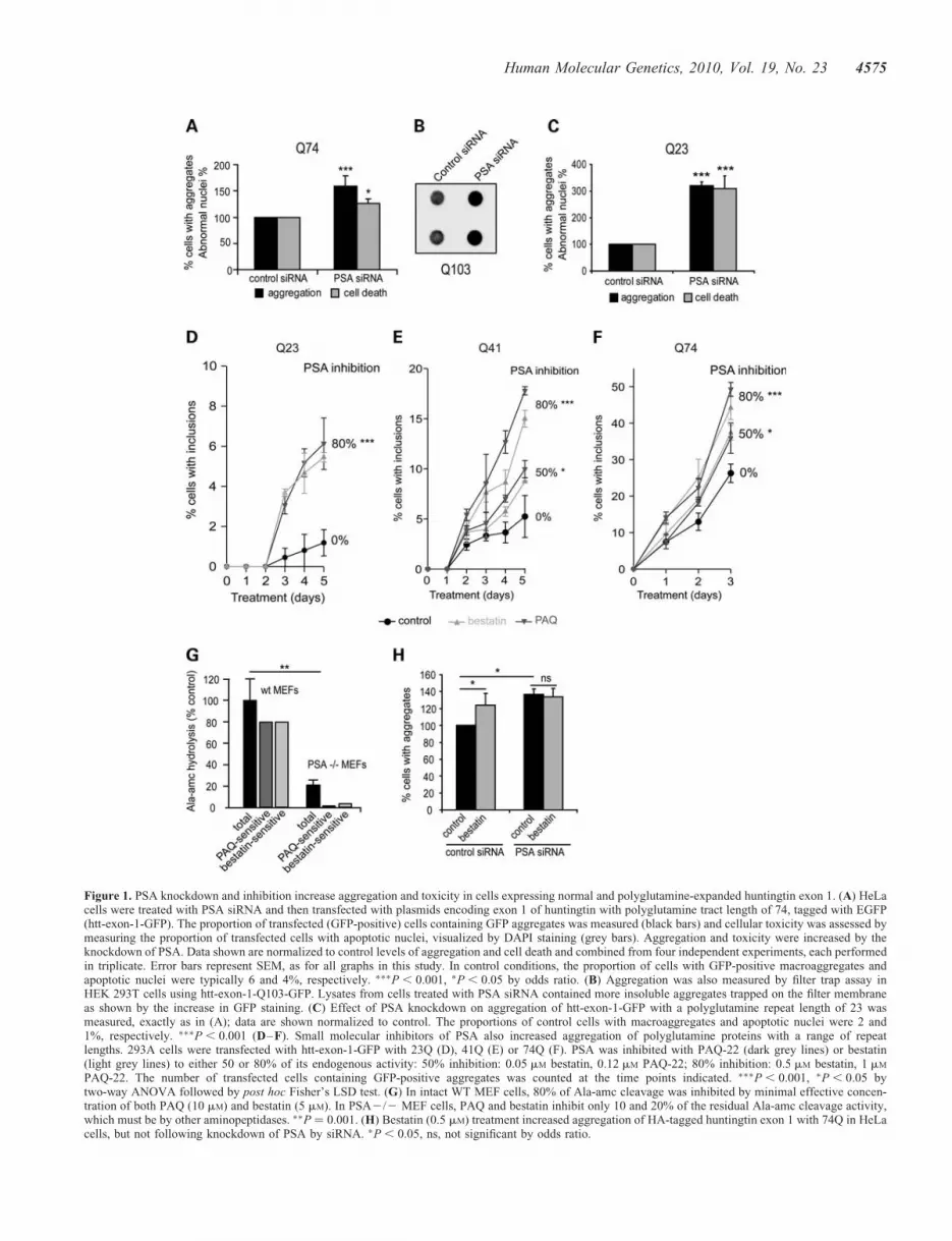
Figure 1. PSA knockdown and inhibition increase aggregation and toxicity in cells expressing normal and polyglutamine-expanded huntingtin exon 1. (A) HeLacells were treated with PSA siRNA and then transfected with plasmids encoding exon 1 of huntingtin with polyglutamine tract length of 74, tagged with EGFP(htt-exon-1-GFP). The proportion of transfected (GFP-positive) cells containing GFP aggregates was measured (black bars) and cellular toxicity was assessed bymeasuring the proportion of transfected cells with apoptotic nuclei, visualized by DAPI staining (grey bars). Aggregation and toxicity were increased by theknockdown of PSA. Data shown are normalized to control levels of aggregation and cell death and combined from four independent experiments, each performedin triplicate. Error bars represent SEM, as for all graphs in this study. In control conditions, the proportion of cells with GFP-positive macroaggregates andapoptotic nuclei were typically 6 and 4%, respectively. ∗∗∗P , 0.001, ∗P , 0.05 by odds ratio. (B) Aggregation was also measured by filter trap assay inHEK 293T cells using htt-exon-1-Q103-GFP. Lysates from cells treated with PSA siRNA contained more insoluble aggregates trapped on the filter membraneas shown by the increase in GFP staining. (C) Effect of PSA knockdown on aggregation of htt-exon-1-GFP with a polyglutamine repeat length of 23 wasmeasured, exactly as in (A); data are shown normalized to control. The proportions of control cells with macroaggregates and apoptotic nuclei were 2 and1%, respectively. ∗∗∗P , 0.001 (D–F). Small molecular inhibitors of PSA also increased aggregation of polyglutamine proteins with a range of repeatlengths. 293A cells were transfected with htt-exon-1-GFP with 23Q (D), 41Q (E) or 74Q (F). PSA was inhibited with PAQ-22 (dark grey lines) or bestatin(light grey lines) to either 50 or 80% of its endogenous activity: 50% inhibition: 0.05 mM bestatin, 0.12 mM PAQ-22; 80% inhibition: 0.5 mM bestatin, 1 mM
PAQ-22. The number of transfected cells containing GFP-positive aggregates was counted at the time points indicated. ∗∗∗P , 0.001, ∗P , 0.05 bytwo-way ANOVA followed by post hoc Fisher’s LSD test. (G) In intact WT MEF cells, 80% of Ala-amc cleavage was inhibited by minimal effective concen-tration of both PAQ (10 mM) and bestatin (5 mM). In PSA2/2 MEF cells, PAQ and bestatin inhibit only 10 and 20% of the residual Ala-amc cleavage activity,which must be by other aminopeptidases. ∗∗P ¼ 0.001. (H) Bestatin (0.5 mM) treatment increased aggregation of HA-tagged huntingtin exon 1 with 74Q in HeLacells, but not following knockdown of PSA by siRNA. ∗P , 0.05, ns, not significant by odds ratio.
Human Molecular Genetics, 2010, Vol. 19, No. 23 4575

both bestatin and PAQ-22 are potent inhibitors of PSAaminopeptidase activity [assayed fluorometrically with thealanine amino methylcoumarin (Ala-amc) substrate] withIC50s of 0.07 and 0.08 mM, respectively (SupplementaryMaterial, Fig. S1C). By measuring the potency of these inhibi-tors of PSA in intact 293A cells (Supplementary Material, Fig.S1D) at concentrations where these drugs do not alter cellularviability for several days (Supplementary Material, Fig. S1E),we were able to assess how the level of PSA activity influ-ences aggregate content in 293A cells expressing huntingtinexon 1 GFP with either 23 [wild-type (WT)], 41 or 74 gluta-mines (Fig. 1D–F and Supplementary Material, Fig. S1F).PSA inhibition increased aggregate content 2–4-fold not onlywith expanded polyQ, where aggregates are more abundant(Q74 and Q41), but also with the WT (23Q) huntingtin exon 1GFP. In fact, the relative stimulation appeared larger with the23Q construct, perhaps because the appearance of inclusions iseasier to detect with this construct, since it fails to aggregate sig-nificantly under normal circumstances. It is noteworthy that largeincreases in aggregate content were seen with only a 50%decrease in enzyme activity using either inhibitor.
We confirmed the relative specificity of these inhibitors bycomparing their activities in mouse embryonic fibroblasts(MEFs) from WT and PSA-null mice. In intact cells, the clea-vage of Ala-amc was five times higher in WT MEFs than inMEFs from PSA knockout mice. In WT MEFs, 80% of theAla-amc cleavage was sensitive to both PAQ (10 mM) and bes-tatin (5 mM), whereas PAQ and bestatin had negligible effectson its hydrolysis in the PSA knockout MEFs (Fig. 1G). Furtherevidence that bestatin increased aggregate number throughPSA inhibition was the finding that, following RNAi knock-down of PSA, this inhibitor had no effect in cells expressinghuntingtin exon 1 Q74 GFP (Fig. 1H). Together, these findingsindicate that PSA is involved in the clearance of bothnormal and polyQ-expanded huntingtin exon 1 and reducingits activity by 50% increases huntingtin accumulation andtoxicity.
PSA overexpression reduces aggregates and toxicityof mutant huntingtin exon 1
Consistent with the deleterious effects of PSA knockdown andinhibition, we found that PSA overexpression (SupplementaryMaterial, Fig. S2A) decreased the frequency of expanded hun-tingtin exon 1 GFP aggregates and the associated cell death inneuroblastoma cells (Fig. 2A). Furthermore, in HeLa cells,PSA overexpression not only reduced the fraction of cellswith inclusions, it also reduced the cells’ content of insolublehuntingtin exon 1 GFP and the sizes of the inclusions(Fig. 2B and Supplementary Material, Fig. S2B and C).Because of the inability of the proteasome to cleave efficientlywithin polyQ sequences, polyQ-rich peptides are likely to begenerated and released during the degradation of polyQ pro-teins (6). In an attempt to mimic these isolated polyQsequences in the absence of a linked protein such as hunting-tin, we expressed a GFP–ubiquitin–polyQ fusion protein (23).In cells, the GFP–ubiquitin and the C-terminal polyQsequence are efficiently cleaved by deubiquitinatingenzymes, releasing pure polyQ polypeptide (Fig. 2C). When293T cells were transfected with GFP–ubiquitin–polyQ
containing 65 or 112 glutamines, with or without PSA–GFP,PSA overexpression reduced the levels of both the solublepolyQ polypeptides and insoluble polyQ aggregates (assayedby filter trap) (Fig. 2D and Supplementary Material,Fig. S2D and E), in accord with our findings on expanded hun-tingtin exon 1 (Fig. 2A and B and Supplementary Material,Fig. S2B and C). However, it was not possible to assesswhether raising PSA levels had similar protective effects incells expressing WT huntingtin exon 1 GFP due to theirvery low frequency of aggregates and cell death.
PSA suppresses accumulation of mutant huntingtinaggregates and toxicity in vivo
To test whether overexpression of PSA causes a similar sup-pression of huntingtin aggregates in mouse tissues in vivo,we electroporated GFP-tagged huntingtin exon 1 constructsbearing different polyQ lengths into the tibialis anteriormuscles of adult mice. One week after electroporation, thehuntingtin Q23 GFP was diffusely distributed in the electropo-rated fibres. However, transfection with expanded huntingtinwith 41 and 74Q caused the formation of GFP-labelledinclusions in all the electroporated fibres (Fig. 3A). When aPSA-encoding plasmid was simultaneously electroporated, itdecreased the number and size of inclusions formed by theexpanded huntingtin exon 1 mCherry (74Q) in the tibialisanterior muscle 1 week after electroporation (Fig. 3B). Onthe other hand, transfecting a PSA RNAi construct enhancedthe formation of mutant huntingtin exon 1 mCherry aggregates(Fig. 3C). Furthermore, when huntingtin exon 1 GFP with 74Qor 41Q was electroporated in heterozygous PSA knockoutmice, more inclusions were evident than in WT mice(Fig. 3D–E). The inclusions formed by huntingtin exon 1(41Q) were larger in muscle of heterozygous PSA knockoutmice (Fig. 3E). Extracts of muscles from the heterozygousmice showed an approximately 50% reduction in PSA activity(data not shown). Thus, a decrease of only 50% in theendogenous PSA activity is sufficient to significantlyenhance mutant huntingtin aggregate formation, as was alsofound upon treatment of 293A cells with PSA inhibitors.
In order to test the effects of PSA on huntingtin toxicity inneurons in vivo, we used two distinct Drosophila models withdifferent quantifiable readouts. In the first, mutant huntingtinexon 1 was expressed in the nervous system (24) andreduced lifespan. In these flies, PSA knockdown (with twodifferent RNAi constructs) further shortened the lifespan,whereas PSA overexpression increased their lifespan(Fig. 4A). In the second model, mutant huntingtin wasexpressed in the photoreceptors. Fly photoreceptors thatexpress a mutant huntingtin fragment with 120 polyglutaminerepeats exhibit degeneration that is not observed in flies thatexpress the WT fragment with 23 polyglutamine repeats(25). The compound eye of Drosophila consists of about800 ommatidia, each composed of eight photoreceptorneurons with light-gathering parts called rhabdomeres, sevenof which can be visualized by light microscopy using the pseu-dopupil technique (26). Neurodegeneration in these HD flies isprogressive and associated with a decrease over time in thenumber of visible rhabdomeres in each ommatidium (25).However, PSA overexpression increased the number of
4576 Human Molecular Genetics, 2010, Vol. 19, No. 23

visible photoreceptors (Fig. 4B and D), indicating suppressionof polyglutamine toxicity. On the other hand, the expression ofeither of two different PSA RNAi constructs enhanced toxicity(Fig. 4C and E). Thus, as in cell culture, modulation of PSAlevels alters polyQ toxicity in vivo.
PSA decreases aggregate number in cellular modelsof other neurodegenerative diseases
To investigate whether PSA can also promote the clearance ofother aggregation-prone mutant proteins, we tested its effectson different neurodegenerative disease-associated proteinsincluding ataxin-3, another polyglutamine-containing protein,whose expansion causes spinocerebellar ataxia type 3. In293A cells expressing WT (28Q) and mutant (84Q) full-lengthataxin-3 GFP, inhibition of PSA caused the accumulation ofmore aggregates than in untreated control cells (Fig. 5A andB and Supplementary Material, Fig. S4A). We also studied
aggregate-prone proteins that lack polyglutamine repeatregions. In cells expressing a mutant form of SOD1, whichcauses an inherited form of amyotrophic lateral sclerosis, thenumber of SOD1 GFP inclusions was increased when PSAwas inhibited (Fig. 5C, D and Supplementary Material, Fig.S4B). Furthermore, using PC12 cells, we were able to investi-gate the clearance of A53T mutant a-synuclein, which causesa familial form of Parkinson’s disease. Its levels can be accu-rately measured by western blotting of the soluble fractionbecause this protein does not aggregate in these cells. Tofollow its degradation, a-synuclein expression was inducedby the addition of doxycycline to the media, and then switchedoff by removal of the antibiotic. The degradation of A53Ta-synuclein was slowed (i.e. its level was higher 16 h aftersynthesis was terminated) in the cells treated with the PSAinhibitors (Fig. 5E and F). Thus, this enzyme promotes theclearance and thus reduces aggregate content in a widevariety of cellular models of neurodegenerative disease.
Figure 2. PSA overexpression reduces toxicity and aggregate content in cells expressing expanded polyQ huntingtin exon 1 and expanded polyglutamine tracts.(A) Effect of PSA overexpression on aggregate content in SK-N-SH cells expressing htt-exon-1-Q74-GFP. The proportions of cells with GFP-positive macro-aggregates and apoptotic nuclei were decreased by the overexpression of PSA, when assayed as in Figure 1A and C. Data shown are normalized to control foreach experiment and the average of data from four independent experiments carried out in triplicate are shown. In control conditions, typically around 30% oftransfected cells contained macroaggregates and 10% had apoptotic nuclei. ∗∗∗P , 0.001 by odds ratio. (B) Overexpression of PSA in 293T cells also decreasedlevels of insoluble aggregates of htt-exon-1-Q103-GFP, as measured by filter-trap assay. Q103 aggregates were visualized using anti-GFP antibody. (C) Tomimic the polyQ sequences that may be released by the proteasome, we used a GFP–ubiquitin–polyQ construct (23). Upon expression, the GFP–UB andthe polyQ peptide are efficiently cleaved by deubiquitinating enzyme(s), leading to the release of a polyQ polypeptide. (D) Levels of soluble and insoluble poly-glutamine peptides were also decreased by overexpression of PSA in HEK293T cells transfected with GFP–UB–polyQ containing 65 or 112 glutamines. Levelsof both soluble polyQ peptides (upper panel, arrows mark monomeric polyQ peptides and arrowheads mark high molecular weight oligomeric structures) andinsoluble aggregates (lower panel) detected by an anti-polyglutamine antibody decreased, when PSA-GFP was co-transfected. Actin levels are shown as aloading control.
Human Molecular Genetics, 2010, Vol. 19, No. 23 4577

Figure 3. Effect of PSA overexpression and knockdown on the formation of polyglutamine aggregates in mouse muscle. (A) Cross-sections of mouse tibialisanterior muscle electroporated with GFP or huntingtin exon 1 with 23, 41, 74Q fused to GFP (Htt23Q, Htt41Q, Ht74Q). One week after electroporation, theformation of inclusions was observed in all fibres electroporated with Htt41Q and Htt74Q. Fibres electroporated with GFP or Htt23Q showed a diffuse fluor-escence and no inclusions. Scale bar represents 100 mM. (B) PSA overexpression decreased the number and size of polyglutamine inclusions found in tibialisanterior muscle 1 week after electroporation with expanded huntingtin exon 1 (74Q) and PSA. Note that Htt74Q is seen as red (fused to mCherry) in theseimages. Graph shows inclusion number and size in fibres where PSA (blue bars) or GFP (green bars) is overexpressed. (C–E) Reduction in the expressionof PSA by electroporation of RNAi increased the number and size of polyglutamine inclusions in mouse skeletal muscle electroporated with expanded huntingtinexon 1. (C) Tibialis anterior muscle 1 week after electroporation with Htt74Q and a plasmid expressing either a scrambled RNAi, or an RNAi targeting PSA.Reduction of PSA expression by RNAi increased the number and size of polyglutamine inclusions formed by expanded huntingtin exon 1 (74Q). (D) Electro-poration of expanded huntingtin exon 1 (74Q) to tibialis anterior muscles of PSA+/2 heterozygous mice led to more inclusions than in muscle from WT(PSA+/+) mice, 1 week after electroporation. These inclusions also seemed larger in PSA+/2 mice, but this effect did not reach statistical significance(P ¼ 0.09). (E) Electroporation of expanded huntingtin exon 1 (41Q) in PSA+/2 heterozygous mice led the formation of more, larger inclusions than inmuscle from WT (PSA+/+) mice 1 week after electroporation. Scale bar (B–E) represents 50 mM. ∗P , 0.05 by t-test.
4578 Human Molecular Genetics, 2010, Vol. 19, No. 23

PSA enhances autophagy
Many of the proteins found here to accumulate on PSA inhi-bition or to be cleared faster upon PSA overexpression areautophagy substrates, including mutant huntingtin exon 1, full-length ataxin-3, mutant SOD1 and A53T a-synuclein (27,28).In fact, a large fraction of long-lived cellular proteins, the bulkof cell constituents, are degraded by this process (29). We
therefore tested whether changes in PSA activity influencethe overall rate of degradation of long-lived proteins.In order to determine the rates of proteolysis by autophagy,lysosomes and proteasomes, we measured protein degradationrates in the presence or absence of specific inhibitors of theproteasome (bortezomib/velcade), lysosomal acidification(chloroquine) or autophagy [3-methyladenine (3MA)], as wepreviously described (29). Upon deprivation for serum and
Figure 4. Effect of PSA overexpression and knockdown in Drosophila. (A) PSA expression levels alter lifespan in flies expressing htt-exon1 with 93 polyglu-tamine repeats in the brain. Knockdown of PSA expression levels by expression of two different PSA RNAi constructs enhanced toxicity (elav-Gal4 Q93PSA-RNAi1 and elav-Gal4 Q93 PSA-RNAi2), compared with flies expressing mutant huntingtin alone (elav-Gal4 Q93). Overexpression of PSA (elav-Gal4Q93 PSA) extended lifespan relative to flies expressing Q93 alone. Graph shows Kaplan–Meier survival curves, P , 0.0001 in both cases. Expression ofPSA RNAi or PSA in flies expressing WT huntingtin (elav-Gal4 Q20) did not affect lifespan (Supplementary Material, Fig. S3). (B–E) Degeneration wasalso decreased by overexpression of PSA. Overexpression of PSA in the WT fly eye using the glass promoter (gl-PSA) caused a slight decrease in rhabdomerenumber compared with flies expressing Q23 alone. However, when co-expressed with GMR-Q120, PSA protected against the neurodegeneration seen in theseflies (B). (C) Expression of PSA RNAi enhanced neurodegeneration in flies expressing GMR-Q120, using two RNAi lines (elav-Gal4 GMR-Q120 PSA-RNAi1and elav-Gal4 GMR-Q120 PSA-RNAi2). Expression of PSA RNAi in flies expressing Q23 did not affect rhabdomere number (Supplementary Material, Fig. S3),∗P , 0.02, 2 tailed t-test. (D) Examples of rhabdomeres in flies expressing GMR-Q120 with or without PSA (gl-PSA). (E) Examples of rhabdomeres in fliesexpressing mutant huntingtin GMR-Q120 when PSA is downregulated using two RNAi lines (PSA-RNAi1 and PSA-RNAi2).
Human Molecular Genetics, 2010, Vol. 19, No. 23 4579

amino acids to activate autophagy, PSA overexpressionincreased the total rate of protein degradation by enhancingthe rate of lysosomal and autophagy-dependent proteolysisbut did not affect the proteasomal process (Fig. 6A).However, PSA had no measurable effect on any of these pro-cesses in complete medium (Supplementary Material, Fig.S5A–C) where autophagy makes only a minor contributionto the degradation of long-lived proteins (SupplementaryMaterial, Fig. S5D).
On the other hand, knockdown of PSA expression usingRNAi (Fig. 6B) or treatment with inhibitors of its activity(Fig. 6C) decreased the total and lysosomal proteolysismeasured after nutrient deprivation, but not proteasomaldegradation. Again, no significant effect of PSA was seen incomplete medium (Supplementary Material, Fig. S5B andC). Thus, the activity of this cytosolic enzyme, PSA, appearsto regulate the capacity of the autophagic–lysosomalpathway to degrade normal cell proteins.
In order to test whether this alteration of autophagicactivity by PSA was a major contributor to the effects onprotein clearance seen following PSA knockdown or overex-pression, we used 3MA to inhibit autophagy (30). Thedecrease in aggregate content upon PSA overexpression(Fig. 6D) and the increase following by PSA inhibition(Fig. 6E) were both markedly attenuated by 3MA. Thesefindings further suggest that most of PSA’s ability to
protect against expanded huntingtin exon 1 inclusions wasvia its ability to promote autophagy.
In order to directly assess the effects of PSA on the autop-hagy pathway, we measured the steady-state levels of autopha-gosomes using the microtubule-associated protein 1 lightchain 3 (LC3). All of these experiments were performed incomplete medium, to assess whether PSA could modulatebasal autophagy levels. During autophagic vacuole formation,LC3 is modified post-translationally to form LC3-I, and thenconverted to LC3-II, which associates with autophagosomemembranes. LC3-II levels, which thus reflect autophagosomenumber when assayed relative to the loading control, actin,were decreased by PSA inhibitors (Fig. 6F and G) and alsoby transfection with PSA RNAi (Fig. 6H and I). Sincedecreased LC3-II content could result from either impairedsynthesis or enhanced degradation after lysosomal fusion,we assessed LC3-II synthesis by blocking its degradationwith the lysosomal proton pump inhibitor Bafilomycin A1.As expected, this agent increased LC3-II levels in all con-ditions (Fig. 6H and I), but after Bafilomycin A1 treatment,the levels of LC3-II were lower in cells expressing PSAsiRNA (Fig. 6H and I) than in control cells (Fig. 6J and K).Conversely, in PSA-overexpressing cells, LC3-II levels wereincreased over control levels. These findings imply that PSApromotes autophagy and clearance of the misfolded proteinsby enhancing autophagosome formation.
Figure 5. PSA inhibition enhances aggregate formation in cells expressing other aggregate-prone proteins (ataxin-3, mutant SOD1, mutant a-synuclein). (A andB) PSA inhibition increased aggregate formation in 293A cells expressing full-length ataxin-3 GFP with 28Q (A) and 84Q (B). The proportion of transfectedcells with macroaggregates was also increased by inhibition of PSA in cells transfected with mutant SOD1 GFP G37R (C) or G93A (D). ∗∗∗P , 0.001, ∗∗P ,
0.01, ∗P , 0.05 by t-test. (E and F) Clearance of mutant a-synuclein (A53T) was slowed by inhibition of PSA. Stable inducible PC12 cells were treated withdoxycycline to induce expression of mutant a-synuclein, and subsequently expression was turned off by removal of doxycycline from the medium. When PSAinhibitors were added after transgene expression was switched off, more a-synuclein remained present, indicating a decreased clearance. Levels of a-synucleinremaining after 16 h were assayed by western blot, a representative image is shown in (E). Densitometric quantification of this triplicate experiment is shown in(F). ∗∗P , 0.01, ∗P , 0.05, by ANOVA.
4580 Human Molecular Genetics, 2010, Vol. 19, No. 23

Figure 6. PSA activity enhances protein degradation by the autophagic/lysosomal pathway. (A–C) Effect of PSA modulation on different proteolytic pathways.Degradation of long-lived protein was measured in 293A cells. Proteolysis rates were measured in the presence of inhibitors of proteasomes, lysosomes or autop-hagy to determine flux through each of these pathways in cells starved of serum and amino acids. Proteasomes, lysosomes and autophagy inhibitors were velcade/bortezomib 1 mM, chloroquine 50 mM and 3-MA 10 mM, respectively. (A) PSA overexpression increased total, lysosomal and autophagy-dependent proteolysis.(B) PSA knockdown by RNAi decreased total and lysosomal proteolysis. (C) PSA inhibitors reduced total and lysosomal proteolysis. ∗∗∗P , 0.0001. (D) Inhibit-ing autophagy in SK-N-SH cells expressing Q74 huntingtin exon-1 GFP using 3-MA, reduced the protective effect of PSA overexpression on aggregate content.Co-expression of PSA with htt-exon-1-GFP-74Q reduced the percentage of transfected cells with aggregates (grey bars). However, in the presence of 3-MA,added immediately after transfection, the reduction in aggregation caused by PSA expression was decreased (black bars). Control values with and without3-MA are set to 100% in each case to allow ease of comparison, although Q74 aggregation was higher in the presence of 3-MA. Actual mean values were24% of cells having aggregates in control and 30% in 3-MA treated cells. (E) Inhibitors of PSA caused an increase in the proportion of cells expressinghtt-exon-1-GFP-Q74 with aggregates (grey bars), but if 3-MA was added at the same time as these inhibitors they did not increase the aggregate content(black bars). Actual mean values for aggregation were 19% in control cells and 32% in 3-MA treated cells. ∗∗P , 0.01, ∗∗∗P , 0.001, ns, not significant,by odds ratio, relative to control. (F–K) Effect of PSA modulation on LC3-II levels. (F) Treatment of PC12 cells with PSA inhibitors also resulted in a decreasein LC3-II levels, a marker for autophagosomes. The western blot shown is a representative data set, with actin shown as a loading control. Quantification of dataobtained in triplicate samples is shown in (G). (H and I) In HeLa cells treated with PSA siRNA, LC3-II levels were decreased, whereas cells overexpressing PSAshowed increased levels of LC3-II (J and K). (I) and (K) show densitometric quantification of data from four experiments carried out in triplicate. LC3-II levelchanges were seen in the presence (grey bars) and absence (black bars) of bafilomycin A1, a lysosomal proton pump inhibitor. LC3-II levels are shown relative toactin, as a loading control in each case. Values are normalized to control, in the presence or absence of bafilomycin, to allow for comparison between differentgels, although the levels of LC3-II were higher with bafilomycin than without. ∗P , 0.05, ∗∗P , 0.01.
Human Molecular Genetics, 2010, Vol. 19, No. 23 4581

DISCUSSION
The protection conferred by PSA against polyQ and otheraggregation-prone proteins was observed here in a very widerange of types of cells in culture, as well as in Drosophilaand mouse models of neurodegenerative diseases. In allcases examined, cellular protection against these toxic proteinscoincided with decreased content of large inclusions contain-ing the mutant polypeptide. However, this correlation doesnot imply that the large aggregates are in fact the toxicspecies, since aggregate formation and cytotoxicity probablyboth depend on levels of misfolded proteins or microaggre-gated species whose clearance is accelerated by PSA. Inaddition to using various standard approaches to evaluateaggregate formation and cell protection, we have introduceda novel in vivo method—electroporation of a mutant geneinto muscle fibre in adult mouse—that offers several majoradvantages for such studies (e.g. rapid development ofinclusions, ability to use host animals of different genotypeswithout lengthy mating, comparisons with control cells inthe same animals). Moreover, with this approach, increasingthe length of the polyQ tract of huntingtin exon 1 yieldedmore inclusions, as observed in cell culture. In mousemuscle fibres, PSA knockdown increased while PSA overex-pression reduced the content of inclusions formed byexpanded huntingtin exon 1, as we found in cell culture. Sur-prisingly, in contrast to these marked results (obtained in threelabs), an earlier study had concluded that PSA does not influ-ence toxicity of mutant huntingtin in Drosophila (16), but thatstudy assessed polyglutamine toxicity by inspection of exter-nal eye morphology, a rather insensitive qualitative approach,in contrast to the quantitative analysis of the number of rhab-domeres used here (Fig. 4).
The activation of autophagy by PSA
The ability of PSA to decrease aggregate content was notrestricted to cells expressing expanded huntingtin exon 1,but was also observed with unattached long polyQ peptides,full-length ataxin-3, mutant SOD1 and mutant a-synuclein.This capacity of PSA to reduce the toxicity and accumulationof aggregates containing expanded huntingtin exon 1 and theseother disease-associated proteins can be largely attributed to asurprising new function of PSA: its ability to promote proteindegradation by autophagy. The enhancement of autophagy byPSA constitutes a novel mechanism that seems to account forthe protective effects of PSA against this wide range ofaggregation-prone proteins. Since LC3-II levels and theamount of protein degradation that is sensitive to 3MA orchloroquine were increased by PSA overexpression andreduced by PSA knockdown or inhibition, this enzymesomehow must promote autophagosome formation. PSA waspreviously shown to decrease the toxicity in Drosophila ofWT and mutant form of tau, which is associated with fronto-temporal dementia (16). These effects were attributed to directdegradation of tau by the aminopeptidase activity of PSA (31);however, such an exoproteolytic mechanism would be quiteslow and inefficient for digesting a protein as long as tauand would generate heterogenous variants of tau of diverselengths. Since tau has been demonstrated to be an autophagy
substrate (27), it seems more likely that the enhancement ofautophagy by PSA might better explain the stimulation ofdegradation of tau and the reduction in its toxicity uponPSA overexpression. Interestingly, the two different PSA-deficient mouse lines are both characterized by abnormalmotor behaviour (17,21), which might also be a consequenceof reduced autophagy, since the blockage of this process inAtg7-deficient mice leads to major behavioural deficits andwidespread inclusion bodies (32). On the other hand, upregu-lation of autophagy via either mTOR-dependent or mTOR-independent pathways protects cells from severalpro-apoptotic insults (33,34). Therefore, it seems likely thatPSA protects primarily by enhancing the removal of thetoxic proteins, although it possibly may also have additionalantiapoptotic effects (e.g. by eliminating toxic peptidesreleased by proteasomes as discussed below).
It is indeed unexpected that the activity of a cytosolic ami-nopeptidase can regulate autophagic vacuole formation. PSAinhibition was achieved using two unrelated small molecularweight compounds: bestatin, a natural, slow binding, competi-tive inhibitor of many aminopeptidases (35), and PAQ-22, asynthetic, non-competitive inhibitor that does not act as asubstrate-mimic and binds to PSA at a distinct site (36–38).Both compounds increased the formation of aggregates andthe toxicity of expanded huntingtin exon 1 at concentrationsthat caused only a modest (50%) inhibition of its activity.Thus, even minor variations in PSA activity, e.g. in humanpolymorphisms, if they exist, have the potential of influencingsusceptibility to neurodegenerative disease.
PSA seems to be the major cytosolic aminopeptidase in cul-tured cells as well as in brain, muscle and kidney (39). Itcleaves rapidly most N-terminal amino acids (especiallyalanine) from peptides, and PSA accounts for approximately80% of the total soluble aminopeptidase activity in human cer-ebral cortex (40). Aminopeptidases such as PSA catalyse thefinal stages of the degradation of intracellular normal andabnormal proteins releasing amino acids from peptides gener-ated by the catabolism of proteins by the proteasome(13,15,41). Thus, PSA potentially might link late steps inprotein degradation by the ubiquitin proteasome pathway tofunctioning of the autophagic pathway. Certainly, regulationof any cellular process by the activity of PSA or any amino-peptidase represents an unusual mode of enzyme regulationsince amino acid removal would cause irreversible changesin a protein. Alternatively, perhaps it may act to destroy aregulatory peptide that somehow inhibits autophagy. It is note-worthy that certain amino acids (especially leucine) activatemTORC1 and thus inhibit macroautophagy (42). However,the digestion by PSA of proteasome products would increasethe pool of free amino acids and thus should reduce autop-hagy, while the exact opposite was observed. It remains poss-ible that PSA could be involved in earlier stages in thecatabolism of some proteins; in fact, there is a general slow-down in the degradation of proteins in bacteria lackingmultiple aminopeptidases (41), although such effects couldbe indirect. For example, the in vivo half-life of a protein isdetermined in part by the nature of its N-terminalamino acid (termed the ‘N-end rule’). Thus, the removal ofthe N-terminal amino acid from certain proteins by PSAcould expose destabilizing residues leading to their rapid
4582 Human Molecular Genetics, 2010, Vol. 19, No. 23

degradation by the ubiquitin–proteasome pathway. Conceiva-bly, PSA might in this way alter the levels (or activity) of aprotein that is critical in the regulation of autophagy.However, in most globular proteins, the N-terminal aminoacid is buried in the interior, so PSA would be expected toact only on proteins with ‘loose ends’ and generally aminopep-tidases preferentially attack shorter peptides.
A major challenge for future work will be to establish themechanism by which PSA promotes autophagy. The bestcharacterized physiological inhibitor of autophagy is mTOR.However, we found no evidence for PSA inhibiting theactivity of mTOR (Supplementary Material, Fig. S5E andF), and it seems quite unlikely that PSA promotes autophagysimply by causing nutrient deprivation (since its overexpres-sion does not impede growth). In fact, PSA had clear effectson autophagic protein degradation in cells deprived of serumand amino acids, where mTOR is markedly inhibited. Autop-hagy is regulated by other mechanisms such as the phos-phorylation of Bcl-2, which modulates autophagy via itsinteraction with Beclin-1 (43). However, we did not observeany effect of PSA on the levels of Bcl-2 phosphorylation (Sup-plementary Material, Fig. S5E and F). Thus, we believe thatdegradation or destabilization of an unknown inhibitory com-ponent of autophagy remains the most likely explanation ofthe activation of autophagy by PSA.
PSA can also act after the proteasome independentlyof autophagy
While these studies focused on mutant huntingtin exon 1 andother aggregation-prone proteins whose clearance seems to bedue to autophagy, it is noteworthy that PSA was also found toprevent the formation of aggregates in cells expressing the WTexon 1 protein and also with the WT full-length ataxin-3. Inother words, as suggested previously (15), this enzymeappears to be an important defence against aggregate for-mation due to non-expanded polyQ sequences as are foundin various normal proteins. Our previous data also indicatethat the clearance of WT huntingtin exon 1 does not dependon autophagy and is mainly via the proteasome (30,44). Theproteasome would be predicted to continually produce shortpolyglutamine tracts from this and other polyQ proteins,since it cannot cleave between successive glutamine residuesin the polyQ stretch (6). Such isolated polyglutamine tracts(even ones as short as 10Qs) are highly aggregate-prone ifnot cleared from the cell. Because PSA is the only non-lysosomal enzyme in tissue extracts that can degrade suchpolyglutamine products (15), polyglutamine stretches shouldaccumulate and aggregate in cells when PSA is inhibited, aswas found here. In cells, the isolated polyglutamine tractsnot only aggregate but also sequester GFP or other tagged hun-tingtin constructs with flanking sequences (23,45). BecausePSA is an aminopeptidase that digests soluble polypeptidesone residue at a time and dissociates after each cleavage, itshould degrade these aggregation-prone polyglutaminestretches only slowly. Thus, reducing its activity will haveimportant consequences on accumulation of shorter polygluta-mine stretches, whereas the more aggregate-prone mutant pro-teins (e.g. expanded polyQ proteins) tend to aggregate rapidlydue to their inherent tendency to misfold, and their clearance
by PSA appears to be more dependent on autophagy. In prin-ciple, the aggregates seen with mutant huntingtin could arisefrom seeds that are either the whole exon 1 fragment or theproposed post-proteasome-isolated polyQ tract. Isolatedexpanded polyQ tracts will seed and sequester GFP-taggedexon 1 of mutant huntingtin. Since PSA regulates overallaggregation mediated from exon 1 of huntingtin in anautophagy-dependent manner, any contribution towards aggre-gation of PSA acting as an aminopeptidase on isolatedexpanded polyQ tracts derived from larger huntingtin frag-ments will be very minor. Indeed, once huntingtin is engulfedin an autophagosome, it will be inaccessible to the ubiquitin–proteasome machinery and to PSA. Thus, our data show thatthe predominant effect of PSA in these models of HD is viaautophagy.
Presumably, the physiological levels of PSA have evolvedso as to ensure the rapid elimination of peptides from thenucleus and cytosol, but under the present experimental con-ditions when an aggregation-prone protein is overexpressed,PSA is not present in large excess over what is necessary toclear the misfolded, toxic proteins or even the products ofWT huntingtin exon 1. Indeed, only a 50% reduction in PSAactivity using inhibitors in cell culture or the 50% reductionin expression in PSA in tissues of heterozygous PSA+/2mice led to greater content of aggregates formed by expandedhuntingtin exon 1. These observations and the protection byoverexpression argue that activation of autophagy by PSA isa tightly regulated process, and that only a modest increasein its activity might have beneficial effects. Increasing PSAactivity thus represents a novel potential approach fortherapy of neurodegenerative diseases. It is noteworthy thattreatment of cells with PSA inhibitors for several days wasremarkably non-toxic. In contrast, proteasome or lysosomalinhibitors are highly toxic when applied for more than 6–12 h. While a low toxicity would thus be expected foragents that would moderately increase the activity orexpression of PSA, a high level of PSA overexpression byitself was toxic in Drosophila (data not shown). It is also note-worthy that PSA expression is elevated in PC12 cells expres-sing polyQ expanded exon 1 (22) and also in some neuronsexpressing mutant tau (16). Thus, induction of PSA maywell represent an important cellular adaptation that reducesthe accumulation of toxic proteins and functions in hostdefences against neurodegenerative disease (15).
MATERIALS AND METHODS
Details of plasmids used as well as cell culture methodologyand protocols for aggregate analysis, western blotting andfilter trap assays can be found in Supplementary information.
Inhibitors
PAQ-22 was from Wako, puromycin aminonucleoside wasfrom Biomol, PS-341 (Bortezomid or Velcade) was a giftfrom Millenium. Bestatin, puromycin dihydrochloride, chloro-quine, 3MA, NH4Cl and staurosporin were from Sigma. Freshstock solutions of chloroquine (50 mM) were prepared justbefore experiments (final concentration 50 mM). Fresh 3MA
Human Molecular Genetics, 2010, Vol. 19, No. 23 4583
solutions (100 mM) were prepared in boiled PBS just beforeexperiments (final concentration 10 mM).
Muscle electroporation
Adult CD1 male mice (25–35 g) were anaesthetized by intra-peritoneal injection of Avertin (0.2 ml/10 g as 1.2% solution).Fur overlying the hindlimb was removed, and the surgical sitewas cleansed with betadine swab followed by isopropylalcohol wipe. Using sterile technique, a 0.5–1.0 cm longitudi-nal incision was made in the skin overlying the tibialis anteriormuscle, and the underlying muscle was exposed by blunt dis-section of the surrounding skin. A paddle electrode was thenintroduced between the muscle and the underlying tibia.Plasmid DNA in sterile saline (40 ml, 25 mg) was injectedinto the muscle. A second paddle electrode was appliedgently over the muscle and electroporation performed (12 V,20 ms duration, 5 pulses, 200 ms intervals) using anECM830 Electro Square Porator (BTX, Harvard Apparatus).The electrodes were removed and the skin was closed bysuture. Post-operative analgesia was administered every 12 hfor the first 24 h with buprenorphine 0.05 mg/kg SQ.Additional analgesia was administered thereafter if there wasany evidence of continued animal discomfort. The animalswere followed for 1 week, after which time they are eutha-nized by Halothane inhalation followed by cervical dislo-cation, and the muscles harvested, quickly frozen inliquid-nitrogen-cooled isopentane and stored at 2808C. Tenmicrometer muscle cryosections were analyzed for GFP/mCherry fluorescence after fixation with 4% paraformalde-hyde and Hoechst staining (10 mg/ml in PBS for 10 min)using a Nikon 80i upright microscope in the Nikon ImagingCenter at Harvard Medical School. No gross evidence ofnecrosis or inflammation as a result of the transfection pro-cedure was noted. Inclusion size and number were analysedwith Metamorph using fixed size and intensity thresholds.
Drosophila
Fly crosses and experiments were performed at 258C. Allcrosses for individual experiments were performed at thesame time and under the same conditions.
For survival assays, virgins carrying eitherP{UAS-dPsa}8.10 [PSA (47)] or UAS-RNAi constructs thatknockdown Psa [PSA-IR-R2 (referred to as PSA-RNAi1)and PSA-IR-R3 (referred to as PSA-RNAi2), National Insti-tute of Genetics Fly Stock Center, Japan] were crossed withmales expressing w; P{UAS-Q93httexon1}4F1 [Q93 (24)] andw; P{UAS-Q20httexon1} [Q20 (24)] in the nervous system[using P{GawB}elavC155, elav-Gal4 (46)]. Control flies wereprogeny of flies expressing Q93 or Q20 under control ofelav-GAL4, crossed to w1118 males that segregated fromsome stocks of other UAS-RNAi lines and were thereforerepresentative of the genetic background of these lines. Theanalysis was based on 100 female flies per genotype dividedin groups of 10 flies per vial. Flies were transferred to newvials and counted every 2 days. Survival curves were plottedusing the Kaplan–Meier estimator. The statistical significancewas calculated using the log-rank test (SPSS 11.0).
Pseudopupil analysis was performed at 4 days post-eclosionas previously described (26). To evaluate the effect of PSAoverexpression, virgins of genotype y w; P{GMR-HD.Q120}2.4 (GMR-Q120) or y w; P{GMR-HD.Q23} [GMR-Q23 (25)] were crossed with males y w; P{gl-dPSA}A[gl-PSA (16)]. As controls, the above flies (GMR-Q120,GMR-Q23 and gl-PSA) were crossed with w1118 isogenicmales (48). To evaluate the effect of PSA downregulationon neurodegeneration, virgins of genotype elav-GAL4C155;{GMR-HD.Q120}4.62/TM3 (elav-Gal4 GMR-Q120) or elav-GAL4C155; {GMR-HD.Q23} (elav-Gal4 GMR-Q23) werecrossed with the same UAS-RNAi or w1118 control males asdescribed for the survival assay, and the progeny werescored using the pseudopupil assay. Pictures were acquiredusing an ×100 objective (Zeiss Axioscope2 microscope).Comparisons were performed using paired t-tests using datafrom five to seven independent experiments, each based onapproximately 10 individuals of each genotype, in which 15ommatidia each were scored.
Protein degradation measurement
293A cells were seeded in six-well plates and transfected 24 hlater (confluence 50%). Forty-eight hours after transfection,fresh culture medium containing 5 mCi/ml L-[3,5-3H]-tyrosine(Perkin Elmer) was applied for 24 h to label long-lived pro-teins. Cells were then washed twice for an hour with a chasemedium containing 2 mM tyrosine (in order to limit reincor-poration of the 3H-tyrosine and to allow degradation of short-lived proteins, total chase duration 2 h). Cells were thenwashed with either culture medium (basal condition) orHBSS (serum and amino acid starvation) containing 2 mmtyrosine and the PSA, proteasome, lysosomes or autophagyinhibitors for 1 h (pre-treatment before protein degradationmeasurement). Cells were then washed with the samemedium and 200 ml of the medium was collected after 0, 1,2, 3 and 4 h for quantitation of 3H-tyrosine release (proteindegradation measurement). Proteins were precipitated withTCA (10% final concentration) and pelleted. Radioactivity inthe TCA-soluble supernatant was measured using a 1900TRliquid scintillation analyser (Packard). At the end of themeasurement period, cells were solubilized in 0.2 N NaOHand an aliquot was taken to measure the residual radioactivityin the cells. Total radioactivity is the sum of the residual radio-activity in the cells and the TCA-soluble radioactivities atdifferent time points. Protein breakdown rates were expressedas 3H-tyrosine released over time as a percentage of total3H-tyrosine incorporated. Proteasomal, lysosomal andautophagy-dependent proteolysis rates were determined pre-cisely as done before by treating cells with 1 mM bortezo-mib/velcade, 50 mM chloroquine or 10 mM 3MA, respectively.
PSA activity measurement
To prepare cytosolic extracts, 293A or MEF cells were washedwith ice-cold PBS twice and scraped in ice-cold lysis buffercontaining 50 mM phosphate buffer (pH 7.4), 0.5 mM CaCl2,5 mM MgCl2, 1 mM DTT and 5% glycerol, collected in aneppendorf and homogenized manually on ice. This homogen-ate was spun at 10 000g for 15 min to remove the nuclear frac-
4584 Human Molecular Genetics, 2010, Vol. 19, No. 23

tion and then at a higher speed of 100 000g for an hour toremove membranous organelles. Protein concentration wasobtained using Coomassie Plus Protein Assay (ThermoFisher).PSA activity was determined by monitoring for 5–20 min theincrease in fluorescence (excitation 380 nm, emission 460 nm,SpectraMax M5, Molecular Devices) caused by hydrolysis ofthe substrate Ala-amc (Bachem, 100 mM final) in 100 ml oflysis buffer containing 2–5 mg of protein from the cellextract. Background from the fluorescence of Ala-amc in theabsence of enzyme was subtracted.
To measure Ala-amc hydrolysis by intact cells, 293A cellswere plated in 96-well plate adapted for fluorescence measure-ment, grown in culture medium without phenol red containingvarious concentrations of inhibitors for 24 h. Cells werewashed with fresh culture medium containing Ala-amc100 mM and inhibitors and the increase in fluorescence wasmonitored for 1 h. Background from the fluorescence ofmedium containing Ala-amc in the absence of cells was sub-tracted.
SUPPLEMENTARY MATERIAL
Supplementary Material is available at HMG online.
Conflict of Interest statement. None declared.
FUNDING
This work was supported by the Medical Research Council(Programme grant to D.C.R.), and the Wellcome Trust(Senior Fellowship to D.C.R.); A.L.G.’s laboratory is sup-ported by grants from the National Institutes of Health/National Institute for Aging (AR055255 and GM51923),Harvard Neurodiscovery Center and the Ellison Foundation;R.H. is supported by Horlait-Dapsens Medical Foundation,Belgian Neurological Society, Hereditary Disease Foundationand Fonds National de la Recherche Scientifique; E.R.’s lab-oratory is supported by a Vidi grant from De NederlandseOrganisatie voor Wetenschappelijk Onderzoek—ZonMWand a grant from the Dutch Cancer Foundation. We wouldlike to thank George Jackson for sending flies. Funding topay the Open Access Charge was provided by The WellcomeTrust.
REFERENCES
1. Williams, A., Jahreiss, L., Sarkar, S., Saiki, S., Menzies, F.M.,Ravikumar, B. and Rubinsztein, D.C. (2006) Aggregate-prone proteins arecleared from the cytosol by autophagy: therapeutic implications. Curr.Top Dev. Biol., 76, 89–101.
2. Ravikumar, B., Vacher, C., Berger, Z., Davies, J.E., Luo, S., Oroz, L.G.,Scaravilli, F., Easton, D.F., Duden, R., O’Kane, C.J. et al. (2004)Inhibition of mTOR induces autophagy and reduces toxicity ofpolyglutamine expansions in fly and mouse models of Huntington disease.Nat. Genet., 36, 585–595.
3. Verhoef, L.G., Lindsten, K., Masucci, M.G. and Dantuma, N.P. (2002)Aggregate formation inhibits proteasomal degradation of polyglutamineproteins. Hum. Mol. Genet., 11, 2689–2700.
4. Qin, Z.H., Wang, Y., Kegel, K.B., Kazantsev, A., Apostol, B.L.,Thompson, L.M., Yoder, J., Aronin, N. and DiFiglia, M. (2003)Autophagy regulates the processing of amino terminal huntingtinfragments. Hum. Mol. Genet., 12, 3231–3244.
5. Shibata, M., Lu, T., Furuya, T., Degterev, A., Mizushima, N., Yoshimori,T., MacDonald, M., Yankner, B. and Yuan, J. (2006) Regulation ofintracellular accumulation of mutant Huntingtin by Beclin 1. J. Biol.
Chem., 281, 14474–14485.
6. Venkatraman, P., Wetzel, R., Tanaka, M., Nukina, N. and Goldberg, A.L.(2004) Eukaryotic proteasomes cannot digest polyglutamine sequencesand release them during degradation of polyglutamine-containingproteins. Mol. Cell, 14, 95–104.
7. Holmberg, C.I., Staniszewski, K.E., Mensah, K.N., Matouschek, A. andMorimoto, R.I. (2004) Inefficient degradation of truncated polyglutamineproteins by the proteasome. EMBO J., 23, 4307–4318.
8. Reits, E., Neijssen, J., Herberts, C., Benckhuijsen, W., Janssen, L.,Drijfhout, J.W. and Neefjes, J. (2004) A major role for TPPII in trimmingproteasomal degradation products for MHC class I antigen presentation.Immunity, 20, 495–506.
9. Saric, T., Beninga, J., Graef, C.I., Akopian, T.N., Rock, K.L. andGoldberg, A.L. (2001) Major histocompatibility complex class I-presentedantigenic peptides are degraded in cytosolic extracts primarily by thimetoligopeptidase. J. Biol. Chem., 276, 36474–36481.
10. York, I.A., Mo, A.X., Lemerise, K., Zeng, W., Shen, Y., Abraham, C.R.,Saric, T., Goldberg, A.L. and Rock, K.L. (2003) The cytosolicendopeptidase, thimet oligopeptidase, destroys antigenic peptides andlimits the extent of MHC class I antigen presentation. Immunity, 18, 429–440.
11. Beninga, J., Rock, K.L. and Goldberg, A.L. (1998) Interferon-gamma canstimulate post-proteasomal trimming of the N terminus of an antigenicpeptide by inducing leucine aminopeptidase. J. Biol. Chem., 273, 18734–18742.
12. Constam, D.B., Tobler, A.R., Rensing-Ehl, A., Kemler, I., Hersh, L.B. andFontana, A. (1995) Puromycin-sensitive aminopeptidase. Sequenceanalysis, expression, and functional characterization. J. Biol. Chem., 270,26931–26939.
13. Saric, T., Graef, C.I. and Goldberg, A.L. (2004) Pathway for degradationof peptides generated by proteasomes: a key role for thimet oligopeptidaseand other metallopeptidases. J. Biol. Chem., 279, 46723–46732.
14. Stoltze, L., Schirle, M., Schwarz, G., Schroter, C., Thompson, M.W.,Hersh, L.B., Kalbacher, H., Stevanovic, S., Rammensee, H.G. and Schild,H. (2000) Two new proteases in the MHC class I processing pathway.Nat. Immunol., 1, 413–418.
15. Bhutani, N., Venkatraman, P. and Goldberg, A.L. (2007)Puromycin-sensitive aminopeptidase is the major peptidase responsiblefor digesting polyglutamine sequences released by proteasomes duringprotein degradation. EMBO J., 26, 1385–1396.
16. Karsten, S.L., Sang, T.K., Gehman, L.T., Chatterjee, S., Liu, J., Lawless,G.M., Sengupta, S., Berry, R.W., Pomakian, J., Oh, H.S. et al. (2006) Agenomic screen for modifiers of tauopathy identifies puromycin-sensitiveaminopeptidase as an inhibitor of tau-induced neurodegeneration. Neuron,51, 549–560.
17. Osada, T., Ikegami, S., Takiguchi-Hayashi, K., Yamazaki, Y.,Katoh-Fukui, Y., Higashinakagawa, T., Sakaki, Y. and Takeuchi, T.(1999) Increased anxiety and impaired pain response inpuromycin-sensitive aminopeptidase gene-deficient mice obtained by amouse gene-trap method. J. Neurosci., 19, 6068–6078.
18. Hersh, L.B. and McKelvy, J.F. (1981) An aminopeptidase from bovinebrain which catalyzes the hydrolysis of enkephalin. J. Neurochem., 36,171–178.
19. Hersh, L.B., Smith, T.E. and McKelvy, J.F. (1980) Cleavage ofendorphins to des-Tyr endorphins by homogeneous bovine brainaminopeptidase. Nature, 286, 160–162.
20. Botbol, V. and Scornik, O.A. (1983) Peptide intermediates in thedegradation of cellular proteins. Bestatin permits their accumulation inmouse liver in vivo. J. Biol. Chem., 258, 1942–1949.
21. Towne, C.F., York, I.A., Neijssen, J., Karow, M.L., Murphy, A.J.,Valenzuela, D.M., Yancopoulos, G.D., Neefjes, J.J. and Rock, K.L. (2008)Puromycin-sensitive aminopeptidase limits MHC class I presentation indendritic cells but does not affect CD8 T cell responses during viralinfections. J. Immunol., 180, 1704–1712.
22. Kita, H., Carmichael, J., Swartz, J., Muro, S., Wyttenbach, A., Matsubara,K., Rubinsztein, D.C. and Kato, K. (2002) Modulation ofpolyglutamine-induced cell death by genes identified by expressionprofiling. Hum. Mol. Genet., 11, 2279–2287.
Human Molecular Genetics, 2010, Vol. 19, No. 23 4585

23. Raspe, M., Gillis, J., Krol, H., Krom, S., Bosch, K., van Veen, H. andReits, E. (2009) Mimicking proteasomal release of polyglutaminepeptides initiates aggregation and toxicity. J. Cell Sci., 122, 3262–3271.
24. Steffan, J.S., Bodai, L., Pallos, J., Poelman, M., McCampbell, A., Apostol,B.L., Kazantsev, A., Schmidt, E., Zhu, Y.Z., Greenwald, M. et al. (2001)Histone deacetylase inhibitors arrest polyglutamine-dependentneurodegeneration in Drosophila. Nature, 413, 739–743.
25. Jackson, G.R., Salecker, I., Dong, X., Yao, X., Arnheim, N., Faber, P.W.,MacDonald, M.E. and Zipursky, S.L. (1998) Polyglutamine-expandedhuman huntingtin transgenes induce degeneration of Drosophilaphotoreceptor neurons. Neuron, 21, 633–642.
26. Franceschini, N. and Kirschfeld, K. (1971) Pseudopupil phenomena in thecompound eye of drosophila. Kybernetik, 9, 159–182.
27. Berger, Z., Ravikumar, B., Menzies, F.M., Oroz, L.G., Underwood, B.R.,Pangalos, M.N., Schmitt, I., Wullner, U., Evert, B.O., O’Kane, C.J. et al.(2006) Rapamycin alleviates toxicity of different aggregate-proneproteins. Hum. Mol. Genet., 15, 433–442.
28. Kabuta, T., Suzuki, Y. and Wada, K. (2006) Degradation of amyotrophiclateral sclerosis-linked mutant Cu,Zn-superoxide dismutase proteins bymacroautophagy and the proteasome. J. Biol. Chem., 281, 30524–30533.
29. Zhao, J., Brault, J.J., Schild, A., Cao, P., Sandri, M., Schiaffino, S.,Lecker, S.H. and Goldberg, A.L. (2007) FoxO3 coordinately activatesprotein degradation by the autophagic/lysosomal and proteasomalpathways in atrophying muscle cells. Cell Metab., 6, 472–483.
30. Ravikumar, B., Duden, R. and Rubinsztein, D.C. (2002) Aggregate-proneproteins with polyglutamine and polyalanine expansions are degraded byautophagy. Hum. Mol. Genet., 11, 1107–1117.
31. Sengupta, S., Horowitz, P.M., Karsten, S.L., Jackson, G.R., Geschwind,D.H., Fu, Y., Berry, R.W. and Binder, L.I. (2006) Degradation of tauprotein by puromycin-sensitive aminopeptidase in vitro. Biochemistry, 45,15111–15119.
32. Komatsu, M., Waguri, S., Chiba, T., Murata, S., Iwata, J., Tanida, I.,Ueno, T., Koike, M., Uchiyama, Y., Kominami, E. et al. (2006) Loss ofautophagy in the central nervous system causes neurodegeneration inmice. Nature, 441, 880–884.
33. Ravikumar, B., Berger, Z., Vacher, C., O’Kane, C.J. and Rubinsztein,D.C. (2006) Rapamycin pre-treatment protects against apoptosis. Hum.Mol. Genet., 15, 1209–1216.
34. Sarkar, S., Davies, J.E., Huang, Z., Tunnacliffe, A. and Rubinsztein, D.C.(2007) Trehalose, a novel mTOR-independent autophagy enhancer,accelerates the clearance of mutant huntingtin and alpha-synuclein. J.Biol. Chem., 282, 5641–5652.
35. Taylor, A. (1993) Aminopeptidases: structure and function. FASEB J., 7,290–298.
36. Kakuta, H., Koiso, Y., Nagasawa, K. and Hashimoto, Y. (2003)Fluorescent bioprobes for visualization of puromycin-sensitiveaminopeptidase in living cells. Bioorg. Med. Chem. Lett., 13, 83–86.
37. Kakuta, H., Koiso, Y., Takahashi, H., Nagasawa, K. and Hashimoto, Y.(2001) Novel specific puromycin-sensitive aminopeptidase inhibitors:3-(2,6-diethylphenyl)2,4(1H, 3H)-quinazolinedione andN-(2,6-diethylphenyl)2-amino-4H-3,1-benzoxazin-4-one. Heterocycles,
55, 1433–1438.
38. Komoda, M., Kakuta, H., Takahashi, H., Fujimoto, Y., Kadoya, S., Kato,F. and Hashimoto, Y. (2001) Specific inhibitor of puromycin-sensitiveaminopeptidase with a homophthalimide skeleton: identification of the
target molecule and a structure-activity relationship study. Bioorg. Med.
Chem., 9, 121–131.
39. McDermott, J.R., Mantle, D., Lauffart, B. and Kidd, A.M. (1985)Purification and characterization of a neuropeptide-degrading
aminopeptidase from human brain. J. Neurochem., 45, 752–759.
40. Mantle, D., Lauffart, B., Perry, E.K. and Perry, R.H. (1989) Comparisonof major cortical aminopeptidase activity in normal brain and brain from
patients with Alzheimer’s disease. J. Neurol. Sci., 89, 227–234.
41. Gonzales, T. and Robert-Baudouy, J. (1996) Bacterial aminopeptidases:properties and functions. FEMS Microbiol. Rev., 18, 319–344.
42. Kanazawa, T., Taneike, I., Akaishi, R., Yoshizawa, F., Furuya, N.,Fujimura, S. and Kadowaki, M. (2004) Amino acids and insulincontrol autophagic proteolysis through different signaling pathways inrelation to mTOR in isolated rat hepatocytes. J. Biol. Chem., 279,8452–8459.
43. Liang, X.H., Jackson, S., Seaman, M., Brown, K., Kempkes, B.,Hibshoosh, H. and Levine, B. (1999) Induction of autophagy andinhibition of tumorigenesis by beclin 1. Nature, 402, 672–676.
44. Williams, A., Sarkar, S., Cuddon, P., Ttofi, E.K., Saiki, S., Siddiqi, F.H.,Jahreiss, L., Fleming, A., Pask, D., Goldsmith, P. et al. (2008) Noveltargets for Huntington’s disease in an mTOR-independent autophagypathway. Nat. Chem. Biol., 4, 295–305.
45. Narain, Y., Wyttenbach, A., Rankin, J., Furlong, R.A. and Rubinsztein,D.C. (1999) A molecular investigation of true dominance in Huntington’sdisease. J. Med. Genet., 36, 739–746.
46. Lin, D.M. and Goodman, C.S. (1994) Ectopic and increased expression ofFasciclin II alters motoneuron growth cone guidance. Neuron, 13,507–523.
47. Schulz, C., Perezgasga, L. and Fuller, M.T. (2001) Genetic analysis ofdPsa, the Drosophila orthologue of puromycin-sensitive aminopeptidase,suggests redundancy of aminopeptidases. Dev. Genes. Evol., 211,581–588.
48. Ryder, E., Blows, F., Ashburner, M., Bautista-Llacer, R., Coulson, D.,Drummond, J., Webster, J., Gubb, D., Gunton, N., Johnson, G. et al.
(2004) The DrosDel collection: a set of P-element insertions forgenerating custom chromosomal aberrations in Drosophila melanogaster.Genetics, 167, 797–813.
4586 Human Molecular Genetics, 2010, Vol. 19, No. 23