Embed Size (px)
Citation preview

Prospects in computational molecular medicine:a millennial mega-project on peptide folding
M.A. Berga, G.A. Chassea, E. Dereteya, A.K. Fuzerya, B.M. Funga, D.Y.K. Funga,H. Henry-Riyada, A.C. Lina, M.L. Maka, A. Mantasa, M. Patela, I.V. Repyakha,
M. Staikovaa, S.J. Salpietroa, Ting-Hua Tanga, J.C. Vanka, A. Perczelb,‡, G.I. Csonkac,‡,O. Farkasb,‡, L.L. Tordayd,‡, Z. Szekelya,‡, I.G. Csizmadiaa,*
aDepartment of Chemistry, University of Toronto, Toronto, Ontario, Canada M5S 3H6bDepartment of Organic Chemistry, Lora´nd Eotvos University, H-1117 Budapest, Hungary
cDepartment of Inorganic Chemistry, Technical University, H-1111 Budapest, HungarydDepartment of Pharmacology and Pharmacotherapy, Albert Szent-Gyo¨rgyi Medical University,
H-6720 Szeged, Do´m ter 12, P.O. Box 417, Hungary
Abstract
During the second half of the 20th century,Molecular Computationshave reached to a level that can revolutionize chemistry.The next target will be structural biology, which will be followed soon byMolecular Medicine. The present paper outlineswhere we are at, in this field, at the end of the 20th century, and in what direction the development may take in the newmillennium. In view of the gigantic nature of the problem, it is suggested that a suitably designed cooperativeMillennial Mega-project might accelerate our schedule.q 2000 Elsevier Science B.V. All rights reserved.
Keywords: Biomolecular computations; Peptide models; Peptide folding; Molecular medicine
1. Preamble
Every disease starts at the molecular level. Thus,ultimately every cure has to be achieved at themolecular level. This recognition leads us inevitablyto the direction of Molecular Medicine.1
By the end of the 20th century, a powerful network
of new disciplines has been developed which can infact support molecular medicine. This interdisci-plinary approach emerges as a newmega discipline.Its translation to practice, i.e. to the drug discoveryprocess, involves at least five major fields, as illu-strated in Scheme 1.
Genomics (human genome project).Structural biochemistry and biology.Biomolecular computation.Solid phase supported combinatorial chemistry.High throughput screening.
Based on the soon-to-be completed human genomeproject, a massive number of protein sequences are to
Journal of Molecular Structure (Theochem) 500 (2000) 5–58
0166-1280/00/$ - see front matterq 2000 Elsevier Science B.V. All rights reserved.PII: S0166-1280(00)00448-6
www.elsevier.nl/locate/theochem
* Corresponding author. Tel.:11-416-978-3598; fax:11-416-978-3598.
‡ Senior authors.E-mail address:[email protected]
(I.G. Csizmadia).1 Gene Therapy, which is also part of Molecular Medicine, will
not be discussed in this paper.

be released; however, the biological functions forsome of these proteins are yet to be identified. Usingeasily available biotechnological methods, a highlydiverse protein collection will soon be available forstructural studies.
X-ray diffraction and NMR spectroscopy, the twodeterminative arms of the structuralproteomics,aredeveloping rapidly for high throughput data collec-tion. For example, synchrotron sources for X-raystudies are currently in use. Furthermore, multi-magnet NMR facilities will become functional inthe near future. These NMR installations whichcontain 10–20 magnets, each in the vicinity of 1GHz magnitude, are equipped with semi-automaticco-ordinate determination. This development willlead to a high-throughput NMR set-up. Such acomplex facility would be able to handle the boomingproteomics-depot.
The re-organized Protein Data Bank, supported byall other data banks derived from the high throughputscreening, is serving a reliable platform to establishone of the biggest challenges in molecular medicine,the interface between chemistry and biology.
Since the late 1980s, a very important paradigm shifthas taken place. There was a move from individuallysynthesized (and tested) compounds to combinatoriallysynthesized (and tested) mixtures. These collections ofmolecules are calledlibraries by the new terminology.This approach has been initiated in peptide chemistrydue to its simplicity and the availability of automated
procedures developed on solid phase. The successachieved with peptides [1–5] immediately triggeredthe development of solid phase organic chemistry [6].Finally this led to small, pharmacologically relevantcompound libraries.
Using robotics, the problem of screening of theemerging number of libraries, containing syntheticcompounds, as well as natural products, is nowbeing solved. It has to be emphasized that for success-ful handling of the above mentioned multi-source datacollections, up-to-date data processing softwareshould be used.
In spite of our outstanding and ever developingtechnologies, as outlined above, we do have concep-tual shortcomings. The paramount obstacle to sophis-ticated drug discovery is our ignorance of the law ofprotein folding [7,8]. If we could pronounce the lawby which proteins fold, we would be able to proceedwith unprecedented speed and efficiency. However,this is precisely the basic knowledge that is missing,and we may not find the answer to this burningquestion for centuries, unless we change our methodof investigation.
Until this point, in scientific practice, scientistshave always started with folded proteins, of whichthere are literally billion types in the human body,and tried to find the rules that govern their foldingpatterns. Although some success has been made, theproblem is far from being solved. For example, wecannot even understand what forces determine the
M.A. Berg et al. / Journal of Molecular Structure (Theochem) 500 (2000) 5–586
Scheme 1.

way a short polypeptide, containing for instance 10amino acids, is folded. How can we then expect tounderstand why a long polypeptide chain, containing,for instance, 1000 amino acids, is folded in the way itis? Clearly, in order to gain a deeper understanding,we need to launch a project in deciphering the rules ofpeptide folding, yet even this smaller problem willprove to be amega-project.
2. Computational background
Today, most peptide and protein chemists areusing one of the varieties of empirical force fieldsto study peptide and protein conformations. Theresults of such methods are essentially educatedguesses of the potential energy hyper-surface(PEHS) that describes the energetics of folding.The validity of such empirical methods, of course,is dependent on the accuracy of parameterization.The advantages of such computational methods arethat they are fast while their disadvantage is that theyare not completely reliable. Consequently, there is acalculated risk in their usage, as they could producemisleading results in certain cases but one would notknow when this would occur. The method is about aquarter of a century old and no one has had neitherthe commitment nor the money to polish up theparameterization to the level that is desirable forscientific precision.
In contrast to the above, serious efforts have beenmade by thousands of university researchers duringthe past half a century to develop non-empirical orab initio (“from the beginning”) methods to computePEHS from first principles (i.e. from quantummechanics). The advantage of these methods is thatthey are reliable while their disadvantage is that theyare computationally intensive. In the 1960s up to fouror six atoms could be handled at the ab initio level oftheory (for example, molecules such as HCOF [9–11]or even the isoelectronic HCONH2 [12], the latterstructure containing one peptide bond). However, allcalculations were performed on a fixed structure,without geometry optimization. Nevertheless, by theturn of the millennium, the largest peptide on whichab initio calculations have been carried contains 12amino acids [13].
The present paper aims to propose amega-project
which will rely on the use of this second method,carrying out computations on relatively small peptidesat the reliable ab initio level of theory.
Both methods yield gas phase results, whichimplies that the intrinsic properties of the moleculesare computed in the absence of environmental inter-actions. Thus, in order to gain an understanding of therole that solvents may play in peptide and proteinfolding, the effect of water needs to be subsequentlysimulated.
The reason ab initio methods are being suggested,as the method of investigation, is twofold:
1. the result would be reliable for a considerablelength of time;
2. the results would provide a primary standard forfuture force-field parameterization.
In spite of these good intentions, one may wonder ifit is prudent to set our standards so high. Software andhardware development during the past half-centurysuggest that our computational development in thenext half-century will supercede all imagination.Consequently, it seems not only desirable but alsopractical as well to aim for the more reliable method,even if it is computationally intensive.
Fig. 1 shows the century of development, from1950 to 2050. In 1950, Boys suggested the use ofGaussian-type orbitals and Roothaan published hisSCF procedure in 1957. However, in the 1950s andearly 1960s, all digital computers were vacuum-tubebased. The firstGaussian calculation on an organicmolecule (HCOF), which in fact is isoelectronic toformamide (HCONH2), was carried out in 1963 onan IBM 709 computer that was still using vacuumtubes. The leading software was POLYATOM atthat time. The first transistorized mainframe computer(such as the IBM 7090 and 7094) arrived a bit later onthe scene.
The vertical bars in Fig. 1 show the hardwaredevelopment, after transistorization, in units ofFLOPS (Floating point Operations Per Second).Clearly there was a dramatic change from mega(106) FLOPS through giga (109) FLOPS, all the wayto the current tera (1012) FLOPS. However, it has beensuggested that future computers can solve problems in30 s—what today’s 1012-FLOPS supercomputerswould take 10 billion years to solve. The ratio (1010
years: 30 s) is of the order of 1016. Thus, we
M.A. Berg et al. / Journal of Molecular Structure (Theochem) 500 (2000) 5–58 7

may consider the limit of the growth-curve to be1012 × 1016 � 1028 FLOPS: Fig. 1 shows two curves,the lower of which is the pessimistic prediction andthe upper of which is the optimistic one. Note thateven the optimistic curve levels off at about1025 FLOPS, which is about 1000 times more conser-vative than the prediction currently lingering around(1028 FLOPS).
This dramatic hardware development is paralleledby a heroic effort in software development. Suchdevelopment is not quantified by values of benchmarkcomputations; they are simply presented at their timeof appearance. The most durable package isGaussian [14], which has had several editions from1970 to 1998. However, other softwares (e.g.
Spartan, Jaguar, etc.), not shown in Fig. 1, arenow challenging theGaussian‘s dominance. Thedates of the WATOC congresses (from 1987 to2005) where molecular computational chemists andbiologists are reporting their progress are also indi-cated. Finally, the dates of the first and therefore thesmallest [HCONH2] [12] and last and therefore largest[HCO–(Ala)12–NH2] [13] ab initio peptide calcula-tions of the 20th century are also marked.
3. Peptide conformational background
A polypeptide chain on its own, or as part of aprotein molecule, consists of a string of amino acid
M.A. Berg et al. / Journal of Molecular Structure (Theochem) 500 (2000) 5–588
Fig. 1. Development of computer hardware and software as the basis of biomolecular computations. Note that after this figure has beencompleted (July 1999), IBM announced the creation of the “Blue Gene” ultra supercomputer with one petaflops capability, to be ready by 2005.This makes the optimistic (upper) growth curve a conservative estimate of the future development.

residues as shown below inI :
where R(i) specifies the side chain of the ith amino acidresidue.
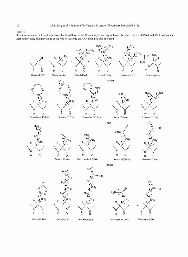
The second index,j, specifies that one of the 20different side-chains of the 20 naturally occurringamino acid residues2 [15] may be involved�1 # j #20�: Mono-peptides (II ), dipeptides (III ) and tripep-tides (IV ) contain 1, 2 and 3 amino acid residues,respectively. In such models, the chain may be termi-nated by methyl groups or by hydrogens, as shownbelow for the above three cases (P�Q� CH3 or H).
Again, R(j) represents the side-chain of one of the 20naturally occurring amino acids�1 # j # 20� given inTable 1.
The torsional angles�v0; f1; c1; v1;…� areresponsible for folding. The energy of folding is amulti-variable function where the torsional anglesare the independent variables. The PEHS for themono(18)-, di(28)- and tri(38)-peptides, given below,are functions containing 4, 7 and 10 independentvariables, respectively.
E�18� � E�v0;v1;f1;c1� �1�
E�28� � E�v0;v1;v2;f1;c1;f2;c2� �2�
E�38� � E�v0;v1;v2;v3;f1;c1;f2;c2;f3;c3� �3�Clearly, for a degree of polymerization ofn amino
acids, there aren torsional angle pairs off i andc i
�1 # i # n�; two terminal peptide functionalities (v0
andvn) and�n 2 1� mid-chain peptide bonds [v i for1 # i # �n 2 1��: Thus, the total number of foldingvariables for a polypeptide containingn amino acidsis
N � ��n 2 1�1 2�v 1 nf 1 nc � 3n 1 1 �4�The trans-peptide bond is more stable than thecis-
peptide bond. Consequently, it has been traditional toset v i � 1808 for all i. This limitation reduces thedimensionality of the problem substantially
E�18� � Etrans�f1;c1� �5�E�28� � Etrans�f1;c1;f2;c2� �6�E�38� � Etrans�f1;c1;f2;c2;f3;c3� �7�
The total number of folding variables, after thereduction of the dimensionality, becomes
N � nf 1 nc � 2n �8�This reduction in dimensionality does not represent adenial of importance of thecis-configuration of anygiven peptide bond; it only means that we are parti-tioning the problem. First, we study the backboneconformations fortrans-peptide bonds as this repre-sents the primary problem and subsequently we maystudy the same problem for any peptide bond being inthe cis-conformation.
Most of the study carried out so far, has beencentered on the generation and analysis of theE(18)potential energy surface (PES). The contour diagramof this type of PES is frequently referred to as the“Ramachandran Map”, in honour of the Indian
M.A. Berg et al. / Journal of Molecular Structure (Theochem) 500 (2000) 5–58 9
2 The 20 naturally occurring amino acids have both DNA andRNA codons. Selenocysteine nowadays are called the 21st aminoacid because it has an RNA codon (UCA stop codon) [15].
M.A. Berg et al. / Journal of Molecular Structure (Theochem) 500 (2000) 5–5810
Table 1Structures of amino acid residues. Note that in addition to the 20 naturally occurring amino acids, which have both DNA and RNA codons, the21st amino acid, Selenocysteine (Sec), which has only an RNA codon, is also included
M.A. Berg et al. / Journal of Molecular Structure (Theochem) 500 (2000) 5–58 11
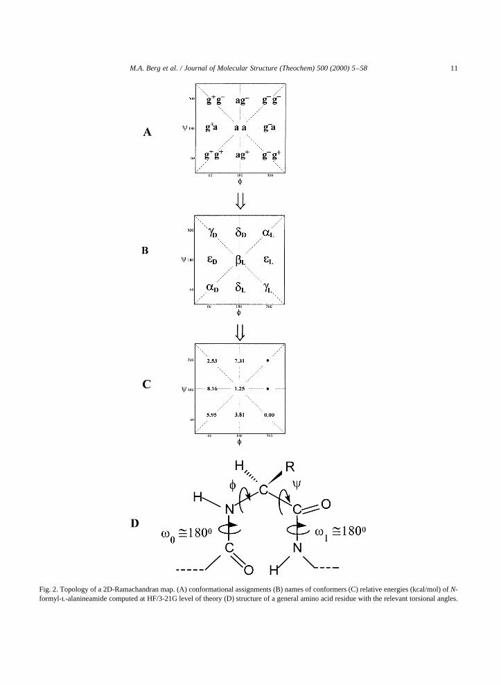
Fig. 2. Topology of a 2D-Ramachandran map. (A) conformational assignments (B) names of conformers (C) relative energies (kcal/mol) ofN-formyl-l-alanineamide computed at HF/3-21G level of theory (D) structure of a general amino acid residue with the relevant torsional angles.

chemist, Professor Ramachandran, who first calledattention to the importance of such a PES.
A topological representation of the Ramachandranmap for Rj � CH3 (i.e. for the alanine residue) isshown in Fig. 2. The various minima of the PES,representing the stable conformers, are marked bysubscripted Greek letters. Although not all 20 aminoacids were subjected to ab initio computationalconformational analysis, several amino acids wereinvestigated, and it was recognized that side-chainand backbone conformation can be studied indepen-dently, at least in the first approximation.
The relative energies ofN-formyl alaninamide withtrans-peptide bond are given in Fig. 2C in the form ofa PES topology. The corresponding conformationalenergy hyper-surface (PEHS) topology, involvingboth thetrans- as well as thecis-isomers is depictedin Fig. 3.
As yet, these residues have been considered in theabsence of conformationally variable side-chains.However, side-chains make contributions to the totalenergy and they are also involved in backbone/side-chain as well as side-chain/side-chain interactions,thus they ultimately help to determine protein folding.To include these further degrees of freedom inthe analytic evaluations requires the extension of
the above equations. Labeling of the torsion anglevariables in the side-chain is accomplished usingx1,x2, x3,… and so on, beginning at the Ca. As the side-chain is the only differentiating structural elementbetween amino acids, each analytic equation alsobecomes unique. The equations then become:
E�18� � Etrans�f1;c1; �x 11 ; x
12;…; x1
k�� �9�
E�28� � Etrans�f1;c1; �x 11 ; x
12;…; x1
k�;f2;c2;
�x 21 ;x
22 ;…; x2
k �� �10�
E�38� � Etrans�f1;c1; �x 11 ; x
12;…; x1
k�;f2;c2;
�x 21 ;x
22 ;…; x2
k �;f3;c3�x 31 ;x
32;…;x3
k�� �11�where �x1; x2;…; xk� is specific to each amino acidand its side-chain, with the superscript denotingwhich residue that side-chain belongs to in the poly-peptide chain.
Extending this concept to a generalized form, weobtain the following multi-variable function:
E�n8� � Etrans�f1;c1; �x 11 ;x
12 ;…; x1
k�;…;fn;cn;
�x n1 ; x
n2 ;…;x n
k �� �12�
M.A. Berg et al. / Journal of Molecular Structure (Theochem) 500 (2000) 5–5812
Fig. 3. Topology of a 3D-Ramachandran PEHS. (A) name of conformers (B) relative energies of HCO–(v)–NH– (f)–CHMe–(c)–CONH2.

4. Conformational study of single amino acidpeptide models
There are two ways to represent the rotation about asingle bond. Traditionally, the 08! 1808! 3608range is used but recently, IUPAC recommended theconvention of21808! 0! 11808: This conventionhas the advantage of designating the 08! 11808segment as a clockwise rotation and the 08! 21808segment as a counter clockwise rotation. However, ithas the disadvantage that certain minima fall on theedges or the corners of the Ramachandran map. Thetwo representations are presented in Fig. 4.
Peptide models, such as CH3CONH–CHR–CONHCH3 or simply HCONH–CHR–CONH2 canmimic the ith amino acid residue in a protein chain.Thef andc torsional angles are defined in Fig. 2D.The conformational assignments (g1g1, g1a, …etc.)are shown in Fig. 2A.
The names of the minima (Fig. 2B) are subscriptedGreek letters. The Greek letters originate from earliernomenclature (involvinga, b andg) while thel anddsubscripts originate from the observation thatl-aminoacids favor l conformations whiled-amino acidsfavor d conformations (c.f. lower part of Fig. 5).The names also suggest the combination of thechirality of a constitutional structure (R or S config-uration) and the chirality of the conformational twistor folding. This is summarized in Fig. 6.
The top of Fig. 7 shows the symbolic representationof a conformational PES for two full cycles of rotation�23608! 08! 13608� of both f andc . The PEScan be partitioned into four quadrants, in the tradi-tional way, or it can be partitioned according toIUPAC convention, as shown by the broken lines.
M.A. Berg et al. / Journal of Molecular Structure (Theochem) 500 (2000) 5–58 13
Fig. 4. Two kinds of partition of the PES. The central square corresponds to the traditional cut�08! 1808! 3608�; while the lower left handsquare represents the IUPAC conventional cut�21808! 08! 11808�:
Fig. 5. Underlying principles for choosing subscripted Greek letter(e.g.al, ad, bl…etc.) as names for the peptide conformations.

An energy contour diagram of the conformationalPES of a peptide (PCONH–CHR–CONHQ), present-ing two full cycles of rotation�23608! 08! 3608�of bothf andc , is shown at the bottom of Fig. 7. Thecentral square is the IUPAC conventional cut, whilethe four quadrants are the traditional cuts. One ofthese traditional cuts (e.g. the upper right handquadrant) is shown in pseudo-3D-representation inFig. 8.
There are 20 naturally occurring amino acids. Atotal of 18 of them have the same type of backbonefolding as shown in Fig. 2 (i.e. nine discrete confor-mations). The two other amino acids are exceptions.
One exception is proline, which is built intoproteins like any other amino acid, but its N is lockedin a five-membered ring. For proline residue V,f canonly be in the vicinity of2608 (i.e.13008) and, there-fore, only three backbone conformations are possible:al, el andgl. The other unique amino acid is glycineVI , which is achiral.
In the case of the glycine residue, double degeneracyoccurs in its conformational PES as shown in Fig. 9.
Finally, it should be mentioned that for certainmolecular residues molecular computations haveestablished the actual location of the nine minima,as shown above. The values off and c deviatesomewhat from the ideal values, although not toosignificantly. Table 2 lists these numerical values for
M.A. Berg et al. / Journal of Molecular Structure (Theochem) 500 (2000) 5–5814
Fig. 6. Stereochemical relationships ofg-turns. Note, that not onlythea carbon has chirality, but there is also chirality in the twistingof the backbone. The combination of these two types of chiralitiesleads to enantiomeric and diastereoisomeric structures.d and ldenote the chirality of the Ca configuration, whilegl andgd denotethe chirality of the conformation.
Fig. 7. (Top) A schematic representation of the conformational PESof a peptide (PCONH–CHR–CONHQ). Subscripted Greek letterssymbolize the approximate locations of the conformations.(Bottom) Contour diagram of the 2D Ramachandran potentialenergy surface of HCONH–CHCH3–CONH2, presented in the23608 # f # 3608 and23608 # c # 3608 range of independentvariables. The central square (broken lines) is the IUPAC conven-tional cut, while the four quadrants are the traditional cuts.
alaninediamide. This information is also presentedgraphically in Fig. 10.
4.1. Previously published single amino acid peptidemodels
The topological representation of the conforma-tional PES (Ramachandran map) of For-Ala-NH2 isshown in Fig. 2A, while that of the conformationalPEHS is shown in Figs. 7 and 8. Work has beencompleted for the following N- and C-protectedamino acids containing atrans-peptide bond: Gly[16,17], Ala [16,17], Val [18], Phe [19,20] and Ser[21–23]. Preliminary studies have been completedon Pro [24], Asp [25], Asn [26], Cys [27], and Sec(selenocysteine) [15].
The following protected amino acid residues, againwith trans-peptide bonds, are currently under investi-gation: Arg, Lys, His, Tyr, Leu, Thr and Trp.
Proline is fundamentally different from all the other18 chiral amino acids in more than one respect:
(i) the R group forms a five-member ring with thebackbone;(ii) there is no peptidic N–H group in the residue tobe involved in hydrogen bonding, and(iii) since there are two carbon atoms connected tothe nitrogen, there is a greater chance ofcis/transisomerization in the peptide bond.
The potential energy cross-sections of the typeE �E�c�; for the Ramachandran map of HCO–Pro–NH2
containing cis- and trans- peptide bonds [28] are
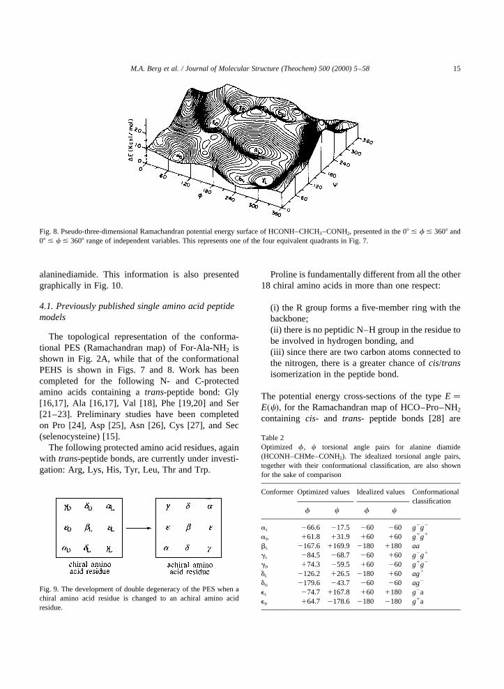
M.A. Berg et al. / Journal of Molecular Structure (Theochem) 500 (2000) 5–58 15
Fig. 9. The development of double degeneracy of the PES when achiral amino acid residue is changed to an achiral amino acidresidue.
Fig. 8. Pseudo-three-dimensional Ramachandran potential energy surface of HCONH–CHCH3–CONH2, presented in the 08 # f # 3608 and08 # c # 3608 range of independent variables. This represents one of the four equivalent quadrants in Fig. 7.
Table 2Optimized f , c torsional angle pairs for alanine diamide(HCONH–CHMe–CONH2). The idealized torsional angle pairs,together with their conformational classification, are also shownfor the sake of comparison
Conformer Optimized values Idealized values Conformationalclassification
f c f c
al 266.6 217.5 260 260 g2g2
ad 161.8 131.9 160 160 g1g1
bl 2167.6 1169.9 2180 1180 aagl 284.5 268.7 260 160 g2g1
gd 174.3 259.5 160 260 g1g2
dl 2126.2 126.5 2180 160 ag1
dd 2179.6 243.7 260 260 ag2
el 274.7 1167.8 160 1180 g2aed 164.7 2178.6 2180 2180 g1a

shown in Fig. 11. Preliminary investigation on thecis-peptide bond has been completed and thecis-Rama-chandran map is currently under construction [29].
4.2. Single amino acid peptide models in progress
4.2.1. Aspartic acid and asparagineAspartic acid (Asp) and its side-chain deproto-
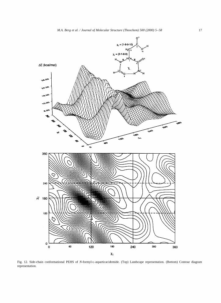
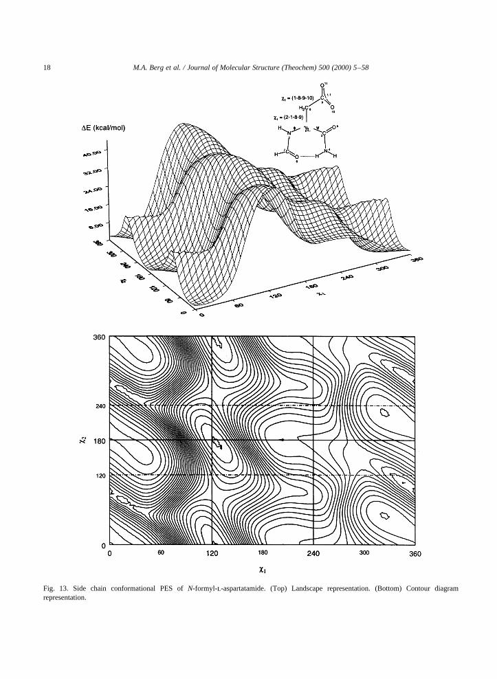
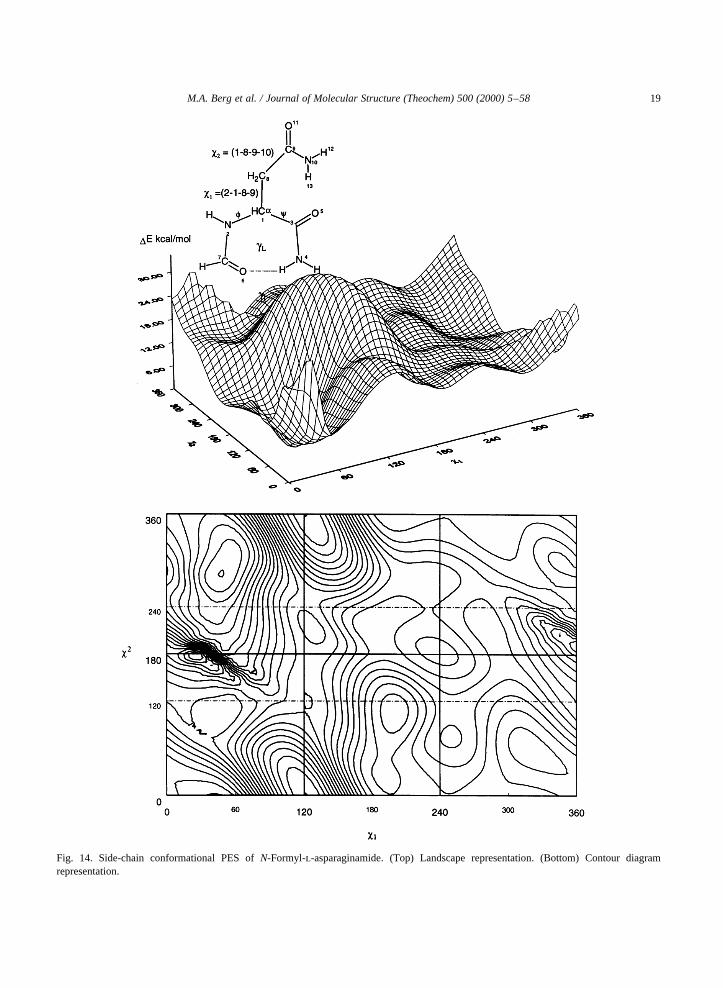
nated form, i.e. the aspartate [25] ion as well as itsside-chain amide asparagine (Asn) [26] have beenstudied in a preliminary fashion. In both cases, theN-formyl-l-aspartic acidamide,N-formyl-l-asparta-tamide andN-formyl-l-asparaginamide were inves-tigated in their gl backbone conformation. Theside-chain conformational PES for the threecompounds are shown in the following illustrations:Figs. 12–14.
4.2.2. Arginine (Arg)Arginine is an amino acid with a positive charge at
neutral pH due to its polar side-chain, the guanidiumgroup. The typical pKa value of arginine is 12.0, the
highest pKa of all amino acids. It is, therefore, impor-tant in salt bridge coupling, which is part of certaindocking processes.
Arginine is the source of nitric oxide (NO) inbiological systems. NO is a free radical whichserves as an intracellular second messenger and anintercellular messenger that regulates neighbouring,and possibly distant cells. NO takes part in manybiological processes, such as vasodilation forexample. NO is also involved in the central nervoussystem (CNS), such as the modification of painperception. One of the two equivalent nitrogens ofthe terminal guanidine inl-arginine undergoes five-electron oxidation in the presence of the enzymeNO synthase (NOS), yielding NO andl-citrulline.N-methyl guanidine has been used as a modelcompound for arginine to study the mechanism ofNO release using ab initio molecular computations[30].
Ethylguanidine or ethylguanidium ion is the term-inal portion of the side chain (R-group) of the arginineresidue, as exemplified byN-formyl-argininamide andits side chain terminalN-protonated form respectively.Arginine can be divided into two portions: the back-bone, which is analogous to glycine (or alanine) andthe side chain, which is an alkyl guanidine.
When a planar moiety is twisted about a singlebond with respect to a tetrahedral moiety, in principle,up to six minima may appear. In the case ofethylguanidine [VII ] and ethylguanidium ion [VIII ],there are two torsional anglesx3 and x4. These aretwo types of bonds, C–C and C–N, with respect to
M.A. Berg et al. / Journal of Molecular Structure (Theochem) 500 (2000) 5–5816
Fig. 10. A schematic illustration of the PES of an average aminoacid residue, obtained from the calculations carried out so far onmono-, di- and tri-peptides. The idealized positions are markedby shaded squares and the computationally determined positionsare shown as open circles. The names of the conformers aregiven as subscripted Greek letters. Note that a single aminoacid residue might not be able to take on all of the shownconformations.
Fig. 11. Conformational Potential Energy CurvesE � E�c� for cisandtrans-N formyl-l-prolinamide.
M.A. Berg et al. / Journal of Molecular Structure (Theochem) 500 (2000) 5–58 17
Fig. 12. Side-chain conformational PEHS ofN-formyl-l-asparticacidemide. (Top) Landscape representation. (Bottom) Contour diagramrepresentation.
M.A. Berg et al. / Journal of Molecular Structure (Theochem) 500 (2000) 5–5818
Fig. 13. Side chain conformational PES ofN-formyl-l-aspartatamide. (Top) Landscape representation. (Bottom) Contour diagramrepresentation.
M.A. Berg et al. / Journal of Molecular Structure (Theochem) 500 (2000) 5–58 19
Fig. 14. Side-chain conformational PES ofN-Formyl-l-asparaginamide. (Top) Landscape representation. (Bottom) Contour diagramrepresentation.

which the guanidine group could be eclipsed or beperpendicular to it.
Fig. 15 shows the torsional potential, as a functionof x4 for ethylguanidine in itss-cis/exo[VII(a) ] struc-ture calculated using two basis sets. For each basis set,the global minimum occurs at 1808 (the anti form).The energy values of the 1D-scans were computed atthe HF/6-31G level of theory. For all four structures[Ia,Ib ,Ic andId ] g1, a,g2 were shown to have energyminima. The upper portion of Fig. 16 shows the poten-tial energy curveE � E�x5� of structureVIII calcu-lated at HF/6-31G [31].
The protonated form of the ethyl guanidine [VII ] isthe ethyl guanidium structural ion [VIII ]. Due to theinternal symmetry of the N–C(NH2)2
1 moiety, thisspecies has only one arrangement.
Two vertical proton affinity values may be identi-fied using these PECs. The two vertical ones involveg! g anda! a:
The full side-chain orientation of N- and C-protected arginine and its conjugate acid depends onx1, x2, x3, and x4 as illustrated by molecularstructuresIX andXI .
Consider the diamides of the two amino acidsarginineIX and aspartic acidX. Under physiologicalconditions the amino acid side chains occur inthe protonatedXI and deprotonatedXII forms
M.A. Berg et al. / Journal of Molecular Structure (Theochem) 500 (2000) 5–5820
Fig. 15. Conformational PEC of ethyl guanidine, which may model the rotation (x4) about the C–N bond in neutral arginine.
Fig. 16. Rotational potential energy curves of ethylguadinium ion(top) and propionate ion (bottom). Note that each molecule has onlyone discrete conformation.

respectively. The side chain conformations could bemimicked for XI by XIII and to some extentXIVand forXII by XV . The potential energy curve,E �E�x5�; for XIV and that for XV , E � E�x2�; isshown in Fig. 16. The side chain PES ofXII hasalso been reported and is shown in Fig. 13. It isclear that torsional angles within the amino acidside chains do increase the dimensionality of theconformational space. Nevertheless, they are veryimportant, because the side chain makes each
amino acid unique. Such distinction can be madebecause, to a good degree of approximation, theside chain conformational pattern is independent ofthe backbone conformational behavior. For all prac-tical purposes, the backbone of all amino acidsshows the same conformational pattern (c.f. Figs.2, 7, 8 and 10).
4.2.3. Cysteine (Cys) and selenocysteine (Sec)Each of serine [21–23,32], cysteine [27] and
M.A. Berg et al. / Journal of Molecular Structure (Theochem) 500 (2000) 5–58 21

selenocysteine [15] is expected to have 9× 9� 81conformations�3 × 3� 9 backbone:c�g1
;a;g2� ×f�g1
; a;g2� and 3× 3� 9 side-chain:x1�g1;a;g2� ×
x2�g1;a;g2��: To investigate the effects of side-chain/
backbone conformational interactions, all torsionalmodes of the side-chain (x1: rotation about theCa–Cb andx2: rotation about the Cb–X bonds) werestudied in the relaxedgl backbone��f;c� � �g2
; g1��conformation. Six out of the nine expected minima forserine and seven out of the nine expected minima forcysteine and selenocysteine were found at the RHF/3-21G level of theory.
The stabilization energy exerted by the –CH2–SeH
side-chain has been compared to that of –CH2–SHand –CH2–OH. The stabilization energies were calcu-lated with respect to thegl backbone conformation ofN- and C-protected glycine [33,34] using the follow-ing isodesmic (same number of bonds) reaction,where P and Q may be H or CH3 and R� CH2–XH(X �O,S,Se):
PCONH–CH2–CONHQ1 CH3–Rconformationgl
! PCONH–CHR–CONHQ1 CH3–Hconformation X
�13�
M.A. Berg et al. / Journal of Molecular Structure (Theochem) 500 (2000) 5–5822
Fig. 17. Calculation of stabilization energies (DEstabil.) with various substituents (R may be CH2-OH, CH2-SH and CH2-SeH) relative to thegl
conformation of Glycine�R� H�:

The stabilization energy may be calculated as follows:
DEstabilization� { E�PCONH–CHR–CONHQ�x1 E�CH3–H�}2 { E�PCONH–CH2–CONHQ�gl
1 CH3 2 R�} �14�These equations are also illustrated in Fig. 17.
From Fig. 18, it can be observed that the effect ofside-chain orientation with respect to the peptidebackbone, going from Serine to Selenocysteine, isgradually decreasing. This is due to the increase inthe size of the atoms O! S! Se and the resultinggradual lengthening of C–O! C–S! C–Se bondlengths as well as the reduced hydrogen bonding abil-ity in going from OH to SH and all the way to SeH.
Topological analysis of the electron density hasbeen performed for cysteine and selenocysteine,using Bader’s Atoms in Molecule (AIM) approachat the B3LYP/6-31G(d,p) level of theory. The hydro-gen bonds were verified by the existence of bondcritical points. Three conformations: (g1 g1), (g1 a)and (ag2) exhibited such interactions as illustratedgraphically in Fig. 19 for For-Sec-NH2.
4.2.4. Leucine (Leu)Leucine is one of the naturally occurring essential
amino acids. It is an important amino acid, havingmany implications in medicine, one of them beingits role in Maple Syrup Urine disease (MSUD).Maple syrup disease occurs when plasma leucine,isoleucine, alloisoleucine and valine are elevatedto very high levels. These amino acids will alsobe present in the urine and hence the urine releases
M.A. Berg et al. / Journal of Molecular Structure (Theochem) 500 (2000) 5–58 23
Fig. 18. Spectrum of conformational dependence of the side-chain stabilization energy on thegl backbone conformation of formyl Ser-, Cys-and Sec-amide.

an odor similar to maple syrup. This disease hassevere neurological and gastrointestinal effects.The current treatment for it involves intense controlof the intake of leucine, isoleucine and valine.This careful restriction of leucine also includesmonitoring of the branched chain amino acids inplasma.
Leucine is also found in some small proteoglycans.This group of small, interstitial proteoglycans has ahigh degree of homology in their protein coresequence. Each has between 10 and 12 highlyconserved leucine rich tandem repeats which makesup the central portion of the core protein. The currentmembers of the group are fibromodulin and lumican,which are keratan sulphate substituted, as well asdecorin and biglycan, which are both chondroitinsulphate substituted. Other proteoglycans whichhave had functions ascribed to them are the smallleucine rich interstitial proteoglycans decorin andfibromodulin which have been shown to modulatecollagen fibrillogenesis.
The knowledge of the conformational potentialenergy surface for leucine would allow a better under-
standing of diseases such as MSUD and the functionof leucine in biological processes such as collagenfibrillogenesis. Each of the nine possible backboneconformations is associated with nine possible sidechain conformations. In addition, there are fourchain-end conformations associated with thecis andtrans peptide bond that also must be explored. Thusthe total number of conformations to be considered is9 × 9 × 4� 324: The results of some calculations thathave been carried out for thebl backbone conforma-tion are shown in Fig. 20.
4.2.5. Tryptophan (Trp)Tryptophan is one of the essential amino acids the
body cannot manufacture itself. It is the least abun-dant in proteins and is destroyed easily by the liver.This amino acid is necessary for the production of thevitamin B niacin, which is essential for the brain tomanufacture the key neurotransmitter serotonin. Lowlevels of serotonin have been linked with insomnia,anxiety and depression.
Tryptophan is believed to be effective for insomniaand jet lag. It is also believed to have anti-anxietyeffects and control aggressive behavior in someindividuals. Other studies have shown that its anti-depressant effect lasts longer than that of the popular
M.A. Berg et al. / Journal of Molecular Structure (Theochem) 500 (2000) 5–5824
Fig. 19. The contour map of the Laplacian of the electron density forthe (g1g1) conformation of For-l-Sec-NH2 calculated at theB3LYP/6-311G(d,p)//RHF/3-21G level of theory. Bond paths aredenoted by lines, bond critical points (BCPs) are denoted by blackdots, ring critical points (RCPs) are denoted by open triangles andthe nuclei are denoted by crosses.
Fig. 20. Relative energies of the N- and C-protected leucine withvarying side chain conformations for thebl backbone conformationretaining trans peptide bonds calculated at HF/3-21G level oftheory.

anti-depressant drug, Imipramine. In addition, somepreliminary studies of combined Vitamin B-6 andtryptophan show that they may be effective inreducing the severity of hyperventilation as well asthe panic attacks produced by it.
Although it is safe to use, tryptophan is no longer
available in supplement form, because of potentialadverse reactions. Tryptophan supplements are notrecommended in cases of pregnancy, asthmatics, orauto-immune disorders like Lupus or Scleroderma. Asa preliminary investigation, a scan at HF/3-21G levelof theory was performed on ethyl indole (Fig. 21).
M.A. Berg et al. / Journal of Molecular Structure (Theochem) 500 (2000) 5–58 25
Fig. 21. Potential energy curve of D(C1–C2–C3–C4) of ethyl indole.
Minima were found to exist at torsional angles (D) of0 and in the vicinity of 1058.
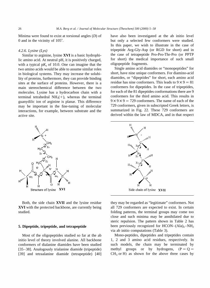
4.2.6. Lysine (Lys)Similar to arginine, lysineXVI is a basic hydrophi-
lic amino acid. At neutral pH, it is positively charged,with a typical pKa of 10.0. One can imagine that thetwo amino acids would be able to assume similar rolesin biological systems. They may increase the solubi-lity of proteins, furthermore, they can provide bindingsites at the surface of proteins. However, there is amain stereochemical difference between the twomolecules. Lysine has a hydrocarbon chain with aterminal tetrahedral NH3(1), whereas the terminalguanydilic ion of arginine is planar. This differencemay be important in the fine-tuning of molecularinteractions, for example, between substrate and theactive site.
Both, the side chainXVII and the lysine residueXVI with the protected backbone, are currently beingstudied.
5. Dipeptide, tripeptide, and tetrapeptide
Most of the oligopeptides studied so far at the abinitio level of theory involved alanine. All backboneconformers of dialanine diamides have been studied[35–38]. Analogously trialanine diamide (tripeptide)[39] and tetraalanine diamide (tetrapeptide) [40]
have also been investigated at the ab initio levelbut only a selected few conformers were studied.In this paper, we wish to illustrate in the case oftripeptide Arg-Gly-Asp (or RGD for short) and inthe case of tetrapeptide Pro-Pro-Thr-Pro (or PPTPfor short) the medical importance of such smalloligopeptide fragments.
Single amino acid diamides or “monopeptides” forshort, have nine unique conformers. For diamino-aciddiamides, or “dipeptides” for short, each amino acidresidue has nine conformers. This leads to 9× 9� 81conformers for dipeptides. In the case of tripeptides,for each of the 81 dipeptides conformations there are 9conformers for the third amino acid. This results in9 × 9 × 9� 729 conformers. The name of each of the729 conformers, given in subscripted Greek letters, issummarized in Fig. 22. These 729 conformers arederived within the law of MDCA, and in that respect
they may be regarded as “legitimate” conformers. Notall 729 conformers are expected to exist. In certainfolding patterns, the terminal groups may come tooclose and such minima may be annihilated due tosteric repulsion. The pattern shown in Table 2 hasbeen previously recognized for HCON–(Ala)n–NH2
via ab initio computations (Table 3).Mono-peptides, dipeptides and tripeptides contain
1, 2 and 3 amino acid residues, respectively. Insuch models, the chain may be terminated bymethyl groups or by hydrogens,�P� Q�CH3 or H� as shown for the above three cases by
M.A. Berg et al. / Journal of Molecular Structure (Theochem) 500 (2000) 5–5826

M.A
.B
erg
et
al.
/Jo
urn
alo
fM
ole
cula
rS
tructure
(Th
eo
che
m)
50
0(2
00
0)
5–
58
27
Fig. 22. Names, in terms of subscripted Greek letters, of the 729 legitimate backbone conformers of tripeptides.

II , III and IV . The symbol R(j) represents the side-chain of one of the 20 naturally occurring aminoacids �1 # j # 20�:
5.1. Tripeptide sequence, RGD
The tripeptide Arg-Gly-Asp (RGD) is an adhesivemolecule which has a positively and a negativelycharged side chain as shown inXVIII in accordancewith XI and XII . The positively and negativelycharged side chains may be engaged in saltbridgeXIX formation either via an intra- or an inter-molecular connection.
The structure shown inXVIII is the amide of theN-formyl derivative of the RGD-motif and it is given in itsglglgl backbone and extended (fullyanti) side chainconformations. Once again, assuming that all peptidebonds are oftransconfiguration, the PES can be repre-sented analytically by the following functionXX .
The backbone may be represented by a 6D-confor-mation subspace (f1,c1,f2,c2,f3,c3). Since we may
expect only one distinct conformer alongx5 andx20,
the side chain may be represented by a 5D-confor-mational chain subspace (x1, x2, x3, x4, x1
0).Consequently, we may expect 35 � 243 side-chainconformations. The total number of distinct RGDconformers may, therefore, be 729× 243� 177147:In view of the enormity of the problem only a selectedfew conformations were subjected to molecularcomputations.
Table 4 summarizes the geometrical parameters(e.g. torsional angles), conformational assignmentsand relative stabilities of the RGD structures opti-mized at HF/3-21G level of theory.
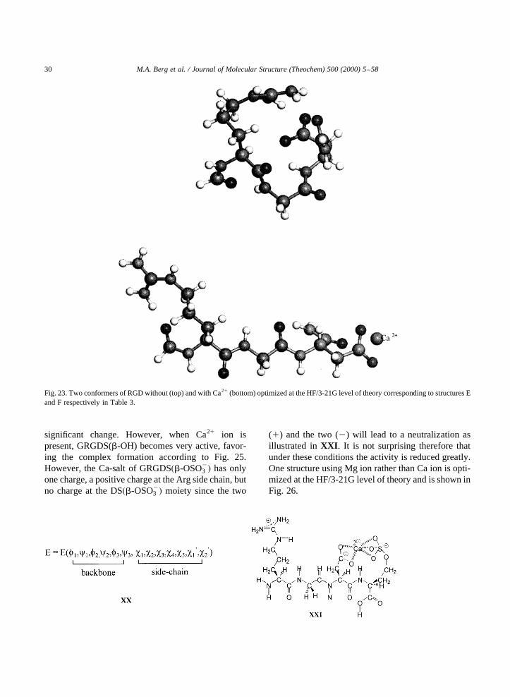
The last two structures of the table are depicted inFig. 23. Note that one of them is without the involve-ment of Ca21, and therefore, it exhibits intramolecularsalt bridge. The other structure is with Ca21, implying
that the previously negatively charged side chain(–COO2) now contains a mono-positive (–COO2·· ·Ca11) ending. Consequently, there is no chance forintramolecular salt-bridge formation. This chargedifference in side-chain has implication for the dock-ing mechanism. Fig. 24 shows a possible dockingmechanism without the involvement of Ca21.
Fig. 25 indicates that in the presence of Ca21, thereceptor would need two negatively charged sidechains (i.e. two carboxylate ions).
It is interesting to recall the result of the physio-logical study [41], involving GRGDS(b-OH) (c.f.Fig. 26) and GRGDS�b-O–SO2
3 �: The final resultsare summarized in Table 5.
It seems that when Ca21 ion is not present themechanism given in Fig. 24 is operative and thus anextra negative charge at the C-terminal makes no
M.A. Berg et al. / Journal of Molecular Structure (Theochem) 500 (2000) 5–5828
Table 3Legitimate and existing conformers of small peptides
n Legitimate conformers Conformers found
1 9 72 81 49a
3 729 Not investigated
a These 49 conformers were found by optimizing the 81 “legit-imate” conformers. Subsequently, Prof. Lothar Schafer andcoworkers [Can. J. Chem. 76 (1998) 566] have scanned the confor-mational space computing 11 664 grid points at the HF/4-31G levelof theory and located one more conformer. Now the number ofconformers stands at 50.

M.A
.B
erg
et
al.
/Jo
urn
alo
fM
ole
cula
rS
tructure
(Th
eo
che
m)
50
0(2
00
0)
5–
58
29
Table 4Geometrical parameters, conformational assignment, total energies and relative stabilities of collected RGD conformers without (A–E) and with (F) the inclusion of Ca21,optimized at the HF/3-21G level of theory
Residue A B C D E Fa
Geometry Confor-
mation
Geometry Confor-
mation
Geometry Confor-
mation
Geometry Confor-
mation
Geometry Confor-
mation
Geometry Confor-
mation
Arginine (R) v 0 177.5804 – 172.5255 – 2175.5717 – 2177.5620 – 2171.2285 – 2176.4709 –
f 1 2153.0601 dl 2155.4259 dl 46.2751 ad 281.9119 gl 282.2705 gl 2153.6854 bl
c 1 47.7288 53.3474 41.4783 66.0202 66.0650 165.4315
v 1 2179.4318 – 173.4531 – 2178.5048 – 2179.8780 – 2179.5433 – 175.9534 –
x 1 44.2825 g1 58.1487 g1 57.9267 g2 57.1272 g2 261.4889 g2 260.7727 g2
x 2 75.7941 g2 123.3340 a 96.1472 g1 142.7702 a 264.8448 g2 266.1861 g2
x 3 292.5497 g2 2166.2056 a 2144.5144 a 264.6406 g2 257.8581 g2 2175.4661 a
x 4 155.5904 a 79.6676 g2 96.1651 g1 169.7310 a 142.8879 a 296.2058 g2
Glycine (G) f 2 120.6121 bl 96.0964 dl 123.2492 dd 287.4209 gl 265.5310 al 173.2842 bl
c 2 2131.5270 2103.2997 234.2922 58.5616 226.2768 179.6252
v 2 2175.4871 – 171.9142 – 171.0358 – 2159.1781 – 2169.9989 – 2174.1726 –
Aspartate (D) f 3 266.4906 el 284.2133 el 2161.4273 dd 293.1384 gl 281.2064 gl 283.6510 gl
c 3 171.5693 2141.3300 2119.3301 53.6705 69.7315 52.2155
v 3 2177.6658 – 2155.8824 – 2158.9678 – 2171.2286 – 173.9757 – 2172.5103 –
x 1 59.2826 g1 64.5079 g1 48.5048 g1 37.1034 g1 53.9209 g2 168.2582 a
x 2 89.7330 g1 106.2422 g1 89.8874 g1 29.3742 g1 155.2624 a 37.4610 g2
Energy (hartree) 21328.6774261 21328.7079568 21328.6899608 21328.6819226 21328.69329353 22001.8202808
DE (kcal/mol) 19.16 0.00 11.29 16.34 9.20 N/A
Backbone
conformation
dldlel dldlel addddd glglgl glalgl blblgl
a Includes a Ca21 in the vicinity of the asparate side chaincarboxylate moiety.
significant change. However, when Ca21 ion ispresent, GRGDS(b-OH) becomes very active, favor-ing the complex formation according to Fig. 25.However, the Ca-salt of GRGDS�b-OSO2
3 � has onlyone charge, a positive charge at the Arg side chain, butno charge at the DS�b-OSO2
3 � moiety since the two
(1) and the two (2) will lead to a neutralization asillustrated inXXI . It is not surprising therefore thatunder these conditions the activity is reduced greatly.One structure using Mg ion rather than Ca ion is opti-mized at the HF/3-21G level of theory and is shown inFig. 26.
M.A. Berg et al. / Journal of Molecular Structure (Theochem) 500 (2000) 5–5830
Fig. 23. Two conformers of RGD without (top) and with Ca21 (bottom) optimized at the HF/3-21G level of theory corresponding to structures Eand F respectively in Table 3.

5.2. Tetrapeptide sequence, PPTP
For a discussion of a tetrapeptide example, let usbriefly review immunoglobulin A (IgA). Antibodiesor immunoglobulins (Ig) represent the great defenseforce in the army of the immune system. Ig moleculesare produced during a humoral immune response tobind and neutralize invading antigens. Subsequently,the antigen is labeled for removal by phagocytosis.The fight, however, is not one-sided. To help neutra-lize the role of Ig in antigen recognition, bacteria havedeveloped effective deactivation mechanisms. One ofthem involves a site-specific cleavage of an Ig mole-cule at the hinge region with the aid of an extracellularprotease produced by the bacteria.
Although there are differences in their catalyticmechanisms, all reported IgAses fall into one of threetypes: serine-, cysteine-, or metallo-proteases [42]. TheIgAse fromN. gonorrheahas been classified as a serineprotease [43]. A comparison between the gene andamino acid sequences ofN. gonorrheaandH. influenzae[44,45] showed that they are 50% identical and contain afingerprint region for the chymotrypsin/trypsin family ofserine proteases [42]. The most conserved sequence,
M.A. Berg et al. / Journal of Molecular Structure (Theochem) 500 (2000) 5–58 31
Fig. 24. A possible docking mechanism of RGD operative in the absence of Ca21 ion. Note that the backbone is shown arbitrarily in itsglglgl
conformation.
Fig. 25. Receptor-RGD-complex after docking, in the presence ofCa21 ion.

GDSGGPL (S� active serine site) is found in thevicinity of Ser 250 inN. gonorrheaIgAse.
There is an equal amount of anionic forming (Glu,Asp, Tyr) and cationic forming (Arg, Lys, His) aminoacid side chains present [44]. Based on the nature ofthe inhibitors, there would be reason to believe thatthese oppositely charged moieties are not uniformly
distributed in the tertiary structure. Instead, it is likelythat this enzyme contains concentrated areas of posi-tive charge that would enable the IgAse to seek outsubstrates that are negatively chargedXXII . The factthat serine proteases contain histidine at the active sitesuggests that the active site of the enzyme will also bepositively charged.
M.A. Berg et al. / Journal of Molecular Structure (Theochem) 500 (2000) 5–5832
Fig. 26. (A) An internal salt-bridge forming conformer of RGDS(b-OH). Conformer optimized at the HF/3-21G level of theory. (B) An Mg21
complex of RGD (b-OSO3-). The conformer shown was optimized at the HF/3-21G level of theory.
Table 5Summary of physiological effects of GRGDS(b-OH) and GRGDS(b-OSO2
3 ) in the absence and in the presence of Ca ion
Biological phenomena [Ca21] Peptide sequence
GRGDS(b-OH) GRGDS(b-OSO32)
Platelet aggregation Not present Active ActiveVasodilator effect Present Very active Not active

There have also been inhibition studies done onshort peptide segments of the hinge region [46],such as Pro-Pro-Thr-Pro, as well as longer onesand these have been found to act as mild inhibitors.They exhibit IC50 values in the micro-molar(1026 M) range or higher. Thus, they are weakinhibitors when compared with the ideal nano-molar (1029 M) range values expected for a drugcandidate.
Successful drug candidates, therefore, may needto mimic the heptapeptide segment of the hingeregion containing the negative charges of the twosialylic acid moietiesXXIII . The structure mustbe appropriately folded and should not exceed acertain molecular weight. Because bacteria becameresistant to most antibiotics such an inhibitor maybecome a successful lead compound for the devel-opment of several possible antibacterial agents.While these target oriented research projects maybe laudable nevertheless it is lamentable whetherwe have enough information to search for thesehighly desirable compound. What is needed is anextensive database, which includes peptide, cyclo-peptide, and peptidomimetic conformations.
To date, neither X-ray structure for the IgA hingeregion nor for the IgA protease produced byN.gonorrhea are available. Until such structuresbecome available, we can carry out some necessaryconformational structure analyses. With the aid ofthe enzyme structure, the drug design process mayproceed at an accelerated pace. The most obviousplace to start is with the tetrapeptide Pro-Pro-Thr-
Pro. The scission occurs at the Pro-Thr peptide bondand we have included at least the nearest neighboursat each end (XXII).
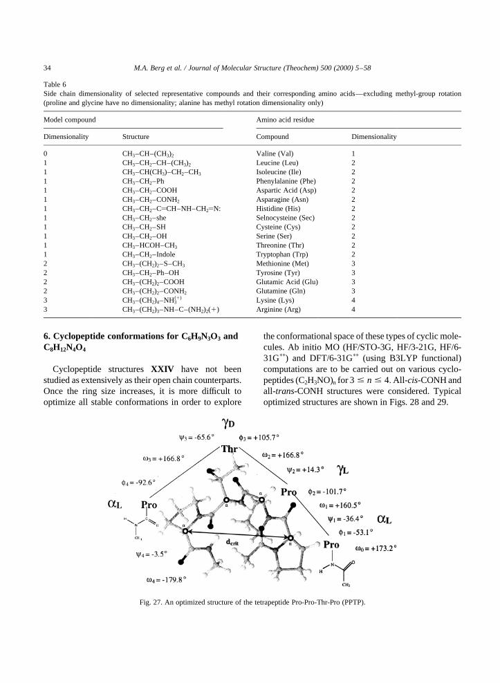
Clearly, this Pro-Pro-Thr-Pro tetrapeptide is morecomplicated than Ala-Ala-Ala-Ala [40] because Promay exhibit cis- and trans-isomerism in its peptidebonds, as well assyn- and anti-ring puckering.Thus, 2× 4� 4 isomeric forms may exist for eachbackbone conformer. Up to three backbone confor-mers (gl, el, al) may be expected; thus each prolinemoiety may have 4× 3� 12 conformations (Table 6).Consequently, for the dipeptide Me-CO-Pro-Pro-CO-NHMe, we may anticipate 12× 12� 144 conforma-tions. However, even if one disregards ring puckeringand considers onlytrans-peptide bonds, Pro-Pro-Thr-Pro may exhibit 3× 3 × 9 × 3� 243 backboneconformations. Including the 3× 3� 9 side chainconformations of the Thr side chain moiety, this willlead to a total of 243× 9 or 2187 conformers. Thus,the 243 backbone conformations may be best studiedon Pro-Pro-Ala-Pro and the Ala side chain may beextended to a Thr side chain subsequently. One ofthe 2187 conformers has been optimized in our preli-minary study and the structure is shown in Fig. 27.The central Pro-Thr structure turned out to begl gd,which has been previously identified as a new typeof b-turn [36]. It is interesting to note that several ofthe oxygen atoms occupy a region of the spacecreating an electron-rich domain. Such a display ofhigh electron density may play a role in the dockingof the PPTP moiety within the hinge into the cavity ofthe IgAse.
M.A. Berg et al. / Journal of Molecular Structure (Theochem) 500 (2000) 5–58 33
6. Cyclopeptide conformations for C6H9N3O3 andC8H12N4O4
Cyclopeptide structuresXXIV have not beenstudied as extensively as their open chain counterparts.Once the ring size increases, it is more difficult tooptimize all stable conformations in order to explore
the conformational space of these types of cyclic mole-cules. Ab initio MO (HF/STO-3G, HF/3-21G, HF/6-31Gpp) and DFT/6-31Gpp (using B3LYP functional)computations are to be carried out on various cyclo-peptides (C2H3NO)n for 3 # n # 4:All- cis-CONH andall-trans-CONH structures were considered. Typicaloptimized structures are shown in Figs. 28 and 29.
M.A. Berg et al. / Journal of Molecular Structure (Theochem) 500 (2000) 5–5834
Table 6Side chain dimensionality of selected representative compounds and their corresponding amino acids—excluding methyl-group rotation(proline and glycine have no dimensionality; alanine has methyl rotation dimensionality only)
Model compound Amino acid residue
Dimensionality Structure Compound Dimensionality
0 CH3–CH–(CH3)2 Valine (Val) 11 CH3–CH2–CH–(CH3)2 Leucine (Leu) 21 CH3–CH(CH3)–CH2–CH3 Isoleucine (Ile) 21 CH3–CH2–Ph Phenylalanine (Phe) 21 CH3–CH2–COOH Aspartic Acid (Asp) 21 CH3–CH2–CONH2 Asparagine (Asn) 21 CH3–CH2–CyCH–NH–CH2yN: Histidine (His) 21 CH3–CH2–she Selnocysteine (Sec) 21 CH3–CH2–SH Cysteine (Cys) 21 CH3–CH2–OH Serine (Ser) 21 CH3–HCOH–CH3 Threonine (Thr) 21 CH3–CH2–Indole Tryptophan (Trp) 22 CH3–(CH2)2–S–CH3 Methionine (Met) 32 CH3–CH2–Ph–OH Tyrosine (Tyr) 32 CH3–(CH2)2–COOH Glutamic Acid (Glu) 32 CH3–(CH2)2–CONH2 Glutamine (Gln) 33 CH3–(CH2)4–NH3
(1) Lysine (Lys) 43 CH3–(CH2)3–NH–C–(NH2)2(1) Arginine (Arg) 4
Fig. 27. An optimized structure of the tetrapeptide Pro-Pro-Thr-Pro (PPTP).
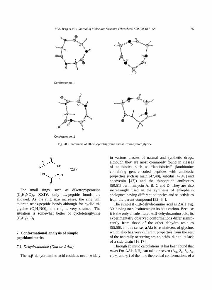
For small rings, such as diketropyperazine(C2H3NO)2, XXIV , only cis-peptide bonds areallowed. As the ring size increases, the ring willtolerate trans-peptide bonds although for cyclic tri-glycine (C2H3NO)3, the ring is very strained. Thesituation is somewhat better of cyclotetraglycine(C2H3NO)4.
7. Conformational analysis of simplepeptidomimetics
7.1. Dehydroalanine (Dha orDAla)
Thea,b-dehydroamino acid residues occur widely
in various classes of natural and synthetic drugs,although they are most commonly found in classesof antibiotics such as “lantibiotics” (lanthioninecontaining gene-encoded peptides with antibioticproperties such as nisin [47,48], subtilin [47,49] andancovenin [47]) and the thiopeptide antibiotics[50,51] berninamycin A, B, C and D. They are alsoincreasingly used in the synthesis of enkephalinanalogues having different potencies and selectivitiesfrom the parent compound [52–54].
The simplesta,b-dehydroamino acid isDAla Fig.30, having no substituents on its beta carbon. Becauseit is the only unsubstituteda,b-dehydroamino acid, itsexperimentally observed conformations differ signifi-cantly from those of the other dehydro residues[55,56]. In this sense,DAla is reminiscent of glycine,which also has very different properties from the restof the naturally occurring amino acids, due to its lackof a side chain [16,17].
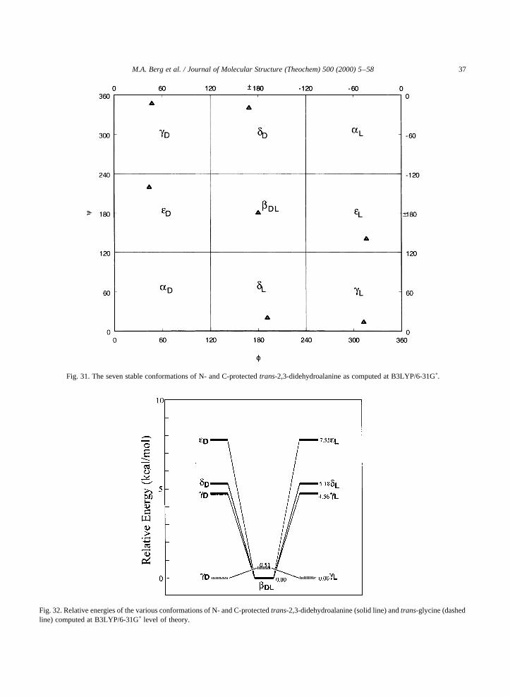
Through ab initio calculations, it has been found thattrans-For-DAla-NH2 can take on seven (bdl, dd, dl, ed,el, gd andgl) of the nine theoretical conformations of a
M.A. Berg et al. / Journal of Molecular Structure (Theochem) 500 (2000) 5–58 35
Fig. 28. Conformers of all-cis-cyclotriglycine and all-trans-cyclotriglycine.

saturated amino acid residue Fig. 31, whereas, even Nand C protected glycine, can actually only take on five.
Furthermore, it has also been found, by means ofrelative and stabilization energies, that althoughDAlais more rigid than glycine Fig. 32, it has a muchgreater stabilizing effect on the peptide chain asshown in Fig. 33. In fact,DAla has a greater stabilizing
M.A. Berg et al. / Journal of Molecular Structure (Theochem) 500 (2000) 5–5836
Fig. 29. Conformers of all-cis-cyclotetraglycine and all-trans-cyclotetraglycine.
Fig. 30. Torsional anglesv0, f andc of N- and C-protected 2,3-didehydroalanine.v0 is approximately 1808 for trans-2,3-didehy-droalanine.
M.A. Berg et al. / Journal of Molecular Structure (Theochem) 500 (2000) 5–58 37
Fig. 31. The seven stable conformations of N- and C-protectedtrans-2,3-didehydroalanine as computed at B3LYP/6-31Gp.
Fig. 32. Relative energies of the various conformations of N- and C-protectedtrans-2,3-didehydroalanine (solid line) andtrans-glycine (dashedline) computed at B3LYP/6-31Gp level of theory.
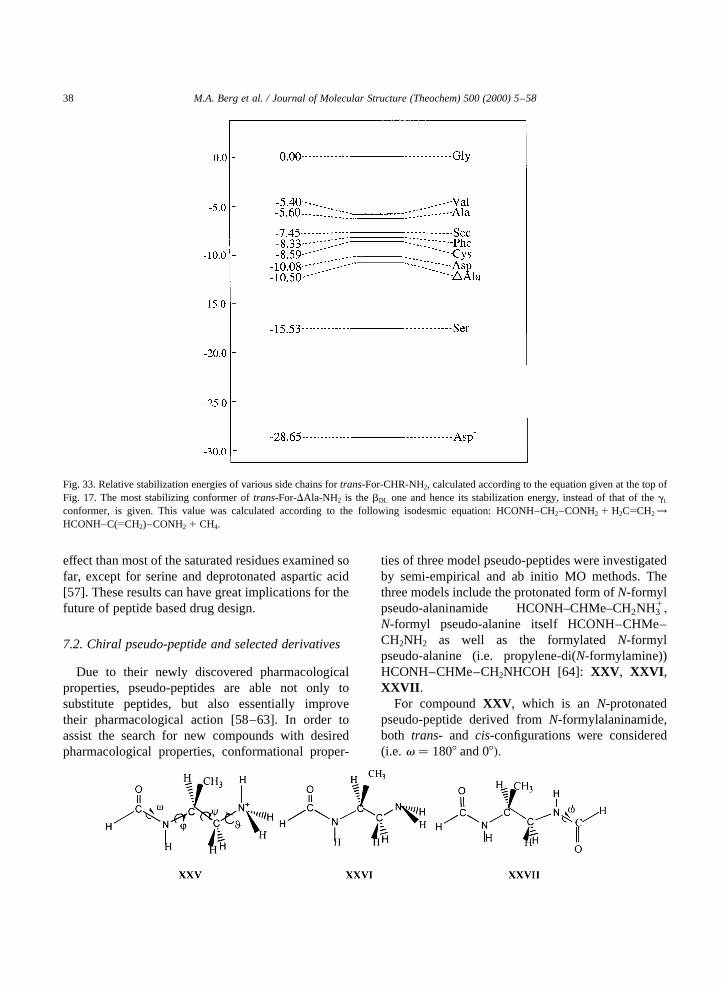
effect than most of the saturated residues examined sofar, except for serine and deprotonated aspartic acid[57]. These results can have great implications for thefuture of peptide based drug design.
7.2. Chiral pseudo-peptide and selected derivatives
Due to their newly discovered pharmacologicalproperties, pseudo-peptides are able not only tosubstitute peptides, but also essentially improvetheir pharmacological action [58–63]. In order toassist the search for new compounds with desiredpharmacological properties, conformational proper-
ties of three model pseudo-peptides were investigatedby semi-empirical and ab initio MO methods. Thethree models include the protonated form ofN-formylpseudo-alaninamide HCONH–CHMe–CH2NH1
3 ;
N-formyl pseudo-alanine itself HCONH–CHMe–CH2NH2 as well as the formylatedN-formylpseudo-alanine (i.e. propylene-di(N-formylamine))HCONH–CHMe–CH2NHCOH [64]: XXV , XXVI ,XXVII .
For compoundXXV , which is an N-protonatedpseudo-peptide derived fromN-formylalaninamide,both trans- and cis-configurations were considered(i.e. v � 1808 and 08�:
M.A. Berg et al. / Journal of Molecular Structure (Theochem) 500 (2000) 5–5838
Fig. 33. Relative stabilization energies of various side chains fortrans-For-CHR-NH2, calculated according to the equation given at the top ofFig. 17. The most stabilizing conformer oftrans-For-DAla-NH2 is thebDL one and hence its stabilization energy, instead of that of thegl
conformer, is given. This value was calculated according to the following isodesmic equation: HCONH–CH2–CONH2 1 H2CyCH2!HCONH–C(yCH2)–CONH2 1 CH4.
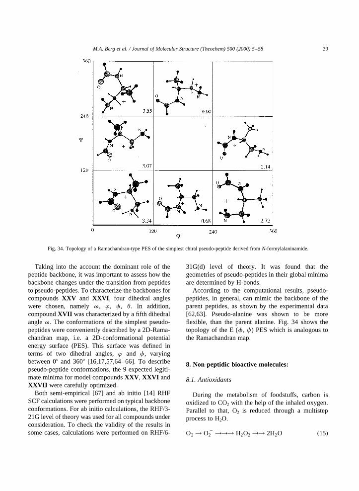
Taking into the account the dominant role of thepeptide backbone, it was important to assess how thebackbone changes under the transition from peptidesto pseudo-peptides. To characterize the backbones forcompoundsXXV and XXVI , four dihedral angleswere chosen, namelyv , w , c , u . In addition,compoundXVII was characterized by a fifth dihedralanglev . The conformations of the simplest pseudo-peptides were conveniently described by a 2D-Rama-chandran map, i.e. a 2D-conformational potentialenergy surface (PES). This surface was defined interms of two dihedral angles,w and c , varyingbetween 08 and 3608 [16,17,57,64–66]. To describepseudo-peptide conformations, the 9 expected legiti-mate minima for model compoundsXXV , XXVI andXXVII were carefully optimized.
Both semi-empirical [67] and ab initio [14] RHFSCF calculations were performed on typical backboneconformations. For ab initio calculations, the RHF/3-21G level of theory was used for all compounds underconsideration. To check the validity of the results insome cases, calculations were performed on RHF/6-
31G(d) level of theory. It was found that thegeometries of pseudo-peptides in their global minimaare determined by H-bonds.
According to the computational results, pseudo-peptides, in general, can mimic the backbone of theparent peptides, as shown by the experimental data[62,63]. Pseudo-alanine was shown to be moreflexible, than the parent alanine. Fig. 34 shows thetopology of the E (f , c ) PES which is analogous tothe Ramachandran map.
8. Non-peptidic bioactive molecules:
8.1. Antioxidants
During the metabolism of foodstuffs, carbon isoxidized to CO2 with the help of the inhaled oxygen.Parallel to that, O2 is reduced through a multistepprocess to H2O.
O2 ! O22 !!! H2O2 !! 2H2O �15�
M.A. Berg et al. / Journal of Molecular Structure (Theochem) 500 (2000) 5–58 39
Fig. 34. Topology of a Ramachandran-type PES of the simplest chiral pseudo-peptide derived fromN-formylalaninamide.
Somewhere along this line, hydroxy radical (HOz) isreleased in small amounts. Due to the non-discrimi-native reactivity of (HOz), it damages a number of keyorganic compounds in the human body. Among theseare DNA, the damage of which leads to prematureaging as well as numerous including cancer.Collectively, these processes are labeled as “oxidativestress” and can be relieved by free radical scavengers,called antioxidants, such as flavones, selenocysteineand lycopene. It has also been recognized recently thatcertain drugs can act as antioxidants.
8.1.1. Flavone conformationsFlavone is the parent molecule of a broad range of
compounds belonging to the family of flavonoids.Flavonoids are polyphenolic compounds, whichoccur as yellow pigments in plants. They are believedto be effective against HIV, cancer, coronary arterydisease and aging [68].
Conformational analyses were performed onflavone and three other related compounds, namely2-phenyl pyranone, b-phenyl naphthalene andbiphenyl. In particular, the phenyl rotation withrespect to the rest of the molecule was studied usingHartree–Fock (HF) calculations performed onGaussian 94. The basis sets employed were STO-3G and 3-21G. It was concluded that the hydrocarbons(biphenyl andb-phenyl naphthalene) are noticeablydifferent from oxygen containing heterocyclicanalogues such as 2-phenyl pyranone and flavone, asfar as phenyl rotation is concerned. One would alsoexpect a difference in their reactivity towards freeradicals. It seems that only the oxygen heterocyclicanalogues can be reactive in nucleophilic and radicalattack because they contain positively charged atoms,as shown by computed Mulliken charges. Neverthe-less, the topology at the potential energy curves(number of minima and transitions structures) is thesame for all four compounds.
The potential energy curve of flavone is shown inFig. 35. The antioxidant mechanism of flavoneskeleton is now under investigation.
8.1.2. Selenocysteine as antioxidantSelenocysteine is involved in a variety of biochem-
ical interactions and has recently been considered asthe 21st amino acid used by nature for RNA directedprotein synthesis [69,70]. Several selenoproteins
containing selenocysteine at their active sites havebeen identified using neutron activation analysis. Atthe end of the 20th century, possibly 28 of theseproteins are known to exist. At present, however, weknow very little about the structure and function ofmost of these proteins. Those that have been charac-terized and for which functions are known include thefollowing:
(a) Four species of glutathione peroxidase [71–73](cellular or classic, extracellular or plasma,gastrointestinal and phospholipid hydroperoxideglutathione peroxidase) have been identified.Glutathione (GSH) peroxidase catalyses (Fig. 36)the reduction of a variety of hydroperoxides includ-ing lipid hydroperoxides (which, in the presence oftrace metals, can form toxic lipid radicals). Thismechanism is believed to protect biomembranesand other essential cellular components againstoxidative challenge [74–76], cancer [77] andgastric ulceration [78].(b) Selenoprotein P is an extra-cellular proteinpresumably containing 10 Selenocysteine residuesthat are encoded by the UGA stop codon in the openreading frame of the mRNA. Some indirectevidence suggests that selenoprotein P acts as afree radical scavenger [79].(c) Selenoprotein W is found in muscles [80].(d) Iodothyronine deiodinase catalyzes the de-iodination of thyroxine to the biologically moreactive thyroid hormone triidothyronine [81].(e) Thiroredoxin reductase catalyzes the NADPH-dependent reduction of the redox protein thio-redoxin. Thioredoxin stimulates cell growth and isover-expressed in a number of human cancerouscells [82,83].(f) Mitochondrial selenoprotein [84] was localizedin the mitochondrial membranes of endocrineorgans.(g) Prostatic selenoprotein and testicular seleno-protein were found in spermatid nuclei. Recentfindings suggest that it takes part in the process ofreplacing and condensation of the DNA, whichoccur in the spermatid nuclei, and thus may havean important function in sperm development[85,86].
In addition to the above, new evidence, based onsequence analysis, suggests that HIV-1 encodes a
M.A. Berg et al. / Journal of Molecular Structure (Theochem) 500 (2000) 5–5840

protein containing selenocysteine (Sec) [87–89]. Ithas been observed previously that plasma seleniumand glutathione levels are subnormal in HIV-infectedindividuals [90] and that specifically, four75Se-containing proteins are lower in HIV-infected cellpopulations than in uninfected cell populations.
8.1.3. Lycopene and free radicalsLycopene is a very important free radical scavenger
[91,92]. It is a polyene with 15 conjugated doublebonds. There are numerouscis- and trans-isomers oflycopene (c.f. Fig. 37). The alltrans isomer is thepredominant species in tomatoes and tomato products(<95%). The 5cis-, 13 cis- and 9cis- lycopene are
M.A. Berg et al. / Journal of Molecular Structure (Theochem) 500 (2000) 5–58 41
Fig. 35. Phenyl torsional potential of the flavone skeleton.
Fig. 36. Putative catalytic mechanism of glutathione peroxidase.

found in human serum, making up approximately halfthe total lycopene content. Isomers with multiplecis-double bonds are also known. For example 7,9,90,70
cis-lycopene is the naturally occurring form of lyco-pene in fresh Tangerine-type tomatoes is also shownin Fig. 37.
Two isomers of 1,3,5 hexatriene were used tomodel cis- and trans-lycopene. The central doublebond (D [4–6]) of hexatriene was used in thecis-and trans-forms. For the computational convenience,free radical fluorine atom (zF) was used instead ofthe biologically more important hydroxy radical( zOH).
Through ab initio calculations, using ROHF/6-3111G(d,p) and UHF/6-31G(d,p) methods, it hasbeen found that if C2 or C3 of the 1,3,5 hexatrieneis fluorinated, both thetransandcis isomers have thesame relative energies. On the other hand, fluorinatingC1 makes a difference of 3.7 kcal/mol between thetrans and the cis isomer, the trans isomer beingmore stable Fig. 38.
8.1.4. Carvedilol as a new antioxidantThe Merck Index recognizes carvedilol (XXVIII )
as a nonselectiveb-blocker with vasodilating activity,
M.A. Berg et al. / Journal of Molecular Structure (Theochem) 500 (2000) 5–5842
Fig. 37. Geometrical isomers of lycopene.

which is mediated by its antagonizing effect ona-adrenoceptors. It was firstly patented in Germanyin 1979 and subsequently in the US in 1985.
Carvedilol has been used in the treatment ofhypertension [93], angina pectoris and congestiveheart failure. In these fields, carvedilol is at least aseffective as reference drugs (metoprolol [94],enalapril, verapamil [95]) but its side effect profile isbetter, and it has more beneficial effects.
According to clinical trials, carvedilol effectivelyreduced the morbidity and mortality rates in-patients suffering from chronic heart failure tosuch an extent that the US Data and Safety Moni-toring Board stopped, for ethical reasons, the inves-tigation before its completion [96]. This was thefirst time in the history of drug investigations thata clinical trial was stopped because the drug wastoo effective. It seemed unethical to deprive themedication from placebo group. Carvedilol alsoreduces the frequency of reversible myocardialischaemic events after thrombolysis [97] andprevents cardiac remodeling in-patients sufferingfrom left ventricular dysfunction after acute
myocardial infraction [98]. This latter effect hasbeen attributed to its scavenging action of oxygenfree radicals and preventing the lipid peroxidation[97]. These reactive radicals are implicated in theprocess of programmed cardiac cells death (apop-tosis) [99] and the concomitant loss of myocardialcells leading to progressive decrease of left ventri-cular mass and function [100].
Originated from this antioxidant property,carvedilol may also prevent the development ofnitrate tolerance in, patients receiving continuousnitrate therapy [101,102].
Apart from carvedilol, at least one of its 3-hydro-xylated metabolites (XXIX ) has marked antioxidantproperties. The metabolite is approximately athousandfold more effective than vitamin E in certainexperimental setups and more effective thancarvedilol itself. Although the exact antioxidantmechanism of these compounds is not known, recentstructure activity relationships indicate that its activityresides in the carbazol ring. A computational mechan-istic study on this exceptional antioxidant capability isnow in progress.
M.A. Berg et al. / Journal of Molecular Structure (Theochem) 500 (2000) 5–58 43
Fig. 38. Relative energies of radical addition products to lycopene models computed at RHF/3-21G level of theory.

8.2. Medicinal chemistry and drugs
Most of today’s marketed medications were foundby chance or by systematic screening of large collec-tions of compounds (called “libraries”) either fromnatural or man-made sources. Current emphasis onhigh-throughput screening and combinatorial chemis-try suggests that an educated guess is pivotal for thediscovery of tomorrow’s drugs. Computer assistedmolecular modeling (CAMM) is a relatively newand rapidly developing tool in drug design. Computergraphics techniques allow the transformation ofcomplex data sets, obtained e.g. from theoreticalchemical calculations or X-ray diffraction patterns,into an image on a computer screen. Chemicalstructures and their properties may thus bevisualized, manipulated, and matched or combinedwith other relevant molecules. The availability ofthe three-dimensional architecture of HIV protease,thymidylate synthase, thrombin, carbonic anhydrase,and many more has led to at least an equal number ofnew chemical entities being evaluated and used now inclinics. In such cases, CAMM has certainly led to amore rational approach toward drug discovery, inparticular when the binding domains for inhibitorsand substrates are known from cocrystallizationexperiments. Knowing the “lock” definitely helps indesigning the “key”, although the interaction betweennewly synthesized ligands and the target macromole-cule may appear sometimes unexpectedly.
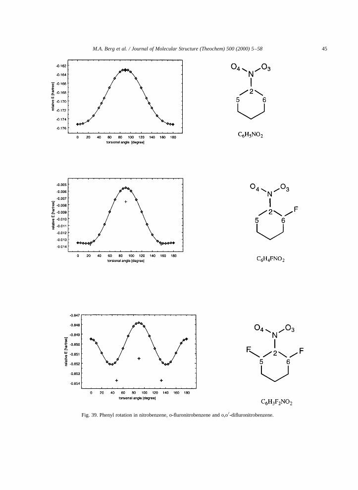
Locking into certain stereochemically importantconformations is sometimes achieved by heavysubstitution, such aso,o0 difluorophenyl groups.This principle has been applied in one of the anti-HIV drugs whereo,o0 difluoride substitutions areused [103].
Calculations of the barriers and associated one-dimensional torsional potential are performed for
the internal rotation of the nitro-group in nitroben-zene as well as foro-fluoro- ando,o0-difluoro-nitro-benzenes [104]. This structure is related to certaindrugs, such as the one shown inXXX , that areunder investigation. It is shown that the presenceof substituents inortho-positions forces the benzenering to rotate about the C–N bond, out of the planeof the nitro group. In these conjugated molecules,the inclusion of electron correlation is shown to benecessary for reliable barrier prediction. The poten-tial curves at HF level are shown on Fig. 39.Corresponding MP2 values are given by “1 ”symbol. It is found that thep -stabilization (reso-nance) is of relatively small contribution to thestructure of the nitroaromatics, whereas the stericrepulsion between the ortho-substituents andoxygen atoms of the NO2 group is rather signifi-cant. The interplay between them accounts for thefact that for o,o0-C6H3F2NO2, the barrier height at08 is larger than that at 908, when electron correla-tion is included. The values for the barrier at 08were 2.67 and 3.81 kcal/mol computed without andwith electron correlation and the correspondingvalues at 908 were 1.42 and 0.91 kcal/mol, respec-tively.
A fairly large area of drug design is related to G-protein coupled receptors. A preliminary structuralanalysis of theb2 adrenergic G-protein coupledreceptor was considered. This receptor is a trans-membrane protein, and it usually consists of sevenbundles ofa-helices. Each of these seven helicesconsists of approximately 21–24 amino acids, asrequired to span the cell membrane. In order to deter-mine the type of interaction between the helices, ana-helix made up of seven alanine residues was builtusing HYPERCHEM. Two other helices were builtin the same manner, except one of the amino acidswas changed to serine and tryptophan respectively. Itwas found that amino acids arrange so that many ofthe polar amino acids can face the cavity and non-polar amino acids can face away from the cavity.This is expected because a hydrophobic lipidmembrane surrounds the outer circumference of thebundle of seven helices. Interactions between resi-dues of a helix such as that between tryptophanand serine are practically thermo-neutral. Interactionbetween the Ala-Ala helices has a stabilizationenergy of 23.0 kcal/mol, while that between the
M.A. Berg et al. / Journal of Molecular Structure (Theochem) 500 (2000) 5–5844
M.A. Berg et al. / Journal of Molecular Structure (Theochem) 500 (2000) 5–58 45
Fig. 39. Phenyl rotation in nitrobenzene, o-fluronitrobenzene and o,o0-difluronitrobenzene.

Ser-Ser helices has a stabilization energy of28.2 kcal/mol [105]. The interaction between twoserines result in a greater stabilization energy thanthat of the two alanine residues interacting. Thus,replacing the alanine with serine resulted in largerstabilization energy but replacing serine with trypto-phan did not significantly change the stabilizationenergy. An ab initio study on the helical structure
of For-(Ala)n–NH2 has been carried out forn�6; 8;10 and 12 (c.f. Fig. 40).
Cell cycle inhibitors or modulators that haltuncontrollable tumor growth are regarded as highlypromising new therapeutic agents against humancancer. Certain sulfonamides [106] inhibit micro-tubule assembly owing to its reversible binding tothe colchicine-binding site on tubulin. They also
M.A. Berg et al. / Journal of Molecular Structure (Theochem) 500 (2000) 5–5846
Fig. 40. Helical structure of N- and C-protected polyalanines HCO-(Ala)n-NH2 �n� 6; 8;10;12�:

exhibit good in vivo antitumor activity against variousrodent tumors and human tumor xenografts. The focuson making a sulfonamide compound library is basedupon templateXXXI ever since compoundXXXIIwas found to inhibit cellular growth and mitosis invitro, but not quite potent in vivo. In the design ofthis template, the sulfonamide moiety located betweentwo aromatic rings was fixed as a basic motif, and theNH group at theorthoposition of the sulfonamide wasconsidered a key functionality for substantial antipro-liferative activity in cell-based assays [106].
A substantially different aromatic sulfonamide(XXXIII ) has also been reported [107] to interactwith microtubules during cell division. Concerningthe recently reported polyfluorodiarylsulfonamide(XXXIII ), we are now involved in assessing itsmechanism of action.
9. Multidimensional conformational analysis ofallyl methyl disulfide: a key component of garlic
Garlic has been one of the most popular medi-cally researched plants, with over 1300 researcharticles in only the last 100 years [108]. Organo-sulfur compounds in garlic, like allyl methyldisulfide, have been found to be involved in anti-mutagenic, anticarcinogenic, antithrombotic, andlipid-lowering activities, and it has also beenfound to act as an antioxidant. Ab initio molecularcomputations were performed on allyl methyldisulfide (XXXIV )
with respect to torsional anglest1 � t�H3C2·CH2·S–S·CH3�; t2 � t�H3C2·CH2–S–S·CH3�; and t3 �t�H3C2–CH2·S·S·CH3�: Potential energy scans,
resulting in potential energy curves (PEC), wereperformed alonga, b and c as shown in Fig. 40,at the HF/3-21G level of theory. All conformationswere optimized at the HF/6-31Gp level of theoryand their energy values are provided in Fig. 41 aswell. The potential energy hypersurface surface(PEHS) of XXXIV , i.e. E � E�t1; t2; t3�; revealedsix lower energy pairs of enantiomeric minima (i.e.,�g1g1g1ug2g2g2�; �g1ag2ug2ag1�; �g1g2g1ug2g1g2�; �g1g1g2ug2g2g1�; �g1ag2ug2ag1�; and�g1g2g2ug2g1g1� as well as three higher energyminima (i.e. g1g1sug2g2s�; g1asug2as�; and�g1g2sug2g1s�� were optimized att1 � ^908usingthe two HF/6-31Gp and B3LYP/6-31Gp methods atthis level of theory (Fig. 40). In our previous studyon the multidimensional conformational analysis ofethyl benzene [109], we found that very often twominima result when a planar moiety is rotated againsta tetrahedral moiety, one of them being higher (08rotation) and the other being lower (908 rotation) inenergy.
As illustrated in Fig. 41, it is apparently clearthat the center of symmetry of the PEHS ofXXXIV is at t1 � t2 � t3 � 1808; i.e. at the fullyanti–anti–anti orientation. Therefore, if an imagin-ary line can be drawn through this fullyanti centerfrom one energy platform to the other on the PEHSof XXXIV , then the structures along such a lineare enantiomers. Conversely, if an imaginary linejoining two conformers cannot be drawn throughfully anti center, then the structures are diastereo-mers. Therefore, although there are no stereocentresin XXXIII , there is chirality in the conformationaltwist with respect to the fully symmetricalanti-anti-anti structure [aaa] through t1 � t2 � t3 �1808:
In order to denote the enantiomeric relationship ofeach pair of conformers, a convenient notation hasbeen adopted, with the symbol “u” representing a
M.A. Berg et al. / Journal of Molecular Structure (Theochem) 500 (2000) 5–58 47

mirror between two enantiomers. The global mini-mum was determined to be the�g1g2g1ug2g1g2�conformer, and the fully symmetricalanti [aaa]conformer was determined to be a second order saddlepoint (Fig. 42) [126].
Based on the energies and MO diagrams ofXXXIV , the HOMO and LUMO1 1 orbitals weredetermined to be involved in electron donating andaccepting activity ofXXXIV . The putative anti-carcinogenic and cholesterol lowering mechanism ofactivity of XXXIV is presented in Fig. 43 [126].
10. Macromolecular interactions
10.1. Protein–protein interactions
Molecular recognition forms the basis of protein–protein interactions. Proteins that contain covalentlybound oligosaccharides, called glycoproteins, areoften tailored to be specific recognition sites. Theyform a diverse group that includes enzymes,hormones structural proteins, and transport proteins.
The presence of one or more oligosaccharide chains
M.A. Berg et al. / Journal of Molecular Structure (Theochem) 500 (2000) 5–5848
Fig. 41. Topology of the conformational the PEHS ofXXXIV (allyl methyl disulfide), whereE � E�t1; t2; t3�: (A) The notation of the 6 pairs oflower energy conformers (along a, a0, b and b0), the 3 pairs of higher energy conformers (along c and c0) and the fully symmetricalanti structure.(B) The relative energies in kcal/mol of the 6 pairs of lower energy conformers, the 3 pairs of higher energy conformers, and the fullysymmetricalanti structure. (All energy values obtained at the HF/6-31Gp level of theory). Note that the centre of symmetry is denoted by asolid dot and corresponds to thet1 � t2 � t3 � 1808; fully anti conformation (a a a) as shown in Fig. 42. [Cross-sections were studied ata,a0,b,b0,c and c0]. All conformations were optimized at the HF/6-31Gp level of theory.
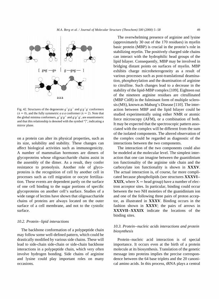
on a protein can alter its physical properties, such asits size, solubility and stability. These changes canaffect biological activities such as immunogenicity.A number of mammalian hormones are dimers ofglycoproteins whose oligosaccharide chains assist inthe assembly of the dimer. As a result, they conferresistance to proteolysis. Another role of glyco-proteins is the recognition of cell by another cell inprocesses such as cell migration or oocyte fertiliza-tion. These events are dependent partly on the surfaceof one cell binding to the sugar portions of specificglycoproteins on another cell’s surface. Studies of awide range of lectins have shown that oligosaccharidechains of proteins are always located on the outersurface of a cell membrane, and not to the cystolicsurface.
10.2. Protein–lipid interactions
The backbone conformation of a polypeptide chainmay follow some well-defined pattern, which could bedrastically modified by various side chains. These willlead to side-chain side-chain or side-chain backboneinteractions in a polypeptide chain, which very ofteninvolve hydrogen bonding. Side chains of arginineand lysine could play important roles on manyoccasions.
The overwhelming presence of arginine and lysine(approximately 30 out of the 170 residues) in myelinbasic protein (MBP) is crucial in the protein’s role instabilizing myelin. The positively charged side chainscan interact with the hydrophilic head groups of thelipid bilayer. Consequently, MBP may be involved inbridging distant points on surfaces of myelin. MBPexhibits charge microheterogeneity as a result ofvarious processes such as post-translational deamina-tion, phosphorylation and the deamination of arginineto citrulline. Such changes lead to a decrease in thestability of the lipid-MBP complex [109]. Eighteen outof the nineteen arginine residues are citrullinated(MBP Citl8) in the fulminant form of multiple sclero-sis (MS), known as Maburg’s Disease [110]. The inter-action between MBP and the lipid bilayer could bestudied experimentally using either NMR or atomicforce microscopy (AFM), or a combination of both.It may be expected that the spectroscopic pattern asso-ciated with the complex will be different from the sumof the isolated components. The altered observation ofthe complex could be regarded as diagnostic of theinteractions between the two components.
The interaction of the two components could alsobe modeled at the molecular level. The simplest inter-action that one can imagine between the guanidiniumion functionality of the arginine side chain and thecarboxylate ion functionality is shown inXXXV .The actual interaction is, of course, far more compli-cated because phospholipids (see structuresXXXVI–XXIX , where X� head group) have a number of elec-tron acceptor sites. In particular, binding could occurbetween the two NH moieties of the guanidinium ionand one of the following three pairs of proton accep-tor, as illustrated inXXXV . Binding occurs in thefashion shown inXXXV ; the pairs of arrows inXXXVII–XXXIX indicate the locations of thebinding sites.
10.3. Protein–nucleic acids interactions and proteinbiosynthesis
Protein–nucleic acid interaction is of specialimportance. It occurs even at the birth of a proteinmolecule at its biosynthesis. Translation of the geneticmessage into proteins implies the precise correspon-dence between the 64 base triplets and the 20 canoni-cal amino acids. In this process, tRNA plays a central
M.A. Berg et al. / Journal of Molecular Structure (Theochem) 500 (2000) 5–58 49
Fig. 42. Structures of the degenerateg1g-g1 andg-g1g- conformers�l � 0�; and the fully symmetrica a aconformer�l � 2�: Note thatthe global minima conformers,g1g-g1 andg-g1g-, are enantiomericand that this relationship is denoted with the symbol “u”, indicating amirror plane.

M.A. Berg et al. / Journal of Molecular Structure (Theochem) 500 (2000) 5–5850
Fig. 43. Putative mechanism of the (A) anticholesterol and the (B) anticarcinogenic activity ofXXXIV (allyl methyl disulfide).

role by providing the nascent polypeptide with theamino acids to which they are esterified in responseto codons on RNA. In this step, the amino acylatedtRNAs decipher the base triplet (codon) usingWatson–Crick base pairings between mRNA and acomplimentary region on the tRNA. The anticodonis independent of the nature of the amino acid ester-ified to the tRNA. Therefore, the accuracy of thetRNA aminoacylation reaction, ensured by theaminoacylation tRNA synthetases aaRS (a givenaminoacyl-tRNA synthetase is abbreviated by itscognate amino acid three-letter code followed byRS; e.g., GlnRS stands for glutaminyl-tRNA synthe-tase), is of primary importance in all living cells, sinceit will govern to a large extent the fidelity of thetranslation process [111].
Aminoacylation of tRNA proceeds through a twostep mechanism. The first step involves the formationof a stable aaRS-amino acyladenylate complex
resulting from the specific binding and reaction withATP:Mg21 of the amino acid; pyrophosphate is subse-quently released. In the following step the 30 terminaladenosine of enzyme-bound tRNA reacts with theaminoacyladenylate, leading to both the esterificationof the tRNA and the release of AMP. Notably, thebinding of tRNA occurs in the presence of a poly-amine or a divalent cation, usually magnesium [112].
aaRS1 ATP : Mg21 1 aa
! aaRS: aa, AMP 1 PPi;Mg �16�
aaRS: aa, AMP 1 tRNA
! aaRS1 aa2 tRNA 1 AMP �17�where aa stands for amino acid; aa, AMP foraminoacyladenylate.
M.A. Berg et al. / Journal of Molecular Structure (Theochem) 500 (2000) 5–58 51

Because the two steps involve the specific recogni-tion of both the amino acid and the tRNA they imposespecificity throughout the overall reaction. As ageneral rule, there is one synthetase for each of the20 canonical amino acids in a given organism or orga-nelle. Two main functions are carried out by an aaRS:the activation of the amino acid and the recognition ofthe tRNA molecule. In the activation of the aminoacid, the carboxylate of the amino acid becomeslinked to the AMP moiety of an ATP molecule toform the aminoacyladenylate, which remains firmlybound to the synthetase [113].
The mixed anhydride bond between the carboxylateand the phosphoryl group of AMP is highly suscep-tible to the attack by a hydroxyl group, thereby facil-itating further transfer of the amino acid moiety ontothe tRNA. Consequently, the reaction of the activatedamino acid with tRNA can be viewed as the result of aproductive positioning of the 30-terminal adenosine ofthe nucleic acid within the active center of the syn-thetase. This allows the attachment of the aminoacyl-AMP by either the 20- or 30 hydroxyl group ofthe terminal ribose. In this context, the accuracy of theactivation reaction is of importance in the furtherspecificity of the tRNA amino acetylation reaction.In all cases the rapid formation of a ternary complexof the enzyme with both ATP:Mg21 and amino acidwas evident. The amino acid acyladenylate isproduced through a rate-limiting isomerization ofthis ternary complex PP:-Mg, one of the products, isthen released.
The other function of the aaRS is the recognition ofthe tRNA molecule. Many recent studies have pointedout that the occurrence of discrete nucleotidic recog-nition elements on a tRNA molecule dictates thespecificity of aminoacylation by the correspondingaaRS. Briefly, three major recognition sites havebeen identified:
(i) the acceptor stem, including the “discriminatorbase”;(ii) the three bases of the anticodon;(iii) the d-loop.
In some systems, the three sites are important forcorrect aminoacylation by the corresponding aminoacid, whereas in others, one site is enough. However,at least two sites appear to be required systematicallyfor achievement of maximal aminoacylation rates.
Because a limited number of recognition sites arepresent on the tRNA molecule, it appears that theremay be a limited number of recognition sites (peptidesregions) on the cognate aaRS, too. The aaRS–aminoacid interaction is based on protein structure rules, butother kinds of rules govern the tRNA–aaRS inter-action. An unsolved question in the molecular biologyof gene expression is the determination of these rules.Discrimination between tRNAs by the aaRS enzymesis complex because all tRNAs are about 76 nucleotideresidues in length and form the typical tRNA clover-leaf secondary structure [114–117].
In the process of investigating protein synthesis ingeneral, an exploratory set of ab initio molecularorbital computations were carried out on the variousconformations of ribose and deoxy-ribose modelcompounds to establish the nature of possible inter-actions between the 50-hydroxyl and the oxygen of theribose ring. Moreover, the investigation was extendedto the hydrogen bonding between the two adjacenthydroxyls, 20- and 30-, to explore their effect on thestabilization of the whole system. The consistentpresence of a rather unusual weak hydrogen bondbetween the proton of C20–H and the O50 of the hydro-xyl methyl group for theg2g2 conformation of the–CH2-OH functional group has been studied usingBader’s atoms in molecules (AIM) method.
Density functional calculations coupled withBader’s Atoms in Molecule (AIM) method arebecoming a very important tool in examining intra-molecular interactions that are responsible for thesystem’s stabilization. As has been demonstrated[118], an unusual hydrogen bond between C50–O50·· ·H20 is present for the specificg2g2 orientationof the –CH2-OH moiety in the ribose system.
10.4. Carbohydrates and molecular recognition
Carbohydrates are sometime referred to asinforma-tion-molecules. This characterization of carbohy-drates is due to the fact that carbohydrates have avery large number of structural variants. First of allthere are several hexoses and they can connect to eachother to form oligosaccharides in different sequences.The chain can be branched or unbranched and theconnection can be viaa- or b-glycosidic linkage.Furthermore, the numerous OH groups can assumeg1, a and g2 rotamers. Such tremendous structural
M.A. Berg et al. / Journal of Molecular Structure (Theochem) 500 (2000) 5–5852

variation can code a great deal of information. Oligo-saccharides are frequently attached to proteins via O-or N-glycosidic linkage. The former one involveseither serine or threonine while the latter utilizesalmost exclusively asparagine. These carbohydrateantennas are involved in molecular recognitionprocesses. During the molecular recognition process,hydrogen bonds change from intramolecular to inter-molecular type. These processes could be studied interms of glycopeptides. However, before embarkingto the field of glycopeptide research one must under-stood the conformational intricacies of carbohydrateconformations.
We have modeled hydrogen bonding betweenadjacent OH groups in ethylene glycole [119,120].We have also investigate internal hydrogen bondingin a 6-dehydro hexose:l-fucose [121,122].d-glucose[123,124] as well asd-mannose [124] have also beenstudied in some details. An explanatory study of anoligosaccharide: the Lewis X trisaccharide [125] hasjust been completed. We are now engaged to studyglycopeptides.
11. The mystery of protein folding
Protein synthesis occurs sequentially, one aminoacid at a time. Initially, it was tacitly assumed thatonce the synthesis of the primary sequence iscompleted, the nascent macromolecule is folded toits bioactive conformation. Originally, it was furtherassumed that the folded structure is the global mini-mum, and thermodynamics will drive the structureto this uniquely folded structure. Relatively soon, itbecame obvious that it takes an enormous amount oftime “to search the entire conformational spaceavailable for a polypeptide chain” [8]. This principlebecame known as the Levinthal’s paradox [7].Latter, it was envisaged that the bioactive formmay not be the global minimum, but in fact itcould be one of the local minima. Thus, thermody-namics could not be treated as “Maxwell’s demons”to guarantee such a specifically folded structure,even if the necessary time was available. At thattime, chaperones were invoked as an explanationof post biosynthesis folding. This view effectivelyallows the interaction between the chaperone,and the nascent polypeptide while it is being
synthesized. Thus, the folding could in fact occurwhile the synthesis is in progress.
Nowadays, it is customary to look upon folding interms of statistical ensembles of states. However, suchensembles of states are nothing more than energeti-cally and conformationally closely spaced minima ona rather complex multivariable potential energyhypersurface (PEHS). Professor Honig greets the“light at the end of the tunnel” with the followingacclamation [8]:
It is likely that a complete solution to all aspectsof the protein folding problem will require thatthe information available from the analysis oflarge structural and sequence databases becombined with the principles and methodsof physical chemistry and computationalchemistry.
We concur with his view. However, we wish to addthat computational chemistry should go beyondcurrently used empirical force fields. It must startwith ab initio molecular computations, which willeventually lead to an accurate analytic function thatencompasses all possible protein conformers as theroots of the gradient of such an analytic function.The search for the secret of folding patterns can reallystart in the possession of such roots. The mysteryassociated with protein folding will gradually fadein the light of new understanding.
12. Proposal for a millennial mega-project andconclusion
Once a complete set of data exists for all 21 aminoacids and all of their ‘found’ backbone conformations,a database may be constructed. This large data setmay then be used in the construction of di-, tri- andeven large arrays of peptides, with the ability ofaccepting changes at any time. However, as thenumber of units in a polypeptide chain grows linearly,the number of possible combinations and conforma-tions grows exponentially. Therefore, without ques-tion, an automated procedure is necessary for boththe construction of input files as well for the extractionand tabulation of relevant output parameters.
Even with a successful and efficient digital script orprogram to perform these tasks, the current problem
M.A. Berg et al. / Journal of Molecular Structure (Theochem) 500 (2000) 5–58 53

of insufficient computational power and other hard-ware resources would still exist. This is the largest andomnipresent limitation in computational chemistry,amicably referred to as the ‘limiting reagent’ in thesynthesis of ab initio solutions to chemical problems.The present solution of continually upgrading tolarger and faster hardware will not bring about theelimination of this low yield.
The implication of truly parallelized software andmore efficient optimization algorithms is one part ofthe solution. The other lies in the creation of a distrib-uted network, through which calculations may be sentout to various individuals, institutions and organiza-tions willing to donate free CPU down-time.
As PC’s have come to the forefront of the cost toperformance ratio, many nodes are now available torun large arrays of computations, both quickly andefficiently. All that is required is a means to managethe incoming and outgoing data, in an organized anderror-free manner. The fragmentation of jobs, forparallelization, may be accomplished by sending acopy of the electron-density and Fock matrices toeach node in the array. Portions of the Hessian arethen constructed and assembled into a complete setafterwards.
This may be accomplished using a standardizednumbering method for the molecules to be studied.With this common input parameter definition, poly-peptides may be constructed without error and opti-mized quickly.
A novel and sound proposal has been made to opti-mize all possible tri-peptide (IV ) combinations andconformations. The reason for choosing tri-peptideslies in that fact that allN-acetyl andN-methyl inter-actions and resultant conformations possible with acentral peptide would be evaluated. Hence, oncecompleted, two tri-peptide blocks could then bebound together and optimized more quickly than asingle calculation of a hexapeptide. This is due tothe fact that the internal structure of each tri-peptideunit would not change dramatically, upon biding to asecond one. These ‘blocks’ could subsequently beused to give insight into the most stable geometry ofmuch larger polypeptide chains, both quickly andefficiently.
For the automation of this input generation andsubsequent data extraction from the log files, thenumbering of the atomic sequence in the peptides
must be standardized. In this way, scripts andprograms may be used to reorganize immediatepatterns necessary for one’s chemical analysis. Asan example, we use ArginineXL for this:
As all amino acids contain the same backbonesequence, the numbering is started from the nitrogenattached to the alpha C, following through to thecarbonyl carbon all the way to the carbonyl oxygen.The groups on the nitrogen and subsequently the alphaC are numbered next, with the side chain the hydrogenattached to the alpha C. TheN-Acetyl or N-Methylgroups are defined separately and then added at anytime, as their coordinates do not disturb the definitionsof the internal parameters specific to the core (i.e.amino acid residue).
In the case of di-peptides or larger polypeptideslengths, the numbering is left ‘open’, in the form ofvariables, so that the files may be modified at any timeat any of these groups without disturbing the structureof the remaining coordinates, or the molecular envir-onment it defines. As the internal coordinate systemrelies on fixing subsequent atoms to previouslydefined ones, the next amino acid would join to thecarbonyl carbon of the first. Hence, any of the stan-dardized amino acids in this database, numberedbeginning at the N attached to the Ca, would then‘mesh’ with the internal coordinates of the firstwithout problem. In fact, any of the groups or atomsattached to the backbone may be denoted by a variableof choice and modified or truncated accordingly.
M.A. Berg et al. / Journal of Molecular Structure (Theochem) 500 (2000) 5–5854

Although an extensive explanation of the abovenumbering sequence would be required to show allthe inherent benefits of this system, the reader isencouraged to attempt trials on various input files.
In this way, it is possible to make an additive orintegrateable database for the 20 amino acids, with thepoint of attachment in a polypeptide chain beingdefined as the #1 atom, in the coordinate systemused. Other previously optimized structures can thenbe added to the database, including theN-acetyl andN-methyl portions, or even long chain fatty acids,provided they use the standardized numbering system.Simply adding the coordinate definitions onto theinternal coordinate system of that original structuremay then extend an optimized structure. Any poly-peptide combination of any possible conformationcan be easily created and submitted for optimizationefficiently and can be easily automated. With the newmillennium, a massive mega-project is proposed touse the above process to explore the large numberof potential energy surfaces created by this giganticnumber of peptide structures. For example, a tri-peptide sequence would contain many millions ofpossible combinations and conformations to be opti-mized; this number grows exponentially with theextension of the polypeptide chain.
To create a surface large enough to help in theunraveling of the protein folding problem, one mustcontinue to explore such surfaces with progressivelylarger sections of the amino acid polymer. Thiscreates a massive demand for both CPU time, hardstorage space and data management and extraction;the latter being a much less significant obstacle. Theall too common problem of CPU power being the‘bottle neck’ in the creation of meaningful ab initioresults may be solved via a different avenue. Ratherthan throwing massive amounts of hardware in tightlycoupled networks at the challenge, instead it isproposed to farm-out the CPU time to the millionsof stand alone PC’s and workstations residing on theloosely coupled network of the internet.
Some computational problems and data analysisventures have indeed used this approach with muchsuccess. SETI@home is one example, whereby radio-telescope data from Arescebo in Puerto Rico areanalyzed by millions of individuals in their homes,using their PC’s. At last count, over 1.3 million peoplewere contributing computer time toward this effort.
The process is automated to use ‘down CPU time’,whereby the individual’s computer usage is notaffected by the running program, nor are theyburdened by any direct responsibility of managingtheir chunk of the data. Once the user needs theircomputer, the calculating program is stopped andthe CPU is entirely free for the user. Another suchventure is the DES/RC5 project, whose goal is toshow how US export limitations on the strength ofcryptographic algorithms is naive. They are under-taking this by having contributors’ computers tryevery possible decryption key in sequence againsta cipher. With over 100,000 CPUs working on thejob, cracking such encryption is relatively easy.The megaproject could indeed make use of a simi-lar such automated and ‘loosely coupled’ network,whereby surfaces of the many independent vari-ables may be analyzed. Those who wish to parti-cipate may register at the following e-mail address:[email protected].
It is part of human nature to ask what good willcome out of a project that aims to decode the secretof protein folding. Such a discovery may well beregarded as the greatest human achievement inhistory. Its social impact would be greater than thecombined social impact of:
the discovery of fire;the discovery of writing;the discovery of the wheel.
Even though this topic is very important for the wholehuman race, it cannot be done due to lack of fundsunless individual contributions come from millions ofpeople.
The deciphering of the law of protein folding wouldopen new dimensions to all life sciences, includingmedicine. For example, the drug discovery processwould be shortened dramatically, by a more cost-effective method for the production of pharmaceuticalproducts. Not only would the cure be found reason-ably soon (say by the year of 2025) for cancer, HIV, aswell as other diseases, preventive medicine wouldalso reach new heights whereby pathogen infections(bacteria, viruses and prions) could be eliminated.
Anyone, who has lost a loved one to an incurabledisease, appreciates the prospects of eliminating allhuman sufferings. This heartache includes the painof the ill, as well as the anguish of those who would
M.A. Berg et al. / Journal of Molecular Structure (Theochem) 500 (2000) 5–58 55

mourn after the patient has passed away. Needless tosay, a person lost prematurely by a family is also lostby society as a whole, with all of his/her talents andproductivity. Global hunger could also be eliminatedby improved agricultural and food-processing tech-nologies, in which the knowledge of protein foldingalso plays a significant role.
The peptide folding project, which one can partici-pate in, at the above e-mail address, does not offer aquick fix, but gradually it will lead to a new under-standing of protein folding, which in turn will producea healthier society enjoyed by generations to come.
Acknowledgements
The authors wish to acknowledge the continuoussupport of the National Research Council (NRC),and its funding-successor, the National Sciences andEngineering Research Council (NSERC) of Canadafor the period of 1964 to 1999. We would like tothank Dr. Carlos Sosa of Silicon Graphics Inc. forproviding computational resources at their facility inEagan, MN. We are also grateful to the NationalCancer Institute (NCI) for the use of services atFrederick Biomedical Supercomputing Center.Finally, we also wish to thank Velocet Communica-tions Inc. for database management, network support,software and distributive processing development.
References
[1] A. Furka, F. Sebestyen, M. Asgedom, G. Dibo, in: Highlightsof Modern Biochemistry, Proceedings of the 14th Inter-national Congress of Biochemistry, vol. 5, VSP, Utrecht,The Netherlands, 1988, p. 47.
[2] A. Furka, F. Sebestyen, M. Asgedom, G. Dibo, in: Proceed-ings of the 10th International Symposium of MedicinalChemistry, Budapest, Hungary, 1988, p. 288 (Abstract P-168).
[3] A. Furka, F. Sebestyen, M. Asgedom, G. Dibo, Int. J. Pept.Protein Res. 37 (1991) 487.
[4] K.S. Lam, S.E. Salmon, E.M. Hersh, V.J. Hruby, W.M.Kazmierski, R.J. Knapp, Nature 354 (1991) 82.
[5] R.A. Houghten, C. Pinilla, S.E. Bloudelle, J.R. Appel, C.T.Dooley, J.H. Cuervo, Nature 354 (1991) 84.
[6] B.A. Bunin, J.A. Ellman, J. Am. Chem. Soc. 114 (1992)10 997.
[7] C. Levinthal, J. Chim. Phys. 65 (1968) 44.[8] B. Honig, J. Mol. Biol. 293 (1999) 283.
[9] I.G. Csizmadia, M.C. Harrison, B.T. Sutcliffe, Q Prog. Rep.(MIT-SSMTG) 50 (1963) 1.
[10] I.G. Csizmadia, M.C. Harrison, B.T. Sutcliffe, Q Prog. Rep.(MIT-SSMTG) 59 (1966) 43.
[11] I.G. Csizmadia, M.C. Harrison, B.T. Sutcliffe, Theor. Chim.Acta 6 (1966) 217.
[12] M.A. Robb, I.G. Csizmadia, Theor. Chim. Acta 10 (1968)269.
[13] I.A. Topol, S.K. Burt, E. Deretey, T.-H. Tang, A. Perczel,I.G. Csizmadia, in preparation.
[14] M.J. Frisch, G.W. Trucks, H.B. Schlegel, P.M.W. Gill, B.G.Johnson, M.A. Robb, J.R. Cheeseman, T. Keith, G.A. Peters-son, J.A. Montgomery, K. Raghavachari, M.A. Al-Laham,V.G. Zakrzewski, J.V. Ortiz, J.B. Foresman, J. Cioslowski,B.B. Stefanov, A. Nanayakkara, M. Challacombe, C.Y.Peng, P.Y. Ayala, W. Chen, M.W. Wong, J.L. Andres, E.S.Replogle, R. Gomperts, R.L. Martin, D.J. Fox, J.S. Binkley,D.J. Defrees, J. Baker, J.P. Stewart, M. Head-Gordon, C.Gonzalez, J.A. Pople, Gaussian 94, Revision D.2, Gaussian,Inc., Pittsburgh, PA, 1995.
[15] J.C. Vank, C.P. Sosa, A. Perczel, I.G. Csizmadia, Can. J.Chem. 178 (2000) 395–408.
[16] A. Perczel, J.G. Angyan, M. Viviani, J.-L. Rivail, J.-F.Marcoccia, I.G. Csizmadia, J. Am. Chem. Soc. 113 (1991)6265.
[17] M.A. McAllister, A. Perczel, P. Csa´szar, W. Viviani, J.L.Rivail, I.G. Csizmadia, J. Mol. Struct. (Theochem) 288(1993) 161.
[18] W. Viviani, J.-L. Rivail, A. Perczel, I.G. Csizmadia, J. Am.Chem. Soc. 115 (1993) 8321.
[19] O. Farkas, M.A. McAllister, J.H. Ma, A. Perczel, M. Hollo´si,I.G. Csizmadia, J. Mol. Struct. (Theochem) 369 (1996) 105.
[20] A. Perczel, O. Farkas, I.G. Csizmadia, Can. J. Chem. 75(1997) 1120.
[21] O. Farkas, A. Perczel, J.F. Marcoccia, M. Hollo´si, I.G. Csiz-madia, J. Mol. Struct. (Theochem) 331 (1995) 27.
[22] A. Perczel, O. Farkas, I.G. Csizmadia, J. Comp. Chem. 17(1996) 821.
[23] A. Perczel, O. Farkas, I.G. Csizmadia, J. Am. Chem. Soc.118 (1996) 7809.
[24] H.A. Baldoni, A.M. Rodriguez, G. Zamarbide, R.D. Enriz,O. Farkas, P. Csaszar, L.L. Torday, C.P. Sosa, I. Jakli, A.Perczel, M. Hollosi, I.G. Csizmadia, J. Mol. Struct. (Theo-chem) 465 (1999) 79.
[25] S.J. Salpietro, A. Perczel, O¨ . Farkas, R.D. Enriz, I.G. Csiz-madia, J. Mol. Struct. (Theochem) 497 (2000) 39–63.
[26] M. Berg, S.J. Salpietro, I.G.. Csizmadia, J. Mol. Struct.(Theochem) (2000) (in press) paper 6593.
[27] M.A. Zamora, H.A. Baldoni, A.M. Rodindez, R.D. Enriz,C.P. Sosa, J.C. Vank, A. Perczel, A. Kucsman, O¨ . Farkas,E. Deretey, I.G. Csizmadia, in preparation.
[28] O. Farkas, G.N. Zamarbide, H.A. Baldoni, L.L. Torday,A.M. Rodriguez, R.D. Enriz, C.P. Sosa, I. Jakli, A. Perczel,I.G. Csizmadia, in: Proline, the Maverick Amino AcidWATOC 99, Fifth World Congress of Theoretically OrientedChemists, Imperial College, London, UK, 1–6 August, 1999,p. 212.
M.A. Berg et al. / Journal of Molecular Structure (Theochem) 500 (2000) 5–5856
[29] H.A. Baldoni, A. Perczel, R.D. Enriz, I.G. Csizmadia, J. Mol.Struct. 315 (2000) 97–111.
[30] M.B. Santillan, G.M. Ciuffo, E.A. Jauregui, I.G. Csizmadia,J. Mol. Struct. (Theochem) 463 (1999) 237–250.
[31] M.L. Mak, S.J. Salpietro, R.D. Enriz, I.G. Csizmadia, Can. J.Chem. 78 (2000) in press.
[32] A. Perczel, O. Farkas, I. Jakli, I.G. Csizmadia, Theochem315 (1998) 455.
[33] W. Viviani, J.-L. Rivail, A. Perczel, I.G. Csizmadia, J. Am.Chem. Soc. 115 (1993) 8321.
[34] M.A. McAllister, G. Endredi, W. Viviani, A. Perczel, P.Csaszar, J. Ladik, J.L. Rivail, I.G. Csizmadia, Can. J.Chem. 73 (1995) 1563.
[35] M.A. McAllister, A. Perczel, P. Csaszar, I.G. Csizmadia,J. Mol. Struct. (Theochem) 288 (1993) 181.
[36] A. Perzcel, M.A. McAllister, P. Csaszar, I.G. Csizmadia,J. Am. Chem. Soc. 115 (1993) 4849.
[37] A. Perzcel, M.A. McAllister, P. Csaszar, I.G. Csizmadia,Can. J. Chem. 72 (1994) 2050.
[38] M. Ramek, C.H. Yu, L. Schafer, Can. J. Chem. 76 (1998)566.
[39] M. Cheung, M.E. McGovern, T. Jiu, D.C. Zhao, M.A.McAllister, P. Csaszar, O. Farkas, I.G. Csizmadia, J. Mol.Struct. (Theochem) 309 (1994) 151.
[40] G. Endredi, M.A. McAllister, O. Farkas, A. Perzcel, J. Ladik,I.G. Csizmadia, J. Mol. Struct. (Theochem) 331 (1995) 11.
[41] L. Torday, J. Patrica, G.E. Balogh, B. Penke, J.Gy. Papp,J. Pharm. Pharmacol. 50 (1998) 667.
[42] A.G. Plaut, W.W. Bachovchin, Methods Enzymol. 244(1994) 137.
[43] N.D. Rawlings, A.J. Barett, Methods Enzymol. 244 (1994)19.
[44] K. Poulsen, J. Brandt, J.P. Hjorth, H.C. Thogersen, M.Kilian, Infect. Immun. 57 (1989) 3097.
[45] J. Pohlner, R. Halter, K. Bayreuther, T.F. Meyer, Nature 325(1987) 458.
[46] S.G. Wood, M. Lynch, A.G. Plaut, J. Burton, J. Med. Chem.32 (1989) 2407.
[47] H. Sahl, R.W. Jack, G. Bierbaum, Eur. J. Biochem. 230(1995) 827.
[48] E. Gross, J. Morell, J. Am. Chem. Soc. 89 (1967) 2791.[49] E. Gross, J. Morell, L.C. Craig, Proc. Natl. Acad. Sci. (USA)
62 (1969) 952.[50] R.C.M. Lau, K.L. Rinehart, J. Am. Chem. Soc. 117 (1995)
7606.[51] C.J. Pearce, K.L. Rinehart, J. Am. Chem. Soc. 101 (1979)
5069.[52] A.F. Spatola, in: B. Weinstein (Ed.), Chemistry and
Biochemistry of Amino Acids, 7, Marcel Dekker, NewYork, 1983, pp. 295–345.
[53] Y. Shimohigashi, C.H. Stammer, Int. J. Pept. Protein Res. 20(1982) 199.
[54] Y. Shimohigashi, M.L. English, C.H. Stammer, Biochem.Biophys. Res. Commun. 104 (1982) 583.
[55] G. Pietrzynski, B. Rzeszotarska, E. Ciszak, M. Lisowski,Z. Kubica, G. Boussard, Int. J. Pept. Protein Res. 48 (1996)347.
[56] G. Pietrzynski, B. Rzeszotarska, Z. Kubica, Int. J. Pept.Protein Res. 40 (1992) 524.
[57] A.K. Fuzery, I.G. Csizmadia, J. Mol. Struct. (Theochem)(2000) (in press).
[58] Y. Ohnishi, H. Fujii, K. Murakami, T. Sakamoto, K.Tsukada, M. Fujimaki, M. Kojima, I. Saiki, Cancer Lett.124 (1998) 157–163.
[59] E. Okuda-Ashitaka, T. Minami, S. Tashibana, Y. Yoshihara,T. Nishiuchi, T. Kimura, S. Ito, Nature 392 (1998) 286–289.
[60] G. Calo, R. Guerrini, R. Bigoni, A. Rizzi, C. Bianchi, D.Regoli, S. Salvadori, J. Med. Chem. 41 (1998) 3360–3366.
[61] J.-L. Butour, C. Moisand, C. Mollereau, J.-C. Meunier, Eur.J. Pharmacol. 349 (1998) R5–R6.
[62] G. Guichard, S. Calbo, S. Muller, Ph. Kourilsky, J.-P. Briand,J.-P. Abastsdo, J. Biol. Chem 270 (N3) (1995) 26 057–26 059.
[63] W.M. Kazmierski, R.D. Ferguson, R.J. Knapp, G.K. Lui, H.I.Yamamura, V.J. Hruby, Int. J. Pept. Protein Res. 39 (1992)401–404.
[64] I.V. Repyakh, E. Deretey, I.G. Csizmadia, J. Mol Struct.(Theochem) 503 (2000) 81–96.
[65] G. Endredi, A. Perczel, O¨ . Farkas, M.A. McAllister, G.I.Csonka, J. Ladik, I.G. Csizmadia, J. Mol. Struct. (Theochem)391 (1997) 15.
[66] A.M. Rodriguez, H.A. Baldoni, F. Suvire, R.N. Vazquez, G.Zamarbide, R.D. Enriz, O¨ . Farkas, A. Perczel, M.A.McAllister, L.L. Torday, J.G. Papp, I.G. Csizmadia, J. Mol.Struct. (Theochem) 455 (1998) 275.
[67] J.J.P. Stewart, MOPAC 6.00: A Semiempirical MolecularOrbital Program. 6th ed., F.J. Seiler Research Lab., US AirForce Academy, Boulder, CO, USA, 1990.
[68] J. Anderson, B. Deskins. The Nutrition Bible, WilliamMorrow & Company, Inc., 1995.
[69] A. Bock, K. Forchhammer, J. Heider, W. Leinfelder, G.Sawers, B. Veprek, F. Zinoni, Mol. Microbiol. 5 (1991) 515.
[70] T.C. Stadtman, Annu. Rev. Biochem. 59 (1990) 111.[71] K. Wingler, M. Bocher, L. Flohe, H. Kollms, R. Brigelius-
Flohe, Eur. J. Biochem. 259 (1/2) (1999) 149.[72] V. Gladyshev, T.C. Stadtman, D.L. Hatfield, K.T. Jeang,
Proc. Natl. Acad. Sci. (USA) 96 (3) (1999) 835.[73] B.A. Zachara, J. Trace. Elem. Electrolytes Health Dis. 6
(1992) 137.[74] L. Flohe, Glutathione: Chemical, Biochemical and Medical
Aspects, Wiley, New York, 1989.[75] C. Little, J.P. O’Brien, Biochem. Biophys. Res. Commun.
(1968) 145.[76] B.O. Christophersen, Biochim. Biophys. Acta 176 (1969)
463.[77] V.N. Gladyshev, V.M. Factor, F. Housseau, D.L. Hatfield,
Biochem. Biophys. Res. Commun. 251 (2) (1998) 488.[78] S. Maity, J.R. Vedasiromoni, D.K. Ganguly, Jpn J. Pharma-
col. 78 (3) (1998) 285.[79] Y. Saito, T. Hayashi, A. Tanako, Y. Watanabe, M. Suzuki, E.
Saito, K. Takahashi, J. Biol. Chem. 274 (5) (1999) 2866.[80] S.C. Vendeland, M.A. Beilstein, C.L. Chen, O.N. Jensen, E.
Barofsky, P.D. Whanger, J. Biol. Chem. 268 (23) (1993)17 103.
M.A. Berg et al. / Journal of Molecular Structure (Theochem) 500 (2000) 5–58 57

[81] C. Buettner, J.W. Harney, P.R. Larsen, J. Biol. Chem. 273(50) (1998) 33374.
[82] M.M. Berggren, J.F. Mangin, J.R. Gasdaska, G. Powis,Biochem. Pharmacol. 57 (2) (1999) 187.
[83] V.N. Gladyshev, K.T. Jeang, T.C. Stadtman, Proc. Natl.Acad. Sci. (USA) 93 (1996) 12.
[84] A. Kyriakopoulos, C. Hammel, H. Gessner, D. Behne, Am.Biotech Lab 114 (1966) 22.
[85] M. Kalklosch, A. Kyriakopoulos, C. Hammel, D. Behne,Biochem. Biophys. Res. Commun. 217 (1995) 162.
[86] D. Behne, A. Kyriakopoulos, M. Kalcklosch, C. Weiss-Nowak, H. Pfeifer, H. Gessner, C. Hammel, Biomed.Enviorn. Sci. 10 (1997) 340.
[87] E.W. Taylor, C.S. Ramanathan, R.K. Jalluri, R.G. Nadim-palli, J. Med. Chem. 37 (1994) 2637.
[88] E.W. Taylor, R.G. Nadimpalli, C.S. Ramanathan, Biol. TraceElement Res. 56 (1997) 63.
[89] E.W. Taylor, A. Bhat, R.G. Nadimpalli, W. Zhang, J. Kece-cioglu, J. AIDS Hum. Retrovirol. 17 (1997) 1977.
[90] M.K. Baum, G. Shor-Posner, S. Lai, G. Zhang, H. Lai, M.A.Fletcher, H. Sauberlich, J.B. Page, J. AIDS Hum. Retrovirol.15 (1997) 370.
[91] P.H. Gann, J. Ma, E. Giovannucci, W. Willet, F.M. Sacks,C.H. Hennekens, M.J. Stampfer, Cancer Res. 59 (1999)1225–1230.
[92] M.L. Nguyen, S.J. Schwartz, Food Technol. 53 (2) (1999)38–45.
[93] S. Basu, R. Senior, E.B. Raftery, A. Lahiri, Eur. Heart J. 17(1996) 43.
[94] R. van der Does, U. Hauf-Zachariou, E. Pfarr, W. Holt-brugge, S. Konig, M. Griffiths, A. Lahiri, Am. J. Cardiol.83 (1999) 643.
[95] U. Hauf-Zachariou, R.A. Blackwood, K.A. Gunawardena,J.G. O’Donnell, S. Garnham, E. Pfarr, Eur. J. Clin. Pharma-col. 52 (1997) 95.
[96] M. Packer, M.R. Bristow, J.N. Cohn, W.S. Colucci, M.B.Fowler, E.M. Gilbert, N.H. Shusterman, N. Engl. J. Med.334 (1996) 1349.
[97] B. Tadolini, F. Franconi, Free Radic. Res. 29 (1998) 337.[98] R. Senior, S. Basu, C. Kinsey, S. Schaeffer, A. Lahiri, Am.
Heart J. 137 (1999) 646.[99] R.R. Ruffollo Jr, G.Z. Feuerstein, J. Cardiovasc. Pharmacol.
32 (Suppl. 1 ) (1998) S22.[100] G. Feuerstein, T.L. Yue, X. Ma, R.R. Ruffolo, Prog.
Cardiovasc. Dis. 41 (Suppl. 1) (1998) 17.[101] H. Watanabe, M. Kakihana, S. Ohtsuka, Y. Sugishita, J. Am.
Coll. Cardiol. 32 (1998) 1201.[102] H. Watanabe, M. Kakihana, S. Ohtsuka, Y. Sugishita, J. Am.
Coll. Cardiol. 32 (1998) 1194.[103] G. Milligan, G-Protein signal transduction and diseases,
Academic Press, Harcourt Brace Jovanovich, UK, 1992 pp.323–343.
[104] M. Staikova, I.G. Csizmadia, J. Mol. Struct. (Theochem) 466(1999) 181–186.
[105] M. Patel, E. Deretey, I.G. Csizmadia, J. Mol. Struct.(Theochem) 492 (1999) 1–18.
[106] T. Owa, H. Yoshino, T. Okauchi, K. Yoshimatsu, Y. Ozawa,N.H. Sugi, T. Nagasu, N. Koyanagi, K. Kitoh, J. Med. Chem.42 (1999) 3789.
[107] B. Shan, J.C. Medina, E. Santha, W.P. Frankmoelle, T.-C.Chou, R.M. Learned, M.R. Narbut, D. Stott, P. Wu, J.C. Jaen,T. Rosen, P.B.M.W. Timmermans, H. Beckmann, Proc. Natl.Acad. Sci. (USA) 96 (1999) 5686–5691.
[108] K.C. Agarwal, Med. Res. Rev. 16 (1996) 111.[109] O. Farkas, S.J. Salpietro, P. Csa´szar, I.G. Csizmadia, J. Mol.
Struct. (Theochem) 367 (1996) 25.[110] J.M. Boggs, et al., Biochemistry 36 (16) (1997) 5065–5071.[111] L. Cao, R. Goodin, D. Wood, M.A. Moscarello, J.N.
Whitaker, Biochemistry 38 (1999) 6157.[112] C.W. Carter, Annu. Rev. Biochem. 62 (1993) 715.[113] J. Cavarelli, B. Rees, M. Ruff, J.C. Thierry, D. Moras, Nature
362 (1993) 181.[114] P. Schimmel, Annu. Rev. Biochem. 56 (1987) 125.[115] D. Soll, L. RajBhandary, L. Uttam (Eds.), tRNA: Structure,
Biosynthesis, and Function ASM Press, Washington, DC,1995 chap. 14,16,18–20.
[116] V.M. Mazza, Divalent Metal Ion Catalysis in the Hydrolysisof Aminoacyl Alkyl Phosphates, Master of Science Thesis,University of Toronto, 1996.
[117] S.M. Hecht (Ed.), Bioorganic Chemistry of Nucleic AcidsOxford University Press, New York, 1996.
[118] H. Henry-Riyad, T. Tang, I.G. Csizmadia, J. Mol. Struct.(Theochem) 492 (1999) 67.
[119] G.I. Csonka, I.G. Csizmadia, Chem. Phys. Lett. 243 (1995)419.
[120] G.I. Csonka, N. Anh, J. Angyan, I.G. Csizmadia, Chem.Phys. Lett. 245 (1995) 129.
[121] G.I. Csonka, K. Elias, I.G. Csizmadia, J. Comp. Chem. 18(1997) 330.
[122] G.I. Csonka, K. Elias, I. Kolossvary, C.P. Sosa, I.G.Csizmadia, J. Phys. Chem. 102 (1998) 1219.
[123] G.I. Csonka, K. Elias, I.G. Csizmadia, Chem. Phys. Lett. 257(1996) 49.
[124] G.I. Csonka, I. Kolossvary, P. Csaszar, K. Elias, I.G.Csizmadia, J. Mol. Struct. (Theochem) 395/396 (1997) 29.
[125] G.I. Csonka, C.P. Sosa, I.G. Csizmadia, J. Phys. Chem. 104(2000) 3381–3390.
[126] A.C. Lin, S.J. Salpietro, E. Deretey, I.G. Csizmadia, Can. J.Chem. 78 (2000) 362–382.
M.A. Berg et al. / Journal of Molecular Structure (Theochem) 500 (2000) 5–5858


