Embed Size (px)
Citation preview

Journal of Biomaterials and Nanobiotechnology, 2013, 4, 20-29 Published Online December 2013 (http://www.scirp.org/journal/jbnb) http://dx.doi.org/10.4236/jbnb.2013.44A003
Open Access �����������������������������������������������������������������������������JBNB
Properties of Microcrystalline Chitosan-Calcium Phosphate Complex Composite
Luciano Pighinelli1,2*, Magdalena Kucharska1,2
1Lutheran University of Brazil (ULBRA), Canoas, Brazil; 2Institute of Biopolymers and Chemical Fibers—IBWCh, Lodz, Poland. Email: *[email protected], [email protected], [email protected] Received October 19th, 2013; revised November 22nd, 2013; accepted December 4th, 2013 Copyright © 2013 Luciano Pighinelli, Magdalena Kucharska. This is an open access article distributed under the Creative Commons Attribution License, which permits unrestricted use, distribution, and reproduction in any medium, provided the original work is properly cited. In accordance of the Creative Commons Attribution License all Copyrights © 2013 are reserved for SCIRP and the owner of the intellectual property Luciano Pighinelli, Magdalena Kucharska. All Copyright © 2013 are guarded by law and by SCIRP as a guardian.
ABSTRACT Nature itself uses materials like, cellulose to provide the structure of plants, chitin as the exoskeleton of several insects and molluscs, collagen for mechanical support in connective tissues and so on. At present, the socioeconomic situation of the modern world has raised the interest in renewable materials being used in regenerative medicine. The composi- tion of MCCh/ß-TCP complex in sponge shape is derived from the junction of two or more different materials, contain- ing organic and inorganic materials, including bioactivity and biodegradability as a characteristic. The chemical charac- teristics of MCCh/ß-TCP complex composites showed that both of the components organic and inorganic exist in the material. All sponge preparations, with MCCh/ß-TCP have a well-shaped 3-dimentional structure, a highly porous and interconnected and homogenous pore structure to ensure a biological environment conducive to cell attachment and pro- liferation as well as tissue growth, providing the passage of nutrient flow. These materials can be used in future for medical applications as a base for scaffolds production and as implants in regenerative medicine. Keywords: Microcrystalline Chitosan; Calcium Phosphate; Functional Biopolymer; Medical Applications
1. Introduction Different types of materials have been developed from chitosan and calcium phosphates that are biocompatible, osteoconductive, resorbable and osteoinductive. The large wide range of materials available for hard tissue regeneration reflects of methods of production and ap- plication to reconstruct part of skeleton.
All methods may achieve restoration and/or replace- ment. They all possess inherent limitations, such as use site, donor-site morbidity, an obligatory graft resorption phase, contour irregularities, insufficient autogenous re- sources, disease transmission, major histocompatibility, structural failure, stress shielding, infection and so on [1,2].
Calcium phosphates, polysaccharides and composite materials, are developed during last 20 years to be used for bone problems. Natural polymers have low mechani- cal strength compared to bone, and on the other hand
have good biocompatibility and bioactivity. Ceramics are brittle and have low fracture toughness. With all these properties of polymers, ceramics are together producing composite materials having a reasonable approach [2].
Bone is of composite nature and consists of 69 wt% calcium phosphate, 21% collagen (polymer), 9% water and 1% other constituents building up a very complex microstructure which is very difficult to imitate and gives most of the superior mechanical properties to bone [2-4].
Biomaterials studies in this research are classified into surface-active materials such as hydroxyapatite (HAp), ß-tricalcium phosphate (ß-TCP), and chitosan and its de- rivatives such as MCCh [5-7].
Surface-active materials bind to bone through an apa- tite layer formed on the material surface after implanta- tion. It is assumed that the velocity of layer formation and new cell correlates with bioactivity. However, how bioactivity affects the formation of the apatite layer is still understudying. It has not been made clear whether the apatite layer can be formed only under the influence *Corresponding author.

Properties of Microcrystalline Chitosan-Calcium Phosphate Complex Composite 21
of the bone tissue [8-10]. Chitosan exhibits a variety of physicochemical and
biological properties, such as low toxicity and allergenic- ity. Its biocompatibility, biodegradability and bioactivity make it a very attractive substance for diverse applica- tions as a biomaterial in pharmaceutical and medical fields [11-14].
Polyaminosaccharides, such as chitosan (poly(B-(1,4)-2- amino-2-amino-2-deoxy-D-glucopiranose)) and its de- rivatives, are characterized by excellent biostimulation properties which facilitate reconstruction and vasculari- zation of damage tissues, and also compensate the short- comings of cells components [13,15]. This cationic prop- erty is the basis of many of the potential applications of chitosan and its derivatives that can be considered as a linear polyelectrolyte with a high charge density which can interact with negative charged surfaces, like proteins and anionic polysaccharides [13,15].
Microcrystalline chitosan is characterized by special properties of initial chitosan such as biocompatibility, bioactivity, non-toxic, hydrophility with same creation of molecular and super-molecular structure during its manu- facture, in addition, a inner surface formation and reduc- ing the size of crystals in the polymer. It’s suitable for medical application, especially for wound dressings and drug delivery [15-17].
The calcium phosphate family constitutes mainly of inorganic materials currently used in dental and ortho- pedic reconstructive medicine. Specifically, hydroxyapa- tite (HAp) and ß-tricalcium phosphate (ß-TCP) were de- veloped as bioceramics in the early 1980s and nowadays are the most common calcium phosphates used in medi- cal applications [8,18]. Despite their relative importance, both ceramics show a number of drawbacks that reduce their clinical performance. The biodegradation of HAp in physiological environments is too low to achieve the optimal formation of bone tissue [8,15,17,18].
On the other hand, ß-TCP shows fast release of Ca2+ and ions when exposed to physiological fluids and could be considered as bioactive [10]. Selecting the appropriate blend of both calcium phosphates, the mix- ture gradually dissolves in the physiological environment, releasing Ca2+ and ions and inducing the bioac- tive behavior suggesting mainly mechanical bonding. The material that remains during dissolution acts as a template for the newly formed bone [9-11,17,19]. Bi- phasic alcium phosphates composites with ß-tricalcium phosphate (ß-TCP), Ca3(PO4)2, and hydroxyapatite (HAp), Ca10(PO4)6(OH)2, are compounds with a high potential for bio applications, which combine the excellent bioac- tivity of HAp with the good bioresorbability of ß-TCP [10-12,17,19]. To improve the suitability of chitosan and its derivatives for bone tissue engineering, the biocom-
posites of MCCh, ß-TCP and hydroxyapatite could be applied.
34PO �
34PO �
2. Materials and Methods 2.1. Materials
Initial chitosan from Primex Co trade name Chito Clear FG90 was used, characterized by: average molecular weight (Mv) = 342 kD, deacetylation degree (DD) = 82%, ash content = 1.7%. Microcrystalline chitosan (MCCh paste) characterized by: average molecular weight (Mw) = 330 kD, deacetylation degree (DD) = 82%, ash content = 0.7%, water retention value (WRV) = 598%, dry polymer content = 2.79%, pH = 7.38. Microcrystalline chitosan/ß-TCP paste characterized by: water retention value (WRV) = 560%, dry complex content = 3.6%, (Content of MCCh: 80% and ß-TCP: 20%), pH = 7.40. Tri-calcium phosphate (ß-TCP), (Ca3(Po4)2)—Sigma Aldrich Lab., Germany. Hydroxyapatite (Ca10(Po4)6(OH)2) —Sigma Aldrich Lab., Germany. Plasticizer-Glycerol (C3H8O3) 99%, pure p.a., Sigma-Aldrich, Germany. Hy- drochloric ac. 37.8% p.a., manufactured by Fluka.
2.2. Methods
2.2.1. Analytical Methods for MCCh/ß-TCP Characterization
1) The water retention value of complex or MCCh (WRV) is determined by submerging 0.5 g ± 0.0001 g of MCCh or complex in 50 cm3 of distilled water. Next it is centrifuged for 10 min at 4000 rpm. The weight of the sample is determined after centrifuging and after drying to constant weight after 20 hours at 105˚C ± 1˚C.
2) Determination of complex or MCCh polymer con- tent - approx. 5 g ± 0.0001 g of complex sample is placed in a laboratory dryer and dried at the temperature of 105˚C ± 1˚C to constant weight.
3) Deacetylation degree of chitosan or MCCh is de- termined by the potentiometric titration method using the first derivative UV-spectrophotometry (DUVS). The con- centration of chitosan solution is up to 100 cm3 with an- hydrous acetic acid 0.0100 M. The presence of D-glu- cosamine was corrected for by a reference curve for N-acetyl-D-glucosamine.
4) Ash content in chitosan or MCCh—the quartz cru- cible is heated at 800˚C for 3 h to constant weight. After cooling to ambient temperature in the desiccators and weighed on the analytical balance.
2.2.2. Preparation of Microcrystalline Chitosan/ ß-TCP Complex
Microcrystalline chitosan/ß-TCP complex was prepared according to a method elaborated in the Institute of Bio- polymers and Chemical Fibers, Polish patent application P 393758 (27/01/2011).
Open Access �����������������������������������������������������������������������������JBNB

Properties of Microcrystalline Chitosan-Calcium Phosphate Complex Composite 22
2.2.3. Elaboration of the Quantitative and Qualitative Composite MCCh/ß-TCP Complex and HAp
The main objective of this study was to elaborate qualita- tive and quantitative composition of MCCh/ ß-TCP com- plex and HAp. The study used a microcrystalline com- plex form. The quantitative and qualitative compositions of composite are given in Table 1.
2.2.4. Preparation of Composite Sponge The preparations were carefully homogenized and next freeze-dried method was applied by dryer ALFA 1 - 4 made by Christ Co. The temperature range from (í25) to 10˚C and vacuum from 0.1 to 0.53 mbar during 20 to 24 hours depending upon size of the charge. Drying accom- plished that way resulted in the preparation of sponges with a smooth surface without defects.
2.2.5. Preparation of Microcrystalline Chitosan Powder
The microcrystalline chitosan powder was prepared by freeze-drying method using ALFA 1 - 4 made by Christ Co. The freeze drying proceeded in the temperature range of (í20) to 10˚C and vacuum 0.1 - 0.7 mbar. The drying time was 20 - 24 hours to obtain sponge and after the powder.
2.2.6. SEM Study The particle size of commercial ß-TCP and HAp powder and distribution in the polymer matrix in a sponge form, was observed using a scanning electron microscopy (SEM)—FEI Quanta 200, USA.
2.2.7. Infrared Spectroscopy Infrared spectroscopy Fourier transform (FTIR) is a non-destructive technique that was used to identify the functional groups through their chemical bonds, which generate a spectrum of infrared bands characteristic of each connection type. The infrared analysis was in the Table 1. The formulations characteristic of composites in sponge form.
Weight proportion of components (wt%) Symbol of
sample MCCh/ß-TCP complex suspension
MCCh (powder) HAp Glycerol
Sample 1 1* 0 0 0.5
Sample 2 1* 0 0.5 0.5
Sample 3 1* 0 1 0.5
Sample 4 1* 0 2 0.5
Sample 5 1** 0 2 0.5
Sample 6 1* 1 2 0.5
*Dry complex content 2.0%; **Dry complex content 2.95%.
range of 500 to 4000 cmí1, resolution 4.0 cmí1 with a Spectrum Genesis Series. Samples were prepared for analysis with KBr, in the form of tablets. To verify the presence of functional characteristic groups of ß-TCP, HAp and MCCh in the composite material.
2.2.8. Determination of Particles Size in HAp and ß-TCP Powders
The technique used was by laser particles sizer by Sym- patec Hellos H1330, type BF (sympatec GmbH, Claust- hal, Germany).
2.2.9. X-Ray Diffraction Two calcium-phosphates in form of powder have been investigated by means of WAXS-diffraction. Both pow- ders HAp and ß-TCP were simply placed between two polypropylene films for running the WAXS-scan in transmission mode. The following parameters were ad- justed. X-ray radiation—CuKĮ-doublet, non-monochro- matic Ǻ-portion suppressed by Ni-filter. Mode of opera- tion—Transmission, anode Voltage—40 kV, tube cur- rent—40 mA, step width—0.025˚, scan rate—5 sec/step, aperture slit—1 mm, anti-diffusion slit—1 mm, detector slit—1 mm.
2.2.10. Determination of Ca and P in HAp and ß-TCP Powders and Composites
The aim of this study is to determine and compare the quantity of Ca and P that remains after sponges prepara- tion of the composites, by ICP-OES, Germany, by method of microwave digestion according to SOP2.5L126, edition 3-quantification of Calcium and Phosphorus (DIN EN ISO 11885).
3. Results and Discussion 3.1. FTIR Study
The main peaks of energy vibration identified in the composite the ß-TCP, HAp and MCCh (Figures 1 and 2). The characteristic functional groups of orthophosphate ( 3
4PO� ), hydroxyl (OH)í, phosphate ( ) the latter one in trace amount, shown characteristic peaks of cal- cium phosphates material. The presence of carbonate was observed, in trace, in the commercial ß-TCP and HAp material, that suggest in some commercial ß-TCP and HAp the CaO and Ca(OH)2 are used to get an ideal stoichiometric relation between Ca/P in the material and also related with the preparation process of the samples that were prepare in atmosphere conditions with presence of carbon dioxide and air.
24HPO�
It is known that synthetic ß-TCP is the material more stable and more soluble in human body, than HAp, among the calcium phosphates [9,18,20]. These features can increase the regeneration of bone tissue in the pres- ence of an implant.
Open Access �����������������������������������������������������������������������������JBNB

Properties of Microcrystalline Chitosan-Calcium Phosphate Complex Composite
Open Access �����������������������������������������������������������������������������JBNB
23
Figure 1. FTIR spectrum of the MCCh/ß-TCP and HAp composites.
Figure 2. FTIR spectrum of the MCCh/ß-TCP before and after purification process.
In the composite, the absence of bands at 460 and 740
(cmí1) and an isolated band, 600 cmí1; characteristic of a-TCP, indicating that the starting material is only com- posed of ß-TCP, also shows a small amount of 2
3CO� in
1428 cmí1. This calcium phosphate is easily identified by presenting a broad band in 900 - 1200 cmí1; observed the presence of the peak at 724 cmí1, characteristic of the symmetric mode (P-O-P) assigned to distortion of P-O.

Properties of Microcrystalline Chitosan-Calcium Phosphate Complex Composite 24
Since the peak at 1211 cmí1 is characteristic of a non- degenerate deformation of hydrogen groups -OPO3, -H, O-PO3, common ions , the presence of this group may be a consequence of the interaction of water mole- cules in the structure [18,20-22].
24HPO�
Also in the composite samples showed in 3338 cmí1 and 1650 cmí1 the O-H, H-O-H group peak, referring to the adsorbed water. The peak observed at 968 cmí1 of low intensity, corresponds to non-degenerate symmetric stretching of P-O bonds of phosphate groups. The bands 1041, 1081 and 1098 (cmí1) represent the asymmetric stretch modes, respectively, the P-O bonds of phosphate groups [8,18,21].
In the composite is confirmed also the presence of the functional groups NH2 (1539 cmí1), Amide I (1648 cmí1), amide II (1557 cmí1) from MCCh material. The main peaks of energy (1026, 1086 cmí1) identified the C-O skeletal vibrations stretch of saccharide structure.
The peak observed at 1154 cmí1, of high intensity, corresponds to anti-symmetric stretching of C-O-C bond groups. The bands 1026 and 1086 (cmí1) represent the symmetric stretch modes, respectively, the C-O bonds characteristics of the saccharide structure groups [16,21, 23]. The Figure 2 shows the FTIR spectra of complex before and after purification process. The spectrum ex- hibits peaks of the same characteristic such as: a strong and broad peak between 1154 cmí1 and 1026 cmí1 and 1086 cmí1 that refer to the skeletal vibrations stretch of saccharide structure and a peak at 1636 cmí1 attributed to the free primary amino group (NH2) in MCCh [16,21,23]. The main peaks of energy vibration identified in the ß-TCP are characteristic functional groups of orthophos- phate ( ) at 1100 cmí1, hydroxyl (OH)í at 3430 cmí1,
phosphate (
34PO�
24HPO� ) at 1000 cmí1.
A broad peak appears from 3338 cmí1 corresponding to the -OH stretching absorption band in the samples. The chemical interactions between the inorganic and or- ganic constituents in the composite, probably take place via the ionic and electrostatic bonding between Ca2+, phosphate groups and the amino group of chitosan, that confirm the positive charge of the MCCh and the nega- tive charge of the ß-TCP [21,23].
The FTIR spectrum before and after purification proc- ess show very similar peaks that suggest the same prod- uct, validating the method of preparation of MCCh/ ß-TCP.
3.2. SEM Study of the Sponges
The objective of the investigation was to estimate the ß-TCP and HAP, suitability in the sponge preparations form and distribution in the polymer matrix, showed in the Figures 3-5 and according with Table 1.
The preparations of composites sponges formed a well-shaped 3-dimensional structure and interconnected porous. This was also supported by the high porosity which can be used in future as a base for scaffolds pro- duction. The crystals of HAp and ß-TCP and clusters formation from both calcium phosphates aggregate well in the polymer matrix. Addition of MCCh in powder form in the suspension doesn’t affect the uniformity and distribution of the HAp and ß-TCP in the polymer matrix showed in Figure 6. The SEM analysis showed great ability of calcium phosphates to agglomerate, the small particles tend to agglomerate due to the increased inten- sity of the attraction forces compare to repulsive forces,
(a) (b)
Figure 3. SEM microphotos cross-section 1000× (a), sample 1 (b) sample 2.
Open Access �����������������������������������������������������������������������������JBNB

Properties of Microcrystalline Chitosan-Calcium Phosphate Complex Composite 25
(a) (b)
Figure 4. SEM microphotos cross-section 1000× (a), sample 3 (b) sample 4.
(a) (b)
Figure 5. SEM microphotos cross-section 1000× (a), sample 5 (b) sample 6. because the charge distribution on the surfaces and the water adsorption. The addition of hydroxyapatite gradu- ally increased the rough surface.
3.3. X-Ray Diffraction The aim of this study is to confirm the crystalline and amorphous phases of the HAp, ß-TCP powders showed in Figure 6.
The crystalline phase of powders is directed related with calcium phosphate dissolution and less time for new tissue stimulation and formation [20].
However the intensity of peak at 32.5˚ and the intensi- ties of peaks at 26˚ and 40˚ is also a characteristic peaks of HAp. Regarding that the peaks values were observed, the structure showed is hydroxyapatite and no different new crystal phase was observed in the Figure 6. The increased sharpness and intensities and decreased broad- ness of mentioned peaks can be explained by the increase in crystal sizes according with particles size analyses showed in Section 3.4, relatively small crystals of the HAp and ß-TCP which were in powders form and with particle size range less than 10 microns.
Open Access �����������������������������������������������������������������������������JBNB

Properties of Microcrystalline Chitosan-Calcium Phosphate Complex Composite 26
The X-ray diffraction of the calcium phosphate pow- ders showed a considerable amorphous halo in the HAp powder between 10˚ < 2ș < 20˚, the ß-TCP was found to be completely amorphous.
In material applications for engineering tissue regen- eration, the relatively high solubility of amorphous cal- cium phosphate in aqueous environments make suitable or a good choice as a mineralizing agent. When com- pounded with appropriate polymeric material, amorphous calcium phosphate bioactivity may be particularly useful in enhancing the performance of composites in physio- logical environmental [19].
3.4. Determination of Particles Size in HAp and ß-TCP Powders
The aim of this study was to determine the particle size and morphology of the commercial HAp and ß-TCP in form of powder used in this research. The Figures 7(a) and (b) show the ß-TCP and HAp respectively.
Notice the morphologies of the powder differ in shape, the ß-TCP powder is shown in the Figure 6(a), is more spherical that HAp powder. Also the powders showed great ability to agglomeration and cluster formation, ex- plained by a not homogenous charge distribution on the surfaces and high ability for water adsorption. Typically the very small particles tend to agglomerate due to the increased intensity of the attractive forces over the repul- sive forces, due to charge distribution on the surfaces.
The analysis of particles size shown a standard devia- tion cannot be given so easy. The particle size depends firstly on particle shape (aspect relation). Most calcula- tion models refer to ball-shaped bodies. The more the particles deviate from this model the more the deviation rises. Secondly, the broadness (width) of particle size
distribution has an influence on the standard deviation using this method. In a case where this distribution cov- ers only a narrow part, for instance 2 to 5 µm, then the deviation is lower than 10%. However, the measurement itself is very precise. The measurement of the powders used in this research had procedure of three-times for each sample and receives deviations of results lower than 2%, the samples of both powders had a range from 0.5 to 50 µm. The analysis showed a ß-TCP grain size fits in the range 3.0 - 9.0 ȝm and grain size of HAp fits mainly in the range 2.0 - 7.0 ȝm, and with fair agreement with the literature [9].
The particles size distribution graphics of the commer- cial calcium phosphate powders HAp and ß-TCP used in this research are shown in the Figures 8 and 9 respec- tively.
The measurements of particles size by laser particles size gave a specified surface area covered by each pow- der HAp is 3.41 e + 04 cm2/g and ß-TCP 2.53 e + 04
Figure 6. X-Ray spectrum of the ß-TCP and HAp powder.
(a) (b)
Figure 7. Morphology of particles of the commercial ß-TCP and HAp respectively, powder form.
Open Access �����������������������������������������������������������������������������JBNB

Properties of Microcrystalline Chitosan-Calcium Phosphate Complex Composite 27
Figure 8. Grain size and distribution of synthetic HAp powder.
Figure 9. Grain size and distribution of synthetic ß-TCP powder.
cm2/g (Table 2). Also it was found around 10% of the calcium phosphate particles used in this research (HAp and ß-TCP) was nano size that also indicate a faster and easier release of calcium and phosphate ions in physio- logical environmental following the literature [9,19]. There are many factors related with the performance of the biocomposite for hard tissue regeneration that reflect in the preparation of suspension, mechanical properties and calcium and phosphate ions in the physiological en- vironmental as a particles size, shape, particles distribu- tion and also ratio of inorganic part in the polymer ma- trix.
3.5. Determination of Ca and P Content in Composites
The aim of this analyze is to determine the content of Ca
and P that remains after sponges preparation. This rela- tion is also mentioned in the literature that minerals in study are the most important elements for hard tissue regeneration activity and are direct related with calcium phosphate dissolution and less time for new tissue stimu- lation [9,19]. Table 3 shows the proportion of the Ca and P in composites using complex as a base for prepara- tions.
The presence of Ca and P in the samples describes a good relation between those materials involved, suggest- ing a good bone tissue regeneration activity. The cationic nature of the HAp and microcrystalline chitosan and anionic nature of the ß-TCP also responsible for high electrostatic bounding reflect in the structure, a ceramic composite formation in all preparations that also is re- lated with solubility and viscosity of the composite in the
Open Access �����������������������������������������������������������������������������JBNB
Properties of Microcrystalline Chitosan-Calcium Phosphate Complex Composite 28
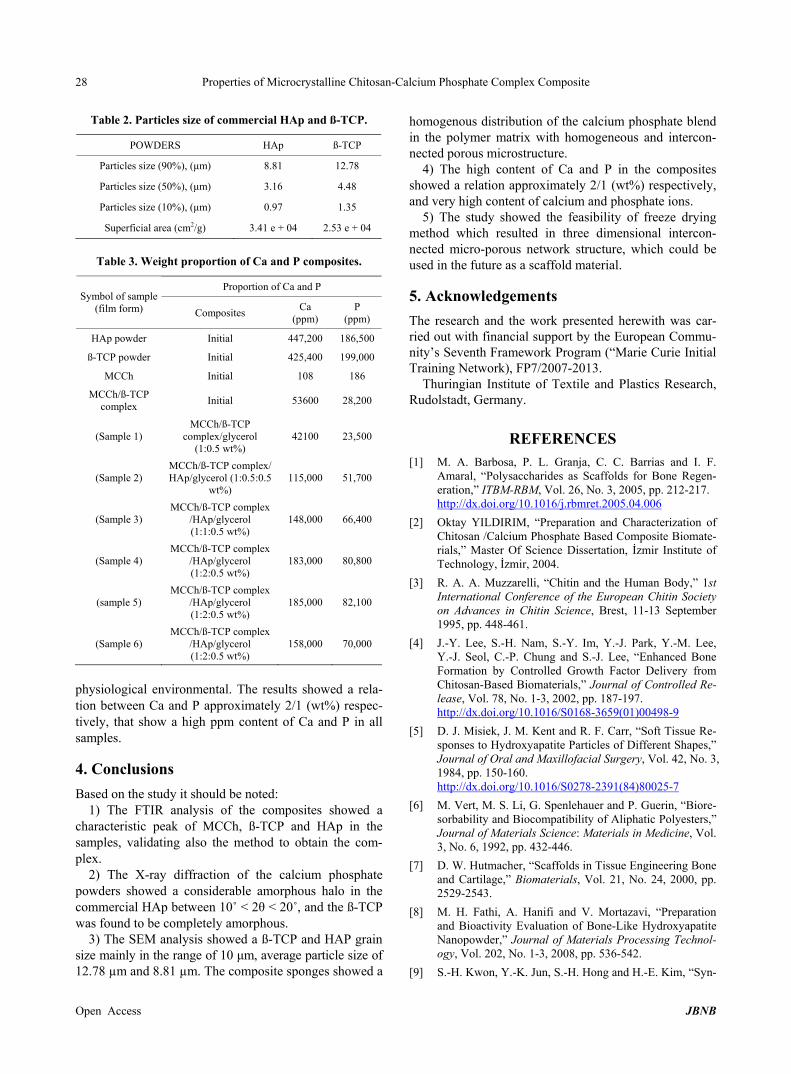
Table 2. Particles size of commercial HAp and ß-TCP.
POWDERS HAp ß-TCP
Particles size (90%), (ȝm) 8.81 12.78
Particles size (50%), (ȝm) 3.16 4.48
Particles size (10%), (ȝm) 0.97 1.35
Superficial area (cm2/g) 3.41 e + 04 2.53 e + 04
Table 3. Weight proportion of Ca and P composites.
Proportion of Ca and P Symbol of sample
(film form) Composites Ca (ppm)
P (ppm)
HAp powder Initial 447,200 186,500
ß-TCP powder Initial 425,400 199,000
MCCh Initial 108 186
MCCh/ß-TCP complex Initial 53600 28,200
(Sample 1) MCCh/ß-TCP
complex/glycerol (1:0.5 wt%)
42100 23,500
(Sample 2) MCCh/ß-TCP complex/ HAp/glycerol (1:0.5:0.5
wt%) 115,000 51,700
(Sample 3) MCCh/ß-TCP complex
/HAp/glycerol (1:1:0.5 wt%)
148,000 66,400
(Sample 4) MCCh/ß-TCP complex
/HAp/glycerol (1:2:0.5 wt%)
183,000 80,800
(sample 5) MCCh/ß-TCP complex
/HAp/glycerol (1:2:0.5 wt%)
185,000 82,100
(Sample 6) MCCh/ß-TCP complex
/HAp/glycerol (1:2:0.5 wt%)
158,000 70,000
physiological environmental. The results showed a rela- tion between Ca and P approximately 2/1 (wt%) respec- tively, that show a high ppm content of Ca and P in all samples.
4. Conclusions Based on the study it should be noted:
1) The FTIR analysis of the composites showed a characteristic peak of MCCh, ß-TCP and HAp in the samples, validating also the method to obtain the com- plex.
2) The X-ray diffraction of the calcium phosphate powders showed a considerable amorphous halo in the commercial HAp between 10˚ < 2ș < 20˚, and the ß-TCP was found to be completely amorphous.
3) The SEM analysis showed a ß-TCP and HAP grain size mainly in the range of 10 ȝm, average particle size of 12.78 µm and 8.81 µm. The composite sponges showed a
homogenous distribution of the calcium phosphate blend in the polymer matrix with homogeneous and intercon- nected porous microstructure.
4) The high content of Ca and P in the composites showed a relation approximately 2/1 (wt%) respectively, and very high content of calcium and phosphate ions.
5) The study showed the feasibility of freeze drying method which resulted in three dimensional intercon- nected micro-porous network structure, which could be used in the future as a scaffold material.
5. Acknowledgements The research and the work presented herewith was car- ried out with financial support by the European Commu- nity’s Seventh Framework Program (“Marie Curie Initial Training Network), FP7/2007-2013.
Thuringian Institute of Textile and Plastics Research, Rudolstadt, Germany.
REFERENCES [1] M. A. Barbosa, P. L. Granja, C. C. Barrias and I. F.
Amaral, “Polysaccharides as Scaffolds for Bone Regen- eration,” ITBM-RBM, Vol. 26, No. 3, 2005, pp. 212-217. http://dx.doi.org/10.1016/j.rbmret.2005.04.006
[2] Oktay YILDIRIM, “Preparation and Characterization of Chitosan /Calcium Phosphate Based Composite Biomate- rials,” Master Of Science Dissertation, øzmir Institute of Technology, øzmir, 2004.
[3] R. A. A. Muzzarelli, “Chitin and the Human Body,” 1st International Conference of the European Chitin Society on Advances in Chitin Science, Brest, 11-13 September 1995, pp. 448-461.
[4] J.-Y. Lee, S.-H. Nam, S.-Y. Im, Y.-J. Park, Y.-M. Lee, Y.-J. Seol, C.-P. Chung and S.-J. Lee, “Enhanced Bone Formation by Controlled Growth Factor Delivery from Chitosan-Based Biomaterials,” Journal of Controlled Re- lease, Vol. 78, No. 1-3, 2002, pp. 187-197. http://dx.doi.org/10.1016/S0168-3659(01)00498-9
[5] D. J. Misiek, J. M. Kent and R. F. Carr, “Soft Tissue Re-sponses to Hydroxyapatite Particles of Different Shapes,” Journal of Oral and Maxillofacial Surgery, Vol. 42, No. 3, 1984, pp. 150-160. http://dx.doi.org/10.1016/S0278-2391(84)80025-7
[6] M. Vert, M. S. Li, G. Spenlehauer and P. Guerin, “Biore- sorbability and Biocompatibility of Aliphatic Polyesters,” Journal of Materials Science: Materials in Medicine, Vol. 3, No. 6, 1992, pp. 432-446.
[7] D. W. Hutmacher, “Scaffolds in Tissue Engineering Bone and Cartilage,” Biomaterials, Vol. 21, No. 24, 2000, pp. 2529-2543.
[8] M. H. Fathi, A. Hanifi and V. Mortazavi, “Preparation and Bioactivity Evaluation of Bone-Like Hydroxyapatite Nanopowder,” Journal of Materials Processing Technol- ogy, Vol. 202, No. 1-3, 2008, pp. 536-542.
[9] S.-H. Kwon, Y.-K. Jun, S.-H. Hong and H.-E. Kim, “Syn-
Open Access �����������������������������������������������������������������������������JBNB

Properties of Microcrystalline Chitosan-Calcium Phosphate Complex Composite
Open Access �����������������������������������������������������������������������������JBNB
29
thesis and Dissolution Behavior of B-TCP and HA/B- TCP Composite Powder,” Journal of European Ceramic Society, Vol. 23, No. 7, 2003, pp. 1039-1045. http://dx.doi.org/10.1016/S0955-2219(02)00263-7
[10] H. Ç. Arca and S. ùenel, “Chitosan Based Systems for Tissue Engineering Part 1: Hard Tissues,” Journal of Pharmaceutical Sciences, Vol. 33, 2008, pp. 35-49.
[11] R. A. A. Muzzarelli, “Chitosan Composites with Inor-ganics, Morphogenetic Proteins and Stem Cells, for Bone Regeneration,” Carbohydrate Polymers, Vol. 83, No. 4, 2011, pp. 1433-1445. http://dx.doi.org/10.1016/j.carbpol.2010.10.044
[12] S.-J. Ding, “Preparation and Properties of Chitosan/Cal- cium Phosphate Composites for Bone Repair,” Dental Materials Journal, Vol. 25, No. 4, 2006, pp. 706-712. http://dx.doi.org/10.4012/dmj.25.706
[13] M. Rinaudo, “Chitin and Chitosan: Properties and Appli- cations,” Progress in Polymer Science, Vol. 31, No. 7, 2006, pp. 603-632. http://dx.doi.org/10.1016/j.progpolymsci.2006.06.001
[14] M. N. V. R. Kumar, “A Review of Chitin and Chitosan Applications,” Reactive & Functional Polymers, Vol. 46, No. 1, 2000, pp. 1-27. http://dx.doi.org/10.1016/S1381-5148(00)00038-9
[15] M. Kucharska, A. Niekraszewicz, M. Wisniewska-Wrona, E. Wesolowska and H. Struszczyk, “Dressing Sponges Made of Chitosan and Chitosan-Alginate Fibrids,” Pro-gress on Chemistry and Application of Chitin and Its De-rivatives, Vol. IX, PTChit, 2003, pp. 69-72.
[16] K. H. Bodek, “Evaluation of Properties Microcrystalline Chitosan as a Drug Carrier,” Acta Poloniac Pharmaceu-tica, Vol. 57, No. 6, 2000, pp. 431-441.
[17] M. Sakkinen, J. Marvola, H. Kanerva, K. Lindevall, M. Lipponen, T. Kekki, A. Ahonen and M. Marvola, “Gamma Scintigraphic Evaluation of the Fate of Microcrystalline Chitosan Granules in Human Stomach,” European Jour-
nal of Pharmaceutics and Biopharmaceutics, Vol. 57, No. 1, 2004, pp. 133-143. http://dx.doi.org/10.1016/S0939-6411(03)00097-3
[18] R. Fujita, A. Yokoyama, Y. Nodasaka, T. Kohgo and T. Kawasaki, “Ultrastructure of Ceramic-Bone Interface Using Hydroxyapatite and B-Tricalcium Phosphate Ce- ramics and Replacement Mechanism of B-Tricalcium Phosphate in Bone,” Tissue & Cell, Vol. 35, No. 6, 2003, pp. 427-440. http://dx.doi.org/10.1016/S0040-8166(03)00067-3
[19] D. Skrtic, J. M. Antonucci and E. D. Eanes, “Amorphous Calcium Phosphate-Based Bioactive Polymeric Compos- ites for Mineralized Tissue Regeneration,” Journal of Research of the National Institute of Standards and Tech- nology, Vol. 108, No. 3, 2003, pp. 167-182. http://dx.doi.org/10.6028/jres.108.017
[20] S.-C. Liou and S.-Y. Chen, “Transformation Mechanism of Different Chemically Precipitated Apatitic Precursor into B-Tricalcium Phosphate upon Calcinations,” Bioma- terials, Vol. 23, No. 23, 2002, pp. 4541-4547. http://dx.doi.org/10.1016/S0142-9612(02)00198-9
[21] D. A. Skoog, F. J. Holler and T. A. Nieman, “Principios de analise Instrumental,” 5th Edition, Bookman, Brazil, 2006, pp. 342-384.
[22] J. Pena and M. Vallet-Regi, “Hydroxyapatite, Tricalcium Phosphate and Biphasic Materials Prepared by a Liquid Mix Technique,” Journal of European Ceramic Society, Vol. 23, No. 10, 2003, pp. 1687-1696. http://dx.doi.org/10.1016/S0955-2219(02)00369-2
[23] J. Brugnerotto, J. Lizardi, F. M. Goycoolea, W. Argüelles- Monal, J. Desbrières and M. Rinaudo, “An Infrared In- vestigation in Relation with Chitin and Chitosan Charac- terization,” Polymer, Vol. 42, No. 8, 2001, pp. 3569-3580. http://dx.doi.org/10.1016/S0032-3861(00)00713-8


