Embed Size (px)
Citation preview
�����������������
Citation: Zaki, R.M.; Alfadhel, M.M.;
Alshahrani, S.M.; Alsaqr, A.;
Al-Kharashi, L.A.; Anwer, M.K.
Formulation of Chitosan-Coated
Brigatinib Nanospanlastics:
Optimization, Characterization,
Stability Assessment and In-Vitro
Cytotoxicity Activity against H-1975
Cell Lines. Pharmaceuticals 2022, 15,
348. https://doi.org/10.3390/
ph15030348
Academic Editors: Ljiljana Djekic and
Andelija M. Malenovic
Received: 10 February 2022
Accepted: 11 March 2022
Published: 13 March 2022
Publisher’s Note: MDPI stays neutral
with regard to jurisdictional claims in
published maps and institutional affil-
iations.
Copyright: © 2022 by the authors.
Licensee MDPI, Basel, Switzerland.
This article is an open access article
distributed under the terms and
conditions of the Creative Commons
Attribution (CC BY) license (https://
creativecommons.org/licenses/by/
4.0/).
pharmaceuticals
Article
Formulation of Chitosan-Coated Brigatinib Nanospanlastics:Optimization, Characterization, Stability Assessment andIn-Vitro Cytotoxicity Activity against H-1975 Cell LinesRanda Mohammed Zaki 1,2, Munerah M. Alfadhel 1, Saad M. Alshahrani 1,* , Ahmed Alsaqr 1,Layla A. Al-Kharashi 3 and Md Khalid Anwer 1,*
1 Department of Pharmaceutics, College of Pharmacy, Prince Sattam Bin Abdulaziz University, P.O. Box 173,Al-Kharj 11942, Saudi Arabia; [email protected] (R.M.Z.); [email protected] (M.M.A.);[email protected] (A.A.)
2 Department of Pharmaceutics and Industrial Pharmacy, Faculty of Pharmacy, Beni-Suef University,Beni-Suef 62514, Egypt
3 Department of Pharmacology and Toxicology, Faculty of Pharmacy, King Saud University, P.O. Box 2457,Riyadh 11451, Saudi Arabia; [email protected]
* Correspondence: [email protected] (S.M.A.); [email protected] (M.K.A.)
Abstract: The purpose of the current study was to develop Brigatinib (BGT)-loaded nanospanlastics(BGT-loaded NSPs) (S1-S13) containing Span 60 with different edge activators (Tween 80 and PluronicF127) and optimized based on the vesicle size, zeta potential (ZP), and percent entrapment efficiency(%EE) using Design-Expert® software. The optimum formula was recommended with desirabilityof 0.819 and composed of Span-60:Tween 80 at a ratio of 4:1 and 10 min as a sonication time (S13).It showed predicted EE% (81.58%), vesicle size (386.55 nm), and ZP (−29.51 mv). The optimizednanospanlastics (S13) was further coated with chitosan and further evaluated for Differential ScanningCalorimetry (DSC), X-ray Diffraction (XRD), in vitro release, Transmission Electron Microscopy(TEM), stability and in-vitro cytotoxicity studies against H-1975 lung cancer cell lines. The DSC andXRD revealed complete encapsulation of the drug. TEM imagery revealed spherical nanovesicleswith a smooth surface. Also, the coated formula showed high stability for three months in twodifferent conditions. Moreover, it resulted in improved and sustained drug release than free BGTsuspension and exhibited Higuchi kinetic release mechanism. The cytotoxic activity of BGT-loadedSPs (S13) was enhanced three times in comparison to free the BGT drug against the H-1975 cell lines.Overall, these results confirmed that BGT-loaded SPs could be a promising nanocarrier to improvethe anticancer efficacy of BGT.
Keywords: brigatinib; nanospanlastics; optimization; chitosan; sustained release; cytotoxicity
1. Introduction
Lung cancer is the second most common types of cancer in the United States andthe main cause of cancer mortality. In 2020, an anticipated 247,270 new instances of lungcancer would be diagnosed, including 130,340 male cases and 116,930 female cases [1,2].Anticancer drugs are considered successful when they exhibit maximum activity at targetcancer cells, that can be achieved by a targeted drug delivery system [3]. Nanomaterialsare an optimal choice as a targeted delivery system for the delivery of anti-cancer drugs byelectively localizing them in tumor cells, lowering the risk of harm to healthy cells. Thisthen reduces toxicity with increased efficacy [4].
Brigatinib (BGT) is a second-generation anaplastic lymphoma kinase (ALK) inhibitorthat is used to treat a certain type of non-small cell lung cancer (NSCLC) by inhibitingan abnormal protein that causes cancer cells to multiply [5–7]. This slows or stops thespread of cancer cells [8]. It was approved by the Food and Drug Administration (FDA)
Pharmaceuticals 2022, 15, 348. https://doi.org/10.3390/ph15030348 https://www.mdpi.com/journal/pharmaceuticals
Pharmaceuticals 2022, 15, 348 2 of 18
in April 2017 and used to treat patients with advanced stages of metastatic ALK-positiveNSCLC who are resistant to certain other ALK inhibitors including crizotinib, ceritinib,and alectinib [9]. BGT is available in a tablet form (30, 90, and 180 mg) with the best dosebeing 180 mg once a day for NSCLC. This drug has received the attention of researchers toformulate it in different forms in order to improve its action at the targeted cancer cells.
Ansari et al., 2020 [10] developed a self-nanoemulsifying drug delivery system (SNEDDS)of BGT, to enhance permeability of BGT to the targeted site. They found that cytotoxicactivity of BGT-SNEDDS against A549 human lung cancer cell lines during WST 1 assaywere significantly improved, as compared to pure BGT. Camidge and co-workers found thatpatients who received BGT had a substantially longer progression-free survival than thosewho received crizotinib in patients with ALK-positive NSCLC [11]. Moreover, BGT wasreported to have clinically relevant effectiveness in Japanese patients with ALK+ NSCLCresistant to alectinib (with or without previous use of crizotinib) [12]. Also, BGT-loadedethyl cellulose nanosponges and solid lipid nanoparticles were formulated for prolongeddrug release to extend anti-cancer activity and found that BGT-loaded nanosponges andsolid lipid nanoparticles dramatically reduced the cell viability of A549 human lung cancercell lines [13,14].
Nanovesicles (liposomes and niosomes) can be used for the treatment of cancer withexcellent results. It can also improve the stability of encapsulated drugs [15,16]. Theseconventional carriers, on the other hand, are rigid and lack deformability while passingthrough biological membranes. As a result, current research has focused on enhancingthe deformability of these traditional nanovesicles to improve their permeability acrossbiological membranes [17]. Nanospanlastics (NSPs) are flexible nanovesicles that arenon-immunogenic, biodegradable, and harmless. They’re also more chemically stablethan conventional liposomes [16]. For these reasons, several studies are focusing on theuse of NSPs formulations as a promising delivery system in preference to the conven-tional nanovesicles.
NSPs are a highly elastic surfactant-based deformable nanocarrier system that weredeveloped by Kakkar and Kaur [18]. Non-ionic surfactant (Span-60 and Span-80) and anedge activator (EA) are the key components of NSPs. The role of EA is in destabilizing thenanocarrier vesicular membranes, by squeezing through the narrow pores of the biologicalmembranes without rupture, enhancing their flexibility and permeability across the biolog-ical membranes [19,20]. To the best knowledge of the authors, NSPs formulations of BGThave not yet been investigated in literature, in spite of several favorable characteristics ofBGT including poor water-solubility.
Nanovesicles have a tendency to aggregate/merge leading to drug leakage duringstorage. Furthermore, there is a risk of fast blood clearance following intravenous injectionwhen employing such drug carrier systems. Surface coating the vesicles with polymersleads to an increase in their stability, lengthening of their life in the blood stream, and offerssustained release of the contained medicine. The polymer chitosan was selected to coatthe NSPs in order to target medications to maximize their absorption [21]. Chitosan, anatural polysaccharide derived from marine crustaceans, mollusks, insects, and fungi, is ofgreat interest, particularly in drug delivery and biomedical application [22,23]. It may beprocessed into a variety of forms for various uses, including solutions, gels, mixes, sponges,tablets, membranes, and paste.
The current study includes formulation and evaluation of BGT-loaded SPs containingSpan with different EA (Tween 80 and Pluronic F127) to improve the solubility, permeabilityof BGT. Optimization of the developed formulation was performed by Design Expertsoftware to study the independent variables, namely, sonication time, type of EA, andSpan-60:EA ratio on the dependent variables, entrapment efficiency, vesicles size, and zetapotential. Thereafter, the optimized NSPs were further coated and evaluated for DSC, XRD,in vitro release, TEM, stability and in vitro cytotoxicity studies against H-1975 lung cancercell lines.
Pharmaceuticals 2022, 15, 348 3 of 18
2. Results and Discussion2.1. Analysis of I Optimal Design for Optimization of BGT Loaded NSPs
Design Expert Version 12.0.3.0 was used to study the effect of the independent variablesnamely, sonication time (X1), type of EA (X2), and Span-60:EA ratio (X3) on the dependentvariables, entrapment efficiency (EE%) (Y1), vesicles size (Y2), and zeta potential (Y3) ofBGT-loaded SPs according to I optimal design (Table 1).
Table 1. Different variables utilized in I-optimal design for optimization of BGT-loaded NSPs.
Independent VariableLevels
Low (−1) High (+1)
Sonication Time (X1) 5 10
Type of EA (X2) Pluronic F127 Tween 80
Span-60: EA Ratio (X3) 3:2 4:1
Dependent Variables R2 Adjusted R2 Predicted R2 Constraints p Value F Value AdequatePrecision
Y1: % EE 0.9993 0.9959 0.9846 Maximize 0.0034 293.68 51.9945Y2: vesicles size (nm) 0.9998 0.9989 0.9672 Minimize 0.0009 1065.42 95.8437
Y3: zeta potential (mV) 0.9951 0.9804 0.8818 Maximize 0.0026 67.62 24.4528
Regression equations exhibited the effect of independent variables on the experimen-tally studied dependent responses by comparing the factor coefficients. In this model,adequate precision values for responses Y1, Y2, and Y3 were found greater than 4 as51.9945, 95.8437, and 24.4528, respectively, hence this model could be used successfullyfor experimental design [24]. The data of responses Y1, Y2 and Y3 exhibited excellentlinearity with their R2 values as 0.9993, 0.9998 and 0.9951, respectively. Hence, the obtainedequations were found to be statistically valid and an excellent fit to the obtained data [25].The predicted R2 values measured the response value consistency gave knowledge onhow good the model could fit with the new results that came from the same relationshipthat was modeled. The adjusted R2 value is the modified form of R2 value that examineshow well the present model would fit to the observed results. Subsequently, the predictedand adjusted R2 should be close to each other. In the event that they are not, there mightbe a problem with either the model or data. The difference between the predicted andadjusted R2 of Y1, Y2 and Y3 values were found to be less than 0.2, suggesting reasonableagreement [26].
2.2. Evaluation of the Prepared BGT Loaded SPS2.2.1. Entrapment Efficiency (EE%)
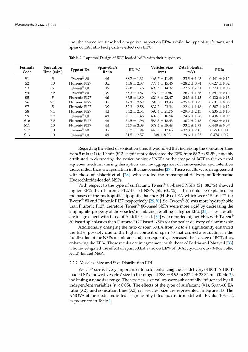
The EE% values were found to be ranged from 45.8 ± 2.37 to 88.7 ± 1.31% (Table 2). So,BGT was successfully entrapped in the NSPs’ formulations, indicating that span 60 basednanovesicles can be used as a successful delivery system for BGT.
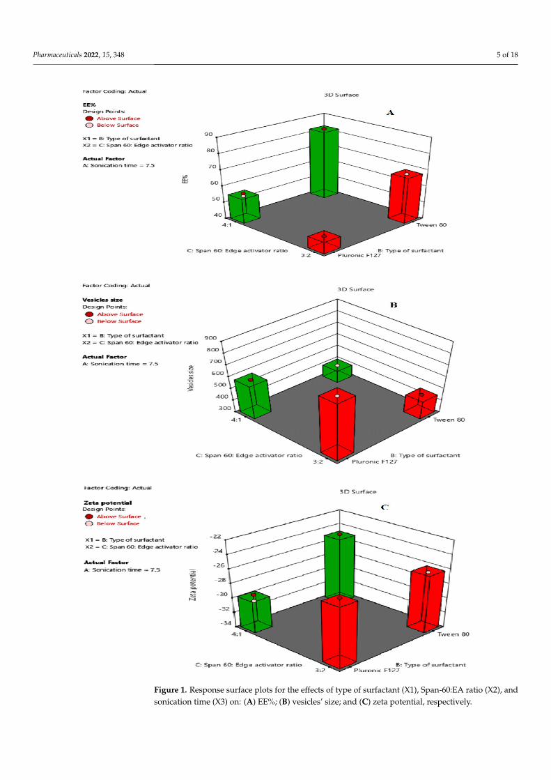
The EE% values were substantially influenced by all the independent variables(p < 0.05). The effects of the type of surfactant (X1), Span-60:EA ratio (X2), and sonica-tion time (X3) on EE% are represented in Figure 1A. ANOVA suggested a quadratic modelwith a F-value of 239.68 (p < 0.05) (Table 1), indicating a significant model. The followingregression equation describes the effect of independent variable on %EE:
Y1 = +63.49 − 3.70A + 12.21B + 5.69C + 0.1250AB − 0.3000AC + 1.54BC + 2.13A2 + 0.2750ABC − 0.6591 A2B+ 0.7841A2C (1)
where A is the sonication time, B is the type of surfactant, and C is span 60:EA ratio. Thepositive and negative sign in the equation indicated the favorable and unfavorable natureof independent variables over the response [24]. It is clear from the regression Equation (1)
Pharmaceuticals 2022, 15, 348 4 of 18
that the sonication time had a negative impact on EE%, while the type of surfactant, andspan 60:EA ratio had positive effects on EE%.
Table 2. I-optimal Design of BGT-loaded NSPs with their responses.
FormulaCode
SonicationTime (min.) Type of EA Span-60:EA
Ratio EE (%) Vesicles Size(nm)
Zeta Potential(mV) PDIa
S1 5 Tween® 80 4:1 88.7 ± 1.31 465.7 ± 11.45 −23.5 ± 1.03 0.441 ± 0.12S2 10 Pluronic F127 3:2 45.8 ± 2.37 773.4 ± 15.46 −28.2 ± 0.74 0.627 ± 0.02S3 5 Tween® 80 3:2 72.8 ± 1.76 493.5 ± 14.32 −22.5 ± 2.31 0.573 ± 0.06S4 7.5 Tween® 80 3:2 68.3 ± 3.57 460.2 ± 8.56 −26.2 ± 1.76 0.351 ± 0.14S5 5 Pluronic F127 4:1 63.5 ± 1.89 621.6 ± 22.47 −24.3 ± 1.45 0.432 ± 0.15S6 7.5 Pluronic F127 3:2 47.3 ± 2.67 794.3 ± 13.45 −25.4 ± 0.83 0.631 ± 0.05S7 5 Pluronic F127 3:2 52.3 ± 2.58 832.2 ± 23.34 −22.4 ± 1.48 0.507 ± 0.12S8 7.5 Pluronic F127 4:1 56.2 ± 2.54 592.4 ± 21.76 −29.3 ± 2.43 0.235 ± 0.10S9 7.5 Tween® 80 4:1 83.1 ± 1.45 402.6 ± 16.54 −24.6 ± 1.98 0.436 ± 0.09
S10 7.5 Pluronic F127 4:1 54.5 ± 1.96 589.3 ± 18.43 −30.2 ± 2.45 0.602 ± 0.11S11 10 Pluronic F127 4:1 54.7 ± 2.03 579.4 ± 25.43 −33.2 ± 1.73 0.648 ± 0.07S12 10 Tween® 80 3:2 65.7 ± 1.94 441.3 ± 17.65 −32.8 ± 2.45 0.553 ± 0.1S13 10 Tween® 80 4:1 81.5 ± 2.57 388 ± 8.93 −29.6 ± 1.85 0.474 ± 0.2
Regarding the effect of sonication time, it was noted that increasing the sonication timefrom 5 min (S1) to 10 min (S13) significantly decreased the EE% from 88.7 to 81.5%, possiblyattributed to decreasing the vesicular size of NSPs or the escape of BGT to the externalaqueous medium during disruption and re-aggregation of nanovesicles and retentionthere, rather than encapsulation in the nanovesicles [27]. These results were in agreementwith those of Elsherif et al. [28], who studied the transungual delivery of TerbinafineHydrochloride-loaded NSPs.
With respect to the type of surfactant, Tween® 80-based NSPs (S1, 88.7%) showedhigher EE% than Pluronic F127-based NSPs (S5, 63.5%). This could be explained onthe bases of the hydrophilic–lipophilic balance (HLB) of EA which were 15 and 22 forTween® 80 and Pluronic F127, respectively [29,30]. So, Tween® 80 was more hydrophobicthan Pluronic F127, therefore, Tween® 80-based NSPs were more rigid by decreasing theamphiphilic property of the vesicles’ membrane, resulting in higher EE% [31]. These resultsare in agreement with those of Abdelbari et al. [32] who reported higher EE% with Tween®
80-based splanlastics than Pluronic F127-based NSPs for the ocular delivery of clotrimazole.Additionally, changing the ratio of span 60:EA from 3:2 to 4:1 significantly enhanced
the EE%, possibly due to the higher content of span 60 that caused a reduction in thefluidization of the NSPs membrane and, consequently, decreased the leakage of BGT, thus,enhancing the EE%. These results are in agreement with those of Badria and Mazyed [33]who investigated the effect of span 60:EA ratio on EE% of (3-Acetyl-11-Keto -β-BoswellicAcid)-loaded NSPs.
2.2.2. Vesicles’ Size and Size Distribution PDI
Vesicles’ size is a very important criteria for enhancing the cell delivery of BGT. All BGT-loaded SPs showed vesicles’ size in the range of 388 ± 8.93 to 832.2 ± 23.34 nm (Table 2),indicating a nanosize range. The vesicles’ size values were substantially influenced by allindependent variables (p < 0.05). The effects of the type of surfactant (X1), Span-60:EAratio (X2), and sonication time (X3) on vesicles’ size are represented in Figure 1B. TheANOVA of the model indicated a significantly fitted quadratic model with F-value 1065.42,as presented in Table 1.
Pharmaceuticals 2022, 15, 348 5 of 18Pharmaceuticals 2022, 15, x FOR PEER REVIEW 5 of 19
Figure 1. Response surface plots for the effects of type of surfactant (X1), Span-60:EA ratio (X2), and sonication time (X3) on: (A) EE%; (B) vesicles’ size; and (C) zeta potential, respectively.
Figure 1. Response surface plots for the effects of type of surfactant (X1), Span-60:EA ratio (X2), andsonication time (X3) on: (A) EE%; (B) vesicles’ size; and (C) zeta potential, respectively.
Pharmaceuticals 2022, 15, 348 6 of 18
The regression equation for the effect of independent variable on vesicles size isgiven below:
Y2 = +562.35 − 28.86A − 130.95B − 64.90C − 3.61AB − 1.11AC + 38.99BC + 12.04A2 − 5.26ABC + 3.69A2B + 4.19A2C (2)
where A is the sonication time, B is the type of surfactant, and C is span 60:EA ratio.It is worth noting from the regression Equation (2) that all independent variables (A,
B, and C) had negative effects on vesicles’ size values. Regarding the effect of sonicationtime, it was found that increasing the sonication time from 5 min to 10 min significantlydecreased the vesicles’ size from 465.7 (S1) to 388 nm (S13). This finding is in accor-dance with Elsherif et al. [28], who reported a decrease in the vesicles’ size of TerbinafineHydrochloride-loaded NSPs upon increasing the sonication time. With respect to the typeof surfactant, formulations prepared with Pluronic F127 (S5, 621.6 nm) showed largervesicles’ size than those prepared with Tween® 80 (S1, 465.7 nm), this could be attributed tothe higher hydrophilicity of Pluronic F127 (HLB > 20) than Tween® 80 (HLB = 15), leadingto greater water uptake by the vesicle membranes and thus increasing in the vesicles’ size.Using EA of a lower hydrophilicity (lower HLB) caused a decrease in the surface energyand hence formation of smaller size nanovesicles [34].
Moreover, the Span-60:EA ratio 3:2 showed larger vesicles’ size than those of 4:1 ratioand this may be due to the higher EA concentration that led to the larger vesicles’ size.Both Tween® 80 and Pluronic F127 are hydrophilic nonionic surfactants [35] that impartflexibility to the bilayer membranes of NSPs [36] and, thus, increase the elasticity of thevesicles and water uptake so leading to an increase in the vesicles’ size. PDI is an indicatorof the vesicles’ size distribution and its value ranges from 0.0 (for completely uniformvesicles’ size distribution) to 1.0 (for highly polydispersed vesicles). The PDI values werefound to be in the range 0.235 ± 0.10 to 0.648 ± 0.07, confirming low variation in thevesicles’ sizes (Table 2).
2.2.3. Zeta Potential
Zeta potential is a measure for vesicles’ attraction or repulsion. Therefore, it is usedto predict the nanovesicles’ stability. The higher the zeta potential values, the higher thestability. Formulations with zeta potential values greater than +30 or less than −30 arehighly stable systems [37]. All BGT-loaded SPs showed that the zeta potential valuesranged from −22.4 ± 1.48 to −33.2 ± 1.73 mv (Table 2), indicating a low tendency for NSPsaggregation and, consequently, high stable nanoformulations.
The zeta potential values were substantially influenced by all independent variables(p < 0.05). The effects of the type of surfactant (X1), Span-60: EA ratio (X2), and sonicationtime (X3) on zeta potential are shown in Figure 1C. ANOVA suggested a quadratic modelwith F-value 67.62 (p < 0.05), indicating a significant model (Table 1).
The following regression equation describes the effect of independent variable onzeta potential:
Y3 = −26.53 − 3.89A + 1.13B − 0.6358C − 0.2125AB + 0.1375AC + 1.27BC − 0.5300A2 + 0.9125ABC − 1.17A2B (3)
where A is the sonication time, B is the type of surfactant, and C is span 60:EA ratio. It isclear from the regression equation that the zeta potential values are significantly affectedby all three independent factors at (p < 0.05).
It was clear that the sonication time had a negative impact on the zeta potential valuesalthough it showed an interactive effect with both the surfactant type and span 60:EAratio. As shown in Equation (3), sonication time and the type of surfactant collectivelyshowed a negative impact on the zeta potential which may be attributed to the main effect ofsonication time. Similarly, although sonication time and span 60:EA ratio showed a negativeeffect separately, they collectively showed a positive effect on the zeta potential values.
Moreover, the type of surfactant showed a positive impact and the span 60:EA ratioshowed a negative impact on the zeta potential values but collectively showed a positive
Pharmaceuticals 2022, 15, 348 7 of 18
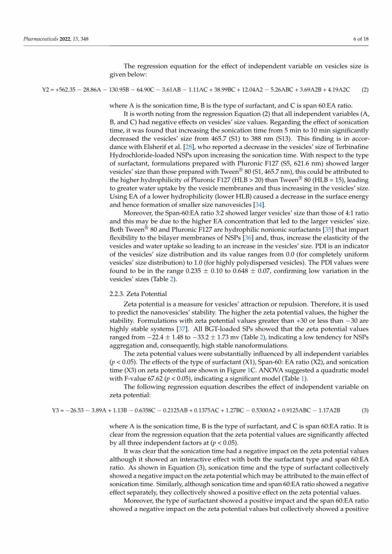
impact mainly due to the effect of surfactant type. Moreover, the effect of different indepen-dent factors (X1, X2, X3) on different dependent variables (Y1, Y2, Y3) was represented as acontour plot (Figure 2).
Pharmaceuticals 2022, 15, x FOR PEER REVIEW 7 of 19
It was clear that the sonication time had a negative impact on the zeta potential values although it showed an interactive effect with both the surfactant type and span 60:EA ratio. As shown in Equation (3), sonication time and the type of surfactant collectively showed a negative impact on the zeta potential which may be attributed to the main effect of sonication time. Similarly, although sonication time and span 60:EA ratio showed a negative effect separately, they collectively showed a positive effect on the zeta potential values.
Moreover, the type of surfactant showed a positive impact and the span 60:EA ratio showed a negative impact on the zeta potential values but collectively showed a positive impact mainly due to the effect of surfactant type. Moreover, the effect of different independent factors (X1, X2, X3) on different dependent variables (Y1, Y2, Y3) was represented as a contour plot (Figure 2).
.
Pharmaceuticals 2022, 15, x FOR PEER REVIEW 8 of 19
Figure 2. Contour plot of different responses.
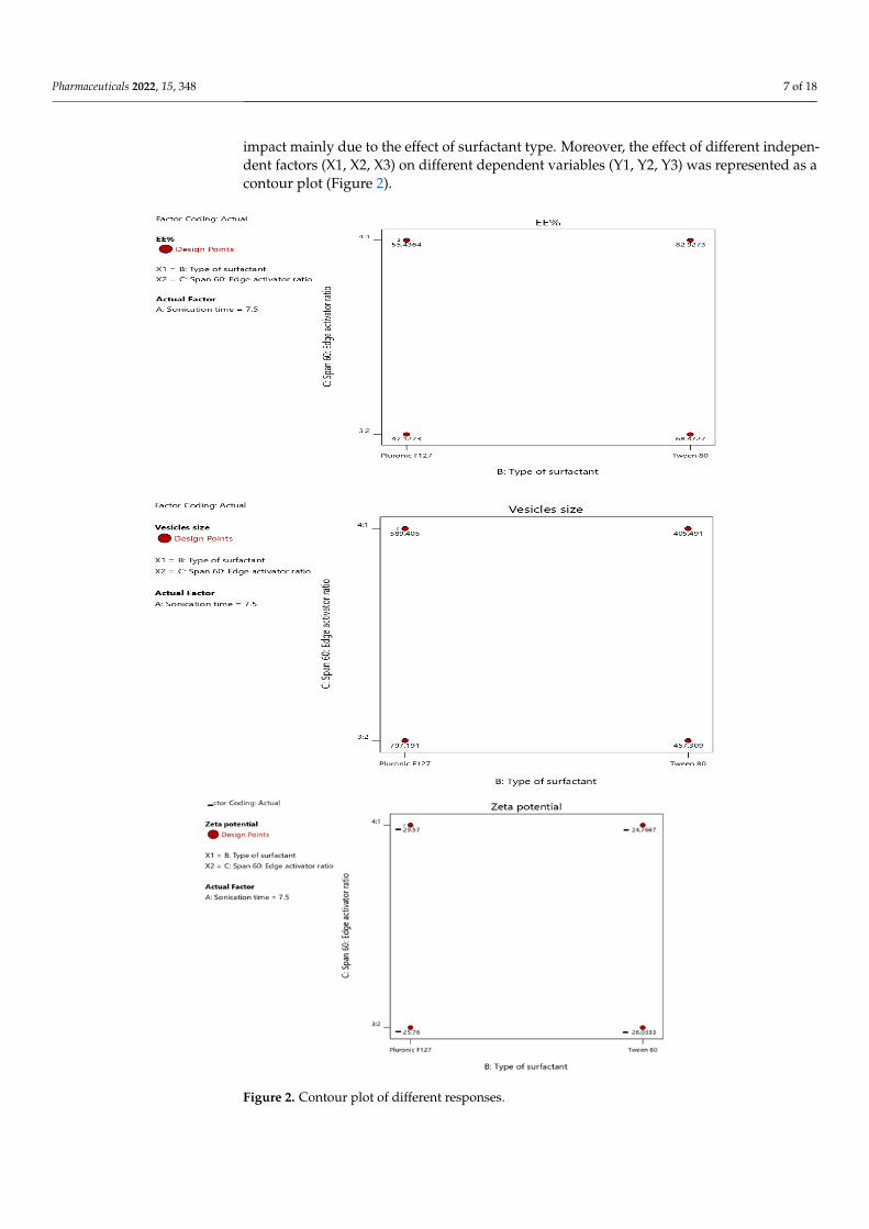
2.3. Selection of the Optimized BGT Loaded SPs Design-Expert® software was used for optimization by choosing the formula of high
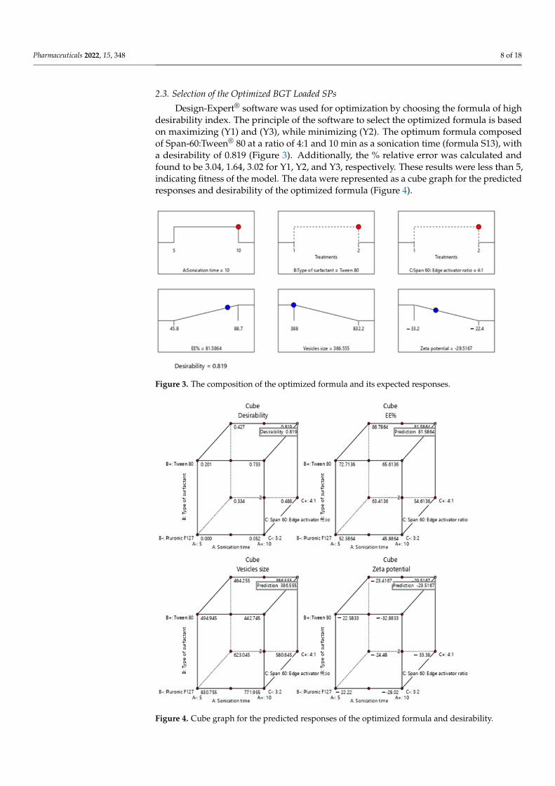
desirability index. The principle of the software to select the optimized formula is based on maximizing (Y1) and (Y3), while minimizing (Y2). The optimum formula composed of Span-60:Tween® 80 at a ratio of 4:1 and 10 min as a sonication time (formula S13), with a desirability of 0.819 (Figure 3). Additionally, the % relative error was calculated and found to be 3.04, 1.64, 3.02 for Y1, Y2, and Y3, respectively. These results were less than 5, indicating fitness of the model. The data were represented as a cube graph for the predicted responses and desirability of the optimized formula (Figure 4).
Figure 3. The composition of the optimized formula and its expected responses.
Figure 2. Contour plot of different responses.
Pharmaceuticals 2022, 15, 348 8 of 18
2.3. Selection of the Optimized BGT Loaded SPs
Design-Expert® software was used for optimization by choosing the formula of highdesirability index. The principle of the software to select the optimized formula is basedon maximizing (Y1) and (Y3), while minimizing (Y2). The optimum formula composedof Span-60:Tween® 80 at a ratio of 4:1 and 10 min as a sonication time (formula S13), witha desirability of 0.819 (Figure 3). Additionally, the % relative error was calculated andfound to be 3.04, 1.64, 3.02 for Y1, Y2, and Y3, respectively. These results were less than 5,indicating fitness of the model. The data were represented as a cube graph for the predictedresponses and desirability of the optimized formula (Figure 4).
Pharmaceuticals 2022, 15, x FOR PEER REVIEW 8 of 19
Figure 2. Contour plot of different responses.
2.3. Selection of the Optimized BGT Loaded SPs Design-Expert® software was used for optimization by choosing the formula of high
desirability index. The principle of the software to select the optimized formula is based on maximizing (Y1) and (Y3), while minimizing (Y2). The optimum formula composed of Span-60:Tween® 80 at a ratio of 4:1 and 10 min as a sonication time (formula S13), with a desirability of 0.819 (Figure 3). Additionally, the % relative error was calculated and found to be 3.04, 1.64, 3.02 for Y1, Y2, and Y3, respectively. These results were less than 5, indicating fitness of the model. The data were represented as a cube graph for the predicted responses and desirability of the optimized formula (Figure 4).
Figure 3. The composition of the optimized formula and its expected responses. Figure 3. The composition of the optimized formula and its expected responses.
Pharmaceuticals 2022, 15, x FOR PEER REVIEW 9 of 19
Figure 4. Cube graph for the predicted responses of the optimized formula and desirability.
2.4. Evaluation of the Optimized Coated Formula 2.4.1. Vesicle Size, %EE, and Zeta Potential
The optimum coated formula showed an increase in the EE% (86.55%) compared to the uncoated optimum formula (81.58%), possibly due to the change in the surface properties of the NSPs that prevents the leakage of the drug after chitosan coating, as reported by Alshraim et al. [38]. Additionally, the vesicles’ size increased from 386.55 to 395.4 nm, indicating the binding of chitosan to the surface of the NSPs [39]. Moreover, the coated formula showed a shift for zeta potential value from negative to positive, confirming the presence of the chitosan coating on the external surface of NSPs. This result is in agreement with Cuomo et al. [40].
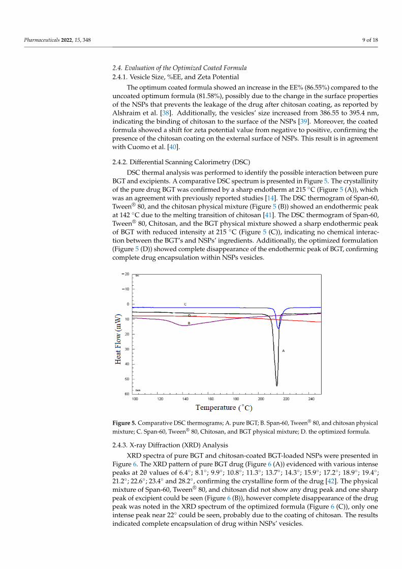
2.4.2. Differential Scanning Calorimetry (DSC) DSC thermal analysis was performed to identify the possible interaction between
pure BGT and excipients. A comparative DSC spectrum is presented in Figure 5. The crystallinity of the pure drug BGT was confirmed by a sharp endotherm at 215 °C (Figure 5 (A)), which was an agreement with previously reported studies [14]. The DSC thermogram of Span-60, Tween® 80, and the chitosan physical mixture (Figure 5 (B)) showed an endothermic peak at 142 °C due to the melting transition of chitosan [41]. The DSC thermogram of Span-60, Tween® 80, Chitosan, and the BGT physical mixture showed a sharp endothermic peak of BGT with reduced intensity at 215 °C (Figure 5 (C)), indicating no chemical interaction between the BGT’s and NSPs’ ingredients. Additionally, the optimized formulation (Figure 5 (D)) showed complete disappearance
Figure 4. Cube graph for the predicted responses of the optimized formula and desirability.
Pharmaceuticals 2022, 15, 348 9 of 18
2.4. Evaluation of the Optimized Coated Formula2.4.1. Vesicle Size, %EE, and Zeta Potential
The optimum coated formula showed an increase in the EE% (86.55%) compared to theuncoated optimum formula (81.58%), possibly due to the change in the surface propertiesof the NSPs that prevents the leakage of the drug after chitosan coating, as reported byAlshraim et al. [38]. Additionally, the vesicles’ size increased from 386.55 to 395.4 nm,indicating the binding of chitosan to the surface of the NSPs [39]. Moreover, the coatedformula showed a shift for zeta potential value from negative to positive, confirming thepresence of the chitosan coating on the external surface of NSPs. This result is in agreementwith Cuomo et al. [40].
2.4.2. Differential Scanning Calorimetry (DSC)
DSC thermal analysis was performed to identify the possible interaction between pureBGT and excipients. A comparative DSC spectrum is presented in Figure 5. The crystallinityof the pure drug BGT was confirmed by a sharp endotherm at 215 ◦C (Figure 5 (A)), whichwas an agreement with previously reported studies [14]. The DSC thermogram of Span-60,Tween® 80, and the chitosan physical mixture (Figure 5 (B)) showed an endothermic peakat 142 ◦C due to the melting transition of chitosan [41]. The DSC thermogram of Span-60,Tween® 80, Chitosan, and the BGT physical mixture showed a sharp endothermic peakof BGT with reduced intensity at 215 ◦C (Figure 5 (C)), indicating no chemical interac-tion between the BGT’s and NSPs’ ingredients. Additionally, the optimized formulation(Figure 5 (D)) showed complete disappearance of the endothermic peak of BGT, confirmingcomplete drug encapsulation within NSPs vesicles.
Pharmaceuticals 2022, 15, x FOR PEER REVIEW 10 of 19
of the endothermic peak of BGT, confirming complete drug encapsulation within NSPs vesicles.
Figure 5. Comparative DSC thermograms; A. pure BGT; B. Span-60, Tween® 80, and chitosan physical mixture; C. Span-60, Tween® 80, Chitosan, and BGT physical mixture; D. the optimized formula.
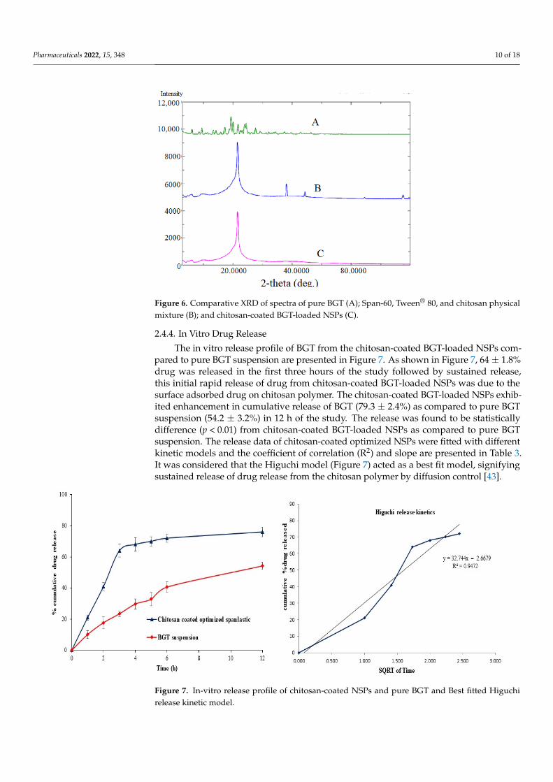
2.4.3. X-ray Diffraction (XRD) Analysis XRD spectra of pure BGT and chitosan-coated BGT-loaded NSPs were presented in
Figure 6. The XRD pattern of pure BGT drug (Figure 6 (A)) evidenced with various intense peaks at 2θ values of 6.4°; 8.1°; 9.9°; 10.8°; 11.3°; 13.7°; 14.3°; 15.9°; 17.2°; 18.9°; 19.4°; 21.2°; 22.6°; 23.4° and 28.2°, confirming the crystalline form of the drug [42]. The physical mixture of Span-60, Tween® 80, and chitosan did not show any drug peak and one sharp peak of excipient could be seen (Figure 6 (B)), however complete disappearance of the drug peak was noted in the XRD spectrum of the optimized formula (Figure 6 (C)), only one intense peak near 22° could be seen, probably due to the coating of chitosan. The results indicated complete encapsulation of drug within NSPs’ vesicles.
Figure 5. Comparative DSC thermograms; A. pure BGT; B. Span-60, Tween® 80, and chitosan physicalmixture; C. Span-60, Tween® 80, Chitosan, and BGT physical mixture; D. the optimized formula.
2.4.3. X-ray Diffraction (XRD) Analysis
XRD spectra of pure BGT and chitosan-coated BGT-loaded NSPs were presented inFigure 6. The XRD pattern of pure BGT drug (Figure 6 (A)) evidenced with various intensepeaks at 2θ values of 6.4◦; 8.1◦; 9.9◦; 10.8◦; 11.3◦; 13.7◦; 14.3◦; 15.9◦; 17.2◦; 18.9◦; 19.4◦;21.2◦; 22.6◦; 23.4◦ and 28.2◦, confirming the crystalline form of the drug [42]. The physicalmixture of Span-60, Tween® 80, and chitosan did not show any drug peak and one sharppeak of excipient could be seen (Figure 6 (B)), however complete disappearance of the drugpeak was noted in the XRD spectrum of the optimized formula (Figure 6 (C)), only oneintense peak near 22◦ could be seen, probably due to the coating of chitosan. The resultsindicated complete encapsulation of drug within NSPs’ vesicles.
Pharmaceuticals 2022, 15, 348 10 of 18Pharmaceuticals 2022, 15, x FOR PEER REVIEW 11 of 19
Figure 6. Comparative XRD of spectra of pure BGT (A); Span-60, Tween® 80, and chitosan physical mixture (B); and chitosan-coated BGT-loaded NSPs (C).
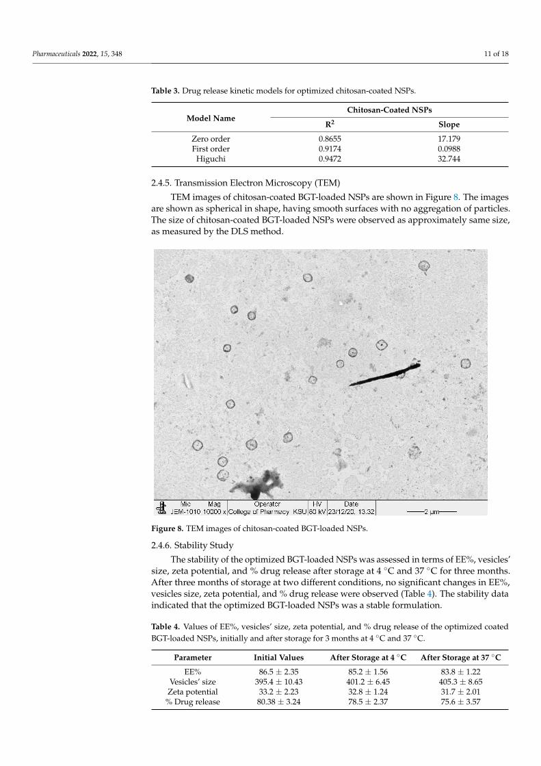
2.4.4. In Vitro Drug Release The in vitro release profile of BGT from the chitosan-coated BGT-loaded NSPs
compared to pure BGT suspension are presented in Figure 7. As shown in Figure 7, 64 ± 1.8% drug was released in the first three hours of the study followed by sustained release, this initial rapid release of drug from chitosan-coated BGT-loaded NSPs was due to the surface adsorbed drug on chitosan polymer. The chitosan-coated BGT-loaded NSPs exhibited enhancement in cumulative release of BGT (79.3 ± 2.4%) as compared to pure BGT suspension (54.2 ± 3.2%) in 12 h of the study. The release was found to be statistically difference (p < 0.01) from chitosan-coated BGT-loaded NSPs as compared to pure BGT suspension. The release data of chitosan-coated optimized NSPs were fitted with different kinetic models and the coefficient of correlation (R2) and slope are presented in Table 3. It was considered that the Higuchi model (Figure 7) acted as a best fit model, signifying sustained release of drug release from the chitosan polymer by diffusion control [43].
Table 3. Drug release kinetic models for optimized chitosan-coated NSPs.
Model Name Chitosan-Coated NSPs R2 Slope
Zero order 0.8655 17.179 First order 0.9174 0.0988
Higuchi 0.9472 32.744
Figure 6. Comparative XRD of spectra of pure BGT (A); Span-60, Tween® 80, and chitosan physicalmixture (B); and chitosan-coated BGT-loaded NSPs (C).
2.4.4. In Vitro Drug Release
The in vitro release profile of BGT from the chitosan-coated BGT-loaded NSPs com-pared to pure BGT suspension are presented in Figure 7. As shown in Figure 7, 64 ± 1.8%drug was released in the first three hours of the study followed by sustained release,this initial rapid release of drug from chitosan-coated BGT-loaded NSPs was due to thesurface adsorbed drug on chitosan polymer. The chitosan-coated BGT-loaded NSPs exhib-ited enhancement in cumulative release of BGT (79.3 ± 2.4%) as compared to pure BGTsuspension (54.2 ± 3.2%) in 12 h of the study. The release was found to be statisticallydifference (p < 0.01) from chitosan-coated BGT-loaded NSPs as compared to pure BGTsuspension. The release data of chitosan-coated optimized NSPs were fitted with differentkinetic models and the coefficient of correlation (R2) and slope are presented in Table 3.It was considered that the Higuchi model (Figure 7) acted as a best fit model, signifyingsustained release of drug release from the chitosan polymer by diffusion control [43].
Pharmaceuticals 2022, 15, x FOR PEER REVIEW 12 of 19
Figure 7. In-vitro release profile of chitosan-coated NSPs and pure BGT and Best fitted Higuchi release kinetic model.
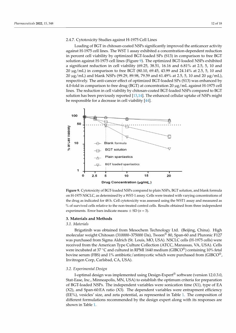
2.4.5. Transmission Electron Microscopy (TEM) TEM images of chitosan-coated BGT-loaded NSPs are shown in Figure 8. The images
are shown as spherical in shape, having smooth surfaces with no aggregation of particles. The size of chitosan-coated BGT-loaded NSPs were observed as approximately same size, as measured by the DLS method.
Figure 8. TEM images of chitosan-coated BGT-loaded NSPs.
Figure 7. In-vitro release profile of chitosan-coated NSPs and pure BGT and Best fitted Higuchirelease kinetic model.
Pharmaceuticals 2022, 15, 348 11 of 18
Table 3. Drug release kinetic models for optimized chitosan-coated NSPs.
Model NameChitosan-Coated NSPs
R2 Slope
Zero order 0.8655 17.179First order 0.9174 0.0988
Higuchi 0.9472 32.744
2.4.5. Transmission Electron Microscopy (TEM)
TEM images of chitosan-coated BGT-loaded NSPs are shown in Figure 8. The imagesare shown as spherical in shape, having smooth surfaces with no aggregation of particles.The size of chitosan-coated BGT-loaded NSPs were observed as approximately same size,as measured by the DLS method.
Pharmaceuticals 2022, 15, x FOR PEER REVIEW 12 of 19
Figure 7. In-vitro release profile of chitosan-coated NSPs and pure BGT and Best fitted Higuchi release kinetic model.
2.4.5. Transmission Electron Microscopy (TEM) TEM images of chitosan-coated BGT-loaded NSPs are shown in Figure 8. The images
are shown as spherical in shape, having smooth surfaces with no aggregation of particles. The size of chitosan-coated BGT-loaded NSPs were observed as approximately same size, as measured by the DLS method.
Figure 8. TEM images of chitosan-coated BGT-loaded NSPs. Figure 8. TEM images of chitosan-coated BGT-loaded NSPs.
2.4.6. Stability Study
The stability of the optimized BGT-loaded NSPs was assessed in terms of EE%, vesicles’size, zeta potential, and % drug release after storage at 4 ◦C and 37 ◦C for three months.After three months of storage at two different conditions, no significant changes in EE%,vesicles size, zeta potential, and % drug release were observed (Table 4). The stability dataindicated that the optimized BGT-loaded NSPs was a stable formulation.
Table 4. Values of EE%, vesicles’ size, zeta potential, and % drug release of the optimized coatedBGT-loaded NSPs, initially and after storage for 3 months at 4 ◦C and 37 ◦C.
Parameter Initial Values After Storage at 4 ◦C After Storage at 37 ◦C
EE% 86.5 ± 2.35 85.2 ± 1.56 83.8 ± 1.22Vesicles’ size 395.4 ± 10.43 401.2 ± 6.45 405.3 ± 8.65Zeta potential 33.2 ± 2.23 32.8 ± 1.24 31.7 ± 2.01
% Drug release 80.38 ± 3.24 78.5 ± 2.37 75.6 ± 3.57
Pharmaceuticals 2022, 15, 348 12 of 18
2.4.7. Cytotoxicity Studies against H-1975 Cell Lines
Loading of BGT in chitosan-coated NSPs significantly improved the anticancer activityagainst H-1975 cell lines. The WST 1 assay exhibited a concentration-dependent reductionin percent cell viability by optimized BGT-loaded SPs (S13) in comparison to free BGTsolution against H-1975 cell lines (Figure 9). The optimized BGT-loaded NSPs exhibiteda significant reduction in cell viability (69.25, 38.51, 16.16 and 6.81% at 2.5, 5, 10 and20 µg/mL) in comparison to free BGT (80.10, 69.45, 43.99 and 24.14% at 2.5, 5, 10 and20 µg/mL) and blank NSPs (99.29, 89.98, 79.59 and 61.49% at 2.5, 5, 10 and 20 µg/mL),respectively. The anti-cancer effect of optimized BGT-loaded SPs (S13) was enhanced by4.0-fold in comparison to free drug (BGT) at concentration 20 µg/mL against H-1975 celllines. The reduction in cell viability by chitosan-coated BGT-loaded NSPs compared to BGTsolution has been previously reported [13,14]. The enhanced cellular uptake of NSPs mightbe responsible for a decrease in cell viability [44].
Pharmaceuticals 2022, 15, x FOR PEER REVIEW 13 of 19
2.4.6. Stability Study The stability of the optimized BGT-loaded NSPs was assessed in terms of EE%,
vesicles’ size, zeta potential, and % drug release after storage at 4 °C and 37 °C for three months. After three months of storage at two different conditions, no significant changes in EE%, vesicles size, zeta potential, and % drug release were observed (Table 4). The stability data indicated that the optimized BGT-loaded NSPs was a stable formulation.
Table 4. Values of EE%, vesicles’ size, zeta potential, and % drug release of the optimized coated BGT-loaded NSPs, initially and after storage for 3 months at 4 °C and 37 °C.
Parameter Initial Values After Storage at 4 °C After Storage at 37 °C EE% 86.5 ± 2.35 85.2 ± 1.56 83.8 ± 1.22
Vesicles’ size 395.4 ± 10.43 401.2 ± 6.45 405.3 ± 8.65 Zeta potential 33.2 ± 2.23 32.8 ± 1.24 31.7 ± 2.01
% Drug release 80.38 ± 3.24 78.5 ± 2.37 75.6 ± 3.57
2.4.7. Cytotoxicity Studies against H-1975 Cell Lines Loading of BGT in chitosan-coated NSPs significantly improved the anticancer
activity against H-1975 cell lines. The WST 1 assay exhibited a concentration-dependent reduction in percent cell viability by optimized BGT-loaded SPs (S13) in comparison to free BGT solution against H-1975 cell lines (Figure 9). The optimized BGT-loaded NSPs exhibited a significant reduction in cell viability (69.25, 38.51, 16.16 and 6.81% at 2.5, 5, 10 and 20 µg/mL) in comparison to free BGT (80.10, 69.45, 43.99 and 24.14% at 2.5, 5, 10 and 20 µg/mL) and blank NSPs (99.29, 89.98, 79.59 and 61.49% at 2.5, 5, 10 and 20 µg/mL), respectively. The anti-cancer effect of optimized BGT-loaded SPs (S13) was enhanced by 4.0-fold in comparison to free drug (BGT) at concentration 20 µg/mL against H-1975 cell lines. The reduction in cell viability by chitosan-coated BGT-loaded NSPs compared to BGT solution has been previously reported [13,14]. The enhanced cellular uptake of NSPs might be responsible for a decrease in cell viability [44].
Figure 9. Cytotoxicity of BGT-loaded NSPs compared to plain NSPs, BGT solution, and blank formula on H-1975 NSCLC, as determined by a WST-1 assay. Cells were treated with varying concentrations of the drug as indicated for 48 h. Cell cytotoxicity was assessed using the WST1 assay and measured as % of survived cells relative to the non-treated control cells. Results obtained from three independent experiments. Error bars indicate means ± SD (n = 3).
3. Materials and Methods 3.1. Materials
Figure 9. Cytotoxicity of BGT-loaded NSPs compared to plain NSPs, BGT solution, and blank formulaon H-1975 NSCLC, as determined by a WST-1 assay. Cells were treated with varying concentrations ofthe drug as indicated for 48 h. Cell cytotoxicity was assessed using the WST1 assay and measured as% of survived cells relative to the non-treated control cells. Results obtained from three independentexperiments. Error bars indicate means ± SD (n = 3).
3. Materials and Methods3.1. Materials
Brigatinib was obtained from Mesochem Technology Ltd. (Beijing, China). Highmolecular weight Chitosan (310000–375000 Da), Tween® 80, Span-60 and Pluronic F127was purchased from Sigma Aldrich (St. Louis, MO, USA). NSCLC cells (H-1975 cells) werereceived from the American Type Culture Collection (ATCC, Manassas, VA, USA). Cellswere incubated at 37 ◦C and cultured in RPMI 1640 medium (GIBCO®) containing 10% fetalbovine serum (FBS) and 1% antibiotic/antimycotic which were purchased from (GIBCO®,Invitrogen Corp, Carlsbad, CA, USA).
3.2. Experimental Design
I-optimal design was implemented using Design-Expert® software (version 12.0.3.0,Stat-Ease, Inc., Minneapolis, MN, USA) to establish the optimum criteria for preparationof BGT-loaded NSPs. The independent variables were sonication time (X1), type of EA(X2), and Span-60:EA ratio (X3). The dependent variables were entrapment efficiency(EE%), vesicles’ size, and zeta potential, as represented in Table 1. The composition ofdifferent formulations recommended by the design expert along with its responses areshown in Table 1.
Pharmaceuticals 2022, 15, 348 13 of 18
3.3. Development of BGT-Loaded NSPs
Different BGT-loaded NSPs were formulated by ethanol injection method with minormodification [45]. Two different surfactants (EA), namely, Pluronic F-127 and Tween®
80, were used in the preparation as shown in Table 1. The amount of BGT was keptconstant (30 mg), while Span-60 and the EA were used in different ratios. Briefly, accuratelyweighed Span-60 and BGT were dissolved in 5 mL of the organic phase consisting ofchloroform:methanol (1:1, v/v), while the surfactant (EA) was dissolved in 10 mL aqueousphase separately. The organic phase was injected slowly into 10 mL of hot aqueous phaseat a temperature of 50 ◦C, followed by continuous stirring at 1000 rpm for 1h. A whitemilky suspension of BGT-loaded NSPs was formed. Then the formed NSPs were sonicatedfor a time as specified in the design. Finally, all formulations were kept overnight in thefreezer at 4 ◦C and kept in a tight closed container for further evaluation.
3.4. Entrapment Efficiency %
The free (unencapsulated) BGT was separated from different formulations by coolingcentrifugation at 15,000 rpm and 4 ◦C for 1h. Later, the supernatant was isolated, filteredthrough a 0.45 µm filter, and suitably diluted to be evaluated for BGT content spectropho-tometrically at 283 nm [10]. The experiment was repeated three times and the EE% wascomputed according to the following formula:
EE% = (intial added drug − free drug in supernatant)/(intial added drug) × 100)
3.5. Measurement of Vesicles Size, Size Distribution (PDI) and Zeta Potential (ZP)
The vesicles’ size of all NSPs formulations (S1-S13) were measured by Dynamic LightScattering technique (DLS) using Zetasizer Nano ZS instrument (Malvern Instruments,Worcestershire, UK). The polydispersity index (PDI) was used to indicate the degree ofdistribution and uniformity of vesicles’ size. PDI values of less than <0.3 were consideredmonodispersed in size [46]. Zeta potential measurements give an indication of the mag-nitude of repulsion and attraction between vesicles as it measures the electric charges onthe surface of nanovesicles. Therefore, zeta potential was used to predict the stability ofnanovesicles. The zeta potential of all formulations was measured by a Malvern Zetasizer(Malvern Instruments, Worcestershire, UK) at 25 ± 1 ◦C. Freshly prepared samples werediluted (1:200), transferred into cuvette and analyzed for NSPs size, PDI and ZP.
3.6. Optimization of BGT Loaded SPs
Design-Expert® software was applied to select the best formula by utilizing the desir-ability function [47]. The software selected the optimized formula based on maximizingEE% and zeta potential while minimizing the vesicles’ size. Additionally, to validate theoptimized formula, the experimental values of EE%, vesicle size, and zeta potential werecompared with the predicted values and the % relative error was calculated using thefollowing formula [20].
% Relative error = (predicted value − experimental value)/predicted value) × 100
Furthermore, the optimized formula was prepared and coated with chitosan forfurther examinations.
3.7. Coating the Optimized Formula with Chitosan
The optimized formula was coated with 0.05 (%w/v) chitosan (high molecular weight).Briefly, 0.05% (w/v) chitosan was selected as a best concentration based on trials (unpub-lished data). Firstly, the accurate amount of chitosan was dissolved in 0.1% glacial aceticacid by the aid of magnetic stirrer. Then 10 mL of chitosan solution was added slowlyto an equal volume of the optimized BGT-loaded NSPs, followed by magnetic stirringat 25 ◦C for 20 min. Finally, the coated formula was sonicated for 5 min for vesicles’homogenization [48].
Pharmaceuticals 2022, 15, 348 14 of 18
3.8. Evaluations for the Coated Optimized Formula3.8.1. Vesicles’ Size, % EE, and Zeta Potential
The coated optimized formula was evaluated for vesicles’ size, % EE and zeta potential,as previously described.
3.8.2. Differential Scanning Calorimetry (DSC)
The thermal properties of pure BGT, additives, BGT-additives physical mixture, andthe optimized BGT-loaded NSPs were examined by DSC (N-650; Scinco, Italy). Accuratelyweighed (5 mg) of each sample was pressed into a hermetically sealed aluminum pan,placed in DSC sample holder, and heated for a temperature that ranged from 50 ◦C to250 ◦C at a heating rate of 10 ◦C/min [49]. The instrument was continuously purged withinert nitrogen gas with a flow rate 20 mL/min during experiment.
3.8.3. X-ray Diffraction (XRD) Analysis
X-ray diffraction (XRD) analysis was performed to assess the crystalline state of pureBGT and optimized chitosan-coated BGT-loaded NSPs in Ultima IV Diffractometer (RigakuInc. Tokyo, Japan, at College of Pharmacy, King Saud University, Riyadh, KSA). The XRDspectra was scanned in the range of 0–80◦ (2θ) at a rate of 10◦/min speed.
3.8.4. In Vitro Drug Release
The in vitro release of BGT from the chitosan-coated BGT-loaded NSPs compared topure BGT suspension was inspected employing the dialysis bags’ method. The chitosan-coated BGT-loaded NSPs and BGT suspension (each one equivalent to 5 mg BGT) wereplaced in the bags (Mol. Wt.:14 kDa). After that, the bags were suspended into beakers filledwith 100 mL phosphate-buffered saline (pH 7.4) [13], kept at 37 ± 0.5 ◦C, with constantstirring at 100 rpm by a magnetic stirrer. At specific time intervals, samples of 2 mL wereremoved and replenished with an identical quantity of fresh medium to preserve the sinkcondition. Filtration for all samples were completed, followed by measuring BGT contentspectrophotometrically at 283 nm [10]. All measurements were completed in triplicate.
Release data of chitosan-coated SPs were fitted with different kinetics’ models suchas zero order, first order, Higuchi and Korsmeyer–Peppas models and were calculated byfollowing equations [50]:
Zero order, Qt = Q0 + k0t
First order, logQt = logQ0 − k1t/2.303
Higuchi, Qt = kHt1/2
Korsmeyer-Peppas, Mt/M∞ = ktn
3.8.5. Transmission Electron Microscopy (TEM)
The morphology and approximate vesicle size of optimized BGT-loaded NSPs werestudied by TEM analysis (TEM; JEOL JEM-1010, Tokyo, Japan) [51]. The optimized BGT-loaded NSPs were diluted with Milli-Q water and vortexed for three minutes. A drop ofsuspended vesicles was put on parafilm, and the slide of the TEM grid was put on the dropand left for 10 min, The TEM grid with the slide was dried for 40 min using tissue paper,then scanned for vesicle imaging.
3.9. Stability Study
The optimized chitosan-coated SPs was kept in vials and stored at 4 ◦C and 37 ◦C intightly closed glass vials for three months to estimate the presence of any aggregations,sedimentations or leakage during storage [52]. Samples were withdrawn and assessed forEE%, vesicles’ size, zeta potential, and in vitro release.
Pharmaceuticals 2022, 15, 348 15 of 18
3.10. Cytotoxicity Studies against H-1975 Cell Lines
The in vitro cytotoxicity activity of chitosan-coated BGT-loaded NSPs, Plain NSPs(without drug), BGT solution, and blank formula (5%DMSO + 5%Methanol + 90%H2O)was analyzed using NSCLC cells (H-1975 cells) by WST-1 assay (WST-1; cat. No. ab155902;Abcam, Cambridge, UK). In brief, a total of 5000 cells/well were seeded into 96-wellmicrotiter plates in a final volume of 100 µL appropriate culture medium and incubatedovernight. All the samples with varying concentrations of 0, 2.5, 5, 10 and 20 µg/mL wereadded to each well and incubated for additional 48 h. After cell treatment, a 10 µL of WST1reagent was added and incubated for 4 h at 37 ◦C. Blank control wells: 100 µL culturemedium + 10 µL WST-1. The intensity of formazan dye was measured at 440 nm using anELISA microplate reader (Thermo Fisher Scientific, Waltham, MA, USA). The % cytotoxicitywas calculated by using the following formula;
% Cytotoxicity = (100 × (control-treated sample))/Control
3.11. Statistical Analysis
Results were expressed as the mean ± standard error of the mean (SEM). The GraphPad prism software was used for statistical analysis.
4. Conclusions
In conclusion, the I-optimal Design-Expert® assisted the BGT-loaded SPs to be success-fully developed using formulation variables viz., non-ionic surfactant (Span-60) and EA(Twee 80, Pluronic F127), sonication time and Span-60:Edge activator ratio. The optimizedBGT-loaded SPs (S13) showed a spherical image by TEM, and stable and improved anti-cancer activity against H-1975 lung cancer cell lines. The optimized BGT-loaded SPs (S13)were coated with chitosan polymer in order to sustain the release of BGT. A comparativedrug release of optimized chitosan-coated BGT-loaded SPs showed improvement with asustained release of BGT. Therefore, developed chitosan-coated brigatinib NSPs could bean alternate drug delivery system to overcome poor solubility of the drug.
Author Contributions: Conceptualization, R.M.Z. and M.M.A.; methodology, M.K.A. and A.A.;software, R.M.Z.; validation, M.M.A., M.K.A. and L.A.A.-K.; formal analysis, S.M.A.; investigation,R.M.Z.; resources, S.M.A.; writing—original draft preparation, M.K.A. and R.M.Z.; writing—reviewand editing, S.M.A.; supervision, M.M.A.; project administration, S.M.A.; funding acquisition, S.M.A.All authors have read and agreed to the published version of the manuscript.
Funding: This research was funded by the Deputyship for Research and Innovation, Ministry ofEducation in Saudi Arabia, grant number IF/PSAU-2021/03/18826.
Institutional Review Board Statement: Not applicable.
Informed Consent Statement: Not applicable.
Data Availability Statement: The data is contained in the manuscript.
Acknowledgments: The authors extend their appreciation to the Deputyship for Research andInnovation, Ministry of Education in Saudi Arabia for funding the research work.
Conflicts of Interest: The authors declare no conflict of interest.
References1. Alexander, M.; Kim, S.Y.; Cheng, H. Update 2020: Management of Non-Small Cell Lung Cancer. Lung 2020, 198, 897–907.
[CrossRef]2. Siegel, R.L.; Miller, K.D.; Jemal, A. Cancer statistics, 2020. CA Cancer J. Clin. 2020, 70, 7–30. [CrossRef]3. Yingchoncharoen, P.; Kalinowski, D.S.; Richardson, D.R. Lipid-Based Drug Delivery Systems in Cancer Therapy: What Is
Available and What Is Yet to Come. Pharmacol. Rev. 2016, 68, 701–787. [CrossRef]4. Su, S.; Kang, P.M. Recent Advances in Nanocarrier-Assisted Therapeutics Delivery Systems. Pharmaceutics 2020, 12, 837. [CrossRef]
Pharmaceuticals 2022, 15, 348 16 of 18
5. Kim, D.W.; Tiseo, M.; Ahn, M.J.; Reckamp, K.L.; Hansen, K.H.; Kim, S.W.; Leighl, N.B. Brigatinib in patients with crizotinib-refractory anaplastic lymphoma kinase-positive non-small-cell lung cancer: A randomized, multicenter phase II trial. J. Clin.Oncol. 2017, 35, 2490–2498. [CrossRef]
6. Alshahrani, S.M.; Ahmed, M.M.; Anwer, M.K.; Fatima, F.; Alshetaili, A.S.; Alalaiwe, A.; Alsulays, B.B.; Shakeel, F. Temperaturedependent solubility studies of brigatinib in some pure solvents useful in dosage form development. Acta. Pol. Pharm. 2019, 76,226–232.
7. Huang, W.S.; Liu, S.; Zou, D.; Thomas, M.; Wang, Y.; Zhou, T.; Romero, J.; Kohlmann, A.; Li, F.; Qi, J.; et al. Discovery of brigatinib(AP26113), a phosphine oxide-containing, potent, orally active inhibitor of anaplastic lymphoma kinase. J. Med. Chem. 2016, 59,4948–4964. [CrossRef]
8. Spencer, S.A.; Riley, A.C.; Matthew, A.; Di Pasqua, A.J. Brigatinib: Novel ALK Inhibitor for Non–Small-Cell Lung Cancer. Ann.Pharmacother. 2019, 53, 621–626. [CrossRef]
9. Bedi, S.; Khan, S.A.; AbuKhader, M.M.; Alam, P.; Siddiqui, N.A.; Husain, A. A comprehensive review on Brigatinib—A wonderdrug for targeted cancer therapy in non-small cell lung cancer. Saudi Pharm. J. 2018, 26, 755–763. [CrossRef]
10. Ansari, M.J.; Alnakhli, M.; Al-Otaibi, T.G.; Meanazel, O.T.; Anwer, M.K.; Ahmed, M.M.; Alshahrani, S.M.; Alshetaili, A.S.;Aldawsari, M.F.; Alalaiwe, A.S.; et al. Formulation and evaluation of self-nanoemulsifying drug delivery system of brigatinib:Improvement of solubility, in vitro release, ex-vivo permeation and anticancer activity. J. Drug Deliv. Sci. Technol. 2020, 61, 102204.[CrossRef]
11. Camidge, D.R.; Kim, H.R.; Ahn, M.J.; Yang, J.; Han, J.Y.; Hochmair, M.J.; Lee, K.H.; Delmonte, A.; García Campelo, M.R.;Kim, D.W.; et al. Brigatinib Versus Crizotinib in Advanced ALK Inhibitor-Naive ALK-Positive Non-Small Cell Lung Cancer:Second Interim Analysis of the Phase III ALTA-1L Trial. J. Clin. Oncol. 2020, 38, 3592–3603. [CrossRef]
12. Nishio, M.; Yoshida, T.; Kumagai, T.; Hida, T.; Toyozawa, R.; Shimokawaji, T.; Goto, K.; Nakagawa, K.; Ohe, Y.; Seto, T.; et al.Brigatinib in Japanese Patients With ALK-Positive NSCLC Previously Treated With Alectinib and Other Tyrosine Kinase Inhibitors:Outcomes of the Phase 2 J-ALTA Trial. J. Thorac. Oncol. 2021, 16, 452–463. [CrossRef]
13. Ahmed, M.M.; Fatima, F.; Anwer, M.K.; Aldawsari, M.F.; Alsaidan, Y.; Alfaiz, S.A.; Haque, A.; Az, A.; Alhazzani, K. Developmentand characterization of Brigatinib loaded solid lipid nanoparticles: In-vitro cytotoxicity against human carcinoma A549 lung celllines. Chem. Phy. Lipids 2020, 233, 105003. [CrossRef]
14. Ahmed, M.; Fatima, F.; Anwer, M.; Ansari, M.; Das, S.; Alshahrani, S. Development and characterization of ethyl cellulosenanosponges for sustained release of brigatinib for the treatment of non-small cell lung cancer. J. Polym. Eng. 2020, 40, 823–832.[CrossRef]
15. Dai, W.; Ruan, C.; Zhang, Y.; Wang, J.; Han, J.; Shao, Z.; Sun, Y.; Liang, J. Bioavailability enhancement of EGCG by structuralmodification and nano-delivery: A review. J. Func. Foods 2020, 65, 103732. [CrossRef]
16. Mazyed, E.A.; Helal, D.A.; Elkhoudary, M.M.; Abd Elhameed, A.G.; Yasser, M. Formulation and Optimization of Nanospanlasticsfor Improving the Bioavailability of Green Tea Epigallocatechin Gallate. Pharmaceuticals 2021, 14, 68. [CrossRef]
17. El Maghraby, G.M.; Williams, A.C.; Barry, B.W. Oestradiol skin delivery from ultradeformable liposomes: Refinement of surfactantconcentration. Int. J. Pharm. 2000, 196, 63–74. [CrossRef]
18. Kakkar, S.; Kaur, I.P. Spanlastics—A novel nanovesicular carrier system for ocular delivery. Int. J. Pharm. 2011, 413, 202–210.[CrossRef]
19. Al-Mahallawi, A.M.; Khowessah, O.M.; Shoukri, R.A. Enhanced non invasive trans-tympanic delivery of ciprofloxacin throughencapsulation into nano-spanlastic vesicles: Fabrication, in-vitro characterization, and comparative ex-vivo permeation studies.Int. J. Pharm. 2017, 522, 157–164. [CrossRef]
20. Mazyed, E.A.; Abdelaziz, A.E. Fabrication of Transgelosomes for Enhancing the Ocular Delivery of Acetazolamide: StatisticalOptimization, In Vitro Characterization, and In Vivo Study. Pharmaceutics 2020, 12, 465. [CrossRef]
21. Hilit,anu, L.N.; Mititelu-Tart,ău, L.; Popa, G.E.; Buca, B.R.; Pavel, L.L.; Pelin, A.-M.; Meca, A.-D.; Bogdan, M.; Pricop, D.A. TheAnalysis of Chitosan-Coated Nanovesicles Containing Erythromycin-Characterization and Biocompatibility in Mice. Antibiotics2021, 10, 1471. [CrossRef]
22. Sharifi-Rad, J.; Quispe, C.; Butnariu, M.; Rotariu, L.S.; Sytar, O.; Sestito, S.; Rapposelli, S.; Akram, M.; Iqbal, M.; Krishna, A.; et al.Chitosan nanoparticles as a promising tool in nanomedicine with particular emphasis on oncological treatment. Cancer Cell Int.2021, 21, 318. [CrossRef]
23. Kukushkina, E.A.; Hossain, S.I.; Sportelli, M.C.; Ditaranto, N.; Picca, R.A.; Cioffi, N. Ag-based synergistic antimicrobial composites.A critical review. Nanomaterials 2021, 11, 1687. [CrossRef]
24. Aziz, D.E.; Abdelbary, A.A.; Elassasy, A.I. Implementing Central Composite Design for Developing Transdermal Diacerein-Loaded Niosomes: Ex vivo Permeation and In vivo Deposition. Curr Drug Deliv. 2018, 15, 1330–1342. [CrossRef]
25. Turk, C.T.S.; Oz, U.C.; Serim, T.M.; Hascicek, C. Formulation and optimization of nonionic surfactants emulsified nimesulide-loaded PLGA-based nanoparticles by design of experiments. AAPS PharmSciTech 2014, 15, 161–176. [CrossRef]
26. Al-mahallawi, A.M.; Khowessah, O.M.; Shoukri, R.A. Nano-transfersomal ciprofloxacin loadedvesicles for non-invasive trans-tympanic ototopical delivery: In-vitro optimization, ex-vivo permeation studies and in-vivo assessment. Int. J. Pharm. 2014, 472,304–314. [CrossRef]
Pharmaceuticals 2022, 15, 348 17 of 18
27. Ngan, C.L.; Basri, M.; Lye, F.F.; Masoumi, H.R.F.; Tripathy, M.; Karjiban, R.A.; Abdul-Malek, E. Comparison of process parameteroptimization using different designs in nanoemulsion-based formulation for transdermal delivery of fullerene. Int. J. Nanomed.2014, 9, 4375. [CrossRef]
28. Elsherif, N.I.; Shamma, R.N.; Abdelbary, G. Terbinafine hydrochloride trans-ungual delivery via nanovesicular systems: In vitrocharacterization and ex vivo evaluation. AAPS PharmSciTech 2017, 18, 551–562. [CrossRef]
29. Zhao, J.; Chong, J.Y.; Shi, L.; Wang, R. PTFE-assisted immobilization of Pluronic F127 in PVDF hollow fiber membranes withenhanced hydrophilicity through nonsolvent-thermally induced phase separation method. J. Membr. Sci. 2021, 620, 118914.[CrossRef]
30. Foo, K.S.; Bavoh, C.B.; Lal, B.; Shariff, A.M. Rheology Impact of Various Hydrophilic-Hydrophobic Balance (HLB) Index Non-IonicSurfactants on Cyclopentane Hydrates. Molecules 2020, 25, 3725. [CrossRef]
31. El Zaafarany, G.M.; Awad, G.A.S.; Holayel, S.M.; Mortada, N.D. Role of edge activators and surface charge in developingultradeformable vesicles with enhanced skin delivery. Int. J. Pharm. 2010, 397, 164–172. [CrossRef]
32. Abdelbari, M.A.; El-mancy, S.S.; Elshafeey, A.H.; Abdelbary, A.A. Implementing Spanlastics for Improving the Ocular Deliveryof Clotrimazole: In vitro Characterization, Ex vivo Permeability, Microbiological Assessment and In vivo Safety Study. Int. J.Nanomed. 2021, 16, 6249–6261. [CrossRef]
33. Badria, F.; Mazyed, E. Formulation of Nanospanlastics as a Promising Approach for Improving the Topical Delivery of a NaturalLeukotriene Inhibitor (3-Acetyl-11-Keto -β-Boswellic Acid): Statistical Optimization, in vitro Characterization, and ex vivoPermeation Study. Drug Des. Dev. Ther. 2020, 14, 3697–3721. [CrossRef]
34. ElMeshad, A.N.; Mohsen, A.M. Enhanced corneal permeation and antimycotic activity of itraconazole against Candida albicansvia a novel nanosystem vesicle. Drug Deliv. 2016, 23, 2115–2123. [CrossRef]
35. Mahmoud, D.B.; Bakr, M.M.; Al-karmalawy, A.A.; Moatasim, Y.; El Taweel, A.; Mostafa, A. Scrutinizing the Feasibility of NonionicSurfactants to Form Isotropic Bicelles of Curcumin: A Potential Antiviral Candidate Against COVID-19. AAPS PharmSciTech 2022,23, 44. [CrossRef]
36. Elsaied, E.H.; Dawaba, H.M.; Ibrahim, E.A.; Afouna, M.I. Effect of pegylated edge activator on span 60 based-nanovesicles:Comparison between myrj 52 and myrj 59. Univ. J. Pharm. Res. 2019, 4, 1–8. [CrossRef]
37. Dave, V.; Yadav, R.B.; Kushwaha, K.; Yadav, S.; Sharma, S.; Agrawal, U. Lipid-polymer hybrid nanoparticles: Development &statistical optimization of norfloxacin for topical drug delivery system. Bioact. Mater. 2017, 2, 269–280. [CrossRef]
38. Alshraim, M.O.; Sangi, S.; Harisa, G.I.; Alomrani, A.H.; Yusuf, O.; Badran, M.M. Chitosan-Coated Flexible Liposomes Magnifythe Anticancer Activity and Bioavailability of Docetaxel: Impact on Composition. Molecules 2019, 24, 250. [CrossRef]
39. Bruinsmann, F.A.; Pigana, S.; Aguirre, T.; Souto, G.D.; Pereira, G.G.; Bianchera, A.; Fasiolo, L.T.; Colombo, G.; Marques, M.;Pohlmann, A.R.; et al. Chitosan-Coated Nanoparticles: Effect of Chitosan Molecular Weight on Nasal Transmucosal Delivery.Pharmaceutics 2019, 11, 86. [CrossRef]
40. Cuomo, F.; Cofelice, M.; Venditti, F.; Ceglie, A.; Miguel, M.; Lindman, B.; Lopez, F. In-vitro digestion of curcumin loadedchitosan-coated liposomes. Colloid Surf. B Biointerfaces 2018, 168, 29–34. [CrossRef]
41. Dong, Y.; Ruan, Y.; Wang, H.; Bi, D.; Zhao, Y. Studies on glass transition temperature of chitosan with four techniques. J. Appl.Polym. Sci. 2004, 93, 1553–1558. [CrossRef]
42. Rozamus, L.W.; Sharma, P. Crystalline Forms of 5-Chloro-n4-[-2-(dimethylphosphoryl) Phenyl]-n2-{2-methoxy-4-[4-(4-methylpiperazin-1-yl) Piperidin-1-yl]pyrimidine-2,4-diamine. European Patent EP 3 209 647 B1, 2020.
43. Siepmann, J.; Peppas, N.A. Modeling of drug release from delivery systems based on hydroxypropyl methylcellulose (HPMC).Adv. Drug Deliv. Rev. 2001, 48, 139–157. [CrossRef]
44. Md, S.; Abdullah, S.T.; Alhakamy, N.A.; Bani-Jaber, A.; Radhakrishnan, A.K.; Karim, S.; Shahzad, N.; Gabr, G.A.; Alamoudi, A.J.;Rizg, W.Y. Ambroxol Hydrochloride Loaded Gastro-Retentive Nanosuspension Gels Potentiate Anticancer Activity in LungCancer (A549) Cells. Gels 2021, 7, 243. [CrossRef]
45. Badria, F.; Fayed, H.A.; Ibraheem, A.K.; Mazyed, E.A. Formulation of Sodium Valproate Nanospanlastics as a Promising Approachfor Drug Repurposing in the Treatment of Androgenic Alopecia. Pharmaceutics 2020, 12, 866. [CrossRef]
46. Danaei, M.; Dehghankhold, M.; Ataei, S.; Hasanzadeh Davarani, F.; Javanmard, R.; Dokhani, A.; Khorasani, S.; Mozafari, M.R.Impact of Particle Size and Polydispersity Index on the Clinical Applications of Lipidic Nanocarrier Systems. Pharmaceutics 2018,10, 57. [CrossRef]
47. Ali, A.A.; Hassan, A.H.; Eissa, E.M.; Aboud, H.M. Response Surface Optimization of Ultra-Elastic Nanovesicles Loaded withDeflazacort Tailored for Transdermal Delivery: Accentuated Bioavailability and Anti-Inflammatory Efficacy. Int. J. Nanomed.2021, 16, 591–607. [CrossRef]
48. Sebaaly, C.; Trifan, A.; Sieniawska, E.; Greige-Gerges, H. Chitosan-Coating Effect on the Characteristics of Liposomes: A Focus onBioactive Compounds and Essential Oils: A Review. Processes 2021, 9, 445. [CrossRef]
49. Raza, K.; Ratan, S.; Kumar, M.; Kumar, P.; Chaturvedi, S.; Katare, O.P. Aceclofenac polymorphs: Preparation, characterization andintestinal permeation studies. J. Drug Deliv. Sci. Technol. 2017, 39, 69–74. [CrossRef]
50. Almutairy, B.K.; Alshetaili, A.; Alali, A.S.; Ahmed, M.M.; Anwer, M.K.; Aboudzadeh, M.A. Design of Olmesartan Medoxomil-Loaded Nanosponges for Hypertension and Lung Cancer Treatments. Polymers 2021, 13, 2272. [CrossRef]
Pharmaceuticals 2022, 15, 348 18 of 18
51. Almomen, A.; El-Toni, A.M.; Badran, M.; Alhowyan, A.; Abul Kalam, M.; Alshamsan, A.; Alkholief, M. The Design of AnionicSurfactant-Based Amino-Functionalized Mesoporous Silica Nanoparticles and their Application in Transdermal Drug Delivery.Pharmaceutics 2020, 12, 1035. [CrossRef]
52. Aboud, H.M.; Mahmoud, M.O.; Abdeltawab, M.M.; Sabry, D. Preparation and appraisal of self-assembled valsartan-loadedamalgamated Pluronic F127/Tween 80 polymeric micelles: Boosted cardioprotection via regulation of Mhrt/Nrf2 and Trx1pathways in cisplatin-induced cardiotoxicity. J. Drug Target. 2020, 28, 282–299. [CrossRef]


