Embed Size (px)
Citation preview

� 2008 Wiley-Liss, Inc. American Journal of Medical Genetics Part A 9999:1–5 (2008)
Primary Microcephaly With ASPM Mutation ShowsSimplified Cortical Gyration With Antero-Posterior
Gradient Pre- and Post-Natally
Julie Desir,1,2* Marie Cassart,3 Philippe David,3 Patrick Van Bogaert,4 and Marc Abramowicz1,2
1DepartmentQ1 of Medical Genetics, Hopital Erasme, Brussels, Belgium2Laboratory of Medical Genetics, ULB, Free University of Brussels, Brussels, Belgium
3Department of Radiology, Hopital Erasme, Brussels, Belgium4Department of Pediatric Neurology, Hopital Erasme, Brussels, Belgium
Received 24 December 2007; Accepted 19 February 2008
Primary microcephaly is a disorder of brain developmentcharacterized by a congenitally small but normally formedbrain, and non-progressive mild-to-moderate mental retarda-tion. Most cases are inherited in an autosomal recessivepattern, with genetic heterogeneity, the ASPM locus beingmost common. Postnatal imaging data are scarce andprenatal imaging unreported. Microcephaly with simplifiedgyral pattern shares features with primary microcephaly,but it is not clear whether these disorders are part of aphenotypic continuum. We examined a consanguineousfamily with a daughter affected with primary microcephalyand an ongoing pregnancy. We performed prenatal andpostnatal brain magnetic resonance imaging and geneticanalyses in the course of genetic evaluation. The affecteddaughter and the fetus were homozygous for polymorphicmarkers linked to the ASPM locus, and we identified a novel,
truncating ASPM mutation by direct sequencing of the gene.Imaging at 30 and 35 gestational weeks showed micro-cephaly with simplified gyration, more severe anteriorly. Theantero-posterior gradient of gyration persisted 1 week afterbirth. Brain imaging in the affected sister also showed somedegree of a predominantly anterior simplification of gyra-tion. Our data suggest that one form of autosomal recessivemicrocephaly is allelic to at least a subset of microcephalywith simplified gyral pattern, and that the neuronal depletionassociated with the ASPM defect predominantly affects theanterior cortex. � 2008 Wiley-Liss, Inc.
Key words: brain development; primary microcephaly;prenatal diagnosis; genotype–phenotype correlation; con-sanguinity; asymmetric cell division
How to cite this article: Desir J, Cassart M, David P, Van Bogaert P, Abramowicz M. 2008. PrimaryMicrocephaly with ASPM mutation shows simplified cortical gyration with antero-posterior gradient
pre- and post-natally. Am J Med Genet Part A 9999:1–5.
INTRODUCTION
Primary microcephaly is a disorder of brain volumedevelopment with a congenitally small but normallyformed brain, and non-progressive mild-to-moderatemental retardation. It is usually transmitted as anautosomal recessive trait, and is referred to as MCPH[Woods et al., 2005]. Autosomal recessive micro-cephaly is genetically heterogeneous with at leastseven loci, and four genes identified so far [Cox et al.,2006]. Three of these genes encode centrosomalproteins, and the cellular knock-down of the beststudied of these genes, ASPM, hampers symmetriccell division of neural progenitors [Fish et al., 2006],consistent with a decreased production of neuronsduring fetal development [Kouprina et al., 2005].Mutations in ASPM mutation are prevalent amongpatients with MCPH [Woods et al., 2005]. Most of themutations are truncating, with no obvious clustering
of mutations and no apparent genotype–phenotypecorrelation [Bond et al., 2003].
Primary microcephaly with simplified gyral pattern(MSG; OMIM 603802) is defined by congenitalmicrocephaly, reduced number and shallow appea-rance of gyri, and normal to thin cortex [Peifferet al., 1999]. Both MCPH and MSG are classified ingroup I of malformations of cortical developmentand both diseases are believed to result from
AJMA-07-0941.R1(32312)
This article contains supplementary material, which may be viewedat the American Journal of Medical Genetics website at http://www.interscience.wiley.com/jpages/1552-4825/suppmat/index.html.
Grant sponsor: FRSMQ2; Grant number: 3.4567.02; Grant sponsor:Erasme Fund, Hopital Erasme, ULB.
*Correspondence to: Julie Desir, IRIBHM, 808 Route de Lennik,Brussels 1070, Belgium. E-mail: [email protected]
Published online 00 Month 2008 in Wiley InterScience(www.interscience.wiley.com)
DOI 10.1002/ajmg.a.32312

abnormal neuronal proliferation [Barkovich et al.,2005]. Primary microcephaly and MSG might hencerepresent a phenotypic continuum. Five types ofMSG have however been delineated on the basis ofMRI and neurodevelopmental findings [Barkovichet al., 1998], and the clinical phenotype may be moresevere than for MCPH. Mental retardation rangesfrom mild to moderate (MSG type 1) to severe (types2–5), and pyramidal signs may be observed. Someforms are associated with early-onset seizures anda poor prognosis. A novel, lethal form of MSGwas recently reported [Rajab et al., 2007]. Althoughautosomal recessive transmission of the latter andother types of MSG have been reported, no locus hasbeen identified to date. It is thus unclear whether theMSG phenotype relates to mutations in the samegenes as MCPH. Published reports of brain imagingdata are scarce in children and adults with MCPH,and to our knowledge virtually no data are reportedin affected human fetuses. We report on prenatal andpostnatal MRI findings in siblings with a novel ASPMmutation.
CLINICAL REPORT
The proband, a female first child born to healthyyoung consanguineous parents of Moroccan de-scent, presented with congenital microcephaly. She
was born at term after an unremarkable pregnancy,with an OFC of 30 cm (��3.5 SD), a weight of 3.3 kg(�50th centile) and a length of 47.5 cm (�10thcentile). Microcephaly was observed prenatally byultrasonography at 31 weeks of gestation. Motordevelopment was normal. Language was delayed.The child presented two episodes of seizures at age4 years, which were evaluated but eventually nottreated. A brain MRI was performed at age 5 years(Fig. 2)Q3. It showed a simplified pattern of gyration,more severe in the frontal lobes, with a decreasingseverity towards the parietal and temporal regions.She was evaluated again at the age of 6 years forhyperactivity—attention deficit disorder. Her IQ was50 (McCarthy scale, homogenous profile) and shewas referred to special schooling. At that time shehad an OFC of 42 cm (��7 SD) with no dysmorphicfeatures, an otherwise normal physical examination,normal motor milestones, and hyperactive behavior.
The parents consulted us for recurrence riskcounseling during their second pregnancy, whenthe proband was 5 years old. The pregnancy was inits 26th week. Fetal ultrasonography at 26 showed aBPD of 59 mm, an OFD of 80 mm and a calculatedOFC of 219 mm (�10th centile). At 30 weeks, BPDwas at 70 mm (�3rd centile). The parents were toldof the very likely autosomal recessive inheritanceof microcephaly in their family, and of an a priori
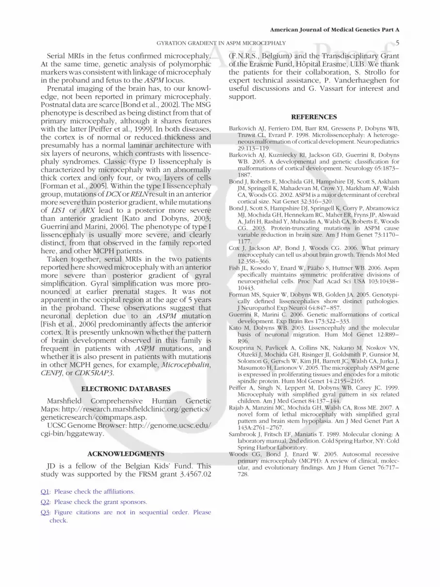
FIG. 1. Pre- and post-natal brain imaging in affected brother. A: Axial and (B) sagittal T2-weighted images of the affected fetus at 30 weeks of gestational age. Thefrontal lobes are short and hypoplastic, with an anterior to posterior (i.e., anterior more severe than posterior) gradient of simplification of the gyral pattern. C: Axial and(D) sagittal T2-weighted images of the same fetus at 35 weeks of gestational age. Gyration has progressed, but the anomalies noted at 30 weeks persist and maintain thegyral gradient pattern. E: Axial T2-weighted and (F) sagittal T1-weighted images exutero at 7 days of age, showing a global reduction in the sizeof the cerebral structuresassociated with microcephaly, a sloping forehead, and preserved midline structures.
2 DESIR ET AL.
American Journal of Medical Genetics Part A

25% risk of recurrence in each pregnancy. Theywere told that recurrence was likely in the currentpregnancy considering the fetal head measurementson ultrasonography. Fetal brain MRI was performedat 30 weeks of gestation (Fig. 1A,B). It showeda reduced number of cortical gyri, more severeanteriorly, that is, an anterior to posterior gradient ofgyration. The frontal lobes of the fetal brain weresmall and squared-off. To evaluate the progressionof gyration, a second MRI of the fetal brain wasperformed at 35 weeks gestation (Fig. 1C,D). Itshowed short and hypoplastic frontal lobes, withdiffuse, albeit anterior more than posterior, simpli-fication of the gyral pattern. The results of theimaging studies and their interpretation were dis-cussed with the parents at 26, 30, 32, and 35 weeks ofgestation. Termination of the pregnancy was pre-sented as a possible option and was declined by theparents. A baby boy was born at term by standardvaginal vertex delivery. His weight, length andOFC were 3.250 kg (�25th centile), 48.5 cm (�10thcentile) and 31 cm (��3 SD), respectively. Bloodwas sampled from the umbilical cord with informedconsent for DNA analysis. Physical and neurologicalexaminations were normal except for congenitalmicrocephaly. Abrain MRIwas repeated 1 week afterbirth, and showed improved but persistent simplifi-cation of the gyral pattern with anterior-to-posteriordecreasing severity (Fig. 1E,F). The boy was fol-lowed until age 2 years. He walked at 18 months,persistently on tiptoes.He hadno spasticity. Hehad aspeechdelaywithonly 10 isolatedwords at the ageof2 years, and a hyperactive behavior with attentiondeficit disorder like his sister. His electroencephalo-gram was normal.
LINKAGE AND MUTATION ANALYSES
Peripheral blood was sampled from the probandand both parents and cord blood from the boywith informed consent for DNA analysis. DNAwas extracted by the standard phenol–chloroform
method [Sambrook et al., 1989]. Linkage analyses tothe six known MCPH loci and the RELN gene wascarried out with microsatellite markers (see Supple-mentary Data). Marker order was obtained fromelectronic databases (see Electronic Databases Sec-tion). DNA was amplified by PCR using 15 ng of eachindividual’s DNA in a 15 ml final volume, followed byPolyAcrylamide Gel Electrophoresis and silver stain-ing. No evidence of linkage was found to the RELNgene nor to MCPH1, 2, 3, 4 and 6. A 14 cM region ofhomozygosity, shared between the two affectedsiblings, was identified at the MCPH5 (ASPM) locusat chromosome 1q31.2–q32.1 between markersD1S518 and D1S1171 (Fig. 3).
The 28 exons and flanking intronic sequencesof the ASPM gene were direct sequenced fromgenomic DNA of both parents and affected sibs. PCRproducts were purified using ExoSAP-IT1 ForPCR Product Clean-Up (USB) and sequenced usingthe Big Dye Terminator cycle sequencing kit v2
FIG. 2. Brain MRI in proband at age 5 years. A: Axial and (B) sagittal T1-weighted images. The gyral pattern is simplified with an anterior to posteriorgradient. The thickness of the cortex is normal. The midline structures arepreserved. Sloping of the forehead reflects the severe decrease in the cranial-to-facial proportions.
FIG. 3. Linkage analysis. The parents are first cousins once removed, the twoaffected children are represented in black. The ASPM locus is closely flankedby markers GATA135F02 and D1S1660. Genetic distances are indicated incentimorgans (cM). The homozygous haplotype shared by the affected siblingsis boxed. No evidence of linkage was found to the RELN gene nor to the fiveother MCPH loci reported to date (data not shown).
GYRATION GRADIENT IN ASPM MICROCEPHALY 3
American Journal of Medical Genetics Part A

(Applied Biosystems, Foster City, CA), and analyzedon a 3130 Genetic Analyzer sequencing machine(Applied Biosystems). Sequences were inspected insilico for mutations using the SeqScape softwareversion 2.0 (Applied Biosystems).
We identified a previously unreported mutation,consisting of an insertion of one base pair in exon 18,c.4195_4196insA (GenBank reference NM_018136.2)that introduced a frameshift and a premature stopcodon (p.Thr1399AsnfsX20). The mutation washomozygous in the two affected siblings, and, asexpected from the haplotype analysis, heterozygous
in both parents (Fig. 4). This study was approved bythe Ethical Committee of ULB, Hopital Erasme.
DISCUSSION
We were consulted for genetic evaluation duringthe second pregnancy of a young consanguineouscouple whose first child presented with congenitalmicrocephaly, moderate mental retardation andattention-hyperactivity disorder. The clinical picturewas typical of primary microcephaly. In view ofparental consanguinity, MCPH was likely.
FIG. 4. Mutation c.4195_4196insA (p.Thr1399AsnfsX20) of the ASPM gene. The black arrow indicates nucleotide 4195, where an A was inserted, causing a frameshiftand an in-frame premature stop codon (TGA, underlined). The mutation was heterozygous in the unaffected mother and father, while homozygous in the proband andaffected brother, and absent in a control. [Color figure can be viewed in the online issue, which is available at www.interscience.wiley.com.]
4 DESIR ET AL.
American Journal of Medical Genetics Part A
Serial MRIs in the fetus confirmed microcephaly.At the same time, genetic analysis of polymorphicmarkers was consistent with linkage of microcephalyin the proband and fetus to the ASPM locus.
Prenatal imaging of the brain has, to our knowl-edge, not been reported in primary microcephaly.Postnatal data are scarce [Bond et al., 2002]. The MSGphenotype is described as being distinct from that ofprimary microcephaly, although it shares featureswith the latter [Peiffer et al., 1999]. In both diseases,the cortex is of normal or reduced thickness andpresumably has a normal laminar architecture withsix layers of neurons, which contrasts with lissence-phaly syndromes. Classic (type I) lissencephaly ischaracterized by microcephaly with an abnormallythick cortex and only four, or two, layers of cells[Forman et al., 2005]. Within the type I lissencephalygroup, mutations of DCX or RELN result in an anteriormore severe than posterior gradient, while mutationsof LIS1 or ARX lead to a posterior more severethan anterior gradient [Kato and Dobyns, 2003;Guerrini and Marini, 2006]. The phenotype of type Ilissencephaly is usually more severe, and clearlydistinct, from that observed in the family reportedhere, and other MCPH patients.
Taken together, serial MRIs in the two patientsreported here showed microcephaly with an anteriormore severe than posterior gradient of gyralsimplification. Gyral simplification was more pro-nounced at earlier prenatal stages. It was notapparent in the occipital region at the age of 5 yearsin the proband. These observations suggest thatneuronal depletion due to an ASPM mutation[Fish et al., 2006] predominantly affects the anteriorcortex. It is presently unknown whether the patternof brain development observed in this family isfrequent in patients with ASPM mutations, andwhether it is also present in patients with mutationsin other MCPH genes, for example, Microcephalin,CENPJ, or CDK5RAP3.
ELECTRONIC DATABASES
Marshfield Comprehensive Human GeneticMaps: http://research.marshfieldclinic.org/genetics/geneticresearch/compmaps.asp.
UCSC Genome Browser: http://genome.ucsc.edu/cgi-bin/hggateway.
ACKNOWLEDGMENTS
JD is a fellow of the Belgian Kids’ Fund. Thisstudy was supported by the FRSM grant 3.4567.02
(F.N.R.S., Belgium) and the Transdisciplinary Grantof the Erasme Fund, Hopital Erasme, ULB. We thankthe patients for their collaboration, S. Strollo forexpert technical assistance, P. Vanderhaeghen foruseful discussions and G. Vassart for interest andsupport.
REFERENCES
Barkovich AJ, Ferriero DM, Barr RM, Gressens P, Dobyns WB,Truwit CL, Evrard P. 1998. Microlissencephaly: A heteroge-neousmalformation of cortical development. Neuropediatrics29:113–119.
Barkovich AJ, Kuzniecky RI, Jackson GD, Guerrini R, DobynsWB. 2005. A developmental and genetic classification formalformations of cortical development. Neurology 65:1873–1887.
Bond J, Roberts E, Mochida GH, Hampshire DJ, Scott S, AskhamJM, Springell K, Mahadevan M, Crow YJ, Markham AF, WalshCA, Woods CG. 2002. ASPM is a major determinant of cerebralcortical size. Nat Genet 32:316–320.
Bond J, Scott S, Hampshire DJ, Springell K, Corry P, AbramowiczMJ, Mochida GH, Hennekam RC, Maher ER, Fryns JP, AlswaidA, Jafri H, Rashid Y, Mubaidin A, Walsh CA, Roberts E, WoodsCG. 2003. Protein-truncating mutations in ASPM causevariable reduction in brain size. Am J Hum Genet 73:1170–1177.
Cox J, Jackson AP, Bond J, Woods CG. 2006. What primarymicrocephaly can tell us about brain growth. Trends Mol Med12:358–366.
Fish JL, Kosodo Y, Enard W, Paabo S, Huttner WB. 2006. Aspmspecifically maintains symmetric proliferative divisions ofneuroepithelial cells. Proc Natl Acad Sci USA 103:10438–10443.
Forman MS, Squier W, Dobyns WB, Golden JA. 2005. Genotypi-cally defined lissencephalies show distinct pathologies.J Neuropathol Exp Neurol 64:847–857.
Guerrini R, Marini C. 2006. Genetic malformations of corticaldevelopment. Exp Brain Res 173:322–333.
Kato M, Dobyns WB. 2003. Lissencephaly and the molecularbasis of neuronal migration. Hum Mol Genet 12:R89–R96.
Kouprina N, Pavlicek A, Collins NK, Nakano M, Noskov VN,Ohzeki J, Mochida GH, Risinger JI, Goldsmith P, Gunsior M,Solomon G, Gersch W, Kim JH, Barrett JC, Walsh CA, Jurka J,Masumoto H, Larionov V. 2005. The microcephaly ASPM geneis expressed in proliferating tissues and encodes for a mitoticspindle protein. Hum Mol Genet 14:2155–2165.
Peiffer A, Singh N, Leppert M, Dobyns WB, Carey JC. 1999.Microcephaly with simplified gyral pattern in six relatedchildren. Am J Med Genet 84:137–144.
Rajab A, Manzini MC, Mochida GH, Walsh CA, Ross ME. 2007. Anovel form of lethal microcephaly with simplified gyralpattern and brain stem hypoplasia. Am J Med Genet Part A143A:2761–2767.
Sambrook J, Fritsch EF, Maniatis T. 1989. Molecular cloning: Alaboratory manual, 2nd edition. Cold Spring Harbor, NY: ColdSpring Harbor Laboratory.
Woods CG, Bond J, Enard W. 2005. Autosomal recessiveprimary microcephaly (MCPH): A review of clinical, molec-ular, and evolutionary findings. Am J Hum Genet 76:717–728.
Q1: Please check the affiliations.
Q2: Please check the grant sponsors.
Q3: Figure citations are not in sequential order. Pleasecheck.
GYRATION GRADIENT IN ASPM MICROCEPHALY 5
American Journal of Medical Genetics Part A

1 1 1 R I V E R S T R E E T, H OBOKEN, N J 0 7 0 3 0
***IMMEDIATE RESPONSE REQUIRED***
Your article will be published online via Wiley's EarlyView® service (www.interscience.wiley.com) shortly after receipt ofcorrections. EarlyView® is Wiley's online publication of individual articles in full text HTML and/or pdf format before release ofthe compiled print issue of the journal. Articles posted online in EarlyView® are peer-reviewed, copyedited, author corrected,and fully citable. EarlyView® means you benefit from the best of two worlds--fast online availability as well as traditional, issue-based archiving.
READ PROOFS CAREFULLY• This will be your only chance to review these proofs.• Please note that the volume and page numbers shown on the proofs are for position only.
ANSWER ALL QUERIES ON PROOFS (Queries for you to answer are attached as the last page of your proof.)• Mark all corrections directly on the proofs. Note that excessive author alterations may ultimately result in delay of
publication and extra costs may be charged to you.
CHECK FIGURES AND TABLES CAREFULLY (Color figures will be sent under separate cover.)• Check size, numbering, and orientation of figures.• All images in the PDF are downsampled (reduced to lower resolution and file size) to facilitate Internet delivery.
These images will appear at higher resolution and sharpness in the printed article.• Review figure legends to ensure that they are complete.• Check all tables. Review layout, title, and footnotes.
COMPLETE REPRINT ORDER FORM• Fill out the attached reprint order form. It is important to return the form even if you are not ordering reprints. You
may, if you wish, pay for the reprints with a credit card. Reprints will be mailed only after your article appears inprint. This is the most opportune time to order reprints. If you wait until after your article comes off press, thereprints will be considerably more expensive.
RETURN PROOFSREPRINT ORDER FORMCTA (If you have not already signed one)
RETURN IMMEDIATELY AS YOUR ARTICLE WILL BE POSTED IN ORDER OF RECEIPT.
QUESTIONS? Christopher Sannella, Production EditorPhone: 201-748-5949E-mail: [email protected] to journal acronym and article production number(i.e., AJMA 00-0001 for American Journal of Medical Genetics ms 00-0001).
AJMA, e-proof:C1/Wiley-Liss

CCOOPPYYRRIIGGHHTT TTRRAANNSSFFEERR AAGGRREEEEMMEENNTT
Date:
To:
Production/ContributionID#______________Publisher/Editorial office use only
Re: Manuscript entitled ____________________________________________________________________________________________________________________________________________________________ (the "Contribution")
for publication in __________________________________________________________________ (the "Journal")published by Wiley-Liss, Inc., a subsidiary of John Wiley & Sons, Inc. ("Wiley").
Dear Contributor(s):
Thank you for submitting your Contribution for publication. In order to expedite the publishing process and enable Wiley todisseminate your work to the fullest extent, we need to have this Copyright Transfer Agreement signed and returned to us assoon as possible. If the Contribution is not accepted for publication this Agreement shall be null and void.
A. COPYRIGHT1. The Contributor assigns to Wiley, during the full term of copyright and any extensions or renewals of that term, all
copyright in and to the Contribution, including but not limited to the right to publish, republish, transmit, sell, distributeand otherwise use the Contribution and the material contained therein in electronic and print editions of the Journaland in derivative works throughout the world, in all languages and in all media of expression now known or laterdeveloped, and to license or permit others to do so.
2. Reproduction, posting, transmission or other distribution or use of the Contribution or any material contained therein,in any medium as permitted hereunder, requires a citation to the Journal and an appropriate credit to Wiley asPublisher, suitable in form and content as follows: (Title of Article, Author, Journal Title and Volume/Issue Copyright [year] Wiley-Liss, Inc. or copyright owner as specified in the Journal.)
B. RETAINED RIGHTSNotwithstanding the above, the Contributor or, if applicable, the Contributor's Employer, retains all proprietary rights otherthan copyright, such as patent rights, in any process, procedure or article of manufacture described in the Contribution,and the right to make oral presentations of material from the Contribution.
C. OTHER RIGHTS OF CONTRIBUTORWiley grants back to the Contributor the following:
1. The right to share with colleagues print or electronic "preprints" of the unpublished Contribution, in form and contentas accepted by Wiley for publication in the Journal. Such preprints may be posted as electronic files on theContributor's own website for personal or professional use, or on the Contributor's internal university or corporatenetworks/intranet, or secure external website at the Contributor’s institution, but not for commercial sale or for anysystematic external distribution by a third party (e.g., a listserve or database connected to a public access server). Priorto publication, the Contributor must include the following notice on the preprint: "This is a preprint of an articleaccepted for publication in [Journal title] copyright (year) (copyright owner as specified in the Journal)". Afterpublication of the Contribution by Wiley, the preprint notice should be amended to read as follows: "This is a preprintof an article published in [include the complete citation information for the final version of the Contribution aspublished in the print edition of the Journal]", and should provide an electronic link to the Journal's WWW site,located at the following Wiley URL: http://www.interscience.Wiley.com/. The Contributor agrees not to update thepreprint or replace it with the published version of the Contribution.
2. The right, without charge, to photocopy or to transmit online or to download, print out and distribute to a colleague acopy of the published Contribution in whole or in part, for the Contributor's personal or professional use, for the

-2-
advancement of scholarly or scientific research or study, or for corporate informational purposes in accordance withParagraph D.2 below.
3. The right to republish, without charge, in print format, all or part of the material from the published Contribution in a
book written or edited by the Contributor.
4. The right to use selected figures and tables, and selected text (up to 250 words, exclusive of the abstract) from theContribution, for the Contributor's own teaching purposes, or for incorporation within another work by the Contributorthat is made part of an edited work published (in print or electronic format) by a third party, or for presentation inelectronic format on an internal computer network or external website of the Contributor or the Contributor's employer.
5. The right to include the Contribution in a compilation for classroom use (course packs) to be distributed to students at
the Contributor’s institution free of charge or to be stored in electronic format in datarooms for access by students atthe Contributor’s institution as part of their course work (sometimes called “electronic reserve rooms”) and for in-house training programs at the Contributor’s employer.
D. CONTRIBUTIONS OWNED BY EMPLOYER1. If the Contribution was written by the Contributor in the course of the Contributor's employment (as a "work-made-for-
hire" in the course of employment), the Contribution is owned by the company/employer which must sign thisAgreement (in addition to the Contributor’s signature), in the space provided below. In such case, thecompany/employer hereby assigns to Wiley, during the full term of copyright, all copyright in and to the Contributionfor the full term of copyright throughout the world as specified in paragraph A above.
2. In addition to the rights specified as retained in paragraph B above and the rights granted back to the Contributorpursuant to paragraph C above, Wiley hereby grants back, without charge, to such company/employer, its subsidiariesand divisions, the right to make copies of and distribute the published Contribution internally in print format orelectronically on the Company's internal network. Upon payment of the Publisher's reprint fee, the institution maydistribute (but not resell) print copies of the published Contribution externally. Although copies so made shall not beavailable for individual re-sale, they may be included by the company/employer as part of an information packageincluded with software or other products offered for sale or license. Posting of the published Contribution by theinstitution on a public access website may only be done with Wiley's written permission, and payment of anyapplicable fee(s).
E. GOVERNMENT CONTRACTS
In the case of a Contribution prepared under U.S. Government contract or grant, the U.S. Government may reproduce,without charge, all or portions of the Contribution and may authorize others to do so, for official U.S. Government purposesonly, if the U.S. Government contract or grant so requires. (U.S. Government Employees: see note at end).
F. COPYRIGHT NOTICEThe Contributor and the company/employer agree that any and all copies of the Contribution or any part thereofdistributed or posted by them in print or electronic format as permitted herein will include the notice of copyright asstipulated in the Journal and a full citation to the Journal as published by Wiley.
G. CONTRIBUTOR'S REPRESENTATIONSThe Contributor represents that the Contribution is the Contributor's original work. If the Contribution was preparedjointly, the Contributor agrees to inform the co-Contributors of the terms of this Agreement and to obtain their signature tothis Agreement or their written permission to sign on their behalf. The Contribution is submitted only to this Journal andhas not been published before, except for "preprints" as permitted above. (If excerpts from copyrighted works owned bythird parties are included, the Contributor will obtain written permission from the copyright owners for all uses as set forthin Wiley's permissions form or in the Journal's Instructions for Contributors, and show credit to the sources in theContribution.) The Contributor also warrants that the Contribution contains no libelous or unlawful statements, does notinfringe on the rights or privacy of others, or contain material or instructions that might cause harm or injury.

-3-
CHECK ONE:___________________________________ _____________________
[____]Contributor-owned work Contributor's signature Date
___________________________________________________________Type or print name and title
___________________________________ _____________________Co-contributor's signature Date
___________________________________________________________Type or print name and title
ATTACHED ADDITIONAL SIGNATURE PAGE AS NECESSARY
___________________________________ _____________________[____]Company/Institution-owned work Company or Institution (Employer-for-Hire) Date
(made-for-hire in thecourse of employment) ___________________________________ ______________________
Authorized signature of Employer Date
[____]U.S. Government work
Note to U.S. Government Employees
A Contribution prepared by a U.S. federal government employee as part of the employee's official duties, or which is an officialU.S. Government publication is called a "U.S. Government work," and is in the public domain in the United States. In such case,the employee may cross out Paragraph A.1 but must sign and return this Agreement. If the Contribution was not prepared aspart of the employee's duties or is not an official U.S. Government publication, it is not a U.S. Government work.
[____]U.K. Government work (Crown Copyright)
Note to U.K. Government Employees
The rights in a Contribution prepared by an employee of a U.K. government department, agency or other Crown body as part ofhis/her official duties, or which is an official government publication, belong to the Crown. In such case, the Publisher willforward the relevant form to the Employee for signature.

111 RIVER STREET, HOBOKEN, NJ 07030
To: Mr. Christopher Sannella
Phone: 201-748-5949
Fax: 201-748-6281
From:
Date:
Pages includingthis cover page:
Comments:

C1
REPRINT BILLING DEPARTMENT •• 111 RIVER STREET, HOBOKEN, NJ 07030PHONE: (201) 748-8789; FAX: (201) 748-6281
E-MAIL: [email protected] REPRINT ORDER FORM
Please complete this form even if you are not ordering reprints. This form MUST be returned with your corrected proofsand original manuscript. Your reprints will be shipped approximately 4 weeks after publication. Reprints ordered after printingwill be substantially more expensive.
JOURNAL VOLUME ISSUE
TITLE OF MANUSCRIPT
MS. NO. NO. OF PAGES AUTHOR(S)
No. of Pages 100 Reprints 200 Reprints 300 Reprints 400 Reprints 500 Reprints$ $ $ $ $
1-4 336 501 694 890 10525-8 469 703 987 1251 14779-12 594 923 1234 1565 185013-16 714 1156 1527 1901 227317-20 794 1340 1775 2212 264821-24 911 1529 2031 2536 303725-28 1004 1707 2267 2828 338829-32 1108 1894 2515 3135 375533-36 1219 2092 2773 3456 414337-40 1329 2290 3033 3776 4528
**REPRINTS ARE ONLY AVAILABLE IN LOTS OF 100. IF YOU WISH TO ORDER MORE THAN 500 REPRINTS, PLEASE CONTACT OUR REPRINTSDEPARTMENT AT (201) 748-8891 FOR A PRICE QUOTE.
Please send me _____________________ reprints of the above article at $
Please add appropriate State and Local Tax (Tax Exempt No.____________________) $for United States orders only.
Please add 5% Postage and Handling $
TOTAL AMOUNT OF ORDER** $**International orders must be paid in currency and drawn on a U.S. bankPlease check one: Check enclosed Bill me Credit CardIf credit card order, charge to: American Express Visa MasterCard
Credit Card No Signature Exp. Date
BILL TO: SHIP TO: (Please, no P.O. Box numbers)Name Name
Institution Institution
Address Address
Purchase Order No. Phone Fax

Softproofing for advanced Adobe Acrobat Users - NOTES toolNOTE: ACROBAT READER FROM THE INTERNET DOES NOT CONTAIN THE NOTES TOOL USED IN THIS PROCEDURE.
Acrobat annotation tools can be very useful for indicating changes to the PDF proof of your article.By using Acrobat annotation tools, a full digital pathway can be maintained for your page proofs.
The NOTES annotation tool can be used with either Adobe Acrobat 4.0, 5.0 or 6.0. Other annotation tools are also available in Acrobat 4.0, but this instruction sheet will concentrateon how to use the NOTES tool. Acrobat Reader, the free Internet download software from Adobe,DOES NOT contain the NOTES tool. In order to softproof using the NOTES tool you must havethe full software suite Adobe Acrobat 4.0, 5.0 or 6.0 installed on your computer.
Steps for Softproofing using Adobe Acrobat NOTES tool:
1. Open the PDF page proof of your article using either Adobe Acrobat 4.0, 5.0 or 6.0. Proofyour article on-screen or print a copy for markup of changes.
2. Go to File/Preferences/Annotations (in Acrobat 4.0) or Document/Add a Comment (in Acrobat6.0 and enter your name into the “default user” or “author” field. Also, set the font size at 9 or 10point.
3. When you have decided on the corrections to your article, select the NOTES tool from theAcrobat toolbox and click in the margin next to the text to be changed.
4. Enter your corrections into the NOTES text box window. Be sure to clearly indicate where thecorrection is to be placed and what text it will effect. If necessary to avoid confusion, you canuse your TEXT SELECTION tool to copy the text to be corrected and paste it into the NOTEStext box window. At this point, you can type the corrections directly into the NOTES textbox window. DO NOT correct the text by typing directly on the PDF page.
5. Go through your entire article using the NOTES tool as described in Step 4.
6. When you have completed the corrections to your article, go to File/Export/Annotations (inAcrobat 4.0) or Document/Add a Comment (in Acrobat 6.0).
7. When closing your article PDF be sure NOT to save changes to original file.
8. To make changes to a NOTES file you have exported, simply re-open the original PDFproof file, go to File/Import/Notes and import the NOTES file you saved. Make changes and re-export NOTES file keeping the same file name.
9. When complete, attach your NOTES file to a reply e-mail message. Be sure to include yourname, the date, and the title of the journal your article will be printed in.


