Embed Size (px)
Citation preview
pharmaceuticals
Article
Pre-Clinical Investigation of Liquid Paclitaxel forLocal Drug Delivery: A Pilot Study
Claire V. Cawthon 1 , Kathryn Cooper 1 , Clifton Huett 1, Alyssa Lloret 2,Estefanny Villar-Matamoros 2, Lauren Stokes 2, Uwe Christians 3, Michele Schuler 4 andSaami K. Yazdani 2,*
1 Department of Mechanical Engineering, University of South Alabama, Mobile, AL 36688, USA;[email protected] (C.V.C.); [email protected] (K.C.);[email protected] (C.H.)
2 Department of Engineering, Wake Forest University, Winston-Salem, NC 27101, USA;[email protected] (A.L.); [email protected] (E.V.-M.); [email protected] (L.S.)
3 iC42 Clinical Research and Development, University of Colorado, Aurora, CO 80045, USA;[email protected]
4 Department of Comparative Medicine, University of South Alabama, Mobile, AL 36688, USA;[email protected]
* Correspondence: [email protected]
Received: 25 September 2020; Accepted: 24 November 2020; Published: 28 November 2020 �����������������
Abstract: The purpose of this pilot study was to investigate the feasibility of a perfusion catheterto deliver liquid paclitaxel into arterial segments. A clinically relevant rabbit ilio-femoral injurymodel was utilized to determine the impact of liquid paclitaxel delivered locally into the vesselwall using a perfusion catheter at 1 h to 14 days. Treatment by two clinically available forms ofliquid paclitaxel, a solvent-based (sb) versus an albumin-bound (nab), along with a control (uncoatedballoons), were investigated. Pharmacokinetic results demonstrated an increase in the retention ofthe sb-paclitaxel versus the nab-paclitaxel at 1 h; however, no other differences were observed atdays one, three, and seven. Histological findings at 14 days showed significantly less neointimalarea in the sb-paclitaxel treated arteries as compared with the nab-paclitaxel and the uncoatedballoon-treated arteries. Additionally, percent area stenosis was significantly less in the sb-paclitaxelgroup. These results support the concept of local liquid delivery of paclitaxel into the arterial segments.
Keywords: peripheral arterial disease; liquid paclitaxel; local drug delivery; perfusion catheter;pre-clinical modeling
1. Introduction
There are more than three million people affected by peripheral artery disease (PAD) every year inthe U.S. Disparate from coronary artery disease, PAD is characterized by a substantial plaque burdenand often presents with long and complex lesions [1,2]. Endovascular treatment of PAD focusesfirst on re-establishing blood flow by angioplasty and, more frequently, de-bulking by atherectomy.Angioplasty, described as a control injury, expands the lumen by outward stretching of the arterialwall. The vascular smooth muscle cells (VSMCs), residing in the vessel wall, respond to this injury byproliferating and migrating inward, re-narrowing the vessel lumen. This process is termed restenosis.To minimize the injury response and to inhibit VSMC proliferation, anti-proliferative drugs have beenlocally delivered using drug-eluting stents (DES) or drug-coated balloons (DCBs) [3,4].
Current endovascular treatments, including DES and DCBs, have limitations leading to inconsistentoutcomes, patient readmission, and repeat revascularization. Stents tend to fracture due to the severe
Pharmaceuticals 2020, 13, 434; doi:10.3390/ph13120434 www.mdpi.com/journal/pharmaceuticals
Pharmaceuticals 2020, 13, 434 2 of 11
biomechanical environment of peripheral arteries [5–8]. Peripheral arteries undergo unique movementsnot observed in other parts of the body including compression, shortening, bending and twisting [9–11].The use of a permanent device such as a metallic stent also limits re-interventional procedure in casesof restenosis.
To overcome the limitations of stents, DCBs were developed to locally deliver therapeutics toinhibit restenosis without the need of a permanent platform serving as a drug reservoir. DCBs rely onthe transfer of the anti-proliferative drug, which is coated on the surface of the balloon, to the lumen ofthe arterial wall by simple contact of the balloon to the artery. DCBs are coated with a crystalline formof paclitaxel designed to adhere to the abluminal surface of the artery. Clinical studies have shownpromising results with DCBs, but only in relatively short and simple lesions and mostly to treat diseasein arteries above the knee [4,12].
In the periphery, paclitaxel has been the anti-proliferative drug of choice of DCBs due to itspotency and binding affinity, making it an attractive drug for single use with long lasting effects [13,14].Paclitaxel binds to tubulin, inhibiting depolymerization, which inhibits cell division and growth [15].However, recent clinical publications have cast doubts on the safety profile of paclitaxel to treatPAD [16,17]. The INPACT-DEEP clinical study showed higher rates of amputation in the paclitaxelgroup than in controls, which was attributed to microembolization of particles related to means ofdrug attachment to the balloon [18]. Most notably the meta-analysis by Katsanos et al. showedpaclitaxel-coated balloons and stents in femoral-popliteal arteries had an increased risk of all-causemortality at three and five years [16]. Reviews of this analysis and the controversy surrounding ithave encouraged further research into devices with alternative delivery systems [19]. Furthermore,Katsanos and his group published an additional meta-analysis study showing safety concerns forbelow-the-knee applications [17].
Current DCBs and DES use the dry (or powder) form of paclitaxel, which has a crystallinestructure. Crystallinity of the paclitaxel drug increases its residency and local arterial pharmacokineticsand reduces solubility of the drug. This increase in residency is somewhat needed as the drug ispositioned on the luminal surface and not within the medial wall. However, by placing the drugonto the luminal surface, there is a potential for the crystalline paclitaxel drug to become dislodged(mobilized), travelling to distal organs and tissue. Previously reported pre-clinical studies havedemonstrated fibrinoid necrosis in downstream tissues of DCB-treated peripheral segments, along withhigher paclitaxel levels in nontargeted tissue, suggesting increased emboli debris from crystallinepaclitaxel [20,21].
In this pilot study, we investigated a new approach to deliver the therapeutic drug paclitaxel forperipheral applications. Specifically, we tested the use of a perfusion catheter to locally deliver theliquid form of paclitaxel directly into the vessel wall. Currently two available forms of liquid paclitaxelexist, a solvent-based (sb-) paclitaxel and an albumin-bound (nab-) paclitaxel, primarily used to treatcancer patients. The aim of this pilot study was to investigate the effectiveness of the solvent-basedpaclitaxel versus the albumin-bound paclitaxel form delivered locally by a perfusion catheter in aclinically relevant rabbit femoral-iliac injury model.
2. Results
2.1. Pharmacokinetic Analysis
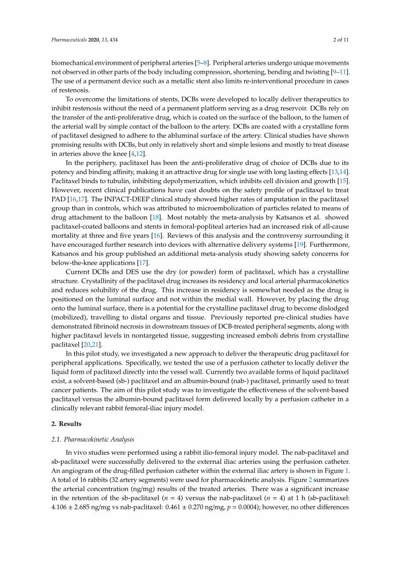
In vivo studies were performed using a rabbit ilio-femoral injury model. The nab-paclitaxel andsb-paclitaxel were successfully delivered to the external iliac arteries using the perfusion catheter.An angiogram of the drug-filled perfusion catheter within the external iliac artery is shown in Figure 1.A total of 16 rabbits (32 artery segments) were used for pharmacokinetic analysis. Figure 2 summarizesthe arterial concentration (ng/mg) results of the treated arteries. There was a significant increasein the retention of the sb-paclitaxel (n = 4) versus the nab-paclitaxel (n = 4) at 1 h (sb-paclitaxel:4.106 ± 2.685 ng/mg vs nab-paclitaxel: 0.461 ± 0.270 ng/mg, p = 0.0004); however, no other differences
Pharmaceuticals 2020, 13, 434 3 of 11
were observed at days one, three, and seven. There was a significant drop in sb-paclitaxel between 1 hand one day (1 h: 4.106 ± 2.685 ng/mg vs one day: 0.108 ± 0.089 ng/mg, p < 0.001), but none betweenthe other treatment time points.
Pharmaceuticals 2020, 13, x FOR PEER REVIEW 3 of 11
were observed at days one, three, and seven. There was a significant drop in sb-paclitaxel between 1 h and one day (1 h: 4.106 ± 2.685 ng/mg vs one day: 0.108 ± 0.089 ng/mg, p < 0.001), but none between the other treatment time points.
Figure 1. Angiogram of the perfusion catheter during delivery. (a) Liquid paclitaxel is shown filling the treatment chamber (blue arrow). The occlusion perfusion balloons are shown in the red arrows. (b) Angiogram following treatment of vessel by liquid paclitaxel.
Figure 2. Mean tissue paclitaxel concentrations of the perfusion catheter treated arteries segments with the nab-paclitaxel and sb-paclitaxel liquid forms (bars represent standard deviation). A significant difference (*** p < 0.001) was observed at 1 h.
2.2. Morphological and Histological Findings of Treated Arteries
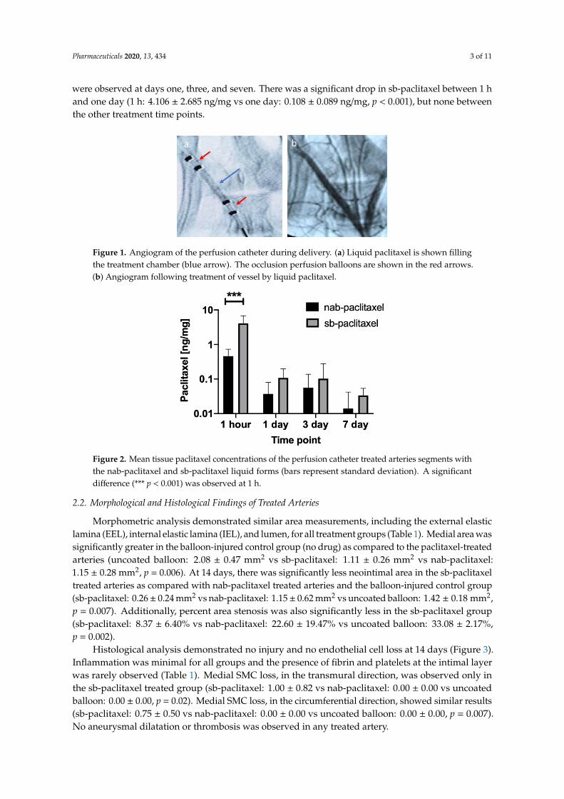
Morphometric analysis demonstrated similar area measurements, including the external elastic lamina (EEL), internal elastic lamina (IEL), and lumen, for all treatment groups (Table 1). Medial area was significantly greater in the balloon-injured control group (no drug) as compared to the paclitaxel-treated arteries (uncoated balloon: 2.08 ± 0.47 mm2 vs sb-paclitaxel: 1.11 ± 0.26 mm2 vs nab-paclitaxel: 1.15 ± 0.28 mm2, p = 0.006). At 14 days, there was significantly less neointimal area in the sb-paclitaxel treated arteries as compared with nab-paclitaxel treated arteries and the balloon-injured control group (sb-paclitaxel: 0.26 ± 0.24 mm2 vs nab-paclitaxel: 1.15 ± 0.62 mm2 vs uncoated balloon: 1.42 ± 0.18 mm2, p = 0.007). Additionally, percent area stenosis was also significantly less in the sb-paclitaxel group (sb-paclitaxel: 8.37 ± 6.40% vs nab-paclitaxel: 22.60 ± 19.47% vs uncoated balloon: 33.08 ± 2.17%, p = 0.002).
Histological analysis demonstrated no injury and no endothelial cell loss at 14 days (Figure 3). Inflammation was minimal for all groups and the presence of fibrin and platelets at the intimal layer was rarely observed (Table 1). Medial SMC loss, in the transmural direction, was observed only in the sb-paclitaxel treated group (sb-paclitaxel: 1.00 ± 0.82 vs nab-paclitaxel: 0.00 ± 0.00 vs uncoated balloon: 0.00 ± 0.00, p = 0.02). Medial SMC loss, in the circumferential direction, showed similar results (sb-paclitaxel: 0.75 ± 0.50 vs nab-paclitaxel: 0.00 ± 0.00 vs uncoated balloon: 0.00 ± 0.00, p = 0.007). No aneurysmal dilatation or thrombosis was observed in any treated artery.
ba
Figure 1. Angiogram of the perfusion catheter during delivery. (a) Liquid paclitaxel is shown fillingthe treatment chamber (blue arrow). The occlusion perfusion balloons are shown in the red arrows.(b) Angiogram following treatment of vessel by liquid paclitaxel.
Pharmaceuticals 2020, 13, x FOR PEER REVIEW 3 of 11
were observed at days one, three, and seven. There was a significant drop in sb-paclitaxel between 1 h and one day (1 h: 4.106 ± 2.685 ng/mg vs one day: 0.108 ± 0.089 ng/mg, p < 0.001), but none between the other treatment time points.
Figure 1. Angiogram of the perfusion catheter during delivery. (a) Liquid paclitaxel is shown filling the treatment chamber (blue arrow). The occlusion perfusion balloons are shown in the red arrows. (b) Angiogram following treatment of vessel by liquid paclitaxel.
Figure 2. Mean tissue paclitaxel concentrations of the perfusion catheter treated arteries segments with the nab-paclitaxel and sb-paclitaxel liquid forms (bars represent standard deviation). A significant difference (*** p < 0.001) was observed at 1 h.
2.2. Morphological and Histological Findings of Treated Arteries
Morphometric analysis demonstrated similar area measurements, including the external elastic lamina (EEL), internal elastic lamina (IEL), and lumen, for all treatment groups (Table 1). Medial area was significantly greater in the balloon-injured control group (no drug) as compared to the paclitaxel-treated arteries (uncoated balloon: 2.08 ± 0.47 mm2 vs sb-paclitaxel: 1.11 ± 0.26 mm2 vs nab-paclitaxel: 1.15 ± 0.28 mm2, p = 0.006). At 14 days, there was significantly less neointimal area in the sb-paclitaxel treated arteries as compared with nab-paclitaxel treated arteries and the balloon-injured control group (sb-paclitaxel: 0.26 ± 0.24 mm2 vs nab-paclitaxel: 1.15 ± 0.62 mm2 vs uncoated balloon: 1.42 ± 0.18 mm2, p = 0.007). Additionally, percent area stenosis was also significantly less in the sb-paclitaxel group (sb-paclitaxel: 8.37 ± 6.40% vs nab-paclitaxel: 22.60 ± 19.47% vs uncoated balloon: 33.08 ± 2.17%, p = 0.002).
Histological analysis demonstrated no injury and no endothelial cell loss at 14 days (Figure 3). Inflammation was minimal for all groups and the presence of fibrin and platelets at the intimal layer was rarely observed (Table 1). Medial SMC loss, in the transmural direction, was observed only in the sb-paclitaxel treated group (sb-paclitaxel: 1.00 ± 0.82 vs nab-paclitaxel: 0.00 ± 0.00 vs uncoated balloon: 0.00 ± 0.00, p = 0.02). Medial SMC loss, in the circumferential direction, showed similar results (sb-paclitaxel: 0.75 ± 0.50 vs nab-paclitaxel: 0.00 ± 0.00 vs uncoated balloon: 0.00 ± 0.00, p = 0.007). No aneurysmal dilatation or thrombosis was observed in any treated artery.
ba
Figure 2. Mean tissue paclitaxel concentrations of the perfusion catheter treated arteries segments withthe nab-paclitaxel and sb-paclitaxel liquid forms (bars represent standard deviation). A significantdifference (*** p < 0.001) was observed at 1 h.
2.2. Morphological and Histological Findings of Treated Arteries
Morphometric analysis demonstrated similar area measurements, including the external elasticlamina (EEL), internal elastic lamina (IEL), and lumen, for all treatment groups (Table 1). Medial area wassignificantly greater in the balloon-injured control group (no drug) as compared to the paclitaxel-treatedarteries (uncoated balloon: 2.08 ± 0.47 mm2 vs sb-paclitaxel: 1.11 ± 0.26 mm2 vs nab-paclitaxel:1.15 ± 0.28 mm2, p = 0.006). At 14 days, there was significantly less neointimal area in the sb-paclitaxeltreated arteries as compared with nab-paclitaxel treated arteries and the balloon-injured control group(sb-paclitaxel: 0.26± 0.24 mm2 vs nab-paclitaxel: 1.15± 0.62 mm2 vs uncoated balloon: 1.42 ± 0.18 mm2,p = 0.007). Additionally, percent area stenosis was also significantly less in the sb-paclitaxel group(sb-paclitaxel: 8.37 ± 6.40% vs nab-paclitaxel: 22.60 ± 19.47% vs uncoated balloon: 33.08 ± 2.17%,p = 0.002).
Histological analysis demonstrated no injury and no endothelial cell loss at 14 days (Figure 3).Inflammation was minimal for all groups and the presence of fibrin and platelets at the intimal layerwas rarely observed (Table 1). Medial SMC loss, in the transmural direction, was observed only inthe sb-paclitaxel treated group (sb-paclitaxel: 1.00 ± 0.82 vs nab-paclitaxel: 0.00 ± 0.00 vs uncoatedballoon: 0.00 ± 0.00, p = 0.02). Medial SMC loss, in the circumferential direction, showed similar results(sb-paclitaxel: 0.75 ± 0.50 vs nab-paclitaxel: 0.00 ± 0.00 vs uncoated balloon: 0.00 ± 0.00, p = 0.007).No aneurysmal dilatation or thrombosis was observed in any treated artery.
Pharmaceuticals 2020, 13, 434 4 of 11
Table 1. Summary of the morphometric and histological measurements in the rabbit iliac-femoralinjury model.
Measurements nab-Paclitaxel sb-Paclitaxel Uncoated-Balloons p Value
MorphometricMeasurements
EEL, mm2 6.23 ± 1.42 4.92 ± 2.45 6.39 ± 0.68 0.43IEL, mm2 5.10 ± 1.17 3.81 ± 2.31 4.31 ± 0.70 0.69
Lumen, mm2 3.93 ± 1.06 3.55 ± 2.19 2.89 ± 0.53 0.60Media, mm2 1.15 ± 0.28 * 1.11 ± 0.26 * 2.08 ± 0.47 0.006
Neointimal area, mm2 1.15 ± 0.62 0.26 ± 0.24 *,# 1.42 ± 0.18 0.007Percent area stenosis, % 22.60 ± 19.47 8.37 ± 6.40 *,# 33.08 ± 2.17 0.002Light Microscopy Analysis
Injury 0.00 ± 0.00 0.00 ± 0.00 0.00 ± 0.00 1.00EC loss 0.00 ± 0.00 0.00 ± 0.00 0.00 ± 0.00 1.00
Inflammation (intimal) 0.50 ± 0.578 0.25 ± 0.50 0.00 ± 0.00 0.32Inflammation (adv) 0.25 ± 0.50 0.50 ± 0.578 0.25 ± 0.50 0.75Fibrin and Platelets 0.00 ± 0.00 0.25 ± 0.50 0.00 ± 0.00 0.41
SMC Loss (trans) 0.00 ± 0.00 1.00 ± 0.82 *,# 0.00 ± 0.00 0.02SMC Loss (circum) 0.00 ± 0.00 0.75 ± 0.50 *,# 0.00 ± 0.00 0.007
Abbreviations: nab—albumin-bound, sb—solvent-based, EEL—external elastic lamina, IEL—internal elastic lamina,EC—endothelial cell, SMC—smooth muscle cell, adv—adventitia, trans—transmural, circum—circumferential,*—denotes significant difference as compared to uncoated balloon (control) group, #—denotes significant differenceas compared to nab-paclitaxel group.
Pharmaceuticals 2020, 13, x FOR PEER REVIEW 4 of 11
Table 1. Summary of the morphometric and histological measurements in the rabbit iliac-femoral injury model.
Measurements nab-Paclitaxel sb-Paclitaxel Uncoated-Balloons p Value Morphometric Measurements
EEL, mm2 6.23 ± 1.42 4.92 ± 2.45 6.39 ± 0.68 0.43 IEL, mm2 5.10 ± 1.17 3.81 ± 2.31 4.31 ± 0.70 0.69
Lumen, mm2 3.93 ± 1.06 3.55 ± 2.19 2.89 ± 0.53 0.60 Media, mm2 1.15 ± 0.28 * 1.11 ± 0.26 * 2.08 ± 0.47 0.006
Neointimal area, mm2 1.15 ± 0.62 0.26 ± 0.24 *,# 1.42 ± 0.18 0.007 Percent area stenosis, % 22.60 ± 19.47 8.37 ± 6.40 *,# 33.08 ± 2.17 0.002 Light Microscopy Analysis
Injury 0.00 ± 0.00 0.00 ± 0.00 0.00 ± 0.00 1.00 EC loss 0.00 ± 0.00 0.00 ± 0.00 0.00 ± 0.00 1.00
Inflammation (intimal) 0.50 ± 0.578 0.25 ± 0.50 0.00 ± 0.00 0.32 Inflammation (adv) 0.25 ± 0.50 0.50 ± 0.578 0.25 ± 0.50 0.75 Fibrin and Platelets 0.00 ± 0.00 0.25 ± 0.50 0.00 ± 0.00 0.41
SMC Loss (trans) 0.00 ± 0.00 1.00 ± 0.82 *,# 0.00 ± 0.00 0.02 SMC Loss (circum) 0.00 ± 0.00 0.75 ± 0.50 *,# 0.00 ± 0.00 0.007
Abbreviations: nab—albumin-bound, sb—solvent-based, EEL—external elastic lamina, IEL—internal elastic lamina, EC—endothelial cell, SMC—smooth muscle cell, adv—adventitia, trans—transmural, circum—circumferential, *—denotes significant difference as compared to uncoated balloon (control) group, #—denotes significant difference as compared to nab-paclitaxel group.
Figure 3. Histological assessment of liquid paclitaxel treated arteries. Representative hematoxylin and eosin (H&E) staining of (a) nab-paclitaxel, (b) sb-paclitaxel treated arteries, and (c) balloon-injured arteries. Blue arrows represent internal elastic laminae (IEL). Lumen is represented by L. (d) Significant differences (* p < 0.05, ** p < 0.01) in percent area stenosis were observed between the varying treatment groups (error bars represent standard deviation).
3. Discussion
Figure 3. Histological assessment of liquid paclitaxel treated arteries. Representative hematoxylin andeosin (H&E) staining of (a) nab-paclitaxel, (b) sb-paclitaxel treated arteries, and (c) balloon-injuredarteries. Blue arrows represent internal elastic laminae (IEL). Lumen is represented by L. (d) Significantdifferences (* p < 0.05, ** p < 0.01) in percent area stenosis were observed between the varying treatmentgroups (error bars represent standard deviation).
Pharmaceuticals 2020, 13, 434 5 of 11
3. Discussion
This study was designed to evaluate the efficacy of local liquid paclitaxel delivery to arterialsegments in a clinically relevant rabbit ilio-femoral injury model. Two clinically available forms ofpaclitaxel, a solvent-based liquid paclitaxel and an albumin-bound liquid paclitaxel, were utilized toquantify the pharmacokinetics of paclitaxel retention within the arterial wall and histologically evaluatethe treated arteries. Pharmacokinetic results indicated greater retention of the solvent-based liquidpaclitaxel as compared with albumin-bound paclitaxel. Histologically, there was a significant decreasein neointimal hyperplasia and stenosis in arteries treated with the solvent-based liquid paclitaxelas compared to the albumin-bound liquid paclitaxel and the balloon-injured arteries. Additionally,drug-induced medial smooth muscle cell loss was more frequently observed in the solvent-basedliquid paclitaxel treated arteries. These results indicate the effectiveness of a local liquid approach todeliver and retain liquid paclitaxel in arterial segments.
In this pilot study, the use of liquid forms of paclitaxel was investigated a potential therapy toinhibit restenosis. The selected intravenous (liquid) forms of paclitaxel have been utilized to treatpatients with breast, ovarian, prostate and other forms of solid tumor cancers for the past four decades.In comparing the different forms of paclitaxel, the half-life of liquid (intravenous) paclitaxel is withinhours [15,22], whereas the crystalline paclitaxel is weeks to months [14,23,24]. Thus, the potential forany lost (or mobilized) liquid paclitaxel to remain and accumulate within distal tissue or organs isminimal. Furthermore, the approach of liquid delivery offers an alternative to the current standards ofDES and DCBs in which the anti-proliferative drug is deposited on the luminal surface. The describedapproach directly delivers paclitaxel into the medial layer, the residing region of proliferating smoothmuscle cells.
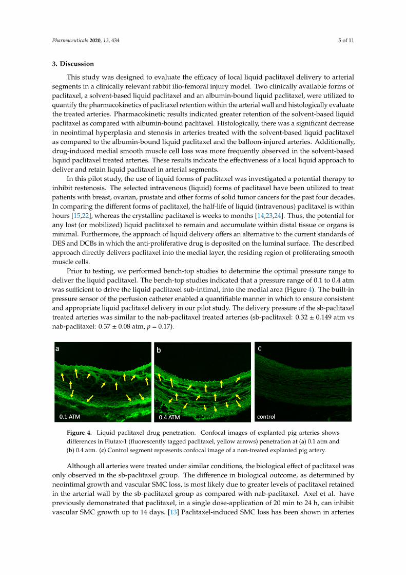
Prior to testing, we performed bench-top studies to determine the optimal pressure range todeliver the liquid paclitaxel. The bench-top studies indicated that a pressure range of 0.1 to 0.4 atmwas sufficient to drive the liquid paclitaxel sub-intimal, into the medial area (Figure 4). The built-inpressure sensor of the perfusion catheter enabled a quantifiable manner in which to ensure consistentand appropriate liquid paclitaxel delivery in our pilot study. The delivery pressure of the sb-paclitaxeltreated arteries was similar to the nab-paclitaxel treated arteries (sb-paclitaxel: 0.32 ± 0.149 atm vsnab-paclitaxel: 0.37 ± 0.08 atm, p = 0.17).
Pharmaceuticals 2020, 13, x FOR PEER REVIEW 5 of 11
This study was designed to evaluate the efficacy of local liquid paclitaxel delivery to arterial segments in a clinically relevant rabbit ilio-femoral injury model. Two clinically available forms of paclitaxel, a solvent-based liquid paclitaxel and an albumin-bound liquid paclitaxel, were utilized to quantify the pharmacokinetics of paclitaxel retention within the arterial wall and histologically evaluate the treated arteries. Pharmacokinetic results indicated greater retention of the solvent-based liquid paclitaxel as compared with albumin-bound paclitaxel. Histologically, there was a significant decrease in neointimal hyperplasia and stenosis in arteries treated with the solvent-based liquid paclitaxel as compared to the albumin-bound liquid paclitaxel and the balloon-injured arteries. Additionally, drug-induced medial smooth muscle cell loss was more frequently observed in the solvent-based liquid paclitaxel treated arteries. These results indicate the effectiveness of a local liquid approach to deliver and retain liquid paclitaxel in arterial segments.
In this pilot study, the use of liquid forms of paclitaxel was investigated a potential therapy to inhibit restenosis. The selected intravenous (liquid) forms of paclitaxel have been utilized to treat patients with breast, ovarian, prostate and other forms of solid tumor cancers for the past four decades. In comparing the different forms of paclitaxel, the half-life of liquid (intravenous) paclitaxel is within hours [15,22], whereas the crystalline paclitaxel is weeks to months [14,23,24]. Thus, the potential for any lost (or mobilized) liquid paclitaxel to remain and accumulate within distal tissue or organs is minimal. Furthermore, the approach of liquid delivery offers an alternative to the current standards of DES and DCBs in which the anti-proliferative drug is deposited on the luminal surface. The described approach directly delivers paclitaxel into the medial layer, the residing region of proliferating smooth muscle cells.
Prior to testing, we performed bench-top studies to determine the optimal pressure range to deliver the liquid paclitaxel. The bench-top studies indicated that a pressure range of 0.1 to 0.4 atm was sufficient to drive the liquid paclitaxel sub-intimal, into the medial area (Figure 4). The built-in pressure sensor of the perfusion catheter enabled a quantifiable manner in which to ensure consistent and appropriate liquid paclitaxel delivery in our pilot study. The delivery pressure of the sb-paclitaxel treated arteries was similar to the nab-paclitaxel treated arteries (sb-paclitaxel: 0.32 ± 0.149 atm vs nab-paclitaxel: 0.37 ± 0.08 atm, p = 0.17).
Figure 4. Liquid paclitaxel drug penetration. Confocal images of explanted pig arteries shows differences in Flutax-1 (fluorescently tagged paclitaxel, yellow arrows) penetration at (a) 0.1 atm and (b) 0.4 atm. (c) Control segment represents confocal image of a non-treated explanted pig artery.
Although all arteries were treated under similar conditions, the biological effect of paclitaxel was only observed in the sb-paclitaxel group. The difference in biological outcome, as determined by neointimal growth and vascular SMC loss, is most likely due to greater levels of paclitaxel retained in the arterial wall by the sb-paclitaxel group as compared with nab-paclitaxel. Axel et al. have previously demonstrated that paclitaxel, in a single dose-application of 20 min to 24 h, can inhibit vascular SMC growth up to 14 days. [13] Paclitaxel-induced SMC loss has been shown in arteries treated with paclitaxel-eluting stents and paclitaxel-coated balloons. Our results provide the first evidence that liquid paclitaxel delivered locally can induce biological effect.
Differences in the pharmacokinetic and biological outcomes of these two forms of liquid paclitaxel—solvent-based and albumin-bound forms—may be attributed to differences in solvents.
Figure 4. Liquid paclitaxel drug penetration. Confocal images of explanted pig arteries showsdifferences in Flutax-1 (fluorescently tagged paclitaxel, yellow arrows) penetration at (a) 0.1 atm and(b) 0.4 atm. (c) Control segment represents confocal image of a non-treated explanted pig artery.
Although all arteries were treated under similar conditions, the biological effect of paclitaxel wasonly observed in the sb-paclitaxel group. The difference in biological outcome, as determined byneointimal growth and vascular SMC loss, is most likely due to greater levels of paclitaxel retainedin the arterial wall by the sb-paclitaxel group as compared with nab-paclitaxel. Axel et al. havepreviously demonstrated that paclitaxel, in a single dose-application of 20 min to 24 h, can inhibitvascular SMC growth up to 14 days. [13] Paclitaxel-induced SMC loss has been shown in arteries
Pharmaceuticals 2020, 13, 434 6 of 11
treated with paclitaxel-eluting stents and paclitaxel-coated balloons. Our results provide the firstevidence that liquid paclitaxel delivered locally can induce biological effect.
Differences in the pharmacokinetic and biological outcomes of these two forms of liquidpaclitaxel—solvent-based and albumin-bound forms—may be attributed to differences in solvents.Paclitaxel is extremely lipophilic and hydrophobic and therefore must be delivered in a mixture ofsolvents that allow it to be administered in the liquid form. The primary solvent in sb-paclitaxelis Cremophor EL (polyethoxylated castor oil), which surrounds the drug in a micelle and allowsit to be transported in blood. The nab-paclitaxel does not require a solvent to solubilize the drugand is formulated with human serum albumin. Our results indicated greater acute retention of thesb-paclitaxel versus the nab-paclitaxel at 1 h (sb-paclitaxel: 4.106 ± 2.685 ng/mg vs nab-paclitaxel:0.461 ± 0.270 ng/mg, p = 0.0004). The nearly 10-fold increase in paclitaxel measurement suggest theacute adhesion of the paclitaxel Cremophor EL to the extracellular matrix of the artery, consisting mostlyof collagen, is greater as compared to the nanoparticle albumin carrier. A recent study demonstratedgreater drug delivery of albumin-conjugated cancer drug when combined with a collagen-bindingdomain [25]. On the other hand, Cremophor EL has shown to increase paclitaxel retention, enhancing theability of the paclitaxel to interact with tubulin and reducing cellular proliferation [26]. Further studiesare warranted to elucidate differences in binding capacity of the Cremophor EL and the nano-particlealbumin carrier to arterial wall collagen and elastin protein structures.
Although our studies were performed in clinically relevant models, we only restricted ourstudies to a healthy animal model and not one that is representative of patients with peripheralatherosclerotic disease. Additionally, the treated arteries lack major side branches, bifurcations, fibrosis,calcification, hemorrhage, and the need for debulking—all complexities that are often present in clinicalsettings. Lastly, appropriate controls and longer-time points are warranted to further demonstrate andcharacterize the impact of liquid paclitaxel onto arterial remodeling.
4. Materials and Methods
4.1. Perfusion Catheter and Liquid Paclitaxel Preparation
In order to deliver liquid paclitaxel locally to the selected ilio-femoral arterial segments,a multi-lumen balloon perfusion catheter was employed (Figure 5). The perfusion catheter (OcclusionPerfusion Catheter, Advanced Catheter Therapies, Chattanooga, TN, USA) is a universal deliverycatheter that delivers therapeutic agents by creating a treatment chamber between two occlusionballoons, through which the liquid therapeutic is delivered. The delivery of the therapeutic agent isthen mechanically driven, using pressure that is continuously monitored by a built-in sensor.
Two forms of liquid paclitaxel were delivered via the perfusion catheter—a nanoparticlealbumin-bound (nab)-paclitaxel (Abraxane, Celgene, Summit, NJ, USA) and a solvent-based(sb)-paclitaxel (Paclitaxel Injection, Actavis Pharma, Parsippany, NJ, USA). The nab-paclitaxel isavailable in a solid form designed to be reconstituted with saline to make an injectable suspension.For this pilot study, the nab-paclitaxel particles were measured by weight and reconstituted withsaline to a concentration of 6 mg/mL. The reconstituted nab-paclitaxel was then combined with salineand iohexol (Ominipaque, GE Healthcare, Wauwatosa, WI, USA) in a 1:2:2 ratio by volume (1-partnab-paclitaxel, 2-part saline, 2-part iohexol) to achieve a final paclitaxel concentration of 1.2 mg/mL.Similarly, the sb-paclitaxel purchased in liquid form (6 mg/mL) was combined with saline and iohexolin a 1:2:2 ratio by volume to achieve a final paclitaxel concentration of 1.2 mg/mL.
In our study, the perfusion catheter was used to deliver both forms of the liquid paclitaxel.The delivery of the liquid paclitaxel is accomplished by pressure differences created by an increase intreatment chamber pressure by the device, thereby driving the drug across tissue and into the mediallayer. Because of this technique, it was not necessary for the therapeutic agent to be aspirated priorto removal of the device. Most importantly, this approach avoids the accumulation of drug on the
Pharmaceuticals 2020, 13, 434 7 of 11
luminal surface, as seen with drug coated balloons and drug eluting stents. Lastly, potential drug lossduring tracking and positioning of the delivery device to the location of treatment is eliminated.Pharmaceuticals 2020, 13, x FOR PEER REVIEW 7 of 11
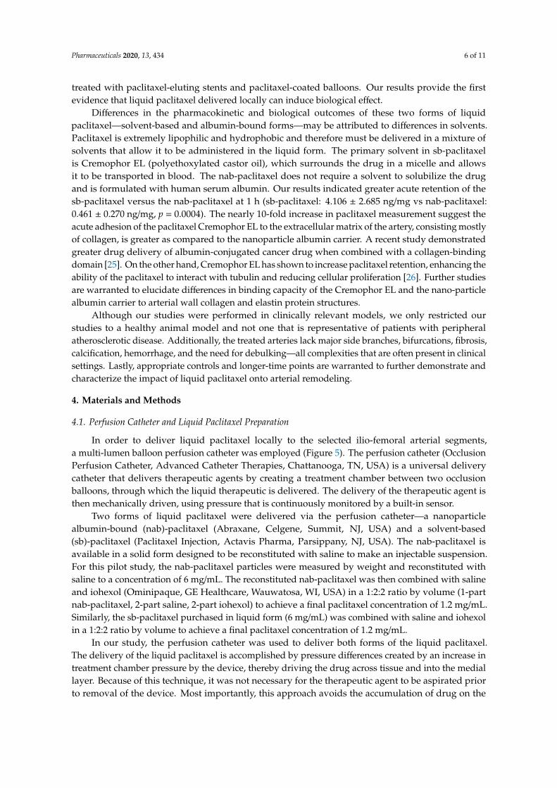
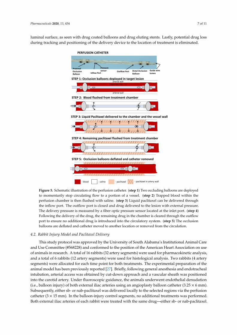
Figure 5. Schematic illustration of the perfusion catheter. (step 1) Two occluding balloons are deployed to momentarily stop circulating flow to a portion of a vessel. (step 2) Trapped blood within the perfusion chamber is then flushed with saline. (step 3) Liquid paclitaxel can be delivered through the inflow port. The outflow port is closed and drug delivered to the lesion with external pressure. The delivery pressure is measured by a fiber optic pressure sensor located at the inlet port. (step 4) Following the delivery of the drug, the remaining drug in the chamber is cleared through the outflow port to ensure no additional drug is introduced into the circulatory system. (step 5) The occlusion balloons are deflated and catheter moved to another location or removed from the circulation.
4.2. Rabbit Injury Model and Paclitaxel Delivery
This study protocol was approved by the University of South Alabama’s Institutional Animal Care and Use Committee (#568228) and conformed to the position of the American Heart Association on use of animals in research. A total of 16 rabbits (32 artery segments) were used for pharmacokinetic analysis, and a total of 6 rabbits (12 artery segments) were used for histological analysis. Two rabbits (4 artery segments) were allocated for each time point for both treatments. The experimental preparation of the animal model has been previously reported [27]. Briefly, following general anesthesia and endotracheal intubation, arterial access was obtained by cut-down approach and a vascular sheath was positioned into the carotid artery. Under fluoroscopic guidance, the animals underwent endothelial denudation (i.e., balloon injury) of both external iliac arteries using an angioplasty balloon catheter (3.25 × 6 mm). Subsequently, either sb- or nab-paclitaxel was delivered locally to the selected regions via the perfusion catheter (3 × 15 mm). In the balloon-injury control segments, no additional treatments was performed. Both external iliac arteries of each rabbit were treated with the same drug—either sb- or nab-paclitaxel. Liquid paclitaxel was delivered at a treatment chamber pressure range between 0.1 to 0.4 atm for 2 min. Antiplatelet therapy consisted of aspirin (40 mg/d), given orally 24 h before the procedure with continued dosing throughout the in-life phase of the study, and single-dose intra-arterial heparin (150 IU/kg) administered at the time of catheterization.
STEP 2: Blood flushed from treatment chamber
STEP 3: Liquid Paclitaxel delivered to the chamber and the vessel wall
STEP 5: Occlusion balloons deflated and catheter removed
STEP 4: Remaining paclitaxel flushed from treatment chamber
Distal Occlusion Balloon
Guide wire lumen
Proximal Occlusion Balloon
Pressure sensor Outflow Port
PERFUSION CATHETER
- blood - paclitaxel- saline - paclitaxel in artery wall
STEP 1: Occlusion balloons deployed in target lesionarterial wall
arterial wall
treatment chamber
Inflow Port
Figure 5. Schematic illustration of the perfusion catheter. (step 1) Two occluding balloons are deployedto momentarily stop circulating flow to a portion of a vessel. (step 2) Trapped blood within theperfusion chamber is then flushed with saline. (step 3) Liquid paclitaxel can be delivered throughthe inflow port. The outflow port is closed and drug delivered to the lesion with external pressure.The delivery pressure is measured by a fiber optic pressure sensor located at the inlet port. (step 4)Following the delivery of the drug, the remaining drug in the chamber is cleared through the outflowport to ensure no additional drug is introduced into the circulatory system. (step 5) The occlusionballoons are deflated and catheter moved to another location or removed from the circulation.
4.2. Rabbit Injury Model and Paclitaxel Delivery
This study protocol was approved by the University of South Alabama’s Institutional Animal Careand Use Committee (#568228) and conformed to the position of the American Heart Association on useof animals in research. A total of 16 rabbits (32 artery segments) were used for pharmacokinetic analysis,and a total of 6 rabbits (12 artery segments) were used for histological analysis. Two rabbits (4 arterysegments) were allocated for each time point for both treatments. The experimental preparation of theanimal model has been previously reported [27]. Briefly, following general anesthesia and endotrachealintubation, arterial access was obtained by cut-down approach and a vascular sheath was positionedinto the carotid artery. Under fluoroscopic guidance, the animals underwent endothelial denudation(i.e., balloon injury) of both external iliac arteries using an angioplasty balloon catheter (3.25 × 6 mm).Subsequently, either sb- or nab-paclitaxel was delivered locally to the selected regions via the perfusioncatheter (3 × 15 mm). In the balloon-injury control segments, no additional treatments was performed.Both external iliac arteries of each rabbit were treated with the same drug—either sb- or nab-paclitaxel.
Pharmaceuticals 2020, 13, 434 8 of 11
Liquid paclitaxel was delivered at a treatment chamber pressure range between 0.1 to 0.4 atm for2 min. Antiplatelet therapy consisted of aspirin (40 mg/d), given orally 24 h before the procedurewith continued dosing throughout the in-life phase of the study, and single-dose intra-arterial heparin(150 IU/kg) administered at the time of catheterization.
4.3. Pharmacokinetic Analysis
Following 1 h, 1 day, 3 days, and 7 days, animals were anesthetized and euthanized (intravenousFATAL-PLUS®, 85–150 mg/kg, single injection), and the treated artery segments were removed basedon landmarks identified by angiography. The time points were selected to demonstrate the acuteretention of liquid paclitaxel delivery. The explanted segments were then stored at −80 ◦C and shippedon dry ice to the bioanalytical laboratory (iC42 Clinical Research and Development, Aurora, CO, USA).As previously described, quantification of arterial paclitaxel levels was performed using a validatedhigh-performance liquid chromatography (HPLC)-electrospray ionization- tandem mass spectrometrysystem (LC-MS/MS) [27–29].
4.4. Histological Analysis
Following the 14-day timepoint, animals were anesthetized and euthanized, and the treated arterysegments were removed based on landmarks identified by angiography. The arteries were perfused bysaline and formalin-fixed under physiological pressure prior to removal. The segments were stored in10% formalin at room temperature and then processed to paraffin blocks, sectioned, and stained withhematoxylin and eosin (H&E) or Verhoeff’s elastin stain (VEG).
4.5. Histomorphometric Analysis
Histological sections were digitized, and measurements were performed using ImageJ software(NIH). Cross-sectional area measurements included the external elastic lamina (EEL), internal elasticlamina (IEL), and lumen area of each section. Using these measurements, the medial area (EEL-IEL),neointimal area (IEL-lumen), and percent area stenosis (100 × (IEL-Lumen)/(IEL)) were calculated aspreviously described [30–32].
Morphological analysis was performed by light microscopy using a grading criterion as previouslypublished [30–32]. Parameters assessed included intimal healing as judged by injury, endothelialcell loss, intimal inflammation, and fibrin/platelet deposition. The medial wall was also assessed fordrug-induced biological effect, specifically looking at smooth muscle cell loss, both in the transmuraland circumferential directions. The presence of inflammation within the medial or adventitial regionswas also evaluated. These parameters were semi-quantified using a scoring a system of 0 (none), 1(minimal), 2 (mild), 3 (moderate), and 4 (severe) as previously described [30,32].
4.6. Statistical Analysis
All values are expressed as mean ± standard deviation (SD). Quantitative data were comparedwith analysis of variance (ANOVA), followed by Tukey’s test for multiple comparisons, using GrapPadPrism 8 (GraphPad Software, La Jolla, CA, USA). Non-parametric data were evaluated by Wilcoxonsigned-rank test. A value of p < 0.05 was considered statistically significant.
5. Conclusions
Our overall results support the concept of local liquid delivery of paclitaxel into the arterialsegments. In comparison of two clinically available forms of liquid paclitaxel, the solvent-basedpaclitaxel demonstrated greater arterial retention following treatment, accompanied with a decrease inneointimal growth. The histopathologic findings showed biological effect, as indicated by vascularsmooth muscle cell loss, without evidence of thrombus, toxicity or inflammation. Together, theseresults indicate the effectiveness of a local liquid approach to deliver and retain liquid paclitaxel in
Pharmaceuticals 2020, 13, 434 9 of 11
arterial segments. Additional studies are warranted to further evaluate the safety and efficacy of theuse of sb-paclitaxel in local liquid delivery devices—an approach that has the potential to impactmillions of patients suffering with PAD by improving outcomes and quality of life.
Author Contributions: Conceptualization, C.V.C., K.C. and S.K.Y.; methodology C.V.C., K.C., C.H., A.L., E.V.-M.,L.S., U.C., M.S., S.K.Y. formal analysis, C.V.C., K.C., C.H., A.L., E.V.-M., L.S., U.C., S.K.Y., data curation, C.V.C., K.C.,C.H., U.C., S.K.Y.; writing—original draft preparation, C.V.C., S.K.Y.; writing—review and editing, all authors;funding acquisition, S.K.Y. All authors have read and agreed to the published version of the manuscript.
Funding: This work was supported by the American Heart Association [#15SDG25880000] and National Instituteof Health [#1R15HL127596, #1R01EB028798].
Conflicts of Interest: Saami K. Yazdani serves on the Scientific Advisory Board of Advanced Catheter and hasreceived grant support from Advanced Catheter Therapies, Lutonix, Inc, Alucent Biomedical and Toray Industries.Other co-authors have no conflict of interest to report.
References
1. Gornik, H.L.; Beckman, J.A. Cardiology patient page. Peripheral arterial disease. Circulation 2005, 111,e169-72. [CrossRef] [PubMed]
2. Fanelli, F.; Cannavale, A.; Corona, M.; Lucatelli, P.; Wlderk, A.; Salvatori, F.M. The “DEBELLUM”—Lowerlimb multilevel treatment with drug eluting balloon—Randomized trial: 1-year results. J. Cardiovasc. Surg.2014, 55, 207–216.
3. Sousa, J.E.; Costa, M.A.; Abizaid, A.C.; Rensing, B.J.; Abizaid, A.S.; Tanajura, L.F.; Kozuma, K.; VanLangenhove, G.; Sousa, A.G.; Falotico, R.; et al. Sustained suppression of neointimal proliferation bysirolimus-eluting stents: One-year angiographic and intravascular ultrasound follow-up. Circulation 2001,104, 2007–2011. [CrossRef] [PubMed]
4. Werk, M.; Langner, S.; Reinkensmeier, B.; Boettcher, H.F.; Tepe, G.; Dietz, U.; Hosten, N.; Hamm, B.; Speck, U.;Ricke, J. Inhibition of restenosis in femoropopliteal arteries: Paclitaxel-coated versus uncoated balloon:Femoral paclitaxel randomized pilot trial. Circulation 2008, 118, 1358–1365. [CrossRef] [PubMed]
5. Babaev, A.; Hari, P.; Zavlunova, S.; Kurayev, A. Role of nitinol stent fractures in the development of in-stentrestenosis in the superficial femoral artery. Vasc. Dis. Manag. 2016, 1, E7–E16.
6. Lin, Y.; Tang, X.; Fu, W.; Kovach, R.; George, J.C.; Guo, D. Stent fractures after superficial femoral arterystenting: Risk factors and impact on patency. J Endovasc. 2015, 22, 319–326. [CrossRef]
7. Singh, G.D.; Armstrong, E.J.; Laird, J.R. Femoropopliteal in-stent restenosis: Current treatment strategies.J. Cardiovasc. Surg. 2014, 55, 325–333.
8. Davaine, J.M.; Querat, J.; Guyomarch, B.; Brennan, M.A.; Costargent, A.; Chaillou, P.; Patra, P.; Goueffic, Y.Incidence and the clinical impact of stent fractures after primary stenting for TASC C and D femoropopliteallesions at 1 year. Eur. J. Vasc. Endovasc. Surg. Off. J. Eur. Soc. Vasc. Surg. 2013, 46, 201–212. [CrossRef]
9. Desyatova, A.; Poulson, W.; Deegan, P.; Lomneth, C.; Seas, A.; Maleckis, K.; MacTaggart, J.; Kamenskiy, A.Limb flexion-induced twist and associated intramural stresses in the human femoropopliteal artery. J. R. Soc.Interface 2017, 14. [CrossRef]
10. MacTaggart, J.N.; Phillips, N.Y.; Lomneth, C.S.; Pipinos, I.I.; Bowen, R.; Baxter, B.T.; Johanning, J.; Longo, G.M.;Desyatova, A.S.; Moulton, M.J.; et al. Three-dimensional bending, torsion and axial compression of thefemoropopliteal artery during limb flexion. J. Biomech. 2014, 47, 2249–2256. [CrossRef]
11. Maleckis, K.; Anttila, E.; Aylward, P.; Poulson, W.; Desyatova, A.; MacTaggart, J.; Kamenskiy, A. Nitinolstents in the femoropopliteal artery: A mechanical perspective on material, design, and performance.Ann. Biomed. Eng. 2018, 46, 684–704. [CrossRef] [PubMed]
12. Tepe, G.; Zeller, T.; Albrecht, T.; Heller, S.; Schwarzwalder, U.; Beregi, J.P.; Claussen, C.D.; Oldenburg, A.;Scheller, B.; Speck, U. Local delivery of paclitaxel to inhibit restenosis during angioplasty of the leg. N. Engl.J. Med. 2008, 358, 689–699. [CrossRef] [PubMed]

Pharmaceuticals 2020, 13, 434 10 of 11
13. Axel, D.I.; Kunert, W.; Goggelmann, C.; Oberhoff, M.; Herdeg, C.; Kuttner, A.; Wild, D.H.; Brehm, B.R.;Riessen, R.; Koveker, G.; et al. Paclitaxel inhibits arterial smooth muscle cell proliferation and migrationin vitro and in vivo using local drug delivery. Circulation 1997, 96, 636–645. [CrossRef] [PubMed]
14. Speck, U.; Cremers, B.; Kelsch, B.; Biedermann, M.; Clever, Y.P.; Schaffner, S.; Mahnkopf, D.; Hanisch, U.;Bohm, M.; Scheller, B. Do pharmacokinetics explain persistent restenosis inhibition by a single dose ofpaclitaxel? Circ. Cardiovasc. Interv. 2012, 5, 392–400. [CrossRef]
15. Rowinsky, E.K.; Donehower, R.C. Paclitaxel (taxol). N. Engl. J. Med. 1995, 332, 1004–1014. [CrossRef]16. Katsanos, K.; Spiliopoulos, S.; Kitrou, P.; Krokidis, M.; Karnabatidis, D. Risk of death following application
of paclitaxel-coated balloons and stents in the femoropopliteal artery of the leg: A systematic review andmeta-analysis of randomized controlled trials. J. Am. Heart Assoc. 2018, 7, e011245. [CrossRef]
17. Katsanos, K.; Spiliopoulos, S.; Kitrou, P.; Krokidis, M.; Paraskevopoulos, I.; Karnabatidis, D. Risk of deathand amputation with use of paclitaxel-coated balloons in the infrapopliteal arteries for treatment of criticallimb ischemia: A systematic review and meta-analysis of randomized controlled trials. J. Vasc. Interv. Radiol.2020, 31, 202–212. [CrossRef]
18. Zeller, T.; Baumgartner, I.; Scheinert, D.; Brodmann, M.; Bosiers, M.; Micari, A.; Peeters, P.; Vermassen, F.;Landini, M.; Snead, D.B.; et al. Drug-eluting balloon versus standard balloon angioplasty for infrapoplitealarterial revascularization in critical limb ischemia: 12-month results from the IN.PACT DEEP randomizedtrial. J. Am. Coll. Cardiol. 2014, 64, 1568–1576. [CrossRef]
19. Mills, J.L.; Conte, M.S.; Murad, M.H. Critical review and evidence implications of paclitaxel drug-elutingballoons and stents in peripheral artery disease. J. Vasc. Surg. 2019, 70, 3–7. [CrossRef]
20. Torii, S.; Jinnouchi, H.; Sakamoto, A.; Romero, M.E.; Kolodgie, F.D.; Virmani, R.; Finn, A.V. Comparison ofbiologic effect and particulate embolization after femoral artery treatment with three drug-coated balloons inhealthy swine model. J. Vasc. Interv. Radiol. 2019, 30, 103–109. [CrossRef]
21. Kolodgie, F.D.; Pacheco, E.; Yahagi, K.; Mori, H.; Ladich, E.; Virmani, R. Comparison of particulateembolization after femoral artery treatment with IN.PACT admiral versus Lutonix 035 paclitaxel-coatedballoons in healthy swine. J. Vasc. Interv. Radiol. 2016, 27, 1676–1685. [CrossRef] [PubMed]
22. Sonnichsen, D.S.; Relling, M.V. Clinical pharmacokinetics of paclitaxel. Clin. Pharmacokinet. 1994, 27, 256–269.[CrossRef] [PubMed]
23. Gongora, C.A.; Shibuya, M.; Wessler, J.D.; McGregor, J.; Tellez, A.; Cheng, Y.; Conditt, G.B.; Kaluza, G.L.;Granada, J.F. Impact of paclitaxel dose on tissue pharmacokinetics and vascular healing: A comparativedrug-coated balloon study in the familial hypercholesterolemic swine model of superficial femoral in-stentrestenosis. JACC Cardiovasc. Interv. 2015, 8, 1115–1123. [CrossRef] [PubMed]
24. Granada, J.F.; Stenoien, M.; Buszman, P.P.; Tellez, A.; Langanki, D.; Kaluza, G.L.; Leon, M.B.; Gray, W.;Jaff, M.R.; Schwartz, R.S. Mechanisms of tissue uptake and retention of paclitaxel-coated balloons: Impact onneointimal proliferation and healing. Open Heart 2014, 1, e000117. [CrossRef] [PubMed]
25. Sasaki, K.; Ishihara, J.; Ishihara, A.; Miura, R.; Mansurov, A.; Fukunaga, K.; Hubbell, J.A. Engineeredcollagen-binding serum albumin as a drug conjugate carrier for cancer therapy. Sci. Adv. 2019, 5, eaaw6081.[CrossRef]
26. Gelderblom, H.; Verweij, J.; van Zomeren, D.M.; Buijs, D.; Ouwens, L.; Nooter, K.; Stoter, G.; Sparreboom, A.Influence of Cremophor El on the bioavailability of intraperitoneal paclitaxel. Clin. Cancer Res. Off. J. Am.Assoc. Cancer Res. 2002, 8, 1237–1241.
27. Atigh, M.K.; Turner, E.; Christians, U.; Yazdani, S.K. The use of an occlusion perfusion catheter to deliverpaclitaxel to the arterial wall. Cardiovasc 2017, 35. [CrossRef]
28. Turner, E.; Erwin, M.; Atigh, M.; Christians, U.; Saul, J.M.; Yazdani, S.K. In vitro and in vivo Assessment ofKeratose as a Novel Excipient of Paclitaxel Coated Balloons. Front. Pharmacol. 2018, 9, 808. [CrossRef]
29. Turner, E.A.; Atigh, M.K.; Erwin, M.M.; Christians, U.; Yazdani, S.K. Coating and pharmacokinetic evaluationof air spray coated drug coated balloons. Cardiovasc. Eng. Technol. 2018, 9, 240–250. [CrossRef]
30. Yazdani, S.K.; Pacheco, E.; Nakano, M.; Otsuka, F.; Naisbitt, S.; Kolodgie, F.D.; Ladich, E.; Rousselle, S.;Virmani, R. Vascular, downstream, and pharmacokinetic responses to treatment with a low dose drug-coatedballoon in a swine femoral artery model. Catheter. Cardiovasc. Interv. 2014, 83, 132–140. [CrossRef]
31. Yazdani, S.K.; Sheehy, A.; Nakano, M.; Nakazawa, G.; Vorpahl, M.; Otsuka, F.; Donn, R.S.; Perkins, L.E.;Simonton, C.A.; Kolodgie, F.D.; et al. Preclinical evaluation of second-generation everolimus- andzotarolimus-eluting coronary stents. J. Invasive Cardiol 2013, 25, 383–390. [PubMed]

Pharmaceuticals 2020, 13, 434 11 of 11
32. Goel, E.; Erwin, M.; Cawthon, C.V.; Schaff, C.; Fedor, N.; Rayl, T.; Wilson, O.; Christians, U.; Register, T.C.;Geary, R.L.; et al. Pre-clinical investigation of keratose as an excipient of drug coated balloons. Molecules2020, 25, 1596. [CrossRef] [PubMed]
Publisher’s Note: MDPI stays neutral with regard to jurisdictional claims in published maps and institutionalaffiliations.
© 2020 by the authors. Licensee MDPI, Basel, Switzerland. This article is an open accessarticle distributed under the terms and conditions of the Creative Commons Attribution(CC BY) license (http://creativecommons.org/licenses/by/4.0/).


