Embed Size (px)
Citation preview
Chapter 3 Hypersensitivity
M.G.Rajanandh, Dept. of Pharmacy Practice, SRM College of Pharmacy, SRM University.
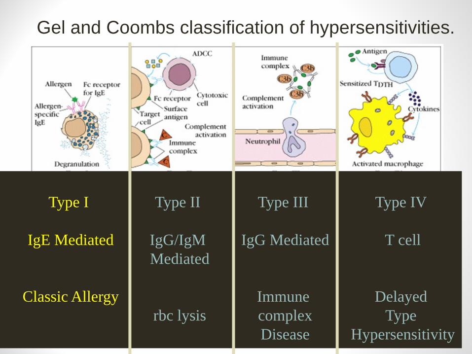
Type I
IgE Mediated
Classic Allergy
Type II
IgG/IgM Mediated
rbc lysis
Type III
IgG Mediated
Immune complexDisease
Type IV
T cell
Delayed Type
Hypersensitivity
Gel and Coombs classification of hypersensitivities.
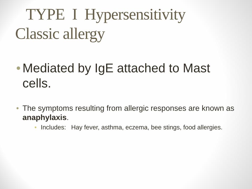
TYPE I Hypersensitivity Classic allergy
• Mediated by IgE attached to Mast cells.
• The symptoms resulting from allergic responses are known as anaphylaxis.
• Includes: Hay fever, asthma, eczema, bee stings, food allergies.
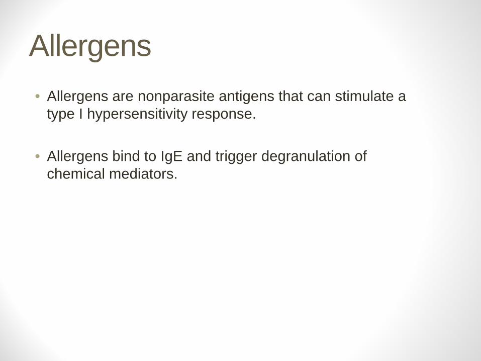
Allergens • Allergens are nonparasite antigens that can stimulate a
type I hypersensitivity response.
• Allergens bind to IgE and trigger degranulation of chemical mediators.
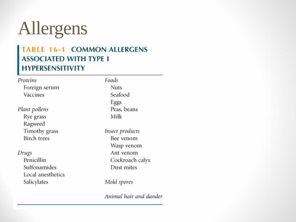
Allergens
In the US ---36 million people said to have hay fever!
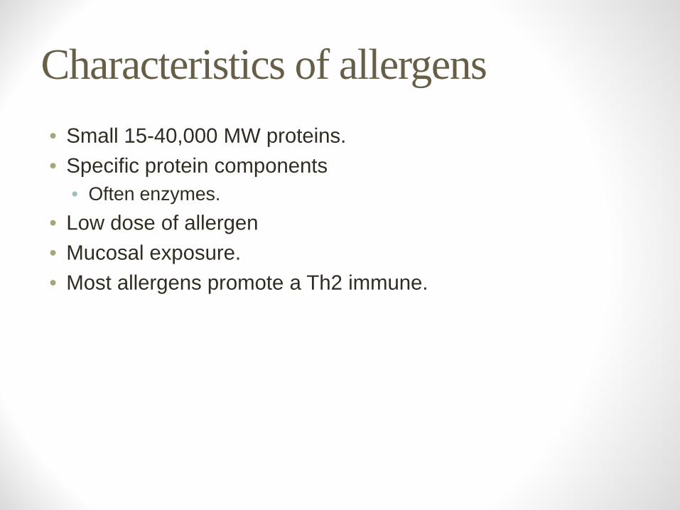
Characteristics of allergens • Small 15-40,000 MW proteins. • Specific protein components
• Often enzymes.• Low dose of allergen • Mucosal exposure. • Most allergens promote a Th2 immune.
Allergens
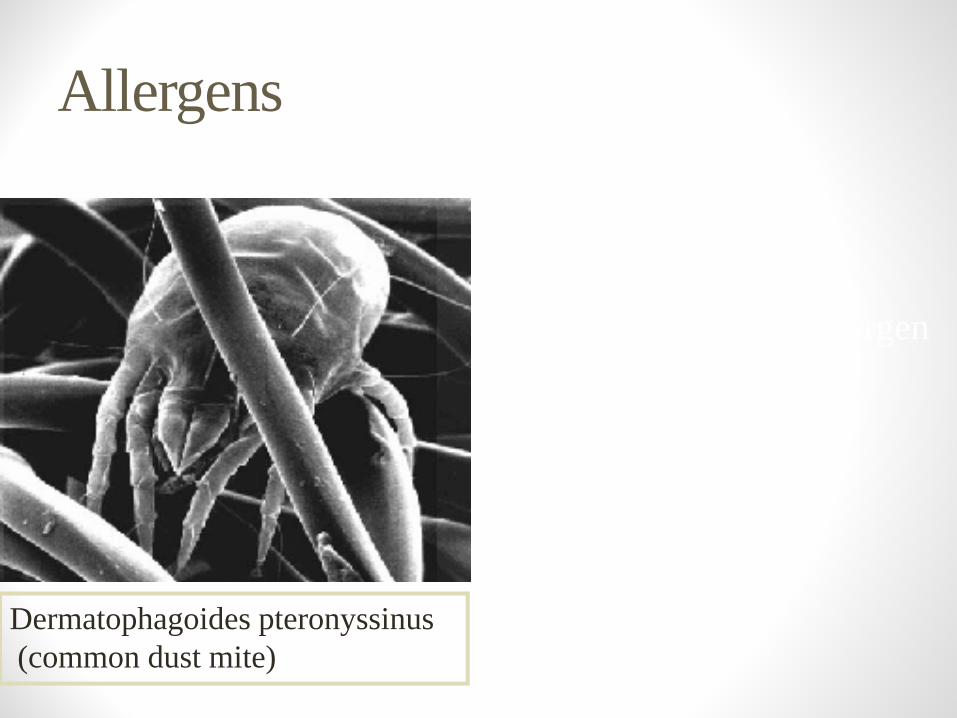
Dermatophagoides pteronyssinus(common dust mite)
Example: Der P1
Der P1 is an enzyme allergenfrom the fecal pellets of the dust mite.
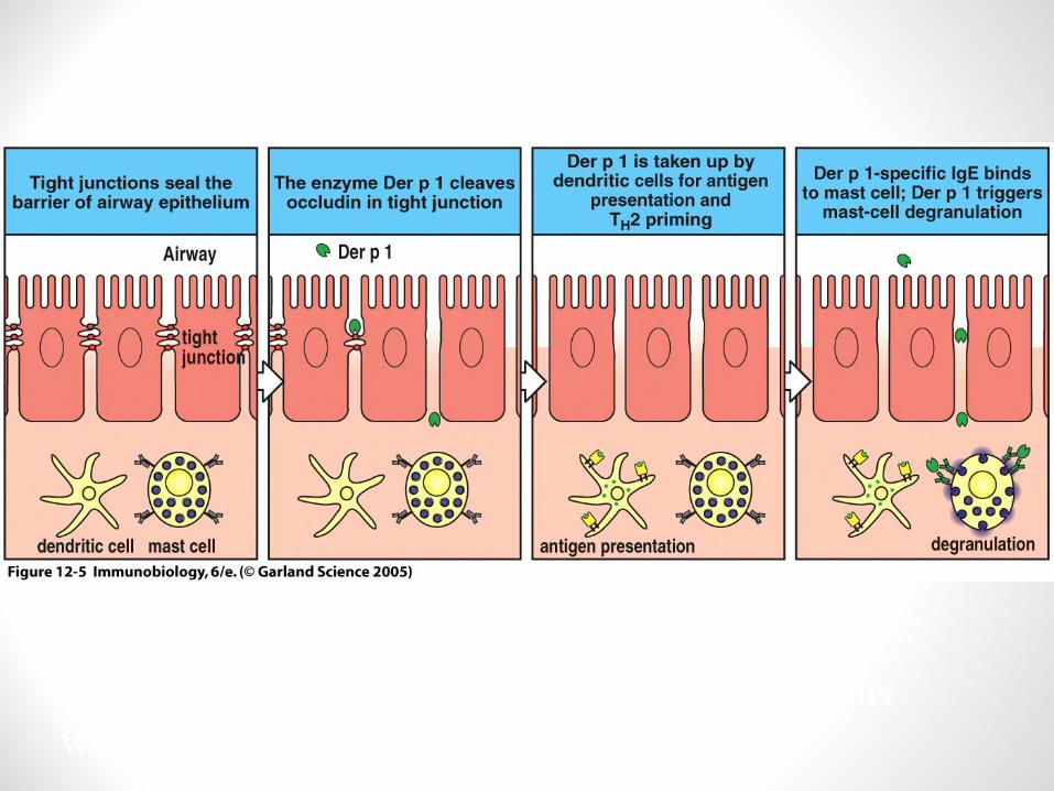
Allergen is easily aerosolized and inhaled.Der P1 breaks down components of tight junctions which helps it to cross mucosa.
Der P1 Allergen

Atopy
• Atopy is the term for the genetic trait to have a predisposition for localized anaphylaxis.
• Atopic individuals have higher levels of IgE and eosinophils.
Genetic Predisposition Type I hypersensitivity
• Candidate polymorphic genes include: • IL-4 Receptor. • IL-4 cytokine (promoter region). • FcεRI. High affinity IgE receptor. • Class II MHC
(present peptides promoting Th2 response). • Inflammation genes.
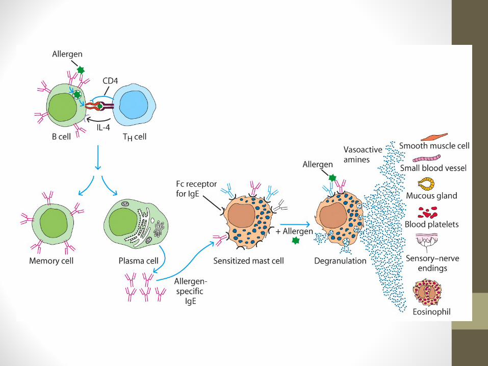
Mechanisms of allergic response
SensitizationRepeated exposure to allergens initiates immune response
that generates IgE isotype.
Th2 cells required to provide the IL-4 required to get isotype switching to IgE.
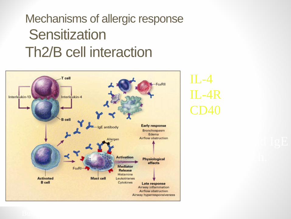
Mechanisms of allergic responseSensitization Th2/B cell interaction
IL-4IL-4RCD40Drive B cell Activation and IgEisotype switch.
Busse and Lemanske NEJM Feb 2001. 344:350
Mechanisms of allergic response
Sensitization• The IgE can attach to Mast cells by Fc receptor, which
increases the life span of the IgE. • Half-life of IgE in serum is days whereas attached to
FcεR it is increased to months.
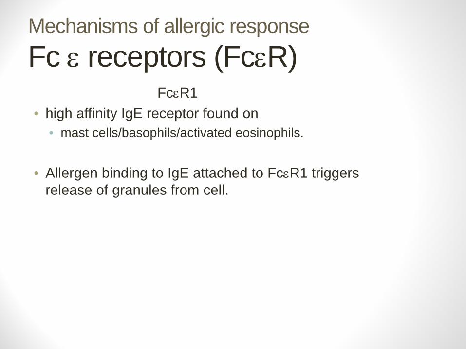
Mechanisms of allergic responseFc ε receptors (FcεR)
FcεR1• high affinity IgE receptor found on
• mast cells/basophils/activated eosinophils.
• Allergen binding to IgE attached to FcεR1 triggers release of granules from cell.
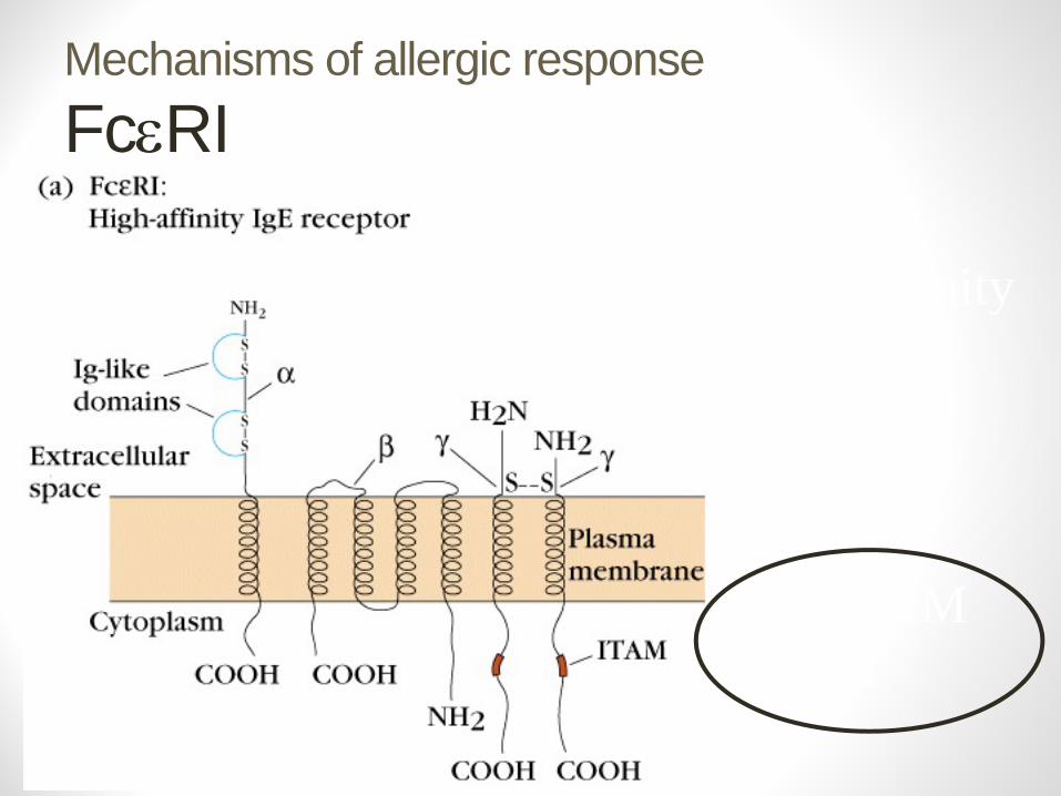
Mechanisms of allergic responseFcεRI
High affinityIgE Fc Receptor
Has ITAMmotifs
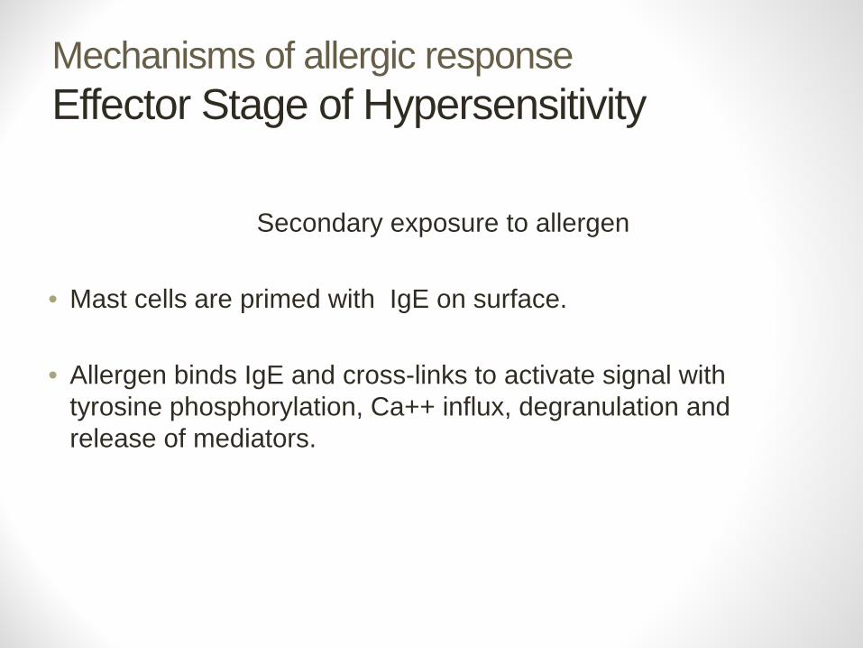
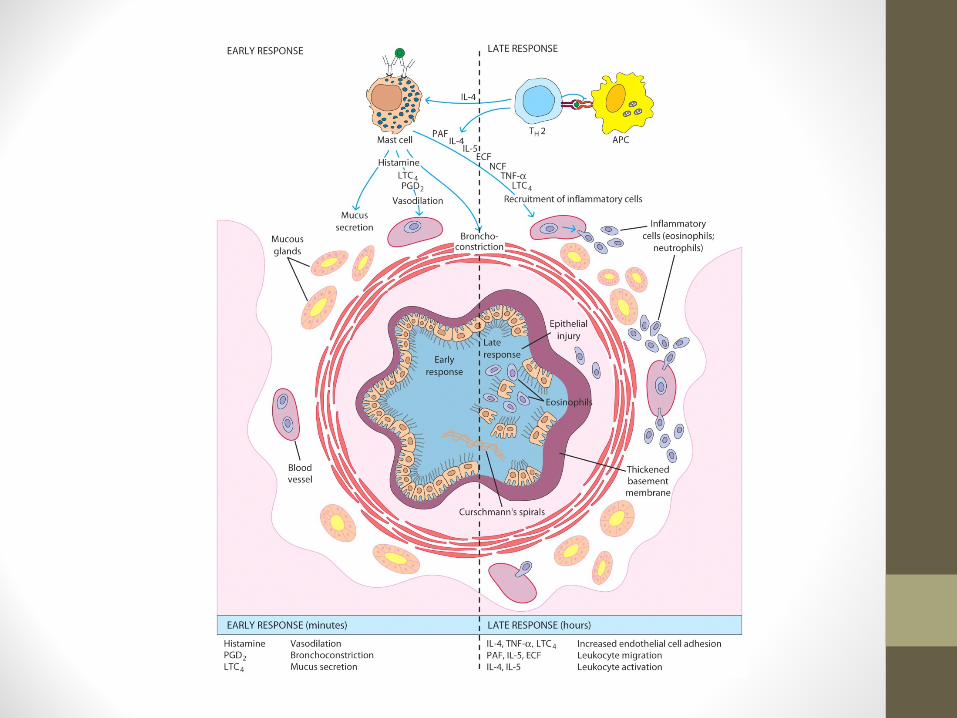
Mechanisms of allergic responseEffector Stage of Hypersensitivity
Secondary exposure to allergen
• Mast cells are primed with IgE on surface.
• Allergen binds IgE and cross-links to activate signal with tyrosine phosphorylation, Ca++ influx, degranulation and release of mediators.
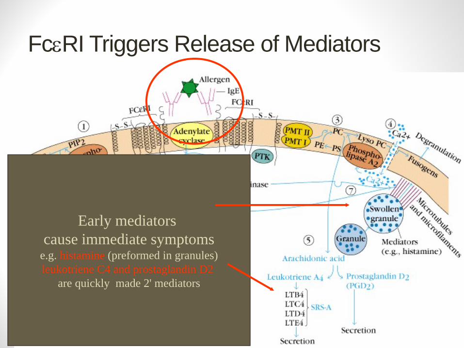
FcεRI Triggers Release of Mediators
Early mediators cause immediate symptoms
e.g. histamine (preformed in granules)leukotriene C4 and prostaglandin D2
are quickly made 2' mediators
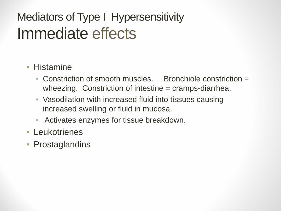
Mediators of Type I HypersensitivityImmediate effects
• Histamine• Constriction of smooth muscles. Bronchiole constriction =
wheezing. Constriction of intestine = cramps-diarrhea.• Vasodilation with increased fluid into tissues causing
increased swelling or fluid in mucosa. • Activates enzymes for tissue breakdown.
• Leukotrienes• Prostaglandins
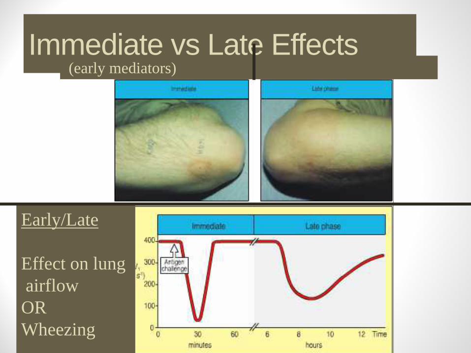
Immediate vs Late Effects(early mediators)
Early/Late
Effect on lung airflowORWheezing
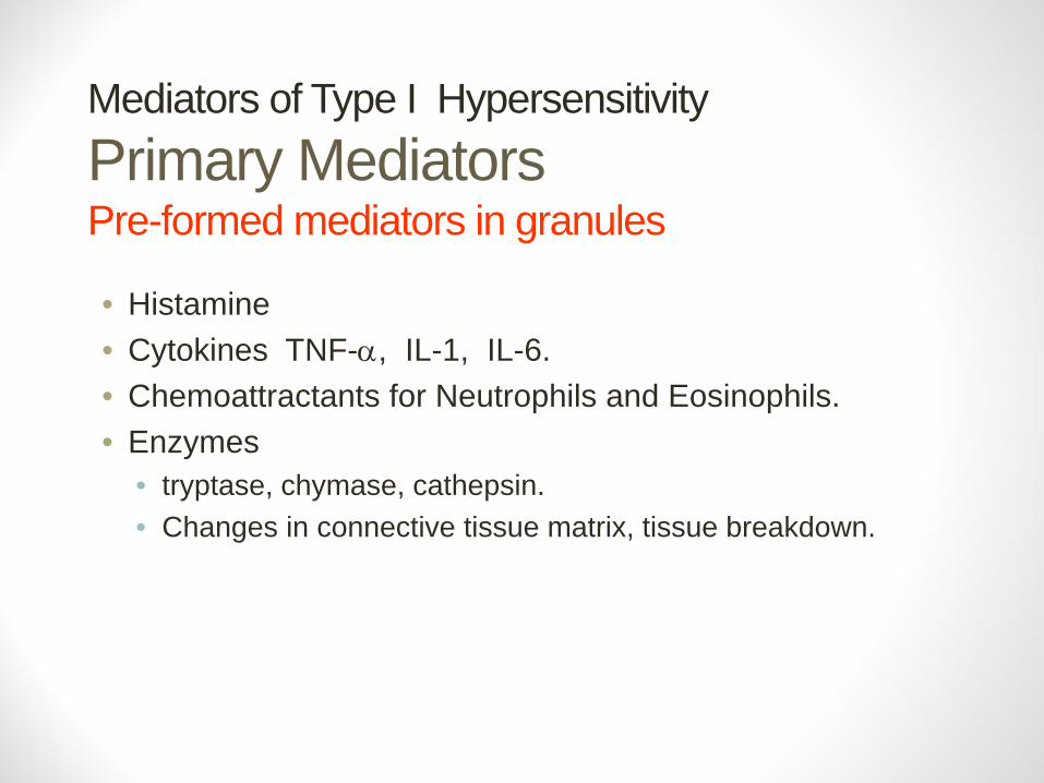
Mediators of Type I HypersensitivityPrimary MediatorsPre-formed mediators in granules
• Histamine• Cytokines TNF-α, IL-1, IL-6. • Chemoattractants for Neutrophils and Eosinophils. • Enzymes
• tryptase, chymase, cathepsin. • Changes in connective tissue matrix, tissue breakdown.
Type I Hypersensitivity
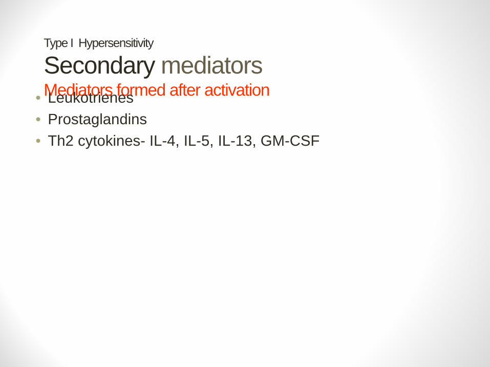
Secondary mediatorsMediators formed after activation• Leukotrienes
• Prostaglandins• Th2 cytokines- IL-4, IL-5, IL-13, GM-CSF
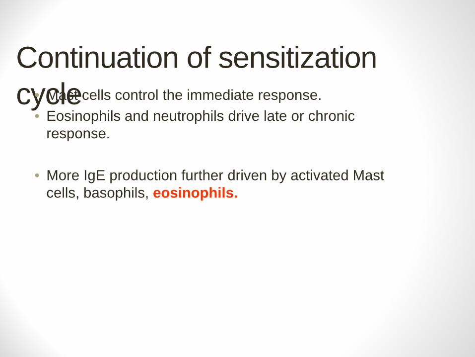
Continuation of sensitization cycle• Mast cells control the immediate response.
• Eosinophils and neutrophils drive late or chronic response.
• More IgE production further driven by activated Mast cells, basophils, eosinophils.
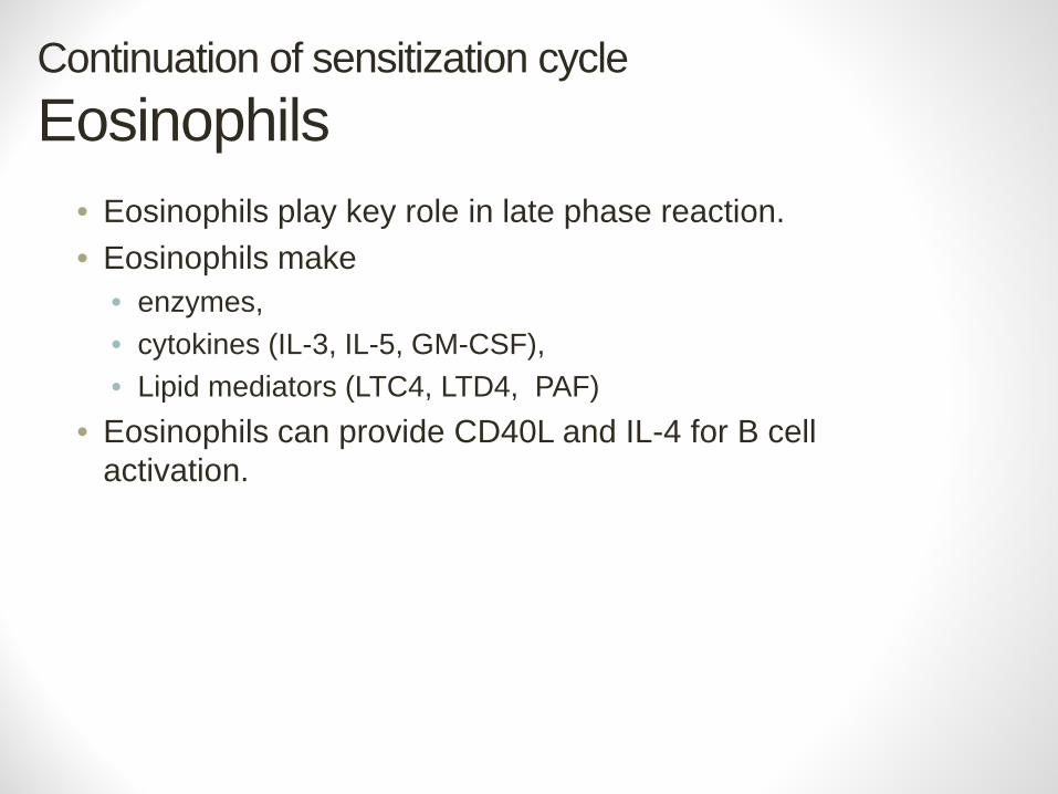
Continuation of sensitization cycleEosinophils
• Eosinophils play key role in late phase reaction. • Eosinophils make
• enzymes, • cytokines (IL-3, IL-5, GM-CSF), • Lipid mediators (LTC4, LTD4, PAF)
• Eosinophils can provide CD40L and IL-4 for B cell activation.
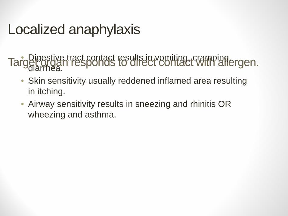
Localized anaphylaxis
Target organ responds to direct contact with allergen.• Digestive tract contact results in vomiting, cramping, diarrhea.
• Skin sensitivity usually reddened inflamed area resulting in itching.
• Airway sensitivity results in sneezing and rhinitis OR wheezing and asthma.
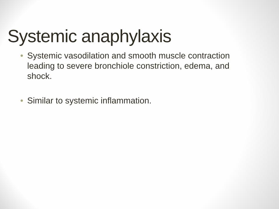
Systemic anaphylaxis• Systemic vasodilation and smooth muscle contraction
leading to severe bronchiole constriction, edema, and shock.
• Similar to systemic inflammation.
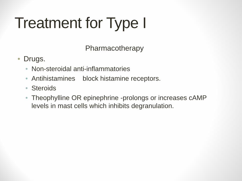
Treatment for Type IPharmacotherapy
• Drugs. • Non-steroidal anti-inflammatories • Antihistamines block histamine receptors.• Steroids• Theophylline OR epinephrine -prolongs or increases cAMP
levels in mast cells which inhibits degranulation.
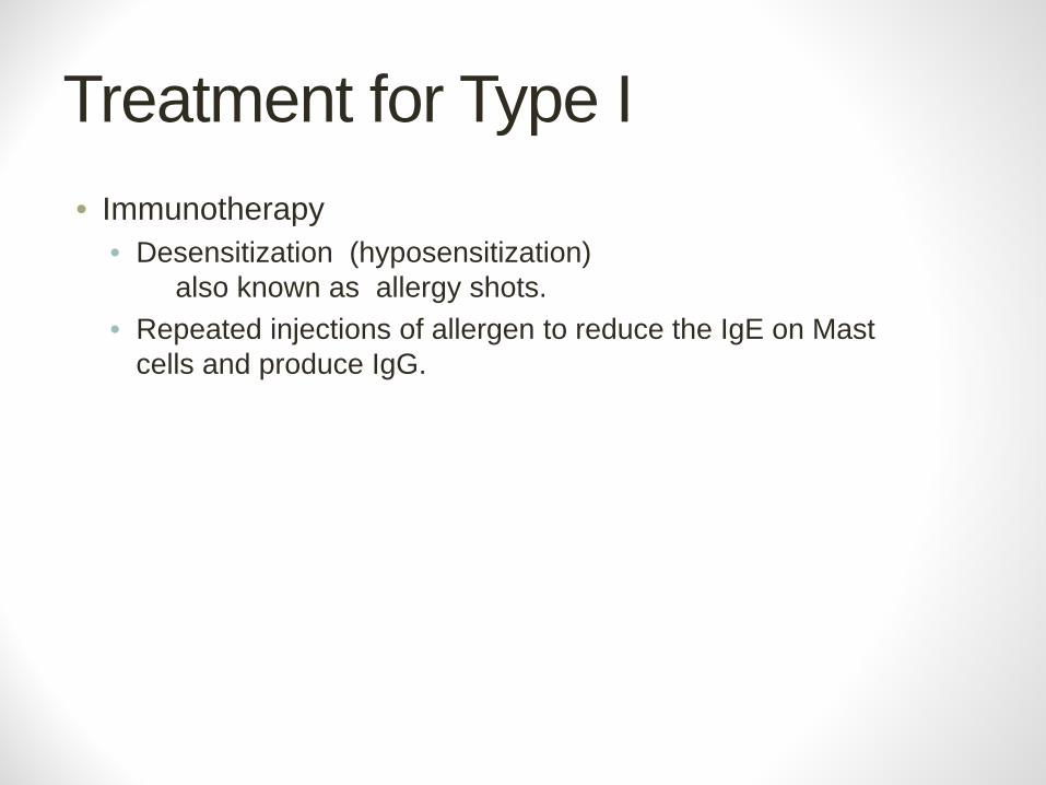
Treatment for Type I• Immunotherapy
• Desensitization (hyposensitization) also known as allergy shots.
• Repeated injections of allergen to reduce the IgE on Mast cells and produce IgG.

Treatment for Type IEffect of allergy shotsAllergen Specific Antibodies
Change in amount of each isotype from more IgE to more IgG.
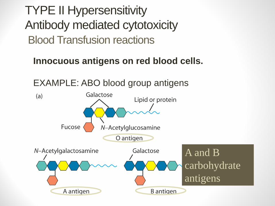
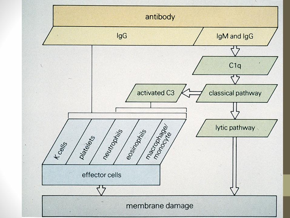
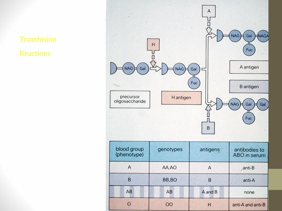
TYPE II HypersensitivityAntibody mediated cytotoxicityBlood Transfusion reactions
Innocuous antigens on red blood cells.
EXAMPLE: ABO blood group antigens
A and B carbohydrateantigens
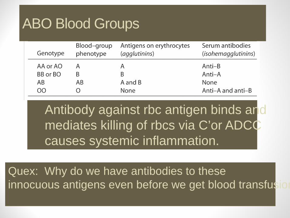
Antibody against rbc antigen binds and mediates killing of rbcs via C’or ADCC causes systemic inflammation.
ABO Blood Groups
Quex: Why do we have antibodies to these innocuous antigens even before we get blood transfusion
TYPE II Antibody mediated cytotoxicity
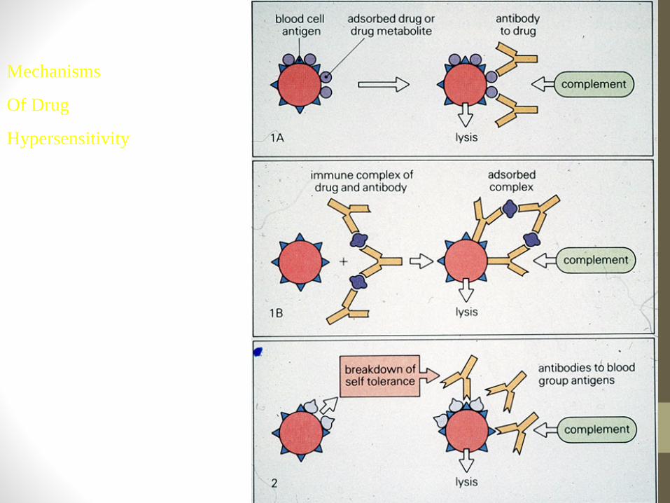
Drug reactions • Drug binds to rbc surface and antibody against drug
binds and causes lysis of rbcs. • Immune system sees antibody bound to "foreign antigen"
on cell. ADCC
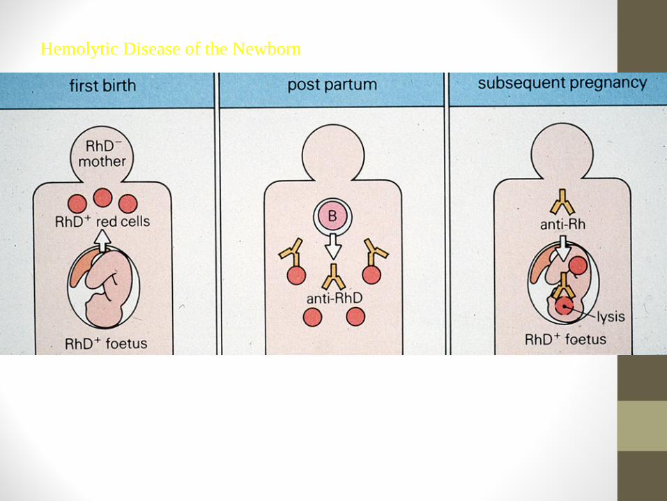
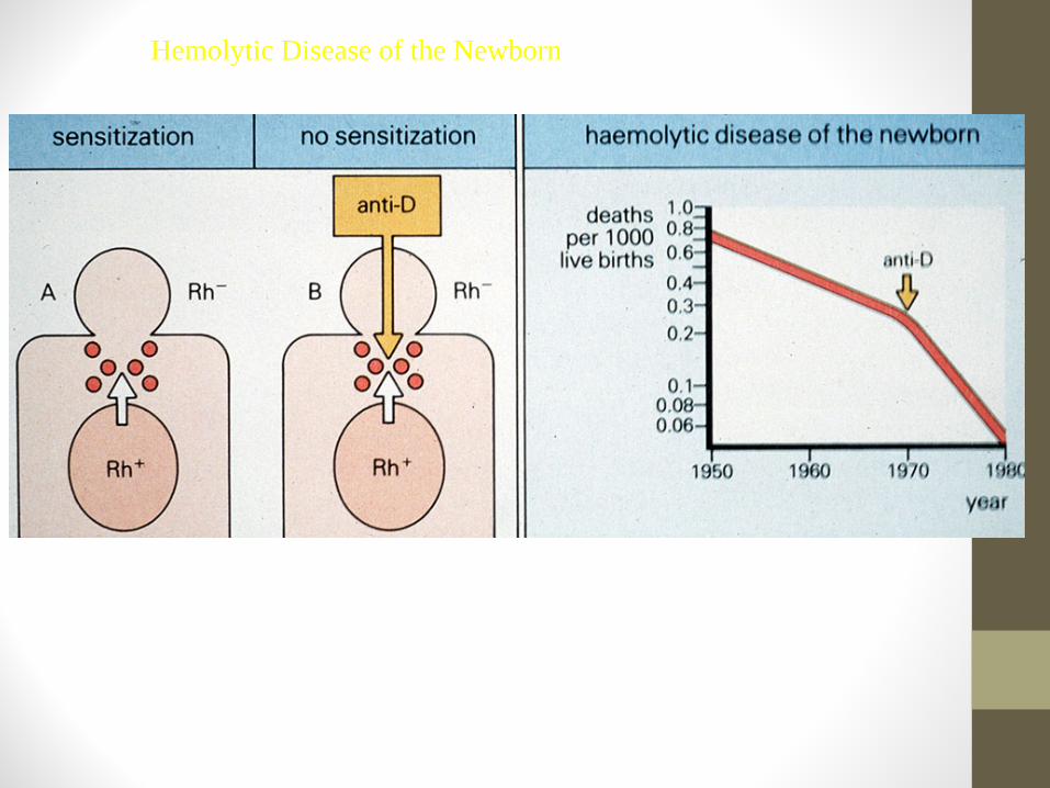
Rh factor incompatibility • IgG abs to Rh an innocuous rbc antigen
• Rh+ baby born to Rh- mother first time fine. 2nd time can have abs to Rh from 1st pregnancy.
• Ab crosses placenta and baby kills its own rbcs. • Treat mother with ab to Rh antigen right after birth and
mother never makes its own immune response.
TYPE IIHemolytic disease of newborn
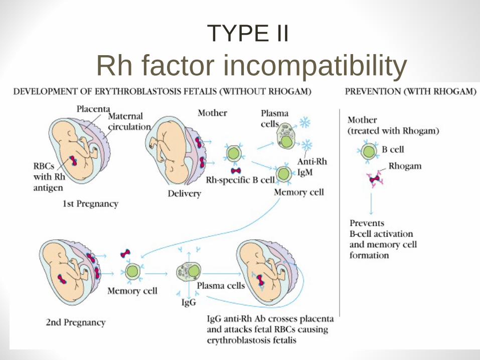
TYPE IIRh factor incompatibility
TYPE III Antigen antibody immune complexes. IgG mediated
Immune Complex Disease• Large amount of antigen and antibodies form complexes
in blood.
• If not eliminated can deposit in capillaries or joints and trigger inflammation.
TYPE III Immune Complexes
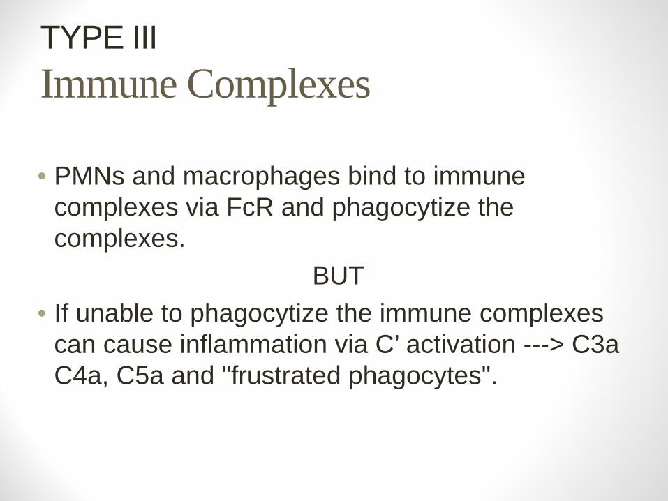
• PMNs and macrophages bind to immune complexes via FcR and phagocytize the complexes.
BUT• If unable to phagocytize the immune complexes
can cause inflammation via C’ activation ---> C3a C4a, C5a and "frustrated phagocytes".
TYPE III Immune Complex Disease"Frustrated Phagocytes"
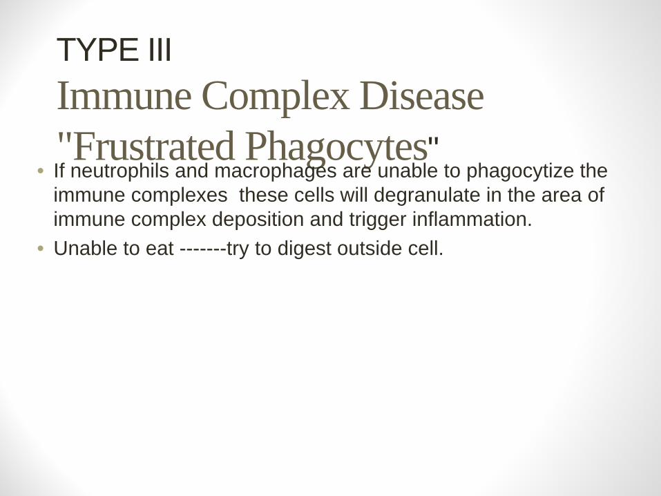
• If neutrophils and macrophages are unable to phagocytize the immune complexes these cells will degranulate in the area of immune complex deposition and trigger inflammation.
• Unable to eat -------try to digest outside cell.
TYPE III Immune Complex Disease
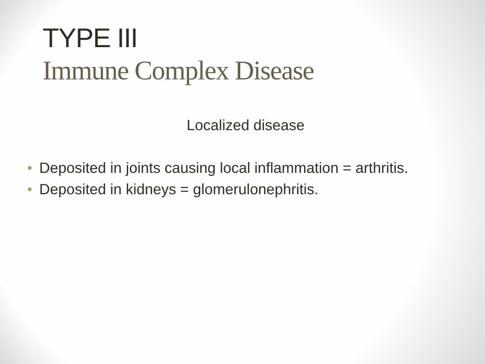
Localized disease
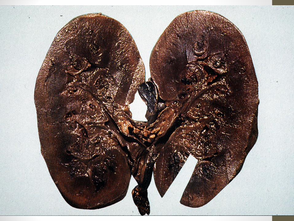
• Deposited in joints causing local inflammation = arthritis. • Deposited in kidneys = glomerulonephritis.
TYPE III Immune Complex Disease
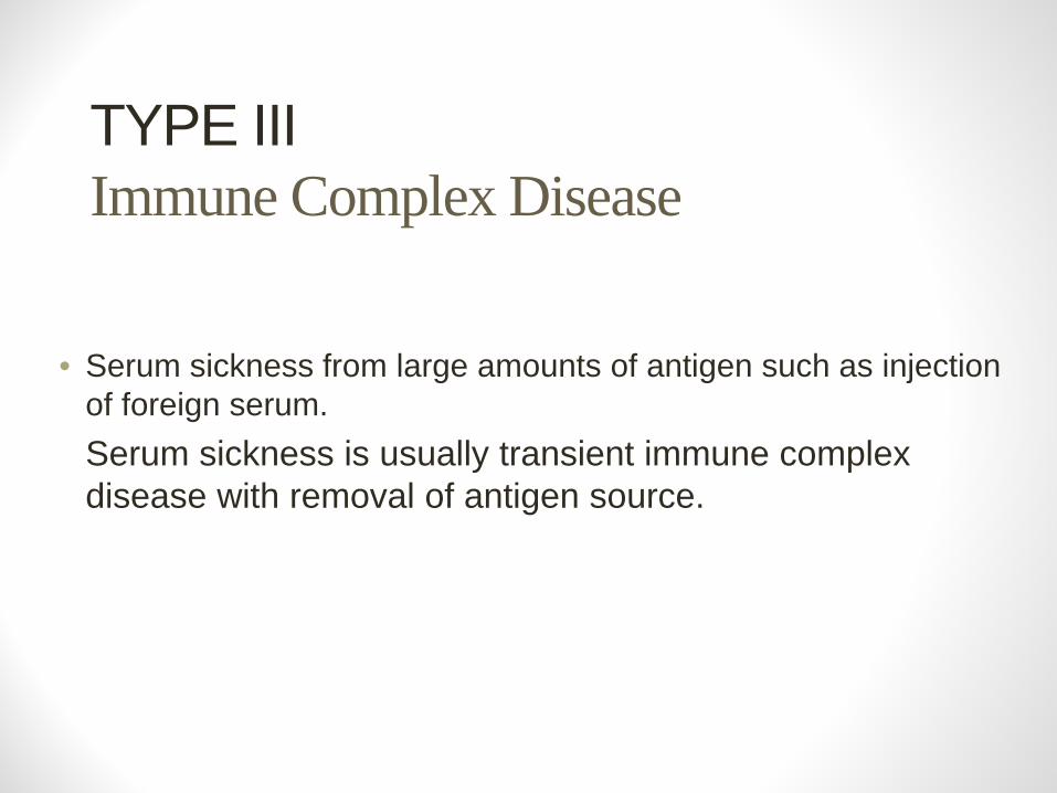
• Serum sickness from large amounts of antigen such as injection of foreign serum.
• Serum sickness is usually transient immune complex disease with removal of antigen source.
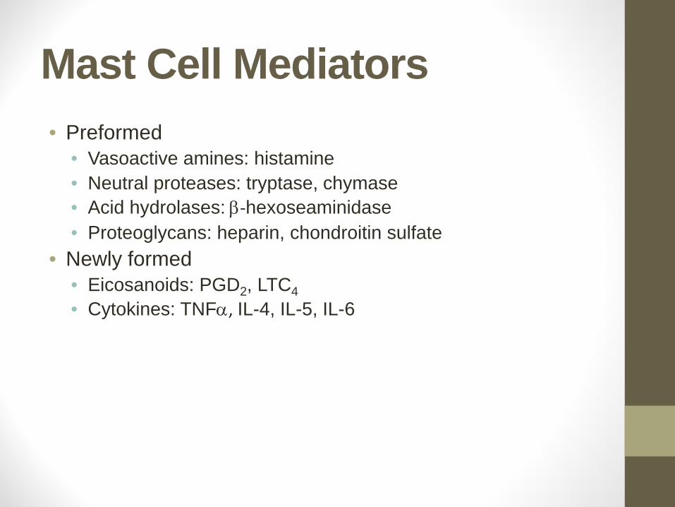
Mast Cell Mediators• Preformed
• Vasoactive amines: histamine• Neutral proteases: tryptase, chymase• Acid hydrolases: β‐hexoseaminidase• Proteoglycans: heparin, chondroitin sulfate
• Newly formed• Eicosanoids: PGD2, LTC4• Cytokines: TNFα, IL-4, IL-5, IL-6
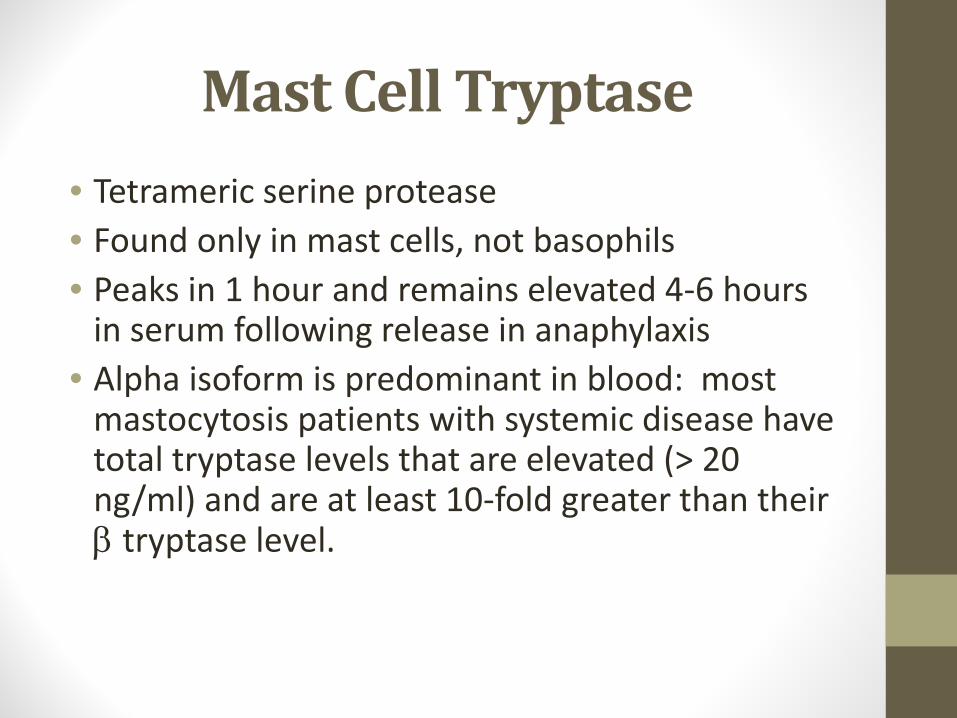
Mast Cell Tryptase
• Tetrameric serine protease• Found only in mast cells, not basophils• Peaks in 1 hour and remains elevated 4‐6 hours in serum following release in anaphylaxis
• Alpha isoform is predominant in blood: most mastocytosis patients with systemic disease have total tryptase levels that are elevated (> 20 ng/ml) and are at least 10‐fold greater than their β tryptase level.
Histamine• Produced almost exclusively by basophils and mast cells (3-8 pg/cell)• Immediate pharmacologic effects:
– pruritus (H1)– ↑ vascular permeability/vasodilatation (H1)– smooth muscle contraction (H1)– gastric acid secretion (H2)
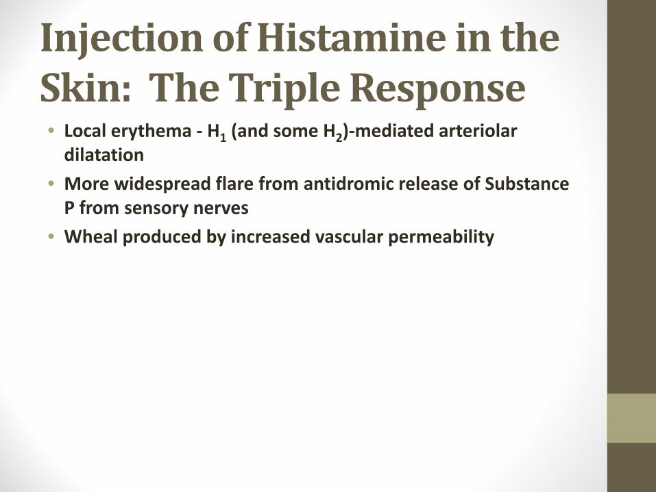
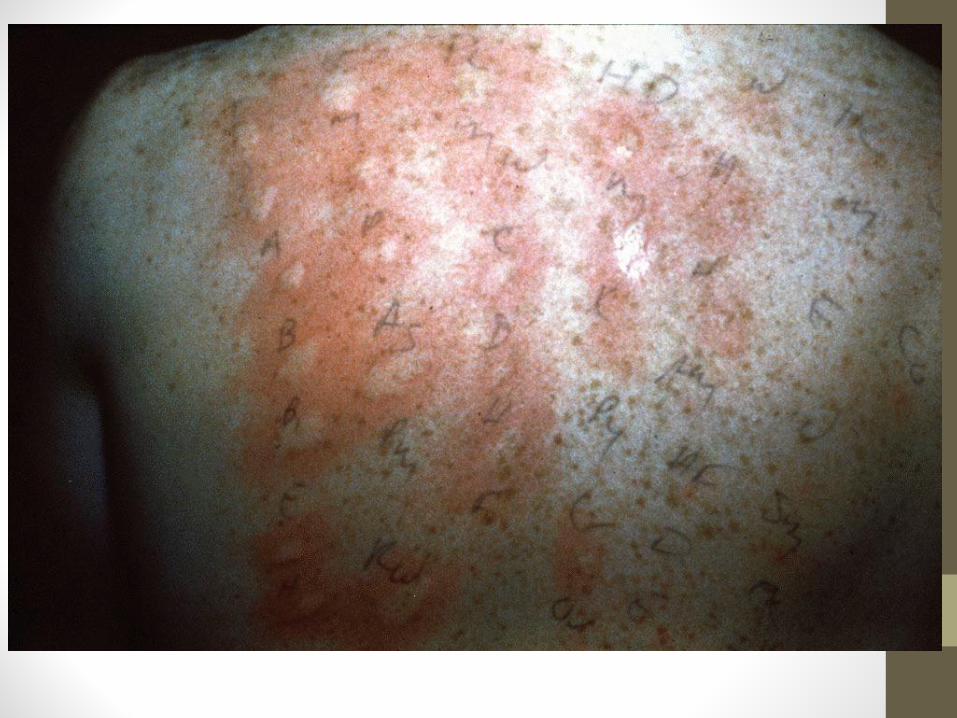
Injection of Histamine in the Skin: The Triple Response• Local erythema ‐ H1 (and some H2)‐mediated arteriolar dilatation
• More widespread flare from antidromic release of Substance P from sensory nerves
• Wheal produced by increased vascular permeability
Acute Phase Allergic Reaction:
• Occurs within seconds to minutes of IgE receptor activation (mast cell mediator release) and resolving within an hour
• Intense pruritus, edema, erythema• Almost all effects can be replicated with histamine
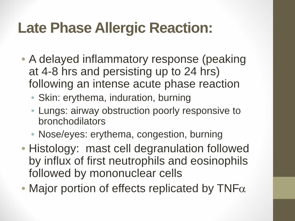
Late Phase Allergic Reaction:
• A delayed inflammatory response (peaking at 4-8 hrs and persisting up to 24 hrs) following an intense acute phase reaction• Skin: erythema, induration, burning• Lungs: airway obstruction poorly responsive to
bronchodilators• Nose/eyes: erythema, congestion, burning
• Histology: mast cell degranulation followed by influx of first neutrophils and eosinophils followed by mononuclear cells
• Major portion of effects replicated by TNFα
Therapy of Allergic Disease• Inhibition of IgE synthesis: Immunotherapy• Inhibition of IgE binding to receptor:
• Monoclonal anti‐IgE (Xolair (Omalizumab)• Inhibition of mast cell mediator release:
• Topical corticosteroids• Cromolyn, nedocromil
• Inhibition of mediator action:• Antihistamines• Leukotriene receptor antagonists• Topical and systemic corticosteroids
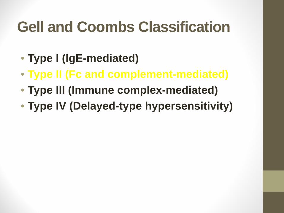
Gell and Coombs Classification
• Type I (IgE-mediated)• Type II (Fc and complement-mediated)• Type III (Immune complex-mediated)• Type IV (Delayed-type hypersensitivity)
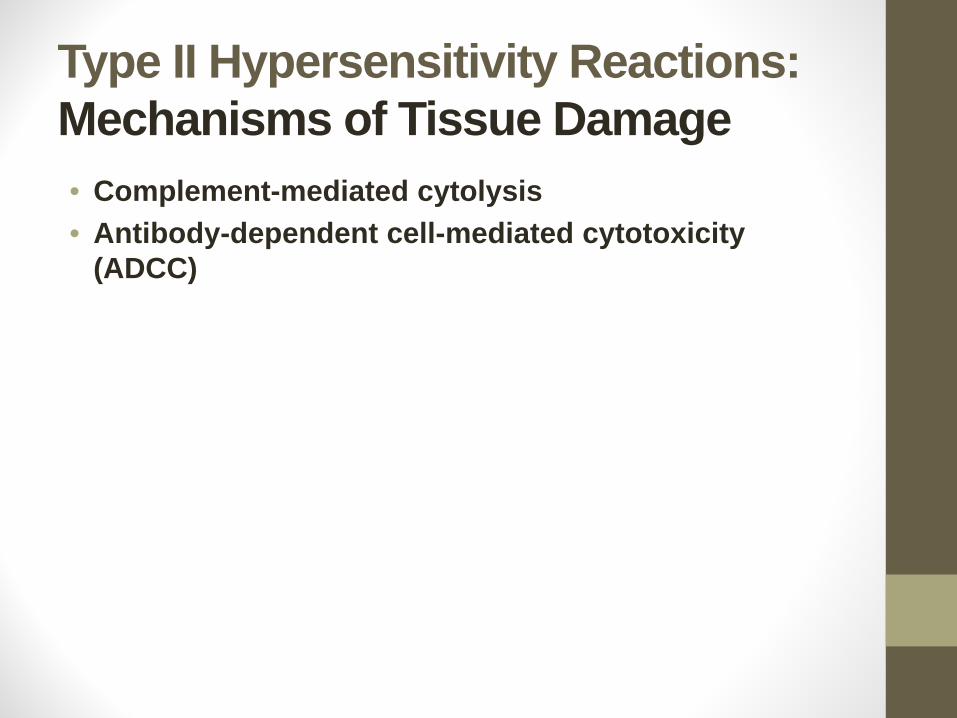
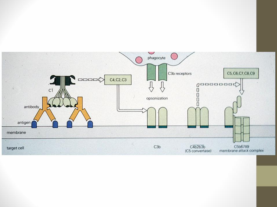
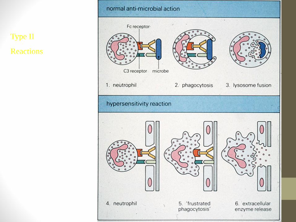
Type II Hypersensitivity Reactions:Mechanisms of Tissue Damage• Complement-mediated cytolysis• Antibody-dependent cell-mediated cytotoxicity
(ADCC)
Type II
Reactions
Type II Hypersensitivity Reactions:Examples of Diseases• Transfusion reactions• Hemolytic disease of the newborn (Rh
incompatibility)• Hyperacute graft rejection• Drug-induced hemolytic anemia
Transfusion
Reactions
Hemolytic Disease of the Newborn
Hemolytic Disease of the Newborn
Mechanisms
Of Drug
Hypersensitivity
Gell and Coombs Classification
• Type I (IgE-mediated)• Type II (Fc and complement-mediated)• Type III (Immune complex-mediated)• Type IV (Delayed-type hypersensitivity)
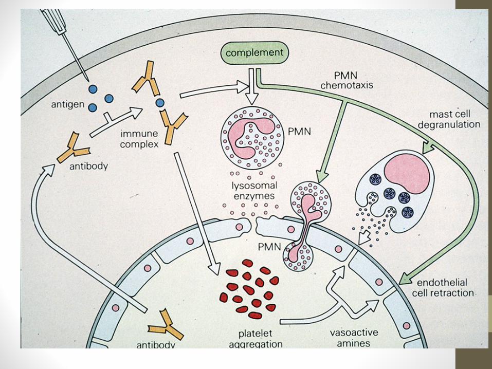
Type III HypersensitivityMechanisms of Tissue Injury
• In situ activation of complement• Anaphylatoxin-mediated activation of mast cells and
phagocytes• Complex-mediated phagocytosis and release of
phagocyte granule enzymes and cytokines into the local microenvironment
Type III HypersensitivityExamples of Diseases
• Arthus reaction• Hypersensitivity pneumonitis• Immune complex-mediated glomerulonephritis• Serum sickness
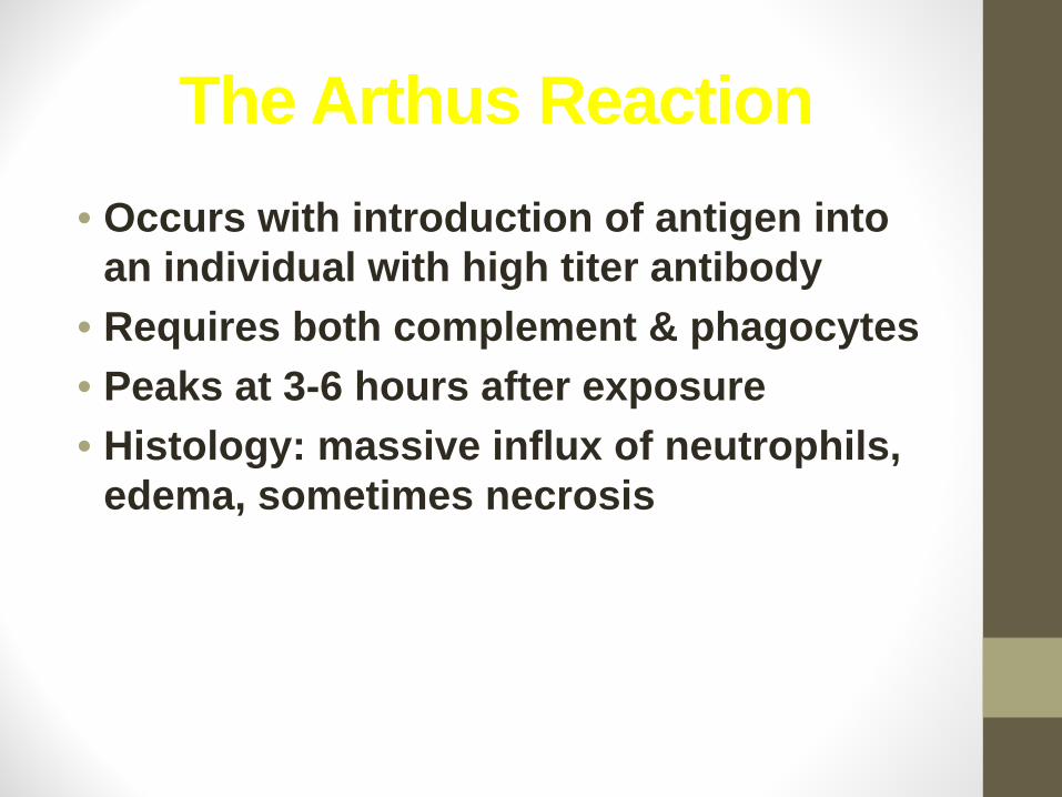
The Arthus Reaction• Occurs with introduction of antigen into
an individual with high titer antibody• Requires both complement & phagocytes• Peaks at 3-6 hours after exposure• Histology: massive influx of neutrophils,
edema, sometimes necrosis
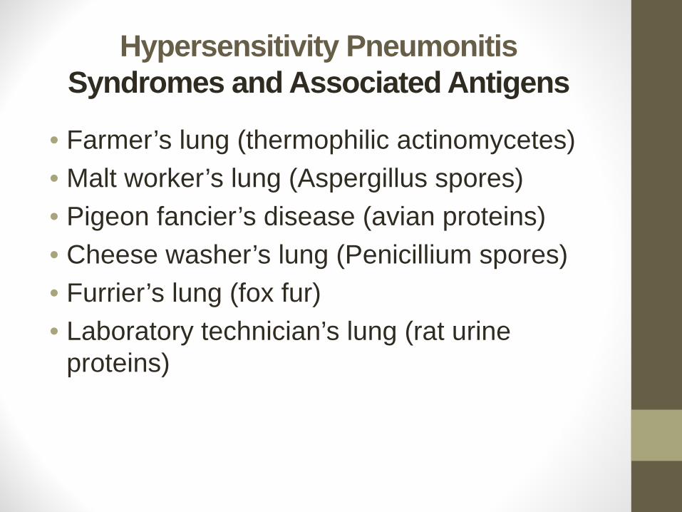
Hypersensitivity Pneumonitis Syndromes and Associated Antigens
• Farmer’s lung (thermophilic actinomycetes)• Malt worker’s lung (Aspergillus spores)• Pigeon fancier’s disease (avian proteins)• Cheese washer’s lung (Penicillium spores)• Furrier’s lung (fox fur)• Laboratory technician’s lung (rat urine
proteins)
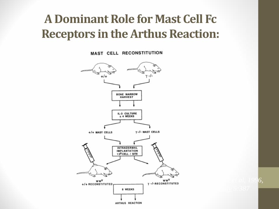
A Dominant Role for Mast Cell Fc Receptors in the Arthus Reaction:
Sylvestre et al, 1996, Immunity 5:387
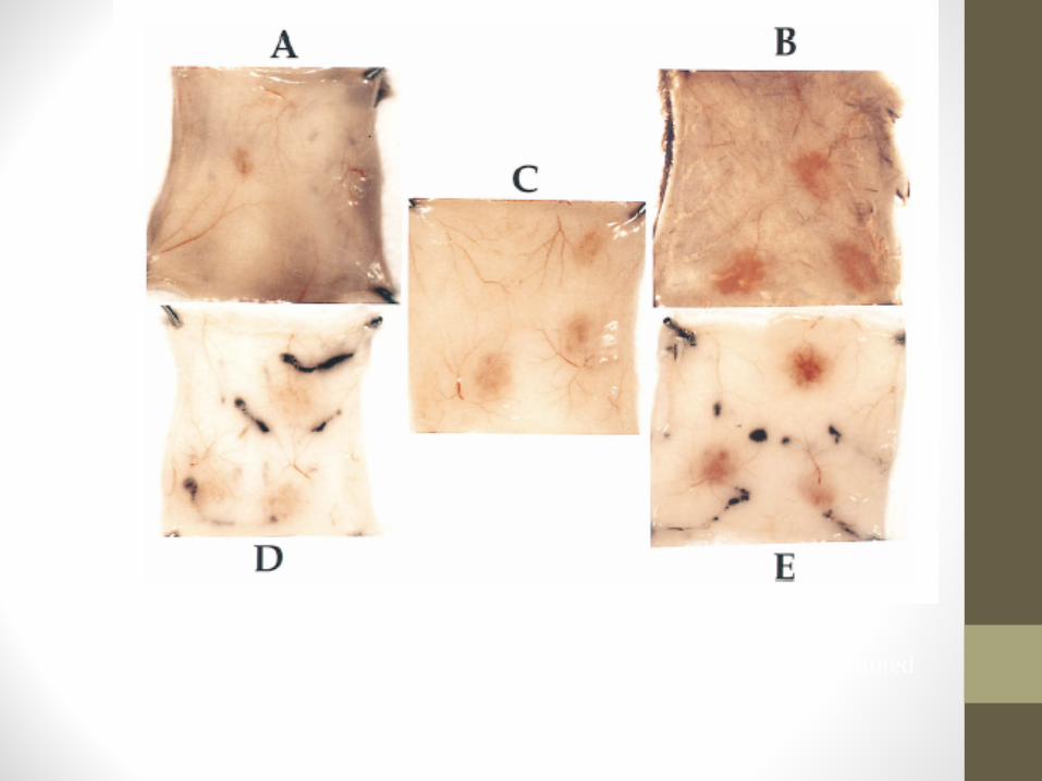
A: Control γ-/-; B: Control γ+/+; C: Control W/Wv; D: W/Wv reconstituted with γ-/- mast cells or E: γ+/+ mast cells
Serum Sickness• Fever, rash, joint pain, lymphadenopathy, occasionally glomerulonephritis
• Timecourse: days to weeks after introduction of foreign antigen
• Causes: allogeneic serum, drugs, infections, autoimmune disorders
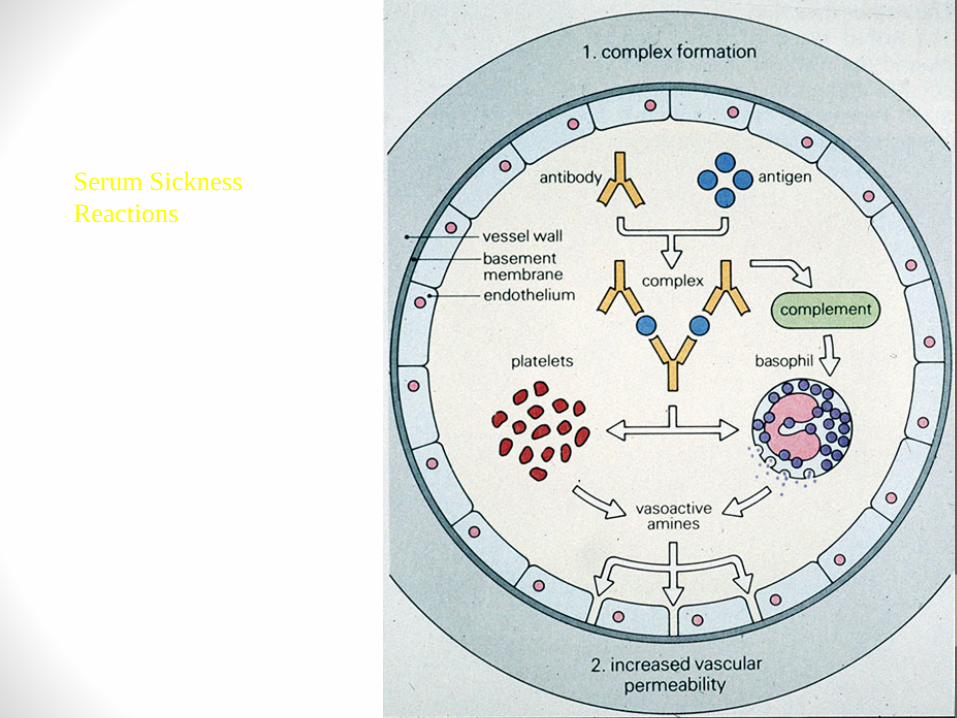
Serum Sickness Reactions
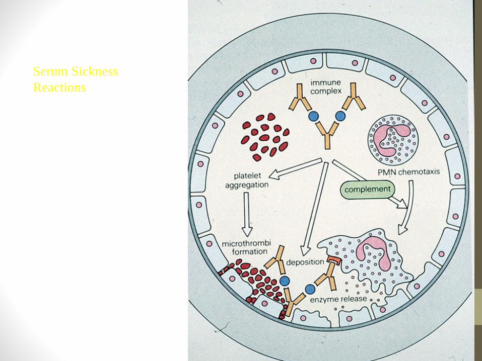
Serum Sickness Reactions
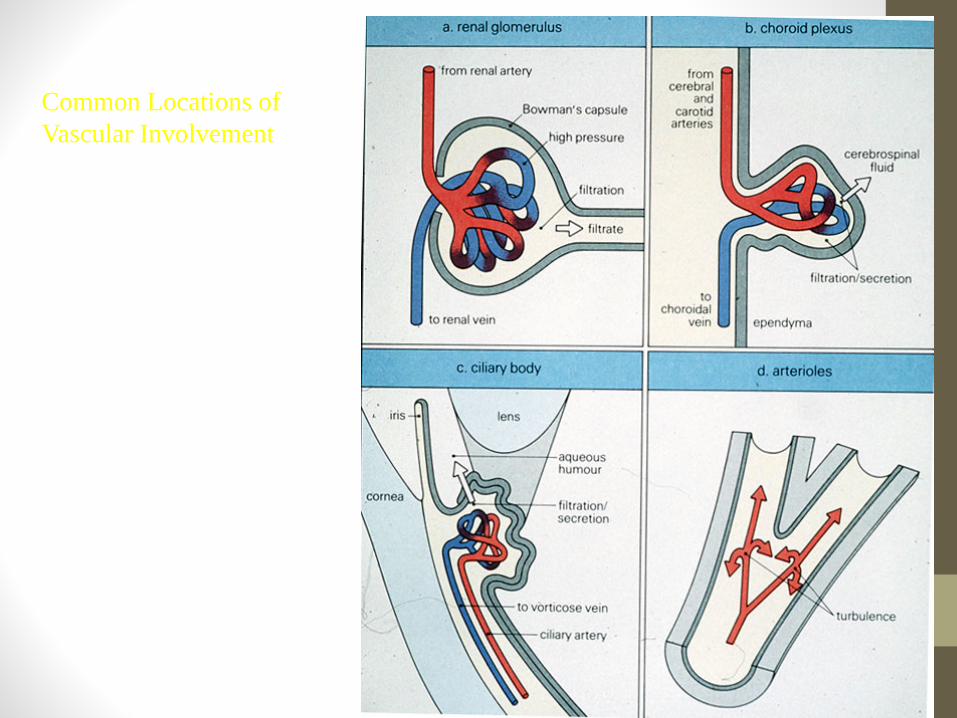
Common Locations of Vascular Involvement
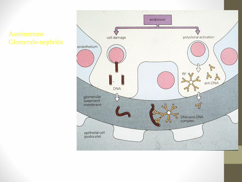
Autoimmune Glomerulo-nephritis
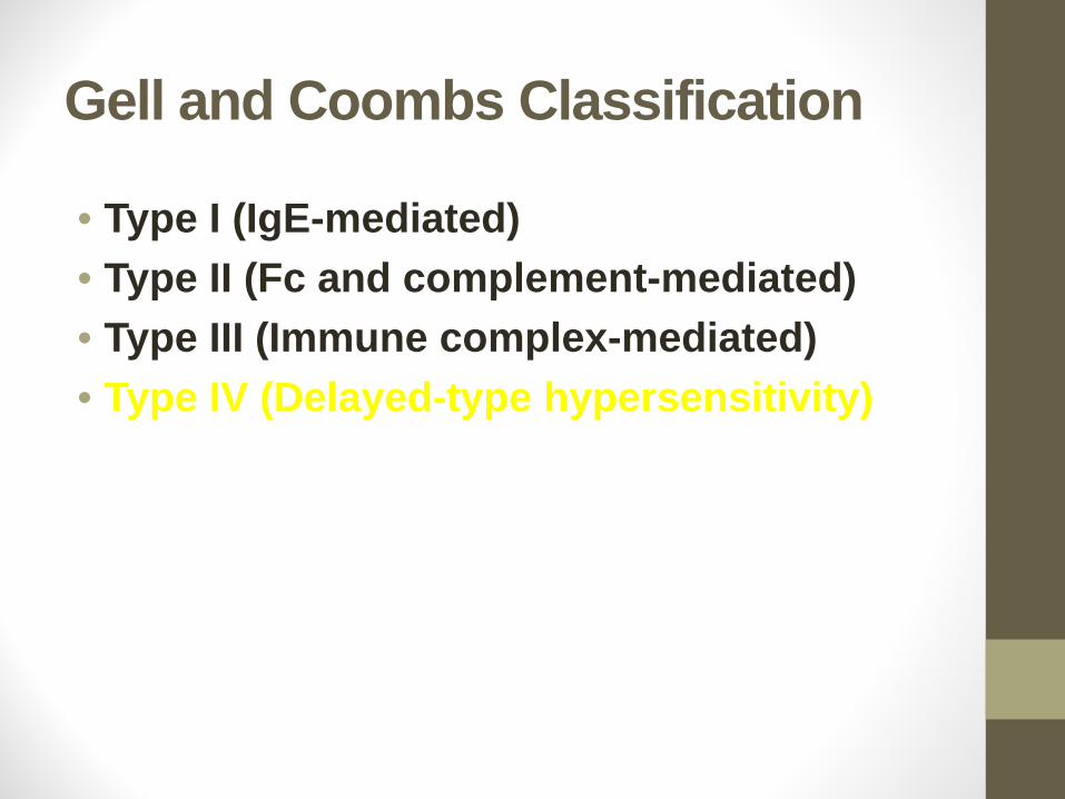
Gell and Coombs Classification
• Type I (IgE-mediated)• Type II (Fc and complement-mediated)• Type III (Immune complex-mediated)• Type IV (Delayed-type hypersensitivity)

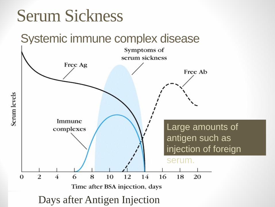
Serum SicknessSystemic immune complex disease
Days after Antigen Injection
Large amounts of antigen such as injection of foreign serum.
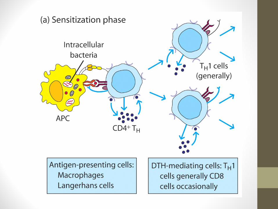
Delayed type hypersensitivity Th1 cells and macrophages
• DTH response is from:• Th1 cells release cytokines to activate macrophages causing
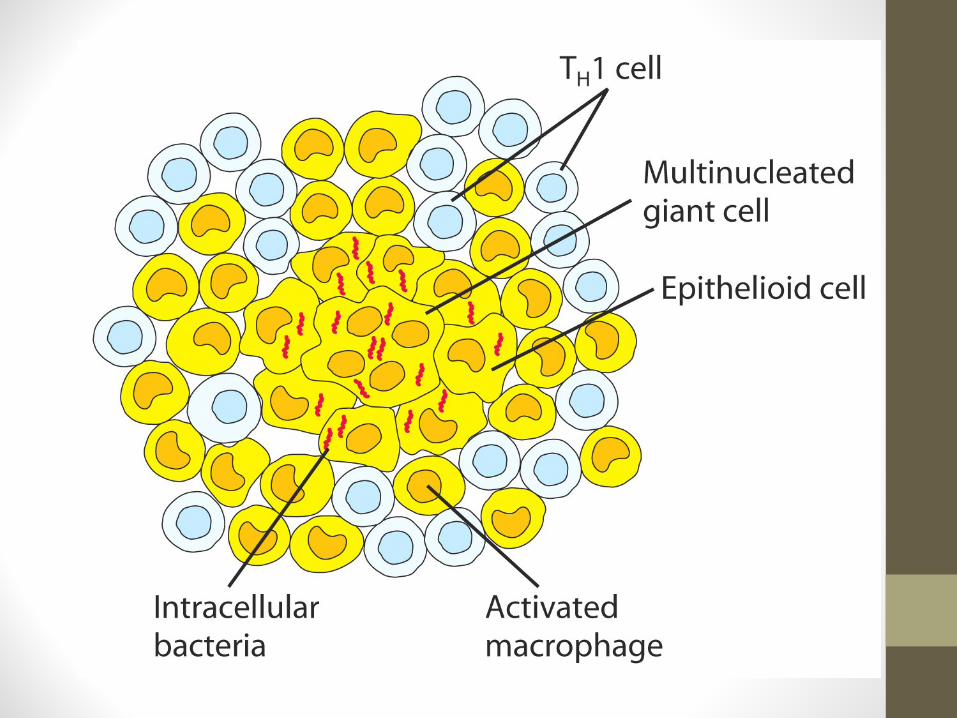
inflammation and tissue damage. • Continued macrophage activation can cause chronic inflammation
resulting in tissue lesions, scarring, and granuloma formation.
• Delayed is relative because DTH response arise 24-72 hours after exposure rather than within minutes.
Stages of Type IV DTHSensitization stage
• Memory Th1 cells against DTH antigens are generated by dendritic cells during the sensitization stage.
• These Th1 cells can activate macrophages and trigger inflammatory response.
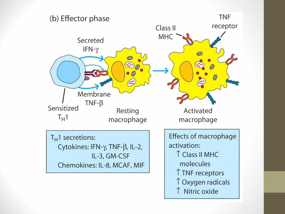
Stages of Type IV DTHEffector stage
• Secondary contact yields what we call DTH. • Th1 memory cells are activated and produce cytokines.
• IFN-γ, TNF-α, and TNF-β which cause tissue destruction, inflammation.
• IL-2 that activates T cells and CTLs.• Chemokines- for macrophage recruitment. • IL-3, GM-CSF for increased monocyte/macrophage
Stages of Type IV DTHEffector stage
Secondary exposure to antigen• Inflamed area becomes red and fluid filled can form lesion.
• From tissue damage there is activation of clotting cascades and tissue repair.
• Continued exposure to antigen can cause chronic inflammation and result in granuloma formation.
Type IV DTHContact dermatitis
• The response to poison oak is a classic Type IV. • Small molecules act as haptens and complex with skin proteins to be
taken up by APCs and presented to Th1 cells to get sensitization. • During secondary exposure Th1 memory cells become activated to
cause DTH.
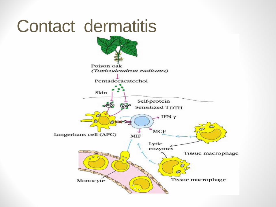
Contact dermatitis
Delayed type hypersensitivity (DTH)
DTH is a type of immune response classified by Th1 and macrophage activation that results in tissue damage.
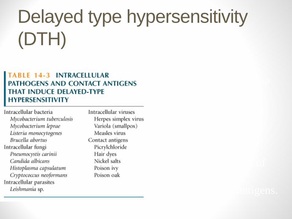
DTH can be the result ofChronic infection or Exposure to some antigens.
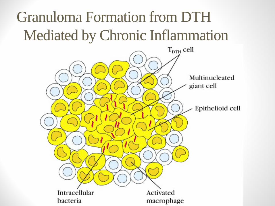
Granuloma Formation from DTHMediated by Chronic Inflammation
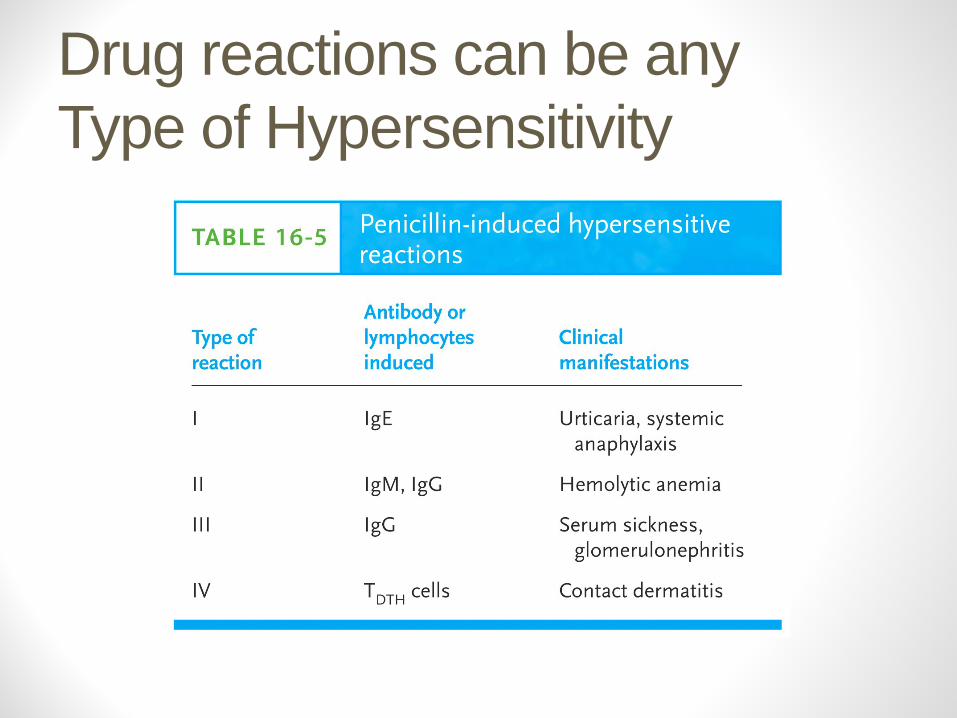
Drug reactions can be any Type of Hypersensitivity


