Embed Size (px)
Citation preview
Page 1/15
Buerger's disease: autoimmune disease involvingmultiple hypersensitivity typesPing Zheng ( [email protected] )
Shanghai Jiao Tong University, School of Medicine a�liated Ruijin HospitalWanchao Wang
Shanghai Jiao Tong University, School of Medicine, Shanghai Institute of Immunology
Research Article
Keywords: thromboangiitis obliterans, autoimmune disease, humoral immunity, immune-labelingtechniques, Type III, IV, II, I hypersensitivity reaction
Posted Date: October 6th, 2020
DOI: https://doi.org/10.21203/rs.3.rs-86283/v1
License: This work is licensed under a Creative Commons Attribution 4.0 International License. Read Full License
Page 2/15
AbstractBuerger’s disease is an autoimmune disease? And what is the immunological pathogenesis It has notbeen deeply researched but still attracts the attention of scholars. In our early study we reported that TAOmay be an autoimmune disease involving hypersensitivity Type III & Type IV. In this study we furtherexplored the immune pathogenesis of TAO based on initial research. We detected humoral immunity (IgE) in 28 cases using enzyme-linked immunosorbent assay (ELISA). Antigen-antibody complex depositingon the vessel wall in 18 cases, and anti-vessel antibodies in 28 cases using three kinds of immune-labeling techniques (immuno�uorescence labeling, immunoenzymatic staining and immuno-gold-silverstaining). The result shows Ig E levels were signi�cantly high (P<0.01). As high as 86% of anti-vesselantibodies in serum were found (P<0.001), and the auto-antibodies against the vessel were combineddirectly with vascular collagen. Antigen-antibody complexes deposited on the vascular wall. These�ndings further con�rm TAO is an autoimmune disease involving multiple hypersensitivity reactions. Thisis mainly Type III hypersensitivity and type II in addition to type IV. The elevated Ig E suggest that TAOmay a type I hypersensitivity involved.
BackgroundLeo Buerger �rst described Buerger’s disease (TAO or thromboangiitis obliterans) in 1908. TAO is anin�ammatory disease of small and medium-sized blood vessel walls accompanied by thrombosis, and itprimarily involves the limbs. Historically, the disease occurs more often in young men, and the severe painfrom TAO has led to drug abuse and limb loss; Major limb amputation occurs in nearly 20% of cases [1].TAO has a global yet uneven distribution [1]. In North America and Western Europe, TAO accounts for0.75% to 5.6% of peripheral vascular diseases. However, in Eastern Europe, the Middle East, theMediterranean, and Asia, it accounts for 60% to 80% of peripheral vascular diseases and may beassociated with the use of certain types of tobacco [1]. Interestingly, the incidence of TAO in women hasrisen [1] in accordance with an increase in cigarette smoking among women. The incidence of TAO isalso associated with economic development [2], as evidence suggests a correlation with improvements innutrition; most patients in these populations are malnourished [3].
It is more di�cult to treat TAO in its acute stages; therefore, it is important to study its pathogenesis.Researchers have recently paid special attention to immune pathogenesis. We conducted a previousstudy exploring TAO as an autoimmune disease involving type III related to antigen-antibody complexdeposited on the vessel wall and type IV delayed hypersensitivity related to sensitized T cells andreleasing lymphokines [4]. This study further explores the pathogenesis of TAO as an autoimmunedisease and its mechanism.
MethodsPatients
Page 3/15
Our study included 74 men patients, aged 21 to 50 years, with a history of TAO lasting 2 to 28 years.Patients had a recurrence history ranging from 1 to 6 times, with a recent onset within 1 to 10 months.The diagnosis of TAO was based on the following criteria: the onset occurred when the patient was 20 to40 years old, the patient reported being a smoker, the symptoms were usually accompanied by migratorysuper�cial phlebitis in an extremity, symptoms and signs of ischemia in the limbs were present, and otherperipheral vascular diseases (such as arteriosclerosis obliterans, diabetic foot) were excluded. In somecases, a TAO diagnosis was con�rmed by pathological �ndings.
We obtained biopsy blood vessel tissue from super�cial phlebitis and from limb amputation from TAOpatients. 18 cases of antigen-antibody (Ag-Ab) complex deposition, and 28 cases of anti-vessel auto-antibodies detected. Patients in this study were in the acute and sub-acute stages. 28 cases of Ig Epatients were assigned to 3 groups: acute TAO (n=5), sub-acute TAO (n=17), and chronic TAO n=6).
Observations and methods
We used enzyme-linked immunosorbent assay (ELISA) to detect humoral immunity (IgE) in 28 patientswho had no other allergic disorders. There are healthy people as a control group.
We used 3 types of immune labeling techniques to accurately detect Ag-Ab complex depositions on thevessel walls for 18 patients. We used immuno�uorescence (IF) labeling, where samples of patient vesselswere �xed in cooling alcohol (95%) and embedded in low-temperature para�n. The specimens were thensectioned via pancreatic digestion and direct immuno�uorescence staining. Prepared samples wereobserved under �uorescent microscopy. Using a similar technique, we also evaluated samples usingimmunoenzymatic staining (avidin-biotin complex, ABC staining) and immuno-gold-silver staining (IGSS),assessed via a light microscope.
For the detection of anti-vessel autoantibodies, healthy vessel tissue served as an antigen substitute. Theserum from 28 TAO patients was used as the �rst antibody, rabbit antihuman �uorescein isothiocyanate(FITC-Ig), ABC-Ig, and IGSS-Ig were used as secondary antibodies. We used indirect staining for allsamples.
The control serum was obtained from healthy volunteer blood donors at Shanghai Rui-Jin Hospital. Thehealthy vessel tissue was obtained from healthy controls through biopsy or amputation from patients atShanghai Rui-Jin Hospital. Other control comparators included rabbit serum substitution for humanserum, buffer solution substitution for human serum, anti-mouse Ab substitution for Ab labeling, buffersolution substitution for Ab labeling, and saline as a blank control. We conducted paired t-tests anddetermined the mean and standard deviations. P-values <0.05 were considered statistically signi�cant,P<0.01 was highly signi�cant, and P<0.001 was extremely signi�cant.
Results
Page 4/15
Ig E levels were highly signi�cantly elevated in all groups (P<0.01) and signi�cantly elevated in thesubacute and chronic TAO groups (P <0.05).
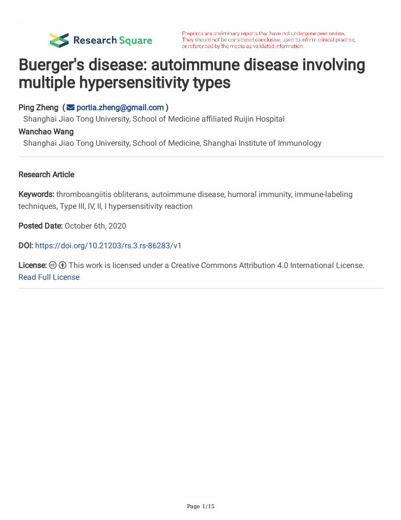
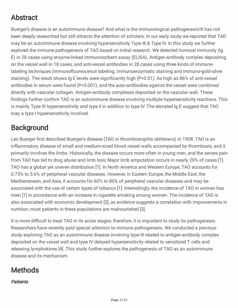
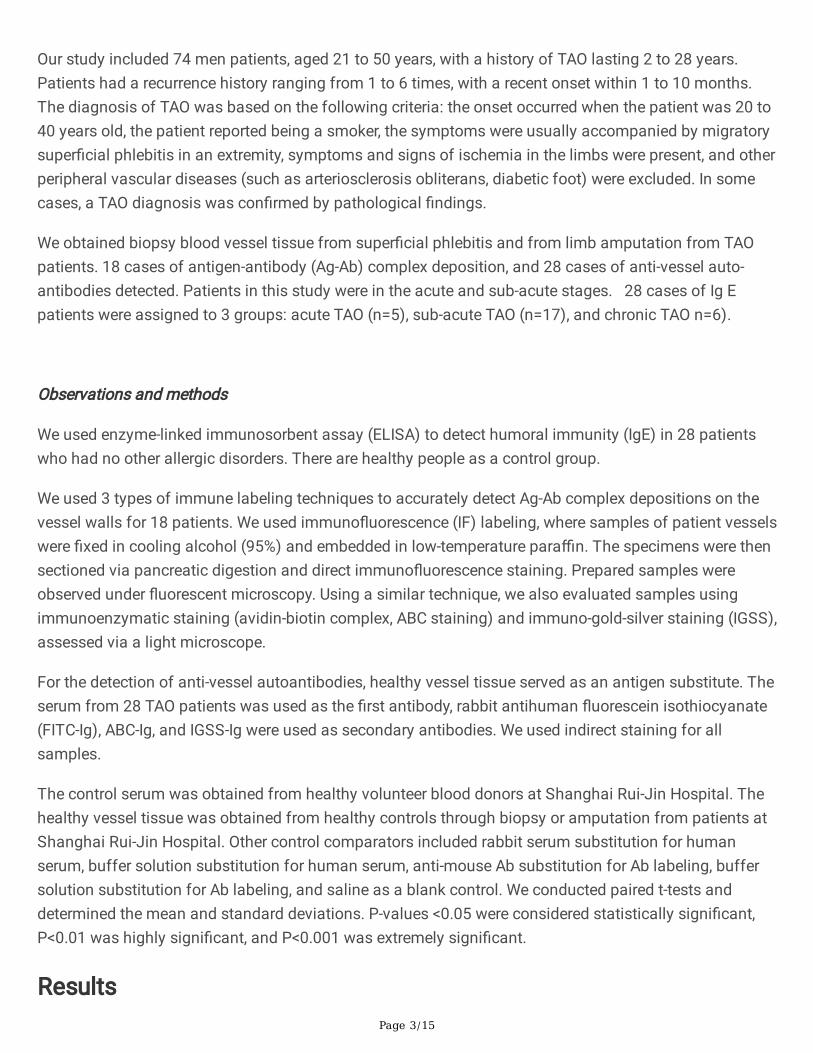
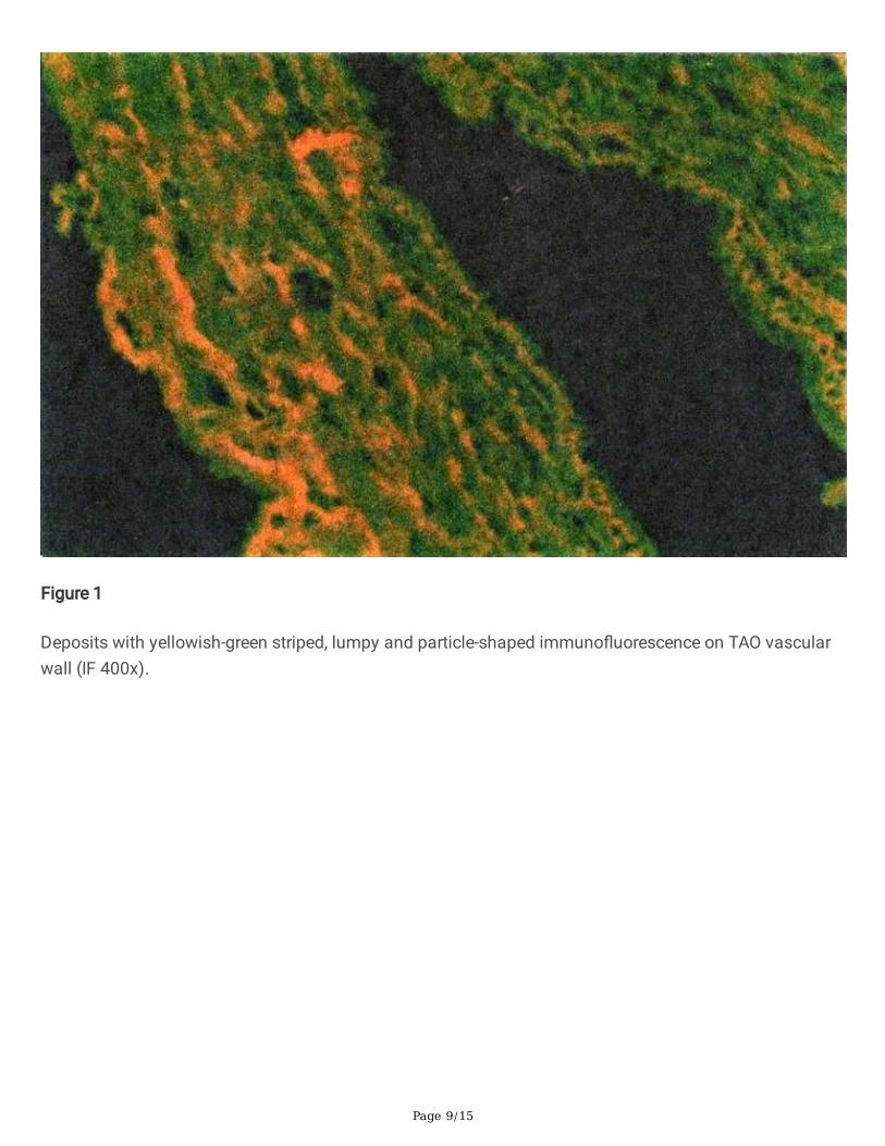
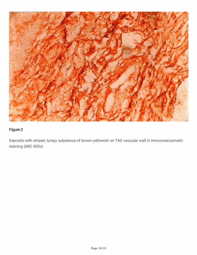
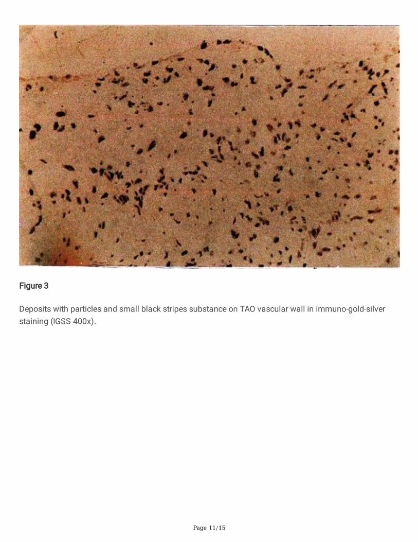
All immune-labeling techniques found Ag-Ab complex deposition on the vessel walls. IF staining �ndingsare illustrated in Fig. 1. ABC labeling results are illustrated in Fig. 2. To demonstrate the results of IF andto exclude the nonspeci�c cross-reaction, we observed section staining with the ABC methodsimultaneously. The ABC results were identical to those found with IF staining. The results of the IGSSevaluation are illustrated in Fig. 3 and were identical to the IF and ABC �ndings. The IGSS techniqueshowed good speci�city and sensitivity. In most vessels, we could see thickening of the intima, stenosisof the cavity, and the organization and recanalization of the thrombus in the vascular cavity. We alsonoted disorder of the structure, changing of media, �ber proliferation, and pathological changes of alllayers to various degrees along with local damage, breakdown, disappearance, and a large number ofstreaky particles or patches deposited in all layers—among which the �uorescent intensity of IgG and IgMwas strong. The controls were negative.
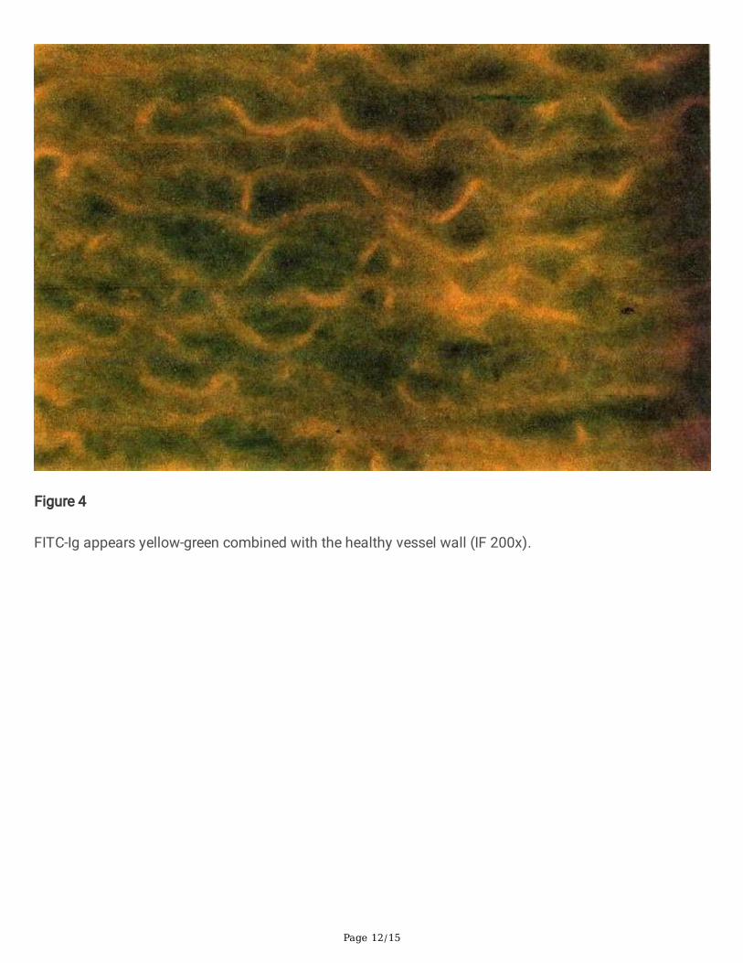
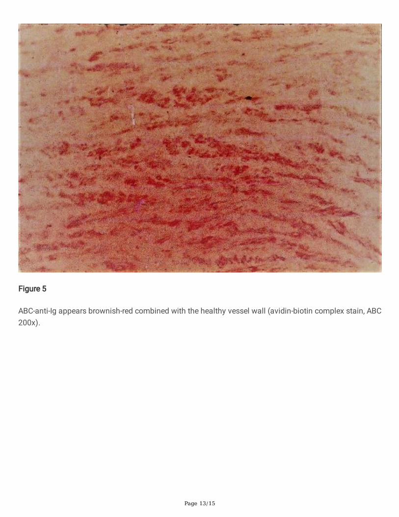
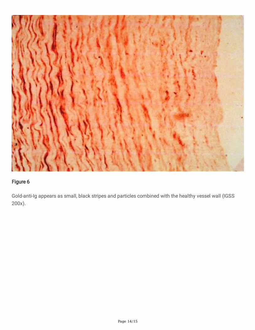
The results of our anti-vessel auto-antibodies assessments are illustrated in Figs. 4-6. Anti-vessel auto-antibodies were presented in 72% to 86% of TAO samples (P<0.001). The auto-antibodies against thevessel were combined directly with vascular collagen (as seen in Figs. 4-6). The speci�city and sensitivityof the IGSS method were better than those of the IF and ABC methods.
DiscussionVarious antibodies in TAO have been reported, including anti-endothelial cell antibodies [5], anti-neutrophilcytoplasmic antibodies [6], anti-phospholipid antibodies [7], anti-cardiolipin antibodies [8], and agonisticauto-antibodies directly against the G-protein coupled receptor [9]. TAO is associated with elevatedcytokine levels of tumor necrosis factor (TNF)-α, interleukin (IL)-1β, IL-4, IL-6, IL-17, and IL-23 [10, 11]. TAOis also associated with an increased expression of vascular cell adhesion molecules, intracellularadhesion molecules, and E-selectin on the endothelial cells of the affected arteries [12]. Kobayashi et al.[13] reported IgG, IgA, IgM, and complement factors C3d and C4c deposit along the internal elasticlamina, and cell in�ltration was observed in the thrombus and the intima of patients with TAO (CD3+ Tcells greatly outnumbered CD20+ B cells, CD68+ macrophages or S-100+ dendritic cells). Fu et al. [14]found that TAO patients have low erythrocyte immune function, so the erythrocytes are less able toadhere to the circulating immune complex, and Cui et al. [15] found an immune complex deposit in thevessel wall under electron microscopy, which aligns with our �ndings [4].The evidence suggests TAOpathogenesis involves humoral and cellular immunity, with TAO vessel cell immune response activation,in�ammation, damage, and thrombosis. Immunosuppressive agents suppress the immune response inTAO patients [16]. The use of immunoadsorption to remove TAO antibodies from the blood showedpromising results in the clinic [17, 18].
In the present study, TAO humoral immunology showed a highly signi�cant increase in IgE. Combinedwith TAO clinical manifestations, repeated acute episodes, and the initiating factors (e.g. tobacco use,
Page 5/15
climate, trauma, and malnutrition) [3]. It has long been recognized that persistent cigarette smoking is amajor risk factor for TAO persistence, progression, recurrence, and amputation. TAO patients seem to bein a sensitized state to cigarette smoking, indicating the presence of a Type I allergy. Elevated IgE levelsmay play some role in the pathogenesis of TAO as an autoimmune disease [19], but this should be furtherexplored for con�rmation. Three types of immuno-labeling techniques all con�rmed anti-vessel auto-antibodies in the sera of TAO patients. Furthermore, Ag-Ab complex deposits were observed directly onthe vessel wall in multiple immune label samples. The existence of Type III hypersensitivity reaction wasagain con�rmed. The auto-antibodies against the vessel were combined directly with damaged vascularcollagen, which re�ects a Type II hypersensitivity reaction (as noted in Figs. 4-6).
According to the etiology analysis in our previous study of 876 cases [3], cigarette smoking was thegreatest factor, but we also found other factors closely related to the disease. Most patients (80%) had ahistory of exposure to dampness and sudden temperature changes (eg, intense exercise with sweatingfollowed by freezing), and exposure to hot or cold extremes. 29% of patients had a history of trauma, and70% of patients had a history of malnutrition. Also, 17% of TAO patients were not smokers but sharedother risk factors as described above—risk factors that induce damage to the vessel walls to becomeauto-antigen or/and become sensitive factors to induce an allergic reaction. Cigarette smoking can notonly introduce an allergic antigen, which may induce a Type 1 allergic reaction in TAO, but nicotine is alsoa hapten with blood red cells, intracellular histones or DNA binding, and therefore alters their composition,resulting in the production of auto-antibodies [20].
TAO shares many characteristics with autoimmune diseases. For example, TAO patients haveautoantibodies and sensitized lymphocytes, high levels of r-globulin in serum [4]; deposition of the Ag-Abcomplex in damaged vessels; and lymphocyte, plasma cell, and monocyte in�ltration in the target vessels[4]. TAO patients also have a variety of auto-antibodies, and TAO has a genetic component [21, 22]. In theclinic, patients with TAO often have repeated recurrences and chronic stability.stage In acute episodes ofTAO, the course of the disease is managed using immunosuppressive agents [16] and immunoadsorptionagents to remove TAO antibodies [17, 18]. TAO patients will sometimes experience damage to bothextremities and viscera [23, 24], which indicates TAO may be a systemic autoimmune disease. Changesin sex hormones have an impact on immune function and may lead to autoimmune diseases. Althoughmost autoimmune diseases occur in women, TAO is more common in men, suggesting that manhormones may play a role.
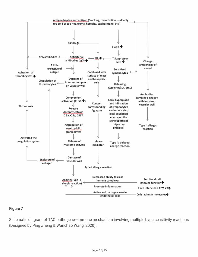
ConclusionsOverall, this study provides further evidence to classify TAO as an autoimmune disease involving multiplehypersensitivities reaction. Mainly Type III hypersensitivity. However, Type IV 4 and type II reactions arealso involved . This study found IgE elevated in the sera of TAO patients, suggesting a Type Ihypersensitivity reaction may be involved or plays some role in TAO autoimmunity. TAO vascular wallin�ammation, blood hypercoagulation [25] and thrombosis in the vascular lumen are considered a
Page 6/15
consequence of TAO autoimmune pathogenesis (Fig.7). This study not only elucidates the autoimmunepathogenesis of TAO. But also provides a new approach for clinical treatment.
DeclarationsEthical Statement
This study is another collaboration between the two authors on the immunology of Burger's diseasebefore retirement in China (the �rst cooperative paper was published in Chin Med J (Engl) 1989; 102:129-36). At the time of this study, China had not yet set up an ethical committee. But the research was stillcarried out in accordance with the ethical norms of that time. The study samples were obtained with theconsent of the patients or volunteers. Completion of this paper was done after the authors came to theUnited States, when they discovered that their research �ndings are still at the forefront of Burger'sdisease mechanism research. The publication of this paper will greatly promote the immune mechanismresearch in this �eld and provide theoretical basis for the treatment of this di�cult disease.
Ethics approval and consent to participate
Not applicable.
Consent for publication
Not applicable.
Availability of data and materials
All data are available in the article text and �gures.
Competing interests
The authors declare that they have no competing interests.
Funding
This study was funded by the Shanghai Institute of Immunology with support from the World HealthOrganization.
Authors’ contributions
PZ was responsible for the design of the study and drafting the article. WW provided assistance with thestudy design, performed immunological techniques, and reviewed and edited the draft article.
Acknowledgements
Page 7/15
We are sincerely grateful to Li Li, Xie YL of Shanghai Institute of immunology for their collaboration insome immunological techniques; Professor Ma BL, former director, Chief of the department in ShanghaiInstitute of Immunology for her instruction and Professor Su BH for statistical processing. We appreciateProfessor Yang BH, Director of Peripheral Vascular Surgery, Dongzhimen Hospital, Beijing University ofChinese Medicine, Chairman of the Peripheral Vascular Disease Committee of China for his review andinput. We also appreciate Dr. Mao M, principle immunological scientist of California Antagene Corp forhis review.
Authors' information
None
References1. Mills JL. Buerger’s disease in the 21st century: diagnosis, clinical features, and therapy. Semin Vasc
Surg. 2003;16:179-89.
2. Wang JJ, Zao WG, Sun SQ, Zhang Y. Changes in the incidence of thromboangiitis obliterans. Chin JVas Surg. 2009;1:9-12.
3. Zheng P. Etiology and pathogenesis of thromboangiitis obliterans (TAO). North Med. 1983;5:13-5.
4. Zheng P, Fu PB, Wang WC, Xu WY, Tang XM, Ye M, et al. Immunological studies on thromboangiitisobliterans. Chin Med J (Engl). 1989;102:129-36.
5. Eichhorn J, Sima D, Lindschau C, Turowski A, Schmidt H, Schneider W, et al. Antiendothelial cellantibodies in thromboangiitis obliterans. Am J Med Sci. 1998;315:17-23.
�. Halacheva KS, Manolova IM, Petkov DP, Andreev AP. Study of anti-neutrophil cytoplasmic antibodiesin patients with thromboangiitis obliterans (Buerger’s disease). Scand J Immunol. 1998;48:544-50.
7. Maslowski L, McBane R, Alexewicz P, Wysokinski WE. Antiphospholipid antibodies in thromboangiitisobliterans. Vasc Med. 2002;7:259-64.
�. de Godoy JMP, Braile DM. Buergerʼs disease and anticardiolipin antibodies. J Cardiovasc Med.2009;10:792-4.
9. Klein-Weigel PF, Bimmler M, Hempel P, Schöpp S, Dreusicke S, Valerius J, et al. G-protein coupledreceptor auto-antibodies in thromboangiitis obliterans (Buerger's disease) and their removal byimmunoadsorption. Vasa. 2014;43:347-52.
10. Dellalibera-Joviliano R, Joviliano EE, Silva JS, Evora PRB. Activation of cytokines corroborate withdevelopment of in�ammation and autoimmunity in thromboangiitis obliterans patients. Clin ExpImmunol. 2012;170:28-35.
11. Wei Z, Jiang W, Wang H, Li H, Tang B, Liu B, et al. The IL-6/STAT3 pathway regulates adhesionmolecules and cytoskeleton of endothelial cells in thromboangiitis obliterans. Cell Signal.2018;44:118-26.
Page 8/15
12. Halacheva K, Gulubova MV, Manolova I, Petkov D. Expression of ICAM-1, VCAM-1, E-selectin andTNF-α on the endothelium of femoral and iliac arteries in thromboangiitis obliterans. ActaHistochem. 2002;104:177-84.
13. Kobayashi M, Ito M, Nakagawa A, Nishikimi N, Nimura Y. Immunohistochemical analysis of arterialwall cellular in�ltration in Buerger's disease (endarteritis obliterans). J Vasc Surg. 1999;29:451-8.
14. Fu XM, Jing ZP, Guo F. Erythrocyte immune function and regulatory factors in patients withthromboangiitis obliterans. Shanghai Med. 1997;20:403-4.
15. Cui XM, Chen Y, Pan L. Vascular medial membrane deposition of immune complexes inthromboangiitis observed by electron microscopy. Bethune Med Univ. 2001;27:379-80.
1�. Saha K, Chabra N, Gulati SM. Treatment of patients with thromboangiitis obliterans withcyclophosphamide. Angiology. 2001;52:399-407.
17. Baumann G, Stangl V, Klein-Weigel P, Stangl K, Laule M, Enke-Melzer K. Successful treatment ofthromboangiitis obliterans (Buerger’s disease) with immunoadsorption: results of a pilot study. ClinRes Cardiol. 2011;100:683-90.
1�. Klein-Weigel P, Sophia Volz T, Gutsche-Petrak B, M. Boehnlein J, Bohlen A, Dreusicke S, et al.Immunoadsorption in Buergers disease (thromboangiitis obliterans): a promising therapeutic option:results of a consecutive patient cohort treated in clinical routine care. Lupus. 2016;1:111-4.
19. Zhu W, Cao MD, Zhou YJ, Meng, Wei JF. Role of Ig E against autoantigens in autoimmune diseases.Chin J Allergy Clin Immunol. 2017;11:57-60.
20. Papa M, Bass A, Adar R, Halperin Z, Schneiderman J, Becker CG, et al. Autoimmune mechanisms inthromboangiitis obliterans (Buerger's disease): the role of tobacco antigen and the majorhistocompatibility complex. Surgery. 1992;111:527-31.
21. Abhishek V, Rahul T, Vinod KP. Thromboangiitis obliterans (Buerger’s disease)—current practices. Int JIn�am. 2013;2013:156905.
22. Kimura A, Kobayashi Y, Takahashi M, Nobuhisa O, Kitamura H, Nakamura T, et al. MICA genepolymorphism in Takayasu's arteritis and Buerger's disease. Int J Cardiol. 1998;66:S107-13.
23. Fakour F, Fazeli B. Visceral bed involvement in thromboangiitis obliterans: a systematic review. VascHealth Risk Manag. 2019;15:317-53.
24. Zheng P, Zhang LN. Thromboangiitis obliterans with myocardial infarction in 3 cases. CardiovascDis. 1978;6:19-21.
25. Zheng P, Chen SJ, Shao HZ. Studies on hypercoagulation state in thromboangiitis obliterans. ChinMed J (Engl). 1989;102:67-71.
Figures
Page 9/15
Figure 1
Deposits with yellowish-green striped, lumpy and particle-shaped immuno�uorescence on TAO vascularwall (IF 400x).
Page 10/15
Figure 2
Deposits with striped, lumpy substance of brown-yellowish on TAO vascular wall in immunoenzymaticstaining (ABC 400x).
Page 11/15
Figure 3
Deposits with particles and small black stripes substance on TAO vascular wall in immuno-gold-silverstaining (IGSS 400x).
Page 12/15
Figure 4
FITC-Ig appears yellow-green combined with the healthy vessel wall (IF 200x).
Page 13/15
Figure 5
ABC-anti-Ig appears brownish-red combined with the healthy vessel wall (avidin-biotin complex stain, ABC200x).
Page 14/15
Figure 6
Gold-anti-Ig appears as small, black stripes and particles combined with the healthy vessel wall (IGSS200x).
Page 15/15
Figure 7
Schematic diagram of TAO pathogene---immune mechanism involving multiple hypersensitivity reactions(Designed by Ping Zheng & Wanchao Wang, 2020).


