Embed Size (px)
Citation preview

12
Postoperative Ileus: Pathophysiology and Treatment
N.S. Tropskaya and T.S. Popova The Sklifosovsky Research Institute for Emergency Medicine, Moscow,
Russia
1. Introduction
Postoperative ileus (POI) is a temporary impairment of gastrointestinal (GI) motility occurring universally after major abdominal surgery (Holte & Kehlet, 2002). For the majority of affected surgical patients, POI is transient, lasting approximately 3 to 5 days (Hotokezaka, et al., 1997). The adverse effects of POI are composed of not only physiologic effects such as reduced bowel function, exacerbation of nausea and vomiting, and increased postoperative pain, but also other clinically related effects such as delay of oral feeding, prolonged hospitalization, and increased use of human and material resources. Determination of the end of POI is somewhat controversial. The studies in the literature
have used varying end points, and each has its own weakness. Bowel sounds are sometimes
used as an end point, but they require frequent auscultation, their presence does not
necessarily indicate propulsive activity, and they can be the result of small-bowel activity
and not colonic function (Holte & Kehlet, 2000). Flatus also is not the ideal end point. It
requires a conscious patient who is comfortable reporting its occurrence to the investigator.
Also, there is some question as to the correlation between flatus and bowel movements
(Waldhausen, et al., 1990). Bowel movements are seemingly the most reliable end point.
The migrating myoelectric complex (MMC) is the basal level of activity in the bowel in the
fasting state, serving a “housekeeping” function (Szurszewksi, 1969). The resumption of this
myoelectric complex after surgery is responsible for recovery from POI.
In humans the MMC can be recognized from the lower oesophagus to the distal small
intestine, but it is most prominent in proximal jejunum. This enteric rhythm usually exhibits
three phases: Phase I when only slow waves are observed without spike bursts (without
actual muscle contractions); phase II when spike bursts are observed on slow waves
irregularly (the occurrence of intermittent muscle contractions); and phase III with spike
bursts on every slow wave for a few minutes (the contractions with the maximal contractile
frequency). Rats are particularly suited for studying the MMC as the cycling period is only
about 15 min in the conventional state.
Motility of the GI tract is temporarily impaired after surgery and is characterized by disorganized electrical activity and lack of coordinated propulsion (Behm & Stollman, 2003). In the stomach, studies have consistently demonstrated a postoperative period of gastric hypomotility associated with irregular and disorganized electrical activity (Clevers et al., 1991). Gastric propulsion may be orad, and there may also be increased pyloric tone that
www.intechopen.com

Current Concepts in Colonic Disorders
204
contributes to abnormal gastric emptying (Dauchel et al., 1976). Motor activity is similarly disorganized in the small bowel. Morris et al. (1983) observed the disappearance of phase III contractions 2 days after the operation. Miedema et al. (2002) also detected retrograde contractions, leading to significant delays in small bowel transit. Normal colonic motility is typically the last to return after surgery. Studies evaluating postoperative colonic motility frequently have found a period of relative hypomotility that is generally associated with random, disorganized bursts of electrical activity (Wilson, 1975; Condon et al., 1986). The type of surgical procedure performed can have significant effects on postoperative GI motility. Skin incision has no effect on the MMC activity of the bowel, whereas opening the peritoneum will completely inhibit the MMC (Livingston & Passaro, 1990). Some controversies exist over the timing of the MMC return after abdominal surgery. Benson et al. (1994) have shown that MMCs are present in the small bowel within a few hours after surgery. On the contrary, others investigators found that MMCs were abolished for 1 or 2 days after surgery, taking from 3 to 7 days for normal pattern to reestablish (Smith et al., 1977; Morris et al., 1983; Schippers et al., 1991). Several mechanisms are thought to play a role in POI, including sympathetic neural reflexes, local and systemic inflammatory mediators, and changes in various neural and hormonal transmitters. An imbalance between sympathetic and parasympathetic nervous-system input to the intestine has been postulated as an underlying cause. Sympathetic (adrenergic) hyperactivity results in reduction of propulsive motility, and an increase in sphincter tone. Parasympathetic (cholinergic) hypoactivity results in a decrease in gastrointestinal motility. There is finding supported the involvement nitrergic neurons in the pathogenesis of POI. Many potential etiologic agents have been investigated to determine a cause for the development of POI. Pharmacological modulation aimed at increasing excitatory activity has principally involved the administration of para-sympathomimetic agents which increase cholinergic transmission, such as bethanecol or neostigmine (Gerring & Hunt, 1986). Similarly, cisapride works as an indirect parasympathomimetic by stimulating serotonin receptors and so enhancing acetylcholine release (Reynolds & Putman, 1992; Wiseman & Faulds, 1994). Attempts to block inhibitory components of contractility have focused on the sympathetic system. Sympathetic hyperactivity should respond to ┚-adrenergic blocker such as propranolol. Metaclopramide, which among other activities has antidopaminegic properties and domperidone (selective peripheral dopamine (DA2 receptor) antagonist) have also been used to intervene in ileus cases (Reynolds & Putman, 1992). Investigations evaluating inhibitors of nitric oxide synthesis, have shown improved postoperative bowel motility in animal studies. Local anaesthetics has been most effective in the prevention of POI. Trimebutine maleate helped in improving the postoperative conditions of patients as their abdominal and colonic discomfort, abdominal pain and nausea decreased. Although some of the mechanisms underlying the abnormal intestinal motility found after surgery have been elucidated, an integrated understanding of the pathophysiology of POI remains elusive.
2. Experimental study of postoperative ileus
We investigated the possible role of cholinergic, adrenergic, dopaminergic, serotonergic, nitrergic mechanisms, and also local anaesthetics in POI. Objective: To investigate altered gastrointestinal motility in POI and effects of potential etiological agents on the GI electrical activity to determine a cause for the development of
www.intechopen.com
Postoperative Ileus: Pathophysiology and Treatment
205
POI and treatment. We were solving the following questions: 1) Whether the administration of different pharmacological agents stimulate the MMC or its separate phases; 2) Which pharmacological agents will induce early recovery of the MMC from POI?
2.1 Materials and methods 2.1.1 Experimental animals and grouping
Fifty four male Wistar rats weighing 300-350 g were used. Animals were divided into nine groups. Control group (n = 6) which included the rats with implanted three bipolar electrodes and an infusion cannula in that the electrical activity was measured on the 10th day post surgery (after recovery). LAP group (n = 6) in which an exploratoty laparotomy, implantation of three bipolar electrodes and an infusion cannula were considered as trauma and the study measurements were conducted on the 1st – 6th days postoperative. Other seven LAP groups in which an exploratoty laparotomy, implantation of three bipolar electrodes and an infusion cannula were considered as trauma and the study measurements were conducted on the 1st – 6th days postoperative. These rats received from the 1st trough 3rd postoperative days neostigmine 0,2 mg/kg (n = 6), or propranolol 0,15 mg/kg (n = 6), or metaclopramide 0,5 mg/kg (n = 6), or cisapride 0,2 mg/kg (n = 6), or domperidone 0,5 mg/kg (n = 6), or L-NAME 0,1 mg/kg (n = 6), or trimebutine 2,86 mg/kg (n = 6).
2.1.2 Laparotomy
The rats were anesthetized with 5% ketamine solution i.p. in the dose of 0.3 ml/100 g body weight, and the operation was performed aseptically. After shaving the hair on rat abdomen, an incision (about 4 cm in length) was made. One of the three bipolar stainless steel electrodes were implanted into the muscular wall of the antrum, and the other two were implanted into the muscular wall of the small intestine at 3 cm (duodenum) and 30 cm distally to the pylorus (jejunum). An infusion cannula for drug administration was inserted into the jejunum (5cm proximal to the jejunal electrode). The wires of the electrodes and the infusion cannula were tunneled subcutaneously in the rat's tail and drawn outside at 5-6 cm of the tail tip. The incision was sutured with silk in double layers. Approximately 2 h was required from start to end of the operation. Post-operatively, the rats are kept in individual cages with a special tether allowing the
animal to move freely within the cage. This cage design enabled to manipulate with the
infusion cannula for drug administration during experiment and to connect the electrodes
for recording the electrical activity.
The animals were housed in an air-conditioned room at 22˚C with a 12-h light cycle, fed
standard laboratory diet and given water ad libitum. The animals were fasted for 24 h
before surgery (ad libitum intake of water was permitted).
2.1.3 Gastrointestinal motility recordings
The rats were given no food or water during the recording of gastrointestinal motility. In the control group, a 10-day recovery period was provided post surgery. The recording of
antroduodenojejunal electromyograms was performed after the animals had been fasted for
24 h with free access to water. Fasting gastrointestinal motility was recorded for 1 h.
www.intechopen.com
Current Concepts in Colonic Disorders
206
In the other groups gastrointestinal motility was recorded for 6 days after the operation. Fasting gastrointestinal motility was recorded for 1 h before- and for 2 h after administration of the drugs. The electrodes were connected to the amplifier with the sensitivity of 0.1 mV and the frequency band of 0,03 - 100 Hz. The signals are entered in the computer IBM PC for the review, analysis and storage. The evaluation of the GI electrical activity is performed using the spectral analysis (the assessment of slow waves) and non-linear filtration algorithm (assessment of spike potentials). Percentage of slow waves on which spike bursts were superimposed at the levels of stomach, duodenum and jejunum was calculated. In addition, a visual analysis of electromyograms is performed, i.e. the calculation of time parameters of electrical activity that characterize the MMC. The main feature of the MMC, the activity front or phase III, was identified as a period of clearly distinguishable intense spiking activity. The amplitude should be at least twice that of preceding baseline, and propagating aborally through the whole recording segment and followed by a period of quiescence, phase I of the MMC. Phase II was characterised as a period of irregular spiking activity preceding the activity front. Periods with no detectable spike potentials were considered as quiescence. Characteristics of the MMC, such as period of the MMC, percentage of duration of the MMC phases, duration and propagation time of phase III of the MMC were evaluated. Period of the MMC was defined as the time lapse between the end of two consecutive phases III (for the duodenum and the jejunum). Percentage of duration of the MMC phases was defined as the ratio between phase duration, in seconds, and period of the MMC, in seconds, and then multiplied on 100. In case of the MMC absence than percentage of duration of the MMC phases was defined as the ratio between phase duration, in seconds, and duration of recording (3600 s), and then multiplied on 100. Phase III duration was defined as the time lapse between beginning and end of a propagated activity front with spikes on top of every slow wave. Phase III propagation time was defined as the time lapse between the end of a phase III at the duodenum and the jejunum.
2.1.4 Statistical analysis
Results were expressed as the Ме (25; 75)% . The parameters of GI electrical activity were
evaluated with the Mann–Whitney U-test and Friedman –ANOVA.
P values less than 0.05 were considered significant.
2.1.5 Drugs and other chemicals
L-NAME were purchased from ICN Biomedicals Inc. Propranolol (Anaprilin) was obtained
from ICN Medicinal (Moscow, Russia), cisapride (Coordinax) and domperidone (Motilium)
from Janssen Pharmaceutica (Beerse, Belgium), neostigmine (Proserini) from
Pharmstandard-October (Moscow, Russia), metoclopramide (Cerucal) from AWD pharma
(Germany), trimebutine (Tambutin) from Dae Han New Pharm. Co. (Corea). All compounds
were dissolved in saline. All drugs were administered intraintestinally in volumes of 0.2 ml.
All the protocols and procedures were approved by the Institutional Review Board for
Experimental Studies.
www.intechopen.com

Postoperative Ileus: Pathophysiology and Treatment
207
2.2 Results 2.2.1 Normal GI electrical activity
The interdigestive motility pattern, the MMC, recorded in the duodenum and proximal
jejunum, was observed in all animals (control group). Approximately 5 MMC was noted
during 1 h. Period of the MMC in the duodenum and in the jejunum were 700 (620; 810)s
and 710 (680;770)s, respectively. Percentage of slow waves on which spike bursts were
superimposed for 1 h recording at the levels of stomach, duodenum and jejunum was 12,0
(9,8; 12,2)%; 40,5 (38,9; 42,4)% and 44,0 (42,1; 46,4)%, respectively.
The MMC consisted of three distinct phases: phase I is period quiescence (only slow waves are observed without spike bursts); phase II is period of irregular activity (when spike bursts are observed on slow waves irregularly); and phase III is period of regular activity (spike bursts on every slow wave). Characteristics of the MMC are presented in Table 1.
Duodenum Jejunum
percentage of duration of phase I, % 37,3 (35; 41,5) 35,6 (31,5; 40,1)
percentage of duration of phase II, % 37,4 (36,8; 45) 43 (35; 44,1)
percentage of duration of phase III, % 22,9 (20; 25,2) 23,1 (21,1; 24,9)
phase III duration, s 175 (150; 180) 170 (160; 180)
phase III migration time from
duodenum to jejunum, s 410 (320; 490)
Table 1. Characteristics of the MMC
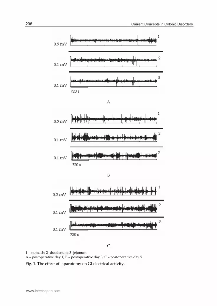
2.2.2 The effect of laparotomy on GI electrical activity
Percentage of slow waves on which spike bursts were superimposed at the levels of
stomach, duodenum and jejunum was decreased on the 1st postoperative day after
laparotomy in rats (Table 2). Also gastric dysrhythmias were noticed. Suppression of the
MMC and weak irregular contractions were observed in all segments of the
gastrointestinal tract (Figure 1). Phase III activity was absent in the duodenum and
jejunum. The duration of phase II for these segments was longer than that of the normal
animals (Figure 2).
Post-
operative
time, day
Stomach Duodenum Jejunum
1 4,0 (3,5; 4,5)* 22,8 (19,3; 23,4)* 24,5 (22,0; 29,8)*
2 7,0 (5,2; 7,8)* 35,2 (32,7; 35,8)* 40,4 (40,0; 42,6)
3 7,5 (6,0; 7,9)* 38,9 (36,0; 39,8) 45,6 (42,3; 46,0)
4 8,0 (6,5; 8,5)* 36,8 (36,2; 39,6) 43,8 (42,9; 44,5)
5 10,1 (9,4; 10,3) 38,8 (37,5; 39,3) 43,7 (42,5; 44,9)
6 12,0 (9,8; 12,2) 40,5 (38,9; 42,4) 44,0 (42,1; 46,4) * - р<0,05 vs. control group
Table 2. Percentage of slow waves on which spike bursts were superimposed at the levels of stomach, duodenum and jejunum in each stage of the study (Me (25; 75)%).
www.intechopen.com
Current Concepts in Colonic Disorders
208
A
B
C
1 – stomach; 2- duodenum; 3- jejunum. A – postoperative day 1; B – postoperative day 3; C – postoperative day 5.
Fig. 1. The effect of laparotomy on GI electrical activity.
www.intechopen.com

Postoperative Ileus: Pathophysiology and Treatment
209
Fig. 2. Postoperative change of percentage of duration of the MMC phases (dark circle - р<0,05 vs. control group).
Percentage of slow waves on which spike bursts were superimposed at the levels of stomach
and duodenum was decreased on the 2nd postoperative day. The spike activity in the
jejunum returned to normal. Gastric dysrhythmias were absent. The duration of phase II for
the duodenum was longer than that of normal animals, phase III was not observed. We
observed appearance of phase III in the jejunum. However, the duration of phase III was
shorter than that of normal animals (140 (120; 150) s, p<0,05).
Percentage of slow waves on which spike bursts were superimposed at the levels of stomach was decreased on the 3rd postoperative day. The spike activity in the duodenum returned to
www.intechopen.com
Current Concepts in Colonic Disorders
210
normal. We observed all phases of the MMC in the duodenum and jejunum. However, the duration of phases for duodenum was significantly different from normal. Not all phases III migrated from the duodenum to the jejunum. Normalization of the MMC phase ratio in jejunum was noticed. Percentage of slow waves on which spike bursts were superimposed at the levels of stomach was decreased on the 4th postoperative day. Normalization of the MMC phase ratio in duodenum was observed. Full recovery of GI electrical activity and the MMC of small intestine we obtained on the 5th postoperative day after laparotomy. Thus, the recovery of spike electrical activity occurs in the jejunum, then in the duodenum, and at last in the stomach. Also phase III contractions are observed at first only in the lower part of the gastrointestinal tract (jejunum) and then, gradually, in the upper part (duodenum). Normalization of spike electrical activity in the stomach occurs at the same time when the MMC from duodenum to the jejunum is observed.
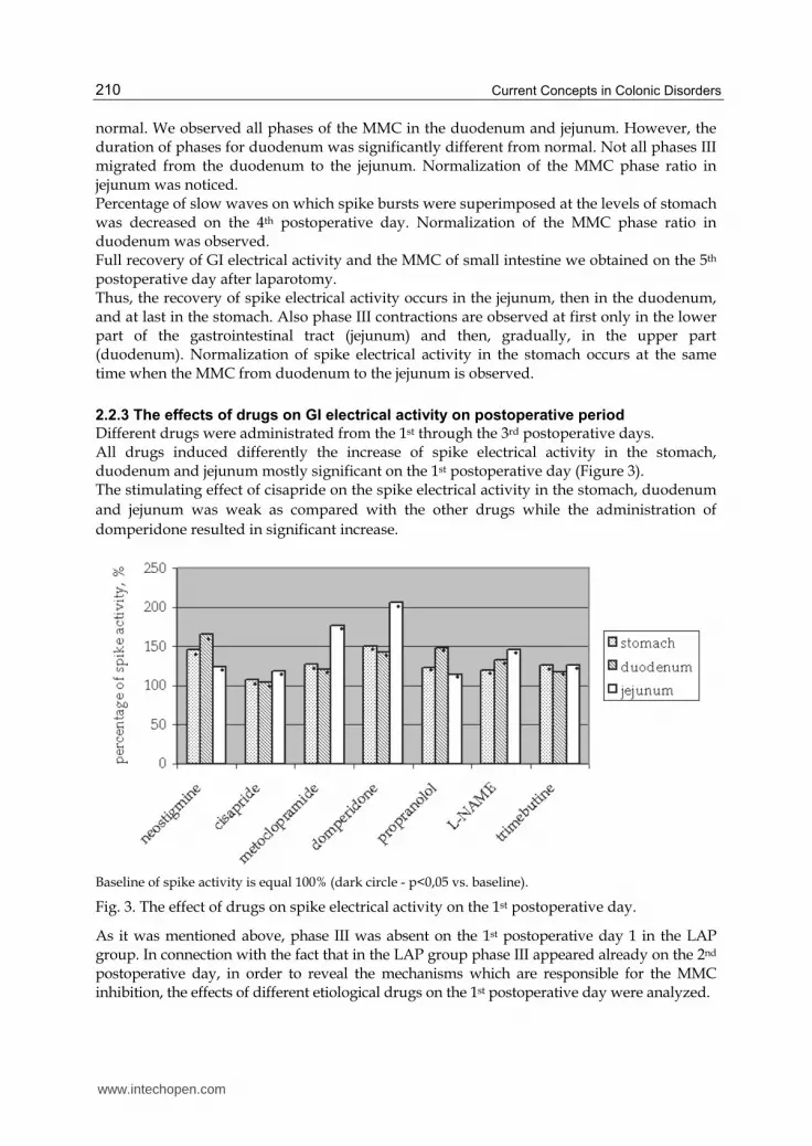
2.2.3 The effects of drugs on GI electrical activity on postoperative period
Different drugs were administrated from the 1st through the 3rd postoperative days. All drugs induced differently the increase of spike electrical activity in the stomach, duodenum and jejunum mostly significant on the 1st postoperative day (Figure 3). The stimulating effect of cisapride on the spike electrical activity in the stomach, duodenum
and jejunum was weak as compared with the other drugs while the administration of
domperidone resulted in significant increase.
Baseline of spike activity is equal 100% (dark circle - р<0,05 vs. baseline).
Fig. 3. The effect of drugs on spike electrical activity on the 1st postoperative day.
As it was mentioned above, phase III was absent on the 1st postoperative day 1 in the LAP group. In connection with the fact that in the LAP group phase III appeared already on the 2nd postoperative day, in order to reveal the mechanisms which are responsible for the MMC inhibition, the effects of different etiological drugs on the 1st postoperative day were analyzed.
www.intechopen.com

Postoperative Ileus: Pathophysiology and Treatment
211
The different drugs stimulated different phases of the MMC in the duodenum and jejunum (Figure 4).
Fig. 4. The effect of drugs on percentage of phases duration of the MMC on the 1st postoperative day (dark circle - р<0,05 vs. LAP group).
The administration of neostigmine stimulated phase II in the duodenum and jejunum. The duration of phase II was increased. Irregular activity was represented by the groups of spike bursts (duration 20-60 s) with periods of quiescence (duration 10-20 s). The duration of phase I was decreased. We didn’t observe phase III. Spastic activity was noted in the
www.intechopen.com

Current Concepts in Colonic Disorders
212
duodenum and jejunum. This type of activity consisted of the groups of spike bursts occupating 2-3 slow wave. Moreover, we observed the migrating action potential complexes (MAPCs). These complexes rapidly propagated from the duodenum to the jejunum (Fig.5).
Fig. 5. Migrating action potential complex on the 1st postoperative day after administration of neostigmine 1 – duodenum; 2 - jejunum
The administration of cisapride stimulated phase II in the duodenum and jejunum. Irregular
activity was represented by the groups of spike bursts (duration 20-30 s) with periods of
quiescence (duration 30-60 s). The incidence of distally propagated clustered activity was
observed. We didn’t detect phase III.
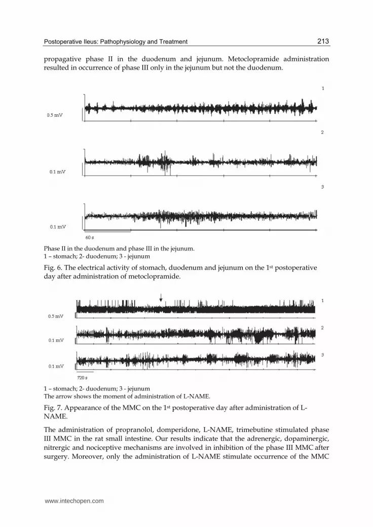
The administration of metoclopramide stimulated phase II in the duodenum. Irregular
activity was represented by the groups of spike bursts (duration 15-20 s or 30-40 s) with
periods of quiescence (duration 30-40 s). We didn’t observe phase III in the duodenum.
Metocloptamide induced phases III in the jejunum. The duration of phase III was 100 (80;
130) s, it was less than that of the normal animals (Fig.6). The duration of phase I decreased
in the duodenum and jejunum. We also observed spastic activity.
The administration of domperidone stimulated appearance of phase III both on the
duodenum and jejunum.
After the administration of propranolol disappearance of phase I and appearance of phase
III both on the duodenum and jejunum was observed.
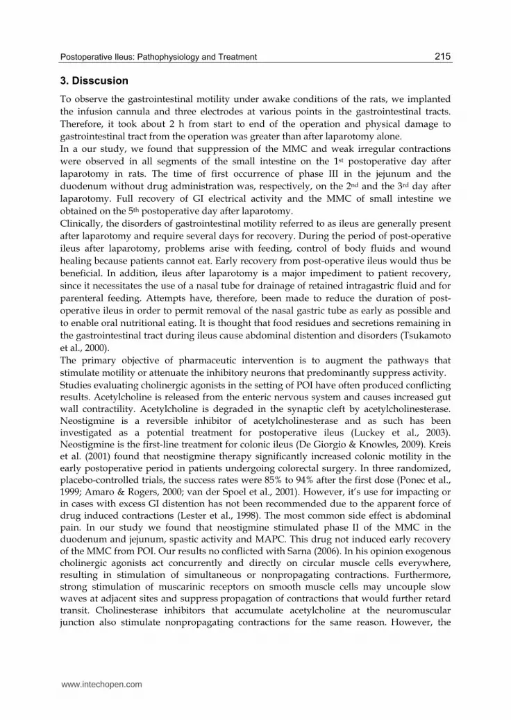
After the administration of L-NAME the MMC propagating from duodenum to the jejunum
was registrated (Fig. 7).
After the administration of trimebutine decrease of phase I duration and appearance of
phase III in the duodenum and jejunum were observed.
So, we investigated the possible role of cholinergic, adrenergic, dopaminergic, serotonergic,
nitrergic mechanisms, and also local anaesthetics in POI. We found that the administration
of different pharmacological agents stimulated the MMC and its separate phases in the rat
small intestine on the 1st postoperative day after laparotomy.
Neostigmine and cisapride induced phase II, but not phase III of MMC in the duodenum and jejunum. The phase II after cisapride differed from the phase II after neostigmine. The contractile pattern induced by neostigmine was spastic. We also observed occurrence the MAPCs in the duodenum and jejunum. Cisapride induced a prolonged and highly
www.intechopen.com
Postoperative Ileus: Pathophysiology and Treatment
213
propagative phase II in the duodenum and jejunum. Metoclopramide administration resulted in occurrence of phase III only in the jejunum but not the duodenum.
Phase II in the duodenum and phase III in the jejunum. 1 – stomach; 2- duodenum; 3 - jejunum
Fig. 6. The electrical activity of stomach, duodenum and jejunum on the 1st postoperative day after administration of metoclopramide.
1 – stomach; 2- duodenum; 3 - jejunum The arrow shows the moment of administration of L-NAME.
Fig. 7. Appearance of the MMC on the 1st postoperative day after administration of L-NAME.
The administration of propranolol, domperidone, L-NAME, trimebutine stimulated phase
III MMC in the rat small intestine. Our results indicate that the adrenergic, dopaminergic,
nitrergic and nociceptive mechanisms are involved in inhibition of the phase III MMC after
surgery. Moreover, only the administration of L-NAME stimulate occurrence of the MMC
www.intechopen.com

Current Concepts in Colonic Disorders
214
propagating from the duodenum to the jejunum. Therefore, the main role in disappearance
the MMC after surgery belongs to the activation of nonadrenergic noncholinergic
mechanisms. In the early postoperative period, endogenous NO is a major inhibitory
component that seems to constitute the common final pathway of mediators and the neural
pathways inhibiting the MMC in rats. On the 2nd postoperative day in baseline recordings in the duodenum phase III was absent in the LAP group, neostigmine and metoclopramide groups. Phase III in baseline recordings in the jejunum was observed in all groups including the LAP group, however it was absent in the group with the administration of neostigmine (Table 3). On the 3rd postoperative day in baseline recordings in the jejunum phase III was observed in all groups, at the same time phase III in duodenum was absent only in the group with the administration of neostigmine.
Drugs 2nd postoperative day 3rd postoperative day
duodenum jejunum duodenum jejunum
LAP - + + +
neostigmine - - - +
cisapride + + + +
metoclopramide - + + +
domperidone + + + +
propranolol + + + +
L-NAME + + + +
trimebutine + + + +
+ presence of phase III; - absence of phase III
Table 3. Phase III of the MMC on the 2nd and the 3rd postoperative days in baseline recordings in each of the groups
The administration of almost all drugs on the 1st through the 3rd postoperative day reversed recovery of GI electrical activity. All drugs, except for neostigmine induced the MMC recovery 3-4 days later after laparotomy (Figure 8).
0246LAP
neostigmine
cisapride
metoclopramide
domperidone
propranolol
L-NAME
trimebutine
The figures show postoperative days.
Fig. 8. Time of the MMC recovery on the postoperative period.
Our finding demonstrated that cisapride, domperidone, metaclopramide, propranolol, L-NAME, trimebutine may be useful as prokinetic agents to induce early recovery of the MMC from postoperative ileus.
www.intechopen.com
Postoperative Ileus: Pathophysiology and Treatment
215
3. Disscusion
To observe the gastrointestinal motility under awake conditions of the rats, we implanted
the infusion cannula and three electrodes at various points in the gastrointestinal tracts.
Therefore, it took about 2 h from start to end of the operation and physical damage to
gastrointestinal tract from the operation was greater than after laparotomy alone.
In a our study, we found that suppression of the MMC and weak irregular contractions
were observed in all segments of the small intestine on the 1st postoperative day after
laparotomy in rats. The time of first occurrence of phase III in the jejunum and the
duodenum without drug administration was, respectively, on the 2nd and the 3rd day after
laparotomy. Full recovery of GI electrical activity and the MMC of small intestine we
obtained on the 5th postoperative day after laparotomy.
Clinically, the disorders of gastrointestinal motility referred to as ileus are generally present
after laparotomy and require several days for recovery. During the period of post-operative
ileus after laparotomy, problems arise with feeding, control of body fluids and wound
healing because patients cannot eat. Early recovery from post-operative ileus would thus be
beneficial. In addition, ileus after laparotomy is a major impediment to patient recovery,
since it necessitates the use of a nasal tube for drainage of retained intragastric fluid and for
parenteral feeding. Attempts have, therefore, been made to reduce the duration of post-
operative ileus in order to permit removal of the nasal gastric tube as early as possible and
to enable oral nutritional eating. It is thought that food residues and secretions remaining in
the gastrointestinal tract during ileus cause abdominal distention and disorders (Tsukamoto
et al., 2000).
The primary objective of pharmaceutic intervention is to augment the pathways that
stimulate motility or attenuate the inhibitory neurons that predominantly suppress activity.
Studies evaluating cholinergic agonists in the setting of POI have often produced conflicting results. Acetylcholine is released from the enteric nervous system and causes increased gut wall contractility. Acetylcholine is degraded in the synaptic cleft by acetylcholinesterase. Neostigmine is a reversible inhibitor of acetylcholinesterase and as such has been investigated as a potential treatment for postoperative ileus (Luckey et al., 2003). Neostigmine is the first-line treatment for colonic ileus (De Giorgio & Knowles, 2009). Kreis et al. (2001) found that neostigmine therapy significantly increased colonic motility in the early postoperative period in patients undergoing colorectal surgery. In three randomized, placebo-controlled trials, the success rates were 85% to 94% after the first dose (Ponec et al., 1999; Amaro & Rogers, 2000; van der Spoel et al., 2001). However, it’s use for impacting or in cases with excess GI distention has not been recommended due to the apparent force of drug induced contractions (Lester et al., 1998). The most common side effect is abdominal pain. In our study we found that neostigmine stimulated phase II of the MMC in the duodenum and jejunum, spastic activity and MAPC. This drug not induced early recovery of the MMC from POI. Our results no conflicted with Sarna (2006). In his opinion exogenous cholinergic agonists act concurrently and directly on circular muscle cells everywhere,
resulting in stimulation of simultaneous or nonpropagating contractions. Furthermore, strong stimulation of muscarinic receptors on smooth muscle cells may uncouple slow waves at adjacent sites and suppress propagation of contractions that would further retard transit. Cholinesterase inhibitors that accumulate acetylcholine at the neuromuscular
junction also stimulate nonpropagating contractions for the same reason. However, the
www.intechopen.com
Current Concepts in Colonic Disorders
216
accumulation of acetylcholine at the neuroeffector junction may also stimulate giant migrating contractions – GMCs (analog MAPCs). Cisapride is an orally administered prokinetic agent which facilitates or restores motility throughout the length of the gastrointestinal tract. It is a substituted piperidinyl benzamide, chemically related to metoclopramide, but unlike metoclopramide, cisapride is largely devoid of central depressant or antidopaminergic effects (Wiseman & Faulds, 1994). Cisapride is a 5-HT-4 agonist that facilitates acetylcholine release from the intrinsic plexus (Tonini, 1999). Several trials have shown that cisapride actually has a beneficial effect in shortening POI (Tollesson et al., 1991; Brown et al., 1999). In our study we found that cisapride stimulated phase II of the MMC in the duodenum and jejunum. Cisapride significantly increased the incidence of distally propagated clustered activity and induced a prolonged and highly propagative phase II jejunal electrical activity. Moreover, cisapride reduced the duration of POI. Metoclopramide, a derivative of para-aminobenzoic acid, is a dopamine antagonist with central and peripheral effects. It possesses both anti-emetic and prokinetic effects; the anti-emetic effects relate to dopamine (D2) antagonism and serotonin (5-HT3) receptor antagonism on vagal and brainstem pathways; the prokinetic effects result from acetylcholine release from enteric cholinergic neurons (via 5-HT4 receptors), D2 receptor antagonism in the myenteric plexus, and muscarinic receptor sensitization (Rabine, 2001). Metoclopramide treatment may be beneficial in the treatment of canine postoperative ileus by increasing myoelectric and contractile activity of the proximal gastrointestinal tract. Graves et al. (1989) reported that treatment with metoclopramide partially reversed the MMC phase III inhibition at the duodenum and jejunum. Motility index values were restored to preoperative baseline values with metoclopramide treatment. Other trials that assessed efficacy of metoclopramide for POI failed to show any beneficial effect (Cheape et al., 1991; Seta & Kale-Pradhan, 2001). We established that metoclopramide induced phase III in the jejunum, but not in the duodenum on the 1st and the 2nd postoperative days. However, administration of metoclopramide induced early recovery of the MMC from POI. An important mechanism for the inhibition of motility is dopamine acting at neural D2 receptors. Previous studies have shown that stimulation of D2 receptors decreases acetylcholine release from cholinergic motoneurons innervating the gastrointestinal tract (Kurosawa, 1991). Domperidone is a dopamine antagonist that acts primarily through peripheral D2 receptors and does not cross the blood-brain barrier; thus, its use is not associated with the majority of the central nervous system side effects of metoclopramide. We found that administration of domperidone stimulate phase III MMC in the rat small intestine and induced early recovery of the MMC from POI. Smith and co-workers (1977) reported a transient increase in plasma epinephrine simultaneously with a sustained increase of norepinephrine after laparotomy in the dog. In their study, ileus persisted for a long time after plasma concentrations of epinephrine returned to basal values. In addition, chemical destruction of sympathetic nerves by pretreatment with 6-hydroxydopamine prevents inhibition of gastric emptying and intestinal transit after abdominal surgery in the rat (Dubois, 1973). Increased synthesis and release of norepinephrine from the intestinal wall in the rat have been reported (Dubois et al., 1973, 1974). In our study we found that propranolol (┚ -adrenergic blocker) prevented inhibition of motility after laparotomy and reduce the duration of postoperative ileus.
www.intechopen.com

Postoperative Ileus: Pathophysiology and Treatment
217
Meile et al. (2006) investigated the effects of NO synthase inhibition on gastric, small intestinal and colonic motility in awake rats under baseline conditions and in a postoperative ileus model. L-NMMA (NO synthase inhibitior) injection prior to surgery did not prohibit intraoperative inhibition of gastrointestinal motility, but did result in immediate recovery of gastric, small intestinal and colonic motility postoperatively. The major observation in our study is that inhibition of endogenous NO synthase by L-NAME results in early recovery of the MMC in the small intestine. In the early postoperative period, endogenous NO is a major inhibitory component that seems to constitute the common final pathway of mediators and the neural pathways inhibiting gastrointestinal motility in rats. Trimebutine, a weak non selective opioid agonist unable to cross blood-brain barrier, has long been used in the treatment of functional bowel disease (Delvaux & Wingate, 1997). Peripheral κ-opioid agonists are not associated with GI dysmotility but they do have anti-nociceptive effects in the GI tract. For example, preclinical studies suggest that peritoneal irritation induced pain is reversed with κ-agonist. Likewise, κ - opioid agonists inhibit the response of peripheral primary afferents to colorectal distention (Corazziari, 1999; Junien & Riviere, 1995). De Winter ( 2003) also demonstrated that blockade of the afferent limb of the reflex pathway by peripheral κ -opioid agonists ameliorated postoperative ileus. In animal studies, bowel motility was normalized with use of fedotozine (selective κ - opioid receptor agonist) (De Winter et al., 1997; Friese et al., 1997; Riviere et al., 1994). These results support a role for peripheral κ -opioid receptors in the pathogenesis of postoperative ileus induced by abdominal surgery. N. Friese and co-workers (1997) believe that peripheral κ -opioid receptors could modulate the transmission of visceral nociceptive stimuli in the periphery and might represent an alternative to μ-agonists in the treatment of abdominal pain associated with motility and transit impairments, such as after abdominal surgery. Moreover, a non-opioid mechanism via sodium channel blocking properties has been proposed for κ-opioid agonists suggesting that these agents may also act as a local anesthetic (Junien & Riviere, 1995; Barber & Gottschlich, 1997). Fedotozine was the first κ - agonist studied in humans. In clinical trials it proved to be better than placebo in relieving bloating, abdominal pain, postprandial fullness and nausea in patients with functional dyspepsia while it was slightly superior to placebo in getting symptom relief in patients with irritable bowel syndrome (Corazziari, 1999; Delvaux, 2001). The sodium channel-blocking activity was confirmed by the potent local anesthetic effect of trimebutine, which was 17-fold more active than lidocaine in terms of both potency and duration of action. (Roman et al., 1999). The blocking effect of trimebutine on sodium channel currents may account for this antinociceptive effect. (Roman et al., 1999). In our study we found that trimebutine stimulated phase III of the MMC in the duodenum and jejunum on the 1st postoperative day. Also administration of this drug induced early recovery of the MMC from POI. Thus, inhibition of nociceptive pathways after laparotomy is significant for occurrence of phase III and recovery of the MMC from POI.
4. Conclusion
We investigated altered gastrointestinal motility in POI and effects of potential etiological agents on gastrointestinal electrical activity to determine a cause for the development of POI and treatment. Our results are consistent with the hypothesis that small intestinal motility is under tonic inhibition by adrenergic, dopaminenergic, nitrergic and nociceptive
www.intechopen.com

Current Concepts in Colonic Disorders
218
mechanisms, and release from this inhibition results in phase III activity. The main role in disappearance the MMC after surgery belongs to the activation of nonadrenergic noncholinergic mechanisms. In the early postoperative period, endogenous NO is a major inhibitory component that seems to constitute the common final pathway of mediators and the neural pathways inhibiting the MMC in rats. The administration of almost all drugs on the 1st through the 3rd postoperative day reversed recovery of GI electrical activity. All drugs, except for neostigmine induced early recovery of the MMC from POI. Our finding demonstrated that cisapride, domperidone, metaclopramide, propranolol, L-NAME, trimebutine may be useful as prokinetic agents to induce early recovery of the MMC from POI.
5. References
Amaro, R. & Rogers, A.I. (2000) Neostigmine Infusions: New Standard of Care for Acute Colonic Pseudo-obstruction? Am J Gastroenterol; Vol.95, pp. 304–305.
Barber, A. & Gottschlich, R. (1997) Novel Developments with Selective, Non- peptidic Kappa-opioid Receptor Agonists, Expert Opin Investig Drugs, Vol.6, pp. 1351-1368.
Behm, B. & Stollman, N. (2003) Postoperative Ileus: Etiologies and Interventions, Clinical Gastroenterology and Hepatology, Vol.1, №2, pp. 71-80.
Benson, M. J., Roberts, J. P., Wingate, D.L. et al. (1994) Small Bowel Motility following Major Intra-Abdominal Surgery: the Effects of Opiate and Rectal Cisapride, Gastroenterology,Vol.106, pp. 924-936.
Brown, T.A., McDonald, J. & Williard, W. (1999) A Prospective, Randomized, Double-blinded, Placebo-controlled Trial of Cisapride After Colorectal Surgery, Am J Surg.,Vol.177, pp. 399-401.
Cheape, J.D., Wexner, S.D., James, K. et al. (1994) Does Metoclopramide Reduce the Length of Ileus after Colorectal Surgery? A Prospective Randomized Trial, Dis Colon Rectum, Vol.34, pp. 437-441.
Clevers, G.J., Smout, A.J., Van der Schee, E.J. & Akkermans, L.M. (1991) Myoelectrical and Motor Activity of the Stomach in the First Few Days after Abdominal Surgery: Evaluation by Electrogastrography and Impedance Gastrography, J Gastroenterol Hepatol, Vol.6, pp. 253–259.
Condon, R.E., Frantzides, C.T., Cowles, V.E. et al. (1986) Resolution of Postoperative Ileus in Humans, Ann Surg., Vol.203, pp. 574–581.
Corazziari, E. (1999) Role of Opioid Ligands in the Irritable Bowel Syndrome, Can J Gastroenterol, Vol.13, Suppl. A, pp. 71A-75A.
Dauchel, J., Schang, J.C., Kachelhoffer, J. et al. (1976) Gastrointestinal Myoelectrical Activity During the Postoperative Period in Man, Digestion, Vol.14, pp. 293–303.
De Giorgio, R. & Knowles, C.H. (2009) Acute Colonic Pseudo-obstruction, Br J Surg., Vol. 96, pp. 229–239.
Delvaux, M. & Wingate, D. (1997) Trimebutine: Mechanism of Action, Effects on Gastrointestinal Function and Clinical Results, J Int Med Res., Vol. 25, pp. 225-246.
Delvaux, M. (2001) Pharmacology and Clinical Experience with Fedotozine, Expert Opin Investig Drugs, Vol.10, pp. 97-110.
De Winter, B.Y., Boeckxstaens, G.E., De Man, J.G. et al. (1997) Effects of Mu- and Kappa-opioid Receptors on Postoperative Ileus in Rats, Eur J Pharmacol, Vol. 339, pp. 63–67.
www.intechopen.com
Postoperative Ileus: Pathophysiology and Treatment
219
De Winter, B.Y. (2003) Study of the Pathogenesis of Paralytic Ileus in Animal Models of Experimentally Induced Postoperative and Septic Ileus, Verh K Acad Geneeskd Belg., Vol.65, №5, pp. 293-324.
Dubois, A., Weise, V. K. & Kopin, I. J. (1973) Postoperative Ileus in the Rat: Physiopathology, Etiology and Treatment, Ann. Surg., Vol.178, pp.781-786.
Dubois, A., Kopin I. J., Pettigrew, K. D. & Jacobowitz, D. M. (1974) Chemical and Histochemical Studies of Post-operative Sympathetic Nerve Activity in the Digestive Tract in Rats, Gastroenterology, Vol.66, pp.403-407.
Friese, N., Chevalier, E., Angel, F. et al. (1997) Reversal by Kappa-agonists of Peritoneal Irritation-induced Ileus and Visceral Pain in Rats, Life Sci, Vol. 60, pp. 625–634.
Gerring, E.E.L. & Hunt, J.M. (1986) Pathophysiology of Equine Ileus:Effect of Adrenergic Blockade, Parasympathetic Stimulation and Metaclopramide in an Experimental Model, Equine Vet J., Vol.18, pp.249-255.
Graves, G.M., Becht, J.L. & Rawlings, C.A. (1989) Metoclopramide Reversal of Decreased Gastrointestinal Myoelectric and Contractile Activity in a Model of Canine Postoperative Ileus, Veterinary Surgery, Vol.18, №1, pp. 27–33.
Holte, K. & Kehlet, H. (2000) Postoperative Ileus: a Preventable Event, Br J Surg, Vol.87, pp.1480-1493.
Holte, K. & Kehlet, H. (2002) Postoperative Ileus: Progress Towards Effective Management, Drugs, Vol.62, №18, pp.2603-2615.
Hotokezaka, M., Mentis, E.P., Patel, S.P. et al. (1997) Recovery of Gastrointestinal Tract Motility and Myoelectric Activity Change After Abdominal Surgery, Arch Surg.,Vol.132, pp.410-417.
Junien, J.L. & Riviere, P. (1995) Review Article: the Hypersensitive Gut - Peripheral Kappa Agonists as a New Pharmacological Approach, Aliment Pharmacol Ther, Vol.9, pp.117-126.
Kreis, M.E., Kasparek, M., Zittel, T.T. et al. (2001) Neostigmine Increases Postoperative Colonic Motility in Patients Undergoing Colorectal Surgery, Surgery, Vol.130, pp.449-456.
Kurosawa, S., Hasler, W. L. & Owyang, C. (1991) Characterization of Dopamine Receptors in the Guinea Pig Stomach: Dopaminergic vs Adrenergic Receptors, Gastroenterology, Vol.100, pp.1224-1231, 1991.
Lester, G.D., Merritt, A.M., Neuwirth, L. et al. (1998) Effect of ┙2-adrenergic, Cholinergic, and Nonsteroidal Anti-inflammatory Drugs on Myoelectrical Activity of Ileum, Cecum and Right Ventral Colon and Cecal Emptying of Radiolabeled Markers in Clinically Normal Ponies, Am J Vet Res., Vol.59, pp.320-327.
Livingston, E.H. & Passaro, E.P. (1990) Postoperative Ileus, Dig Dis Sci., Vol.35, pp.121–132. Luckey, A., Livingston, E.H. & Tache, Y. (2003) Mechanisms and Treatment of Postoperative
Ileus, Archives of Surgery,Vol.138, №2, pp.206–214. Meile, T., Glatzle, J., Habermann, F.M. et al. (2006) Nitric Oxide Synthase Inhibition Results
in Immediate Postoperative Recovery of Gastric, Small Intestinal and Colonic Motility in Awake Rats, Int J Colorectal Dis, Vol.21, №2, pp.121-129.
Miedema, B.W., Schillie, S., Simmons, J.W. et al. (2002) Small Bowel Motility and Transit After Aortic Surgery, J Vasc Surg., Vol.36, pp.19–24.
Morris, I.R., Darby, C.F., Hammond, P. & Taylor, I. (1983) Changes in Small Bowel Myoelectrical Activity following Laparotomy, Br. J. Surg, Vol.70,pp.547–548.
www.intechopen.com

Current Concepts in Colonic Disorders
220
Ponec, R.J., Saunders, M.D. & Kimmey, M.B. (1999) Neostigmine for the Treatment of Acute Colonic Pseudo-obstruction, N Engl J Med., Vol.341, pp.137–141.
Rabine, J.C: (2001) Management of the Patient with Gastroparesis, J Clin Gastroenterol, Vol. 32, pp.11-18.
Reynolds, J.C. & Putman, P.E. (1992) Prokinetic Agents, Gastroenterol Clin of North Am, Vol.21, pp.567-596.
Riviere, P.J., Rascaud, X., Chevalier, E. & Junien, J.L. (1994) Fedotozine Reversal of Peritoneal-irritation-induced Ileus in Rats: Possible Peripheral Action on Sensory Afferents, J Pharmacol Exp Ther,Vol. 270, pp.846–850.
Roman, F.J., Lanet, S., Hamon, J. et al. (1999) Pharmacological Properties of Trimebutine and N-monodesmethyltrimebutine, J Pharmacol Exp Ther., Vol.289, pp.1391–1397.
Sarna, S.K. (2006) Molecular, Functional, and Pharmacological Targets for the Development of Gut Promotility Drugs, Am.J. Physiol.(Gastrointest. Liver Physiol.), Vol.291, №4, pp. G545-555.
Schippers, E., Holscher, A.H., Bollschweiter, E. & Siewert, J.R. (1991) Return of Interdigestive Motor Complex after Abdominal Surgery: End of Postoperative Ileus? Dig. Dis. Sci., Vol.36, pp. 621-626.
Seta, M.L. & Kale-Pradhan, P.B. (2001) Efficacy of Metoclopramide in Postoperative Ileus After Exploratory Laparotomy, Pharmacotherapy, Vol.21, pp. 1181-1186.
Smith, J., Kelly, K.A. & Weinshilboum, R.M. (1977) Pathophysiology of Postoperative Ileus, Arch. Surg., Vol.112, pp.203–209.
Szurszewksi, J.H. (1969) A Migrating Electrical Complex of the Canine Small Intestine, Am J Physiol, Vol.217, pp.1757–1763.
Tollesson, P.O, Cassuto. J., Rimback, G. et al. (1991) Treatment of Postoperative Paralytic Ileus with Cisapride, Scand J Gastroenterol, Vol.26, pp.477-482.
Tonini, M., De Ponti, F., Di Nucci, A. & Crema, F. (1999) Review Article: Cardiac Adverse Effects of Gastrointestinal Prokinetics, Alimentary Pharmacology & Therapeutics, Vol.13, pp. 1585–1591.
Tsukamoto, K., Mizutani, M., Yamano, M. et al. (2000) The Effect of SK-896 on Post-operative Ileus in Dogs: Gastrointestinal Motility Pattern and Transit, European Journal of Pharmacology, Vol.401, pp.97–107.
van der Spoel, J.I., Oudemans-van Straaten, H.M., Stroutenbeek, C.P. et al. (2001) Neostigmine Resolves Critical Illness-related Colonic Ileus in Intensive Care Patients with Multiple Organ Failure: a Prospective, Double-blind, Placebo-controlled Trial, Intensive Care Med., Vol.27, pp.822–827.
Waldhausen, J.H., Shaffrey, M.E., Skenderis, B.S. et al (1990) Gastrointestinal Myoelectric and Clinical Patterns of Recovery After Laparotomy, Ann Surg., Vol.211, pp.777-784.
Wilson, J.P. (1975) Postoperative Motility of the Large Intestine in Man, Gut, Vol.16, pp.689–692.
Wiseman, L. & Faulds, D. (1994) Cisapride: an Updated Review of its Pharmacology and Therapeutic Efficacy as a Prokinetic in Gastrointestinal Motility Disorders, Drugs, Vol. 47, pp.116-152.
www.intechopen.com
Current Concepts in Colonic DisordersEdited by Dr. Godfrey Lule
ISBN 978-953-307-957-8Hard cover, 276 pagesPublisher InTechPublished online 05, January, 2012Published in print edition January, 2012
InTech EuropeUniversity Campus STeP Ri Slavka Krautzeka 83/A 51000 Rijeka, Croatia Phone: +385 (51) 770 447 Fax: +385 (51) 686 166www.intechopen.com
InTech ChinaUnit 405, Office Block, Hotel Equatorial Shanghai No.65, Yan An Road (West), Shanghai, 200040, China
Phone: +86-21-62489820 Fax: +86-21-62489821
The 21st Century has seen a resurgence of research of the gastrointestinal tract, especially since it wasestablished that it plays a central role as an immune system organ and consequentially has a huge impact oncausation, impact and transmission of most human ailments. New diseases such as the AcquiredImmunodeficiency Syndrome, hepatitis and tumours of the gastrointestinal tract have emerged and they arecurrently subjects of intensive research and topics of scientific papers published worldwide. Old diseases likediarrhea have become extremely complex to diagnose with new and old pathogens, drugs, tumours andmalabsorptive disorders accounting for the confusion. This book has set out algorithms on how to approachsuch conditions in a systematic way both to reach a diagnosis and to make patient management cheaper andmore efficient. "Current Concepts in Colonic Disorders" attempts to put all the new information into properperspective with emphasis on aetiopathogenesis and providing rational approach to management of variousold and new diseases. As the book editor, I have found this first edition extremely interesting and easy tounderstand. Comments on how to improve the content and manner of presentation for future editions areextremely welcome.
How to referenceIn order to correctly reference this scholarly work, feel free to copy and paste the following:
N.S. Tropskaya and T.S. Popova (2012). Postoperative Ileus: Pathophysiology and Treatment, CurrentConcepts in Colonic Disorders, Dr. Godfrey Lule (Ed.), ISBN: 978-953-307-957-8, InTech, Available from:http://www.intechopen.com/books/current-concepts-in-colonic-disorders/postoperative-ileus-pathophysiology-and-treatment


