Embed Size (px)
Citation preview
Plasmodium falciparum origin recognition complex subunit5: functional characterization and role in DNA replicationfoci formation
Ashish Gupta,† Parul Mehra† andSuman Kumar Dhar*Special Centre for Molecular Medicine, JawaharlalNehru University, New Delhi 110067, India.
Summary
The mechanism of DNA replication initiation andprogression is poorly understood in the parasites,including human malaria parasite Plasmodiumfalciparum. Using bioinformatics tools and yeastcomplementation assay, we identified a putativehomologue of Saccharomyces cerevisiae origin rec-ognition complex subunit 5 in P. falciparum (PfORC5).PfORC5 forms distinct nuclear foci colocalized withthe replication foci marker proliferating cell nuclearantigen (PfPCNA) and co-immunoprecipitates withPCNA during early-to-mid trophozoite stage replicat-ing parasites. Interestingly, these proteins separatefrom each other at the non-replicating late schizontstage, citing the evidence of the presence of bothPCNA and ORC components in replication foci duringeukaryotic DNA replication. PfORC1, another ORCsubunit, colocalizes with PfPCNA and PfORC5 at thebeginning of DNA replication, but gets degraded atthe late schizont stage, ensuring the regulation ofDNA replication in the parasites. Further, we haveidentified putative PCNA-interacting protein box inPfORC1 that may explain in part the colocalization ofPfORC and PfPCNA. Additionally, use of specific DNAreplication inhibitor hydroxyurea affects ORC5/PCNAfoci formation and parasitic growth. These resultsstrongly favour replication factory model in the para-sites and confer great potential to understand theco-ordination between ORC and PCNA during eukary-otic DNA replication in general.
Introduction
In eukaryotes, DNA replication takes place in subnuclearfoci termed as replication foci (Gilbert, 2001). These focicontain several DNA replication accessory factors thatinclude, but are not limited to, DNA polymerases, prolifer-ating cell nuclear antigen (PCNA), DNA ligase, DNA meth-yltransferase, etc. (Leonhardt et al., 2000). It is believedthat each focus point harbours several replication forksand the number may vary from 10 to 40 in metazoan cells(Berezney et al., 2000; Cook, 2001). Analysis of a stablemammalian cell line expressing low level of GFP-PCNAand a similar strategy using GFP-PCNA expression inSaccharamyces pombe reveal that the pattern and distri-bution of replication foci change with progression throughS phase in eukaryotes (Leonhardt et al., 2000; Meisteret al., 2007).
A six-protein origin recognition complex (ORC), con-served from yeast to mammals, binds to the DNA replica-tion sites followed by concomitant binding of otherreplication initiation and elongation factors like cdc6,minichromosome maintenance proteins, cdt1, cdc45,PCNA, DNA polymerase, etc. (Bell and Dutta, 2002).Among these proteins, PCNA is considered to be themarker for active replication foci as it interacts andenhances the processivity of the DNA polymeraseenzymes (Moldovan et al., 2007). Attempts for completecolocalization of ORC subunits with replication foci con-taining PCNA has not been successful yet, probablybecause of the fact that ORC subunits have differentfunctions other than DNA replication. While ORC1 mighthave role in heterochromatin silencing, ORC2 and ORC6are involved in centrosome copy number control andcytokinesis respectively (Pak et al., 1997; Prasanth et al.,2002; Prasanth et al., 2004).
Little is known regarding the control of DNA replicationin human malaria parasite Plasmodium falciparum. Duringblood stage asexual development, each haploid merozo-ite following invasion in red blood cell forms ring stageparasite followed by trophozoite and multinucleatedmature schizont before releasing new merozoites in acycle of ~48 h. Based on studies on the incorporation ofradiolabelled precursors during in vitro P. falciparum
Accepted 20 May, 2008. *For correspondence. E-mail [email protected], [email protected]; Tel. (+91) 1126 742 572,Fax (+91) 1126 741 781. †These authors contributed equally to thiswork.Re-use of this article is permitted in accordance with the CreativeCommons Deed, Attribution 2.5, which does not permit commercialexploitation.
� OnlineOpen: This article is available free online at www.blackwell-synergy.com
Molecular Microbiology (2008) 69(3), 646–665 � doi:10.1111/j.1365-2958.2008.06316.xFirst published online 19 June 2008
© 2008 The AuthorsJournal compilation © 2008 Blackwell Publishing Ltd
culture, it has been suggested that the majority of DNAsynthesis starts in synchronized P. falciparum culture~28–31 h after merozoite invasion and DNA content thencontinues to increase for around 8–10 h (Inselburg andBanyal, 1984; Graeser et al., 1996; Gantt et al., 1998;Arnot and Gull, 1998) that include several rounds of DNAreplication. This is followed by the schizogony where fourto five nuclear divisions take place in the common cytosolbefore the nuclei and other organelles are segregated intonew merozoites (Leete and Rubin, 1996).
The biochemistry and enzymology of nuclear Plasmo-dium DNA replication has been restricted to the cloningand characterization of some replication factors likePfORC1, PfMCMs (mini-chromosome-maintenanceproteins), PfPCNA, PfRPA and few DNA polymeraseenzymes (Ridley et al., 1991; Kilbey et al., 1993; Patter-son et al., 2002; Voss et al., 2002; Mehra et al., 2005;Gupta et al., 2006; Patterson et al., 2006; Nunthawarasilpet al., 2007). Few Plasmodium homologues of cyclins andcdk-like kinases have been reported (Doerig et al., 2002;Ward et al., 2004). Neither their roles in Plasmodium DNAreplication nor their cellular targets have been establishedyet.
Because of the scarcity of knowledge regarding theDNA replication machinery in Plasmodium, detailedanalysis and understanding of the replication componentsare important. This may also be fruitful to find suitabletargets for much needed novel intervention strategies.We are particularly interested to identify new replicationfactors in Plasmodium and their role in DNA replicationfoci formation in P. falciparum during development. Tofollow replication foci formation and progression duringparasite development, we have used two marker proteins,namely ORC component and PCNA respectively. Wehave recently reported the cloning and functional charac-terization of P. falciparum ORC1 homologue that is essen-tial for initiation of DNA replication (Mehra et al., 2005).PfORC1 is expressed in the nucleus during trophozoiteand early schizont stages and the recombinant proteinshows ATPase activity, the hallmark of ORC function.Apart from ORC1, ORC5 has also been reported tocontain ATP binding activity, essential for ORC activityboth in vivo and in vitro (Takahashi et al., 2004; Giordano-Coltart et al., 2005). In addition to its conserved role inDNA replication initiation, ORC1 has also been implicatedin heterochromatin silencing in higher eukaryotes (Paket al., 1997). After careful analysis of P. falciparumgenomic database, we have identified a putative PfORC5homologue that gave us the opportunity to track the ORCbinding sites in P. falciparum.
In order to track the formation and progression of rep-lication foci during different erythrocytic developmentalstages in P. falciparum, we used PfPCNA1 as a markerfor replication foci. The cloning and characterization of
PfPCNA1 was initially described in 1993 (Kilbey et al.,1993). Although only one form of PCNA is found in S.cerevisiae and mammals, two or three types of PCNAhave been reported recently in apicomplexan Toxoplasmagondii, archaeans, higher plants and Drosophila (Hataet al., 1992; Guerini et al. 2000; Daimon et al., 2002;Ruike et al., 2006). These different forms of PCNA mayform homo- or hetero-trimeric sliding clamps with theirinvolvement in DNA replication or repair or both. Interest-ingly, two PCNA homologues have been reported(PfPCNA1 and PfPCNA2 respectively) in Plasmodium (Liet al., 2002; Patterson et al., 2002). Although both thePCNA are expressed during asexual and sexual stages,only PfPCNA1 seems to contain all the conserved motifsfor PCNA, including the conserved helix-turn-helix DNA-binding domain at the N terminus. Moreover, in a phylo-genetic analysis, PfPCNA1 is grouped with T. gondiiPCNA1 (Li et al., 2002) that has been suggested to be themajor replisomal PCNA using biochemical analysis andgenetic tools (Guerini et al., 2005). Biochemical experi-ments suggest that PfPCNA1 and PfPCNA2 are part ofthe same replisome complex (Patterson et al., 2002).Therefore, we have considered PfPCNA1 as the markerfor replication foci in P. falciparum and hereafter it will betermed as PfPCNA.
Using specific antibodies against PfORC1, PfORC5and PfPCNA, here we show that PfORC components andPfPCNA co-immunoprecipitate with each other and theyform distinct colocalized foci following immunofluores-cence experiments just at the onset of DNA replication inthe early trophozoite stage parasites. As the DNA synthe-sis progresses, two key changes take place in thereplication factories. PfORC5 and PfPCNA foci slowlydissociate from each other while PfORC1 is degraded in aproteasome-mediated pathway- suggesting a regulatoryrole of PfORC1 during blood stage parasite development.We suggest that a putative PCNA-interacting protein motif(PIP) identified in PfORC1 and other ORC1 homologuesmay facilitate the complex formation among ORC com-ponents and PCNA during DNA replication. Use of DNAreplication inhibitor hydroxyurea affects parasite growthand replication foci formation. These results illustrate theconservation of factory model of replication in Plasmo-dium and confer a great potential to understand the impor-tance of ORC proteins for replication foci formation andprogression during S phase in general.
Results
Cloning, amino acid sequence analysis and expressionof putative homologue of PfORC5
Upon BLAST search using full-length as well as C- andN-terminal regions of yeast and human ORC5 proteins as
Replication foci in P. falciparum 647
© 2008 The AuthorsJournal compilation © 2008 Blackwell Publishing Ltd, Molecular Microbiology, 69, 646–665
queries, one open reading frame (ORF) (PFB0720c)showed ~20% identity and ~43% homology with the yeastcounterpart at the carboxyl-terminal region. Interestingly,HsORC5p shows ~24% identity (~48% similarity) withScORC5p (Quintana et al., 1998) that is comparablewith the homology of PFB0720c C-terminal region withScORC5. This ORF has been annotated as conservedhypothetical protein in PLASMODB although the corre-sponding homologues in Plasmodium vivax (Pv002750)and Plasmodium yoelii (PY01116) have been annotatedas putative ORC5 homologues. The average length ofORC5 in other eukaryotes is between ~430 and 480residues whereas that of PFB0720c is 899 residueswith an unusual long N-terminal extension containingtwo asparagine/aspartic acid/lysine repeat-rich regions(Fig. S1). This is not unusual feature for Plasmodium pro-teins as reported earlier (Singh et al., 2004), including thelargest subunit of ORC, PfORC1 that also shows a longN-terminal extension with asparagines and lysine repeatresidues (Mehra et al., 2005). Excluding the length of theN-terminal extension region with the repeat sequences,the length of C-terminal region of PFB0720c is ~440amino acid residues, within the range of ORC5 proteins inother species.
Full-length putative PfORC5 gene was amplified bypolymerase chain reaction using two specific primers (P1and P2, Table S1, supplementary information) and 3D7genomic DNA. Following PCR, a single product (~2.7 kb)was obtained (as shown in Fig. S2A) that was cloned intopET28a vector and it was subsequently sequenced usingsuitable overlapping primers.
The alignment of amino acid sequence of putativePfORC5 homologue with S. cerevisiae ORC5 showedmany interesting features (Fig. S1). A putative nucleotidebinding domain (306–310 residues), one of the hallmarksof the ORC5 proteins, was found between two asparagineand aspartic acid-rich regions at the N terminus (repeatregions I and II respectively, Fig. S1). The ORC5 homol-ogy domain was found at the carboxyl-terminal region(484–899 residues). A putative nuclear localization signal-containing motif was also identified at the N-terminalregion of PfORC5.
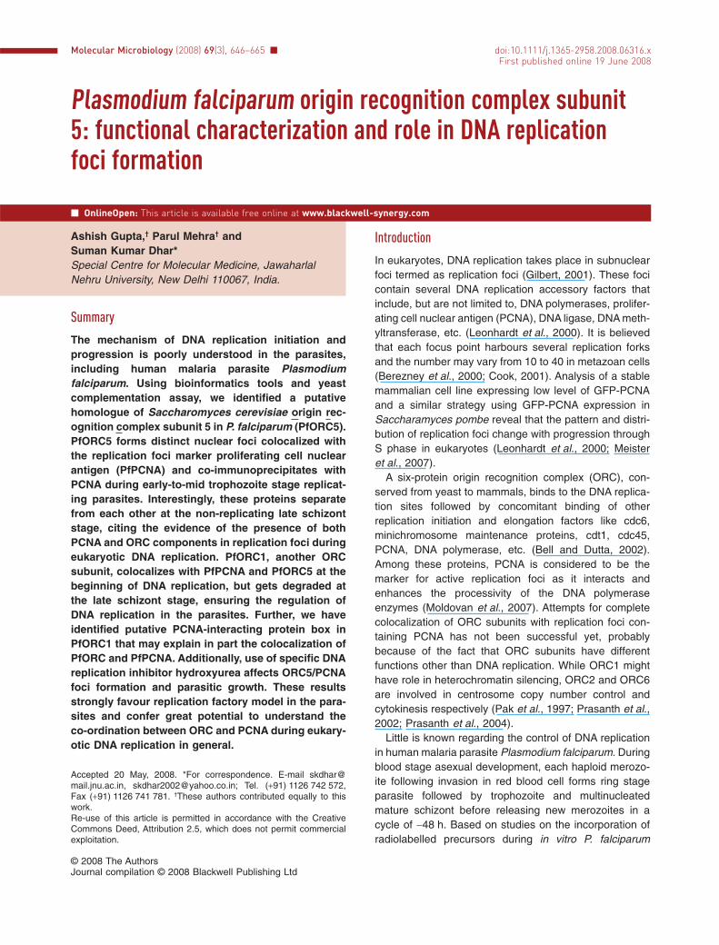
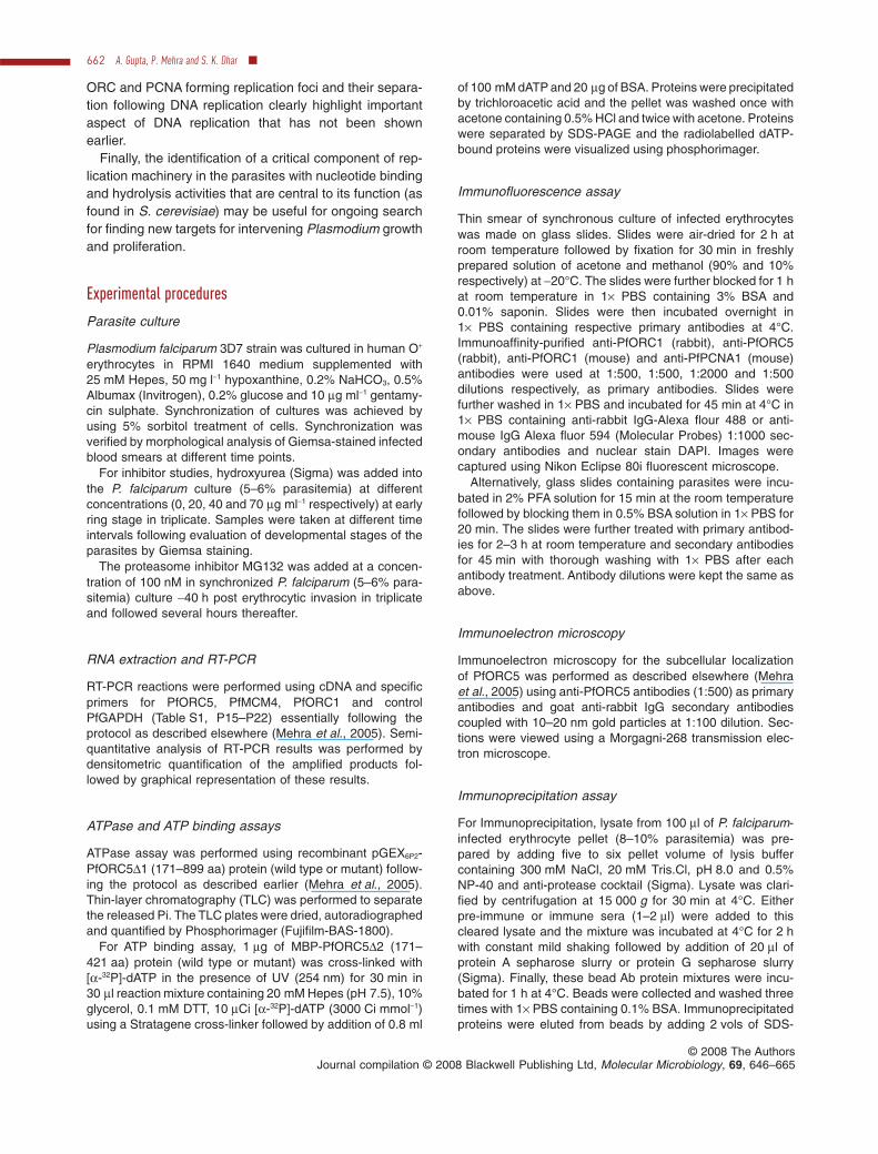
In order to investigate the expression pattern ofPfORC5 during asexual erythrocytic stages, semi-quantitative RT-PCR analysis was performed using cDNAisolated from synchronized ring, trophozoite and schizontstage parasites and specific primers (P15 and P16,Table S1, supplementary information) as shown in theschematic diagram of PfORC5 (Fig. S2B). Total RNA wastreated with DNase I before processing for cDNA prepa-ration either in the presence or absence of reversetranscriptase enzyme (+RT and -RT lanes respectively,Fig. 1A). Comparison of RT products from different eryth-rocytic developmental stages reveals that the expression
level of PfORC5 goes up several folds during trophozoiteand schizont stages in comparison with ring stagewhereas the control PfGAPDH expression pattern doesnot fluctuate to the similar extent during all three stages(Fig. 1B). PfORC1 and PfMCM4, the other two membersof pre-replication complex (pre-RC) are also expressedmostly in the trophozoite and schizont stage parasitessimilar to PfORC5 expression pattern (all the primers forRT-PCR analysis are listed in Table S1, P15–P22).RT-PCR analysis of PfORC5, PfORC1 and PfMCM4 sup-ports the published microarray data, confirming a peakof transcription coincident with onset of DNA replication(Bozdech et al., 2003).
To investigate the expression level of PfORC5 at theprotein level, two polyclonal antibodies were raised in therabbit and mouse, respectively, against C-terminal regionof PfORC5 (PfORC5C1 and PfORC5C2 respectively) asshown in Fig. S2B. Polyclonal antibodies raised in rabbitpredominantly recognized a band around molecular massof ~105 kDa following Western blot analysis of parasitelysate (Fig. 1C). Pre-immune sera under the same experi-mental conditions do not recognize any such band,suggesting that these antibodies are specific againstPfORC5. Polyclonal antibodies raised in mouse alsorecognized a similar band in Western blot experiments(Fig. S2C).
Further, to investigate the subcellular localization ofPfORC5 expression within the parasite, electron micro-scopic analysis of sections of trophozoite stage parasiteswere performed using either rabbit pre-immune sera orimmune sera raised against PfORC5. Analysis of goldparticles deposition within the parasite clearly shows thatPfORC5 is expressed primarily within the nucleus andnot in the cytoplasm or red blood cell (RBC) (Fig. 1D).Several nuclei were scanned and it was found that thegold particles were distributed all over the nuclei. Onaverage, approximately six to eight particles were found inthe trophozoite stage nuclei in each section. Pre-immunesera under the same experimental conditions do not showany such gold particle deposition, suggesting the speci-ficity of these antibodies to detect PfORC5 in the nucleus.
ATP binding, ATPase activity of PfORC5 and functionalcomplementation in S. cerevisiae
One of the hallmarks of ORC5 is its affinity to ATPbecause of the presence of Walker A NTP binding domain(Bell, 2002). Mutation in the Walker A domain of S. cer-evisiae ORC5 causes temperature-sensitive growth phe-notype, suggesting that ATP binding to ORC5 is importantfor chromosomal DNA replication (Takahashi et al., 2004).In vitro, recombinant ORC containing a mutation in theATP binding domain of HsORC5 shows reduced DNAbinding affinity (Giordano-Coltart et al., 2005). PfORC5
648 A. Gupta, P. Mehra and S. K. Dhar �
© 2008 The AuthorsJournal compilation © 2008 Blackwell Publishing Ltd, Molecular Microbiology, 69, 646–665
Protein
Purification
ATP-Binding
M (k
Da)
PfORC5Δ2W
t
PfORC5Δ2M
ut
66
5545
37
PfORC5Δ1 (171-899)
PfORC5Δ2 (171-421)
GSTGST-PfO
5Δ1
0 25 50 100 200 400 Protein (ng)
Released Pi
ATP
*
ORC5Δ1Wt
ORC5Δ1Mut
0
20
40
60
80
100120
% A
TP
ase
ac
tiv
ity
ORC5Δ1Wt ORC5Δ1Mut
A
F
G
H
Repeat I Repeat IIPfORC5 (1-899)
NTP
12097
66
55
Pre-Im
mune
Imm
une
M (k
Da) Pre-Immune Immune
D
E
RingTrophSchizont
Fo
ld C
han
ge
PfGAPDH
PfORC1
PfORC5
PfMCM
4
PfORC1
PfORC5
PfMCM4
PfGAPDH
RT + - + -
RING TROPH SCHIZONT
0
2
4
6
8B
C
+ -
Fig. 1. Expression of PfORC5 at the transcript and protein level and ATP binding/hydrolysis activity of PfORC5.A. RT-PCR analysis of PfORC1, PfORC5, PfMCM4 and control GAPDH using cDNA obtained from different developmental stages. RT (+) andRT (-) lanes indicate the PCR products obtained from cDNA prepared either in the presence or absence of reverse transcriptase enzymerespectively.B. Fold changes in expression of different genes at different stages. The relative intensity of each band was calculated using densitometryscanning and the absolute values were calculated by normalizing against background intensity. Each experiment was repeated three timesand the values obtained including the standard deviations were plotted graphically.C. Western blot analysis to analyse the expression of PfORC5 at the protein level using either anti-PfORC5 or pre-immune sera. Molecularmass markers are shown on the right.D. Immunoelectron-microscopic localization of PfORC5 within paraffin-embedded parasite sections using either pre-immune or anti-PfORC5antibodies respectively. Arrows indicate gold particle depositions and ‘N’ stands for nucleus.E. Schematic diagram of wild type or different deletion mutants of PfORC5 or full-length S. cerevisiae ORC5. Amino acid co-ordinates wereshown on the right.F. ATP binding activity of PfORC5. One microgram of the MBP-fused PfORC5D2Wt or PfORC5D2mut proteins were incubated with[a-32P]-dATP and was further cross-linked using UV light as described in Experimental procedures. Upper panel shows the SDS-PAGEanalysis of these proteins. The molecular mass markers are shown on the left. The bottom panel shows the autoradiogram of the above gel.G. ATPase activity of GST-PfORC5D1. ATPase assays were performed either using GST-PfORC5D1 or GST as a control. The positions of theradiolaelled ATP and the released Pi are marked. The right panel shows the ATPase activity of GST-PfORC5D1 with increasing amount ofprotein.H. Comparison of the ATPase activity of wild type and mutant proteins. ATPase assays were performed using 100 ng of GST-PfORC5D1Wt orGST-PfORC5D1Mut and phosphate release was quantified and plotted graphically with standard deviation after repeating the experiments forthree times. The inset shows the purification of the wild type and mutant forms of the proteins.
Replication foci in P. falciparum 649
© 2008 The AuthorsJournal compilation © 2008 Blackwell Publishing Ltd, Molecular Microbiology, 69, 646–665
contains a putative Walker A domain at the N terminus ofthe protein (306GMGKT310) flanked by repeat regions I andII (Fig. S1E).
To investigate the ATP binding and ATPase activity ofPfORC5, we have purified different domains of PfORC5(Fig. 1E). Unfortunately, we failed to express full-lengthPfORC5 because of the presence of asparagines andaspartic acid repeat regions at the extreme N-terminalregion. However, we have been able to expressPfORC5D1 (excluding the N-terminal repeat regions)and PfORC5D2 (containing the NTP-binding domain)(Fig. 1E). First, we investigated the ATP binding propertyof PfORC5 using recombinant PfORC5D2 because of itsbetter yield over PfORC5D1. We also made a mutant formof the protein by introducing a point mutation at the WalkerA domain (K to A). Equal amount of wild type and mutatedform of the proteins were incubated with [a-32P] dATP andsubjected to UV cross-linking analysis. Following autora-diography, wild-type PfORC5D2 was found to be in ATP-bound form whereas the mutated protein did not bind toATP under the same experimental conditions (Fig. 1F).
Although ORC1 has been reported to contain ATPaseactivity, no such activity has been attributed to ORC5 yet(Mehra et al., 2005). This is due to the absence of a clearWALKER B domain designated as nucleotide hydrolysisdomain in ORC5 homologues from yeast to mammals.However, there is always a possibility of a crypticWALKER B domain present in these proteins. In order tofind out whether PfORC5 contains ATPase activity,release of inorganic phosphate (Pi) from [g-32P] ATP wasmonitored in the presence of GST-PfORC5D1 or GST asa control. The recombinant protein showed ATPase activ-ity in a concentration-dependent manner whereas thecontrol GST protein did not show any such activity underthe same experimental conditions (Fig. 1G). A mutantform of PfORC5D1, containing a point mutation in theATP binding domain (K to A within the ‘GMGKT’ NTPbinding motif) showed drastically reduced ATPase activitywhen compared with that of wild-type protein (Fig. 1H).The residual activity in PfORC5D1Mut could be due tothe presence of some minor ATPase contaminant in theprotein preparation. Identification of WALKER B NTPhydrolysis domain in PfORC5 will be helpful to ascertainthe ATPase activity of PfORC5.
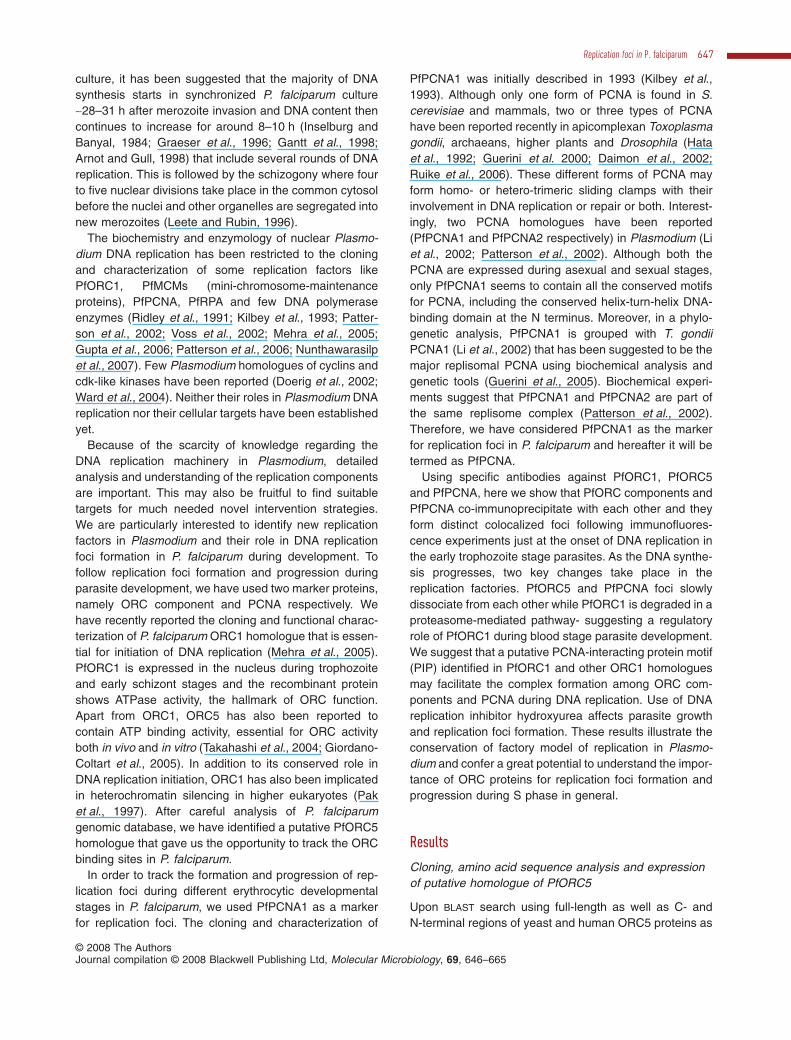
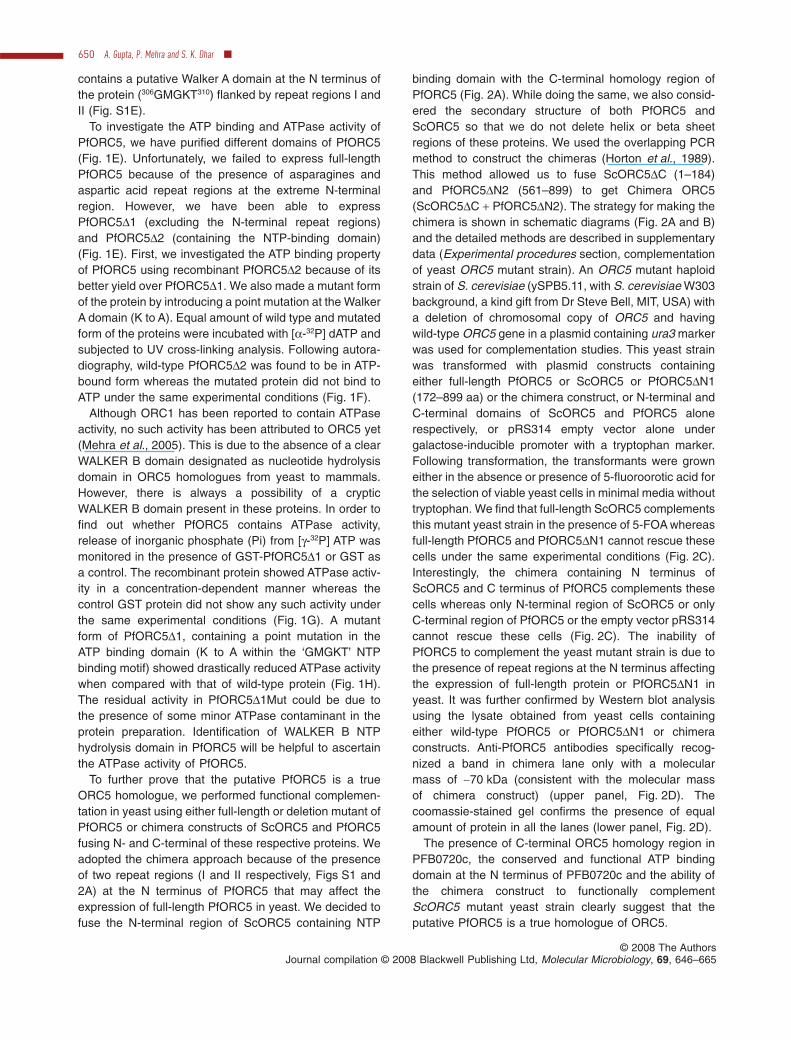
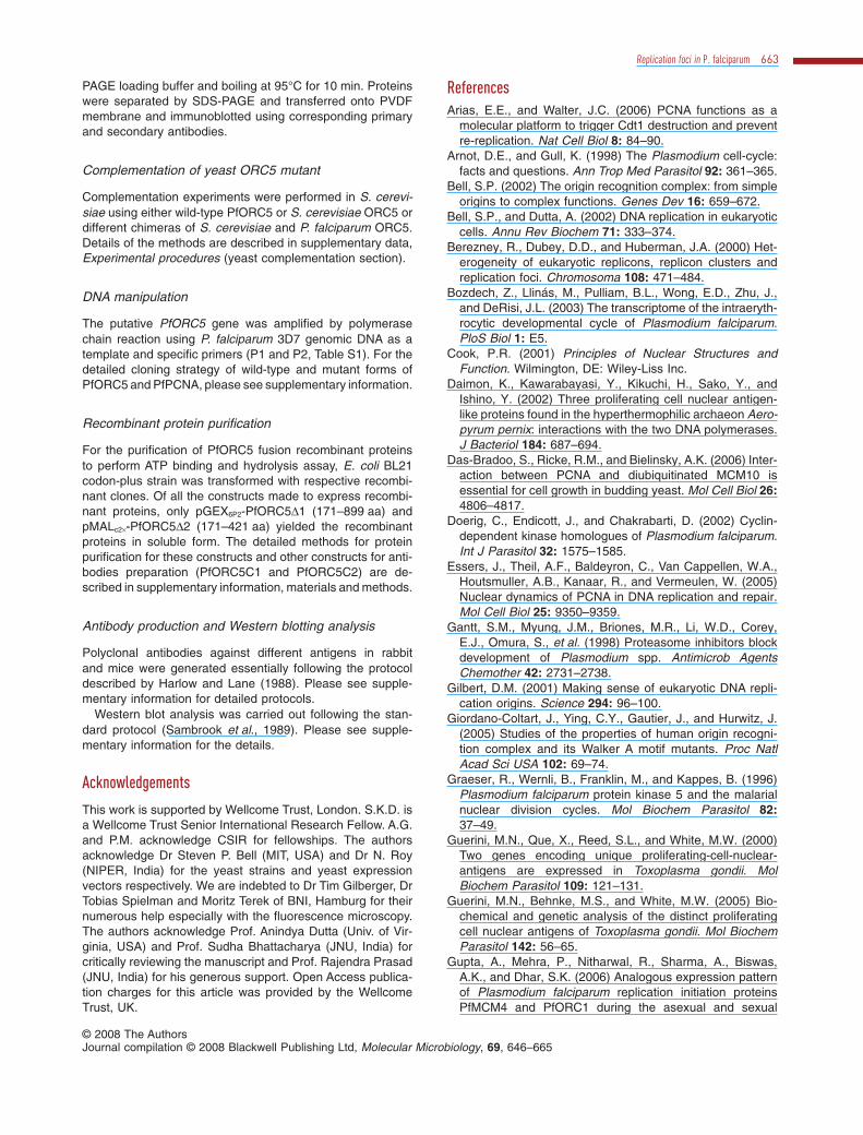
To further prove that the putative PfORC5 is a trueORC5 homologue, we performed functional complemen-tation in yeast using either full-length or deletion mutant ofPfORC5 or chimera constructs of ScORC5 and PfORC5fusing N- and C-terminal of these respective proteins. Weadopted the chimera approach because of the presenceof two repeat regions (I and II respectively, Figs S1 and2A) at the N terminus of PfORC5 that may affect theexpression of full-length PfORC5 in yeast. We decided tofuse the N-terminal region of ScORC5 containing NTP
binding domain with the C-terminal homology region ofPfORC5 (Fig. 2A). While doing the same, we also consid-ered the secondary structure of both PfORC5 andScORC5 so that we do not delete helix or beta sheetregions of these proteins. We used the overlapping PCRmethod to construct the chimeras (Horton et al., 1989).This method allowed us to fuse ScORC5DC (1–184)and PfORC5DN2 (561–899) to get Chimera ORC5(ScORC5DC + PfORC5DN2). The strategy for making thechimera is shown in schematic diagrams (Fig. 2A and B)and the detailed methods are described in supplementarydata (Experimental procedures section, complementationof yeast ORC5 mutant strain). An ORC5 mutant haploidstrain of S. cerevisiae (ySPB5.11, with S. cerevisiae W303background, a kind gift from Dr Steve Bell, MIT, USA) witha deletion of chromosomal copy of ORC5 and havingwild-type ORC5 gene in a plasmid containing ura3 markerwas used for complementation studies. This yeast strainwas transformed with plasmid constructs containingeither full-length PfORC5 or ScORC5 or PfORC5DN1(172–899 aa) or the chimera construct, or N-terminal andC-terminal domains of ScORC5 and PfORC5 alonerespectively, or pRS314 empty vector alone undergalactose-inducible promoter with a tryptophan marker.Following transformation, the transformants were growneither in the absence or presence of 5-fluoroorotic acid forthe selection of viable yeast cells in minimal media withouttryptophan. We find that full-length ScORC5 complementsthis mutant yeast strain in the presence of 5-FOA whereasfull-length PfORC5 and PfORC5DN1 cannot rescue thesecells under the same experimental conditions (Fig. 2C).Interestingly, the chimera containing N terminus ofScORC5 and C terminus of PfORC5 complements thesecells whereas only N-terminal region of ScORC5 or onlyC-terminal region of PfORC5 or the empty vector pRS314cannot rescue these cells (Fig. 2C). The inability ofPfORC5 to complement the yeast mutant strain is due tothe presence of repeat regions at the N terminus affectingthe expression of full-length protein or PfORC5DN1 inyeast. It was further confirmed by Western blot analysisusing the lysate obtained from yeast cells containingeither wild-type PfORC5 or PfORC5DN1 or chimeraconstructs. Anti-PfORC5 antibodies specifically recog-nized a band in chimera lane only with a molecularmass of ~70 kDa (consistent with the molecular massof chimera construct) (upper panel, Fig. 2D). Thecoomassie-stained gel confirms the presence of equalamount of protein in all the lanes (lower panel, Fig. 2D).
The presence of C-terminal ORC5 homology region inPFB0720c, the conserved and functional ATP bindingdomain at the N terminus of PFB0720c and the ability ofthe chimera construct to functionally complementScORC5 mutant yeast strain clearly suggest that theputative PfORC5 is a true homologue of ORC5.
650 A. Gupta, P. Mehra and S. K. Dhar �
© 2008 The AuthorsJournal compilation © 2008 Blackwell Publishing Ltd, Molecular Microbiology, 69, 646–665
97
66
56
43
Chimera
(Sc+Pf)
PfORC5W
t
PfORC5ΔN1
M (k
Da)
Anti-PfORC5
Western Blot
Coomassie
*
Repeat I Repeat II
C terminal ORC5 homology
domain
ScORC5 I
(1-127)
ScORC5 II
(128-479)
PfORC5 I (1-483) PfORC5 II (484-899)
PfORC5 (1-899)
ScORC5 (1-479)
ScORC5 (1-479)
PfORC5Wt (1-899)
PfORC5ΔN1 (172-899)PfORC5ΔN2 (561-899)
ScORC5ΔC (1-184)
Chimera (ScORC5ΔC + PfORC5ΔN2)
NTP
pRS314 Control
-trp-FOA -trp+FOA
A
B
C
D
27
43
56
PfORC5ΔN1 (172-899)
PfORC5ΔN2 (561-899)
ScORC5ΔC (1-184)
ScORC5 (1-479)
PfORC5Wt (1-899)
Chimera (ScORC5ΔC + PfORC5ΔN2)
NTP
Fig. 2. Complementation of PfORC5 in S. cerevisiae.A. Schematic diagrams of PfORC5 and ScORC5. The repeat regions (I and II) and NTP binding domains are shown as open boxes whereasthe filled box shows the C-terminal ScORC5 homology region. The grey box in ScORC5 shows the N-terminal region containing NTP bindingdomain. Both PfORC5 and ScORC5 can be divided into two parts (I and II) respectively.B. The schematic diagrams show different regions of PfORC5 and ScORC5 or the chimera containing N terminus of ScORC5 and C terminusof PfORC5 as used for complementation assay. The rationale for chimera construction has been described in Results section andsupplementary method section.C. A swapper strain of S. cerevisiae ORC5 with the deletion of chromosomal copy of ScORC5 gene and maintaining the same gene with aplasmid containing ura marker was transformed with either wild-type or different chimeras of PfORC5 and ScORC5 (with trp selection) eitherin the presence or absence of 5-fluoroorotic acid. Growth of these transformants was followed by spot test using serial dilutions.D. Western blot analysis to show the expression of different constructs as used in the complementation assay. Anti-PfORC5 polyclonalantibodies were used for Western blot analysis. The coomassie-stained gel following protein transfer on PVDF membrane is shown as loadingcontrol. * indicates the expression of chimera protein in yeast. The molecular mass marker is shown on the right.
Replication foci in P. falciparum 651
© 2008 The AuthorsJournal compilation © 2008 Blackwell Publishing Ltd, Molecular Microbiology, 69, 646–665
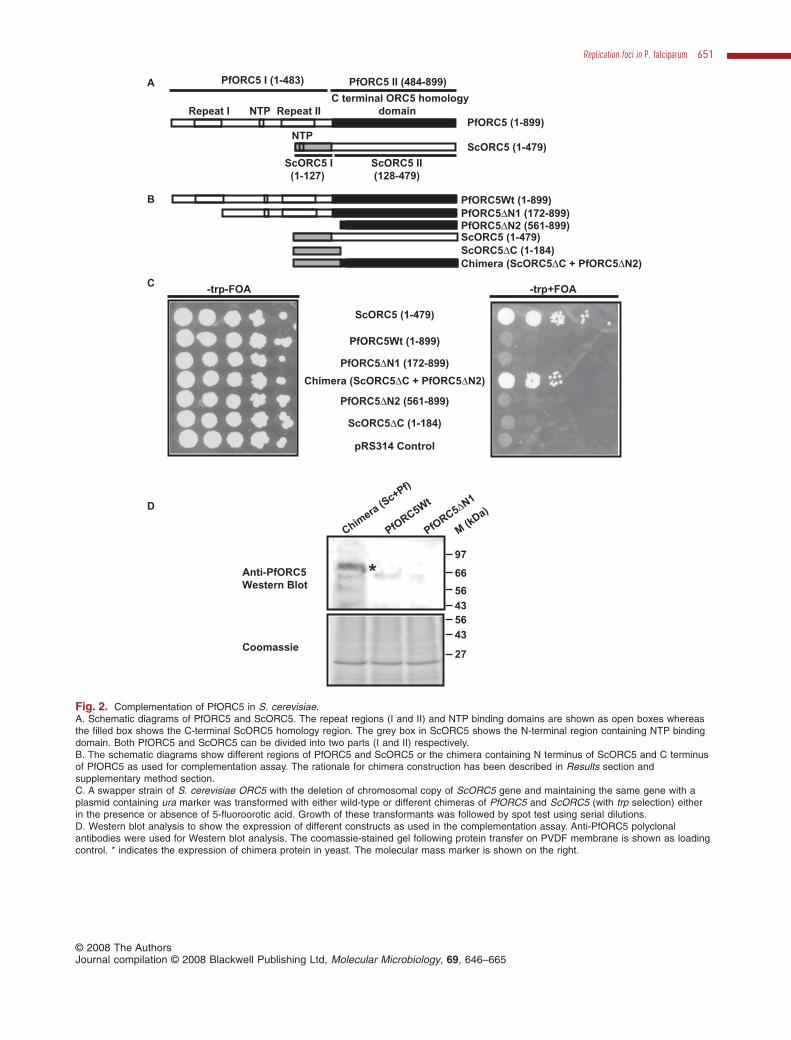
PfORC5 forms distinct foci within nuclei duringintraerythrocytic developmental stages
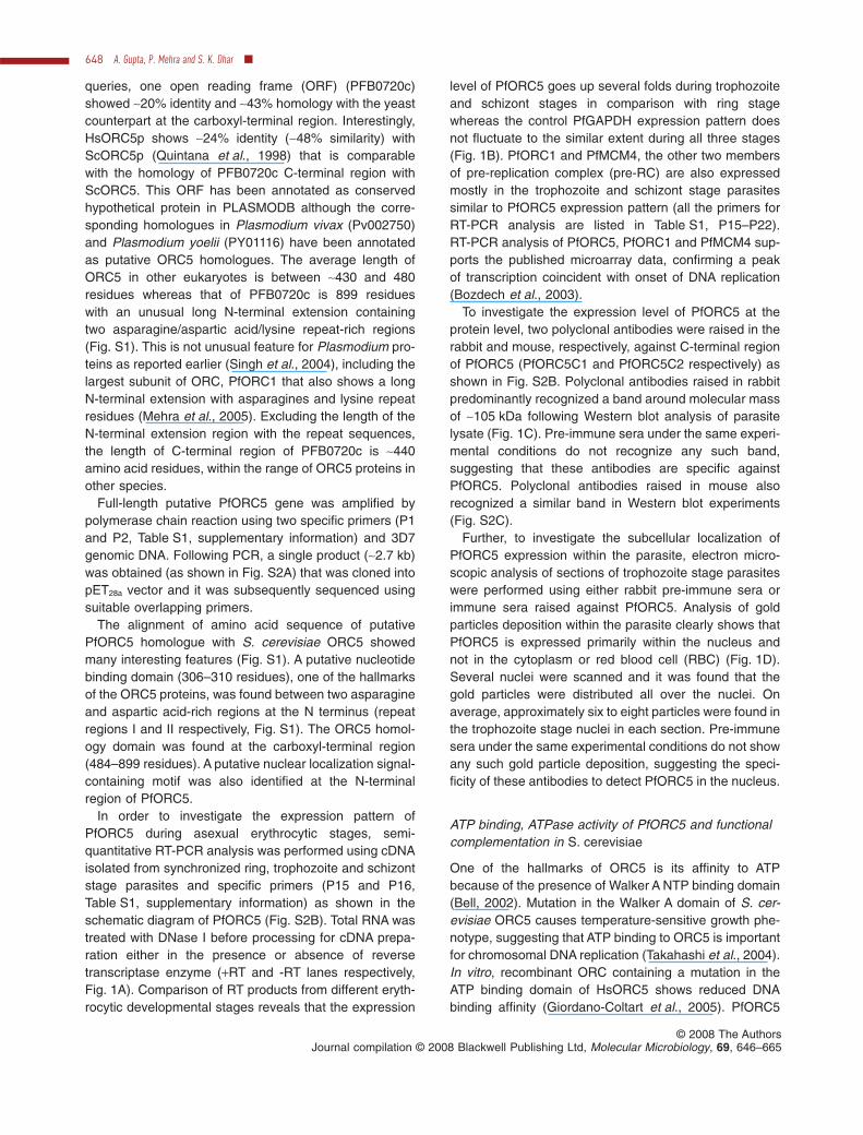
In an effort to investigate subcellular localization and thepattern of PfORC5 expression during intraerythrocyticdevelopmental stages, we performed thorough immunof-luorescence analysis at different time points in a synchro-nized parasite population using immuno-affinity-purifiedanti-PfORC5 antibodies. We also used anti-PfPCNA anti-bodies to locate the active replication foci in these cells asPCNA has been widely used as a marker for growingreplication forks (Leonhardt et al., 2000; Meister et al.,2007).
Analysis of immunofluorescence images at different timepoints revealed the presence of PfORC5 at the ring andearly trophozoite phase parasites with a diffused stainingpattern. Interestingly, PfORC5 forms distinct foci withinnuclei as the parasites mature further (mid-trophozoitephase parasites) (Fig. 3A). The size and the number ofsuch foci increase as the parasites undergo further matu-ration until late schizont stage where the average size ofsuch foci decreases compared with those found in mid-trophozoite and early schizont stages. When the sameparasites were screened for PCNA(replication foci marker)signals, it showed somewhat diffused pattern during earlytrophozoite stage like PfORC5.As the parasites developedfurther into mid-trophozoite stages, PfPCNA showed dis-tinct foci as PfORC5 and these dots mostly merged withPfORC5 dots as seen from the merged panels (Fig. 3A,panel 3). The number and average size of the PCNA focialso increased till mid-schizont stages and both theseparameters decreased at the late schizont stages.Although the PfORC5 and PfPCNA foci merged mostlywith each other at these stages, these foci slowly dissoci-ated from each other as the parasites developed furtherand the majority of these spots separated from each otherat the late schizont stage (Fig. 3A, panel 7). Please seeenlarged and high-resolution Fig. S3A for the expressionpattern of these proteins at late schizont stage. In a controlset of experiments, none of the pre-immune sera show anyimmunofluorescence signal under the same experimentalconditions, confirming the specificity of these antibodies(Fig. 3B).
It will be interesting to see whether the replication fociformation and progression are correlated with the extentof DNA replication through intra-erythrocytic developmentcycle. Additionally, the immunofluorescence studies pre-sented here are based on the slides containing parasitesmears that have been fixed using acetone-methanol fixa-tion method (please see the Experimental proceduressection for details). The slides were washed very exten-sively during immunofluorescence assay (IFA) to avoidany background staining. Although this method givesus distinct pattern of replication foci formation,the DAPI (9,6-diaminido-2-phenylindole) shows diffusedstaining pattern. In order to avoid any bias that may arisebecause of a particular method of fixation, we repeatedthe IFA using synchronized parasites that were fixed usingparaformaldehyde (PFA). For this purpose, we synchro-nized the 3D7 parasites at the ring stage and the samewas divided into four aliquots and they were allowed togrow further. The growth of the parasites was monitoredtime to time. The parasites were harvested at ring, tropho-zoite, early schizont and late schizont stages. The glassslides were made to confirm their stages by giemsa stain-ing (Fig. S4A) and they were fixed with PFA and pre-served for further IFA analysis. Total genomic DNA wasisolated in each case and they were analysed by agarosegel electrophoresis (Fig. S4B). The total DNA was furtherquantified spectrophotometrically and plotted accordingly(Fig. S4C). We find that as the parasites grow, the totalDNA content increases many folds from ring to schizontstages, reflecting a huge amount of concomitant DNAreplication with growth progression.
Further analysis of the parasites obtained from thesame stages as described above following IFA experi-ments using anti-PfORC5 and anti-PfPCNA antibodiessuggests a similar pattern of replication foci formationand progression during development. Both PfPCNA andPfORC5 formed colocalized distinct foci during early-to-mid replicating trophozoite stages. These foci starteddissociating from each other with further growth progres-sion and finally they separated completely during lateschizont stage as shown earlier (Fig. S5). The averagenumber of PfPCNA or PfORC5 foci or the colocalizedfoci and their pattern were found to be similar as
Fig. 3. Colocalization and interaction between PfORC5 and PfPCNA.A. Immunofluorescence assay to show colocalization of PfORC5 and PfPCNA as replication foci marker. Glass slides containing thin smearsof P. falciparum-infected erythrocytes from the different erythrocytic stages were incubated with affinity-purified rabbit anti-PfORC5 and mouseanti-PfPCNA antibodies followed by respective secondary antibodies. Merged 1 panel shows the merged images of nuclear DAPI, greenPfORC5 staining and red PfPCNA staining whereas merged 2 panel shows the merged images of green PfORC5 and red PfPCNA signalsonly. The bar as shown in the inset is equivalent to 3 mm.B. Immunofluorescence assay under the same experimental conditions as shown in A using pre-immune sera against respective antibodies.C. Co-immunoprecipitation of PfORC5 and PfPCNA from parasite extract obtained from synchronized parasite pellet collected from eithertrophozoite or late schizont stage parasites. The extracts were first immunoprecipitated (IP) with anti-PfORC5 antibodies followed byimmunoblotting (IB) with either anti-PfORC5 (top panel) or anti-PfPCNA (bottom panel) antibodies. The input lanes show the presence ofthese proteins in both the stages. Arrowhead indicates the position of the respective proteins. Molecular mass markers are shown on the right.
652 A. Gupta, P. Mehra and S. K. Dhar �
© 2008 The AuthorsJournal compilation © 2008 Blackwell Publishing Ltd, Molecular Microbiology, 69, 646–665
DAPI PfORC5 PCNA MERGED 1 MERGED 2
RING
TROPH.
SCHIZONT
DAPIALEXA
MERGED
PI-Sera
PfORC5
PfPCNA
PfORC1
A
B C
Imm
unoppt.
Imm
unoppt.
Hours Post-
infection
~12-14
~20-22
~26-30
~32-34
~34-36
~40-42
~46-48
116
97
37
66
27
56
Input
Input
M (k
Da)
IP: Anti-PfORC5
IB: Anti-PfORC5
IP: Anti-PfORC5
IB: Anti-PfPCNA *
Trophozoite Schizont
PfORC5
PfPCNA**
* and ** indicate heavy chain and light chain
of the immunoglobulins
Replication foci in P. falciparum 653
© 2008 The AuthorsJournal compilation © 2008 Blackwell Publishing Ltd, Molecular Microbiology, 69, 646–665
compared with those shown in Fig. 3 by methanol-acetone fixation method.
Although ORC components colocalize with PCNAduring S phase to a limited extent in mammalian cells(Prasanth et al., 2004), no biochemical evidence is avail-able yet to prove that these proteins are truly the compo-nents of replication foci.
To verify whether ORC components and PCNA are trulythe members of replication factories, we performed immu-noprecipitation reactions using parasite extracts and anti-bodies against PfORC5 and PfPCNA. Both PfPCNA andPfORC5 can be immunoprecipitated from mixed tropho-zoite stage parasite extract using immune sera but notwith pre-immune sera (Fig. S3B). To find out whetherthese proteins co-immunoprecipitate with each other, wefirst immnuprecipitated PfORC5 from the parasite extractderived from either mixed stage trophozoites or lateschizont stage parasites followed by immunoblottingusing anti-PfORC5 or anti-PfPCNA antibodies respec-tively (Fig. 3C, top and bottom panels). We found thatPfORC5 was immunoprecipitated from both trophozoiteand schizont stages (Fig. 3C, top panel). Interestingly, weobserved that PfPCNA was co-immunoprecipitated withPfORC5 during trophozoite stage only but not at the lateschizont stage. PfPCNA was expressed at both thestages to the similar extent as shown by the input lanes(Fig. 3C, bottom panel). These results confirm the colo-calization of PfORC5 and PfPCNA during trophozoitestages but not at the schizont stages.
PfORC1, another member of the ORC colocalizeswith PfORC5 and PfPCNA but gets degraded atthe late schizont stage
Although ORC functions collectively as a replication ini-tiator protein, different components of ORC are regulateddifferentially in a cell cycle manner. The level of ORC2–5does not change through cell cycle whereas ORC1 comeson and off the chromatin in a cell cycle-regulated mannerin mammalian cells (Méndez et al., 2002). In hamstercells, ORC1 is dissociated from chromatin as cells enter Sphase, transformed into a mono- or diubiquitinated form,followed by deubiquitination and re-binding to chromatinduring the M-to-G1 transition (Li and DePamphilis,
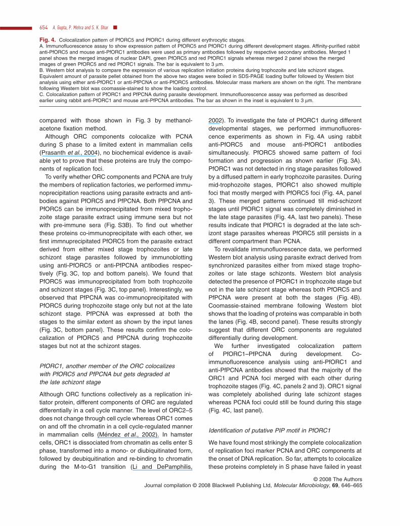
2002). To investigate the fate of PfORC1 during differentdevelopmental stages, we performed immunofluores-cence experiments as shown in Fig. 4A using rabbitanti-PfORC5 and mouse anti-PfORC1 antibodiessimultaneously. PfORC5 showed same pattern of fociformation and progression as shown earlier (Fig. 3A).PfORC1 was not detected in ring stage parasites followedby a diffused pattern in early trophozoite parasites. Duringmid-trophozoite stages, PfORC1 also showed multiplefoci that mostly merged with PfORC5 foci (Fig. 4A, panel3). These merged patterns continued till mid-schizontstages until PfORC1 signal was completely diminished inthe late stage parasites (Fig. 4A, last two panels). Theseresults indicate that PfORC1 is degraded at the late sch-izont stage parasites whereas PfORC5 still persists in adifferent compartment than PCNA.
To revalidate immunofluorescence data, we performedWestern blot analysis using parasite extract derived fromsynchronized parasites either from mixed stage tropho-zoites or late stage schizonts. Western blot analysisdetected the presence of PfORC1 in trophozoite stage butnot in the late schizont stage whereas both PfORC5 andPfPCNA were present at both the stages (Fig. 4B).Coomassie-stained membrane following Western blotshows that the loading of proteins was comparable in boththe lanes (Fig. 4B, second panel). These results stronglysuggest that different ORC components are regulateddifferentially during development.
We further investigated colocalization patternof PfORC1–PfPCNA during development. Co-immunofluorescence analysis using anti-PfORC1 andanti-PfPCNA antibodies showed that the majority of theORC1 and PCNA foci merged with each other duringtrophozoite stages (Fig. 4C, panels 2 and 3). ORC1 signalwas completely abolished during late schizont stageswhereas PCNA foci could still be found during this stage(Fig. 4C, last panel).
Identification of putative PIP motif in PfORC1
We have found most strikingly the complete colocalizationof replication foci marker PCNA and ORC components atthe onset of DNA replication. So far, attempts to colocalizethese proteins completely in S phase have failed in yeast
Fig. 4. Colocalization pattern of PfORC5 and PfORC1 during different erythrocytic stages.A. Immunofluorescence assay to show expression pattern of PfORC5 and PfORC1 during different development stages. Affinity-purified rabbitanti-PfORC5 and mouse anti-PfORC1 antibodies were used as primary antibodies followed by respective secondary antibodies. Merged 1panel shows the merged images of nuclear DAPI, green PfORC5 and red PfORC1 signals whereas merged 2 panel shows the mergedimages of green PfORC5 and red PfORC1 signals. The bar is equivalent to 3 mm.B. Western blot analysis to compare the expression of various replication initiation proteins during trophozoite and late schizont stages.Equivalent amount of parasite pellet obtained from the above two stages were boiled in SDS-PAGE loading buffer followed by Western blotanalysis using either anti-PfORC1 or anti-PfPCNA or anti-PfORC5 antibodies. Molecular mass markers are shown on the right. The membranefollowing Western blot was coomassie-stained to show the loading control.C. Colocalization pattern of PfORC1 and PfPCNA during parasite development. Immunofluorescence assay was performed as describedearlier using rabbit anti-PfORC1 and mouse anti-PfPCNA antibodies. The bar as shown in the inset is equivalent to 3 mm.
654 A. Gupta, P. Mehra and S. K. Dhar �
© 2008 The AuthorsJournal compilation © 2008 Blackwell Publishing Ltd, Molecular Microbiology, 69, 646–665
DAPI PfORC5 PfORC1 MERGED 1 MERGED 2R
ING
TR
OP
H.
SC
HIZ
ON
TA
Anti-
PfORC1
Anti-
PfORC5
Anti-
PfPCNA
Coomassie
Trophozo
ite
Schizont (
late
)
M (k
Da)
158
116
97
66
227
B
DAPI PfORC1 PCNA MERGED 1 MERGED 2C
RIN
GT
RO
PH
.S
CH
IZO
NT
Replication foci in P. falciparum 655
© 2008 The AuthorsJournal compilation © 2008 Blackwell Publishing Ltd, Molecular Microbiology, 69, 646–665
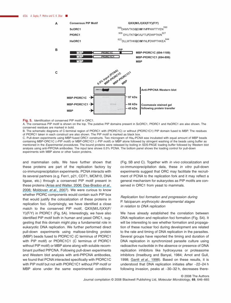
and mammalian cells. We have further shown thatthese proteins are part of the replication factory byco-immunoprecipitation experiments. PCNA interacts withits several partners (e.g. Fen1, p21, CDT1, MCM10, DNAligase, etc.) through a conserved PIP motif present inthese proteins (Arias and Walter, 2006; Das-Bradoo et al.,2006; Moldovan et al., 2007). We were curious to knowwhether PfORC components would contain such PIP boxthat would justify the colocalization of these proteins inreplication foci. Surprisingly, we have identified a closematch to the conserved PIP motif, QXX(M/L/I)XX(F/Y)(F/Y) in PfORC1 (Fig. 5A). Interestingly, we have alsoidentified PIP motif both in human and yeast ORC1, sug-gesting that this domain might play a fundamental role ineukaryotic DNA replication. We further performed directpull-down experiments using maltose-binding protein(MBP) beads fused to PfORC1C (C terminus of PfORC1with PIP motif) or PfORC1C1 (C terminus of PfORC1without PIP motif) or MBP alone along with soluble recom-binant purified PfPCNA. Following pull-down experimentsand Western blot analysis with anti-PfPCNA antibodies,we found that PCNA interacted specifically with PfORC1Cwith PIP motif but not with PfORC1C1 without PIP motif orMBP alone under the same experimental conditions
(Fig. 5B and C). Together with in vivo colocalization andco-immunoprecipitation data, these in vitro pull-downexperiments suggest that ORC may facilitate the recruit-ment of PCNA to the replication fork and it may reflect ageneral mechanism for eukaryotes as PIP motifs are con-served in ORC1 from yeast to mammals.
Replication foci formation and progression duringP. falciparum erythrocytic developmental stagesin relation to DNA replication
We have already established the correlation betweenDNA replication and replication foci formation (Fig. S4). Itwill be interesting to see whether formation and propaga-tion of these nuclear foci during development are relatedto the rate and timing of DNA replication in the parasites.Several groups have reported the timing and duration ofDNA replication in synchronized parasite culture usingradioactive nucleotide in the absence or presence of DNAreplication inhibitors like hydroxyurea or proteasomeinhibitors (Inselburg and Banyal, 1984; Arnot and Gull,1998; Gantt et al., 1998). Based on these results, it isunderstood that DNA replication initiates after ~22–24 hfollowing invasion, peaks at ~30–32 h, decreases there-
MBP-PfORC1C
MBP-PfORC1C1
MBP
MBP-P
fORC1C
MBP-P
fORC1C1
MBP
0.5%
Input (
PCNA)
97 kDa
66 kDa
45 kDa
Anti-PfPCNA Western blot
Coomassie stained gel
following protein transfer
QXX(M/L/I)XX(F/Y)(F/Y)Consensus PIP Motif
PfORC1 DYLITKTQKVLFTLFDWPTKIN906 927
ScORC1 DAMVTKSQDIMYNFFNWTTYEN569 590
HsORC1 DLLWTHKQDIMYNLFDWPTHKEA622 644
MBP-PfORC1C (694-1189)
MBP-PfORC1C1 (694-899)
MBPMBPMBP
MBPMBP
MBPMBP PfORC1C
PfORC1C1
B
C
PIP
A
Fig. 5. Identification of conserved PIP motif in ORC1.A. The consensus PIP motif is shown on the top. The putative PIP domains present in ScORC1, PfORC1 and HsORC1 are also shown. Theconserved residues are marked in bold.B. The schematic diagrams of C-terminal region of PfORC1 with (PfORC1C) or without (PfORC1C1) PIP domain fused to MBP. The residuesof PfORC1 taken in each construct are also shown. The PIP motif is marked as black box.C. Pull-down experiments using MBP-fused ORC1 constructs. Two microgram of His6-PCNA was incubated with equal amount of MBP beadscontaining MBP-ORC1C (+PIP motif) or MBP-ORC1C1 (-PIP motif) or MBP alone followed by stringent washing of the beads using buffer asmentioned in the Experimental procedures. The bound proteins were released by boiling in SDS-PAGE loading buffer followed by Western blotanalysis using anti-PfPCNA antibodies. The input lane shows 0.5% PCNA. The bottom panel shows the loading control for pull-downexperiments with MBP alone or other fusion proteins.
656 A. Gupta, P. Mehra and S. K. Dhar �
© 2008 The AuthorsJournal compilation © 2008 Blackwell Publishing Ltd, Molecular Microbiology, 69, 646–665
after although continuing till 40–44 h. The DNA replicationpattern is shown in Fig. S6A based on the results obtainedfrom various studies.
In order to find out whether replication pattern coincideswith foci formation pattern, we calculated the number offoci formed at each stage during development for ORC1,ORC5 and PCNA. Number of colocalized foci for ORC5–PCNA and ORC1–ORC5 were also counted. We usedacetone-methanol fixation method as it gave us moredistinct and slightly higher number of replication foci prob-ably because of the better access of the antibodies to theproteins. Several parasites (more than 100 in each case)from multiple slides were scanned at each developmentalstage to rule out the possibility of any bias for the stage ofthe parasites and number of foci found in each stage.Graphical representation of these results indicates thatoverall pattern of the foci formation strongly correlateswith the DNA replication pattern. Comparison of individualPCNA and ORC5 foci formation reveals that foci formationstarts during early-to-mid trophozoite stage for both ofthem (Fig. S6B). The number of foci further increaseswith further development, reaching the maximum numberduring mid-to-late trophozoite stages where DNA replica-tion also peaks. Number of ORC5 foci do not changedrastically thereafter whereas the number of PCNA focidrops at the late schizont stage. It is possible that theincrease in replication foci per parasite at later stages isdue to the increase in genome content or number of nucleifollowing each round of replication.
Analysis of PCNA–ORC5 merged foci clearly indicatesthat the formation and progression of such merged focialso follows the DNA replication pattern (Fig. S6C). Thenumber of merged foci peaks during mid-trophozoite stagewith a value of ~15 per parasite, suggesting the presence ofthese many active replication foci containing both PfPCNAand PfORC proteins during peak replication phase. Thenumbers decline drastically thereafter with very little asso-ciation with each other during late schizont stage. ORC1–ORC5 merged foci also follow the same pattern with themaximum number of merged foci visible during mid-trophozoite stages followed by reduction of these numbersat the following stages until late schizont stages whereORC1 foci were completely abolished (Fig. S6D).
It is important to note that the formation, progressionand colocalization of PfORC and PfPCNA foci stronglycomplement biochemical co-immunoprecipitation data toreconfirm our conclusions. The actual number of replica-tion foci per nucleus in the infected parasite may vary asthe replication foci number counting is based on the visualinspection of IFA analysis of the different fixed parasitesat different time points. Further live cell imaging usingtransgenic fluorescent parasite line expressing PfORC orPfPCNA fusion protein will be required for quantitativeanalysis of replication foci formation and progression.
Perturbation of parasite growth with the intervention ofinhibitors affects replication foci formation
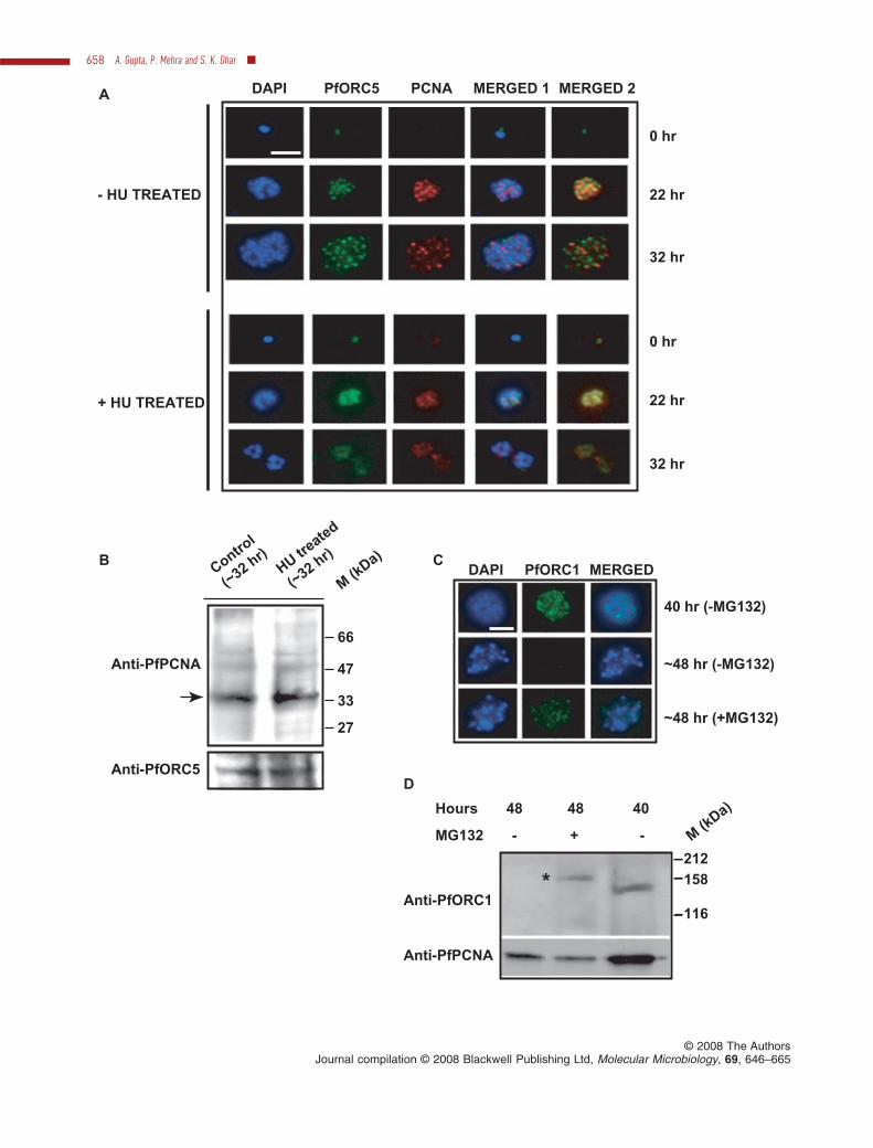
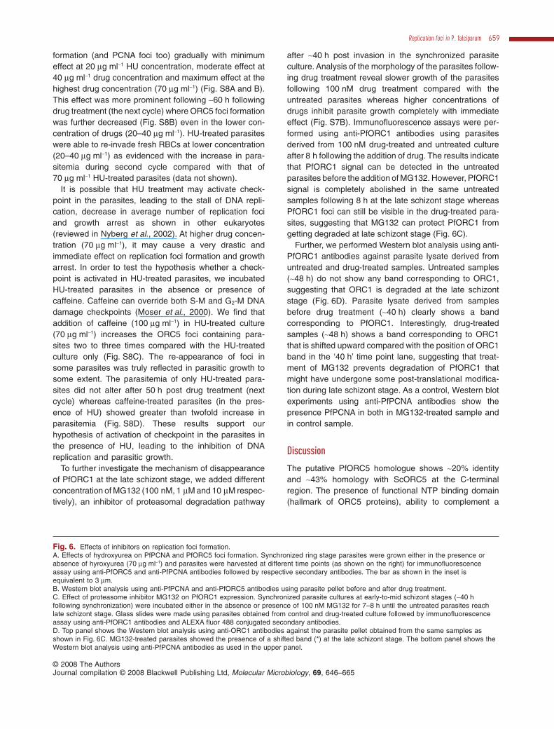
To investigate further whether PfORC and PfPCNA fociare true representation of replication foci, we used DNAreplication inhibitor hydroxyurea that blocks the elonga-tion of DNA replication fork by depleting the pool ofnucleotides (Thelander and Reichard, 1979). It has beenreported earlier that the addition of hydroxyurea(~60 mg ml-1 or more) in synchronized ring stage parasitesarrests them in early trophozoite stage corresponding tothe time of initiation of DNA synthesis (Inselburg andBanyal, 1984). Accordingly, P. falciparum 3D7 parasiteswere synchronized at the ring stage and parasites weregrown either in the absence or presence of hydroxyurea(~70 mg ml-1). Analysis of the morphology of the parasitesfollowing Giemsa staining after ~32 h of drug treatment(Fig. S7A) shows that they are blocked at the trophozoitestage as reported earlier (Inselburg and Banyal, 1984)whereas the untreated parasites grow normally. Immun-ofluorescence assay using anti-PfORC5 and anti-PfPCNA antibodies show normal progression of fociformation and propagation in the absence of drugs. Incu-bation of the parasites in the presence of drugs (~32 h,post treatment) results in reduced and fragmented nucleicompared with the control parasites as shown in DAPIstained parasites (Fig. 6A, panels 3 and 6 respectively).Interestingly, the majority of the parasites (~85–90%)showed diffused staining pattern for both ORC5 andPCNA following ~22 h and ~32 h of drug treatment com-pared with the distinct foci containing parasites present inthe untreated culture. Finally, Western blot analysis usinganti-PfPCNA and anti-PfORC5 antibodies against para-site lysate obtained from drug-treated and untreatedsamples revealed the presence of ORC5 and PCNA bothin the absence and presence of drug although they failedto form distinct foci in the majority of the parasites follow-ing hydroxyurea (HU) treatment at higher concentration(Fig. 6B).
The mode of action of HU has been reported to be thedepletion of the pool of dNTPs by inhibiting ribonucleotidereductase and it should not affect the loading of pre-RCcomponents like ORC. The absence of ORC5 foci forma-tion in the presence of ~70 mg ml-1 HU was a bit surprisingto us. Therefore, to address this issue thoroughly, we useddifferent concentration of HU (0, 20, 40 and 70 mg ml-1) andfollowed the formation of replication foci at different timepoints by immunofluorescence assay (Fig. S8A). Theaverage number of foci per RBC-infected parasite was alsocalculated (~100 RBC-infected parasites were counted ateach HU concentration at two different time points, 22 hand 60 h post treatment respectively) and plotted accord-ingly (Fig. S8B). We find that following ~22 h of drug treat-ment, increasing concentration of HU affects ORC5 foci
Replication foci in P. falciparum 657
© 2008 The AuthorsJournal compilation © 2008 Blackwell Publishing Ltd, Molecular Microbiology, 69, 646–665
DAPI PfORC5 PCNA MERGED 1 MERGED 2
0 hr
22 hr
32 hr
0 hr
22 hr
32 hr
- HU TREATED
+ HU TREATED
Control
(~32 h
r)HU tr
eated
(~32 h
r)
M (k
Da)
66
47
33
27
Anti-PfPCNA
Anti-PfORC5
40 hr (-MG132)
~48 hr (-MG132)
~48 hr (+MG132)
MERGEDDAPI PfORC1
MG132 - + -
Hours 48 48 40
Anti-PfORC1
A
B C
D
116
158
212
M (k
Da)
*
Anti-PfPCNA
658 A. Gupta, P. Mehra and S. K. Dhar �
© 2008 The AuthorsJournal compilation © 2008 Blackwell Publishing Ltd, Molecular Microbiology, 69, 646–665
formation (and PCNA foci too) gradually with minimumeffect at 20 mg ml-1 HU concentration, moderate effect at40 mg ml-1 drug concentration and maximum effect at thehighest drug concentration (70 mg ml-1) (Fig. S8A and B).This effect was more prominent following ~60 h followingdrug treatment (the next cycle) where ORC5 foci formationwas further decreased (Fig. S8B) even in the lower con-centration of drugs (20–40 mg ml-1). HU-treated parasiteswere able to re-invade fresh RBCs at lower concentration(20–40 mg ml-1) as evidenced with the increase in para-sitemia during second cycle compared with that of70 mg ml-1 HU-treated parasites (data not shown).
It is possible that HU treatment may activate check-point in the parasites, leading to the stall of DNA repli-cation, decrease in average number of replication fociand growth arrest as shown in other eukaryotes(reviewed in Nyberg et al., 2002). At higher drug concen-tration (70 mg ml-1), it may cause a very drastic andimmediate effect on replication foci formation and growtharrest. In order to test the hypothesis whether a check-point is activated in HU-treated parasites, we incubatedHU-treated parasites in the absence or presence ofcaffeine. Caffeine can override both S-M and G2-M DNAdamage checkpoints (Moser et al., 2000). We find thataddition of caffeine (100 mg ml-1) in HU-treated culture(70 mg ml-1) increases the ORC5 foci containing para-sites two to three times compared with the HU-treatedculture only (Fig. S8C). The re-appearance of foci insome parasites was truly reflected in parasitic growth tosome extent. The parasitemia of only HU-treated para-sites did not alter after 50 h post drug treatment (nextcycle) whereas caffeine-treated parasites (in the pres-ence of HU) showed greater than twofold increase inparasitemia (Fig. S8D). These results support ourhypothesis of activation of checkpoint in the parasites inthe presence of HU, leading to the inhibition of DNAreplication and parasitic growth.
To further investigate the mechanism of disappearanceof PfORC1 at the late schizont stage, we added differentconcentration of MG132 (100 nM, 1 mM and 10 mM respec-tively), an inhibitor of proteasomal degradation pathway
after ~40 h post invasion in the synchronized parasiteculture. Analysis of the morphology of the parasites follow-ing drug treatment reveal slower growth of the parasitesfollowing 100 nM drug treatment compared with theuntreated parasites whereas higher concentrations ofdrugs inhibit parasite growth completely with immediateeffect (Fig. S7B). Immunofluorescence assays were per-formed using anti-PfORC1 antibodies using parasitesderived from 100 nM drug-treated and untreated cultureafter 8 h following the addition of drug. The results indicatethat PfORC1 signal can be detected in the untreatedparasites before the addition of MG132. However, PfORC1signal is completely abolished in the same untreatedsamples following 8 h at the late schizont stage whereasPfORC1 foci can still be visible in the drug-treated para-sites, suggesting that MG132 can protect PfORC1 fromgetting degraded at late schizont stage (Fig. 6C).
Further, we performed Western blot analysis using anti-PfORC1 antibodies against parasite lysate derived fromuntreated and drug-treated samples. Untreated samples(~48 h) do not show any band corresponding to ORC1,suggesting that ORC1 is degraded at the late schizontstage (Fig. 6D). Parasite lysate derived from samplesbefore drug treatment (~40 h) clearly shows a bandcorresponding to PfORC1. Interestingly, drug-treatedsamples (~48 h) shows a band corresponding to ORC1that is shifted upward compared with the position of ORC1band in the ‘40 h’ time point lane, suggesting that treat-ment of MG132 prevents degradation of PfORC1 thatmight have undergone some post-translational modifica-tion during late schizont stage. As a control, Western blotexperiments using anti-PfPCNA antibodies show thepresence PfPCNA in both in MG132-treated sample andin control sample.
Discussion
The putative PfORC5 homologue shows ~20% identityand ~43% homology with ScORC5 at the C-terminalregion. The presence of functional NTP binding domain(hallmark of ORC5 proteins), ability to complement a
Fig. 6. Effects of inhibitors on replication foci formation.A. Effects of hydroxyurea on PfPCNA and PfORC5 foci formation. Synchronized ring stage parasites were grown either in the presence orabsence of hyroxyurea (70 mg ml-1) and parasites were harvested at different time points (as shown on the right) for immunofluorescenceassay using anti-PfORC5 and anti-PfPCNA antibodies followed by respective secondary antibodies. The bar as shown in the inset isequivalent to 3 mm.B. Western blot analysis using anti-PfPCNA and anti-PfORC5 antibodies using parasite pellet before and after drug treatment.C. Effect of proteasome inhibitor MG132 on PfORC1 expression. Synchronized parasite cultures at early-to-mid schizont stages (~40 hfollowing synchronization) were incubated either in the absence or presence of 100 nM MG132 for 7–8 h until the untreated parasites reachlate schizont stage. Glass slides were made using parasites obtained from control and drug-treated culture followed by immunofluorescenceassay using anti-PfORC1 antibodies and ALEXA fluor 488 conjugated secondary antibodies.D. Top panel shows the Western blot analysis using anti-ORC1 antibodies against the parasite pellet obtained from the same samples asshown in Fig. 6C. MG132-treated parasites showed the presence of a shifted band (*) at the late schizont stage. The bottom panel shows theWestern blot analysis using anti-PfPCNA antibodies as used in the upper panel.
Replication foci in P. falciparum 659
© 2008 The AuthorsJournal compilation © 2008 Blackwell Publishing Ltd, Molecular Microbiology, 69, 646–665
yeast mutant ORC5 strain and colocalization of ORC5with ORC1 and PCNA during DNA replication suggestthat it is the best candidate for ORC5 homologue inP. falciparum.
Apart from PfORC1 and PfORC5, only a putativehomologue of ORC2 has been identified in P. falciparumgenome, suggesting that either PfORC contains limitednumber of subunits or it contains functional homologuesof other subunits.
We propose the following model for the regulation ofDNA replication in P. falciparum (Fig. 7). Followingschizogony, the newly released merozoites invade newerythrocytes to initiate the ring stage (G1 phase). PfORC1is not present at the protein level during early ring stagesbut gets expressed during late ring/early trophozoitestage parasites (Fig. 4A). ORC5 and PCNA are present atthe late ring/early trophozoite stage parasites (Fig. 3A).As the parasites mature further, these proteins form dis-tinct colocalized replication foci. ORC1 with putative PIPmotif may facilitate the recruitment of PCNA. Thesereplication foci remain intact during the period of DNAreplication (S phase) and slowly the proteins forming thereplication foci follow different pathways. ORC1 getsdegraded during the late schizont stage parasites, whilePCNA and ORC5 separate from each other during the lateschizont stage ensuring no further DNA replication at thisstage. The post-DNA replication stage that includes chro-mosome segregation, formation of daughter nuclei andcytokinesis leading to the production of new merozoites isconsidered to be the G2/M phase. This model proposes
co-ordinated processing of clustered replication forks(replication factory model) during S phase progression inthe parasites and sheds light into our understanding ofcomplex mechanism of DNA replication initiation and pro-gression in P. falciparum.
We have identified putative cyclin-cdk phosphorylationsites (SPTK and TPKK respectively) at the N terminus ofPfORC1. These putative cdk-phosphorylation sites mightundergo phosphorylation followed by ubiquitination-mediated degradation of PfORC1. This is similar to thefate of human ORC1 that is expressed and targetedto chromatin as cells exit mitosis and pre-replicativecomplexes are assembled. As the cells enter S phase,HsORC1 probably gets phosphorylated in cyclinA-dependent manner followed by ubiquitination and deg-radation in a proteasome-mediated pathway (Méndezet al., 2002). In hamster cells, ORC1 has been reported tobe ubiquitinated following release from the chromatin asthe cells enters S phase (Li and DePamphilis, 2002). Thepost-translational modification of HsORC1 and its subse-quent degradation at the late schizont stage clearlysuggest that PfORC1 is similarly modified and performregulatory role in Plasmodium DNA replication asdescribed above for other species.
The immunocolocalization and co-immunoprecipitationof ORC components and PCNA during DNA replication,the presence of putative PIP domain in PfORC1 and the invitro pull-down experiments with PfORC1 and PCNA arealso important and novel findings. These results clearlysuggest that ORC and PCNA are the active components
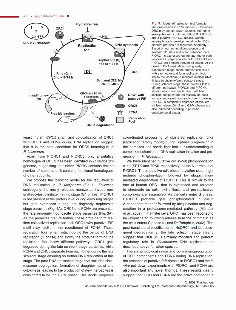
Fig. 7. Model of replication foci formationand progression in P. falciparum. P. falciparumORC may contain fewer subunits than othereukaryotes with confirmed PfORC1, PfORC5and a putative PfORC2 subunit. Duringintraerythrocytic developmental cycle (IDC),different proteins are regulated differently.Based on our immunofluorescence andWestern blot data and other published data,PfORC1 is expressed during late ring or earlytrophozoite stage whereas both PfPCNA1 andPfORC5 are present through all stages. At theonset of DNA replication, during earlytrophozoite stage, these proteins colocalizewith each other and form replication foci.These foci continue to replicate nuclear DNAtill late trophozoite/early schizont stage.During schizont stage, these proteins followdifferent pathways. PfORC5 and PfPCNAslowly detach from each other until lateschizont stage where the majority of thesefoci are separated from each other. However,PfORC1 is completely degraded at this lateschizont stage. G1, S and G2/M phases arealso indicated according to parasiticdevelopmental stages.Free Merozoites
ORC5
PCNA
Replication Foci
ORC1 degradation
Ring (G1)
0 to ~16/18 h
Trophozoite (S)
~18 to ~ 34 h
Schizont (G2: M)
~34 to ~48 h
DNA synthesis
~24h
~34hIDC
X
ORC1 with
putative PIP
ORC1
Replication
foci
ORC5 and PCNA
dissociates
1 5
2
3,4,6??
Hydroxyurea
Invading new
RBC
PIP
ORC in P. falciparum
660 A. Gupta, P. Mehra and S. K. Dhar �
© 2008 The AuthorsJournal compilation © 2008 Blackwell Publishing Ltd, Molecular Microbiology, 69, 646–665
of replication foci. The finding of putative PIP motifs inScORC1 and HsORC1 homologues may suggest its con-served role during eukaryotic DNA replication. ORC1 withthe putative PIP box may facilitate loading of PCNA at thereplication fork. Recently, a similar function has been pro-posed for MCM10 (Das-Bradoo et al., 2006). Alternatively,it may be suggested that ORC components are not onlythe part of the pre-RC, they are also active componentsof the growing replication fork where the putative PIPdomain in ORC1 plays a major role to hold these proteinstogether along with PCNA. The degradation of PfORC1 atthe later stage with the concomitant separation of ORCand PCNA foci strengthens the above hypothesis. Theseparation of PCNA from ORC binding sites can also beattributed to the differential affinity of PCNA towards dif-ferent proteins during development. In fact, it has beenreported earlier that binding of PCNA to its different part-ners is modulated by different modifications of PCNA, likeubiquitination and sumoylation that help to attract specificbinding partners like translesion polymerases and Srs2helicase respectively, because of the presence of specificbinding motifs in these proteins during bypass DNA rep-lication following DNA lesions (Kannouche et al., 2004;Papouli et al., 2005).
Presence of PfPCNA during the late schizont stage maybe required for genome maintenance processes dealingwith stalled replication forks, such as translesion synthe-sis as reported in mammalian cells (Essers et al., 2005;Moldovan et al., 2007). Additionally, accumulated PCNAon DNA at late stages might recruit some replicationfactors like Cdt1 that is ubiquitinated by DDB1-Cul4 ubiq-uitin ligase following interaction with PCNA, thereby clear-ing the nucleus from Cdt1-mediated licensing activity(Arias and Walter, 2006).
Presence of PfORC5 at the late stage may ensure itsavailability for the next cycle of DNA replication. Alterna-tively, as ORC components have been shown to beessential for silencing Hidden MAT left and Hidden MATright mating-type loci in S. cerevisiae, PfORC5 may followsimilar suits in gene silencing particularly in var genesilencing that needs to be explored further.
In mammalian cells, the organization of replication fac-tories is dependent on chromatin architecture and struc-ture of chromosomal domains that are dictated by nuclearmatrix. The presence of histones, chromatin-modifyingenzymes (histone acetylases and deacetylases) andnuclear matrix protein coding genes in Plasmodiumgenome suggests that chromatin structure will also playcritical role in deciding the fate of the replication foci.
In mammalian cells, ~10 000 replication sites with anaverage 1 mbp of DNA per replication site account forchromosomal DNA replication during S phase withapproximately six replicons per replication site (Ma et al.,1998). In contrast, we find average ~15 colocalized
(PfORC and PfPCNA) replication foci in Plasmodiumduring the peak of DNA replication. Considering that thePlasmodium genome size is ~24 mbp, each replicationfoci can account for replication of ~1.5 mbp DNA, a valueclose to the other eukaryotes. It is interesting to note thatthe replication rate is faster in Plasmodium than in highereukaryotes. Plasmodium genome content increases from1 N to 16 to 32 N within a span of 12–16 h, whereasmammalian nuclear DNA replicates only once duringthe same time period. Therefore, the replication foci inPlasmodium must contain higher number of repliconscompared with that of mammals. It is possible that originfrequency in Plasmodium is much higher than in eukary-otes, accounting for the higher number of replicons perfoci. This may also explain the distinct colocalization ofPCNA and ORC in Plasmodium that is not common inhigher eukaryotes.
The gradual disappearance of ORC5 foci in the pres-ence of increasing concentration of HU is very surprising.In general, HU depletes the pool of dNTPs and it shouldnot affect the loading of pre-RC components. We believethat increasing concentration of HU has a direct effect ondepleting the dNTP pool that may affect the foci formationat later time points. P. falciparum undergoes multiplerounds of DNA replication. Depletion of dNTP pools mayaffect severely the late rounds of DNA replication. Addi-tionally, the foci that are formed initially may diffuse withtime. This may explain the gradual decrease of foci for-mation with increasing drug concentration. This is inconsistence with the finding reported by Santocanale andDiffley (1998) that suggests specific prevention of late-Sorigins in the presence of HU. Higher concentration of HU(70 mg ml-1) may activate a checkpoint immediately thatcan affect foci formation drastically. Re-appearance ofORC5 foci in some parasites and the increase in para-sitemia in the presence of caffeine following HU treatmentmay suggest that DNA damage checkpoint pathwaysare active in the parasites. Interestingly, we have foundtwo ORFs in Plasmodium database (PF14-0516 andPFB0815w respectively) that show considerable homol-ogy with human Chk1 and Chk2 proteins. However, thisneeds to be verified further.
DNA replication is a fundamental process from bacteriato mammals. However, this important aspect of biologyhas not been studied thoroughly except the modelsystems like Escherichia coli, S. cerevisiae and Xenopus.The thorough understanding of this process in protozoanparasites like Plasmodium not only highlights the unique-ness of these lower eukaryotes but also confirms theconserved role of the replication proteins among differentspecies. While the presence of previously un-noticed con-served PIP domain in ScORC1, HsORC1 and PfORC1ensures the conserved role of PCNA in DNA replicationamong different species, the unique colocalization of
Replication foci in P. falciparum 661
© 2008 The AuthorsJournal compilation © 2008 Blackwell Publishing Ltd, Molecular Microbiology, 69, 646–665
ORC and PCNA forming replication foci and their separa-tion following DNA replication clearly highlight importantaspect of DNA replication that has not been shownearlier.
Finally, the identification of a critical component of rep-lication machinery in the parasites with nucleotide bindingand hydrolysis activities that are central to its function (asfound in S. cerevisiae) may be useful for ongoing searchfor finding new targets for intervening Plasmodium growthand proliferation.
Experimental procedures
Parasite culture
Plasmodium falciparum 3D7 strain was cultured in human O+
erythrocytes in RPMI 1640 medium supplemented with25 mM Hepes, 50 mg l-1 hypoxanthine, 0.2% NaHCO3, 0.5%Albumax (Invitrogen), 0.2% glucose and 10 mg ml-1 gentamy-cin sulphate. Synchronization of cultures was achieved byusing 5% sorbitol treatment of cells. Synchronization wasverified by morphological analysis of Giemsa-stained infectedblood smears at different time points.
For inhibitor studies, hydroxyurea (Sigma) was added intothe P. falciparum culture (5–6% parasitemia) at differentconcentrations (0, 20, 40 and 70 mg ml-1 respectively) at earlyring stage in triplicate. Samples were taken at different timeintervals following evaluation of developmental stages of theparasites by Giemsa staining.
The proteasome inhibitor MG132 was added at a concen-tration of 100 nM in synchronized P. falciparum (5–6% para-sitemia) culture ~40 h post erythrocytic invasion in triplicateand followed several hours thereafter.
RNA extraction and RT-PCR
RT-PCR reactions were performed using cDNA and specificprimers for PfORC5, PfMCM4, PfORC1 and controlPfGAPDH (Table S1, P15–P22) essentially following theprotocol as described elsewhere (Mehra et al., 2005). Semi-quantitative analysis of RT-PCR results was performed bydensitometric quantification of the amplified products fol-lowed by graphical representation of these results.
ATPase and ATP binding assays
ATPase assay was performed using recombinant pGEX6P2-PfORC5D1 (171–899 aa) protein (wild type or mutant) follow-ing the protocol as described earlier (Mehra et al., 2005).Thin-layer chromatography (TLC) was performed to separatethe released Pi. The TLC plates were dried, autoradiographedand quantified by Phosphorimager (Fujifilm-BAS-1800).
For ATP binding assay, 1 mg of MBP-PfORC5D2 (171–421 aa) protein (wild type or mutant) was cross-linked with[a-32P]-dATP in the presence of UV (254 nm) for 30 min in30 ml reaction mixture containing 20 mM Hepes (pH 7.5), 10%glycerol, 0.1 mM DTT, 10 mCi [a-32P]-dATP (3000 Ci mmol-1)using a Stratagene cross-linker followed by addition of 0.8 ml
of 100 mM dATP and 20 mg of BSA. Proteins were precipitatedby trichloroacetic acid and the pellet was washed once withacetone containing 0.5% HCl and twice with acetone. Proteinswere separated by SDS-PAGE and the radiolabelled dATP-bound proteins were visualized using phosphorimager.
Immunofluorescence assay
Thin smear of synchronous culture of infected erythrocyteswas made on glass slides. Slides were air-dried for 2 h atroom temperature followed by fixation for 30 min in freshlyprepared solution of acetone and methanol (90% and 10%respectively) at -20°C. The slides were further blocked for 1 hat room temperature in 1¥ PBS containing 3% BSA and0.01% saponin. Slides were then incubated overnight in1¥ PBS containing respective primary antibodies at 4°C.Immunoaffinity-purified anti-PfORC1 (rabbit), anti-PfORC5(rabbit), anti-PfORC1 (mouse) and anti-PfPCNA1 (mouse)antibodies were used at 1:500, 1:500, 1:2000 and 1:500dilutions respectively, as primary antibodies. Slides werefurther washed in 1¥ PBS and incubated for 45 min at 4°C in1¥ PBS containing anti-rabbit IgG-Alexa flour 488 or anti-mouse IgG Alexa fluor 594 (Molecular Probes) 1:1000 sec-ondary antibodies and nuclear stain DAPI. Images werecaptured using Nikon Eclipse 80i fluorescent microscope.
Alternatively, glass slides containing parasites were incu-bated in 2% PFA solution for 15 min at the room temperaturefollowed by blocking them in 0.5% BSA solution in 1¥ PBS for20 min. The slides were further treated with primary antibod-ies for 2–3 h at room temperature and secondary antibodiesfor 45 min with thorough washing with 1¥ PBS after eachantibody treatment. Antibody dilutions were kept the same asabove.
Immunoelectron microscopy
Immunoelectron microscopy for the subcellular localizationof PfORC5 was performed as described elsewhere (Mehraet al., 2005) using anti-PfORC5 antibodies (1:500) as primaryantibodies and goat anti-rabbit IgG secondary antibodiescoupled with 10–20 nm gold particles at 1:100 dilution. Sec-tions were viewed using a Morgagni-268 transmission elec-tron microscope.
Immunoprecipitation assay
For Immunoprecipitation, lysate from 100 ml of P. falciparum-infected erythrocyte pellet (8–10% parasitemia) was pre-pared by adding five to six pellet volume of lysis buffercontaining 300 mM NaCl, 20 mM Tris.Cl, pH 8.0 and 0.5%NP-40 and anti-protease cocktail (Sigma). Lysate was clari-fied by centrifugation at 15 000 g for 30 min at 4°C. Eitherpre-immune or immune sera (1–2 ml) were added to thiscleared lysate and the mixture was incubated at 4°C for 2 hwith constant mild shaking followed by addition of 20 ml ofprotein A sepharose slurry or protein G sepharose slurry(Sigma). Finally, these bead Ab protein mixtures were incu-bated for 1 h at 4°C. Beads were collected and washed threetimes with 1¥ PBS containing 0.1% BSA. Immunoprecipitatedproteins were eluted from beads by adding 2 vols of SDS-
662 A. Gupta, P. Mehra and S. K. Dhar �
© 2008 The AuthorsJournal compilation © 2008 Blackwell Publishing Ltd, Molecular Microbiology, 69, 646–665
PAGE loading buffer and boiling at 95°C for 10 min. Proteinswere separated by SDS-PAGE and transferred onto PVDFmembrane and immunoblotted using corresponding primaryand secondary antibodies.
Complementation of yeast ORC5 mutant
Complementation experiments were performed in S. cerevi-siae using either wild-type PfORC5 or S. cerevisiae ORC5 ordifferent chimeras of S. cerevisiae and P. falciparum ORC5.Details of the methods are described in supplementary data,Experimental procedures (yeast complementation section).
DNA manipulation
The putative PfORC5 gene was amplified by polymerasechain reaction using P. falciparum 3D7 genomic DNA as atemplate and specific primers (P1 and P2, Table S1). For thedetailed cloning strategy of wild-type and mutant forms ofPfORC5 and PfPCNA, please see supplementary information.
Recombinant protein purification
For the purification of PfORC5 fusion recombinant proteinsto perform ATP binding and hydrolysis assay, E. coli BL21codon-plus strain was transformed with respective recombi-nant clones. Of all the constructs made to express recombi-nant proteins, only pGEX6P2-PfORC5D1 (171–899 aa) andpMALc2¥-PfORC5D2 (171–421 aa) yielded the recombinantproteins in soluble form. The detailed methods for proteinpurification for these constructs and other constructs for anti-bodies preparation (PfORC5C1 and PfORC5C2) are de-scribed in supplementary information, materials and methods.
Antibody production and Western blotting analysis
Polyclonal antibodies against different antigens in rabbitand mice were generated essentially following the protocoldescribed by Harlow and Lane (1988). Please see supple-mentary information for detailed protocols.
Western blot analysis was carried out following the stan-dard protocol (Sambrook et al., 1989). Please see supple-mentary information for the details.
Acknowledgements
This work is supported by Wellcome Trust, London. S.K.D. isa Wellcome Trust Senior International Research Fellow. A.G.and P.M. acknowledge CSIR for fellowships. The authorsacknowledge Dr Steven P. Bell (MIT, USA) and Dr N. Roy(NIPER, India) for the yeast strains and yeast expressionvectors respectively. We are indebted to Dr Tim Gilberger, DrTobias Spielman and Moritz Terek of BNI, Hamburg for theirnumerous help especially with the fluorescence microscopy.The authors acknowledge Prof. Anindya Dutta (Univ. of Vir-ginia, USA) and Prof. Sudha Bhattacharya (JNU, India) forcritically reviewing the manuscript and Prof. Rajendra Prasad(JNU, India) for his generous support. Open Access publica-tion charges for this article was provided by the WellcomeTrust, UK.
ReferencesArias, E.E., and Walter, J.C. (2006) PCNA functions as a
molecular platform to trigger Cdt1 destruction and preventre-replication. Nat Cell Biol 8: 84–90.
Arnot, D.E., and Gull, K. (1998) The Plasmodium cell-cycle:facts and questions. Ann Trop Med Parasitol 92: 361–365.
Bell, S.P. (2002) The origin recognition complex: from simpleorigins to complex functions. Genes Dev 16: 659–672.
Bell, S.P., and Dutta, A. (2002) DNA replication in eukaryoticcells. Annu Rev Biochem 71: 333–374.
Berezney, R., Dubey, D.D., and Huberman, J.A. (2000) Het-erogeneity of eukaryotic replicons, replicon clusters andreplication foci. Chromosoma 108: 471–484.
Bozdech, Z., Llinás, M., Pulliam, B.L., Wong, E.D., Zhu, J.,and DeRisi, J.L. (2003) The transcriptome of the intraeryth-rocytic developmental cycle of Plasmodium falciparum.PloS Biol 1: E5.
Cook, P.R. (2001) Principles of Nuclear Structures andFunction. Wilmington, DE: Wiley-Liss Inc.
Daimon, K., Kawarabayasi, Y., Kikuchi, H., Sako, Y., andIshino, Y. (2002) Three proliferating cell nuclear antigen-like proteins found in the hyperthermophilic archaeon Aero-pyrum pernix: interactions with the two DNA polymerases.J Bacteriol 184: 687–694.
Das-Bradoo, S., Ricke, R.M., and Bielinsky, A.K. (2006) Inter-action between PCNA and diubiquitinated MCM10 isessential for cell growth in budding yeast. Mol Cell Biol 26:4806–4817.
Doerig, C., Endicott, J., and Chakrabarti, D. (2002) Cyclin-dependent kinase homologues of Plasmodium falciparum.Int J Parasitol 32: 1575–1585.
Essers, J., Theil, A.F., Baldeyron, C., Van Cappellen, W.A.,Houtsmuller, A.B., Kanaar, R., and Vermeulen, W. (2005)Nuclear dynamics of PCNA in DNA replication and repair.Mol Cell Biol 25: 9350–9359.
Gantt, S.M., Myung, J.M., Briones, M.R., Li, W.D., Corey,E.J., Omura, S., et al. (1998) Proteasome inhibitors blockdevelopment of Plasmodium spp. Antimicrob AgentsChemother 42: 2731–2738.
Gilbert, D.M. (2001) Making sense of eukaryotic DNA repli-cation origins. Science 294: 96–100.
Giordano-Coltart, J., Ying, C.Y., Gautier, J., and Hurwitz, J.(2005) Studies of the properties of human origin recogni-tion complex and its Walker A motif mutants. Proc NatlAcad Sci USA 102: 69–74.
Graeser, R., Wernli, B., Franklin, M., and Kappes, B. (1996)Plasmodium falciparum protein kinase 5 and the malarialnuclear division cycles. Mol Biochem Parasitol 82:37–49.
Guerini, M.N., Que, X., Reed, S.L., and White, M.W. (2000)Two genes encoding unique proliferating-cell-nuclear-antigens are expressed in Toxoplasma gondii. MolBiochem Parasitol 109: 121–131.
Guerini, M.N., Behnke, M.S., and White, M.W. (2005) Bio-chemical and genetic analysis of the distinct proliferatingcell nuclear antigens of Toxoplasma gondii. Mol BiochemParasitol 142: 56–65.
Gupta, A., Mehra, P., Nitharwal, R., Sharma, A., Biswas,A.K., and Dhar, S.K. (2006) Analogous expression patternof Plasmodium falciparum replication initiation proteinsPfMCM4 and PfORC1 during the asexual and sexual
Replication foci in P. falciparum 663
© 2008 The AuthorsJournal compilation © 2008 Blackwell Publishing Ltd, Molecular Microbiology, 69, 646–665
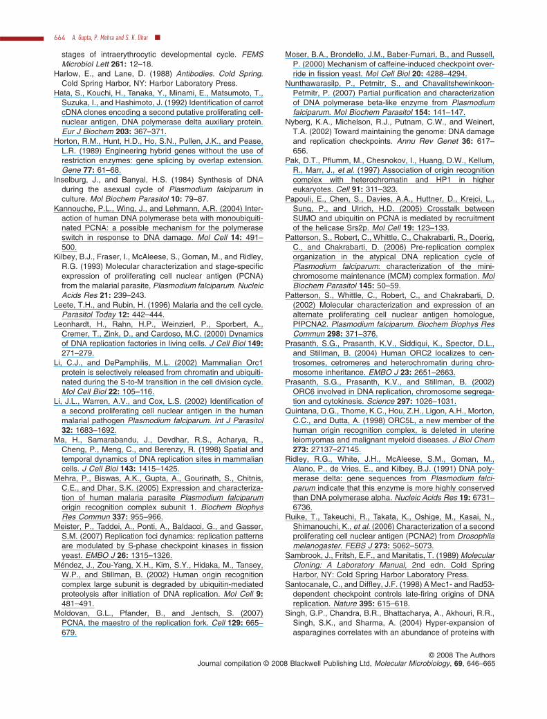
stages of intraerythrocytic developmental cycle. FEMSMicrobiol Lett 261: 12–18.
Harlow, E., and Lane, D. (1988) Antibodies. Cold Spring.Cold Spring Harbor, NY: Harbor Laboratory Press.
Hata, S., Kouchi, H., Tanaka, Y., Minami, E., Matsumoto, T.,Suzuka, I., and Hashimoto, J. (1992) Identification of carrotcDNA clones encoding a second putative proliferating cell-nuclear antigen, DNA polymerase delta auxiliary protein.Eur J Biochem 203: 367–371.
Horton, R.M., Hunt, H.D., Ho, S.N., Pullen, J.K., and Pease,L.R. (1989) Engineering hybrid genes without the use ofrestriction enzymes: gene splicing by overlap extension.Gene 77: 61–68.
Inselburg, J., and Banyal, H.S. (1984) Synthesis of DNAduring the asexual cycle of Plasmodium falciparum inculture. Mol Biochem Parasitol 10: 79–87.
Kannouche, P.L., Wing, J., and Lehmann, A.R. (2004) Inter-action of human DNA polymerase beta with monoubiquiti-nated PCNA: a possible mechanism for the polymeraseswitch in response to DNA damage. Mol Cell 14: 491–500.
Kilbey, B.J., Fraser, I., McAleese, S., Goman, M., and Ridley,R.G. (1993) Molecular characterization and stage-specificexpression of proliferating cell nuclear antigen (PCNA)from the malarial parasite, Plasmodium falciparum. NucleicAcids Res 21: 239–243.
Leete, T.H., and Rubin, H. (1996) Malaria and the cell cycle.Parasitol Today 12: 442–444.
Leonhardt, H., Rahn, H.P., Weinzierl, P., Sporbert, A.,Cremer, T., Zink, D., and Cardoso, M.C. (2000) Dynamicsof DNA replication factories in living cells. J Cell Biol 149:271–279.
Li, C.J., and DePamphilis, M.L. (2002) Mammalian Orc1protein is selectively released from chromatin and ubiquiti-nated during the S-to-M transition in the cell division cycle.Mol Cell Biol 22: 105–116.
Li, J.L., Warren, A.V., and Cox, L.S. (2002) Identification ofa second proliferating cell nuclear antigen in the humanmalarial pathogen Plasmodium falciparum. Int J Parasitol32: 1683–1692.
Ma, H., Samarabandu, J., Devdhar, R.S., Acharya, R.,Cheng, P., Meng, C., and Berenzy, R. (1998) Spatial andtemporal dynamics of DNA replication sites in mammaliancells. J Cell Biol 143: 1415–1425.
Mehra, P., Biswas, A.K., Gupta, A., Gourinath, S., Chitnis,C.E., and Dhar, S.K. (2005) Expression and characteriza-tion of human malaria parasite Plasmodium falciparumorigin recognition complex subunit 1. Biochem BiophysRes Commun 337: 955–966.
Meister, P., Taddei, A., Ponti, A., Baldacci, G., and Gasser,S.M. (2007) Replication foci dynamics: replication patternsare modulated by S-phase checkpoint kinases in fissionyeast. EMBO J 26: 1315–1326.
Méndez, J., Zou-Yang, X.H., Kim, S.Y., Hidaka, M., Tansey,W.P., and Stillman, B. (2002) Human origin recognitioncomplex large subunit is degraded by ubiquitin-mediatedproteolysis after initiation of DNA replication. Mol Cell 9:481–491.
Moldovan, G.L., Pfander, B., and Jentsch, S. (2007)PCNA, the maestro of the replication fork. Cell 129: 665–679.
Moser, B.A., Brondello, J.M., Baber-Furnari, B., and Russell,P. (2000) Mechanism of caffeine-induced checkpoint over-ride in fission yeast. Mol Cell Biol 20: 4288–4294.
Nunthawarasilp, P., Petmitr, S., and Chavalitshewinkoon-Petmitr, P. (2007) Partial purification and characterizationof DNA polymerase beta-like enzyme from Plasmodiumfalciparum. Mol Biochem Parasitol 154: 141–147.
Nyberg, K.A., Michelson, R.J., Putnam, C.W., and Weinert,T.A. (2002) Toward maintaining the genome: DNA damageand replication checkpoints. Annu Rev Genet 36: 617–656.
Pak, D.T., Pflumm, M., Chesnokov, I., Huang, D.W., Kellum,R., Marr, J., et al. (1997) Association of origin recognitioncomplex with heterochromatin and HP1 in highereukaryotes. Cell 91: 311–323.
Papouli, E., Chen, S., Davies, A.A., Huttner, D., Krejci, L.,Sung, P., and Ulrich, H.D. (2005) Crosstalk betweenSUMO and ubiquitin on PCNA is mediated by recruitmentof the helicase Srs2p. Mol Cell 19: 123–133.
Patterson, S., Robert, C., Whittle, C., Chakrabarti, R., Doerig,C., and Chakrabarti, D. (2006) Pre-replication complexorganization in the atypical DNA replication cycle ofPlasmodium falciparum: characterization of the mini-chromosome maintenance (MCM) complex formation. MolBiochem Parasitol 145: 50–59.
Patterson, S., Whittle, C., Robert, C., and Chakrabarti, D.(2002) Molecular characterization and expression of analternate proliferating cell nuclear antigen homologue,PfPCNA2. Plasmodium falciparum. Biochem Biophys ResCommun 298: 371–376.
Prasanth, S.G., Prasanth, K.V., Siddiqui, K., Spector, D.L.,and Stillman, B. (2004) Human ORC2 localizes to cen-trosomes, cetromeres and heterochromatin during chro-mosome inheritance. EMBO J 23: 2651–2663.
Prasanth, S.G., Prasanth, K.V., and Stillman, B. (2002)ORC6 involved in DNA replication, chromosome segrega-tion and cytokinesis. Science 297: 1026–1031.
Quintana, D.G., Thome, K.C., Hou, Z.H., Ligon, A.H., Morton,C.C., and Dutta, A. (1998) ORC5L, a new member of thehuman origin recognition complex, is deleted in uterineleiomyomas and malignant myeloid diseases. J Biol Chem273: 27137–27145.
Ridley, R.G., White, J.H., McAleese, S.M., Goman, M.,Alano, P., de Vries, E., and Kilbey, B.J. (1991) DNA poly-merase delta: gene sequences from Plasmodium falci-parum indicate that this enzyme is more highly conservedthan DNA polymerase alpha. Nucleic Acids Res 19: 6731–6736.
Ruike, T., Takeuchi, R., Takata, K., Oshige, M., Kasai, N.,Shimanouchi, K., et al. (2006) Characterization of a secondproliferating cell nuclear antigen (PCNA2) from Drosophilamelanogaster. FEBS J 273: 5062–5073.
Sambrook, J., Fritsh, E.F., and Manitatis, T. (1989) MolecularCloning: A Laboratory Manual, 2nd edn. Cold SpringHarbor, NY: Cold Spring Harbor Laboratory Press.
Santocanale, C., and Diffley, J.F. (1998) A Mec1- and Rad53-dependent checkpoint controls late-firing origins of DNAreplication. Nature 395: 615–618.
Singh, G.P., Chandra, B.R., Bhattacharya, A., Akhouri, R.R.,Singh, S.K., and Sharma, A. (2004) Hyper-expansion ofasparagines correlates with an abundance of proteins with
664 A. Gupta, P. Mehra and S. K. Dhar �
© 2008 The AuthorsJournal compilation © 2008 Blackwell Publishing Ltd, Molecular Microbiology, 69, 646–665
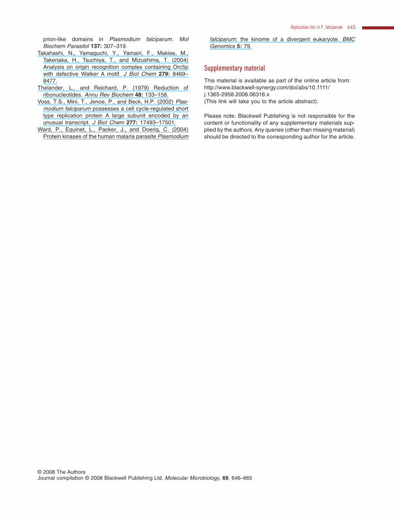
prion-like domains in Plasmodium falciparum. MolBiochem Parasitol 137: 307–319.
Takahashi, N., Yamaguchi, Y., Yamairi, F., Makise, M.,Takenaka, H., Tsuchiya, T., and Mizushima, T. (2004)Analysis on origin recognition complex containing Orc5pwith defective Walker A motif. J Biol Chem 279: 8469–8477.
Thelander, L., and Reichard, P. (1979) Reduction ofribonucleotides. Annu Rev Biochem 48: 133–158.
Voss, T.S., Mini, T., Jenoe, P., and Beck, H.P. (2002) Plas-modium falciparum possesses a cell cycle-regulated shorttype replication protein A large subunit encoded by anunusual transcript. J Biol Chem 277: 17493–17501.
Ward, P., Equinet, L., Packer, J., and Doerig, C. (2004)Protein kinases of the human malaria parasite Plasmodium
falciparum: the kinome of a divergent eukaryote. BMCGenomics 5: 79.
Supplementary material
This material is available as part of the online article from:http://www.blackwell-synergy.com/doi/abs/10.1111/j.1365-2958.2008.06316.x(This link will take you to the article abstract).
Please note: Blackwell Publishing is not responsible for thecontent or functionality of any supplementary materials sup-plied by the authors. Any queries (other than missing material)should be directed to the corresponding author for the article.
Replication foci in P. falciparum 665
© 2008 The AuthorsJournal compilation © 2008 Blackwell Publishing Ltd, Molecular Microbiology, 69, 646–665


