Embed Size (px)
Citation preview

Photolysis intermediates of the artificial visual pigmentcis-5,6-dihydro-isorhodopsin
A. Albeck,* N. Friedman,* M. Ottolenghi,* M. Sheves,* C. M. Einterz,§ S. J. Hug,§ J. W. Lewis,§and D. S. Kliger§*Department of Organic Chemistry, Weizmann Institute of Science, Rehovot 76100, Israel; *Department of PhysicalChemistry, The Hebrew University of Jerusalem, Jerusalem 91904, Israel; §Department of Chemistry, University ofCalifornia, Santa Cruz, California 95064
ABSTRACT The photolysis interme-diates of an artificial bovine rhodopsinpigment, cis-5,6-dihydro-isorhodopsin(cis-5,6,-diH-ISORHO, Xmax 461 nm),which contains a cis-5,6-dihydro-9-cis-retinal chromophore, are investigatedby room temperature, nanosecondlaser photolysis, and low temperatureirradiation studies. The observationsare discussed both in terms of lowtemperature experiments of Yoshizawaand co-workers on trans-5,6-diH-ISORHO (Yoshizawa, T., Y. Shichida,and S. Matuoka. 1984. Vision Res.
24:1455-1463), and in relation to thephotolysis intermediates of native bo-vine rhodopsin (RHO). It is suggestedthat in 5,6-diH-ISORHO, a primarybathorhodopsin intermediate analo-gous to the bathorhodopsin interme-diate (BATHO) of the native pigment,rapidly converts to a blue-shifted inter-mediate (BSI, X.,,. 430 nm) which is notobserved after photolysis of nativerhodopsin. The analogs from lumi-rhodopsin (LUMI) to meta-lI rhodopsin(META-Il) are generated subsequent toBSI, similar to their generation from
BATHO in the native pigment. It is pro-posed that the retinal chromophore inthe bathorhodopsin stage of 5,6-diH-ISORHO is relieved of strain induced bythe primary cis to trans isomerizationby undergoing a geometrical rear-
rangement of the retinal. Such a rear-
rangement, which leads to BSI, wouldnot take place so rapidly in the nativepigment due to ring-protein interac-tions. In the native pigment, the strain inBATHO would be relieved only on a
longer time scale, via a process with a
rate determined by protein relaxation.
INTRODUCTION
Initiation of visual pigment activation (Fig. 1) in rhodop-sin (RHO, containing an 11 -cis retinal chromophorebound to the protein via a protonated Schiff base linkage)is associated with the early red-shifted intermediate,bathorhodopsin (BATHO) (for reviews, see Packer,1982). BATHO, which at room temperature decays to thenext photointermediate, lumirhodopsin (LUMI), over a
10--10-7 s time scale, appears to be formed in thesubnanosecond (10-2-10-1o s) range from a precursor
denoted as prebathorhodopsin (PBATHO; Dinur et al.,1981), based on the low temperature experiments ofPeters et al. (1977), or photorhodopsin (PHOTO), basedon the room temperature data of Yoshizawa et al. (1984)and Shichida et al. (1984; Shichida, 1986). PBATHOand PHOTO are either precursors of BATHO, or are
primary products formed in parallel with BATHO whichdecay back to ground state pigments.Much of the attention devoted to BATHO, the first
transient species produced by photolysis which can betrapped at low temperatures, is due to its property ofstoring 36 kcal/mol of the photon energy for use insubsequent stages of the transduction process (Boucherand Leblanc, 1985; Cooper, 1979; Honig et al., 1979a,b;Schick et al., 1987). The exact mechanism of energy
storage in BATHO and the origin of its bathochromicspectral shift are presently unclear (for recent discussions
see Birge et al., 1988; Ottolenghi and Sheves, 1987;Palings et al., 1987). The relative contributions of factorssuch as electrostatic interactions between the chromo-phore and the protein or surrounding environment, andconformational distortions of the polyene chromophoreare still open questions (Honig et al., 1979a,b; Palings etal., 1987; Birge and Hubbard, 1980; Warshel and Bar-boy, 1982).
Artificial pigments in which the native retinal chromo-phore is substituted by a synthetic retinal analog are
potential tools for elucidating the nature of photolysisintermediates (for a review, see Derguini and Nakanishi,1986). For example, as shown in the case of bacteriorho-dopsins, the photocycles of artificial pigments can be usedto discriminate between light-induced changes associatedwith the Schiff base moiety, bond rotations and isomeri-zations, interactions with protein charges, etc. (Otto-lenghi and Sheves, 1987; Sheves et al., 1987). The lack ofphotoreaction in the case of visual pigments with a
blocked C1l=C12 bond has confirmed the role of the11-cis to all-trans isomerization in the primary photo-chemical event (Akita et al., 1980; Mao et al., 1981;Fukada et al., 1984; Hubbard and Kropf, 1958; Rosenfeldet al., 1977). Additionally, studies performed on a pig-ment with blocked Cg-C,, bonds has shown thatC,0-C,, bond rotation is not needed for photoreactions toproceed as in the native pigment (Sheves et al., 1986).To date, no systematic study has been carried out on
Biophys. J. Biophysical SocietyVolume 55 February 1989 233-241
0006-3495/89/02/233/09 $2.00 233
0006-3495/89/02/233/09 $2.00 233
RHO*-.PHOTO or PBATHO. 5,6-diH-ISOI(560) hv
hv|40ps 5 HYPSO -.." (435)
BATHO -..---- 5,6-diH-BATHO(543) [535,55]a. (5)C
*....[3 ns 1 5-. 6 d SI31Kc
1 33K [ 36ns 170ns]a *-.5,6-diH-BSI*. [430] (41 4)c
[200 ns]|
LUMI .. __ 5,6-diH-LUMI(497) [490]b
203K| 1183KC
META ,---- ----- - 5,6-diH-META I
(478) (473)
258K ' [200 ]b [500 LSb j223KCMETA II ---------- 5,6-diH-META 11
(380) [380] [370] (340)
RHO(502) [498]
5,6-diH-ISORHO[461]
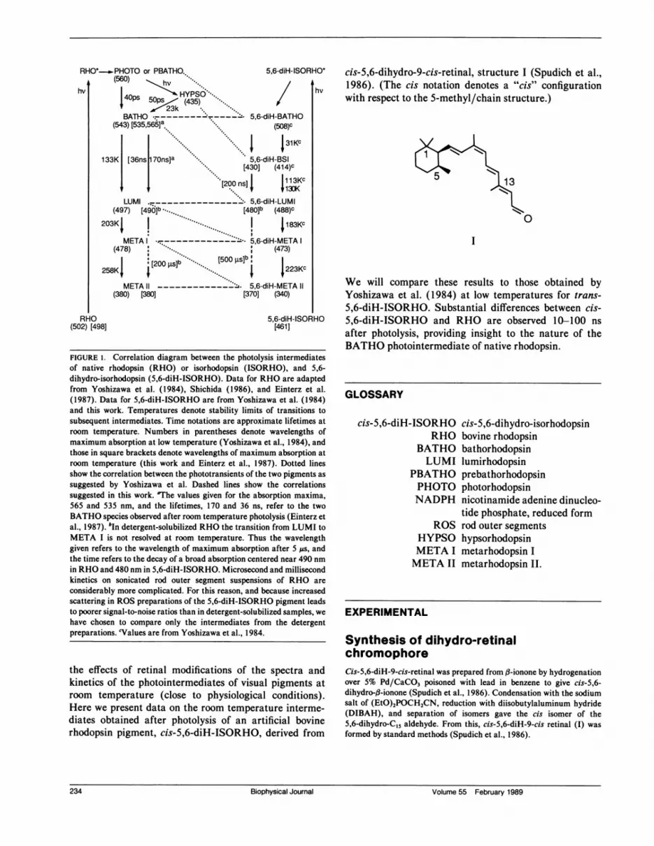
FIGURE 1. Correlation diagram between the photolysis intermediatesof native rhodopsin (RHO) or isorhodopsin (ISORHO), and 5,6-dihydro-isorhodopsin (5,6-diH-ISORHO). Data for RHO are adaptedfrom Yoshizawa et al. (1984), Shichida (1986), and Einterz et al.(1987). Data for 5,6-diH-ISORHO are from Yoshizawa et al. (1984)and this work. Temperatures denote stability limits of transitions tosubsequent intermediates. Time notations are approximate lifetimes atroom temperature. Numbers in parentheses denote wavelengths ofmaximum absorption at low temperature (Yoshizawa et al., 1984), andthose in square brackets denote wavelengths of maximum absorption atroom temperature (this work and Einterz et al., 1987). Dotted linesshow the correlation between the phototransients of the two pigments assuggested by Yoshizawa et al. Dashed lines show the correlationssuggested in this work. 'The values given for the absorption maxima,565 and 535 nm, and the lifetimes, 170 and 36 ns, refer to the twoBATHO species observed after room temperature photolysis (Einterz etal., 1987). bIn detergent-solubilized RHO the transition from LUMI toMETA I is not resolved at room temperature. Thus the wavelengthgiven refers to the wavelength of maximum absorption after 5 gs, andthe time refers to the decay of a broad absorption centered near 490 nmin RHO and 480 nm in 5,6-diH-ISORHO. Microsecond and millisecondkinetics on sonicated rod outer segment suspensions of RHO areconsiderably more complicated. For this reason, and because increasedscattering in ROS preparations of the 5,6-diH-ISORHO pigment leadsto poorer signal-to-noise ratios than in detergent-solubilized samples, wehave chosen to compare only the intermediates from the detergentpreparations. cValues are from Yoshizawa et al., 1984.
the effects of retinal modifications of the spectra andkinetics of the photointermediates of visual pigments atroom temperature (close to physiological conditions).Here we present data on the room temperature interme-diates obtained after photolysis of an artificial bovinerhodopsin pigment, cis-5,6-diH-ISORHO, derived from
io* cis-5,6-dihydro-9-cis-retinal, structure I (Spudich et al.,hv 1986). (The cis notation denotes a "cis" configuration
with respect to the 5-methyl/chain structure.)
13
I
We will compare these results to those obtained byYoshizawa et al. (1984) at low temperatures for trans-5,6-diH-ISORHO. Substantial differences between cis-5,6-diH-ISORHO and RHO are observed 10-100 nsafter photolysis, providing insight to the nature of theBATHO photointermediate of native rhodopsin.
GLOSSARY
cis-5,6-diH-ISORHORHO
BATHOLUMI
PBATHOPHOTONADPH
ROSHYPSOMETA IMETA II
cis-5,6-dihydro-isorhodopsinbovine rhodopsinbathorhodopsinlumirhodopsinprebathorhodopsinphotorhodopsinnicotinamide adenine dinucleo-tide phosphate, reduced formrod outer segmentshypsorhodopsinmetarhodopsin Imetarhodopsin II.
EXPERIMENTAL
Synthesis of dihydro-retinalchromophoreCis-5,6-diH-9-cis-retinal was prepared from f1-ionone by hydrogenationover 5% Pd/CaCO3 poisoned with lead in benzene to give cis-5,6-dihydro-,B-ionone (Spudich et al., 1986). Condensation with the sodiumsalt of (EtO)2POCH2CN, reduction with diisobutylaluminum hydride(DIBAH), and separation of isomers gave the cis isomer of the5,6-dihydro-C15 aldehyde. From this, cis-5,6-diH-9-cis retinal (I) wasformed by standard methods (Spudich et al., 1986).
234 Biophysical Journal Volume 55 February 1989
)RH
234 Biophysical Journal Volume 55 February 1989

Regeneration /nanosecondphotolysisBovine rod outer segments (ROS) were prepared from frozen retinas as
previously described (Lewis et al., 1984). Freshly prepared, pH 7.0hydroxylamine stock was added to produce a final concentration of 20mM, and the ROS were bleached for 2 min using a 25-W bulb within 2in. Hydroxylamine was removed by three washes which consisted ofsedimentation of the ROS, pouring off the supernatant, and resuspend-ing in pH 7.0 buffer containing 10mM Tris, 60 mM KCI, 30 mM NaCl,2 mM MgCI2, 0.1 mM EDTA. Immediately after the last wash,sufficient chromophore to produce a 50% excess was added from an
ethanol stock solution. The mixture was then incubated in the dark at370C for 3 h. NADPH (0.1 mg/mg opsin) was then added and themixture was incubated for 45 min to convert the remaining aldehydechromophore to the alcohol form, using the endogenous ROS retinoldehydrogenase. Pigment yields were typically 70%.
Detergent suspensions were prepared in 2% octyl-fl-D-glucopyrano-side (octyl-glucoside, in pH 7.0 Tris buffer; Calbiochem-Behring Corp.,La Jolla, CA) after washing the ROS three times with pH 7.0, 2 mMEDTA solution to remove the extrinsic membrane proteins. Detergentsuspensions were frozen before use and usually developed an opsinprecipitate when thawed. This was removed by centrifugation. Theoptical density of the solutions (1-cm path length) used in the nano-
second experiments was typically 0.6 at the X.. of the pigment (461 nmin the case of cis-5,6-diH-ISORHO). Nanosecond photolysis measure-
ments were performed as previously described (Einterz et al., 1987;Lewis et al., 1987) except that the actinic source was a Nd/YAGpumped dye laser which produced a 7-ns fwhm pulse of 477-nm light.Pulses with typical energies of 0.5 mJ were used to irradiate a samplearea of 1 x 10 mm. A new sample was pumped into the irradiatedportion of the cell after each pulse.
Low temperature measurementsCis-5,6-diH-ISORHO was mixed with glycerol resulting in a finalglycerol concentration of 66%. For low temperature experiments, thesample was placed in a glass cryostat with quartz windows. Irradiationwas carried out with a 100-W bulb and wavelengths were selected usingglass cutoff filters. The difference spectra were measured using a
Hewlett Packard Co. (Palo Alto, CA) 8450A diode array spectropho-tometer. No photolysis was observed by the spectrophotometer monitor-ing source alone.
RESULTS
Fig. 2 a shows the transient difference spectra observedfrom 20 ns to 4 ,us after laser excitation of cis-5,6-
0.025 a
1120ns^170~7ns w
Q0
-0.025
-0.05
0.06
Correction for ground statebleachingAmmonyx detergent was added to the octyl-glucoside-solubilized pig-ments on which photolysis measurements were performed to obtain a
final solution containing 1% ammonyx. This ensured rapid total bleach-ing of the pigment when photolyzed by the laser pulse (as opposed to theslow decay of Meta II and formation of a Meta III intermediate). Singleshot irradiation of the pigment then allowed measurement of the amountof ground state bleached by each laser pulse as follows. The spectraldistribution of light transmitted through a fresh sample of unbleachedpigment was measured using a single flashlamp pulse, the intensity ofwhich caused <1% bleaching. The pigment was then irradiated with a
single laser pulse. After several seconds another flashlamp pulse was
used to probe the light transmitted through the bleached pigment. Thedifference spectrum, which we denote as the "bleaching spectrum," was
then calculated as for the transient measurements. This process was
repeated from 20 to 30 times, with a standard deviation of 5%.The average of the bleaching spectra was used to calculate the
amount of pigment bleached by a single laser pulse as follows. Thebleaching spectrum was modeled as the difference between spectrarepresenting the initial pigment and the stable end product in ammonyzafter bleaching. The amplitude of the spectrum for the pigment used inthis model thus represented the amount of pigment bleached by thelaser. This pigment spectrum was then added back to the transientdifference spectra to generate transient absorption spectra. The result-ing spectra of intermediates did not change qualitatively even when theamount of pigment spectrum added back was changed by ± 20%, whichis significantly greater than our experimental uncertainty.
0.04
0
0.02
350 400 450 500 550 600Wavelength (nm)
FIGURE 2. Transient spectra of detergent-solubilized 5,6-diH-ISORHO after 477-nm laser photolysis at room temperature. (a)Transient difference spectra from 20 ns to 4 As after photolysis. Notethat the predominant features in the earlier times correspond to ableaching of the ground state and an increase in absorption to the blue ofthe parent pigment. (b) Transient absorption spectra calculated fromthe difference spectra in a according to the procedure outlined in theexperimental section. In contrast to transient spectra observed afterphotolysis of RHO (see Fig. 3), the intermediate present 20 ns afterphotolysis of 5,6-diH-ISORHO is blue-shifted (X. 420 nm), ratherthan red-shifted, from the parent pigment (X,,.. 461 nm).
Albeck et al. Photolysis Intermediates 235Albeck et al. Photolysis Intermediates 235

diH-ISORHO. Fig. 3 a shows the spectral changes whichoccur after photolysis ofRHO on an analogous time scale.In contrast to rhodopsin (Packer, 1982; Einterz et al.,1987) and isorhodopsin (Hug et al., 1988), no initialabsorbance increase in the red is observed in the case ofcis-5,6-diH-ISORHO. (Note that the small amount ofpositive AOD seen to the red of the maximum absorptionof the pigment in the 20-ns data for cis-5,6-diH-ISORHOincreases at longer times, and is thus attributed to a laterintermediate, rather than a red-shifted BATHO interme-diate.)
Figs. 4 and 5 present steady-state illumination experi-
0.1a
20ns
60ns
0.05 ,oons0
0
00n
Wavelength (nm)
FIGURE 4 (a) Difference spectra after 430-nm illumination of cis-5,6-diH-ISORHO at 93 K. Curves 1-7 represent illumination for 2, 6,16, 46, 80, 106, and 166 s, respectively. (b) Difference spectra after370-nm illumination at 93 K of a sample that was previously irradiatedwith 430 nm (Fig. 4 a). Curves 1-6 represent illumination for 2, 12, 42,102, 122, and 462 s, respectively.
FIGURE 3. Transient spectra of detergent-solubilized RHO after 477-nm laser photolysis at room temperature. (a) Transient differencespectra from 20 to 600 ns after photolysis. Note that the predominantfeatures in the earlier times correspond to a bleaching of the parentpigment and an increased absorption to the red of the parent pigment.The shifting isosbestic is due to the decay of two spectrally distinctBATHO products. (b) Transient absorption spectra calculated from thedifference spectra in a. As expected, the intermediates seen at 20 ns,corresponding to the BATHO stage, are red-shifted from the parentpigment, and decay over several hundred nanoseconds to an absorptionnear 490 nm, corresponding to LUMI. It should be noted that the sectraobserved after photolysis of ISORHO are qualitatively very similar tothose shown here for RHO (Hug et al., 1988).
ments of cis-5,6-diH-ISORHO carried out in a glycerol-water mixture at low temperatures. After illumination at93 K with 430-nm light, a bleaching was observed at 470nm, accompanied by an absorbance increase at 405 nmcorresponding to a blue-shifted photoproduct (Fig. 4 a).Subsequent illumination with 370-nm light (Fig. 4 b)reverses the effect, partially regenerating a red-shiftedabsorption band at 480 nm. The red-shifted band (rela-tive to the original 470-nm absorption) is probably due toa mixture of 9-cis and 1 1-cis isomers. The initiallyblue-shifted band induced by 430-nm illumination at 93K is stable up to 130 K. At about 130 K (Fig. 5), theabove difference spectrum red shifts to a spectrum repre-senting a 480-nm absorber. Similar results were obtainedfor illuminations carried out at 77 K.
Spectra of the photolysis intermediates at room tem-perature were obtained from the corresponding differencespectra of Figs. 2 a and 3 a by correcting for thecontribution of the bleached pigment according to theprocedure outlined in the experimental section. The spec-
236 Biophysical Journal Volume 55 February 1989
0.18
0,12
Ch0
0.06
0-350 450 550 650
-Wavebngth (nm)
236 Biophysical Journal Volume 55 February 1989

Wavelength (nm)
FIGURE 5 Difference spectra of cis-5,6-diH-ISORHO that was irra-diated first at 93 K with 430 nm and then warmed to 113 K andsubsequently to 133 K. Curve 1 is the difference spectrum between 113K and 133 K, following the warming process. Curves 2-5 are thedifference spectra between 113 K and the spectrum at 133 K after 5, 8,13, and 18 min, respectively, at this temperature.
tra of the various intermediates obtained after photolysisof cis-5,6-diH-ISORHO and RHO are shown in Figs. 2 band 3 b.
It is evident from Figs. 2 and 3 that in the case ofcis-5,6-diH-ISORHO, the transient absorption observed20 ns after photolysis (which in both RHO and ISORHOcorresponds to the BATHO intermediate) is markedlydifferent from that of BATHO. In contrast to RHO, thetransient observed 20 ns after photolysis of cis-5,6-diH-ISORHO, which we denote as BSI, is blue-shifted ratherthan red-shifted, relative to its parent pigment. Identicalconclusions may be derived from the low temperatureexperiments of Fig. 4. Namely, the light-induced changein absorption at 93 K associated with the red-shiftedBATHO intermediate in RHO (Packer, 1982) is replacedby a difference spectrum which is qualitatively similar tothat of the blue-shifted intermediate observed at room
temperature.
DISCUSSION
Identification of thephotointermediates of5,6-diH-ISORHO and correlationswith those of RHOWhen attempting to characterize BSI in terms of thephotointermediates of the native rhodopsin or isorhodop-sin pigments, kinetic differences (i.e., decay times and
thermal stability) as well as spectroscopic differencesshould be examined. Given these considerations, we pro-pose below that BSI is a decay product of 5,6-diH-BATHO which is formed via changes in the retinalconformation. BSI decays to 5,6-diH-lumirhodopsin via aprocess which, like the decay of BATHO in the nativepigment (and unlike the decay of 5,6-diH-BATHO), israte-limited by protein conformational changes.We first note that BSI decays over a time scale of
several hundred nanoseconds, which is only slightlylonger than that of BATHO in the native pigment. It isrelevant to interject that it has been shown that transientabsorption spectra of RHO and ISORHO are consistentwith the decay of two spectrally distinct, red-shiftedBATHO products (Einterz et al., 1987; Hug et al., 1988).While there is clear evidence in the transient spectra of5,6-diH-ISORHO presented here that more than one
blue-shifted species is decaying on the submicrosecondtime scale, our signal-to-noise ratio precludes quantitativeresolution of the kinetics or spectra of these species. Wethus feel it would be premature to interpret BSI in termsof its spectral components. Instead, for the purposes ofthis discussion we consider only a single decay on thenanosecond timescale, with a lifetime of 200 ns (theclosest single exponential fit to the kinetic decay at 470nm). Of the intermediates identified after photolysis ofrhodopsin, this lifetime compares most closely with thoseof the BATHO products, which have lifetimes of 36 and170 ns.
Given the similar lifetimes of BATHO from RHO andof BSI from cis-5,6-diH-ISORHO, it is tempting toidentify BSI as a transient comparable to BATHO, butwith a blue-shifted, rather than red-shifted, absorption.Alternatively, BSI may constitute an additional stepbetween a red-shifted cis-5,6-diH-BATHO intermediateand a cis-5,6-diH-LUMI intermediate, which is unre-
solved after photolysis of the native pigment. Since we see
no red absorption 20 ns after photolysis of cis-5,6-diH-ISORHO, this would imply that any red-shifted cis-5,6-diH-BATHO intermediate formed is characterizedby a room-temperature lifetime of less than 10 ns (thetime resolution of our apparatus). Moreover, it would alsoimply that cis-5,6-diH-BATHO is thermally unstable at77 K, whereas BATHO is stable up to 133 K (Packer,1982; Shichida, 1986).
Evidence favoring the alternative of an additional BSIintermediate can be obtained from the steady-state photo-lysis experiments of trans-5,6-diH-ISORHO carried outby Yoshizawa et al. (1984) at 4 K. As summarized in Fig.1, irradiation at 4 K forms a thermally stable red-shiftedintermediate (X,A, 508 nm), which was proposed to betrans-5,6-diH-PHOTO (i.e., identical to the picosecondprecursor of BATHO at room temperature [Yoshizawa etal., 1984; Shichida, 1986]). Upon warming to 31 K this
Albeck et al. Photofysis Intermediates 237Albeck et al. Photolysis Intermediates 237
species transforms to a blue-shifted intermediate (Xmax414 nm), proposed to be analogous to hypsorhodopsin(HYPSO) (see Shichida, 1986, for a discussion of theHYPSO intermediate). Upon warming to 187 K, 223 K,and 253 K, species interpreted as trans-5,6-diH-BATHO(488 nm), trans-5,6-diH-LUMI (473 nm), and trans-5,6-diH-META I (340 nm) were observed. No stageattributed to trans-5,6-diH-META II was assigned.Although not shown here, the transient absorption ofdetergent-solubilized cis-5,6-diH-ISORHO was alsomonitored from 5 ,us to 2 ms after laser photolysis at roomtemperature. The results were qualitatively similar tothose observed for RHO on analogous time scales. Bothexhibited a decay of the absorption observed after 5 ,us toan intermediate blue-shifted by -6,000 cm-' (see Fig. 1).It is evident that the basic photochemical phenomenaobserved at low temperatures for trans-5,6-diH-ISORHO are in keeping with those observed by us forcis-5,6-diH-ISORHO at both low and room tempera-tures.We wish to interpret our observations, as well as those
of Yoshizawa et al., by suggesting that both cis- andtrans-5,6-diH-ISORHO exhibit analogous photolysisintermediates. However, taking advantage of informationobtained since the original measurements of Yoshizawa etal., we propose assignments for the various photointerme-diates which differ from their assignments. As shown inFig. 1, we identify the first (508 nm) low-temperatureintermediate as 5,6-diH-BATHO (rather than 5,6-diH-PHOTO), and we propose that the second (414 nm)intermediate represents our BSI species (rather than5,6-diH-HYPSO). The subsequent phototransients are
identified as the analogs of LUMI, META I and Meta II(rather than BATHO, LUMI and META I, as suggestedearlier [Yoshizawa et al., 1984]). Namely, these are
identified as 5,6-diH-LUMI (488 nm), 5,6-diH-META I
(473 nm), and 5,6-diH-META 11 (340 nm), respectively.According to this interpretation the analogies between thenative pigment and 5,6-diH-ISORHO are drawn on thebasis of the spectral similarities between the early red-shifted (BATHO) species and the later blue-shifted(META 11) species. This implies that 5,6-diH-BATHO ishighly destabilized with respect to BATHO, rather thanstabilized as previously suggested.
Several major arguments favor the present interpreta-tion over the previous one:
(a) Later experiments by Yoshizawa and co-workers(see Shichida, 1986, and Fig. 1) have shown that HYPSOis a photoproduct of PHOTO, while the 414-nm interme-diate is a thermal product of its 508-nm precursor.
Moreover, the 414-nm species is stable up to 113 K, whileHYPSO in bovine rhodopsin decays at 22 K (Yoshizawaet al., 1984).
(b) The markedly blue shifted 340-nm transient ismore likely to represent a deprotonated Schiff base suchas in META II (Xm,, 380 nm) than a protonated speciessuch as META I (XmaX 478 nm).
(c) Identifying the 340-nm species as 5,6-diH-METAII (assuming that BSI is a new phototransient whichcannot be correlated to an intermediate in the nativepigment) would suggest that the low temperature 488-and 473-nm phototransients of trans-5,6-diH-ISORHOshould be analogous to LUMI and META I, respectively.(Accordingly, we identify the 130 K transient of cis-5,6-diH-ISORHO as cis-5,6-diH-LUMI; see also legendto Fig. 1.) This would be in keeping not only with therespective low-temperature thermal stabilities and withthe room temperature decay rates, but also with thecorresponding absorption maxima (see Fig. 1). This inter-pretation is consistent with the idea that thermal stabili-ties of the early intermediates may be perturbed more bychanges in the chromophore (which lead to bleaching)than later intermediates, which are more directly affectedby protein conformational changes.
Furthermore, the suggestion that in 5,6-diH-ISORHOthe PHOTO intermediate is stable at 4 K (Yoshizawa etal., 1984), is only in keeping with the observation of a psec
precursor of BATHO (analogous to PHOTO) at thistemperature (Peters et al., 1977) if one assumes a largestabilization of PHOTO in 5,6-diH-ISORHO. While notnecessarily implausible, this point will only be clarified bypsec studies of 5,6-diH-ISORHO at temperatures as lowas 4 K.
Implications for the primary eventin visual pigment photolysisThe absorption maximum of ISORHO (Xmax 483 nm) isred shifted relative to a protonated Schiff base of 9-cisretinal in ethanol (Xma, 440 nm) (Derguini and Nakanishi,1986). The corresponding frequency difference of 2,100cm-', denoted the "opsin shift," has been attributed to theeffect of a nonconjugated protein charge in the vicinity ofC12-C14 (Honig et al., 1979a,b). Cis-5,6-diH-ISORHO(Xmax 461 nm) exhibits a similar opsin shift (2,000 cm-1)with respect to the corresponding cis-5,6-dihydro-proton-ated Schiff base in ethanol (Xma, 425 nm). This suggeststhat the dihydro chromophore binding interaction withthe protein is similar to that in native ISORHO. Thus, theobservation of a reduced barrier for the 5,6-diH-BATHOdecay and the accompanying appearance of the new BSIintermediate should be discussed in terms of differentchromophore-protein interactions which occur after pig-ment photolysis.
It is now well accepted that the stage of bathorhodopsinrepresents an isomerized all-trans chromophore (Akita et
238 Biophysical Journal Volume 55 February 1989238 Biophysical Journal Volume 55 February 1989

al., 1980; Mao et al., 1981; Fukada et al., 1984; Hubbardand Kropf, 1958; Rosenfeld et al., 1977; Eyring et al.,1980, 1982). However, the resonance Raman spectrum ofbathorhodopsin shows unusually intense hydrogen out-of-plane wagging vibrations in the 800-920 cm-' region,which indicate that the chromophore is conformationallydistorted (Eyring et al., 1980, 1982). It is possible thatsuch a distortion may account for a substantial part of theenergy storage in BATHO (Palings et al., 1987; Birge andHubbard, 1980; Warshel and Barboy, 1982). FTIR dataindicate that the transition from BATHO to LUMIinvolves a relaxation of the above strain via conforma-tional changes in both the retinal and in the protein(DeGrip et al., 1987; Rothschild and DeGrip, 1986). Wesuggest that saturation of the 5,6 double bond affects theretinal-protein interactions at the BATHO stage bychanging the ring-chain and/or the ring-5 methyl con-
formation. Consequently, the initially formed 5,6-diH-BATHO is kinetically destabilized and undergoes a fastchange in the retinal conformation which results in theBSI intermediate. It is possible that in RHO the BSIintermediate is not observed either because this retinalconformation change is blocked, or occurs simultaneouslywith protein conformational changes which generateLUMI.The drastic effect of merely saturating the 5,6 double
bond is indicative of a tightly packed protein geometry inthe vicinity of the $l-ionone ring at the BATHO stage. Arelatively tight protein geometry with respect to the#-ionone ring at this stage has also been suggested by lowtemperature studies of the back photoreactions ofBATHO and LUMI (Shichida, 1986; Maeda et al., 1978,1979). As indicated by the resonance Raman spectrum ofBATHO, the primary 9-cis to all-trans isomerization inthe native pigment induces a strain due to twisting aroundthe CIO-CII, C12-CI3, and C14-C15 bonds (Palings etal., 1987; Eyring et al., 1980, 1982). We suggest that thisprocess also induces strain in the ,B-ionone ring region,which in the 5,6-dihydro pigment is in part rapidlyrelieved. This is due to the capability of the ring toreadjust its geometry, giving rise to BSI. Such strain reliefin BSI is supported by FTIR studies (Siebert et al.,manuscript in preparation). Due to yet undeterminedsteric factors (see discussion below), such a readjustmenttakes place more slowly in the native pigment, occurringonly at the stage of LUMI, when the ring-region strain,as well as the skeletal CI0 ... C15 strain (Palings et al.,1987; DeGrip et al., 1987; Rothschild and DeGrip, 1986)are both relieved by the protein rearrangement. It isinteresting that, in contrast to rhodopsin, the K(BATHO) photointermediates in the photocycles of arti-ficial bacteriorhodopsins are insensitive to modificationsof the ring region (Ottolenghi and Sheves, 1987; Sheves et
al., 1987). This could be evidence for a more looselypacked protein environment in the bacterial system.
It is highly relevant to note that destabilization ofBATHO, followed by generation of a blue-shifted inter-mediate between the BATHO and LUMI stages, has alsobeen reported for the artificial pigment formed frombovine opsin and 9-cis- 13-demethylretinal (13dm-ISORHO, Xn 494 nm) (Shichida et al., 1981). Illumina-tion of the pigment at 83 K forms 13dm-BATHO (XAx532 nm), which at 93 K converts to an intermediate whichhas been called 13dm-BL (BL, Xm,, 475 nm), followed bythe formation of 13dm-LUMI (XAx 517 nm) at 133 K.Preliminary laser photolysis experiments on both 13dm-ISORHO and 13dm-RHO indicate that a blue-shiftedintermediate, which corresponds to the low temperatureBL intermediate, is also a precursor of the LUMI stage atroom temperature (D. S. Kliger et al., manuscript inpreparation).The analogy with the BSI intermediate of 5,6-diH-
ISORHO suggests that relieving the polyene strain at theBATHO stage, via a change in the retinal conformation,is also made possible by lack of the 13-methyl group. Inother words, the changes in polyene conformation gener-
ating BSI and BL both involve a release of strain in theBATHO species, in either the ring or the C13 regions ofthe molecule, respectively. An early blue-shifted interme-diate has also been observed in preliminary laser photoly-sis measurements in this lab on two other dihydro analogs,4,5-dehydro-5,6-dihydro-ISORHO and 7,8-dihydro-ISORHO. However, no such blue-shifted product isobserved after photolysis of 3,4-dehydro-ISORHO(which forms normal photolysis intermediates) (D. S.Kliger et al., manuscript in preparation). These resultssuggest that conformational stability near the ring may belargely determined by electronic or steric effects of conju-gation between the ring and the chain regions of thechromophore.The question obviously arises as to the mechanism
accounting for the blue-shifted spectrum of either BL or
BSI. No definite suggestion can be made at this time,since at present even the spectra of rhodopsin and isorho-dopsin, and especially the basic red shift in BATHO, are
not fully understood. Several factors, such as externalprotein charges (Derguini and Nakanishi, 1986; Honig etal., 1979; Birge et al., 1988), Schiff base-counterion andH-bond interactions (Sheves et al., 1985, 1987; Lugten-burg et al., 1986; Spudich et al., 1986), and single bondrotations (Shichida et al., 1981), may participate indetermining the spectrum of the above blue-shifted inter-mediates.We finally point out the similarity in the low tempera-
ture thermal stabilities of BATHO, BSI, and BL and intheir decay rates at room temperature. If BATHO and
Albeck et at. Photolysis IntermediatesAlbeck et al. Photolysis Intermediates 239

BSI (or BL) are characterized by different retinal confor-mations, it is plausible to suggest that the rate-determining step in their decay to the correspondingLUMI intermediates involves a conformation change inthe protein rather than in the polyene. Namely, it isimplied that in the visual process protein structuralchanges account for the decay of photointermediates atstages as early as BATHO. Thus, the only non-proteinrate-determining step may involve the PHOTO (orPBATHO) to BATHO transformation.
Future work on both 5,6-diH-BSI and 13dm-BL couldaddress questions such as the structure of the retinal inboth species and its interactions with the protein environ-ment. FTIR and resonance Raman spectroscopy would beinstrumental in this respect. Also of interest will bemeasurements of the energy stored in both intermediatesin comparison to the amount (36 Kcal/mol) stored inBATHO (Honig et al., 1979; Cooper 1979; Boucher andLeblanc, 1985; Schick et al., 1987). This could clarify therelationship between the red shift in BATHO and themechanism of energy storage (Ottolenghi and Sheves,1987; Palings et al., 1987).
This work was supported by the United States-Israel Binational ScienceFoundation, by the Fund for Basic Research (administered by theIsraeli Academy of Sciences and Humanities), and by National Insti-tutes of Health grant No. EY00983.
Received for publication 13 June 1988 and in final form 5September 1988.
REFERENCES
Akita H., S. P. Tanis, M. Adams, V. Balogh-Nair, and K. Nakanishi.1980. Nonbleachable rhodopsins retaining full natural chromophore.J. Am. Chem. Soc. 102:6370-6372.
Birge, R. R., and L. M. Hubbard. 1980. Molecular dynamics ofcis-trans isomerization in rhodopsin. J. Am. Chem. Soc. 102:2195-2205.
Birge, R. R., C. M. Einterz, H. M. Knapp, and L. P. Murray. 1988. Thenature of the primary photochemical events in rhodopsin and isorho-dopsin. Biophys. J. 53:367-385.
Boucher, F., and R. M. Leblanc. 1985. Energy storage in the primaryphotoreaction of bovine rhodopsin. A photoacoustic study. Photo-chem. Photobiol. 41:459-465.
Cooper, A. 1979. Energy uptake in the first step of visual excitation.Nature (Lond.). 282:531-533.
DeGrip, W. J., J. Gillespie, P. H. M. Bovee-Guerts, and K. J.Rothschild. 1987. FTIR study of light induced conformationalchanges in bovine rhodopsin: transient exposure of active sites.Retinal Proteins. VNU Science Press, Utrecht, The Netherlands.133-143.
Derguini, F., and K. Nakanishi. 1986. Synthetic rhodopsin analogs.Photobiochem. Photobiophys. 13:259-283.
Dinur, U., B. Honig, and M. Ottolenghi. 1981. Analysis of primaryphotochemical processes in bacteriorhodopsin. Photochem. Photo-biol. 33:523-527.
Einterz, C. M., J. W. Lewis, and D. S. Kliger. 1987. Spectral and kineticevidence for the existence of two forms of bathorhodopsin. Proc. Natl.Acad. Sci. USA. 84:3699-3703.
Eyring, G., B. Curry, R. Mathies, R. Fransen, I. Palings, and J.Lugtenburg. 1980. Interpretation of the resonance Raman spectrumof bathorhodopsin based on visual pigment analogues. Biochemistry.19:2410-2418.
Eyring, G., B. Curry, A. Broek, J. Lugtenburg, and R. Mathies. 1982.Assignment and interpretation of hydrogen out-of-plane vibrations inthe resonance Raman spectra of rhodopsin and bathorhodopsin.Biochemistry. 21:384-393.
Fukada, Y., Y. Schichida, T. Yoshizawa, M. Ito, A. Kodama, and K.Tsukida. 1984. Studies on structure and function of rhodopsin by useof cyclopentatrienylidene 1 1-cis-locked-rhodopsin. Biochemistry.23:5826-5832.
Honig, B., U. Dinur, K. Nakanishi, V. Balogh-Nair, M. A. Gawinowicz,M. Arnaboldi, M. G. Motto. 1979a. An external point charge modelfor wavelength regulation in visual pigments. J. Am. Chem. Soc.101:7084-7086.
Honig, B., T. Ebrey, R. H. Callender, U. Dinur, and M. Ottolenghi.1979b. Photoisomerization, energy storage, and charge separation: amodel for light energy transduction in visual pigments and bacterio-rhodopsin. Proc. Natl. Acad. Sci. USA. 76:2503-2507.
Hubbard, R., and A. Kropf. 1958. The action of light on rhodopsin.Proc. Nati. Acad. Sci. USA. 44:130-139.
Hug, S. J., J. W. Lewis, and D. S. Kliger. 1988. Evidence for a commonbatho intermediate of rhodopsin and isorhodopsin. J. Am. Chem. Soc.110:1998-1999.
Lewis, J. W., J. L. Miller, J. Mendel-Hartvig, L. E. Schaechter, D. S.Kliger, and E. A. Dratz. 1984. Sensitive light scattering probe ofenzymatic processes in rod photoreceptor membranes. Proc. Natl.Acad. Sci. USA. 81:743-747.
Lewis, J. W., J. Warner, C. M. Einterz, and D. S. Kliger. 1987. Noisereduction in laser photolysis studies of photolabile samples using anoptical multichannel analyzer. Rev. Sci. Instr. 58:945-949.
Lugtenburg, J., M. Muradin-Szweykowska, C. Heeremans, J. A. Par-doen, G. Harbison, J. Herzfeld, R. G. Griffin, S. 0. Smith, and R. A.Mathies. 1986. Mechanism for the opsin shift of retinal's absorptionin bacteriorhodopsin. J. Am. Chem. Soc. 108:3104-3105.
Maeda, A., T. Ogurusu, Y. Schichida, F. Tokunaga, and T. Yoshizawa.1978. Formation of a 7-cis retinal pigment by irradiating cattlerhodopsin at low temperatures. FEBS (Fed. Eur. Biochem. Soc.)Lett. 92:77-80.
Maeda, A., Y. Shichida, and T. Yoshizawa. 1979. Formation of 7-cisand 13-cis retinal pigments by irradiating squid rhodopsin. Biochem-istry. 18:1449-1453.
Mao, B., M. Tsuda, T. G. Ebrey, H. Akita, V. Balogh-Nair, and K.Nakanishi. 1981. Flash photolysis and low temperature photochemis-try of bovine rhodopsin with a fixed 1 1-ene. Biophys. J. 35-543-546.
Muto, O., F. Tokunaga, T. Yoshizawa, V. Kamat, H. A. Blatchly, V.Balogh-Nair, and K. Nakanishi. 1984. Photochemical reaction of 7,8dihydrorhodopsin at low temperatures. Biochim. Biophys. Acta.766:597-602.
Ottolenghi, M., and M. Sheves. 1987. On the nature of the primaryphotochemical events in rhodopsin and bacteriorhodopsin. PrimaryProcesses in Photobiology. T. Kobayashi, ed. Springer-Verlag, Berlin.144-153.
240 Biophysical Journal Volume 55 February 1989
Packer, L. 1982. Biomembranes. Part I. Visual pigments and purplemembranes. Methods Enzymol., 81.
Palings, I., J. A. Pardoen, E. van den Berg, C. Winkel, J. Lugtenburg,and R. A. Mathies. 1987. Assignment of the fingerprint vibrations inthe resonance Raman spectra of rhodopsin, isorhodopsin and batho-rhodopsin: implications for chromophore structure and environment.Biochemistry. 26:2544-2556.
Peters, K., M. L. Applebury, and P. M. Rentzepis. 1977. Primaryphotochemical event in vision: proton translocation. Proc. Natl. Acad.Sci. USA. 74:3119-3123.
Rosenfeld, T., B. Honig, M. Ottolenghi, J. Hurley, and T. G. Ebrey.1977. Cis-trans isomerization in the photochemistry of vision. PureAppl. Chem. 49:341-351.
Rothschild, K. J., and W. J. DeGrip. 1986. FTIR studies of therhodopsin transduction mechanism. Photochem. Photobiophys.13:245-258.
Schick, G. A., T. M. Cooper, R. A. Holloway, L. P. Murray, and R. R.Birge. 1987. Energy storage in the primary photochemical events ofrhodopsin and isorhodopsin. Biochemistry. 26:2556-2562.
Sheves, M., N. Friedman, A. Albeck, and M. Ottolenghi. 1985. Primaryphotochemical event in bacteriorhodopsin: study with artificial pig-ments. Biochemistry. 24:1260-1265.
Sheves, M., A. Albeck, M. Ottolenghi, P. H. M. Bovee-Guerts, W. J.DeGrip, C. M. Einterz, J. W. Lewis, L. E. Schaechter, and D. S.Kliger. 1986. An artificial visual pigment with restricted Cg-C,l
motion forms normal photolysis intermediates. J. Am. Chem. Soc.108:6440-6441.
Sheves, M., A. Albeck, T. Baasov, N. Friedman, and M. Ottolenghi.1987. The binding site and molecular changes in the photocycle ofbacteriorhodopsin. Studies with synthetic retinal analogs. RetinalProteins. VNU Science Press, Utrecht, The Netherlands. 205-216.
Shichida, Y. 1986. Primary intermediates of photobleaching of rhodop-sin. Photobiochem. Photobiophys. 13:287-307.
Shichida, Y., A. Kropf, and T. Yoshizawa. 1981. Photochemicalreactions of 13-demethyl visual pigment analogues at low tempera-ture. Biochemistry. 20:1962-1968.
Shichida, Y., S. Matuoka, and T. Yoshizawa. 1984. Formation ofphotorhodopsin, a precursor of bathorhodopsin, detected by picosec-ond laser photolysis at room temperature. Photobiochem. Photobio-phys. 7:221-228.
Spudich, J. L., D. A. McCain, K. Nakanishi, M. Okabe, N. Shimizu, H.Rodman, B. Honig, and R. A. Bogmolni. 1986. Chromophore/proteininteraction in bacterial sensory rhodopsin and bacteriorhodopsin.Biophys. J. 49:479-483.
Warshel, A., and N. Barboy. 1982. Energy storage and reactionpathways in the first step of the vision process. J. Am. Chem. Soc.104:1469-1476.
Yoshizawa, T., Y. Shichida, and S. Matuoka. 1984. Primary interme-diates of rhodopsin studied by low temperature spectrophotometryand laser photolysis: bathorhodopsin, hypsorhodopsin and photorho-dopsin. Vision Res. 24:1455-1463.
Albeck et al. Photolysis Intermediates 241


![N ′-[1-(2,4-Dioxo-3,4-dihydro-2 H -1-benzopyran-3-ylidene)ethyl]thiophene-2-carbohydrazide](https://img.dokumen.tips/doc/110x75/63528c4c0f35c933db00b52a/n-1-24-dioxo-34-dihydro-2-h-1-benzopyran-3-ylideneethylthiophene-2-carbohydrazide.jpg)


