Embed Size (px)
Citation preview

BioMed CentralBMC Cancer
ss
Open AcceResearch articlePHA665752, a small-molecule inhibitor of c-Met, inhibits hepatocyte growth factor-stimulated migration and proliferation of c-Met-positive neuroblastoma cellsHal E Crosswell1, Anindya Dasgupta2, Carlos S Alvarado2, Tanya Watt2, James G Christensen3, Pradip De2, Donald L Durden2 and Harry W Findley*2Address: 1Division of Pediatric Hematology/Oncology, Children's Hospital and University Medical Group of the Greenville Hospital System, Greenville, SC 29605, USA, 2Division of Pediatric Hematology/Oncology and Bone Marrow Transplantation, AFLAC Cancer Center and Blood Disorders Service, Department of Pediatrics, Emory University School of Medicine, Atlanta, GA 30022, USA and 3Cancer Biology, Pfizer Inc., La Jolla, CA 92121, USA
Email: Hal E Crosswell - [email protected]; Anindya Dasgupta - [email protected]; Carlos S Alvarado - [email protected]; Tanya Watt - [email protected]; James G Christensen - [email protected]; Pradip De - [email protected]; Donald L Durden - [email protected]; Harry W Findley* - [email protected]
* Corresponding author
AbstractBackground: c-Met is a tyrosine kinase receptor for hepatocyte growth factor/scatter factor (HGF/SF), and both c-Met andits ligand are expressed in a variety of tissues. C-Met/HGF/SF signaling is essential for normal embryogenesis, organogenesis, andtissue regeneration. Abnormal c-Met/HGF/SF signaling has been demonstrated in different tumors and linked to aggressive andmetastatic tumor phenotypes. In vitro and in vivo studies have demonstrated inhibition of c-Met/HGF/SF signaling by the small-molecule inhibitor PHA665752. This study investigated c-Met and HGF expression in two neuroblastoma (NBL) cell lines andtumor tissue from patients with NBL, as well as the effects of PHA665752 on growth and motility of NBL cell lines. The effectof the tumor suppressor protein PTEN on migration and proliferation of tumor cells treated with PHA665752 was alsoevaluated.
Methods: Expression of c-Met and HGF in NBL cell lines SH-EP and SH-SY5Y and primary tumor tissue was assessed byimmunohistochemistry and quantitative RT-PCR. The effect of PHA665752 on c-Met/HGF signaling involved in NBL cellproliferation and migration was evaluated in c-Met-positive cells and c-Met-transfected cells. The transwell chemotaxis assay andthe MTT assay were used to measure migration and proliferation/cell-survival of tumor cells, respectively. The PPAR-γ agonistrosiglitazone was used to assess the effect of PTEN on PHA665752-induced inhibition of NBL cell proliferation/cell-survival andmigration
Results: High c-Met expression was detected in SH-EP cells and primary tumors from patients with advanced-stage disease. C-Met/HGF signaling induced both migration and proliferation of SH-EP cells. Migration and proliferation/cell-survival wereinhibited by PHA665752 in a dose-dependent manner. We also found that induced overexpression of PTEN following treatmentwith rosiglitazone significantly enhanced the inhibitory effect of PHA665752 on NBL-cell migration and proliferation.
Conclusion: c-Met is highly expressed in most tumors from patients with advanced-stage, metastatic NBL. Furthermore, usingthe NBL cell line SH-EP as a model, PHA665752 was shown to inhibit cMet/HGF/SF signaling in vitro, suggesting c-Met inhibitorsmay have efficacy for blocking local progression and/or metastatic spread of c-Met-positive NBL in vivo. These are novel findingsfor this disease and suggest that further studies of agents targeting the c-Met/HGF axis in NBL are warranted
Published: 25 November 2009
BMC Cancer 2009, 9:411 doi:10.1186/1471-2407-9-411
Received: 31 December 2008Accepted: 25 November 2009
This article is available from: http://www.biomedcentral.com/1471-2407/9/411
© 2009 Crosswell et al; licensee BioMed Central Ltd. This is an Open Access article distributed under the terms of the Creative Commons Attribution License (http://creativecommons.org/licenses/by/2.0), which permits unrestricted use, distribution, and reproduction in any medium, provided the original work is properly cited.
Page 1 of 10(page number not for citation purposes)

BMC Cancer 2009, 9:411 http://www.biomedcentral.com/1471-2407/9/411
BackgroundChildren with metastatic neuroblastoma (NBL) who areolder than 12 months at diagnosis typically have a pooroutcome despite modern multimodal therapy. In most ofthese patients, the tumor has unfavorable biological char-acteristics such as MYCN oncogene amplification, dele-tions of the short arm of chromosome 1, deletions of 11q,expression of the TrkB neurotrophin receptor and its lig-and, and/or other cytogenetic and molecular abnormali-ties [1]. However, recurrent disease and poor outcomemay also occur in children with tumors lacking theseadverse biological features. This suggests that other as yetundefined factors contribute to an aggressive neuroblast-oma phenotype.
C-Met is a tyrosine-kinase receptor for hepatocyte growthfactor/scatter factor (HGF/SF), and both receptor and lig-and are expressed in a number of different tissues [2,3].Binding of activated HGF/SF to the extracellular domainof c-Met causes multimerization of the receptor and phos-phorylation of tyrosine residues at the juxtamembraneand cytoplasmic regions. This is followed by recruitmentand phosphorylation of multiple adaptor proteins, i.e.Grb2, Gab1, SHC, and c-Cbl, as well as activation of sign-aling molecules such as phosphatidylinositol-3-OHkinase (PI3-K), PLC-γ, STAT3, phospholipase C-γ, Erk 1and 2, and FAK [4-8]. PI3-K and Erk are necessary not onlyfor c-Met-mediated regulation of cell motility, adhesion,and invasion, but also for control of cell survival (via theAkt pathway) and mitogenesis [9].
C-Met/HGF/SF signaling is essential for normal cell prolif-eration, migration, angiogenesis, embryogenesis, organo-genesis, and tissue regeneration. Additionally, there isnow considerable evidence suggesting that aberrant c-Met/HGF/SF signaling, resulting from mutation or overex-pression of the c-Met proto-oncogene and/or its ligand,plays a major role in tumorigenesis, invasion, and meta-static spread in many human tumors [10,11]. Tumor lineswith mutated c-Met or overexpressed c-Met and/or HGF/SF [12-14] are tumorigenic in vitro and in vivo; and tumorcells transfected with c-Met and HGF/SF are capable offorming tumors with an invasive and metastatic pheno-type in the nude mice [15]. HGF/SF transgenic micedevelop a wide array of mesenchymal- and epithelial-derived tumors which overexpress HGF/SF and c-Met[16]. Similarly, transgenic mice carrying the TPR-METgene (coding for an oncogenic TPR-MET fusion protein)develop Met-driven T-cell lymphomas [17]. Expression ofc-Met and/or HGF has been detected in cell lines estab-lished from pediatric tumors including rhabdomyosar-coma, osteogenic sarcoma, and neuroblastoma[12,18,19]. Furthermore, abnormal c-Met/HGF/SF signal-ing has been noted in different types of malignant solidtumors and correlates with advanced stages and poor
prognosis [20,21]. More recently, overepression of c-Metand HGF has also been observed in hematopoietic malig-nancies, i.e. multiple myeloma and adult T- cell leukemia[22,23].
Given the oncogenic role of aberrant c-Met/HGF/SF sign-aling, c-Met has become an attractive therapeutic target[2,24]. One way to effectively block c-Met signaling is byinhibiting its catalytic activity with small-molecule inhib-itors. One such inhibitor is PHA665752, a highly selectivec-Met inhibitor which competitively inhibits binding ofATP to the tyrosine kinase domain of c-Met. In vitro,PHA665752 inhibits constitutive and HGF/SF-stimulatedc-Met phosphorylation, cell growth, motility, and migra-tion of different tumor cell lines [22,25-27]. At nanomo-lar concentrations, it induces massive apoptosis of gastriccarcinoma cells with amplified c-Met [28]. In vivo, dailyadministration of PHA665752 into athymic mice blockedc-Met phosphorylation and caused growth inhibition oftumor xenografts [14,27].
Phosphatase and tensin homologue (PTEN) is a tumorsuppressor protein that modulates several cell functionsincluding proliferation, survival, migration, and tumor-induced angiogenesis mainly by antagonizing PI3K-Aktsignaling [29-31]. Mutation or loss of PTEN function hasbeen observed in some cases of NBL and other solidtumors and results in a more aggressive tumor phenotype[32]. In contrast, upregulation of PTEN inhibits prolifera-tion of malignant solid tumor cells in vitro [33,34].
Studies of the expression and role of c-Met expression inNBL have thus far been limited to cell lines (12, 18,19).We here report for the first time that c-Met is expressed athigh levels in advanced-stage, primary NBL tumor tissue.Furthermore, we describe the effects of a small-moleculec-Met inhibitor, PHA665752, on HGF-induced migrationand proliferation of NBL cells. We also report the effect ofaugmented PTEN expression on PHA665752-mediatedinhibition of c-Met-HGF/SF signaling in this tumor.
MethodsCell lines and tumor tissueThe human NBL lines SH-EP and SH-SY5Y were used todetermine HGF and c-Met gene expression and to assessthe effects of PHA665752 (Pfizer) on Met/HGF-inducedproliferation and migration of tumor cells. Both cell lineshave a single copy of the MYCN oncogene [35]. Thesecells lines were also chosen because SH-EP expresses c-Met, whereas SH-SY5Y is c-Met negative [19]. Addition-ally, SH-EP cells show significant proliferative and migra-tory responses to HGF. SKN-AS served as positive controlfor HGF expression in qRT-PCR and immunoblot assays.Cells were grown in Dulbecco's modified Eagle mediumsupplemented with 10% fetal bovine serum (FBS) and 1%
Page 2 of 10(page number not for citation purposes)

BMC Cancer 2009, 9:411 http://www.biomedcentral.com/1471-2407/9/411
penicillin/streptomycin (Sigma Chemical Co., St. Louis,Mo.). Primary tumor samples were obtained during diag-nostic surgery from patients treated at Children's Health-care of Atlanta hospitals following parental informedconsent and Emory University IRB approval. RNA wasextracted for quantitative RT-PCR studies of c-Met expres-sion as described below.
Transfection experimentsSH-SY5Y cells were plated in 6-well plates and transientlytransfected with increasing concentrations of full-lengthhuman c-Met cDNA in a pMOG vector or vector alone (akind gift from G. Vande Woude, Van Andel Research Insti-tute, Grand Rapids, MI) using Lipofectamine Plus (Invit-rogen, Carlsbad, CA). At 48 hours, cells were washed oncewith PBS and harvested.
Antibodies and reagentsC-Met inhibitor PHA665752 (Pfizer, Inc., La Jolla, CA)was dissolved in DMSO. LY294002 (in DMSO; Calbio-chem) and PD98059 (aqueous solution; Calbiochem)were used at the concentrations described below. Anti-bodies to c-terminus of c-Met (C-12) and Erk (C-16) werefrom Santa Cruz Biotechnology (Santa Cruz, CA); p-Erk 1and 2 (p44/42), p-Akt (Ser473), Akt and phosphospecificc-Met (Tyr 1234/1235) were from Cell Signaling Technology(Beverly, MA); mouse polyclonal HGF antibody was fromR&D Systems (Minneapolis, MN). Rosiglitazone, a PPAR-γ agonist and inducer of PTEN, was obtained from Cay-man Chemical (Ann Arbor, MI) [34]. Recombinanthuman HGF was from PeproTech (Rocky Hill, NJ).
Reverse transcription and quantitative real-time polymerase chain reaction (qRT-PCR)qRT-PCR was used to assay for c-Met and HGF mRNAexpression in cell lines, conditioned media, and tumor tis-sue. Briefly, total RNA from neuroblastoma lines wasextracted using the RNAeasy kit (Qiagen, Valencia, CA).The corresponding cDNAs were synthesized by Quant-itech Reverse Transcription kit (Qiagen). Quantitative RT-PCR assays for c-Met and HGF were set up with gene spe-cific primers using Quantitect SYBR-Green PCR kit (Qia-gen) and executed on a 7500 Real-Time PCR instrument(Applied Biosystems, Foster City, CA). Mean expressionvalues for each mRNA sample were normalized against itsGAPDH mRNA level. The c-MET and GAPDH primerswere purchased as proprietary SYBR-Green validatedprimer sets (Qiagen).
The HGF primers were designed by us and synthesized bythe Microchemical Facility, Emory University, using thefollowing sequences:
HGF (forward): 5'CTAGATCTTTCCAGTTAAT-CACACAAC 3'
HGF (reverse): 5'TTCGGAGTCAGTGCCTAAAAGAG3'
PCR cycling conditions consisted of an initial enzymeactivation step at 95° for 15 min followed by a total of 40cycles including denaturation at 94°C for 15 sec, anneal-ing at 55°C for 30 sec. and final extension at 72°C for 34sec. The extension step served for fluorescence detection.Detection of gene specific amplicons was verified by dis-sociation curve analyses. The SH-SY5Y line was designatedas the calibrator to quantitate both HGF and c-METmRNA expression levels.
Proliferation/cell-survival assayCells were plated at 1-104 cells/well in 96-well plates andgrown in presence of factors described below. After 72hours of growth, cells were washed once with PBS andanalyzed by the MTT proliferation/viability assay (Invitro-gen, Carlsbad, CA). Effects on cell viability were furtherconfirmed by trypan-blue assay. Student's t-test was usedto determine significant differences among means forindependent proliferation/cell-survival assays performedin triplicate, as well as to evaluate the significance of dif-ferences in c-Met expression between stage 3-4 vs stage 1-2 primary tumors.
Migration assayCell-migration was assessed using a trans-well chemotaxisassay as previously described [36,37]. In brief, bottommembranes of transwell chambers of diameter 6.5 mm, 8um pore size (Costar Corp., Cambridge, MA) were coatedwith vitronectin (Sigma Chemical Co., St. Louis, Mo) at10 ug/ml for 1 hour and inserted in 12-well plates. Cellswere treated with specific inhibitor (PHA66572 or otherinhibitor) for 60 minutes and then washed. Treated cellswere plated in equal numbers at 1-2 × 10⁵ in the upperchamber and allowed to migrate across the membranetoward 200 ng/ml HGF (bottom chamber) for 6 hours at37°C, 5% humidified CO2. Migration was quantifiedeither with 1% crystal violet and manual counting or byautomated counting of nuclear-stained cells. Student's t-test was used to determine significant differences amongmeans for independent migration experiments performedin triplicate.
Immunoblot analysisWhole cell lysates were prepared by washing cells oncewith ice-cold PBS and adding 400 uL of lysis buffer. Pro-tein was quantitated (Bio-Rad Laboratories, Hercules,CA), and equal amounts of protein were resolved by SDS-PAGE and transferred to nitrocellulose. Membranes wereblocked with 5% non-fat milk and probed overnight withantisera-specific antibodies at 1:500 dilution. Incubationbuffer consisted of either TBST (10 mM Tris [pH 7.6], 50mM NaCl, and 0.1% Triton X-100) containing 5% non-fat
Page 3 of 10(page number not for citation purposes)

BMC Cancer 2009, 9:411 http://www.biomedcentral.com/1471-2407/9/411
milk or 5% bovine serum albumin (for phosphorylatedantibodies). Bound primary antibodies were visualizedafter washing and probing with appropriate horseradishperoxidase-conjugated detection antibodies at 1:2000dilution for one hour, using signal-enhanced chemilumi-nescence (SuperSignal Chemiluminescent substrate,Pierce, Rockford IL). Membranes were stripped and rep-robed after washing and blocking. Equal loading wasdetermined by probing for β-actin (Sigma Chemical Co.,St. Louis, Mo).
Resultsc-Met and HGF expression in NBL cell linesSH-EP cells expressed significantly more c-Met than didSH-SY5Y cells at both the mRNA and protein level (Figure1A/B). In contrast, both SH-SY5Y and SH-EP cells wereeither very low or negative for HGF mRNA, respectively.Both lines lacked detectable HGF protein (Figure 1C/D).Conditioned media from unstimulated and vitronectin-
stimulated SH-EP cells did not contain measurableamounts of HGF (data not shown).
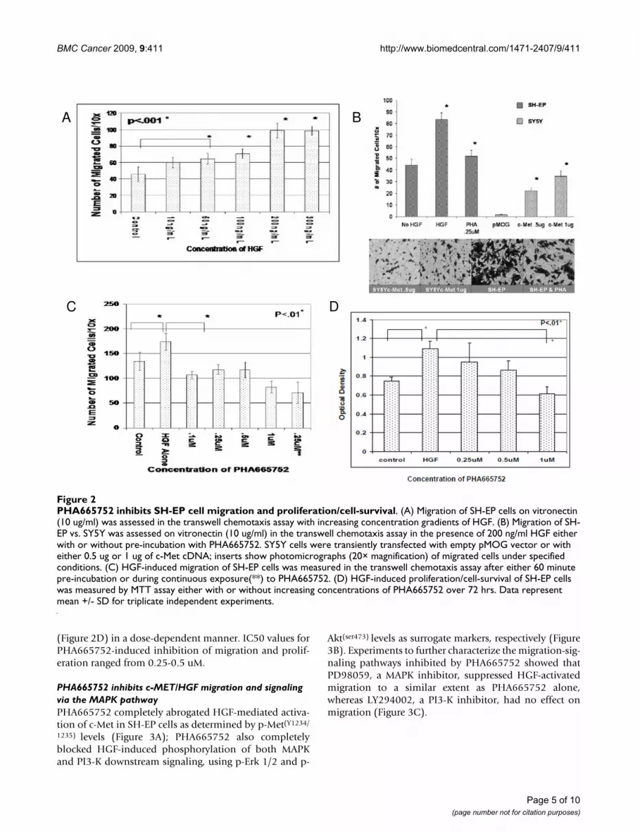
PHA665752 inhibits HGF-stimulated migration and proliferation/cell-survival of c-Met-positive neuroblastoma cellsIn a semi-quantitative wound-healing assay, we foundthat SH-EP cells migrated in response to HGF, and thisresponse was greater than that observed with low-c-Metexpressing SH-SY5Y cells (data not shown). In the tran-swell chemotaxis assay, SH-EP cells demonstrated a dose-dependent migration response to HGF (Figure 2A). Trans-fection experiments showed that only SH-SY5Y cells trans-fected with c-Met migrated in response to HGF;furthermore, response correlated with the amount oftransfected c-Met DNA (Figure 2B). In the MTT-prolifera-tion assay, SH-EP cells showed a proliferative response toHGF in both 72-hr (Figure 2D) and 7-day growth assays(data not shown). PHA665752 inhibited both HGF-medi-ated migration (Figure 2C) and proliferation/cell-survival
Expression of c-Met and HGF by SH-EP and SH-SY5Y neuroblastoma cell linesFigure 1Expression of c-Met and HGF by SH-EP and SH-SY5Y neuroblastoma cell lines. Quantitative RT-PCR assay for c-Met (A) and HGF (C) was performed in triplicate using relative quantification, with expression values normalized against GAPDH. Western blots for the 140 kD c-Met beta-chain (B) and 82 kD HGF (D) proteins, with SKN-AS neuroblastoma cell line as the positive control for HGF protein expression. β-Actin serves as loading control.
A B
C D
Page 4 of 10(page number not for citation purposes)
BMC Cancer 2009, 9:411 http://www.biomedcentral.com/1471-2407/9/411
(Figure 2D) in a dose-dependent manner. IC50 values forPHA665752-induced inhibition of migration and prolif-eration ranged from 0.25-0.5 uM.
PHA665752 inhibits c-MET/HGF migration and signaling via the MAPK pathwayPHA665752 completely abrogated HGF-mediated activa-tion of c-Met in SH-EP cells as determined by p-Met(Y1234/
1235) levels (Figure 3A); PHA665752 also completelyblocked HGF-induced phosphorylation of both MAPKand PI3-K downstream signaling, using p-Erk 1/2 and p-
Akt(ser473) levels as surrogate markers, respectively (Figure3B). Experiments to further characterize the migration-sig-naling pathways inhibited by PHA665752 showed thatPD98059, a MAPK inhibitor, suppressed HGF-activatedmigration to a similar extent as PHA665752 alone,whereas LY294002, a PI3-K inhibitor, had no effect onmigration (Figure 3C).
PHA665752 inhibits SH-EP cell migration and proliferation/cell-survivalFigure 2PHA665752 inhibits SH-EP cell migration and proliferation/cell-survival. (A) Migration of SH-EP cells on vitronectin (10 ug/ml) was assessed in the transwell chemotaxis assay with increasing concentration gradients of HGF. (B) Migration of SH-EP vs. SY5Y was assessed on vitronectin (10 ug/ml) in the transwell chemotaxis assay in the presence of 200 ng/ml HGF either with or without pre-incubation with PHA665752. SY5Y cells were transiently transfected with empty pMOG vector or with either 0.5 ug or 1 ug of c-Met cDNA; inserts show photomicrographs (20× magnification) of migrated cells under specified conditions. (C) HGF-induced migration of SH-EP cells was measured in the transwell chemotaxis assay after either 60 minute pre-incubation or during continuous exposure(**) to PHA665752. (D) HGF-induced proliferation/cell-survival of SH-EP cells was measured by MTT assay either with or without increasing concentrations of PHA665752 over 72 hrs. Data represent mean +/- SD for triplicate independent experiments.
A B
C D
Page 5 of 10(page number not for citation purposes)

BMC Cancer 2009, 9:411 http://www.biomedcentral.com/1471-2407/9/411
PTEN induction augments the inhibitory effect of PHA66572 on HGF-mediated proliferation and migrationTo evaluate PTEN's ability to potentiate the effects ofPHA665752 on c-Met/HGF signaling, we treated SH-EPcells with rosiglitazone, an inducer of PTEN expression[34]. To assess rosiglitazone's effect on proliferation, SH-EP cells were grown in the presence or absence of HGF,with or without rosiglitazone. Rosiglitazone had no effecton SH-EP proliferation in the absence of HGF, although itsomewhat reduced HGF-stimulated proliferation (Figure4A). Importantly, combined PHA665752 and rosiglita-zone was significantly (p < .01) more inhibitory for HGF-stimulated SH-EP cell proliferation than was either agentalone HGF (Figure 4A). Furthermore, migration ofPHA665752-treated SH-EP cells was significantly reducedwhen pretreated with rosiglitazone, demonstrating thatrosiglitazone augments the migration-inhibitory effects of
PHA665752, although the magnitude of this effect wasless than that on NBL cell growth (Figure 4B). Rosiglita-zone's inhibitory effects on HGF-stimulated proliferation/cell-survival and migration correlated with greater thantwo-fold inductionof PTEN protein as shown by immu-noblotting (Figure 4C).
Expression levels of c-Met mRNA in primary NBL tumor tissue correlates with advanced clinical stageTo evaluate the possible clinical significance of c-Metexpression in NBL, we used quantitative RT-PCR to deter-mine c-Met expression levels in mRNA collected from 20primary neuroblastoma tumors at different clinical stages.Seven tumors were stage 4, five were stage 3, two werestage 2, and six were stage 1. Tumors from patients withmore advanced clinical stages (stages 3 and 4) generallyhad higher c-Met expression levels than did tumors from
PHA665752 inhibits HGF-induced migration through blockade of MAPK pathwayFigure 3PHA665752 inhibits HGF-induced migration through blockade of MAPK pathway. (A) SH-EP cells were pretreated for 60 minutes with PHA665752, PD98059 or LY294002 as shown, washed, and analyzed by transwell chemotaxis assay in presence of HGF (200 ng/mL). (B, C) SH-EP cells were pretreated with inhibitors for 60 minutes, followed by addition of HGF (200 ng/mL for 15 mins) and immunoblotting of cell lysates. (in Figure 3C, # indicates incubation with vitronectin alone without HGF; + indicates addition of HGF). Data represent mean +/- SD for triplicate independent experiments.
A B
C
Page 6 of 10(page number not for citation purposes)

BMC Cancer 2009, 9:411 http://www.biomedcentral.com/1471-2407/9/411
patients with stages 1 and 2. Four of six stage-4 tumorsand one of five stage-3 tumors had c-Met values greaterthan the corresponding value for SH-EP cells, whereas nostage-1 or 2 tumors had c-Met values in this range (Figure5).
DiscussionIn this study, we investigated the expression and role of c-Met in NBL. We found that primary tumor cells frompatients with clinically aggressive, advanced-stage NBLexpressed high-levels of c-Met. Tumor cells from patientswith metastatic tumor (stage 4) or locally advanced tumor(stage 3) expressed significantly higher c-Met levels thanthose from patients with localized disease, i.e. stages 1and 2. This novel finding strongly suggests that inhibitingc-Met may have therapeutic value in this disease. Accord-
ingly, we chose to test a c-Met inhibitor (PHA665752)with high specificity and potency for blocking c-Met func-tion.
To evaluatethe efficacy of PHA66572 on the migrationand proliferation of c-Met expressing NBL cells, we used ac-Met-positive NBL line (SH-EP) as an in vitro model. Wefound that activation of the c-Met/HGF/SF pathwayresulted in increased migration and proliferation-surviv-alof SH-EP cells but not of c-Met-negative SH-SY5Y cells.This is the first report of the effects of specifically blockingc-Met in NBL cells. Our results agree with those reportedby Hecht et al, who showed that exposure of c-Met-expressing NBL cell lines to exogenous HGF resulted in c-Met phosphorylation and induction of migration [19].These investigators were also able to inhibit migration of
PHA665752-mediated inhibition of proliferation and migration is augmented by PTEN-agonist rosiglitazoneFigure 4PHA665752-mediated inhibition of proliferation and migration is augmented by PTEN-agonist rosiglitazone. (A) SH-EP cells were cultured with HGF (200 ng/ml) and either PHA665752 (0.25 uM), PPAR-γ agonist rosiglitazone (10 uM), or both PHA665752 and rosiglitazone for 72 hrs; HGF alone served as a control. Proliferation/cell-survival was analyzed by MTT assay. (B) SH-EP cells were exposed to 10 uM rosiglitazone and 0.25 uM PHA665752 overnight, washed, and assessed for migration in the presence of 200 ng/ml HGF in the transwell chemotaxis assay (+ indicates addition of HGF). (C) PTEN and p-Akt expression were measured in lysates of SH-EP cells after exposure to 10 uM rosiglitazone for stated time period. Data represent mean +/- SD for triplicate independent experiments.
A B
C
Page 7 of 10(page number not for citation purposes)

BMC Cancer 2009, 9:411 http://www.biomedcentral.com/1471-2407/9/411
NBL cells in Matrigel using an HGF-specific neutralizingantibody and MAPK/ERK inhibitors such as PD98059,respectively. Similarly, Hov et al reported that c-Met acti-vation stimulated both proliferation and migration ofmyeloma cells in vitro [22]. A role for c-Met in NBL cellmigration is further suggested by our finding that HGFtriggered migration of SH-SY5Y cells only after transfec-tion with c-Met.
In our experiments to evaluate the effects of PHA665752,we found that this small-molecule inhibitor was able toblock HGF-induced phosphorylation of both c-Met anddownstream signaling proteins Akt and Erk 1/2 (p44/42).Studies of myeloma and carcinoma cells have yielded sim-ilar results [22,26,27]. However, in the latter studies,tumor cells were exposed to PHA665752 continuously,whereas in the present study NBL cells were only brieflyexposed to low concentrations of this agent. This suggeststhat NBL cells may be more sensitive to c-Met targetingthan some other c-Met expressing tumors.
We also found that PHA665752 showed a marked dose-dependent inhibitory effect on the HGF/c-Met pathway ofproliferation and migration in c-Met-expressing NBL cells.These inhibitory effects appeared to be specific for HGF-stimulated proliferation/migration, since PHA665752
had no significant effects on these parameters in theabsence of HGF stimulation. Thus, PHA665752 couldpotentially inhibit HGF-stimulated tumor proliferation/migration resulting from either paracrine (i.e. tumormicroenvironment) or autocrine exposure to HGF. Fur-thermore, brief exposure to PHA665752 completelyblocked downstream signaling via the PI3-K/Akt andMAPK/Erk pathways, agreeing with results from studies inseveral types of carcinoma [25,27]. Additional studieswith MAPK/Erk and PI3-K/Akt pathway-specific inhibi-tors suggested that PHA665752 can inhibit c-Met/HGF/SF-stimulated migration and proliferation/survival via theMAPK/Erk and PI3-K/Akt pathways, respectively.
Since PHA665752 inhibits the PI3-K/Akt pathway, wehypothesized that its effect might be enhanced by rosigli-tazone, an inducer of PTEN expression. Indeed, combinedPHA665752 and rosiglitazone induced significantlygreater inhibition of both HGF-stimulated proliferation/cell-survival and migration in c-Met-expressing NBL cellsthan did PHA665752 alone. This finding suggests that thecombination of PTEN-inducing agents with small-mole-cule inhibitors or drugs that block c-Met/HGF/SF signal-ing may have an augmented anti-tumor effect.
Although we do not expect PHA665752 will be suitablefor clinical use due to its tendency to form pulmonary pre-cipitates in animal studies [17], we believe this agent pro-vides an excellent tool for studying c-Met function in NBLdue to its high specificity and activity. Finally, our studysupports the notion that c-Met blockade, either throughderivatives of PHA665752 with higher bioavailability orthrough other agents targeting this receptor, deserves fur-ther study as a potential therapeutic strategy for NBL.
ConclusionElevated c-Met expression is more commonly observed inprimary NBL tumor tissue from patients with metastatictumors. Furthermore, the small-molecule inhibitorPHA665752 is capable of antagonizing HGF-inducedmigration and proliferation/survival of c-Met-expressingNBL cells, an effect which is enhanced by upregulation ofPTEN. These are novel findings in NBL and suggest a ther-apeutic potential for targeting c-Met in this tumor.
Competing interestsOne of the co-authors (JGC) is employed by Pfizer, whichprovided the drug under study (PHA665752).
Authors' contributionsHEC, HWF, and CSA designed the study and wrote themanuscript. HEC also performed the migration, prolifera-tion and transfection experiments with the assistance ofTW, DLD and PD. AD grew the cell lines and performedqRT-PCR studies on tumor tissue, as well as assisted in
Advanced clinical stage in patients with neuroblastoma corre-lates with high c-Met mRNA levelsFigure 5Advanced clinical stage in patients with neuroblast-oma correlates with high c-Met mRNA levels. mRNA was isolated from banked tumor samples from patients with localized or metastatic neuroblastoma. Quantitative RT-PCR for c-Met was performed using GADPH as internal control and the SH-EP line as a positive control. Values are expressed relative to the SH-SY5Y line, which is set equal to one; thus, SH-EP c-Met value = 15.8. c-Met values represent the mean of triplicate samples and are plotted according to INSS clinical stage. Median c-Met values for each stage are 0.87 (stage 1), 2.6 (stage 2), 5.2 (stage 3), and 17.5 (stage 4). c-Met values for stage 3-4 tumors are significantly higher (p = 0.048) than values for stage 1-2 tumors.
Page 8 of 10(page number not for citation purposes)

BMC Cancer 2009, 9:411 http://www.biomedcentral.com/1471-2407/9/411
MTT/viability assays for proliferation/cell-survival. JGCkindly provided PHA665752 and advice regarding exper-imental design. All authors read and approved the manu-script.
AcknowledgementsThis work was supported by grants from CURE Childhood Cancer, the Emory-Egleston Children's Research Council, the Georgia Cancer Coali-tion, and CA94233 from the NIH. H.E.C was the recipient of a CURE Child-hood Cancer Fellowship award
References1. Maris JM, Hogarth MD, Bagatelle R, Cohn SL: Neuroblastoma. Lan-
cet 2007, 369:2106-20.2. Ma PC, Maulik G, Christensen J, Salgia R: c-Met: Structure, func-
tions and potential for therapeutic inhibition. Cancer & MetRev 2003, 22:309-25.
3. Hammond DE, Carter S, Clague MJ: Met receptor dynamics andsignalling. Curr Topics Microbiol & Immunol 2004, 286:21-44.
4. Furge KA, Zhang YW, Woude GF Vande: Met receptor tyrosinekinase: enhanced signaling through adapter proteins. Onco-gene 2000, 19:5582-89.
5. Fan S, Ma YX, Gao M, Yuan RQ, Meng Q, Goldberg ID, Rosen EM:The multisubstrate adapter Gab1 regulates hepatocytegrowth factor (scatter factor)-c-Met signaling for cell sur-vival and DNA repair. Mol & Cell Biol 2001, 21:4968-4984.
6. Abounader R, Ranganathan S, Kim BY, Nichols C, Laterra J: Signalingpathways in the induction of c-met receptor expression byits ligand scatter factor/hepatocyte growth factor in humanglioblastoma. J Neurochem 2001, 76:1497-508.
7. Maulik G, Madhiwala P, Brooks S, Ma PC, Kijima T, Tibaldi EV,Schaefer E, Parmar K, Salgia R: Activated c-Met signals throughPI3K with dramatic effects on cytoskeletal functions in smallcell lung cancer. J Cell & Mol Med 2002, 6:539-53.
8. Gherardi E, Sandin S, Petoukhov MV, Finch J, Youles ME, OfverstedtL-G, Miguel RN, Blundell TL, Woude GF Vande, Skoglund U, SvergunDI: Structural basis of hepatocyte growth factor/scatter fac-tor and MET signaling. Proc Natl Acad Sci USA 2006, 103:4046-51.
9. Birchmeier C, Birchmeier W, Gherardi E, Woude GF Vande: Met,metastasis, motility and more. Nat Rev Mol Cell Biology 2003,4:915-25.
10. Danilkovitch-Miagkova A, Zbar B: Dysregulation of Met receptortyrosine kinase activity in invasive tumors. J Clin Invest 2002,109:863-7.
11. Gao CF, Woude GF Vande: HGF/SF-Met signaling in tumor pro-gression. Cell Res 2005, 15:49-51.
12. Jankowski K, Kucia M, Wysoczynski M, Reca R, Zhao D, Trzyna E,Trent J, Peiper S, Zembala M, Ratajczak J, Houghton P, Janowska-Wieczorek A, Ratajczak MZ: Both hepatocyte growth factor(HGF) and stromal-derived factor-1 regulate the metastaticbehavior of human rhabdomyosarcoma cells, but only HGFenhances their resistance to radiochemotherapy. Cancer Res2003, 63:7926-35.
13. Ma PC, Kijima T, Maulik G, Fox EA, Sattler M, Griffin JD, Johnson BE,Salgia R: c-MET mutational analysis in small cell lung cancer:novel juxtamembrane domain mutations regulatingcytoskeletal functions. Cancer Res 2003, 63:6272-8.
14. Puri N, Khramtsov A, Ahmed S, Nallasura V, Hetzel JT, JagadeeswaranR, Karczmar G, Salgia R: A selective small molecule inhibitor ofc-Met, PHA66 inhibits tumorigenicity and angiogenesis inmouse lung cancer xenografts. Cancer Res 5752, 67:3529-34.
15. Jeffers M, Rong S, Anver M, Woude GF Vande: Autocrine hepato-cyte growth factor/scatter factor-Met signaling inducestransformation and the invasive/metastastic phenotype inC127 cells. Oncogene 1996, 13:853-6.
16. Takayama H, La Rochelle WJ, Sharp R, Otsuka T, Kriebel P, Anver M,Aaronson SA, Merlino G: Diverse tumorigenesis associatedwith aberrant development in mice overexpressing hepato-cyte growth factor/scatter factor. Proc Natl Acad Sci USA 1997,94:701-706.
17. Accornero P, Lattanzio G, Mangano T, Chiarle R, Taulli R, Bersani F,Forni PE, Miretti S, Scuoppo C, Dastru W, Christensen JG, CrepaldiT, Ponzetto C: An In vivo Model of Met-Driven Lymphoma as
a Tool to Explore the Therapeutic Potential of Met Inhibi-tors. Clin Cancer Res 2008, 14:2220-26.
18. Ferracini R, Di Renzo MF, Scotlandi K, Baldini N, Olivero M, Lollini P,Cremona O, Campanacci M, Comoglio PM: The Met/HGF recep-tor is over-expressed in human osteosarcomas and is acti-vated by either a paracrine or an autocrine circuit. Oncogene1995, 10:739-49.
19. Hecht M, Papoutsi M, Tran HD, Wilting J, Schweigerer L: Hepato-cyte growth factor/c-Met signaling promotes the progres-sion of experimental human neuroblastomas. Cancer Res 2004,64:6109-18.
20. Cruz J, Reis-Filho JS, Silva P, Lopes JM: Expression of c-met tyro-sine kinase receptor is biologically and prognostically rele-vant for primary cutaneous malignant melanomas. Oncology2003, 65:72-82.
21. Qian C-N, Guo X, Cao B, Kort EJ, Lee C-C, Chen J, Wang L-M, MaiW-Y, Min H-Q, Hong M-H, Woude GF Vande, Resau JH, Teh BT:Met protein expression level correlates with survival inpatients with late-stage nasopharyngeal carcinoma. CancerResearch 2002, 62:589-96.
22. Hov H, Holt RU, Ro TB, Figural U-M, Mort-Hansen H, Bayou V,Christensen JG, Wage A, Sudan A, Borsht M: A selective c-metinhibitor blocks an autocrine hepatocyte growth factor loopin ANBL-6 cells and prevents migration and adhesion ofmyeloma cells. Clin Cancer Res 2004, 10:6686-6694.
23. Choi YL, Tsakasaki L, O'Neill MC, Yamada Y, Onimaru Y, MatsumotoK, Ohashi J, Yamashita Y, Tsatsumi S, Kaneda R, Takada S, AburatamiH, Kamihira S, Nakamura T, Tononaga M, Mano H: A genomic anal-ysis of adult T-cell leukemia. Oncogene 2007, 26:1245-55.
24. Christensen JG, Burrows J, Salgia R: c-Met as a target for humancancer and characterization of inhibitors for therapeuticintervention. Cancer Lett 2005, 225:1-26.
25. Chattopadhyay C, El-Naggar AK, Williams MD, Clayman GL: SmallMolecule c-Met Inhibitor PHA665752: Effect on Cell Growthand Motility in Papillary Thyroid Carcinoma. Head & Neck5752, 30:991-1000.
26. Watson GA, Zhang X, Stang MT, Levy RM, Queiroz de Oliverira PE,Gooding WE, Christensen JG, Hughes SJ: Inhibition of c-Met as atherapeutic strategy for esophageal adenocarcinoma. Neo-plasia 2006, 8:949-955.
27. Christensen JG, Schreck R, Burrows J, Kuruganti P, Chan E, Le P,Chen J, Wang X, Ruslim L, Blake R, Lipson KE, Ramphal J, Do S, CuiJJ, Cherrington JM, Mendel DB: A selective small molecule inhib-itor of c-Met kinase inhibits c-Met-dependent phenotypes invitro and exhibits cytoreductive antitumor activity in vivo.Cancer Res 2003, 63:7345-55.
28. Smolen GA, Sordella R, Muir B, Mohapatra G, Barmettler A,Archibald H, Kim WJ, Okimoto RA, Bell DW, Sgroi DC, ChristensenJG, Settleman J, Haber DA: Amplification of MET may identify asubset of cancers with extreme sensitivity to the selectivetyrosine kinase inhibitor PHA-665752. Proc Natl Acad Sci USA2006, 103:2316-21.
29. Chung JH, Eng C: Nuclear-cytoplasmic partitioning of phos-phatase and tensin homologue deleted on chromosome 10(PTEN) differentially regulates cell cycle and apoptosis. Can-cer Res 2005, 65:8096-100.
30. Di Cristofano A, Pandolfi PP: The multiple roles of PTEN intumor suppression. Cell 2000, 100:387-90.
31. Wen S, Stolarov J, Myers MP, Su JD, Wigler MH, Tonks NK, DurdenDL: PTEN controls tumor-induced angiogenesis. Proc NatlAcad Sci USA 2001, 98:4622-7.
32. Moritake H, Horii Y, Kuroda H, Sugimoto T: Analysis of PTEN/MMAC1 alteration in neuroblastoma. Cancer Gent & Cytogenet2001, 125:151-5.
33. Farrow B, Evers BM: Activation of PPARgamma increasesPTEN expression in pancreatic cancer cells. Biochem & BiophysRes Com 2003, 301:50-3.
34. Patel L, Pass I, Coxon P, Downes CP, Smith SA, Macphee CH:Tumor suppressor and anti-inflammatory actions of PPAR-gamma agonists are mediated via upregulation of PTEN.Curr Biol 2001, 11:764-8.
35. Ross RA, Biedler JL, Spengler BA: A role for distinct cell types indetermining malignancy in human neuroblastoma cell linesand tumors. Cancer Lett 2003, 197:35-9.
Page 9 of 10(page number not for citation purposes)

BMC Cancer 2009, 9:411 http://www.biomedcentral.com/1471-2407/9/411
Publish with BioMed Central and every scientist can read your work free of charge
"BioMed Central will be the most significant development for disseminating the results of biomedical research in our lifetime."
Sir Paul Nurse, Cancer Research UK
Your research papers will be:
available free of charge to the entire biomedical community
peer reviewed and published immediately upon acceptance
cited in PubMed and archived on PubMed Central
yours — you keep the copyright
Submit your manuscript here:http://www.biomedcentral.com/info/publishing_adv.asp
BioMedcentral
36. Klemke RL, Leng J, Molander R, Brooks PC, Vuori K, Cheresh D:CAS/Crk coupling serves as a "molecular switch" for induc-tion of cell migration. J Cell Biol 1998, 140:961-72.
37. Pradip D, Peng X, Durden DL: Rac2 specificity in macrophageintegrin signaling: potential role for Syk kinase. J Biol Chem2003, 278:4161-9.
Pre-publication historyThe pre-publication history for this paper can be accessedhere:
http://www.biomedcentral.com/1471-2407/9/411/prepub
Page 10 of 10(page number not for citation purposes)


