Embed Size (px)
Citation preview
481
TISSUE ENGINEERINGVolume 6 Number 5 2000Mary Ann Liebert Inc
Hepatocyte Encapsulation for Enhanced Cellular Functions
SER-MIEN CHIA BS1 KAM W LEONG PhD2 JUN LI PhD3 XI XU MS3
KAIYANG ZENG PhD4 POH-NEE ER BS1 SHUJUN GAO MS3
and HANRY YU PhD1
ABSTRACT
An efficient bioartificial liver-assisted device can sustain the lives of patients with acuteliver failure Among different configurations of the bioreactor design hepatocyte encap-sulation has important features that satisfy most requirements of the device We have en-capsulated rat hepatocytes in a two-layer polymeric membrane by complex coacervationusing a simple setup and demonstrated enhanced cellular functions up to three timeshigher than those of the monolayer control These microcapsules of the functioning he-patocytes have a 2- to 3-mm outer layer of synthetic polymer with 25 2-hydroxyethylmethacrylate 25 methacrylic acid and 50 methyl methacrylate and an inner layerof positively charged modified collagen as a suitable substrate for the enhanced cellularfunctions Permeable only to small molecules up to albumin the microcapsules should al-low unimpeded exchange of nutrients oxygen growth factors and metabolites but pre-vent attack by immunoglobulins of the immune system and no ldquoskin effectrdquo of the col-lagen has been observed Mechanical properties of the microcapsules measured with anano-indentation method suggest that the microcapsules should be suitable for use in abioartificial liver-assisted device
INTRODUCTION
ACUTE LIVER FAILURE is a disease with high mortality The medical cost associated with caring for thepatients with liver diseases is high and the incidents are increasing every year worldwide1 If the lives
of the patients with acute liver failure can be sustained long enough there is a good chance that the pa-tientsrsquo own liver can regenerate or a liver can be transplanted Therefore research to engineer a functionalartificial liver support with the aim of temporarily substituting liver functions is important as a life-savingsolution (UNOS Scientific Registry 1996 report and On Line Data)
1National University Medical Institutes National University of Singapore Singapore2Department of Biomedical Engineering Johns Hopkins University Baltimore Maryland3Biomaterials Program-Tissue Engineering Institute of Materials Research amp Engineering Singapore4Central Characterization Laboratory Institute of Materials Research amp Engineering Singapore
Hybrid bioartificial liver-assisted devices (BLAD) are extracorporeal devices that involve a biologicaland synthetic component coupled in such a way that will temporarily substitute for the functions of the pa-tientsrsquo failed liver The core of the device is a bioreactor where hepatocytes are grown2 There are severalcrucial considerations in the design of the bioreactor such as maintaining the optimal hepatocyte functionsallowing maximal density of hepatocytes to be cultured in the bioreactor protecting the xenogenic hepato-cytes from immunological attack and optimizing the mass transport of nutrients waste products and tox-ins2 Hepatocytes lose their functions readily in culture because they are anchorage-dependent and sensi-tive to environment factors such as density and nature of the neighboring cells3 Under suboptimal culturingconditions hepatocytes start to spread and proliferate while losing most of the characteristic differentiatedhepatocyte functions Therefore it is important to design a bioreactor that satisfies most of these require-ments to develop an effective liver-assisted device The most commonly used bioreactor configurations in-volve the culture of hepatocytes on or in hollow-fiber membranes microcarriers microcapsules or poly-mer foams Each set of configuration has its own advantages and disadvantages4
Microencapsulation of biological substances was first described three decades ago3 Microcapsules arecomposed of ultrathin semipermeable membranes of cellular dimensions They can be prepared of variouspolymers and their contents can consist of enzymes cells and other biological materials Microcapsulesare prepared in such a way as to prevent their contents from leaking out and causing an immunological re-action but the microcapsules still allow the nutrients and metabolites to exchange freely This method hasfound applications primarily in transplantation of foreign materials in vivo without immunosuppression Foruse in BLAD microencapsulation of hepatocytes is suitable because the high surface-to-volume ratio of aspherical microcapsule facilitates maximal transport of nutrients gases or metabolites exchange across themembrane4 In addition encapsulation of hepatocytes allows better control of the microenvironment for op-timal cellular functions via selection of suitable substrate and incorporation of controlled-release featuresinto the local microenvironment Other physical characteristics such as mass transport and mechanical andchemical stability can also be configured as desired without drastically affecting the functions of the hepa-tocytes inside the microcapsules Therefore hepatocyte encapsulation has important features to satisfy mostrequirements for an ideal bioreactor design
The commonly used techniques for cell encapsulation are complex coacervation and interfacial precipi-tation Complex coacervation involves the electrostatic interaction of two oppositely charged polyelec-trolytes At the right matching charge density the two polyions combine and migrate to form a colloid-richor water-insoluble phase The molecular weight and chain conformation parameters of the polyions mayalso play an important role in the complexation process Interfacial precipitation simply relies on the so-lidification of a dissolved polymer upon contact with an aqueous phase
The most extensively studied cell encapsulation scheme5 is the one that involves an alginatendashgelationcomplex coacervation method In this system alginate a glycuranan extracted from the brown seaweed al-gae can be chelated by calcium or other multivalent counterions to form a gel6 These early in vivo resultswith the alginatendashpolylysine system have not been consistent because of the uncontrolled purity of alginateand the incorporation of cells into the external membrane As a result a two-step encapsulation5 was de-veloped to shield sensitive cells further such as hepatocytes from the extracapsular environment The liv-ing hepatocytes were mixed with sodium alginate and extruded into calcium chloride to form calcium al-ginate gel droplets These gel droplets were incorporated into larger alginate gel spheres and then reactedwith a poly-amino acid such as poly-L-lysine to form a semipermeable membrane Incubating with sodiumcitrate liquefied the interior to form microcapsules Unfortunately the addition of sodium citrate seemed tohave affected the functions of the hepatocytes5 Furthermore the water-soluble alginate and poly-lysinewere shown to be not particularly biocompatible as individual polymers other matrices such as collagenmay be better substrates for cellular functions than alginate7
To encapsulate hepatocytes or hepatoma cells (HepG2) in natural matrices such as collagen interfacialprecipitation has been used In this method hydroxylethylmethacrylate-methylmethacrylate (HEMA-MMA)solution in dimethyl formamide and cell suspension in collagen or Matrigel were extruded separately throughtwo concentrically configured needles into a precipitating bath containing largely water with a floating layerof dodecane8 Polyacrylates are water-insoluble substances that enhance the in vivo stability of the micro-capsules9 The hepatoma cells encapsulated this way (especially with Matrigel) survive well8 The interfa-
CHIA ET AL
482
cial precipitation requires a more elaborate setup than the complex coacervation to control the microcap-sule sizes and minimize the contact of cells with organic solvents
Here we use a terpolymer of hydroxylethyl methacrylate-methyl methacrylate-methylacrylic acid(HEMA-MMA-MAA) to encapsulate hepatocytes The MAA added into the terpolymer enhances the wa-ter solubility of the polymer allowing the entire encapsulation to be carried out in an aqueous environmentHence we were able to use the complex coacervation method that required a simple setup for easy controlof the microcapsule size The resulting hepatocytes microcapsules exhibit enhanced cellular functions aswell as desirable physical characteristics for use in BLAD
MATERIALS AND METHODS
All reagents were purchased from Sigma-Aldrich unless otherwise stated
Terpolymer preparation
The terpolymers of MAA HEMA and MMA were synthesized (Fig 1) by solution polymerization in2-propanol using 229-azobisisobutyronitrile (AIBN) as initiator The monomers were distilled under nitro-gen at reduced pressure The polymerization was performed with an initiator concentration of 01 mol ofmonomers under nitrogen with a magnetic stirrer at 78degC in an oil bath The feed ratio of MAA HEMAand MMA was fixed at 252550 (molmol) or other ratios as desired (such as 255025) and the ratio oftotal monomer to solvent at 16 (wtvol) The reaction was allowed to proceed for overnight and quenchedby cooling to room temperature The polymer was precipitated by addition to a large excess of petroleumether The precipitate was redissolved in a minimum volume of ethanol and reprecipitated in distilled wa-ter Recovered polymer was then dissolved in a 1 M sodium hydroxide solution and further purified by re-peated dialysis against distilled water with molecular-weight cut-off (MWCO) of 3500 and lyophilizedThe yield of the polymer was found to be 63 for the terpolymer with 25MAA25HEMA50MMAThe polymer composition was determined by proton nuclear magnetic resonance (NMR) and the actual ra-tio of MAA HEMA and MMA was found to be 204274522 (molmol) for the feed ratio of 252550and 200560240 (molmol) for the feed ratio of 255025 The molecular weights of the terpolymer afterdialysis were determined by gel permeation chromatography [with tetrahydrofuran (THF) as eluent] to be113000 and 373000 respectively
Modification of collagen
Collagen can be modified to be cationic and anionic by the removal of either the negative or the posi-tive charge from the collagen chains In our case cationic collagen was obtained through the modificationof the carboxyl group by esterification with low-molecular-weight alcohol A total of 20 mL of stock so-lution (3 mgmL) of collagen (Vitrogen 100 Collagen Corp Palo Alto CA) was first precipitated with 400mL of acetone The precipitated collagen was dissolved in 200 mL of 01 M HCl containing methanol(Merck) stirred at 4degC for 6 days under sterile conditions The modified collagen solution was then dia-lyzed against distilled water for an additional 4 days at 4degC followed by freeze-drying The lyophilized
HEPATOCYTE ENCAPSULATION
483
FIG 1 Polyelectrolytes used in the complex coacervation to form microcapsules A chemical reaction depicting thesynthesis of the HEMA-MMA-MAA terpolymer
modified collagen can then be stored up to 6 months in 220degC in the presence of desiccant The modifi-cation was monitored by titration
Isolation of hepatocytes
Hepatocytes were harvested from male Wistar rats weighing from 250 to 300 g by a two-step in situ col-lagenase perfusion as described previously10 with some modifications The rat was given 100 Ukg of hep-arin 30 min before anesthesia pentobarbital was administered at a dose of 30 mgkg intraperitoneally atthe start of the operation After laparotomy a portal cannula was placed and fixed in a position along theportal vein A cut was rapidly made in the lower vena cava In the initial first 2ndash3 min preperfusion (withCa21-free perfusion buffer) was performed while the liver remained in situ The perfusate flow was startedat a rate of 50 mL per minute While preperfusion was carried out the liver was transferred to a petri dishand placed in a position similar to its in situ site After 10 min of preperfusion with Ca21-free medium theliver was then perfused with recirculating 005 collagenase buffer for another 10 min This was termi-nated when the vena cava ruptured The entire perfusion procedure was performed under oxygenation thatgreatly improved the cell viability The cells were liberated from the connective vascular tissue and resus-pended in fresh growth medium This was followed by incubation of the cell suspension in a 37degC CO2 in-cubator for 30 min The cell suspension was then filtered through a nylon mesh with a 60-mm pore size tofurther remove the connective tissue debris The filtrate was then centrifuged at 50 g for 1 min to obtainthe cell pellet The cells were collected and washed twice with growth medium The viability of the hepa-tocytes was determined to be 90ndash95 in all cases using the conventional Trypan Blue exclusion test10
Preparation of microencapsulated hepatocytes
Microencapsulation was performed at room temperature with the aid of a syringe pump (IVAC P6000Alaris Medical Systems San Diego CA) Briefly the hepatocytes were suspended in a modified collagensolution that was prepared by first dissolving the lyophilized modified collagen in phosphate-buffered saline(PBS) to obtain a collagen concentration of 15 mgmL The hepatocyte suspension was maintained at 4degCbefore the experiment to prevent the gelation of the collagen solution The hepatocyte suspension was ex-truded from an 185-gauge needle attached to the syringe pump at various flow rates (Table 1) into a 10terpolymer solution11 Thus the positively charged collagen molecules bound to the negatively charged ter-polymer molecules at the outer surface of the microcapsules to form a polyelectrolyte complex The mi-crocapsules were incubated at 37degC for 1 h to allow the collagen to gel before the microcapsules were har-vested by sedimentation method and washed twice with 13 PBS for further culturing
Preparation of spheroids
Spheroids were prepared as described previously12 using the static culture condition Freshly harvestedhepatocytes were inoculated at 3ndash8 3 104 cellscm2 in 60 3 15 mm Primaria dishes in 5 mL of the spher-oid medium12 and incubated at 37degC 5 CO2 The medium was changed at 24 h At that time the hepa-tocytes were attached in a monolayer and began to aggregate Floating spheroids were harvested three daysafter inoculation and cultured as described previously12
CHIA ET AL
484
TABLE 1 NUMBER OF ENCAPSULATED HEPATOCYTES PER MICROCAPSULE VARIES
WITH THE FLOW RATE OF THE HEPATOCYTE SUSPENSION EXTRUSION (n 5 30)
Flow rate (mL per hour)
Needle size 1321 1321 30 60 100 125
27 12 2 6 018 12 6 6 2 10 6 20 25 6 50 50 6 50 75 6 11Number of 46 6 05 10 6 01 10 6 01 34 6 06 12 6 03 08 6 02
capsulesmin 3 105 3 105 3 105 3 104 3 104 3 104
In vitro culture
The microcapsules were cultured for the required amount of time in Hepatozym serum-free medium(SFM11 GIBCO Laboratories Chagrin Falls OH) in a 35-mm polystyrene dish in a humidified atmospherewith 5 CO2 The culture medium was supplemented with 1027 M dexamethasone 10 nM insulin(Boehringer Mannheim) 20 ngmL epidermal growth factor and 1 penicillin and streptomycin After 1day of culture the microcapsules were incubated in the medium with 1 mM of NH4Cl for 90 min beforethe medium was collected for urea and ammonia assays The microcapsules were then cultured in freshmedium again
Permeability assay for the microcapsules
Both 1 (BSA) and 1 fluoroisothiocyanate (FITC)-dextrans molecules (with different molecularweights) were suspended in 15 mgmL of modified collagen and microcapsules were formed as usual withan outer terpolymer shell comprised of 10 terpolymer The microcapsules were incubated at 37degC for 1h for the necessary gelation of the collagen After the incubation the microcapsules were transferred to asolution containing equal concentration of BSA or FITC-dextran and allowed to equilibrate for 2 h Themicrocapsules were then washed with PBS and the release profile was obtained within a 2-h interval Aseries of dilutions ranging from 0 to 1 BSA or FITC-dextrans were used to quantify the amount of BSAor FITC-dextran released BSA level was determined using the Detergent Compatible (DC) protein assay(BioRad) whereas the FITC-dextran level was determined by measuring the absorbance at 492 nm wherelight was absorbed most by the FITC The percentage of released BSA or FITC-dextran in culture mediumper total amount of BSA or FITC-dextran encapsulated was plotted over time
Functional analysis of the microencapsulated hepatocytes
The production of albumin by the encapsulated hepatocytes was determined by performing a direct en-zyme-linked immunosorbent assay (ELISA) analysis13 First 100-mL of the samples (medium collected atvarious time points) as well as a series of known concentrations of rat albumin were coated onto a Max-isorp plate (NUNC) overnight The unabsorbed samples were discarded and washed with PBS-Tween-20(PBS-T) for 10 min The coated plate was blocked with 3 BSA for 1 h at room temperature At the endof 1 h the plate was washed once with PBS-T for 10 min Next 100 mL of the horseradish peroxidase(HRP)-conjugated anti-rat albumin antibody (ICN) at a dilution of 1200 was added into each well and theplate incubated at 37degC for an additional 2 h The amount of albumin was quantified by the conversion byHRP of O-phenylenediamine dihydrochloride a chromogen (Sigma) to its soluble end-product which hasan absorption maximum at 492 nm with reference to the standard albumin curve derived from known con-centrations of rat albumin The samples collected at each time point were assayed for the ammonia metab-olism and urea production colorimetrically with kits from Sigma Diagnostic13 Cytochrome P450 activitywas assessed12 with the quantification of the ethoxyresorufin O-dealkylation under a confocal microscope(Carl Zeiss) 7-Ethoxyresorufin is the substrate for the cytochrome P450IA1 Briefly a small quantity ofmicrocapsules were removed and allowed to settle by gravity P450 incubation buffer was added to the pel-let and incubated for 5 min Then P450 reaction buffer was added to the microcapsules and they were im-aged on the confocal microscope with a rhodamine filter within 20 min after the 7-ethoxyresorufin addi-tion Data from three independent encapsulation experiments were analyzed and values are normalizedagainst 106 cells
Light microscope imaging of the microcapsules
We viewed the microcapsules in an inverted microscope (Carl Zeiss) with phase-contrast optics andcounted the number of hepatocytes within microcapsules with the aid of a hemocytometer (Fuchs-Rosen-thal)
An Olympus FLUOVIEW confocal microscope was used to image the terpolymer and the encapsulatedhepatocytes For the labeling of terpolymer 01 g of terpolymer was stirred in 5 mL of PBS solution con-taining 0001 mg of free FITC The solution was then dialyzed against deionized water for a day and freeze-
HEPATOCYTE ENCAPSULATION
485
dried The FITC-labeled terpolymer was used to encapsulate hepatocytes as described above Confocal imag-ing of the microcapsules was performed with excitation by a 488-nm argon-ion laser Fourteen optical sec-tions were recorded for the reflective imaging of encapsulated hepatocytes Deconvolution was performedwith AutoDeblur (AutoQuant NY) using a blind deconvolution algorithm Three-dimensional reconstruc-tion and visualization with sum projection algorithm was performed with AutoVisualize-3D (AutoQuantNY) and further processed in Photoshop (Adobe CA) Terpolymer was imaged in the FITC channelprocessed in Adobe Photoshop and displayed as a single optical section The thickness of the terpolymershell was measured with the software associated with the Olympus FLUOVIEW confocal microscope
Scanning electron microscopy
The microcapsules were fixed with 3 glutaraldehye on a coverglass coated with poly-L-lysine for 1 hafter which they were washed gently with 13 PBS for 5 min The microcapsules were then post-fixed withosmium tetraoxide for 1 h and dehydration was accomplished using a graded series of ethanol (25 5075 95 and 100) The microcapsules were then critical point dried for about 2 h in absolute alcoholand mounted onto an aluminum stub and sputter coated with gold before viewing under a scanning elec-tron microscope (SEM) (Joel 5600 LV)
Nano-indentation for measurement of the hardness and elasticity of microcapsules
Indentation measurement14 was done using an UMIS-2000 Nano-indenter (Australian Scientific Instru-ments) The indenter is a three-faced pyramid diamond called a Berkovich indenter The load and depth ofpenetration were measured with two linear variable different transformer (LVDT) sensors independentlyFrom the experimentally determined load-penetration data hardness and modulus were determined throughthe following analysis14
Hardness H 5 PA
Elastic modulus 1 2
Ev2 5
ddPh
Where H is hardness of the specimen P is indentation load A is the true contact area at the maximum loadE is the elastic modulus of the microcapsules and v is Poissonrsquos ratio dPdh is called unloading stiffnessand essentially it is the slope of unloading portion of the indentation load penetration data at the maxi-mum indentation load Average pressure that the microcapsules can withstand under a sharp point can bedefined by applied load divided by contact area The area of the indentation is therefore related to the depthof penetration for an ideal sharp Berkovich indenter is A 5 2456h2
Microcapsules were added onto a 13-mm coverglass coated with poly-L-lysine We determined the elas-tic modulus and hardness of the microcapsules in the nano-indentation experiments with the maximum loadof about 015 mN This gave the penetration depth into the polymer of slightly less than 2 mm before rup-ture The load was applied through a piezoelectric actuator in 15 steps to the maximum load Eight to tenmicrocapsules were indented at the same load with one indent on each microcapsule
RESULTS AND DISCUSSION
Hepatocyte microcapsules
We have encapsulated hepatocytes in a two-layer polymeric membrane using a process of complex coac-ervation between two oppositely charged polymers The outer layer is made of a terpolymer of HEMA-MMA-MAA HEMA and MAA are providing the hydrophilicity of the terpolymer whereas MMA is pro-viding the mechanical strength toughness and elasticity of the microcapsules MAA is also responsible forproviding the negative charge to interact with the positively charged inner layer and to allow the terpoly-mer to be water soluble so that the entire encapsulation can be performed in the physiological aqueousbuffer without any organic solvent The terpolymer is the polymer responsible for maintaining the semi-
1Iuml Aw
Iuml pw2
CHIA ET AL
486
permeability of the microcapsules whereas the other polymer collagen is totally permeable to the nutrientsThe inner layer is an esterified collagen with net positive charge The balance between the two chargedpolymers determines the physical characteristics of the microcapsules Using a 10 terpolymer and 15mgmL of modified collagen we could form microcapsules with a thin layer of outer shell ( 2ndash3 mm) anda semi-gel-like inner layer that minimizes impedance to mass transport across the membrane but remainsstable as microcapsules for days The semi-gel-like inner collagen layer is able to provide a ldquolooserdquo extra-cellular matrix configuration15 that mimics the in vivo situation therefore allowing the microcapsules tohave higher functions We have tried to optimize the characteristics of the microcapsules to satisfy the re-quirements for a BLAD
Optimization of the hepatocyte functions in microcapsules
To optimize the functions of the hepatocytes we must use hepatocytes with consistently high viabilitybecause hepatocytes with variable viability often exhibit variable levels of functions Therefore we havedeveloped a hepatocyte-isolation procedure so that we could isolate rat hepatocytes with consistent 90ndash95viability These hepatocytes were then used to optimize conditions for microencapsulation Urea synthesiswas our primary assay in optimization because it is one of the most important indicators of the hepatocytefunctions16
The first parameter that we optimized was the number of hepatocytes within each microcapsule Becausehepatocytes prefer to be in close contact with their neighboring cells to have a high level of functions17
we encapsulated different numbers of hepatocytes within each microcapsule by varying the flow rates (Table1) with which the hepatocyte suspension was extruded into the terpolymer solution By increasing the num-ber of hepatocytes per microcapsule up to 75 hepatocytes per microcapsule (diameter 150 mm) we ob-served an increase in urea synthesis (see Fig 3) Further increase in the number of hepatocytes per micro-capsule to above 100 led to a decrease in urea synthesis It is possible that 75 hepatocytes per microcapsuleprovide the ideal microcapsule size for hepatocyte functions whereas a larger number of hepatocytes in-side each microcapsule might lead to necrosis of the hepatocytes in the center of the microcapsules Withthe complex coacervation method we have been able to encapsulate easily a small number of hepatocytesin the microcapsule for enhanced functions and efficient mass transport In other polyacrylate systems based
HEPATOCYTE ENCAPSULATION
487
FIG 2 Imaging of the microcapsules (A) A confocal micrograph (deconvolution and sum projection of the recon-structed three-dimensional volume in x-y plane) of the encapsulated hepatocytes imaged in reflective mode shows thatthe encapsulated hepatocytes do not form tight aggregates The insert is an optical section of a microcapsule demon-strating that the FITC-labeled terpolymer forms a thin shell ( 2ndash3 mm) around the microcapsule (B) A scanning elec-tron micrograph of the microcapsules demonstrate that they are approximately 150 mm in diameter with a short tail
CHIA ET AL
488
FIG 3 Optimization of the hepatocyte number per microcapsule Represented are 75 (s ) 50 (d ) 25 ( n ) 10 (3)6 (1) and 2 (e ) hepatocytes per microcapsule (u ) Monolayer Seventy-five hepatocytes per microcapsule showedthree-fold higher level of urea synthesis than microcapsules of other sizes
on interfacial precipitation an elaborate setup has been used to minimize cell contact with organic solventsand to control the microcapsule size The optimal size range seems to be 400ndash900 mm in diameter11
which is three to six times bigger than our optimal microcapsule size Therefore we can expect better masstransport properties of our smaller microcapsules for use in BLAD It is intriguing to observe that the he-patocytes inside our microcapsules do not form tight aggregates (Fig 2A) but still exhibit high levels ofurea synthesis up to three times the level of the monolayer control (Fig 3) on the initial days of experi-ment
Because hepatocytes are known to prefer a state of aggregation for optimal functions17 we have encap-sulated higher densities of hepatocytes inside microcapsules by increasing the hepatocytecollagen ratio inthe hepatocyte suspension before extrusion into the terpolymer solution Surprisingly when the hepatocytedensities were increased two-fold and five-fold from 1 3 107 cellsmL to 2 3 107 cellsmL and 5 3 107
cellsmL there was a decrease in urea synthesis (Fig 4) to a level below that of the monolayer controlThis might have resulted from the depletion of collagen as a necessary substrate for optimal hepatocyte sur-vival and maintenance of functions when the hepatocytecollagen ratio is too high We have avoided en-capsulating preformed aggregates or spheroids because we prefer an easy control of the hepatocyte num-ber per microcapsule as well as a fast and simple procedure for use in a BLAD It is often difficult to controlprecisely the sizes of the spheroids because their aggregation is a random process For other applicationssuch as hepatocyte transplantation it might be worthwhile to explore encapsulating small spheroids for mi-crocapsules with sustained hepatocyte functions
All of the optimizations described above were performed based on fixed polymer composition and con-centrations The initial terpolymer composition was based on prior studies using HEMA-MMA-MAA toencapsulate other cell types for transplantation18 We then varied the terpolymer composition for a highlevel of hepatocyte functions The collagen concentration was a major constraint that was imposed by the
permeability requirements as described below Under these conditions the ideal terpolymer composition isthe one that is made up of 25 HEMA 25 MAA and 50 MMA at a concentration of 10 in PBS(Fig 5) When the terpolymer composition was modified to one that was composed of 25 MAA 50HEMA and 25 MMA the urea-synthesis of the encapsulated hepatocytes decreases to the level belowthe monolayer control Terpolymer concentration of 10 gave a thin capsular membrane of 2ndash3 mm forgood permeability properties (Fig 2A) Higher concentrations would produce microcapsules with mem-branes too thin to be handled and lower concentrations would produce thicker capsular membranes with re-duced permeability and hepatocyte functions (unpublished results) There might be needs to vary the poly-mer composition and concentrations to achieve enhanced mechanical stability and other physicalcharacteristics Any further optimization of the polymer composition and concentrations must be performedwith a complete reoptimization of other parameters such as cell numbers and densities
Selective permeability of the microcapsules
Collagen gel has been observed to exhibit a ldquoskin effectrdquo that impedes mass transport when a high con-centration of collagen leads to gelation19 Such ldquoskin effectrdquo is concentration and temperature dependentExtracellular matrices like collagen or Matrigel have a gelling temperature of 22ndash35degC depending on theconcentration of these proteins At 37degC where hepatocytes are normally cultured in a bioreactor or trans-plantation is performed in vivo the ldquoskin effectrdquo can be most pronounced Because mass transport is oneof the most important considerations for the design of bioreactor in a BLAD we searched for the optimalconcentration of collagen such that the ldquoskin effectrdquo is minimized but there is still enough collagen to com-plex with the synthetic polyanion-forming stable microcapsules
We have used albumin as our model molecule for the permeability optimization of the microcapsules
HEPATOCYTE ENCAPSULATION
489
FIG 4 Optimization of hepatocyte density per microcapsule Represented are 1 3 107 (d ) hepatocyte density 2 3
107 (r ) hepatocyte density 5 3 107 (3) hepatocyte density in microencapsulation (j ) Monolayer control An in-creasing hepatocytecollagen ratio does not improve hepatocyte functions
Albumin (MW 67000 Da) is one of the secreted proteins of hepatocytes It acts as a carrier to bind mostmetabolic wastes in the liver for removal from the blood20 Another major scavenger protein is bilirubin( 10000 Da)21 which is smaller than albumin This study demonstrated that albumin was freely perme-able to the microcapsules When we added known concentration (1 wtvol) of albumin to collagen andformed microcapsules we equilibrated microcapsules in the culture medium with the same concentrationof albumin (1 wtvol) at 37degC for 2 h to allow the collagen to gel completely and the possible ldquoskin ef-fectrdquo to occur The addition of 1 wtvol of the albumin to the equilibration buffer was to ensure that theencapsulated albumin would not leak out before the permeability measurements were made Such a 2-h de-lay of the permeability measurements after the microcapsule formation is essential for detecting the ldquoskineffectrdquo from the thoroughly gelled collagen Equilibration for up to 5 days indicated that the ldquoskin effectrdquowas only marginally more pronounced than the 2-h equilibration (data not shown) Then we rinsed the mi-crocapsules briefly in fresh culture medium (without albumin) and monitored the albumin release from themicrocapsules into the fresh culture medium (with no albumin added) With 15 mgmL of the modifiedcollagen most of the encapsulated albumin was released from the microcapsules within 15 min (Fig 6A)As the concentration of collagen was increased to 4 mgmL ( 04 wtvol) the release of albumin wasgreatly inhibited For collagen concentration below 15 mgmL we could not encapsulate hepatocytes pos-sibly due to insufficient positive charge from the diluted collagen Therefore we used 15 mgmL of mod-ified collagen for all other experiments
Another requirement of the bioreactor or hepatocyte transplantation is immunoprotection When FITC-dextran ( 150000 Da) matching the size of the smallest immunoglobulin molecule was encapsulated nodetectable amount of FITC-dextran was released from the microcapsules throughout a period of up to 6days even at the collagen concentration of 15 mgmL (Fig 6B) On day 7 we have begun to observe re-
CHIA ET AL
490
FIG 5 Optimization of terpolymers ( s ) Terpolymer comprising of 25 MAA 25 HEMA and 50 MMA (e )terpolymer composing of 50 MAA 25 HEMA 25 MMA used in microencapsulation ( u ) Monolayer controlMicrocapsules formed using 25 MAA 25 HEMA and 50 MMA show the highest level of urea synthesis
lease of FITC-dextran from the microcapsules This further demonstrated that 15 mgmL of collagen issuitable for encapsulating hepatocytes for use in a BLAD
Functional profiles of the encapsulated hepatocytes
The performance of the hepatocytes within the optimized microcapsules was characterized by compar-ing the major functions of the liver namely the synthetic metabolic and detoxifying activities The mi-croencapsulated hepatocytes exhibited up to three times higher level of urea synthesis (Fig 7A) than themonolayer culture and approach that of the spheroids17 generally regarded as the ldquogold standardrdquo for he-patocyte culture22 The microencapsulated hepatocytes also exhibited a three times higher level of albuminsynthesis than the monolayer (Fig 7B) Measurement of the ammonia metabolism further supported the ob-servation that encapsulated hepatocytes exhibit higher level of functions than the monolayer culture (Fig7C) Ammonia level remaining in the culture medium containing encapsulated hepatocytes was lower thanthat containing the hepatocyte monolayer indicating that the encapsulated hepatocytes had a higher am-monia metabolic activity The detoxifying activity was also measured by the cytochrome P450 activity withthe quantification of the ethoxyresorufin O-dealkylation under a confocal microscope 7-Ethoxyresorufin isthe substrate for the cytochrome P450IA1 When the detoxifying activity of the microencapsulated hepa-tocytes was compared with that of the monolayer we have observed that the encapsulated hepatocytes havea higher level of the detoxifying activity especially after day 5 (Fig 7D) Spheroids can maintain functionsfor longer period than the encapsulated hepatocytes (Fig 7A) but the number of hepatocytes in microcap-sules can be more easily controlled For use in short-term liver support such as BLAD encapsulated he-patocytes might offer some advantages
Characterization of the physical properties of the microcapsules
To ensure that the microcapsules have the necessary mechanical stability for use in the high-flow situa-tion of a BLAD we have developed an assay to characterize the physical properties of the microcapsulesThe nano-indentation technique also called ultra-low load indentation or instrumented indentation systemwas used to measure the elasticity and hardness of the microcapsules The technique was designed to mea-
HEPATOCYTE ENCAPSULATION
491
FIG 6 Permeability profiles of microcapsules (A) 1 BSA (MW 67000 Da) was loaded into microcapsules andthe percentage of loaded BSA released into medium was monitored (s ) 15 mgmL ( u ) 2 mgmL (e ) 3 mgmL (3)4 mgmL of collagen A total of 15 mgmL of modified collagen allows unimpeded release of BSA whereas higherconcentrations of collagen show ldquoskin effectrdquo (B) 1 FITC-dextran (MW 150000 Da) was loaded into the micro-capsules and the percentage of loaded FITC-dextran released into the medium was monitored ( s ) 15 mgmL (e ) 2mgmL ( u ) 3 mgmL (1) 4 mgmL of collagen The microcapsules should be impermeable to immunoglobulins( 150000 Da) or larger cells of the immune system
A B
sure the mechanical properties of materials in submicron scale with the load range below 500 mN (50 g)or the indenter penetrated into a material surface in either nanometer or micron range The resolution of themicroscope sets the limits of the indentation size that can be measured accurately
The microcapsules show a time-dependent deterioration of their mechanical stability because the depthof penetration of the indenter increases from 05 mm on day 2 after microcapsule formation to 25 mmon day 7 (Fig 8) After day 7 the same maximum force applied via the indenter (015 mN) ruptured themicrocapsules It is likely that the weaker microcapsules after day 7 allowed penetration of the indenter(with 015 mN force) through the terpolymer shell leading to the rupture of the microcapsules This is inagreement with a confocal micrograph which shows that the shell thickness of the optimized microcapsulesis 2ndash3 mm (Fig 2A) We have also observed a decrease over time in the elastic modulus (from 900MPa to 300 MPa) as well as a drop in the average pressure over time (from 60 MPa to 10 MPa) in
CHIA ET AL
492
A B
C D
FIG 7 Functional profiles of microcapsules (A) Urea synthesis of the microcapsules is three times higher than thatof the monolayer control reaching the level of the spheroids ( d ) Microencapsulation ( j ) monolayer (r ) spheroids(B) Albumin production of microcapsules is better than that of the monolayer control ( d ) Microencapsulation (j )monolayer (C) Cytochrome P450 activity profile indicates that the microcapsules have higher detoxifying ability thanthat of the monolayer culture ( d ) Microencapsulation (j ) monolayer (D) Ammonia metabolism of microcapsules isbetter than that of the monolayer culture ( d ) Microencapsulation (j ) monolayer
which the microcapsules can withstand before rupture This confirmed our observation that the mechanicalstability of the microcapsules deteriorated over a period of days The decrease in the mechanical stabilityof the microcapsules is likely due to the terpolymerrsquos solubility in the aqueous environment Neverthelessthe average pressure at rupture is in the MPa range suggesting that these microcapsules are suitable for aplug flow reactor design The maximal flow rate that can be applied will have to await further calculationof the pressure drop for a given bioreactor size
We report here the encapsulation of hepatocytes in a physiological aqueous environment by complexcoacervation using a simple setup The hepatocytes are encapsulated within a two-layered polymeric mem-brane that has a configurable outer terpolymer shell and a suitable interior polymer substrate for enhancedhepatocyte functions The enhanced cellular functions of the encapsulated hepatocytes are up to three timeshigher than the monolayer control and comparable to that of the spheroids even though the encapsulatedhepatocytes do not form aggregates These microcapsules were permeable to the small molecules such asnutrients oxygen and metabolites while impermeable to large molecules such as immunoglobulins Wehad developed a nano-indentation scheme to characterize the physical properties of the microcapsules in-dicating that the microcapsules should be strong enough for use in a BLAD at least for the first week ofthe microcapsule formation There might be a need for a more mechanically stable microcapsule in certainapplications such as hepatocyte transplantation where the microcapsules need to be stable for months Fur-
HEPATOCYTE ENCAPSULATION
493
FIG 8 Mechanical stability of the microcapsules Depth of penetration into microcapsules was measured with re-spect to time of incubation in culture medium when the same force was applied ( s ) Microcapsules at day 2 (n 5 11)(1) microcapsules at day 3 (n 5 6) ( e ) microcapsules at day 7 (n 5 12) The indenter penetrates deeper into the ter-polymer shell of the microcapsules as the incubation time increases
ther work will be necessary to optimize the conditions to encapsulate hepatocytes for these other applica-tions
ACKNOWLEDGMENTS
This work is supported by an infrastructure fund from the National Science amp Technology Board of Sin-gapore We are grateful to the faithful support from Prof C Fong Shih and Prof Chorh-Chuan Tan for theprogram We acknowledge invaluable administrative assistance from Ms Foong-Chun Lai Winnie Jofe-lyn Lye and Ei-Ling Lim We are also grateful for the technical assistance from Ms Chai-Hoon Quekmembers of the Leong laboratory Yu laboratory and the Microscopy amp Cytometry Unit of the NationalUniversity Medical Institutes and AProf Mah-Lee Ng and her Electron Microscopy Unit of the NationalUniversity of Singapore
REFERENCES
1 Bismuth H Figueiro J and Samuel D What should we expect from a bioartificial liver in fulminant hepaticfailure Artif Organs 22 26 1998
2 Dixit V and Gitnick G Artificial liver support state of the art Scand J Gastroenterology 31 101 19963 Dixit V Development of a bioartificial liver using isolated hepatocytes Artif Organs 18 371 19944 Chang TMS Semipermeable microcapsules Science 146 524 19645 Leung YF OrsquoShea GM Goosen MF and Sun AM Microencapsulation of crystalline insulin or islets of
Langerhans an insulin diffusion study Artif Organs 7 208 19836 Cai ZH Shi ZQ Sherman M and Sun AM Development and evaluation of a system of microencapsulation
of primary rat hepatocytes Hepatology 10 855 19897 Matthew HW Salley SO Peterson WD and Klein MD Complex coacervate microcapsules for mammalian
cell culture and artificial organ development Biotechnol Prog 9 510 19938 Babensee JE De Boni U and Sefton MV Morphological assessment of hepatoma cells (HepG2) microen-
capsulated in a HEMA-MMA copolymer with and without Matrigel J Biomed Mater Res 26 1401 19929 Sugamori ME and Sefton MV Microencapsulation of pancreatic islets in a water insoluble polyacrylate ASAIO
Trans 35 791 198910 Seglen PO Preparation of isolated rat liver cells Methods Cell Biol 13 29 197611 Wells GDM Fisher MM and Sefton MV Microencapsulation of viable hepatocytes in HEMA-MMA mi-
crocapsules a preliminary study Biomaterials 14 615 199312 Friend JR Wu FJ Hansen LK Remmel RP and Hu WS Formation and characterization of hepatocyte
spheroids In Morgan JR and Yarmush ML eds Tissue Engineering Methods and Protocols New Jersey Hu-mana Press 1999 pp 245
13 Miyoshi H Yanagi K Fukuda H and Ohshima N Long-term performance of albumin secretion of hepato-cytes cultured in a packed-bed reactor utilizing porous resin Artif Organs 20 803 1996
14 Doerner MF and Nix WD A method for interpreting the data from depth-sensing indentation instruments JMat Res 1 601 1986
15 Selden C Khalil M and Hodgson HJF What keeps hepatocytes on the straight and narrow Maintaining dif-ferentiated function in the liver Gut 44 443 1999
16 Rahmatullah M and Boycle TR Improvements in the determination of urea using diacetylmonoxime methodswith and without deproteinisation Clin Chim Acta 107 3 1980
17 Takabatake H Koide N and Tsuji T Encapsulated multicellular spheroids of rat hepatocytes produce albuminand urea in a spouted bed circulating culture system Artif Organs 15 474 1991
18 Shao W and Leong KW Living cells microencapsulated in a polymeric membrane having two layersUS5620883 The Johns Hopkins University Baltimore MD April 15 1997
19 Kleinman H McGoodwin EB Rennard SI and Martin GR Preparation of collagen substrates for cell at-tachment effect of collagen concentration and phosphate buffer Anal Biochem 94 308 1979
20 Kamisaka Maezawa H Inagaki T and Okano K A low molecular weight binding protein for organic anions(Z protein) from human hepatic cytosol purification and quantitation Hepatology 1 221 1981
21 Martin MT Jacobs FA and Brushmiller JG Low molecular weight copper-binding ligands in human bileProc Soc Exp Biol Med 181 249 1986
CHIA ET AL
494
22 Hamaoto R Yamada K Kamihira M and Lijima S Differentiation and proliferation of primary rat hepato-cytes cultured as spheroids J Biochem (Tokyo) 124 972 1998
Address reprint requests toHanry Yu PhD
Tissue Engineering InitiativeBlock MD11 03-02A
Clinical Research Centre10 Medical DriveSingapore 117597
E-mail nmiyuhnusedusg
HEPATOCYTE ENCAPSULATION
495
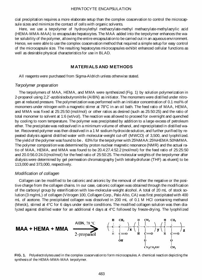
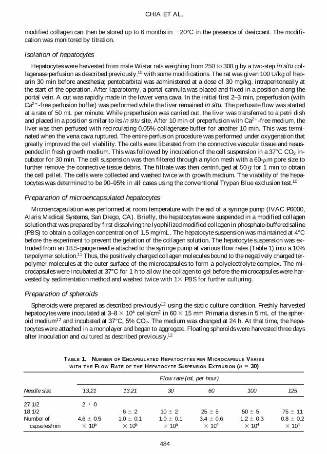
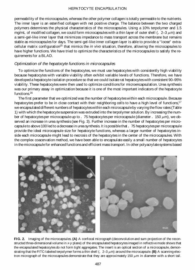
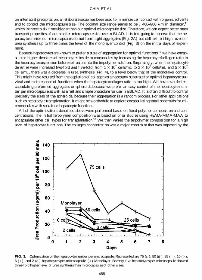
CHIA ET AL
488
FIG 3 Optimization of the hepatocyte number per microcapsule Represented are 75 (s ) 50 (d ) 25 ( n ) 10 (3)6 (1) and 2 (e ) hepatocytes per microcapsule (u ) Monolayer Seventy-five hepatocytes per microcapsule showedthree-fold higher level of urea synthesis than microcapsules of other sizes
on interfacial precipitation an elaborate setup has been used to minimize cell contact with organic solventsand to control the microcapsule size The optimal size range seems to be 400ndash900 mm in diameter11
which is three to six times bigger than our optimal microcapsule size Therefore we can expect better masstransport properties of our smaller microcapsules for use in BLAD It is intriguing to observe that the he-patocytes inside our microcapsules do not form tight aggregates (Fig 2A) but still exhibit high levels ofurea synthesis up to three times the level of the monolayer control (Fig 3) on the initial days of experi-ment
Because hepatocytes are known to prefer a state of aggregation for optimal functions17 we have encap-sulated higher densities of hepatocytes inside microcapsules by increasing the hepatocytecollagen ratio inthe hepatocyte suspension before extrusion into the terpolymer solution Surprisingly when the hepatocytedensities were increased two-fold and five-fold from 1 3 107 cellsmL to 2 3 107 cellsmL and 5 3 107
cellsmL there was a decrease in urea synthesis (Fig 4) to a level below that of the monolayer controlThis might have resulted from the depletion of collagen as a necessary substrate for optimal hepatocyte sur-vival and maintenance of functions when the hepatocytecollagen ratio is too high We have avoided en-capsulating preformed aggregates or spheroids because we prefer an easy control of the hepatocyte num-ber per microcapsule as well as a fast and simple procedure for use in a BLAD It is often difficult to controlprecisely the sizes of the spheroids because their aggregation is a random process For other applicationssuch as hepatocyte transplantation it might be worthwhile to explore encapsulating small spheroids for mi-crocapsules with sustained hepatocyte functions
All of the optimizations described above were performed based on fixed polymer composition and con-centrations The initial terpolymer composition was based on prior studies using HEMA-MMA-MAA toencapsulate other cell types for transplantation18 We then varied the terpolymer composition for a highlevel of hepatocyte functions The collagen concentration was a major constraint that was imposed by the
permeability requirements as described below Under these conditions the ideal terpolymer composition isthe one that is made up of 25 HEMA 25 MAA and 50 MMA at a concentration of 10 in PBS(Fig 5) When the terpolymer composition was modified to one that was composed of 25 MAA 50HEMA and 25 MMA the urea-synthesis of the encapsulated hepatocytes decreases to the level belowthe monolayer control Terpolymer concentration of 10 gave a thin capsular membrane of 2ndash3 mm forgood permeability properties (Fig 2A) Higher concentrations would produce microcapsules with mem-branes too thin to be handled and lower concentrations would produce thicker capsular membranes with re-duced permeability and hepatocyte functions (unpublished results) There might be needs to vary the poly-mer composition and concentrations to achieve enhanced mechanical stability and other physicalcharacteristics Any further optimization of the polymer composition and concentrations must be performedwith a complete reoptimization of other parameters such as cell numbers and densities
Selective permeability of the microcapsules
Collagen gel has been observed to exhibit a ldquoskin effectrdquo that impedes mass transport when a high con-centration of collagen leads to gelation19 Such ldquoskin effectrdquo is concentration and temperature dependentExtracellular matrices like collagen or Matrigel have a gelling temperature of 22ndash35degC depending on theconcentration of these proteins At 37degC where hepatocytes are normally cultured in a bioreactor or trans-plantation is performed in vivo the ldquoskin effectrdquo can be most pronounced Because mass transport is oneof the most important considerations for the design of bioreactor in a BLAD we searched for the optimalconcentration of collagen such that the ldquoskin effectrdquo is minimized but there is still enough collagen to com-plex with the synthetic polyanion-forming stable microcapsules
We have used albumin as our model molecule for the permeability optimization of the microcapsules
HEPATOCYTE ENCAPSULATION
489
FIG 4 Optimization of hepatocyte density per microcapsule Represented are 1 3 107 (d ) hepatocyte density 2 3
107 (r ) hepatocyte density 5 3 107 (3) hepatocyte density in microencapsulation (j ) Monolayer control An in-creasing hepatocytecollagen ratio does not improve hepatocyte functions
Albumin (MW 67000 Da) is one of the secreted proteins of hepatocytes It acts as a carrier to bind mostmetabolic wastes in the liver for removal from the blood20 Another major scavenger protein is bilirubin( 10000 Da)21 which is smaller than albumin This study demonstrated that albumin was freely perme-able to the microcapsules When we added known concentration (1 wtvol) of albumin to collagen andformed microcapsules we equilibrated microcapsules in the culture medium with the same concentrationof albumin (1 wtvol) at 37degC for 2 h to allow the collagen to gel completely and the possible ldquoskin ef-fectrdquo to occur The addition of 1 wtvol of the albumin to the equilibration buffer was to ensure that theencapsulated albumin would not leak out before the permeability measurements were made Such a 2-h de-lay of the permeability measurements after the microcapsule formation is essential for detecting the ldquoskineffectrdquo from the thoroughly gelled collagen Equilibration for up to 5 days indicated that the ldquoskin effectrdquowas only marginally more pronounced than the 2-h equilibration (data not shown) Then we rinsed the mi-crocapsules briefly in fresh culture medium (without albumin) and monitored the albumin release from themicrocapsules into the fresh culture medium (with no albumin added) With 15 mgmL of the modifiedcollagen most of the encapsulated albumin was released from the microcapsules within 15 min (Fig 6A)As the concentration of collagen was increased to 4 mgmL ( 04 wtvol) the release of albumin wasgreatly inhibited For collagen concentration below 15 mgmL we could not encapsulate hepatocytes pos-sibly due to insufficient positive charge from the diluted collagen Therefore we used 15 mgmL of mod-ified collagen for all other experiments
Another requirement of the bioreactor or hepatocyte transplantation is immunoprotection When FITC-dextran ( 150000 Da) matching the size of the smallest immunoglobulin molecule was encapsulated nodetectable amount of FITC-dextran was released from the microcapsules throughout a period of up to 6days even at the collagen concentration of 15 mgmL (Fig 6B) On day 7 we have begun to observe re-
CHIA ET AL
490
FIG 5 Optimization of terpolymers ( s ) Terpolymer comprising of 25 MAA 25 HEMA and 50 MMA (e )terpolymer composing of 50 MAA 25 HEMA 25 MMA used in microencapsulation ( u ) Monolayer controlMicrocapsules formed using 25 MAA 25 HEMA and 50 MMA show the highest level of urea synthesis
lease of FITC-dextran from the microcapsules This further demonstrated that 15 mgmL of collagen issuitable for encapsulating hepatocytes for use in a BLAD
Functional profiles of the encapsulated hepatocytes
The performance of the hepatocytes within the optimized microcapsules was characterized by compar-ing the major functions of the liver namely the synthetic metabolic and detoxifying activities The mi-croencapsulated hepatocytes exhibited up to three times higher level of urea synthesis (Fig 7A) than themonolayer culture and approach that of the spheroids17 generally regarded as the ldquogold standardrdquo for he-patocyte culture22 The microencapsulated hepatocytes also exhibited a three times higher level of albuminsynthesis than the monolayer (Fig 7B) Measurement of the ammonia metabolism further supported the ob-servation that encapsulated hepatocytes exhibit higher level of functions than the monolayer culture (Fig7C) Ammonia level remaining in the culture medium containing encapsulated hepatocytes was lower thanthat containing the hepatocyte monolayer indicating that the encapsulated hepatocytes had a higher am-monia metabolic activity The detoxifying activity was also measured by the cytochrome P450 activity withthe quantification of the ethoxyresorufin O-dealkylation under a confocal microscope 7-Ethoxyresorufin isthe substrate for the cytochrome P450IA1 When the detoxifying activity of the microencapsulated hepa-tocytes was compared with that of the monolayer we have observed that the encapsulated hepatocytes havea higher level of the detoxifying activity especially after day 5 (Fig 7D) Spheroids can maintain functionsfor longer period than the encapsulated hepatocytes (Fig 7A) but the number of hepatocytes in microcap-sules can be more easily controlled For use in short-term liver support such as BLAD encapsulated he-patocytes might offer some advantages
Characterization of the physical properties of the microcapsules
To ensure that the microcapsules have the necessary mechanical stability for use in the high-flow situa-tion of a BLAD we have developed an assay to characterize the physical properties of the microcapsulesThe nano-indentation technique also called ultra-low load indentation or instrumented indentation systemwas used to measure the elasticity and hardness of the microcapsules The technique was designed to mea-
HEPATOCYTE ENCAPSULATION
491
FIG 6 Permeability profiles of microcapsules (A) 1 BSA (MW 67000 Da) was loaded into microcapsules andthe percentage of loaded BSA released into medium was monitored (s ) 15 mgmL ( u ) 2 mgmL (e ) 3 mgmL (3)4 mgmL of collagen A total of 15 mgmL of modified collagen allows unimpeded release of BSA whereas higherconcentrations of collagen show ldquoskin effectrdquo (B) 1 FITC-dextran (MW 150000 Da) was loaded into the micro-capsules and the percentage of loaded FITC-dextran released into the medium was monitored ( s ) 15 mgmL (e ) 2mgmL ( u ) 3 mgmL (1) 4 mgmL of collagen The microcapsules should be impermeable to immunoglobulins( 150000 Da) or larger cells of the immune system
A B
sure the mechanical properties of materials in submicron scale with the load range below 500 mN (50 g)or the indenter penetrated into a material surface in either nanometer or micron range The resolution of themicroscope sets the limits of the indentation size that can be measured accurately
The microcapsules show a time-dependent deterioration of their mechanical stability because the depthof penetration of the indenter increases from 05 mm on day 2 after microcapsule formation to 25 mmon day 7 (Fig 8) After day 7 the same maximum force applied via the indenter (015 mN) ruptured themicrocapsules It is likely that the weaker microcapsules after day 7 allowed penetration of the indenter(with 015 mN force) through the terpolymer shell leading to the rupture of the microcapsules This is inagreement with a confocal micrograph which shows that the shell thickness of the optimized microcapsulesis 2ndash3 mm (Fig 2A) We have also observed a decrease over time in the elastic modulus (from 900MPa to 300 MPa) as well as a drop in the average pressure over time (from 60 MPa to 10 MPa) in
CHIA ET AL
492
A B
C D
FIG 7 Functional profiles of microcapsules (A) Urea synthesis of the microcapsules is three times higher than thatof the monolayer control reaching the level of the spheroids ( d ) Microencapsulation ( j ) monolayer (r ) spheroids(B) Albumin production of microcapsules is better than that of the monolayer control ( d ) Microencapsulation (j )monolayer (C) Cytochrome P450 activity profile indicates that the microcapsules have higher detoxifying ability thanthat of the monolayer culture ( d ) Microencapsulation (j ) monolayer (D) Ammonia metabolism of microcapsules isbetter than that of the monolayer culture ( d ) Microencapsulation (j ) monolayer
which the microcapsules can withstand before rupture This confirmed our observation that the mechanicalstability of the microcapsules deteriorated over a period of days The decrease in the mechanical stabilityof the microcapsules is likely due to the terpolymerrsquos solubility in the aqueous environment Neverthelessthe average pressure at rupture is in the MPa range suggesting that these microcapsules are suitable for aplug flow reactor design The maximal flow rate that can be applied will have to await further calculationof the pressure drop for a given bioreactor size
We report here the encapsulation of hepatocytes in a physiological aqueous environment by complexcoacervation using a simple setup The hepatocytes are encapsulated within a two-layered polymeric mem-brane that has a configurable outer terpolymer shell and a suitable interior polymer substrate for enhancedhepatocyte functions The enhanced cellular functions of the encapsulated hepatocytes are up to three timeshigher than the monolayer control and comparable to that of the spheroids even though the encapsulatedhepatocytes do not form aggregates These microcapsules were permeable to the small molecules such asnutrients oxygen and metabolites while impermeable to large molecules such as immunoglobulins Wehad developed a nano-indentation scheme to characterize the physical properties of the microcapsules in-dicating that the microcapsules should be strong enough for use in a BLAD at least for the first week ofthe microcapsule formation There might be a need for a more mechanically stable microcapsule in certainapplications such as hepatocyte transplantation where the microcapsules need to be stable for months Fur-
HEPATOCYTE ENCAPSULATION
493
FIG 8 Mechanical stability of the microcapsules Depth of penetration into microcapsules was measured with re-spect to time of incubation in culture medium when the same force was applied ( s ) Microcapsules at day 2 (n 5 11)(1) microcapsules at day 3 (n 5 6) ( e ) microcapsules at day 7 (n 5 12) The indenter penetrates deeper into the ter-polymer shell of the microcapsules as the incubation time increases
ther work will be necessary to optimize the conditions to encapsulate hepatocytes for these other applica-tions
ACKNOWLEDGMENTS
This work is supported by an infrastructure fund from the National Science amp Technology Board of Sin-gapore We are grateful to the faithful support from Prof C Fong Shih and Prof Chorh-Chuan Tan for theprogram We acknowledge invaluable administrative assistance from Ms Foong-Chun Lai Winnie Jofe-lyn Lye and Ei-Ling Lim We are also grateful for the technical assistance from Ms Chai-Hoon Quekmembers of the Leong laboratory Yu laboratory and the Microscopy amp Cytometry Unit of the NationalUniversity Medical Institutes and AProf Mah-Lee Ng and her Electron Microscopy Unit of the NationalUniversity of Singapore
REFERENCES
1 Bismuth H Figueiro J and Samuel D What should we expect from a bioartificial liver in fulminant hepaticfailure Artif Organs 22 26 1998
2 Dixit V and Gitnick G Artificial liver support state of the art Scand J Gastroenterology 31 101 19963 Dixit V Development of a bioartificial liver using isolated hepatocytes Artif Organs 18 371 19944 Chang TMS Semipermeable microcapsules Science 146 524 19645 Leung YF OrsquoShea GM Goosen MF and Sun AM Microencapsulation of crystalline insulin or islets of
Langerhans an insulin diffusion study Artif Organs 7 208 19836 Cai ZH Shi ZQ Sherman M and Sun AM Development and evaluation of a system of microencapsulation
of primary rat hepatocytes Hepatology 10 855 19897 Matthew HW Salley SO Peterson WD and Klein MD Complex coacervate microcapsules for mammalian
cell culture and artificial organ development Biotechnol Prog 9 510 19938 Babensee JE De Boni U and Sefton MV Morphological assessment of hepatoma cells (HepG2) microen-
capsulated in a HEMA-MMA copolymer with and without Matrigel J Biomed Mater Res 26 1401 19929 Sugamori ME and Sefton MV Microencapsulation of pancreatic islets in a water insoluble polyacrylate ASAIO
Trans 35 791 198910 Seglen PO Preparation of isolated rat liver cells Methods Cell Biol 13 29 197611 Wells GDM Fisher MM and Sefton MV Microencapsulation of viable hepatocytes in HEMA-MMA mi-
crocapsules a preliminary study Biomaterials 14 615 199312 Friend JR Wu FJ Hansen LK Remmel RP and Hu WS Formation and characterization of hepatocyte
spheroids In Morgan JR and Yarmush ML eds Tissue Engineering Methods and Protocols New Jersey Hu-mana Press 1999 pp 245
13 Miyoshi H Yanagi K Fukuda H and Ohshima N Long-term performance of albumin secretion of hepato-cytes cultured in a packed-bed reactor utilizing porous resin Artif Organs 20 803 1996
14 Doerner MF and Nix WD A method for interpreting the data from depth-sensing indentation instruments JMat Res 1 601 1986
15 Selden C Khalil M and Hodgson HJF What keeps hepatocytes on the straight and narrow Maintaining dif-ferentiated function in the liver Gut 44 443 1999
16 Rahmatullah M and Boycle TR Improvements in the determination of urea using diacetylmonoxime methodswith and without deproteinisation Clin Chim Acta 107 3 1980
17 Takabatake H Koide N and Tsuji T Encapsulated multicellular spheroids of rat hepatocytes produce albuminand urea in a spouted bed circulating culture system Artif Organs 15 474 1991
18 Shao W and Leong KW Living cells microencapsulated in a polymeric membrane having two layersUS5620883 The Johns Hopkins University Baltimore MD April 15 1997
19 Kleinman H McGoodwin EB Rennard SI and Martin GR Preparation of collagen substrates for cell at-tachment effect of collagen concentration and phosphate buffer Anal Biochem 94 308 1979
20 Kamisaka Maezawa H Inagaki T and Okano K A low molecular weight binding protein for organic anions(Z protein) from human hepatic cytosol purification and quantitation Hepatology 1 221 1981
21 Martin MT Jacobs FA and Brushmiller JG Low molecular weight copper-binding ligands in human bileProc Soc Exp Biol Med 181 249 1986
CHIA ET AL
494
22 Hamaoto R Yamada K Kamihira M and Lijima S Differentiation and proliferation of primary rat hepato-cytes cultured as spheroids J Biochem (Tokyo) 124 972 1998
Address reprint requests toHanry Yu PhD
Tissue Engineering InitiativeBlock MD11 03-02A
Clinical Research Centre10 Medical DriveSingapore 117597
E-mail nmiyuhnusedusg
HEPATOCYTE ENCAPSULATION
495
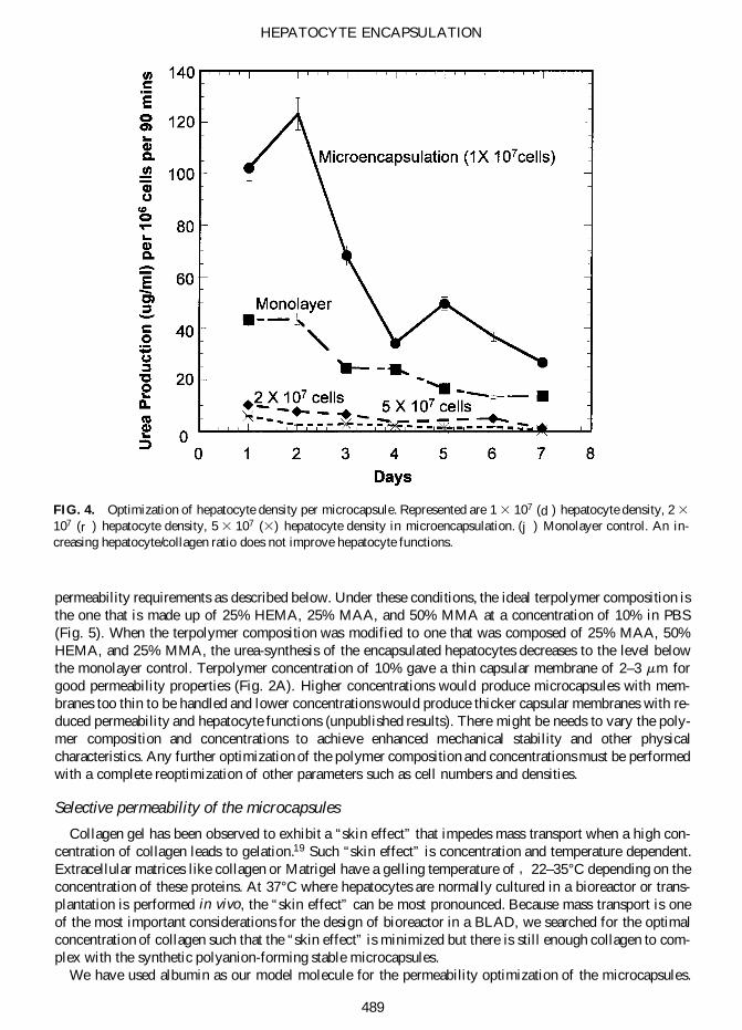
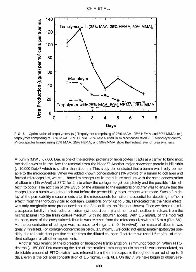
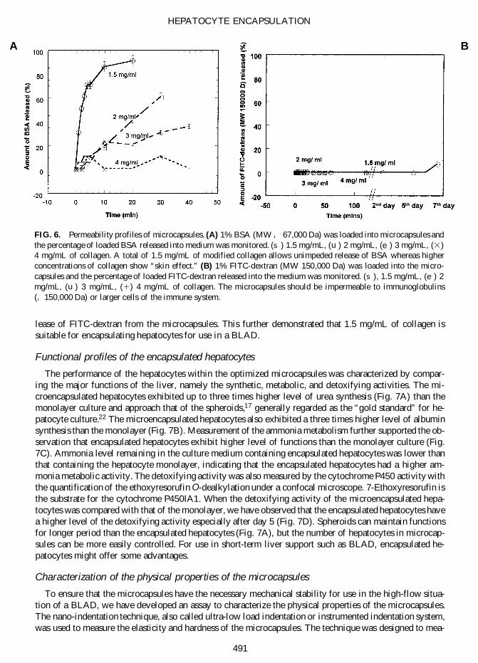
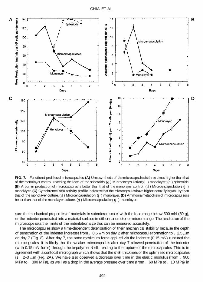
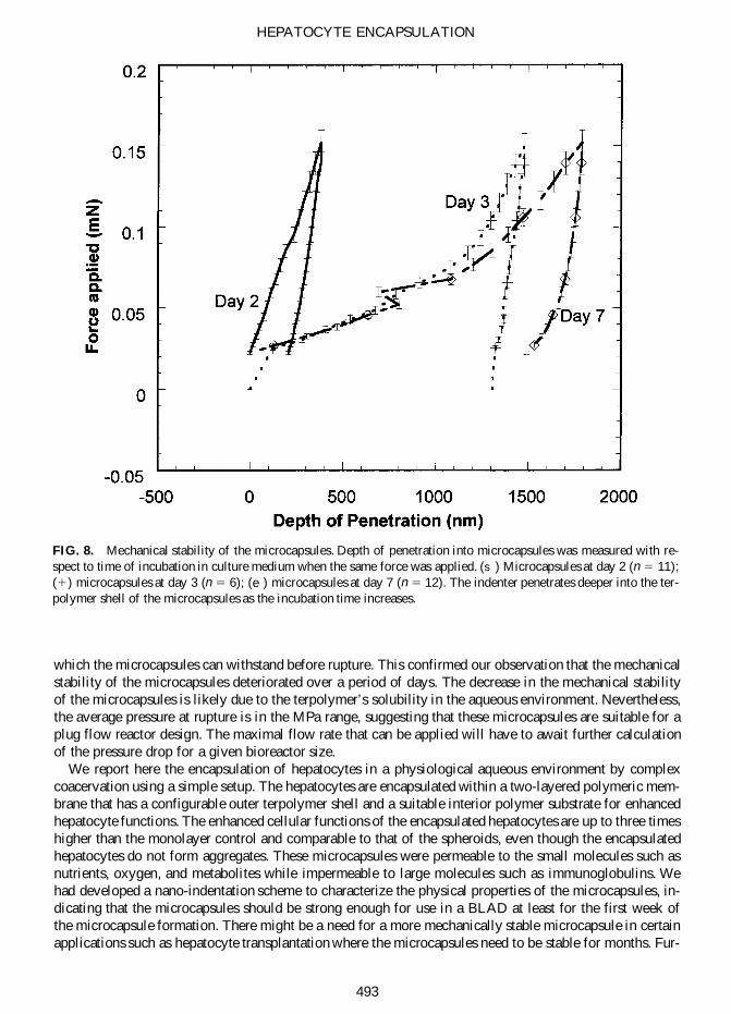


