Embed Size (px)
Citation preview
Coronary Rotational A erieme in 3 res
SIMON H. STERTZER, MD, FACC. JOSEPH ROSENBLUM, Do. FACC,
RICHARD E. SHAW, PHD, IRAWAN SUGENG. MD. BENITO HIDALGO, RT,
COLMAN RYAN, MD, FACC. HEIDI N. HAhSELL. RN. MS. MARY C. MURPHY, RN, MS,
RlCHARD K. MYLER, MD, FACC
Daly City, Cnlifwnio
U&rm%ws. The aim of this study was to assess the utility of percuteneous h’anWninal coronary rotational ablation in Ihe twatment of coronary artery disease.
Bwkgwmd. Although numerous advances have been made in the treatment of corenary artery disezw, rh=ze are 1sion.s with complex morphology that mu not amenable lo current k&was- celar fhempy.
Melk& A conseattive series of 242 patients having 302 c3mmry rdatiooal ailladm prlwdmxs was analyzed. one hml- dred ninetctn WW of the patients had prcviou.sly undergone pttempted cornnary an&plssty, which was unsucc45sful in 31 patints (13%). The left ventricular cjeetion frpaioo was normal in 1% patients (81%). Tk ablation procedure war attempted ia Jo8 VeEspLr and 346 l&Ins. ofihe 315 lesieits treeled, 26 (7.5%) were cl&lied lls Am&an Col@e oi Cardi@y~Anmican Hcml Aswciarion type A, and 320 (92.5%) as tither type B or &De c. - - Ralts. Promtornl success WBF achieved in 284 (94%) of the
302 prondures and 330 (95.4%) of the 346 l&m in which
Since the introduction of percutaneous rransluminal coro- nary angioplasty, the types of lesions considered treatable have been expanded by increased operator experience and equipment improvement. Coronary atherectomy has rela- tively recently been added to the amtamenturium of the invasive cardiologist (I). Although directional atherectomy has been explored for several years. it has limitations in both delivery and effectiveness when the atherectomy device is introduced into mtrrow, calcified, tortuous and iung core+ nary lesions (2). In contrast, the advent (3-S) of the Heart
Technology Rotablator has overcome most ofihe aforemen- tioned drawbacks of the currently available directional atberectomy devices (9-15). Accordingly, this repart details the methods. aspects of the learning curve, immediate re- suits, comphcations and extended indications developed from the experience in the 6rst 302 consecutive procedures performed with the Heart Technology Rotablator at our
Methods
Patients. Between October 1!3% and May 1992, 242 pa- tients underwent 302 discrete percutaneous transluminal coronary rotational ablation procedures at our institution. Patients were enrolled as part of a multicenter evaluation of the Rolabiator for Food and Drug Adminis~tion approval. Patients with visible thrombus, unprotected left main coro- nary artery or old saphenous vein grafts were excluded. Patients who had undergone a prior intervention were not excluded unless it had been performed within 1 month before rotational ablation and involved a significant dissection. All patients signed an informedconsent document that had been
288 STERTZER ET AL. CORONARY RclT*TmNAC ABL4TIclN
approved by the Institutional Review Board of our institu- tion. In five patients with a left ventricular ejection fraction <30%, assisted (that is. intraaortic balloon pump or cardio- pulmonary bypass) rotational ablation was carried out as compassionate use after notification of the chair of that board.
Many patients had lesion morphology suitable for balloon angioplasty. These patients generally had American College of Cardiology/American Heart Association type A and type El stenoses (16). As more experience was gained with the ablation device, its use was extended to selected type C lesions. Compating the last ZOO procedures with the first 100, there was a significantly greater use of rotational ablation in calcified stenoses (61.5% vs. 31.4%, p i 0.01), eccentric lesions (65.1% vs. 44.7%, p < 0.05), lesions >I cm long (36.1% vs. 24.5%. p < 0.05) and vessels with diffuse or tandem lesions present (30.6% vs. 18.7%. p < 0.05).
Procedure. The procedure for the use of rotational abla- tion consisted of the passage of a flexible 0.009-in. (0.023 cm) guide wire with aO.Ol’I-in. (0.043 cm) spring tip through a 9F guiding catheter into the coronary artery to be treated by rotational ablation. Although the bare wire technique was successful in 85% of cases, 15% required the passage of a balloon catheter preceded by a O.O14-in. (0.36cm) guide wire to exchange for the noncoated Rotablator wire beyond the vessel’s narrowest stenosis. In these cases the balloon nose was advanced as far as possible into the diseased segment, and the 0.014~in. wire was removed. The flexible Heart Technology (type C wire or the relatively steerable type A) O.OO?Gn. (0.01~in. tip) wire was then passed through the balloon nose and positioned as far as feasible beyond the stenotic segment. The burr-bearing catheter was then aMe to be passed over its uncoated O.CK@-in. wire for definitive treatment. The 0.0+&n. wire owes its occasional difficulties to its high coefficient of friction, a feature related to the absence of Teflon or other lubricious coating common to standard angioplasty guide wires.
After ihe O.GOP-in. wire was positioned distally in the appropriate vessel, the diamond-coated burr was advanced through the 9F guiding catheter and carefully set just prox- imal to the ingress of the lesion. Extreme care to maintain the coaxial position of the burr with respect to the guiding catheter was required to prevent the stalling of the air turbine when entering vessels that bent away fmm the guiding catheter at a sharp angle.
Once the position of the burr and the burr/artery ratio were fluoroscopically verified, the device was actuated by a foot switch, and the spinning burr advanced slowly into the lesion at a starting in vitro speed of 190,OlM to ~OO,CIOO rpm. The general approach was to not allow a decrease >lO% in rotationa: speed of the burr when it cam‘. into contact with the stenotic surface. A continuous saline infusion was used tc> lubricate the drive shaft. This infusate was maintained at 300 mm Hg during the entire course of the use of each device.
In the 346 lesions in which ablation was attempted, an
average of 2 i 1 burrsilesion was used. A mean of d ? 2 passes;lesion was required, with the maximal time of rotation for each burr of 40 -t 16 s and an average total time for all burrs used of 108 2 63 s. The average time for rotational ablation proxdures was 47 f 28 min, or approx- imate!y 10 min longer than the average balloon angioplasty procedure at our institution (37 + 23 min).
Pacemaker capability was continuously maintained be- cause both sinus arrest and a:tioventricular (AV) block, although transient, distorted the cardiac rhythm in approri- mately 75% of cases. Spasm was treated with nitroglycerin or molsidomine (usually by intracoronary administration). Significant hypotension occurred in 4.8% of cases but re- sponded to fluid and restoration of normal sinus conduction. All patients received lO,ooO U of intravenous heparin at the beginning of the procedure, and 3,000 U every hour in longer procedures. or were treated accordina to the activated clotting time determinations made duri& the intervention. The activated clotting time was kept at >300 s during the procedure.
Burr sizes were chosen according to the severity of the worst stenosis in any given iesion segment, but usually the procedure was begun with a 1.25-mm device with sequenc- ing of the upsizing to Z.25 mm according to the effects of downstream debris (i.e., plaque burden) and vessel size. Burr sizes >Z.i5 mm generally require a IOF guiding cathe- ter. In our experience, the use of a 9F guiding catheter with a 2.25 burr resulted in one case in which the guide was damaged (II), although problems have not been encountered passing the 2.25-mm burr through the 9F Sherpa guiding catheter (Medtronic).
The early learning curve with this device mandated a conservative approach and liberal use of complementary balloon angioplasty to achieve safe reduction in percent diameter stenosis. As experience was gained, an attempt was made to debulk the lesions more aggressively so that a higher percent of full lumen diameter was obtained with the rotational device alone. BallDon angioplasty in those cases (approximately 42% of this series) was performed as a touch-up procedure and carried out at a very low pressure (0.5 to 2.5 atm), mainly to eliminate spasm or to smooth the orthogonally viewed appearance of the vessel, or both.
The postprocedural care was similar to that utilized for balloon angioplasty, although comparison with a concurrent series of natients showed that natients underaoine rotational ablation were more likely to ak receiving intkve~ous nitro- glycerin (80% vs. 64%, p < 0.001) for a longer interval (30 vs. 24 h. p < 0.05) than were patients undergoing angioplasty. Lenglh of hospital stay was not significantly dilTerent be- tween the ablation and angioplasty groups (d f 5 vs. 4 + 3 days).
Delinitii~s. The following definitions were used. Lesion: n ~50% narrowing in vessel lumen diameter on
m-teriography. Primary lesion: one in which no prior ret ascularization
therapy had been attempted.
Reslenoric lesion: one in which prior interventional iher- spy had been initially successful.
A~&nc~ive angioplasty: a procedure in which a balloon is osed at normal or high pressure after rotational ablation.
Complemenroiy ongioplostyr a procedure in which a balloon is used at very low pressure (0.5 to 2.5 atm) after rotationai ablation to relieve vasospasm or to smooth a satisfactorily ablated segment.
M&F cardiac event: occurrence during the initial hospi- tal stay of death, a new Q wave myocardial infarction or the aced for emergency coronary bypass surgery.
Non-Q ww myocardial infarcrion: elevation of creatine kinase (CK), MB fraction to twice the normal value (200 &ml; 6% MB fixaction) with no new electrocardio- graphic (ECG) Q waves.
Q WOW myocordiul infarction: development of new ECG Q waves with serum enzymes elevated to twice normal levels.
Technical success~ 8 reduction in the diameter stenosis after treatment to a residual stenosis of <SO% and no in-hospital occurrence of a major cardiac event caused by the treated artery.
Uasaccessfulluncomplicated: a technically unsuccessful ablation attempt that resulted in no major cardiac event.
Procedural success: technical success achieved in all critical &noses with the occurrence of no major cardiac events during the hospital stay.
Lute cardiac event: occurrence of death or myocardial infarction after discharge from the initial hospital stay for percutaneous transluminal coronary mtatianal ablation.
Evenr-fie survival: freedom horn late cardiac events. Estimated restenosis rate: the number of patients who
were estimated to have recurrent stettoses based on a combination of angiographic results, occurrence of myocar- dial infarction or the need for reneat revascularization.
Statis&! analyses. AU data were entered prospectively in the Institute Interventional Cardiology Database. All continuous variables are expressed as the mean value r SD. Analyses were performed with use of the SPSS statistical package.
Results Base& &aracteristLx (Table 1). The 242 Datients were
predominantly male. Seventy-one percent were in Canadian Cardiovascular Society angina class II1 or IV or had unstable angina. More than half presented with a history of hypetten- sion or hypercholesterolemia. One hundred nineteen (49%) had previously mdergonc attempted coronary angioplasty, which was unsuccessful in 31 patients (13%). The left ventricular ejection fractionwas Xl.45 in 1% patients @I%), 0.35 toU4in34(14%), 0.25 b0.34in7 (‘L%)and <0.25in 5 (Z.i%). Of these patients, 133 (55%) had single-vessel disease (defined as a vessel with at least one lesion of >5G% diameterstenosis), 52 (21.5%) had double-vessel disease, 40
Prior corom~ bypars sumry 54 22.3
CCS = Canadian Cardiov~ulaf Society.
(16.5%) had triole-vessel disease and 17 (7%) had cntadrttnle- vessel chsease,.for a total of 425 diseased vessels:
These 242 patients underwent 302 procedures in which rotational ablation was attempted in 308 vessets and ;66 lesions. Rotational ablation was performed in 92 right coro- nary arteries. 158 lef? anterior descending and 58 left circum- Rex arteries. Of the 346 lesions treated, 26 (7.5%) were classified as American College of CardiotogylAmcdcan Heart Association type A. and 320 (92.5%) either type B or
type C. Procedural nutumtc. Procedural sttccess was achieved in
284 (94%) of the 302 procedures in which rotational ablation was used. In five procedures (1.7%). all attempts with a Rotablator or balloon were unsuccessful, but no cardiac event occurred during the hospital stay. These pnxedures involved eccentric and type C lesions. A major cardiac event was associated with 13 procedures (4.3%); nine (3%)of these complications were due to the ablation procedure. Six patients sustained aQ wave myocardial infarctionalone. two had a Q wave infarction and required emergency surgery and one needed emergency surgery hut did not have a Q wave infarction. No procedural deaths were atttibuted to the ablation procedure.
Of the first 302 procedures, 69 (22.8%) were performed as a stand-alone procedure. Success was achieved in 95.7% of these stand-alone procedures; a major cardiac cYent oc- curred in 2.9% of these cases, Sttccess was achieved in 93.6% of the procedures in which the complementary or adjunctive use of a balloon was required; a major cardiac event occurred in 4.7% of these cases.
Lesion outcome. In analyzing !esion outcome immedi- ately after use of the abtation procedure before any use of adjunct& or complementary balloott therapy, technical success was achieved in304 (87.9%) of the 346 stenoses with attempted ablation. Of the remaining 42 stenoses, I9 ’ ve unsuccessfully treated with ablation. In 1 I of these 19 cases,

290 STERTZER ET AL. l&x Vol. 21. No. 1 CORONARY ROTATICJNAL ABI.ATION Pebruary 159w.87-9.’
Table 2. Results by Lesion and Mcn’phology Subgroups After Rotalional Ablation and Afler All Procedures (Ablation and Balloon Dilation) Used
n Success -Event* Unsuccesrfult Succclr -Event* Uns”ccrssf”n
All lesions 346 87.9 2.6 9.5 95.4 2.6 2.0 Tw A 26 100.0 0.0 0.0 96.2 0.0 3P Type fb’C 320 86.9 2.8 10.3 95.3 2.8 1.9 NW 240 85.0 2.9 12.1 94.6 2.9 2.5 Reltcnotic 106 94.3 1.9 3.8 97.2 1.9 0.9 Occlusions 23 81.0 0.0 13.0 loo.0 0.0 0.0
stcnoser 323 87.9 2.8 9.3 95.0 2.8 2.2 Stcnorir subgroups
Cl cm 229 89. I 2.2 8.7 95.2 2.2 3.6 t, cm 94 85.1 4.3 10.6 94.6 4.3 1.1 Ostial 13 loo.0 0.0 iJ.0 1W.O 0.0 0.0
Eccrollic 205 86.8 4.4 8.8 93.2 4.4 2.4 Calcified 107 91.6 1.9 6.5 97.2 I.9 0.9
‘Definedasmyafatdialinfarclian. emqtnfy rurgeryardeath. tlkfinedas >5d residual diamelrrrlenosis with nomajorcardiac evenl. Dataarepresrnled BS number(n) of lesions or as percent of lesions treated with ralalional ablation alone or with ablution and k&on dilation.
the device crossed the lesion but produced an inadequate result; in 6 there was a failure to advance the guide wire and in 2 there was a device failure. A successful result was achieved with adjunctive balloon therapy in 14 of these 19 cases. Of the other 23 stenoses, rotational ablation produced a flow-limiting dissection in 5, reoeclusion occurred shortly after use of the device in I6 and pseudoaneurysmal perfora- tions developed in 2 sites. Of those 23 lesions. 17 were successfully treated with adjunctive halloon therapy. Snc- cess after all intervention (ablation alone or with adjunctive or compiementary balloon angioplasty) was achieved in 330 (95.4%) of the 3461esions in which treatment was attempted.
Resultsof rotational ablation alone and with adjunctive or complementary balloon therapy for various lesion types are presented in Table 2. The success rate !#as higher for ablation in type A than in type B or C lesions, In ore type A lesion, a successful but not optimal result that was followed by adjunctive balloon angidasty led to compromised flow and an unsuccessful but uncomplicated result. Rotational ablatirn was more successful in restenotic than in primary lesions, and in lesions 51 cm than in those >l cm long. The success rates were extremely high for occlusions and oskial stenos:s.
Fottow-op. Follow-up was obtained 23 months after hos- pital discharge or in patients who had returned early for evaluation because of a return or worsening of symptoms. Of tie 242 patients, 186 were in the group evaluated 23 months after hospital discharge. Follow-up was obtained on 182 of these 186 patients at a mean follow-up interval of 9 -+ 5 months (Table 3). Of the 182 patients, 169 (92.9%) were alive and free of myocardiat infarction. Angiogmphic fol- low-up was obtained in 87 patients. Of this sample, 56 required some reintervention. Although a true restenosis rate was not possible to obtain because many asymptomatic patients could not be convinced io undergo an&graphic
follow-up, an attempt was made to calculate an estimated restenosis rate by combining angiographic and clinical out- come. This analysis yielded an overati estimated restenosis rate of 37.4% (68 of 182)). Analysis of selected subgroups showed no difference in occurrence of restenosis between type A lesions and type B or C lesions (33% vs. 38%) or between stand-alone rotational ablation versus ablation with adjnnctive complementary balloon therapy (32% vs. 39%). In contrast to prior reports, primary lesions had a lower restenosis rate than that of restenotic tesions (28% vs. 44%. p < 0.01). 0; the 31 patients with prior unsuccessful balloon angioplasty, 24 were followed up; of these, 7 (2%) devel- oped clinical or argiogmphic evidence of restenosis.
Coronary angioplasty using present balloon technology is still a mainstay of interventional cardiology. Directional, laser and rotational atherectomy have not clearly and pre- dictably decreased the rate of restenosis. However, the impulse to exptore new devices is spurred not only by a desire to decrease the rate of restenosis, but by a need to treat, through moderately invasive procedures, coronary
Tahh 3. Follow-Uo Aher Relational Ablation in I82 Patienls
arteries with more difficult momholoev that would ordinarily . _. require coronary bypass surgery. Consequently. narrow, calcified and long lesions, so dilfuse as to be inoperable.
provide the motivation to pursue new approaches. &sirmmu@&gv. Although a randomized trial between
coronary angioplasty and rotational ablation (now in progress at this institution) is required to assess the debulk- ingeffecl on type A. Band 8, lesions, or indeed any stenosis that can be treated with balloon angioplasty, some indica- tions of the value of the Rotablator are suggested by this study and by comparison with our own balloon angioplasty data. In an analysis of l.W lesions treated with balloon angioplasty during I998 and 1991 at this institution (18). success was not achieved in 8.6% of the 140 calcified lesions and in 7.1% of the 14 ostial lesions with attempted dilation. Of the 414 lesions >I cm long in which angioplasty was attempted, the procedure was unsuccessful in 9.4%. The success rates of 95% to lOG% obtained in these subgroups with rotational ablation are encouraging although they do not differ statistically from those obtained with angioplasty. The following are examples of the successful use of rotational ablation in several circumstances that are less than optimal for conventional balloon angioplasty:
I. Calcified ostial lesions (see Fig. 1). 2. Lesions crossable hv wire and balloon that do not
yield lo high pressures (e.i., 20 atm) (see Fig. 2). 3. Any lesion in which tortuosity of vessel or profile of
stenosis is so severe that the 0.014-in. wire crosses, yet no balloon will follow (see Fit. 3).
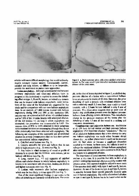
4. Long, narrow (e.g.: 27 cm) segments of calcified diffuse and tubular disease in which balloon angioplasry is known to be associated with dissection, inadequate dilatiun and higher restenosis rates (see Fig. 4).
5. Lesions with restenosis after balloon angioplasty. which may be less likely to recur again (19) (see Pig. 5).
One of the most significant findings in our series is the effect of debulking on extremely calcified lesions. For exam-
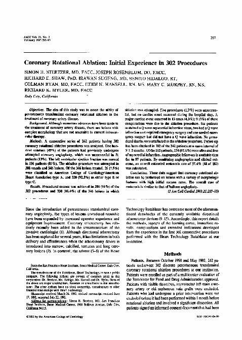
Figure t. 8, Right coronary artsy wi!b a Ime cakiikd 0stiaJ &ion (mow). b, The same vessel I year law after satnd-alone rotalional ablarion of the ortial lesion.
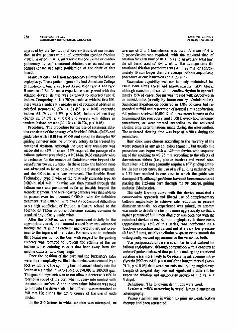
pie. in the class of lesion depicted in Figre 2, a calcified ring prevents dilation of a lesion with a conventional balloon (even at pressures in excess of 20 atm). However, rhe prior dsoulking of such a stenosis with rotational ablation even with a relatively small tl.5 mm) burr. may render a vessel treatable with a 3.5mm balloon i&ted to only 6 atm of pressure. This observation suggests that an inner core OF napkin ring calcification prevents even the highest pressure balloons from affecting certain old lesions. This remarkable change in the pressure response of the lesion after the debulking oi only I.5 mm of the vessel is a striking and consistent phenomeoon.
Complementary and adjuuetive b&boa use. Buchbinder et al. (19) have termed normal and high pressure balloon angioplasty after rotational ablation “adjunctive.” Tlx use of an adjunctive balloon means that a true attempt to carry out balloon angioplasty was m&z either because abrupt closure (i.e.. no reflow phenomenon) could not be reversed with the burr alone or because a significant dissection resulted in the lesion. In these cases, the b&on is used to salvage the rotational ablation. Salvage balloon angioplasty after rotational ablation in our series occurredin 23 (6.7%) of the lesions with attempted ablation.
The importance of complementary baIIoon angioplasty is unclear at this time. It is possible that 60% to 80% of all patients undergoing rotational ablation could be treated with ablation alone. This approach might be utilized in most cases if the operator deals solely with type A and type B, lesions. However, the useof the device in type A and type B, lesions is argua.ble because those vessels generally respond quite well to balloon angioplasty. The utility of rotational ablation in type A and type B, lesions has to be based either an the
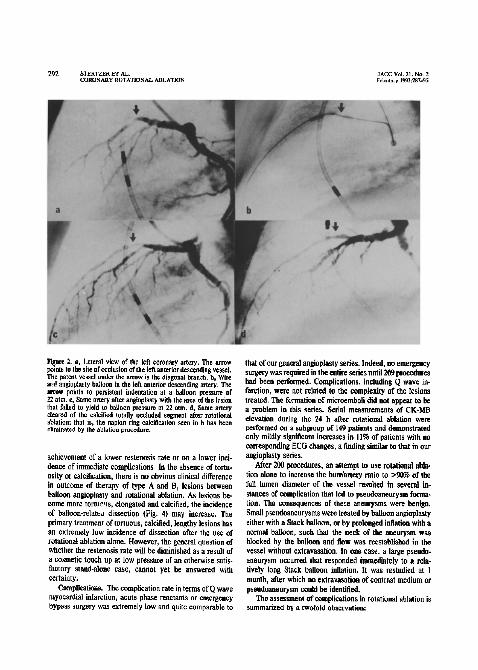
Figttre 2. a, Lateral view of the left caronsry artery. The arrow points to the site of occlusion of the left anterior descendig vcsscl. The patent vessel under the arrow is the diagonal branch. b, Wire and angioplssty b&en in the left anterior descending artery. The arrow pohftr to persistent indentation et a balloon pressure of 22 atm. c, Same artery after angioplasty with the area of the lesion that failed to yield to ballcan pressure at 22 atm. d, Same artery cleared of the calcified totally occluded segment r&r rotational ablation; that is. the napkin ring calcification seen in b has txea eliminated by the ablation pmwdurc.
achievement of a lower restenosis rate or on a lower inci- dence of immediate complications. In the absence of tortu- osity or calcification, there is no obvious clinical difference in &come of therapy of type A and 8, lesions between balloon attgioplasty and rotational ablation. As lesions be- come more tot~~ts, elongated and calcified, the incidence of balloon-relattti dissection (Fig. 4) may increase. The primary treatment of tortuous, calcified, lengthy lesions has an extremely low incidence of dissection after the use of rotational ablation alone. However, the general question of whether the restenosis rate will be diminished as a result of a cosmetic touch up at low pressure of an otherwise satis- factory stand-alone case, cannot yet be answered with certainty.
Camplieatkxis. The complication rate in terms of Q wave myocardial infarction, acute phase reactants or emergency bypass surgery was extremely low and quite comparable to
that dour general angioplasty series. Indeed, no emergency surgery was required in the entire series unhl20!3 procedures had been performed. Complications, including Q wave in- farction, were not related to the complexity of the lesions treated. The formation d microsmboli did not appear to be a pmblem in this series. Serial measurements of CK-MB elevation during the 24 h after rotational ablation were performed on a submoup of 149 patients and demonstrated only mildly significant increases in 1 I% of patients with no corresponding ECG changes, a finding similar to that in our angioplasty series.
After 200 procedures, an attempt to use rotational t&la- tion alone to increase the burr/artery ratio to >%t% of the full lumen diameter of the vessel resulted in several in- stances of complication that led to pseudoaneurysm forma- tion. The consequences of these aneurysms wore benign. Small psettdoaaeurysms were treated by balloon angioplasty either with a Stack balloon, or by prolonged inflation with a normal balloon, such that the neck of the aneurysm was blocked by the balloon and Bow was reestablished in the vessel without extravasation. In one ease, a large pseudo aneurysm occurred that responded immediately to a rela- tively long Stack balloon inflation. It was restudied at 1 month, after which no extravasation of contrast medium or psettdoaneutysm could be identified.
The assessment of complications in rotational ablation is summarized by a twofold observation:

JACC vol. II. Nn. 2 STERTZER ET AL. Feblary I593287.95 293
CORONARY ROTATIONAL ABLATION
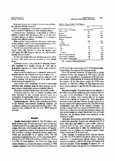
1. The sequentiai utilization of small to large burrs Figure 3. a. Left circumflex coronary artery with revere subtotal without “skipping” to the final burr sire too early in the stenorls(arrowl. b,Althcqh a0.014-in.l0.036cm)wire hascrossed
procedure decreases the plaque burden and lowers the risk the tesion. no balloon (Axed or over the wire1 could woss the severe
of dissection, macnxmbolus or severe vasospasm. This obser- narrowing (arrow). Note that the balloon center Mat) aligns with the
vation is suppxted by an analysis of the 23 lesions in which sternal wire (compare withal. e, Rotablator wire is visible disraRy in
there was either severe dissection. occlusion or perfor&ion the mtery. The lesion (arrow) has already been successfully dc- bulked by the mtating diamond bun. Significant vawspa~m is
tier the ablation procedure; in II (63.8%) of these cases. burr preset in the entire lefi circumflex system. d. Final rotational
sizes were skipped and in 6(26.1%) a relatively targc burr(l.75 ablation result after ballowl treatment for left circumtkr artery SPPSrn.
Figure 4. A, Right coronary’ artery in the right anterior oblique view showing adiffuse, calcified lengthy atherosclerotic segment. B, Final result after multiple burr ablations from I.25 up to 2.15 mm. The singkihir. arrow shows the ectatic reference point present in A, that is. before treatment.
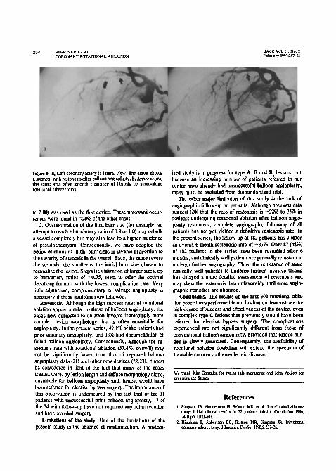
Figwe 5. 8, Left coronary artery in lateral view. The arrow shows a segment with restenosis after balloon angioplasty. b, Arvmv shows the same area after smooth clearance of fibrosis by stind.alone rotational atherectomy.
to 2.00) was used as the lint device. These untoward occur- rences were found in ~20% of the other cases.
2. Overestimation of the final burr size (for example, an attempt to reach a butrlartety ratio of 0.9 or 1.0) may debulk a vessel completely but may alsu lead to a higher incidence of pseudoaneurysm. Consequently, we have adopted the policy of choosing initial burr sizes in inverse propartion to the severity of stenosis in the vessel. Thus, the more severe the stenosis, the smaller is the initial burr size chosen to recanalize the lesion. Stepwise utilization of larger sizes, up to burr/artery ratios of ~0.75, seem to offer the optimal debullcing formula with the lowest complication rate. Very little adjunctive, complementary or salvage angioplasty is necessary if these guidelines are followed.
Rertcnosls. Although the high success rates of rotational ablation appear similar to those of balloon angioplasty, the cases now subjected to ablation involve increasingly more complex lesion morphology that is often unsuitable for angioplasty. In the present series, 49.1% of the patients had prior coronary angioplasty, and 13% had documentation of failed balloon angioptasty. Consequently, although the re- stenosis rate with rotational ablation (37.4%. overall) may not be significantly lower than that of reported balloon angioplasty data (21) and other new devices (22.23). it must be considered in light of the fact that many of the cases treated were, by lesion length and diffuse morphology alone, unsuitable for balloon angioplasty and, hence, would have been referred for elective bypass surgery. The importance of this observation is underscored by the fact that of the 31 patients with unsuccessful prior balloon angioplasty, 17 of the 24 with follow-up have not required any reintervention and have avoided surgery.
Lllitatlons of the study. One of the limitations of thr present study is the absence of randomization. A random-
ized study is in progress for type A, B and 8, lesions, but because an increasing number of patients referred to our center have already had unsuccessful balloon angloplasty, many must be excluded from the randomized trial.
The other major llltation of this study is the lack of angiogruphic foilow-up on patients. Although prevlons data suggest (20) that the rate of restenosis is -22% to 25% in patients undergoing rotational ablation after balloon an&- plasty restenosis, complete an&graphic follow-up of all patients has not yet yielded a definitive restenosis rate. In the present series, the follow-up of IS2 patients has yielded an overall Qmonth restenosls rate of -37%. Only 87 (48%) of 182 patients in the series have been restudied after 6 months, and clinically well patients are generally reluctant to undergo further angiography. Thus, the reluctance of some clinically well patients to undergo further invasive tasting has delayed a more detailed assessment of restenosis and may skew the restenosis data unfavorably until more at&lo- graphic restudies are obtained.
Conclusioas. The results of the lirst 302 rotational abla- tion procedures performed in our institution demon&are the high degree of success and effectiveness of the device, even in complex type C lesions tbat previously would have been referred for elective bypass surgery. The complications experienced are not significantly different from those of conventional balloon angioplasty, provided that plaque bur- den is slowly generated. Consequently, tbe availabllky of rotational ablation doubtless will extend the spectrum of treatable coronary atherosclerotic disease.
We thank Kim Gonulw far typing lhis marmxtipt and lohn Volkerl fw prepark@ lhr SSUSS.
References
8. ErbelR, D’Neill W. Auth D, it al. Higbfrcquency ra1ablationofoccluded camnary artery during hean catheteriution. Cuber Cardwax DUE% 1989;17:S6-8.
JO. Mealy TB, Frkdmul HZ. O’Neil, ww. COnw?r; rouILo”al at&.x- tomy: clinical application. 1 Invasive Cacdiol 1991.3:19-24.
It. R&man M, Lc& MB, Riven I. Pichard AD. Salter LF. Buthbmder hl Use of the mtablatar in patients witb “unditaable” coronary lesions (abrtr). Circulation 159l;S4&~pl 11):11$2.
12. T&stein PS. Warth IX. Haq N. et al. High speed rotational coronary stbcreclomy for patients uith diKuure caronari winery dtrexe. J Am CoU Cadiol IwI:ls:I494-701.
t3. ScbechtmannNA. Rosenblum J. Stenzer SH. of al Roradonal ab!ntion of chronic coronary occlusions. Cathet Cardiovasc D&n 1991.24:X-9.


