Embed Size (px)
Citation preview

FEBRUARY 2015
UpfrontWhat could HER2 guidance update mean for you?
10
In PracticeMass spec grows in newborn screening
35 – 37
NextGenPainting nanoparticles with gold
40 – 41
ProfessionIs Big Pharma a foreign landscape for pathologists?
46 – 49
05
Pathology takes social media by storm
16 – 29
#

Building on the success and principles of SMART Automation, Sakura Finetek proudly introduces the next step in Total Laboratory Automation. It is called Tissue-Tek® AutoSection®, automated microtome.This fully-automated and programmable microtome aligns and trims blocks with optimal precision, section after section.AutoAlign™, the core technology behind AutoSection®, automatically orients blocks and dramatically reduces the risk of losing tissue; revolutionary for re-cuts.In addition, with the Autotrim™ technology, blocks are faced and trimmed in seconds, and ready for sectioning.Optimized for use with Tissue-Tek® Paraform® Cassettes, as well as all other conventional tissue cassettes.
Sakura Finetek Europe [email protected]
Consistent high-quality sectioning
Preservation of valuable tissue
Improved productivity
Minimal repetitive motions
Tissue-Tek® AutoSection® offers you:
Microtomy at the touch of a button

Online this Month
Last Month’s Top Tweets@pathologistmagWhere is the next generation of #pathologists? http://bit.ly/15kmmT7 12:00 AM - 25 Jan 2015
The brain of a pathologist... http://bit.ly/1zZhCyR1:55 PM - 27 Jan 2015
Pathology stereotypes: we’re writing a follow-up to our article and would like to hear your views. Click & comment http://bit.ly/1Fs4FSH11:04 AM - 30 Jan 2015
Biopsy tissue sample preparation in a tube promises to be cheap, fast, reproducible and automated. http://bit.ly/1zHzTMl11:25 AM - 3 Feb 2015
Best practice guidance for #molecular #pathology labs #labmedicine http://bit.ly/1yTFdjT 10:45 PM - 1 Feb 2015
Cast Your Vote – The Pathologist Power List 2015Ranking the 100 most influential people in pathologyThis is your chance to tell us who you think are the role models and thought leaders shaping pathology.
The Pathologist Power List 2015 will survey the achievements of the outstanding men and women across pathology. In doing so, it will celebrate those achievements and offer insight into the field’s contribution to society as a whole. Help us to put the game changers, opinion shapers and unsung heroes of pathology in the spotlight.
Nominate Today!We are inviting you, our readers, to nominate the people that you believe are having the greatest influence. Your suggestions will be considered by our panel of judges who will select the Power List, and the results will be published in our May issue.
Get more information on our selection process at http://tp.txp.to/0515/powerlist and nominate your candidate today at http://tp.txp.to/0515/nominate
“I’m a dyed-in-the-wool cheerleader for pathology”On page 50 we sit down with Carolyn Compton, a crusader for biospecimen quality control. Head over to our website (www.thepathologist.com) to read our extended interview with Carolyn; including her suggestion that data production from poor quality specimens at breakneck speed could potentially set back both research and clinical medicine, and why information on pre-analytical variables is worthy of publication on its own.
“If pathology steps up to the plate and claims its rightful place in patient care as well as translational and clinical research, we can and will make a difference.”
Read our in-depth interview online at: http://tp.txp.to/0515/sdw

03 Online This Month
07 Editorial Tweet Up! By Fedra Pavlou
08 Contributors
On The Cover
Is the social media storm about to hit pathology?
Upfront
10 How Will Updated HER2 Guidance Affect You?
11 Swell Microscopy
12 Microplate Hack
12 Perilous Pathology
13 Talented Tattoos
14 The 100,000 Genomes Project
15 Going for Gold
Contents
FEBRUARY 2015
UpfrontWhat could HER2 guidance update mean for you?
10
In PracticeMass spec grows in newborn screening
35 – 37
NextGenPainting nanoparticles with gold
40 – 41
ProfessionIs Big Pharma a foreign landscape for pathologists?
46 – 49
05
Pathology takes social media by storm
16 – 29
#
Feature
16 #SAYWHAT? Social media’s influence in pathology is growing – it has the power to raise the specialty’s profile, and could even help progress your career. But the advocates remain firmly in the minority. Social media gurus provide their top tips and tell you why you need to get involved.
In Practice
32 From Cell Cultures to Multiplex PCR Infectious disease diagnostics have greatly improved – but with new techniques constantly being introduced, which ones should you focus on? Udo Margraff discusses the best options for rapid diagnosis.
13 12

42
ISSUE 05 - FEBRUARY 2015
Editor - Fedra [email protected]
Associate Editor - Roisin [email protected]
Associate Editor - Michael [email protected]
Senior Designer - Marc [email protected]
Chief Executive Officer - Andy [email protected]
Chief Operating Officer - Tracey [email protected]
Publisher - Mark [email protected]
Audience Development Manager - Tracey Nicholls
Digital Content Manager - David [email protected]
Mac Operator Web/Print - Peter [email protected]
Social Media / Analytics - Stephen [email protected]
Published by Texere Publishing Limited, Booths Hall,
Booths Park, Chelford Road, Knutsford, Cheshire, WA16 8GS, UK
General enquiries: [email protected]
+44 (0) 1565 752883 [email protected]
Distribution:The Pathologist is distributed worldwide through
10,000 printed copies to a targeted European mailing list of industry professionals and 22,500
electronic copies worldwide. ISSN 2055-8228
35 Targeted Metabolomics Tandem mass spectrometry is a proven analytical technique, but is lab screening for inborn errors of metabolism an overlooked application?
NextGen
40 The Noninvasive Eye-Opener Optical biopsy is an exciting and rapidly developing technique that could potentially improve diagnostic accuracy in a variety of medical specialties, but will pathologists ever accept it?
42 A Golden Opportunity? Dror Fixler describes a new, noninvasive approach to cancer imaging, which uses gold nanorods and reflected light to detect even small tumors, and could theoretically be sold directly to patients.
Profession
46 The Pharmaceutical Pathologist Toxicologic pathology isn’t limited to the hospital; Peter Hall discusses the vital role pathologists have played in developing ground-breaking cancer therapies.
48 Why Did I Choose Toxicology? Five toxicologic pathologists tell us why they chose their field, and share the benefits – and challenges – of forging a career in toxicology.
Sitting Down With
50 Carolyn Compton, Adjunct Professor of Pathology, Johns Hopkins Medical Institutions, and Professor of Pathology, University of Arizona, USA.

2015SCIENTIFIC APPROACHES AND REGULATORY STRATEGIES
SHERATON HOTEL SILVER SPRING, MARYLAND USA
MARCH 23–24, 2015
For program updates, hotel and registration information, abstract submission and information on exhibiting and sponsoring, please visit the Symposium Website frequently at www.casss.org. www.casss.org
TOPICS THAT WILL BE DISCUSSED:Bioassays for Cell and Gene Therapy ProductsBioassays to Support Commercialization of Drug ProductsBioassay Challenges during Product GlobalizationAdvances in Bioassay Technologies and PlatformsVendor Scientific Showcase

T his month we’re talking social media. We’re talking about it a lot. Why? Based on what the experts are telling us, pathology really needs it.
At The Pathologist, our prime aim is to keep our ear to the ground and to discuss the issues at the beating heart of pathology. And that includes the things that need to be celebrated and eagerly-anticipated, as well as those that are throwing up obstacles. The noises we hear from the ground about challenges are loud and clear; they go something like this: financial constraints, lack of recognition, negative perception, escalating workloads, poor sway with government policy, lack of public awareness, difficulties in attracting new talent and training… Sound familiar?
Social media could help. For me, Queen Rania of Jordan – a somewhat unexpected social media guru – sums up both the equalizing nature and the potential power of modern communication tools: “Social media are a catalyst for the advancement of everyone’s rights. It’s where we’re reminded that we’re all human and all equal. It’s where people can find and fight for a cause, global or local, popular or specialized, even when there are hundreds of miles between them.”
Indeed, by providing pathologists with platforms to reach outside the workplace and into the view of policymakers and the public, Twitter, Facebook, LinkedIn – whatever your preference – can actually help drive change. Despite some of the controversy around its use (cue inappropriate video footage, indecent photos and government information leaks), it’s a tool that can create global superstars, lobby government policy, bring criminals to justice, or generate protest rallies within hours… Used wisely, it’s extremely powerful.
The profession needs what Tim Allen refers to as a “force multiplier”; something that overcomes the “seemingly insurmountable limitation of [pathologist] numbers” and “increases the effect of a force” (1). The force multiplier that pathologists need, he tells us in this month’s cover feature, is social media.
The road less trodden will always be an unnerving one, so it’s natural that many pathologists will feel reluctant to try out social media for professional reasons. But trust me (and I’m one of a few people above toddler age who doesn’t have a Facebook account), engaging with social media can be as quick and easy as you like. But the result? It could be game-changing.
Fedra PavlouEditor
Editor ia lTweet Up! We are successfully communicating with the pathology community in 140 characters. Why aren’t you?
Reference1. TC Allen, “Social media: pathologists’ force multiplier,” Arch Pathol Lab Med, 138, 1000–1 (2014). PMID: 25076289.
2015SCIENTIFIC APPROACHES AND REGULATORY STRATEGIES
SHERATON HOTEL SILVER SPRING, MARYLAND USA
MARCH 23–24, 2015
For program updates, hotel and registration information, abstract submission and information on exhibiting and sponsoring, please visit the Symposium Website frequently at www.casss.org. www.casss.org
TOPICS THAT WILL BE DISCUSSED:Bioassays for Cell and Gene Therapy ProductsBioassays to Support Commercialization of Drug ProductsBioassay Challenges during Product GlobalizationAdvances in Bioassay Technologies and PlatformsVendor Scientific Showcase

Contr ibutors
David BentleyAfter studying at both Oxford and Cambridge Universities, UK, David became head of Human Genetics and a member of the board of management at the Sanger Center (now Wellcome Trust Sanger Institute). He went on to join start-up company Solexa, whose sequencing technology was later acquired by Illumina and used to sequence the first African genome. He is now Vice President and Chief Scientist at Illumina Inc.: “We are partnering with Genomics England and embarking on the biggest project yet – sequencing 100,000 genomes.”We speak to David about advancing technology, his challenging new project, and what he hopes to achieve on page 14.
Bruce FriedmanA semi-retired pathologist, informatician and keen blogger, Bruce is Emeritus Professor of Pathology at University of Michigan Medical School, US, president of the non-profit Pathology Education Consortium and recipient of the 2006 Association for Pathology Informatics Honorary Fellow Award. His medical blog (Lab Soft News) operates under this non-profit company. “I like to add an element of controversy to my blogs… Now, when I go to conferences, it’s the first thing people want to talk about.”On page 23, Bruce discusses social media, courting controversy and incubating new ideas.
Emad RakhaEmad is a clinical associate professor and honorary consultant pathologist at the University of Nottingham and Nottingham University Hospitals, UK, and has authored over 190 peer-reviewed publications and several book chapters on breast disease. A member of the National Coordinating Committee for Breast Pathology, the Breast Cancer Campaign Scientific Advisory Board, and the National Cancer Research Institute Breast Cancer Clinical Studies Translational subgroup, Emad was recently involved in publishing updated UK guidelines on HER2 assessment in breast cancer.On page 10, we ask how these updates address problems in previous recommendations, and what they mean for lab workload.
Peter HallEducated at Bristol University, UK, where he received degrees in veterinary medicine and molecular and cellular pathology, Peter moved to Glasgow University to earn a PhD in oncology. He received his RCPath board certification while working at AstraZeneca, where he currently provides pathology support for a number of projects, from discovery to regulatory approval. “Big pharma may not seem like a natural home for the anatomic pathologist; but our expertise in the mechanisms of cellular and tissue response to injury means we’re well placed to play an important role in delivering drugs to the clinic.”Read about the crucial role of pathology in drug development on page 46.

AppiPad edition o� ers an engaging multimedia experience.Available for free on the Apple Newsstand. WebsiteActs as a hub for both our content and our community.Access to articles and multimedia elements, interact through comments.
Digital Magazine� e PDF version of the magazine replicates theprint issue.Accessible to a truly global audience.
Optimization for MobileContent is optimized for handheld devices.Instant and easily accessible at any time.
Social MediaA strong presence on social media channels.Provides valuable feedback for reader engagement and trend awareness.
Print� is version will ensure your marketing message has a true professional look and feel. � e printed version is free of charge in Europe.
To subscribe go towww.thepathologist.com/subscribe
Or email [email protected]
Multiplatform Reach
The Last Respite of the Socially Inept?Negative pathology stereotypes are damaging the field and need to be dispelled18 – 25
DECEMBER 2014
UpfrontBRCA Mutations Could Reduce 10-Year Survival by Half
12
In PracticeTaking on the Mitosis Detection Challenge Through Automation
31 – 33
NextGenMobile Microscopy the Pathology Game-Changer?
39 – 41
ProfessionChanges to Pathology Services Needed Now More Than Ever
44 – 46
# 03
SIGN UP NOW FOR
FREE

UpfrontReporting on research, innovations, policies and personalities that are shaping pathology today.
Do you want to share some interesting research or an issue that will impact pathology?
Email: [email protected]
Upfront10
How Will the Updated HER2 Guidance Affect You? Revised guidelines call for increased accuracy and greater pathologist-oncologist collaboration, but will impact workload
The UK has recently issued revised guidance for assessing HER2 in breast cancer (1). While the aim is to improve the accuracy of the test, it could also impact pathologists’ workloads.
The UK guidelines were last updated in 2004, with the American Society of Clinical Oncology and the College of American Pathologists publishing joint recommendations in 2007. “Since the last update, we have seen compelling evidence from Phase III randomized trials that HER2-targeted therapies are both efficient and safe in HER2 positive patients,” explains lead report author Emad Rakha. The incidence of false positive and false negative results have, also, subsequently reduced. “But in negative patients, these same therapies may cause side effects, while also being ineffective and costly – so it’s crucial that testing is as accurate as possible.”
HER2 has a growing role in breast cancer diagnosis and treatment – overexpression is thought to be present in almost a third of breast cancer patients and is found in around 15 percent of early invasive breast cancers. But are assessment methods for the biomarker keeping up?
According to Rakha, the existing recommendations have some shortcomings (2); “In the previous guidelines, a number of new recommendations based on either weak or unpublished evidence were included
– such as changes to the definition of a negative test – without enough supportive evidence.” He also believes that some of those recommendations have considerable financial and administrative implications in the lab: for example, requiring labs to record the time, duration, type, control status and number of observers for every tissue fixation. “It was also recommended that an approved assay kit (from the FDA or equivalent authority) is used for assessment of molecular predictive markers. This may appear reasonable, but approved kits are typically far more expensive,” he adds.
The newest update aims to address these issues, but will still emphasize the role of tissue fixation and processing, and provide details of assay methodology, all of which fall at the pathologists’ benchtop. “Implementing these guidelines require labs to improve control of sample pre-fixation time and fixation type,” says Rakha.
The new recommendations are still likely to increase the workload of laboratories, he warns: “The need to test all breast cancer cases, including recurrent and multiple tumors, and to repeat some tests to reduce borderline and inconclusive results, is expected to increase test volume. However, the guidelines stress the role of both the pathologist and oncologist in interpreting results, demanding better communication and promoting a multidisciplinary approach – and most importantly, the aim of these updates is to improve the overall quality of cancer care.” RM
Key Updates to HER2 Assessment Guidelines in the UK:
• HER2 status should be assessed in all invasive primary breast cancers, and in recurrent and metastatic tumors. Bilateral carcinomas, widely separated carcinomas and any carcinomas considered to be primary tumors should be assessed separately. • The two-tier system of immunohistochemistry with

Swell Microscopy A material found in diapers could change the way large tissue samples are analyzed
What can a highly absorbent polymer found in babys’ diapers do to improve tissue analysis? Researchers from MIT, Cambridge, USA, have used it in their unusual approach to creating high resolution images. “For centuries, a scientist’s ability to look at cells has been constrained by the power of the lenses they used to magnify them. We decided to try something different, and physically magnify the cells themselves,” explains lead author of the associated paper (1), Edward Boyden.
Despite the precision of classical and electron microscopy, Boyden did not find
them suitable for the imaging of large, intact 3D tissue samples. “We got frustrated with existing methods of imaging,” he adds “and half-jokingly started talking about just making everything bigger. Then we found papers on swellable polymers that can vastly change their size, and decided to give it a try – the key material in diapers, sodium polyacrylate, is one such polymer, which can absorb a lot of water, swelling enormously in the process.”
The team were excited to discover that their idea worked in practice. They infused the precursors to this polymer into preserved brain tissue, and then triggered the formation of the polymer chains. The net result: the chains permeate throughout the tissue. “After a few chemical processing steps that make the polymer-embedded tissue very even, we add water, and the polymer chains swell – but because they’re
winding their way through the tissue, as they grow, they take the tissue with them, making the tissue itself bigger,” he explains.
Using this technique, which they named expansion microscopy, Boyden and his team were able to expand mouse brain tissue to over four times its original size (Figure 1), without distorting the anatomy, allowing for a more detailed analysis of the morphology. They are now working to increase the expansion factor to allow for even greater magnification. They also hope to adapt other techniques, such as in situ hybridization, in order to identify DNA, RNAs, proteins and biomolecules when analyzing expanded tissues. RM
Reference1. F Chen et al., “Optical imaging. Expansion microscopy”, Science, 347, 543–548 (2015). PMID: 25592419.
reflex in situ hybridization (ISH) is still recommended, when required, but bright-field ISH is now an acceptable alternative to fluorescence ISH. Other techniques including PCR, ELISA, southern blotting, mRNA assay and DNA microarray are not recommended.• Clear definitions of positive and negative HER2 status depending
upon assay platform are provided, and algorithms for testing are updated. Options for repeating tests or choosing an alternative in the event of inconclusive or ambiguous results are given.• The issues of genomic and testing heterogeneity have been addressed, and detailed quality assurance recommendations have been provided.
References1. E Rakha et al., “Updated UK recommendations for HER2 assessment in breast cancer”, J Clin Pathol, 68, 93–99 (2015). PMID: 25488926.2. E Rakha et al., “The updated ASCO/CAP guideline recommendations for HER2 testing in the management of invasive breast cancer: a critical review of their implications for routine practice”, Histopathology, 64, 609–615 (2014). PMID: 24382093.
Upfront 11
Figure 1. High resolution microscopy of mouse brain tissue following expansion microscopy.

Upfront12
Microplate Hack Tracking DNA samples using an app for your tablet
Pipetting – love it or loathe it – it’s a necessary part of laboratory work. But in high-throughput experiments and busy hospital labs, keeping track of which sample goes where can be a challenge. A team from the Whitehead Institute, Cambridge, USA, have developed a creative system for fast and precise pipetting, in the form of a web-based app for tablet computers.
iPipet is an online benchtop tool – users upload a standard CSV file detailing the source and destination microplate wells for their samples, then access this experimental design from a tablet computer. Placing the plates onto the tablet screen illuminates the correct wells and columns, making it easier to track progress.
“Bench work typically involves pipetting small and variable volumes of clear liquids, and scaling up this tedious task can be daunting,” says co-author of the associated paper (1), Dina Zielinski. “Sequencing technology has advanced at an unprecedented pace since the Human Genome Project both in cost and scale,
but one thing that hasn‘t changed is the fact that DNA samples are finite and often difficult to obtain in sufficient quantity and quality. Liquid-handling robots work well for protocols where volume is not an issue but this is rarely the case, especially in genomics. Additionally, these systems have price tags beyond the reach of many labs, and require time-consuming optimization,” she adds.
iPipet is free to use, and its creators plan to keep it as an open-source tool, this means users will be able to access the source code, adapting and adding new features to the program as needed. “Semi-automated solutions are improving,” Zielinski says, “but for now, iPipet can take half the time of a top-of-the-line robot, and minimize the risk to precious samples. Tracking helps ensure work is reproducible, which is important for research, and often even more important in hospitals, where many patients are tested simultaneously.” RM
Reference1. D Zielinski et al., “iPipet: sample handling using a tablet”, Nature Methods, 11, 784–785, (2011). PMID: 25075904.
Perilous Pathology The lead pathologist attending Alexander Litvinenko’s postmortem speaks to the public inquiry
In November 2006, Russian fugitive Alexander Litv inenko died of polonium-210 (210Po) poisoning in London, UK. Now, the consultant forensic pathologist on the case has spoken to a public inquiry about his postmortem. “It’s been described as one of the most dangerous postmortems ever under taken in the western
world,” Nathanial Cary said to the Litvinenko Enquiry, “and I think that’s probably right.”
The famous case is thought to be the first documented use of 210Po as a poison, and initially left doctors baffled. The chemical is extremely toxic (over 250,000 times more so than hydrogen cyanide), and the body was so radioactive that it was left in situ for 48 hours after Litvinenko died. Cary told the enquiry that he was tasked with disconnecting the body from various drips and hospital equipment, putting the corpse into a body bag and taking a muscle sample from the right thigh in order to confirm polonium poisoning.
He later carried out a postmortem; all those present wore protective clothing and battery-powered ventilation hoods – a radioactive protection officer was standing by in case anyone collapsed. A second pathology exam was not possible because of the extreme radiation hazard. “In protective clothing, you tend to get quite hot and it would have been a disaster if anyone had fainted or had had some acute medical problem,” Cary told the enquiry.
At the time of the postmortem, Cary concluded, “It is apparent that Mr Litvinenko ingested a large quantity of polonium-210 on or around 1 November 2006, largely, if not wholly, by oral ingestion rather than by inhalation. The calculated amount absorbed was far in excess of known survivability limits.”
Thankfully, this is an extreme example of pathology, but one that demonstrates the far-reaching impact the field can actually have – on criminal investigation and even global politics. The inquiry into Litvinenko’s death continues. RM
Reference1. The Litvinenko Inquiry Hearings, “Pathology and Introductory Scientific Evidence”, (2015). Available at: http://bit.ly/1DxCxbt. Accessed February 3, 2015.

Talented Tattoos The race is on to develop a non-invasive glucose test to replace the finger prick. Could a temporary tattoo come out on top?
Monitoring glucose levels is a bit of a pain for doctors and patients – finger prick tests remain the standard for self-monitoring in diabetes, but they’re uncomfortable and invasive; sometimes causing compliance problems. A research team from the University of California, USA, may have found a solution in a temporary, stick-on tattoo.
The tattoo contains carefully positioned electrodes, and when a charge is applied, sodium ions in the interstitial fluid carrying glucose molecules migrate to the electrodes, allowing the built-in sensor to measure the strength of the electrical charge produced by the glucose (1). Sounds like it could be a hit with patients if it makes it to market.
Noninvasive monitoring is a popular research goal though, and the University of California team have plenty of competition: Google is working on an ambitious project to measure the glucose in tears using their “smart contact lens” (2), while a team at Princeton University, NJ, USA, recently published research on a laser that reads glucose levels when it’s pointed at your palm (3).
As for electrochemical techniques, the tattoo sensor is far from the first attempt – in 2002, the US Food and Drug Administration approved GlucoWatch, a device worn on the wrist which used an electrical current to detect glucose levels and provide readings. However, the technology had problems. Users found it uncomfortable or even painful to wear, and some experienced skin irritation caused by the electric current. It also required a two-hour warm up period, and had to
be calibrated with a finger prick test. Far from ideal.
So what makes the tattoo sensor different? It uses a lower current density, and a layer of agarose gel covers the electrodes to minimize skin irritation. Made of temporary tattoo paper, the sensor is low-cost and easily disposed of after use, and it can detect glucose at micromolar levels, even in the presence of other substances. Although it can’t yet display a direct numerical measurement to the user (it must be removed and analyzed), the creators are hopeful this will be the next step. “Our eventual aim is to create a device with bluetooth capabilities, which will send this information directly to the patients’ doctor in real-time, or store data in the cloud,” says study author Amay Bandodkar.
Diabetes monitoring isn’t their only aim – other potential uses include alcohol
or drugs monitoring, biomonitoring of other chemical markers, or possibly even transcutaneous drug delivery.
With many approaches being taken to non-invasively monitor diabetes, it’s likely to be only a matter of time before the finger prick test is a thing of the past – but it remains to be seen which alternative will come out on top. RM
References1. AJ Bandodkar et al., “Tattoo-based noninvasive glucose monitoring: a proof-of-concept study”, Anal Chem, 87, 394–398 (2015). PMID: 25496376.2. Google Official Blog, “Introducing our smart contact lens project”, (2014). Available at: http:// bit.ly/LcvX4I. Accessed February 3, 2015. 3. S Liakat et al., “Noninvasive in vivo glucose sending on human subjects using mid-infrared light”, Biomed Opt Express, 5, 2387–2404 (2014). PMID: 25071973.
Nanoengineers at the University of California, San Diego have tested a temporary tattoo that both extracts and measures the level of glucose in the fluid in between skin cells.
Cre
dit:J
acob
s Sch
ool o
f Eng
inee
ring/
UC
San
Die
go.

Upfront14
The 100,000 Genomes Project Ambitious UK sequencing project aims to learn more about patients with cancer and rare diseases
The Human Genome Project (HGP) was declared complete in 2003 to great applause from the scientific community. But then a big question quickly presented itself: how can we use the data? Time to think big.
The 100,000 Genomes Project was launched by Genomics England in 2014 with the aim of sequencing and analyzing 100,000 genomes from patients and their families affected by cancer or rare disease. The first 2,000 of the 100,000 genomes have already been sequenced and in January 2015, 11 Genomic Medical Centres were appointed to continue to gain patient consent and collect samples. We found out more from David Bentley, vice president and chief scientist at Illumina, who is leading a team at Illumina Cambridge to help bring genome sequencing to the bedside in partnership with Genomics England.
It can’t be as easy as it sounds – can it?The time is right to do it and the concept is easy to grasp, but we must remember this is the first time in the world that a project of this scale has been attempted. The technology we’re using is Illumina’s HiSeq X Ten sequencer but it’s not just about instrumentation; the project requires a huge infrastructure to track the samples being collected from hospitals and the regional centers, log all the processes and quality control steps, and monitor how we analyze the data afterwards.
How did the project get started? Genetics play some part in almost every disease, which means that we would ultimately have to develop an almost infinite number of different tests to cover them. Instead, the idea behind this project is to sequence the whole genome of each patient and learn how to extract the clinically useful (or actionable) information for each case.
The starting points for the 100,000 Genomes Project are the collection of patients and their clinical information, the sequencing technology, and the information that came from the human reference sequence created by the HGP. The HGP promised a great deal – many said early on that it had not delivered on this promise, but people need to understand that it can take a long time to develop the necessary understanding and all the tools needed to make proper use of the reference sequence. We have a fantastic human genome sequence – it’s just that we didn’t have the right tools to use it at the beginning.
How has technology advanced since the HGP?When I was a PhD student, I did manual sequencing using the Fred Sanger method. I sequenced one piece of DNA in a test tube, and if I wanted to sequence four pieces then I used four test tubes. The number of sequences I did at once was determined by the number of tubes I could handle. Fast forward to the HGP, and machines were used that could manipulate a hundred fragments at a time. Now, with our technology we can do five billion fragments at once in a single run on one HiSeq X Ten sequencer machine.
What are your hopes for the project?I really do believe that it will achieve a very long-held goal: introducing precision medicine. Using information from each genome, each patient, and all the results of the 100,000 Genomes Project in aggregate will massively increase the precision with which we understand and diagnose diseases of all kinds, and it will help doctors every day when they make diagnoses and take clinical decisions.

Going For Gold Genomic processor is named innovation of the year
After careful scrutiny by a panel of judges, The Scientist magazine has announced its top 10 life science innovations of 2014 (1) and… genomics came out on top. In fact, technologies used in sequencing snagged the top five positions on the list (Table 1). The number one spot was taken by the DRAGEN Bio-IT processor from US-based company, Edico Genome.
The idea behind the processor is to shrink the effort needed for genomic analysis – instead of a group of servers, DRAGEN can store and analyze data on a small card which can be installed on a server as small as a desktop computer. According to CEO Pieter van Rooyen, it can also drastically reduce analysis time. “The card is available on a pre-configured server that can be easily integrated into a next generation sequencing (NGS) bioinformatics pipeline,” he says. “The processor is loaded with optimized algorithms for NGS analysis including decompression, mapping, aligning, sorting, and haplotype variant calling. This can reduce the time it takes to analyze a whole human genome at 30X coverage from 24 hours to just 24 minutes. It could also reduce the costs of storage and IT infrastructure,” he explains.
So what are Edico’s plans for DRAGEN? “Sequenom is our first customer, and our technology is also being used by the University of California, San Diego, US. We only began selling DRAGEN a few months ago, but we are talking with a number of potential partners and customers – ranging from small startups to well established companies, to academic institutions and even government agencies. Potential future areas include clinical genomics, especially
cancer genomics, and non-invasive prenatal testing, which I think will grow tremendously in coming years.”
Looking ahead, Edico want to extend the platform beyond the genome, exome and panel workflows they currently offer, to include RNA-seq, cancer, transcriptome, methylome and microbiome analysis. “We are honored and humbled to have been named the number one innovation of 2014 – it’s a true testament to the hard work and dedication of our team,” says van Rooyen. RM
Reference1. The Scientist, “Top 10 Innovations 2014”, (2014). Available at: http://bit.ly/12jXGIZ. Accessed February 11, 2015.
No. Innovation Company Fast Facts#1 DRAGEN Bio-IT
ProcessorEdico Genome A bioinformatics processing system
for fast, cost-effective analysis of genomic data
#2 MiSeqDx Illumina A next gen sequencer the size of a bread box, and the first to be FDA approved for clinical diagnostics
#3 HiSeq X Ten Illumina Illumina’s newest sequencer, which produced the $1,000 genome
#4 IrysChip V2 BioNano Genomics
A sophisticated electrophoresis cham-ber on a chip, able to capture high res images of large genome structures
#5 Raindrop Digital PCR System
RainDance Technologies
A sensitive digital PCR system which can quantify rare sequences and provide gene expression information
Table 1. The top five innovations of 2014, as ranked by The Scientist.

Pathology takes social media by storm
By Fedra Pavlou and Michael Schubert

Feature 17
...social media’s influence in pathology is growing. Increasingly counted among Facebook’s 1.23 billion users and Twitter’s 284 million members, pathologists are starting to recognize how powerful social media can be – not only for raising the public profile of their specialty (something that is desperately needed), but even for career progression. So why are the social media cheerleaders still in the minority?
In spite of the fact that it’s not new, this form of communication is still way out of most pathologists’ comfort zones. Social media enthusiast Bryan Vartebedian thinks it’s time to stop “introducing” scientists to social media and says that, “Repeatedly pitching the terminally skeptical doesn’t work,” (1). As quick and easy as it is to create a digital footprint, though, it’s permanent, which creates fear. With conflicting advice flying around the Internet, it’s no wonder pathologists are wary.
Take the reaction to a recent article in The Lancet Oncology as an example, which reported that one in seven doctors had accepted Facebook friend requests from patients (2). It spurred The Medical and Dental Defence Union of Scotland to claim that, “doctors who interact with patients on social media risk blurring the boundaries of the professional relationship” (3). They also reported a 74 percent increase in calls from doctors on the subject of social media in 2014 compared with the previous year. Vartabedian’s reaction to the statistics: “Fantastic… expect lots of questions to emerge.” He warns, “Doctors who don’t interact beyond their immediate physical space risk becoming irrelevant” (4).
Though “irrelevant” might seem somewhat drastic, if used intelligently, social media can see a person or a campaign’s visibility skyrocket overnight. A great example of this is the #hellomynameis campaign, launched by UK doctor and terminally ill cancer patient Kate Granger. Motivated by the fact that very few of the medical staff looking after her actually knew her name, she started a campaign on Twitter. In the months
since she started it, it’s gained over 75 million impressions, public endorsements from celebrities, politicians and the monarchy, and hundreds of mentions on local, national and international media. Such is the power of social media. If you’re looking for something a little more low-key though, consider it as a tool to boost your professional credibility. Not convinced it can? One recent study looked at the social media use of the most highly-cited US nanoscientists and found that public engagement actually boosted the scientific influence of their research (5). “There is a normative assumption among scientists that communication efforts outside the ivory tower are not valuable to their academic careers. We found that interaction with journalists and social media can make scientists more visible and their work cited more often,” says lead author Xuan Liang.
Confused about which way to go? It’s hardly surprising. Social media engagement is not without risk, but with a bit of know-how and a dash of common sense, this mighty force could see your profile achieve international status. But don’t just take my word for it, read the real life examples that follow and decide for yourself if you want to #goforit!
References1. B Vartabedian, “Social media has been introduced to physicians,” 33 charts, January 24, 2014, http://bit.ly/1vpRc3W2. L Fallowfield et al., “Blurring of boundaries in the doctor–patient relationship,” Lancet Oncol, 15, 1423–1424 (2014). PMID: 25456361.3. MDDUS, “MDDUS report steep rise in social media calls,” December 17, 2014. http://bit.ly/1zzXHSn4. B Vartabedian, “Scottish physicians and fear-based information,” 33 charts, January 7, 2014, http://bit.ly/1vpTIY25. X Liang et al., “Building buzz (scientists) communicating science in new media environments,” Journ Mass Comm Quarterly, 91, 772–791 (2014).
Love it or Loath it...
@pathologistmag

I started using social media professionally around six or seven years ago; I thought it would be a good idea to use it to connect with pathologists that I met. If you think about it, you might meet someone at a meeting but then maybe you don’t see them again for a couple years. If you connect with them on Facebook, the next time you see them in real life, you feel like you’ve been friends forever.
I love to teach, and I had for a long time wanted to create a website or a blog to share interesting cases and educate others about them. But then I got to thinking… “What if I create groups on Facebook to share and discuss cases?” I knew friends who could join; what I didn’t expect was just how many people would follow suit. I set up two different groups in October 2013 (Dermatopathology http://on.fb.me/1L7n5Yg and Bone and Soft Tissue Pathology http://on.fb.me/1EHYJQG) and in less than six months each group had 5–6,000 members! There are now around 13,000 members in each of my groups; imagine having access to that many pathologists at your fingertips. These groups have now become great venues for me not only to share and to teach, but also to learn – and I learn a lot…perhaps even more than I teach. I see very interesting and rare cases shared almost daily from colleagues around the world. And some well-respected pathologists are involved, too, like Mark Wick, for example. After getting to know me on Facebook, Mark has repeatedly promoted me and boosted my “real world” career. He invited me to speak at the USCAP annual meeting dermatopathology session in 2014. He’s also given me multiple opportunities to write and collaborate with him and others. So I am seeing career benefits from my Facebook interactions.
Around the time I started my discussion groups, I also gave a small roundtable course at the CAP 2013 meeting on how to use social media. It sold out ahead of time, so I was asked to stay another day to do a repeat course – it also sold out! I then presented a “practice changers” session about Facebook and Twitter for Pathologists at USCAP 2014. It was meant to be a small session, but it became standing room only. I realized my pathologist colleagues really wanted to hear about how they could use social media professionally. In the past year alone, I’ve been invited to lecture on it over half a dozen times at meetings for pathologists and other medical specialties.
Less than a year ago, I joined a patient support group on Facebook for a disease called dermatofibrosarcoma protuberans (DFSP; http://on.fb.me/1KFCyMY). DFSP is a rare skin sarcoma that doesn’t behave like typical skin cancer, so a lot of people don’t know about it and patients struggle finding doctors who have heard of it. I have a lot of experience with DFSP and felt I was suited to working with this group. I decided to introduce myself to the members as a sarcoma pathologist who wanted to be more involved with patients and to answer general questions. Pip Çalışkan (group founder) and the other members welcomed me with open arms and were so thankful I was interacting with them. Pip told me: “No doctor or any other medical professional has ever contacted us before or tried to join our group in six years; you’re the first to show any interest in us.” I was blown away. How could we as physicians be missing out on this opportunity? I now know over 100 people with DFSP, and they tell me the same story: that doctors diagnosed their condition as a benign cyst and it wasn’t correctly diagnosed as a rare sarcoma until years later. They tell me things I never learned or only faintly grasped from my reading on DFSP in the medical literature. I think these patients teach me more about their disease than I teach them. I now have greater involvement in other sarcoma support groups.
My interaction with these people has been truly life-changing: in the compassion I feel towards all of my patients, in particular those suffering from rare diseases, but it’s also changed the way I practice. I think about these diseases more and I teach my medical students differently; I show them, and those that attend any of my seminars, pictures that patients have shared with me on Facebook. And when you show a graphic picture of angiosarcoma destroying a once beautiful woman’s face, and when you tell her story, let me tell you – stoic pathologists who have seen many bad things over their careers will still sit on the edge of their seats and listen. I’ve got a real drive to be a champion for people with these diseases and to help raise awareness about them. It makes it so real, what we do every day; it’s more than just a glass slide with a number on it, there’s a patient at the other end. And working with these groups reminds me of that continually. It has changed the course of my career, but even more, it really has changed my life.
My activities with the DFSP group have led to another amazing “first”. Members of the group (over 800 of them) asked that I conduct a research project on them; particularly because they wanted to know about a rare variant of their condition that
The Social Pathologist
Feature18
By Jerad MGardner

medical literature is divided on. I thought it was a great idea, but I needed to get Institutional Review Board (IRB) approval. I was sure the IRB would hear “Facebook” and say “No way!” But not only was the head of our IRB excited about it, but they actually wanted to help me find a way to fund it, too! This was because it is “patient centered research” and unbeknownst to me, it’s a hot new trend. I’m now collaborating with another researcher and her team and with the DFSP patients to create one of the largest DFSP research studies ever undertaken. If you would have told me a year ago that I would be doing something like this, I would have thought you were crazy. And it would never have been possible had I not met these people on Facebook. Most of them had never met a pathologist, and few had any idea what we do. Not so anymore. Go ask these patients how they feel about the importance of pathologists, and they will tell you that we are crucial to patient care. So my work with these groups is also good for the reputation of our specialty, and I would love to see more pathologists get involved with other groups in a similar way. It would totally change how patients view us…and how we view patients. Everyone would win.
I’m less than three years into practice, and already I’ve met thousands of pathologists from around the world, I’ve been invited to co-author research, to sit on editorial boards, to speak in the US and internationally, and had numerous other amazing opportunities. Why? Is it because of the voluminous research I’ve done or the excellent training I’ve had? I’d like to think I’m a good pathologist who has done solid research, but no. It’s because “Oh hey, you’re that guy who posts about pathology all the time on Facebook and Twitter!” Social media has opened so many doors for me. So if people ask me why I “waste”all of my time on it, that’s why.
I understand why pathologists and other doctors might be hesitant when it comes to using social media; it’s only been around for about 10 years, so it’s still pretty new. Even I initially wondered what the legal risks might be. Here’s my take on it: When I comment to other pathologists about interesting cases in my discussion groups, I am careful to word my comments so that they are still useful, but not 100% committal. And when I’m talking to patients directly in support groups, I make it clear that I am not their doctor. Remember, these patients already have their cancer diagnosis, so there is minimal risk of me “misdiagnosing” them; I just try to help them understand their disease so they can better interact with the medical system they’re already plugged into. Contrary to the stereotype, many pathologists I know are actually outgoing people, but most of us are not used to talking to patients directly. It’s something very few of us have done, so add social media to the mix – something pathologists already feel
uneasy about – and they’re thrown right out of their comfort zone. Interacting with patients is probably the kind of thing most will need to work up to, so I’d recommend not doing this on day one.
Thinking of risk, here is the way I see it: I look at skin biopsies every day and have to decide if they are benign nevi or melanoma. That is true risk. And yet I don’t wake up every morning and dread going to work over that. Social media is unfamiliar risk, but the risk of it truly going bad or getting you into real trouble is so infinitesimally small if you just use some basic common sense and professionalism.
If you want to get started, I suggest signing up to Twitter or Facebook – it’s so easy. I’ve developed a guide on how to use them, which will help (1). You don’t have to even post anything; just watch, soak it up and learn, and see how it works at first. Pretty soon, it’ll start to feel natural. And if you don’t like it, you can just close your account.
I spend a lot of time on social media because it’s really important to me. But it doesn’t need to take as much time as I spend on it; you could get involved while you’re eating lunch, standing in a queue… I refer you to a great example from a conference I was speaking at last year. During my 20 minute talk on social media, two well-known GI pathologists started up a GI pathology Facebook group using their cell phones! You don’t need to be a tech guru to get started.
It’s important to remember that it’s not about how many followers you have or how many “likes” you get; in the end, it’s about real-life relationships and being part of a community, and that’s one of the best things about social media. It’s just a tool that enhances and augments what we are already doing in “real life”. All of the big organizations are doing it now – CAP, USCAP, RCPath. I think the critical mass will draw everyone to it eventually. So why not start now and get ahead of the crowd?
I hope my story would inspire others to get involved. Social media is just too awesome and too powerful for us to waste.
Jerad M Gardner is an assistant professor of pathology and dermatology (specializing in bone, soft tissue, and dermatopathology) and the dermatopathology fellowship program director at the University of Arkansas for Medical Sciences in Little Rock, Arkansas, USA.
Reference1. JM Gardner, “Social Media Guide for Pathologists”, Pathology Resident Wiki http://bit.ly/1Aaxuiz
Feature 19
@JMGardnerMD
Pathologists’ use of social media is limited to a significant degree by our lack of understanding and comfort with it. Even those who use Facebook or other popular forums socially are hesitant to use it professionally; many fear they will inadvertently violate HIPAA’s regulations if they did. Others simply don’t believe in its value; or feel the time commitment would be too much for their already hectic schedules. But there is a benefit to it today – particularly Twitter and Facebook – and that is the wide reach a pathologist may have in educating people about diseases and about ourselves.
It’s important to recognize that social media sets the stage for pathologists to substantiate our role as patient advocates. It gives us a voice to explain who we are, what we do, why we are vital to medicine. And this voice, carried internationally, is extremely important given that pathologists represent only a small percentage of the total number of physicians.
The most common misperception about pathologists is that we are forensic pathologists whose practices mirror that of “Ducky” or “Quincy.” Social media allows us to accurately portray ourselves to the public as the engaged, quality-focused patient advocates that we are.
Social media is pathologists’ force multiplier.
Frankly, all medical specialities lag behind the rest of society in engaging in social media. Pathologists indeed lag behind many of our colleagues; the reasons are not entirely clear, but one might speculate that our general lack of routine interaction with patients may be a factor.
Social media’s credibility as a tool for pathologists is, however, gaining momentum, as it has in other areas of public discourse. The increasingly sophisticated imaging technology has bolstered that credibility in the realm of pathology, allowing for the easy display of microscopic images.
Twitter, my primary tool, allows me to share medical information efficiently, correspond with physician and non-physician colleagues, and interact in a trustworthy manner with the public. I and several of my pathology colleagues have invited others, and have ourselves been invited, to present nationally
and internationally through acquaintances originally developed through Twitter. I know of pathologists whose professional use of social media ultimately led to professional advancement, including practice opportunities.
Engaging in social media can be fun and remarkably time-efficient. In developing a professional presence, it’s always best to choose a forum that you’re most comfortable with using personally already. Twitter is great for pathologists; it is simple to sign up with, and once you begin to “follow” others, you gain “followers”. Make sure to provide an avatar; the egg should be considered merely a placeholder. Increasingly, physicians, including pathologists, are using Twitter to live tweet and catalogue medical conferences. Involving oneself in a conference’s live Twitter feed is a good way to learn from, and even contribute to, conferences in real time. Facebook, traditionally a social media forum predominantly used for entertainment purposes, is being used professionally more and more, in particular by colleges and students, and I predict this trend will continue. A more recent social media entry, Instragram, may prove to be an innovative method for a pathologist to manage, for example, a digital microblog.
As with any form of communication, social media must be used in a safe manner; however, judiciously following a few basic ground rules and understanding HIPAA basics can help avoid trouble. A quick online search will present many sites that give useful tips. Of course, when in doubt about a situation, it is best to not share it on social media.
Another risk to consider is the sharing of inappropriate material. A good rule of thumb is to stay away from social media when, for example, attending festive gatherings – what might seem extremely funny on Friday night often is not on Monday morning. A limited amount of appropriate humor is helpful, though, as it allows a pathologist’s personality to shine thorough. That helps demonstrate pathologists as human beings, which encourages trust. But professionalism is key; and by following a few simple rules, risks can be avoided. Everyone has to take responsibility for appropriately managing their digital footprints.
In all fairness, social media is a relatively new method of communication and hesitance in getting involved with it is understandable. Some pathologists with whom I have spoken
Social Media is Our Force Multiplier
Feature20
By
Timothy
Craig
Allen

who are not yet social media aficionados share with me their concerns about great time commitments, lack of value, and annoyance from other, perhaps less scrupulous, persons on social media. I try to allay their time concerns by explaining that social media time commitments do not have to be great for them to have a significant professional impact. Indeed, modest time commitments can provide real education to the public. In addressing their sense that social media involvement lacks value, I highlight the successes of pathologists engaged in social media, such as Dr Michael Misialek (@DrMisialek).
Regarding the annoyances that many fear are inherent in social media, I remind them that in fact there are potential annoyances in all forms of public communication. Expecting those annoyances to disappear is unrealistic; however, they can be dealt with in the same way as in other aspects of life. Just as one changes the radio station or flips to another channel, the pathologist engaged in social media must merely recognize and avoid those annoyances.
The house of medicine is today hyper-turbulent. Patients and their families, payors, and policymakers are looking for successful ways of obtaining, paying for, and providing efficient, patient-
centered, quality medical care. Just as we are now seeing political elections turn to a significant degree on social media impact, we should expect the future of medicine to also be significantly influenced by social media. The pathologist’s role in social media has never been more critical to the wellbeing not only of pathology as a profession — by sharing who we are and what we do with the public — but to the wellbeing of our patients — by providing trustworthy, expert knowledge about health and disease that can be used globally.
Timothy Craig Allen is clinical professor, department of pathology, director of anatomic pathology at the University of Texas Medical Branch, Galveston, Texas, USA.
Feature 21
"THE PATHOLOGIST'S ROLE IN SOCIAL MEDIA HAS NEVER BEEN MORE CRITICAL
TO THE WELLBEING NOT ONLY OF PATHOLOGY AS A PROFESSION... BUT TO
THE WELLBEING OF OUR PATIENTS."
@TimAllenMDJD

I started The Pathology Blawg in February 2012, mainly because I wanted to bring attention to important legal, regulatory, professional and ethical issues affecting pathology and laboratory medicine. The blog allowed me to do this in a timely fashion, and in a user-friendly and easily accessible way. I now have just under 11,000 email subscribers and 45,475 page views per month.
Based on feedback, I understand that pathologists use the site as a way to keep tabs on what is happening in the industry outside of their own practices. The issues I write about and cover in the webinar program help them understand what labs of all sizes are doing to capture market share, remain compliant with laws and regulations, prepare and react to changes in reimbursement, and improve their business practices.
I’m also regularly contacted by non-pathologists, including everyone from attorneys to financial analysts. I believe this reflects the complexity of the laboratory medicine industry.
Because I don’t often write about the “science” of pathology and lab medicine, my audience is overwhelmingly domestic, with approximately 94 percent of my hits coming from the US. But I was contacted just the other day by someone interested in helping me generate more international interest, so things may change.
Probably my biggest success is simply that I am attracting more and more readers every single day without the use of any form of advertising. This means my content remains relevant to people in the industry, which is very important to me.
In terms of something tangible where the blog has played a significant role, the one that surprised me the most is the “Doctor of Anatomic Pathology” (DAP) degree issue. Rosalind Franklin University (RFU) very quietly filed a Notice of Intent to create a new doctoral-level degree program for pathologist assistants and planned to enroll students in June 2014. I published an article about the new program on August 29, 2013.
According to a source of mine within the College of American Pathologists (CAP), phones there started “ringing off the hook” the day I published the article. Within about two weeks, the Board of Trustees of the American Association of Pathologists’ Assistants unanimously decreed that the DAP title should be reserved only for physicians board certified in anatomic pathology. RFU scuttled
the DAP program soon thereafter, which has stuck in my mind as a concrete example of the power of social media.
In spite of the upsides, there remains one big downside to the blog, and that’s the amount of time it takes. If it were my only job, it would be a lot easier, but balancing a full-time medical practice, writing the blog, responding to emails related to the blog, and most importantly, spending time with my family, is difficult at times. I go out of my way to make my articles accurate, fair, easy to read and as comprehensive as possible, and that takes a lot of work. But I love doing it!
Surprisingly, despite the fact that I often write about sensitive legal issues, I have yet to receive a cease and desist. This is most likely because, although I often receive information before it is publicly available, I wait to write about it until it is in the public domain.
As with so many things, I believe there are multiple factors that contribute to pathologists’ hesitance to use social media professionally. A multitude of sometimes confusing options, paucity of free time, underestimation of how useful and powerful social media can be, and a mistaken impression that social media is only for young people, all likely contribute. But to be fair, I do not believe pathology is the only medical specialty that uses social media too little.
To those who are cynical of social media, I would say there are very few ways in this day and age in which a “normal” person, at little or no cost, can reach and potentially influence thousands of people around the globe.
In my case, this is evidenced by the fact that I have been able to assemble a large and growing network of people in the legal, corporate, regulatory, financial and medical sectors who regularly provide me with comments, material, insight, and advice despite the fact that they do not know who I am. Anonymity is important to me simply because I want to keep my life as uncomplicated as possible – and it is not my identity, but the social media platform I have built, that is most important to these people.
The material I write about unfortunately does not always put the lab medicine industry in the best light, but I believe it could help the field in the long run. By drawing more attention to the “ugly” side of the industry, conversations can perhaps begin to take place that will hopefully lead to a correction.
The owner of The Pathology Blawg is a surgical pathologist with interests in the medicolegal aspects of pathology and medicine and inappropriate physician self-referral. He wishes to remain anonymous.
The Cautious Blogger
Feature22
By The Pathology Blawg
pathologyblawg.com

The Controversial
Blogger
When I retired in 2006, I wanted to do something that would keep me occupied, but also in the professional game. I came up with the idea of writing a blog. Nine years on, I’ve posted 2,484 notes on Lab Soft News, and I have around 600 daily readers. My audience finds my blog via search engines and my Twitter and LinkedIn accounts.
Pathology informatics is my area of greatest professional competence, hence the name of the blog. But to really succeed at blogging you need to feel passionately about what you’re writing, and over the years I’ve drifted into other areas of pathology, healthcare delivery, medical affairs and ethics.
During my 33 years in academic pathology, if I wanted to publish my ideas, the work needed to be vetted by layers of journal referees, editors, my chairman, and so forth. So it was energizing and liberating to have no layers between me and my audience. I write a blog note, click a button, and it’s published. Blogs offer a way of incubating new ideas and testing them in a somewhat less serious way than an academic article; I can get an idea and publish it easily in a day rather than months in a refereed journal.
I see myself as an information filter for lab professionals and educated consumers; I create blogs that give my audience food for thought. Some of my notes may be controversial, but a popular blog has to be slightly edgy – you need to write passionately and add controversy, otherwise it won’t get picked up.
I don’t worry about occasional typographical errors creeping into my blog notes, because I think that can add authenticity to my blog. That’s why corporate blogs never succeed – they’re too formal; they have to go through too many layers of approval and they end up being rigid and uninteresting. With a blog, you shouldn’t need to ask permission – you just do it, which allows creativity.
I don’t receive many comments on my blog. But when I attend pathology conferences or I’m in professional social settings, it’s the first thing people want to talk to me about. I get invited to give three or four lectures each year, and every one of them is about a topic I explored on my blog.
In 2006, I posted a note giving ten reasons why radiology and pathology should merge. In subsequent notes, I referred to this idea
as “integrated diagnostics.” I eventually realized that merging the two specialties was naïve, but that from a quality perspective, there was great benefit in correlating diagnostic results. Launching this idea led to a recent invitation to address the International Society for Strategic Studies in Radiology. The president of the society introduced me as “the person who invented integrated diagnostics.” I’ve never done any formal publications about it – this was purely a result of my blog!
I like to add an element of controversy to my blogs, but that’s not to say there aren’t repercussions. A notable example is in my critical blogs about electronic health records (EHRs). One company in particular writes its contracts with hospitals in such a way that criticism of the product is suppressed. This is a major problem, because software problems can jeopardize patients’ lives. We need more transparency about these issues. I probably would not have posted those blogs if I were still an active faculty member, but my retirement has given me the latitude to address controversial topics.
As a retiree, blogging is ideal for me. I do recognize that it’s often not suited to pathologists, because they are constrained by time. They also need to have the ability to write in a journalistic manner, churning out some 2,000 words a week under a self-imposed deadline.
If a pathologist wants to get involved in social media without being controversial, I think posting on Twitter about professional topics might be a good alternative option. I see social media as a very powerful vehicle for facilitating daily communication on a global basis.
For pathologists interested in trying out blogging, I’d suggest using a blog hosting website, but not widely publicizing your blog at first. Then you can see if you enjoy it. It is hard to create an audience without controversy, so I’d recommend at least some.
Take my blog as an example: I’ve accumulated a daily following from around the world. I have gained this following relatively quickly and inexpensively, and I am often invited to give lectures at reputable conferences to thousands of people on the basis of the ideas I raise in my blog. This is powerful stuff! To succeed in this game, you have to have a passion for it and believe that you’re accomplishing something worthwhile. That’s why I’ve kept at it, and it’s now a very important part of my life.
Bruce Friedman is Emeritus Professor of Pathology at University of Michigan Medical School, and president of the non-profit Pathology Education Consortium. His blog Lab Soft News blog operates under this non-profit company.
Feature 23
By
Bruce
Friedman
labsoftnews.typepad.com

LIMITED TOCHARACTERS
CAN INCLUDE LINKS, PHOTOS OR VIDEOS
POPULAR FOR UPDATING IN
REAL TIMER E Q U I R E S V E R Y LITTLE TIMEPERSONAL, PROFESSIONAL OR A MIXTURE OF BOTH
USUALLY PUBLIC, THOUGH ACCOUNTS ARE
LOCKABLE“I like the tidiness of Twitter, with its 140
character limit and clear public nature.” CT
“Twitter is an incredibly powerful tool for sharing ideas and connecting with people.” JG
“I love meeting people in real life after interacting with them on Twitter – people often come up to me
at meetings and introduce themselves.” SL
NO LENGTH LIMIT
CAN INCLUDE LINKS, PHOTOS OR VIDEOS
POPULAR FOR
GROUP I N T E R A C T I O N S
WIDE VARIETY OF TIME REQUIREMENTS
U S E R -CUSTOMIZABLE P R I VA C Y S E T T I N G S
CAN BE PERSONAL (PROFILES)
OR PROFESSIONAL (PAGES)
“Facebook... is being used professionally more and more, in particular by colleges and students, and I
predict this trend will continue.” TCA
“People mix up that very personal side of Facebook with their professional one, which can be risky.” CT

BLOGS
CAN INCLUDE MULTIPLE LINKS,
PHOTOS OR VIDEOS
LESS INTERACTIVE, MORE DIDACTIC
R E Q U I R E S
A LOT OF TIME
USUALLY PUBLIC, THOUGH BLOGS CAN BE PRIVATE
CAN BE PERSONAL OR PROFESSIONAL
“There are few ways in this day and age in which a "normal" person, at little or no cost, can reach and
potentially influence thousands of people around the globe.”TPB
“It’s energizing and liberating to have no layers between me and my audience. I write a blog note,
click a button, and it’s published.” BF
NO LENGTH LIMIT
OTHERS
LinkedIn – a business-oriented social networking
service. Good for professional networking.
Google+ – a social networking and identity service. Good for social
networking and for associating web content
with its creator.
Instagram –a mobile photo- and video-sharing social networking service that enables sharing on
multiple platforms. Good for posting images and
short videos.
Pinterest – a social networking service for discovering, collecting,
sharing and storing online items visually. Good for
assembling resource collections based on user-defined
criteria.
Scoop.it – a content curation and social
networking service. Good for automated resource
discovery, curation, and publishing across social
media platforms.
CT: Chris TipladyJG: Jerad Gardner
TCA: Timothy Craig AllenTPB: The Pathology Blawg
BF: Bruce FriedmanSL: Suzy Lishman
StumbleUpon – a discovery engine and
social networking service. Good for using known
content ratings to discover new resources.

We spoke with Michael Bonert, founder and owner of Libre Pathology.
Libre Pathology is an open access, wiki-based website for pathology information.
Created May 2010; launched July 2014.
Before starting the wiki, I scribbled things I needed to know on pieces of paper I was never able to find when I needed them. The wiki let me quickly find information, and I reasoned it would also be a way of sharing the notes – something I envisioned from the start.
It should be noted that Libre Pathology doesn't allow anonymous edits – an account is needed. Also, the wiki is not a place to put forward original research or theories; those should be submitted to a peer-reviewed journal. The site supplements the primary literature and strives to be practical, and may even speed the adoption of best practices.
We raise awareness primarily by word of mouth. As a participant in one of the largest pathology residency programs, I showed it to my colleagues and presented at the Residents’ Research Day. When I was on staff at Eastern Health, I made it available to the residents and other staff, and presented it at various meetings. In 2012, a poster I presented with Serge Jothy (St. Michael's Hospital) garnered an award at the Canadian Association of Pathologists Meeting. The site officially launched at a subsequent meeting. Since its launch, Libre Pathology has developed a presence on Twitter and Facebook, and I posted about it to PATHO-L (an online pathology discussion group). There will be a poster about Libre Pathology at this year’s USCAP meeting in Boston, and I will be presenting about pathology wikis and wikis in general.
In the near term, I would like to establish a discussion forum for residents, where they can ask study questions, get answers, and point out things that are missing or need improvements on the wiki. Finding editors is the greatest challenge, and resident involvement is important, as they are generally more comfortable with Internet-based interaction.
In the medium term, I envision a not-for-profit entity running the website and keeping it free of advertisements.
I imagine a world in which up-to-date pathology information is freely available, easy to locate and understand quickly – which I think is slowly happening, but I want to speed it up because I think it would improve pathologists’ quality of the work and patient care.
Unique monthly visitors 8,100Number of monthly visits 16,900Number of visits within six months of launch 66,000Number of monthly page views 106,000Number of pages views within six months of launch 513,000Number of pages 5,900Number of diagnoses 1,300
- To find information.- To build a resource for pathologists without commercial influence.- To help tell the wonderful story of pathology on an open access platform.- To write in an environment that is “living,” constantly updating.- To learn to write about pathology.- To collaborate with people around the globe.
Feature26
Libre Pathology
Join In!
Why participate?
librepathology.org
Case Study

We spoke with Chris Tiplady, an @TeamHaem founder, about what inspired its creation and about his personal social media journey.
TeamHaem is a group set up by hematologists Emily Graves, Andrew McGregor and Jennifer Young to share and discuss hematology cases via Twitter and a blog.
Launched November 2012.
When I first started using Twitter, I was just reading other people’s tweets. I joined in with @Twitjc (a journal club), followed @amcunningham, and discovered hashtags to find subjects. I saw #FOAMEd, which tags Free Open Access Medical Education resources, and that helped me find more. One day, I received a tweet from @gasclass, a group set up by a colleague in the region to discuss anesthetic cases. They asked for my hematological opinion on their case. It became a great discussion and was very educational, so I started looking around to see if anything similar had been set up for hematologists, but I couldn’t find anything.
I approached one of our registrars, Emily Graves, and she and her colleagues Andrew McGregor and Jennifer Young have built @TeamHaem up from there over the last two years. To grow our user base, we link our Twitter group to our blog, which generates traffic for both – which helps us share our passion for hematology and illustrate how we work across specialties in a supportive, approachable way.
We did encounter a problem once; a representative of a patient support group thought that @TeamHaem was an official NHS body and was critical of a delay in the publication of some guidelines to the account. We discussed how to respond to this criticism and decided it was best not to engage in discussion, but we were all worried about how it might
appear. You can sometimes feel a duty to respond to every Tweet – you don’t have to.
We hope we will influence patient care by improving knowledge and awareness. We also hope it will promote our work in this region of the UK; @TeamHaem reflects much of the ethos of the North East and Cumbria – friendly, supportive and keen to train.
Chris Tiplady is a consultant hematologist and clinical tutor at Northumbria Healthcare Foundation Trust, UK @christiplady | christiplady.wordpress.com
Feature 27
TeamHaem
Twitter 1,200 followersBlog 25,000 viewsEngagement Greatest from the UK, USA, Canada, Saudi Arabia, Australia and Malaysia
- Discussion is public; tweets never vanish.- Follow the General Medical Council’s social media guidance (http://bit.ly/1CcUSbJ).- Raise your profile. Social media will raise the public profile of those of us in pathology; it will help our profession, recruitment and public understanding of our role. - Engage. Social media has a great future in pathology!
Join In!
to rememberSome tips
@TeamHaem
Case
Study

We interview Suzy Lishman, President of the Royal College of Pathologists and avid social media user.
I mainly use Twitter and also contribute to the College’s Facebook page, as well as having personal Facebook and Pinterest accounts. I write a blog for the College and have contributed to others’ blogs too.
I started my Twitter account several years ago to highlight the College’s public engagement activity. I used to tweet about events for the public, science communication training and tweet a few photos of my events.
Since I was elected President of the College last year my Twitter contributions have broadened to cover a wide range of pathology-related topics. So now, as well as public engagement, which remains dear to my heart, I tweet about health policy, pathology in the news and highlight what the College does on behalf of its members. I retweet a lot of health-related material from individuals and news websites, anything that I think people would be interested in. I enjoy following the progress of meetings on Twitter and always try to sit near the front so I can photograph the speakers to illustrate their key points. I tweet links to the College website with news, new guidelines and announcements such as the date of this year’s National Pathology Week, which has just been announced as November 2–8, 2015. I tweet links to pathology-related television and radio programmes, particularly ones with College input, so people can find them easily and watch or listen if they missed them first time round.
I’m the first (and only) College President to have my Twitter username on my College business cards.
Firstly, I enjoy it. It’s fun and I learn a lot. I love meeting people in real life after interacting with them on Twitter – people often come up to me at meetings and introduce themselves. Professionally it helps me keep up to date with what’s happening in healthcare. I don’t have time to scour all the news websites so rely on Twitter to keep me updated on what’s in the news. There’s a huge amount related to pathology but people might not recognize it as such – I
like to highlight the pathologists’ contribution.As President, social media is a great way to keep in touch
with some of the College’s members. It allows easy, instant communication that’s much more accessible than many of the other methods the College uses. Our College Bulletin, which has news about what’s going on in pathology in the UK and abroad, is published every three months. Social media allows me to give updates about College activities several times a day.
I make some good contacts through social media and am often invited to speak or write for others’ publications, which helps spread the word about the importance of pathology even wider.
I think my biggest achievement is communicating the importance of pathology to non-pathologists. The majority of my followers on Twitter are not medical. I try to give the story behind the news headlines – well, as much as I can in 140 characters! Social media also appeals to younger people, a group traditionally difficult to engage through more traditional methods of communication. It is essential that people understand how their lifestyle can influence their future health – social media is an easy way to do this.
It’s a great way for pathologists to engage with fellow pathologists around the world, to share opinions and ideas and work together for the benefit of patients. It also allows them to explain to the public what they do and why it’s important too. By debunking some of the myths about pathology and raising its profile with the public, everyone in the specialty benefits.
I recognize that social media is not for everyone but I’d say, give it a go. You might be surprised how much you get out of it. The great thing about social media is that you can choose what you use and when you use it – you don’t have to be permanently glued to your mobile phone or laptop to enjoy the benefits.
The Presidential
Advocate
@ilovepathology

John Mandrola (@drjohnm; drjohnm.org) is a cardiac electrophysiologist practicing in Louisville Kentucky, USA. “The bottom line is always the same: success comes from mastery of the obvious. Common sense, decency, truth and admitting one’s mistakes will rarely steer you wrong.”
Remember the permanency of digital media. You are a doctor, not a journalist. You have time.
Ten Simple Rules for Doctors on Social Media
It’s an amazing tool for advancing the greater good. I believe the greatest problem with medicine is not the lack of available treatments, but rather, a lack of patient education. Both patients and doctors are starved of candid unfiltered information. Social media does real, real well.
Never is a big word but it fits well here. Just don’t do it. A corollary: Do not post while neurologically impaired – I’ve said some really dumb things in the haze that encompasses one right after a bike race.
People will read what you post. I’ve written many times that blog posts are not journal articles, but that doesn’t mean you should get lazy with words.
Changing details of the case is not enough. It’s especially important not to post in real-time. Avoid terms like, “this morning,” or “today.” Don’t underestimate privacy.
If you want to write about a specific case, get permission from the patient.
Don’t say anything online that you wouldn’t say in person. If you are critical of someone pretend that you are going to run into him or her at a meeting next week. Put yourself in their shoes. Try to understand their position. You think they are conflicted; what about your conflicts?
I’ve been in healthcare for two decades and can testify that truly bad people are a rarity. Most of us aim to do what is right. Some say doctors are too protective of each other; but social media tempts one to toss stones. Resist that urge.
I say careful because I don’t like rules. The lines here are blurry. My attempt at a solution is to have a DrJohnM Facebook page and a regular John page. I try to steer patients to the professional page. I am also a bit old-fashioned with Facebook. I try to avoid posting compromising stuff.
One of the best references for caregivers interested in learning more about social media is Kevin Pho’s new book “Establishing, Managing, and Protecting Your Online Reputation: A Social Media Guide for Physicians and Medical Practices.” Another nifty thing about social media is that many of the experts are approachable.
Cut o
ut an
d keep
!
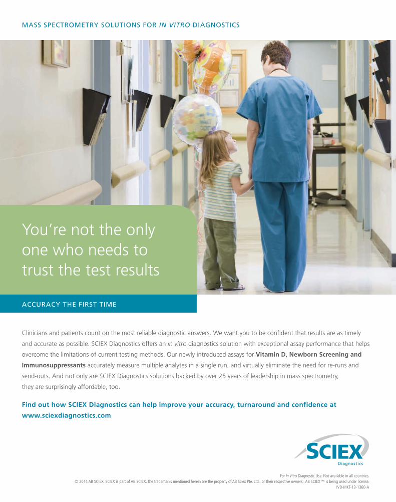
You’re not the only one who needs to trust the test results
ACCURACY THE FIRST TIME
For In Vitro Diagnostic Use. Not available in all countries.© 2014 AB SCIEX. SCIEX is part of AB SCIEX. The trademarks mentioned herein are the property of AB Sciex Pte. Ltd., or their respective owners. AB SCIEX™ is being used under license.
IVD-MKT-13-1360-A
Clinicians and patients count on the most reliable diagnostic answers. We want you to be confident that results are as timely
and accurate as possible. SCIEX Diagnostics offers an in vitro diagnostics solution with exceptional assay performance that helps
overcome the limitations of current testing methods. Our newly introduced assays for Vitamin D, Newborn Screening and
Immunosuppressants accurately measure multiple analytes in a single run, and virtually eliminate the need for re-runs and
send-outs. And not only are SCIEX Diagnostics solutions backed by over 25 years of leadership in mass spectrometry,
they are surprisingly affordable, too.
Find out how SCIEX Diagnostics can help improve your accuracy, turnaround and confidence at
www.sciexdiagnostics.com
MASS SPECTROMETRY SOLUTIONS FOR IN VITRO DIAGNOSTICS
Diagnostics

InPractice
Technologies and techniquesQuality and compliance
Workflow
32-34From Cell Cultures to Multiplex PCRUdo Margraff discusses the new techniques and methods being introduced to infectious disease diagnostics, and asks, what ones should pathologists be focusing on?
35-37Targeted MetabolomicsTandem mass spectrometry is a proven analytical technique now used routinely in UK newborn screening. Find out why advocates of the technology believe it should be used to screeng for inborn errors of metabolism.
You’re not the only one who needs to trust the test results
ACCURACY THE FIRST TIME
For In Vitro Diagnostic Use. Not available in all countries.© 2014 AB SCIEX. SCIEX is part of AB SCIEX. The trademarks mentioned herein are the property of AB Sciex Pte. Ltd., or their respective owners. AB SCIEX™ is being used under license.
IVD-MKT-13-1360-A
Clinicians and patients count on the most reliable diagnostic answers. We want you to be confident that results are as timely
and accurate as possible. SCIEX Diagnostics offers an in vitro diagnostics solution with exceptional assay performance that helps
overcome the limitations of current testing methods. Our newly introduced assays for Vitamin D, Newborn Screening and
Immunosuppressants accurately measure multiple analytes in a single run, and virtually eliminate the need for re-runs and
send-outs. And not only are SCIEX Diagnostics solutions backed by over 25 years of leadership in mass spectrometry,
they are surprisingly affordable, too.
Find out how SCIEX Diagnostics can help improve your accuracy, turnaround and confidence at
www.sciexdiagnostics.com
MASS SPECTROMETRY SOLUTIONS FOR IN VITRO DIAGNOSTICS
Diagnostics

From Cell Cultures to Multiplex PCR Infectious disease diagnostics have improved in leaps and bounds – but with new techniques and methods constantly being introduced, which ones should pathologists focus on?
By Udo Margraff
Infectious diseases are among the most challenging and urgent issues facing healthcare delivery worldwide. With diseases like Ebola, influenza and Middle East Respiratory Syndrome (MERS) making headlines every day, the spotlight is increasingly bright. But despite these headliners, diseases that have slipped out of the public eye (like malaria or glandular fever), as well as the traditional burdens on clinicians (for instance, infections in patients immunocompromised by transplant or chemotherapy) remain just as important.
As treatments develop, there is a growing need for fast and efficient techniques to identify pathogens. From
the early days of detecting disease agents through cultures, to more modern polymerase chain reaction (PCR) methods, early and efficient diagnosis continues to be crucial.
Recent developments in infectious disease diagnostics mean we can identify rare conditions in a fraction of the time it once took, and with much greater accuracy. Nonetheless, there are still bridges to be crossed to ensure the best possible results not only for clinicians, but for patients.
The traditional approachTraditionally, cultures have been used to detect pathogens in clinical samples. Many bacteria, fungi, parasites and viruses can be grown in a lab under the correct conditions, and the precise nature of the culture can identify the characteristics of the microbe itself. For example, successfully growing Staphylococcus aureus in a culture that contains beta-lactam antibiotics indicates methicillin-resistant S. aureus (MRSA).
Techniques used to identify microbes include solid and liquid cultures, as well as cell cultures, and these are commonly used with samples isolated from urine, stool, the genital tract, the throat or the skin. Though culture is traditionally the benchmark for identifying organisms, it has one major drawback – time. Results may not be available for days or weeks, and not all pathogens can be cultured. In a situation where effective treatment depends on rapid and accurate diagnosis, this kind of delay is simply not an option. Therefore, it’s clear that there is a real need for alternative methods.
PCR-based systems More and more, we’re seeing molecular diagnostics used in clinical settings. These techniques are revolutionizing infectious disease diagnosis; heralded as a diagnostic tool for the new millennium, some researchers believe molecular
diagnostics could render traditional hospital laboratories obsolete (1).
Quantitative real-time PCR (qPCR)-based systems detect the agents of disease directly from clinical samples, without the need to wait for a culture to grow. This is particularly valuable in the rapid detection of fastidious microorganisms, along with those that can’t be grown in the laboratory at all. Additionally, sequence analysis of amplified microbial DNA allows for better identification and characterization of pathogens.
One of the earliest recognized applications of PCR in clinical practice was for the detection of Mycobacterium tuberculosis. Before its advent, there were week- to month-long delays associated with standard testing for M. tuberculosis infection – despite the crucial public health impact of early recognition, isolation, and treatment of infected patients. Although PCR assays resulted in significantly decreased time to diagnosis, the only FDA-approved use was when employed alongside a conventional smear and culture. It was suggested, though, that more widespread use of these assays might significantly improve both cost efficacy and clinical outcomes.
Today, qPCR is the most well-developed molecular technique, more widely used than ligase chain reaction and delivering more sensitivity than signal amplification techniques (2). It
In Pract ice32
At a Glance• As we develop better treatments for infectious diseases, our diagnostic methods need to keep up• Traditional diagnosis is done using culture, but the time required can delay necessary treatment• Molecular techniques like PCR-based tests can reduce the time to diagnosis from days to hours• Multiplex PCR not only shortens the time to diagnosis, but allows testing for many potential pathogens simultaneously
“...some researchers believe molecular diagnostics could
render traditional hospital laboratories
obsolete.”

Culture setup
Culture setup
Culturegrowth
Culture check and report
5 m
ins
24 hours 24 hours
30 mins
Culture check and report
Final report 48 hoursTech time: 11 minutes per sampleNegative samples per day: 13Total tech time per day: 143 minutes
Final report 48–96 hoursTech time: 36 minutes per samplePositive samples per day: 2Total tech time per day: 72 minutes
Sampleextraction setup
PCR setup
Sample extraction PCR
Identi�cation and culture setup
50 mins
30 mins
Final report 5 hoursTech time: 10 minutes per sampleNegative samples per day: 13Total tech time per day: 130 minutes
Identi�cation 5 hoursFinal report 48–96 hoursTech time: 40 minutes per samplePositive samples per day: 2Total tech time per day: 80 minutes
1 m
ins
5 m
ins
3 m
ins
3 m
ins
80 mins
Interpretation of results
4 m
ins
Sampleextraction setup
PCR setup
Sample extraction PCR
Identi�cation and culture setup
50 mins
30 mins
Final report 4 hoursTech time: 7 minutes per sampleNegative samples per day: 13Total tech time per day: 91 minutes
Identi�cation 4 hoursFinal report 48–96 hoursTech time: 37 minutes per samplePositive samples per day: 2Total tech time per day: 74 minutes
2 m
ins
2 m
ins
80 mins
Interpretation of results
3 m
ins
Traditional Culture
Multiplex PCR
Multiplex PCR with Optimized Setup
Culturegrowth

In Pract ice34
has a wide range of fulfilled and potential clinical applications, including the evaluation of emerging infections, specific and broad-spectrum pathogen detection, and antimicrobial resistance profiling. But as molecular methods continue to make headway in disease diagnosis, the technology must keep up with growth. We need further development to improve automation, optimize detection sensitivity and specificity, and expand the capacity to detect multiple targets simultaneously – an advancement known as “multiplexing.”
Multiplex PCR As the number of microbial agents detectable by qPCR increases, the ideal end goal is the simultaneous detection of multiple agents that cause similar or identical symptoms. The term “multiplex” refers to the fact that numerous pathogens are detected in a single assay. This technique is useful for synchronized identification of viruses, bacteria, fungi and parasites, and it can save significant time, effort and money – making it a
practical choice for busy laboratories.Multiplex PCR involves amplifying
unique regions of DNA in either individual pairs or combinations of many primers. All of these amplifications take place under a single set of reaction conditions. In a real-time PCR thermocycler that allows the analysis of each frequency specific to the individual amplicon, it is possible to identify specific pathogens.
Using multiplex PCR, clinicians are able perform complete screenings for pathogens known to cause specific clinical syndromes. One syndromic approach in current use screens for the underlying causes of most infections encountered in clinical labs – respiratory infections; gastroenteritis; meningitis; sexually transmitted infections; fever, rash and childhood infections; eye infections; infections of the immunosuppressed; hepatitis; and tropical fever. The benefit of such a broad-spectrum approach is that clinicians don’t have to request testing for a specific pathogen, but can identify a single cause, or rule out multiple options, in a single test. This level of detail can go a long way toward influencing treatment decisions.
Since adopting multiplex PCR approaches, clinical laboratories have found themselves better equipped to provide fast, reliable analysis. Within our own laboratory, Laboratoires Réunis in Luxembourg, qPCR multiplex technology has helped us to ensure that 85 to 90 percent of our reports are completed on the same day. Every day, we perform three runs with plates that include viruses, bacteria, parasites, yeasts and dermatophytes. But despite the extent of our analyses, we are still able to keep turnaround at an all-time low.
Working in the diagnostic sector, I’ve seen a remarkable difference in the way clinical tests are carried out over recent years. It’s good news, though – these latest developments
have made a marked increase in the number of patients and clinicians served. Since adopting qPCR multiplex kits, we’ve increased the number of bacterial gastroenteritis multiplex tests we complete from 2,600 in 2010 to approximately 6,000 in 2014 – all without any increase in the hands-on time required from our staff. Furthermore, our turnaround times are dramatically reduced and urgent samples are still guaranteed to be processed within four to five hours of their arrival (see infographic). These positive effects on our laboratory’s day-to-day operation have made us able to provide clinicians with the option of starting any necessary treatment with first choice antibiotics.
Looking forward However, even with these advancements, continuous development is still underway. Although each multiplex qPCR assay must be individually optimized for its specific reagents, sample-based techniques, such as lyophilization, can enhance stability and dramatically increase ease of use, making a real difference to molecular diagnostic methods in a laboratory setting. Whatever approach further refinements to multiplex qPCR take, we can certainly expect to see exciting developments in the near future – propelling the field of infectious disease diagnostics even farther into the 21st century.
Udo Margraff is a pharmacist and clinical pathologist at Laboratoires Réunis, Junglinster, Luxembourg.
References1. S Yang, RE Rothman, “PCR-based diagnostics for infectious diseases: uses, limitations, and future applications in acute-care settings”, Lancet Infect Dis, 4, 337–348 (2004). PMID: 15172342.2. S Read et al., “Molecular techniques for clinical diagnostic virology”, J Clin Pathol, 53, 502–506 (2000). PMID: 14706081.
“...we‘ve increased the number of bacterial gastroenteritis multiplex tests we complete from 2,600 in 2010 to approximately 6,000 in 2014.”

Targeted Metabolomics Should tandem mass spectrometry be used in the screening of inborn errors of metabolism?
By Hannah Noel
In assays, from pharmacokinetics to proteomics, tandem mass spectrometry (known as MS/MS) coupled with high-pressure liquid chromatography (HPLC) is the analytical technique of choice. It is commonly used during drug discovery alongside protein, peptide and oligonucleotide analysis. One often overlooked application though, is its use in clinical biochemistry and toxicology for laboratory screening of inborn errors of metabolism (IEM).
The technique, which can be used with or without chromatographic separation, offers increased analytical sensitivity and selectivity by reducing the interference from other sample components. It also
facilitates the development of better analytical methods for complex mixtures – ones that are rapid, require less painstaking sample preparation and separation (so they consume less solvent), and enable higher overall sample throughput.
MS/MS can be used to gain structural information about a compound by simple collision-induced dissociation of molecules, followed by scanning of the resultant fragment ions. When pieced together, this information allows researchers to generate a putative structure of the parent, or intact molecule. This feature is particularly important in toxicology and screening for drugs of abuse. However, MS/MS using stable isotope dilution, is a powerful quantitative tool in the majority of clinical laboratory diagnoses.
At the forefront of bioanalysisIEM comprise a rare group of genetic disorders that can have serious clinical consequences for neonates, children, and adults. Left undiagnosed and untreated, they can cause physical disability, irreversible mental retardation, and neurological damage and, in certain cases, can be fatal. Although, as rare disorders their incidence is infrequent, their potentially devastating medical effect has made them of considerable significance to public health.
An expert in the field of MS/MS for IEM screening is Neil Dalton, professor of pediatric biochemistry, King’s College London, director of the WellChild Laboratory, Evelina London Children’s Hospital and a founding director of
SpOtOn Clinical Diagnostics. Dalton is one of a number of experts who were instrumental in introducing the routine use of MS in newborn screening in the UK. “The role of an inherited metabolic disease laboratory is to provide accurate, rapid, and cost-effective screening and confirmatory diagnostic tests for an expanding range of rare disorders with common clinical presentations. The advent of generic diagnostics has allowed us to maximize disease coverage while minimizing costs, laboratory logistics, and the requesting dilemmas of clinicians,” he says.
The intrinsic analytical specificity of multiple reaction monitoring acquisition with MS/MS, combined with the quantitative precision of stable isotope dilution, has placed the technology at the forefront of bioanalysis. Nonetheless, the obvious impact of it on newborn dried blood spot (DBS) screening for inherited metabolic diseases is yet to be fully recognized – plasma/urine amino acid and urine organic acid analyses (and an increasing plethora of disease-specific tests) remain the diagnostic methods of choice.
Screening: the government stanceIn the UK, governmental policy has supported research into extending the use of MS/MS as an IEM diagnostic, but progress has been slow. Back in 1997, the NHS R&D Health Technology Assessment (HTA) program commissioned two reviews of neonatal screening for inherited metabolic disorders (1,2). Both reviews called for further studies on the application of MS/MS for
At a Glance• Tandem mass spectrometry (MS/MS) is a proven analytical technique, which is now routinely used in newborn screening in the UK• Laboratory screening of inborn errors of metabolism (IEM) is, however, an often overlooked application• Latest research and case studies recommend expanded screening programs for these rare but devastating conditions• Large scale metabolomic studies have not only served to emphasize the utility of MS/MS technology in these screening programs, but also how analytically robust modern platforms can be

neonatal screening of IEM, with certain caveats. At the time, it was recommended that widespread introduction of the technology should be withheld pending further evaluation; a position supported by the Child Health Subgroup of the National Screening Committee (NSC). Since then, a 2004 systematic review, which incorporated economic modeling and built on the two HTA reports, has brought the evidence base more up-to-date (3). The review supported the introduction of MS/MS into the UK neonatal screening program for phenylketonuria (PKU) and medium-chain acyl-coA dehydrogenase deficiency (MCADD) combined, although the “wholesale inclusion of all disorders detectable by MS/MS” was not recommended. Currently all babies in England are screened for sickle cell disease, cystic fibrosis, congenital hypothyroidism as well as PKU and MCADD via expanded newborn DBS screening.
Searching for the evidenceBetween 2004 and 2006 Carol Dezateux, her team at University College Hospital in London and numerous UK laboratories and metabolic services, participated in the UK Collaborative Study of Newborn Screening (UKCSNS), funded by the Department of Health and the NSC. In their MCADD study, they trialed a prospective multicenter pilot screening service with testing at age five to eight days, standardized screening, and management protocols using MS/MS (4). Around 60 babies each year (or one in every 10,000 born in England) is currently diagnosed with MCADD by newborn screening. The majority of MCADD variants detected during the trial were predicted to be of definite clinical importance, but this was found to vary according to ethnic group (with certain clinically important variants most commonly seen in Asian babies). The initial UKCSNS findings therefore supported MCADD screening, but highlighted the need to take into account
the ethnic diversity of the population tested at implementation.
Another arm of the UKCSNS study has addressed how MCADD screening programs can estimate their sensitivity directly, because at present only individuals with a positive result undergo a definitive diagnostic test. The study group proposed a framework incorporating a Bayesian model, which simultaneously combined
available prevalence data on the most common mutation of MCADD (c.985A>G) in screened and non-screened populations by using the relationship between true and apparent prevalence of disease to overcome this limitation (4). True prevalence of c.985A>G homozygotes in England was found to be 6.2 per 100,000 individuals and the sensitivity of the screening program was 94 percent (95 percent confidence interval [CI]: 74, 100 percent) compared with a detection rate in non-screened areas of 48 percent (95 percent CI: 30, 68 percent) by the age of 5 years. Hence, the screening program detected (95 percent CI: 30, 60 percent) an additional 47 percent of cases compared with no screening. The researchers concluded that, owing to the high sensitivity of England’s screening program, their estimation approach could be adapted to inform other rare diseases and national screening initiatives.
Furthermore, a 2014 review of mass spectrometry in clinical analysis (5) explained, that “there is consistent evidence of benefits from newborn screening for many disorders detected by MS/MS as well as for congenital hypothyroidism, cystic fibrosis, congenital adrenal hyperplasia by immune-enzymatic methods”. The author went on to explain that real-time PCR tests have been proposed for the detection of certain severe combined immunodeficiencies (SCID) along with MS/MS for adenosine deaminase deficiency (ADA) and purine nucleoside phosphorylase deficiency (PNP) SCID, although initial cost-benefit analyses are still ongoing. According to the author, fundamental to the success of a newborn screening program is using specific biomarkers to avoid false negatives and conducting second-tier tests to minimize the false positive rate.
A targeted metabolomic approach Dalton explains that a targeted metabolomic approach using MS/MS would help rationalize the clinical diagnosis of IEM. According to him, at the Evelina London Children’s Hospital, the strategy has been to develop validated analytical methods for amino acids, acylcarnitines, organic acids, purines and pyrimidines, and disease-specific tests, which are then multiplexed into a single assay: essentially an IEM metabolite analyzer. The further diagnostic and research potential of this approach has been augmented by the recent introduction of an API6500 Q trap instrument, which has an increased linear range and true positive-negative ion switching, to include novel infection, specific organ function, and tumor biomarkers.
Dalton provides a genuine example of the rapid nature of MS/MS in practice: “The Evelina London received an acute IEM screen request over the phone for a baby with severe acidosis and hyperammonemia at another major London hospital at
In Pract ice36
“There are obvious challenges that limit
the introduction of routine clinical
diagnostic multiplexed assays.”

15.10 one Friday. By 16.48, the sample had been transported to the laboratory, MS/MS analysis performed, and an Evelina London consultant had phoned the other hospital to report increased propionylcarnitine (m/z 218) with absence of methylmalonylcarnitine (m/z 262), normal methylmalonic acid and increased methylcitrate. A reported diagnosis of propionic acidemia was subsequently confirmed by enzymology results,” he explains. This illustrates the strength of MS/MS as a metabolomic approach to diagnose IEM – it facilitates rapid diagnosis, earlier treatment intervention, better patient outcome and immediate reassurance for the parents.
Expanding the screening programIn May 2014, the NSC announced its recommendation to screen every baby in the UK for an additional four IEMs as part of newborn blood spot screening (6). These diseases are maple syrup urine disease (MSUD), isovaleric acidemia (IVA), glutaric aciduria type 1 (GA1), homocystinuria (pyridoxine unresponsive) (HCU). Six of the 13 inherited metabolic disease laboratories in England took part in a pilot study and are already screening for these conditions; the other seven English laboratories began screening in January 2015 and Wales will also begin to screen early this year.
Dalton and his team support expanded IEM screening and believe that analytical platforms which can evaluate multiplexed metabolites, proteins and enzymes from a single DBS, would hold great value for this application.
Diagnostic sensitivity of these tests can be improved using new and established biomarkers and comparison of contentious biomarkers, for example in the case of type 1 tyrosinemia, porphobilinogen synthase activity versus tyrosine versus succinylacetone, or for homocystinuria, total homocysteine versus methionine.
Point proven?Yvonne Daniel (lead scientist and operational lead – hematology, Viapath) and colleagues at the WellChild Laboratory at Guy’s & St Thomas’ NHS Foundation Trust (of which Dalton is a director) have developed an MS/MS method for newborn hemoglobinopathy screening. This screen is usually undertaken using HPLC or isoelectric focusing (IEF). The team devised a rapid and specific electrospray MS/MS method using multiple reaction monitoring (MRM) based peptide analysis, for simultaneous detection of the clinically significant hemoglobinopathies: hemoglobin (Hb)S, HbC, HbE, HbD-Punjab and HbO-Arab (7), and subsequently for the detection of Hb Lepore and HbA2 quantitation (8).
The method has been validated successfully by comparing its performance with the conventional IEF methods in 40,000 newborn DBS in collaboration with Leeds Teaching Hospitals NHS Trust. This study was commissioned by the NHS Sickle Cell and Thalassaemia Programme Centre to evaluate its potential in England’s newborn screening program. To facilitate uptake of the technique the number of processing steps required for conventional tryptic digestion of proteins was reduced and, together with a 30-minute digestion, sample preparation for MS/MS was modified so that it is no more onerous than for IEF. Furthermore, the consumable costs associated with MS/MS can be offset by high-throughput integrated with current IEM MS/MS screening. The specificity of MRM analyses implies that hemoglobinopathy detection can be limited to specified conditions – based on agreed screening policies – and can eliminate the need for costly and time-consuming second-line testing. In addition, subsequent product ion scanning on linear ion trap instruments provides unequivocal sequence data.
Dalton concludes, “There are obvious challenges that limit the introduction of
routine clinical diagnostic multiplexed assays: calibration, quality control, technical competence, and interpretation skills. However, large scale metabolomic studies have not only served to emphasize these challenges but they’ve also demonstrated how analytically robust modern MS/MS platforms can be.”
Hannah Noel is a medical journalist with a PhD in molecular biology, based near Bath, UK. Many of the findings reported by Noel were outlined in Neil Dalton’s presentation at Viapath’s Fourth Innovation Academy held in December, 2014, London.
References1. RJ Pollitt et al., “Neonatal screening for inborn errors of metabolism: cost, yield and outcome,” Health Technol Assess, 1, 1–202 (1997). PMID: 9483160.2. CA Seymour et al., “Newborn screening for inborn errors of metabolism: a systematic review,” Health Technol Assess, 1, 1–97 (1995). PMID: 9483156.3. A Pandor et al., “Clinical effectiveness and cost- effectiveness of neonatal screening for inborn errors of metabolism using tandem mass spectrometry: a systematic review,” Health Technol Assess 8, 1–121 (2004). PMID: 14982654.4. JM Khalid et al., “UK Collaborative Study of Newborn Screening for MCADD. Ethnicity of children with homozygous c.985A>G medium-chain acyl-CoA dehydrogenase deficiency: findings from screening approximately 1.1 million newborn infants,” J Med Screen, 15, 112–117 (2008). PMID: 18927092.5. G La Marca, “Mass spectrometry in clinical chemistry: the case for newborn screening,” J Pharm Biomed Anal. 101, 174–182 (2014). PMID: 24844843.6. Public Health England, UK National Screening Committee, “Expanded Newborn Bloodspot Screening,” available at http://bit.ly/1zTnjgC7. YA Daniel et al., “Rapid and specific detection of clinically significant haemoglobinopathies using electrospray mass spectrometry-mass spectrometry,” Br J Haem, 130, 635–643 (2005). PMID: 16098080.8. YA Daniel et al., “Quantification of hemoglobin A2 by tandem mass spectrometry,” Clin. Chem, 53, 1448–1454 (2007). PMID: 17556646.
In Pract ice 37

Virtual Events
Event Overview:Assessment of molecular heterogeneity in tumor is a challenging task. For the first time, Palanisamy et al. have demonstrated the existence of a rare subset of prostate cancer with heterogeneous molecular aberrations involving ETS family genes and SPINK1 expression utilizing both standard immunohistochemistry (IHC) and RNA in situ hybridization (Advanced Cell Diagnostics RNAscope® Technology). The presenter will discuss the application of RNA ISH technology to reveal hitherto unidentified molecular subtypes of prostate cancer.
Learning Objectives of Webinar 1. Strategies for interrogating interpatient and intratumor heterogenity 2. RNA biomarker analysis of fusion genes, non-coding genes and pseudo genes3. Applications of RNAscope ISH along with other methods such as IHC to characterize molecular profiles of tumors
On Demand now!http://tp.txp.to/0115/ACD/webreg
About Us:Advanced Cell Diagnostics is a provider of RNAscope® Technology, the most sensitive in situ hybridization technology commercially available. Based on ACD’s unique patented probe design strategy which enables simultaneous signal amplification and background noise suppression, RNAscope technology represents one of the most significant advances in ISH technology in over 40 years. In 2014, applications of this new technology appeared in 83 peer-reviewed publications.
Elucidating Tumor Heterogeneity in Prostate Cancer by Combined IHC & Novel RNA ISH
SpeakerNallasivam Palanisamy, PhDAssociate Scientist
My research interests are on the discovery and characterization of gene fusions in cancer and understanding their role in carcinogenesis from a translational research perspective. Using genomic technologies such as high-density array comparative genomic hybridization, advanced molecular cytogenetic technologies including FISH, CGH, spectral karyotyping, gene expression microarrays, and next generation sequencing technology, my laboratory investigates the transcriptional and genomic architectures of solid cancer genomes. In-depth analysis of genomic amplifications provided an unprecedented view and identified rare gene fusions formed at theboundaries of amplification and deletions. I also pioneered the application of next generation sequencing technology for the discovery of new recurrent gene fusions in cancer.
Webinars

NextGenResearch advancesNew technologiesFuture practice
40-41The Noninvasive Eye-Opener Optical biopsy has faced hurdles on the road to acceptance, but it could greatly benefit medicine and should be viewed as a support, not a threat, to pathology.
42-43A Golden Opportunity?Imagine gold nanoparticles having a place in the future of cancer diagnosis. Could you one day be tested for oral cancer at the dentist? Dror Fixler discusses this novel cancer imaging method.
The Noninvasive Eye-Opener Optical biopsy could provide a wide range of diagnoses in minutes and should be viewed as a support, not a threat, to pathologists
By R. Condon Hughes, III
The term “optical biopsy” has entered into common use among researchers in biomedical optics. It’s defined as an optical measurement, often a type of spectroscopy, used to perform a noninvasive tissue diagnosis in vivo and in real-time. It’s hoped that these techniques will reduce the need for surgical removal of biopsy tissue samples; instead, a spectral analysis of the tissue in vivo is recorded by an imaging system, or with an optical probe placed on or near the surface of the tissue. Then, based on the optical measurements, a diagnosis can be made.
Optical biopsy has some specific advantages over traditional methods. Consider, for example, the clinical diagnosis of melanoma and other skin
cancers, which is still performed primarily by sight. Since even the most sophisticated eye is fallible, there’s a chance that skin cancer might be visually diagnosed as benign, or that a benign mole might be unnecessarily biopsied in an invasive procedure. As reported in the journal Dermatologic Surgery, nearly 80 percent of all skin biopsies performed result in benign diagnoses (1) – so patients undergoing traditional biopsy must endure not only the chance of being misdiagnosed with cancer, but also the pain and potential subsequent scarring if their doctor deems an invasive biopsy necessary.
Nervous patients, especially, face difficulties with traditional biopsy methods. Many skin lesions occur on cosmetically sensitive parts of the body, such as the face, so apprehensive patients might choose not to undergo the procedure at all. If they do, they might experience anxiety while waiting for the biopsy results, which can take up to two weeks with traditional biopsies. Using optical biopsy techniques avoids issues like these; the procedure is not invasive and the images obtained can be diagnosed at the point of care, eliminating long wait times.
A bumpy road to acceptanceDespite the apparent value of this new approach, optical biopsy has faced some hurdles on the road to general acceptance. First, the development of the associated imaging technology has taken time. For example, confocal microscopy, a laser technology, has the ability to produce high resolution images of living tissue at various depths. It works by concentrating on one focal plane at a time and reducing the out-of-focus light from above and below that plane. A computer then reconstructs the images, which are taken point-by-point rather than projected through an eyepiece.
The principle for this kind of microscopy was developed in 1953, but it didn’t become a standard technique until the late 1980s, when the high quality lasers
needed to make the technology viable were developed.
Doctors interested in optical biopsy face the task of familiarizing themselves with the new technology before they can use it properly. In general, though, technicians are capable of obtaining the actual images – whether they result from an endoscopic procedure or a spectral analysis of the skin – using established protocols. It’s the interpretation of those images that requires specialized training.
How will it affect pathologists?If optical biopsy eventually sees widespread adoption as a standard technique, it may not be utterly transformative, but it is likely to impact the pathology community in several ways. It might, for instance, attract a new cohort of patients who are attracted by the advantages it brings: no scarring and no long wait times for diagnosis, along with a reduction in psychological trauma. This has the potential to improve rates of diagnosis. But some pathologists worry that, even with the influx of new patients, optical biopsy might reduce the need for pathology services. Far from taking away from the roles of qualified pathologists, their expertise will be as necessary as ever to interpret the optical biopsy images collected by technicians – but with increased convenience. The pathologist doesn’t need to be in the room when the procedure is performed, nor even at the same facility; the digital images can be analyzed from anywhere in the world.
The steps involved in obtaining optical biopsy images will vary according to the organ of interest. If an endoscopic procedure is needed, a very small confocal laser microscope can be used. The microscope sits at the end of a long, thin, flexible tube that can be threaded through a traditional endoscope to enter the body. It detects fluorescent light triggered by a laser; a filter removes the laser light, allowing the fluorescent light to pass through a small aperture. The light then
At a Glance• Optical biopsy is a new way of making tissue diagnoses in real-time at the point of care• Not only is it quicker than conventional biopsy techniques, but it’s also noninvasive as it doesn’t require excision of a tissue sample• Optical biopsy isn’t just a surface technique – it can be used to examine internal organs and even tissues that aren’t accessible by traditional biopsy• New techniques like this will increase convenience for pathologists because tissue analysis and diagnosis will be possible from anywhere in the world
NextGen40 NextGen40

hits a photodetector that converts the image to electronic signals, which are then displayed as images on a computer screen for the pathologist to examine.
Optical biopsy of the skin works a little differently. While conventional microscopes work by illuminating thin tissue layers from below (transmitted light), high resolution confocal microscopes designed for dermatology use incident light, which means that the skin is illuminated from above, in the horizontal plane, with a focused laser. The light is reflected at interfaces where the refractive index changes, typically at highly reflective skin structures like keratin, melanin and collagen. The reflected light is directed through a pinhole onto a detector, so that only signals from a defined horizontal plane are used for high resolution imaging. This technique limits penetration depth, but usually provides physicians with ample information to determine whether a traditional biopsy is required or whether the lesion can simply be monitored. The entire procedure takes less than 10 minutes and collects all the images needed to make an accurate, reliable diagnosis at the point of care.
The evidenceOver the past two decades, a slew of studies have reported on the efficacy of optical biopsy as a technique in tandem with various imaging technologies, involving both endoscopic procedures and skin imaging (2–4). A 2013 overview
by Alfano and Pu (5) concluded, “Optical biopsy with lasers and LEDs, as an emerging technology in biomedical optical imaging, holds a promising future armamentarium for clinical diagnosis and other important medical applications.” In the same year, the Association of the Scientific Medical Societies in Germany published a guideline on behalf of the German Dermatological Society stating, “Confocal laser scanning microscopy [a key imaging technique used in tandem with optical biopsy] is suitable for dermatological, noninvasive diagnostic of near-surface skin changes. In the area of skin tumors, it is especially of interest to assess melanocytic lesions with respect to their benign or malign character in order to enable the early detection of melanoma and avoid unnecessary excisions” (6). A recent update to the Guideline on Basal Cell Carcinoma published by the European Dermatology Forum agrees, identifying confocal microscopy as an emerging technique in digital imaging diagnostics and reporting that it “has shown high diagnostic accuracy for the diagnosis of basal cell carcinoma” (7).
Skin cancer isn’t the only application for optical biopsy, though. So far, human tissues including prostate, breast, lung, colon and gastrointestinal have been studied – but applications where optical biopsy could be especially powerful, such as where conventional excisional biopsy is hazardous or impossible, should be emphasized. A good example is in
ophthalmology, where traditional biopsy of the retina is impossible, but optical biopsy (in tandem with optical coherence tomography) can provide high-resolution images of pathology that can’t be obtained any other way. Hopefully, this new noninvasive technique will one day allow pathologists access to tissues – like those in the eye – that we currently have no way of examining safely. In the meantime, optical biopsy is an exciting, rapidly developing technique with the potential to improve the diagnostic accuracy for a wide variety of medical specialties.
R. Condon Hughes, III, is a partner at Diagnostic Tissue/Cytology Group of Meridian, Mississippi, USA. He is an MD and board certified in anatomic/clinical pathology with subspecialty board certification in dermatopathology.
References1. SR Fuller et al., “Digital dermoscopic monitoring of atypical nevi in patients at risk for melanoma”, Dermatol Surg, 33, 1198–1206 (2007). PMID: PMC2292405.2. TD Wang, J Van Dam, “Optical biopsy: a new frontier in endoscopic detection”, Clin Gastroenterol Hepatol, 2, 744–753 (2004). PMID: PMC2169359.3. IJ Bigio and JR Mourant. “Optical biopsy”, Encyclopedia of Optical Engineering, 1577–1593. Marcel Dekker: 2007.4. GJ Tearney et al., “In vivo endoscopic optical biopsy with optical coherence tomography”, Science, 276, 2037–2039 (1989). PMID: 9197265.5. R Alfano, Y Pu, “Optical biopsy for cancer detection”, Laser for Medical Applications, 325–367. Woodhead: 2013.6. Association of the Scientific Medical Societies in Germany, “Confocal laser microscopy in dermatology. AWMF Registry No. 013/076 Class: S1.” Available at: http://bit.ly/1AmBInX. Accessed November 7, 2014.7. European Dermatology Forum, “Update on the guideline on basal cell carcinoma (update 2012).” Available at: http://bit.ly/1AmC65U. Accessed November 7, 2014.
NextGen 41

NextGen42 NextGen42
A Golden Opportunity? A new, noninvasive approach to cancer imaging uses gold nanorods and reflected light to detect even small tumors
By Dror Fixler
Cancer imaging has been contributing to patient care since X-rays were discovered over a century ago. In its various forms, it impacts almost every aspect of cancer research, diagnosis and treatment (1), so it isn’t hard to understand why we are so eager to update and improve it.
Though we’ve developed a wide variety of imaging techniques – including ultrasound, X-ray computed tomography (CT), magnetic resonance imaging (MRI) and more – these techniques are not without drawbacks. For instance, ultrasound lacks resolution, MRI can’t be used on patients with implanted devices or metal in their bodies, and both X-ray
CT scans and newer technologies, like positron emission tomography (PET), involve exposing the patient to radiation.
There’s a clear need to improve cancer imaging, which is what motivated me to develop a new approach that is noninvasive, doesn’t involve radiation and – in my opinion – can accurately visualize cancer and define tumor margins in real-time.
The hybrid approachFive years ago, it occurred to me that we could combine two separate imaging approaches to create a new methodology – one that could have applications in a clinical setting and even beyond.
The two techniques I combined have been around for years. The first, diffusion reflection, deals with the way a surface reflects light: it can be scattered, absorbed, transferred or reflected. Physicists in the 1980s and 1990s suspected that detecting the intensity of light reflected from tissue would give us information about its properties, but there were simply too many variables; among other things, tissue contains bone and dilated blood vessels that affect light, so detecting a tumor this way is not clinically viable.
The other aspect, nanophotonics, has
gained traction in the last 10–20 years. It deals with the behavior of light on a nanometer scale, and is combined with the use of agents – for instance gold nanoparticles – that enhance contrast during medical imaging, so that structures within the body are easier to detect. Conjugating the nanoparticles to anticancer antibodies allows tumor imaging, and this method is already being used in several FDA-approved tests – but it doesn’t work as well in the early stages of cancer, when tumors are too small to be effectively visualized.
To overcome the limitations of each individual technique, my colleagues and I use both – first, we use antibody-conjugated gold nanorods to coat the structure to be imaged, then we use a hyperspectral imaging system to examine the diffusion of light within the tissue. In diffusion reflection, a light source illuminates a sample at a single point on its surface and an array of detectors collects the diffuse light reflected back. In its industrial form, the system is portable, featuring a scanning head with an illumination source and a detector array. With the aid of diffusion reflection, we can detect much lower concentrations of nanoparticles than can be achieved by other methods, down
At a Glance• Current tumor imaging methods can pose risks to the patient and are expensive, and this has stimulated research efforts into alternatives• Combining the diffusion reflection theory with the application of gold nanoparticles means that even early stage tumors and tumor margins can be detected in real-time• My team and I have developed a new imaging system that conjugates the nanoparticles to anticancer antibodies allowing tumor imaging on that basis • It has the potential to go beyond the clinic; the main initial hurdle to overcome is regulatory approval, but the wheels are in motion

NextGen 43
to 0.01 mg/mL – less than one cubic centimeter of tumor tissue.
From modeling to the clinicOnce we’d come up with the idea, the first thing we needed to do was adapt the relevant diffusion equations (2, 3) for a contrast agent like gold and build a mathematical model to correlate our theories with our eventual results. Next, we used imaging phantoms (tissue-like elements designed to behave in the same way as, for example, an arm) to verify that we could detect the gold nanoparticles within living tissues. Third, we conducted in vitro testing on cultured cells, using spectroscopy to determine the tumor specificity of antibody-conjugated gold nanorods (4). Finally, we moved to testing in in vivo models – mice and rats with human-derived head and neck tumors. We injected the rodents with immunotargeted gold nanorods, scanned them with a digital microscope, and performed diffusion reflection measurements to test our technique’s ability to sensitively and specifically detect tumors. In every case, there was a clear distinction between tumor and healthy tissue (5).
Because we now have the ability to deliver high concentrations of gold nanorods specifically to tumor tissue, we’re able to raise the absorption coefficient of the tumor and discriminate quite clearly between healthy and cancerous tissue. With further research, we hope to improve detection and use the technique for determining tumor size as well as presence and location. Meanwhile, we hope to advance our technique to testing for application in the clinic.
Taking a broader viewI feel that this new imaging method can greatly benefit oncologists, but our hope is that it can go far beyond the hospital setting. My dream is for it to be far more widely available – the design is straightforward and fairly cheap, and I would estimate it
could be sold commercially for home use at under US$100 a box. With cancers like those of the skin, mouth or throat, the gold doesn’t need to be injected; you could just gargle with the gold solution or spray it onto your skin, then use a home scanning kit to get immediate results. I would like to get the test into many clinical settings, as it’s straightforward and doesn’t require a doctor. For example, individuals at high risk of oral cancer, like tobacco users, could be tested while visiting their dentist – a noninvasive examination with no injection or biopsy – and doing so could potentially catch cancers early and save lives with just a mouthwash and a quick and painless scan.
Whether sold commercially or not, our new method can be added to the pathologist’s current suite of clinical tools. Laboratory professionals will have the option of scanning the suspected area using our system, which will provide them with an additional degree of freedom. This can only work, though, if we overcome our main obstacle – regulatory approval for injecting the gold nanoparticles into the bloodstream. The wheels are turning in the United States to approve this kind of use, but until now, only local injections and sprays have been validated, so the system can detect only surface tumors.
Exploring other avenuesWe’re also looking into our system’s potential for applications other than cancer diagnostics. Detecting and characterizing early-stage atherosclerotic vascular injury is a challenge, and we have demonstrated that macrophage cells – which are significant components of unstable, active atherosclerotic plagues – uptake our gold nanoparticles and have a unique diffusion reflection profile (6). In rats, we found that animals with carotid artery injuries were easily distinguishable from healthy ones using our noninvasive imaging method. That project is in its early stages, but we’re working with two groups, one from Germany and one
from the UK, who are very interested in developing the technology.
As a researcher and a physicist specializing in nanotechnology and fluorescence imaging, people don’t necessarily expect me to get directly involved in the world of medicine. Now, we’ve joined forces with two medical doctors who are interested in adopting our system and introducing it to their patients. It’s currently being trialed in humans by Michael Wolf at the Sheba Medical Center, Tel Hashomer, and by Avraham Hirshberg at Tel Aviv University, both in Israel. Their efforts in the clinic mean that our test could soon have a meaningful impact on people’s lives, and I find that incredibly exciting.
Dror Fixler is an expert in electro-optics and photonics research, a member of the Nano Photonics Center at the Institute of Nanotechnology and Advanced Materials, and a senior lecturer in the Faculty of Engineering at Bar-Ilan University, Israel.
References1. G Esposito, “Grand challenges for cancer imaging and diagnosis”, FONC, 1, [Epub] (2011). PMID: PMC3355941.2. R Ankri et al., “On phantom experiments on the photon migration model in tissues”, J Open Optic, 5, 28–32 (2011).3. R. Ankri et al., “In-vivo tumor detection using diffusion reflection measurements of targeted gold nanorods – a quantitative study”, J Biophotonics, 5, 263–273 (2012). PMID: 22234916.4. R Ankri et al., “A new method for cancer detection based on diffusion reflection measurements of targeted gold nanorods”, Int J Nanomedicine, 7, 449–455 (2012). PMID: 22334777. 5. D Fixler et al., “Diffusion reflection: a novel method for detection of oral cancer”, J Dent Res, 93, 602–606 (2014). PMID: 24695671.6. R Ankri et al., “Gold nanorods as absorption contrast agents for the noninvasive detection of arterial vascular disorders based on diffusion reflection measurements”, Nano Lett, 14, 2681–2687 (2014). PMID: 24697682.

The RNA RevolutionA guide to RNA as a biomarker and its detection
Gene expression profiling yields many insights into the disease state, particularly in discovering those molecular indicators known as biomarkers. Indeed, the widespread application of transcriptomic techniques in cancer research over recent years has proven that, like protein, RNA is a rich source of clinically valuable biomarkers for diagnosis, prognosis and predicting therapeutic response. Although such approaches may identify many potential biomarkers, translating these discoveries into the clinic for routine measurement has traditionally been hindered by established analytical technologies. While it is commonplace to detect and visualize DNA and proteins in their native context within single cells, until now the best routine measurement tools for RNA have been those that detect and quantify RNA in solution, losing all morphological context. Times are changing, however, and the ‘RNA Revolution’ is here.
The intriguing molecule of RNA is no longer viewed as merely the ‘messenger’, especially with new classes of non-coding RNAs being discovered on a regular basis that have a hand in genetic regulatory control and a wide range of cellular activities. The discovery of this “new world” of RNA has sparked an unprecedented drive towards better tools to characterize the complexity of RNA – in terms of quantity, function and spatial distribution. In particular, pinpointing the localization of specific RNAs within cells and tissue architecture is an important factor in realizing its true potential as a biomarker.
Exploring how RNA presents an
ideal biomarker, especially in light of novel RNA analysis methodologies, the new whitepaper from Advanced Cell Diagnostics (ACD) will discuss:
• The Biomarker: What makes a valuable biomarker, and how a direct path from RNA biomarker discovery to the clinic is vital, avoiding the use of DNA or protein surrogates.
• The Method: A biomarker is only as good as its routine analysis methodology, but what constitutes the optimal biomarker method? Advantages and pitfalls of existing methods for routine biomarker analysis will also be discussed.
• The Future: How the utilization of RNA as a biomarker is achieved
through the latest RNA analysis methods, such as ACD’s RNAscope®.
Novel RNA analysis technologies are unlocking the potential of RNA as a clinically valuable biomarker. This new whitepaper examines the utility of RNA as a biomarker, and how this is profoundly linked to the methods now available for its validation, detection and localization.
To read the full whitepaper, please visit the website: www.acdbio.com/[email protected]
Appl icat ion Note44
Revealing the RNA expression signature of individual cells within the tissue architecture. The above image shows human breastcancer FFPE tissue probed for MMP9 mRNA expression using ACD’s RNAscope® technology

ProfessionYour career
Your businessYour life
46-47The Pharmaceutical PathologistAs a pathologist, drug development may seem like a foreign landscape – but your skills are directly applicable to the industry. Peter Hall explains why big pharma needs pathology.
48-49Why Did I Choose Toxicology?Five toxicologic pathologists tell us why they chose their field, what challenges they face, and the biggest benefits of forging a career in toxicology.

The Pharmaceutical Pathologist The drug development industry needs pathologists – toxicology plays a central role in getting therapies from the “bench to the bedside”
By Peter Hall
Big pharma may not seem like a natural home for the anatomic pathologist; historically, our function has been to diagnose disease through examining human or animal tissues, but our expertise in the mechanisms of cellular and tissue response to injury means we’re well placed to play an important role in delivering drugs to the clinic. Drug development – from the “bench to bedside” – needs an ever-increasing number of disciplines and technologies to combine to bring a molecule to market.
To launch a new pharmaceutical, regulatory approval must first be secured
– key to this is demonstrating therapeutic efficacy (how well does the drug work compared with others?) and safety (what are the potential adverse effects?). Toxicologic pathologists are crucial to this process, working on the front line to address both of these questions during preclinical studies. If you’re unfamiliar with drug discovery, this may seem like a foreign landscape, but it utilizes all of the same tools and skills pathologists routinely apply in clinical practice and research – namely, the assessment of gross and microscopic changes combined with technical a ids l ike e lectron microscopy, immunocytochemistry, in situ hybridization and image analysis.
The role of toxicologic pathology in the pharmaceutical industry is perfectly demonstrated by the development of several new classes of oncologic drugs that target tumor-derived blood vessels (angiogenesis) and tumor proliferation – two of the six hallmarks of cancer (1). In each case, careful “down the microscope” observations contributed to the proof-of-principle, thereby increasing confidence that they could translate into a meaningful medicine that might go on to affect the lives of thousands of patients.
Proving theories behind therapies Tumor reliance on angiogenesis may seem obvious to us today, but it was not until 1971 that the hypothesis was proposed by the scientist Judah Folkman. Writing in the New England Journal of Medicine, he suggested that tumor angiogenesis presented a new therapeutic approach for fighting cancer (2). Over thirty years later, this idea was validated by the approval of several new molecules, including bevacizumab, sunitinib and sorafenib. So what role did the pathologist play in these landmark approvals?
Using a combination of CD31 immunostaining of vascular endothelium and image analysis, toxicologic pathologists proved (at least in human
xenograft tumors) that treatment with these compounds resulted in reduced vascular density and size, and therefore inhibited tumor growth.
As anti-angiogenics were being developed, the research community was also exploring other areas of tumor biology, searching for the all-important “druggable target”. The most notable of these was the epidermal growth factor receptor (EGFR), a tyrosine kinase receptor that, when activated (i.e. mutated), provided a growth advantage to non-small cell lung cancer cells. Treatment with EGFR inhibitors resulted in reduced xenograft tumor growth in mice, and histological analysis using EGFR phospho-specific antibodies demonstrated that this was associated with dephosphorylation of the receptor – i.e., inhibited activation. However, the first-generation of anti-EGFRs weren’t perfect, because adaptive tumor resistance almost always developed: commonly a T790M point mutation that renders the receptor insensitive to inhibition. Fast forward a decade, and third-generation anti-EGFRs were being created – these irreversibly bind to T790M, solving the problem. To test this, standard xenograft models were supplemented with more sophisticated transgenic mouse models which inducibly expressed the mutated T790M receptor in type II pneumocytes. These mice reliably developed multiple lung adenocarcinomas (3). Regression of established tumors only occurred with third-generation inhibitors – and once again, histological imaging was able to demonstrate that reduced tumor mass was associated with dephosphorylation of the mutant EGFR, and inhibition of the downstream signal transduction cascade.
Finally, pathologists have also played a key role in the development of a newer class of agent: the aurora kinase inhibitors. Aurora kinases are known to play an important role in chromosome alignment and segregation, and cytokinesis. They are over-expressed in a number of human
At a Glance• Drug development might seem like a foreign landscape, but your skills as a pathologist are directly applicable to the pharmaceutical industry• Assessing efficacy and safety are key to gaining regulatory approval, and tissue analysis provides a wealth of information during preclinical trials• Pathology has played a key role in the development of ground-breaking therapies, including angiogenesis and EGFR inhibitors • Techniques and technologies will change, but tissue response to injury won’t – toxicologic pathology will remain a core part of drug discovery
Profession46

malignancies so targeting them should (theoretically) result in the tumors failing to correctly divide, leading to apoptosis. This should be observable because tumor cells would be expected to become tetrapoid or even octapoid because of disruption of the mitotic machinery. Clearly, histological assessment was ideally placed to provide physical evidence of targeted molecular inhibition. When the experiments were performed, the correlation with the theory was uncanny. Significant effects on tumor growth were observed, along with degenerating cells that showed unmistakable signs of nuclear enlargement – proving that the drugs were working as expected.
Ensuring safety and predicting problemsWhen developing therapeutic molecules, efficacy is nothing without safety, and toxicologic pathologists are ideally placed to inform safety assessments. Like all pathologists, we’re trained to assess the response of tissues to all types of noxious stimuli, including drug-induced toxicity. During the development of vascular endothelial growth factor receptor (VEGFR) inhibitors, the toxicity profile was first established in rodents, where pathologists noted a triad of changes characterized by ovarian atrophy, growth plate dysplasia and incisor tooth dental dysplasia (4). The affected organs are all dependent on a continuously growing vascular supply, and once the process was inhibited, degeneration or atrophy of the organ was an inevitable tissue response. In addition, some other changes were noted, two of which – hypertension and increased glomerular mesangial matrix with proteinuria – had important implications for humans. Hypertension was associated with improved clinical outcome, whereas proteinuria was associated with poorer survival (5).
Similarly, when EGFR inhibitors were being developed, preclinical assessment identified epithelial atrophy as one of
the major toxicities associated with this class of compound; a direct result of receptor inhibition in epidermal tissues. In rodents, this change was characterized by thinning of the skin and an associated follicular dysplasia, inflammation and redness. In the clinic, patients treated with these drugs often develop epidermal rash, and this is also thought to be associated with improved prognosis and response (6).
Preclinical histological assessment isn’t only for characterizing adverse effects – it can also predict them before irreversible damage occurs. Take the example of FGFR/MAPK (fibroblast growth factor receptor/MAP kinase). During development, widespread metastatic mineralization was a significant preclinical concern. In order to choose potential treatments with a low risk of toxicity, the presence of mineralization could be correlated with changes in serum biomarkers (such as calcium or phosphate concentration), and then used in a predictive way to select new molecules, or even to screen patients for early signs of mineralization.
The need for safety, as well as the development of patient-specific efficacy biomarkers, is sure to continue to grow as personalized medicine becomes mainstream in our hospitals. It’s likely that toxicologic pathology will play a big part in the early development of many of them.
An evolving roleIt’s inevitable that, going forward, existing technologies like image analysis and in situ slide techniques will become more sophisticated. New technologies (such as microRNAs) and genome editing techniques (like CRISPR-Cas) are going to be introduced into the drug development process – and toxicologic pathologists, like those in any field, will have to adapt and specialize in order to keep pace. Despite this, I think pathology as we know it, is here to stay: tissue responses to injury do not change, and our work provides a window into the physical reality of how drugs affect cells. However, our role and our purview are likely to evolve as we use new and creative ways to validate in vitro techniques, help develop biomarkers, and aid in integrating and interpreting the clinical relevance of bioanalytical data. The roadmap may be changing, but the role of the toxicologic pathologist is likely to remain front and center in the drug discovery process.
Peter Hall is a toxicologic pathologist at AstraZeneca, Alderley Park, Macclesfield, UK.
References 1. D Hanahan, RA Weinberg, “The hallmarks of cancer”, Cell, 100, 57–70 (2000). PMID: 106479312. 2. J Folkman, “Tumor angiogenesis: therapeutic implications”, N Engl J Med, 285, 1182–1186 (1971). PMID: 4938153.3. L Regales et al., “Development of new mouse lung tumor models expressing EGFR T790M mutants associated with clinical resistance to kinase inhibitors”, PLoS One, 2, e810 (2007). PMID: 17726540.4. AP Hall, “The role of angiogenesis in cancer” Comp Clin Path, 13, 95–99 (2005).5. L Khoja et al., “Evaluation of hypertension and proteinuria as markers of efficacy in antiangiogenic therapy for metastatic colorectal cancer”, J Clin Gastroenterol 48, 430–434 (2014). PMID: 24153157.6. R Pérez-Soler, “Can rash associated with HER1/ EGFR inhibition be used as a marker of treatment outcome?” Oncology (Williston Park), 17, 23–28 (2003). PMID: 14682120.
Profession 47
“When the experiments were
performed, the correlation with the
theory was uncanny.”

Profession48
Why Did I Choose Toxicology? Five toxicologic pathologists offer their perspectives on the benefits and challenges of forging a career in toxicology.
What inspired you to specialize in toxicology?
Peter Hall: One of my driving passions in life has been biology, and more specifically, the mechanisms that underlie biological processes – whether as a schoolboy learning about DNA replication and transcription, or as an undergraduate learning about the pathogenesis of Cushing’s disease in dogs. What continues to attract me is an underlying curiosity to delve into the mechanisms of disease, which as a toxicologic pathologist, manifests as drug-induced toxicity and efficacy. Understanding how cells work, and
how they respond in health and disease has never ceased to fascinate me and (I hope) will continue to surprise me.
Schantel Hayes: After two years of “ologies” (pathology, toxicology, pharmacology, etc.) in veterinary school, I moved into the clinic. Many find this the most exciting part – to don the “white coat” and perform surgery. But my interest in veterinary medicine was waning, and I decided medical school best suited my needs. One of my favorite professors was baffled, and suggested comparative pathology. I took his advice and completed a joint anatomic residency/PhD program, then sought a career that encompassed my veterinary training and my newly acquired molecular training.
Jeffrey Brent: As a young resident in emergency medicine, I found poisoned patients to be truly fascinating from both a clinical and a mechanistic point of view. Clinically, a lot of them were very ill, yet unlike other seriously sick patients, they were often relatively young and free of multiple underlying
morbidities, and if given the right treatment, they tended to survive.
Naren Gunja: I had a strong interest in chemistry, pharmacology and the mechanisms by which small molecules can cause harm and this led me to further study in toxicology. As a teenager, I read many a crime novel and Agatha Christie was my favorite author. It manifests today in my continued work in forensic toxicology, which I find to be the most inspiring.
David Vearrier: When I finished my residency in emergency medicine, I realized that although I love the field, I wasn’t sure that solely working clinically in an emergency department would provide me with the stimulation and enjoyment to build a rewarding career. The institution where I did my residency had a strong presence in medical toxicology, with internationally renowned doctors, and the residents received extensive exposure to toxicology – coupled with my need to find a challenging sub-specialty, the decision to complete a fellowship in medical toxicology came naturally.

What do you like most about your job?
PH: Without a doubt, the successful registration of new oncology drugs onto the market, and the knowledge that in some small way, I have contributed to the health and well-being of people throughout the world.
SH: The diversity of working with a contract research organization, such as Charles River, gives me the unique option to work in various pathology subspecialties. I have the opportunity to work in discovery, safety assessment, investigative pathology and comparative pathology, where I subspecialize in evaluating genetically engineered mouse models at different ages, with a particular interest in in utero development and molecular pathology.
JB: Physiologically, witnessing the effects of chemically-induced perturbations on organ systems is an unparalleled opportunity – there is no other way of seeing the effects of high-dose chemical exposure on humans. I enjoy intensive care medicine, and many of the patients I see have unique physiological effects that require unique interventions.
NG: It is a multi-faceted specialty and I work as a clinician, researcher and educator in my various roles as toxicologist. I enjoy the holistic approach to patient care, including psychosocial factors; furthermore, medical toxicology is not restricted to a single organ or body system. The most rewarding aspect involves being a bit of a sleuth and trying to work out what poison could cause the scenario presented to me at any given point.
DV: Medical toxicology offers the opportunity to help people when they need it most. We help people with self-injurious ingestions or exposures (e.g.
suicide attempts or gestures) recover from their poisoning. We help small children who have been inadvertently poisoned through exploratory ingestions and those who were intentionally poisoned by adults. We help adults and children with adverse reactions to medications, iatrogenic poisonings, and chronic overdoses. We also serve as public health advocates: we are the unbiased source of information and recommendations regarding potential health hazards associated with substances and exposures.
What are the most challenging aspects of the role?
PH: Uncovering the relevance and mechanisms behind unexpected drug-induced toxicity. When I first began as a toxicologic pathologist, toxicity was largely an observational science, where drug-induced changes were recorded and cataloged, whereas today, the aim is to understand whether the same changes are on-target (i.e. effecting the intended biological target), off-target (i.e. affecting a receptor or pathway other than the intended target), or because of some other mechanism. The aim is to move to the next phase of drug development: predicting drug-induced toxicity based on the structure and expected properties of the molecule alone. I think this will make the next 10 years or so both rewarding and challenging!
SH: Being able to switch between our support to the National Toxicology Program and a variety of commercial projects (discovery and safety assessment) can be quite challenging as our clients’ requirements can vary. They may need different levels of pathology support which may include study design and/or be at different stages of test article development (proof-of-concept, efficacy
and dose range exploration studies). However, these parts of my job are also the ones I enjoy most.
JB: It is important to be sure that other physicians on the treatment team do not make errors, such as attempting to treat toxin-induced seizures with phenytoin. It can also be disheartening that some patients are hospitalized because of self-harm attempts, are successfully treated, but will later return for the same reason.
NG: Bringing evidence-based practice into toxicology is a challenging but important part of the developing specialty. So too are professional accreditation and recognition of a less prominent field of medicine. Also, keeping up to date with new drugs and chemicals coming to the market, both licit and illicit, is always a challenge.
DV: As a toxicologist you may need to examine a problem through one or more of many different lenses: molecular/macromolecular homology, s t ructure-act iv i t y re lat ionships , t ox i cogene t i c s / tox i cogenomic s , consideration of populations at risk, causal inference, the list goes on... The breadth and depth of toxicology is incredible.
Peter Hall (PH) is a toxicologic pathologist at AstraZeneca, UK.Schantel Hayes (SH) is a veterinary toxicologic pathologist at Charles River Laboratories, USA.Jeffrey Brent ( JB) is distinguished clinical professor of medicine at the University of Colorado, USA.Naren Gunja (NG) is associate professor of emergency medicine at Westmead Clinical School, Australia. David Vearrier (DV) is assistant professor and program director of the Medical Toxicology Fellowship at Drexel University, USA.
Profession 49

Stepping Up for Specimen StandardsSitting Down With… Carolyn Compton, Adjunct Professor of Pathology, Johns Hopkins Medical Institutions, Professor of Pathology, University of Arizona and Professor of Life Sciences, Arizona State University, USA

How did you become a crusader for pre-analytical biospecimen quality control?I’ve been an academic pathologist for most of my career, so I’m very familiar with the issue of poor specimens yielding poor data. But in 2005, I was recruited by the National Cancer Institute (NCI) to head up a new initiative: the Office of Biorepositories and Biospecimen Research (OBBR). The goal was to develop and implement evidence based standards for biobanking to ensure that human biospecimens used in NCI-funded research would be of uniform quality, fit for the analysis purpose for which they were intended, and would produce high quality, reliable data when analyzed. It was unthinkable that any part of the NCI budget of US$5.6 billion was funding science based on poor quality analytes, resulting in poor quality data.
Pre-analytical variation is a well-known source of error in clinical pathology and is dealt with in the laboratory accreditation process. For anatomic pathology, however, it hasn’t been widely studied, corrected or regulated. A real technological revolution has come to anatomic pathology – but the spectacular tools we now have to analyze specimens raise the bar for quality, bringing us back to the basic need for analyte quality standards. The quality of analysis can never be higher than the quality of the starting materials. Garbage in, garbage out.
What’s changed since you started working on standards for biobanking?There was little to no data on how pre-analytical variables affected different specimens. There was no quality control of samples when they entered the bank, only when they were accessed – in one large flagship team science project at the NCI, we found that 80 to 95 percent of banked samples didn’t meet the quality requirements! At OBBR, we realized we needed evidence based standards for
human biospecimen collection, handling, processing, storage, and distribution. We started a program to investigate the impact of pre-analytical variation on the biology of human samples to serve as a foundation for data driven standard operating procedures (SOPs). We sought expert input globally to define the technical, ethical, legal and policy standards for high quality biobanking, and eventually developed a standardized national biobank for translational research. This was revolutionary at the time – it threw a monkey wrench into translational research because it brought into question the quality of specimens banked around the world. I left the NCI in 2012, but reunited with my colleagues in Arizona where we formed the nonprofit National Biomarker Development Alliance (NBDA). As the major source of biomarkers, the NBDA’s key focus is on biospecimens – and a landmark step was accomplished by going directly to the professionals who could lead change: the pathologists! I organized two convergence conferences that brought together pathologists, researchers, regulators and patient advocates to identify issues with specimen quality and propose workable solutions. We prioritized the “top five” critical issues to be addressed, and when CAP enforces the control of these variables through their Laboratory Accreditation Program, it will be the first time in history that all human samples from the clinic will be quality controlled and fit for translational research.
What are the current obstacles to improving standards?We need more science behind biobanking. It’s very difficult to convince people that biospecimen research is worthy of funding and publication. Pathologists and researchers alike are left to develop SOPs that lack scientific rigor because the data is non-existent or extremely
difficult to find. OBBR tried to change that by creating the Biospecimen Research Network – we even coined the term “biospecimen science” because the concept didn’t really exist. It’s now regarded as publishable, but it’s still difficult to compete for funding in this arena. Nobody wants to be a bad pathologist or a bad scientist. We all want reliable data to build on, and that starts with reliable analytes.
I think the issue of irreproducible results caused by pre-analytical variation has been known only to pathologists who either do their own research or work in a medical laboratory. But the same issue affects samples in all areas of pathology; it just hasn’t been visible enough until recently. So it’s going to be a confluence of issues – patient awareness, physician motivation, push from funders and regulators, and pathology leadership – that really drives the solution here.
We’re starting to realize that technology alone isn’t the answer to better science; in fact, it’ll obfuscate the truth if we pollute our results with poor data based on poor samples. We need to address this problem now so that, going forward, we can be confident that we’re doing the best things for our patients and for scientific progress. It’s a unique opportunity – there’s rarely a time when pathologists can have such a far-reaching impact on scientific quality by fixing one thing.
How important are pathologists in driving change?They are absolutely key. I’m proud of what we’ve accomplished, but there’s still a lot of work to do. I’d like my colleagues in pathology to recognize that precision medicine won’t happen without them – that they are central to the future of precision medicine. No matter what their practice setting, they are part of the research enterprise that’s going to change medicine for the better.
Sitt ing Down With 51

For Molecular Biology Applications (MBA), not intended for diagnosis. Refer to appropriate regulations. RNAscope is a registered trademark of Advanced Cell Diagnostics, Inc. Doc# 321393
NO ANTIBODY. NO PROBLEM.Breakthrough multiplex RNA in situ hybridization for any gene with RNAscope® technology. Be Amazed.Because up to 76% of protein-coding genes have no reliable antibody for immunohistochemistry (IHC), your research often comes to a screeching halt while you wait for a new antibody to be developed. We say forget your antibody problems. Choose RNAscope® technology. With rapid probe design and universal assay workflows for any gene, RNAscope® frees you from the hassles of antibody screening, saving you precious time and effort, while driving publication quality data.
Learn more about RNAscope® at www.acdbio.com
HER2 expression in human breast cancer FFPE tissue using RNAscope® 2.0 HD Reagent Kit-BROWN
ACD_Brand_Print_Ad_Pathologist_FINAL.indd 1 1/19/15 10:42 AM

www.humanityinscienceaward.com
Do you want to know who it is?Find out at our special symposium on March 10 at Pittcon.
� e winner of the Humanity in Science Award will present their essay and receive a humble $25,000 prize. Contact Tracey Nicholls for details on how to attend the symposium: [email protected]
The winner has been chosen!