Embed Size (px)
Citation preview
Parkinson’s Disease, the Subthalamic Nucleus, Inhibition, andImpulsivity
Marjan Jahanshahi, PhD,1* Ignacio Obeso, PhD,2 Christelle Baunez, PhD,3 Manuel Alegre, MD, PhD,4 and Paul Krack, MD5
1Cognitive Motor Neuroscience Group and Unit of Functional Neurosurgery, Sobell Department of Motor Neuroscience and Movement Disorders,
UCL Institute of Neurology, London, United Kingdom2Reward and decision making group, Cognitive Neuroscience Center, CNRS, UMR 5229, Bron, France
3Basal Ganglia, Motivation and Reward’ (BAGAMORE), Institut de Neurosciences de la Timone, UMR7289 CNRS and AMU (Aix Marseille Univer-
site), Marseille, France4Neurophysiology Laboratory, Neuroscience Area, CIMA, University of Navarra, Pamplona, Spain
5INSERM U836, F-38000 Grenoble, France; University Grenoble Alpes, GIN, Grenoble, France, and CHU de Grenoble, Movement Disorder Unit,
Grenoble, France
ABSTRACT: Although Parkinson’s disease (PD)is primarily considered a disorder of initiation of actions,patients also have deficits in inhibitory control, both inthe motor and cognitive domains. Impulse control disor-ders, which can develop in association with dopaminer-gic medication in a small proportion of patients withPD, are the symptoms most commonly considered asrepresenting inhibitory deficits. However, there is nowalso a body of evidence suggesting a role for the sub-thalamic nucleus (STN), which is ordinarily hyperactivein PD, in inhibitory control. Here, we review evidencefrom animal studies, imaging studies, and investigationsrecording STN activity intra- or perioperatively inpatients with PD having surgery for DBS of the STN
(STN-DBS). We also highlight relevant hypotheses aboutthe role of the STN and consider evidence from studiesthat have examined the effect of STN-DBS in patientswith PD on performance of experimental tasks requiringinhibition of prepotent or habitual responses or decisionmaking under conflict, as well as the psychiatric sideeffects of STN-DBS. Though the results are not alwaysconsistent, nevertheless, this body of evidence sup-ports the role of the STN in inhibitory and executivecontrol. VC 2014 International Parkinson and MovementDisorder Society
Key Words: Parkinson’s disease; subthalamicnucleus; inhibition; impulsivity
Given that bradykinesia and akinesia are among themain symptoms of Parkinson’s disease (PD), the disor-der is commonly considered to involve an initiation andexecution deficit, but also reduction of automatic move-ments, such as blinking, gesturing, or arm swing. How-
ever, bradykinesia and akinesia can also beconceptualized as a failure of phasic release of corticalmotor and premotor areas from the tonic inhibition ofthe basal ganglia, which is the way it has been concep-tualized in the Albin et al.1 and De Long2 models.Excessive inhibition of the intention to move is alsoreflected by freezing of gait, episodes when initiation ofmovement is temporarily blocked and patients feel as iftheir feet are glued to the floor, which are common inPD and induced by turning, fatigue, confined spaces,and stressful situations.3 In contrast, levodopa-induceddyskinesias, involuntary movements that develop in PDafter long-term therapy, represent excessive disinhibi-tion of movement and are considered to reflect reducedinhibitory output from the basal ganglia.4
The profile of executive dysfunction in PD includesdeficits in inhibitory control.5 There is evidence fromstudies using tasks, such as go no-go reaction times(RTs) and stop signal tasks, which respectively
------------------------------------------------------------*Correspondence to: Marjan Jahanshahi, Cognitive Motor NeuroscienceGroup and Unit of Functional Neurosurgery, Sobell Department of MotorNeuroscience and Movement Disorders, UCL Institute of Neurology,Queen Square, London WC1N 3BG, United Kingdom;[email protected]
Funding agencies: This work was supported by a grant from Departa-mento de Salud, Gobierno de Navarra (14/2009).
Relevant conflicts of interest/financial disclosures: Nothing to report.
Full financial disclosures and author roles may be found in the online ver-sion of this article.
Received: 9 May 2014; Revised: 21 August 2014; Accepted: 7September 2014
Published online 00 Month 2014 in Wiley Online Library(wileyonlinelibrary.com). DOI: 10.1002/mds.26049
R E V I E W
Movement Disorders, Vol. 00, No. 00, 2014 1
measure action restraint and motor inhibition, that PDpatients have deficits in motor inhibition (e.g., see pre-vious works6-10). In addition, PD patients have inhibi-tory deficits on executive control tasks necessitatinginhibition of habitual or prepotent responses for selec-tion of appropriate responses, such as on the Stroop,random number generation, and the Hayling, Simon,or Eriksen flanker tasks.8,10-13
PD is characterized by hyperactivity and synchron-ized oscillatory activity of the STN and the internalsegment of the globus pallidus (GPi).14,15 In PD, inhib-itory deficits are commonly considered in relation toimpulse control disorders (ICDs) associated with treat-ment with dopaminergic medication. However, thereis now a body of evidence that relates the STN toinhibition and impulsivity (for review, see16,17). In thisarticle, our aim is to examine the role of the STN ininhibitory control by reviewing evidence from animallesion studies, imaging in humans, electrophysiologicaland behavioral studies following surgery in PD, andconsideration of the psychiatric problems encounteredafter such surgery, which suggest failure of inhibitionand impulsivity.
The STN, Inhibition, and Impulsivity
The STN is a small, lens-shaped nucleus, which ispart of the indirect pathway and also receives inputsfrom various frontal areas, including the motor cortex,pre-SMA (supplementary motor area), caudal and dor-sal premotor cortex, dorsolateral prefrontal cortex,anterior cingulate, and inferior frontal cortex throughthe hyperdirect pathway.18-20 The hyperdirect path-way is the shortest and quickest route for influencingthe tonic inhibition of the GPi/SNr over cortical areasand achieving inhibition of action. Dorsal, central,and medial sections of the STN have been related tomotor, associative, and limbic functions, respectively,identified in monkeys using anterograde tracing21 andalso in humans with imaging,22 although, from ameta-analysis of the evidence, this tripartite divisionhas been questioned.23
DBS of the STN (STN-DBS) is now established inrandomized, controlled trials as an effective therapyfor the motor symptoms of PD.24-26 It has been pro-posed that, in PD, STN-DBS interferes with the nor-mal function of the STN in situations of conflict,which is to send a “hold your horses” or “no go” sig-nal to temporarily raise the response threshold toallow time for information accumulation before adecision is made and a response is produced. There-fore, alteration of STN activity by STN-DBS in PD ispredicted to result in fast, impulsive responding whenfaced with conflict.27
Behavioral inhibition is most commonly studied interms of its failure. This failure includes impulsivity,
perseveration, disinhibition, obsessions, and compul-sions, symptoms that are features of different psychiat-ric disorders. The STN has been shown to play a rolein these various forms of inhibitory failure. Impulsivity,one of the symptoms associated with inhibitory deficits,can take many forms. Responding fast without takingtime for reflection (impulsive action or reflection impul-sivity), a preference for immediate small rewards ratherthan delayed larger rewards (aversion to delayed gratifi-cation), inability to withhold or delayed inhibition ofprepotent responses (delayed motor inhibition), andengaging in more risky decision making (risk-taking)are some of the characteristics of impulsive individu-als.28,29 Various types of behavioral inhibition havebeen distinguished, including reactive (e.g., stopping ata zebra crossing when a motorbike approaches), proac-tive (e.g., refrain from smoking when trying to quit),global, and selective,30 with varying degrees of rele-vance to impulsivity and other symptoms of psychiatricdisorders. Inhibition is also relevant to conflict resolu-tion or in decision making and response selection underconflict as these necessitate, inhibition of inappropriate,habitual, or prepotent responses to allow selection ofthe appropriate response.16,17
There are several experimental tasks commonly usedto measure motor inhibition. On the go no-go reactiontime task, participants respond on go trials when a gostimulus is presented and withhold a response onmore infrequent no go trials. The stop signal task pro-vides an estimate of the time for cancellation of aresponse when a stop signal is presented with a vari-able delay after a go signal that triggers the response.The Eriksen flanker and the Simon tasks allow assess-ment of how well participants can ignore irrelevantstimuli and engage in response selection under conflict.A number of cognitive tasks require executive andinhibitory control over prepotent responses in order togenerate alternative strategic responses. Tasks such asthe Stroop color word interference task (color wordssuch as red, blue, and green are printed in incongruentink and participants have to name the color of inkthey are presented in) requires inhibition of the prepo-tent response of reading the words to engage in thealternative response of naming the color of ink theyare presented in. Similarly, in random number genera-tion, participants have to inhibit the prepotent andhabitual response of counting in series and insteadgenerate responses in a random fashion.
Evidence From Animal Lesion Studies
The first study reporting the effects of STN lesionson animal behavior was that by Whittier and Met-tler31 showing ballism in the monkey. STN was littlestudied before the end of the 1980s, when there wasrenewed interest in the context of parkinsonism.Then, Bergman et al.32 showed the beneficial effect
J A H A N S H A H I E T A L
2 Movement Disorders, Vol. 00, No. 00, 2014
of STN lesions in MPTP monkeys, restoring grossmotor behavior. When assessing more subtle motorfunctions, it was established that STN lesions wereinducing nonmotor effects that were related to thecontrol of inhibition, given that they increased pre-mature responding and perseverative behavior.33
Further studies have since shown that STN lesionsincreased impulsivity, especially impulsive actionamong the various possible forms of impulsivity (forreview of animal evidence, see a previous work34).
Animal studies showing inhibitory deficits followingSTN lesions are summarized in Table 1. The mostcommon measures of impulsive action in the rat arepremature or perseverative responses. The 5-choiceserial reaction time task (5-CSRTT) is often used tomeasure impulsive action.35 Rats are trained to detecta brief visual stimulus presented in one of five aper-tures and respond by a nose poke in it. The criticalissue is that the animal has to withhold its responseduring a fixed (5s) or variable intertrial interval, lead-ing to the possibility that the animal engages in“impulsive action” while waiting for the imperativecue. Bilateral lesions of the STN increase premature
responding on this task,36 as well as in various formsof RT task33,37,38 or other behavioral tasks, such as adecision-making task mimicking a gambling task orthe differential reinforcement at low rate task.39,40
Interestingly, pharmacological blockade or STN-DBS do not always induce entirely similar results tothose observed after STN lesions. Muscimol infusioninto the STN, as well as bilateral STN-DBS increasedperseverative responses with no premature responsesin the 5-CSRTT41,42 or even decreased prematureresponses depending on the parameters.43
In a go no-go RT task, when measuring the abilityof rats to withhold a prepotent response to a “no-go”stimulus, STN lesions impaired the equivalent of a no-go response.44 In the monkey, electrophysiologicalrecording has revealed that STN neurons show theproperty to respond either specifically to go or no-gotrials, highlighting the involvement of STN in the inhi-bition of undesired saccades and facilitation of selecteddesired saccades.45 In a task developed to assessreward-related activity, but measuring RT to withdrawa lever, it was shown that the STN neuronal activityrecorded at the presentation of the cue light predicting
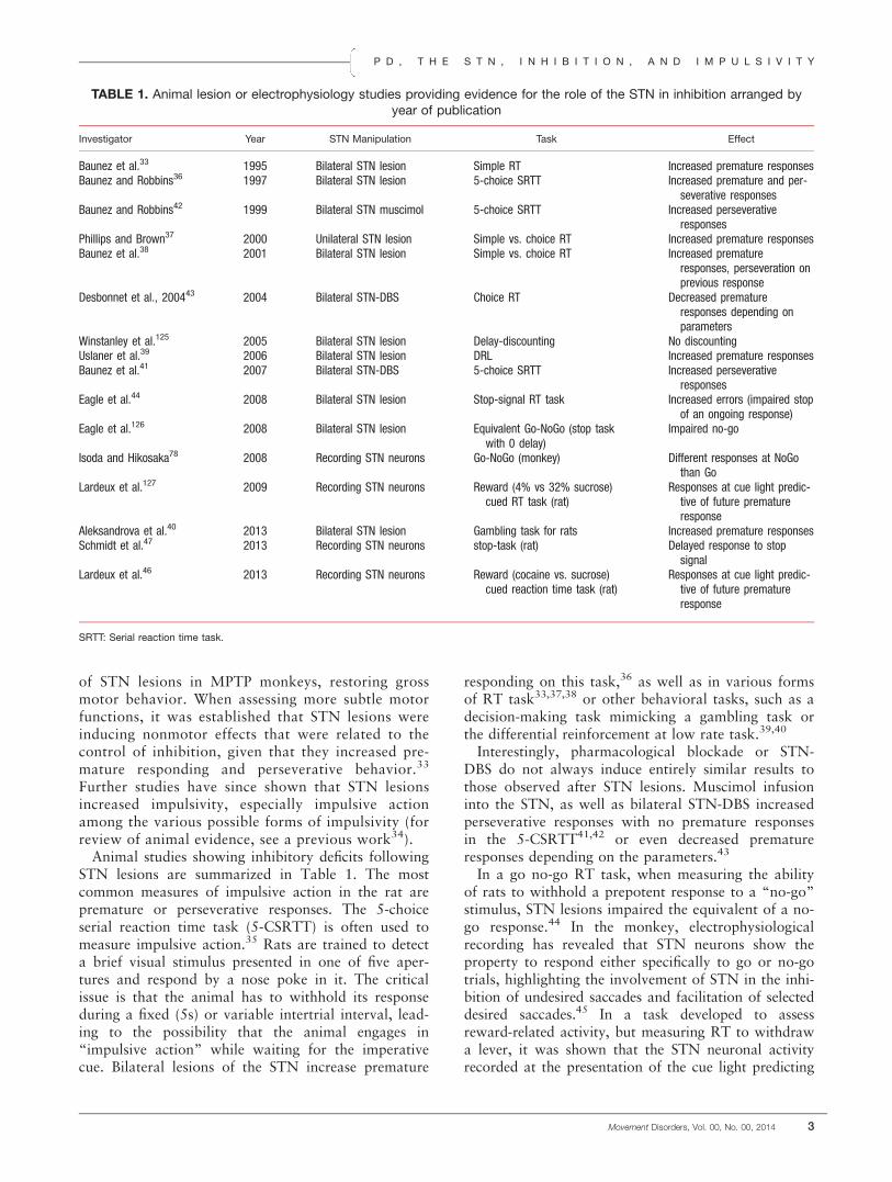
TABLE 1. Animal lesion or electrophysiology studies providing evidence for the role of the STN in inhibition arranged byyear of publication
Investigator Year STN Manipulation Task Effect
Baunez et al.33 1995 Bilateral STN lesion Simple RT Increased premature responsesBaunez and Robbins36 1997 Bilateral STN lesion 5-choice SRTT Increased premature and per-
severative responsesBaunez and Robbins42 1999 Bilateral STN muscimol 5-choice SRTT Increased perseverative
responsesPhillips and Brown37 2000 Unilateral STN lesion Simple vs. choice RT Increased premature responsesBaunez et al.38 2001 Bilateral STN lesion Simple vs. choice RT Increased premature
responses, perseveration onprevious response
Desbonnet et al., 200443 2004 Bilateral STN-DBS Choice RT Decreased prematureresponses depending onparameters
Winstanley et al.125 2005 Bilateral STN lesion Delay-discounting No discountingUslaner et al.39 2006 Bilateral STN lesion DRL Increased premature responsesBaunez et al.41 2007 Bilateral STN-DBS 5-choice SRTT Increased perseverative
responsesEagle et al.44 2008 Bilateral STN lesion Stop-signal RT task Increased errors (impaired stop
of an ongoing response)Eagle et al.126 2008 Bilateral STN lesion Equivalent Go-NoGo (stop task
with 0 delay)Impaired no-go
Isoda and Hikosaka78 2008 Recording STN neurons Go-NoGo (monkey) Different responses at NoGothan Go
Lardeux et al.127 2009 Recording STN neurons Reward (4% vs 32% sucrose)cued RT task (rat)
Responses at cue light predic-tive of future prematureresponse
Aleksandrova et al.40 2013 Bilateral STN lesion Gambling task for rats Increased premature responsesSchmidt et al.47 2013 Recording STN neurons stop-task (rat) Delayed response to stop
signalLardeux et al.46 2013 Recording STN neurons Reward (cocaine vs. sucrose)
cued reaction time task (rat)Responses at cue light predic-tive of future prematureresponse
SRTT: Serial reaction time task.
P D , T H E S T N , I N H I B I T I O N , A N D I M P U L S I V I T Y
Movement Disorders, Vol. 00, No. 00, 2014 3
the reward (sucrose or cocaine) could be predictive offuture early withdrawal of the lever in case of an acti-vation, whereas inhibition after the cue was predictiveof a correct response.46 This further supports the roleof the STN in the control of inhibition.
On a modified stop signal task, STN lesions in ratsimpaired the stopping behavior, even when the stop sig-nal was presented very early in the trials (even withzero delay, which is equivalent to a no go trial).44 Arecent electrophysiological study in rats47 showed thatSTN neurons had low latency responses to the stopcue, regardless of whether or not the animal was ableto stop the go response, suggesting that the STN pro-vides fast signals to stop action. In contrast, SNr neu-rons only responded to stop signals on successfullyinhibited trials, whereas striatal neurons were active onpresentation of go, but not stop, signals. It was pro-posed that the results support the interactive racemodel, with the relative timing of the distinct inputs tothe SNr from the striatum or STN, respectively, deter-mining stopping failure and success. Perhaps a missingcomponent in earlier works is the contribution fromthe GPi and SNr47,48 to such a stopping process, giventhat these final output pathways convey the final signalthrough the thalamus back to the cortex before anaction is withheld or exerted.
Evidence From Imaging and TranscranialMagnetic Stimulation Studies
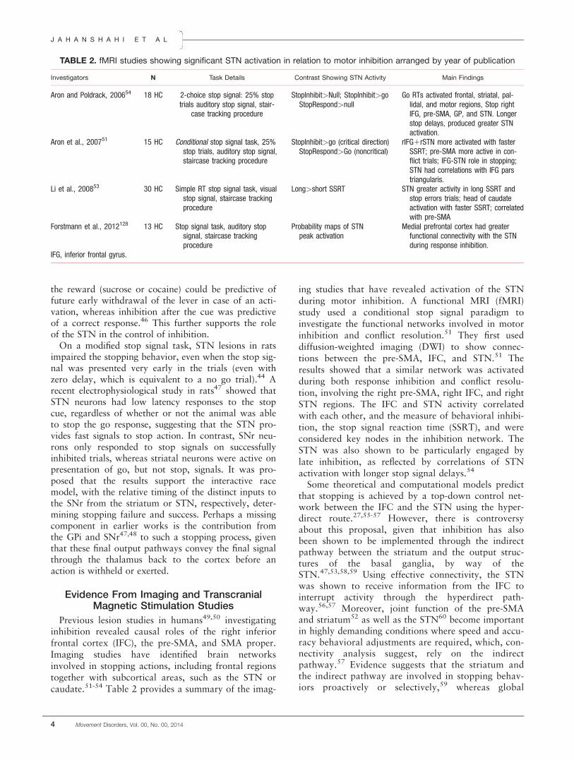
Previous lesion studies in humans49,50 investigatinginhibition revealed causal roles of the right inferiorfrontal cortex (IFC), the pre-SMA, and SMA proper.Imaging studies have identified brain networksinvolved in stopping actions, including frontal regionstogether with subcortical areas, such as the STN orcaudate.51-54 Table 2 provides a summary of the imag-
ing studies that have revealed activation of the STNduring motor inhibition. A functional MRI (fMRI)study used a conditional stop signal paradigm toinvestigate the functional networks involved in motorinhibition and conflict resolution.51 They first useddiffusion-weighted imaging (DWI) to show connec-tions between the pre-SMA, IFC, and STN.51 Theresults showed that a similar network was activatedduring both response inhibition and conflict resolu-tion, involving the right pre-SMA, right IFC, and rightSTN regions. The IFC and STN activity correlatedwith each other, and the measure of behavioral inhibi-tion, the stop signal reaction time (SSRT), and wereconsidered key nodes in the inhibition network. TheSTN was also shown to be particularly engaged bylate inhibition, as reflected by correlations of STNactivation with longer stop signal delays.54
Some theoretical and computational models predictthat stopping is achieved by a top-down control net-work between the IFC and the STN using the hyper-direct route.27,55-57 However, there is controversyabout this proposal, given that inhibition has alsobeen shown to be implemented through the indirectpathway between the striatum and the output struc-tures of the basal ganglia, by way of theSTN.47,53,58,59 Using effective connectivity, the STNwas shown to receive information from the IFC tointerrupt activity through the hyperdirect path-way.56,57 Moreover, joint function of the pre-SMAand striatum52 as well as the STN60 become importantin highly demanding conditions where speed and accu-racy behavioral adjustments are required, which, con-nectivity analysis suggest, rely on the indirectpathway.57 Evidence suggests that the striatum andthe indirect pathway are involved in stopping behav-iors proactively or selectively,59 whereas global
TABLE 2. fMRI studies showing significant STN activation in relation to motor inhibition arranged by year of publication
Investigators N Task Details Contrast Showing STN Activity Main Findings
Aron and Poldrack, 200654 18 HC 2-choice stop signal: 25% stoptrials auditory stop signal, stair-
case tracking procedure
StopInhibit>Null; StopInhibit>goStopRespond>null
Go RTs activated frontal, striatal, pal-lidal, and motor regions, Stop rightIFG, pre-SMA, GP, and STN. Longerstop delays, produced greater STNactivation.
Aron et al., 200751 15 HC Conditional stop signal task, 25%stop trials, auditory stop signal,staircase tracking procedure
StopInhibit>go (critical direction)StopRespond>Go (noncritical)
rIFG1rSTN more activated with fasterSSRT; pre-SMA more active in con-flict trials; IFG-STN role in stopping;STN had correlations with IFG parstriangularis.
Li et al., 200853 30 HC Simple RT stop signal task, visualstop signal, staircase trackingprocedure
Long>short SSRT STN greater activity in long SSRT andstop errors trials; head of caudateactivation with faster SSRT; correlatedwith pre-SMA
Forstmann et al., 2012128 13 HC Stop signal task, auditory stopsignal, staircase trackingprocedure
Probability maps of STNpeak activation
Medial prefrontal cortex had greaterfunctional connectivity with the STNduring response inhibition.
IFG, inferior frontal gyrus.
J A H A N S H A H I E T A L
4 Movement Disorders, Vol. 00, No. 00, 2014
inhibition may be achieved through the hyperdirectpathway.48
To address the question of which corticosubcorticalpathways mediate motor inhibition, the use of trans-cranial magnetic stimulation (TMS) combined withimaging allows for direct testing of causal influencesfrom cortical regions to subcortical networks andpathways during response inhibition. Increased rightstriatal and decreased motor cortex activity was foundafter TMS was applied over the right IFC and pre-SMA.61. Another combined TMS and imaging studyduring a stop signal task showed that TMS to theright pre-SMA was associated with significant activa-tion of the left pre-SMA.62 Finally, a study examinedcorrelations between TMS effect sizes (between motorcortex and the pre-SMA and IFC) and white matterconnecting pre-SMA and IFC to STN63 during aswitching task and reported significant correlationswith the STN (at a latency of 12 ms).
Several imaging studies have examined the effects ofSTN-DBS on patterns of brain activation during per-formance of cognitive tasks requiring inhibitory andexecutive control in PD patients. These have shownthat switching STN stimulation on is associated withsignificant decreased activation in key frontal areas,including the pre-SMA, IFC, dorsolateral prefrontalcortex, and anterior cingulate cortex, during tasks thatrequire inhibitory and executive control for responseselection under conflict and suppression of habitual orprepotent responses, such as the Stroop,64 fast-pacedrandom number generation (RNG),65 or the go no-gotask.66
In summary, imaging and TMS studies have identi-fied the pre-SMA and IFC as critical cortical areas andhighlighted the importance of both the indirect andhyperdirect basal-ganglia-cortical pathways when anaction needs to be cancelled.
Evidence From Electrophysiological Studies inPD Patients Undergoing STN-DBS Surgery
The implantation of electrodes for DBS in the STNhas allowed the recording of STN activity in PDpatients. Intraoperative microrecordings help toimprove STN localization before DBS implantationand offer an excellent window to explore neuronalresponses to motor or cognitive tasks. In addition, theDBS electrode can also be used to record local fieldpotential (LFP) activity from the STN during surgeryor, more usually, in the immediate postoperative phasebefore internalizing the connection cables and theimplantable pulse generator. Most of the direct neuro-physiological evidence of the STN role in inhibitioncomes from postoperative studies, given that theseallow longer recording times.
Recent studies from three groups67-69 have specifi-cally looked for changes in STN LFP activity during
stop signal tasks. The changes observed, which con-firm the role of the STN in motor inhibition, involvethe three most relevant bands described in STN activ-ity: beta, gamma, and theta.
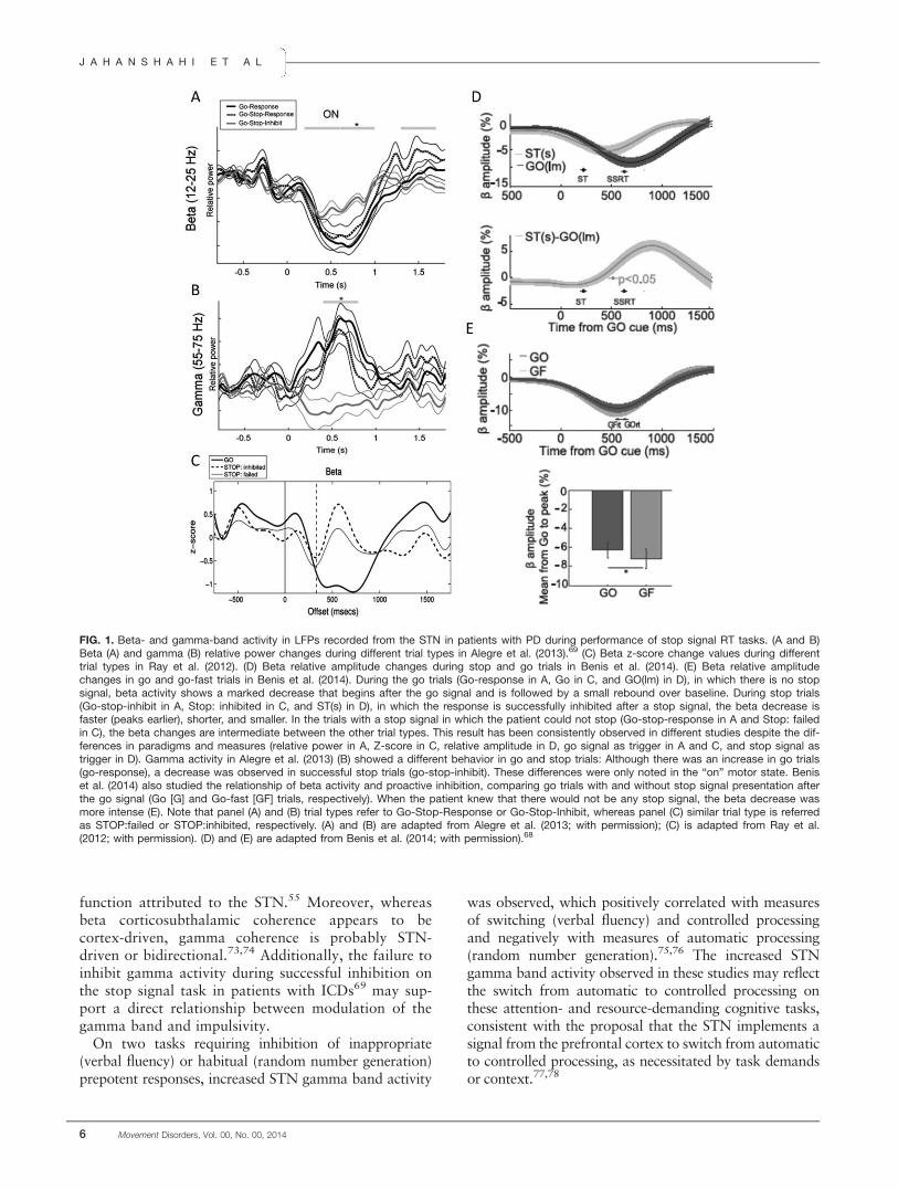
Despite some methodological differences, the threestudies show parallel results in the beta band (Fig.1A,C.D). A voluntary movement is usually accompa-nied by a decrease in beta activity in the STN. Thisdecrease begins before the movement, reaches its mini-mum value shortly after the movement has begun, andis followed by a “rebound” after the movement iscompleted.68 This pattern was observed in the threestudies in the “go” trials. However, in the “stop” tri-als, when the patient successfully inhibited theresponse, the beta decrease was consistently smaller,faster, and shorter. This difference in beta activitybetween these two types of trials (Fig. 1C, bottom)strongly suggests that beta-band subthalamic activityis involved in reactive inhibition. Similar inhibition orconflict-resolution–related beta changes have beenfound in go no-go and Stroop tasks, using similarpostoperative LFP recordings.70,71 Additionally, betaactivity might also have a role in proactive inhibition.Benis et al.68 added an additional type of trial to thestop signal task, a group of trials in which the patientknew that there would be no stop signal (named go-fasttrials). When beta changes were compared between thego (where occurrence of a stop signal on a proportion oftrials was possible) and the go-fast trials, the betadecrease was deeper in the go-fast condition (Fig. 1E).This difference may indicate that a higher level of betaactivity is also related to proactive inhibition, given thatits decrease is smaller when the patient expects thatthere may be a stop signal indicating inhibition of theresponse (go trial), than when he or she knows for cer-tain that there will be no stop signal (go fast trial).
Gamma changes in the STN were also investigatedusing the stop signal task in two of these studies.67,69
A voluntary movement is accompanied by a gammaincrease in the STN, which is proportional to themotor effort.72 Alegre et al.69 found that in the goand failed stop trials in the “on” motor state, therewas a gamma increase around the time of the motorresponse (as expected), whereas in the stop-inhibit tri-als (successful inhibition trials), there was a decreasein gamma activity (Fig. 1B). In line with these find-ings, Ray et al.67 also described a higher gammaincrease during failed stop trials than during success-fully inhibited trials, although the difference was notstatistically significant. These results again suggest theexistence of an active process in the STN related tothe presence of the stop signal and the successful inhi-bition of the response. The suppression of gammaactivity may be related to the suppression of the inten-tion to move and indeed could represent the physio-logical marker of the braking or hold your horses
P D , T H E S T N , I N H I B I T I O N , A N D I M P U L S I V I T Y
Movement Disorders, Vol. 00, No. 00, 2014 5
function attributed to the STN.55 Moreover, whereasbeta corticosubthalamic coherence appears to becortex-driven, gamma coherence is probably STN-driven or bidirectional.73,74 Additionally, the failure toinhibit gamma activity during successful inhibition onthe stop signal task in patients with ICDs69 may sup-port a direct relationship between modulation of thegamma band and impulsivity.
On two tasks requiring inhibition of inappropriate(verbal fluency) or habitual (random number generation)prepotent responses, increased STN gamma band activity
was observed, which positively correlated with measuresof switching (verbal fluency) and controlled processingand negatively with measures of automatic processing(random number generation).75,76 The increased STNgamma band activity observed in these studies may reflectthe switch from automatic to controlled processing onthese attention- and resource-demanding cognitive tasks,consistent with the proposal that the STN implements asignal from the prefrontal cortex to switch from automaticto controlled processing, as necessitated by task demandsor context.77,78
FIG. 1. Beta- and gamma-band activity in LFPs recorded from the STN in patients with PD during performance of stop signal RT tasks. (A and B)Beta (A) and gamma (B) relative power changes during different trial types in Alegre et al. (2013).69 (C) Beta z-score change values during differenttrial types in Ray et al. (2012). (D) Beta relative amplitude changes during stop and go trials in Benis et al. (2014). (E) Beta relative amplitudechanges in go and go-fast trials in Benis et al. (2014). During the go trials (Go-response in A, Go in C, and GO(lm) in D), in which there is no stopsignal, beta activity shows a marked decrease that begins after the go signal and is followed by a small rebound over baseline. During stop trials(Go-stop-inhibit in A, Stop: inhibited in C, and ST(s) in D), in which the response is successfully inhibited after a stop signal, the beta decrease isfaster (peaks earlier), shorter, and smaller. In the trials with a stop signal in which the patient could not stop (Go-stop-response in A and Stop: failedin C), the beta changes are intermediate between the other trial types. This result has been consistently observed in different studies despite the dif-ferences in paradigms and measures (relative power in A, Z-score in C, relative amplitude in D, go signal as trigger in A and C, and stop signal astrigger in D). Gamma activity in Alegre et al. (2013) (B) showed a different behavior in go and stop trials: Although there was an increase in go trials(go-response), a decrease was observed in successful stop trials (go-stop-inhibit). These differences were only noted in the “on” motor state. Beniset al. (2014) also studied the relationship of beta activity and proactive inhibition, comparing go trials with and without stop signal presentation afterthe go signal (Go [G] and Go-fast [GF] trials, respectively). When the patient knew that there would not be any stop signal, the beta decrease wasmore intense (E). Note that panel (A) and (B) trial types refer to Go-Stop-Response or Go-Stop-Inhibit, whereas panel (C) similar trial type is referredas STOP:failed or STOP:inhibited, respectively. (A) and (B) are adapted from Alegre et al. (2013; with permission); (C) is adapted from Ray et al.(2012; with permission). (D) and (E) are adapted from Benis et al. (2014; with permission).68
J A H A N S H A H I E T A L
6 Movement Disorders, Vol. 00, No. 00, 2014
Theta-band activity has been related to decision con-flict, both in the frontal cortex and in the STN. Thepotential role of the STN in this regard is also sup-ported by the findings of a conflict-related increase infiring rate in an intraoperative study using a probabilis-tic decision task.79 More direct evidence relating STNLFP low-frequency activity to impulsivity and presenceof ICD in PD has been provided in two studies. In PDpatients with preoperative ICDs, theta activity in theSTN was maximal in ventral electrodes, in agreementwith impulsivity-related oscillatory changes80 andbehavioral effects of DBS.81,82 The results of Rosaet al.83 directly related low-frequency STN activity toadoption of risky strategies in patients with PD andpathological gambling. Thus, the current neurophysio-logical evidence strongly suggests that the STN isinvolved in inhibition, but does not conclusively dem-onstrate that it leads the cortex in inhibition.
Evidence From Experimental Studies ofSTN-DBS or Subthalamotomy in PD
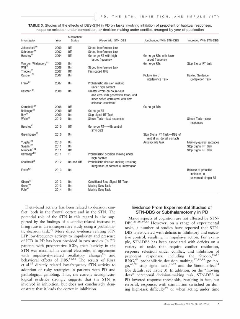
Major aspects of cognition are not affected by STN-DBS.25,26,84,85 However, on a range of experimentaltasks, a number of studies have reported that STN-DBS is associated with deficits in inhibitory and execu-tive control, resulting in impulsive action. For exam-ple, STN-DBS has been associated with deficits on avariety of tasks that require conflict resolution,response selection under conflict, and inhibition ofprepotent responses, including the Stroop,86,87
RNG,65 probabilistic decision making,27,88,89 go no-go,66,90 stop signal task,91-93 and the Simon effect94
(for details, see Table 3). In addition, on the “movingdots” perceptual decision-making task, STN-DBS inPD lowered response thresholds, resulting in fast, buterrorful, responses with stimulation switched on dur-ing high-task difficulty95 or when acting under time
TABLE 3. Studies of the effects of DBS-STN in PD on tasks involving inhibition of prepotent or habitual responses,response selection under competition, or decision making under conflict, arranged by year of publication
Investigator Year
Medication
Status Worse With STN-DBS Unchanged With STN-DBS Improved With STN-DBS
Jahanshahi86 2000 Off Stroop interference taskSchroeder64 2002 Off Stroop interference taskHershey90 2004 Off Go no-go RT with high
target frequencyGo no-go RTs with lowertarget frequency
Van den Wildenberg92 2006 On Go no-go RTs Stop Signal RT taskWitt87 2006 On Stroop interference taskThobois65 2007 Off Fast-paced RNGCastner129 2007 On Picture Word
Interference TaskHayling SentenceCompletion Task
Frank27 2007 On Probabilistic decision makingunder high conflict
Castner130 2008 On Greater errors on noun-nounand verb-verb generation tasks, andlatter deficit correlated with itemselection constraint
Campbell131 2008 Off Go no-go RTsBallanger66 2009 Off Go no-go RTRay91 2009 On Stop signal RT TaskWylie95 2010 On Simon Task—fast responses Simon Task—slow
responsesHershey97 2010 Off Go no-go RT—with ventral
STN-DBSGreenhouse98 2010 On Stop Signal RT Task—DBS of
ventral vs. dorsal contactsYugeta132 2010 On Antisaccade task Memory-guided saccadesSwann133 2011 On Stop Signal RT taskMirabella134 2011 Off Stop Signal RT taskCavanagh89 2011 ? Probabilisitic decision making under
high conflictCoulthard88 2012 On and Off Probabilistic decision making requiring
integration of conflictual informationFavre124 2013 On Release of proactive
inhibition inunwarned simple RT
Obeso93 2013 On Conditional Stop Signal RT TaskGreen95 2013 On Moving Dots TaskPote96 2014 On Moving Dots Task
P D , T H E S T N , I N H I B I T I O N , A N D I M P U L S I V I T Y
Movement Disorders, Vol. 00, No. 00, 2014 7

pressure.96 However, as evident from Table 3, not allstudies involving inhibitory processing have showndeficits with STN stimulation. Variations in the natureand prepotency of the response (e.g., percent of go tri-als in a go no-go or stop signal task) and the preciseactive contact position in the STN97,98 are likely toaccount for some of the differences in results acrossstudies. Another important consideration is the natureof impulsivity being studied. Other than deficits inmotor inhibition summarized in Table 3 and increasedloss-chasing in a gambling task,135 investigators havenot found any detrimental effects of STN-DBS onother features of impulsivity and risk taking.136,137
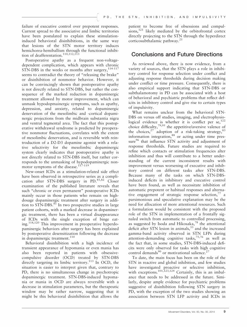
More direct and “causal” evidence of the role of theSTN in inhibitory and executive control comes from ahandful of studies of subthalamotomy in PD. Postsub-thalamotomy deficits on the Stroop in 30% of the PDpatients and deterioration in release from proactiveinhibition on a memory test, with normal performancein 98% of the sample before and 50% of the sampleafter surgery, have been reported.99,100 In a recentstudy, Obeso et al.101 used the conditional stop signaltask to investigate the role of the STN in reactive andproactive inhibition and conflict resolution and inadjusting response thresholds and speed-accuracytrade-offs. Patients with right subthalamotomy hadsignificantly faster Go RTs, but made significantlymore discrimination errors with their contralesionalhand than the unoperated PD patients (see Fig. 2A,B),suggesting that right subthalamotomy influencedspeed-accuracy trade-offs. The patients with right sub-thalamotomy could not engage in late-phase, fast inhi-bition of the response and showed minimal proactiveinhibition when tested with the contralesional hand.
These results provide strong evidence that the STN isinvolved in response inhibition.
Evidence From Psychiatric Side Effects ofSTN-DBS or Subthalamotomy in PD
STN lesions lead to the “syndrome of the body ofLuys,” a mixture of hemichorea-hemiballism, typicallyaccompanied by nonmotor behavioral complica-tions.102 The marked involuntary movements led tothe clinical concept of the STN as a motor controlnucleus.103 Hemiballism tends to improve over time,but it can more rarely persist as a permanent sequelae,which can be improved by pallidotomy or pallidalDBS.104,105
Behavioral side effects, such as hypomania, hyper-sexuality, logorrhea and disinhibition of mood andeuphoria, impulsivity, and aggression, have beendescribed to accompany hemiballism in spontaneousSTN lesions in humans.106-108 Following bilateral sub-thalamotomy in PD, “hyperactive” behaviors, such asdisinhibiton, euphoria, and irritability, transientlyincreased and their course of evolution was similar tothe postoperative increase in dyskinesias.109
In PD patients treated with STN-DBS, disinhibitionof complex behavioral programs, such as mirthfullaughter, mania, depression, intermittent explosive dis-order, kleptomania, emotivity, creativity, and new-onset ICDs (hypersexuality, pathological buying, path-ological gambling, and bulimia), are compatible withthe role of the STN in control and release not only ofmotor plans, but also of nonmotor behaviors (forreview, see previous works110,111). Some of theseSTN-induced behaviors represent disinhibition or
FIG. 2. (A) The mean “critical” Go RTs for the contralesional hand of the patients with PD right or left subthalamotomy (R STN, L STN) andthe dominant hand of the unoperated patients with PD and healthy control participants. Error bars are standard errors of the mean. *P < 0.05.(B) Mean discrimination errors with the contralesional hand for the patients with PD with right or left subthalamotomy (R STN, L STN) andthe dominant hand for the unoperated patients with PD and healthy control participants. Error bars are standard errors of the mean.*P < 0.05. Reproduced from Obeso et al. (2014; with permission).101 [Color figure can be viewed in the online issue, which is available atwileyonlinelibrary.com.]
J A H A N S H A H I E T A L
8 Movement Disorders, Vol. 00, No. 00, 2014
failure of executive control over prepotent responses.Current spread to the associative and limbic territorieshave been postulated to explain these stimulation-induced behavioral disinhibitions, in the same waythat lesions of the STN motor territory induceshemichorea-hemiballism through the functional inhibi-tion of deafferentation.110,112,113
Postoperative apathy as a frequent non-voltage-dependent complication, which appears with chronicSTN-DBS in the weeks or months after surgery,114-117
seems to contradict the theory of “releasing the brake”or disinhibition of nonmotor behavior. However, itcan be convincingly shown that postoperative apathyis not directly related to STN-DBS, but rather the con-sequence of the marked reduction in dopaminergictreatment allowed by motor improvement, which canunmask hypodopmainergic symptoms, such as apathy,depression, and anxiety, related to dopaminergicdenervation of the mesolimbic and -cortical dopami-nergic projections from the midbrain substantia nigraand ventral tegmental area. The fact that this postop-erative withdrawal syndrome is predicted by preopera-tive nonmotor fluctuations, correlates with the extentof mesolimbic denervation, and is reversible with rein-troduction of a D2-D3 dopamine agonist with a rela-tive selectivity for the mesolimbic dopaminergicsystem clearly indicates that postoperative apathy isnot directly related to STN-DBS itself, but rather cor-responds to the unmasking of hypodopaminergic non-motor symptoms of the disease.117-119
New-onset ICDs as a stimulation-related side effecthave been observed in retrospective series as a compli-cation after STN-DBS surgery in PD.81,82 Closerexamination of the published literature reveals thatsuch “chronic or even permanent” postoperative ICDsmainly occur in those patients who remain on high-dosage dopaminergic treatment after surgery in addi-tion to STN-DBS.81 In two prospective studies in largepatient cohorts, with a marked decrease in dopaminer-gic treatment, there has been a virtual disappearanceof ICDs with the single exception of binge eat-ing.118,120 This improvement in preoperative hyperdo-paminergic behaviors after surgery has been explainedby postoperative desensitization following the decreasein dopaminergic treatment.110
Behavioral disinhibition with a high incidence oftransient appearance of hypomania or even mania hasalso been reported in patients with obsessive-compulsive disorder (OCD) treated by STN-DBSdirectly targeting its limbic territory.113 In OCD, thesituation is easier to interpret given that, contrary toPD, there is no simultaneous change in psychotropicdopaminergic treatment. STN-DBS-induced hypoma-nia or mania in OCD are always reversible with adecrease in stimulation parameters, but the therapeuticwindow may be rather narrow, suggesting that itmight be this behavioral disinhibition that allows the
patient to become free of obsessions and compul-sions,121 likely mediated by the orbitofrontal cortexdirectly projecting to the STN through the hyperdirectcorticosubthalamic pathway.21
Conclusions and Future Directions
As reviewed above, there is now evidence, from avariety of sources, that the STN plays a role in inhibi-tory control for response selection under conflict andadjusting response thresholds during decision makingunder conflict or time pressure. Consequently, there isalso empirical support indicating that STN-DBS orsubthalamotomy in PD can be associated with a hostof behavioral and psychiatric problems that reflect def-icits in inhibitory control and give rise to certain typesof impulsivity.
What remains unclear from the behavioral STN-DBS on versus off studies, imaging, and electrophysio-logical evidence is whether it is conflict per se,122
choice difficulty,79,95 the appetitive/aversive valence ofthe choices,27 adoption of a risk-taking strategy,83
information integration,88 or acting under time pres-sure96 that influence STN activity and adjustment ofresponse thresholds. Future studies are required torefine which contacts or stimulation frequencies alterinhibition and thus will contribute to a better under-standing of the current inconsistent results withimprovement versus worsening of executive and inhib-itory control on different tasks after STN-DBS.Because many of the tasks on which STN-DBS-induced deficits in inhibitory and executive controlhave been found, as well as necessitate inhibition ofautomatic prepotent or habitual responses and alterna-tive engagement of strategic response selection,77 aparsimonious and speculative explanation may be theneed for allocation of more attentional resources. Sucha formulation would be consistent with the proposedrole of the STN in implementation of a frontally sig-naled switch from automatic to controlled processing,as suggested by Isoda and Hikosaka,78 the attentionaldeficit after STN lesion in animals,33 and the increasedgamma-band activity observed in STN LFPs duringattention-demanding cognitive tasks,75,76 as well asthe fact that, in some studies, STN-DBS-induced defi-cits were only observed for tasks with high cognitivecontrol demands90 or motivational salience.27
To date, the main focus has been on the role of theSTN in reactive and global inhibition, and few studieshave investigated proactive or selective inhibition,with exceptions.101,123,124 Certainly, this is an imbal-ance that needs to be addressed in the future. Simi-larly, despite ample evidence for psychiatric problemssuggestive of disinhibition following STN surgery inPD, with the exception of the two studies showing anassociation between STN LFP activity and ICDs in
P D , T H E S T N , I N H I B I T I O N , A N D I M P U L S I V I T Y
Movement Disorders, Vol. 00, No. 00, 2014 9
PD,80,83 no efforts have been made to directly link thepsychiatric sequelae of STN surgery to deficits ininhibitory control. Provision of such direct evidence isimportant from both clinical and theoretical perspec-tives. The results linking low-frequency STN activityfunctions to ICDs impulsivity in PD79,82 open up anew avenue of research, which allows for merging theroles of the STN in selection and motivational sali-ence, which could potentially enhance our understand-ing of ICDs in PD as well as the roles of the STN inhumans.
References1. Albin RL, Young AB, Penney JB. The functional anatomy of basal
ganglia disorders. Trends Neurosci 1989;12:366-375.
2. DeLong MR. Primate models of movement disorders of basalganglia origin. Trends Neurosci 1990;13:281-285.
3. Rahman S, Griffin HJ, Quinn NP, Jahanshahi M. The factors thatinduce or overcome freezing of gait in Parkinson’s disease. BehavNeurol 2008;19:127-136.
4. Obeso JA, Rodriguez-Oroz MC, Rodriguez M, DeLong MR,Olanow CW. Pathophysiology of levodopa-induced dyskinesias inParkinson’s disease: problems with the current model. Ann Neu-rol 2000;47(4 Suppl 1):S22-S32.
5. Dirnberger G, Jahanshahi M. Executive dysfunction in Parkin-son’s disease: a review. J Neuropsychol 2013;7:193-224.
6. Cooper JA, Sagar HJ, Tidswell P, Jordan N. Slowed central proc-essing in simple and go/no-go reaction time tasks in Parkinson’sdisease. Brain 1994;117:517-529.
7. Gauggel S, Rieger M, Feghoff TA. Inhibition of ongoing responsesin patients with Parkinson’s disease. J Neurol Neurosurg Psychia-try 2004;75:539-544.
8. Obeso I, Wilkinson L, Casabona E, et al. Deficits in inhibitorycontrol and conflict resolution on cognitive and motor tasks inParkinson’s disease. Exp Brain Res 2011;212:371-384.
9. Obeso I, Wilkinson L, Jahanshahi M. Levodopa medication doesnot influence motor inhibition or conflict resolution in a condi-tional stop-signal task in Parkinson’s disease. Exp Brain Res2011;213:435-445.
10. O’Callaghan C, Naismith SL, Hodges JR, Lewis SJ, HornbergerM. Fronto-striatal atrophy correlates of inhibitory dysfunction inParkinson’s disease versus behavioural variant frontotemporaldementia. Cortex 2013;49:1833-1843.
11. Dirnberger G, Frith CD, Jahanshahi M. Executive dysfunction inParkinson’s disease is associated with altered pallidal-frontalprocessing. Neuroimage 2005;25:588-599.
12. Praamstra P, Plat FM. Failed suppression of direct visuomotor acti-vation in Parkinson’s disease. J Cogn Neurosci 2001;13:31-43.
13. Wylie SA, Stout JC, Bashore TR. Activation of conflicting responsesin Parkinson’s disease: evidence for degrading and facilitating effectson response time. Neuropsychologia 2005;43:1033-1043.
14. Kuhn AA, Tsui A, Aziz T, et al. Pathological synchronisation inthe subthalamic nucleus of patients with Parkinson’s diseaserelates to both bradykinesia and rigidity. Exp Neurol 2009;215:380-387.
15. Vila M, Levy R, Herrero MT, et al. Consequences of nigrostriataldenervation on the functioning of the basal ganglia in human andnonhuman primates: an in situ hybridization study of cytochromeoxidase subunit I mRNA. J Neurosci 1997;17:765-773.
16. Jahanshahi M. Risky choices link the subthalamic nucleus withpathological gambling in Parkinson’s disease. Mov Disord 2013;28:1617-1619.
17. Jahanshahi M. Effects of deep brain stimulation of the subthala-mic nucleus on inhibitory and executive control over prepotentresponses in Parkinson’s disease. Front Syst Neurosci 2013;7:118.
18. Parent A, Hazrati LN. Functional anatomy of the basal ganglia. I.The cortico-basal ganglia-thalamo-cortical loop. Brain Res BrainRes Rev 1995;20:91-127.
19. Nambu A, Tokuno H, Inase M, Takada M. Corticosubthalamicinput zones from forelimb representations of the dorsal and ven-tral divisions of the premotor cortex in the macaque monkey:comparison with the input zones from the primary motor cortexand the supplementary motor area. Neurosci Lett 1997;239:13-16.
20. Afsharpour S. Topographical projections of the cerebral cortex tothe subthalamic nucleus. J Comp Neurol 1985;236:14-28.
21. Haynes WI, Haber SN. The organization of prefrontal-subthalamic inputs in primates provides an anatomical substratefor both functional specificity and integration: implications forbasal ganglia models and deep brain stimulation. J Neurosci2013;33:4804-4814.
22. Lambert C, Zrinzo L, Nagy Z, et al. Confirmation of functionalzones within the human subthalamic nucleus: patterns of connec-tivity and sub-parcellation using diffusion weighted imaging. Neu-roimage 2012;60:83-94.
23. Keuken MC, Muller-Axt C, Langner R, Eickhoff SB, ForstmannBU, Neumann J. Brain networks of perceptual decision-making:an fMRI ALE meta-analysis. Front Hum Neurosci 2014;8:445.
24. Deuschl G, Schade-Brittinger C, Krack P, Volkmann J, Sch€afer H,et al. A randomized trial of deep-brain stimulation for Parkin-son’s disease. N Engl J Med 2006;355:896-908.
25. Follett KA, Weaver FM, Stern M, et al. Pallidal versus subthala-mic deep-brain stimulation for Parkinson’s disease. N Engl J Med2010;362:2077-2091.
26. Weaver FM, Follett K, Stern M, et al. Bilateral deep brain stimu-lation vs best medical therapy for patients with advanced Parkin-son disease: a randomized controlled trial. JAMA 2009;301:63-73.
27. Frank MJ, Samanta J, Moustafa AA, Sherman SJ. Hold yourhorses: impulsivity, deep brain stimulation, and medication inparkinsonism. Science 2007;318:1309-1312.
28. Evenden JL. Varieties of impulsivity. Psychopharmacology (Berl)1999;146:348-361.
29. Dalley JW, Everitt BJ, Robbins TW. Impulsivity, compulsivity,and top-down cognitive control. Neuron 2011;69:680-694.
30. Aron AR. From reactive to proactive and selective control: devel-oping a richer model for stopping inappropriate responses. BiolPsychiatry 2011;69:e55-e68.
31. Whittier JR, Mettler FA. Studies on the subthalamus of the rhesusmonkey; hyperkinesia and other physiologic effects of subthala-mic lesions; with special reference to the subthalamic nucleus ofLuys. J Comp Neurol 1949;90:319-372.
32. Bergman H, Wichmann T, DeLong MR. Reversal of experimentalparkinsonism by lesions of the subthalamic nucleus. Science1990;249:1436-1438.
33. Baunez C, Nieoullon A, Amalric M. In a rat model of parkinson-ism, lesions of the subthalamic nucleus reverse increases of reac-tion time but induce a dramatic premature responding deficit.J Neurosci 1995;15:6531-6541.
34. Eagle DM, Baunez C. Is there an inhibitory-response-control sys-tem in the rat? Evidence from anatomical and pharmacologicalstudies of behavioral inhibition. Neurosci Biobehav Rev 2010;34:50-72.
35. Robbins TW. The 5-choice serial reaction time task: behaviouralpharmacology and functional neurochemistry. Psychopharmacol-ogy (Berl) 2002;163:362-380.
36. Baunez C, Robbins TW. Bilateral lesions of the subthalamicnucleus induce multiple deficits in an attentional task in rats. EurJ Neurosci 1997;9:2086-2099.
37. Phillips JM, Brown VJ. Anticipatory errors after unilateral lesionsof the subthalamic nucleus in the rat: evidence for a failure ofresponse inhibition. Behav Neurosci 2000;114:150-157.
38. Baunez C, Humby T, Eagle DM, Ryan LJ, Dunnett SB, RobbinsTW. Effects of STN lesions on simple vs choice reaction timetasks in the rat: preserved motor readiness, but impaired responseselection. Eur J Neurosci 2001;13:1609-1616.
39. Uslaner JM, Robinson TE. Subthalamic nucleus lesions increaseimpulsive action and decrease impulsive choice—mediation byenhanced incentive motivation? Eur J Neurosci 2006;24:2345-2354.
40. Aleksandrova LR, Creed MC, Fletcher PJ, Lobo DS, Hamani C,Nobrega JN. Deep brain stimulation of the subthalamic nucleus
J A H A N S H A H I E T A L
10 Movement Disorders, Vol. 00, No. 00, 2014
increases premature responding in a rat gambling task. BehavBrain Res 2013;245:76-82.
41. Baunez C, Christakou A, Chudasama Y, Forni C, Robbins TW.Bilateral high-frequency stimulation of the subthalamic nucleuson attentional performance: transient deleterious effects andenhanced motivation in both intact and parkinsonian rats. Eur JNeurosci 2007;25:1187-1194.
42. Baunez C, Robbins TW. Effects of transient inactivation of thesubthalamic nucleus by local muscimol and APV infusions on per-formance on the five-choice serial reaction time task in rats. Psy-chopharmacology (Berl) 1999;141:57-65.
43. Desbonnet L, Temel Y, Visser-Vandewalle V, Blokland A,Hornikx V, Steinbusch HW. Premature responding followingbilateral stimulation of the rat subthalamic nucleus is amplitudeand frequency dependent. Brain Res 2004;1008:198-204.
44. Eagle DM, Baunez C, Hutcheson DM, Lehmann O, Shah AP,Robbins TW. Stop-signal reaction-time task performance: role ofprefrontal cortex and subthalamic nucleus. Cereb Cortex 2008;18:178-188.
45. Isoda M, Hikosaka O. Cortico-basal ganglia mechanisms forovercoming innate, habitual and motivational behaviors. Eur JNeurosci 2011;33:2058-2069.
46. Lardeux S, Paleressompoulle D, Pernaud R, Cador M, Baunez C.Different populations of subthalamic neurons encode cocaine vs.sucrose reward and predict future error. J Neurophysiol 2013;110:1497-1510.
47. Schmidt R, Leventhal DK, Mallet N, Chen F, Berke JD. Cancel-ing actions involves a race between basal ganglia pathways. NatNeurosci 2013;16:1118-1124.
48. Sano H, Chiken S, Hikida T, Kobayashi K, Nambu A. Signalsthrough the striatopallidal indirect pathway stop movements byphasic excitation in the substantia nigra. J Neurosci 2013;33:7583-7594.
49. Aron AR, Fletcher PC, Bullmore ET, Sahakian BJ, Robbins TW.Stop-signal inhibition disrupted by damage to right inferior fron-tal gyrus in humans. Nat Neurosci 2003;6:115-116.
50. Alexander MP, Stuss DT, Picton T, Shallice T, Gillingham S.Regional frontal injuries cause distinct impairments in cognitivecontrol. Neurology 2007;68:1515-1523.
51. Aron AR, Behrens TE, Smith S, Frank MJ, Poldrack RA. Triangu-lating a cognitive control network using diffusion-weighted mag-netic resonance imaging (MRI) and functional MRI. The Journalof Neuroscience 2007;27:3743-3752.
52. Forstmann BU, Dutilh G, Brown S, et al. Striatum and pre-SMAfacilitate decision-making under time pressure. Proc Natl AcadSci U S A 2008;105:17538-17542.
53. Li CS, Yan P, Sinha R, Lee TW. Subcortical processes of motorresponse inhibition during a stop signal task. Neuroimage 2008;41:1352-1363.
54. Aron AR, Poldrack RA. Cortical and subcortical contributions toStop signal response inhibition: role of the subthalamic nucleus.J Neurosci 2006;26:2424-2433.
55. Frank MJ. Hold your horses: a dynamic computational role forthe subthalamic nucleus in decision making. Neural Netw 2006;19:1120-1136.
56. Duann JR, Ide JS, Luo X, Li CS. Functional connectivity delin-eates distinct roles of the inferior frontal cortex and presupple-mentary motor area in stop signal inhibition. J Neurosci 2009;29:10171-10179.
57. Jahfari S, Waldorp L, van den Wildenberg WP, Scholte HS,Ridderinkhof KR, Forstmann BU. Effective connectivityreveals important roles for both the hyperdirect (fronto-subthala-mic) and the indirect (fronto-striatal-pallidal) fronto-basal gangliapathways during response inhibition. J Neurosci 2011;31:6891-6899.
58. Sano H, Chiken S, Hikida T, Kobayashi K, Nambu A. Signalsthrough the striatopallidal indirect pathway stop movements byphasic excitation in the substantia nigra. J Neurosci 2013;33:7583-7594.
59. Majid DS, Cai W, Corey-Bloom J, Aron AR. Proactive selectiveresponse suppression is implemented via the basal ganglia.J Neurosci 2013;33:13259-13269.
60. Mansfield EL, Karayanidis F, Jamadar S, Heathcote A,Forstmann BU. Adjustments of response threshold during task
switching: a model-based functional magnetic resonance imagingstudy. J Neurosci 2011;31:14688-14692.
61. Zandbelt BB, Bloemendaal M, Hoogendam JM, Kahn RS, VinkM. Transcranial magnetic stimulation and functional MRI revealcortical and subcortical interactions during stop-signal responseinhibition. J Cogn Neurosci 2013;25:157-174.
62. Obeso I, Cho SS, Antonelli F, et al. Stimulation of the pre-SMAinfluences cerebral blood flow in frontal areas involved withinhibitory control of action. Brain Stimul 2013;6:769-776.
63. Neubert FX, Mars RB, Buch ER, Olivier E, Rushworth MF. Cort-ical and subcortical interactions during action reprogrammingand their related white matter pathways. Proc Natl Acad Sci U SA 2010;107:13240-13245.
64. Schroeder U, Kuehler A, Haslinger B, et al. Subthalamic nucleusstimulation affects striato-anterior cingulate cortex circuit in aresponse conflict task: a PET study. Brain 2002;125:1995-2004.
65. Thobois S, Hotton GR, Pinto S, et al. STN stimulation alterspallidal-frontal coupling during response selection under competi-tion. J Cereb Blood Flow Metab 2007;27:1173-1184.
66. Ballanger B, van Eimeren T, Moro E, et al. Stimulation of thesubthalamic nucleus and impulsivity: release your horses. AnnNeurol 2009;66:817-824.
67. Ray NJ, Brittain JS, Holland P, et al. The role of the subthalamicnucleus in response inhibition: evidence from local field potentialrecordings in the human subthalamic nucleus. Neuroimage 2012;60:271-278.
68. Benis D, David O, Lachaux JP, et al. Subthalamic nucleus activitydissociates proactive and reactive inhibition in patients with Par-kinson’s disease. Neuroimage 2014;91:273-281.
69. Alegre M, Lopez-Azcarate J, Obeso I, et al. The subthalamicnucleus is involved in successful inhibition in the stop-signal task:a local field potential study in Parkinson’s disease. Exp Neurol2013;239:1-12.
70. Kuhn AA, Williams D, Kupsch A, Limousin P, et al. Event-relatedbeta desynchronization in human subthalamic nucleus correlateswith motor performance. Brain 2004;127:735-746.
71. Brittain JS, Watkins KE, Joundi RA, et al. A role for the subtha-lamic nucleus in response inhibition during conflict. J Neurosci2012;32:13396-13401.
72. Tan H, Pogosyan A, Anzak A, et al. Complementary roles of dif-ferent oscillatory activities in the subthalamic nucleus in codingmotor effort in Parkinsonism. Exp Neurol 2013;248:187-195.
73. Lalo E, Thobois S, Sharott A, et al. Patterns of bidirectionalcommunication between cortex and basal ganglia during move-ment in patients with Parkinson disease. J Neurosci 2008;28:3008-3016.
74. Williams D, Tijssen M, Van Bruggen G, et al. Dopamine-depend-ent changes in the functional connectivity between basal gangliaand cerebral cortex in humans. Brain 2002;125:1558-1569.
75. Anzak A, Gaynor L, Beigi M, et al. A gamma band specific roleof the subthalamic nucleus in switching during verbal fluencytasks in Parkinson’s disease. Exp Neurol 2011;232:136-142.
76. Anzak A, Gaynor L, Beigi M, et al. Subthalamic nucleus gammaoscillations mediate a switch from automatic to controlled proc-essing: a study of random number generation in Parkinson’s dis-ease. Neuroimage 2013;64:284-289.
77. Redgrave P, Rodriguez M, Smith Y, et al. Goal-directed andhabitual control in the basal ganglia: implications for Parkinson’sdisease. Nat Rev Neurosci 2010;11:760-772.
78. Isoda M, Hikosaka O. Role for subthalamic nucleus neurons inswitching from automatic to controlled eye movement. J Neurosci2008;28:7209-7218.
79. Zaghloul KA, Weidemann CT, Lega BC, Jaggi JL, Baltuch GH,Kahana MJ. Neuronal activity in the human subthalamic nucleusencodes decision conflict during action selection. J Neurosci2012;32:2453-2460.
80. Rodriguez-Oroz MC, Lopez-Azcarate J, Garcia-Garcia D, et al.Involvement of the subthalamic nucleus in impulse controldisorders associated with Parkinson’s disease. Brain 2011;134:36-49.
81. Lim SY, O’Sullivan SS, Kotschet K, et al. Dopamine dysregulationsyndrome, impulse control disorders and punding after deep brainstimulation surgery for Parkinson’s disease. J Clin Neurosci 2009;16:1148-1152.
P D , T H E S T N , I N H I B I T I O N , A N D I M P U L S I V I T Y
Movement Disorders, Vol. 00, No. 00, 2014 11
82. Halbig TD, Tse W, Frisina PG, et al. Subthalamic deep brainstimulation and impulse control in Parkinson’s disease. Eur JNeurol 2009;16:493-497.
83. Rosa M, Fumagalli M, Giannicola G, et al. Pathological gamblingin Parkinson’s disease: subthalamic oscillations during economicsdecisions. Mov Disord 2013;28:1644-1652.
84. Parsons TD, Rogers SA, Braaten AJ, Woods SP, Troster AI. Cog-nitive sequelae of subthalamic nucleus deep brain stimulation inParkinson’s disease: a meta-analysis. Lancet Neurol 2006;5:578-588.
85. Witt K, Daniels C, Reiff J, et al. Neuropsychological andpsychiatric changes after deep brain stimulation for Parkinson’sdisease: a randomised, multicentre study. Lancet Neurol 2008;7:605-614.
86. Jahanshahi M, Ardouin CM, Brown RG, et al. The impact ofdeep brain stimulation on executive function in Parkinson’s dis-ease. Brain 2000;123:1142-1154.
87. Witt K, Pulkowski U, Herzog J, et al. Deep brain stimulation ofthe subthalamic nucleus improves cognitive flexibility but impairsresponse inhibition in Parkinson’s disease. Arch Neurol 2004;61:697-700.
88. Coulthard EJ, Bogacz R, Javed S, et al. Distinct roles of dopamineand subthalamic nucleus in learning and probabilistic decisionmaking. Brain 2012;135:3721-3734.
89. Cavanagh JF, Wiecki TV, Cohen MX, et al. Subthalamic nucleusstimulation reverses mediofrontal influence over decision thresh-old. Nat Neurosci 2011;14:1462-1467.
90. Hershey T, Revilla FJ, Wernle A, Gibson PS, Dowling JL,Perlmutter JS. Stimulation of STN impairs aspects of cognitivecontrol in PD. Neurology 2004;62:1110-1114.
91. Ray NJ, Jenkinson N, Brittain J, et al. The role of the subthala-mic nucleus in response inhibition: evidence from deep brain stim-ulation for Parkinson’s disease. Neuropsychologia 2009;47:2828-2834.
92. van den Wildenberg WP, van Boxtel GJ, van der Molen MW,Bosch DA, Speelman JD, Brunia CH. Stimulation of the subthalamicregion facilitates the selection and inhibition of motor responses inParkinson’s disease. J Cogn Neurosci 2006;18:626-636.
93. Obeso I, Wilkinson L, Rodriguez-Oroz MC, Obeso JA,Jahanshahi M. Bilateral stimulation of the subthalamic nucleushas differential effects on reactive and proactive inhibition andconflict-induced slowing in Parkinson’s disease. Exp Brain Res2013;226:451-462.
94. Wylie SA, Ridderinkhof KR, Elias WJ, et al. Subthalamicnucleus stimulation influences expression and suppression ofimpulsive behaviour in Parkinson’s disease. Brain 2010;133:3611-3624.
95. Green N, Bogacz R, Huebl J, Beyer AK, Kuhn AA, Heekeren HR.Reduction of influence of task difficulty on perceptual decisionmaking by STN deep brain stimulation. Curr Biol 2013;23:1681-1684.
96. Pote I, Torkamani M, Kefalopoulou ZM, et al. Deep brain stimu-lation of the subthalamic nucleus lowers response thresholdswhen acting under time pressure. Submitted 2014.
97. Hershey T, Campbell MC, Videen TO, et al. Mapping Go-No-Goperformance within the subthalamic nucleus region. Brain 2010;133:3625-3634.
98. Greenhouse I, Gould S, Houser M, Hicks G, Gross J, Aron AR.Stimulation at dorsal and ventral electrode contacts targeted atthe subthalamic nucleus has different effects on motor and emo-tion functions in Parkinson’s disease. Neuropsychologia 2010;49:528-534.
99. Patel NK, Heywood P, O’Sullivan K, McCarter R, Love S, GillSS. Unilateral subthalamotomy in the treatment of Parkinson’sdisease. Brain 2003;126:1136-1145.
100. McCarter RJ, Walton NH, Rowan AF, Gill SS, Palomo M. Cog-nitive functioning after subthalamic nucleotomy for refractoryParkinson’s disease. J Neurol Neurosurg Psychiatry 2000;69:60-66.
101. Obeso I, Wilkinson L, Casabona E, et al. The subthalamicnucleus and inhibitory control: impact of subthalamotomy in Par-kinson’s disease. Brain 2014;137:1470-1480.
102. Martin JP. Hemichorea resulting from a local lesion of the brain(the syndrome of the body of Luys). Brain 1927:637-651.
103. Carpenter MB, Whittier JR, Mettler FA. Tremor in the rhesusmonkey produced by diencephalic lesions and studied by agraphic method. J Comp Neurol 1950;93:1-15.
104. Alvarez L, Macias R, Pavon N, et al. Therapeutic efficacy of uni-lateral subthalamotomy in Parkinson’s disease: results in 89patients followed for up to 36 months. J Neurol Neurosurg Psy-chiatry 2009;80:979-985.
105. Orthner H, Roeder F. [Experiences with stereotactic interven-tions. I. Pathogenesis and therapy of extrapyramidal movementdisorders; successful treatment of cases of severe hemiballismwith aimed electrocoagulation of the globus pallidus]. Dtsch ZNervenheilkd 1956;175:419-434.
106. Trillet M, Vighetto A, Croisile B, Charles N, Aimard G. [Hemi-ballismus with logorrhea and thymo-affective disinhibition causedby hematoma of the left subthalamic nucleus]. Rev Neurol (Paris)1995;151:416-419.
107. Absher JR, Vogt BA, Clark DG, et al. Hypersexuality and hemi-ballism due to subthalamic infarction. Neuropsychiatry Neuro-psychol Behav Neurol 2000;13:220-229.
108. Park HK, Kim HJ, Kim SJ, Kim JS, Shin HW. From Jekyll toHyde after limbic subthalamic nucleus infarction. Neurology2011;77:82-84.
109. Bickel S, Alvarez L, Macias R, et al. Cognitive and neuropsychi-atric effects of subthalamotomy for Parkinson’s disease. Parkin-sonism Relat Disord 2010;16:535-539.
110. Castrioto A, Lhommee E, Moro E, Krack P. Mood and behaviou-ral effects of subthalamic stimulation in Parkinson’s disease. Lan-cet Neurol 2014;13:287-305.
111. Temel Y, Blokland A, Steinbusch HW, Visser-Vandewalle V. Thefunctional role of the subthalamic nucleus in cognitive and limbiccircuits. Prog Neurobiol 2005;76:393-413.
112. Krack P, Kumar R, Ardouin C, et al. Mirthful laughter inducedby subthalamic nucleus stimulation. Mov Disord 2001;16:867-875.
113. Mallet L, Schupbach M, N’Diaye K, et al. Stimulation of subter-ritories of the subthalamic nucleus reveals its role in the integra-tion of the emotional and motor aspects of behavior. Proc NatlAcad Sci U S A 2007;104:10661-10666.
114. Krack P, Batir A, Van Blercom N, et al. Five-year follow-up ofbilateral stimulation of the subthalamic nucleus in advanced Par-kinson’s disease. N Engl J Med 2003;349:1925-1934.
115. Funkiewiez A, Ardouin C, Caputo E, et al. Long term effects ofbilateral subthalamic nucleus stimulation on cognitive function,mood, and behaviour in Parkinson’s disease. J Neurol NeurosurgPsychiatry 2004;75:834-839.
116. Drapier D, Drapier S, Sauleau P, et al. Does subthalamic nucleusstimulation induce apathy in Parkinson’s disease? J Neurol 2006;253:1083-1091.
117. Thobois S, Ardouin C, Lhommee E, et al. Non-motor dopaminewithdrawal syndrome after surgery for Parkinson’s disease: pre-dictors and underlying mesolimbic denervation. Brain 2010;133:1111-1127.
118. Lhommee E, Klinger H, Thobois S, et al. Subthalamic stimulationin Parkinson’s disease: restoring the balance of motivated behav-iours. Brain 2012;135:1463-1477.
119. Thobois S, Lhommee E, Klinger H, et al. Parkinsonian apathyresponds to dopaminergic stimulation of D2/D3 receptors withpiribedil. Brain 2013;136:1568-1577.
120. Eusebio A, Witjas T, Cohen J, et al. Subthalamic nucleus stimula-tion and compulsive use of dopaminergic medication in Parkin-son’s disease. J Neurol Neurosurg Psychiatry 2013;84:868-874.
121. Krack P, Hariz MI, Baunez C, Guridi J, Obeso JA. Deep brainstimulation: from neurology to psychiatry? Trends Neurosci2010;33:474-484.
122. Fumagalli M, Giannicola G, Rosa M, et al. Conflict-dependentdynamic of subthalamic nucleus oscillations during moral deci-sions. Soc Neurosci 2011;6:243-256.
123. van Belle J, Vink M, Durston S, Zandbelt BB. Common andunique neural networks for proactive and reactive response inhi-bition revealed by independent component analysis of funtionalMRI data. Neuroimage 2014;103C:65-74.
124. Favre E, Ballanger B, Thobois S, Broussolle E, Boulinguez P.Deep brain stimulation of the subthalamic nucleus, but not dopa-minergic medication, improves proactive inhibitory control of
J A H A N S H A H I E T A L
12 Movement Disorders, Vol. 00, No. 00, 2014
movement initiation in Parkinson’s disease. Neurotherapeutics2013;10:154-167.
125. Winstanley CA, Theobald DE, Dalley JW, Robbins TW. Interac-tions between serotonin and dopamine in the control of impulsivechoice in rats: therapeutic implications for impulse control disor-ders. Neuropsychopharmacology 2005;30:669-682.
126. Eagle DM, Bari A, Robbins TW. The neuropsychopharmacologyof action inhibition: cross-species translation of the stop-signaland go/no-go tasks. Psychopharmacology (Berl) 2008;199:439-456.
127. Lardeux S, Pernaud R, Paleressompoulle D, Baunez C. Beyondthe reward pathway: coding reward magnitude and error in therat subthalamic nucleus. J Neurophysiol 2009;102:2526-2537.
128. Forstmann BU, Keuken MC, Jahfari S, et al. Cortico-subthalamicwhite matter tract strength predicts interindividual efficacy instopping a motor response. Neuroimage 2012;60:370-375.
129. Castner JE, Copland DA, Silburn PA, Coyne TJ, Sinclair F,Chenery HJ. Lexical-semantic inhibitory mechanisms in Parkin-son’s disease as a function of subthalamic stimulation. Neuropsy-chologia 2007;45:3167-3177.
130. Castner JE, Chenery HJ, Silburn PA, et al. Effects of subthalamicdeep brain stimulation on noun/verb generation and selectionfrom competing alternatives in Parkinson’s disease. J Neurol Neu-rosurg Psychiatry 2008;79:700-705.
131. Campbell MC, Karimi M, Weaver PM, et al. Neural correlates ofSTN DBS-induced cognitive variability in Parkinson disease. Neu-ropsychologia 2008;46:3162-3169.
132. Yugeta A, Terao Y, Fukuda H, et al. Effects of STN stimulationon the initiation and inhibition of saccade in Parkinson disease.Neurology 2010;74:743-748.
133. Swann N, Poizner H, Houser M, et al. Deep brain stimulation ofthe subthalamic nucleus alters the cortical profile of responseinhibition in the Beta frequency band: a scalp EEG study in Par-kinson’s disease. J Neurosci 2011;31:5721-5729.
134. Mirabella G, Iaconelli S, Romanelli P, et al. Deep brain stimula-tion of subthalamic nuclei affects arm response inhibition in Par-kinson’s patients. Cereb Cortex 2012;22:1124-1132.
135. Rogers RD, Wielenberg B, Wojtecki L, Elben S, Campbell-Meiklejohn D, Schnitzler A. Deep brain stimulation of the sub-thalamic nucleus transiently enhances loss-chasing behaviour inpatients with Parkinson’s disease. Exp Neurol 2011;231:181-189.
136. Torta DM, Vizzari V, Castelli L, et al. Impulsivities andParkinson’s disease: delay aversion is not worsened by deep brainstimulation of the subthalamic nucleus. PLoS one 2012;7:e43261.
137. Djamshidian A, O’Sullivan SS, Foltynie T, et al. Dopamine ago-nists rather than deep brain stimulation cause reflection impulsivityin Parkinson’s disease. Journal of Parkinson’s disease 2013;3:139-144.
P D , T H E S T N , I N H I B I T I O N , A N D I M P U L S I V I T Y
Movement Disorders, Vol. 00, No. 00, 2014 13


