Embed Size (px)
Citation preview
Pakistan Journal of Scientific and Industrial Research Vol. 51, No. 3 Contents May - June 2008
Physical Sciences
Synthesis and Reactions of Some New Substituted Benzoxazin-4-One and Quinazolin-4-OneM. E. Shaban, A. M. Youssef, N. K. El-Aasar and A. K. El-Ziaty 119
Synthesis of Some 2-Methyl-3-(Arylthiocarbamido) Quinazol-4-Ones and 2-Methyl-3-(Arylidencarboxamido) Quinazol-4-Ones as Potential Antimicrobial AgentsB. D. Gupta 124
Some Physical Characteristics and Nutritional Composition of the Seeds of Wild Pepper(Erythrococca anomala, Benth)E. A. Akande, G. O. Adegoke and F. C. Mathooko 128
Relative Study of the Colour Fastness of Cotton, Woolen and Silk Fabrics Dyed WithWalnut Bark DyeBushra Khalid, Azra Yaqub, Lubna Liaquat and Mohammad Sohaib 131
Quantitation of Fatty Acids by GLC and Separation of Omega-6 Nutraceutical Fatty AcidFrom Carthamus tinctorius L. Seed Oil Cultivated in PakistanRubina Saleem, Ambrat, Zahra Yaqeen and Tehmina Sohail 136
Major Ion Chemistry of Groundwaters From the Peshawar Intermontane Basin,NWFP, PakistanM. Asim Yousafzai 140
Short Communication
Determination of Positional Isomers of Monoenoic Fatty Acids Separated From Seed Oils ofNicotiana tabacum L. and Nicotiana rusticaAmran Waheed, Muhammad Saleem and Shahid Mahmud 147
Biological Sciences
Stimulatory Effect of Medium Ingredients on Alkaline Protease Production by Bacillus licheniformis N-2and Compatibility Studies With Commercial DetergentsMuhammad Nadeem, Javed Iqbal Qazi, Shahjahan Baig and Quratulain Syed 150
Antidiarrhoeal Evaluation of Some Nigerian Medicinal Plants Used in Bini TraditionalFolk MedicineFlossy I. Obuekwe and Ihuoma C. Onyejekwe 158

Short Communication
Proximate Composition of Head of Wild and Farmed Catla catlaAmana Nadeem, Farhana Liaqat, Bilal Hussain and K. Iqbal 163
Review
A Comprehensive Systematic Pharmacological Review on Harpagophytum procumbens DC.(Devil's claw)Elisabetta Miraldi, Marco Biagi and Daniela Giachetti 165
Synthesis and Reactions of Some New SubstitutedBenzoxazin-4-One and Quinazolin-4-One
M. E. Shaban, A. M. Youssef, N. K. El-Aasar and A.K. El-Ziaty*Laboratory of Organic Chemistry, Chemistry Department, Faculty of Science, Ain Shams University, Cairo, Egypt
(received July 16, 2007; revised June 10, 2008; accepted June 14, 2008)
IntroductionThe present investigation deals with synthesis of some newbenzoxazine and quinazolinone derivatives bearing a bulkymoiety at position -2 in order to study the stability and reac-tivity of their nucleus towards different nucleophiles. Herewe report reactions of 6,8-dibromo-2-(3,4-dichlorophenyl)-4H-benzo[d][1,3]oxazin-4-one (1) with nitrogen and carbonnucleophiles, aiming to synthesize condensed and non-condensed heterocyclic systems involving quinazolinemoiety due to its significant biological activities as anticon-vulsant (Dandia et al., 2005) as well as antihistamic agents,(Amine et al., 1996), inhibition of cathepsin (Gutschowet al., 2002), besides other antihyperglycemic activities(Ram et al., 2003) and in continuation of other investiga-tions directed towards the synthesis and reaction of somebenzoxazine and quinazoline derivatives (Ma et al., 2006;Zheng et al., 2006).
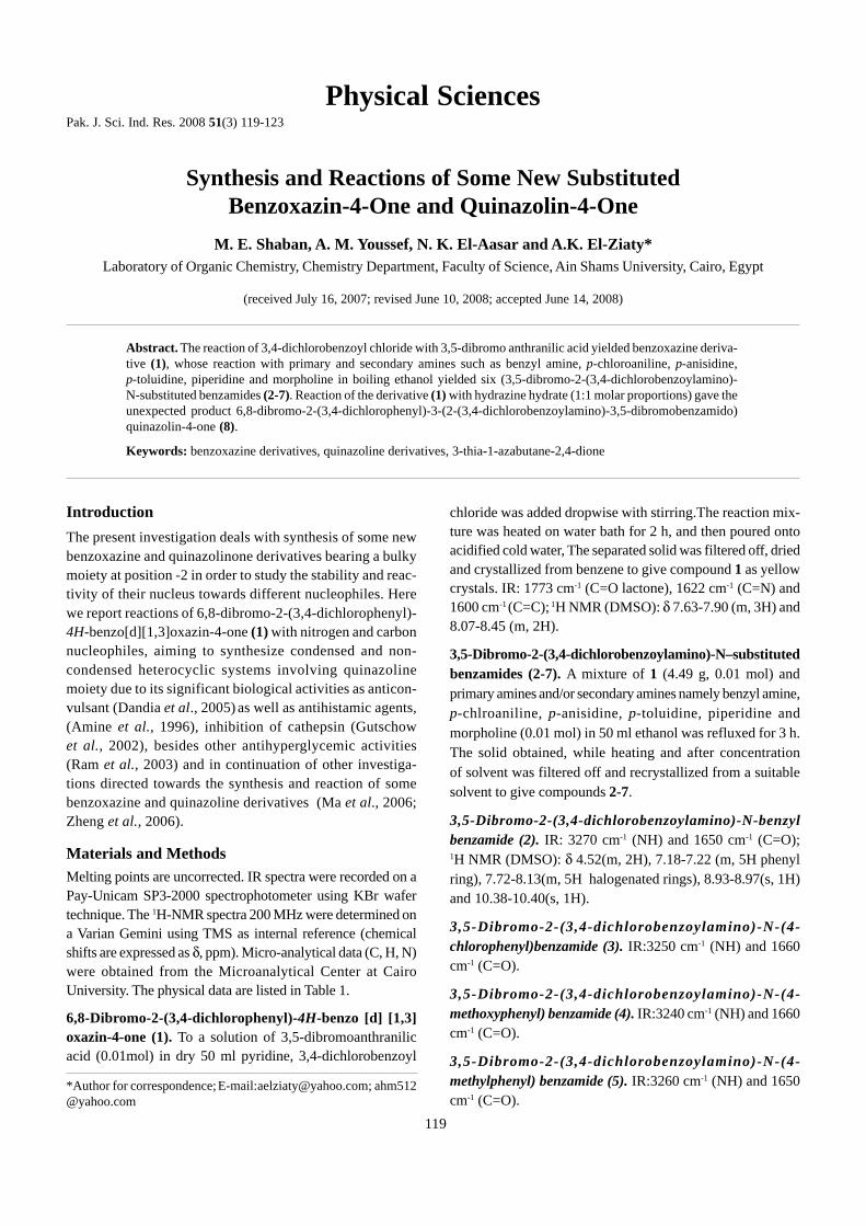
Materials and MethodsMelting points are uncorrected. IR spectra were recorded on aPay-Unicam SP3-2000 spectrophotometer using KBr wafertechnique. The 1H-NMR spectra 200 MHz were determined ona Varian Gemini using TMS as internal reference (chemicalshifts are expressed as δ, ppm). Micro-analytical data (C, H, N)were obtained from the Microanalytical Center at CairoUniversity. The physical data are listed in Table 1.
6,8-Dibromo-2-(3,4-dichlorophenyl)-4H-benzo [d] [1,3]oxazin-4-one (1). To a solution of 3,5-dibromoanthranilicacid (0.01mol) in dry 50 ml pyridine, 3,4-dichlorobenzoyl
chloride was added dropwise with stirring.The reaction mix-ture was heated on water bath for 2 h, and then poured ontoacidified cold water, The separated solid was filtered off, driedand crystallized from benzene to give compound 1 as yellowcrystals. IR: 1773 cm-1 (C=O lactone), 1622 cm-1 (C=N) and1600 cm-1 (C=C); 1H NMR (DMSO): δ 7.63-7.90 (m, 3H) and8.07-8.45 (m, 2H).
3,5-Dibromo-2-(3,4-dichlorobenzoylamino)-N–substitutedbenzamides (2-7). A mixture of 1 (4.49 g, 0.01 mol) andprimary amines and/or secondary amines namely benzyl amine,p-chlroaniline, p-anisidine, p-toluidine, piperidine andmorpholine (0.01 mol) in 50 ml ethanol was refluxed for 3 h.The solid obtained, while heating and after concentrationof solvent was filtered off and recrystallized from a suitablesolvent to give compounds 2-7.
3,5-Dibromo-2-(3,4-dichlorobenzoylamino)-N-benzylbenzamide (2). IR: 3270 cm-1 (NH) and 1650 cm-1 (C=O);1H NMR (DMSO): δ 4.52(m, 2H), 7.18-7.22 (m, 5H phenylring), 7.72-8.13(m, 5H halogenated rings), 8.93-8.97(s, 1H)and 10.38-10.40(s, 1H).
3,5-Dibromo-2-(3,4-dichlorobenzoylamino)-N-(4-chlorophenyl)benzamide (3). IR:3250 cm-1 (NH) and 1660cm-1 (C=O).
3,5-Dibromo-2-(3,4-dichlorobenzoylamino)-N-(4-methoxyphenyl) benzamide (4). IR:3240 cm-1 (NH) and 1660cm-1 (C=O).
3,5-Dibromo-2-(3,4-dichlorobenzoylamino)-N-(4-methylphenyl) benzamide (5). IR:3260 cm-1 (NH) and 1650cm-1 (C=O).
Pak. J. Sci. Ind. Res. 2008 51(3) 119-123
Abstract. The reaction of 3,4-dichlorobenzoyl chloride with 3,5-dibromo anthranilic acid yielded benzoxazine deriva-tive (1), whose reaction with primary and secondary amines such as benzyl amine, p-chloroaniline, p-anisidine,p-toluidine, piperidine and morpholine in boiling ethanol yielded six (3,5-dibromo-2-(3,4-dichlorobenzoylamino)-N-substituted benzamides (2-7). Reaction of the derivative (1) with hydrazine hydrate (1:1 molar proportions) gave theunexpected product 6,8-dibromo-2-(3,4-dichlorophenyl)-3-(2-(3,4-dichlorobenzoylamino)-3,5-dibromobenzamido)quinazolin-4-one (8).
Keywords: benzoxazine derivatives, quinazoline derivatives, 3-thia-1-azabutane-2,4-dione
*Author for correspondence; E-mail: [email protected]; [email protected]
119
Physical Sciences
Table 1. Characterization and physical data of synthesized compounds
Compound M.p.(°C) Solvent M.F Calc.% (found%)nos. (yield%) (M. Wt.) C H N
1 187-188 B C14H5Br2Cl2NO2 37.37 1.12 3.1180 449.91 36.96 1.97 2.98
2 269-270 D C21H14Br2Cl2N2O2 45.27 2.53 5.0370 557.06 45.21 2.51 4.90
3 281-282 B/EtOH C20H11Br2Cl3N2O2 41.59 1.92 4.8580 577.48 41.27 1.73 4.84
4 271-272 B/EtOH C21H14Br2Cl2N2O3 44.01 2.46 4.8880 573.06 43.86 2.34 4.80
5 234-235 D C21H14Br2Cl2N2O2 45.27 2.53 5.0380 557.06 45.94 2.81 4.96
6 130-132 Pet. (80/100)/B C19H16Br2Cl2N2O2 42.65 3.01 5.2370 535.05 42.34 2.87 5.08
7 252-254 D C18H14Br2Cl2N2O3 40.25 2.63 5.2270 533.87 40.72 2.09 4.96
8 254-256 D C28H12Br4Cl4N4O3 36.80 1.32 6.1390 913.85 36.33 1.23 5.98
9 Over300 DMF C14H6Br2Cl2N2O2 36.16 1.30 6.0260 464.92 37.56 1.28 5.56
10 230-232 D C14H7Br2Cl2N3O 35.53 1.59 11.0570 463.94 35.80 1.30 10.07
11 250-252 D C15H8Br2Cl2N4O2 36.24 1.52 9.0680 506.96 36.09 1.42 8.97
12 191-192 D C29H12Br4Cl4N4O4S 35.76 1.24 5.7560 973.92 34.98 1.21 5.72
13 211-212 B/EtOH C17H7Br2Cl2NO5 38.09 1.32 2.6140 535.95 38.12 1.51 2.59
14 220-221 B/EtOH C14H7Br2Cl2NO3 35.93 1.51 2.9950 467.92 35.67 1.55 2.92
15 Over300 DMF C14H7Br2Cl2N2O 37.45 1.35 6.2580 448.93 37.34 1.24 6.00
16 191-192 Pet. (80/100)/B C114H6Br2C12N4O2 34.11 1.22 11.3670 492.94 33.98 1.12 11.23
EtOH =ethanol; B = benzene; D = 1,4-dioxane; DMF = N,N-dimethylformamide; Pet. = petroleum ether
3,4-Dichloro-N-(2,4-dibromo-6-(piperidine-1-carbonyl)phenyl)benzamide (6). IR: 3230 cm-1 (NH) and 1660 cm-1 (C=O).
3,4-Dichloro-N-(2 ,4-dibromo-6-(morphol ine-4-carbonyl)phenyl) benzamide (7). IR:3240 cm-1 (NH) and1680 cm-1 (C=O).
6,8-Dibromo-2-(3,4-dichlorophenyl)-3-(2-(3,4-dichloro-benzoylamino)-3,5-dibromobenzamido)quinazolin-4-
one (8). A mixture of compound 1 (4.49 g,0.01 mol) andhydrazine hydrate (0.5 g, 0.01mol) in 50 ml. ethanol wasrefluxed for one h. The solid that separated while refluxingwas filtered off and recrystallized from 1,4-dioxane to givecompound 8 as white crystals. IR: 3300-3500 cm-1
(NH) orenolic (OH), 1645 cm-1 (CO); 1H NMR (DMSO): δ 8.50(d, 1H), 8.45 (d, 1H), 8.36 (s, 1H), 7.91 (m, 4H), and 5.71(br, 1H, NH).
120 A. K. El-Ziaty et al.
6,8-Dibromo-2-(3,4-dichlorophenyl)-3-N-substitutedquinazolin-4-one (9,10, 11). A mixture of compound 1 (4.49 g,0.01 mol) and primary amines, namely, hydrazine hydrate,hydroxylamine hydrochloride and/or semicarbazidehydrochloride (0.01 mol) was refluxed in 50 ml n-butanol for4 h. The solid that formed while refluxing was filtered off andrecrystallized from a suitable solvent to give comounds 9, 10,and 11.
6,8-Dibromo-2-(3,4-dichlorophenyl)-3-hydroxyquina-zoline-4-(3H)-one (9). IR: 3440 cm-1 (NH), 1684cm-1 (CO).
3-Amino-6,8-dibromo-2-(3,4-dichlorophenyl)quinazoline-4-(3H)-one (10). IR: 3200 cm-1 (NH), 3450-3315 cm-1 (NH2)and 1688 cm-1 (C=O).
1-(6,8-Dibromo-2-(3,4-dichlorophenyl)-4-oxoquinazolin-3-(4H)-yl)urea (11). IR: 3211 cm-1 (NH), 3330-3260 cm-1 (NH2)and 1672 cm-1 (C=O).
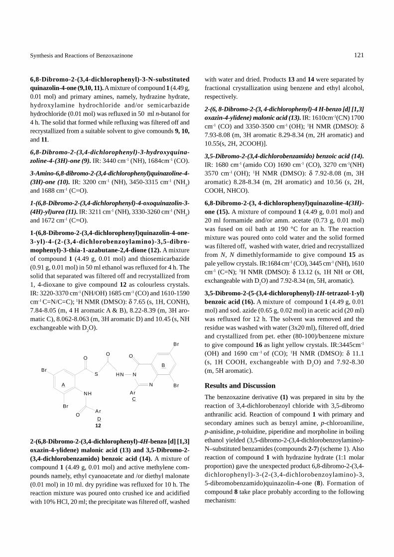
1-(6,8-Dibromo-2-(3,4-dichlorophenyl)quinazolin-4-one-3-yl)-4-(2-(3,4-dichlorobenzoylamino)-3,5-dibro-mophenyl)-3-thia-1-azabutane-2,4-dione (12). A mixtureof compound 1 (4.49 g, 0.01 mol) and thiosemicarbazide(0.91 g, 0.01 mol) in 50 ml ethanol was refluxed for 4 h. Thesolid that separated was filtered off and recrystallized from1, 4-dioxane to give compound 12 as colourless crystals.IR: 3220-3370 cm-1 (NH/OH) 1685 cm-1 (CO) and 1610-1590cm-1 C=N/C=C); 1H NMR (DMSO): δ 7.65 (s, 1H, CONH),7.84-8.05 (m, 4 H aromatic A & B), 8.22-8.39 (m, 3H aro-matic C), 8.062-8.063 (m, 3H aromatic D) and 10.45 (s, NHexchangeable with D2O).
with water and dried. Products 13 and 14 were separated byfractional crystallization using benzene and ethyl alcohol,respectively.
2-(6, 8-Dibromo-2-(3, 4-dichlorophenyl)-4 H-benzo [d] [1,3]oxazin-4-ylidene) malonic acid (13). IR: 1610cm-1(CN) 1700cm-1 (CO) and 3350-3500 cm-1 (OH); 1H NMR (DMSO): δ7.93-8.08 (m, 3H aromatic 8.29-8.34 (m, 2H aromatic) and10.55(s, 2H, 2COOH)].
3,5-Dibromo-2-(3,4-dichlorobenzamido) benzoic acid (14).IR: 1680 cm-1 (amido CO) 1690 cm-1 (CO), 3270 cm-1(NH)3570 cm-1 (OH); 1H NMR (DMSO): δ 7.92-8.08 (m, 3Haromatic) 8.28-8.34 (m, 2H aromatic) and 10.56 (s, 2H,COOH, NHCO).
6,8-Dibromo-2-(3, 4-dichlorophenyl)quinazoline-4(3H)-one (15). A mixture of compound 1 (4.49 g, 0.01 mol) and20 ml formamide and/or amm. acetate (0.73 g, 0.01 mol)was fused on oil bath at 190 °C for an h. The reactionmixture was poured onto cold water and the solid formedwas filtered off, washed with water, dried and recrystallizedfrom N, N dimethlyformamide to give compound 15 aspale yellow crystals. IR:1684 cm-1 (CO), 3445 cm-1 (NH), 1610cm-1 (C=N); 1H NMR (DMSO): δ 13.12 (s, 1H NH or OH,exchangeable with D2O) and 7.92-8.34 (m, 5H, aromatic).
3,5-Dibromo-2-(5-(3,4-dichlorophenyl)-1H-tetrazol-1-yl)benzoic acid (16). A mixture of compound 1 (4.49 g, 0.01mol) and sod. azide (0.65 g, 0.02 mol) in acetic acid (20 ml)was refluxed for 12 h. The solvent was removed and theresidue was washed with water (3x20 ml), filtered off, driedand crystallized from pet. ether (80-100)/benzene mixtureto give compound 16 as light yellow crystals. IR:3445cm-1
(OH) and 1690 cm–1 of (CO); 1H NMR (DMSO): δ 11.1(s, 1H COOH, exchangeable with D2O) and 7.92-8.30(m, 5H aromatic).
Results and DiscussionThe benzoxazine derivative (1) was prepared in situ by thereaction of 3,4-dichlorobenzoyl chloride with 3,5-dibromoanthranilic acid. Reaction of compound 1 with primary andsecondary amines such as benzyl amine, p-chloroaniline,p-anisidine, p-toluidine, piperidine and morpholine in boilingethanol yielded (3,5-dibromo-2-(3,4-dichlorobenzoylamino)-N–substituted benzamides (compounds 2-7) (scheme 1). Alsoreaction of compound 1 with hydrazine hydrate (1:1 molarproportion) gave the unexpected product 6,8-dibromo-2-(3,4-dichlorophenyl)-3-(2-(3,4-dichlorobenzoylamino)-3,5-dibromobenzamido)quinazolin-4-one (8). Formation ofcompound 8 take place probably according to the followingmechanism:
2-(6,8-Dibromo-2-(3,4-dichlorophenyl)-4H-benzo [d] [1,3]oxazin-4-ylidene) malonic acid (13) and 3,5-Dibromo-2-(3,4-dichlorobenzamido) benzoic acid (14). A mixture ofcompound 1 (4.49 g, 0.01 mol) and active methylene com-pounds namely, ethyl cyanoacetate and /or diethyl malonate(0.01 mol) in 10 ml. dry pyridine was refluxed for 10 h. Thereaction mixture was poured onto crushed ice and acidifiedwith 10% HCl, 20 ml; the precipitate was filtered off, washed
121Synthesis and Reactions of Benzoxazinone
Br
Br
O
NH
OAr
S
O
N
N
HN
Ar
O
Br
Br
12
A
B
D
C
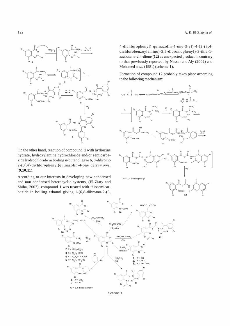
On the other hand, reaction of compound 1 with hydrazinehydrate, hydroxylamine hydrochloride and/or semicarba-zide hydrochloride in boiling n-butanol gave 6, 8-dibromo2-(3′,4′-dichlorophenyl)quinazolin-4-one derivatives.(9,10,11).
According to our interests in developing new condensedand non condensed heterocyclic systems, (El-Ziaty andShiba, 2007), compound 1 was treated with thiosemicar-bazide in boiling ethanol giving 1-(6,8-dibromo-2-(3,
122
Scheme 1
4-dichlorophenyl) quinazolin-4-one-3-yl)-4-(2-(3,4-dichlorobenzoylamino)-3,5-dibromophenyl)-3-thia-1-azabutane-2,4-dione (12) as unexpected product in contraryto that previously reported, by Nassar and Aly (2002) andMohamed et al. (1981) (scheme 1).
Formation of compound 12 probably takes place accordingto the following mechanism:
A. K. El-Ziaty et al.
Ar = 3,4 dichlorophenyl
Br
N
O
O
1
NH2NH2
Br
Br
N
O
O
Ar
2 2 -H
Br
N
O
ArNH
O
O
Ar
21
O
NH2
O
NO
-H +H
OO
N
BrO
N
O
N
8
H
-H2O
Br NHNH2
Ar
Br NH NH ++ ++
OH
Br
BrBr
NHNH
Br
Br
NHCOAr
NH+
Ar
Br
Br+ +
Br
NHCOAr Ar
NNH HHO
Br
Br
Br
NH
NHCOAr
Br
Cl
Cl
Br
N
OBr
BrAr
OBr
Br
O
X
Alc.
Alc.
NX
Br
Br
O
NN
Br
Br
O
ArO
Ar
OBr
Br
n-Butanol
N
N
Br
BrAr
O
91011
Alc.
Br
Br
O
O Ar
S
O
NN
Ar
O
Br
Br
N
OBr
BrAr
Alc.
Pyridine
Br
Br
O
N
Br
BrAr
O
Heat
NN
NN
Br
Br
O
Ar/
Ar = 3,4 dichlorophenyl
+
15
NH
14
OH
NHCOAr
H OOC COOH
13
12
NH
NH
R'
2CH RCOOE t
NH2NHCSNH2
1
Ac OHOH NaN3
16
NHR
NHCOAr
RNH2
2 R =
3 R =6 5CH2 - C H
-CI(p)6C H4
NHCOA r
6 X =7 X = O
CH2
NH
NH2NH2
'R NH2
R' = OH R' = NH2
2R' = NHCONH
NH
NH8
CH3COONH4
5 R = -CH (p)6C H4 3
R = - (p)6C H4 OCH34
C
S
N C 2C
SN C
SNH2
+
C
SN H
2
1O
S
N
O
S
N
O
S
O
S
O
NO
O
S
O
N
O
S N
N
O
H2O
O
S
O
N
N
O
SH
NHH2N
HN NH2
H2N
H3N
OH
ArBr
+H
NNH2
NH2
Ar
NNH2
2NH
Br
Br
NH
2NHNH
1NHCOAr
Br
Br
Br
NH2NH
NHBr
Ar
+H- H
BrNH
Br
NH
NHCOAr
Br
BrBr
-
Ar
Br
HO
NH
Ar
NH NH
ArNHCO
ArNHCO
Br
Br
Br
Br
NHCOAr Ar
NH
Br
+ HN
Br
H2NNH2HNH2N
12
+
+ + +
Br
H2O
123
By studying reaction of compound 1 with active methylenecompounds, namely, ethyl cyanoacetate and/or diethyl mal-onate in pyridine afforded 2-(6,8-dibromo-2-(dichlorophenyl)-4H-benzo[d][1,3]oxazin-4-ylidene)malonic acid (13) and 3,5-dibromo-2-(3,4-dichlorobenzamido)benzoic acid (14), theopen form of 1, respectively.
Formation of compound 13 probably takes place accordingto the following mechanism:
ReferencesAmine, M.S., Nassar, S.A., EL-Hashash, M.A., Essawy, S.A.,
Hashish, A.A. 1996. Synthesis and characterization ofsome biologically active pyrimidine derivatives contain-ing sulphur-Part 1. Indian. J. Chem. 35B: 388-391.
Dandia, A., Singh, R., Sarawgi, P. 2005. Green chemicalmulti-component one-pot synthesis of fluorinated 2,3-disubstituted quinazolin-4(3H)-ones under solvent-freeconditions and their anti-fungal activity. J. Fluor. Chem.126: 307-312.
El-Ziaty, A.K., Shiba, S.A. 2007. Antibacterial activities of new(E) 2-cyano-3-(3′,4′dimethoxyphenyl)-2-propenoylamidederivatives. Synth. Comm. 37: 4043-4057.
Gutschow, M., Kuerschner, L., Pietsch, M., Ambrozak, A.,Newmann, U., Gunther, R., Hofmann, H. J. 2002. Inhi-bition of cathepsin G by 2-amino-3,1-benzoxazin-4-ones:Kinetic investigation and docking studies. Arch. Biochem.Biophy. 402: 180-191.
Mohamed, M.M., EL-Hashash, M.A., Essawy, A., Shaban,M. E. 1981. Reactions of 2-cyanomethyl-3,1-benzoxazin-4(H)-one with nucleophilic reagent, acid anhydride andacid amides. Indian J. Chem. 20B: 718-719.
Nassar, S.A., Aly, A. A. 2002. Synthesis of some new sub-stituted β-(quinazolin-2-yl) acrylic acid derivativesof expected biological activity. Egypt. J. Chem. 45:205-217.
Ram, V.J., Farhanullah, Tripathi, B.K., Srivastava, A.K. 2003.Synthesis and antihyperglycemic activity of suitablyfunctionalized 3H-quinazoline-4-one. Bioorganic Med.Chem. 11: 2439-2444.
Ma, S., Li, J., Sun, Y., Zhao, J., Zhao, X.,Yang, X., Zhang, L.,Wang, L., Zhou, Z. 2006. Synthesis of 1,2-dihydro-4H-3,1-benzoxazine derivatives via ZnCl2 catalyzedcyclocondensation reaction. Tetrahedron 62: 7999-8005.
Zheng, X., Hodgetts, K.J., Brielmann, H., Hutchison, A.,Burkamp, F., Brian Jones A., Blurton, P., Clarkson, R.,Chandrasekhar, J., Bakthavatchalam, R., De Lombaert,S., Crandall, M., Cortright, D., Blum, C.A. 2006. Fromarylureas to biarylamides to aminoquinazolines: Discov-ery of a novel, potent TRPV1 antagonist. Bioorg. Med.Chem. Lett. 16: 5217-5221.
Fusion of compound 1 with ammonium acetate and/orformamide yielded the corresponding 6,8-dibromo-2-(3,4-dichlorophenyl) quinazolin-4-(3H)-one (15) as previouslyreported (Nassar and Aly, 2002). Ring opening of 1 withhydrazoic acid gave 3,5-dibromo-2-(5-(3,4-dichlorophenyl)-1H-tetrazol-1-yl) benzoic acid (16) (scheme 1).
Formation of compound 16 probably takes place accordingto the following mechanism:
Synthesis and Reactions of Benzoxazinone
:B + BH
N
OBr
Br
Ar
O
1
+N
Br
Br Ar
O
ON
Br
Br Ar
O
BH
N
Br
BrAr
N
OBr
Br
ArN
OBr
Br
Ar
13
CNHC
COOEtCH2COOE t
CN
COOEt
CNHC
CH
CN
COOE t CH
CN
OHCOOEt
NC COOEt
OHOH
-H2O
COOE tNCHOOC COOH
H2O
N
OBr
Br
Ar
O
N N N- + O
Br
Br
Ar
O
NN
H
N N
Br
N
O
OAr
NN
NH
Br
-+
-
+
NN
NN
Br
Br
Ar
H+
-H/+ H+ +
COOH
16
1
Synthesis of Some 2-Methyl-3-(Arylthiocarbamido) Quinazol-4-Ones and2-Methyl-3-(Arylidencarboxamido) Quinazol-4-Ones as
Potential Antimicrobial AgentsB. D. Gupta
Department of Chemistry, Northern India Engineering College, Lucknow 226001, India
(received July 23, 2007; revised June 10, 2008; accepted June 12, 2008)
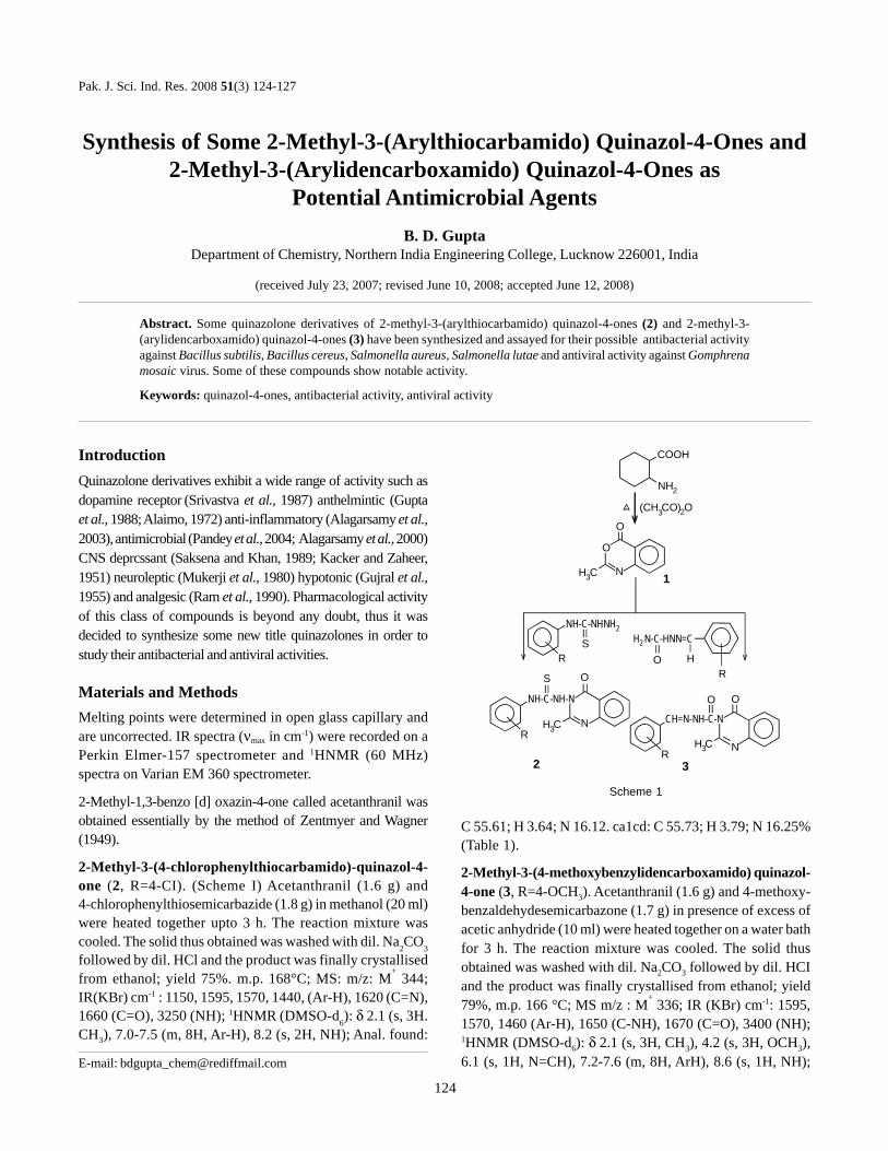
IntroductionQuinazolone derivatives exhibit a wide range of activity such asdopamine receptor (Srivastva et al., 1987) anthelmintic (Guptaet al., 1988; Alaimo, 1972) anti-inflammatory (Alagarsamy et al.,2003), antimicrobial (Pandey et al., 2004; Alagarsamy et al., 2000)CNS deprcssant (Saksena and Khan, 1989; Kacker and Zaheer,1951) neuroleptic (Mukerji et al., 1980) hypotonic (Gujral et al.,1955) and analgesic (Ram et al., 1990). Pharmacological activityof this class of compounds is beyond any doubt, thus it wasdecided to synthesize some new title quinazolones in order tostudy their antibacterial and antiviral activities.
Materials and MethodsMelting points were determined in open glass capillary andare uncorrected. IR spectra (νmax in cm-1) were recorded on aPerkin Elmer-157 spectrometer and 1HNMR (60 MHz)spectra on Varian EM 360 spectrometer.
2-Methyl-1,3-benzo [d] oxazin-4-one called acetanthranil wasobtained essentially by the method of Zentmyer and Wagner(1949).
2-Methyl-3-(4-chlorophenylthiocarbamido)-quinazol-4-one (2, R=4-CI). (Scheme I) Acetanthranil (1.6 g) and4-chlorophenylthiosemicarbazide (1.8 g) in methanol (20 ml)were heated together upto 3 h. The reaction mixture wascooled. The solid thus obtained was washed with dil. Na2CO3followed by dil. HCl and the product was finally crystallisedfrom ethanol; yield 75%. m.p. 168°C; MS: m/z: M
+ 344;
IR(KBr) cm-1 : 1150, 1595, 1570, 1440, (Ar-H), 1620 (C=N),1660 (C=O), 3250 (NH); 1HNMR (DMSO-d6): δ 2.1 (s, 3H.CH3), 7.0-7.5 (m, 8H, Ar-H), 8.2 (s, 2H, NH); Anal. found:
C 55.61; H 3.64; N 16.12. ca1cd: C 55.73; H 3.79; N 16.25%(Table 1).
2-Methyl-3-(4-methoxybenzylidencarboxamido) quinazol-4-one (3, R=4-OCH3). Acetanthranil (1.6 g) and 4-methoxy-benzaldehydesemicarbazone (1.7 g) in presence of excess ofacetic anhydride (10 ml) were heated together on a water bathfor 3 h. The reaction mixture was cooled. The solid thusobtained was washed with dil. Na2CO3 followed by dil. HCIand the product was finally crystallised from ethanol; yield79%, m.p. 166 °C; MS m/z : M
+ 336; IR (KBr) cm-1: 1595,
1570, 1460 (Ar-H), 1650 (C-NH), 1670 (C=O), 3400 (NH);1HNMR (DMSO-d6): δ 2.1 (s, 3H, CH3), 4.2 (s, 3H, OCH3),6.1 (s, 1H, N=CH), 7.2-7.6 (m, 8H, ArH), 8.6 (s, 1H, NH);
Pak. J. Sci. Ind. Res. 2008 51(3) 124-127
E-mail: [email protected]
Abstract. Some quinazolone derivatives of 2-methyl-3-(arylthiocarbamido) quinazol-4-ones (2) and 2-methyl-3-(arylidencarboxamido) quinazol-4-ones (3) have been synthesized and assayed for their possible antibacterial activityagainst Bacillus subtilis, Bacillus cereus, Salmonella aureus, Salmonella lutae and antiviral activity against Gomphrenamosaic virus. Some of these compounds show notable activity.
Keywords: quinazol-4-ones, antibacterial activity, antiviral activity
124
Scheme 1
COOH
NH2
(CH CO) O3 2
O
O
NH C3 1
SR O H
R
O O
N
CH=N-NH-C-N
R3
O
N
NH-C-NH-N
S
H C3R
2H C3
H N-C-HNN=C2
NH-C-NHNH2
Anal. found: C 64.20; H 4.64; N 16.52. calcd: C 64.28; H4.79; N 16.66% (Table 1).
Pharmacology. Antibacterial screening. The in vitro anti-bacterial activity of the synthesised compounds was deter-mined by the method of Verma and Nobbles (1968), at aconcentration of 100 μmg/ml. A standard tetracycline wasalso tested under similar conditions to compare the results.The inhibition zone (in cm) against four species viz.:B. subtilis, B. cereus, S.aureus and S. lutes were measured.The results are recorded in Table 2.
Antiviral screening. The in vitro antiviral activity of all thesynthesized compounds reported here was determined by themethod of Verma and Awasthi (1978) on Gomphrena mosaicvirus, taking gaur leaves as host. The concentration of eachsample was 3.0 mg/mole. The percentage activity is recordedin Table 2.
Results and DiscussionAntibacterial activity. All the compounds of this report havebeen screened for their antibacterial activity. Perusal of the
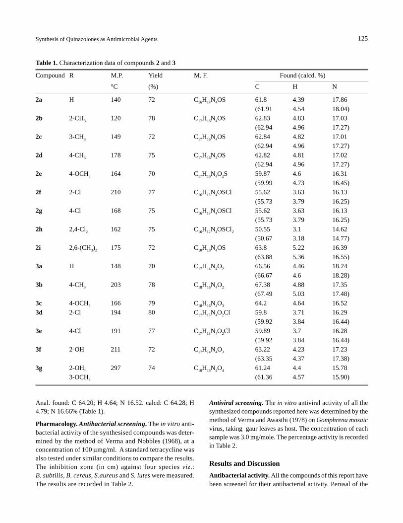
Table 1. Characterization data of compounds 2 and 3
Compound R M.P. Yield M. F. Found (calcd. %)
°C (%) C H N
2a H 140 72 C16H14N4OS 61.8 4.39 17.86(61.91 4.54 18.04)
2b 2-CH3 120 78 C17H16N4OS 62.83 4.83 17.03(62.94 4.96 17.27)
2c 3-CH3 149 72 C17H16N4OS 62.84 4.82 17.01(62.94 4.96 17.27)
2d 4-CH3 178 75 C17H16N4OS 62.82 4.81 17.02(62.94 4.96 17.27)
2e 4-OCH3 164 70 C17H16N4O2S 59.87 4.6 16.31(59.99 4.73 16.45)
2f 2-Cl 210 77 C16H13N4OSCl 55.62 3.63 16.13(55.73 3.79 16.25)
2g 4-Cl 168 75 C16H13N4OSCl 55.62 3.63 16.13(55.73 3.79 16.25)
2h 2,4-Cl2 162 75 C16H12N4OSCl2 50.55 3.1 14.62(50.67 3.18 14.77)
2i 2,6-(CH3)2 175 72 C18H18N4OS 63.8 5.22 16.39(63.88 5.36 16.55)
3a H 148 70 C17H14N4O2 66.56 4.46 18.24(66.67 4.6 18.28)
3b 4-CH3 203 78 C18H16N4O2 67.38 4.88 17.35(67.49 5.03 17.48)
3c 4-OCH3 166 79 C18H16N4O3 64.2 4.64 16.523d 2-Cl 194 80 C17H13N4O2Cl 59.8 3.71 16.29
(59.92 3.84 16.44)3e 4-Cl 191 77 C17H13N4O2Cl 59.89 3.7 16.28
(59.92 3.84 16.44)3f 2-OH 211 72 C17H14N4O3 63.22 4.23 17.23
(63.35 4.37 17.38)3g 2-OH, 297 74 C18H16N4O4 61.24 4.4 15.78
3-OCH3 (61.36 4.57 15.90)
125Synthesis of Quinazolones as Antimicrobial Agents
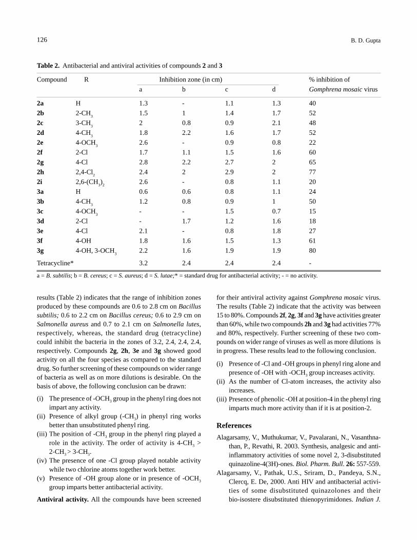
results (Table 2) indicates that the range of inhibition zonesproduced by these compounds are 0.6 to 2.8 cm on Bacillussubtilis; 0.6 to 2.2 cm on Bacillus cereus; 0.6 to 2.9 cm onSalmonella aureus and 0.7 to 2.1 cm on Salmonella lutes,respectively, whereas, the standard drug (tetracycline)could inhibit the bacteria in the zones of 3.2, 2.4, 2.4, 2.4,respectively. Compounds 2g, 2h, 3e and 3g showed goodactivity on all the four species as compared to the standarddrug. So further screening of these compounds on wider rangeof bacteria as well as on more dilutions is desirable. On thebasis of above, the following conclusion can be drawn:
(i) The presence of -OCH3 group in the phenyl ring does notimpart any activity.
(ii) Presence of alkyl group (-CH3) in phenyl ring worksbetter than unsubstituted phenyl ring.
(iii) The position of -CH3 group in the phenyl ring played arole in the activity. The order of activity is 4-CH3 >2-CH3 > 3-CH3.
(iv) The presence of one -Cl group played notable activitywhile two chlorine atoms together work better.
(v) Presence of -OH group alone or in presence of -OCH3
group imparts better antibacterial activity.
Antiviral activity. All the compounds have been screened
for their antiviral activity against Gomphrena mosaic virus.The results (Table 2) indicate that the activity was between15 to 80%. Compounds 2f, 2g, 3f and 3g have activities greaterthan 60%, while two compounds 2h and 3g had activities 77%and 80%, respectively. Further screening of these two com-pounds on wider range of viruses as well as more dilutions isin progress. These results lead to the following conclusion.
(i) Presence of -Cl and -OH groups in phenyl ring alone andpresence of -OH with -OCH3 group increases activity.
(ii) As the number of Cl-atom increases, the activity alsoincreases.
(iii) Presence of phenolic -OH at position-4 in the phenyl ringimparts much more activity than if it is at position-2.
ReferencesAlagarsamy, V., Muthukumar, V., Pavalarani, N., Vasanthna-
than, P., Revathi, R. 2003. Synthesis, analgesic and anti-inflammatory activities of some novel 2, 3-disubstitutedquinazoline-4(3H)-ones. Biol. Pharm. Bull. 26: 557-559.
Alagarsamy, V., Pathak, U.S., Sriram, D., Pandeya, S.N.,Clercq, E. De, 2000. Anti HIV and antibacterial activi-ties of some disubstituted quinazolones and theirbio-isostere disubstituted thienopyrimidones. Indian J.
Table 2. Antibacterial and antiviral activities of compounds 2 and 3
Compound R Inhibition zone (in cm) % inhibition ofa b c d Gomphrena mosaic virus
2a H 1.3 - 1.1 1.3 402b 2-CH3 1.5 1 1.4 1.7 522c 3-CH3 2 0.8 0.9 2.1 482d 4-CH3 1.8 2.2 1.6 1.7 522e 4-OCH3 2.6 - 0.9 0.8 222f 2-Cl 1.7 1.1 1.5 1.6 602g 4-Cl 2.8 2.2 2.7 2 652h 2,4-Cl2 2.4 2 2.9 2 772i 2,6-(CH3)2 2.6 - 0.8 1.1 203a H 0.6 0.6 0.8 1.1 243b 4-CH3 1.2 0.8 0.9 1 503c 4-OCH3 - - 1.5 0.7 153d 2-Cl - 1.7 1.2 1.6 183e 4-Cl 2.1 - 0.8 1.8 273f 4-OH 1.8 1.6 1.5 1.3 613g 4-OH, 3-OCH3 2.2 1.6 1.9 1.9 80
Tetracycline* 3.2 2.4 2.4 2.4 -
a = B. subtilis; b = B. cereus; c = S. aureus; d = S. lutae;* = standard drug for antibacterial activity; - = no activity.
126 B. D. Gupta
Pharm. Sci. 62: 433-437.Alaimo, R.J., 1972. Anthelmintic 2-(5-nitro-2-thienyl)-4-
(substituted amino) quinazolines. J. Med. Chem. 15: 108-109.Gupta, D. P., Ahmad, S., Kumar, A.,Shankar, K. 1988. Newer
quinazolinone derivatives as antihelmintic agents. IndianJ. Chem. 27B: 1060-1062.
Gujral, M.L., Saxena, P.N., Tiwari, R.S., 1955. Comparativeevaluation of quinazolinones: a new class of hypnotics.Indian J. Med. Res. 43: 637.
Kacker, I. K., Zaheer, S.H. 1951. Potential analygesics.Part 1. synthesis of substituted 4-quinazolinones. J. Ind.Chem. Soc. 28: 344-346.
Mukerji, D.D., Nautiyal, S.R., Prasad, C.R., Dhawan, B.N.1980. CNS-depressant activity of some newly synthe-sized 4(3H)-quinazolones. Indian J. Med. Res. 71:480-482.
Pandey, V.K., Tusi, S., Tusi, Z., Raghubir, R., Dixit, M., Joshi,M.N. 2004. Thiadiazolyl quinazolones as potentialantiviral and antihypertensive agents. Indian J. Chem.43B: 180-183.
Ram V.J., Srimal, R.C., Kushwaha, D.S., Mishra, L.J. 1990.Chemotherapeutic agents. XIX. Synthesis of [1,2,4]-triazoloquinazolinones and related compounds asantihypertensive agents. J. Pract. Chem. 332: 629-639.
Saksena, R.K., Khan, M.A. 1989. Synthesis of 2-alkyl/aryl-3-arylhydrazinoquinazolin-4-(3H)-ones as anti-bacterial and CNS active agents. Indian J. Chem. 27B:443-444.
Srivastva, V.K., Singh, S., Gulati, A., Shanker, K. 1987. Anti-parkinsonian agents from quinazolinyl thiazolidinones andazetidinones. Indian J. Chem. 26B: 652-656.
Verma, H.N., Awasthi, L.P. 1978. Effect of biochemic drugson the infection of TMV. Geobios. 5: 84-86.
Verma, R.S., Nobles, W.L. 1968. Synthesis and antibacterialactivity of certain 3-substituted benzoxazolinones.J. Pharm. Sci. 57: 39-44.
Zentmyer, D.T., Wagner, E.C. 1949. The so-called acylanthranils(3,1,4-benzoxazones). I. Preparation; reactions withwater, ammonia and aniline structure. J. Org. Chem. 14:967-981.
127Synthesis of Quinazolones as Antimicrobial Agents
Some Physical Characteristics and Nutritional Composition of the Seedsof Wild Pepper (Erythrococca anomala, Benth)
E. A. Akandea*, G. O. Adegokeb and F. C. Mathookoc
aDepartment of Food Science and Engineering, Ladoke Akintola University of Technology,Ogbomoso, Oyo State, Nigeria
bDepartment of Food Technology, University of Ibadan, Ibadan, Oyo State, NigeriacDepartment of Food Science and Technology, Jommo Kenyatta University of
Agriculture and Technology, Nairobi, Kenya
(received January 31, 2008; revised March 26, 2008; accepted April 04, 2008)
IntroductionSeeds are abundantly found in nature and are good and cheapsources of foods. They have multiple nutritive values and arealso known to contain reasonable quantities of edible oils andfats. The satiety value, flavour enhancing and hunger delay-ing abilities are the particular attributes of fats. Moreover,seeds are cheap source of protein, known to be very impor-tant for the normal body functions in animals. Consideringthe shortage of food nutrients in human diet and reliance ofthe country on imports of food products from foreign coun-tries, lot of efforts have been focussed on the exploitation oflocally available natural raw materials for food production.For example, work has been done on bitter kola (Garciniakola) (Daramola and Adegoke, 2007), African oil bean seed(Pentaclethra macrophylla) (Ajibola, 2005), African bread-fruit seeds (Treculia africana) (Omobuwajo, 2002) etc.
Wild pepper (Erythrococca anomala, Benth) is an indigenousplant whose seeds are popular among the local people in theWestern part of Nigeria, owing to the benefits associated withthem. Its seeds are locally known as Iyere (Yor), Monsoro(Hausa) and Osunrisa (Ghana). They are usually of reddishto yellow colour on ripening, while deep brown after drying.
The plants are creepers, found clustering around the stemsof cocoa and kolanut trees, proliferating freely in parts ofSouthern Nigera. The seeds of E.anomala are aromatic,pungent and medicinally used in treatment of sore throat,
mouth infections, preparation of herbal soups for women andnew mothers etc. The seeds are also used as food additive(in flavouring ‘kunnusaki’, preparation of pepper soups,cooking of rice and meat). Recently, the seeds of E. anomalahave been employed in perfumeries in the northern Nigeria.
Since, no work has been rendered on determining the physi-cal characteristics and the nutritional potential of the seeds,the present study was undertaken to establish the attributesof the seeds of wild pepper (E. anomala, Benth).
Materials and MethodsThe physical characteristic including sphericity index, aspectratio and density (kernel density, bulk density and densityratio) were determined using the methods of Maduako andFaborode (1990) and Mohsenin (1986).
Properly dried seeds of E. anomala were milled usingShromadzu grinding machine (AGG -270 F 005028F4) andsieved using a 6 mm mesh size. The powdered seeds werethen made into 50 packs and kept for further analysis.
The moisture content was determined by drying method,protein by Kjedhal digestion method, and crude fat by Soxhletextraction, according to AOAC (1990). The ash content wasdetermined using Toyo Seisokusho Muffle Furnace (KL420:00004023021) at 550 °C for one h.
Minerals analysis. Use of atomic absorption spectrophoto-meter, AAS-324-75603-84 & 2P88887GM, was made for theminerals analysis.
Abstract. Study of physical properties and nutritional components of whole and powdered wild pepper seeds (Erythrococcaanomala,Benth) revealed that the seeds have good parameters for machineability. The contents of moisture, ash, protein,lipid and carbohydrate and major and trace minerals were found in functional quantities, while the heavy metals werenegligible or absent. Thus the seeds are potential source of nutrients and can be used as additive in food productdevelopment.
Keywords: Erythrococca anomala, nutritional composition, product development, seed machineability
*Author for correspondence; E-mail: [email protected]
Pak. J. Sci. Ind. Res. 2008 51(3) 128-130
128
Samples were prepared by ashing the weighed amount ofpowdered seeds in a Seisokusho Muffle Furnace PK50067FT;4022004) at 500 °C which was then allowed to cool. The ashedsample was digested and diluted serially with 1N HCL. Thedigest was filtered and aspirated into the AAS, where theminerals were automatically quantified.
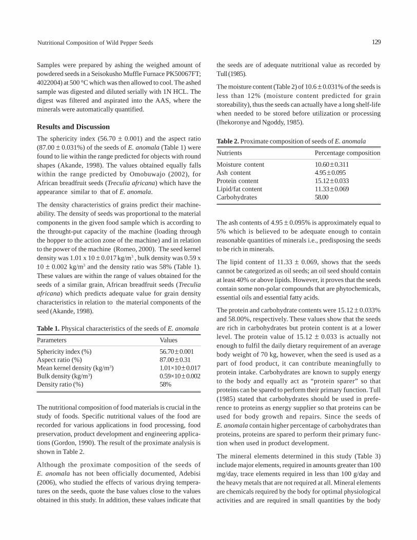
Results and DiscussionThe sphericity index (56.70 ± 0.001) and the aspect ratio(87.00 ± 0.031%) of the seeds of E. anomala (Table 1) werefound to lie within the range predicted for objects with roundshapes (Akande, 1998). The values obtained equally fallswithin the range predicted by Omobuwajo (2002), forAfrican breadfruit seeds (Treculia africana) which have theappearance similar to that of E. anomala.
The density characteristics of grains predict their machine-ability. The density of seeds was proportional to the materialcomponents in the given food sample which is according tothe throught-put capacity of the machine (loading throughthe hopper to the action zone of the machine) and in relationto the power of the machine (Romeo, 2000). The seed kerneldensity was 1.01 x 10 ± 0.017 kg/m3 , bulk density was 0.59 x10 ± 0.002 kg/m3 and the density ratio was 58% (Table 1).These values are within the range of values obtained for theseeds of a similar grain, African breadfruit seeds (Treculiaafricana) which predicts adequate value for grain densitycharacteristics in relation to the material components of theseed (Akande, 1998).
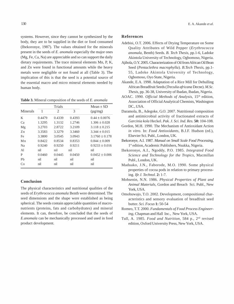
the seeds are of adequate nutritional value as recorded byTull (1985).
The moisture content (Table 2) of 10.6 ± 0.031% of the seeds isless than 12% (moisture content predicted for grainstoreability), thus the seeds can actually have a long shelf-lifewhen needed to be stored before utilization or processing(Ihekoronye and Ngoddy, 1985).
The ash contents of 4.95 ± 0.095% is approximately equal to5% which is believed to be adequate enough to containreasonable quantities of minerals i.e., predisposing the seedsto be rich in minerals.
The lipid content of 11.33 ± 0.069, shows that the seedscannot be categorized as oil seeds; an oil seed should containat least 40% or above lipids. However, it proves that the seedscontain some non-polar compounds that are phytochemicals,essential oils and essential fatty acids.
The protein and carbohydrate contents were 15.12 ± 0.033%and 58.00%, respectively. These values show that the seedsare rich in carbohydrates but protein content is at a lowerlevel. The protein value of 15.12 ± 0.033 is actually notenough to fulfil the daily dietary requirement of an averagebody weight of 70 kg, however, when the seed is used as apart of food product, it can contribute meaningfully toprotein intake. Carbohydrates are known to supply energyto the body and equally act as “protein sparer” so thatproteins can be spared to perform their primary function. Tull(1985) stated that carbohydrates should be used in prefe-rence to proteins as energy supplier so that proteins can beused for body growth and repairs. Since the seeds ofE. anomala contain higher percentage of carbohydrates thanproteins, proteins are spared to perform their primary func-tion when used in product development.
The mineral elements determined in this study (Table 3)include major elements, required in amounts greater than 100mg/day, trace elements required in less than 100 g/day andthe heavy metals that are not required at all. Mineral elementsare chemicals required by the body for optimal physiologicalactivities and are required in small quantities by the body
129Nutritional Composition of Wild Pepper Seeds
Table 1. Physical characteristics of the seeds of E. anomala
Parameters Values
Sphericity index (%) 56.70 ± 0.001Aspect ratio (%) 87.00 ± 0.31Mean kernel density (kg/m3) 1.01×10 ± 0.017Bulk density (kg/m3) 0.59×10 ± 0.002Density ratio (%) 58%
Table 2. Proximate composition of seeds of E. anomala
Nutrients Percentage composition
Moisture content 10.60 ± 0.311Ash content 4.95 ± 0.095Protein content 15.12 ± 0.033Lipid/fat content 11.33 ± 0.069Carbohydrates 58.00
The nutritional composition of food materials is crucial in thestudy of foods. Specific nutritional values of the food arerecorded for various applications in food processing, foodpreservation, product development and engineering applica-tions (Gordon, 1990). The result of the proximate analysis isshown in Table 2.
Although the proximate composition of the seeds ofE. anomala has not been officially documented, Adebisi(2006), who studied the effects of various drying tempera-tures on the seeds, quote the base values close to the valuesobtained in this study. In addition, these values indicate that
systems. However, since they cannot be synthesized by thebody, they are to be supplied in the diet or food consumed(Ihekoronye, 1987). The values obtained for the mineralspresent in the seeds of E. anomala especially the major ones(Mg, Fe, Ca, Na) are appreciable and so can support the dailydietary requirements. The trace mineral elements Mn, P, K,and Zn were found in functional amounts while the heavymetals were negligible or not found at all (Table 3). Theimplication of this is that the seed is a potential source ofthe essential macro and micro mineral elements needed byhuman body.
ReferencesAdebisi, O.T. 2006. Effects of Drying Temperature on Some
Quality Attributes of Wild Pepper (Erythrococcaanomala, Benth) Seeds. B. Tech Thesis, pp.1-6, LadokeAkintola University of Technology, Ogbomoso, Nigeria.
Ajibola, O.Y. 2005. Characterization of Oil from African Oil BeanSeed (Pentaclethra macrophylla). B.Tech Thesis, pp.1-55, Ladoke Akintola University of Technology,Ogbomoso, Oyo State, Nigeria.
Akande, E.A. 1998. Adaptation of a Rice Mill for DehullingAfrican Breadfruit Seeds (Treculia africana Decne). M.Sc.Thesis, pp. 36-38, University of Ibadan, Ibadan, Nigeria.
AOAC. 1990. Official Methods of Analysis, 15th edition,Association of Official Analytical Chemists, WashingtonDC., USA.
Daramola, B., Adegoke, G.O. 2007. Nutritional compositionand antimicrobial activity of fractionated extracts ofGarcinia kola Heckel. Pak. J. Sci. Ind. Res. 50: 104-108.
Gordon, M.H. 1990. The Mechanism of Antioxidant Actionin vitro. In: Food Antioxidants, B.J.F. Hudson (ed.),Elsevier Sci. Publ., London, UK.
Ihekoronye, A.I. 1987. Manual on Small Scale Food Processing,1st edition, Academic Publishers, Nsukka, Nigeria.
Ihekoronye, A.I., Ngoddy, P.O. 1985. Integrated FoodScience and Technology for the Tropics, MacmillanPubl., London, UK.
Maduako, J.N., Faborode, M.O. 1990. Some physicalproperties of cocoa pods in relation to primary process-ing. Ife J. Technol. 2: 1-7.
Mohsenin, N.N. 1986. Physical Properties of Plant andAnimal Materials, Gordon and Breach Sci. Publ., NewYork, USA.
Omobuwajo, T.O. 2002. Development, compositional char-acteristics and sensory evaluation of breadfruit seedbutter. Sci. Focus 1: 50-54
Romeo, T.T. 2000. Fundamentals of Food Process Engineer-ing, Chapman and Hall Inc., New York, USA.
Tull, A. 1985. Food and Nutrition, 584 p., 2nd revisededition, Oxford University Press, New York, USA.
ConclusionThe physical characteristics and nutritional qualities of theseeds of Erythrococca anomala Benth were determined. Theseed dimensions and the shape were established as beingspherical. The seeds contain appreciable quantities of macro-nutrients (proteins, fats and carbohydrates) and mineralelements. It can, therefore, be concluded that the seeds ofE.anomala can be mechanically processed and used in foodproduct development.
130 E. A. Akande et al.
Table 3. Mineral composition of the seeds of E. anomala
Trials Mean ± SDMinerals 1 2 3 (μg/mg)
K 0.4479 0.4339 0.4393 0.44 ± 0.0076Ca 1.3295 1.3132 1.2746 1.306 ± 0.028Mg 3.2703 2.8722 3.2109 3.118 ± 0.215Zn 3.3583 3.3279 3.3460 3.344 ± 0.015Fe 3.3800 3.0545 3.0943 3.1760 ± 0.178Mn 0.8422 0.8534 0.8353 0.844 ± 0.009Na 0.9240 0.9250 0.9211 0.9233 ± 0.016Al nil nil nil nilP 0.0460 0.0445 0.0450 0.0452 ± 0.006Pb nil nil nil nilCo nil nil nil nil
Relative Study of the Colour Fastness of Cotton, Woolen andSilk Fabrics Dyed With Walnut Bark Dye
Bushra Khalid*, Azra Yaqub, Lubna Liaquat and Mohammad SohaibApplied Chemistry Research Centre, PCSIR Laboratories Complex, Lahore-54600, Pakistan
(received February 19, 2008; May 27, 2008; accepted June 12, 2008)
Abstract. Natural walnut dye was extracted from walnut bark and applied to cotton, woolen and silk fabrics with the samedepth of colour and colour fastness was assessed. Walnut dye had good saturation on all the three fabrics and its colourfastness properties ranged between good and excellent.
Keywords: walnut bark dye, Juglan regia, natural dye, colour fastness
IntroductionNatural and synthetic dyes are used for dyeing of fabrics andpottery. Some synthetic dyes such as disperse dyes and azoamine dyes have harmful effects on human beings causingallergy, cancer etc., and are anti-environment (IARC, 1975;Scott, 1952). Natural dyes are less allergenic, non-toxic andenvironment friendly and can be used in textile, pharmaceutical,food and cosmetic industry safely (Ali et al., 2007). Shadesproduced with most of the natural dyes are not bright, somordants are used to produce fast and bright colours(Gulrajani and Gupta, 1992), while some dyes are substantiveand can be directly applied on the textile fabrics, wool andleathers, without any need of mordants.
The drawback associated with natural dyes is that there areno suitable standard shade cards and standard test proce-dures relating to their extraction and other dyeing properties.A lot of work is, therefore in progress to improve poor repro-ducibility and lack of desirable properties of natural dyes(Ali et al., 2007).
The present work is concerned with the extraction ofnatural dye from the Juglan regia (walnut), dyeing ofvarious fabrics (cotton, woolen and silk) with it and thenstudying the fastness properties.
Juglan regia (walnut) belongs to the family Junglandaceae.It is a slow growing tree in northern parts of Pakistan. It isplanted mainly for timber and nuts. The husk is smooth andnuts are easy to split (Cannon and Cannon, 1994). Green hullsor rinds of walnut were used for dyeing. The roots, inner barkreferred to as walnut bark, was also used even though it hadless potency of colour than the rind (Rita, 1971). Fruit isexcellent for eating and baking. It is often used in confec-tionery and ice cream.
All parts of the tree especially bark and nuts contain asubstantive brown dye. This dye can be used to give variousshades of brown and yellow. The colours are fast and perma-nent. Mordant may be used to produce a range of shadesparticularly with chrome, copper, alum and iron etc. The barkdye gives a pure colour to wool by applying bismuth and tinas mordant and brown violet on long simmering (Cannon andCannon, 1994).
The most important dye pigment in walnut is Juglone, whichis a derivative of naphthoquinones. Purified Juglone gives ared orange dye, which can be modified by the presence oftannins and flavonoids in the plant. Biologically activenaphthoquinones are secondary metabolities of many plants.Juglone (5-hydroxy-1,4-naphthaquinone) is a naturallyoccurring naphoquinone that forms derivatives which aremore extractable substances of the roots, leaves and greenskin of walnut. Juglone and its derivatives have a wide spec-trum of applications in folk medicine, cosmetics, pharmaceu-ticals and agro-eco system protection. Naphthoquinonederivatives have been used as antiviral and antifungalconstituent of man preparation for skin colouring and haircolour dyes (Tomaszkiewicz and Vogt, 2004).
Materials and MethodsInstruments. D400 IR dyeing machine (SDL Atlas England);Launderometer (Roaches), Perspirometer kit (SDL AtlasEngland); oven, Ci 3000 + Xenon; weatherometer (AtlasEngland); water bath; grey scales for staining (ISO 105 A03);grey scale for change in shade (ISO 105 A02); crockmeter(SDL Atlas England); multifiber (DW).
Chemicals. Detergent ECE (without optical brightener),sodium per borate, l-histidine monochloride monohydrate,sodium dihydrogen orthophosphate, distilled water, sodium
Pak. J. Sci. Ind. Res. 2008 51(3) 131-135
131
*Author for correspondence; E mail: [email protected]
carbonate, sodium hydroxide, acetic acid, sulphuric acid, andperchloroethylene solvent.
All the chemicals and solvents used were of AR grade.
Collection of bark and extraction of colour. The most oftenused parts of plant for dyeing are leaves and fruit husk (Onalet al., 2004). In this study only bark was used for extractionof the dye.
Walnut bark was purchased from Murree market andthoroughly washed with water and dried. It was ground intopowder and sieved through 22 mesh size strainer. 500 g barkpowder was soaked in 5 litre water overnight, boiled for 2 to3 h and then subjected to stirring for 3 to 4 h at simmeringtemperature. A dark brown coloured dye solution obtainedwas filtered and kept for dyeing and other tests (Kongka-chuichay et al., 2002).
Dyeing with walnut dye. 20 g Fabrics of cotton, wool and silkeach were dyed with the same depth of walnut bark dyeextract in the D400 IR dyeing machine (SDL Atlas England)with programmes to control temperature (100 °C), time 1 h andspeed of circulation 1.5 rpm. The three dyed fabrics were usedto study the colour fastness to washing, perspiration,rubbing fastness, light fastness, dry cleaning, fastness towater and sea water, heat fastness and spotting to alkalineand acid colour fastness (Paul et al., 2003).
Fastness determination. Wash fastness test of all the threedyed fabrics was determined according to ISO 105 C06 method.Light fastness was examined according to ISO 105 standardmethod procedure B02. Rubbing fastness (dry and wet)test was carried out according to ISO 105x12 standard testprocedure. Colour fastness tests to dry cleaning, water, seawater, and perspiration (acidic and basic) were carried outaccording to ISO 105: D01, E01, E02 and E04 methods, respec-tively. Colour fastness to spotting of acids and alkalies testswere performed according to ISO E05 and E06 methods,respectively (BS 1006: 1990).
Washing fastness: Washing fastness was determined bypreparing the soap solution containing 4 g detergent and 1 gsodium perborate per litre of distilled water. Then pH wasadjusted to 10.5±0.1 by addition of approx. 1 g of sodiumcarbonate. Cotton, woolen and silk fabric pieces of size.10 x 4 cm were attached to multifiber DW of the same measure-ments by sewing along with one of the shorter sides. Thethree composite specimens were put into glasses oflaunderometer (Roaches) for 30 min at 60 °C having liquorratio 50:1. Launderometer or Washtec consists of a water bathcontaining a rotatable shaft which supports radially, stainlesssteel container (75±5 mm diameter x 125±10 mm height) of
capacity 550±50 ml, the bottom of the container being 45±10from the centre of the shaft. The shaft/container assembly isrotated at a frequency of 40±2/min. After 30 min, samples wereremoved from the Washtec. Stitches were removed and thespecimens were dried at temperature not more than 60 °C. Thechange in stain and in shade was assessed with the help ofgrey scale.
Colour fastness to perspiration.Tests were carried out bydipping the fabrics into l-histidine monohydrochloridemonohydrate solution according to ISO 105 E04 method.Specimens of cotton, wool and silk of 4 cm x 10 cm measure-ment were attached to pieces of multifibre of the samemeasurement by sewing alongwith one of shorter sides anddipped separately into alkaline and acidic solutions for 30 minhaving liquor ratio 50:1. Then the cotton, wool and silk speci-mens were placed in the perspirometer kits and the desiredpressure was applied. Perspirometer kits are test devices eachconsisting of a frame of stainless steel into which a weightpiece of mass 5 kg and base of 60 mm x 115 mm is closely fittedso that a pressure of 12.5 kpa can be applied on test speci-mens measuring 40 mm x 100 mm, placed between glass oracrylic resin plates measuring 60 mm x 115 mm x 1.5 mm. Thetest device is constructed in such a way that a pressure of12.5 kpa remains unchanged. The perspirometer kits (acidicand basic) for tests of the three fabrics were placed in thevacuum oven for 4 h and then the kits were removed fromthe oven and the stitches were opened except on one shorterside. Specimens were dried at 60 °C by hanging in air. Changein the colour of each specimen and staining of the adjacentfabric (DW) were assessed with grey scale.
Rubbing fastness. Dry rubbing on cotton was carried outwith the help of crockmeter under a pressure of 9N in to andfro movements on standard rubbing cloth. Test samplecotton of 5 cm x 14 cm measurement was taken. Both warpand weft readings were noted. Same procedure was adoptedfor wool and silk and values were taken with the help of greyscale.
Wet rubbing. Wet rubbing on cotton fabric was done underthe same conditions of crockmeter as in the dry rubbingexcept the standard rubbing cloth was soaked into 100%deionized water. Same procedure was repeated with woolenand silk fabrics and the change in colour and in stain wasassessed with the help of grey scale.
Light fastness. Light fastness was carried out according toISO 105 standard procedure B02; in weatherometer by Atlas.Xenon arc lamp was used which is an artificial light sourcerepresentative of natural day light D65. Fabrics of measure-ment 7 cm x12 cm of cotton, wool and silk were exposed to
132 Bushra Khalid et al.
Xenon arc lamp for 24 h, at standard testing conditions usingblue wool as standard reference fabric. The above three treatedfabrics were compared with grey scale for evaluation.
Colour fastness to dry cleaning. Undyed cotton twill bags of10 cm x 10 cm measurement were stitched around three sidesand cotton, woolen and silk pieces of 4 cm x 10 cm measure-ment were placed into separate bags alongwith 12 non-corrodable steel disks and the fourth side of the bag wassewed. Then the bags were placed in separate containers ofWashtec containing 200 ml of perchloroethylene solventand agitated for 30 min at 30±2 °C. Afterwards the bags wereremoved from the container. The samples were squeezed toremove surplus solvent and dried in the air by hanging themat a temperature of 60±5 °C. Assessment of change in colourof samples and change in colour of solvent was carried outwith the help of grey scale.
Colour fastness to water and sea water. Colour fastness towater and sea water was evaluated in the same manner asfor the colour fastness to perspiration. ISO-105 E01 andE02 methods were used for water and sea water, respec-tively. In case of water, fabrics were dipped in deionizedwater, while for colour fastness to sea water, fabricsalongwith multifibers were dipped in NaCl solution (30 g/l)for 30 min. For both water and sea water the above threetreated composite fabrics were put in perspirometer kit.These kits were placed in the oven for 4 h at 37±2 °C. Thenthe specimens were dried at temperature not more than60 °C. Change in shade and in stain were noted with thehelp of grey scale.
Colour fastness to dry heat. Dry hot pressing was doneaccording to ISO 105 XII. Specimens of cotton, wool and silkwere pressed at temp. 110±2 °C with hand iron and change incolour was assessed with grey scale.
Colour fastness to spotting acids and alkali. Spots of aceticacid 300 g/l, sulphuric acid 50 g/l, tartaric acid 100 g/l andNa2CO3 100 g/l of water were put on the specimens andchange in shade was assessed with ISO-105 A02 grey scale.
Results and DiscussionChange in staining. When the washing fastness propertiesof cotton, woolen and silk fabrics were compared, it wasobserved that on diacetate band of multifiber DW for cottonfabric, grey scale gave good (4-5) rating, while for woolenand silk fabrics, staining was excellent (5). For cotton bandof multifiber, staining was good for both woolen and silkfabrics (4-5) rating and (4) for cotton fabric. For nylon bandof multifiber, cotton fabric gave satisfactory (3-4) results,woolen gave (4) rating and silk gave good (4-5) rating. For
polyester band all the fabrics gave the same rating of 4.For polyacrylic and wool bands, all the three fabric gaveexcellent (5) rating for staining (Table 1).
Change in shade. Results for change in shade gave satisfac-tory (3-4) rating for cotton, (4) for woolen and (2-3) rating forsilk which is poor as compared to the other two fabrics.Results for change in shade for cotton, woolen and silk dyedwith walnut bark extract are given in Table 1.
Colour fastness to acidic and basic perspiration. Results ofacidic and basic perspiration can also be seen in Table 1.
Table 1. Washing fastness, alkaline and acidic perspirationfastness of fabrics
Fabric Washing fastness ratingDiacetate Cotton Nylon Polyester Polyacrylic Wool Change
in shade
Cotton 4-5 4 4 5 5 5 3-4Wool 5 4-5 4 5 5 5 4Silk 5 4-5 4 5 5 5 2-3
Fabric Basic perspiration
Cotton 4-5 4 4 5 4-5 4-5 4-5Wool 5 4-5 4 5 5 5 4-5Silk 5 4-5 4-5 5 5 4-5 5
Fabric Acidic perspiration
Cotton 4-5 4 4 5 5 4-5 4Wool 5 4-5 4 5 5 4-5 4-5Silk 4-5 5 4-5 5 4-5 4-5 4-5
133Walnut Bark Dye and Colour Fastness
Cotton fabric. On diacetate band of cotton fabric the resultsof acidic and basic perspirations were good (4-5). For cottonand nylon band acidic and basic perspiration results forstaining were 4. For polyester band both basic and acidicperspiration gave excellent (5) results. For polyacrylic bandit was found excellent (5) for acidic perspiration and good(4-5) for basic perspiration. For wool band, results for bothperspirations were good (4-5). Change in shade for acidicperspiration was 4, while it was good (4-5) for basic perspira-tion for cotton fabric.
Woolen fabric. Acidic and basic perspiration gave excellent(5) rating for diacetate bands of multifiber. For cotton bandschange in stain was good (4-5) for acidic and basic perspira-tions. For nylon bands staining was same i.e. (4). For polyes-ter and polyacrylic bands staining for both perspiration wereexcellent (5). Wool band also gave excellent staining (5) forbasic perspiration and good (4-5) for acidic perspiration;change in shade were good (4-5) for both acidic and basicperspiration.
Silk fabric. For silk fabric dyed with walnut dye, diacetateband showed excellent (5) rating for basic perspiration andgood (4-5) rating for acidic perspiration. For cotton bands ofmultifiber, change in stain was good (4-5) for basic perspira-tion and excellent (5) for acidic perspiration. For polyesterband rating for staining was excellent (5) for both acidic andbasic perspiration. For polyacrylic band it was excellent (5) forbasic perspiration and good (4-5) for acidic perspiration forsilk fabric. For nylon band basic and acidic perspiration gavegood (4-5) rating results on silk. Wool band also gave good(4-5) rating for silk fabric for acidic and basic perspiration.Change in shade gave excellent rating (5) for basic perspira-tion and good rating (4-5) for acidic perspiration.
Light fastness. Results of light fastness are shown in Table 2for cotton, wool and silk dyed with walnut bark dye extract.Cotton and silk gave rating (4) while wool gave good rating(4-5).
Dry cleaning. Results of change in colour were excellent (5)for cotton, woolen and silk fabrics. Change in colour ofsolvents was also excellent (5) for all the three fabrics.
Rubbing fastness: Dry rubbing fastness. Cotton and woolboth gave good rating (4-5) for dry rubbing fastness alongwarp and weft whereas the silk fabric showed excellentrating (5) for dry rubbing fastness along warp and weft.
Wet rubbing fastness. For cotton fabric, wet rubbing alongwarp was 3-4 which was satisfactory and acceptable. Alongweft wet rubbing was 4. For woolen fabric wet rubbing fast-ness along warp and weft was 3-4, which is also acceptable.Silk fabric gave wet rubbing fastness rating good (4-5) alongwarp and weft. Results are shown in Table 2.
for staining. For polyester band results were excellent (5) forall the three fabrics. Polyacrylic band showed good rating(4-5) for cotton and silk and excellent rating (5) for wool. Woolband of multifiber gave rating of 4 for staining of cotton, 4-5rating for woolen and rating 5 for silk fabric (Table 3).
Change in shade. Rating for change of colour fastness towater for all the three fabrics was good (4-5). Results areshown in Table 3.
Colour fastness to sea water. Results of colour fastness tosea water are also given in Table 3.
Staining. For diacetate band cotton fabric showed rating of 4,wool showed excellent (5) rating and silk showed good (4-5)rating for staining. Cotton band of multifiber showed excel-lent (5) rating for staining of cotton, wool and silk with walnutbark dye. Nylon band gave good (4-5) results for wool andsilk and excellent (5) rating for cotton fabric. Polyester bandgave good rating for staining (4-5) for cotton and excellentrating (5) for both wool and silk. Polyacrylic band showedexcellent staining (5) for both wool and silk and good (4-5)rating for cotton fabric. Wool band of multifiber (DW) gave 5,4-5 and 4 stain rating for cotton, wool and silk, respectively(Table 3).
134 Bushra Khalid et al.
Table 2. Rubbing fastness, light fastness and colour fastnessto dry cleaning
Light Colour fastness to dry Rubbing fastness fastness cleaning
Fabric dry rubbing wet rubbing change in change in change inwarp weft warp weft shade of shade of shade of
fabric fabric solvent
Cotton 4-5 4-5 3-4 4 4 5 5Wool 4-5 4-5 3-4 3-4 4-5 5 5Silk 5 5 4-5 4-5 4 5 5
Table 3. Results of colour fastness to water and sea water, dryheat fastness and spotting to acids and alkali
Colour fastness to water
Fabric Diacetate Cotton Nylon Polyester Polyacrylic Wool Changein shade
Cotton 4-5 4-5 4 5 4-5 4 4-5Woolen 5 4-5 4-5 5 5 4-5 4-5Silk 5 4-5 4 5 4-5 5 4-5
Colour fastness to sea water
Fabric Diacetate Cotton Nylon Polyester Polyacrylic Wool Changein shade
Cotton 4 5 5 4-5 4-5 5 4-5Woolen 5 5 4-5 5 5 4-5 4-5Silk 4-5 5 4-5 5 5 5 4-5
Colour fastness to spotting
Fabric Dry heat fastness Acid spotting Alkali(at 110 °C) Acetic acid Sulphuric acid spotting
(300 g/l) (150 g/l) Na2CO3
(100 g/l)
Cotton 4-5 4-5 4 3-4Wool 4-5 4-5 3-4 3Silk 4-5 4-5 4 4
Colour fastness to water: Change in stain. For cottonfabric diacetate band gave good (4-5) rating. For woolen andsilk fabric the rating of change in stain were excellent (5). Forcotton band of multifiber DW all three fabrics showed samerating i.e. 4-5 which was good. For nylon band, cotton and silkgave rating 4 for staining while woolen gave good (4-5) rating
Change in shade for sea water. Change in shade was good(4-5) for all the three fabrics dyed with walnut bark dyeextract for sea water (Table 3).
Dry heat fastness: At 110 °C cotton, woolen and silk fabricsgave good (4-5) rating during dry hot pressing. Results aregiven in Table 3.
Colour fastness to acidic and basic spotting. Change in shadefor cotton, woolen and silk fabrics, on spotting with aceticacid (300 g/l) gave good rating (4-5). Sulphuric acid (150 g/l)spotting gave rating 4 for cotton and silk and satisfactory(3-4) rating for wool. Alkali spotting (100 g/l Na2CO3) gavesatisfactory rating (3-4) for cotton and 3 for wool, while ratingof 4 for silk fabric. Results are tabulated in Table 3.
ConclusionResults showed that natural walnut bark dye has excellentto good colour fastness properties. Colours are fast andpermanent. Dye can be applied without mordant and hasgood saturation on cotton, woolen and silk fabrics. It isenvironment friendly and has no health hazard effects.
ReferencesAli, S., Nisar, N., Hussain, T. 2007. Dyeing properties of
natural dyes extracted from eucalyptus. J. Textile Inst.98: 559-562.
BS 1006:1990. Methods of Test for Colour Fastness of Textileand Leather, ISO 105-C06 (1-3), D01 (1-2), X12 (1-2), E01(1-2), E02 (1-2), E04 (1-3), B02 (9-10), E05 (1-2) and E06
135Walnut Bark Dye and Colour Fastness
(1-2), 5th edition, Society of Dyers and Colourists, Bradford,USA.
Cannon, J., Cannon, M. 1994. Dye Plants and Dyeing, pp. 29-30, Timber Press, Portland in Assoc. with The RoyalBotanic Gardens, Kew, UK.
Gulrajani, M.L., Gupta, D. (eds.) 1992. Natural Dyes and TheirApplications to Textiles, Indian Institute of Technology,New Delhi, India.
IARC, 1975. IARC Monographs on the Evaluation of Carcino-genic Risk of Chemicals to Man, vol. 9, 286 p., Interna-tional Agency for Research on Cancer, Lyon, France.
Kongkachuichay, P., Shitangkoon, A., Chinwongamom. N.2002. Sudies on dyeing of silk yarn with lac dye: effectsof mordants and dyeing conditions. ScienceAsia 28:161-166.
Onal, A., Camci, N., Sari, A. 2004. Extraction of total dye stufffrom walnut leaves (Juglan regia L.) and its dyeingconditions for natural fibers. Asian J. Chem. 16: 1533-1539.
Paul, S., Sharma, A., Grover E. 2003. Standardization ofdyeing variables for dyeing of cotton with walnut bark.Man-Made Textiles in India 46: 152-155.
Rita J.A. 1971. Natural Dyes and Home Dyeing: A PracticalGuide with over 150 Recipes, Dover Publications Inc.,New York, USA.
Scott, T.S. 1952. The incidence of bladder tumours in a dyestuffs factory. Br. J. Ind. Med. 9: 127-132.
Tomaszkiewicz-Potepa, A., Vogt, O. 2004. Juglone - a biologi-cal active metabolite from plants of family Juglandaceae.Wiad. Chem. 58: 881-894.
IntroductionCarthamus tinctorius (safflower) is an annual species of thefamily compositae. This crop is adapted to dry land or irriga-ted cropping system. It is also known as false saffron, thistlesaffron, cartame, saffron baturd in English, while Arab callsit Kazhirah (Eckey and Miller, 1954; George, 1892) and inPakistan (Sindh) it is commonly known as Kushumba,Khusakdana or Powariji bij (Jafri, 1966). Seeds usually maturein September, about four weeks after the end of flowering(Oelke et al., 1989) with a seed oil content between 30-45%.
The plant is cultivated in California, Middle East, Africa andIndia (Eckey and Miller, 1954). It is an indigenous crop inPakistan and is cultivated in Gilgit, Hunza, Kotri and Mirpur(Jafri, 1966). Normally two varieties of C. tinctorius, are found,one that produces oil with high amount of monounsaturatedfatty acid (oleic acid) and the other with high concentrationof polyunsaturated fatty acid (linoleic acid). C. tinctorius is avaluable source of one of the most important nutraceuticalfatty acid, omega-6 (linoleic acid), an essential fatty acid whichcannot be synthesized by the body but plays an importantrole in the control of joint ailments, skin, hair, nail and scalpdisorders like eczema, acne, psoriasis etc.
The present study was made on the seeds of C. tinctoriusvariety producing linoleic acid and growing in Pakistan. Theaim of the study was to assess the fatty acid composition ofC. tinctorius in order to separate the linoleic acid quantita-tively using urea adduct separation technique. This proce-dure is frequently applied to obtaina polyunsaturated orbranched chain fatty acids in concentrated form. Toxicity
Pak. J. Sci. Ind. Res. 2008 51(3) 136-139
Quantitation of Fatty Acids by GLC and Separation of Omega-6Nutraceutical Fatty Acid From Carthamus tinctorius L. Seed Oil
Cultivated in PakistanRubina Saleema*, Ambrata, Zahra Yaqeenb and Tehmina Sohailb
aApplied Chemistry Research Centre, PCSIR Laboratories Complex, Karachi - 75280, PakistanbPharmaceutical Research Centre, PCSIR Laboratories Complex, Karachi - 75280, Pakistan
(received July 13, 2007; revised June 8, 2008; accepted June 15, 2008)
Abstract. The GLC analysis of Carthamus tinctorius (safflower) yielded average hexane extracted oil content of 28%(25-30%); the oil contained high level of linoleic acid (74%). Monounsaturated fatty acid, oleic acid amounted 12.94%,while the saturated fatty acids like palmitic acid and stearic acid were 9.43 and 1.81%, respectively. Iodine value of linoleicacid was found to be 160.1 while its purity was 93.1%.
Keywords: Carthamus tinctorius, linoleic acid, omega-6 fatty acids
*Author for correspondence; E-mail: [email protected]
studies were also carried out to check the feasibility of usingthe oil as a source of essential fatty acid. Such studies basedon the application of urea complexation technique have notyet been reported by any author in Pakistan.
Materials and MethodsAll the reagents (analytical and GC) used, were purchasedfrom E-Merck/Sigma/Aldrich. Pure standards of fatty acidmethyl esters were obtained from Supelco Chemicals Co.
Extraction of oil. Seeds (one kg) were taken from the seedcultivars, cleaned to remove admixtures then grinded in anelectrical grinder and fed to a soxhlet extractor fitted with a2 litre round bottom flask and a condenser. The extractionwas done on a water bath for 4-6 h with 1.5 litre n-hexane.Then the solvent was distilled off under vacuum in rotaryevaporator. The oil was dried over anhydrous sodiumsulphate, filtered and weighed. The procedure was performedin triplicate.
Fatty acid composition. Fatty acid methyl esters were preparedaccording to the standard IUPAC method (IUPAC, 1987) andanalyzed on a Perkin-Elmer gas chromatograph modelClarus 500, fitted with a polar capillary column SP 2340 (60 m x0.25 mm) and a flame ionization detector. Oxygen free nitrogenwas used as a carrier gas at a flow rate of 3.5 ml/min.
Other operational conditions were as follows: initial oventemperature 70 °C for 5 min, increase in temp @ 10 °C/min to180 °C and then @ 3 °C/min to 220 °C, held for 8 min; FIDtemperature: 275 °C, injector: 250 °C.
A sample volume of 1.0 μl was injected and the total analysistime was 37 min. Fatty acid methyl esters were identified by
136
comparing their relative retention time to those of authenticstandard of fatty acid methyl esters obtained from Supelco Co.All of the quantification was done by a built-in data handlingprogramme provided by the manufacturer of gas chromato-graph. The data was transferred to HP Laser Jet-1300 printerattached to the instrument. The fatty acid composition wasreported as a relative percentage of the total peak area.
Toxicity studies of C. tinctorius oil. The prescribed daily doseof the test material was 1-2 table spoon (5-10 ml) twice a dayfor an adult person. To ensure safe evaluation, the test groupanimals (albino rats) were administered 5 ml/kg body weight ofthe test drug. Olive oil was purchased from the local marketin a sealed tin pack and used as standard reference oil forcomparative study; it was also given in a dose of 5 ml/kg ofbody weight.
Before proceeding to toxicity studies, animals (males andfemales) were housed separately and kept under keen obser-vation for a period of 20 days with free access to food andwater. Any animal showing sluggish movement or any sign ofillness was not included in the test.
Acute oral toxicity studies. Acute oral toxicity study was con-ducted in albino rats. The rats were divided into three groups,each group comprising of 10 animals (5 male and 5 female).Test material was administered in a single dose by means of agavage to group A i.e., test group, according to the standardmethod of acute oral toxicity test (Loomis, 1978). Group B wasgiven standard branded olive oil and group C was the control.Observations with reference to physicobehavioural changesand mortality within 24 h were made. Animals were furtherobserved for a period of 72 h for changes in behaviouralpattern and delayed mortalities.
Separation of omega-6 (linoleic) fatty acid from the oil.C. tinctorius oil was converted to its methyl ester by itsmethanolysis in presence of sodium as catalyst. The mixedfatty acid methyl esters were fractionated by urea adductformation. The procedure was adopted as follows:
C. tinctorius oil (100 gm) was dried on steam bath, at control-led temperature of 80 °C. The mixed fatty acid methyl esterswere prepared by transesterifiction process (IUPAC, 1987).
35 g Urea was added to a solution of 50 g of mixed ester in300 ml methanol and dissolved while heating. The solutionwas kept overnight at -5 °C then filtered off. The precipitatewas again washed with methanol, cooled to -5 °C and com-bined with the washed liquid and filtrate. Again 30 g ureawas added to the filtrate and the mass was warmed until theurea was completely dissolved. The solvent was cooled andkept at 5 °C overnight; the crystallizate was filtered off and
washed with 200 ml cold methanol. The washing and filtratewere combined and finally 20 g urea was added and the abovementioned procedure was again repeated. The last filtrate wasconcentrated to obtain linoleate. Pure linoleate was weighedto calculate the percentage recovery. The experiment wasperformed in triplicate.
Purity of the product was determined by iodine value andcompared with the theoretical values, mentioned in theliterature.
Results and DiscussionOil content. The triplicate analysis of C. tinctorius seedsyielded oil content in the range of 25-30% with an averageyield of 28%. This value complies with the values earlierreported by others (Hamrouni et al., 2004; Eckey and Miller,1954). The oil content of C. tinctorius is comparatively higherthan that of the other most commonly used oil seeds such assoybean seeds containing 20% oil (Eckey and Miller, 1954).Hence, in this consideration, C. tinctorius seeds may be usedfor commercial purposes.
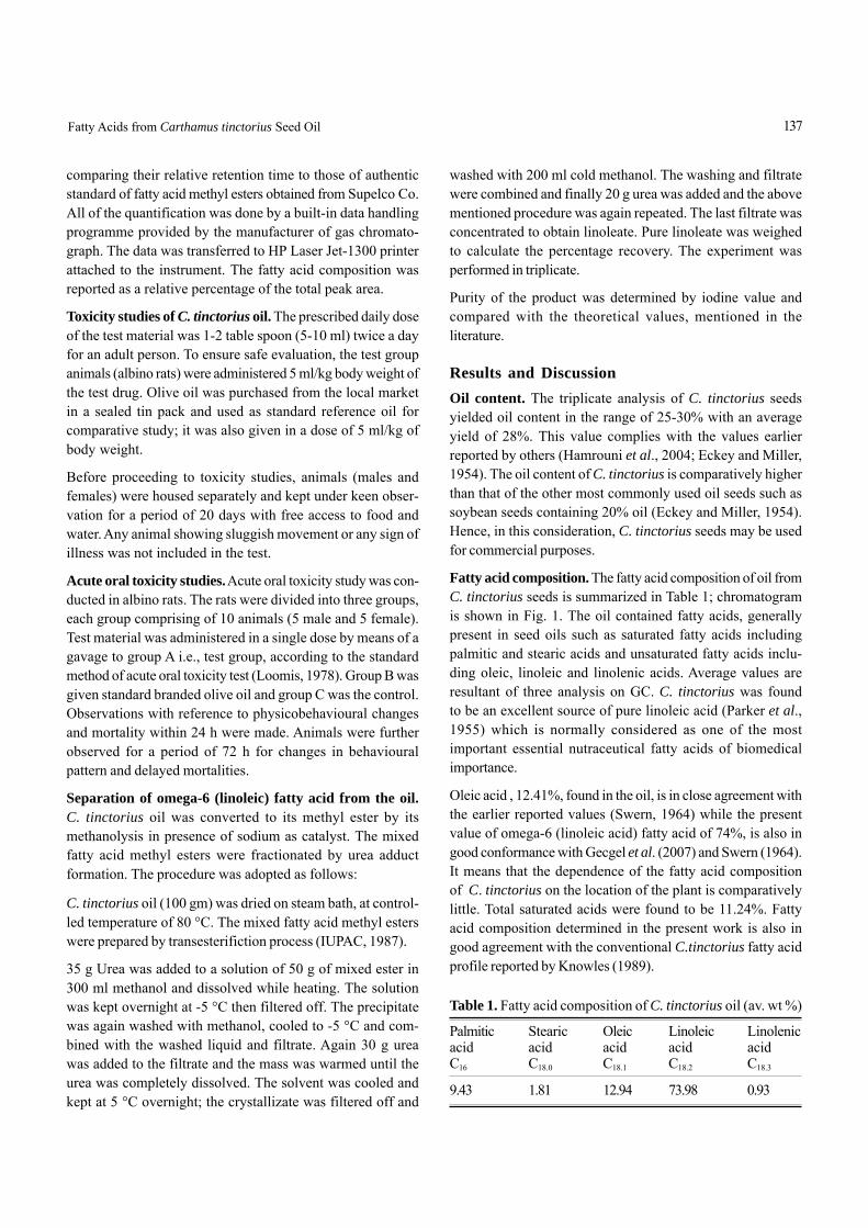
Fatty acid composition. The fatty acid composition of oil fromC. tinctorius seeds is summarized in Table 1; chromatogramis shown in Fig. 1. The oil contained fatty acids, generallypresent in seed oils such as saturated fatty acids includingpalmitic and stearic acids and unsaturated fatty acids inclu-ding oleic, linoleic and linolenic acids. Average values areresultant of three analysis on GC. C. tinctorius was foundto be an excellent source of pure linoleic acid (Parker et al.,1955) which is normally considered as one of the mostimportant essential nutraceutical fatty acids of biomedicalimportance.
Oleic acid , 12.41%, found in the oil, is in close agreement withthe earlier reported values (Swern, 1964) while the presentvalue of omega-6 (linoleic acid) fatty acid of 74%, is also ingood conformance with Gecgel et al. (2007) and Swern (1964).It means that the dependence of the fatty acid compositionof C. tinctorius on the location of the plant is comparativelylittle. Total saturated acids were found to be 11.24%. Fattyacid composition determined in the present work is also ingood agreement with the conventional C.tinctorius fatty acidprofile reported by Knowles (1989).
Table 1. Fatty acid composition of C. tinctorius oil (av. wt %)
Palmitic Stearic Oleic Linoleic Linolenicacid acid acid acid acidC16 C18.0 C18.1 C18.2 C18.3
9.43 1.81 12.94 73.98 0.93
137Fatty Acids from Carthamus tinctorius Seed Oil
Toxicity studies. Oral administration of safflower oil for42 days in a dose of 5.0 ml/kg body weight was not found toproduce any toxic effect in both male and female animals incomparison with the standard and the control group (Table 2).
Preparation of linoleic acid. The technique of separation ofpolyunsaturated fatty acid by urea adduct complexation wasfirst described by Bengen (1940). The main findings of Bengenwas that urea can be used to separate straight chain com-pounds from branched or cyclic compounds; later, numerousinvestigators confirmed this method. The new technique wasused for the preparation of methyl oleate (Swern and Parker,1952; Schlenk and Holman, 1950), for the preparation ofconcentrates of linoleic acid from safflower oil (Kim et al.,2003; Swern and Parker, 1953), for separation of docosahexa-enoic acid from algal oil via urea complexation (Senanayakand Shahidi, 2000), gamma linolenic acid concentrate fromborage oil (Spurvey and Shahidi, 2000) and preparation ofomega-3 PUFA concentrate from fish oil via urea complexation(Ratnayake et al., 1988).
The reviews of Schlenk (1953) gives detailed explanation ofthe application of this technique. In the present findingsabout 21.7% linoleic acid (omega-6) was separated at theend of experiments performed in triplicate. Low yield of theinclusion compound was also observed earlier by Schlenkand Holman (1950), suggesting that the more highly un-saturated fatty acids may be even more difficult to bind asinclusion compounds.
The purity of linoleate in the present study was found to be93.1%. The percentage yield, 21.76%, of the product is in goodagreement with the findings of Keppler et al. (1959) while thepurity shows some variation with the results reported by him.The urea complex of linoleic acid was analyzed by determina-tion of iodine value of the complex (Knight et al., 1952). Theresults are reported in Table 3. The average iodine value of160.1 shows a small variation from that reported by Keppleret al. (1959). It was been observed that the tendency of fattyacids and esters to combine with urea decreases with increa-sing unstauration.
Table 3. Percentage yield of linoleate (quantity and purity)
Iodine Purity of Yield ofvalue (%) linoleate (%)
160.8 93.5 22.8159.9 93.0 21.5159.6 92.8 21.0160.1* 93.1* 21.76*
* = average of triplicate analysis
138 Rubina Saleem et al.
95
90
85
80
75
70
65
60
55
50
45
40
35
30
25
20
15
10
5
0
13 14 15 16 17 18 19 20 21 22 23 24 25 26 27 28
Res
pons
e (m
V)
Time (min)
Fig. 1. Chromatogramme of C. tinctorius seed oil.
Table 2. Acute oral toxicity studies
Group Av. wt. of Dosage Resultsanimals (kg)
A 150-200 5 ml / kg - no mortalitybody wt. - all animals found(safflower normal in their activitiesoil) - there was no sign of
any untoward effects during said period of observation.
B 150-200 5 ml / kg same as abovebody wt.(olive oil)
C 150-200 control same as above(placebo)
It is thus concluded that omega-6 fatty acids, can well beextracted from the seeds of C. tinctorius and are suitable foruse in nutritional products after purification.
ReferencesBengen, F. 1940. The first description of the urea-fatty acid
complexes. German Patent No. OZ 12, 3438, 8th March,1940.
Eckey, E.W., Miller, L.C. 1954. Vegetable Fats and Oils, ACSMonograph Series, 836 p., Reinhold Publishing Corpora-tion, New York, USA.
Gecgel, U., Demirci, M., Esendal, E., Tasan, M. 2007. Fatty acidcomposition of oil from developing seeds of differentvarieties of safflower (Carthamus tinctorius). J. Am. OilChem. Soc. 84: 47-54.
George, W. 1892. A Dictionary of the Economic Products ofIndia, 6 vols., vol. II, pp. 183-184, S.W., Publishers, Culcutta,India.
Hamrouni, I., Touati, S., Dhifi, W., Chahed, T., Ayachi, S., Salah,H., Marzouk, B. 2004. Glycerolipid evolution duringsafflower seed formation and ripening. J. Food Lipid 11:297-311.
IUPAC, 1987. IUPAC Standard Methods for the Analysis ofOils, Fats and Derivatives, 347 p., 7th edition, BlackwellScientific Publication, London, UK.
Jafri, S.H.M. 1966. The Flora of Karachi (Coastal WestPakistan), University of Karachi, Karachi, Pakistan.
Keppler, J.G., Sparreboom, S., Stroink, J.B.A., Von Mikusch,J.D. 1959. A note on the preparation of pure oleic andlinoleic acid. J. Am. Oil Chem. Soc. 36: 308-309.
Kim,Y.J., Lee, K.W., Lee, S., Kim, H., Lee, J. 2003. The produc-tion of high purity conjugated linoleic acid using twostep urea inclusion crystallization and hydrophilicarginine-(CLA) complex. J. Food Sci. 68: 1948-1951.
Knight, H.B., Witnauer, L.P., Coleman, J.E., Noble (Jr.), W.R.,Swern, D. 1952. Dissociation temperatures of ureacomplexes of long chain fatty acid, esters and alcohols.Anal. Chem. 24: 1331-1334.
Knowles, P.F. 1989. Safflower. In: Oil Crops of the World,G. Robbelen, R. K. Downey and A. Ashri (eds.), pp. 363-
374, McGraw Hill, New York, USA.Loomis, T.A. 1978. Essentials of Toxicology, 245 p., 3rd edition,
Lea & Febiger, Philadelphia, USA.Oelke, E.A., Oplinger, E.S., Teynor, T.M., Putnam, D.H., Doll,
J.D., Kelling, K.A., Durgan, B.R., Noetzel, D.M. 1989,Safflower. In: Alternative Field Crops Manual, Univ.Wisconsin and Univ. Minnesota, USA.
Parker, W.E., Koos, R.E., Swern, D., 1955. Biochemical Pre-parations, vol. 4, Wiley, New York, USA.
Ratnayake, W.M.N., Olsson, B., Matthews, D., Ackman, R.G.1988. Preparation of omega 3 PUFA concentrate fromfish oil via urea complexation. Fat. Sci. Technol. 90: 381-386.
Schlenk, H., 1953.Urea inclusion compounds of fatty acids.In: Progress in the Chemistry of Fats and Other Lipids,vol. 11, Pergamon Press Ltd., London, UK.
Schlenk, H., Holman, R.T. 1950. Separation and stabilizationof fatty acids by urea complexes. J. Am. Chem. Soc. 72:5001-5004.
Senanayak, S.P.J., Shahidi, F. 2000. Concentration of docosa-hexaenoic acid from algal oil via urea complexationtechnique. J. Food Lipids 7: 51-61.
Spurvey, S.A., Shahidi, F. 2000. Concentration of gammalinolenic acid (GLA) from borage oil by urea complexa-tion: optimization of reaction conditions. J. Food Lipids7: 163-174.
Swern, D. 1964. Bailey’s Industrial Oil and Fats Products,212 p., 3rd edition., Wiley, London, UK.
Swern, D., Parker, W.E. 1953. Application of urea complexes inthe purification of fatty acids, esters and alcohols. III.Concentrates of natural linoleic and linolenic acid. J. Am.Oil Chem. Soc. 30: 5-7.
Swern, D., Parker,W.E. 1952. Application of urea complexes inthe purification of fatty acids, esters and alcohols. II. Oleicacid and methyl oleate from olive oil. J. Am. Oil Chem.Soc. 29: 614-615.
139Fatty Acids from Carthamus tinctorius Seed Oil
Major Ion Chemistry of Groundwaters From thePeshawar Intermontane Basin, NWFP, Pakistan
M. Asim YousafzaiDepartment of Geology, University of Peshawar, Peshawar, Pakistan
(received April 21, 2008; revised June 12, 2008; accepted June 15, 2008)
IntroductionInvestigation was carried out to determine the field character-istics and major ion chemistry of spring and well waters. Thestudy site covered the Peshawar intermontane basin (PIB)and its surroundings in the Himalayan foreland fold-and-thrustbelt of Pakistan (Fig. 1). The area, located between latitudes33.5°N-34.7°N and longitudes 71°E-73°E, is characterized bysteep topography and V-shaped fluvial valleys in the north,which are drained by the river Indus entering from IndianKashmir and the river Kabul entering from Afghanistan. Coldwinters and warm, dry summers characterize the climate of thestudy area. June through August are the hot months,during which the daily mean maximum air temperature is about40 °C. The mean annual potential evaporation ranges from85 cm in the northern part of the study area to 130 cm in thecentre of the basin. Snowfall occurs in the mountainousnorth during the cold months of December to February, whenmonthly mean minimum temperatures are several degrees Cel-sius below the freezing point.
Peshawar valley is home to two million plus inhabitants withever growing demand for water for drinking, industrial andirrigation purposes. Every water usage (human, industrial, ir-rigation etc.) requires a set of hydrochemical characteristics,suited for that particular consumption. The hydrogeo-logicaldata for the region is scattered and scanty (Shah and Tariq,2007; Tariq, 2001) and this research is the first attempt to fillthis gap. The broader study conducted for this research is thefirst of its kind in that it synthesizes field, laboratory and simu-lation data to bracket the hydrochemical speciation in the PIB.This paper presents the field and laboratory data for ground-water in the study area.
General geology. Information on subsurface geology of thestudy area has been derived from the lithological logs of bore-holes drilled by WAPDA, Pakistan, and made availablethrough various information releases. These data indicate thatthe quaternary sediments vary in thickness from few metersto more than 150 m. However, the total thickness of quater-nary sediments is not known because none of the boreholespenetrates the bedrock. The coarseness of these sedimentsincreases from north to south in the basin (Tariq, 2001). In thecentral part of the basin, the alluvial sediments consist of arelatively large proportion of fine-grained material, where thesandy silt is interbedded with discontinuous alluvial sand andthin gravel layers of various thickness. The main rock types inthis area are slates, phyllites, various types of schists, para-gneisses, sandstones, and quartzitic crystalline conglomerates,all of which are intruded by basic-to-acidic igneous rocks.
Hydrogeology. The study area can be divided into twohydrogeological provinces. The basin is bordered by moun-tains of the Lesser Himalayas on three sides while the south-eastern side is a fluvial valley carved at the confluence ofthe Indus and Kabul rivers. In the mountainous region, thewater table varies in depth in different intermontane valleys,suggesting hydraulic discontinuities. In fact, water tableelevation varies considerably within the area, ranging fromless than 100 m in the basin centre to more than 1600 m inthe mountainous north in relation to mean sea level (Fig. 2).Abundant springs (both normal and high temperature) arepresent in this part of the study area, and locally they consti-tute an important source of drinking water in addition todugwells. The outflow from these springs ranges from lessthan one l/sec to more than 2000 l/sec. The field investiga-tions indicate that most of the springs have good qualitywater, and according to local authorities, there are no water-
Abstract. Investigation of spring and well waters of the Peshawar Intermontane basin and its surroundings in theHimalayan belt of Pakistan revealed that their temperature, pH, total dissolved solids and electrical conductivity valuesfall within the permissible range of drinking and irrigation water except the waters in vicinity of exposed faults. Mineralsin the spring water are in the order of Ca > Mg > (Na + K) with bicarbonate as the dominant anion, whereas in the wellwater, the order is reversed, with sulphate as the dominant anion.
Keywords: Peshawar hydrochemistry, ground water, minerals, Pakistan
Pak. J. Sci. Ind. Res. 2008 51(3) 140-146
E-mail: [email protected]
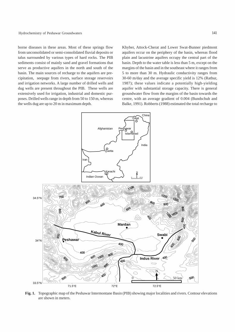
borne diseases in these areas. Most of these springs flowfrom unconsolidated or semi-consolidated fluvial deposits ortalus surrounded by various types of hard rocks. The PIBsediments consist of mainly sand and gravel formations thatserve as productive aquifers in the north and south of thebasin. The main sources of recharge to the aquifers are pre-cipitation, seepage from rivers, surface storage reservoirsand irrigation networks. A large number of drilled wells anddug wells are present throughout the PIB. These wells areextensively used for irrigation, industrial and domestic pur-poses. Drilled wells range in depth from 50 to 150 m, whereasthe wells dug are up to 20 m in maximum depth.
Khyber, Attock-Cherat and Lower Swat-Bunner piedmontaquifers occur on the periphery of the basin, whereas floodplain and lacustrine aquifers occupy the central part of thebasin. Depth to the water table is less than 5 m, except on themargins of the basin and in the southeast where it ranges from5 to more than 30 m. Hydraulic conductivity ranges from30-60 m/day and the average specific yield is 12% (Rathur,1987); these values indicate a potentially high-yieldingaquifer with substantial storage capacity. There is generalgroundwater flow from the margins of the basin towards thecentre, with an average gradient of 0.004 (Bundschuh andBalke, 1991). Robberts (1988) estimated the total recharge to
Peshawar
Islamabad
Gilgit
Quetta
Karachi
Indian Ocean
Afghanistan
India
Iran
0 400 km
N
China
71.5°E 72°E 72.5°E33.5°N
34°N
34.5°N
Peshawar
Mardan
SwabiKabul River
Indus River
Fig. 1. Topographic map of the Peshawar Intermontane Basin (PIB) showing major localities and rivers. Contour elevationsare shown in meters.
50 km0
N
141Hydrochemistry of Peshawar Groundwaters
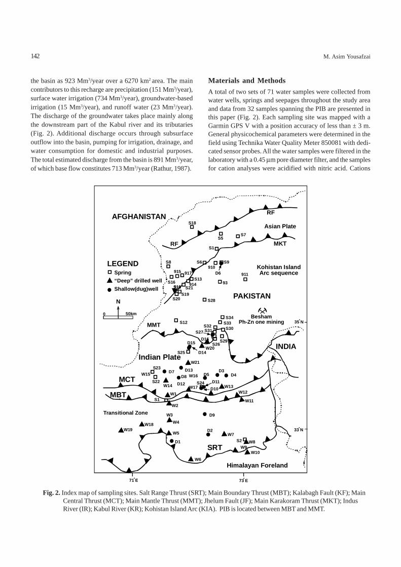
the basin as 923 Mm3/year over a 6270 km2 area. The maincontributors to this recharge are precipitation (151 Mm3/year),surface water irrigation (734 Mm3/year), groundwater-basedirrigation (15 Mm3/year), and runoff water (23 Mm3/year).The discharge of the groundwater takes place mainly alongthe downstream part of the Kabul river and its tributaries(Fig. 2). Additional discharge occurs through subsurfaceoutflow into the basin, pumping for irrigation, drainage, andwater consumption for domestic and industrial purposes.The total estimated discharge from the basin is 891 Mm3/year,of which base flow constitutes 713 Mm3/year (Rathur, 1987).
Materials and MethodsA total of two sets of 71 water samples were collected fromwater wells, springs and seepages throughout the study areaand data from 32 samples spanning the PIB are presented inthis paper (Fig. 2). Each sampling site was mapped with aGarmin GPS V with a position accuracy of less than ± 3 m.General physicochemical parameters were determined in thefield using Technika Water Quality Meter 850081 with dedi-cated sensor probes. All the water samples were filtered in thelaboratory with a 0.45 μm pore diameter filter, and the samplesfor cation analyses were acidified with nitric acid. Cations
142 M. Asim Yousafzai
Fig. 2. Index map of sampling sites. Salt Range Thrust (SRT); Main Boundary Thrust (MBT); Kalabagh Fault (KF); MainCentral Thrust (MCT); Main Mantle Thrust (MMT); Jhelum Fault (JF); Main Karakoram Thrust (MKT); IndusRiver (IR); Kabul River (KR); Kohistan Island Arc (KIA). PIB is located between MBT and MMT.
73 E71 E
AFGHANISTAN RF
MKTRF
MMT
Kohistan IslandArc sequence
PAKISTAN
BeshamPh-Zn one mining
INDIAIndian Plate
MCT
MBT
Transitional Zone
SRT
Himalayan Foreland
LEGEND
Asian PlateS18
S5S7
S6S8 S9910
93
911915 917
914916 S21
S13
S1
S28S20S19
S18S16
S12S32
S34
S30S33
S31S27
S29S26
D16
W20D14
D15
S25
W21S23
D7W15 D8D13
D5W16D3
D4D11D12W14
S22W17 W13
W12
W11W1
S1W2
W3W4
D9
D10S24
W5
D1
D2W7
W8S2W9
W10W6
W19W18
35 N
33 N
Spring“Deep” drilled wellShallow(dug)well
N
0 50km
D6
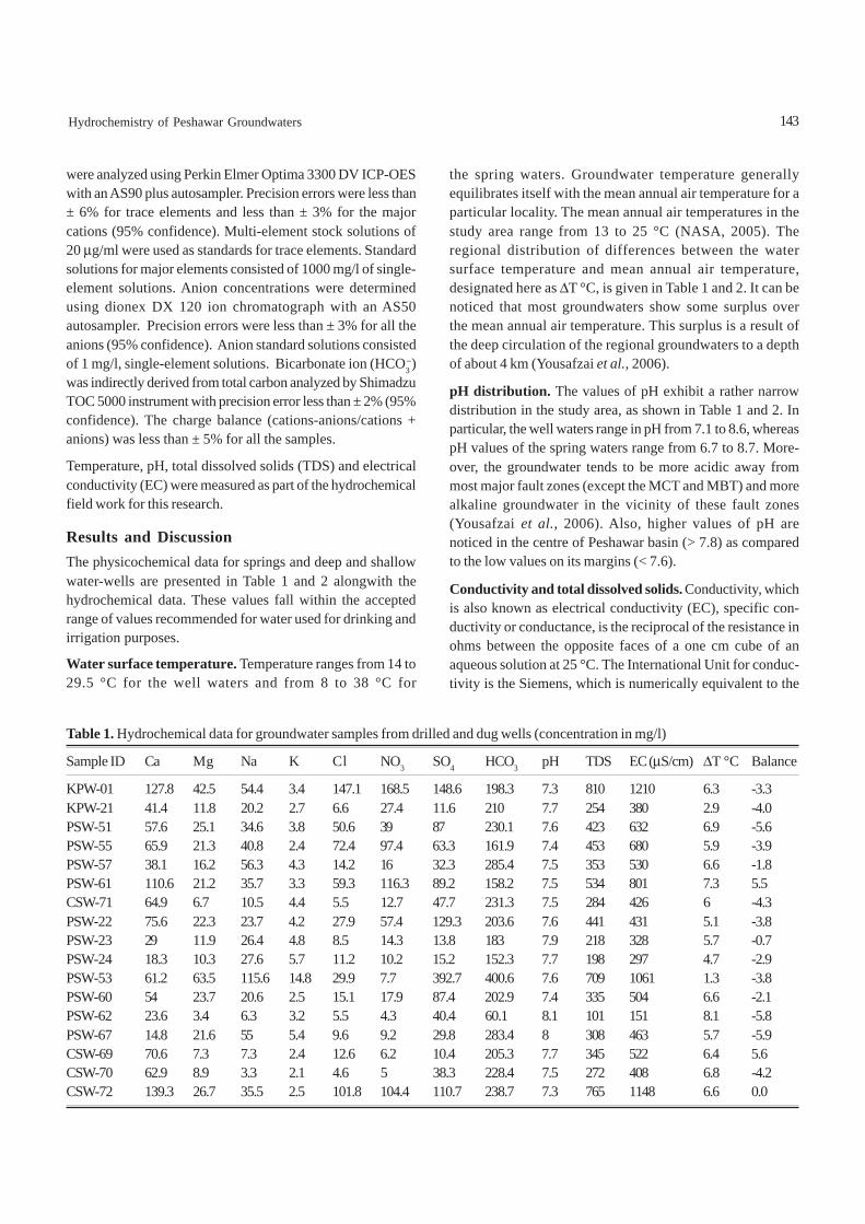
143Hydrochemistry of Peshawar Groundwaters
were analyzed using Perkin Elmer Optima 3300 DV ICP-OESwith an AS90 plus autosampler. Precision errors were less than± 6% for trace elements and less than ± 3% for the majorcations (95% confidence). Multi-element stock solutions of20 μg/ml were used as standards for trace elements. Standardsolutions for major elements consisted of 1000 mg/l of single-element solutions. Anion concentrations were determinedusing dionex DX 120 ion chromatograph with an AS50autosampler. Precision errors were less than ± 3% for all theanions (95% confidence). Anion standard solutions consistedof 1 mg/l, single-element solutions. Bicarbonate ion (HCO3
−)was indirectly derived from total carbon analyzed by ShimadzuTOC 5000 instrument with precision error less than ± 2% (95%confidence). The charge balance (cations-anions/cations +anions) was less than ± 5% for all the samples.
Temperature, pH, total dissolved solids (TDS) and electricalconductivity (EC) were measured as part of the hydrochemicalfield work for this research.
Results and DiscussionThe physicochemical data for springs and deep and shallowwater-wells are presented in Table 1 and 2 alongwith thehydrochemical data. These values fall within the acceptedrange of values recommended for water used for drinking andirrigation purposes.
Water surface temperature. Temperature ranges from 14 to29.5 °C for the well waters and from 8 to 38 °C for
the spring waters. Groundwater temperature generallyequilibrates itself with the mean annual air temperature for aparticular locality. The mean annual air temperatures in thestudy area range from 13 to 25 °C (NASA, 2005). Theregional distribution of differences between the watersurface temperature and mean annual air temperature,designated here as ΔT °C, is given in Table 1 and 2. It can benoticed that most groundwaters show some surplus overthe mean annual air temperature. This surplus is a result ofthe deep circulation of the regional groundwaters to a depthof about 4 km (Yousafzai et al., 2006).
pH distribution. The values of pH exhibit a rather narrowdistribution in the study area, as shown in Table 1 and 2. Inparticular, the well waters range in pH from 7.1 to 8.6, whereaspH values of the spring waters range from 6.7 to 8.7. More-over, the groundwater tends to be more acidic away frommost major fault zones (except the MCT and MBT) and morealkaline groundwater in the vicinity of these fault zones(Yousafzai et al., 2006). Also, higher values of pH arenoticed in the centre of Peshawar basin (> 7.8) as comparedto the low values on its margins (< 7.6).
Conductivity and total dissolved solids. Conductivity, whichis also known as electrical conductivity (EC), specific con-ductivity or conductance, is the reciprocal of the resistance inohms between the opposite faces of a one cm cube of anaqueous solution at 25 °C. The International Unit for conduc-tivity is the Siemens, which is numerically equivalent to the
Table 1. Hydrochemical data for groundwater samples from drilled and dug wells (concentration in mg/l) Sample ID Ca Mg Na K Cl NO3 SO4 HCO3 pH TDS EC (μS/cm) ΔT °C Balance
KPW-01 127.8 42.5 54.4 3.4 147.1 168.5 148.6 198.3 7.3 810 1210 6.3 -3.3KPW-21 41.4 11.8 20.2 2.7 6.6 27.4 11.6 210 7.7 254 380 2.9 -4.0PSW-51 57.6 25.1 34.6 3.8 50.6 39 87 230.1 7.6 423 632 6.9 -5.6PSW-55 65.9 21.3 40.8 2.4 72.4 97.4 63.3 161.9 7.4 453 680 5.9 -3.9PSW-57 38.1 16.2 56.3 4.3 14.2 16 32.3 285.4 7.5 353 530 6.6 -1.8PSW-61 110.6 21.2 35.7 3.3 59.3 116.3 89.2 158.2 7.5 534 801 7.3 5.5CSW-71 64.9 6.7 10.5 4.4 5.5 12.7 47.7 231.3 7.5 284 426 6 -4.3PSW-22 75.6 22.3 23.7 4.2 27.9 57.4 129.3 203.6 7.6 441 431 5.1 -3.8PSW-23 29 11.9 26.4 4.8 8.5 14.3 13.8 183 7.9 218 328 5.7 -0.7PSW-24 18.3 10.3 27.6 5.7 11.2 10.2 15.2 152.3 7.7 198 297 4.7 -2.9PSW-53 61.2 63.5 115.6 14.8 29.9 7.7 392.7 400.6 7.6 709 1061 1.3 -3.8PSW-60 54 23.7 20.6 2.5 15.1 17.9 87.4 202.9 7.4 335 504 6.6 -2.1PSW-62 23.6 3.4 6.3 3.2 5.5 4.3 40.4 60.1 8.1 101 151 8.1 -5.8PSW-67 14.8 21.6 55 5.4 9.6 9.2 29.8 283.4 8 308 463 5.7 -5.9CSW-69 70.6 7.3 7.3 2.4 12.6 6.2 10.4 205.3 7.7 345 522 6.4 5.6CSW-70 62.9 8.9 3.3 2.1 4.6 5 38.3 228.4 7.5 272 408 6.8 -4.2CSW-72 139.3 26.7 35.5 2.5 101.8 104.4 110.7 238.7 7.3 765 1148 6.6 0.0
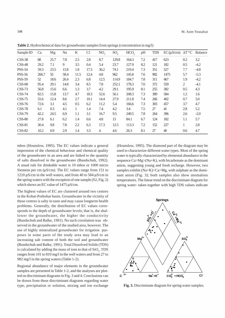
mhos (Hounslow, 1995). The EC values indicate a generalimpression of the chemical behaviour and chemical qualityof the groundwater in an area and are linked to the quantityof salts dissolved in the groundwater (Bundschuh, 1992).A usual rule for drinkable water is 10 mhos or 1000 microSiemens per cm (μS/cm). The EC values range from 151 to1210 μS/cm in the well waters, and from 40 to 584 μS/cm inthe spring waters with the exception of one sample (S2, Fig. 2)which shows an EC value of 1475 μS/cm.
The highest values of EC are clustered around two centresin the Kohat-Pothohar basin. Groundwater in the vicinity ofthese centres is salty in taste and may cause longterm healthproblems. Generally, the distribution of EC values corre-sponds to the depth of groundwater levels; that is, the shal-lower the groundwater, the higher the conductivity(Bundschuh and Balke, 1991). No such correlation was ob-served in the groundwater of the studied area, however. Theuse of highly mineralized groundwater for irrigation pur-poses in some parts of the study area may lead to anincreasing salt content of both the soil and groundwater(Bundschuh and Balke, 1991). Total Dissolved Solids (TDS)is calculated by adding the mass of ions to that of SiO2. TDSranges from 101 to 810 mg/l in the well waters and from 27 to982 mg/l in the spring waters (Table 1-2).
Regional abundance of major elements in the groundwatersamples are presented in Table 1-2, and the analyses are plot-ted on discriminant diagrams in Fig. 3 and 4. Conclusions canbe drawn from these discriminant diagrams regarding watertype, precipitation or solution, mixing and ion exchange
(Hounslow, 1995). The diamond part of the diagram may beused to characterize different water types. Most of the springwater is typically characterized by elemental abundance in thesequence Ca>Mg>(Na+K), with bicarbonate as the dominantanion, suggesting young and fresh recharge. However, twosamples exhibit (Na+K)>Ca>Mg, with sulphate as the domi-nant anion (Fig. 3); both samples also show anomaloustemperatures. The linear trend on the discriminate diagram forspring water- taken together with high TDS values indicate
Table 2. Hydrochemical data for groundwater samples from springs (concentration in mg/l)
Sample ID Ca Mg Na K Cl NO3 SO4 HCO3 pH TDS EC (μS/cm) ΔT °C Balance
CSS-38 68 25.7 7.8 2.5 2.8 8.7 129.8 164.3 7.2 417 623 0.2 3.2CSS-48 29.2 7.1 9 3.5 0.4 5.4 23.7 127.9 8.2 121 182 0.5 -4.2PSS-54 59.3 23.5 15.8 1.8 17.3 36.2 74.1 219.4 7.3 351 527 7.7 -4.8PSS-56 266.7 55 58.4 11.5 12.4 4.8 962 145.8 7.6 982 1475 5.7 -5.3PSS-59 52 18.6 26.4 2.3 6.8 12.5 114.9 184.7 7.8 311 467 1.9 -4.2CSS-68 95.4 29.1 14.8 3.4 8.5 7.8 252.1 178.3 7.6 372 559 2 -4.1CSS-73 56.8 15.6 6.6 1.3 3.7 4.2 29.1 195.9 8.1 255 382 0.5 4.3CSS-74 82.5 15.8 13.7 4.7 18.3 52.6 56.1 208.3 7.3 389 584 1.2 1.6CSS-75 55.6 12.4 8.6 2.7 10.1 14.4 27.9 211.8 7.4 266 402 0.7 5.0CSS-76 72.6 3.1 4.5 0.5 6.2 11.2 5.4 166.6 7.3 303 457 3.7 4.7CSS-78 6.1 0.3 4.1 1 1.4 7.4 4.2 3.4 7.5 27 41 2.8 5.2CSS-79 42.2 24.5 6.9 1.1 3.1 16.7 9.5 249.5 7.8 264 396 2.6 -2.0CSS-80 27.8 6.1 6.2 1.4 6.6 4.8 13 84.1 6.7 124 182 1.1 5.7CSS-81 30.4 9.8 7.8 2.2 6.3 17.3 12.5 113.3 7.2 152 227 1 2.8CSS-82 10.2 0.9 2.9 1.4 3.3 4 4.6 26.3 8.1 27 40 0.6 4.7
144
Fig. 3. Discriminate diagram for spring water samples.
Springs
C 2+
Mg2+
CO32- +
SO42-
Cl-Na++K+ HCO3-
M. Asim Yousafzai
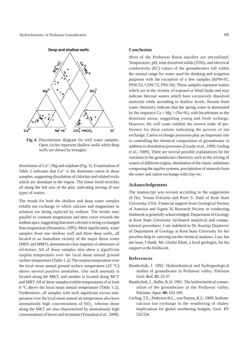
dissolution of Ca2+, Mg and sulphate (Fig. 3). Examination ofTable 2 indicates that Ca2+ is the dominant cation in thesesamples, suggesting dissolution of chlorites and related rockswhich are abundant in the region. The linear trend stretchesall along the left axis of the plot, indicating mixing of twotypes of waters.
The trends for both the shallow and deep water samplesexhibit ion exchange in which calcium and magnesium insolution are being replaced by sodium. The trends startparallel to constant magnesium and then curve towards thesodium apex, suggesting that more calcium is being exchangedthan magnesium (Hounslow, 1995). Most significantly, watersamples from one shallow well and three deep wells, alllocated in an immediate vicinity of the major thrust zones(MBT and MMT), demonstrate clear imprints of admixture ofoil-brines. All of these samples also show a significantsurplus temperature over the local mean annual groundsurface temperature (Table 1-2). The surplus temperature overthe local mean annual ground surface temperature (ΔT °C)shows several positive anomalies. One such anomaly islocated along the MKT, and another is located along MCTand MBT. All of these samples exhibit temperatures of at least6 °C above the local mean annual temperature (Table 1-2).Furthermore, all samples with such significant excess tem-perature over the local mean annual air temperature also haveanomalously high concentrations of SiO2, whereas thosealong the MKT are also characterized by anomalously highconcentrations of boron and strontium (Yousafzai el al., 2008).
ConclusionMost of the Peshawar Basin aquifers are unconfined.Temperature, pH, total dissolved solids (TDS), and electricalconductivity (EC) values of the groundwaters fall withinthe normal range for water used for drinking and irrigationpurposes with the exception of a few samples (KPW-01,PSW-53, CSW-72, PSS-56). These samples represent waterswhich are in the vicinity of exposed or blind faults and mayindicate thermal waters which have excessively dissolvedmaterials while ascending to shallow levels. Results fromwater chemistry indicate that the spring water is dominatedby the sequence Ca > Mg > (Na+K), with bicarbonate as thedominant anion, suggesting young and fresh recharge.However, the well water exhibits the reverse order of pre-ference for these cations indicating the process of ionexchange. Cation exchange processes play an important rolein controlling the chemical composition of groundwater inaddition to dissolution processes (Grasby et al., 1999; Cerlinget al., 1989). There are several possible explanations for thevariation in the groundwater chemistry such as the mixing ofwaters of different origins, dissolution of the clastic sedimentscomposing the aquifer systems, precipitation of minerals fromthe water and cation exchange with clay etc.
AcknowledgementThe manuscript was revised according to the suggestionsof Drs. Yoram Eckstein and Peter S. Dahl of Kent StateUniversity, USA. Financial support from Geological Societyof America and Sigma Xi Research Society in conductingfieldwork is gratefully acknowledged. Department of Geologyat Kent State University facilitated analytical and compu-tational procedures. I am indebted to Dr. Ksenija Dejanovicof Department of Geology at Kent State University for herpriceless help in carrying out the chemical analyses. Last, butnot least, I thank Mr. Ghafar Khan, a local geologist, for hissupport in the fieldwork.
ReferencesBundschuh, J. 1992. Hydrochemical and hydrogeological
studies of groundwater in Peshawar valley. PakistanGeol. Bull. 25: 23-37.
Bundschuh, J., Balke, K.D. 1991. The hydrochemical compo-sition of the groundwater in the Peshawar valley,Pakistan. Aqua. 40: 163-169.
Cerling, T.E., Pederson B.L., von Damm, K.L. 1989. Sodium-calcium ion exchange in the weathering of shales:Implications for global weathering budgets. Geol. 17:522-554.
145Hydrochemistry of Peshawar Groundwaters
Fig. 4. Discriminate diagram for well water samples.Open circles represent shallow wells while deepwells are shown by triangles.
Deep and shallow wells
Ca2+
Mg2+
Na+ +K+ CO32-+HCO3
-
SO42-
Cl-
Grasby, S.E., Hutcheon, I., McFarland, L. 1999. Surface-water-groundwater interaction and influence of ionexchange reactions on river chemistry. Geol. 27: 223-226.
Hounslow, A.W. 1995. Water Quality Data: Analysis andInterpretation, 397 pp, CRC Lewis Publishers, New York,USA.
NASA, GISS, 2005. National Aeronautics and Space Adminis-tration-Goddard Institute for Space Studies, New York.Online climatic data, http://www.giss.nasa.gov/.
Rathur, A.Q. 1987. Groundwater management to eradicate wa-ter logging and salinity in the Upper Indus Basin, Punjab,Pakistan. In: Proceedings of the International Confer-ence on Groundwater and Environment, S. Awadalla andI. M. Noor (eds.), pp. G96-G107, Kebang-saan University,Selangor.
146
Robberts, J.H. 1988. Groundwater in the Peshawar vale,Peshawar district, Mardan district and Malakand agency.NWFP Technical Report V-2, WAPDA HydrogeologyDirectorate, Peshawar and TNO Institute of AppliedGeoscience, Delft, The Netherlands.
Shah, M.T., Tariq, S. 2007. Environmental geochemistry of thesoils of Peshawar basin, NWFP, Pakistan. J. Chem. Soc.Pak. 29: 438-445.
Tariq, S. 2001. Environmental Geochemistry of Surface andSubsurface Water and Soil in Peshawar Basin, NWFP,Pakistan. Ph.D. Thesis, unpublished, 305 pp. Universityof Peshawar, Pakistan.
Yousafzai, M.A., Eckstein, Y., Dahl, P.S. 2006. Hydrochemicalsignatures of deep groundwater circulation in a part ofthe Himalayan foreland basin. Hydrogeol. J. (In press).
M. Asim Yousafzai
Determination of Positional Isomers of Monoenoic Fatty Acids SeparatedFrom Seed Oils of Nicotiana tabacum L. and Nicotiana rustica
Amran Waheed*, Muhammad Saleem and Shahid MahmudPCSlR Laboratories Complex, Lahore-54600, Pakistan
(received January 1, 2008; revised June 6, 2008; accepted June 9, 2008)
Nicotiana tabacum L. and Nicotiana rustica, yield tobaccoand are commonly cultivated as crops (Kirtikar and Basu,1984). The monoenoic fatty acid i.e. octadecenoic acidseparated from the oils of both the species is oxidized todetermine the positional isomers. The literature reveals thatoctadecenoic acid of N. tabacum consists of two positionalisomers (Frega et al., 1991) cis-9-octadecenoic acid andcis-11- octadecenoic acid, whereas no work has been reportedon the positional isomers of N. rustica. The present workreports the positional isomers of N. tabacum and N. rustica.
Liberation, methylation and purification of fatty acids fromthe plants were carried out according to the methods describedby Waheed et al. (2001), Raie et al. (1989) and Devine andWilliams (1961). The purified methyl esters were separatedinto four fractions (I to IV) with AgNO3 impregnatede thinlayer chromatography.
Separation and oxidation of octadecenoic acid were carriedout by the methods described by Chaudri et al. (2001),Hamilton and Raie (1972) and Von Rudloff (1956).
The mixture of mono- and di-carboxylic acids produced inaqueous media was analyzed by gas chromatography usingporapak “Q” column and short chain mono-carboxylic acids(C4-C9) were identified and later, the remaining acids (aboveC10) were analyzed as described by Raie et al. (1989). Themethyl esters of mono- and di-carboxylic acid were separatedby preparative silica gel plates using mixture of hexane:diethyl ether (1 : 1 v/v) as a developing solvent.
The purified methyl esters of mono-carboxylic acid (C9 andC11) were analyzed and identified by the use of 5% SE-30 packedcolumn at 110 °C to 150 °C and those of di-carboxylic acids
*Author for corresponding; E-mail: [email protected]
were analyzed at 150 °C to 200 °C. Their identification wasconfirmed with the help of standards. The Rf values of theseparated four fractions of fatty acids of the two species arereported in Table 1 and their composition in Table 2.
The percentages of saturated fatty acids and monounsaturatedfatty acids in N. tabacum and N. rustica were 12.7% and 12.3%,respectively, whereas, the percentages of diunsaturated andtriunsaturated fatty acids were 73.9% and 71.6% and 1.2%and 0.9%, respectively, in both the species (Table 2).
Table 3 and 4 show that the two positional isomers of themonoenoic fatty acid (octadecenoic acid) are present in theseed oil of N. tabacum i.e., cis-9-octadecenoic acid (11.1%)and cis-11-ctadecenoic acid (1.1%); similarly two positionalisomers of monoenoic fatty acid (octadecenoic acid) i.e.cis-9-octadecenoic acid (14.4%) and cis-11-octadecenoic acid(0.8%) are present in N. rustica.
The oxidation of monounsaturated fatty acid (octadecenoicacid) was carried out to cleave double bonds and eventuallyto produce monobasic and dibasic acids, which helped todetermine the positional isomers. Since complications ariseby the production of a number of monobasic and dibasic
Pak. J. Sci. Ind. Res. 2008 51(3) 147-149
Abstract. The octadecenoic acid separated from the seed oils of Nicotiana tabacum L. and Nicotiana rustica by theapplication of argentation thin layer chromatography was oxidized by modified Von Rudloff’s technique. The liberatedmonofunctional and difunctional carboxylic acids were separated and identified by the application of thin layer chromato-graphy and gas chromatography. The positional isomers determined in both species were cis-9-octadecenoic acid andcis-11-octadecenoic acid.
Keywords: isomers, monoenoic fatty acids, Nicotiana tabacum, Nicotiana rustica, seed oils
Short Communication
Table 1. Rf values of different fatty acid bands (as methylesters) separated by the AgNO3-TLC
Band no. Bands Fatty acid Rf
I saturated acids C12:0, C14:0, C16:0 0.65C18:0, C20:0
II monounsaturated acid C18:1 0.51III diunsaturated acids C18:2 0.30IV triunsaturated acids C18:3 0.18
147
acids after the oxidation of polyunsaturated fatty acids,therefore, the oxidation technique was applied exclusivelyfor monoenoic fatty acid although the diunsaturated fattyacids are present in large amounts as compared to monosatu-rated fatty acid in both the species of Nicotiana (Table 2).
A number of oxidizing agents (Bailey, 1958; Rigby, 1956;Swern, 1948; Milas, 1937) have been used for the cleavage ofdouble bonds but potassium permanganate and sodiummetaperiodate had been claimed to be the best oxidizingagents and the method was known as modified Von Rudloff’soxidation (Hamilton and Raie, 1972). The oxidation of unsatu-rated fatty acids by permanganate did not normally involveany change of configuration and the procedure had beenapplied to many other seed oils (Galun et al., 1984; Hammondet al., 1972; Wilson et al., 1962).
Since the major problem was the analysis of the water-solubleshort chain acids produced as a result of the oxidation ofmonoenoic fatty acid, therefore, short chain acids in aqueousmedia were analysed directly on a column of Pora-Pak Q. Theshort chain mono-carboxylic acids and di-carboxylic acidsproduced by the oxidation of monoenoic fatty acidseparated from the oil of N. tabacum, showed that there aretwo positional isomers i.e. cis-9-octadecenoic acid andcis-11-ctadecenoic acid showing double bond at Δ9:10 andΔ11:12, respectively (Table 3). The literature (Frega et al., 1991)revealed that there were only two positional isomers amongthe monounsaturated fatty acids i.e. cis-9- octadecenoic acidcommonly known as oleic acid (11.1%) and cis-11-octadece-noic acid usually known as vaceenic acid (0.7%). Similarly thepositional isomers of N. rustica are cis-9-octadecenoic acid(14.4%) and cis-11-ctadecenoic acid (0.8%) having the doublebond at Δ9:10 and Δ11:12 (Table 4). These studies reflect thatpositional isomers have a definite biosynthetic relationship.
ReferencesBailey, P.S. 1958. The reaction of ozone with organic com-
pounds. Chem. Rev. 58: 925-1010.Chaudri, T.A., Ahmad, I., Muhammad, D., Ahmad, M. 2001.
Positional isomers of octadecenoic acid separated fromoils of three carum species of Umbelliferae family. Pak.J. Sci. Ind. Res. 44: 75-78.
Devine, J., Williams, P.N. (eds.) 1961. The Chemistry andTechnology of Edible Oils and Fats, 134 p., 1st edition,Pergamon Press, Oxford, UK.
Frega, N., Bocci, F., Conte, L.S., Testa, F. 1991.Chemicalcomposition of Tobacco seeds (Nicotiana tabacum L.).J. Am. Oil Chem. Soc. 68: 29-33.
Galun, A., Grinberg, S., Kampf, A., Shaubi, E. 1984. Oxidationand halogenation of Jojoba wax. J. Am. Oil Chem. Soc.61: 1088-1089.
Hamilton, R.J., Raie, M.Y. 1972. Oxidation of fats: a study usingGLC. J. Chem. Educ. 49: 507-508.
Hammond, E.G., Fehr W.R., Snyder, H.E. 1972. Improvingsoybean quality by plant breeding. J. Am. Oil Chem. Soc.49: 33-35.
Kirtikar, K.R., Basu, B.D. 1984. Indian Medicinal Plants,vol. 2, pp. 936-939, 2nd edition, Bishen Singh andMahendra Pal Singh, Dehra Dun, India.
Milas, N.A. 1937. The hydroxylation of unsaturated substanceIII. The use of vanadium pentoxide and chromium trioxideas catalyst of hydroxylation. J. Am. Chem. Soc. 59: 2342-2344.
Raie, M.Y., Ahmad, I. Javed, M.A., Waheed, I. 1989. Lipidclasses of Apium graveolens oil of the family Umbelliferae.Proc. Pak. Acd. Sci. 26: 199-206.
148
Table 4. Positional isomers in octadecenoic acid of the oil ofN. rustica
Chain length Chain length Position of Percentage Isomersof monobasic of dibasic double bondacid acid
C9 C9 Δ9 14.4 cis-9-octa-decenoic
C7 C11 Δ11 0.8 cis-11-octa-decenoic
Table 2. Percentage of different fatty acids (as methyl esters)separated on the AgNO3-TLC
Band no. Fatty acids N. tabacum N. rustica(%) (%)
I saturated 12.7 12.3II monounsaturated 12.2 15.2III diunsaturated 73.9 71.6IV triunsaturated 1.2 0.9
Table 3. Positional isomers of octadecenoic acid of the oil ofN. tabacumChain Chain Position Percentage Isomers Referredlength of length of of double work*monobasic dibasic bond percentageacid acid
C9 C9 Δ 9 11.1 cis-9-octa- 10.6decenoic
C7 C11 Δ11 1.1 cis-11-octa- 0.6decenoic
* Frega et al., 1991
Amran Waheed et al.

Rigby, W. 1956. Hydroxylation with potassium permanganate.J. Chem. Soc. 2452-2454.
Swern, D. 1948. Chemistry of epoxy compounds. VII. stereochemical relationship between the 9, I0-epoxy-,chlorohydroxy- and dihydroxy Stearic acid. J. Am. Oil.Chem. Soc. 70: 1235-39.
Von Rudloff, E. 1956. Periodate permanganate oxidation. IV.determination of the position of double bonds in unsa-
149
turated fatty acids and esters. J. Am. Oil Chem. Soc. 33:126-128.
Waheed, A., Mahmud, S., Javed, M.A., Saleem, M. 2001.Studies on the lipid classes of Nicotiana tabacum L.seed oil. Nat. Prod. Sci. 7: 110-113.
Wilson, T.L., Smith, C.R., Wolff, T.A. 1962. Lunaria seed oil_arich source of C24 fatty acid. J. Am. Oil Chem. Soc. 39:104-105.
Short Communication: Isomers of Monoenoic Fatty Acids
Stimulatory Effect of Medium Ingredients on Alkaline ProteaseProduction by Bacillus licheniformis N-2 and Compatibility
Studies With Commercial DetergentsMuhammad Nadeema*, Javed Iqbal Qazib, Shahjahan Baiga and Quratulain Syeda
aFood and Biotechnology Research Center, PCSIR Labs. Complex, Lahore-54600, PakistanbDepartment of Zoology, University of the Punjab, New Campus, Lahore, Pakistan
(received April 11, 2008; revised June 3, 2008; accepted June 12, 2008)
*Author for correspondence; E-mail: [email protected]
Abstract. Suitable concentration of ingredients of the growth medium played a vital role in production of alkalineprotease by Bacillus licheniformis. Maximum enzyme activity (875.05 PU/ml) was achieved when the bacterium wasgrown in the medium containing glucose (1%), soybean meal (1%), K2HPO4 (0.5%), MgSO4.7H2O (0.05%), NaCl(0.05%), CaCl2.2H2O (0.05%) at 37 °C on 24 h incubation period with agitation of 140 rpm in shake flask cultures.More than 1% glucose decreased the enzyme production. The protease had excellent stability with wide range ofcommercial detergents such as Ariel, Bonus, Bright Total, Surf Excel, Wheel and non-branded detergents, recommend-ing its use as an effective additive in detergent formulation.
Keywords: medium ingredients, detergent compatibility, B. licheniformis N-2, alkaline protease
IntroductionProteases are one of the most important groups of industrialenzymes used in pharmaceutical industry and in food indus-try for peptide synthesis, in leather industry for de-hairingand in detergent industry as an additive of detergent formu-lation (Joo and Chang, 2005; Pastor et al., 2001). Alkalineproteases are known to constitute 60-65% of the globalindustrial market among various types of proteases (Banerjeeet al., 1999). Alkaline proteases are produced by a wide rangeof microorganisms including bacteria, mould and yeast.Currently, a large portion of commercially available proteasesis derived from Bacillus strains because of their high pHand temperature stability (Gupta et al., 2002; Joo et al.,2002).
The fermentation medium form the environment in which themicroorganisms live, reproduce and carry out their specificmetabolic reactions to produce useful products. Two distinctbiological requirements are considered in most of the indus-trial fermentation processes for medium design, where theproduct is something other than the cell mass itself. First, thenutrient has to be supplied to establish the growth of themicroorganism. Second, proper nutritional conditions haveto be provided to maximize the product formation. It is alsowell established that extracellular protease production inmicroorganisms is greatly influenced by media components,especially carbon and nitrogen sources, metal ions and physi-
cal factors such as pH, temperature, dissolved oxygen andincubation time (Nadeem, et al. 2006; Oberoi et al., 2001;Kuar et al., 2001; Razak, et al. 1994; Moon and Parulekar,1993). The cost of the growth medium is another significantparameter for making the production process industriallyviable. Approximately 30-40% of the production cost of theindustrial enzyme is estimated to be accounted for by the costof the growth medium (Gessesse, 1997). Therefore, selectionof the right medium ingredients and their concentrationsoptimization have become the need of today for high yield ofdesirable enzymes by fermentation.
Considering these facts, the effects of different concentra-tions of carbon and nitrogen sources as well as metal ionsconcentrations were studied to maximize the yield ofalkaline protease by locally isolated Bacillus licheniformisN-2. Compatibility studies of alkaline protease with diffe-rent detergents were also conducted to observe its commer-cial exploitation as an additive in detergent formulation forlaundry industry.
Materials and MethodsMicroorganism and culture maintenance. A proteolyticstrain identified as Bacillus licheniformis N-2 was isolatedfrom decaying organic soil sample (Nadeem et al., 2007) andthe culture was grown on nutrient agar slants at 37 °C for 24 hand preserved at 4 °C for one month. The preserved culturewas revived on fresh nutrient agar slants after every one monthfor subsequent experiments.
Pak. J. Sci. Ind. Res. 2008 51(3) 150-157
150
Biological Sciences
Inoculum preparation. The inoculum was prepared by trans-ferring a loopful of 24 h old culture of B. licheniformis N-2into 50 ml of inoculum medium consisting of glucose (0.5%),soybean meal (1%), K2HPO4 (0.3%), MgSO4.7H2O (0.05%),NaCl (0.05%), and CaCl2.2H2O (0.05%). The inoculatedmedium was incubated in water bath shaker (Eyela, Japan)for 24 h at 37 oC temperature and 140 rpm agitation speed forthe propagation of bacteria up to 108-10 cells/ml.
Fermentation process. The initial growth medium composedof glucose (1%), soybean meal (1%), K2HPO4 (0.3%), MgSO4.7H2O (0.05%), NaCl (0.05%) and CaCl2. 2H2O (0.05%). Itinoculated with 1% (v/v) of 24 h old inoculum broth. The pHof the medium was adjusted at 10 with 1N HCl/NaOHbefore sterilization at 121°C for 15 min. The inoculatedmedium was incubated in water bath shaker (Eyela, Japan)for 24 h at 37 °C and 140 rpm. Thereafter, the fermented brothwas centrifuged at 9000 x g for 10 min at 4 °C to get clearsupernatant enzyme solution.
Optimization of concentrations of medium ingredients.Different concentration levels of carbon and nitrogen sourcesas well as inorganic elements were optimized to make theprocess cost effective for alkaline protease production byB. licheniformis N-2.
Effect of glucose concentrations. Effect of various glucoseconcentrations (0.2, 0.4, 0.6, 0.8, 1.0, 1.25 and 1.50%) werestudied to find the suitable glucose concentration for themaximum yield of alkaline protease by B. licheniformis N-2.
Effect of defatted soybean meal concentrations. Effect ofdifferent concentrations of defatted soy bean meal viz. 0.25,0.50, 0.75, 1.0, 1.25, 1.5, 1.75 and 2.0% on alkaline proteaseproduction was determined by incubating the growth mediumat 37 °C for 24 h in water bath shaker (Eyela, Japan) withagitation speed of 140 rpm.
Effect of metal ions on alkaline protease production.Effects of various metal ions in the form of K2HPO4, MgSO4.7H2O, NaCl, and CaCl2.2H2O on alkaline protease produc-tion were studied to find the suitable concentration level ofeach element in the growth medium. The concentration of eachelement was used in the range of 0.01% to 0.07% (w/v)except K2HPO4, with concentration varying from 0.1% to0.7%. All the experiments were performed in triplicate bychanging concentration of one element but keeping the con-centration of others constant.
Analytical procedures: Total protein contents. Totalprotein contents of samples were determined by the methodof Lowry et al. (1951) using bovine serum albumin (BSA) asthe reference standard.
Total biomass determination. The cell biomass was deter-mined from a known amount of sample centrifuged at 9000 x gfor 10 min at 4 °C. The cell biomass pellet was washed withsterilized normal saline three times to remove the suspendedparticles. The washed cell pellet was dried at 105 °C till theconstant weight is obtained.
Determination of alkaline protease activity. Proteasesactivity was determined by a slightly modified method ofYang and Huang (1994). The reaction mixture containing1 ml of 1.0% casein solution in 0.05 M glycine-NaOH bufferhaving pH 11 and 1 ml of enzyme solution were incubated at60 °C for15 minutes and the reaction was then stopped with3 ml of 10% tri-chloroacetic acid (TCA). After 10 min theentire mixture was centrifuged at 9000 x g for 10 min at4.0 °C and absorbance of the liberated tyrosine was measuredwith respect to sample blank at 280 nm. One proteolytic unit(PU) was defined as the amount of the enzyme that released1μg of tyrosine under the assay conditions.
Compatibility of alkaline protease with various commer-cial detergents. The compatibility of B. licheniformis N-2protease with different detergents was studied using Ariel,Bonus, Bright Total, Surf Excel, Wheel and non-brandeddetergent. All the detergents were diluted with distilled waterto give final concentration of 7 mg/ml to stimulate washingconditions. The enzyme in the detergents were deactivated byheating the detergents solution at 100 °C for 10 min. Afterthat, a known quantity of alkaline protease in the presence of5 mM and 10 mM CaCl2 was added to each detergentsolution separately. The enzyme mixtures were incubated at40 °C for different time intervals (15-120 min) and then theresidual activity was determined according to the standardassay procedure. The enzyme activity was taken as 100% ofthe enzyme mixture without incubation.
Chemical and statistical analysis. All the chemicals used inthis study were of analytical grade. Each experiment wasconducted in triplicate and the standard deviation (SD) wascalculated by using Microsoft Excel Program.
Results and DiscussionEffect of carbon concentrations on alkaline proteaseproduction. As glucose was found to be the best carbon source(un-published data) for alkaline protease production byB. licheniformis N-2, therefore, different concentrations ofglucose varying from 0.2% to 1.5% over the control (withoutexternal source of glucose) were used to determine theoptimum concentration for the maximum yield of alkalineprotease. The highest protease production (678.21 PU/ml) andcell biomass (3.71 g/l) was observed at 1.0% concentration
151Alkaline Protease Production by B. licheniformis N-2
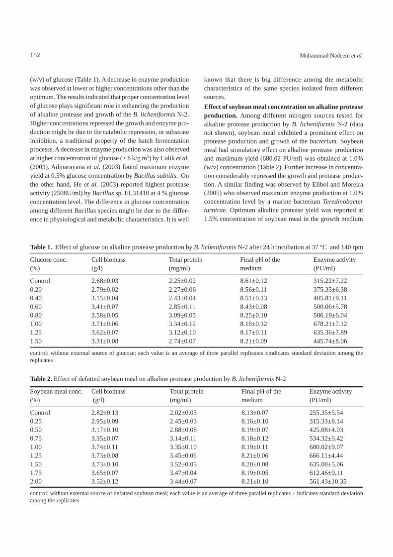
(w/v) of glucose (Table 1). A decrease in enzyme productionwas observed at lower or higher concentrations other than theoptimum. The results indicated that proper concentration levelof glucose plays significant role in enhancing the productionof alkaline protease and growth of the B. licheniformis N-2.Higher concentrations repressed the growth and enzyme pro-duction might be due to the catabolic repression, or substrateinhibition, a traditional property of the batch fermentationprocesss. A decrease in enzyme production was also observedat higher concentration of glucose (> 8 k/g m3) by Calik et al.(2003). Adinarayana et al. (2003) found maximum enzymeyield at 0.5% glucose concentration by Bacillus subtilis. Onthe other hand, He et al. (2003) reported highest proteaseactivity (2508U/ml) by Bacillus sp. EL31410 at 4 % glucoseconcentration level. The difference in glucose concentrationamong different Bacillus species might be due to the differ-ence in physiological and metabolic characteristics. It is well
known that there is big difference among the metaboliccharacteristics of the same species isolated from differentsources.Effect of soybean meal concentration on alkaline proteaseproduction. Among different nitrogen sources tested foralkaline protease production by B. licheniformis N-2 (datanot shown), soybean meal exhibited a prominent effect onprotease production and growth of the bacterium. Soybeanmeal had stimulatory effect on alkaline protease productionand maximum yield (680.02 PU/ml) was obtained at 1.0%(w/v) concentration (Table 2). Further increase in concentra-tion considerably repressed the growth and protease produc-tion. A similar finding was observed by Elibol and Moreira(2005) who observed maximum enzyme production at 1.0%concentration level by a marine bacterium Teredinobacterturnirae. Optimum alkaline protease yield was reported at1.5% concentration of soybean meal in the growth medium
Table 1. Effect of glucose on alkaline protease production by B. licheniformis N-2 after 24 h incubation at 37 °C and 140 rpm
Glucose conc. Cell biomass Total protein Final pH of the Enzyme activity(%) (g/l) (mg/ml) medium (PU/ml)
Control 2.68±0.03 2.25±0.02 8.61±0.12 315.22±7.220.20 2.79±0.02 2.27±0.06 8.56±0.11 375.35±6.380.40 3.15±0.04 2.43±0.04 8.51±0.13 405.81±9.110.60 3.41±0.07 2.85±0.11 8.43±0.08 500.06±5.780.80 3.58±0.05 3.09±0.05 8.25±0.10 586.19±6.041.00 3.71±0.06 3.34±0.12 8.18±0.12 678.21±7.121.25 3.62±0.07 3.12±0.10 8.17±0.11 635.36±7.891.50 3.31±0.08 2.74±0.07 8.21±0.09 445.74±8.06
control: without external source of glucose; each value is an average of three parallel replicates ±indicates standard deviation among thereplicates
Table 2. Effect of defatted soybean meal on alkaline protease production by B. licheniformis N-2
Soybean meal conc. Cell biomass Total protein Final pH of the Enzyme activity(%) (g/l) (mg/ml) medium (PU/ml)
Control 2.82±0.13 2.02±0.05 8.13±0.07 255.35±5.540.25 2.95±0.09 2.45±0.03 8.16±0.10 315.33±8.140.50 3.17±0.10 2.88±0.08 8.19±0.07 425.08±4.030.75 3.35±0.07 3.14±0.11 8.18±0.12 534.32±5.421.00 3.74±0.11 3.35±0.10 8.19±0.11 680.02±9.071.25 3.73±0.08 3.45±0.06 8.21±0.06 666.11±4.441.50 3.73±0.10 3.52±0.05 8.20±0.08 635.08±5.061.75 3.65±0.07 3.47±0.04 8.19±0.05 612.46±9.112.00 3.52±0.12 3.44±0.07 8.21±0.10 561.43±10.35
control: without external source of defatted soybean meal; each value is an average of three parallel replicates ± indicates standard deviationamong the replicates
152 Muhammad Nadeem et al.
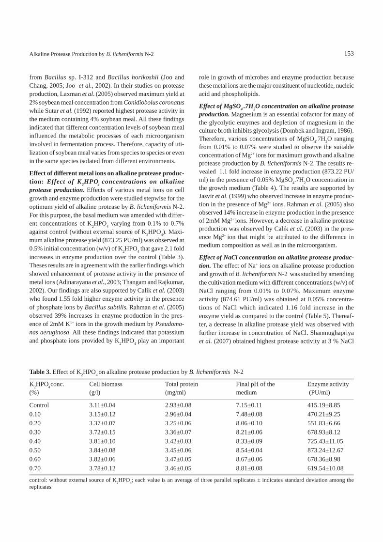
from Bacillus sp. I-312 and Bacillus horikoshii (Joo andChang, 2005; Joo et al., 2002). In their studies on proteaseproduction, Laxman et al. (2005) observed maximum yield at2% soybean meal concentration from Conidiobolus coronatuswhile Sutar et al. (1992) reported highest protease activity inthe medium containing 4% soybean meal. All these findingsindicated that different concentration levels of soybean mealinfluenced the metabolic processes of each microorganisminvolved in fermentation process. Therefore, capacity of uti-lization of soybean meal varies from species to species or evenin the same species isolated from different environments.
Effect of different metal ions on alkaline protease produc-tion: Effect of K2HPO4 concentrations on alkalineprotease production. Effects of various metal ions on cellgrowth and enzyme production were studied stepwise for theoptimum yield of alkaline protease by B. licheniformis N-2.For this purpose, the basal medium was amended with differ-ent concentrations of K2HPO4 varying from 0.1% to 0.7%against control (without external source of K2HPO4). Maxi-mum alkaline protease yield (873.25 PU/ml) was observed at0.5% initial concentration (w/v) of K2HPO4 that gave 2.1 foldincreases in enzyme production over the control (Table 3).Theses results are in agreement with the earlier findings whichshowed enhancement of protease activity in the presence ofmetal ions (Adinarayana et al., 2003; Thangam and Rajkumar,2002). Our findings are also supported by Calik et al. (2003)who found 1.55 fold higher enzyme activity in the presenceof phosphate ions by Bacillus subtilis. Rahman et al. (2005)observed 39% increases in enzyme production in the pres-ence of 2mM K1+ ions in the growth medium by Pseudomo-nas aeruginosa. All these findings indicated that potassiumand phosphate ions provided by K2HPO4 play an important
role in growth of microbes and enzyme production becausethese metal ions are the major constituent of nucleotide, nucleicacid and phospholipids.
Effect of MgSO4 .7H2O concentration on alkaline proteaseproduction. Magnesium is an essential cofactor for many ofthe glycolytic enzymes and depletion of magnesium in theculture broth inhibits glycolysis (Dombek and Ingram, 1986).Therefore, various concentrations of MgSO4.7H2O rangingfrom 0.01% to 0.07% were studied to observe the suitableconcentration of Mg2+ ions for maximum growth and alkalineprotease production by B. licheniformis N-2. The results re-vealed 1.1 fold increase in enzyme production (873.22 PU/ml) in the presence of 0.05% MgSO4.7H2O concentration inthe growth medium (Table 4). The results are supported byJasvir et al. (1999) who observed increase in enzyme produc-tion in the presence of Mg2+ ions. Rahman et al. (2005) alsoobserved 14% increase in enzyme production in the presenceof 2mM Mg2+ ions. However, a decrease in alkaline proteaseproduction was observed by Calik et al. (2003) in the pres-ence Mg2+ ion that might be attributed to the difference inmedium composition as well as in the microorganism.
Effect of NaCl concentration on alkaline protease produc-tion. The effect of Na+ ions on alkaline protease productionand growth of B. licheniformis N-2 was studied by amendingthe cultivation medium with different concentrations (w/v) ofNaCl ranging from 0.01% to 0.07%. Maximum enzymeactivity (874.61 PU/ml) was obtained at 0.05% concentra-tions of NaCl which indicated 1.16 fold increase in theenzyme yield as compared to the control (Table 5). Thereaf-ter, a decrease in alkaline protease yield was observed withfurther increase in concentration of NaCl. Shanmughapriyaet al. (2007) obtained highest protease activity at 3 % NaCl
Table 3. Effect of K2HPO4 on alkaline protease production by B. licheniformis N-2
K2HPO4 conc. Cell biomass Total protein Final pH of the Enzyme activity(%) (g/l) (mg/ml) medium (PU/ml)
Control 3.11±0.04 2.93±0.08 7.15±0.11 415.19±8.850.10 3.15±0.12 2.96±0.04 7.48±0.08 470.21±9.250.20 3.37±0.07 3.25±0.06 8.06±0.10 551.83±6.660.30 3.72±0.15 3.36±0.07 8.21±0.06 678.93±8.120.40 3.81±0.10 3.42±0.03 8.33±0.09 725.43±11.050.50 3.84±0.08 3.45±0.06 8.54±0.04 873.24±12.670.60 3.82±0.06 3.47±0.05 8.67±0.06 678.36±8.980.70 3.78±0.12 3.46±0.05 8.81±0.08 619.54±10.08
control: without external source of K2HPO4; each value is an average of three parallel replicates ± indicates standard deviation among thereplicates
153Alkaline Protease Production by B. licheniformis N-2
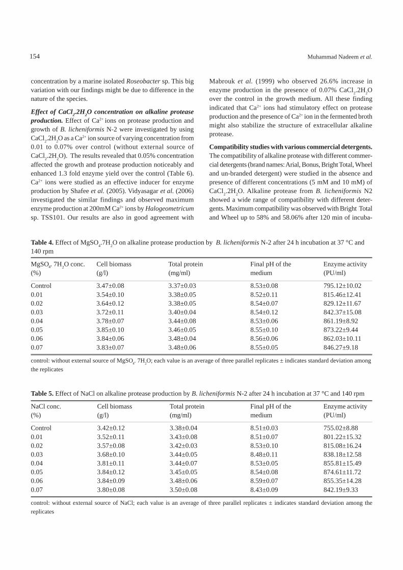
concentration by a marine isolated Roseobacter sp. This bigvariation with our findings might be due to difference in thenature of the species.
Effect of CaCl2.2H2O concentration on alkaline proteaseproduction. Effect of Ca2+ ions on protease production andgrowth of B. licheniformis N-2 were investigated by usingCaCl2.2H2O as a Ca2+ ion source of varying concentration from0.01 to 0.07% over control (without external source ofCaCl2.2H2O). The results revealed that 0.05% concentrationaffected the growth and protease production noticeably andenhanced 1.3 fold enzyme yield over the control (Table 6).Ca2+ ions were studied as an effective inducer for enzymeproduction by Shafee et al. (2005). Vidyasagar et al. (2006)investigated the similar findings and observed maximumenzyme production at 200mM Ca2+ ions by Halogeometricumsp. TSS101. Our results are also in good agreement with
Mabrouk et al. (1999) who observed 26.6% increase inenzyme production in the presence of 0.07% CaCl2.2H2Oover the control in the growth medium. All these findingindicated that Ca2+ ions had stimulatory effect on proteaseproduction and the presence of Ca2+ ion in the fermented brothmight also stabilize the structure of extracellular alkalineprotease.
Compatibility studies with various commercial detergents.The compatibility of alkaline protease with different commer-cial detergents (brand names: Arial, Bonus, Bright Total, Wheeland un-branded detergent) were studied in the absence andpresence of different concentrations (5 mM and 10 mM) ofCaCl2.2H2O. Alkaline protease from B. licheniformis N2showed a wide range of compatibility with different deter-gents. Maximum compatibility was observed with Bright Totaland Wheel up to 58% and 58.06% after 120 min of incuba-
Table 4. Effect of MgSO4.7H2O on alkaline protease production by B. licheniformis N-2 after 24 h incubation at 37 °C and140 rpm
MgSO4. 7H2O conc. Cell biomass Total protein Final pH of the Enzyme activity(%) (g/l) (mg/ml) medium (PU/ml)
Control 3.47±0.08 3.37±0.03 8.53±0.08 795.12±10.020.01 3.54±0.10 3.38±0.05 8.52±0.11 815.46±12.410.02 3.64±0.12 3.38±0.05 8.54±0.07 829.12±11.670.03 3.72±0.11 3.40±0.04 8.54±0.12 842.37±15.080.04 3.78±0.07 3.44±0.08 8.53±0.06 861.19±8.920.05 3.85±0.10 3.46±0.05 8.55±0.10 873.22±9.440.06 3.84±0.06 3.48±0.04 8.56±0.06 862.03±10.110.07 3.83±0.07 3.48±0.06 8.55±0.05 846.27±9.18
control: without external source of MgSO4. 7H2O; each value is an average of three parallel replicates ± indicates standard deviation amongthe replicates
Table 5. Effect of NaCl on alkaline protease production by B. licheniformis N-2 after 24 h incubation at 37 °C and 140 rpm
NaCl conc. Cell biomass Total protein Final pH of the Enzyme activity(%) (g/l) (mg/ml) medium (PU/ml)
Control 3.42±0.12 3.38±0.04 8.51±0.03 755.02±8.880.01 3.52±0.11 3.43±0.08 8.51±0.07 801.22±15.320.02 3.57±0.08 3.42±0.03 8.53±0.10 815.08±16.240.03 3.68±0.10 3.44±0.05 8.48±0.11 838.18±12.580.04 3.81±0.11 3.44±0.07 8.53±0.05 855.81±15.490.05 3.84±0.12 3.45±0.05 8.54±0.08 874.61±11.720.06 3.84±0.09 3.48±0.06 8.59±0.07 855.35±14.280.07 3.80±0.08 3.50±0.08 8.43±0.09 842.19±9.33
control: without external source of NaCl; each value is an average of three parallel replicates ± indicates standard deviation among thereplicates
154 Muhammad Nadeem et al.
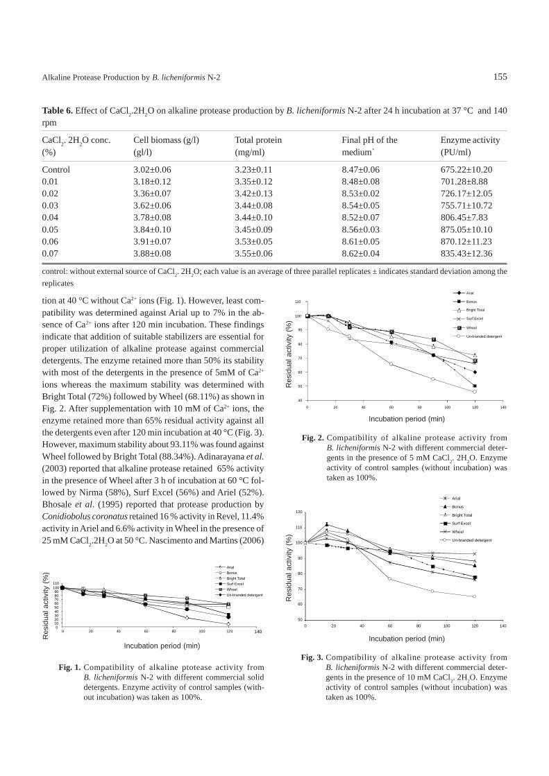
tion at 40 °C without Ca2+ ions (Fig. 1). However, least com-patibility was determined against Arial up to 7% in the ab-sence of Ca2+ ions after 120 min incubation. These findingsindicate that addition of suitable stabilizers are essential forproper utilization of alkaline protease against commercialdetergents. The enzyme retained more than 50% its stabilitywith most of the detergents in the presence of 5mM of Ca2+
ions whereas the maximum stability was determined withBright Total (72%) followed by Wheel (68.11%) as shown inFig. 2. After supplementation with 10 mM of Ca2+ ions, theenzyme retained more than 65% residual activity against allthe detergents even after 120 min incubation at 40 °C (Fig. 3).However, maximum stability about 93.11% was found againstWheel followed by Bright Total (88.34%). Adinarayana et al.(2003) reported that alkaline protease retained 65% activityin the presence of Wheel after 3 h of incubation at 60 °C fol-lowed by Nirma (58%), Surf Excel (56%) and Ariel (52%).Bhosale et al. (1995) reported that protease production byConidiobolus coronatus retained 16 % activity in Revel, 11.4%activity in Ariel and 6.6% activity in Wheel in the presence of25 mM CaCl2.2H2O at 50 °C. Nascimento and Martins (2006)
Table 6. Effect of CaCl2.2H2O on alkaline protease production by B. licheniformis N-2 after 24 h incubation at 37 °C and 140rpm
CaCl2. 2H2O conc. Cell biomass (g/l) Total protein Final pH of the Enzyme activity(%) (gl/l) (mg/ml) medium` (PU/ml)
Control 3.02±0.06 3.23±0.11 8.47±0.06 675.22±10.200.01 3.18±0.12 3.35±0.12 8.48±0.08 701.28±8.880.02 3.36±0.07 3.42±0.13 8.53±0.02 726.17±12.050.03 3.62±0.06 3.44±0.08 8.54±0.05 755.71±10.720.04 3.78±0.08 3.44±0.10 8.52±0.07 806.45±7.830.05 3.84±0.10 3.45±0.09 8.56±0.03 875.05±10.100.06 3.91±0.07 3.53±0.05 8.61±0.05 870.12±11.230.07 3.88±0.08 3.55±0.06 8.62±0.04 835.43±12.36
control: without external source of CaCl2. 2H2O; each value is an average of three parallel replicates ± indicates standard deviation among thereplicates
155
Fig. 1. Compatibility of alkaline ptotease activity fromB. licheniformis N-2 with different commercial soliddetergents. Enzyme activity of control samples (with-out incubation) was taken as 100%.
0102030405060708090
100110
0 20 40 60 80 100 120 140
ArialBonusBright TotalSurf ExcelWheelUn-branded detergent
Incubation period (min)
Fig. 2. Compatibility of alkaline protease activity fromB. licheniformis N-2 with different commercial deter-gents in the presence of 5 mM CaCl2. 2H2O. Enzymeactivity of control samples (without incubation) wastaken as 100%.
Arial
40
50
60
70
80
90
100
110
0 20 40 60 80 100 120 140
Bonus
Bright Total
Surf Excel
Wheel
Un-branded detergent
Incubation period (min)
Fig. 3. Compatibility of alkaline protease activity fromB. licheniformis N-2 with different commercial deter-gents in the presence of 10 mM CaCl2. 2H2O. Enzymeactivity of control samples (without incubation) wastaken as 100%.
50
60
70
80
90
100
110
120
0 20 40 60 80 100 120 140
Arial
Bonus
Bright Total
Surf Excel
Wheel
Un-branded detergent
Incubation period (min)
Alkaline Protease Production by B. licheniformis N-2
Res
idua
l act
ivity
(%)
Res
idua
l act
ivity
(%)
Res
idua
l act
ivity
(%)
found more than 85% activity in the presence of Ca2+ ionsafter 1 h incubation at 60 °C. Comparing these results, thealkaline protease produced by B. licheniformis N-2 was foundto be significantly more stable over a broad range of commer-cial detergents.
ConclusionAlkaline protease production and growth of B. licheniformisN-2 under submerged fermentation were found to be influ-enced by different concentrations of glucose, soybean mealand various metal ions in the growth medium. The maximumprotease production (875.05 PU/ml) was achieved by employ-ing optimized concentrations of glucose (1%), soybean meal(1%), K2HPO4 (0.5%), MgSO4.7H2O (0.05 %), NaCl (0.05%),CaCl2.2H2O (0.05%) after 24 h incubation at 140 rpm. Thecompatibility studies of alkaline protease with different com-mercial detergents in the presence and absence of Ca2+ ionsindicated that the B. licheniformis N-2 protease is suitable fordetergent formulation. More than 65 % stability was observedwith various commercial detergents in the presence of 10 mMCaCl2.2H2O. Collectively, all these results may justify the pos-sibilities of production of alkaline protease by B. licheniformisN-2 for its commercial exploitation in detergent industry.
AcknowledgementThe authors are thankful to the Ministry of Science and Tech-nology (MoST), Government of Pakistan for its financial sup-port, under the PSDP project allocation No.31, to carry outthis research work.
ReferencesAdinarayana, K., Ellaiah, P., Prasad, D.S. 2003. Purification
and characterization of thermostable serine alkaline pro-tease from a newly isolated Bacillus subtilis PE-11.Pharma Sci. Technol. 4: 1-9.
Banerjee, C.U., Sani, R.K, Azmi, W., Soni, R. 1999. Thermo-stable alkaline protease from Bacillus brevis and itscharacterization as laundry detergent additives. ProcessBiochem. 35: 213-219.
Bhosale, S.H., Rao, M.B., Deshpande, V.V., Srinivasan, M.C.1995. Thermo- stability of high-activity alkaline proteasefrom Conidiobolus coronatus (NCL86.8.20). Enz.Microb. Technol. 17: 136-139.
Calik, P. Celik, E., Telli., I.E., Oktar, C., Özdemir, E. 2003.Protein-based complex medium design for recombinantserine alkaline protease production by Bacillus subtilus.Enz. Microb. Technol. 33: 975-986.
Dombek, K.M., Ingram, L.O. 1986. Magnesium limitation andits role in apparent toxicity of ethanol during yeast
156
fermentation. Appl. Environ. Microbiol. 52: 975-981.Elibol, M., Moreira, A.R. 2005. Optimizing some factors
affecting alkaline protease production by a marine bacte-rium Teredinobacter turnirae under solid substratefermentation. Process Biochem. 40: 1951-1956.
Gessesse, A. 1997. The use of nug meal as low-cost substratefor the production of protease by alkaliphilic Bacillus sp.AR-009 and some properties of the enzyme. BioresourceTechnol. 62:59-61.
Gupta , R., Beg, Q.K., Lorenz, P. 2002. Bacterial alkalineprotease: molecular approaches and industrial applica-tions. Appl. Microbiol. Biotechnol. 59: 15-32.
He, G.Q, Chen, Q.H., Zhang, L., Liu, X.J. 2003. Influence ofmedium components on elastase production using crudesources by Bacillus sp. EL.31410. J. Zhejiang Univ. Sci.4: 142-151.
Jasvir, S., Gill, N., Devasahayam, G., Sahoo, D. 1999. Studieson alkaline protease produced by Bacillus sp. NG 312.Appl. Biochem. Biotechnol. 76: 57-63.
Joo, H.S., Kumar, C.G., Park, G.C., Kim, K.T., Paik, S.R.,Chang, C.S. 2002. Optimization of the production of anextracellular alkaline protease from Bacillus horikoshi.Process Biochem. 38: 155-159.
Joo, H.S., Chang, C.S. 2005. Production of protease from anew alkalophilic Bacillus sp. I-312 grown on soybeanmeal: Optimization and some properties. ProcessBiochem. 40: 1263-1270.
Kuar, S., Vohra, R.M., Kapoor , M., Beg, Q.K. Hoondal, G.S.2001. Enhance production and characterization of highlythermostable alkaline protease from Bacillus sp. P-2.World J. Microbiol. Biotechnol. 17: 125-126.
Laxman, R.S., Sonawane, A.P., More, S.V., Rao, B.S., Rele,M.V., Jogdand, V.V., Deshpande, V.V., Rao, M.B. 2005.Optimization and scale up of production of alkalineprotease from Conidiobolus cornatus. Process Biochem.40: 3152-3158.
Lowry, O.H., Roserbrough, N.J., Farr, A.W. Randall, R. 1951.Protein measurement with Folin Phenol Reagent. J. Biol.Chem. 193: 265-275.
Mabrouk, S.S., Hashem, A.M., El-Shayeb, N.M.A., Ismail,A.M.S., Abdel-Fattah, A.F. 1999. Optimization ofalkaline protease productivity by Bacillus licheniformisATCC 21415. Bioresource Technol. 69:155-159.
Moon, S.H., Parulekar, S.J. 1993. Some observations onprotease production in continuous suspension culture ofBacillus firmus. Biotechnol. Bioeng. 41: 43-54.
Nadeem, M., Baig, S.J., Syed, Q.A., Qazi, J.I. 2006. Micro-bial production of alkaline proteases by locally isolatedBacillus subtilis PCSIR-5. Pak. J. Zool. 38:109-114.
Nadeem, M., Qazi, J.I., Baig, S.J., Syed, Q.A. 2007. Studies
Muhammad Nadeem et al.
on the stability of protease from Bacillus subtilis PCSIR-5. Pak. J. Zool. 38: 109-114.
Nascimento, W.C.A., Martins, M.L.L. 2006. Studies on thestability of protease from Bacillus sp. and its compatibil-ity with commercial detergents. Brazil. J. Microbiol. 37:307-311.
Oberoi, R., Beg, Q.K., Puri, S., Saxena, R.K., Gupta, R. 2001.Characterization and wash performance analysis of anSDS-stable alkaline protease from a Bacillus sp. WorldJ. Microbiol. Biotechnol. 17:493-497.
Pastor, M.D., Lorda, G.S., Balatti, A. 2001. Protease obten-tion using Bacillus subtilis 3411 and amaranth seed mealmedium at different aeration rates. Brazil. J. Mirobiol.32: 1-8.
Rahman, R.N.Z.R.A., Geok, L.P., Basri, M., Salleh, A.B.2005. An organic solvent-tolerant protease fromPseudomonas aerugenosa strain K: Nutritional factorsaffecting protease production. Enz. Microb. Technol. 36:749-757.
Razak, N.A., Samad, M.Y.A., Basri, M., Yunus, W., Ampon,K., Salleh, A.B. 1994. Thermostable extracellularprotease of Bacillus stearothermophilus: factors affec-ting its production. World J. Microbiol. Biotechnol. 10:260-263.
Shafee, N., Aris, S.N., Rahaman, R.N.Z.A., Basri, M., Salleh,
A.B. 2005. Optimization of environmental and nutritionalconditions for the production of alkaline protease by anewly isolated bacterium Bacillus cereus strain 146. J.Appl. Sci. Res. 1: 1-8.
Shanmughapriya, S., Krishnaveni, J., Selvin, J., Grandhi-mathi,R., Arunkumar, M., Thangavelu, T., Kiran, G.S.,Natarajaseenivasan, K. 2007. Optimization of extracel-lular thermotolerant alkaline protease produce by marineRosebacter sp. (MMD040). Bioprocess Biosyst. Eng. 31:427-433.
Sutar, I.I., Srinivasan, M.C., Varak H.G. 1992. Production ofan alkaline proteinase from Conidiobolus cornatus andits use to resolve DL-phenylalanine and DL-phenylgly-cine. World J. Microbiol. Biotechnol. 8: 254-258.
Thangam, E.B., Rajkumar, G.S. 2002. Purification and partialcharacterization of alkaline protease from Alcaligenesfaecalis. Biotechnol. Appl. Biochem. 35: 149-154.
Vidyasagar, M., Prakash, S.B., Sreeramula, K. 2006. Optimi-zation of culture conditions for the production of haloal-kaliphilic thermo-stable protease from an extremelyhalophilic archaeon Halogeometricum sp. TSS101.Lett. Appl. Microbiol. 43: 385-391.
Yang, S.S., Huang, C.I. 1994. Proteases production by amy-lolytic fungi in solid state fermentation. J. Chinese Agric.Chem. Soc. 32: 589-601.
157Alkaline Protease Production by B. licheniformis N-2
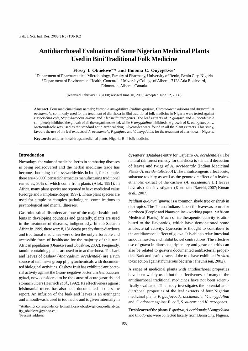
IntroductionNowadays, the value of medicinal herbs in combating diseasesis being rediscovered and the herbal medicine trade hasbecome a booming business worldwide. In India, for example,there are 46,000 licensed pharmacies manufacturing traditionalremedies, 80% of which come from plants (Alok, 1991). InAfrica, many plant species are reported to have medicinal value(George and Pamplona-Roger, 1997). These plant species areused for simple or complex pathological complications topsychological and mental illnesses.
Gastrointestinal disorders are one of the major health prob-lems in developing countries and generally, plants are usedin the treatment of diseases, indigenously. In sub-SaharanAfrica in 1999, there were 8, 181 deaths per day due to diarrhoeaand traditional medicines were often the only affordable andaccessible form of healthcare for the majority of this ruralAfrican population (Obuekwe and Obuekwe, 2002). Frequently,tannin-containing plants are used to treat diarrhoea. The barkand leaves of cashew (Anarcadium occidentale) are a richsource of tannins_a group pf phytochemicals with documen-ted biological activities. Cashew fruit has exhibited antibacte-rial activity against the Gram- negative bacterium Helicobacterpylori, now considered to be the cause of acute gastritis andstomach ulcers (Heirrich et al., 1992). Its effectiveness againstleishmanial ulcers has also been documented in the samereport. An infusion of the bark and leaves is an astringentand a mouthwash, used in toothache and is given internally in
Pak. J. Sci. Ind. Res. 2008 51(3) 158-162
Antidiarrhoeal Evaluation of Some Nigerian Medicinal PlantsUsed in Bini Traditional Folk MedicineFlossy I. Obuekweab* and Ihuoma C. Onyejekwea
aDepartment of Pharmaceutical Microbiology, Faculty of Pharmacy, University of Benin, Benin City, NigeriabDepartment of Environment Health, Concordia University College of Alberta, 7128 Ada Boulevard,
Edmonton, Alberta, Canada
(received February 13, 2008; revised June 10, 2008; accepted June 12, 2008)
Abstract. Four medicinal plants namely; Vernonia amygdalina, Psidium guajava, Chromolaena odorata and Anarcadiumoccidentale, commonly used for the treatment of diarrhoea in Bini traditional folk medicine in Nigeria were tested againstEscherichia coli, Staphylococcus aureus and Klebsiella aerogenes. The leaf extracts of P. guajava and A. occidentalecompletely inhibited the growth of all the organisms tested, while V. amygdalina inhibited the growth of K. aerogenes only.Metronidazole was used as the standard antidiarrhoeal drug. Glycosides were found in all the plant extracts. This study,favours the use of the leaf extracts of A. occidentale, P. guajava and V. amygdalina for the treatment of diarrhoea in Nigeria.
Keywords: antidiarrhoeal drugs, medicinal plants, Nigeria, Bini folk medicine
*Author for correspondence; E-mail: [email protected];[email protected];bPresent address
dysentery (Database entry for Cajueiro -A. occidentale). Thenatural rainforest remedy for diarrhoea is standard decoctionof leaves and twigs of A. occidentale (Indian MecicinalPlants- A. occidentale, 2001). The antiulcerogenic effect acute,subacute toxicity as well as the genotoxic effect of a hydro-ethanolic extract of the cashew (A. occidentale L.) leaveshave also been investigated (Konan and Bacchi, 2007; Konanet al., 2007).
Psidium guajava (guava) is a common shade tree or shrub inthe tropics. The Tikuna Indians decoct the leaves as a cure fordiarrhoea (People and Plants online - working paper 1: AfricanMedicinal Plants). Much of its therapeutic activity is attri-buted to the flavonoids, which have demonstrated someantibacterial activity. Quercetin is thought to contribute tothe antidiarrhoeal effect of guava. It is able to relax intestinalsmooth muscles and inhibit bowel contractions. The effectiveuse of guava in diarrhoea, dysentery and gastroenteritis canalso be related to guava’s documented antibacterial proper-ties. Bark and leaf extracts of the tree have exhibited in-vitrotoxic action against numerous bacteria (Theunissen, 2002).
A range of medicinal plants with antidiarrhoeal propertieshave been widely used; but the effectiveness of many of theantidiarrhoeal traditional medicines have not been scienti-fically evaluated. This study investigates the potential anti-diarrhoeal properties of the leaf extracts of four Nigerianmedicinal plants P. guajava, A. occidentale, V. amygdalinaand C. odorata against E. coli, S. aureus and K. aerogenes.
Fresh leaves of the plants. P. guajava, A. occidentale, V. amygdalinaand C. odorata were collected locally from Benin City, Nigeria.
158
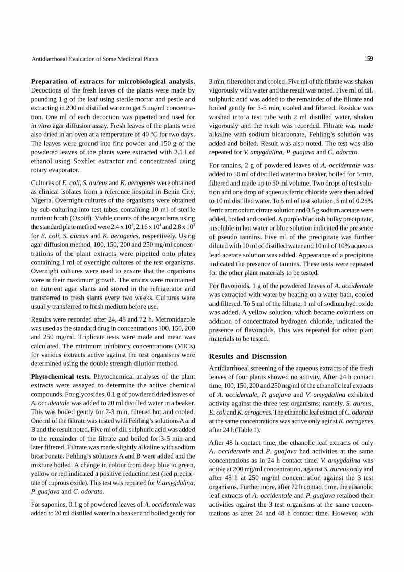
Preparation of extracts for microbiological analysis.Decoctions of the fresh leaves of the plants were made bypounding 1 g of the leaf using sterile mortar and pestle andextracting in 200 ml distilled water to get 5 mg/ml concentra-tion. One ml of each decoction was pipetted and used forin vitro agar diffusion assay. Fresh leaves of the plants werealso dried in an oven at a temperature of 40 °C for two days.The leaves were ground into fine powder and 150 g of thepowdered leaves of the plants were extracted with 2.5 l ofethanol using Soxhlet extractor and concentrated usingrotary evaporator.
Cultures of E. coli, S. aureus and K. aerogenes were obtainedas clinical isolates from a reference hospital in Benin City,Nigeria. Overnight cultures of the organisms were obtainedby sub-culturing into test tubes containing 10 ml of sterilenutrient broth (Oxoid). Viable counts of the organisms usingthe standard plate method were 2.4 x 10³, 2.16 x 10⁴ and 2.8 x 10³
for E. coli, S. aureus and K. aerogenes, respectively. Usingagar diffusion method, 100, 150, 200 and 250 mg/ml concen-trations of the plant extracts were pipetted onto platescontaining 1 ml of overnight cultures of the test organisms.Overnight cultures were used to ensure that the organismswere at their maximum growth. The strains were maintainedon nutrient agar slants and stored in the refrigerator andtransferred to fresh slants every two weeks. Cultures wereusually transferred to fresh medium before use.
Results were recorded after 24, 48 and 72 h. Metronidazolewas used as the standard drug in concentrations 100, 150, 200and 250 mg/ml. Triplicate tests were made and mean wascalculated. The minimum inhibitory concentrations (MICs)for various extracts active against the test organisms weredetermined using the double strength dilution method.
Phytochemical tests. Phytochemical analyses of the plantextracts were assayed to determine the active chemicalcompounds. For glycosides, 0.1 g of powdered dried leaves ofA. occidentale was added to 20 ml distilled water in a beaker.This was boiled gently for 2-3 min, filtered hot and cooled.One ml of the filtrate was tested with Fehling’s solutions A andB and the result noted. Five ml of dil. sulphuric acid was addedto the remainder of the filtrate and boiled for 3-5 min andlater filtered. Filtrate was made slightly alkaline with sodiumbicarbonate. Fehling’s solutions A and B were added and themixture boiled. A change in colour from deep blue to green,yellow or red indicated a positive reduction test (red precipi-tate of cuprous oxide). This test was repeated for V. amygdalina,P. guajava and C. odorata.
For saponins, 0.1 g of powdered leaves of A. occidentale wasadded to 20 ml distilled water in a beaker and boiled gently for
3 min, filtered hot and cooled. Five ml of the filtrate was shakenvigorously with water and the result was noted. Five ml of dil.sulphuric acid was added to the remainder of the filtrate andboiled gently for 3-5 min, cooled and filtered. Residue waswashed into a test tube with 2 ml distilled water, shakenvigorously and the result was recorded. Filtrate was madealkaline with sodium bicarbonate, Fehling’s solution wasadded and boiled. Result was also noted. The test was alsorepeated for V. amygdalina, P. guajava and C. odorata.
For tannins, 2 g of powdered leaves of A. occidentale wasadded to 50 ml of distilled water in a beaker, boiled for 5 min,filtered and made up to 50 ml volume. Two drops of test solu-tion and one drop of aqueous ferric chloride were then addedto 10 ml distilled water. To 5 ml of test solution, 5 ml of 0.25%ferric ammonium citrate solution and 0.5 g sodium acetate wereadded, boiled and cooled. A purple/blackish bulky precipitate,insoluble in hot water or blue solution indicated the presenceof pseudo tannins. Five ml of the precipitate was furtherdiluted with 10 ml of distilled water and 10 ml of 10% aqueouslead acetate solution was added. Appearance of a precipitateindicated the presence of tannins. These tests were repeatedfor the other plant materials to be tested.
For flavonoids, 1 g of the powdered leaves of A. occidentalewas extracted with water by heating on a water bath, cooledand filtered. To 5 ml of the filtrate, 1 ml of sodium hydroxidewas added. A yellow solution, which became colourless onaddition of concentrated hydrogen chloride, indicated thepresence of flavonoids. This was repeated for other plantmaterials to be tested.
Results and DiscussionAntidiarrhoeal screening of the aqueous extracts of the freshleaves of four plants showed no activity. After 24 h contacttime, 100, 150, 200 and 250 mg/ml of the ethanolic leaf extractsof A. occidentale, P. guajava and V. amygdalina exhibitedactivity against the three test organisms; namely, S. aureus,E. coli and K. aerogenes. The ethanolic leaf extract of C. odorataat the same concentrations was active only aginst K. aerogenesafter 24 h (Table 1).
After 48 h contact time, the ethanolic leaf extracts of onlyA. occidentale and P. guajava had activities at the sameconcentrations as in 24 h contact time. V. amygdalina wasactive at 200 mg/ml concentration, against S. aureus only andafter 48 h at 250 mg/ml concentration against the 3 testorganisms. Further more, after 72 h contact time, the ethanolicleaf extracts of A. occidentale and P. guajava retained theiractivities against the 3 test organisms at the same concen-trations as after 24 and 48 h contact time. However, with
159Antidiarrhoeal Evaluation of Some Medicinal Plants
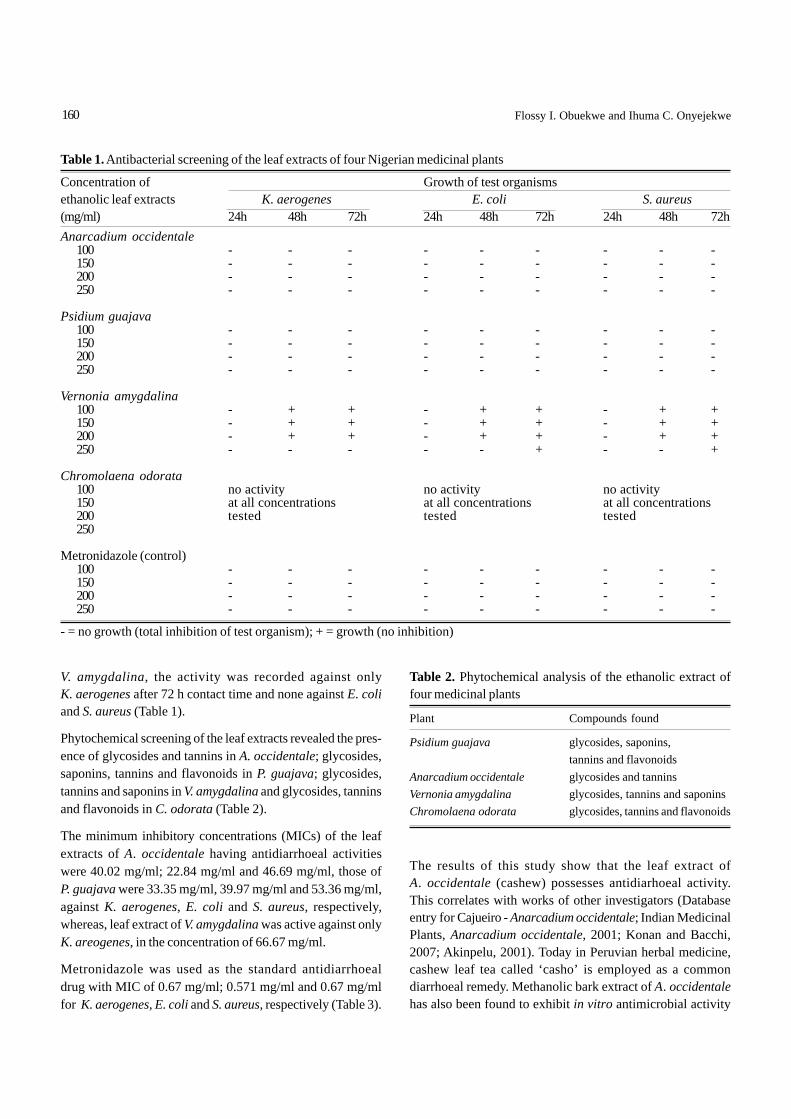
V. amygdalina, the activity was recorded against onlyK. aerogenes after 72 h contact time and none against E. coliand S. aureus (Table 1).
Phytochemical screening of the leaf extracts revealed the pres-ence of glycosides and tannins in A. occidentale; glycosides,saponins, tannins and flavonoids in P. guajava; glycosides,tannins and saponins in V. amygdalina and glycosides, tanninsand flavonoids in C. odorata (Table 2).
The minimum inhibitory concentrations (MICs) of the leafextracts of A. occidentale having antidiarrhoeal activitieswere 40.02 mg/ml; 22.84 mg/ml and 46.69 mg/ml, those ofP. guajava were 33.35 mg/ml, 39.97 mg/ml and 53.36 mg/ml,against K. aerogenes, E. coli and S. aureus, respectively,whereas, leaf extract of V. amygdalina was active against onlyK. areogenes, in the concentration of 66.67 mg/ml.
Metronidazole was used as the standard antidiarrhoealdrug with MIC of 0.67 mg/ml; 0.571 mg/ml and 0.67 mg/mlfor K. aerogenes, E. coli and S. aureus, respectively (Table 3).
Table 2. Phytochemical analysis of the ethanolic extract offour medicinal plants
Plant Compounds found
Psidium guajava glycosides, saponins,tannins and flavonoids
Anarcadium occidentale glycosides and tanninsVernonia amygdalina glycosides, tannins and saponinsChromolaena odorata glycosides, tannins and flavonoids
The results of this study show that the leaf extract ofA. occidentale (cashew) possesses antidiarhoeal activity.This correlates with works of other investigators (Databaseentry for Cajueiro - Anarcadium occidentale; Indian MedicinalPlants, Anarcadium occidentale, 2001; Konan and Bacchi,2007; Akinpelu, 2001). Today in Peruvian herbal medicine,cashew leaf tea called ‘casho’ is employed as a commondiarrhoeal remedy. Methanolic bark extract of A. occidentalehas also been found to exhibit in vitro antimicrobial activity
Table 1. Antibacterial screening of the leaf extracts of four Nigerian medicinal plants
Concentration of Growth of test organismsethanolic leaf extracts K. aerogenes E. coli S. aureus(mg/ml) 24h 48h 72h 24h 48h 72h 24h 48h 72hAnarcadium occidentale
100 - - - - - - - - -150 - - - - - - - - -200 - - - - - - - - -250 - - - - - - - - -
Psidium guajava100 - - - - - - - - -150 - - - - - - - - -200 - - - - - - - - -250 - - - - - - - - -
Vernonia amygdalina100 - + + - + + - + +150 - + + - + + - + +200 - + + - + + - + +250 - - - - - + - - +
Chromolaena odorata100 no activity no activity no activity150 at all concentrations at all concentrations at all concentrations200 tested tested tested250
Metronidazole (control)100 - - - - - - - - -150 - - - - - - - - -200 - - - - - - - - -250 - - - - - - - - -
- = no growth (total inhibition of test organism); + = growth (no inhibition)
160 Flossy I. Obuekwe and Ihuma C. Onyejekwe

against 13 of 15 microorganisms tested at a concentration of20 mg/ml (Akinpelu, 2001). An essential oil obtained by steamdistillation of the leaves of A. occidentale acted as a tranqui-lizer and analgesic and had mild antibacterial and antifungalactivities (Indian Medicinal Plants: Anarcadium occidentale,2001).
Psidium guajava also possesses antidiarrhoeal activities. Itsbark and leaf extracts have been found to possess in vitrotoxic action against numerous bacteria (People and Plantsonline-working paper1: African Medicinal Plants). In severalstudies, guava had shown significant antibacterial activityagainst such common diarrhoea-causing bacteria as Staphy-lococcus, Shigella, Bacillus, Salmonella, Escherichia coli,Clostridium and Pseudomonas (Theunissen, 2002). Theeffectiveness of P. guajava as an antidiarrhoeal agent hasbeen confirmed; the methanolic extract is the only agentshowing significant inhibitory activity against the growthof Salmonella and Shigella species and enteropathogenicE. coli (Caceres et al., 1993).
In this study, V. amygdalina showed antidiarrhoeal activityon K. aerogenes at 250 mg/ml after 72 h contact time and noton the other test organisms even though antidiarrhoeal acti-vity of the plant has been indicated in the reports of otherinvestigators (Akinpelu, 1999; Obaseiki-Ebor et al., 1993).Phytochemical analyses of the leaf extracts revealed thepresence of glycosides, tannins, saponins and flavonoids.Medicinal plants had earlier been screened for groups ofphytochemical compounds with antibacterial and antiamoebicactivities and were found to contain tannins, alkaloids, saponins,flavonoids, steroids and/or triterpenes and reducing sugars(Otshudi et al., 2000).
The search for new active chemical compounds in high bio-logical diversity regions has become a challenge to modernpharmaceutical industries. The history of drug discoveryimplies that the ethnobotanical approach is the most produc-tive of plant surveying methods (Cox and Balick, 1994). The
antiulcerogenic effect and acute toxicity of hydroethanolicextract from the cashew (A. occidentale L.) leaves have beeninvestigated by Konan and Bacchi (2007) and the extract wasfound to inhibit gastric lesions induced by HCl/ethanol infemale rats and its a methanolic fraction (257.12 mg/kg)reduced gastric lesions by 88.20%. Thus the plant is likely tocontain the active principle with antiulcer effect.
In conclusion, the ethanolic extracts of the medicinal plantsinvestigated in this study were found to possess goodantidiarrhoeal potential and supported the claims made inBini folklore medicine. The study suggests that the use ofan effective and economical herbal formulation based onsuch extracts could be beneficial against certain bacterialinfections.
Further studies on these plants are required to identify andisolate the specific glycosides that are responsible for theantidiarrhoeal activities with a view to producing new anti-diarrhoeal drugs from them.
AcknowledgementAuthors are grateful to Mr. A. Abubakar, formerly of theDepartment of Pharmacognosy, Faculty of Pharmacy,University of Benin, Benin City, Nigeria for identifying themedicinal plants used in this study.
ReferencesAkinpelu, D.A. 1999. Antimicrobial activity of Vernonia
amygdalina leaves. Fitoterapia 70: 432-434.Akinpelu, D.A. 2001. Antimicrobial activity of Anacardium
occidentale bark. Fitoterapia 72: 286-287.Alok, J. 1991. Medicinal plants used in Africa. http://www.
rbgkew.org.uk/peopleplants/wp/wpl/africa.htmCaceres, A., Fletes, L., Auguilar, L., Ramfrez, O., Figueroa, L.,
Taracena, A.M., Samaya, B. 1993. Plants used in Guate-mala for the treatment of gastrointestinal disorders. III:Confirmation of activity against enterobacteria of 16 plants.J. Ethnopharmacol. 38: 31-38.
Cox, P.A., Balick, M.J. 1994. The ethnobotanical approach todrug discovery. Sci. Am. 270: 82-87.
Database entry for Cajueiro - Anarcadium occidentale. http://www.raintreenutrition.com/cajueiro.htm
George, D., Pamplona-Roger. 1997. Encyclopedia of MedicinalPlants, vol. 1, pp. 52, 112-115.
Heirrich, M., Rimplker, H., Barrera, N.A. 1992. Indigenousphytotherapy of gastrointestinal disorders in lowlandmixed community (Oaxaca, Mexico): Ethnopharmacologicevaluation. J. Ethnopharmacol. 36: 63-80.
Indian Medicinal Plants. 2001. Anarcadium occidentale. http://www.indmedplantskr.org/Anarcadium occidentale.htm.
Table 3. Minimum inhibitory concentrations (MICs) of the plantextracts having antidiarrhoeal activities
Plant extracts K. aerogenes E. coli S. aureus(mg/ml)Anarcadium occidentale 40.02 22.84 46.69
Psidium guajava 33.35 39.97 53.36
Vernonia amygdalina 66.67 - -
Metrinadizole 0.67 0.571 0.67(standard drug used as control)
161Antidiarrhoeal Evaluation of Some Medicinal Plants
Konan, N.A., Bacchi, E.M. 2007. Antiulcerogenic effect andacute toxicity of a hydroethanolic extract from the cashew(Anacardium occidentale L.) leaves. J. Ethnopharmacol.112: 237-242.
Konan, N.A., Bacchi, E.M., Nilton, L., Soraya, D.V., Eliana, A.V.2007. Acute, subacute toxicity and genotoxic effect of ahydroethanolic extract of the cashew (Anacardiumoccidentale L.). J. Ethnopharmacol. 110: 30-38.
Obaseiki-Ebor, E.E., Odukoya, K., Telikepalli, H., Mitsher, L.A.,Shankel, D.M. 1993. Antimutagenic activity of extracts ofleaves of four common edible plants in Nigeria (WestAfrica). Mutation Res. 302: 109-117.
Obuekwe, I.F., Obuekwe, I.C. 2002. Indigenous methods used
for the management of diarrhea in an urban community inEdo State, Nigeria. J. Med. Biomed. Res. 1: 12-17.
Otshudi, A.L., Vercruysse, A., Foriers, A. 2000. Contributionsto the ethnobotanical, phytochemical and pharmacolo-gical studies of dysentery and diarrhea in Lomela area,Democratic Republic of Congo (DRC). J. Ethnopharmacol.71: 411-423.
People and Plants online - working paper 1: African MedicinalPlants. http://www.org/peopleplants/wp/wp1/Psidiumguajava.htm
Theunissen, I. 2002. Zulu traditional healers’ brew works.Science in Africa, MRC News. http://www.mrc.ac.za/Zulutraditionalhealers’brewworks.htm
162 Flossy I. Obuekwe and Ihuma C. Onyejekwe
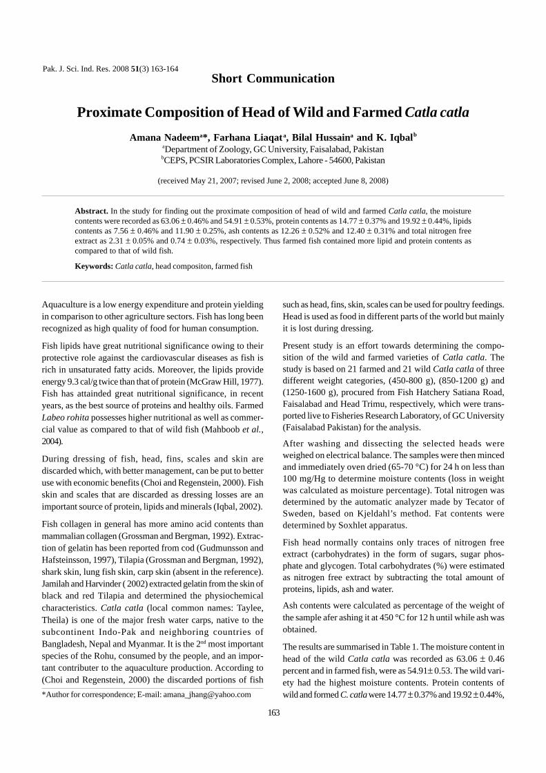
Aquaculture is a low energy expenditure and protein yieldingin comparison to other agriculture sectors. Fish has long beenrecognized as high quality of food for human consumption.
Fish lipids have great nutritional significance owing to theirprotective role against the cardiovascular diseases as fish isrich in unsaturated fatty acids. Moreover, the lipids provideenergy 9.3 cal/g twice than that of protein (McGraw Hill, 1977).Fish has attainded great nutritional significance, in recentyears, as the best source of proteins and healthy oils. FarmedLabeo rohita possesses higher nutritional as well as commer-cial value as compared to that of wild fish (Mahboob et al.,2004).
During dressing of fish, head, fins, scales and skin arediscarded which, with better management, can be put to betteruse with economic benefits (Choi and Regenstein, 2000). Fishskin and scales that are discarded as dressing losses are animportant source of protein, lipids and minerals (Iqbal, 2002).
Fish collagen in general has more amino acid contents thanmammalian collagen (Grossman and Bergman, 1992). Extrac-tion of gelatin has been reported from cod (Gudmunsson andHafsteinsson, 1997), Tilapia (Grossman and Bergman, 1992),shark skin, lung fish skin, carp skin (absent in the reference).Jamilah and Harvinder ( 2002) extracted gelatin from the skin ofblack and red Tilapia and determined the physiochemicalcharacteristics. Catla catla (local common names: Taylee,Theila) is one of the major fresh water carps, native to thesubcontinent Indo-Pak and neighboring countries ofBangladesh, Nepal and Myanmar. It is the 2nd most importantspecies of the Rohu, consumed by the people, and an impor-tant contributer to the aquaculture production. According to(Choi and Regenstein, 2000) the discarded portions of fish
Pak. J. Sci. Ind. Res. 2008 51(3) 163-164
Proximate Composition of Head of Wild and Farmed Catla catlaAmana Nadeema*, Farhana Liaqata, Bilal Hussaina and K. Iqbalb
aDepartment of Zoology, GC University, Faisalabad, PakistanbCEPS, PCSIR Laboratories Complex, Lahore - 54600, Pakistan
(received May 21, 2007; revised June 2, 2008; accepted June 8, 2008)
Abstract. In the study for finding out the proximate composition of head of wild and farmed Catla catla, the moisturecontents were recorded as 63.06 ± 0.46% and 54.91 ± 0.53%, protein contents as 14.77 ± 0.37% and 19.92 ± 0.44%, lipidscontents as 7.56 ± 0.46% and 11.90 ± 0.25%, ash contents as 12.26 ± 0.52% and 12.40 ± 0.31% and total nitrogen freeextract as 2.31 ± 0.05% and 0.74 ± 0.03%, respectively. Thus farmed fish contained more lipid and protein contents ascompared to that of wild fish.
Keywords: Catla catla, head compositon, farmed fish
*Author for correspondence; E-mail: [email protected]
such as head, fins, skin, scales can be used for poultry feedings.Head is used as food in different parts of the world but mainlyit is lost during dressing.
Present study is an effort towards determining the compo-sition of the wild and farmed varieties of Catla catla. Thestudy is based on 21 farmed and 21 wild Catla catla of threedifferent weight categories, (450-800 g), (850-1200 g) and(1250-1600 g), procured from Fish Hatchery Satiana Road,Faisalabad and Head Trimu, respectively, which were trans-ported live to Fisheries Research Laboratory, of GC University(Faisalabad Pakistan) for the analysis.
After washing and dissecting the selected heads wereweighed on electrical balance. The samples were then mincedand immediately oven dried (65-70 °C) for 24 h on less than100 mg/Hg to determine moisture contents (loss in weightwas calculated as moisture percentage). Total nitrogen wasdetermined by the automatic analyzer made by Tecator ofSweden, based on Kjeldahl’s method. Fat contents weredetermined by Soxhlet apparatus.
Fish head normally contains only traces of nitrogen freeextract (carbohydrates) in the form of sugars, sugar phos-phate and glycogen. Total carbohydrates (%) were estimatedas nitrogen free extract by subtracting the total amount ofproteins, lipids, ash and water.
Ash contents were calculated as percentage of the weight ofthe sample afer ashing it at 450 °C for 12 h until while ash wasobtained.
The results are summarised in Table 1. The moisture content inhead of the wild Catla catla was recorded as 63.06 ± 0.46percent and in farmed fish, were as 54.91± 0.53. The wild vari-ety had the highest moisture contents. Protein contents ofwild and formed C. catla were 14.77 ± 0.37% and 19.92 ± 0.44%,
Short Communication
163
respectively. Statistical analysis showed that farmed C. catlagave the maximum protein contents. These results are in linewith Srikanth et al. (1989) who reported that moisture con-tents were lowest and protein deposition, highest under theinfluence of fertilizer treatment in Cyprinus carpio. Decreasein protein contents with an increase in body weight are inagreement with the results of Al-Asgah (1992) who reportedthe crude protein contents had decreasing trend with the in-crease in weight and size of Oreochromis niloticus. Mahboobet al. (2004) also found higher protein content in the farmedLabeo rohita than that in the wild species.
0.03%, respectively, with maximum nitrogen free extract beingrecorded in head of the wild variety.
ReferencesAl-Asgah, N.A. 1992. Variation in carcass composition of
Oreochromics niloticus in relation to body weight andlength. Pak. J. Zool. 24: 47-51.
Choi, S.S., Regenstein, J.M. 2000. Physicochemical and sensorycharacteristics of fish gelatin. J. Food Sci. 65: 194-199.
Grossman, S., Bergman, M. 1992. Process For The Productionof Gelatin from Fish Skin. US Patent No. 5,093,474, 3rd March,1992.
Gudmundsson, M., Hafsteinsson, H. 1997. Gelatin from codskin as affected by chemical treatments. J. Food Sci. 62:37-39, 47.
Jamilah, B., Harvinder, K.G. 2002. Properties of gelatins fromskins of fish black tilapia (Oreochromis mossambicus)and red tilapia (Oreochromis nilotica). Food Chem. 77:81-84.
Hassan, M. 1996. Influence of Pond Fertilization with BroilerDroppings on the Growth Performance and Meat Qualityof Major Carps. Ph.D. Thesis, pp. 1-195, GC University ofFaisalabad, Pakistan.
Iqbal, Z. 2002. Proximate Composition of Scales, Skin in Wildand Farmed Catla catla and Labeo rohita. M.Sc. Thesis,GC University of Faisalabad, Pakistan.
McGraw Hill. 1977. McGraw Hill Encyclopedia of Scienceand Technology. 15th edition, 15 volume set, McGraw HillBook Company Inc., New York, USA.
Mahboob, S., Sheri, A.N. 1997. Relationship among ovaryweight, liver weight and body weight in the grass carpCtenopharyngodon idella. J. Aqua. Trop. 12: 255-259.
Mahboob, S., Liaquat, F., Liaquat, S., Hassan, M., Rafique, M.2004. Proximate composition of meat and dressing lossesof wild and farmed Labeo rohita (Rohu). Pak. J. Zool. 36:39-43.
Shimma, H., Sato, R. 1985. Comparison of proximate compo-sition among the five races of common carp, Cyprinuscarpio. Bull. Natl. Res. Inst. Aquacult. 7: 37-43.
Srikanth, G.N., Nadeesha, M.C., Keshavanath, P., Varghese,T.J., Shetty, H.P.C., Basavaraja, N. 1989. On the applica-bility of mixed feeding schedule for common carpCyprinus carpio Var. Communis. In: Aquaculture in Asia,E.A. Huisman, N. Zoonneveld and A.H. Boumans (eds.),pp. 254-261, Pudoc, Wageningen, The Netherlands.
164
Table 1. Proximate composition of the head of C. catla, wildand farmed varieties (%)
Parameter Catla catla(Mean ± SE) Wild fish Farmed fish
Moisture 63.06±0.46 54.91±0.53Protein 14.77±0.37 19.92±0.44Lipids 7.56±0.46 11.90±0.25Ash 12.26±0.52 12.40±0.31Total nitrogenfree extract 2.31±0.05 0.74±0.03
In the head of wild and formed C. catla, lipids contents weremeasured as 7.56 ± 0.46% and 11.90 ± 0.25%, respectively. Themarked difference in the protein and fat contents of wild fishseem to be due to scarcity of food and the resulting decreasedgrowth. Fish showed progressive reduction in fat resources,yet before reaching a critical low level, proteins began to beutilized for energetic purposes and ultimately a reductionoccured in the protein contents with increased water contents.Hassan, 1996). The fish at first consume lipids from liver andstarts to mobilize muscle proteins only when fat-derived-energy is nearly used up. After that, as protein is utilized,water moves in to take its place. Such a shift results in theincreased water contents that are inversely correlated withprotein and fat reserves of their meats (Shimma and Sato,1985; Mahboob and Sheri, 1997).
Total ash contents in wild and farmed C. catla were 12.26 ±0.52% and 12.40 ± 0.31%, respectively, with little differencebetween them. Total nitrogen free extract in head of wild andfarmed C. catla were recorded as 2.31 ± 0.05% and 0.74 ±
Amana Nadeem et al.
IntroductionHarpagophytum procumbens DC. (Pedaliaceae), commonlyknown as Devil’s claw, is a perennial herbaceous plant nativeto the arid steppes. It is virtually restricted to the southernpart of the African continent, occurring mainly in SouthAfrica, Namibia and Botswana, where it is known locally notonly as Devil’s claw, but also as grapple plant, wood spiderand harpago.
H. procumbens occurs in areas with low annual rainfall(150-500 mm/year) on deep sandy soils of the Kalahari. It isfound in savannah vegetation dominated by Acacia sp., butdoes not compete well with grasses. In fact, H. procumbens ismost often found in areas where grass cover is less than 25%and where the herb cover is less than 20%. It is most abundantin open, trampled and overgrazed areas (Hachfeld, 2002),where it has clumped distribution (Raimondo and Donaldson,2002).
In an area between the northern Cape and north west pro-vinces of south Africa, densities were estimated at 50 plants/ha in the dense grasses of a well managed farm, 150 plants/haon unharvested overgrazed communal land near the village ofMadibeng and only 11 plants/ha on harvested communal landnear Madibeng (Stewart and Cole, 2005).
H. procumbens is a weedy, tuberous plant with creepingannual stems up to 2 m long. The above-ground stems emergeafter the first rains and die back in winter and during droughts.Stems grow from a persistent succulent primary tuber, called
Pak. J. Sci. Ind. Res. 2008 51(3) 165-176
A Comprehensive Systematic Pharmacological Reviewon Harpagophytum procumbens DC. (Devil’s claw)
Elisabetta Miraldi*, Marco Biagi and Daniela GiachettiSection of Pharmaceutical Biology, Department of Environmental Sciences “G. Sarfatti”
University of Siena, Via T. Pendola, 62 - 53100 Siena, Italy
(received March 3, 2008; revised May 28, 2008; accepted June 2, 2008)
Abstract. Popularly known as Devil’s claw, Harpagophytum procumbens DC. (Pedaliaceae) is native to the Kalaharisavannas of southern Africa and Namibia. It has been widely used to treat rheumatism. Its secondary tuberous roots containiridoid glycosides (procumbide, procumboside, harpagoside) as the active principles. This species seems to stimulatemigration of interleukins and leucocytes to painful and inflamed joint areas. The drug is indicated for osteoarthritis,degenerative disease of the joints and arthritic processes. Although, in vivo pharmacological studies have been carried outin different animal models, with different methodologies and different types of extracts, producing contradictory results,recent clinical studies have shown that H. procumbens could be a valid alternative to conventional drugs, especially in thetreatment of lumbar pain.
Keywords: Harpagophytum procumbens, osteoarthritis, joint ailments, irridoid glycosides
*Author for correspondence; E-mail: [email protected]
“mother tuber” by harvesters, the tap root of which can extendto a depth of 2 m. A number of secondary tubers, called“babies”, emanate from the primary tuber via fleshy roots.The secondary tubers are up to 25 cm long and 6 cm thick(Schneider, 1997). The secondary tubers contain up to 46%stachyose, a photosynthetic storage product, which is thoughtto be an adaptation to drought conditions (Stewart andCole, 2005).
The leaves are opposite, blue-green and usually have severallobes. The flowers are tubular and deep mauve-pink withyellow and white throats. They are open for one day and arepollinated by bees (Von Willert and Sanders, 2004). The flatwoody capsules, which give the plant its scientific and com-mon names, bear two rows of curved appendages studdedwith curved spines (Harpagophytum literally means grapplehook plant). The fruit is dispersed by animals (Ernst et al.,1988), as it attaches readily to fur and wool, and is also winddispersed to some degree, as a breeze can carry a fruit somedistance from the parent plant.
Seeds have a high degree of dormancy (Stewart and Cole,2005; De Jong, 1985), which may be an adaptation to drought(Ernst et al., 1988). Ernst et al. (1988) estimated that only20-25% of the seeds in a fruit establish soil contact in a givenyear, suggesting that this may be an adaptation to animal (orwind) dispersal. The same authors also estimated that seedsmay remain viable in the seed bank for more than 20 years, byvirtue of their low respiration rate.
The thick, fleshy, tuberous secondary tap roots of H. procumbensare usually dried and used in south African traditional medicine.
Review
165
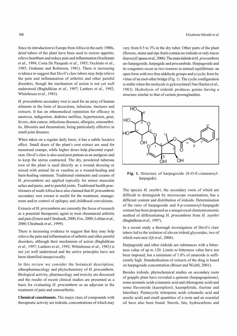
Since its introduction to Europe from Africa in the early 1900s,dried tubers of the plant have been used to restore appetite,relieve heartburn and reduce pain and inflammation (Soulimaniet al., 1994; Costa De Pasquale et al., 1985; Occhiuto et al.,1985; Grahame and Robinson, 1981). There is increasingevidence to suggest that Devil’s claw tubers may help relievethe pain and inflammation of arthritis and other painfuldisorders, though the mechanism of action is not yet wellunderstood (Baghdikian et al., 1997; Lanhers et al., 1992;Whitehouse et al., 1983).
H. procumbens secondary root is used for an array of humanailments in the form of decoctions, infusions, tinctures andextracts. It has an ethnomedical reputation for efficacy inanorexia, indigestion, diabetes mellitus, hypertension, gout,fevers, skin cancer, infectious diseases, allergies, osteoarthri-tis, fibrositis and rheumatism, being particularly effective insmall joint diseases.
When taken on a regular daily basis, it has a subtle laxativeeffect. Small doses of the plant’s root extract are used formenstrual cramps, while higher doses help placental expul-sion. Devil’s claw is also used post partum as an analgesic andto keep the uterus contracted. The dry, powdered tuberousroot of the plant is used directly as a wound dressing ormixed with animal fat or vaseline as a wound-healing andburn-healing ointment. Traditional ointments and creams ofH. procumbens are applied topically for minor muscularaches and pains, and to painful joints. Traditional health prac-titioners of south Africa have also claimed that H. procumbenssecondary root extract is useful for the treatment, manage-ment and/or control of epilepsy and childhood convulsions.
Extracts of H. procumbens are currently the focus of researchas a potential therapeutic agent to treat rheumatoid arthritisand pain (Ernest and Chrubasik, 2000; Fox, 2000; Leblan et al.,2000; Chrubasik et al., 1999).
There is increasing evidence to suggest that they may helprelieve the pain and inflammation of arthritis and other painfuldisorders, although their mechanism of action (Baghdikianet al., 1997; Lanhers et al., 1992; Whitehouse et al., 1983) isnot yet well understood and the active principles have notbeen identified unequivocally.
In this review we consider the botanical description,ethnopharmacology and phytochemistry of H. procumbens.Biological activity, pharmacology and toxicity are discussedand the results of recent clinical studies are presented as abasis for evaluating H. procumbens as an adjuvant in thetreatment of pain and osteoarthritis.
Chemical constituents. The major class of compounds withtherapeutic activity are iridoids, concentrations of which may
vary from 0.5 to 3% in the dry tuber. Other parts of the plant(flowers, stems and ripe fruit) contain no iridoids or only traces(leaves) (Capasso et al., 2006). The main iridoids in H. procumbensare harpagoside, harpagide and procumbide. Harpagoside andits congeners occur as two isomers in mutual equilibrium: anopen form with two free aldehyde groups and a cyclic form byvirtue of an enol-ether bridge (Fig. 1). The cyclic configurationis stable when the molecule is gylcosylated (Van Haelen et al.,1983). Hydrolysis of iridoids produces genins having astructure similar to that of certain prostaglandins.
Fig. 1. Structure of harpagoside (8-O-E-cinnamoyl-harpagide).
The species H. zeyehri, the secondary roots of which aredifficult to distinguish by microscope examination, has adifferent content and distribution of iridoids. Determinationof the ratio of harpagoside and 8-p-coumaroyl-harpagidecontent has been proposed as a unequivocal chemotaxonomicmethod of differentiating H. procumbens from H. zeyehri(Baghdikian et al., 1997).
In a recent study a thorough investigation of Devil’s clawtubers led to the isolation of eleven iridoid glycosides, two ofwhich were new (Qi et al., 2006).
Harpagoside and other iridoids are substances with a bitter-ness value of up to 120. Limits to bitterness value have notbeen imposed, but a minimum of 1.8% of amaroids is suffi-ciently high. Standardisation of extracts of the drug is basedon harpagoside concentration (Bisset and Wichtl, 2001).
Besides iridoids, phytochemical studies on secondary rootsof grapple plant have revealed a quinone (harpagoquinone),some aromatic acids (cinnamic acid and chlorogenic acid) andsome flavonoids (kaempferol, kaempferide, fisetine andluteoline). Pentacyclic triterpenic acids (oleanolic acid andursolic acid) and small quantities of a resin and an essentialoil have also been found. Sterols, fats, hydrocarbons and
166 Elisabetta Miraldi et al.
holosides have also been detected (Van Haelen, 1986). Twonew phenolic glycosides (acteoside and isoacteoside) wereidentified by Burger et al. (1987). Major quantities of carbohy-drates (stachyose) and several tetrahalosides bearing glucoseand sucrose molecules have also been found. The presence ofstachyose indicates a chemotaxonomic relationship of thePedaliaceae family with the Lamiaceae and Verbenaceae (VanHaelen, 1986).
Pharmacology. Commercial preparations of Harpago contain1.4-2.0% of harpagoside. The results of research into thisspecies are difficult to compare and interpret, mainly becauseof the variety of extracts used. Methanolic extract and aqueousextract were the most common but differed in harpagosidecontent.
The World Health Organisation (WHO, 2007) lists Devil’s clawfor rheumatic pain, loss of appetite and dyspepsia and asadjuvant for degenerative disease of the locomotor system.
In vitro experiments. Recent in vitro studies, well listed in thereview published by Grant et al. (2007), indicate that grappleplant preparations may interact with the inflammationcascade, including synthesis and activity of cytokines.Aqueous extract has been found to suppress expression ofcyclooxygenase-2 (COX2), stimulated by lipopolysaccharides(LPS) and nitric oxide synthetase, in murine L929 fibroblasts.Lipopolysaccharides trigger a wide series of cell responsesthat play a major role in the pathogenesis of inflammatoryreactions, including activation of inflammatory cells andproduction of cytokines and other mediators. ProstaglandinE2, synthesized by arachidonic acid via the cyclooxygenaseenzyme complex, is a key mediator. The cyclooxygenase-1(COX1) isoform is constitutional whereas COX2 is onlyexpressed in response to proinflammatory signals, such asthose of cytokines and bacterial endotoxin LPS. COX2produces a large quantity of prostaglandin E2 which causesinflammation (Mitchell et al., 1995).
Nitric oxide (NO), synthesized from L-arginine by NO-synthetase (NOS), plays a major role in the regulation ofmany physiological processes. The various isoforms ofNOS can be classified as inducible (iNOS), endothelial(eNOS) and neuronal NOS (nNOS). The first of the threeis involved in inflammatory phenomena.
The anti-inflammatory and analgesic effect of aqueous extractof Devil’s calw was recently confirmed (Jang et al., 2003) byassessing cell viability (MTT test) and expression of COX2and iNOS (quantity of nitrite in cell supernatant) by reversetranscription polymerase chain reaction (RT-PCR) in L929fibroblasts stimulated with LPS. The suppression of COX2
and iNOS expression found inhibited prostaglandin E2synthesis.
Another study by Kaszkin et al. (2004) evaluated the utilityof Harpago extract in the treatment of inflammatory kidneydisease. Two extracts containing 8.9% (ex1) and 27% (ex2)harpagoside were tested for effects on IL-1-induced produc-tion of NO and on regulation of transcription of iNOS in ratrenal cells. An 80% dose-dependent reduction in the forma-tion of NO, due to inhibition of iNOS expression, wasobserved. A reduction in activity of the iNOS promotor ofnuclear translocation of NF-kB was also found, indicatingthat the extracts impaired transcriptional activation of iNOS.This effect was confirmed by Huang et al. (2006) who demon-strated inhibition of mRNA levels induced by LPS and COX2and iNOS expression in human hepatocarcinoma cells.Harpagoside also blocked activity of the NF-kB promoterstimulated by LPS in a dose-dependent manner in RAW264.7cells. The results of the study show that inhibition of COX2and iNOS expression by harpagoside involves suppression ofNF-kP activation, resulting in inhibition of the inflammatoryprocess and associated pain.
Other extracts containing lower concentrations of harpagosidedid not cause this inhibition. Harpagoside alone only works atconcentrations in the range 0.3-1.0 mg/ml which are much higherthan those of total extracts, and a preparation completelydevoid of the molecule strongly inhibited iNOS expression,indicating that other constituents are also involved in itsactivity. An extract also had a strong antioxidant effect whichharpagoside does not have (Rindone, 2006).
A recent study investigated the mechanism of action ofharpagoside, using human HepG2 hepatocarcinoma andRAW 264.7 macrophage cell lines. Harpagoside inhibitedLPS-induced mRNA levels and protein expression of COX2and inducible nitric oxide in HepG2 cells. These inhibitionsappeared correlated with suppression of NF-kB activationby harpagoside, suggesting that inhibition of expression ofCOX2 and iNOS by harpagoside involves suppression ofNF-kB activation, thereby inhibiting downstream inflamma-tion and pain (Huang et al., 2006). These results show thatcertain extracts of Devil’s claw could be an interesting sourceof drugs for treating glomerular and other inflammatorydiseases.
Other in vitro studies have shown a significant decrease insynthesis of cell membrane degradation enzymes in isolatedchondrocytes and dose-dependent inhibition of elastase. In-flammatory diseases of the joints, such as rheumatoid arthritisand osteoarthritis are characterised by loss of joint cartilagerelated to an imbalance between synthesis and breakdown of
167Pharmacological Review on H. procumbens
the cartilagionous extracellular matrix. These diseases areaccompanied by elevated induction of cytokines such asIL-1b and TNF-a. Increased release of cytokines leads toincreased production of degradation enzymes, such asmetalloproteases (MMPs). A significant reduction in MMPsynthesis was recently demonstrated in human chondrocytesstimulated with IL-1b. Two extracts were used. The first con-tained 210 mg and the other 480 mg of dry extract; the latterwas more effective (Schulze-Tanzil et al., 2004). The effect ofDevil’s claw on arthritis could be due to its capacity to sup-press MMP production by inhibiting synthesis of proinflam-matory cytokines.
As far as leucocyte elastase is concerned, aqueous extractshowed rather weak, dose-dependent inhibitory activity(Boje et al., 2003).
A study by Fiebich et al. (2001) on H. procumbens extractSteiHap 69 (Steiner Harpagophyton procumbens extract 69)demonstrated its close-dependent anti-inflammatory effectsby preventing the LPS-induced synthesis of tumour-necrosisfactor α (TNF*) by human monocytes. However, harpagideand harpagoside had no effect on LPS-induced TNF* release.
In vivo experiments. Anti-inflammatory and analgesicactivity. At first sight, the results of the few pharmacologicalstudies conducted on H. procumbens seem contradictory,since they were based on different methods and on differentanimal models (acute and subacute inflammation) with differentextracts administered by different routes (Bruneton, 2002).Recent results show that aqueous extract administered intra-peritoneally at doses from 100 to 400 mg/kg is active in a dose-dependent manner on carrageenan-induced edema in ratpaws. The same extract was inactive when administered orally,presumably due to gastric breakdown of the active principle.The extract was also inactive when given parenterally if pre-viously treated with acids. It was active when administeredintra-duodenally.
Under the same experimental conditions, harpagoside wasinactive, but was found to promote peripheral analgesicactivity of the aqueous extract (100 mg/kg i.p.). The authorssuggested research into formulations that could improvethe bioavailability of oral harpagoside. In a pilot study,plasma concentrations of 4 ng/ml and 15 ng/ml harpagosidewere found 15 min and 2 h, respectively, after administrationof 600 mg extract containing 50 mg harpagoside (Loew et al.,2001). However, the role of harpagoside remains controver-sial. Oral administration of aqueous extract of Scrophulariafrutescens DC. (Scrophularieceae) to rats is reported to haveantiinflammatory activity on carrageenan-induced edema thatdoes not seem related to harpagoside (Garciá et al., 1996).
Adjuvant-induced arthritis is often used as a model of chronicand subchronic inflammation and is important in the studyof pharmacological control of inflammatory proceses and inassessing the antiinflammatory and analgesic properties ofdrugs. One reason for the widespread use of this model has todo with the strong correlation between efficacy of drugs inthis model and efficacy in humans (rheumatoid arthritis). In astudy aimed at investigating the antiinflammatory effects ofH. procumbens, Andersen et al. (2004) induced arthritis byacute and chronic treatment. A single administration of threedoses of extract (25, 50 and 100 mg/kg) had some analgesiceffect, measured on the basis of response to thermal stimuli(hot plate test). The same effect was observed after chronicadministration.
Kundu et al. (2005) reported a study of the antiinflammatorymechanism of Devil’s claw. Methanol extract was tested foreffects on expression and COX2 induced by TPA (a phorbolester) in mouse skin. Topical application of the extract inhibi-ted directly-induced COX2 expression, decreasing the cata-lytic activity of extracellular signal-regulated protein kinase(ERK), which regulates activation of transcription factors thatmediate COX2 induction. The extract inhibited activation ofactivator protein-1 (AP1) and attenuated expression of its keycomponent c-Fos, while nuclear factor NFB was unaffected(Kundu et al., 2005).
Another mechanism of action study (Na et al., 2004) assessedinhibition of COX2 expression induced by TPA in humanmammary epithelial cells MCF10A and on mouse skin in vivo.Methanol extract of devil’s claw inhibited binding of NFBtranscription factor to DNA in MCF10A cells in a dose-dependent way. Suppression of DNA binding suggests achemopreventive effect.
In a study, Mahomed and Ojewole (2004) evaluated the anal-gesic effect (hot plate and acetic acid test), antiinflammatoryeffect (albumin-induced edema) and antidiabetic effect(streptozotocine-induced diabetes) of aqueous extract in rats.Diclofenac (100 mg/kg i.p.) and clorpropamide were used asreference drugs. Aqueous extract (50-800 mg/kg i.p.) had asignificant analgesic effect on nociceptive thermal andchemical stimulation in mice and a significant dose-dependentreduction in albumin-induced edema. The extract also causeda reduction in plasma levels of glucose in normal rats andrats with streptozotocin-induced edema. These results seemto explain popular use of this plant as an analgesic and toalleviate inflammation and diabetes in south Africa.
Clinical studies. There have not been many clinical tests onhumans and their heterogeneity makes it difficult to drawconclusions. It has been demonstrated that grapple plant
168 Elisabetta Miraldi et al.
alleviates pain in chronic inflammation of the joints but itsefficacy seems less than that of aspirin and indomethacin(Newall et al., 1996). However, certain preparations of the drugand special physical exercises can be a valid alternative tosynthetic drugs, especially in the treatment of lumbago.
The results of studies on H. procumbens are not easy to com-pare and interpret, mainly because of the variety of extractsused. Methanol and aqueous extracts are, however, the mostwidely used in these studies. In Europe, Doloteffin, extractWS 1531® and extract LI174® are used. The preparations differin their harpagoside content.
Since H. procumbens has a broader mechanism of actionthat conventional NSAIDs, inhibiting both cyclooxygenasesand lipooxygenases involved in the metabolic pathway ofarachidonic acid, it has been tested clinically in certainrheumatic inflammatory diseases.
The primary end-points of the clinical trials conducted onH. procumbens were a reduction of inflammation and pain, aswell as improved movement and mobility of patients withrheumatism. As shown by the examples that follow, the plantproved to be just as effective in reducing these parameters ascertain conventional treatments with which it was compared.To assess the efficacy of H. procumbens in the treatmentof lumbago and osteoarthritis, it is worth considering thesystematic review of clinical studies by Grant et al. (2007),by Setty and Sigal (2005), and by Gagnier et al. (2004).
Searches in PubMed, Embase, Cochrane Controlled TrialsRegistry, Cochrane Musculoskeletal Specialized Register,Dissertation Abstracts, BIDS ISI and Cochrane Complemen-tary Medicine Fields Specialized Register brought to light130 references to clinical studies on H. procumbens. Afterexclusion of duplicates and articles based on inadequatequalitative criteria, 12 randomized studies were selected, fouron the treatment of lumbar pain (Chrubasik et al., 2003, 1999,1996), five on the treatment of osteoarthritis (Biller, 2002;Frerik et al., 2001; Chantre et al., 2000; Lecomte and Costa,1992; Schruffler, 1980) and three on the treatment of variousother forms of pain involving muscles and bones (Pugno,2006).
Of the four studies on treatment of lumbago (total number ofpatients 505), two compare H. procumbens with placebo, onewith various conventional therapies (e.g., antiinflammatorydrugs, massage, physical exercise) and one with COX2 inhibi-tor, rofecoxib. Of the five studies on treatment of osteoarthritis(total number of patients 385), three compare the plant withplacebo and two with standard drugs. All three studies ontreatment of other forms of muscle and bone pain comparedH. procumbens with placebo.
A recent study examined, whether the anti-inflammatoryresponse to whole extract of H. procumbens in rats was aconsequence of adrenal corticosteroid release. Carrageenan-induced inflammatory response in the hindpaws was evalu-ated in control, sham-operated and adrenalectomized rats. Theextract was administered orally (by gavage) or intraperitoneally,30 min prior to nociceptive stimulus. Blood samples were thencollected, and the number of circulating leukocytes was esti-mated. The results showed that whole extract of H. procumbensadministered intraperitoneally had an inhibitory effect on acuteinflammatory response, irrespective of the participation ofadrenal corticosteroids. When administered orally, the extractwas ineffective (Catelan et al., 2006).
Lumbar pain. Chrubasik et al. (1997) treated 102 lumbagopatients with 4500 mg/day of aqueous extract of H. procumbens(30 mg/day harpagoside) or with conventional NSAIDs or withphysical manipulation, for 6 weeks. No significant differencesin pain-free time and change in Arthus index were foundbetween patients undergoing the three treatments.
Chrubasik et al. (1999) subsequently treated 197 patientswith pseudoradiating and non radiating lumbago with 4500mg/day of aqueous extract of dried tubers of H. procumbens(50 mg/day harpagoside), with 9000 mg/day of the sameextract (100 mg/day harpagoside) or with placebo. Patientstreated with either dose of extract showed an improvementin pain with respect to patients treated with placebo.
The next prospective, randomized, double-blind study byChrubasik et al. (2003) was particularly interesting, because itcompared the effects of commercial aqueous extract ofdessicated, pulverized tubers with that of the latest genera-tion synthetic antiinflammatory agent, rofecoxib, a selectiveCOX2 inhibitor. The subjects were 88 patients, age 45-75 years,with at least a six-month history of rheumatic back pain, wors-ening in the 8 weeks prior to the study, divided into two groupsof 44 patients. The first group received 2400 mg/day of extract(60 mg/day harpagoside) for 6 weeks plus one tablet/day ofplacebo. The second group received a tablet per day ofrofecoxib (12.5 mg) and two tablets of placebo three times aday. Patients of both groups could also take up to 400 mg/daytramadol drops (2.5 mg/ml), a synthetic opioid receptoragonist.
During treatment, patients kept a diary in which they scoredpain intensity on a 5 point scale (no pain, slight, moderate,strong and acute pain) and the daily dose of tramadol taken.Patients also answered a standardised questionnaire on gen-eral health, daily activities and type of pain, a questionnaireon depression (Beck Depression Inventory BDI) and a healthassessment questionnaire (HAQ).
169Pharmacological Review on H. procumbens
Forty-three patients in the H. procumbens group and 36 in therofecoxib group completed treatment. The number of patientsresponding completely to therapy increased up to the fifthweek, more or less in the same manner in both groups. At week6, the total number of responses reached 17% of patients (10patients in the H. procumbens group and five patients in therofecoxib group). Comparison of the weekly mean pain scoresbetween weeks four and six did not show significant diffe-rences in the number of patients showing 20-50% improve-ment in the two groups. The Arthus pain index decreased byabout 10% during treatment. The greatest reduction wasrecorded in week 2.
Thirty-four of the 88 patients (38.6%) resorted to tramadol, 21in the H. procumbens group and 13 in the rofecoxib group.Mean consumption of tramadol over 6 weeks was 230 mg inthe first and 133 mg in the second group.
Fourteen patients reported side-effects on 39 occasions, 28of which (13 in the H. procumbens group) could be ascribedto treatment. Gastrointestinal disorders were reported by 8patients in the H. procumbens group and 9 in the rofecoxibgroup; in the latter, gastrointestinal effects were more severe,causing 5 patients to drop out.
Analysis of the results did not reveal any statistically signifi-cant differences in effect on lumbar pain between the twotherapies. The study was completed with follow-up one yearlater (Chrubasik et al., 2005). Thirty-eight patients previouslyin the H. procumbens group and 35 in the rofecoxib groupwere treated daily up to 54 weeks with a dose of extractcontaining 60 mg harpagoside. Fifty-three patients completed24 weeks of follow-up and 43 completed 54 weeks. No signifi-cant difference in daily pain score, resort to other analgesics,Arthus index and HAQ score was found between patientspreviously in the two groups. Apart from individual fluctua-tions, follow-up showed a slight improvement in Arthus indexand HAQ with respect to the initial study. On the basis of21,761 patient days, the percentages of patients who werepain-free, or with slight, moderate, strong or acute pain were28, 39, 22, 8.5 and 1.5%, respectively. Three patients reportedminor side-effects.
The authors concluded that maintenance therapy withH. procumbens in patients previously treated with the sameextract or with rofecoxib produced similar effects and thatlong-term treatment with the extract was well tolerated.
Osteoarthritis. In 1980, Schrulffer treated 50 osteoarthritispatients with 2500 mg/day of aqueous extract of H. procumbens(less than 30 mg/day harpagoside) or with phenylbutazonefor 30 days. Grapple plant was found to attenuate pain andimprove physical condition better than phenylbutazone.
Lecomte and Costa (1992) treated 89 patients with arthritisof different joints (spine, neck, hip, knee) with powderedsecondary root of H. procumbens (200 mg/day equivalent to60 mg/day harpagoside) or placebo for 60 days. The Schobertest showed that pain improved more in patients receivingH. procumbens than in those receiving placebo.
Chantre et al. (2000) treated 122 osteoarthritis patients withhip or knee pain with 4500 mg/day of powder of cold-driedsecondary roots of grapple (equivalent to 57 mg/dayharpagoside) or with the NSAID diacerein, also known asdiacetylrhein, for 16 weeks. Intent-to-treat analysis showedthat the plant was not less effective than the drug.
In 2001, Frerik et al., treated 46 osteoarthritis patients with4500 mg/day hydroethanolic extract (TeufelskrallenextraktLoHar 45) (60% ethanol) of H. procumbens (equivalent toless than 30 mg/day harpagoside) or placebo for 20 weeks.Following a specific phytotherapeutic sheme, patients alsotook ibuprofen; among aims of the study, there was to knowthe percentage of patients responding to therapy: 71% ofpatient taking H. procumbens responded to therapy comparedto 41% of those on placebo. The difference was statisticallysignificant (p=0.041) (method of statistical analysis notindicated).
In 2002, Biller, treated 78 osteoarthritis patients with knee painwith 4500 mg/day of hydroalcoholic extract of H. procumbens(60% alcohol; less than 30 mg/day harpagoside) or placebofor 20 weeks. Patients also took 800 mg/day ibuprofen fromweek 1 to week 8, and 400 mg/day from week 9 to week 16.From week 17 to week 20 they took Grapple plant alone orplacebo. The end-points of the study were the percentage ofpatients responding to therapy, variations in the WOMACindex of pain and non-recourse to ibuprofen in weeks 17-20.Ninety percent of patients in the grapple group respondedto therapy compared to 80% in the placebo group. Meanuse of ibuprofen in weeks 17-20 was one tablet/day in theH. procumbens group and 5 tablets in the placebo group. Theplant was therefore more effective than placebo.
In 2007, Chrubasik et al., recruited 114 patients (56 with chronicnonspecific lower back pain, 37 with osteoarthritic knee and21 with osteoarthritic hip pain) into a survey of the effects ofDoloteffins at a dose providing 60 mg harpagoside per day forup to 54 weeks. Their symptoms and well-being were moni-tored at 4-6 week intervals by disease-specific and genericoutcome measures, and patients also kept a diary of their painand need for rescue medication. The principal analyses werebased on Intention to Treat (ITT) with Last Value CarriedForward (LOCF). Multivariate Analysis of Variance (MANOVA)indicated appreciable overall improvement during the survey,
170 Elisabetta Miraldi et al.
similar in the back, knee and hip groups. In separate ANOVAs,most of individual outcome scores decreased significantlyover time. Multiple regression analysis indicated that changesfrom baseline were independent of patient characteristics.Additional analgesic requirements (which were very modest)declined during the year of the survey. “Response duringtreatment” was achieved in 75% of patients, and was reflectedin the percentages of those who rated the treatment “good” or“very good”. Side-effects were few and minor.
In 2007 Warnock and coworkers, published a 8-week openlabel clinical trial to examine the effectiveness of a Devil’s clawpreparation (Bioforce, Scotland, United Kingdom) in thetreatment of arthritis and other rheumatic conditions, such asarthritic dominant hands. A particular form of osteoarthritisis hand osteoarthritis; this form of arthritis does not seem todepend on any evident cause and mainly affects certain jointsof the hand, especially the distal interphalangeal joints, whichgradually develop hard swellings known as Heberden nodules,the proximal interphalangeal joints, the tumefactions of whichare known as Bouchard nodules, and the metacarpal trape-zium, which often becomes partly dislocated. The structuralchanges (reduced thickness of joint cartilage, sclerosis,subchondrial geodes and osteophyte formation) cause chronicpain and dysfunction with relapsing-remitting acute phasesrelated to joint misalignment (distress of compartmentalcapsule and ligaments) and secondary synovitis.
Radiologically, the disease is characterised by restriction ofthe joint line, small pseudocysts and marginal osteophytes.Subluxation can occur with time. The disease is not accompa-nied by changes in humoral parameters. It mainly affects womenover 40 years of age and is progressive, though the speed ofprogression varies from person to person.
No drugs have yet been demonstrated to slow or arrestprogression of joint damage, though cartilage protectorssuch as diacerein and glycosaminoglycans are widelyused. NSAIDs are usually used to control pain and havethe advantage of also controlling secondary inflammatoryreactions. However, NSAIDs are contraindicated in casesof gastritis, gastric ulcer, kidney failure, heart failure andhypertension and cannot be taken with other drugs suchas oral anticoagulants. They have frequent gastrointesti-nal, renal and cardiovascular side-effects.
In the study of Warnock et al. (2007) patients aged 18-75 yearswith mild to moderate rheumatic disorders were enrolled. Thepatients received a daily dose of 2 Devil’s claw tablets contain-ing a total daily dose of 960 mg Devil’s claw dry extract madewith 60% ethanol (DER 1.5-3:1). Effectiveness was measuredusing a global assessment of pain function and stiffness in
the effected area with a numerical rating scale (NRS), as well asjoint-specific assessments. The WOMAC scale was used toassess effects on arthritic dominant hands. In addition, a gen-eral assessment of perceived effectiveness on a 6-point scale,onset of action, the SF-12 Quality of Life Questionnaire, anddaily patient diaries were used to assess effectiveness.
A total of 222 patients were included in the Intention to Treat(ITT) analysis. Compliance at week 8 was 75-100% for 140patients. The average global scores for pain, stiffness, andfunction were significantly improved from baseline to weeks 4and 8 (P<0.0001 for both). The average scores for pain in thehand, wrist, elbow, shoulder, hip, knee and back were signifi-cantly improved from baseline to week 8 (P<0.05) and week 4(P<0.01) (except for right elbow). In addition, the averageWOMAC subscale scores (knee: n=114, hip: n=68) were sig-nificantly improved from baseline to weeks 4 and 8 (P<0.0001for both). The average Algofunctional Hand OsteoarthritisIndex (n=113) scores were also significantly improved frombaseline to week 8 (P<0.0001). Average finger floor distancesof patients with back pain (n=81) were significantly improvedfrom baseline to week 8 (P<0.0001).
In addition, more than half of the patients rated the Devil’sclaw treatment as “excellent” or “good”; and investigatorsrated the Devil’s claw extract’s effectiveness as “excellent” or“good” in more than half of the patients. A total of 171 out of222 patients (77%) reported a beneficial effect due to the devil’sclaw tablets. Of these patients, 104 reported feeling an effectbetween 1 and 4 weeks from baseline. Average scores for pain,daily function, and stiffness in the patient’s daily diaries weresignificantly improved from week 1 to weeks 2 and 8 (P<0.0001)for both). In addition, the average SF-12 scores on the physi-cal and emotional subscales were significantly improved frombaseline to weeks 4 and 8 (n=207, P<0.0001 for both). Almosthalf (44.8%) of patients taking analgesics for rheumatic disor-ders at baseline had reduced their dosage at week 8; and 26%had completely stopped taking analgesics at week 8. Therewere no significant changes in blood parameters, liver func-tion, or vital signs during the study. A total of 49 possibly orprobably drug-related adverse events were reported, noneconsidered serious. The majority of the adverse events repor-ted were mild to moderate gastrointestinal complaints. Thetolerability of the devil’s claw tablets was rated as “good” bythe majority (87.4%) of patients. In addition, 74.3% of thepatients stated that they would take the devil’s claw tabletsagain.
Effect on smooth muscle contraction. A number of studieshave shown that dry extract of Devil’s claw, up to 40 μg/ml,slightly increases the amplitude and tone of contractions ofisolated jejunum. At higher doses tone decreases and the
171Pharmacological Review on H. procumbens
amplitude of contractions declines sharply. The same biphasiceffect on contractions has also been observed in isolatedrabbit uterus (Occhiuto et al., 1985).
Effect on cardiovascular system. Intragastric administrationof 100 mg/kg body weight of an aqueous or methanol extractof H. procumbens root protected rats against ventriculararrhythmias induced by epinephrine-chloroform and calciumchloride (Circosta et al., 1984). Intraperitoneal administrationof 25 mg/kg bw of a methanol extract of H. procumbens rootsinhibited cardiac arrhythmias induced by aconitine, epineph-rine-chloroform and calcium chloride in fasted rats. Intragastricadministration of 300-400 mg/kg bw of a methanol extract ofH. procumbens root to normotensive rats reduced heart rateand arterial blood pressure (Circosta et al., 1984). Anotherstudies have demonstrated that lower doses of the extracthave slight negative chronotropic and positive inotropiceffects (Occhiuto et al., 1985), whereas larger doses have amarked inotropic effect, with reduction in coronary bloodflow. The inotropic effect is attributed to harpagide (Costaet al., 1985).
Antidyspeptic activity. As bitter tonic H. procumbens hassimilar effects to gentian with prokinetic effects on gastrointes-tinal smooth muscle and is indicated in cases of hyposecretorydyspepsia with loss of appetite (WHO, 2007).
A decoction of Radix Harpagophyti is one of the strongestbitter tonics known (Weiss and Fintelmann, 2000). Ingestionof an infusion prepared from the root (dose not specified) overa period of several days led to an improvement in the symp-toms of disorders of the upper part of the small intestine, whichwere accompanied by disturbances of choleresis and bilekinesis (Weiss and Fintelmann, 2000). It has been proposedthat, because the root is very bitter, is a good stomachic andstimulates the appetite, it may also be useful for the treatmentof dyspeptic complaints (Jaspersen-Schib, 1989; Czygan andKrüger, 1977).
Pharmacokinetics. Commercial extract of H. procumbens takenorally has been shown to produce a plasma peak of harpagosidein humans within 1.3-2.5 h (Loew et al., 2001). Iridoid glyco-sides are metabolised by human intestinal flora to aucubin B,a pyridine alkaloid. The reaction seems to be catalyzed by theenzyme α-glycosidase in the presence of ammonium ions(Baghdikian et al., 1999a, 1999b).
A special role is certainly played by gastric acid which mayactivate or inactivate iridoid glycosides. It has been suggestedthat the products of acid hydrolysis have antiinflammatoryand antirheumatic activity and that contact with gastricsecretions is important. Others sustain that glycosides are
more active than the corresponding genines and that gastricacid disactivates the active principle. Indeed, aqueous extractreduces carrageenan-induced edema in rats if administeredintraperitoneally or intraduodenally, but not when takenorally (Soulimani et al., 1994).
Toxicity. Long-term toxicity does not seem to have been stu-died. Certain authors underline the need for such studies,particularly in view of the widespread use of the plant inphytotherapy and as complementary therapy, especially incases in which prescription of anti-inflammatory drugs isunjustified, at least in the early stages of disease. Pharmacolo-gical studies with experimental animals have shown thatH. procumbens is well tolerated: the i.p. DL50 of harpagosideand harpagide in mice is 1 and 3.2 g/kg, respectively (Cardini,2006).
Side-effects. Mild infrequent gastrointestinal symptoms werereported in clinical trials (Chrubasik et al., 1999; Belaiche, 1982)and in cases of known hypersensitivity. Digestive side-effectsinclude diarrhoea (Chantre et al., 2000), dyspepsia and senseof satiety (Wegener and Lupke, 2003), though the risk ofside-effects is much less than that with synthetic analgesics(Chrubasik, 2004) and the plant is very well tolerated (Chrubasik,2004).
Contraindications. Radix Harpagophyti is contraindicatedin cases of gastric and duodenal ulcer and known hypersen-sitivity to the roots, because the bitter taste stimulates pepticacid (Blumenthal, 1998; Hänsel et al., 1993). Owing to a lack ofsafety data, Radix Harpagophyti should not be used duringpregnancy and nursing, due to possible oxytocic effects(Mabey et al., 1988).
Since H. procumbens has antiarrhythmic activity (Circostaet al., 1984), it should not be taken in association with antiar-rhythmic drugs. A possible interaction with anticoagulantssuch as Warfarin, that could promote bleeding, has beenreported (Heck et al., 2000; Shaw et al., 1997). It is well knownthat a number of herbs and foods alter the metabolism ofWarfarin by acting on cytochrome P450. Budzinski et al. (2000)demonstrated that Devil’s claw did not inhibit the activity ofcytochrome P450 isoenzymes in vitro. On the other hand, Ungerand Frank (2004) described a liquid chromatography/massspectrometry method with automated online extraction tosimultaneously determine the in vitro inhibitory potency ofherbal extracts on six major human drug-metabolising cyto-chrome P450 enzymes. The authors concluded that popularherbal remedies, including Devil’s claw root, could be identi-fied as inhibitors of the applied CYP enzymes with IC(50)values between 20 and 1000 μg/ml. Unger and Frank (2004)
172 Elisabetta Miraldi et al.
showed that this inhibition could be the reason for druginteractions of clinical significance.
These controversial results are not easy to compare; morestudies are needed. However, on the basis of pharmacologicalconsiderations, ESCOP (2003) does not exclude interactionwith antiarrhythmics and warns against possible interactionwith Warfarin.
Other precautions. No information is available on precau-tions concerning drug and laboratory test interactions,carcinogenesis, mutagenesis, impairment of fertility,teratogenic effects during pregnancy or paediatric use.
Preparations. Dried root for decoctions and infusions, pow-dered root or extract in capsules, tablets, tinctures and oint-ments (Bisset, 1994; Iwu, 1993).
Store in the dark in a sealed container.
Posology. (Unless otherwise indicated). Daily dose for loss ofappetite: 1.5 g of root in decoction, 3 ml of tincture (1:10, 25%ethanol); for painful arthritis or tendonitis 1.5-3.0 g of root indecoction, three times a day, 1-3 g of root or equivalent (WHO,2007).
ReferencesAndersen, M.L., Santos, E.H., Seabra, Mde, L., da Silva, A.A.,
Tufik, S. 2004. Evaluation of acute and chronic treatmentswith Harpagophytum procumbens on Freund’s adjuvantarthritis in rats. J. Ethnopharmacol. 91: 325-330.
Baghdikian, B., Guiraud-Dauriac, H., Ollivier, E., N’Guyen, A.,Dumenil, G., Balansard, G. 1999a. Formation of nitrogen-containing metabolites from the main iridoids of Harpago-phytum procumbens and H. zeyheri by human intestinalbacteria. Planta Med. 65: 164-166.
Baghdikian, B., Olivier, E., Faure, R., Debrauwer, L., Rathelot,P., Balansard, G. 1999b. Two new pyridine monoterpenealkaloids by chemical conversion of a commercial extractof Harpagophytum procumbens. J. Nat. Prod. 62: 211-213.
Baghdikian, B., Lanhers, M.C., Fleurentin, J., Ollivier, E.,Maillard, C., Balansard, G., Mortier, F. 1997. An analyticalstudy: anti-inflammatory and analgesic effects ofHarpagophytum procumbens and Harpagophytumzeheri. Planta Med. 63: 171-176.
Belaiche, P. 1982. Étude clinique de 630 cas d’arthrose traitspar le nébulisat aqueux d’Harpagophytum procumbens(Radix). Phytotherapy 1: 22-28.
Biller, A. 2002. Ergebnisse zweler randomisieter kontrollierter.Phytopharmaka 7: 86-88.
Bisset, N.G., Wichtl, M. (eds.) 2001. Herbal Drugs and
Phytopharmaceuticals, Med. Scientific Publishers,Stuttgart, Germany.
Bisset, N.G. (ed.) 1994. Herbal Drugs and Phytopharma-ceuticals, CRC Press, Boca Raton, Florida, USA.
Blumenthal, M., (ed.) 1998. The Complete German CommissionE monographs. Therapeutic Guide to Herbal Medicines,American Botanical Council, Austin, Texas, USA.
Boje, K., Lechtenberg, M., Nahrstedt, A. 2003. New andknown iridoid and phenylethanoid glycosides fromHarpagophytum procumbens and their in vitro inhibitionof human leukocyte elastasi. Planta Med. 69: 820-825.
Bruneton, J. 2002. Pharmacognosie, Phytochimie, PlantesMédicinales, 4th edition, Tec & Doc, Paris, France.
Budzinski, J.W., Foster, B.C., Vandenhoek, S., Arnason, J.T.2000. An in vitro evaluation of human cytochrome P4503A4 inhibition by selected commercial herbal extracts andtinctures. Phytomed. 7: 273-282.
Burger, J.F.W., Brandt, E.V., Ferriera, D. 1987. Iridoid andphenolic glycosides from Harpagophytum procumbens.Phytochemistry 26: 1453-1457.
Capasso, F., Grandolini, G., Izzo, A. 2006. Fitoterapia, SpringerVerlag Italia, Milano, Italy.
Cardini, C. 2006. Harpagophytum procumbens: Farmacoci-netica, tossicologia, effetti collaterali, controindicazioni,interazioni, precauzioni speciali. Piante Medicinali 18-19.
Catelan, S.C., Belentani, R.M., Marques, L.C., Silva, E.R., Silva,M.A., Caparroz-Assef, S.M., Cuman, R.K.N., Bersani-Amado, C.A. 2006. The role of adrenal corticosteroids inthe anti-inflammatory effect of the whole extract ofHarpagophytum procumbens in rats. Phytomed. 13:446-451.
Chantre, P., Cappelaere, A., Leblan, D., Guedon, D.,Vandermander, J., Fournie, B. 2000. Efficacy and toleranceof Harpagophytum procumbens versus diacerhein intreatment of osteoarthritis. Phytomed. 7: 177-183.
Chrubasik, S., Chrubasik, C., Kunzel, O., Black, A. 2007.Patient-perceived benefit during one year of treatmentwith Doloteffin®. Phytomed. 14: 371-376.
Chrubasik, S., Künzel, O., Thanner, J., Conradt, C., Black, A.2005. A 1-year follow-up after a pilot study with Doloteffin®
for low back pain. Phytomed. 12: 1-9.Chrubasik, S. 2004. Devil’s claw extract as an example of the
effectiveness of herbal analgesics. Orthopade. 33: 804-808.
Chrubasik, S., Model, A., Black, A., Pollak, S. 2003. A rando-mized double-blind pilot study compating Doloteffinand Vioxx in the treatment of low back pain. Rheumatol.42: 141-148.
173Pharmacological Review on H. procumbens
Chrubasik, S., Junck, H., Breitschwerdt, H., Conradt, C., Zappe,H. 1999. Effectiveness of Harpagophytum procumbensWS 1531 in the treatment of exacerbation of low backpain: a randomized placebo controlled double-blindstudy. Eur. J. Anesth. 16: 118-129.
Chrubasik, S., Schmidt, A., Junck, H., Pfisterer, M. 1997.Wirksamkeit und Wirtschaftlichkeit von Teulfelskrallen-wurzelextrakt bie runckenschmerzen: Erst ergbnisse einertherapeutischen kohortenstudie. Forsch Komplemen-tarmed. 332-336.
Chrubasik, S., Zimpfer, C.H., Schutt, U. 1996. Efficacy andtolerance of Harpagophytum procumbens in the treat-ment of acute low back pain. Phytomed. 7: 1-10.
Circosta, C., Occhiuto, F., Ragusa, S., Trovato, A., Tumino, G.,Briguglio, F., De Pasquale A. 1984. A drug used in tradi-tional medicine: Harpagophytum procumbens DC. II.cardiovascular activity. J. Ethnopharmacol. 11: 259-274.
Costa De Pasquale, R., Busa, G., Circosta, C. 1985. A drug usedin traditional medicine: Harpagophytum procumbens DC.III. Effects on hyperkinetic ventricular arrhythmias byreperfusion. J. Ethnopharmacol. 13: 193-199.
Czygan, F.C., Krüger, A. 1977. Pharmazeutisch-biologischeuntersuchungen der gattung Harpagophytum. PlantaMed. 31: 305-307.
De Jong, F.E. 1985. Further aspects of regeneration andproductivity of the grapple plant Harpagophytumprocumbens DC. under harvesting pressure. The GrapplePlant Project. In: Third Progress Report, unpublishedreport prepared for the National Institute for Develop-ment and Research and Documentation of Botswana.
Ernest, E., Chrubasik, S. 2000. Phyto-anti-inflammatories. Asystematic review of randomized, place bocontrolled,double-blind-trials. Rheum. Dis. Clin. N. Am. 26: 13-27.
Ernst, W.H.O., Tietema, T., Veenedaal, E.M., Masene, R. 1988.Dormancy, germination and seedling growth of the twoKalaharian perennials of the genus Harpagophytum(Pedaliaceae). J. Tropical Ecol. 4: 185-198.
ESCOP Monographs, 2003. The Scientific Foundation forHerbal Medicinal Products, 556 p., 2nd edition, Escope &Thieme, New York, USA.
Fiebich, B.L., Heinrich, M., Hiller, K.O., Kammerer, N. 2001.Inhibition of TNF-á synthesis in LPS-stimulated primaryhuman monocytes by Harpagophytum extract SteiHap69. Phytomed. 8: 28-30.
Fox, D.A. 2000. Cytokine blockade as new strategy to treatrheumatoid arthritis: inhibition of tumor necrosis factor.Arch. Intern. Med. 160: 437-444.
Frerik, H., Biller, A., Schmidt, U. 2001. Stufenschema bei
Coxarthrose. Der Kassenarzt 5: 34-41.Garcià, D., Fernandez, A., Saenz, T., Ahumada, C. 1996.
Antinflammatoy effects of different extracts andharpagoside isolated from Scrophularia frutescens.Farmaco. 51: 443-446.
Gagnier, J.J., Chrubasik, S., Manheimer, E. 2004. Harpago-phytum procumbens for osteoarthritis and low backpain: a systematic review. BMC Compl. Altern. Med. 4:13-22.
Grahame, R., Robinson, B.V., 1981. Devil’s claw (Harpagophy-tum procumbens): pharmacological and clinical studies.Ann. Rheum. Dis. 40: 632.
Grant, L., McBean, D.E., Fyfe, L., Warnock, A.M. 2007. A reviewof the biological and potential therapeutic actions ofHarpogophlytum procumbes. Phytother. Res. 21: 199-209.
Hachfeld, B. 2002. Occurrence and density of Harpagophytumprocumbens in Namibia and South Africa. In: Proceedingsof the Regional Devil’s Claw Conference, pp. 157-163,Windhoek, Namibia.
Hänsel, R., Keller, K., Rimpler, H., Schneider, G. (eds.) 1993.Harpagophytum. In: Hagers Handbuch der Pharmazeu-tischen Praxis, vol. 5, pp. 384-390, 5th edition, Drogen E-O,Springer-Verlag, Berlin, Germany.
Heck, A.M., DeWitt, B.A., Lukes, A.L. 2000. Potential interac-tions between alternative therapies and warfarin. Am. J.Health Syst Pharm. 57: 1221-1227.
Huang, T.W.H., Tran, V.H., Duke, R.J., Tan, S., Chrubasik, S.,Roufogalis, B.D., Duke, C.C. 2006. Harpagoside sup-presses lipopolysaccharide-induced iNOS and COX-2expression through inhibition of NF-kB activation. J.Ethnopharmacol. 104: 149-155.
Iwu, M.M. 1993. Handbook of African Medicinal Plants,CRC Press, Boca Raton, Florida, USA.
Jang, M.H., Lim, S., Han, S.M., Park, H.J., Shin, I., Kim, J.W.,Kim, N.J., Lee, J.S., Kim, K.A., Kim, C.J. 2003. Harpagophytumprocumbens suppresses lipopolysaccharide-stimulatedexpressions of cyclooxygenase-2 and inducible nitricoxide synthase in fibroblast cell line L929. J. Pharmacol.Sci. 93: 367-371.
Jaspersen-Schib, R. 1989. Harpagophyti radix: est-ce vraimentune drogue miracle? [Radix Hargophyti: is it really a miracledrug?] J. Suisse de Pharm., 11: 265-270.
Kaszkin, M., Beck, K.F., Koch, E., Erdelmeier, C., Kusch, S.,Pfeilschifter, J., Loew, D. 2004. Downregulation of iNOSexpression in rat mesangial cells by special extracts ofHarpagophytum procumbens derives from harpagoside-dependent and independent effects. Phytomed. 11: 585-595.
174 Elisabetta Miraldi et al.
Kundu, J.K., Mossanda, K.S., Na H.K., Surh, Y.J. 2005. Inhibi-tory effects of the extracts of Sutherlandia frutescens (L.)R. Br. and Harpagophytum procumbens DC. on phorbolester-induced COX-2 expression in mouse skin: AP-1 andCREB as potential upstream targets. Cancer Lett. 218:21-31.
Lanhers, M.C., Fleurentin, J., Mortier, F., Vinche, A., Younos,C. 1992. Anti-inflammatory and analgesic effects of anaqueous extract of Harpagophytum procumbens. PlantaMed. 58: 117-123.
Leblan, D., Chantre, P., Fournié, B. 2000. Harpagophyton inthe treatment of hip and knee osteoarthritis. Rev. Rhum.67: 634-640.
Lecomte, A., Costa, J.P. 1992. Harpagophytum dans l’arthrose:etudes en double insu contre placebo. Le Magazine 15:27-30.
Loew, D., Möllerfeld, J., Schrödter, A., Puttkammer, S., Kaszkin,M. 2001. Investigations on the pharmacokinetic propertiesof Harpagophytum extracts and their effects on eicosanoidbiosynthesis in vitro and ex vivo. Clin. Pharmacol. Ther.69: 356-364.
Mabey, R., M. McIntyre, Michael, P., Duff, G., Stevens, J. (eds.)1988. The New Age Herbalist, Collier Books, MacmillanPublishing Co., New York, USA.
Mahomed, I.M., Ojewole, J.A. 2004. Analgesic, anti-inflam-matory and antidiabetic properties of Harpagophytumprocumbens DC. (Pedaliaceae) secondary root acqueousextract. Phytother. Res. 18: 982-989.
Mitchell, J.A., Larkin, S., Williams, T.J. 1995. Cyclooxygenase-2: regulation and relevance in inflammation. Biochem.Pharmacol. 50: 1535-1542.
Na, H.-K., Lee, J.-Y., Sush, Y.-J., Mossanda, K.S. 2004. Inhibi-tion of phorbolester induced COX-2 expression by someedile African Plants. BioFactors 21: 149-153.
Newall, C.A., Anderson L.A., Phillipson, J.D. 1996. HerbalMedicines: A Guide for Health-Care Professionals, ThePharmaceutical Press, London, UK.
Occhiuto, F., Circosta, C., Ragusa, S., Ficarra, P., Costa DePasquale, R. 1985. A drug used in traditional medicine:Harpagophytum procumbens DC. IV. Effects on someisolated muscle preparations. J. Ethnopharmacol. 13:201-208.
Pugno, E., 2006. Harpagophytum procumbens: Clinica. PianteMedicinali 13-17.
Qi, J., Chen, J.J., Cheng, Z.H., Zhou, J.H., Yu, B.Y., Qiu, S.X.2006. Iridoid glycosides from Harpagophytum procumbenDC., (Devil; claw), Phytochemistry 67: 1372-1377.
Raimondo, D.C., Donaldson, J.S. 2002. The trade, management
and biological status of Harpagophytum spp. in southernAfrican range states. In: A report submitted to TwelfthMeeting of the CITES Plants Committee, Leiden, TheNetherlands.
Rindone, M. 2006. Harpagophytum procumbens: Farmacolo-gia e meccanismo d’azione. Piante Medicinali 13-17.
Schneider, E. 1997. Sustainable use in semi-wild populationsof Harpagophytum procumbens in Namibia. Med. PlantConserv. 4: 7-9.
Schruffler, H. 1980. Salus teufelskralle-tabletten. DieMedizinischs. 22-55.
Schulze-Tanzil, G., Hansen, C., Shakibaei, M. 2004. Effect ofHarpagophytum procumbens DC extract on matrixmetalloproteinases in human chondrocytes in vitro.Arzneimittelforsch. 54: 213-220.
Setty, A.R., Sigal, L.H. 2005. Herbal medications commonlyused in the practice of rheumatology: mechanisms ofaction, efficacy, and side effects. Semin. Arthritis Rheum.34: 773-784.
Shaw, D., Leon, C., Kolev, S., Murray, V. 1997. Traditionalremedies and food supplements. A 5-year toxicologicalstudy (1991-1995). Drug Saf. 17: 342-356.
Soulimani, R., Younos, C., Mortier, F., Derrieu, C. 1994. The roleof stomachal digestion on the pharmacological activityof plant extracts, using as an example extracts ofHarpagophytum procumbens. Can. J. Pysiol. Pharmacol.72: 1532-1536.
Stewart, K.M., Cole, D. 2005. The commercial harvest of devil’sclaw (Harpagophytum spp.) in southern Africa: The devil’sin the details. J. Ethnopharmacol. 100: 225-236.
Unger, M., Frank, A. 2004. Simultaneous determination of theinhibitory potency of herbal extracts on the activity of sixmajor cytochrome P450 enzymes using liquid chromato-graphy/mass spectrometry and automated onlineextraction.Rapid Commun. Mass Spect. 18: 2273-2281.
Van Haelen, M., Van Haelen-Fastre, R., Samaey-Fontaine, J.Elchamid A., Niebes, P., Matagene, D. 1983. Aspectsbotaniques, constitution chimique et activité pharma-cologique d’Harpagophytum procumbens. Phytother. 1:7-13.
Van Haelen, M. 1986. La biochimie et l’activité de Harpago-phytum procumbens et de Glycirrhiza glabra, toxicité deSymphytum consolida. J. Pharm. Belg: 41.
Von Willert, D.J., Sanders, J. 2004. Results of worldwideecological studies, Devil’s claw: Conservation ThroughCultivation. In: Proceedings of the 2nd Symposium of theA.F.W. Schimper Foundation, S. W. Breckle, B. Schweizerand A. Fangmeier, (eds.), pp. 27-44, Hohenhein, Germany.
175Pharmacological Review on H. procumbens

176 Elisabetta Miraldi et al.
Verlag Gunter Heimbach, Stuttgart, Germany.Warnock, A.M., McBean, D., Suter, A., Tan, J., Whittaker, P.
2007. Effectiveness and safety of Devil’s claw tablets inpatients with general rhumatic disorders. Phytother. Res.21: 1228-1233.
Wegener, T., Lupke, N.P. 2003. Treatment of patients witharthrosis of hip or knee with an aqueous extract of devil’sclaw (Harpagophytum procumbens DC.). Phytother.Res. 17: 1165-1172.
Weiss, R.F., Fintelmann, V. (eds.) 2000. Herbal Medicine,2nd edition, Georg Thieme Verlag, Stuttgart, Germany.
Whitehouse, L.W., Znamirowska, M., Paul, C.J. 1983. Devil’sclaw (Harpagophytum procumbens): no evidence foranti-inflammatory activity in the treatment of arthriticdisease. Can. Med. Ass. J. 129: 249-251.
WHO - World Health Organization 2007. Monographs onSelected Medicinal Plants, WHO Press, Geneva,Switzerland.