Embed Size (px)
Citation preview
© 2014 Olesen et al. This work is published by Dove Medical Press Limited, and licensed under Creative Commons Attribution – Non Commercial (unported, v3.0) License. The full terms of the License are available at http://creativecommons.org/licenses/by-nc/3.0/. Non-commercial uses of the work are permitted without any further
permission from Dove Medical Press Limited, provided the work is properly attributed. Permissions beyond the scope of the License are administered by Dove Medical Press Limited. Information on how to request permission may be found at: http://www.dovepress.com/permissions.php
Journal of Pain Research 2014:7 717–726
Journal of Pain Research Dovepress
submit your manuscript | www.dovepress.com
Dovepress 717
O R i g i n a l R e s e a R c h
open access to scientific and medical research
Open access Full Text article
http://dx.doi.org/10.2147/JPR.S73044
sensitivity of quantitative sensory models to morphine analgesia in humans
anne estrup Olesen1,2
christina Brock1,2
eva sverrisdóttir2
isabelle Myriam larsen1
asbjørn Mohr Drewes1,3
1Mech-sense, Department of gastroenterology and hepatology, aalborg University hospital, aalborg, Denmark; 2Department of Drug Design and Pharmacology, Faculty of health and Medical sciences, University of copenhagen, copenhagen, Denmark; 3Department of clinical Medicine, aalborg University, aalborg, Denmark
correspondence: anne estrup Olesen Mech-sense, Department of gastroenterology and hepatology, aalborg University hospital, Mølleparkvej 4, aalborg, Denmark Tel +45 97 66 35 23 email [email protected]
Introduction: Opioid analgesia can be explored with quantitative sensory testing, but most
investigations have used models of phasic pain, and such brief stimuli may be limited in the
ability to faithfully simulate natural and clinical painful experiences. Therefore, identification
of appropriate experimental pain models is critical for our understanding of opioid effects with
the potential to improve treatment.
Objectives: The aim was to explore and compare various pain models to morphine analgesia
in healthy volunteers.
Methods: The study was a double-blind, randomized, two-way crossover study. Thirty-nine
healthy participants were included and received morphine 30 mg (2 mg/mL) as oral solution
or placebo. To cover both tonic and phasic stimulations, a comprehensive multi-modal, multi-
tissue pain-testing program was performed.
Results: Tonic experimental pain models were sensitive to morphine analgesia compared to
placebo: muscle pressure (F=4.87, P=0.03), bone pressure (F=3.98, P=0.05), rectal pressure
(F=4.25, P=0.04), and the cold pressor test (F=25.3, P,0.001). Compared to placebo, mor-
phine increased tolerance to muscle stimulation by 14.07%; bone stimulation by 9.72%; rectal
mechanical stimulation by 20.40%, and reduced pain reported during the cold pressor test by
9.14%. In contrast, the more phasic experimental pain models were not sensitive to morphine
analgesia: skin heat, rectal electrical stimulation, or rectal heat stimulation (all P.0.05).
Conclusion: Pain models with deep tonic stimulation including C fiber activation and and/or
endogenous pain modulation were more sensitive to morphine analgesia. To avoid false negative
results in future studies, we recommend inclusion of reproducible tonic pain models in deep
tissues, mimicking clinical pain to a higher degree.
Keywords: pain, opioid, experiment pain model
IntroductionOpioid treatment of clinical pain challenges clinicians due to variable individual
responses. This has resulted in continuous research in analgesic effects and underly-
ing drug mechanisms. Confounders such as underlying diseases, comedication, and
psychological status provide an inhomogeneous population in clinical studies, and
therefore, it can be complicated to demonstrate analgesic effects. In contrast, human
experimental pain models offer opportunities to evaluate underlying analgesic mecha-
nisms in standardized laboratories, where reproducible stimuli can be applied and the
pain can be assessed quantitatively.1 However, human experimental pain studies have
also demonstrated variable results regarding opioid effects, and not all compounds
with known analgesic properties are effective in all experimental models.2,3 Therefore,
Journal of Pain Research 2014:7submit your manuscript | www.dovepress.com
Dovepress
Dovepress
718
Olesen et al
identification of appropriate experimental pain models can
be critical for facilitating the rational clinical development
of novel analgesic compounds and drug formulations.4
To assess several elementary attributes of the stimuli in
the experimental pain model, it has been recommended to
use a battery of stimulations (multi-modal) in several tissues
(multi-tissue), rather than a single stimulation.5 This approach
will complicate the experimental procedure, emphasizing
the importance of having consistent, standardized laboratory
facilities and experienced research staff available, providing
reliable methods.1 Identifying combinations of experimental
models and clinical validation seems to be the direction for
the development of experimental pain models toward predic-
tive tools in drug development.6
The different methods can be divided into phasic and
tonic models based on the nature and duration of the different
methods.7,8 Phasic pain models induce fast and sharp pain
predominantly transmitted by Aδ fibers; whereas tonic pain
models induce slow and dull pain transmitted primarily by
C fibers.7,8 Animal studies have demonstrated that opioids
preferentially attenuate nociceptive responses produced by
C fiber activation.9,10 Additionally, human studies have shown
that pain models including C fiber activation and/or charac-
terization of endogenous pain modulation and/or increased
affective component are more sensitive to opioids.1 Despite
this, most investigations on analgesic effects have been per-
formed using models of phasic pain in superficial tissues.2
Therefore, the aim was to investigate the effect of the
gold standard opioid, morphine, on a battery of phasic and
tonic pain stimulations in a multi-modal, multi-tissue, human
experimental pain study in a group of healthy volunteers.
MethodsThe Ethics Committee for the Region of Northern Jutland
(reference no N-20100046) and the Danish Medicines
Agency (reference no 2612–4319) approved the study,
which was carried out in the Research Laboratory at Mech-
Sense, Department of Gastroenterology, Aalborg University
Hospital, Aalborg, Denmark. The study was conducted in the
period November 2010 to April 2012.
The trial was registered at ClinicalTrials.gov
(ClinicalTrials.gov identifier: NCT01245244, EUDRACT
no 2010-020894-17). The study was monitored according to
the rules of Good Clinical Practice (GCP) by the GCP unit
Aarhus University Hospital, Aarhus, Denmark.
ParticipantsForty participants were enrolled by a medical doctor,
gave their informed consent and were compensated for
participation. Inclusion criteria were: 1) age between
20 and 65 years; 2) opioid naïve, ie, has not recently taken
an opioid medication regularly; 3) no known allergy to study
medication; 4) no ongoing participation in other drug studies;
5) not pregnant; 6) no previous addictive behavior; 7) no
previous pain causing diseases or psychiatric disorders. All
included woman were on safe contraceptive medication dur-
ing the study. Before inclusion, a medical doctor conducted a
routine health screening for each participant, ruling out any
pain-related conditions.
study protocolA double-blind, randomized, two-way crossover, single-dose
study was conducted with at least 1-week washout intervals.
A pharmacist not involved in the study generated a random-
ization list by http://www.randomization.com; all participants
were randomized into four blocks to receive morphine or
placebo on day 1 or day 2. The same pharmacist handled
the morphine oral mixture and secured and documented that
all participants received the correct medication for specific
periods. Thus, the experimenters and the participants were
fully blinded for randomization. One-week washout intervals
were chosen to ensure that the morphine was fully excreted
before the second study period. Each participant fasted at
least 6 hours prior to the study. Prior to the first dosing day,
a training session, including all experimental pain procedures
was conducted, in order to familiarize the participants with
the laboratory environment and to verify that the participants
could tolerate the comprehensive experimental pain-testing
procedure. The study was controlled for menstrual cycle
so that each woman was investigated in the same phase of
her individual menstrual cycle. All testing was performed
by well-trained experimenters in a quiet room. The same
experimenter tested each participant at the same time of the
day on both study days.
MedicationEach participant received morphine 30 mg (2 mg/mL) as an
oral solution or placebo. The color and taste of the solution
was masked by adding 5 mL of orange juice concentrate to
the solution; hence, the total amount of one dose was 20 mL
(15 mL morphine solution or pure water [placebo] and 5 mL
of orange juice). Oral solution was used instead of tablets to
avoid variability in dissolution of the tablets.
Pain assessmentSomatic stimulations were interrupted when the partici-
pant reported “pain tolerance threshold” (PTT). A modi-
fied numerical/categorical analog scale for assessment of
Journal of Pain Research 2014:7 submit your manuscript | www.dovepress.com
Dovepress
Dovepress
719
Pain test sensitivity to morphine analgesia
nonpainful and painful sensations was used for the rectal
stimulations and for the cold pressor stimulation. The scale is
a continuous scale from 0 to 10, where 5= the pain detection
threshold (PDT). This scale has been validated for reliability
and robustness, and is described in detail elsewhere.11
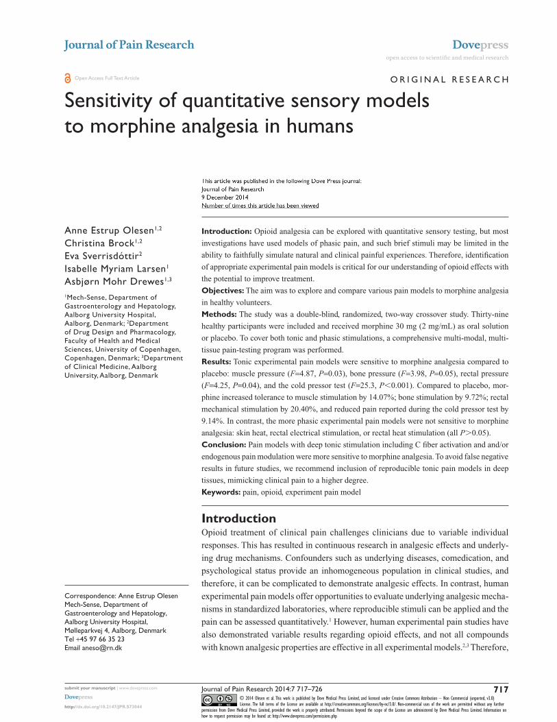
Pain-testing proceduresA comprehensive multi-modal, multi-tissue, pain-testing
program was performed, including pain thresholds and pain
intensity ratings. A variety of pain models were selected to
cover both superficial and deep pain as well as phasic and
tonic stimulations (Table 1 and Figure 1). Hereby, sufficient
resting periods were included in the protocol and the risk for
sensitization was diminished.
skin stimulationThermalThermal heat stimuli were applied by a contact heat-evoked
potential stimulator (CHEPS; Medoc Ltd, Ramat Yishai,
Israel), placed 10 cm distal to the elbow on the right
forearm. The start temperature was set to 32°C and the
temperature increase was set at 1°C/s. Participants were
asked to press a button at PTT, and immediately afterward,
the thermode cooled down to 32°C, with a cooling rate
of 10°C/s. This was repeated three times, and the average
stimulus intensity (°C) was calculated and used for further
data analysis.
Muscle stimulationMechanicalPressure was applied to the supinator muscle on the left
forearm, 15 cm distal to the elbow by a handheld electronic
pressure algometer (Somedic AB, Stockholm, Sweden)
with a standard probe of 1 cm2. The pressure increase
rate was 30 kPa/s and the algometer was set with a safety
maximum of 2,000 kPa. The stimulation was interrupted
when the participants reported PTT and the maximum
stimulus intensity (kPa) was noted and used for further
analysis.
Bone stimulationMechanicalThe mechanical stimulation was conducted on the right tibia
bone, 10 cm distal to the patella by a handheld electronic
pressure algometer (Somedic AB) with a specially designed
probe of 3.1 mm2 (Aalborg University, Aalborg, Denmark).
This probe has been validated in previous studies.12,13 The
pressure increase rate was 999 kPa/s. Stimulation was inter-
rupted when PTT was reported. The maximum stimulus
intensity (kPa) was noted and used for further analysis.
Visceral stimulationFor rectal stimulation, a custom-designed probe (Ditens,
Egaa, Denmark) with a polyester urethane bag for mechani-
cal and thermal stimulation, and stainless steel electrodes
for electrical stimulation was used. The rectal probe has
been validated and is described in detail elsewhere.14 Prior
to the experiment, a 5 mL enema with 2 mg/mL bisacodyl
(Toilax®; Orion Pharma, Espoo, Finland) was adminis-
tered. A lubricated anoscope (Cat No E-03. 19. 925; Heine
Optotechnik, Herrsching, Germany) was placed in the anal
canal and through this, the probe was placed in the rectum
20 cm from the anal sphincter. All data were displayed
online (Openlab; GMC, Hornslet, Denmark) and stored for
later analysis.
MechanicalFor mechanical stimulation of the rectum, the bag was inflated
with 37°C water from a water bath (Julabo VWR 5; Julabo,
Labortechnik GMBH, Seelbach, Germany) controlling the
temperature. A peristaltic pump (Type 111; Ole Dich Instru-
mentmakers, Hvidovre, Denmark) was used to inflate the bag
at a rate of 200 mL/minute. Three distensions to the PDT
were performed with a 1-minute interval to precondition the
tissue. Hereafter, a single distension to “moderate pain” was
performed. The bag was emptied at the same rate as it was
inflated. The volumes in the bag (mL) at PDT and at moderate
pain were noted and used for further analysis.
electricalFor the electrical stimulation, two stainless steel bipolar
electrodes mounted on the tip of the rectal probe with
an inter-electrode distance of 2 mm were connected to a
computer-controlled constant current stimulator (Digitimer
Ltd, Welwyn Garden City, UK). Impedance was kept below
3 kΩ to ensure sufficient mucosal contact. The electrical
Table 1 Overview of included pain models
Tissue Modality Structure Duration
skin Thermal Superficial Phasiccold pressor test Deep Tonic
Muscle Mechanical Deep TonicBone Mechanical Deep TonicViscera electrical Deep Phasic
Thermal PhasicMechanical Tonic
Notes: Models are assigned to the tissue predominantly affected. it should be noted that the cold pressor test is not true skin stimulation, but it will affect skin nociceptors as well and is therefore assigned to skin in this table.
Journal of Pain Research 2014:7submit your manuscript | www.dovepress.com
Dovepress
Dovepress
720
Olesen et al
stimulation intensity slowly increased in increments of
1.0 mA increments. To blind the participant, sham stimula-
tions with same or lower intensities were included. Stimulus
intensities (mA) at PDT and at moderate pain were noted and
used for further analysis.
ThermalBefore thermal stimulation, 60 mL of 37°C water was
added to the polyester urethane bag of the probe. The
temperature elevation was reached by recirculation of 68°C
water through the bag. A water bath (VWR 5; Julabo) con-
trolled the temperature in the closed circuit. A peristaltic
pump (Type 111; Ole Dich Instrument makers) circulated
the water at a flow rate of 150 mL/minute, and a tempera-
ture sensor (Buhl & Bønsøe AS, Virum, Denmark) was
located in the inflated bag. During thermal stimulation,
the anal canal was shielded with the anoscope in order
to minimize stimulation of somatic tissue. The tempera-
ture increased until the participants reported moderate
pain, and immediately hereafter, the heated water was
withdrawn to minimize discomfort for the participant.
Thermal stimulus was computed as area under the time–
temperature curve.
cold pressor testFor the cold pressor stimulation, a cold pressor test apparatus
(Grant instruments; Fischer Scientific, Slangerup, Denmark)
was used. The participants immersed their left hand into the
2°C cooled water up to the wrist for 2 minutes. The partici-
pants rated the perceived pain continuously on the electronic
handheld device.
statistical analysisAll data were baseline corrected before statistical analysis.
Thus, data for statistical analysis represent individual rela-
tive changes from baseline values expressed as percentages.
Coefficients of difference between treatments and associ-
ated confidence intervals were provided for time point
60 minutes.
For statistical comparison of placebo versus morphine
effects on thermal skin stimulation and mechanical muscle
stimulation, data were analyzed by two-way repeated mea-
sures analysis of variance (ANOVA), as these assessments
were performed at several time points (15, 30, 45, 60, and
150 minutes after drug administration) in each study arm.
Factors for these two-way ANOVAs were: 1) treatment and
2) time. For rectal electrical and mechanical stimulations,
analyses were performed at two pain levels (PDT and mod-
erate pain). These data were analyzed by two-way ANOVA,
where factors were: 1) treatment and 2) pain level. When
outcome measures were only assessed at one time point (60
minutes after drug administration), one-way ANOVA was
used for statistical analysis (mechanical bone stimulation,
rectal thermal stimulation, and the cold pressor test). Stata
software (v12.1) was used for analysis. P-values of ,0.05
were considered significant.
As this study investigated the effects of morphine on
multiple endpoints, a precise calculation of sample size for
each endpoint was not possible. A sample size was estimated
based on data from a previous study, from our group,27 of opi-
oid effect assessed by heat stimulation of the skin in healthy
volunteers, as skin heat stimulation was hypothesized to be
the least sensitive outcome. To detect a difference of 4% in
Drug administration
Pain model number
Pain models
0 15 30 45 60 90 120 150
Time (minutes)
1, 31–51–5 1, 31, 31, 3
1 2 3 4 5
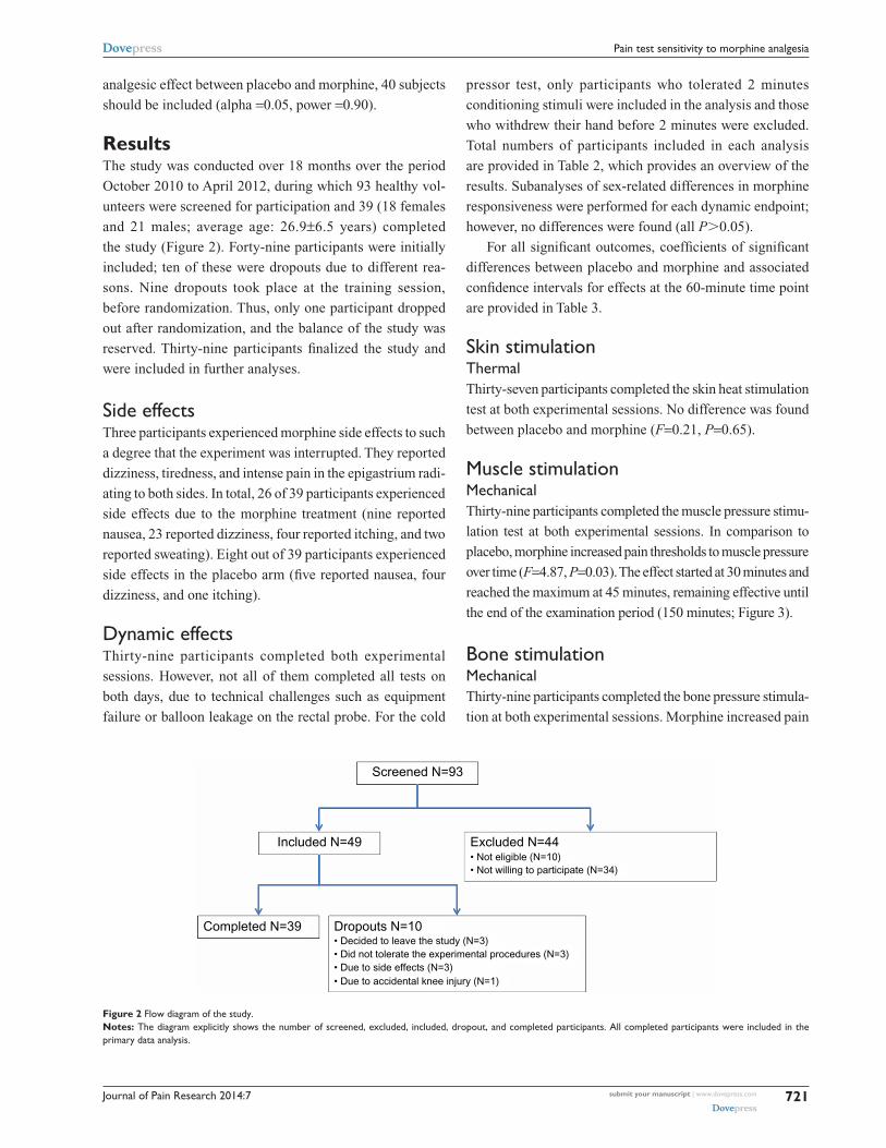
Figure 1 graphical overview of the experimental procedure.Notes: Included pain models (upper) and flowchart (lower) are illustrated. Included pain models were: 1) skin heat; 2) bone pressure; 3) muscle pressure; 4) visceral stimulations, and 5) the cold pressor test. stimulations were applied in the indicated order (1 → 2 → 3 → 4 → 5) at baseline and 60 minutes after drug administration. skin 1) and muscle 3) stimulations were performed at several time points (0, 15, 30, 60, and 150 minutes after drug administration). Blue bars on flowchart indicate duration of test battery. The full battery (1–5) lasted approximately 30 minutes. skin and muscle stimulation lasted approximately 7 minutes in total.
Journal of Pain Research 2014:7 submit your manuscript | www.dovepress.com
Dovepress
Dovepress
721
Pain test sensitivity to morphine analgesia
analgesic effect between placebo and morphine, 40 subjects
should be included (alpha =0.05, power =0.90).
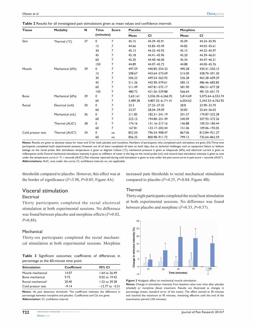
ResultsThe study was conducted over 18 months over the period
October 2010 to April 2012, during which 93 healthy vol-
unteers were screened for participation and 39 (18 females
and 21 males; average age: 26.9±6.5 years) completed
the study (Figure 2). Forty-nine participants were initially
included; ten of these were dropouts due to different rea-
sons. Nine dropouts took place at the training session,
before randomization. Thus, only one participant dropped
out after randomization, and the balance of the study was
reserved. Thirty-nine participants finalized the study and
were included in further analyses.
side effectsThree participants experienced morphine side effects to such
a degree that the experiment was interrupted. They reported
dizziness, tiredness, and intense pain in the epigastrium radi-
ating to both sides. In total, 26 of 39 participants experienced
side effects due to the morphine treatment (nine reported
nausea, 23 reported dizziness, four reported itching, and two
reported sweating). Eight out of 39 participants experienced
side effects in the placebo arm (five reported nausea, four
dizziness, and one itching).
Dynamic effectsThirty-nine participants completed both experimental
sessions. However, not all of them completed all tests on
both days, due to technical challenges such as equipment
failure or balloon leakage on the rectal probe. For the cold
pressor test, only participants who tolerated 2 minutes
conditioning stimuli were included in the analysis and those
who withdrew their hand before 2 minutes were excluded.
Total numbers of participants included in each analysis
are provided in Table 2, which provides an overview of the
results. Subanalyses of sex-related differences in morphine
responsiveness were performed for each dynamic endpoint;
however, no differences were found (all P.0.05).
For all significant outcomes, coefficients of significant
differences between placebo and morphine and associated
confidence intervals for effects at the 60-minute time point
are provided in Table 3.
skin stimulationThermalThirty-seven participants completed the skin heat stimulation
test at both experimental sessions. No difference was found
between placebo and morphine (F=0.21, P=0.65).
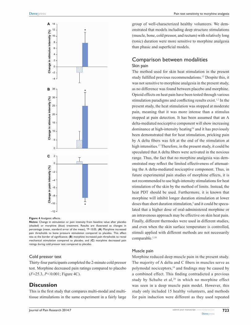
Muscle stimulationMechanicalThirty-nine participants completed the muscle pressure stimu-
lation test at both experimental sessions. In comparison to
placebo, morphine increased pain thresholds to muscle pressure
over time (F=4.87, P=0.03). The effect started at 30 minutes and
reached the maximum at 45 minutes, remaining effective until
the end of the examination period (150 minutes; Figure 3).
Bone stimulationMechanicalThirty-nine participants completed the bone pressure stimula-
tion at both experimental sessions. Morphine increased pain
Dropouts N=10Completed N=39
Included N=49
Screened N=93
Excluded N=44• Not eligible (N=10)
• Decided to leave the study (N=3)
• Not willing to participate (N=34)
• Did not tolerate the experimental procedures (N=3)• Due to side effects (N=3)• Due to accidental knee injury (N=1)
Figure 2 Flow diagram of the study.Notes: The diagram explicitly shows the number of screened, excluded, included, dropout, and completed participants. all completed participants were included in the primary data analysis.
Journal of Pain Research 2014:7submit your manuscript | www.dovepress.com
Dovepress
Dovepress
722
Olesen et al
Table 2 Results for all investigated pain stimulations given as mean values and confidence intervals
Tissue Modality N Time (minutes)
Score Placebo Morphine
Mean CI Mean CI
skin Thermal (°c) 37 0 7 45.15 44.39–45.91 45.09 44.24–45.9515 7 44.66 43.83–45.49 44.82 44.03–45.6130 7 45.13 44.32–45.93 45.15 44.32–45.9745 7 45.18 44.41–45.96 45.20 44.39–46.0160 7 45.20 44.40–46.00 45.34 44.47–46.21150 7 44.89 44.07–45.72 44.88 44.00–45.76
Muscle Mechanical (kPa) 39 0 7 497.59 440.85–554.33 490.28 430.41–550.1515 7 508.67 443.64–573.69 515.00 438.70–591.3030 7 506.23 449.54–562.92 536.28 463.28–609.2945 7 511.26 442.90–579.61 585.15 486.46–683.8560 7 511.49 447.81–575.17 581.90 486.51–677.28150 7 480.72 421.56–539.88 566.64 481.55–651.73
Bone Mechanical (kPa) 39 0 7 5,651.61 5,036.30–6,266.92 5,814.69 5,075.64–6,553.7460 7 5,489.38 4,807.35–6,171.41 6,054.62 5,343.33–6,765.92
Rectal electrical (ma) 30 0 7 32.3 27.25–37.35 28.8 23.90–33.7060 7 33.57 28.04–39.09 30.83 25.64–36.03
Mechanical (ml) 36 0 7 211.85 182.51–241.19 201.57 170.87–232.2860 7 223.15 194.80–251.49 240.09 207.92–272.26
Thermal (aUca) 38 0 7 174.16 131.16–217.16 146.88 109.33–184.4460 7 167.81 133.17–202.44 151.06 109.06–193.05
cold pressor test Thermal (aUcb) 34 0 na 852.50 796.54–908.47 867.06 812.84–921.2760 na 856.35 800.98–911.72 799.12 735.64–862.59
Notes: Results are given as absolute values for mean and ci for both placebo and morphine. numbers of participants who completed each stimulation are given (n). Thirty-nine participants completed both experimental sessions. however, not all of them completed all tests on both days, due to technical challenges such as equipment failure or balloon leakage on the rectal probe. skin stimulation temperature is given as degrees celsius (°c), mechanical pressure is given as kilopascals (kPa), and electrical current is given as milliamperes (ma). Visceral mechanical stimulation intensity is given as milliliters of water in the bag on the rectal probe (ml) and visceral heat stimulation intensity is given as area under the temperature curve in °c × seconds (aUca). Pain intensity reported during cold stimulation is given as area under the pain-score-curve in pain-score × seconds (aUcb).Abbreviations: AUC, area under the curve; CI, confidence interval; na, not applicable.
15
–5
0
5
10
15
20
25
30
30 45
Time (minutes)
Ch
ang
e in
sti
mu
lus
inte
nsi
ty (
%)
60 150
Figure 3 analgesic effect on mechanical muscle stimulation.Notes: change in stimulation intensity from baseline value over time after placebo (shaded) or morphine (blue) treatment. Results are illustrated as changes in percentage (mean, standard error of the mean). The effect started at 30 minutes and reached the maximum at 45 minutes, remaining effective until the end of the examination period (150 minutes).
Table 3 Significant outcomes; coefficients of differences in percentage at the 60-minute time point
Stimulation Coefficient 95% CI
Muscle mechanical 14.07 1.64 to 26.49Bone mechanical 9.72 0.02 to 19.42Rectal mechanicala 20.40 1.52 to 39.28cold pressor test –9.14 –12.77 to –5.51
Notes: aAt pain detection threshold. The coefficient indicates the difference in percentage between morphine and placebo. Coefficients and CIs are given.Abbreviation: CI, confidence interval.
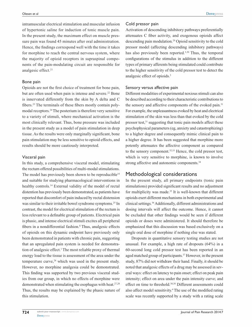
thresholds compared to placebo. However, this effect was at
the border of significance (F=3.98, P=0.05; Figure 4A).
Visceral stimulationelectricalThirty participants completed the rectal electrical
stimulation at both experimental sessions. No difference
was found between placebo and morphine effects (F=0.02,
P=0.88).
MechanicalThirty-six participants completed the rectal mechani-
cal stimulation at both experimental sessions. Morphine
increased pain thresholds to rectal mechanical stimulation
compared to placebo (F=4.25, P=0.04; Figure 4B).
ThermalThirty-eight participants completed the rectal heat stimulation
at both experimental sessions. No difference was found
between placebo and morphine (F=0.33, P=0.57).
Journal of Pain Research 2014:7 submit your manuscript | www.dovepress.com
Dovepress
Dovepress
723
Pain test sensitivity to morphine analgesia
–12
Ch
ang
e in
pai
n in
ten
sity
(%
)C
han
ge
in s
tim
ulu
s in
ten
sity
(%
)C
han
ge
in s
tim
ulu
s in
ten
sity
(%
)
–10
–8
–6
–4
–4
–2
0
2
4
6
8
10
12
14A
B
C
*
*
*
–2
0
0
5
10
15
20
25
30
35
2
Figure 4 analgesic effects.Notes: change in stimulation or pain intensity from baseline value after placebo (shaded) or morphine (blue) treatment. Results are illustrated as changes in percentage (mean, standard error of the mean), *P,0.05. (A) Morphine increased pain thresholds to bone pressure stimulation compared to placebo. This effect was at the border of significance; (B) morphine increased pain thresholds to rectal mechanical stimulation compared to placebo, and (C) morphine decreased pain ratings during cold pressor test compared to placebo.
cold pressor testThirty-four participants completed the 2-minute cold pressor
test. Morphine decreased pain ratings compared to placebo
(F=25.3, P,0.001; Figure 4C).
DiscussionThis is the first study that compares multi-modal and multi-
tissue stimulations in the same experiment in a fairly large
group of well-characterized healthy volunteers. We dem-
onstrated that models including deep structure stimulations
(muscle, bone, cold pressor, and rectum) with relatively long
(tonic) duration were more sensitive to morphine analgesia
than phasic and superficial models.
comparison between modalitiesskin painThe method used for skin heat stimulation in the present
study fulfilled previous recommendations.15 Despite this, it
was not sensitive to morphine analgesia in the present study,
as no difference was found between placebo and morphine.
Opioid effects on heat pain have been tested through various
stimulation paradigms and conflicting results exist.1,2 In the
present study, the heat stimulation was stopped at moderate
pain, meaning that it was more intense than a stimulus
stopped at pain detection. It has been assumed that an A
delta-mediated nociceptive component will show increasing
dominance at high-intensity heating16 and it has previously
been demonstrated that for heat stimulation, pricking pain
by A delta fibers was felt at the end of the stimulation at
high intensities.17 Therefore, in the present study, it could be
speculated that A delta fibers were activated in the noxious
range. Thus, the fact that no morphine analgesia was dem-
onstrated may reflect the limited effectiveness of attenuat-
ing the A delta-mediated nociceptive component. Thus, in
future experimental pain studies of morphine effects, it is
not recommended to use high-intensity stimulations for heat
stimulation of the skin by the method of limits. Instead, the
heat PDT should be used. Furthermore, it is known that
morphine will inhibit longer duration stimulation at lower
doses than short duration stimulation,5 and it could be specu-
lated that a higher dose of oral-administered morphine or
an intravenous approach may be effective on skin heat pain.
Finally, different thermodes were used in different studies,
and even when the skin surface temperature is controlled,
stimuli applied with different methods are not necessarily
comparable.1,18
Muscle painMorphine reduced deep muscle pain in the present study.
The majority of A delta and C fibers in muscles serve as
polymodal nociceptors,19 and findings may be caused by
a combined effect. This finding contradicted a previous
study by Schulte et al,20 in which no morphine effect
was seen in a deep muscle pain model. However, this
study only included 15 healthy volunteers, and methods
for pain induction were different as they used repeated
Journal of Pain Research 2014:7submit your manuscript | www.dovepress.com
Dovepress
Dovepress
724
Olesen et al
intramuscular electrical stimulation and muscular infusion
of hypertonic saline for induction of tonic muscle pain.
In the present study, the maximum effect on muscle pres-
sure pain was found 45 minutes after oral administration.
Hence, the findings correspond well with the time it takes
for morphine to reach the central nervous system, where
the majority of opioid receptors in supraspinal compo-
nents of the pain-modulating circuit are responsible for
analgesic effect.21
Bone painOpioids are not the first choice of treatment for bone pain,
but are often used when pain is intense and severe.22 Bone
is innervated differently from the skin by A delta and C
fibers.23 The terminals of these fibers mostly contain poly-
modal receptors.24 The posterium is therefore very sensitive
to a variety of stimuli, where mechanical activation is the
most clinically relevant. Thus, bone pressure was included
in the present study as a model of pain stimulation in deep
tissue. As the results were only marginally significant, bone
pain stimulation may be less sensitive to opioid effects, and
results should be more cautiously interpreted.
Visceral painIn this study, a comprehensive visceral model, stimulating
the rectum offered possibilities of multi-modal stimulations.
The model has previously been shown to be reproducible14
and suitable for studying pharmacological interventions in
healthy controls.14 External validity of the model of rectal
distention has previously been demonstrated, as patients have
reported that discomfort of pain induced by rectal distension
was similar to their irritable bowel syndrome symptoms.25 In
contrast, the model for electrical stimulation of the rectum is
less relevant to a definable group of patients. Electrical pain
is phasic, and intense electrical stimuli excites all peripheral
fibers in a nondifferential fashion.8 Thus, analgesic effects
of opioids on this dynamic endpoint have previously only
been demonstrated in patients with chronic pain, suggesting
that an upregulated pain system is needed for demonstra-
tion of analgesic effect.1 The most reliable proxy of thermal
energy load to the tissue is assessment of the area under the
temperature curve,26 which was used in the present study.
However, no morphine analgesia could be demonstrated.
This finding was supported by two previous visceral stud-
ies from our group, in which no effects of morphine were
demonstrated when stimulating the esophagus with heat.27,28
Thus, the results may be explained by the phasic nature of
this stimulation.
cold pressor painActivation of descending inhibitory pathways preferentially
attenuates C fiber activity, and exogenous opioids affect
descending pain modulation.29 Opioid sensitivity to the cold
pressor model (affecting descending inhibitory pathways)
has also previously been reported.3,30 Thus, the temporal
configurations of the stimulus in addition to the different
types of primary afferents being stimulated could contribute
to the higher sensitivity of the cold pressor test to detect the
analgesic effect of opioids.3
sensory versus affective painDifferent modalities of experimental noxious stimuli can also
be described according to their characteristic contributions to
the sensory and affective components of the evoked pain.31
For example, the unpleasantness evoked by heat and electrical
stimulation of the skin was less than that evoked by the cold
pressor test,31 suggesting that tonic pain models affect these
psychophysical parameters (eg, anxiety and catastrophizing)
to a higher degree and consequently mimic clinical pain to
a higher degree. It has been suggested that morphine more
potently attenuates the affective component as compared
to the sensory component.32,33 Hence, the cold pressor test,
which is very sensitive to morphine, is known to involve
strong affective and autonomic components.34
Methodological considerationsIn the present study, all primary endpoints (tonic pain
stimulations) provided significant results and no adjustment
for multiplicity was made.35 It is well-known that different
opioids exert different mechanisms in both experimental and
clinical settings.36 Additionally, different administrations and
dosing intervals will affect the outcome. Hence, it cannot
be excluded that other findings would be seen if different
opioids or doses were administered. It should therefore be
emphasized that this discussion was based exclusively on a
single oral dose of morphine if nothing else was stated.
Dropouts in quantitative sensory testing studies are not
unusual. For example, a high rate of dropouts (64%) in a
60-second long cold pressor test has been reported in an
aged matched group of participants.37 However, in the present
study, 87% did not withdraw their hand. Finally, it should be
noted that analgesic effects of a drug may be assessed in sev-
eral ways: effect on latency to pain onset; effect on peak pain
intensity; effect on area under the pain intensity curve; and
effect on time to threshold.30,38 Different assessments could
also affect model sensitivity.4 The use of the modified rating
scale was recently supported by a study with a rating scale
Journal of Pain Research 2014:7 submit your manuscript | www.dovepress.com
Dovepress
Dovepress
725
Pain test sensitivity to morphine analgesia
ranging from “no sensation” to “unbearable pain”, allowing
participants to rate stimulus intensities that are perceived but
are not painful, and may reduce bias and be more reliable for
experimental pain assessment.39
The fact that 34 of 93 screened healthy volunteers were
not willing to participate after further information and con-
sideration indicates a study selection bias, and hence, results
may not reflect the Danish population. However, as participa-
tion is voluntary, experimental pain studies will always be
affected by such study selection bias.
RecommendationsSeveral factors should be considered when planning an
experimental human pain study for assessment of analgesic
effects.1 Using multi-modal tests and multi-tissue stimula-
tions, the present study confirmed that: 1) deep stimuli
should be included to mimic the clinical situation; 2) tonic
stimulations should be used rather than phasic stimulations
to evaluate pain intensity before and after drug administra-
tion; and 3) models involving activation predominantly of
C fibers should be included.
ConclusionPain models with deep tonic stimulation including C fiber
activation and/or endogenous pain modulation were more
sensitive to morphine analgesia. To avoid false negative
results in future studies, we recommend inclusion of repro-
ducible tonic pain models in deep tissues, mimicking clinical
pain to a higher degree.
AcknowledgmentThe study was supported by the Danish Council for Strategic
Research and Det Obelske Familiefond.
DisclosureThe funding sources for this work were not involved in the
conduct of the study or in the development of the submission.
The authors report no conflicts of interest in this work.
References1. Olesen AE, Andresen T, Staahl C, Drewes AM. Human experimental
pain models for assessing the therapeutic efficacy of analgesic drugs. Pharmacol Rev. 2012;64(3):722–779.
2. Staahl C, Olesen AE, Andresen T, Arendt-Nielsen L, Drewes AM. Assessing analgesic actions of opioids by experimental pain models in healthy volunteers – an updated review. Br J Clin Pharmacol. 2009;68(2): 149–168.
3. Koltzenburg M, Pokorny R, Gasser UE, Richarz U. Differential sensitivity of three experimental pain models in detecting the anal-gesic effects of transdermal fentanyl and buprenorphine. Pain. 2006;126(1–3):165–174.
4. Angst MS, Clark JD. Comment on Koltzenburg et al. Differential sensi-tivity of three experimental pain models in detecting the analgesic effects of transdermal fentanyl and buprenorphine. Pain 2006;126:165–74. Pain. 2007;128(3):292–294.
5. Petersen KL. Experimental cutaneous hyperalgesia in humans. In: Berde CB, Rowbotham MC, editors. Technical Corner from IASP Newsletter. Washington, DC: International Association for the Study of Pain; 1997.
6. Oertel BG, Lotsch J. Clinical pharmacology of analgesics assessed with human experimental pain models: Bridging basic and clinical research. Br J Pharmacol. 2012;168(3):534–553.
7. Kandel ER, Schwartz JH, Jessell TM. Part V perception. In: Kandel ER, Schwartz JH, Jessell TM, editors. Principles of Neural Science United States of America. The McGraw-Hill Publishing Company, Inc.; 2000:411–624.
8. Le Bars D, Gozariu M, Cadden SW. Animal models of nociception. Pharmacol Rev. 2001;53(4):597–652.
9. Lu Y, Pirec V, Yeomans DC. Differential antinociceptive effects of spinal opioids on foot withdrawal responses evoked by C fibre or A delta nociceptor activation. Br J Pharmacol. 1997;121(6):1210–1216.
10. Le Bars D, Guilbaud G, Jurna I, Besson JM. Differential effects of mor-phine on responses of dorsal horn lamina V type cells elicited by A and C fibre stimulation in the spinal cat. Brain Res. 1976;115(3):518–524.
11. Drewes AM, Gregersen H, Arendt-Nielsen L. Experimental pain in gastroenterology: a reappraisal of human studies. Scand J Gastroenterol. 2003;38(11):1115–1130.
12. Andresen T, Pfeiffer-Jensen M, Brock C, Drewes AM, Arendt-Nielsen L. A human experimental bone pain model. Basic Clin Pharmacol Toxicol. 2013;112(2):116–123.
13. Andresen T, Staahl C, Oksche A, Mansikka H, Arendt-Nielsen L, Drewes AM. Effect of transdermal opioids in experimentally induced superficial, deep and hyperalgesic pain. Br J Pharmacol. 2011;164(3): 934–945.
14. Brock C, Nissen TD, Gravesen FH, et al. Multimodal sensory testing of the rectum and rectosigmoid: development and reproducibility of a new method. Neurogastroenterol Motil. 2008;20(8):908–918.
15. Rosier EM, Iadarola MJ, Coghill RC. Reproducibility of pain measure-ment and pain perception. Pain. 2002;98(1–2):205–216.
16. McCormack K, Prather P, Chapleo C. Some new insights into the effects of opioids in phasic and tonic nociceptive tests. Pain. 1998;78(2):79–98.
17. Nielsen J, Arendt-Nielsen L. The influence of rate of temperature change and peak stimulus duration on pain intensity and quality. Somatosens Mot Res. 1998;15(3):220–229.
18. Handwerker HO, Kobal G. Psychophysiology of experimentally induced pain. Physiol Rev. 1993;73(3):639–671.
19. Cairns BE. Physiological properties of thin-fiber muscle affer-ents: excitation and modulatory effects. In: Graven-Nielsen T, Arendt-Nielsen L, Mense S, editors. Fundamentals of Musculoskeletal Pain. 1st ed. Seattle, WA: IASP Press; 2008:19–32.
20. Schulte H, Graven-Nielsen T, Sollevi A, Jansson Y, Arendt-Nielsen L, Segerdahl M. Pharmacological modulation of experimental phasic and tonic muscle pain by morphine, alfentanil and ketamine in healthy volunteers. Acta Anaesthesiol Scand. 2003;47(8):1020–1030.
21. Fields H. State-dependent opioid control of pain. Nat Rev Neurosci. 2004;5(7):565–575.
22. Mattia C, Di Bussolo E, Coluzzi F. Non-analgesic effects of opioids: the interaction of opioids with bone and joints. Curr Pharm Des. 2012;18(37):6005–6009.
23. Finocchietti S, Andresen T, Arendt-Nielsen L, Graven-Nielsen T. Pain evoked by pressure stimulation on the tibia bone – influence of probe diameter on tissue stress and strain. Eur J Pain. 2012;16(4): 534–542.
24. Byers MR, Bonica JJ. Peripheral pain mechanisms and nociceptor plasticity. In: Loeser LD, Butler SH, Chapman CR, Turk DC, editors. Bonica’s Management of Pain. Philadelphia, PA: Lippincott Williams & Wilkins; 2001.
Journal of Pain Research
Publish your work in this journal
Submit your manuscript here: http://www.dovepress.com/journal-of-pain-research-journal
The Journal of Pain Research is an international, peer-reviewed, open access, online journal that welcomes laboratory and clinical findings in the fields of pain research and the prevention and management of pain. Original research, reviews, symposium reports, hypoth-esis formation and commentaries are all considered for publication.
The manuscript management system is completely online and includes a very quick and fair peer-review system, which is all easy to use. Visit http://www.dovepress.com/testimonials.php to read real quotes from published authors.
Journal of Pain Research 2014:7submit your manuscript | www.dovepress.com
Dovepress
Dovepress
Dovepress
726
Olesen et al
25. Morgan V, Pickens D, Gautam S, Kessler R, Mertz H. Amitriptyline reduces rectal pain related activation of the anterior cingulate cor-tex in patients with irritable bowel syndrome. Gut. 2005;54(5): 601–607.
26. Brock C, Arendt-Nielsen L, Wilder-Smith O, Drewes AM. Sensory testing of the human gastrointestinal tract. World J Gastroenterol. 2009;15(2):151–159.
27. Staahl C, Christrup LL, Andersen SD, Arendt-Nielsen L, Drewes AM. A comparative study of oxycodone and morphine in a multi-modal, tissue-differentiated experimental pain model. Pain. 2006;123(1–2): 28–36.
28. Olesen AE, Staahl C, Arendt-Nielsen L, Drewes AM. Different effects of morphine and oxycodone in experimentally evoked hyperalgesia: a human translational study. Br J Clin Pharmacol. 2010;70(2):189–200.
29. Arendt-Nielsen L, Andresen T, Malver LP, Oksche A, Mansikka H, Drewes AM. A double-blind, placebo-controlled study on the effect of buprenorphine and fentanyl on descending pain modulation: a human experimental study. Clin J Pain. 2012;28(7):623–627.
30. Jones SF, McQuay HJ, Moore RA, Hand CW. Morphine and ibuprofen compared using the cold pressor test. Pain. 1988;34(2):117–122.
31. Rainville P, Feine JS, Bushnell MC, Duncan GH. A psychophysical comparison of sensory and affective responses to four modalities of experimental pain. Somatosens Mot Res. 1992;9(4):265–277.
32. van der Kam EL, Vry JD, Schiene K, Tzschentke TM. Differential effects of morphine on the affective and the sensory component of carrageenan-induced nociception in the rat. Pain. 2008;136(3): 373–379.
33. Kupers RC, Konings H, Adriaensen H, Gybels JM. Morphine differen-tially affects the sensory and affective pain ratings in neurogenic and idiopathic forms of pain. Pain. 1991;47(1):5–12.
34. Cleeland CS, Nakamura Y, Howland EW, Morgan NR, Edwards KR, Backonja M. Effects of oral morphine on cold pressor tolerance time and neuropsychological performance. Neuropsychopharmacology. 1996;15(3):252–262.
35. Turk DC, Dworkin RH, McDermott MP, et al. Analyzing multiple endpoints in clinical trials of pain treatments: IMMPACT recommendations. Initiative on methods, measurement, and pain assessment in clinical trials. Pain. 2008;139(3):485–493.
36. Drewes AM, Jensen RD, Nielsen LM, et al. Differences between opioids: pharmacological, experimental, clinical and economical per-spectives. Br J Clin Pharmacol. 2013;75(1):60–78.
37. Ruscheweyh R, Stumpenhorst F, Knecht S, Marziniak M. Comparison of the cold pressor test and contact thermode-delivered cold stimuli for the assessment of cold pain sensitivity. J Pain. 2010;11(8):728–736.
38. Grach M, Massalha W, Pud D, Adler R, Eisenberg E. Can coadmin-istration of oxycodone and morphine produce analgesic synergy in humans? An experimental cold pain study. Br J Clin Pharmacol. 2004;58(3):235–242.
39. Kemp J, Despres O, Dufour A. Unreliability of the visual analog scale in experimental pain assessment: a sensitivity and evoked potentials study. Pain Physician. 2012;15(5):e693–e699.


