Embed Size (px)
Citation preview
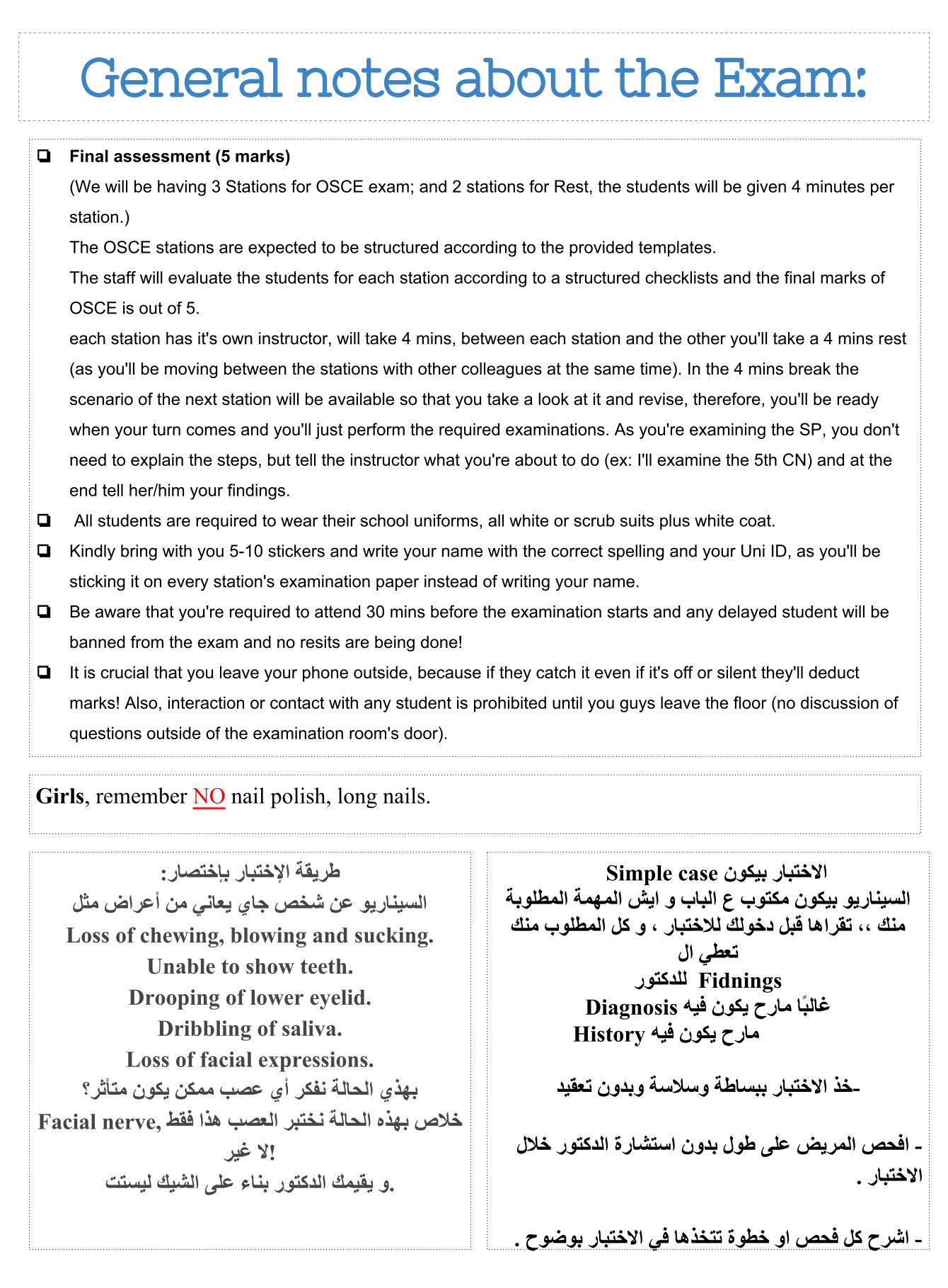
General notes about the Exam:❏ Final assessment (5 marks)
(We will be having 3 Stations for OSCE exam; and 2 stations for Rest, the students will be given 4 minutes per
station.)
The OSCE stations are expected to be structured according to the provided templates.
The staff will evaluate the students for each station according to a structured checklists and the final marks of
OSCE is out of 5.
each station has it's own instructor, will take 4 mins, between each station and the other you'll take a 4 mins rest
(as you'll be moving between the stations with other colleagues at the same time). In the 4 mins break the
scenario of the next station will be available so that you take a look at it and revise, therefore, you'll be ready
when your turn comes and you'll just perform the required examinations. As you're examining the SP, you don't
need to explain the steps, but tell the instructor what you're about to do (ex: I'll examine the 5th CN) and at the
end tell her/him your findings.
❏ All students are required to wear their school uniforms, all white or scrub suits plus white coat.
❏ Kindly bring with you 5-10 stickers and write your name with the correct spelling and your Uni ID, as you'll be
sticking it on every station's examination paper instead of writing your name.
❏ Be aware that you're required to attend 30 mins before the examination starts and any delayed student will be
banned from the exam and no resits are being done!
❏ It is crucial that you leave your phone outside, because if they catch it even if it's off or silent they'll deduct
marks! Also, interaction or contact with any student is prohibited until you guys leave the floor (no discussion of
questions outside of the examination room's door).
Girls, remember NO nail polish, long nails.
طریقة اإلختبار بإختصار:السیناریو عن شخص جاي یعاني من أعراض مثل
Loss of chewing, blowing and sucking.Unable to show teeth.
Drooping of lower eyelid.Dribbling of saliva.
Loss of facial expressions.بھذي الحالة نفكر أي عصب ممكن یكون متأثر؟
Facial nerve, خالص بھذه الحالة نختبر العصب ھذا فقط !ال غیر
.و یقیمك الدكتور بناء على الشیك لیستت
Simple case االختبار بیكون السیناریو بیكون مكتوب ع الباب و ایش المھمة المطلوبة منك ،، تقراھا قبل دخولك لالختبار ، و كل المطلوب منك
تعطي ال Fidnings للدكتور
Diagnosis غالبًا مارح یكون فیھ History مارح یكون فیھ
-خذ االختبار ببساطة وسالسة وبدون تعقید
- افحص المریض على طول بدون استشارة الدكتور خالل االختبار .
- اشرح كل فحص او خطوة تتخذھا في االختبار بوضوح .

Dermatomes and myotomes:
Dermatomes and myotomes
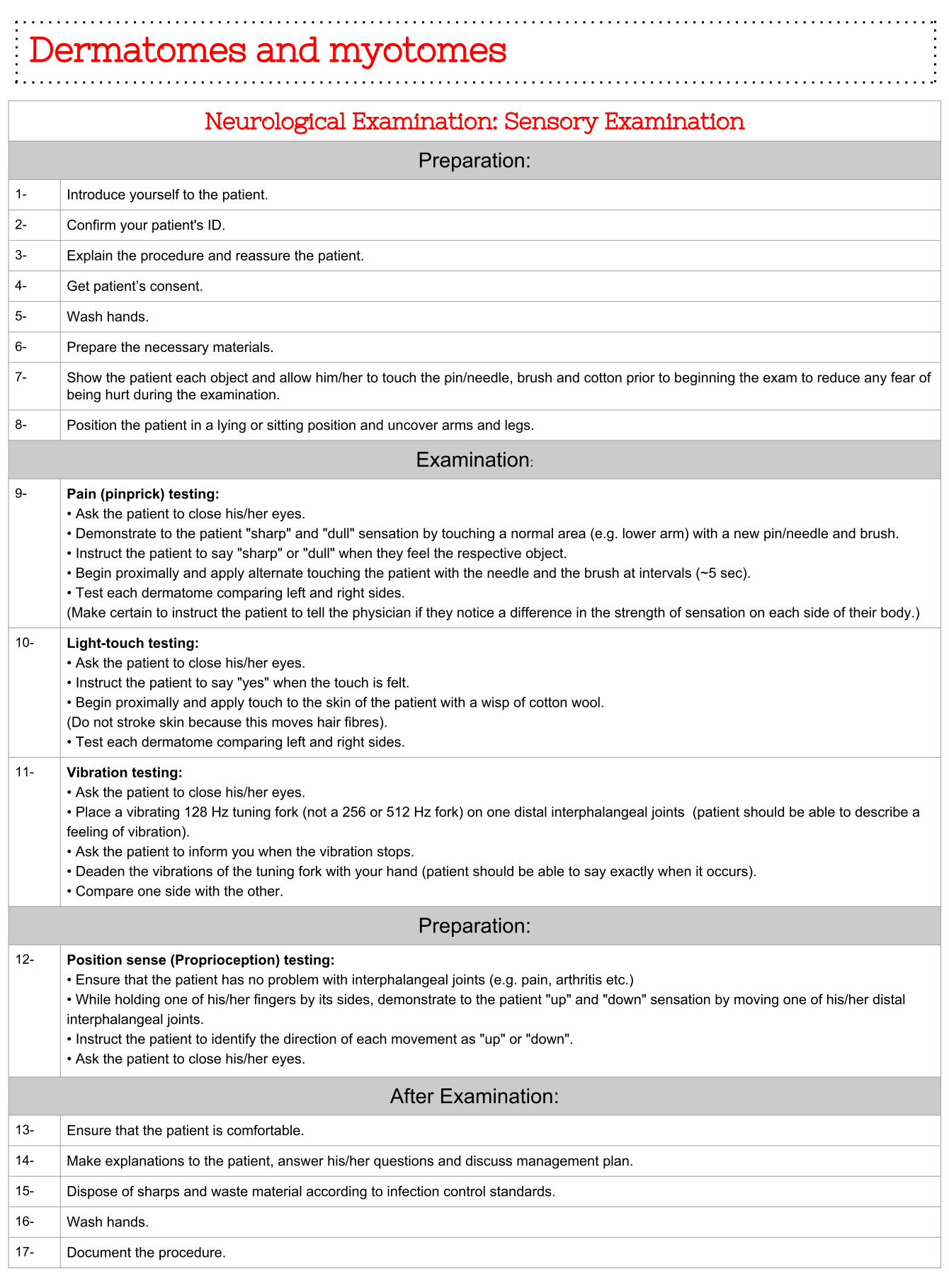
Neurological Examination: Sensory Examination
Preparation:1- Introduce yourself to the patient.
2- Confirm your patient's ID.
3- Explain the procedure and reassure the patient.
4- Get patient’s consent.
5- Wash hands.
6- Prepare the necessary materials.
7- Show the patient each object and allow him/her to touch the pin/needle, brush and cotton prior to beginning the exam to reduce any fear of being hurt during the examination.
8- Position the patient in a lying or sitting position and uncover arms and legs.
Examination:
9- Pain (pinprick) testing:• Ask the patient to close his/her eyes. • Demonstrate to the patient "sharp" and "dull" sensation by touching a normal area (e.g. lower arm) with a new pin/needle and brush.• Instruct the patient to say "sharp" or "dull" when they feel the respective object.• Begin proximally and apply alternate touching the patient with the needle and the brush at intervals (~5 sec).• Test each dermatome comparing left and right sides.(Make certain to instruct the patient to tell the physician if they notice a difference in the strength of sensation on each side of their body.)
10- Light-touch testing:• Ask the patient to close his/her eyes.• Instruct the patient to say "yes" when the touch is felt.• Begin proximally and apply touch to the skin of the patient with a wisp of cotton wool.(Do not stroke skin because this moves hair fibres).• Test each dermatome comparing left and right sides.
11- Vibration testing:• Ask the patient to close his/her eyes.• Place a vibrating 128 Hz tuning fork (not a 256 or 512 Hz fork) on one distal interphalangeal joints (patient should be able to describe a feeling of vibration).• Ask the patient to inform you when the vibration stops.• Deaden the vibrations of the tuning fork with your hand (patient should be able to say exactly when it occurs).• Compare one side with the other.
Preparation:12- Position sense (Proprioception) testing:
• Ensure that the patient has no problem with interphalangeal joints (e.g. pain, arthritis etc.)• While holding one of his/her fingers by its sides, demonstrate to the patient "up" and "down" sensation by moving one of his/her distal interphalangeal joints.• Instruct the patient to identify the direction of each movement as "up" or "down".• Ask the patient to close his/her eyes.
After Examination:13- Ensure that the patient is comfortable.
14- Make explanations to the patient, answer his/her questions and discuss management plan.
15- Dispose of sharps and waste material according to infection control standards.
16- Wash hands.
17- Document the procedure.
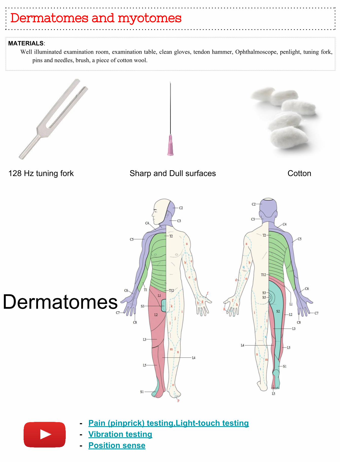
Dermatomes and myotomes
MATERIALS: Well illuminated examination room, examination table, clean gloves, tendon hammer, Ophthalmoscope, penlight, tuning fork,
pins and needles, brush, a piece of cotton wool.
128 Hz tuning fork Sharp and Dull surfaces Cotton
Dermatomes
- Pain (pinprick) testing,Light-touch testing- Vibration testing - Position sense
Examination of
cranial nerves:
Examination of cranial nerves
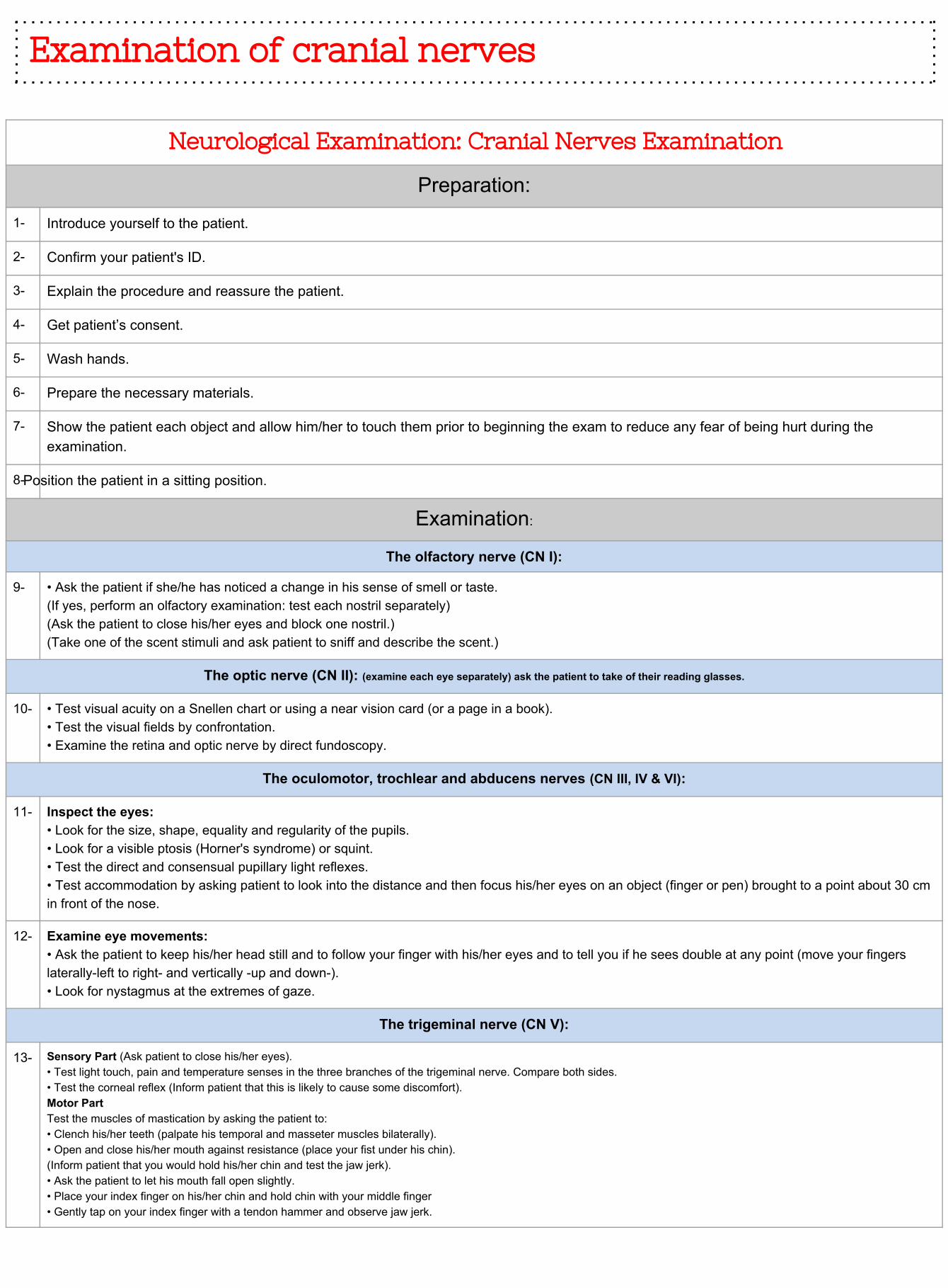
Neurological Examination: Cranial Nerves Examination
Preparation:
1- Introduce yourself to the patient.
2- Confirm your patient's ID.
3- Explain the procedure and reassure the patient.
4- Get patient’s consent.
5- Wash hands.
6- Prepare the necessary materials.
7- Show the patient each object and allow him/her to touch them prior to beginning the exam to reduce any fear of being hurt during the examination.
8-Position the patient in a sitting position.
Examination:
The olfactory nerve (CN I):
9- • Ask the patient if she/he has noticed a change in his sense of smell or taste.(If yes, perform an olfactory examination: test each nostril separately)(Ask the patient to close his/her eyes and block one nostril.)(Take one of the scent stimuli and ask patient to sniff and describe the scent.)
The optic nerve (CN II): (examine each eye separately) ask the patient to take of their reading glasses.
10- • Test visual acuity on a Snellen chart or using a near vision card (or a page in a book).• Test the visual fields by confrontation.• Examine the retina and optic nerve by direct fundoscopy.
The oculomotor, trochlear and abducens nerves (CN III, IV & VI):
11- Inspect the eyes:• Look for the size, shape, equality and regularity of the pupils.• Look for a visible ptosis (Horner's syndrome) or squint.• Test the direct and consensual pupillary light reflexes.• Test accommodation by asking patient to look into the distance and then focus his/her eyes on an object (finger or pen) brought to a point about 30 cm in front of the nose.
12- Examine eye movements:• Ask the patient to keep his/her head still and to follow your finger with his/her eyes and to tell you if he sees double at any point (move your fingers laterally-left to right- and vertically -up and down-).• Look for nystagmus at the extremes of gaze.
The trigeminal nerve (CN V):
13- Sensory Part (Ask patient to close his/her eyes).• Test light touch, pain and temperature senses in the three branches of the trigeminal nerve. Compare both sides.• Test the corneal reflex (Inform patient that this is likely to cause some discomfort).Motor PartTest the muscles of mastication by asking the patient to:• Clench his/her teeth (palpate his temporal and masseter muscles bilaterally).• Open and close his/her mouth against resistance (place your fist under his chin).(Inform patient that you would hold his/her chin and test the jaw jerk).• Ask the patient to let his mouth fall open slightly.• Place your index finger on his/her chin and hold chin with your middle finger• Gently tap on your index finger with a tendon hammer and observe jaw jerk.
Examination of cranial nerves
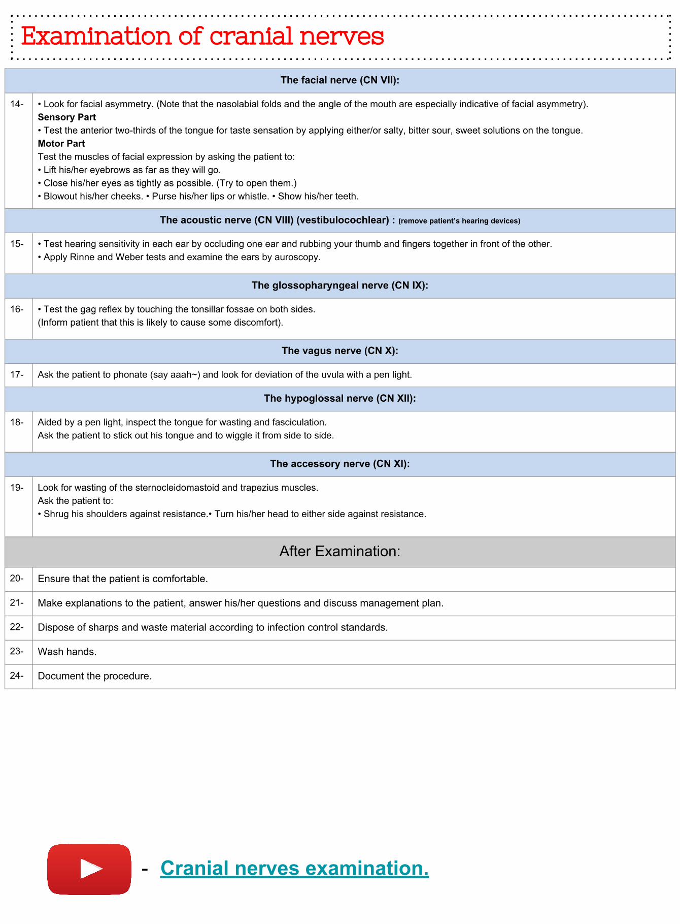
The facial nerve (CN VII):
14- • Look for facial asymmetry. (Note that the nasolabial folds and the angle of the mouth are especially indicative of facial asymmetry).Sensory Part• Test the anterior two-thirds of the tongue for taste sensation by applying either/or salty, bitter sour, sweet solutions on the tongue.Motor PartTest the muscles of facial expression by asking the patient to:• Lift his/her eyebrows as far as they will go.• Close his/her eyes as tightly as possible. (Try to open them.)• Blowout his/her cheeks. • Purse his/her lips or whistle. • Show his/her teeth.
The acoustic nerve (CN VIII) (vestibulocochlear) : (remove patient’s hearing devices)
15- • Test hearing sensitivity in each ear by occluding one ear and rubbing your thumb and fingers together in front of the other.• Apply Rinne and Weber tests and examine the ears by auroscopy.
The glossopharyngeal nerve (CN IX):
16- • Test the gag reflex by touching the tonsillar fossae on both sides.(Inform patient that this is likely to cause some discomfort).
The vagus nerve (CN X):
17- Ask the patient to phonate (say aaah~) and look for deviation of the uvula with a pen light.
The hypoglossal nerve (CN XII):
18- Aided by a pen light, inspect the tongue for wasting and fasciculation.Ask the patient to stick out his tongue and to wiggle it from side to side.
The accessory nerve (CN XI):
19- Look for wasting of the sternocleidomastoid and trapezius muscles.Ask the patient to:• Shrug his shoulders against resistance.• Turn his/her head to either side against resistance.
After Examination:
20- Ensure that the patient is comfortable.
21- Make explanations to the patient, answer his/her questions and discuss management plan.
22- Dispose of sharps and waste material according to infection control standards.
23- Wash hands.
24- Document the procedure.
- Cranial nerves examination.
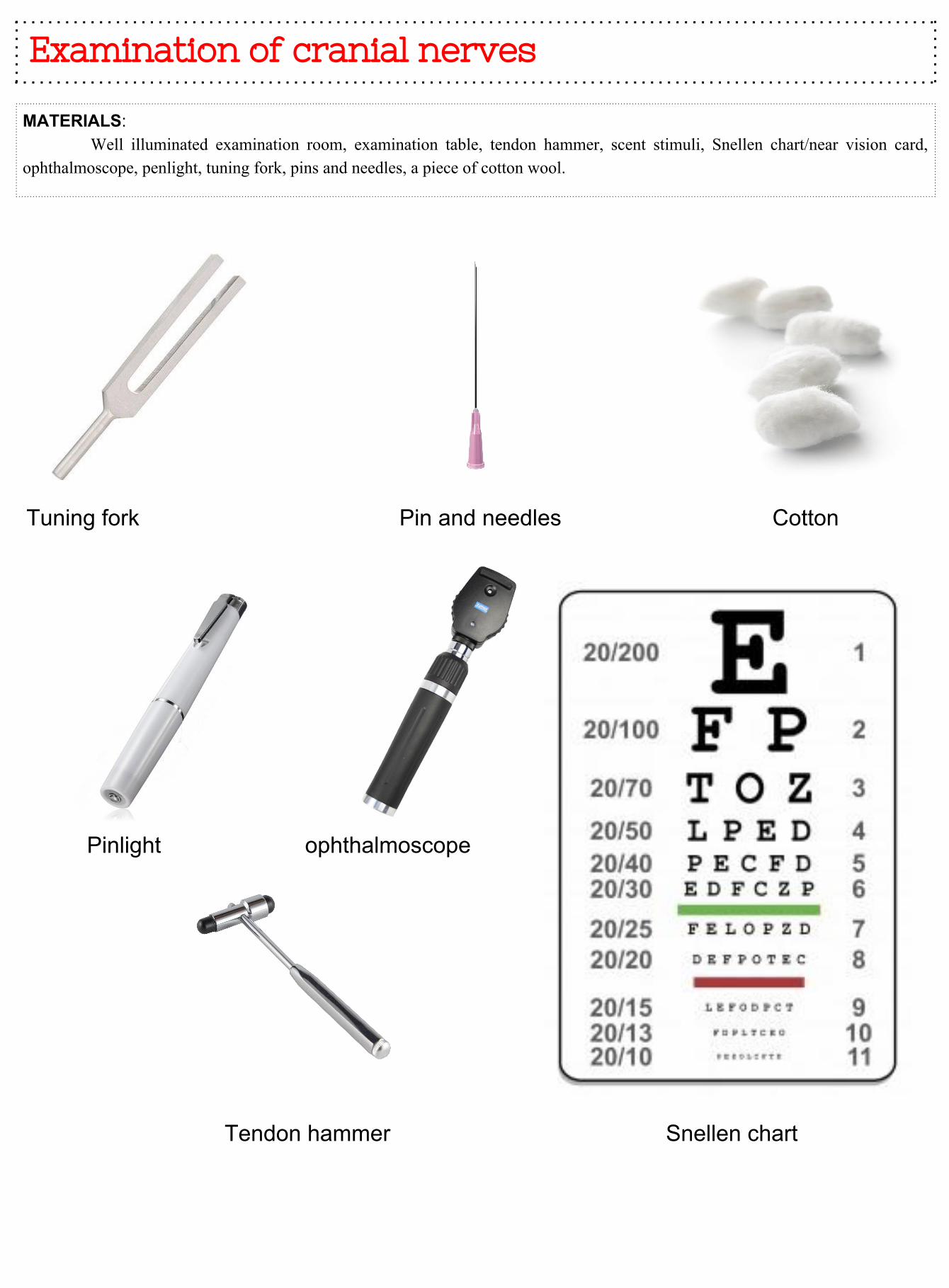
Examination of cranial nerves
MATERIALS: Well illuminated examination room, examination table, tendon hammer, scent stimuli, Snellen chart/near vision card, ophthalmoscope, penlight, tuning fork, pins and needles, a piece of cotton wool.
Tuning fork Pin and needles Cotton
Pinlight ophthalmoscope
Tendon hammer Snellen chart
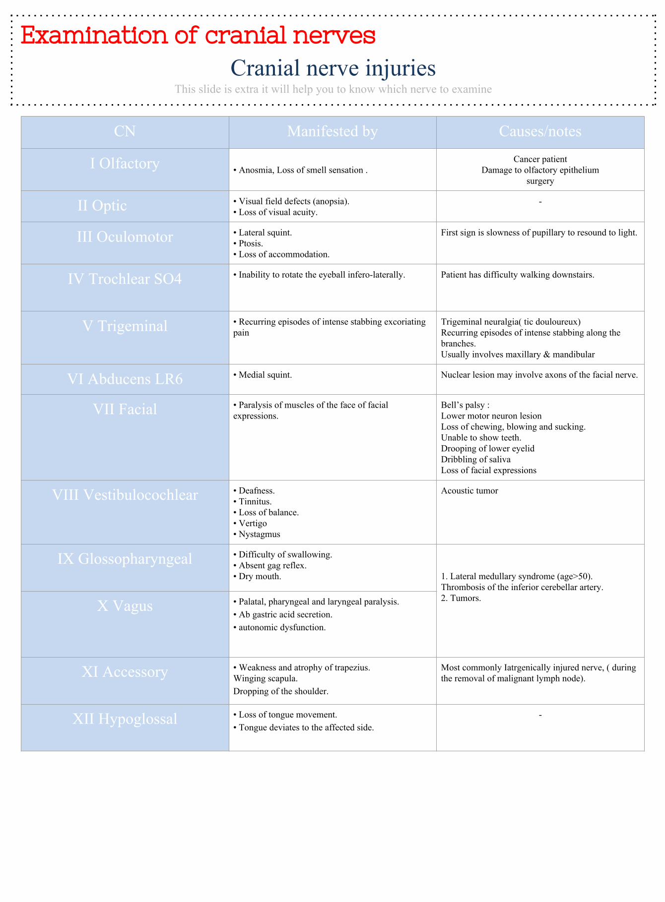
Examination of cranial nerves Cranial nerve injuries
This slide is extra it will help you to know which nerve to examine
CN Manifested by Causes/notes
I Olfactory • Anosmia, Loss of smell sensation .Cancer patient
Damage to olfactory epitheliumsurgery
II Optic • Visual field defects (anopsia). • Loss of visual acuity.
-
III Oculomotor • Lateral squint.• Ptosis.• Loss of accommodation.
First sign is slowness of pupillary to resound to light.
IV Trochlear SO4 • Inability to rotate the eyeball infero-laterally. Patient has difficulty walking downstairs.
V Trigeminal • Recurring episodes of intense stabbing excoriating pain
Trigeminal neuralgia( tic douloureux)Recurring episodes of intense stabbing along the branches.Usually involves maxillary & mandibular
VI Abducens LR6 • Medial squint. Nuclear lesion may involve axons of the facial nerve.
VII Facial • Paralysis of muscles of the face of facial expressions.
Bell’s palsy :Lower motor neuron lesion Loss of chewing, blowing and sucking.Unable to show teeth.Drooping of lower eyelidDribbling of saliva Loss of facial expressions
VIII Vestibulocochlear • Deafness.• Tinnitus.• Loss of balance. • Vertigo • Nystagmus
Acoustic tumor
IX Glossopharyngeal • Difficulty of swallowing. • Absent gag reflex.• Dry mouth. 1. Lateral medullary syndrome (age>50).
Thrombosis of the inferior cerebellar artery.2. Tumors.X Vagus • Palatal, pharyngeal and laryngeal paralysis.
• Ab gastric acid secretion. • autonomic dysfunction.
XI Accessory • Weakness and atrophy of trapezius.Winging scapula.Dropping of the shoulder.
Most commonly Iatrgenically injured nerve, ( during the removal of malignant lymph node).
XII Hypoglossal • Loss of tongue movement.• Tongue deviates to the affected side.
-
Ophthalmoscope and motor system
examination:
Ophthalmoscope and motor system examination
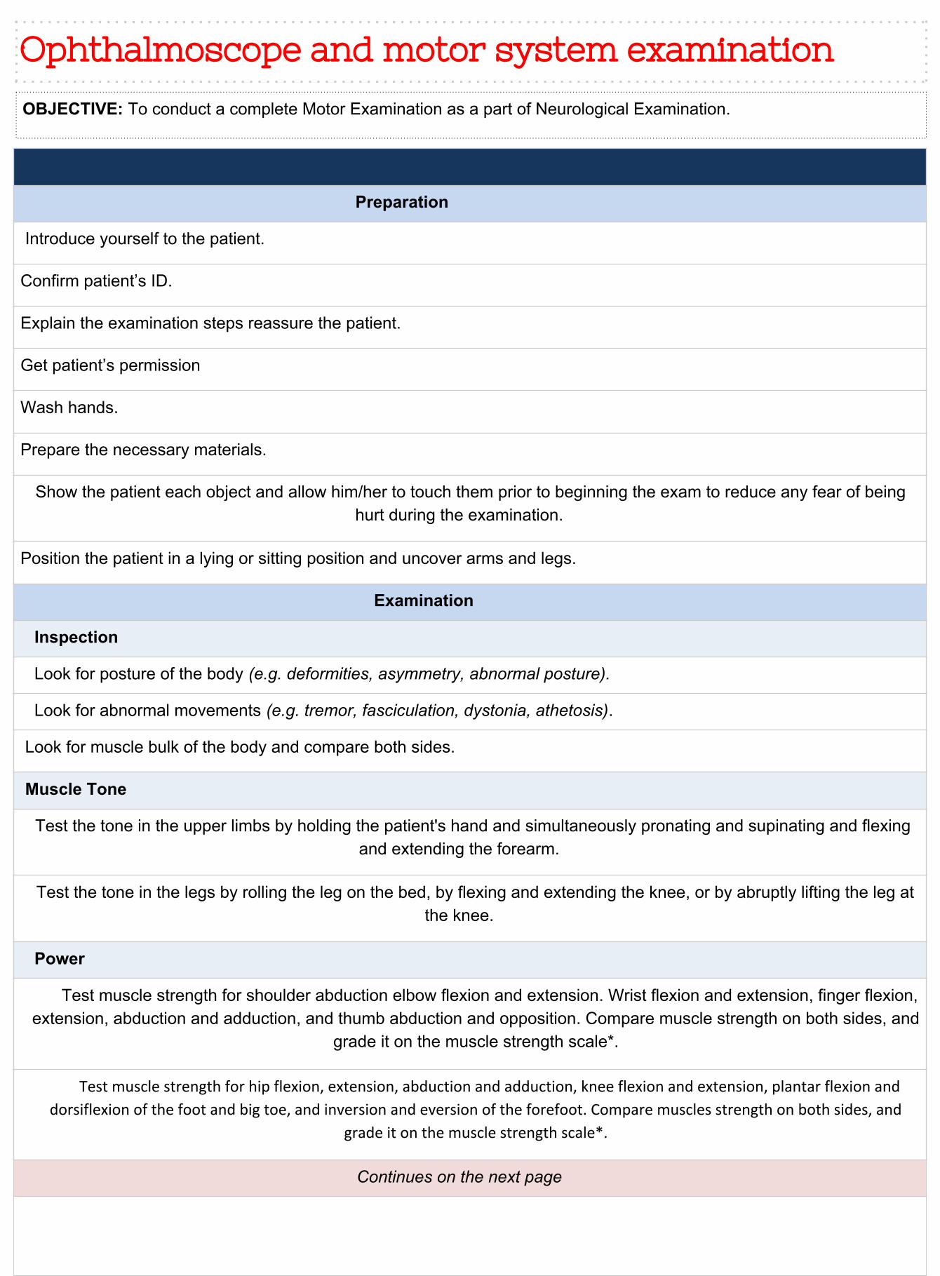
Preparation
Introduce yourself to the patient.
Confirm patient’s ID.
Explain the examination steps reassure the patient.
Get patient’s permission
Wash hands.
Prepare the necessary materials.
Show the patient each object and allow him/her to touch them prior to beginning the exam to reduce any fear of being hurt during the examination.
Position the patient in a lying or sitting position and uncover arms and legs.
Examination
Inspection
Look for posture of the body (e.g. deformities, asymmetry, abnormal posture).
Look for abnormal movements (e.g. tremor, fasciculation, dystonia, athetosis).
Look for muscle bulk of the body and compare both sides.
Muscle Tone
Test the tone in the upper limbs by holding the patient's hand and simultaneously pronating and supinating and flexing and extending the forearm.
Test the tone in the legs by rolling the leg on the bed, by flexing and extending the knee, or by abruptly lifting the leg at the knee.
Power
Test muscle strength for shoulder abduction elbow flexion and extension. Wrist flexion and extension, finger flexion, extension, abduction and adduction, and thumb abduction and opposition. Compare muscle strength on both sides, and
grade it on the muscle strength scale*.
Test muscle strength for hip flexion, extension, abduction and adduction, knee flexion and extension, plantar flexion and
dorsiflexion of the foot and big toe, and inversion and eversion of the forefoot. Compare muscles strength on both sides, and
grade it on the muscle strength scale*.
Continues on the next page
OBJECTIVE: To conduct a complete Motor Examination as a part of Neurological Examination.
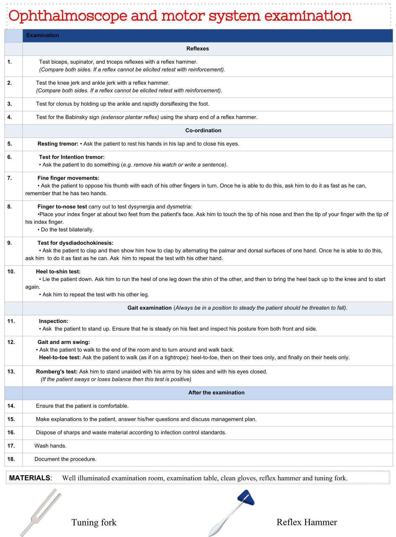
Examination
Reflexes
1. Test biceps, supinator, and triceps reflexes with a reflex hammer. (Compare both sides. If a reflex cannot be elicited retest with reinforcement).
2. Test the knee jerk and ankle jerk with a reflex hammer. (Compare both sides. If a reflex cannot be elicited retest with reinforcement).
3. Test for clonus by holding up the ankle and rapidly dorsiflexing the foot.
4. Test for the Babinsky sign (extensor plantar reflex) using the sharp end of a reflex hammer.
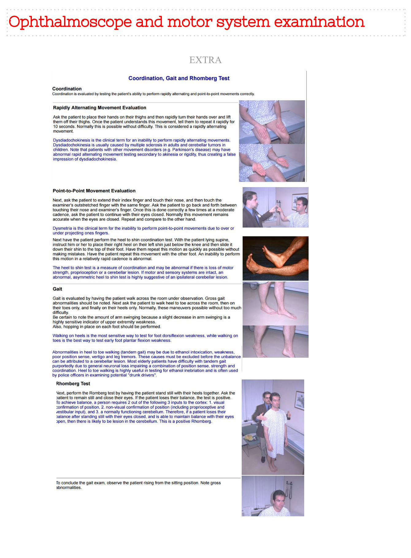
Co-ordination
5. Resting tremor: • Ask the patient to rest his hands in his lap and to close his eyes.
6. Test for Intention tremor: • Ask the patient to do something (e.g. remove his watch or write a sentence).
7. Fine finger movements: • Ask the patient to oppose his thumb with each of his other fingers in turn. Once he is able to do this, ask him to do it as fast as he can, remember that he has two hands.
8. Finger to-nose test carry out to test dysynergia and dysmetria: •Place your index finger at about two feet from the patient's face. Ask him to touch the tip of his nose and then the tip of your finger with the tip of his index finger. • Do the test bilaterally.
9. Test for dysdiadochokinesis: • Ask the patient to clap and then show him how to clap by alternating the palmar and dorsal surfaces of one hand. Once he is able to do this, ask him to do it as fast as he can. Ask him to repeat the test with his other hand.
10. Heel to-shin test: • Lie the patient down. Ask him to run the heel of one leg down the shin of the other, and then to bring the heel back up to the knee and to start again. • Ask him to repeat the test with his other leg.
Gait examination (Always be in a position to steady the patient should he threaten to fall).
11. Inspection: • Ask the patient to stand up. Ensure that he is steady on his feet and inspect his posture from both front and side.
12. Gait and arm swing: • Ask the patient to walk to the end of the room and to turn around and walk back. Heel-to-toe test: Ask the patient to walk (as if on a tightrope): heel-to-toe, then on their toes only, and finally on their heels only.
13. Romberg's test: Ask him to stand unaided with his arms by his sides and with his eyes closed. (If the patient sways or loses balance then this test is positive)
After the examination
14. Ensure that the patient is comfortable.
15. Make explanations to the patient, answer his/her questions and discuss management plan.
16. Dispose of sharps and waste material according to infection control standards.
17. Wash hands.
18. Document the procedure.
Ophthalmoscope and motor system examination
MATERIALS: Well illuminated examination room, examination table, clean gloves, reflex hammer and tuning fork.
Tuning fork Reflex Hammer
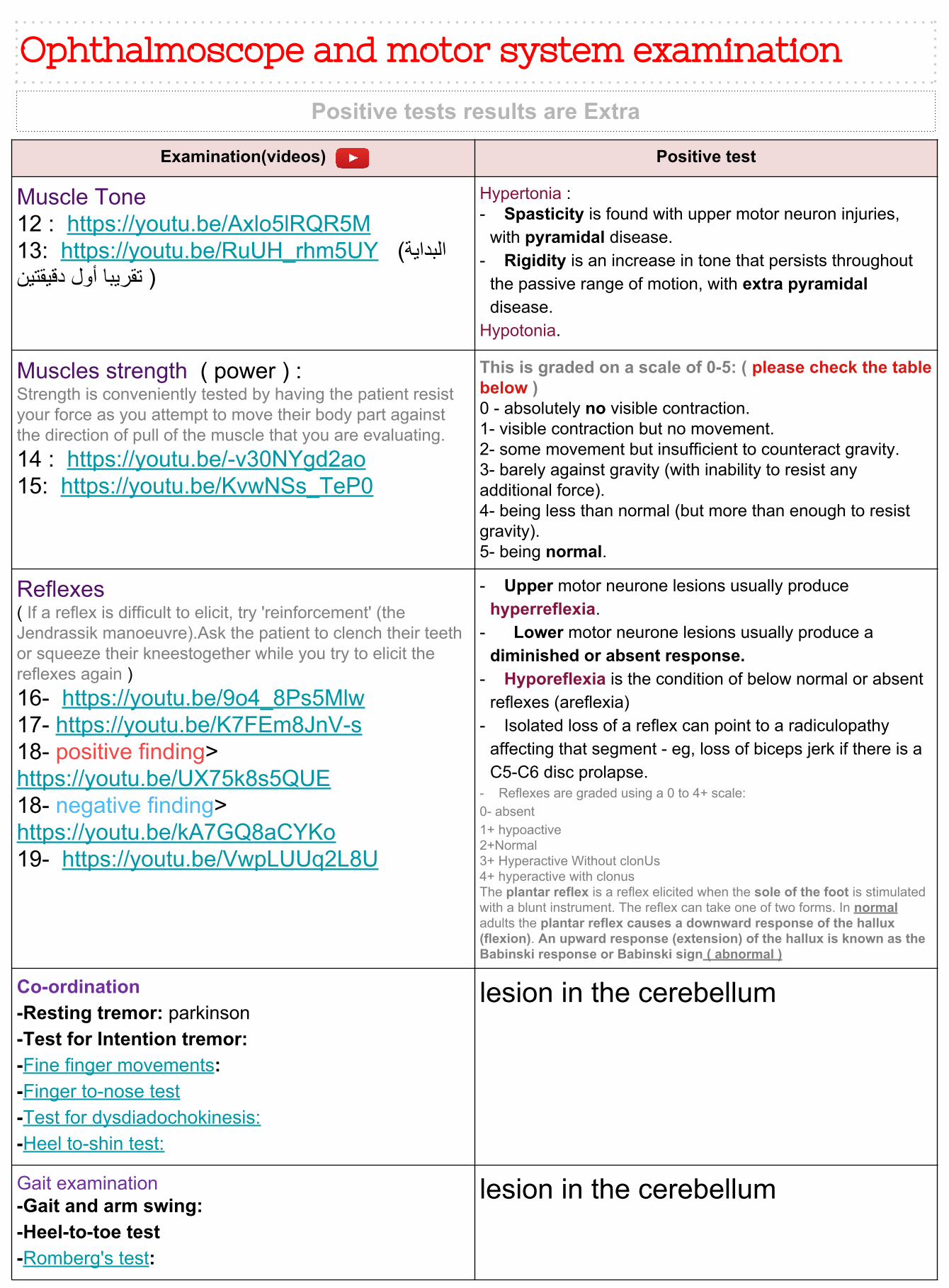
Examination(videos) Positive test
Muscle Tone12 : https://youtu.be/Axlo5lRQR5M13: https://youtu.be/RuUH_rhm5UY (البدایة ( تقریبا أول دقیقتین
Hypertonia :- Spasticity is found with upper motor neuron injuries,
with pyramidal disease.- Rigidity is an increase in tone that persists throughout
the passive range of motion, with extra pyramidal disease.
Hypotonia.
Muscles strength ( power ) :Strength is conveniently tested by having the patient resist your force as you attempt to move their body part against the direction of pull of the muscle that you are evaluating.14 : https://youtu.be/-v30NYgd2ao15: https://youtu.be/KvwNSs_TeP0
This is graded on a scale of 0-5: ( please check the table below )0 - absolutely no visible contraction.1- visible contraction but no movement.2- some movement but insufficient to counteract gravity.3- barely against gravity (with inability to resist any additional force).4- being less than normal (but more than enough to resist gravity).5- being normal.
Reflexes( If a reflex is difficult to elicit, try 'reinforcement' (the Jendrassik manoeuvre).Ask the patient to clench their teeth or squeeze their kneestogether while you try to elicit the reflexes again )16- https://youtu.be/9o4_8Ps5Mlw17- https://youtu.be/K7FEm8JnV-s18- positive finding> https://youtu.be/UX75k8s5QUE18- negative finding> https://youtu.be/kA7GQ8aCYKo19- https://youtu.be/VwpLUUq2L8U
- Upper motor neurone lesions usually produce hyperreflexia.
- Lower motor neurone lesions usually produce a diminished or absent response.
- Hyporeflexia is the condition of below normal or absent reflexes (areflexia)
- Isolated loss of a reflex can point to a radiculopathy affecting that segment - eg, loss of biceps jerk if there is a C5-C6 disc prolapse.
- Reflexes are graded using a 0 to 4+ scale:0- absent1+ hypoactive2+Normal3+ Hyperactive Without clonUs4+ hyperactive with clonusThe plantar reflex is a reflex elicited when the sole of the foot is stimulated with a blunt instrument. The reflex can take one of two forms. In normal adults the plantar reflex causes a downward response of the hallux (flexion). An upward response (extension) of the hallux is known as the Babinski response or Babinski sign ( abnormal )
Co-ordination-Resting tremor: parkinson-Test for Intention tremor:-Fine finger movements:-Finger to-nose test-Test for dysdiadochokinesis:-Heel to-shin test:
lesion in the cerebellum
Gait examination-Gait and arm swing:-Heel-to-toe test-Romberg's test:
lesion in the cerebellum
Ophthalmoscope and motor system examination
Positive tests results are Extra
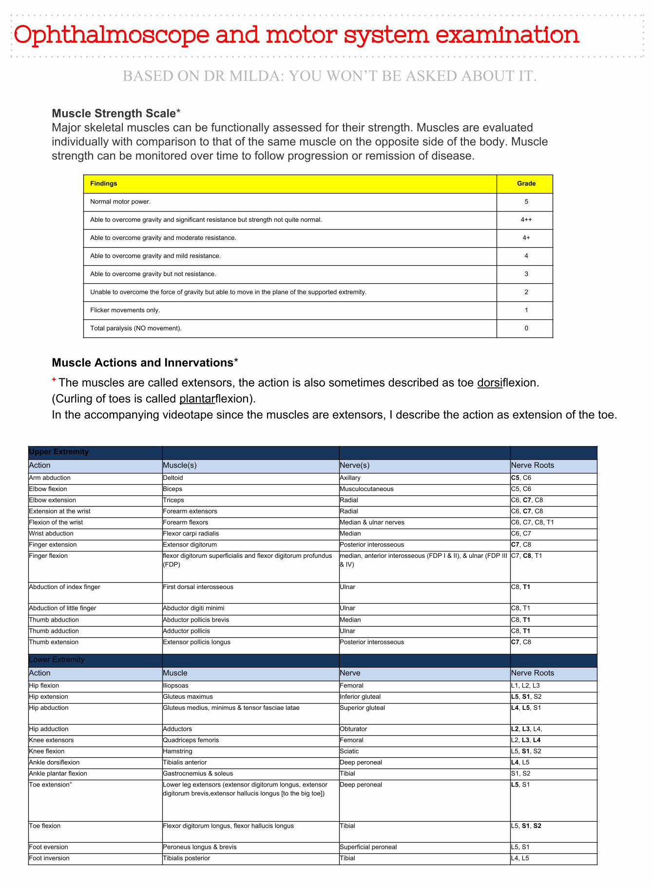
Findings Grade
Normal motor power. 5
Able to overcome gravity and significant resistance but strength not quite normal. 4++
Able to overcome gravity and moderate resistance. 4+
Able to overcome gravity and mild resistance. 4
Able to overcome gravity but not resistance. 3
Unable to overcome the force of gravity but able to move in the plane of the supported extremity. 2
Flicker movements only. 1
Total paralysis (NO movement). 0
Muscle Strength Scale*Major skeletal muscles can be functionally assessed for their strength. Muscles are evaluated individually with comparison to that of the same muscle on the opposite side of the body. Muscle strength can be monitored over time to follow progression or remission of disease.
Upper Extremity
Action Muscle(s) Nerve(s) Nerve RootsArm abduction Deltoid Axillary C5, C6
Elbow flexion Biceps Musculocutaneous C5, C6
Elbow extension Triceps Radial C6, C7, C8
Extension at the wrist Forearm extensors Radial C6, C7, C8
Flexion of the wrist Forearm flexors Median & ulnar nerves C6, C7, C8, T1
Wrist abduction Flexor carpi radialis Median C6, C7
Finger extension Extensor digitorum Posterior interosseous C7, C8
Finger flexion flexor digitorum superficialis and flexor digitorum profundus (FDP)
median, anterior interosseous (FDP I & II), & ulnar (FDP III & IV)
C7, C8, T1
Abduction of index finger First dorsal interosseous Ulnar C8, T1
Abduction of little finger Abductor digiti minimi Ulnar C8, T1
Thumb abduction Abductor pollicis brevis Median C8, T1Thumb adduction Adductor pollicis Ulnar C8, T1Thumb extension Extensor pollicis longus Posterior interosseous C7, C8
Lower Extremity
Action Muscle Nerve Nerve RootsHip flexion Iliopsoas Femoral L1, L2, L3
Hip extension Gluteus maximus Inferior gluteal L5, S1, S2
Hip abduction Gluteus medius, minimus & tensor fasciae latae Superior gluteal L4, L5, S1
Hip adduction Adductors Obturator L2, L3, L4,
Knee extensors Quadriceps femoris Femoral L2, L3, L4Knee flexion Hamstring Sciatic L5, S1, S2
Ankle dorsiflexion Tibialis anterior Deep peroneal L4, L5
Ankle plantar flexion Gastrocnemius & soleus Tibial S1, S2
Toe extension+ Lower leg extensors (extensor digitorum longus, extensor digitorum brevis,extensor hallucis longus [to the big toe])
Deep peroneal L5, S1
Toe flexion Flexor digitorum longus, flexor hallucis longus Tibial L5, S1, S2
Foot eversion Peroneus longus & brevis Superficial peroneal L5, S1
Foot inversion Tibialis posterior Tibial L4, L5
Muscle Actions and Innervations*+ The muscles are called extensors, the action is also sometimes described as toe dorsiflexion.(Curling of toes is called plantarflexion).In the accompanying videotape since the muscles are extensors, I describe the action as extension of the toe.
Ophthalmoscope and motor system examination
BASED ON DR MILDA: YOU WON’T BE ASKED ABOUT IT.
EXTRA
Ophthalmoscope and motor system examination
History taking from a patient with
neuropsychological problem:
History taking from a patient with neuropsychological problem
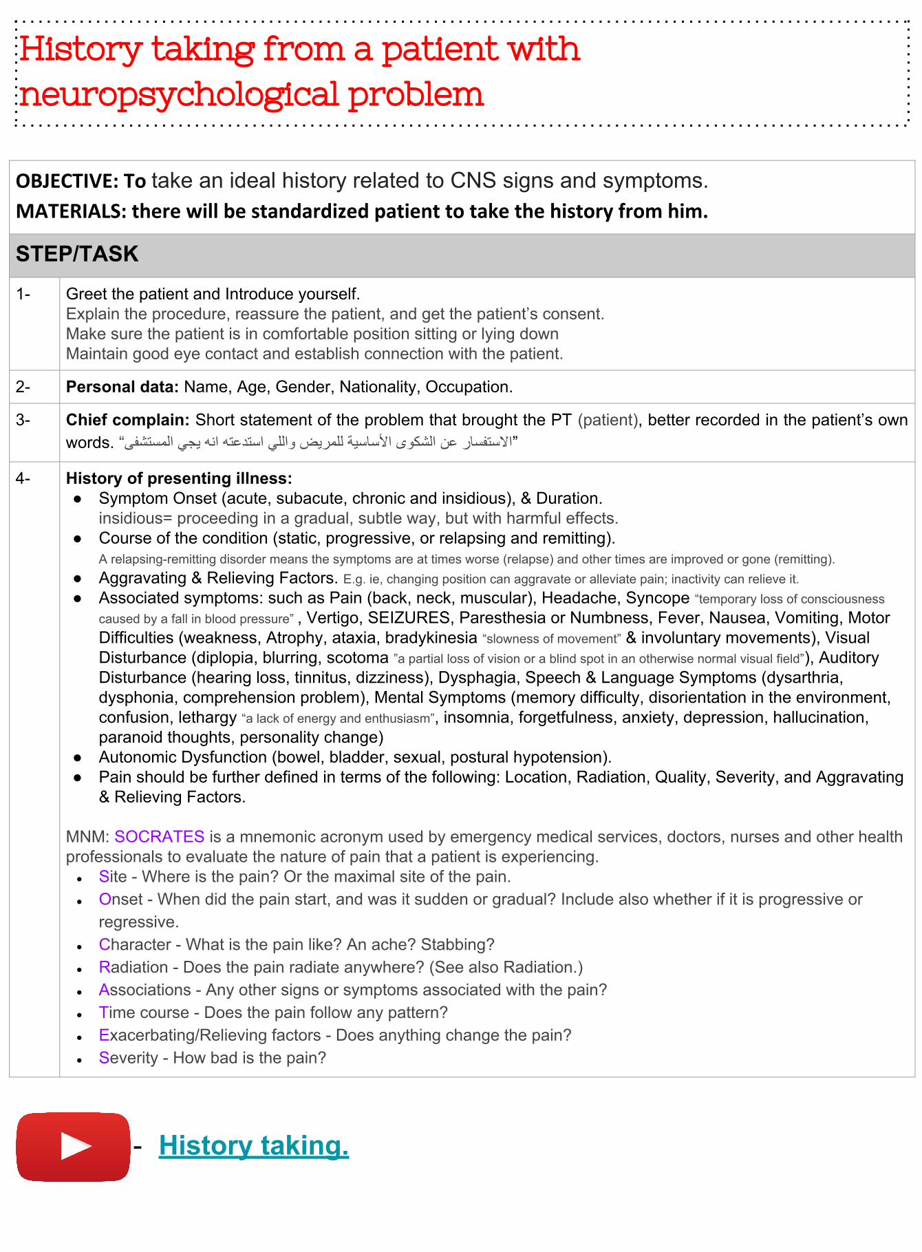
OBJECTIVE: To take an ideal history related to CNS signs and symptoms.MATERIALS: there will be standardized patient to take the history from him.
STEP/TASK1- Greet the patient and Introduce yourself.
Explain the procedure, reassure the patient, and get the patient’s consent.Make sure the patient is in comfortable position sitting or lying downMaintain good eye contact and establish connection with the patient.
2- Personal data: Name, Age, Gender, Nationality, Occupation.
3- Chief complain: Short statement of the problem that brought the PT (patient), better recorded in the patient’s own words. “االستفسار عن الشكوى األساسیة للمریض واللي استدعتھ انھ یجي المستشفى”
4- History of presenting illness:● Symptom Onset (acute, subacute, chronic and insidious), & Duration.
insidious= proceeding in a gradual, subtle way, but with harmful effects.● Course of the condition (static, progressive, or relapsing and remitting).
A relapsing-remitting disorder means the symptoms are at times worse (relapse) and other times are improved or gone (remitting). ● Aggravating & Relieving Factors. E.g. ie, changing position can aggravate or alleviate pain; inactivity can relieve it.● Associated symptoms: such as Pain (back, neck, muscular), Headache, Syncope “temporary loss of consciousness
caused by a fall in blood pressure” , Vertigo, SEIZURES, Paresthesia or Numbness, Fever, Nausea, Vomiting, Motor Difficulties (weakness, Atrophy, ataxia, bradykinesia “slowness of movement” & involuntary movements), Visual Disturbance (diplopia, blurring, scotoma ”a partial loss of vision or a blind spot in an otherwise normal visual field”), Auditory Disturbance (hearing loss, tinnitus, dizziness), Dysphagia, Speech & Language Symptoms (dysarthria, dysphonia, comprehension problem), Mental Symptoms (memory difficulty, disorientation in the environment, confusion, lethargy “a lack of energy and enthusiasm”, insomnia, forgetfulness, anxiety, depression, hallucination, paranoid thoughts, personality change)
● Autonomic Dysfunction (bowel, bladder, sexual, postural hypotension).● Pain should be further defined in terms of the following: Location, Radiation, Quality, Severity, and Aggravating
& Relieving Factors.
MNM: SOCRATES is a mnemonic acronym used by emergency medical services, doctors, nurses and other health professionals to evaluate the nature of pain that a patient is experiencing.
● Site - Where is the pain? Or the maximal site of the pain.● Onset - When did the pain start, and was it sudden or gradual? Include also whether if it is progressive or
regressive.● Character - What is the pain like? An ache? Stabbing?● Radiation - Does the pain radiate anywhere? (See also Radiation.)● Associations - Any other signs or symptoms associated with the pain?● Time course - Does the pain follow any pattern?● Exacerbating/Relieving factors - Does anything change the pain?● Severity - How bad is the pain?
- History taking.

History taking from a patient with neuropsychological problem (cont.)
5- Past Medical History:● Same situation has happened before “similar episodes” , head trauma, toxic exposure.● Chronic disease (DM, HTN, hyperlipidemia , renal or cardiac diseases, connective tissue diseases …)● History of hospitalization : Admission, Surgery.
6- Family & Social History:● Same situation in the family, chronic disease (DM, HTM “HTN?”), congenital & hereditary diseases, history of
stroke or transient ischemic attack.● Marital status, No. of children, housing status, job status & environment / conclude: socioeconomic status.
History of travelling.● Habits: smoking, drinking Alcohol, using prohibited substances. ● Ask politely about emotional problems at home or at work.
Obstetric and Gynecologic History (if patient is female)● Ask about LMP (Last Menstrual Period), regularity, and quality of menstruation, and menopause if patient is
elderly● Ask about number of pregnancies, abortion, number of children, and history complications during the
pregnancy.
Drug history:Any recent medication, long term medication, Allergies, Herbal Medication. Blood transfusion.
Systematic review: (We ask about this to exclude and search for secondary problems for the overall wellness of the patient)
Cardio-respiratory symptoms● Ask about having cough, shortness of breath, chest pain, ankle swelling, etc.
GIT symptoms● Ask about having weight loss, nausea, or vomiting, changes of bowel movement, abdominal pain, etc.
Neurological symptoms● Ask about having headache, dizziness, ringing in the ears, changes in hearing, vision, smell or taste, etc.
Urinary and reproductive symptoms● Ask about having burning on passing urine, frequency of urination, blood in the urine, etc.● Ask about having penile or vaginal discharge, hesitancy or urgency of urination, poor urine stream, or dribbling.
Dermatologic Symptoms● Ask about skin rahses, redness, or itchiness, etc.
Musculoskeletal Sympotoms● Ask about having joint pain, or stiffness, muscle pain or weakness, etc.
EXTRA
Patient came in with a chief complaint of a headache similar to a migraine. They do not have an aura. Headache is accompanied by tears, eye pain, and sneezing.
Know that there are 3 types of headaches: Cluster, Tension, and Migraines. According to the symptoms of our patient, it is clear that they have a Cluster headache.
Try to pinpoint the source of the headache by questions such as: What is the nature of your job? How long do you work for? Any chronic medical conditions such as hypertension? (for females: do you have a headache when you are on your period/pregnant?)
Important notes:When taking the history, ask questions that will exclude from your hypothesis. For example: If the patient does not have a fever, you may exclude meningitis.If the duration of the patient's headache is not chronic, you may exclude tumors.Try to avoid yes/no questions, let the patient explain and describe, and repeat their answers for confirmation.
Cluster Headache
No aura, accompanied by tears, eye pain, sneezing
Tension Headache
Feels like a tight band wrapped around the forehead
Migraine
Throbbing pain. With aura: light irritation, photophobia