Embed Size (px)
Citation preview

[ Color index: Important | Hacker & Moore | 428 | Doctor’s Notes | Kaplan | males notes ]
Editing file link
Antenatal Fetal Assessment
Objectives:
Describe how to test for each of the following: ➢ Fetal well-being. ➢ Fetal growth. ➢ Fetal movements. ➢ Amniotic fluid volume. ➢ Fetal lung maturity
Done by: Samar AlOtaibi & Luluh Alzeghayer Revised by: Haifa Almohsen & Lina Alshehri
● Numbers (weeks) are very important to memorize. ● Here is a very helpful file for preconception, prenatal, intrapartum and postpartum
care. By Dalal Alhuzaimi.

Fetal Assessment (Fetal Well-Being)
● Fetal assessment is to identify fetuses at risk of neurologic injury or death in order to prevent prenatal mortality & morbidity.
● A combination of maternal self-assessment, nonstress testing (NST), and real-time ultrasonic assessment is used to evaluate fetal well-being.
● The most common reasons for fetal testing are decreased fetal movements, diabetes, post dates, chronic hypertension, and IUGR.
The objective is to make sure that the environment inside the uterus is better than the outside. Once there is a parameter showing that the environment inside the uterus is not good it’s an indication for IOL
FETAL AND NEONATAL COMPLICATIONS OF ANTEPARTUM ASPHYXIA: (Bad outcomes if we have bad environment inside the uterus )
➢ Stillbirth (Mortality) ➢ Metabolic acidosis at birth ➢ Hypoxic renal damage ➢ Necrotizing enterocolitis
➢ Intracranial hemorrhage ➢ Seizures ➢ Cerebral palsy
Rationale: If fetal oxygenation challenged:
➢ Blood flow directed to brain, heart & adrenal. Blood flow away from the kidney decrease fetal urine production ⇒ Decrease AF volume. If AFV is decreased in the absence of renal abnormalities, it’s the first indication of fetal hypoxia.
➢ CNS hypoxia ⇒ Decreased Fetal movement. More impairment > even the brain will suffer hypoxia.
➢ Chemoreceptors ⇒ vagally-mediated reflex ⇒ Fetal heart rate abnormality (late deceleration).
CONDITIONS ASSOCIATED WITH INCREASED PERINATAL MORBIDITY/MORTALITY: for which we need to do routine antenatal assessment.
➢ Small fetus for gestational age fetus. ➢ Decreased fetal movement. We ask the lady to count
fetal movement. ➢ Postdates pregnancy (>294 days). > 40 weeks ➢ Pre-eclampsia / chronic hypertension. Why? High BP >
decreased blood flow to the kidneys. ➢ Pre-pregnancy diabetes / Insulin requiring gestational
diabetes. ➢ Preterm premature rupture of membranes. ➢ Chronic (stable) abruption.
When to start fetal Assessment antenatally:
➢➢ Risk assessed individually: ○ For D.M. fetal assessment should start from 32 weeks onward if uncomplicated ○ If complicated D.M. E.g. retinopathy renal impairment start at 24 weeks onward. ○ For Post date pregnancy start at 40 weeks. ○ For any patient with decreased fetal movement start immediately.
➢ Fetal assessment is done once or twice weekly.

Early Pregnancy Assessment
Fetal heart activity
➢ Fetal auscultation (special stethoscope or Doppler) - 12 weeks. →→ ➢ Fetal heart activity seen by USS - from 6 weeks. →→
Routinely in the clinic we can LISTEN to the fetal heart using doppler only after 12 weeks. Why? Because the uterus is very small & behind the symphysis pubis bone. It can be SEEN only using ultrasound starting from the 6th week.
Nuchal translucency
1
➢➢ In women >35 or who have bad obstetric history, we measure the nuchal translucency which is the fold behind neck of the fetus . 2
➢ Measurement for early screening for chromosomal abnormality. ➢ Between 11-13+ weeks. Measurement won’t be accurate before 11 or after 13 weeks,
thus accurate gestational age is important. ➢ If Thick > chromosomal abnormalities like down syndrome or trisomy 18. (Aneuploidy &
Cardiac disease)
Fetal movement
➢ Fetal movement are usually first perceptible to mother ~17w-20w . The first fetal movement felt by the mother is called quickening. Primigravida usually feel it later (almost at 20) while multipara feel it at 16 or 17.
➢ Baby moves from 6th week onwards and can be seen on US but not felt by the mother. Why? Because the baby has to be big enough to be felt under the abdominal wall.
➢ Only 50% of isolated limb movements are perceived, and 80% of trunk and limb (whole body) movements.
Fetal growth 1. By fundal height measurement in the clinic. Measure from symphysis pubis to fundus and compare with gestational age. Distance (cm) = # of gestational weeks
2. By ultrasound (Biometry): ➢ Biparietal diameter (BPD). Widest diameter of fetal head to assess vaginal delivery. ➢ Abdominal Circumference (AC) indication of fetal weight. If small > IUGR, if big >
diabetes.
➢ Femur Length (FL) why the femur? Because you can’t measure the whole fetus on US. if Short > chromosomal (down) or skeletal (dwarfism)
➢ Head Circumference (HC)
Measurements are illustrated on the Growth Chart→→
If below the normal line > lagging of the growth, if above it > large for date as in diabetic mother.
1 Nuchal translucency is a collection of fluid under the skin at the back of your baby's neck. 2 This occurs partly because of the fetus’s tendency to lie on its back and partly because of the laxity of the skin of the neck. Chromosome 21 contains the gene that codes for type VI collagen. In trisomy 21 one subunit of this collagen can be overexpressed, resulting in connective tissue that has a more elastic composition. (Read)
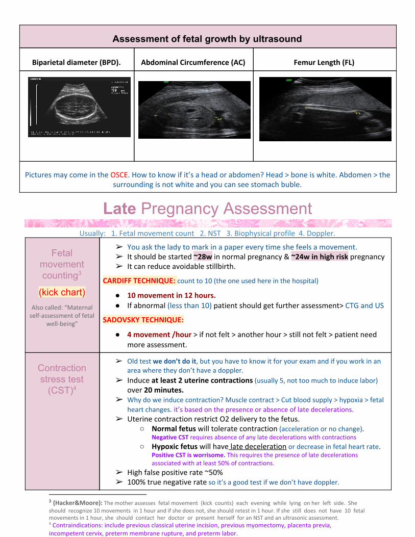
Assessment of fetal growth by ultrasound
Biparietal diameter (BPD). Abdominal Circumference (AC) Femur Length (FL)
Pictures may come in the OSCE. How to know if it’s a head or abdomen? Head > bone is white. Abdomen > the surrounding is not white and you can see stomach buble.
Late Pregnancy Assessment Usually: 1. Fetal movement count 2. NST 3. Biophysical profile 4. Doppler.
Fetal movement counting 3
(kick chart) Also called: “Maternal
self-assessment of fetal well-being”
➢ You ask the lady to mark in a paper every time she feels a movement. ➢ It should be started ~28w in normal pregnancy & ~24w in high risk pregnancy ➢ It can reduce avoidable stillbirth.
CARDIFF TECHNIQUE: count to 10 (the one used here in the hospital)
● 10 movement in 12 hours. ● If abnormal (less than 10) patient should get further assessment> CTG and US
SADOVSKY TECHNIQUE:
● 4 movement /hour > if not felt > another hour > still not felt > patient need more assessment.
Contraction stress test
(CST) 4
➢ Old test we don’t do it, but you have to know it for your exam and if you work in an area where they don’t have a doppler.
➢ Induce at least 2 uterine contractions (usually 5, not too much to induce labor) over 20 minutes.
➢ Why do we induce contraction? Muscle contract > Cut blood supply > hypoxia > fetal heart changes. it’s based on the presence or absence of late decelerations.
➢ Uterine contraction restrict O2 delivery to the fetus. ○ Normal fetus will tolerate contraction (acceleration or no change).
Negative CST requires absence of any late decelerations with contractions
○ Hypoxic fetus will have late deceleration or decrease in fetal heart rate. Positive CST is worrisome. This requires the presence of late decelerations associated with at least 50% of contractions.
➢ High false positive rate ~50% ➢ 100% true negative rate so it’s a good test if we don’t have doppler.
3 (Hacker&Moore): The mother assesses fetal movement (kick counts) each evening while lying on her left side. She should recognize 10 movements in 1 hour and if she does not, she should retest in 1 hour. If she still does not have 10 fetal movements in 1 hour, she should contact her doctor or present herself for an NST and an ultrasonic assessment. 4 Contraindications: include previous classical uterine incision, previous myomectomy, placenta previa, incompetent cervix, preterm membrane rupture, and preterm labor.
Important
Non stress test (NST)
Also called: CTG 5
(Cardiotocography)
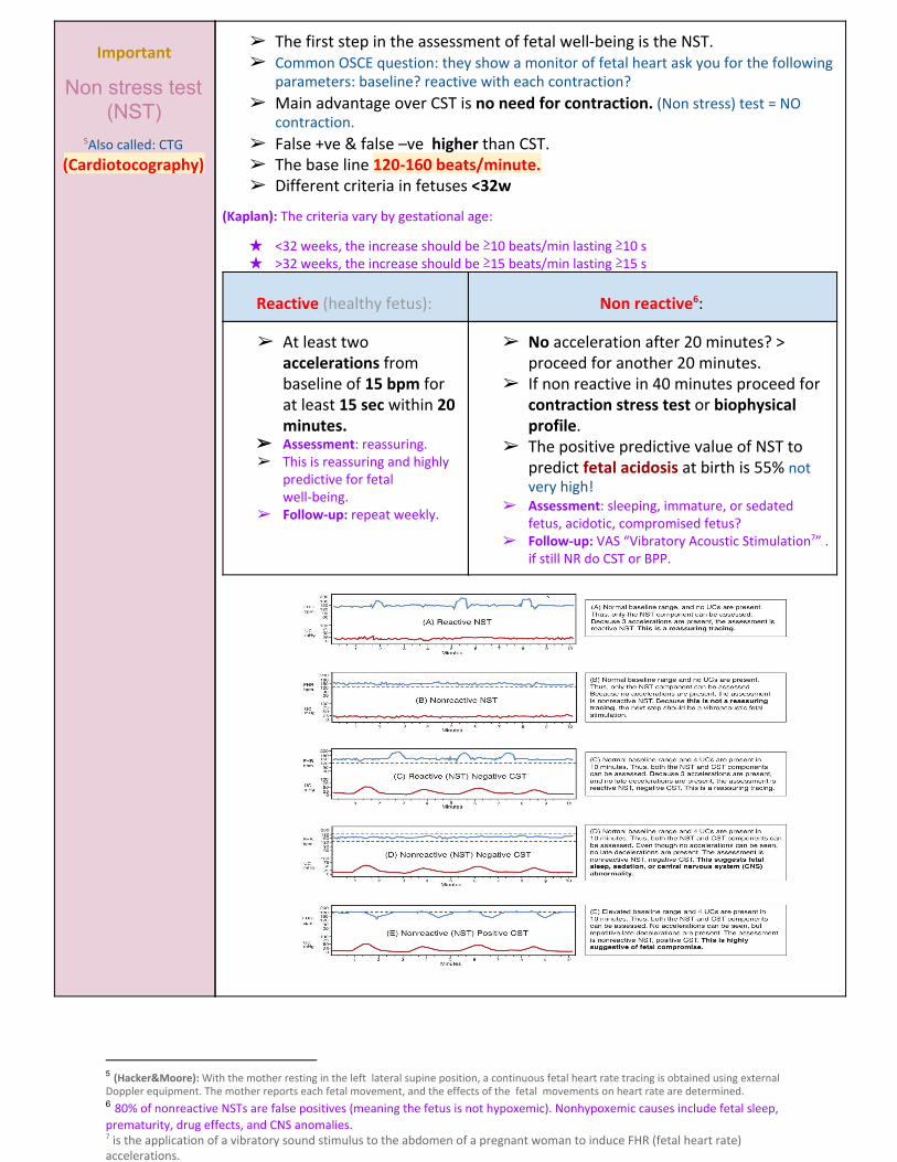
➢ The first step in the assessment of fetal well-being is the NST. ➢ Common OSCE question: they show a monitor of fetal heart ask you for the following
parameters: baseline? reactive with each contraction? ➢ Main advantage over CST is no need for contraction. (Non stress) test = NO
contraction.
➢ False +ve & false –ve higher than CST. ➢ The base line 120-160 beats/minute. ➢ Different criteria in fetuses <32w
(Kaplan): The criteria vary by gestational age:
★ <32 weeks, the increase should be ≥10 beats/min lasting ≥10 s ★ >32 weeks, the increase should be ≥15 beats/min lasting ≥15 s
Reactive (healthy fetus): Non reactive : 6
➢ At least two accelerations from baseline of 15 bpm for at least 15 sec within 20 minutes.
➢➢ Assessment: reassuring. ➢ This is reassuring and highly
predictive for fetal well-being.
➢ Follow-up: repeat weekly.
➢ No acceleration after 20 minutes? > proceed for another 20 minutes.
➢ If non reactive in 40 minutes proceed for contraction stress test or biophysical profile.
➢ The positive predictive value of NST to predict fetal acidosis at birth is 55% not very high!
➢ Assessment: sleeping, immature, or sedated fetus, acidotic, compromised fetus?
➢ Follow-up: VAS “Vibratory Acoustic Stimulation ” . 7
if still NR do CST or BPP.
5 (Hacker&Moore): With the mother resting in the left lateral supine position, a continuous fetal heart rate tracing is obtained using external Doppler equipment. The mother reports each fetal movement, and the effects of the fetal movements on heart rate are determined. 6 80% of nonreactive NSTs are false positives (meaning the fetus is not hypoxemic). Nonhypoxemic causes include fetal sleep, prematurity, drug effects, and CNS anomalies. 7 is the application of a vibratory sound stimulus to the abdomen of a pregnant woman to induce FHR (fetal heart rate) accelerations.

➢➢ Interpretation of CTG:
FHR Baseline ● Normal Baseline FHR 110–160 bpm (some books say 120-160) ● Moderate bradycardia 100–109 bpm ● Moderate tachycardia 161–180 bpm ● Abnormal “Severe” bradycardia < 100 bpm ● Abnormal tachycardia > 180 bpm
Variability ❏❏ Reduced variability. Sedation of mother during labor (pethidine), mother or fetus is sleeping, severe hypoxia.
❏❏ Baseline variability: Fluctuations in the baseline FHR that are irregular in amplitude and frequency. It is a reflection of the autonomic interplay between the sympathetic and parasympathetic nervous system.
ACCELERATION ❖❖ Accelerations are always reassuring.
DECELERATION ❏ Early > Head compression. During 2nd stage of labor.
❏ Late > U-P Insufficiency. Decrease blood flow to the placenta, asphyxia, hypoxia.
❏ Variable > Cord compression, Primary CNS dysfunction. (Variable= Different shapes, sometimes early sometimes late): cord compression also in labor.
❏ Mixed (characteristics of any of the aforementioned patterns)
Mnemonic: VEAL CHOP ★★ VARIABLE > CORD COMPRESSION ★★ EARLY > HEAD COMPRESSION ★★ ACCELERATION > OK ★★ LATE > PLACENTAL INSUFFICIENCY
TACHYCARDIA ➢ Severe Hypoxia ➢ Chorioamnionitis > fever > mom tachycardia > fetal tachycardia ➢ Maternal fever ➢ Mimetic drugs ➢ Fetal anemia, sepsis, HT failure, Arrhythmias.
Bradycardia ➢ When FHR baseline is <110 beats/min ➢ Non-hypoxic explanations include:
– Maternal medications: β-adrenergic blockers, local anesthetics – Fetal arrhythmia: congenital heart block (associated with maternal lupus)

Amniotic fluid index (AFI)
➢ The next step in prenatal assessment is to determine the adequacy of amniotic fluid volume by real-time ultrasonography.
➢ The sum of the maximum vertical fluid pocket diameter in four quarters. ○ AF index: We divide the abdomen to 4 quadrants, we take the longest vertical
diameter of each pocket and add them together. ○ AF Volume: Some people only take 1 vertical diameter of the largest pocket.
➢ The normal value 5-25cm ○ Normal 9 - 25 cm ○ Borderline 5 - 8 cm ○ Reduced fluid (oligohydramnios; AFI < 5 cm) : decreased fetal urinary 8
output lead to decrease amniotic fluid. Can be caused by fetal renal abnormalities or reduced blood supply to the kidneys.
○ Excessive amniotic fluid (polyhydramnios; AFI > 24 cm) : reduced fetal 9
swallowing of AF or fetal GIT anomalies.
Biophysical profile
(BPP)
➢ A complete BPP measures 5 components of fetal well-being: ○ NST, ○ Amniotic fluid volume, ○ Fetal gross body movements, Whole body or 1 limb movement ○ Fetal extremity tone, flexion/ extension or the extension / flexion of limbs ○ Fetal breathing mov. Movement of fetal chest wall on US: over 20 min.
➢ It is a scoring system. Score is either 2 or 0. You can have 10/10 or 8/10 or 6/10 but NOT 7 or 9/10
➢ It is done over 30 minutes. ➢ It measure acute hypoxia (NST, body mov. & breathing) & chronic hypoxia (AFI) ➢ The risk of fetal death within 1 week if BPP is normal~ 1/1300
Fetal Biophysical profile/NST+: Biophysical Variable Normal (score=2) Abnormal (score= 0)
Fetal breathing movements
1 episode FBM of at least 30 s duration in 30 min
Absent FBM or no episode >30 s in 30 min
Fetal movements 3 discrete body/limb movements in 30 min
2 or fewer body/limb movements in 30 min
Fetal tone 1 episode of active extension with return to flexion of fetal limb(s) or trunk. Opening and closing of the hand considered normal tone.
-Either slow extension with return to partial flexion or movement of limb in full extension -Absent fetal movement.
Amniotic fluid volume
1 pocket of AF that measures at least 2 cm in 2 perpendicular planes
Either no AF pockets or a pocket <2 cm in 2 perpendicular planes
○ Score of 8 or 10—highly reassuring of fetal well-being. Management is to repeat the test weekly or as indicated.
○ Score of 4 or 6—worrisome. Management is delivery if the fetus is >36 weeks or repeat the BPP in 12–24 h if <36 weeks. An alternative is to perform a CST.
○ Score of 0 or 2—highly predictive of fetal hypoxia. Management is prompt delivery regardless of gestational age.
➢ Modified BPP (mBPP): includes only the NST and amniotic fluid volume. Its predictive value is almost as high as a complete BPP. Low false negative 0.8/1000, High false positives ~60%. Don’t have time to wait 30 minutes for fetal breathing or movement so only do NST and AF!
➢ Next step > doppler.
8 suggests fetal compromise as a result of umbilical cord compression. 9 can be a sign of poor control in a diabetic pregnancy or an indication that the fetus may have an anomaly.

Doppler Velocimetry
(UAV)
● Measurement of blood flow velocities in maternal & fetal vessels. ● Reflect feto-placental circulation. ● Doppler indices from Umbilical a. (UA), Uterine a. & MCA (middle cerebral a. Of
fetus). ● Doppler studies is mostly valuable for IUGR, as well as fetal anemia in
alloimmunized pregnancies.
● In IUGR, absent or reversed EDF (end diastolic flow) associated with fetal hypoxia.
● Won’t be in the exam but to explain it: Waves of blood flow (systole and diastole), the area between (EDF) is the area we’re interested in.
○ Reduced: low flow due to high blood pressure or pre-eclampsia. management: keep the patient and repeat the test after 2 hours. If it goes back to normal > reassure. If it becomes absent > operate
○ Absent: Severe insufficiency > Fetus will only have blood during systole.
○ Reverse: blood flow from fetus to mother (baby about to die)
UMBILICAL ARTERY DOPPLER: ● This test measures the ratio of systolic and diastolic blood flow in the umbilical artery. ● The umbilical circulation normally has low resistance, so significant diastolic blood flow is
expected. ● The systolic/diastolic (S/D) ratio normally decreases throughout pregnancy. ● This test is predictive of poor perinatal outcome only in IUGR fetuses. ● Nonreassuring findings, which may indicate need for delivery, are absent diastolic flow and
reversed diastolic flow.

Invasive Fetal Assessment
Invasive = you’re gonna stick a needle
AMNIOCENTESIS
*Direct insertion through the abdomen
➢ Obtaining a sample of amniotic fluid during pregnancy. By US ➢ Usually done after 15w (can be done after 11w) ➢ They used to do it at 11 w. But now only done after 15 w. To avoid the
complications.
Indications:
● Genetic (karyotype) if you suspect chromosomal anomalies. NTD screening by AFP and acetylcholinesterase.
● Bilirubin level (in RH-isoimmunisation) ● Fetal lung maturity (L/S) will be discussed below ● Therapeutic in polyhydramnios. Therapeutic = decrease amount of AF
Risks: ROM ~1%, abortion 0.5%, infection 1/1000. ROM=rupture of membrane
CHORIONIC VILLUS SAMPLING
(CVS) 10
*Transcervical
➢ Usually done after 10w. It is the procedure of choice for first trimester prenatal diagnosis of genetic disorders. What’s the difference between amniocentesis and CVS? TIMING!!
➢ Early diagnostic test (CVS) if abnormalities present and abortion is indicated is important because:
❏ lower medicolegal issues. ❏ Lower complications of early abortion than late abortion. ❏ Insurance issues. ❏ Maternal-fetal bonding.
Complication:
● Fetal loss (0.7 % within 14 days of a TA CVS procedure and 1.3 % within 30 days). Abortion within 14 days, after 2 weeks? Not related to the procedure
● Procedure- induced limb defects. Amputation of limbs /limb defect ➢ Second trimester amniocentesis is associated with the lowest risk of
pregnancy loss; chorionic villus samplings safer than early (i.e, before 15 weeks) amniocentesis.
CORDOCENTESIS
*Umbilical blood sampling
➢ Usually done after 18w
Indication:
● Rapid karyotyping. Amniocentesis and CVS results take 2 weeks, cordocentesis is rapid (2-3 days)
● Diagnosis of inherited disorders. ● Fetal HB assessment. ● Fetal plt level. ● Fetal blood transfusion. In Rh alloimmunization you need to measure fetal
Hemoglobin to calculate how much blood to transfuse to the fetus.
Complication:
● Bleeding ● Bradycardia of the fetus ● Infection.
10 The catheter is placed directly into placental tissue without entering the amniotic cavity. This procedure can be performed either transcervically or transabdominally.

FETAL DNA IN MATERNAL
BLOOD
➢ Used if indication by ultrasound for testing, or history of genetic disease ➢ Result available in 2 weeks. ➢ It’s a recent test: we take Venous sample from mother to assess DNA of fetus for
chromosomes, blood group, fetal sex, and genetic test.
➢ Not harmful but very expensive.
FETAL LUNG MATURITY
(FLM)
➢➢ It’s in the objectives and comes in exams but we don’t do it! ➢ Before electively terminating pregnancy (IOL) 32-39 weeks. ➢ The respiratory system is the last system mature functionally ➢ A test for fetal lung maturity is performed before semi-elective but
medically indicated births <39 weeks. ➢ Tests for fetal lung maturity are generally not performed before 32 weeks
of gestation. ➢ RDS (respiratory distress syndrome) develops as a consequence of
surfactant deficiency and immature lung development. 30 years ago they used to do it because RDS was lethal but know we use steroids or synthetic surfactant, thus rarely used now.
➢ L/S ratio is the most commonly used (ratio should be 2:1) IF the test is done they usually do L/S (in 80% of countries).
➢ All tests require amniocentesis for obtaining amniotic fluid. ➢ The 3 types of FLM tests are below
Indications: (PROM >32, Admission, Preterm labor)
❏ Premature rupture of membranes (≥≥32 weeks) – if FLM test is mature, delivery is likely safer than “wait and see” approach.
❏ Assessment of need for NICU – possible only if early delivery has medical mandate and time allows for FLM testing.
❏ Other selected late preterm and early preterm pregnancy issues where FLM may guide management of at-risk pregnancy.
Comparison of FLM Laboratory Testing Options
Lamellar body count (LBC) Phosphatidylglycerol (PG) Lecithin-sphingomyelin ratio (L/S)
★ Initial FLM of choice. ★ Rapid, sensitive. if available
(usually not available) ★ New data indicates that
one can estimate risk of respiratory distress syndrome (RDS) as a function of gestational age and LBC.
★ Not useful unless gestational age ≥35 weeks.
★ Limited availability. ★ Sensitive. ★ For USMLE! But most rarely
used.
★ Main role is in adjudication of immature LBC or PG.
★ Last test of choice. الدكتورة قالت إذا بنستعمل واحد منهم★
بنستعمله هو :\★ Labor intensive,
Imprecise. ★ Limited availability. ★ Results take >24 hrs
unless performed at a local laboratory. (time consuming)
★ Normal L/S ratio is 2:1

Extra Invasive Fetal Assessments from Kaplan!
PERCUTANEOUS UMBILICAL
BLOOD SAMPLE (PUBS)
➢ This transabdominal procedure, performed under ultrasound guidance, aspirates fetal blood from the umbilical vein after 20 weeks’ gestation.
➢ The procedure can be diagnostic (e.g., blood gases, karyotype, IgG and IgM antibodies) as well as therapeutic (e.g., intrauterine transfusion with fetal anemia).
➢ Procedure-related pregnancy loss rate is 1–2%.
FETOSCOPY ➢ A fetoscopy is a transabdominal procedure performed with a fiberoptic scope in the
operating room after 20 weeks under regional or general anesthesia. ➢ Indications for fetoscopy include intrauterine surgery or fetal skin biopsy:
○ Laser is used for coagulating placental vessels in twin−twin transfusion syndrome (TTTS).
○ Skin biopsy may be performed for suspected fetal ichthyosis. ➢ Risks are bleeding, infection, membrane rupture, fetal loss. ➢ The pregnancy loss rate is 2−5%.
Remember ⟲⟲ ➢➢ For D.M. fetal assessment should start from 32 weeks
➢➢ For complicated D.M. fetal assessment start at 24 weeks
➢➢ For Post date pregnancy fetal assessment start at 40 weeks
➢➢ Fetal auscultation (special stethoscope or Doppler) 12 weeks
➢➢ Nuchal translucency 11-12 weeks
➢➢ Fetal movements 17-20 weeks
➢➢ Fetal movement counting ● 28w in normal pregnancy. ● ~24w in high risk pregnancy.
➢➢ AMNIOCENTESIS After 15 weeks
➢➢ CHORIONIC VILLUS SAMPLING After 10 weeks
➢➢ FETAL LUNG MATURITY <39 weeks
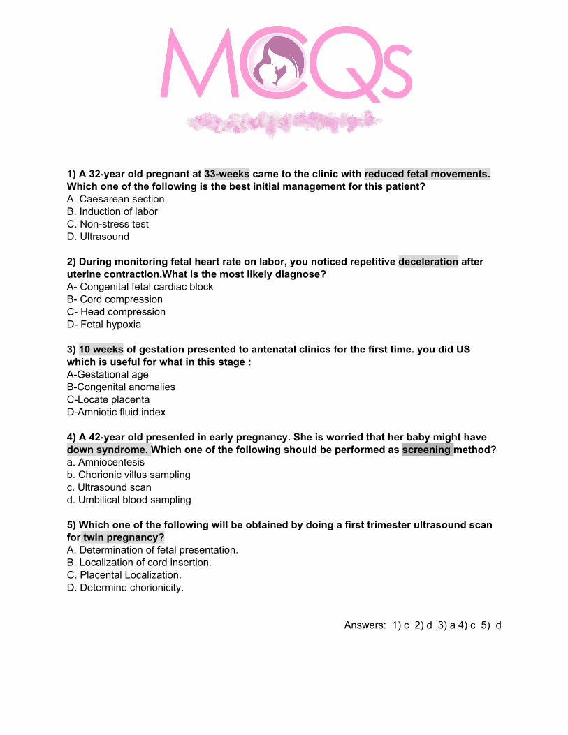
1) A 32-year old pregnant at 33-weeks came to the clinic with reduced fetal movements. Which one of the following is the best initial management for this patient? A. Caesarean section B. Induction of labor C. Non-stress test D. Ultrasound 2) During monitoring fetal heart rate on labor, you noticed repetitive deceleration after uterine contraction.What is the most likely diagnose? A- Congenital fetal cardiac block B- Cord compression C- Head compression D- Fetal hypoxia 3) 10 weeks of gestation presented to antenatal clinics for the first time. you did US which is useful for what in this stage : A-Gestational age B-Congenital anomalies C-Locate placenta D-Amniotic fluid index 4) A 42-year old presented in early pregnancy. She is worried that her baby might have down syndrome. Which one of the following should be performed as screening method? a. Amniocentesis b. Chorionic villus sampling c. Ultrasound scan d. Umbilical blood sampling 5) Which one of the following will be obtained by doing a first trimester ultrasound scan for twin pregnancy? A. Determination of fetal presentation. B. Localization of cord insertion. C. Placental Localization. D. Determine chorionicity.
Answers: 1) c 2) d 3) a 4) c 5) d


