Embed Size (px)
Citation preview
VOL. 25, NO. 3 2013
ORTHOPAEDICTHE MAGAZINE OF THE
ORTHOPAEDIC SECTION, APTA
�h��ica� Thera�� �ractic�
In this issue145 Guest Editorial Christopher R. Carcia
146 Shoulder Injuries in Swimmers: Causes, Evaluation, and Treatment Anthony Herzog, RobRoy Martin, Jason S. Scibek
155 Diathermy: A Literature Review of Current Research and Practices Paul A. Cacolice, Jason S. Scibek, RobRoy Martin
162 Outcome Instruments for the Hip: A Guide to Implementation Benjamin R. Kivlan, RobRoy L. Martin
170 A Critical Review of Performance Tests for Hip-related Dysfunction Benjamin R. Kivlan, Robert L. Martin
Regular features142 President’s Corner
178 Occupational Health SIG Newsletter
184 Foot and Ankle SIG Newsletter
186 Performing Arts SIG Newsletter
188 Pain SIG Newsletter
190 Imaging SIG Newsletter
193 Animal Rehabilitation SIG Newsletter
196 Index to Advertisers
Orthopaedic Physical Therapy Practice (ISSN 1532-0871) is the official magazine of the Orthopaedic Section, APTA, Inc. Copyright 2013 by the Or tho paedic Sec tion, APTA. Non mem ber sub scrip tions are avail able for $50 per year (4 is sues). Opin ions ex pressed by the au thors are their own and do not nec es sar i ly re flect the views of the Or tho paedic Sec tion. The Editor re serves the right to edit manu scripts as nec es sary for pub li ca tion. All re quests for change of ad dress should be di rect ed to the Orthopaedic Section office in La Crosse.
All advertisements that ap pear in or ac com pa ny Or tho paedic Physical Therapy Prac tice are ac cept ed on the ba sis of conformation to ethical physical therapy stan dards, but acceptance does not imply endorsement by the Or tho paedic Section.
Orthopaedic Physical Therapy Practice is indexed by Cu mu la tive Index to Nursing & Allied Health Literature (CINAHL).
Publication Title: Orthopaedic Physical Therapy Practice Statement of Frequency: Quarterly; January, April, July, and OctoberAuthorized Organization’s Name and Address: Orthopaedic Section, APTA, Inc., 2920 East Avenue South, Suite 200, La Crosse, WI 54601-7202
OPTP Mission
To serve as an advocate and resource for the practice of Orthopaedic Physical Therapy by fostering quality patient/client care and promoting professional growth.
Publication StaffManaging Editor & Advertising
Sharon L. KlinskiOrthopaedic Section, APTA2920 East Ave So, Suite 200La Crosse, Wisconsin 54601800-444-3982 x 2020608-788-3965 FAXEmail: [email protected]
EditorChristopher Hughes, PT, PhD, OCS
Advisory CouncilLisa Bedenbaugh, PT, CCRPGerard Brennan, PT, PhDClarke Brown, PT, DPT, OCS, ATCJoseph Donnelly, PT, DHS, OCSJohn Garzione, PT, DPT Duane "Scott" Davis, PT, MS, EdD, OCSIrene Pettet, PT, MPA, OCSJulie O’Connell, PT, DPT, ATCDoug White, DPT, OCS, RMSKMichael Wooden, PT, MS, OCS
VOL. 25, NO. 3 2013
ORTHOPAEDIC�h��ica� Thera�� �ractic�
139Orthopaedic Practice Vol. 25;3:13

President:Stephen McDavitt, PT, DPT, MS, FAAOMPT
Saco Bay Physical Therapy55 Spring St Unit B
Scarborough, ME 04074-8926207-396-5165
[email protected]: 2013-2016
Vice Pres i dent:Gerard Brennan, PT, PhD
Intermountain Healthcare5848 South 300 EastMurray, UT 84107
[email protected]: 2011-2014
Treasurer:Steven R. Clark, PT, MHS, OCS
23878 Scenic View DriveAdel, IA 50003-8509
(515) 440-3439(515) 440-3832 (Fax)[email protected]
Term: 2008-2015
Director 1:Thomas G. McPoil, Jr, PT, PhD, FAPTA
6228 Secrest LaneArvada, CO 80403
(303) 964-5137 (Phone)[email protected]
Term: 2012-2015
Director 2:Pamela A. Duffy, PT, PhD, OCS, CPC, RP
28135 J AvenueAdel, IA 50003-4506
Term: 2013-2016
(608) 788-3982 or (800) 444-3982
Terri DeFlorian, Executive Directorx2040 ......................................... [email protected]
Tara Fredrickson, Executive Associatex2030 ................................................ [email protected]
Sharon Klinski, Managing Editor J/Nx2020 ............................................ [email protected]
Kathy Olson, Managing Editor ISCx2130 ........................................... [email protected]
Carol Denison, ISC Processor/Receptionistx2150 [email protected]
ORTHOPAEDIC SPE CIAL TY COUNCILChair:
Tracy Brudvig, PT, DPT, PhD, OCS22 Duane Street
Quincy, MA 02169(617) 724-4844 (Phone)[email protected]
Term: 2010-2013
Members: Marie Johanson, Daniel Poulsen, Stephanie Jones
PRACTICEChair:
Joseph Donnelly, PT, DHS, OCS3001 Mercer University Dr
Duvall Bldg 165Atlanta, GA 30341
(678) 547-6220 (Phone)(678) 547-6384 (Fax)
Vice Chair:Kathy Cieslek, PT, PhD, OCS, FAAOMPT
Members: Derek Clewley, David Morrisette, Tim Richardson, Jason Tonley, Mary Fran Delaune, Michael Connors
FINANCEChair:
Steven R. Clark, PT, MHS, OCS (See Treasurer)
Members: Jason Tonley, Kimberly Wellborn,Jennifer Gamboa
AWARDSChair:
Gerard Brennan, PT, PhD(See Vice President)
Members: Jennifer Gamboa, Corey Snyder, Jacquelyn Ruen, Karen Kilman
JOSPTEd i tor-in-Chief:
Guy Simoneau, PT, PhD, ATCMarquette University
P.O. Box 1881Milwaukee, WI 53201-1881
(414) 288-3380 (Office)(414) 288-5987 (Fax)
Executive Director/Publisher: Edith Holmes
NOMINATIONSChair:
Bill Egan, PT, DPT, OCS813 Princeton Ave
Haddonfield, NJ 08033(215) 707-7658 (Phone)
Members: Cathy Arnot, RobRoy Martin
SPECIAL INTEREST GROUPS
OCCUPATIONAL HEALTH SIGIrene Pettet, PT, MPA, OCS–President
FOOT AND ANKLE SIGClarke Brown, PT, DPT, OCS, ATC–President
PERFORMING ARTS SIGJulie O’Connell, PT–President
PAIN MAN AGE MENT SIGJohn Garzione, PT, DPT–President
IMAGING SIGDoug White, DPT, OCS, RMSK–Pres i dent
ANIMAL REHABILITATION SIGKirk Peck, PT, PhD, CSCS, CCRT–Pres i dent
EDUCATION INTEREST GROUPSKnee – Open
Manual Therapy – Kathleen Geist, PT, DPT, OCS, COMTPTA – Open
Primary Care – Michael Johnson, PT, PhD, OCS
Officers Chairs
Office Personnel
OR
TH
OPA
ED
IC S
EC
TIO
N D
IRE
CT
OR
Y
Orthopaedic Section:www.orthopt.org
Bulletin Board featurealso included.
MEMBERSHIPChair:
Renata Salvatori, PT, DPT, OCS, FAAOMPT889 1 Belle Rive Blvd
Jacksonville, FL 32256-1628904-854-2090
[email protected]: 2013-2016
Members: Derek Charles, Maureen Watkins, Matthew Lee, Michelle Strauss, Megan Poll, Cuong Pho, John Heick,
Thomas Fliss, Scot Morrison
EDUCATION PRO GRAMChair:
Teresa Vaughn, PT, DPT, OCS, COMT395 Morton Farm Lane
Athens, GA 30605-5074706-742-0082
[email protected]: 2013-2016
Members: Kevin Lawrence, Neena Sharma, Jacob Thorpe,Nancy Bloom, Emmanuel “Manny” Yung, Cuong Pho, John Heick
INDEPENDENT STUDY COURSEEditor:
Christopher Hughes, PT, PhD, OCSSchool of Physical TherapySlippery Rock UniversitySlippery Rock, PA 16057
(724) [email protected]
Managing Editor: Kathy Olson
(800) 444-3982, [email protected]
ORTHOPAEDIC PRACTICEEditor:
Christopher Hughes, PT, PhD, OCSSchool of Physical TherapySlippery Rock UniversitySlippery Rock, PA 16057
(724) [email protected]
Managing Editor: Sharon Klinski
(800) 444-3982, [email protected]
PUBLIC RELATIONS/MARKETINGChair:
Eric Robertson, PT, DPT, OCS5014 Field Crest Dr
North Augusta, SC 29841 (803) 257-0070
Vice Chair:Chad Garvey, PT, DPT, OCS, FAAOMPT
Members: Tyler Schultz (student), Mark Shepherd,Kimberly Varnado
RESEARCHChair:
Duane “Scott” Davis, PT, MS, EdD, OCS412 Blackberry Ridge ,Drive
Morgantown, WV 26508-4869304-293-0264
[email protected]: 2013-2016
Members: Kristin Archer, Paul Mintken, Murry Maitland, Dan White, George Beneck, Ellen Shanley, Dan Rendeiro, Amee Seitz
APTA BOARD LIAISON –Nicole Stout, PT, MPT, CLT-LANA
2013 House of Delegates Representative –Joe Donnelly, PT, DHS, OCS
ICF-based Guidelines Coordinator – Joe Godges, PT, DPT, MA, OCS
ICF-based Guidelines Revision Coordinator – Christine McDonough, PT, PhD
140 Orthopaedic Practice Vol. 25;3:13
Physical therapist practice evolved from a supportive service to a distinguishable pro-fession. For decades we have worked hard to be appreciated as a value to society and rec-ognized as an independent practitioner and profession. Through Vision 2020, we have pursued autonomous practice.
Health care reform prescribes value for the patient and payor within interdependent-multidisciplinary practice models. In physi-cal therapist practice, value is derived from cost and outcomes. Value is inversely propor-tional to unwarranted variation in health care services. In the future of health care models and policies, we as Orthopaedic Physical Therapists will be in the best position to be recognized as a value added discipline by cre-ating paradigms from organized guidelines that identify best practice, facilitate adhering to best practice, and provide a measure of provider performance that will demonstrate value. This should in turn deliver matching the right patient to the right providers and providing interventions at the right time. I believe the Orthopaedic Section’s current strategies and initiatives in developing clini-cal practice guidelines and outcomes data are well underway in addressing this challenge.
The current evolving framework of health care reform advocating interdependent-mul-tidisciplinary practice models is a correspond-ing challenge with value for our recognized identity. Interdependent-multidisciplinary practice models are now creating a paradox for our advocacy initiatives framing auton-omy. Presently we are confronted by deter-mining how we will be individually identified as a value added profession while collaborat-ing, integrating, and improving access to care without becoming isolated by our determina-tion for autonomy. Addressing our identity, autonomy, interdependence, collaboration, and access under this condition are formi-dable tasks. What is our future? What do we do? In addressing these challenges, the Orthopaedic Section appreciates the need for advancing health services research. However, what do we need to understand to develop and deploy other strategies to meet future encounters from health care reform that demands accountability, standardization, and differentiation?
At our recent First Annual Orthopae-dic Section Meeting in Orlando, Florida, Dr. Justin Moore, APTA Vice President of Public Policy, Practice, and Professional Affairs provided us with a rich presentation
on the “The Paradox of Autonomy: Demon-strating Value in a Post Health Care Reform World.” Dr. Moore’s presentation supported how “interdependent practice based on data and evidence will enable autonomous physi-cal therapists to maximize their role and responsibility to the health care system in a post health care reform world.” We felt his presentation was extremely valuable to our membership and should be shared with those who were unable to make the conference. We thank Dr. Moore for reframing his pre-sentation for this publication so we can pro-vide distribution to all of our Orthopaedic Section members. From this printing of his presentation, we hope members will become more informed of our current and future practice and advocacy concerns and further, work toward strategies to meet the related future challenges facing Orthopaedic Physi-cal Therapist Practice.
THE PARADOX OF AUTONOMYJustin Moore, PT, DPT
WHAT’S IN A WORD?When I was beginning my career as a
physical therapist, I would look forward each month to receiving the shrink wrapped pack-age from APTA that included Physical Ther-apy, the Association’s journal. I would like to tell you I was an avid seeker of the latest research, but I was really only interested in the latest commentary from the Editor at that time, Dr. Jules Rothstein. Jules would com-municate through words that would make me stop and think. These ‘notes’ would chal-lenge me to understand more about the phys-ical therapy profession, a profession that has an interdependent relationship with patients when they are most in need. Dr. Rothstein would write close to 200 ‘notes’ but two stood out. These were about autonomy and specifi-cally Jules’ perspective for this word as part of APTA’s Vision 2020. Jules was not satisfied when we attempted to define autonomy in a more inclusive, collaborative fashion or as a descriptor to practice in the pillar of our Vision statement of ‘autonomous’ practice. He felt we had already isolated ourselves with our rhetoric and at best we were creating a paradox of autonomy like jumbo shrimp or healthy air travel.
To capture his perspective on the misfit of autonomy in our professional lexicon, Dr. Rothstein described the connection between organ donors and the individuals that ben-
President’s Corner
efitted from these selfless acts after attend-ing the World Transplant Games. This essay entitled, Autonomy and Dependence was writ-ten more than a decade ago and is as con-temporary today as it was back in 2003. The essay concludes with the passage ‘the word “autonomy” has an attraction, but that attrac-tion can be like a siren’s call, bringing us into peril...Interdependence is not a sign of weakness. Interdependence is a badge that civilized people wear to reaffirm their humanity, the capacity of kindness–and their competence.
Dr. Rothstein felt our quest for inde-pendence was in vain if not for the greater purpose of making us interdependent health professionals. He felt that we truly are the sum of those we serve. I couldn’t agree with him more and that interdependence in health care is essential as we move into an era of transition, an era of transformation. It is the badge we must proudly wear in a post health care reform world.
PAST PERFORMANCE, AN INDICATOR OF FUTURE SUCCESS?
To understand how we arrived at this era of transformation in health care, we must also look back at the path of progress in physical therapy, the state of health care, and the land-mark legislation to kick off this transforma-tion, the Affordable Care Act.
Physical therapy is approaching its 100th birthday in the United States in the next few years. Our history over these 100 years in public policy was aimed at achieving inde-pendence by gaining licensure in all 50 states and achieving varied forms of direct access. This path towards independence prepared us not only to arrive at this era ready for this change but also with the confidence, and potentially the creditability, to adapt and meet the requirements of this era--a require-ment to build partnership, a requirement to be accountable, and a requirement to dem-onstrate value.
Physical therapy is well positioned for this era of health care transformation. Our history shows transformation and respond-
Stephen McDavitt, PT, DPT, MS, FAAOMPT
142 Orthopaedic Practice Vol. 25;3:13
ing to national issues as an integral part of our DNA. We have a remarkable history that guides our future. Dr. Alan Jette describes this remarkable history and its relevance to today in his 43rd McMillan address. Dr. Jette encouraged the profession to look beyond its inward facing vision to an outward facing one as we ‘face into the storm’ of the dual challenges of health care reform and a chang-ing population with changing health care needs (and demands).
Dr. Jette quoted David Brooks’, the New York Times columnist, description of the importance of understanding who we are. Mr. Brooks states, “most people don’t form a self and then lead a life. (Most)…are called by a problem and the self is gradually constructed by their calling.” Dr. Jette proposed this is the same for our profession and that our found-ers foreshadowed our preparation for health care transformation in the early days of our profession. Dr. Jette proposed in his McMil-lian address that, “our foremothers (who) looked outside themselves and focused on a problem that summoned their professional lives, be it the reconstruction aides during WWI…or the physical therapists who responded to the polio epidemic.” The challenges are new, but the ability to look outside ourselves and focus on a problem is still the calling for our profession.
This profession has further been con-structed over the past 100 years by the embracing of evidence and the growth of our practices and professionalism. The Orthopae-dic Section of APTA has been a major force in this construction—a force that has achieved independence from the American Medical Association’s accreditation of our education programs, rejection of the term ‘allied health’ as a classification, obtaining independent bill-ing status under Medicare, and establishing clinical specialization. These achievements also fostered a period of growth over the last decade like no other time in our history. One measure of this growth saw Medicare therapy expenditures, in which physical therapy is 75% of these expenditures, grow from $1.9 billion to $5 billion. Another measure we have seen is physical therapist education pro-grams increase the number of graduates from 4,000 annually to over 7,000. We now stand at almost 200,000 practitioners that see a growing percentage of patients that interact with the health care delivery system each and every year. For a profession that was claiming its death blow in the enactment of the Bal-anced Budget Act of 1997, our demise was greatly exaggerated.
Physical therapy also marked the last decade by our first public vision statement—APTA’s Vision 2020. This became a point of pride and a point of attention in the profes-
sion and outside the profession. If the axiom is true that there is no such thing as nega-tive PR, Vision 2020 was our most success-ful public relations initiative to date. Vision 2020 was cited by our colleagues and part-ners across health care as everything from arrogant to ambitious. This vision drew attention to the profession and unfortunately mostly to the word autonomy. This attention also caused us to revisit who we are and what we do. Our calling potentially became more clear as we had to better understand what we meant when we used the word autonomy and that it was not the dictionary definition of “self-contained, existing independent, and without outside control.”
Vision 2020 served its purpose. It first helped describe physical therapy to physical therapists and also defined us for a new future in health care. Vision 2020 also focused us and moved us from a dependent posture to an independent one and now positions us to re-define ourselves in an interdependence fashion. We are 7 years from Vision 2020’s original target and we can say with confi-dence that we are close to a sense of accom-plishment of several of 6 pillars. These pillars include Indiana becoming the 50th state to have some form of direct access with the sign-ing of a bill by Governor Pence in April 2013, and in 2015 the milestone that all accredited physical therapist education programs will offer the doctor of physical therapy (DPT) degree. The other pillars are still works in progress, but progress has been seen and should be expected to move forward.
This summer APTA will consider a new Vision for the future. The over-riding goal of the vision is to be more outward facing and change our description of ourselves. Instead of defining the profession for itself, the new vision is designed to describe the profession to the public, patients, and other providers of health care. It took a path to independence to be able to position the profession in this new era of health care and a vision of inter-dependence. This recalibration is essential as in 8 months we begin the key implementa-tion date of health care reform of January 1, 2014. Ready or not, the system begins to change and change will be required by most all that interact within our health care deliv-ery system.
AFFORDABLE CARE ACT DRIVING TRANSFORMATION
The changes ahead as part of the Afford-able Care Act set the stage for transformation, but they are the exclusive driver of reforming the system to be more responsive to health and economic measures of success. The driver of the specific legislation that has become the focal point of health care reform but also the
era of transformation that this legislation kicked off is best represented by a concept called the ‘triple aim.’ The triple aim was coined in 2008 by Dr. Donald Berwick. Dr. Berwick was the President of the Institute of Healthcare Improvement and after the elec-tion of President Obama went on to be the Administrator of the Centers for Medicare and Medicaid for two years. Prior to the triple aim goal, the conventional wisdom was that health policy was plagued by the iron triangle of cost, quality, and access. The iron triangle was based on the concept that to achieve the objective of two, came at the detriment of the third. For example, policies that improve quality and access would also increase cost. Dr. Berwick challenged the iron triangle con-cept with the premise you could achieve all three through health care reform–the triple aim. You could improve care for individuals, advance the health of the population, and do so while lowering overall costs in the system. It remains to be seen if this can be done, but it is the rally cry, the bumper sticker, and the buzz word of the new era of health care.
The triple aim defines health care reform’s goals. The Affordable Care Act of 2010 (PL 111-148 and 111-152) defines the policies that will be aimed to change how and by who health care will be transformed. The 2400 pages of legislation were organized into the 3 major areas of coverage and insur-ance reforms, delivery and payment reforms, and financing strategies. The profession is impacted mostly by reforms to coverage, the insurance marketplace, payment, and deliv-ery systems; and you as an individual, will have varied opinions about its impact on you personally.
If successful in its implementation, the Affordable Care Act will increase coverage for 33 million more Americans resulting in 95% coverage. These 33 million would gain cov-erage primarily through Medicaid expansion and the establishment of health care insur-ance exchanges. Each state is determining if they will develop a state-based exchange, partner with other states and/or the federal government, or default to the federal option. Regardless of the option chosen, the big victory for physical therapy in health care reform was that insurance offered under these exchanges must provide 10 essential health benefits, including rehabilitation and assistive devices. We are recognized as essential, now we must move from solely being recognized to being used in our best role–a role that might be a first point of contact for musculo-skeletal care or a role that might be as a criti-cal member of a team to treat an individual with complex medical conditions.
As the Affordable Care Act increases cov-erage, it simultaneously ramps down health
143Orthopaedic Practice Vol. 25;3:13
care spending, including Medicare and Med-icaid spending. The increase of individu-als served and a decrease on expenditures is designed as a pressure point to force effi-ciency by driving care to the right provider for the right patient at the right time. This is a mantra that orthopaedic physical thera-pists are well suited to realize as the cost-effective option to manage musculoskeletal conditions.
In this era of health care transformation, physical therapy must realize and embrace the 5 concepts and the policies that are designed to implement these concepts. First, incentives will be provided to achieve more integrated models of delivery from account-able care organizations to patient centered medical homes. Second, the health care con-tinuum will be realigned to focus on health management over the lifespan not separate episodes of sickness. Third, payment will change and provide incentives and penalties to health care professionals that measure and achieve quality, reduce unnecessary or waste-ful care, and achieve the best return on dollars invested. Fourth, standardization of health care processes, information, and practice will be developed and deployed to provide a more transparent system and empower consumers to make decisions based on provider perfor-mance. And fifth, the government will be aggressively implementing programs to seek out fraud, abuse, and waste at the individual and system level. These 5 concepts are here to stay and for the physical therapist true to the profession’s calling, these are not only consis-tent to our values, but should facilitate even greater recognition of who we are and what we do.
So, health care transformation is here. Physical therapy’s advocacy message during the legislative battles was that physical ther-apy is a solution to a health care system that is too costly, inconsistent in its access, and marginal at best in its quality. To realize that statement, physical therapy must develop its value proposition and then demonstrate it, document it, and deliver it. This value prop-osition goes beyond the definition of value as quality over cost. This definition is only its starting point as quality of life, patient enhancement, and reduction of other ser-vices, including those within the profession, will also be part of our value. The value prop-osition is not new to the profession, but it could require re-calibration—a re-calibration on recognition, role, and results. Recognizing the physical therapist in the right role, dem-onstrating their results for patients and the health care system, and demonstrating results to what we do. Again, as Dr. Jette articulated, “it is now time to stop trying to prove PT works, but to prove what in PT works.”
THE VALUE PROPOSITION IN PHYSICAL THERAPY
To advance a value proposition for the profession, it quickly became apparent that APTA had to move from a command control type approach to an empowerment model. To look at that changing paradigm, one only needs to look at the recent innovation summit where command and control was replaced with a call to action. This approach is also articulated in a recent policy perspec-tive in Physical Therapy entitled, “Deliver-ing the Physical Therapy Value Proposition: A Call to Action.” This article challenged the current approach “what is APTA doing for me” to “how are we working together to advance the profession.” Furthermore, the article articulated that the principle contri-bution of APTA is to provide resources and tools, and connect experts. What APTA cannot do is deliver physical therapy, but you can. The obligation, the accountability, and the responsibility to deliver value are yours. The APTA’s role is to empower and support you in this endeavor.
The value proposition is based on the 5 interrelated components of identification of best practice, provider adherence, measure-ment of performance, policy development and implementation, and research to study its benefit. This is a dynamic framework in which each element feeds off the other and creates a natural cycle of improvement. Once you determine cost-effectiveness, in all like-lihood new knowledge has emerged and the cycle begins again. The components also must be integrated into how we approach patient management. For the value proposition, we embraced the art of caring and science of practice. This statement provides clarity to the unique therapeutic relationship we have with patients and the scientific foundations we strive to advance.
LEADING THE WAY: THE ORTHOPAEDIC SECTION OF APTA
So, what does this all mean to you as lead-ers in the profession, not just this Section? You have already begun this journey to the new era in health care. You leaped into the transition and began transforming. We owe you much gratitude as you didn’t just begin to transform orthopaedic physical therapy, you began to set an example and develop a recipe for others to follow. You have led the way with clinical practice guidelines, the clinical research network (OPT-IN), and your journal. You have led the way with the National Orthopaedic Physical Therapy Outcomes Database, continuing education, and fellowships. Your leadership is the recipe for success for others in physical therapy to
follow. It is a recipe to emerge from transi-tion in a new place, a better place for physical therapy and a better place for our patients. If health care reform was about better health for populations, better care for individuals, and at a lower cost (both financial and personal), then health care reform was aligned with who we say we are. It is now time to back up our works with deeds and deliver the value prop-osition in physical therapy.
CONCLUSIONIn his book, A Checklist Manafesto, Dr.
Atul Guwande described the problem with autonomy as, “In Medicine, we hold up autonomy as a professional lodestar, a principle that stands in direct opposition to discipline. But in a world in which success now requires large enterprises, teams of clinicians, high risk technologies, and knowledge that outstrips one person’s abilities, individual autonomy hardly seems the ideals we should aim for. It has the ring more of protectionism than of excellence.” Our striving to be self-directed, control our clinical decision making, and exercise inde-pendence only brings us to a place where we are more accountable to others and to our-selves. This ‘place’ is where we have to exercise stronger discipline, raise the standard of care, and develop excellence not through holding on to what we use to have (regardless of if we earned it or not), but earning it through our service to our patients and our value to payers and the public.
This is the paradox of autonomy. Your autonomy only empowers you to be account-able–not to act without external control or without the joint partnership of other auton-omous parties. Your autonomy as a health care professional only enables you to be more accountable to the individuals that need your expertise and care. The duality of your autonomy and accountability will allow you to innovate, integrate, and enjoy the interde-pendence of patient care, research, and edu-cation in ways that we do not yet know.
Physical therapy has many challenges in the next couple months, most notably in payment cuts and new requirements, ie, functional limits reporting. These are not insignificant but underscore that health care is transforming. We have the opportunity to deliver a value proposition in health care. That opportunity is here today for the physi-cal therapists that embrace health care trans-formation. Physical therapy will be part of the solution of a reformed health care system, but it will be a different physical therapy than we see today and it will be one that embraces the value proposition.
144 Orthopaedic Practice Vol. 25;3:13
It is truly an honor to write the pref-ace for the Duquesne University edition of OPTP. The edition features a collection of articles that were written by students in either our Doctor of Physical Therapy (DPT) or PhD program in Rehabilitation Science. While both of these programs are relatively small, both have achieved sub-stantial successes. Our DPT program has a 98.5% first time board (NPTE) pass rate since graduating our first DPT class in 2006. Likewise, our PhD program in Reha-bilitation Science, which is largely a joint venture between the Departments of Physi-cal Therapy and Athletic Training, has expe-rienced considerable achievements on the scholarship front. Highlighting this, one of our current doctoral students, Ben Kivlan, along with his mentor RobRoy Martin, PT, PhD, have published an impressive body of work related to non-arthritic hip pain. An example is a recent series of papers that reflect a progression from defining a clinical problem ("hip instability"), to investigating previously unrecognized risk factors ("infe-rior acetabular insufficiency"), and finally modeling the function of the ligamentum teres as stabilizing structure for the hip. This body of work is a testament not only to the dedication and passion of these authors but also reflects positively on our PhD program in Rehabilitation Science. Collectively, the accomplishments of both our programs provide evidence that meaningful and important contributions can and in fact are produced by programs that are smaller in stature.
We advocate incorporating professional students into the research process as oppor-tunities become available and circumstances permit. The collaborative model between student and faculty affords students the chance to become involved in the research and writing process as well as providing the reader with an insightful piece of scientific literature. Depending on the specifics, proj-ects may be either student or faculty driven. If student driven, it has been our experience that despite having access to sophisticated instrumentation in our motion analysis laboratory as well as the practical expertise to utilize, these projects are often better served by minimizing the use of technical measures. We believe this approach increases the likelihood the student driven project will
be completed successfully. Enhancing early success increases the probability students will desire to continue to engage in research as they move forward as students and ulti-mately professionals. In the case of faculty driven projects, our professional students have participated in varying capacities rang-ing from recruiting participants, gaining informed consent, collecting and analyzing data using sophisticated instrumentation as well as contributing to the writing process. We have found, whether projects are student or faculty driven, what is crucial to success is student interest and time for faculty to over-see and guide the process.
There are additional benefits to engaging students in the research process. Students directly apply material from the classroom thereby increasing their comprehension of the material. This elevated level of com-prehension should enhance board (licens-ing exam) scores. Elevated board scores are beneficial for the student as well as con-tributing positively to program outcomes. Perhaps most importantly, content gleaned from participation in or as a product directly from the research itself has the potential to favorably influence patient care. Finally, the end product in the form of a presentation or publication is of value to the student, con-tributing faculty member, and the program they represent.
Specific to this issue, each paper was facilitated by the guidance of RobRoy Martin, PT, PhD, one of our orthopaedic physical therapy faculty. The first article discusses the mechanics, pathomechanics, common injuries, physical examination, and rehabilitation recommendations unique to the swimming athlete. It is worth noting the first author, Tony Herzog, was a com-petitive Division I swimmer while enrolled in our 3+3 DPT program at Duquesne. In addition to the evidence on which this paper is based, it offers a unique glimpse from Tony’s first-hand experience as a competi-tive swimmer. The second article reviews the most recent evidence related to the use of diathermy in a clinical environment. This therapeutic modality has experienced a bit of resurgence over the last few years making this review not only informative but timely. The article’s authorship is also an example of what can be accomplished when the educa-tional model reflects an inter-professional
Guest Editorial Christopher R. Carcia, PT, PhD, SCS, OCS
effort. While inter-professional education has gained traction over recent years, it is a model we have been successfully using within our health sciences school for decades. The final two articles that were first authored by Ben Kivlan delve into content related to the hip which no doubt is an emerging area in orthopaedics and sports medicine.
We would like to thank Orthopaedic Physical Therapy Practice, Dr. Hughes, and his staff for their assistance as well as the opportunity to present some of the work produced from our joint student-faculty projects. We hope you enjoy each of these papers.
Christopher R. Carcia, PT, PhD, SCS, OCSChairperson & Associate Professor,
Department of Physical TherapyProgram Director, PhD Program in
Rehabilitation ScienceRangos School of Health Sciences
Duquesne UniversityPittsburgh, PA
145Orthopaedic Practice Vol. 25;3:13
ABSTRACTCompetitive swimmers commonly
develop shoulder impairments of body structure and function that cause limitations in activity and participation. The required range of motion, strength, and endurance, combined with the repetitive stresses that swimming places on the shoulder may put an individual at risk for injury. The purpose of this paper is to discuss the potential causes of injury, review important considerations for evaluation, and identify specific treat-ment techniques for those with swimming related shoulder injuries. The causes of these injuries are likely to be multifactorial and relate to instability, range of motion imbal-ance, and/or training error. These potential factors need to be considered in the exami-nation process in order to develop the most effective intervention and treatment plan.
Key Words: overhead athlete, mechanics, shoulder impingement
INTRODUCTIONCompetitive swimming requires an indi-
vidual to generate forceful propulsion while attempting to minimize their resistance through the water. This requires a balance of shoulder flexibility, strength, and endur-ance, coupled with proper technique and training. “Swimmer’s shoulder” is a term used to describe the collection of symptoms that occur from the stresses that swimming places on the shoulder complex.1,2 These symptoms can originate from pathology of the rotator cuff, long head of the biceps, glenoid labrum, joint capsule, and/or acro-mioclavicular joint. Multiple structures are often involved simultaneously, which can make diagnosis and treatment of this common problem difficult. The purpose of this paper is to discuss the potential causes of injury, review important considerations for evaluation, and identify specific treatment
techniques for those with swimming related shoulder injuries.
EPIDEMIOLOGYShoulder pathology is a common prob-
lem in swimmers. Shoulder pain was noted in 40% to 80% of those who regularly swim.3 Although swimmer’s shoulder is often compared to the pathology observed in other overhead sports, swimming does not include the high force associated with a rapid deceleration phase seen in other over-head sports, such as throwing.4 However, swimming does involve far more overhead repetitions than other overhead sports. A typical practice may include swimming 10,000 meters per day. If 10 stroke cycles or shoulder revolutions are performed in a 25 meter lap, a swimmer would make approximately 4,000 shoulder revolutions during a practice. Therefore, although the force placed on the shoulder for any swim-ming stroke may be low, the collective force and workload is great throughout an entire practice.
In addition to the high number of over-head repetitions, swimming also requires positions that may put the shoulder at risk for injury. All of the 4 strokes in competitive swimming—butterfly, backstroke, breast-stroke, and freestyle—require excessive internal rotation with elevation to initiate a forceful pull as the hand enters the water. Freestyle is the most frequently performed stroke during practice, regardless of stroke specialty, and can be used for up to 80% of a typical practice.1 During a freestyle stroke an impingement position of internal rotation and elevation was found to occur approxi-mately 25% of the stroke time.5 Because of these mechanics and time spent performing the freestyle stroke, it is the most common stroke studied and discussed in regards to shoulder pathology.
BIOMECHANICSThe freestyle stroke is divided into two
major phases called the pull-through and recovery and 7 subphases (hand entry, catch, mid pull-through, late pull-through, elbow lift, mid-recovery, and late recovery).5,6 The pull-through phase is the time during which the arm is in the water generating a propul-sive force. The recovery phase is the time the arm is out of the water repositioning for another pull-through. The 7 subphases of the freestyle stroke are represented in Fig-ures 1A-G.
Pull-throughThe pull-through phase begins as the
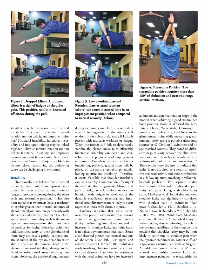
hand first enters the water. During hand entry (Figure 1A), the shoulder is positioned in external rotation and abduction with the ipsilateral side of the body rolling down deeper in the water.6 As the swimmer pro-gresses to the catch (Figure 1B), the shoul-der is brought into internal rotation near full elevation.7 Although this is a position that can contribute to and exacerbate symp-toms associated with shoulder impinge-ment, it is a critical step in the pull-through phase.5,7 The shoulder position in the catch subphase enables the swimmer to have the largest palm and forearm surface area to pull through the water. It is also a common point where stroke mechanics break down secondary to fatigue or pain. A coach often looks for a ‘dropped’ elbow (Figure 2) as a sign of improper mechanics during the catch subphase. A ‘dropped’ elbow occurs when the shoulder is not internally rotated to the extent necessary to maintain the elbow closer to the surface of the water than the hand. This causes the pull to be inefficient and typically happens with pain or fatigue. The mid pull-through subphase (Figure 1C) occurs when the shoulder is in neutral rota-tion and 90° of elevation.6 At this point the body roll into the water is at its maximal
Shoulder Injuries in Swimmers: Causes, Evaluation, and Treatment
Anthony Herzog, DPT1 RobRoy Martin, PhD, PT, CSCS2 Jason S. Scibek, PhD, ATC3
1 Jamie’s Physical Therapy & Sports Medicine, Aliquippa, PA, 2Associate Professor, Department of Physical Therapy; Staff Physical Therapist UPMC Center for Sports Medicine, 3Associate Professor, Department of Athletic Training, John G. Rangos, Sr., School of Health Sciences, Duquesne University, Pittsburgh, PA
146 Orthopaedic Practice Vol. 25;3:13
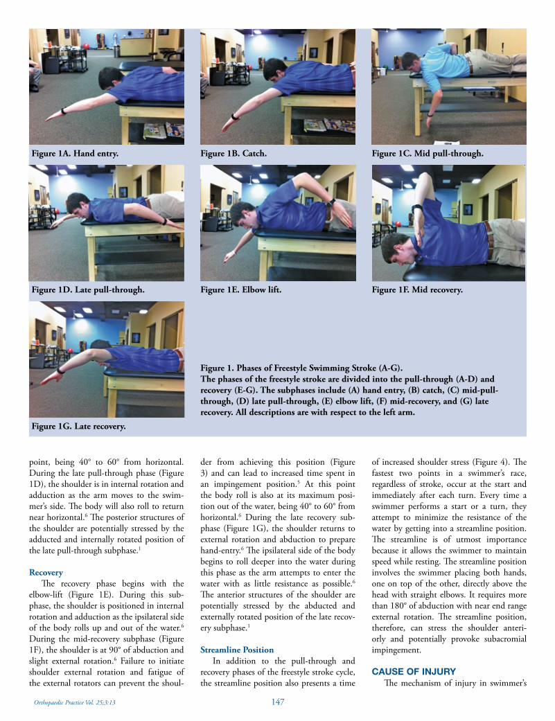
point, being 40° to 60° from horizontal. During the late pull-through phase (Figure 1D), the shoulder is in internal rotation and adduction as the arm moves to the swim-mer’s side. The body will also roll to return near horizontal.6 The posterior structures of the shoulder are potentially stressed by the adducted and internally rotated position of the late pull-through subphase.1
RecoveryThe recovery phase begins with the
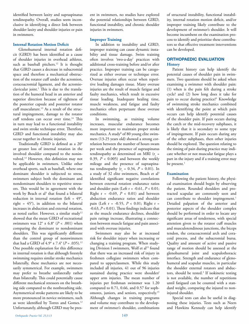
elbow-lift (Figure 1E). During this sub-phase, the shoulder is positioned in internal rotation and adduction as the ipsilateral side of the body rolls up and out of the water.6 During the mid-recovery subphase (Figure 1F), the shoulder is at 90° of abduction and slight external rotation.6 Failure to initiate shoulder external rotation and fatigue of the external rotators can prevent the shoul-
der from achieving this position (Figure 3) and can lead to increased time spent in an impingement position.5 At this point the body roll is also at its maximum posi-tion out of the water, being 40° to 60° from horizontal.6 During the late recovery sub-phase (Figure 1G), the shoulder returns to external rotation and abduction to prepare hand-entry.6 The ipsilateral side of the body begins to roll deeper into the water during this phase as the arm attempts to enter the water with as little resistance as possible.6
The anterior structures of the shoulder are potentially stressed by the abducted and externally rotated position of the late recov-ery subphase.1
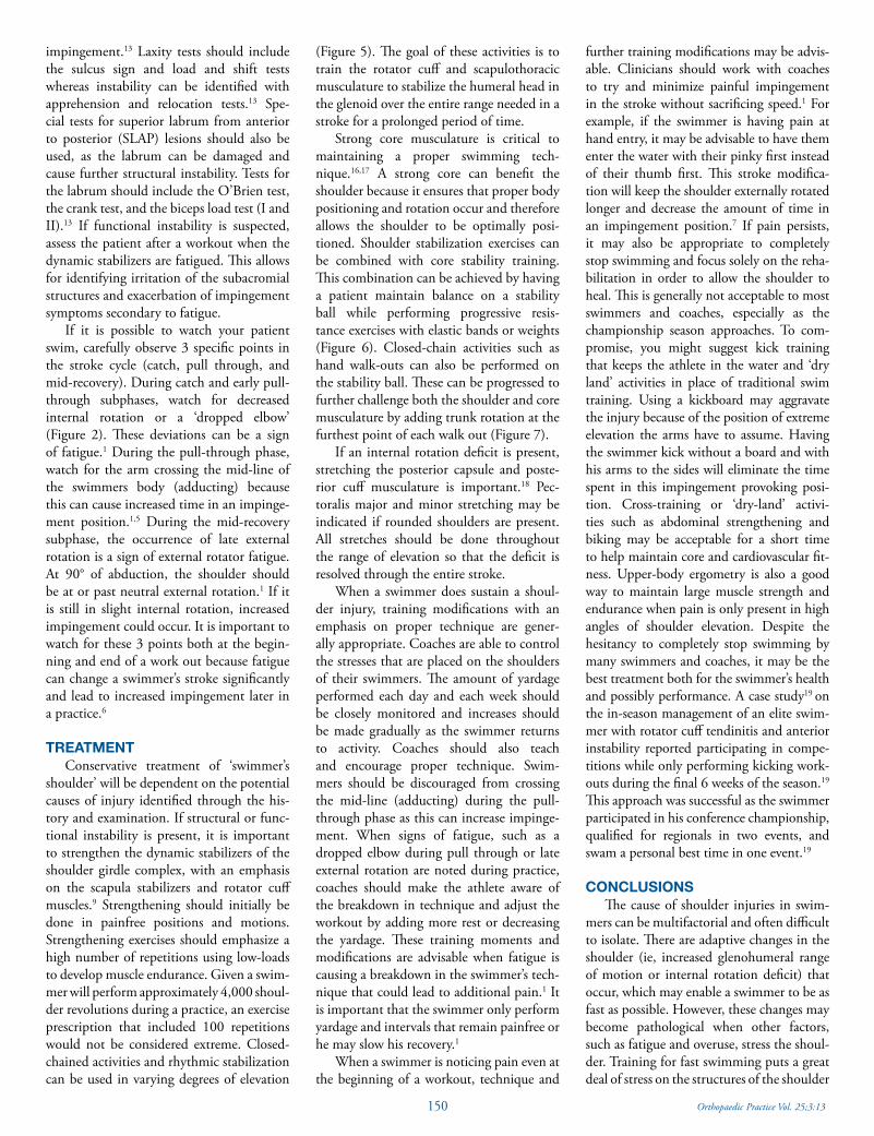
Streamline PositionIn addition to the pull-through and
recovery phases of the freestyle stroke cycle, the streamline position also presents a time
of increased shoulder stress (Figure 4). The fastest two points in a swimmer’s race, regardless of stroke, occur at the start and immediately after each turn. Every time a swimmer performs a start or a turn, they attempt to minimize the resistance of the water by getting into a streamline position. The streamline is of utmost importance because it allows the swimmer to maintain speed while resting. The streamline position involves the swimmer placing both hands, one on top of the other, directly above the head with straight elbows. It requires more than 180° of abduction with near end range external rotation. The streamline position, therefore, can stress the shoulder anteri-orly and potentially provoke subacromial impingement.
CAUSE OF INJURYThe mechanism of injury in swimmer’s
Figure 1. Phases of Freestyle Swimming Stroke (A-G). The phases of the freestyle stroke are divided into the pull-through (A-D) and recovery (E-G). The subphases include (A) hand entry, (B) catch, (C) mid-pull-through, (D) late pull-through, (E) elbow lift, (F) mid-recovery, and (G) late recovery. All descriptions are with respect to the left arm.
Figure 1A. Hand entry. Figure 1B. Catch. Figure 1C. Mid pull-through.
Figure 1D. Late pull-through.
Figure 1G. Late recovery.
Figure 1E. Elbow lift. Figure 1F. Mid recovery.
147Orthopaedic Practice Vol. 25;3:13
shoulder may be categorized as structural instability, functional instability, internal rotation motion deficit, and improper train-ing.8 Structural instability, functional insta-bility, and improper training may be linked together. Likewise internal rotation motion deficit, functional instability, and improper training may also be associated. Since these potential mechanisms of injury are likely to be interrelated, identifying the underlying cause can be challenging in swimmers.
InstabilityTraditionally, it is believed that structural
instability may result from capsular laxity caused by the repetitive, extreme shoulder ranges of motion occurring during the swim cycle and streamline position.1 It has also been noted that swimmers have a tendency to exhibit greater than normal amounts of glenohumeral joint motion, particularly with abduction and external rotation.3 Therefore, special tests for instability, such as the sulcus sign or anterior/posterior shift tests may be positive for laxity. However, swimmers with identified laxity of their glenohumeral joints may not have a painful or symptom-atic shoulder. If the dynamic stabilizers are able to maintain the humeral head in the glenoid (functional stability), damage to the shoulder subacromial structures may not occur. However, the positional requirements
during swimming may lead to a secondary type of impingement of the rotator cuff tendons in the subacromial space if laxity is present with muscular weakness or fatigue. When the rotator cuff fails to dynamically stabilize the glenohumeral joint efficiently, functional instability can occur and con-tribute to the progression of impingement symptoms.9 Also when the rotator cuff is not functioning properly, greater stress will be placed on the passive restraints potentially leading to structural instability.1 Therefore, it seems plausible that shoulder instability can be caused by a combination of laxity of the static stabilizers (ligaments, labrum, and joint capsule), as well as alone or in com-bination with fatigue or weakness of the dynamic stabilizers.1 Structural and func-tional instability may be more likely to occur together in those with chronic injuries.
Evidence indicates that while swim-mers may present with greater than normal amounts of glenohumeral joint motion (laxity), swimming itself does not lead to increases in shoulder laxity and joint laxity is not always synonymous with pain. Beach et al3 observed greater than normal amounts of abduction (196° left, 195° right) and external rotation (100° left, 101° right) in a study involving Division I swimmers. These elevated degrees of motion are consistent with the need swimmers have for increased
abduction and external rotation range in the motion when achieving a good streamlined body position. Borsa et al10 used the Telos system (Telos, Weiterstadt, Germany) to position and deliver a graded force to the glenohumeral joint while measuring gleno-humeral laxity using a portable ultrasound scanner in 42 Division 1 swimmers and 44 age-matched controls. They noted no differ-ence in joint laxity between the elite swim-mers and controls or between subjects with a history of shoulder pain to those without.10
These results were the first to indicate that laxity is not acquired as a result of repeti-tive overhead activity and were corroborated in a follow-up study involving professional baseball pitchers.11 Two separate studies have examined the role of shoulder joint laxity and pain. Using a shoulder laxity score, McMaster et al2 found that increased shoulder laxity was significantly correlated with shoulder pain in swimmers (Pain now: R shoulder = 15, L shoulder = 16, No pain now: R shoulder = 9.8, L shoulder = 10.7; P < 0.05).2 While both McMaster et al2 and Borsa et al10 quantified laxity in relaxed states, thus eliminating the role of the dynamic stabilizers of the shoulder, it is possible that shoulder laxity may be more likely to contribute to shoulder pain when the dynamic stabilizers (ie, rotator cuff and scapular musculature) are weak or fatigued. An additional study by Sein et al8 noted a weak relationship between laxity and impingement pain; yet, no relationship was
Figure 2. Dropped Elbow. A dropped elbow is a sign of fatigue or shoulder pain. This position results in decreased efficiency during the pull.
Figure 3. Late Shoulder External Rotation. Late external rotation (above) can cause increased time in an impingement position when compared to normal recovery (below).
Figure 4. Streamline Position. The streamline position requires more than 180° of abduction and near end range external rotation.
148 Orthopaedic Practice Vol. 25;3:13
identified between laxity and supraspinatus tendinopathy. Overall, studies seem incon-clusive in identifying a direct link between shoulder laxity and shoulder injuries or pain in swimmers.
Internal Rotation Motion DeficitGlenohumeral internal rotation defi-
cit (GIRD) has been identified as a cause of shoulder injuries in overhead athletes, such as baseball pitchers.12 It is thought that GIRD causes a decrease in subacromial space and therefore a mechanical obstruc-tion of the rotator cuff under the acromion, coracoacromial ligament, and the acromio-clavicular joint.1 This is due to the transla-tion of the humeral head in an anterior and superior direction because of tightness of the posterior capsule and posterior rotator cuff musculature.13 As a result of the struc-tural impingement, damage to the rotator cuff tendons can occur over time.14 This in turn may lead to a functional instability and swim stroke technique error. Therefore, GIRD and functional instability may also occur together in chronic injuries.
Traditionally GIRD is defined as a 20° or greater loss of internal rotation in the involved shoulder compared to the nonin-volved.13 However, this definition may not be applicable in swimmers. Unlike other overhead sports, such as baseball, where one dominant shoulder is subjected to stress, swimmers subject both the dominant and nondominant shoulders to repetitive stress-ors. This would be in agreement with the work by Beach et al3 that found a bilateral reduction in internal rotation (left = 49°, right = 45°), in addition to the bilateral increases in abduction and external rotation, as noted earlier. However, a similar study12 showed that the mean GIRD of recreational swimmers was 12° ± 6.8° (P < .001) when comparing the dominant to nondominant shoulders. This was significantly different than the control group of nonswimmers that had a GIRD of 4.9° ± 7.4° (P = .035).12
One possible explanation for this difference in internal rotation is that although freestyle swimming requires similar stroke mechanics bilaterally, these mechanics are not neces-sarily symmetrical. For example, swimmers may prefer to breathe unilaterally rather than bilaterally. This could potentially cause different mechanical stressors on the breath-ing side compared to the nonbreathing side. Asymmetrical stroke patterns are likely to be more pronounced in novice swimmers, such as were identified by Torres and Gomes.12
Unfortunately, although GIRD may be pres-
ent in swimmers, no studies have explored the potential relationships between GIRD, functional instability, and chronic shoulder injuries in swimmers.
Improper TrainingIn addition to instability and GIRD,
improper training can cause dynamic insta-bility and tissue damage. Swim training often involves ‘two-a-day’ practices with additional cross-training before and/or after practice. Improper training can be catego-rized as either overuse or technique error. Overuse injuries often occur when repeti-tive loading damages tissue. Many overuse injuries are the result of muscle fatigue and faulty mechanics, which result in excessive tissue loading. Inadequate healing time, muscle weakness, and fatigue and faulty mechanics often perpetuate these overuse conditions.
In swimming, as training volume increases, muscular endurance becomes more important to maintain proper stroke mechanics. A study8 of 80 young elite swim-mers (13-25 years old) showed a strong cor-relation between the number of hours swam per week and the presence of supraspinatus tendinopathy as determined on MRI (rs = 0.39, P < 0.005) and between the weekly mileage and the presence of supraspina-tus tendinopathy (rs = 0.34, P = 0.01). In a study of 32 elite swimmers, Beach et al3 identified significant negative correlations between external rotation endurance ratios and shoulder pain (Left r = -0.61, P < 0.01; Right r = -0.69, P < 0.01) and between abduction endurance ratios and shoulder pain (Left r = -0.55, P < 0.01; Right r = -0.63, P < 0.01). These findings indicate that as the muscle endurance declines, shoulder pain ratings increase, illustrating a connec-tion between muscle fatigue and pain associ-ated with overuse injuries.
Swimmers may also be at increased risk for shoulder injury when increasing or changing a training program. When study-ing Division I swimmers, Wolf et al15 found that there was an increased risk of injury in freshmen collegiate swimmers when com-pared to upperclassmen. While this study included all injuries, 41 out of 96 injuries sustained during practice were shoulder/upper arm injuries. The mean number of injuries per freshman swimmer was 1.20 compared to 0.71, 0.66, and 0.57 for soph-omores, juniors, and seniors, respectively.15
Although changes in training programs and volume may contribute to the develop-ment of swimmer’s shoulder, combinations
of structural instability, functional instabil-ity, internal rotation motion deficit, and/or improper training likely contribute to the development of swimmer’s shoulder. It will become incumbent on the examination pro-cess to identify and prioritize these contribu-tors so that effective treatment interventions can be developed.
ORTHOPAEDIC EVALUATIONHistory
Patient history can help identify the potential causes of shoulder pain in swim-mers. Two questions should be asked when examining a swimmer with shoulder pain: (1) when is the pain felt during a stroke cycle? and (2) how long does it take for pain to occur during practice?1 Knowledge of swimming stroke mechanics combined with identifying the point at which pain occurs can help identify potential causes of the shoulder pain. If pain occurs during the catch or the mid-recovery subphases, it is likely that it is secondary to some type of impingement. If pain occurs during any of the other subphases, then other injuries should be explored. The question relating to the timing of pain during practice may indi-cate whether or not muscular fatigue plays a part in the injury and if a training error may be present.1
ExaminationFollowing the patient history, the physi-
cal examination should begin by observing the patient. Rounded shoulders and pro-tracted scapulae are commonly seen and can contribute to shoulder impingement.1 Detailed palpation of the anterior and posterior aspects of the shoulder complex should be performed in order to locate any significant areas of tenderness, with special attention given to the rotator cuff muscles and musculotendinous junctions, the biceps tendon, the coracoacromial arch and cora-coid process, and the subacromial bursa. Quality and amount of active and passive range of motion should be assessed at the glenohumeral joint and scapulothoracic interface. Strength and endurance of gleno-humeral and scapular muscles, in particular the shoulder external rotators and abduc-tors, should be tested.3 If isokinetic testing is not available, the number of repetitions until fatigued can be counted with a stan-dard weight, comparing the injured to non-injured shoulder.
Special tests can also be useful in diag-nosing these injuries. Tests such as Neers and Hawkins Kennedy can help identify
149Orthopaedic Practice Vol. 25;3:13
impingement.13 Laxity tests should include the sulcus sign and load and shift tests whereas instability can be identified with apprehension and relocation tests.13 Spe-cial tests for superior labrum from anterior to posterior (SLAP) lesions should also be used, as the labrum can be damaged and cause further structural instability. Tests for the labrum should include the O’Brien test, the crank test, and the biceps load test (I and II).13 If functional instability is suspected, assess the patient after a workout when the dynamic stabilizers are fatigued. This allows for identifying irritation of the subacromial structures and exacerbation of impingement symptoms secondary to fatigue.
If it is possible to watch your patient swim, carefully observe 3 specific points in the stroke cycle (catch, pull through, and mid-recovery). During catch and early pull-through subphases, watch for decreased internal rotation or a ‘dropped elbow’ (Figure 2). These deviations can be a sign of fatigue.1 During the pull-through phase, watch for the arm crossing the mid-line of the swimmers body (adducting) because this can cause increased time in an impinge-ment position.1,5 During the mid-recovery subphase, the occurrence of late external rotation is a sign of external rotator fatigue. At 90° of abduction, the shoulder should be at or past neutral external rotation.1 If it is still in slight internal rotation, increased impingement could occur. It is important to watch for these 3 points both at the begin-ning and end of a work out because fatigue can change a swimmer’s stroke significantly and lead to increased impingement later in a practice.6
TREATMENTConservative treatment of ‘swimmer’s
shoulder’ will be dependent on the potential causes of injury identified through the his-tory and examination. If structural or func-tional instability is present, it is important to strengthen the dynamic stabilizers of the shoulder girdle complex, with an emphasis on the scapula stabilizers and rotator cuff muscles.9 Strengthening should initially be done in painfree positions and motions. Strengthening exercises should emphasize a high number of repetitions using low-loads to develop muscle endurance. Given a swim-mer will perform approximately 4,000 shoul-der revolutions during a practice, an exercise prescription that included 100 repetitions would not be considered extreme. Closed-chained activities and rhythmic stabilization can be used in varying degrees of elevation
(Figure 5). The goal of these activities is to train the rotator cuff and scapulothoracic musculature to stabilize the humeral head in the glenoid over the entire range needed in a stroke for a prolonged period of time.
Strong core musculature is critical to maintaining a proper swimming tech-nique.16,17 A strong core can benefit the shoulder because it ensures that proper body positioning and rotation occur and therefore allows the shoulder to be optimally posi-tioned. Shoulder stabilization exercises can be combined with core stability training. This combination can be achieved by having a patient maintain balance on a stability ball while performing progressive resis-tance exercises with elastic bands or weights (Figure 6). Closed-chain activities such as hand walk-outs can also be performed on the stability ball. These can be progressed to further challenge both the shoulder and core musculature by adding trunk rotation at the furthest point of each walk out (Figure 7).
If an internal rotation deficit is present, stretching the posterior capsule and poste-rior cuff musculature is important.18 Pec-toralis major and minor stretching may be indicated if rounded shoulders are present. All stretches should be done throughout the range of elevation so that the deficit is resolved through the entire stroke.
When a swimmer does sustain a shoul-der injury, training modifications with an emphasis on proper technique are gener-ally appropriate. Coaches are able to control the stresses that are placed on the shoulders of their swimmers. The amount of yardage performed each day and each week should be closely monitored and increases should be made gradually as the swimmer returns to activity. Coaches should also teach and encourage proper technique. Swim-mers should be discouraged from crossing the mid-line (adducting) during the pull-through phase as this can increase impinge-ment. When signs of fatigue, such as a dropped elbow during pull through or late external rotation are noted during practice, coaches should make the athlete aware of the breakdown in technique and adjust the workout by adding more rest or decreasing the yardage. These training moments and modifications are advisable when fatigue is causing a breakdown in the swimmer’s tech-nique that could lead to additional pain.1 It is important that the swimmer only perform yardage and intervals that remain painfree or he may slow his recovery.1
When a swimmer is noticing pain even at the beginning of a workout, technique and
further training modifications may be advis-able. Clinicians should work with coaches to try and minimize painful impingement in the stroke without sacrificing speed.1 For example, if the swimmer is having pain at hand entry, it may be advisable to have them enter the water with their pinky first instead of their thumb first. This stroke modifica-tion will keep the shoulder externally rotated longer and decrease the amount of time in an impingement position.7 If pain persists, it may also be appropriate to completely stop swimming and focus solely on the reha-bilitation in order to allow the shoulder to heal. This is generally not acceptable to most swimmers and coaches, especially as the championship season approaches. To com-promise, you might suggest kick training that keeps the athlete in the water and ‘dry land’ activities in place of traditional swim training. Using a kickboard may aggravate the injury because of the position of extreme elevation the arms have to assume. Having the swimmer kick without a board and with his arms to the sides will eliminate the time spent in this impingement provoking posi-tion. Cross-training or ‘dry-land’ activi-ties such as abdominal strengthening and biking may be acceptable for a short time to help maintain core and cardiovascular fit-ness. Upper-body ergometry is also a good way to maintain large muscle strength and endurance when pain is only present in high angles of shoulder elevation. Despite the hesitancy to completely stop swimming by many swimmers and coaches, it may be the best treatment both for the swimmer’s health and possibly performance. A case study19 on the in-season management of an elite swim-mer with rotator cuff tendinitis and anterior instability reported participating in compe-titions while only performing kicking work-outs during the final 6 weeks of the season.19 This approach was successful as the swimmer participated in his conference championship, qualified for regionals in two events, and swam a personal best time in one event.19
CONCLUSIONSThe cause of shoulder injuries in swim-
mers can be multifactorial and often difficult to isolate. There are adaptive changes in the shoulder (ie, increased glenohumeral range of motion or internal rotation deficit) that occur, which may enable a swimmer to be as fast as possible. However, these changes may become pathological when other factors, such as fatigue and overuse, stress the shoul-der. Training for fast swimming puts a great deal of stress on the structures of the shoulder
150 Orthopaedic Practice Vol. 25;3:13
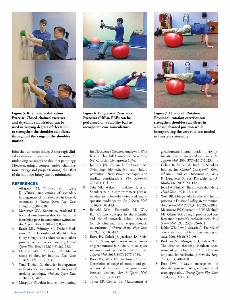
Figure 5. Rhythmic Stabilization Exercise. Closed-chained exercises and rhythmic stabilization can be used in varying degrees of elevation to strengthen the shoulder stabilizers throughout the range of the shoulder motion.
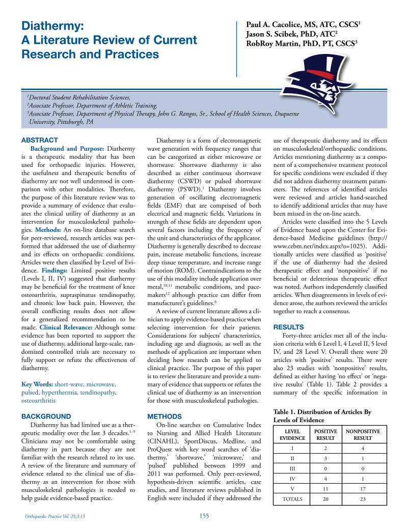
Figure 6. Progressive Resistance Exercises (PREs). PREs can be performed on a stability ball to incorporate core musculature.
Figure 7. Physioball Rotation. Physioball rotation exercises can strengthen shoulder stabilizers in a closed-chained position while incorporating the core rotation needed in freestyle swimming.
joint that can cause injury. A thorough clini-cal evaluation is necessary to determine the underlying causes of the shoulder pathology. However, using a comprehensive rehabilita-tion strategy and proper training, the effect of the shoulder injury can be minimized.
REFERENCES1. Allegrucci M, Whitney SL, Irrgang
JJ. Clinical implications of secondary impingement of the shoulder in freestyle swimmers. J Orthop Sports Phys Ther. 1994;20(6):307-318.
2. McMaster WC, Roberts A, Stoddard T. A correlation between shoulder laxity and interfering pain in competitive swimmers. Am J Sports Med. 1998;26(1):83-86.
3. Beach ML, Whitney SL, Dickoff-Hoff-man SA. Relationship of shoulder flex-ibility, strength and endurance to shoulder pain in competitive swimmers. J Orthop Sports Phys Ther. 1992;16(6):262-268.
4. McLeod WD, Andrews JR. Mecha-nisms of shoulder injuries. Phys Ther. 1986;66(12):1901-1904.
5. Yanai T, Hay JG. Shoulder impingement in front-crawl swimming: II. analysis of stroking technique. Med Sci Sport Exer. 2000;32(1):30-40.
6. Murphy T. Shoulder injuries in swimming.
In: The Athlete's Shoulder. Andrews J, Wilk K, eds. Churchill Livingstone: New York, NY: Churchill Livingstone; 1994.
7. Johnson JN, Gauvin J, Fredericson M. Swimming biomechanics and injury prevention: New stroke techniques and medical considerations. Phys Sportmed. 2003;31(1):41-46.
8. Sein ML, Walton J, Linklater J, et al. Shoulder pain in elite swimmers: primar-ily due to swim-volume-induced supra-spinatus tendinopathy. Br J Sports Med. 2010;44:105-113.
9. Reinold MM, Esacamilla RF, Wilk KE. Current concepts in the scientific and clinical rationale behind exercises for glenohumeral and scapulothoracic musculature. J Orthop Sports Phys Ther. 2009;39(2):105-117.
10. Borsa PA, Scibek JS, Jacobson JA, Meis-ter K. Sonographic stress measurement of glenohumeral joint laxity in collegiate swimmers and age-matched controls. Am J Sports Med. 2005;33(7):1077-1084.
11. Borsa PA, Wilk KE, Jacobson JA, et al. Correlation of range of motion and gle-nohumeral translation in professional baseball pitchers. Am J Sports Med. 2005;33(9):1392-1399.
12. Torres RR, Gomes JLE. Measurement of
glenohumeral internal rotation in asymp-tomatic tennis players and swimmers. Am J Sports Med. 2009;37(5):1017-1023.
13. Cohen B, Romeo A, Bach B. Shoulder injuries. In: Clinical Orthopaedic Reha-bilitation. 2nd ed. Brotzman S, Wilk K, Daugherty K, eds. Philadelphia, PA: Mosby Inc.;2003:151-152.
14. Jobe FW, Pink M. The athlete's shoulder. J Hand Ther. 1994:107-110.
15. Wolf BR, Ebinger AE, Lawler MP. Injury patterns in Division I collegiate swimming. Am J Sports Med. 2009;37(10):2037-2042.
16. Magnusson PS, Constantini NW, McHugh MP, Gleim GG. Strength profiles and per-formance in master's level swimmers. Am J Sports Med. 1995;23(5):626-631.
17. Kibler WB, Press J, Sciascia A. The role of core stability in athletic function. Sports Med. 2006;36(3):189-198.
18. Burkhart SS, Morgan CD, Kibler WB. The disabled throwing shoulder: spec-trum of pathology Part I: pathoanat-omy and biomechanics. J Arth Rel Surg. 2003;19(4):404-420.
19. Russ DW. In-season management of shoulder pain in a collegiate swimmer: A team approach. J Orthop Sports Phys Ther. 1998;27(5):371-376.
151Orthopaedic Practice Vol. 25;3:13

ONLINE WEBINARS:
Pelvic Rotator Cuff • Oct 15-17Beyond Kegels • Sept 10-12 • Nov 12-14Pregnancy & Postpartum • Sept 24-26Mature Woman: Prolapse • Nov 5-7 & Back PainBowel & GI Dysfunction • Oct 8-10
Dear Orthopaedic Section Members: The Orthopaedic Section wants you to know of two positions avail-
able for service within the Section beginning February 2014. If you wish to nominate yourself or someone else, please contact the Nominating Committee Chair, Bill Egan, at [email protected]. Deadline for nom-inations is September 9, 2013. Elections will be conducted during the month of November.
Open Section Offices: • Vice President: Nominations are now being accepted for election to
a three (3) year term beginning at the close of the Orthopaedic Sec-tion Membership Meeting at CSM 2014.
• Nominating Committee Member: Nominations are now being accepted for election to a three (3) year term beginning at the close of the Orthopaedic Section Membership Meeting at CSM 2014.
Be sure to visit https://www.orthopt.org/content/governance/sec-tion_policies for more information about the positions open for election!
10
9
17
16
Call
for
Candidate
s
Deadline fo
r nominati
ons:
Septembe
r 9, 2013
152 Orthopaedic Practice Vol. 25;3:13
ABSTRACTBackground and Purpose: Diathermy
is a therapeutic modality that has been used for orthopaedic injuries. However, the usefulness and therapeutic benefits of diathermy are not well understood in com-parison with other modalities. Therefore, the purpose of this literature review was to provide a summary of evidence that evalu-ates the clinical utility of diathermy as an intervention for musculoskeletal patholo-gies. Methods: An on-line database search for peer-reviewed, research articles was per-formed that addressed the use of diathermy and its effects on orthopaedic conditions. Articles were then classified by Level of Evi-dence. Findings: Limited positive results (Levels I, II, IV) suggested that diathermy may be beneficial for the treatment of knee osteoarthritis, supraspinatus tendinopathy, and chronic low back pain. However, the overall conflicting results does not allow for a generalized recommendation to be made. Clinical Relevance: Although some evidence has been reported to support the use of diathermy, additional large-scale, ran-domized controlled trials are necessary to fully support or refute the effectiveness of diathermy.
Key Words: short-wave, microwave, pulsed, hyperthermia, tendinopathy, osteoarthritis
BACKGROUNDDiathermy has had limited use as a ther-
apeutic modality over the last 3 decades.1–9
Clinicians may not be comfortable using diathermy in part because they are not familiar with the research related to its use. A review of the literature and summary of evidence related to the clinical use of dia-thermy as an intervention for those with musculoskeletal pathologies is needed to help guide evidence-based practice.
Diathermy is a form of electromagnetic wave generation with frequency ranges that can be categorized as either microwave or shortwave. Shortwave diathermy is also described as either continuous shortwave diathermy (CSWD) or pulsed shortwave diathermy (PSWD).1 Diathermy involves generation of oscillating electromagnetic fields (EMF) that are comprised of both electrical and magnetic fields. Variations in strength of these fields are dependent upon several factors including the frequency of the unit and characteristics of the applicator. Diathermy is generally described to decrease pain, increase metabolic functions, increase deep tissue temperature, and increase range of motion (ROM). Contraindications to the use of this modality include application over metal,10,11 metabolic conditions, and pace-makers12 although practice can differ from manufacturer’s guidelines.6
A review of current literature allows a cli-nician to apply evidence-based practice when selecting intervention for their patients. Considerations for subjects’ characteristics, including age and diagnosis, as well as the methods of application are important when deciding how research can be applied to clinical practice. The purpose of this paper is to review the literature and provide a sum-mary of evidence that supports or refutes the clinical use of diathermy as an intervention for those with musculoskeletal pathologies.
METHODSOn-line searches on Cumulative Index
to Nursing and Allied Health Literature (CINAHL), SportDiscus, Medline, and ProQuest with key word searches of ‘dia-thermy,’ ‘shortwave,’ ‘microwave,’ and ‘pulsed’ published between 1999 and 2011 was performed. Only peer-reviewed, hypothesis-driven scientific articles, case studies, and literature reviews published in English were included if they addressed the
use of therapeutic diathermy and its effects on musculoskeletal/orthopaedic conditions. Articles mentioning diathermy as a compo-nent of a comprehensive treatment protocol for specific conditions were excluded if they did not address diathermy treatment param-eters. The references of identified articles were reviewed and articles hand-searched to identify additional articles that may have been missed in the on-line search.
Articles were classified into the 5 Levels of Evidence based upon the Center for Evi-dence-based Medicine guidelines (http://www.cebm.net/index.aspx?o=1025). Addi-tionally articles were classified as ‘positive’ if the use of diathermy had the desired therapeutic effect and ‘nonpositive’ if no beneficial or deleterious therapeutic effect was noted. Authors independently classified articles. When disagreements in levels of evi-dence arose, the authors reviewed the articles together to reach a consensus.
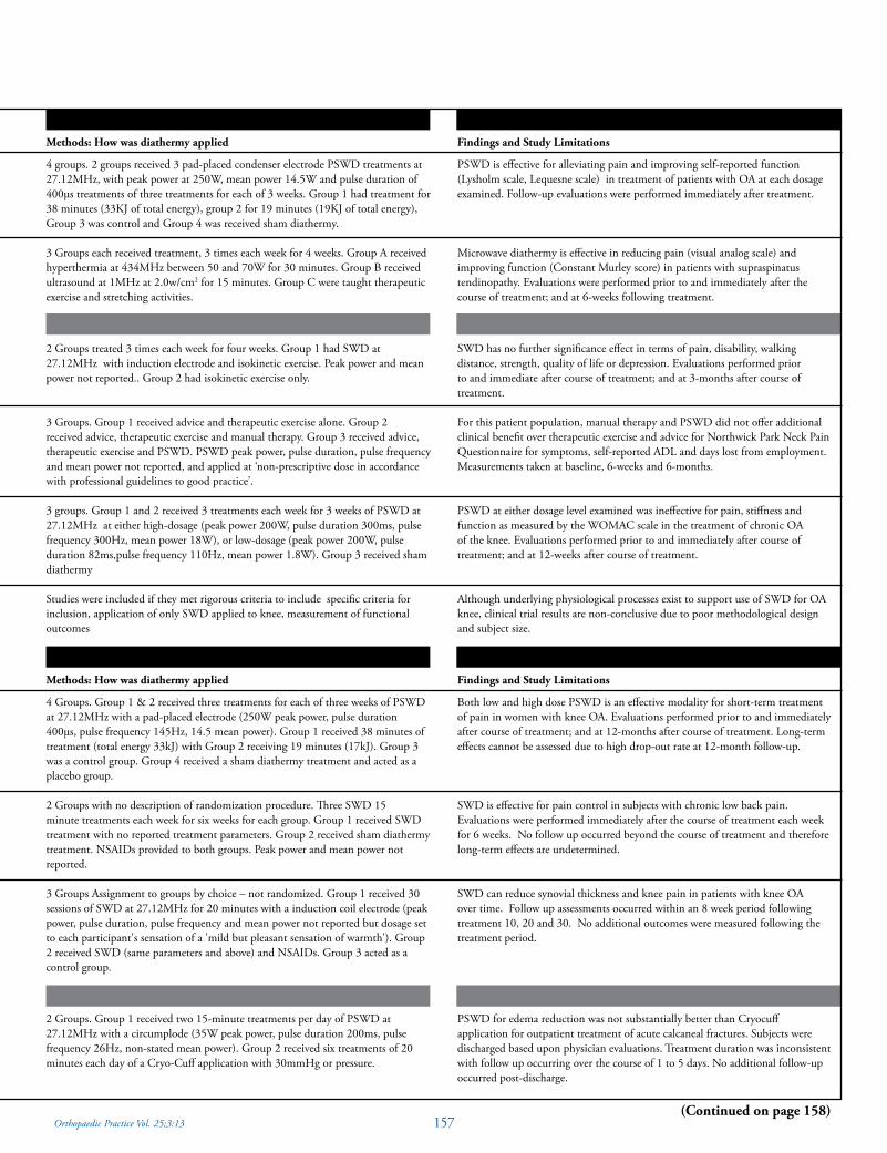
RESULTSForty-three articles met all of the inclu-
sion criteria with 6 Level I, 4 Level II, 5 level IV, and 28 Level V. Overall there were 20 articles with ‘positive’ results. There were also 23 studies with ‘nonpositive’ results, defined as either having ‘no effect’ or ‘nega-tive results’ (Table 1). Table 2 provides a summary of the specific information in
Diathermy: A Literature Review of Current Research and Practices
Paul A. Cacolice, MS, ATC, CSCS1 Jason S. Scibek, PhD, ATC2 RobRoy Martin, PhD, PT, CSCS3
1Doctoral Student Rehabilitation Sciences, 2Associate Professor, Department of Athletic Training, 3 Associate Professor, Department of Physical Therapy, John G. Rangos, Sr., School of Health Sciences, Duquesne University, Pittsburgh, PA
LEVEL POSITIVE NONPOSITIVE EVIDENCE RESULT RESULT
I 2 4
II 3 1
III 0 0
IV 4 1
V 11 17
TOTALS 20 23
Table 1. Distribution of Articles By Levels of Evidence
155Orthopaedic Practice Vol. 25;3:13
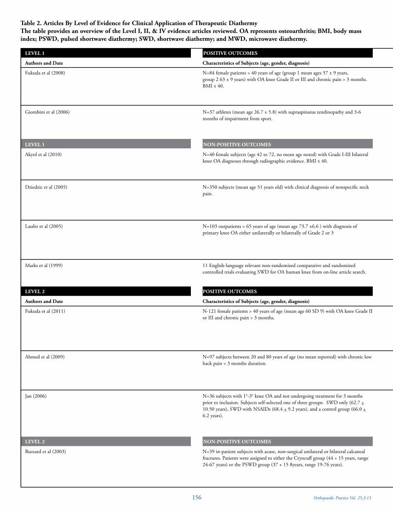
Table 2. Articles By Level of Evidence for Clinical Application of Therapeutic DiathermyThe table provides an overview of the Level I, II, & IV evidence articles reviewed. OA represents osteoarthritis; BMI, body mass index; PSWD, pulsed shortwave diathermy; SWD, shortwave diathermy; and MWD, microwave diathermy.
LEVEL 1
Authors and Date
Fukuda et al (2008)
Giombini et al (2006)
LEVEL 1
Akyol et al (2010)
Dziedzic et al (2005)
Laufer et al (2005)
Marks et al (1999)
LEVEL 2
Authors and Date
Fukuda et al (2011)
Ahmed et al (2009)
Jan (2006)
LEVEL 2
Buzzard et al (2003)
POSITIVE OUTCOMES
Characteristics of Subjects (age, gender, diagnosis)
N=84 female patients > 40 years of age (group 1 mean ages 57 ± 9 years, group 2 63 ± 9 years) with OA knee Grade II or III and chronic pain > 3 months. BMI ≤ 40.
N=37 athletes (mean age 26.7 ± 5.8) with supraspinatus tendinopathy and 3-6 months of impairment from sport.
NON-POSITIVE OUTCOMES
N=40 female subjects (age 42 to 72, no mean age noted) with Grade I-III bilateral knee OA diagnoses through radiographic evidence. BMI ≤ 40.
N=350 subjects (mean age 51 years old) with clinical diagnosis of nonspecific neck pain.
N=103 outpatients > 65 years of age (mean age 73.7 ±6.6 ) with diagnosis of primary knee OA either unilaterally or bilaterally of Grade 2 or 3
11 English-language relevant non-randomized comparative and randomized controlled trials evaluating SWD for OA human knee from on-line article search.
POSITIVE OUTCOMES
Characteristics of Subjects (age, gender, diagnosis)
N-121 female patients > 40 years of age (mean age 60 SD 9) with OA knee Grade IIor III and chronic pain > 3 months.
N=97 subjects between 20 and 80 years of age (no mean reported) with chronic low back pain < 3 months duration.
N=36 subjects with 1°-3° knee OA and not undergoing treatment for 3 months prior to inclusion. Subjects self-selected one of three groups: SWD only (62.7 + 10.50 years), SWD with NSAIDs (68.4 + 9.2 years), and a control group (66.0 + 6.2 years).
NON-POSITIVE OUTCOMES
N=39 in-patient subjects with acute, non-surgical unilateral or bilateral calcaneal fractures. Patients were assigned to either the Cryocuff group (44 + 15 years, range 24-67 years) or the PSWD group (37 + 15 8years, range 19-76 years).
156 Orthopaedic Practice Vol. 25;3:13
Methods: How was diathermy applied
4 groups. 2 groups received 3 pad-placed condenser electrode PSWD treatments at27.12MHz, with peak power at 250W, mean power 14.5W and pulse duration of 400µs treatments of three treatments for each of 3 weeks. Group 1 had treatment for 38 minutes (33KJ of total energy), group 2 for 19 minutes (19KJ of total energy), Group 3 was control and Group 4 was received sham diathermy.
3 Groups each received treatment, 3 times each week for 4 weeks. Group A received hyperthermia at 434MHz between 50 and 70W for 30 minutes. Group B received ultrasound at 1MHz at 2.0w/cm2 for 15 minutes. Group C were taught therapeutic exercise and stretching activities.
2 Groups treated 3 times each week for four weeks. Group 1 had SWD at27.12MHz with induction electrode and isokinetic exercise. Peak power and mean power not reported.. Group 2 had isokinetic exercise only.
3 Groups. Group 1 received advice and therapeutic exercise alone. Group 2 received advice, therapeutic exercise and manual therapy. Group 3 received advice, therapeutic exercise and PSWD. PSWD peak power, pulse duration, pulse frequency and mean power not reported, and applied at ‘non-prescriptive dose in accordance with professional guidelines to good practice’.
3 groups. Group 1 and 2 received 3 treatments each week for 3 weeks of PSWD at 27.12MHz at either high-dosage (peak power 200W, pulse duration 300ms, pulse frequency 300Hz, mean power 18W), or low-dosage (peak power 200W, pulse duration 82ms,pulse frequency 110Hz, mean power 1.8W). Group 3 received sham diathermy
Studies were included if they met rigorous criteria to include specific criteria for inclusion, application of only SWD applied to knee, measurement of functional outcomes
Methods: How was diathermy applied
4 Groups. Group 1 & 2 received three treatments for each of three weeks of PSWDat 27.12MHz with a pad-placed electrode (250W peak power, pulse duration 400µs, pulse frequency 145Hz, 14.5 mean power). Group 1 received 38 minutes of treatment (total energy 33kJ) with Group 2 receiving 19 minutes (17kJ). Group 3 was a control group. Group 4 received a sham diathermy treatment and acted as a placebo group.
2 Groups with no description of randomization procedure. Three SWD 15 minute treatments each week for six weeks for each group. Group 1 received SWD treatment with no reported treatment parameters. Group 2 received sham diathermy treatment. NSAIDs provided to both groups. Peak power and mean power not reported.
3 Groups Assignment to groups by choice – not randomized. Group 1 received 30 sessions of SWD at 27.12MHz for 20 minutes with a induction coil electrode (peak power, pulse duration, pulse frequency and mean power not reported but dosage set to each participant's sensation of a 'mild but pleasant sensation of warmth'). Group 2 received SWD (same parameters and above) and NSAIDs. Group 3 acted as a control group.
2 Groups. Group 1 received two 15-minute treatments per day of PSWD at 27.12MHz with a circumplode (35W peak power, pulse duration 200ms, pulse frequency 26Hz, non-stated mean power). Group 2 received six treatments of 20 minutes each day of a Cryo-Cuff application with 30mmHg or pressure.
Findings and Study Limitations
PSWD is effective for alleviating pain and improving self-reported function(Lysholm scale, Lequesne scale) in treatment of patients with OA at each dosage examined. Follow-up evaluations were performed immediately after treatment.
Microwave diathermy is effective in reducing pain (visual analog scale) and improving function (Constant Murley score) in patients with supraspinatus tendinopathy. Evaluations were performed prior to and immediately after the course of treatment; and at 6-weeks following treatment.
SWD has no further significance effect in terms of pain, disability, walkingdistance, strength, quality of life or depression. Evaluations performed prior to and immediate after course of treatment; and at 3-months after course of treatment.
For this patient population, manual therapy and PSWD did not offer additional clinical benefit over therapeutic exercise and advice for Northwick Park Neck Pain Questionnaire for symptoms, self-reported ADL and days lost from employment. Measurements taken at baseline, 6-weeks and 6-months.
PSWD at either dosage level examined was ineffective for pain, stiffness and function as measured by the WOMAC scale in the treatment of chronic OA of the knee. Evaluations performed prior to and immediately after course of treatment; and at 12-weeks after course of treatment.
Although underlying physiological processes exist to support use of SWD for OA knee, clinical trial results are non-conclusive due to poor methodological design and subject size.
Findings and Study Limitations
Both low and high dose PSWD is an effective modality for short-term treatmentof pain in women with knee OA. Evaluations performed prior to and immediately after course of treatment; and at 12-months after course of treatment. Long-term effects cannot be assessed due to high drop-out rate at 12-month follow-up.
SWD is effective for pain control in subjects with chronic low back pain. Evaluations were performed immediately after the course of treatment each week for 6 weeks. No follow up occurred beyond the course of treatment and therefore long-term effects are undetermined.
SWD can reduce synovial thickness and knee pain in patients with knee OA over time. Follow up assessments occurred within an 8 week period following treatment 10, 20 and 30. No additional outcomes were measured following the treatment period.
PSWD for edema reduction was not substantially better than Cryocuff application for outpatient treatment of acute calcaneal fractures. Subjects were discharged based upon physician evaluations. Treatment duration was inconsistent with follow up occurring over the course of 1 to 5 days. No additional follow-up occurred post-discharge.
(Continued on page 158)157Orthopaedic Practice Vol. 25;3:13
Table 2. Continued from previous page.
LEVEL 4
Authors and Date
Adegoke and Gbeminiyi (2004)
Pasila (1978)
DiCesare et al (2008)
McCray and Patton (1984)
LEVEL 4
Švarcová et al (1988)
POSITIVE OUTCOMES
Characteristics of Subjects (age, gender, diagnosis)
N=14 subjects 40-70 years of age with knee OA. No mean age was reported
N=300 consecutive patients > 15 years of age, <4 days post ankle injury. Subjects were assigned to one of three groups: Diapulse (mean age 30.3 years) Curapulse (29.8 years) and placebo (32.6 years)
N=2, Patient 1 was a 62 year old female with calcific tendonitis of the rotator cuff. Patient 2 was a 55 year old female with calcific tendonitis of the rotator cuff.
N=19 between 21 and 65 years of age who have single or multiple upper spine or shoulder muscle trigger points.
NON-POSITIVE OUTCOMES
N=180 hip and knee patients with bilateral or unilateral osteoarthritis. Mean ageswere 64.2 + 10.6 years (ultrasound group), 63.6 + 10.9 years (galvanic current croup), and 62.4 + 12.5 years (SWD group)
each of the 14 Level I-IV articles that relate to subject characteristics, methods of dia-thermy application, and study findings.
DISCUSSIONThe purpose of this literature review was
to summarize the evidence for the clini-cal use of therapeutic diathermy for those with orthopaedic pathologies. Evidence was found to support the use of shortwave diathermy (SWD) for short-term pain con-trol in those with knee osteoarthritis (OA), supraspinatus tendinopathy, and chronic low back pain. Physiological Level V research provides evidence to link diathermy to gains in ROM, regional metabolic func-tions, and structural changes at the cellular level. Although there is some evidence to support the clinical use of diathermy, there is also evidence that does not support its use. Based on the overall conflicting results, a global recommendation cannot be made.
Additional research is generally needed to guide evidence-based practice in the clinical applications of diathermy.
Of the Level I and II articles reviewed for this paper, 6 sources (5 research and 1 sys-tematic review) addressed the use of SWD in those with knee OA.13–18 The results of 3 studies suggest SWD may be effective for reducing pain,13,17,18 decreasing synovial sac thickness,18 and improving function and quality of life13,17 in patients with grades II-III knee OA. Jan et al18 treated patients for 20 minutes; however, diathermy inten-sity was not noted. Treatment intensities (17 kJ & 33 kJ) and durations (19 minutes and 38 minutes) varied in the two studies by Fukada et al13,17 and Akyol et al.14 Laufer et al15 did not have positive findings in those with knee OA. Jan et al18 noted continu-ous patient improvements when following treatments over an 8-week period, while studies by Fukada et al13,17 noted favorable
treatment results immediately after treat-ment.13 One of the studies with ‘nonposi-tive’ findings incorporated subjects that were relatively older (73.7 + 6.6 years)15 when compared to the 3 studies with positive findings in which subjects age ranged from 57.0 to 68.4 years.13,17,18 The Level I system-atic review evaluated studies between 1955-1997 and acknowledged that the clinical trials reviewed incorporated poor method-ological designs.16 Overall this review noted the results were inconclusive.16 The positive effects noted in our review relative to pain and function may have been attributed to a younger sample age (40-70 years of age) and outcomes measures taken immediately after treatment. Presently, diathermy does not seem to have long-term effects for pain control in those with knee OA.
In addition to the results noted for knee OA, other Level I-II studies suggest micro-wave diathermy (MWD) or SWD may be
158 Orthopaedic Practice Vol. 25;3:13
Methods: How was diathermy applied
2 Group which each received 3 weekly treatments for four weeks to include therapeutic exercise including isotonic quadriceps activity and unloaded bicycle activity on an ergometer for 10 minutes. Group 1 received 20 minutes of ice pack application in a wet towel before therapeutic exercise. Group 2 received 20 minutes of SWD with a pad-placed electrode (peak power and mean power not reported but dosage set to subjects perception of 'comfortable warmth'). No control group. No placebo group.
3 Groups each received 20 minute treatments once per day for 3 successive days. Group 1 received Diapulse PSWD (peak power, pulse duration, pulse frequency not reported, mean power 30 W/s). Group 2 received Curapuls PSWD (peak power, pulse duration, pulse frequency not reported, mean power 40 W/s). Group 3 received placebo treatment. Outcome measurements: strength, ROM edema, return to work.
Treatment of three sessions for each of four weeks with hyperthermia; microwave diathermy at 434MHz (100W maximal power, 50W mean power, 50% SAR for surface of 96cm2).
2 Groups each received treatment for 20 minutes with levels of heat to tolerance. Group 1 received PSWD at 27.12MHz with an induction electrode (peak power, pulse duration, pulse frequency and mean power not reported). Group 2 received moist heat through a hydrocollator pad
3 Groups received treatments every other day for 20 days over a three-week period. Group 1 received ultrasound over large (6.4 cm2) area in three treatments of 5 minutes each (power nor frequency not reported). Group 2 received galvanic current for 20 minutes each with current density of 0.1mA/cm2. Group 3 received two (anterior and posterior) 2 minute treatments of PSWD with no report of electrode utilized (peak power 700W, pulse duration not reported, pulse frequency 46MHz, mean power not reported).
Findings and Study Limitations
SWD is an effective as ice as a pain-relieving modality for knee OA. All outcomesdata was acquired at the completion of the study with no long term follow-up.
Treatment with Curapuls PSWD resulted in statistically significant treatment for reduction of edema, but reduction of edema did not occur with Diapulse PSWD unit. Authors noted limitation in consistency of volumetric measurements for edema. No statistical difference in strength gain, ROM gain or return to work time between the groups was observed. All outcomes data was acquired prior to treatment on the first day and following treatment on the third day. No long term follow-up occurred.
MWD is a safe modality to utilize for calcific tendinopathy of the rotator cuff, but effects of the treatment remains undefined as this condition may spontaneously resolve without intervention. Improvements were noted in SPADI scores, passive range of motion upon discharge. SPADI scores and imaging studies at one year follow-up remained consistent with discharge values.
Both PSWD and moist heat were effective (at reducing pain in sensitive trigger points. PSWD was not significantly different from moist heat (p= .0581); yet tended toward positive treatment effects.
No statistical difference on difference in pain control in any of the three treatment methods. Therapeutic effect was enhances with augmented drug treatment with ultrasound and PSWD, but not galvanic stimulation. No detail on randomization. No control how many subjects were hip OA and how many were knee OA. Very short period of PSWD treatment and very large area of treatment of ultrasound. Authors reported that PSWD penetration is less deep, but no enlightenment on how this occurs. Treatment effectiveness was assessed following days 10 and 20 over a 3 week period.
effective in short-term pain control in those with supraspinatus tendinopathy19 and low back pain20 but not in those with nonspecific neck pain.21 It was noted that MWD was more effective than ultrasound and ROM exercises alone for pain relief and improv-ing function in short term 6 week follow-up assessments in those with supraspinatus tendinopathy.19 Over the course of 6 weeks, CSWD was noted to be effective in manag-ing pain in patients with chronic low back pain, with follow up occurring each week of treatment.20 No difference was noted in Neck Pain Questionnaire scores and missed work days when PSWD with exercise was compared to manual therapy and exercises and exercise alone in those with nonspecific neck pain at 6-month follow-up.21 Further-more, no difference was noted in edema reduction when comparing PSWD to cold application through a Cryo/Cuff in those with acute, nonsurgical calcaneal fractures.22
Of the 5 Level of Evidence IV articles, 4 had positive effects2,23–25 while one had nonpositive effects.26 Diathermy was shown to be effective in decreasing edema in those with acute ankle injuries.23 Similarly, dia-thermy was more effective than moist heat in the treatment of myofascial trigger points24 and was effective in facilitating the recovery of two individuals with calcific tendinopathy when they were treated with MWD.2 Also, diathermy was noted to be as effective as ice in decreasing pain and increasing ROM in those with knee OA.25 Although positive improvements in pain were observed in patients with hip or knee OA following diathermy, these effects were consistent with the improvements observed when patients were treated using ultrasound or electrical stimulation.26
Findings from articles classified as Level of Evidence V offer further theoreti-cal evidence suggesting the effectiveness of
diathermy with pain management27,28
ROM10,27–31 and specific orthopaedic condi-tions.10 Variations in study design and docu-mentation however limit practical treatment conclusions.32,33 Studies performed on tissue samples or health subjects provide for knowl-edge of tissue heating effectiveness3,33–37 and changes at the cellular level.38,39
Based on the literature completed in this review, some evidence exists to support the use of diathermy in the management of orthopaedic injuries. More specifically, evi-dence was identified that supports the use of SWD for short-term pain control in those with knee OA,13,17,18 supraspinatus tendi-nopathy,19 and low back pain.20 Additional studies classified as Level I evidence are nec-essary to fully support or refute the effective-ness of SWD or MWD in the treatment of musculoskeletal injuries.
159Orthopaedic Practice Vol. 25;3:13
REFERENCES1. Prentice WE, Draper DO. The basic sci-
ence of therapeutic modalities. In: Ther-apeutic Modalities in Rehabilitation. 4th ed. New York, NY: McGraw-Hill; 2010.
2. Di Cesare A, Giombini A, Dragoni S, et al. Calcific tendinopathy of the rota-tor cuff. Conservative management with 434 Mhz local microwave diathermy (hyperthermia): A case study. Disabil Rehabil. 2008;30:1578–1583.
3. Leitgeb N, Omerspahic A, Niedermayr F. Exposure of non-target tissues in medical diathermy. Bioelectromagnetics. 2010;31(1):12–19.
4. Shah SGS, Farr A, Esnouf A. Avail-ability and use of electrotherapy devices: A survey. Intl J Ther Rehabil. 2007;14(6):260–264.
5. Shah SGS, Farrow A. Investigation of practices and procedures in the use of therapeutic diathermy: a study from the physiotherapists’ health and safety perspective. Physiother Res Int. 2007;12:228–241.
6. Shields N, O’Hare N, Gormley J. Con-tra-indications to shortwave diathermy: survey of Irish physiotherapists. Physio-therapy. 2004;90(1):42–53.
7. Kitchen SE, Partridge CJ. A survey to examine the clinical use of ultra-sound, shortwave diathermy and laser in England. Br J Ther Rehab. 1996;3(12):644–650.
8. Kitchen SE, Partridge CJ. Ultrasound, shortwave diathermy and laser: a survey to examine patterns of use in England. Br J Ther Rehab. 1997;4(2):75–78.
9. Nadler SF, Prybicien M, Malanga GA, Sicher D. Complications from thera-peutic modalities: results of a national survey of athletic trainers. Arch Phys Med Rehabil. 2003;84(6):849–853.
10. Draper DO, Castel JC, Castel D. Low-watt pulsed shortwave diathermy and metal-plate fixation of the elbow. ATT. 2004;9(5):28–32.
11. Seiger C, Draper DO. Use of pulsed shortwave diathermy and joint mobili-zation to increase ankle range of motion in the presence if surgical implanted metal: A case series. J Orthop Sports Phys Ther. 2006;36(9):670–677.
12. Anon. Mettler Electronics AutoTherm 395 Diathermy Instruction Manual; 2005.
13. Fukuda TY, Alves da Cunha R, Fukuda VO, et al. Pulsed shortwave treatment in women with knee osteoarthritis: A multicenter, randomized, placebo-
controlled clinical trial. Phys Ther. 2011;91(7):1009–1017.
14. Akyol Y, Durmas, D, Alayhi, G, et al. Does short-wave diathermy increase the effectiveness of isokinetic exer-cise on pain, function, knee muscle strength, quality of life, and depres-sion in the patients with knee osteo-arthritis? Eur J Physical Med Rehabil. 2010;46(3):325–336.
15. Laufer Y, Zilberman R, Porat R, Nahir A. Effect of pulsed short-wave dia-thermy on pain and function of sub-jects with osteoarthritis of the knee: a placebo-controlled double-blind clinical trial. Clin Rehabil. 2005;19(3):255.
16. Marks R, Ghassemi M, Duarte R, Van Nguyen J. A review of the literature on shortwave diathermy as applied to osteo-arthritis of the knee. Physiotherapy. 1999;85(6):304–316.
17. Fukuda TY, Ovanessian V, da Cunha RA, et al. Pulsed short wave effect in pain and function in patients with knee osteo-arthritis. J Appl Res. 2008;8(3):189–198.
18. Jan M-H, Chai H-M, Wang C-L, Lin Y-F, Tsai L-Y. Effects of repetitive short-wave diathermy for reducing synovi-tis in patients wth knee osteoarthritis: An ultrasonographic study. Phys Ther. 2006;86(2):236–244.
19. Giombini A. Short-term effectiveness of hyperthermia for supraspinatus ten-dinopathy in athletes: a short-term ran-domized controlled study. Am J Sports Med. 2006;34:1247–1253.
20. Ahmed MS, Shakoor MA, Khan AA. Evaluation of the effects of shortwave diathermy in patients with chronic low back pain. Bangladesh Med Res Council Bulletin. 2009;35:18-20.
21. Dziedzic K, Hill J, Lewis M, et al. Effec-tiveness of manual therapy or pulsed shortwave diathermy in addition to advice and exercise for neck disorders: A pragmatic randomized controlled trial in physical therapy clinics. Arth Rheum. 2005;53:214–222.
22. Buzzard B, Pratt R, Briggs P, et al. Is Pulsed Shortwave diathermy better than ice therapy for the reduction of oedema following calcaneal frac-tures? Preliminary trial. Physiotherapy. 2003;89(12):734–742.
23. Pasila M, Visuri T, Sundholm A. Pulsat-ing shortwave diathermy: value in treat-ment of recent ankle and foot sprains. Arch Phys Med Rehabil. 1978;59.
24. McCray RE, Patton NJ. Pain relief at rigger points: A comparison of moist
heat and shortwave diathermy. J Orthop Sport Phys Ther. 1984;5(4):175–178.
25. Adegoke B, Gbeminiyi M. Efficacy of ice and shortwave diathermy in the management of osteoarthritis on the knee-A preliminary. Afr J Biomed Res. 2010;7(2).
26. Švarcová J, Trnavsky K, Zvárová J. The influence of ultrasound, galvanic current and shortwave diathermy on pain intensity in patients with osteo-arthritis. Scand J Rheumatol. 1988;67 Supplement:83–85.
27. Giombini A, Giovannini V, Cesare AD, et al. Hyperthermia induced by micro-wave diathermy in the management of muscle and tendon injuries. Br Med Bull. 2007;83:379–396.
28. Nosaka K, Muthalib M, Lavender A, Laursen P. Attenuation of muscle damage by preconditioning with muscle hyper-thermia 1-day prior to eccentric exercise. Eur J Appl Physiol. 2007;99:183–192.
29. Peres SE, Draper DO, Knight KL, Richard MD. Pulsed shortwave dia-thermy and prolonged long-duration stretching increase dorsiflexion range of motion more than identical stretch-ing without diathermy. J Ath Train. 2002;37(1):43–50.
30. Robertson V, Ward A, Jung P. The effect of heat on tissue extensibility: A compar-ison of deep and superficial heating. Arch Phys Med Rehabil. 2005;86:819–824.
31. Draper DO, Castro JL, Feland B, Schul-thies S, Eggett D. Shortwave diathermy and prolonged stretching increase ham-string flexibility more than prolonged stretching alone. J Orthop Sports Phys Ther. 2004;34(1):13–20.
32. Al-Mandeel MM, Wat T. An audit of patient records into the nature of pulsed shortwave therapy use. Int J Ther Reha-bil. 2006;13(9):414–420.
33. Low J. Dosage of some pulsed short-wave clinical trials. Physiotherapy. 1995;81(10):811–816.
34. Ichinoski-Sekine N, Naito H, Saga N, et al. Changes in muscle tempera-ture induced by 434 MHz micro-wave hyperthermia. Br J Sports Med. 2007;41:425–429.
35. Murray CC, Kitchen S. Effect of pulse repetition rate on the perception of thermal sensation with pulsed short-wave diathermy. Physiother Res Int. 2000;5(2):73–84.
36. Garrett CL, Draper DO, Knight KL. Heat distribution in the lower leg from pulsed short-wave diathermy and ultra-
160 Orthopaedic Practice Vol. 25;3:13
sound treatments. J Ath Train. 35(1):50–55.37. Draper D, Knight K, Fujiwa T, Castel J. Temperature change in
human muscle during and after pulsed short-wave diathermy. J Orthop Sports Phys Ther. 1999;29(1):13–22.
38. Lehmann JF, McDougall JA, Guy AW, Warren CG, Essel-man PC. Heating patterns produced by shortwave diathermy applicators in tissue substitute models. Arch Phys Med Rehabil. 1983;54:575–577.
39. Wang JL, Chan RC, Cheng HH, et al. Short waves-induced enhancement of proliferation of human chondrocytes: involve-ment of extracellular signal-regulated MAP-kinase (ERK). Clin Exp Pharmacol Physiol. 2007;34(7):581–585.
Earn a PhD in Rehabilitation Science
Duquesne University’s PhD program in Rehabilitation Science features a specialized curriculum in orthopedics and clinical biomechanics. The curriculum is a collaborative effort between our nationally recognized Physical Therapy and Athletic Training departments.
Benefits of the PhD Program include:
• Dedicated lab equipped with contemporary biomechanics research equipment
• Enrolled students typically present at national conferences and are published in peer-reviewed journals
• Part or full-time study • Tuition remission and graduate assistantships available• Applications considered on a rolling basis
Contact Christopher R. Carcia, PhD, PT, SCS, OCS, Program Director, for more information at: 412.396.5545 or [email protected]
Visit Us Online: www.duq.edu/rehabilitation-science
ADVAnCE YOUR CAREER
Have you checked out this Web site yet?
MoveForwardPT.com is APTA’s official consumer information Web site designed to support the national Move Forward braining campaign and serve as the “definitive source” of consumer information about physical therapy. The core of the site is the “Symptoms & Conditions” section that includes more than 60 detailed, physical therapy-specific guides on various topics.
Check it out today!
161Orthopaedic Practice Vol. 25;3:13
ABSTRACTBackground/Purpose: The purpose of
this review is to present current evidence that will assist clinicians in choosing appro-priate outcome instruments for patients with common conditions of the hip. Meth-ods: An electronic database search of patient self-reported outcome instruments for the hip was performed to identify evidence of validity, reliability, and responsiveness. Findings: The Hip Dysfunction and Osteo-arthritis Outcome Score has demonstrated the strongest evidence for use with patients with hip osteoarthritis or total hip arthro-plasty. The Hip Outcome Score had the best evidence of strong psychometric prop-erties in a younger population undergoing hip arthroscopy. Clinical Relevance: Four cases are described that outline the clinical decision making of the selection of appro-priate self-reported outcome instruments. Conclusion: Selecting the most appropriate outcome instrument requires the clinician to evaluate the evidence available to support its use and understand the context in which the instrument was tested.
Key Words: self-report scales outcomes, reliability, validity
INTRODUCTIONSelf-reported outcome instruments can
be useful in determining the effectiveness of treatment interventions. Many outcome instruments exist for patients with hip related pathology making the selection of an appropriate instrument challenging. When choosing the most appropriate instrument, several factors need to be considered. These include the type of instrument, psychomet-ric properties of the instrument, and char-acteristics of the subjects used to provide supporting evidence. The purpose of this review is to present current evidence that will assist clinicians in choosing appropri-ate outcome instruments for patients with common conditions of the hip.
Outcome Instruments for the Hip: A Guide to Implementation
Benjamin R. Kivlan, PT, SCS, OCS,1,2 RobRoy L. Martin1,3
1John G. Rangos Sr. School of Health Sciences, Duquesne University, Pittsburgh, PA2Tri-State Physical Therapy, Seven Fields, PA3UPMC Center for Sports Medicine, Pittsburgh, PA
TYPES OF INSTRUMENTSThere are 4 basic types of self-reported
outcome instruments: generic, disease-spe-cific, region-specific, and patient-specific. Each has inherent strengths and weaknesses.1
Generic instruments assess the global well-being of the patient and are useful in com-paring groups of subjects with a wide range of conditions.2 Disease-specific instruments capture the unique characteristics of a dis-ease and its impact on the patient.2 Region-specific instruments contain items that are specific to a particular area of the body and useful in assessing the effect of disease pro-cesses on that region. Patient-specific instru-ments allow individuals to create items that assess issues they feel are important. These instruments are useful in analyzing the same individual over an extended period of time, but may not allow comparison to other sub-jects or patients.3
PSYCHOMETRIC PROPERTIESValidity, reliability, and responsiveness
are psychometric properties that can be used to determine usefulness of an instrument. It is important to consider the evidence that supports the use of each instrument when deciding which instrument is most appro-priate for a specific group of patients.
Evidence for Content Validity
The ability of an instrument to measure what it claims to measure refers to its valid-ity. Content validity is the extent to which the items of the instrument represent the totality of the domain being measured. It is arguably the most important quality of an instrument.4 Content will be determined by what the individual items on the instrument measure. The International Classification of Functioning, Disability and Health (ICF) model can be used to categorize and define item content.5 According to this model, items could measure the domains of (1) body structure and function and (2) activity and participation. Assessment of symptoms
(ie, pain) and signs (ie, range of motion) are examples of measures of body structure and function. Content validity needs to be established in the early development of an instrument as later statistical analysis will not be able to correct for poor content valid-ity. An instrument with strong evidence of content validity contains items that pertain to every important aspect of the concept being measured.1 Commonly, clinicians create an instrument and determine item content based on what they believe is impor-tant. However, the opinion of clinicians may differ from patients.6 Therefore, incorporat-ing patient input is strongly encouraged in the development of outcome instruments.7-9 Internal consistency, factorial analysis, and item response theory can statistically provide evidence for content validity.1
Evidence for Construct ValidityConstruct validity refers to how the
instrument relates to other measures of the same domain of interest.10 Commonly cor-relations to other established instruments or clinical measures are used to offer evidence for construct validity. Hypothesis testing may also be used to provide evidence of construct validity.1,4,11 For example, we may expect those with a more severe disorder to score poorer than those with a less severe disorder.4
Evidence for ReliabilityTest retest reliability describes the stabil-
ity of the measure over time.1,4 Instruments with evidence for reliability yield the same score in the absence of change over time. Intraclass correlations (ICC) are commonly used to compare the relationship of repeated measures. However, an ICC value by itself offers little information for clinical inter-pretation. The minimal detectable change (MDC) may be more useful as it represents the amount of change necessary to conclude that the change in score over time is beyond measurement error. Therefore, the time
162 Orthopaedic Practice Vol. 25;3:13
between repeated measures testing should represent a timeframe relevant for reassess-ment to be most meaningful.
Evidence for ResponsivenessResponsiveness assesses the ability of the
instrument to detect a meaningful change over time. A responsive outcome instrument will change as the individual improves or worsens over time. The analysis of responsive-ness can be done at the group or individual level.12,13 When focusing on the responsive-ness of a group, statistics of mean differ-ences, effect size, and Guyatt responsiveness index can be used.12,13 Individual level analy-sis can be done with receiver operator char-acteristic (ROC) curves. This ROC analysis can assist with clinical decision making by determining a cut-off value in the change score that best discriminates between those that have improved from those that have not improved. This cut-off value is referred to as minimum clinically important difference (MCID).1 The criterion used to determine ‘improved’ from ‘not improved’ is important to consider when interpreting MCID. Also, the timeframe over which the MCID value was established should match the timeframe in which the patient will be reassessed. As it pertains to outcome studies, it is important for an instrument to have evidence that can detect change in an individual over time.
CHARACTERISTICS OF THE SUBJECTS
Characteristics of age, gender, and the specific condition of the subjects should be considered when selecting an appropri-ate instrument. Subjects used in the stud-ies should match the intended population if the scores from an instrument are to be appropriately interpreted. For example, evi-dence to support the use of an instrument for individuals with osteoarthritis of the hip may not be accurately generalized to those with acetabular labral tears. An instrument that has evidence for validity, reliability, and responsiveness using individuals with a wide range of hip pathologies will allow obtained scores to be generalized and interpreted in a greater range of clinical environments.1
METHODSA comprehensive literature search was
performed in the Medline, Health and Psychological Instruments, CINAHL, Healthstar, and Sport Discus Databases. A combination of key words using boolean connectors was performed for each database search as follows: hip "and" index "or" mea-
sure "or" instrument "or" scale "or" ques-tionnaire "and" reliability "or" validity "or" responsiveness. The results of the searches were then reviewed to identify self-reported outcome instruments that supplied psycho-metric evidence of use with individuals with hip-related pathology.
A list of the self-reported outcome mea-sures was formulated from the search. The list was conferred by a group of experts in hip rehabilitation to ensure instruments were not errantly omitted. The descrip-tion of the instrument, type of instrument, characteristics of the subjects in which the instrument was tested as well as evidence for content validity, construct validity, reliabil-ity, and responsiveness was recorded for each of the identified instruments.
RESULTSThe database searches yielded 425 arti-
cles with reference of a hip related outcome instrument. Instruments without evidence of psychometric properties were omitted from further analysis.
Seventeen self-reported outcome instru-ments that reported psychometric properties of validity, reliability, and/or responsive-ness for those with orthopaedic related hip pathology were identified.6,9,14-40 These instruments were categorized according to their intended population. Instruments for osteoarthritis, total hip arthroplasty, hip fracture, and nonarthritic intraarticular con-ditions along with corresponding evidence to support their use are outlined in Tables 1-4, respectively. These tables may serve as a guide in selecting the most appropriate out-comes measure for patients with various hip pathologies.
Two commonly used instruments were omitted from this analysis. The Lower Extremity Functional Scale has reported evi-dence for reliability and validity as well as MDC and MCID values.39 However, these properties were established on a broad group of patients with lower extremity musculo-skeletal dysfunction. Only 2 out of 107 sub-jects had a hip-related condition.39 Thus, its use as a hip-specific instrument has not been adequately studied. The Harris Hip Score is an instrument that has been commonly used in the assessment of patients with hip pathology.41-49 It was originally developed as a tool administered by the physician that incorporates physical exam and inter-view components. The tool has since been modified as a self-reported instrument.34
Only studies using the self-reported ver-sion referred to as the Modified Harris Hip
Score (MHHS) were included in this analy-sis. The remaining discussion will focus on the instruments identified for osteoarthritis, total hip arthroplasty, hip fracture, and non-arthritic intraarticular conditions.
Osteoarthritis Five instruments had evidence to support
their use for individuals with osteoarthritis (OA) of the hip (Table 1). These include the Hip Dysfunction and Osteoarthri-tis Outcome Score (HOOS),14,15 Western Ontario and McMaster Universities Osteo-arthritis index (WOMAC),16-19 Lequesne Index of Severity for Osteoarthritis of the Hip (LISH),19,20 the American Academy of Orthopedic Surgeons (AAOS)-Lower Limb Core Scale and Hip and Knee Core Scale,21
and the Osteoarthritis Knee and Hip Qual-ity of Life measure (OAKHQOL).9 The HOOS had evidence for all 4 psychomet-ric areas. Only the WOMAC has defined MDC or MCID values for this population. The OAKHQOL differed from the others in that there is considerable focus to social and mental health parameters.9 The HOOS appears to have the strongest evidence for use with this population.11 It also contains a subsection for sports activities that was unique among the other instruments.11 The HOOS is easy to complete and calculate; however, it is rather lengthy containing 40 items. The WOMAC is a shorter instru-ment but equally easy to complete. Both the HOOS and WOMAC are accessible for free on the Internet that make them attractive instruments to be used with patients suffer-ing from hip osteoarthritis.50
One instrument that was not included in the table of instruments (Table 1) was the OA-Function-CAT.29 This instrument was excluded because it is a computer-assisted test (CAT). Use of computer technology allows the test to be tailored to the individ-ual and eliminates redundancy of questions; thereby producing a complete and efficient method of assessing patient outcomes. Jette et al29 established evidence for content valid-ity, construct validity, and reliability of this instrument. Thus it has promise as a tool for measuring outcomes for patients with knee or hip OA and may represent the future direction of outcome assessments.
Total Hip ArthroplastyEight instruments had evidence to sup-
port their use for individuals who have undergone total hip arthroplasty (Table 2). These include the previously men-tioned LISH, OAKHQOL, WOMAC, and
163Orthopaedic Practice Vol. 25;3:13
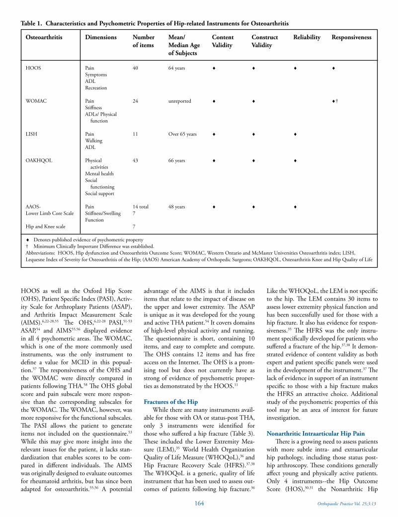
HOOS as well as the Oxford Hip Score (OHS), Patient Specific Index (PASI), Activ-ity Scale for Arthroplasty Patients (ASAP), and Arthritis Impact Measurement Scale (AIMS).6,22-28,55 The OHS,6,22-28 PASI,51-53
ASAP,54 and AIMS55,56 displayed evidence in all 4 psychometric areas. The WOMAC, which is one of the more commonly used instruments, was the only instrument to define a value for MCID in this popual-tion.57 The responsiveness of the OHS and the WOMAC were directly compared in patients following THA.58 The OHS global score and pain subscale were more respon-sive than the corresponding subscales for the WOMAC. The WOMAC, however, was more responsive for the functional subscales. The PASI allows the patient to generate items not included on the questionnaire.53
While this may give more insight into the relevant issues for the patient, it lacks stan-dardization that enables scores to be com-pared in different individuals. The AIMS was originally designed to evaluate outcomes for rheumatoid arthritis, but has since been adapted for osteoarthritis.55,56 A potential
advantage of the AIMS is that it includes items that relate to the impact of disease on the upper and lower extremity. The ASAP is unique as it was developed for the young and active THA patient.54 It covers domains of high-level physical activity and running. The questionnaire is short, containing 10 items, and easy to complete and compute. The OHS contains 12 items and has free access on the Internet. The OHS is a prom-ising tool but does not currently have as strong of evidence of psychometric proper-ties as demonstrated by the HOOS.11
Fractures of the HipWhile there are many instruments avail-
able for those with OA or status-post THA, only 3 instruments were identified for those who suffered a hip fracture (Table 3). These included the Lower Extremity Mea-sure (LEM),35 World Health Organization Quality of Life Measure (WHOQoL),36 and Hip Fracture Recovery Scale (HFRS).37,38 The WHOQoL is a generic, quality of life instrument that has been used to assess out-comes of patients following hip fracture.36
Like the WHOQoL, the LEM is not specific to the hip. The LEM contains 30 items to assess lower extremity physical function and has been successfully used for those with a hip fracture. It also has evidence for respon-siveness.35 The HFRS was the only instru-ment specifically developed for patients who suffered a fracture of the hip.37,38 It demon-strated evidence of content validity as both expert and patient specific panels were used in the development of the instrument.37 The lack of evidence in support of an instrument specific to those with a hip fracture makes the HFRS an attractive choice. Additional study of the psychometric properties of this tool may be an area of interest for future investigation.
Nonarthritic Intraarticular Hip PainThere is a growing need to assess patients
with more subtle intra- and extraarticular hip pathology, including those status post-hip arthroscopy. These conditions generally affect young and physically active patients. Only 4 instruments--the Hip Outcome Score (HOS),30,31 the Nonarthritic Hip
Table 1. Characteristics and Psychometric Properties of Hip-related Instruments for Osteoarthritis
Osteoarthritis Dimensions Number Mean/ Content Construct Reliability Responsiveness of items Median Age Validity Validity of Subjects
HOOS Pain 40 64 years ♦ ♦ ♦ ♦ Symptoms ADL Recreation WOMAC Pain 24 unreported ♦ ♦ ♦† Stiffness ADLs/ Physical function LISH Pain 11 Over 65 years ♦ ♦ ♦ Walking ADL OAKHQOL Physical 43 66 years ♦ ♦ ♦ activities Mental health Social functioning Social support AAOS- Pain 14 total 48 years ♦ ♦ ♦Lower Limb Core Scale Stiffness/Swelling 7 FunctionHip and Knee scale 7
♦ Denotes published evidence of psychometric property† Minimum Clinically Important Difference was established. Abbreviations: HOOS, Hip dysfunction and Osteoarthritis Outcome Score; WOMAC, Western Ontario and McMaster Universities Osteoarthritis index; LISH, Lequesne Index of Severity for Osteoarthritis of the Hip; (AAOS) American Academy of Orthopedic Surgeons; OAKHQOL, Osteoarthritis Knee and Hip Quality of Life
164 Orthopaedic Practice Vol. 25;3:13
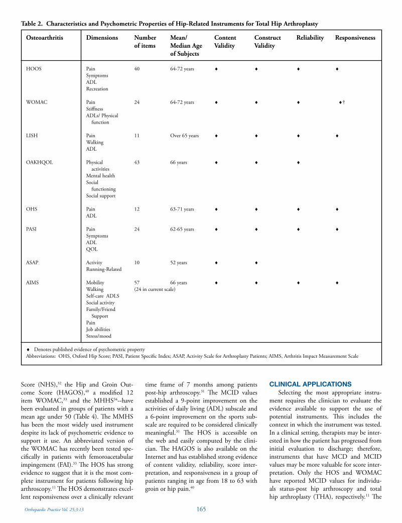
Score (NHS),32 the Hip and Groin Out-come Score (HAGOS),40 a modified 12 item WOMAC,33 and the MHHS34--have been evaluated in groups of patients with a mean age under 50 (Table 4). The MMHS has been the most widely used instrument despite its lack of psychometric evidence to support it use. An abbreviated version of the WOMAC has recently been tested spe-cifically in patients with femoroacetabular impingement (FAI).33 The HOS has strong evidence to suggest that it is the most com-plete instrument for patients following hip arthroscopy.11 The HOS demonstrates excel-lent responsiveness over a clinically relevant
time frame of 7 months among patients post-hip arthroscopy.31 The MCID values established a 9-point improvement on the activities of daily living (ADL) subscale and a 6-point improvement on the sports sub-scale are required to be considered clinically meaningful.31 The HOS is accessible on the web and easily computed by the clini-cian. The HAGOS is also available on the Internet and has established strong evidence of content validity, reliability, score inter-pretation, and responsiveness in a group of patients ranging in age from 18 to 63 with groin or hip pain.40
CLINICAL APPLICATIONSSelecting the most appropriate instru-
ment requires the clinician to evaluate the evidence available to support the use of potential instruments. This includes the context in which the instrument was tested. In a clinical setting, therapists may be inter-ested in how the patient has progressed from initial evaluation to discharge; therefore, instruments that have MCD and MCID values may be more valuable for score inter-pretation. Only the HOS and WOMAC have reported MCID values for individu-als status-post hip arthroscopy and total hip arthroplasty (THA), respectively.11 The
Table 2. Characteristics and Psychometric Properties of Hip-Related Instruments for Total Hip Arthroplasty
Osteoarthritis Dimensions Number Mean/ Content Construct Reliability Responsiveness of items Median Age Validity Validity of Subjects
HOOS Pain 40 64-72 years ♦ ♦ ♦ ♦ Symptoms ADL Recreation WOMAC Pain 24 64-72 years ♦ ♦ ♦ ♦† Stiffness ADLs/ Physical function LISH Pain 11 Over 65 years ♦ ♦ ♦ ♦ Walking ADL OAKHQOL Physical 43 66 years ♦ ♦ ♦ activities Mental health Social functioning Social support OHS Pain 12 63-71 years ♦ ♦ ♦ ♦ ADL
PASI Pain 24 62-65 years ♦ ♦ ♦ ♦ Symptoms ADL QOL
ASAP Activity 10 52 years ♦ ♦ Running-Related
AIMS Mobility 57 66 years ♦ ♦ ♦ ♦ Walking (24 in current scale) Self-care ADLS Social activity Family/Friend Support Pain Job abilities Stress/mood
♦ Denotes published evidence of psychometric propertyAbbreviations: OHS, Oxford Hip Score; PASI, Patient Specific Index; ASAP, Activity Scale for Arthroplasty Patients; AIMS, Arthritis Impact Measurement Scale
165Orthopaedic Practice Vol. 25;3:13
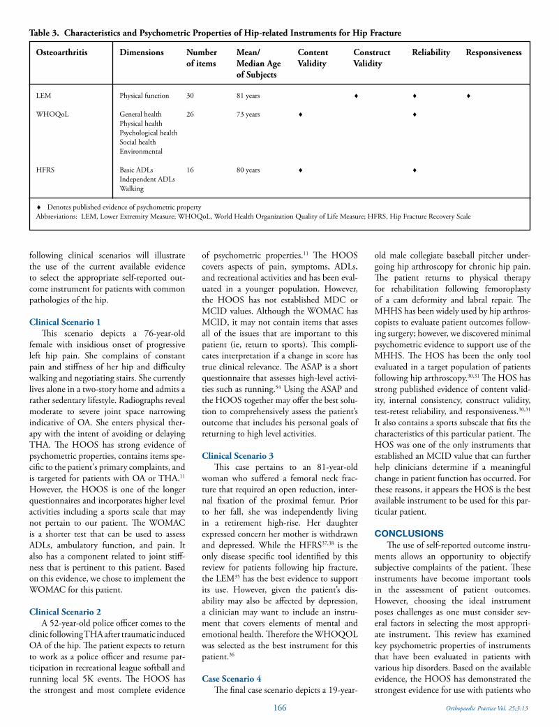
following clinical scenarios will illustrate the use of the current available evidence to select the appropriate self-reported out-come instrument for patients with common pathologies of the hip.
Clinical Scenario 1This scenario depicts a 76-year-old
female with insidious onset of progressive left hip pain. She complains of constant pain and stiffness of her hip and difficulty walking and negotiating stairs. She currently lives alone in a two-story home and admits a rather sedentary lifestyle. Radiographs reveal moderate to severe joint space narrowing indicative of OA. She enters physical ther-apy with the intent of avoiding or delaying THA. The HOOS has strong evidence of psychometric properties, contains items spe-cific to the patient's primary complaints, and is targeted for patients with OA or THA.11
However, the HOOS is one of the longer questionnaires and incorporates higher level activities including a sports scale that may not pertain to our patient. The WOMAC is a shorter test that can be used to assess ADLs, ambulatory function, and pain. It also has a component related to joint stiff-ness that is pertinent to this patient. Based on this evidence, we chose to implement the WOMAC for this patient.
Clinical Scenario 2A 52-year-old police officer comes to the
clinic following THA after traumatic induced OA of the hip. The patient expects to return to work as a police officer and resume par-ticipation in recreational league softball and running local 5K events. The HOOS has the strongest and most complete evidence
of psychometric properties.11 The HOOS covers aspects of pain, symptoms, ADLs, and recreational activities and has been eval-uated in a younger population. However, the HOOS has not established MDC or MCID values. Although the WOMAC has MCID, it may not contain items that asses all of the issues that are important to this patient (ie, return to sports). This compli-cates interpretation if a change in score has true clinical relevance. The ASAP is a short questionnaire that assesses high-level activi-ties such as running.54 Using the ASAP and the HOOS together may offer the best solu-tion to comprehensively assess the patient’s outcome that includes his personal goals of returning to high level activities.
Clinical Scenario 3This case pertains to an 81-year-old
woman who suffered a femoral neck frac-ture that required an open reduction, inter-nal fixation of the proximal femur. Prior to her fall, she was independently living in a retirement high-rise. Her daughter expressed concern her mother is withdrawn and depressed. While the HFRS37,38 is the only disease specific tool identified by this review for patients following hip fracture, the LEM35 has the best evidence to support its use. However, given the patient’s dis-ability may also be affected by depression, a clinician may want to include an instru-ment that covers elements of mental and emotional health. Therefore the WHOQOL was selected as the best instrument for this patient.36
Case Scenario 4The final case scenario depicts a 19-year-
old male collegiate baseball pitcher under-going hip arthroscopy for chronic hip pain. The patient returns to physical therapy for rehabilitation following femoroplasty of a cam deformity and labral repair. The MHHS has been widely used by hip arthros-copists to evaluate patient outcomes follow-ing surgery; however, we discovered minimal psychometric evidence to support use of the MHHS. The HOS has been the only tool evaluated in a target population of patients following hip arthroscopy.30,31 The HOS has strong published evidence of content valid-ity, internal consistency, construct validity, test-retest reliability, and responsiveness.30,31 It also contains a sports subscale that fits the characteristics of this particular patient. The HOS was one of the only instruments that established an MCID value that can further help clinicians determine if a meaningful change in patient function has occurred. For these reasons, it appears the HOS is the best available instrument to be used for this par-ticular patient.
CONCLUSIONSThe use of self-reported outcome instru-
ments allows an opportunity to objectify subjective complaints of the patient. These instruments have become important tools in the assessment of patient outcomes. However, choosing the ideal instrument poses challenges as one must consider sev-eral factors in selecting the most appropri-ate instrument. This review has examined key psychometric properties of instruments that have been evaluated in patients with various hip disorders. Based on the available evidence, the HOOS has demonstrated the strongest evidence for use with patients who
Table 3. Characteristics and Psychometric Properties of Hip-related Instruments for Hip Fracture
Osteoarthritis Dimensions Number Mean/ Content Construct Reliability Responsiveness of items Median Age Validity Validity of Subjects
LEM Physical function 30 81 years ♦ ♦ ♦
WHOQoL General health 26 73 years ♦ ♦ Physical health Psychological health Social health Environmental
HFRS Basic ADLs 16 80 years ♦ ♦ Independent ADLs Walking
♦ Denotes published evidence of psychometric propertyAbbreviations: LEM, Lower Extremity Measure; WHOQoL, World Health Organization Quality of Life Measure; HFRS, Hip Fracture Recovery Scale
166 Orthopaedic Practice Vol. 25;3:13
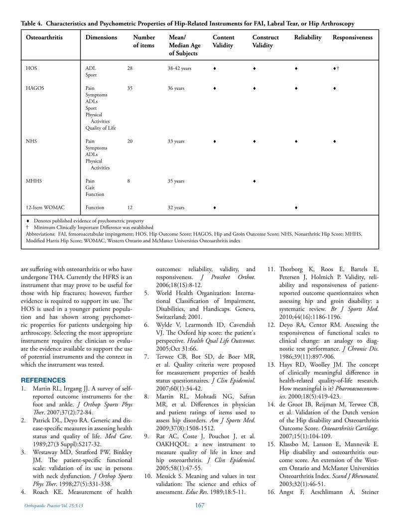
are suffering with osteoarthritis or who have undergone THA. Currently the HFRS is an instrument that may prove to be useful for those with hip fractures; however, further evidence is required to support its use. The HOS is used in a younger patient popula-tion and has shown strong psychomet-ric properties for patients undergoing hip arthroscopy. Selecting the most appropriate instrument requires the clinician to evalu-ate the evidence available to support the use of potential instruments and the context in which the instrument was tested.
REFERENCES1. Martin RL, Irrgang JJ. A survey of self-
reported outcome instruments for the foot and ankle. J Orthop Sports Phys Ther. 2007;37(2):72-84.
2. Patrick DL, Deyo RA. Generic and dis-ease-specific measures in assessing health status and quality of life. Med Care. 1989;27(3 Suppl):S217-32.
3. Westaway MD, Stratford PW, Binkley JM. The patient-specific functional scale: validation of its use in persons with neck dysfunction. J Orthop Sports Phys Ther. 1998;27(5):331-338.
4. Roach KE. Measurement of health
Table 4. Characteristics and Psychometric Properties of Hip-Related Instruments for FAI, Labral Tear, or Hip Arthroscopy
Osteoarthritis Dimensions Number Mean/ Content Construct Reliability Responsiveness of items Median Age Validity Validity of Subjects
HOS ADL 28 38-42 years ♦ ♦ ♦ ♦† Sport
HAGOS Pain 35 36 years ♦ ♦ ♦ ♦ Symptoms ADLs Sport Physical Activities Quality of Life
NHS Pain 20 33 years ♦ ♦ ♦ ♦ Symptoms ADLs Physical Activities
MHHS Pain 8 35 years ♦ Gait Function
12-Item WOMAC Function 12 32 years ♦ ♦
♦ Denotes published evidence of psychometric property† Minimum Clinically Important Difference was establishedAbbreviations: FAI, femoroacetabular impingement; HOS, Hip Outcome Score; HAGOS, Hip and Groin Outcome Score; NHS, Nonarthritic Hip Score; MHHS, Modified Harris Hip Score; WOMAC, Western Ontario and McMaster Universities Osteoarthritis index
outcomes: reliability, validity, and responsiveness. J Prosthet Orthot. 2006;18(1S):8-12.
5. World Health Organization: Interna-tional Classification of Impairment, Disabilities, and Handicaps. Geneva, Switzerland; 2001.
6. Wylde V, Learmonth ID, Cavendish VJ. The Oxford hip score: the patient's perspective. Health Qual Life Outcomes. 2005;Oct 31:66.
7. Terwee CB, Bot SD, de Boer MR, et al. Quality criteria were proposed for measurement properties of health status questionnaires. J Clin Epidemiol. 2007;60(1):34-42.
8. Martin RL, Mohtadi NG, Safran MR, et al. Differences in physician and patient ratings of items used to assess hip disorders. Am J Sports Med. 2009;37(8):1508-1512.
9. Rat AC, Coste J, Pouchot J, et al. OAKHQOL: a new instrument to measure quality of life in knee and hip osteoarthritis. J Clin Epidemiol. 2005;58(1):47-55.
10. Messick S. Meaning and values in test validation: The science and ethics of assessment. Educ Res. 1989;18:5-11.
11. Thorborg K, Roos E, Bartels E, Petersen J, Holmich P. Validity, reli-ability and responsiveness of patient-reported outcome questionnaires when assessing hip and groin disability: a systematic review. Br J Sports Med. 2010;44(16):1186-1196.
12. Deyo RA, Centor RM. Assessing the responsiveness of functional scales to clinical change: an analogy to diag-nostic test performance. J Chronic Dis. 1986;39(11):897-906.
13. Hays RD, Woolley JM. The concept of clinically meaningful difference in health-related quality-of-life research. How meaningful is it? Pharmacoeconom-ics. 2000;18(5):419-423.
14. de Groot IB, Reijman M, Terwee CB, et al. Validation of the Dutch version of the Hip disability and Osteoarthritis Outcome Score. Osteoarthritis Cartilage. 2007;15(1):104-109.
15. Klassbo M, Larsson E, Mannevik E. Hip disability and osteoarthritis out-come score. An extension of the West-ern Ontario and McMaster Universities Osteoarthritis Index. Scand J Rheumatol. 2003;32(1):46-51.
16. Angst F, Aeschlimann A, Steiner
167Orthopaedic Practice Vol. 25;3:13
W, Stucki G. Responsiveness of the WOMAC osteoarthritis index as compared with the SF-36 in patients with osteoarthritis of the legs under-going a comprehensive rehabilita-tion intervention. Ann Rheum Dis. 2001;60(9):834-840.
17. Angst F, Aeschlimann A, Stucki G. Smallest detectable and minimal clini-cally important differences of rehabilita-tion intervention with their implications for required sample sizes using WOMAC and SF-36 quality of life measurement instruments in patients with osteoar-thritis of the lower extremities. Arthritis Rheum. 2001;45(4):384-391.
18. Stucki G, Meier D, Stucki S, et al. Evalu-ation of a German questionnaire version of the Lequesne cox- and gonarthrosis indices. Z Rheumatol. 1996;55(1):50-57.
19. Stucki G, Sangha O, Stucki S, et al. Comparison of the WOMAC (West-ern Ontario and McMaster Uni-versities) osteoarthritis index and a self-report format of the self-adminis-tered Lequesne-Algofunctional index in patients with knee and hip osteo-arthritis. Osteoarthritis Cartilage. 1998;6(2):79-86.
20. Lequesne MG, Mery C, Samson M, Gerard P. Indexes of severity for osteo-arthritis of the hip and knee. Valida-tion--value in comparison with other assessment tests. Scand J Rheumatol Suppl. 1987;65:85-89.
21. Johanson NA, Liang MH, Daltroy L, Rudicel S, Richmond J. American Academy of Orthopaedic Surgeons lower limb outcomes assessment instru-ments. Reliability, validity, and sensi-tivity to change. J Bone Joint Surg Am. 2004;86-A(5):902-909.
22. Dawson J, Fitzpatrick R, Frost S, Gundle R, McLardy-Smith P, Murray D. Evi-dence for the validity of a patient-based instrument for assessment of outcome after revision hip replacement. J Bone Joint Surg Br. 2001;83(8):1125-1129.
23. Dawson J, Fitzpatrick R, Murray D, Carr A. Comparison of measures to assess out-comes in total hip replacement surgery. Qual Health Care. 1996;5(2):81-88.
24. Dawson J, Fitzpatrick R, Murray D, Carr A. The problem of 'noise' in moni-toring patient-based outcomes: generic, disease-specific and site-specific instru-ments for total hip replacement. J Health Serv Res Policy. 1996;1(4):224-231.
25. Dawson J, Jameson-Shortall E, Emer-ton M, et al. Issues relating to long-term
follow-up in hip arthroplasty surgery: a review of 598 cases at 7 years comparing 2 prostheses using revision rates, survival analysis, and patient-based measures. J Arthroplasty. 2000;15(6):710-717.
26. Fitzpatrick R, Morris R, Hajat S, et al. The value of short and simple measures to assess outcomes for patients of total hip replacement surgery. Qual Health Care. 2000;9(3):146-150.
27. Fitzpatrick R, Norquist JM, Dawson J, Jenkinson C. Rasch scoring of outcomes of total hip replacement. J Clin Epide-miol. 2003;56(1):68-74.
28. Ostendorf M, van Stel HF, Buskens E, et al. Patient-reported outcome in total hip replacement. A comparison of five instruments of health status. J Bone Joint Surg Br. 2004;86(6):801-808.
29. Jette AM, McDonough CM, Ni P, et al. A functional difficulty and func-tional pain instrument for hip and knee osteoarthritis. Arthritis Res Ther. 2009;11(4):R107.
30. Martin RL, Philippon MJ. Evi-dence of validity for the hip outcome score in hip arthroscopy. Arthroscopy. 2007;23(8):822-826.
31. Martin RL, Philippon MJ. Evidence of reliability and responsiveness for the hip outcome score. Arthroscopy. 2008;24(6):676-682.
32. Christensen CP, Althausen PL, Mit-tleman MA, Lee JA, McCarthy JC. The nonarthritic hip score: reliable and validated. Clin Orthop Relat Res. 2003;(406):75-83.
33. Rothenfluh DA, Reedwisch D, Muller U, Ganz R, Tennant A, Leunig M. Con-struct validity of a 12-item WOMAC for assessment of femoro-acetabu-lar impingement and osteoarthritis of the hip. Osteoarthritis Cartilage. 2008;16(9):1032-1038.
34. Potter BK, Freedman BA, Andersen RC, Bojescul JA, Kuklo TR, Murphy KP. Cor-relation of Short Form-36 and disability status with outcomes of arthroscopic acetabular labral debridement. Am J Sports Med. 2005;33(6):864-870.
35. Jaglal S, Lakhani Z, Schatzker J. Reli-ability, validity, and responsiveness of the lower extremity measure for patients with a hip fracture. J Bone Joint Surg Am. 2000;82-A(7):955-962.
36. Yao KP, Lee HY, Tsauo JY. Are hip-spe-cific items useful in a quality of life ques-tionnaire for patients with hip fractures? Int J Rehabil Res. 2009;32(3):245-250.
37. Zuckerman JD, Koval KJ, Aharonoff
GB, Hiebert R, Skovron ML. A func-tional recovery score for elderly hip frac-ture patients: I. Development. J Orthop Trauma. 2000;14(1):20-25.
38. Zuckerman JD, Koval KJ, Aharonoff GB, Skovron ML. A functional recovery score for elderly hip fracture patients: II. Validity and reliability. J Orthop Trauma. 2000;14(1):26-30.
39. Binkley JM, Stratford PW, Lott SA, Riddle DL. The Lower Extremity Func-tional Scale (LEFS): scale development, measurement properties, and clinical application. North American Orthopae-dic Rehabilitation Research Network. Phys Ther. 1999;79(4):371-383.
40. Thorborg K, Holmich P, Christensen R, Petersen J, Roos EM. The Copen-hagen Hip and Groin Outcome Score (HAGOS): development and validation according to the COSMIN checklist. Br J Sports Med. 2011;45(6):478-491.
41. Bardakos NV, Villar RN. The ligamen-tum teres of the adult hip. J Bone Joint Surg Br. 2009;91(1):8-15.
42. Byrd JW, Jones KS. Hip arthroscopy for labral pathology: prospective analy-sis with 10-year follow-up. Arthroscopy. 2009;25(4):365-368.
43. Philippon MJ, Briggs KK, Yen YM, Kuppersmith DA. Outcomes following hip arthroscopy for femoroacetabular impingement with associated chon-drolabral dysfunction: minimum two-year follow-up. J Bone Joint Surg Br. 2009;91(1):16-23.
44. Cook KM, Heiderscheit B. Conserva-tive management of a young adult with hip arthrosis. J Orthop Sports Phys Ther. 2009;39(12):858-866.
45. Freedman BA, Potter BK, Dinauer PA, Giuliani JR, Kuklo TR, Murphy KP. Prognostic value of magnetic resonance arthrography for Czerny stage II and III acetabular labral tears. Arthroscopy. 2006;22(7):742-747.
46. Larson CM, Giveans MR. Arthroscopic management of femoroacetabular impingement: early outcomes measures. Arthroscopy. 2008;24(5):540-546.
47. Santori N, Villar RN. Acetabular labral tears: result of arthroscopic partial lim-bectomy. Arthroscopy. 2000;16(1):11-15.
48. Sekiya JK, Martin RL, Lesniak BP. Arthroscopic repair of delaminated acetabular articular cartilage in femo-roacetabular impingement. Orthopedics. 2009;32(9).
49. Voos JE, Shindle MK, Pruett A, Asnis PD, Kelly BT. Endoscopic repair of glu-
168 Orthopaedic Practice Vol. 25;3:13
teus medius tendon tears of the hip. Am J Sports Med. 2009;37(4):743-747.
50. Orthopaedic Scores. Available at: ortho-paedicscores. Accessed March 1, 2012.
51. Wright JG, Young NL. A comparison of different indices of responsiveness. J Clin Epidemiol. 1997;50(3):239-246.
52. Wright JG, Young NL. The patient-specific index: asking patients what they want. J Bone Joint Surg Am. 1997;79(7):974-983.
53. Wright JG, Young NL, Waddell JP. The reliability and validity of the self-reported patient-specific index for total hip arthroplasty. J Bone Joint Surg Am. 2000;82(6):829-837.
54. Domzalski T, Cook C, Attarian DE, Kelley SS, Bolognesi MP, Vail TP. Activ-ity scale for arthroplasty patients after total hip arthroplasty. J Arthroplasty. 2010;25(1):152-157.
55. Meenan RF, Mason JH, Anderson JJ, Guccione AA, Kazis LE. AIMS2. The content and properties of a revised and expanded Arthritis Impact Measure-ment Scales Health Status Question-
BUILD YOUR FUTUREAND MAKE A DIFFERENCE
With a Doctor of Physical Therapy
As part of the John G. Sr. Rangos School of Health Sciences, we have a national reputation for excellence in clinical instruction, research and service. Advantages of earning a Doctor of Physical Therapy (DPT) degree from Duquesne University include:
• Multiple admission points with the primary pathway being freshman admittance (1 of 35 in the country)
• Six year curriculum in which students earn two baccalaureate degrees (biology and health sciences) after four years and graduate with a Doctor of Physical Therapy (DPT) at the end of the 6th year
• Opportunity for a dual degree in Physical Therapy & Athletic Training • Professional phase of DPT program includes two semesters of Human
Anatomy with cadaver dissection • 12 full-time faculty • 5 separate, full-time clinical internships (40 weeks total) • 98 percent first-time pass rate on licensing exam the past five years • 100 percent ultimate pass rate on licensing exam for all graduates • 100 percent job placement rate for graduates the past five years
For more information visit www.duq.edu/physicaltherapy or call 412.396.5541.
naire. Arthritis Rheum. 1992;35(1):1-10.56. Selman SW. Impact of total hip replace-
ment on quality of life. Orthop Nurs. 1989;8(5):43-49.
57. Quintana JM, Escobar A, Bilbao A, Arostegui I, Lafuente I, Vidaurreta I. Responsiveness and clinically important differences for the WOMAC and SF-36 after hip joint replacement. Osteoarthri-tis Cartilage. 2005;13(12):1076-1083.
58. Garbuz DS, Xu M, Sayre EC. Patient's outcome after total hip arthroplasty: a comparison between the Western Ontario and McMaster Universities index and the Oxford 12-item hip score. J Arthroplasty. 2006;21:998-1004.
Upcoming Meetings
2013National Student
Conclave
October 25-27, 2013
Louisville, KY
169Orthopaedic Practice Vol. 25;3:13
ABSTRACTBackground and Purpose: The purpose
of this paper is to provide a review of func-tional performance tests that may be useful for young, active individuals with nonar-thritic hip pain and describe how they may be applied in clinical practice. Methods: A literature search was performed to identify evidence of reliability and validity in func-tional performance tests for the hip joint. Findings: The squat test for depth, single limb balance test, and star excursion bal-ance test have evidence of validity to sup-port their use. The Functional Movement Screen (FMS) and hop tests and agility tests have evidence for reliability and normative data to assist in score interpretation. Clini-cal Relevance: Three cases are provided to demonstrate how tests can be applied in clinical practice. Conclusions: While there is some evidence for these tests, further study is needed to establish the reliability and validity of functional performance tests in a young, athletic population with hip dysfunction.
Key Words: functional testing, reliability, validity
INTRODUCTIONThe International Classification of Func-
tioning, Disability, and Health (ICF) model1 has been used by physical therapists in the management of patients with varied medical conditions. Functional performance tests are important in the ICF model as these tests can uniquely assess the interaction of body structure and function with activity and par-ticipation. An increased awareness of non-arthritic causes of hip pathology affecting younger, active individuals has resulted in the need to develop functional performance tests specific to this population. Proper interpretation of functional performance measures requires that these tests demon-strate reliability and validity. The purpose of this paper is to describe how to use previ-
ously reported functional tests for a popula-tion with nonarthritic hip pain.
Functional performance tests have an advantage over more traditional clini-cal measures in that components of range of motion (ROM), flexibility, muscular strength, endurance, coordination, balance, and motor control of multiple regions can be simultaneously assessed in a single test.2-4 Functional performance tests have emerged as common components of evaluation and re-evaluation procedures for the ankle5-13
and knee,14-19 but have yet to be established for patients with hip pathology.
The clinical utility of functional perfor-mance tests is largely dependent on its reli-ability and validity. Reliability refers to how well the test can be reproduced under the same conditions. Validity refers to how well a test measures what it is intended to mea-sure. The population in which the evidence for reliability and validity of a functional performance test is established is also an important consideration. Evidence of reli-ability and validity should be established among a sample of subjects that are similar to the population of patients for which the test is to be used.20 Normative data may be of particular value to the clinicians as well. Normative data establishes a baseline by which the clinician can compare the results of their patients to the average of a larger population with similar characteristics.
There are a number of potential tests that have been described in the literature that may be appropriate for young individuals with nonarthritic hip pain. These include 4 types of tests: (1) movement tests, (2) bal-ance tests, (3) hop/jump tests, and (4) agil-ity tests. Movement tests such as the deep squat test and single leg squat test have been found to correlate with femoroacetabular impingement21 and hip abductor strength,22 respectively. The Star Excursion Balance Test (SEBT) also relates to hip abductor muscle function23 and the single leg balance test has diagnostic value in detecting Greater Tro-
chanteric Pain Syndrome.24 Hop and agil-ity tests have established normative data on healthy subjects to compare with patients presenting with hip pathology.25-32
Past literature review shows that only the single-leg balance test and the deep squat test have validity in patients with confirmed hip pathology. The single-leg balance test demonstrated high sensitivity (100%) and specificity (97.3%) in detecting tendinopa-thy of the gluteus medius and minimus on subjects with greater than 4 months of lateral hip pain.24 Based on this evidence, provoca-tion of lateral hip pain during a single-leg balance test has clinical value in differentiat-ing gluteal tendinopathy from lumbosacral, sacroiliac, or intra-articular pathology of the hip joint.24 Measuring the amount of drop of the contralateral pelvis from a single leg posi-tion is another way the single leg balance test may be used to assess hip abductor muscle function. Normal pelvic on femur angle in single limb stance is 84° and 82° for men and women, respectively.33 The deep squat test was performed on subjects with radiographi-cally confirmed femoroacetabular impinge-ment. The maximal squat depth in subjects with femoroacetabular impingement (41% of leg length) was significantly less when compared to healthy controls (32% of leg length).21 Clinicians may test maximum squat depth in patients with suspected femo-roacetabular impingement to help confirm the diagnosis. Further study is needed to determine if these tests can accurately predict the presence of specific hip pathology.
While the single leg balance and deep squat tests provided evidence of validity in subjects with hip pathology, the SEBT and single leg squat test provide evidence of validity through an analysis of kinematic and muscle function in normal subjects. The SEBT has been used to predict lower extrem-ity injury34 and has established a relationship to kinematic and muscle function variables of the hip joint.23,35 Hip flexion ROM was shown to explain a high percentage of vari-
A Critical Review of Performance Tests for Hip-Related Dysfunction
Benjamin R. Kivlan, PT, SCS, OCS,1,2 RobRoy L. Martin1,3
1John G. Rangos Sr. School of Health Sciences, Duquesne University, Pittsburgh, PA2Tri-State Physical Therapy, Seven Fields, PA3UPMC Center for Sports Medicine, Pittsburgh, PA
170 Orthopaedic Practice Vol. 25;3:13
ance (92%-95%) in SEBT performance.35
Hip abduction and extension strength has a moderate correlation (r =0.48 - 0.51) to posterior-medial and posterior-lateral reach distances of the SEBT.23 The medial reach of the SEBT was shown to elicit activation of the gluteus medius at 49% of maximal volitional isometric contraction.36 The single leg squat test also demonstrated a relation-ship to hip abductor muscle function.22 However, the strength of this relationship has been disputed. Dimattia et al37 reported poor association (r = 0.21) of the single leg squat to hip abductor strength. The SEBT and the single leg squat test have not been studied on patients with hip pathology but may have some value to help clini-cians screen for ROM and muscle strength impairments. Range of motion and strength deficits are common findings in subjects diagnosed with femoroacetabular impinge-ment, osteoarthritis, or greater trochanteric pain syndrome. Asymmetry or pain on the SEBT or single leg squat test may lead the clinician to further investigate ROM and strength deficits noted from the functional performance tests.
While a literature review found tests with evidence of validity for young, active individuals with nonarthritic hip pain, there were not any functional performance tests that established reliability in this popula-tion. However, the Functional Movement Screen (FMS), hop tests, and agility tests have evidence of reliability in a young, healthy population. The FMS may poten-tially be one of the more useful tests and was found to have good to excellent interrater reliability.38 The FMS may be relevant in assessing patients with varied hip pathology as it tests multiple movement patterns that require different components of hip ROM, strength, and trunk control.39 Such tests may elicit familiar symptoms or indicate impair-ments related to femoroacetabular impinge-ment, labral tears, osteoarthritis, or greater trochanteric pain syndrome. Clinicians may use normative data established for the FMS as a guide to identify abnormal findings on FMS for patients with hip-related pathol-ogy.38 Further study is needed to determine if the FMS is able to accurately predict hip-specific injuries.
Hop tests have become commonly used in the assessment of knee and ankle instabil-ity and shown ability to discriminate injured from uninjured lower extremities.25-28 Hop tests also have established normative, gen-der-specific values17,29 on young, healthy, athletic subjects. These values may serve as
a benchmark that may be helpful in inter-preting an ‘abnormal’ score for a subject with hip-related pathology. Field agility tests have demonstrated evidence of good reli-ability,17,29-31 but have not been able to dis-criminate injured versus uninjured limbs in the same manner as hop tests. This is likely because agility tests require bipedal move-ment. However, agility tests may have value in an athletic population as the tests may more closely mimic the dynamic require-ments of sport activity. Since reliability of hop/jump and agility tests measures have not been established on patients with hip pathology, it remains unclear how patients with hip pathology perform on these tests without further study. For patients with uni-lateral hip symptoms, hop tests can be used with the noninvolved side used as a com-parison. Interpretation of agility test results is limited to a comparison of scores estab-lished on healthy subjects. Whether jump/hop tests or agility tests can be used to dis-criminate subjects with hip-related pathol-ogy requires further investigation.
CLINICAL APPLICATIONSClinicians may apply information of
validity, reliability, and score interpretation in selecting appropriate functional perfor-mance tests for the evaluation of hip-related pathology. The following 3 cases depict sce-narios that illustrate the clinical decision-making used in selecting and interpreting functional performance tests for patients with hip dysfunction.
Case 1A 22-year-old male, football running
back is referred to Physical Therapy for recurrent right sided groin pain provoked while playing and during basic activities of daily living, such as stair climbing and sit-ting. The patient has 105° of flexion, 50° lateral rotation, and 15° medial rotation, each limited by pain. He demonstrates weakness of hip abduction (28% deficit), adduction (15% deficit), extension (21% deficit), lateral rotation (28% deficit), and medial rotation (18%) as compared to the noninvolved side. His pain is provoked with combined flexion-adduction-internal rota-tion (FADDIR test) and dynamic impinge-ment tests. The physical therapist performs the deep squat test suspecting the patient may have femoroacetabular impingement. Squat depth is defined as the distance from the center of the hip joint (marked by the center of the greater trochanter) at its lowest point to the floor.21 Squat depth can then
be standardized to the patient’s limb length determined by the distance from the medial malleolus to the anterior superior iliac spine.21 The deep squat test reveals a maxi-mum squat depth of 48% of leg length. Patients diagnosed with femoroacetabular impingement have limited squat depth (m = 41% of leg length) compared to healthy controls (m = 32% of leg length) and dis-play altered lumbopelvic mechanics.21 The discovery of decreased squat depth can be used in conjunction with other special tests to confirm a diagnosis of femoroacetabular impingement and create a baseline to deter-mine effectiveness of treatment.
To further assess functional performance, the cross-over hop test and the modified agility t-test were chosen. The cross-over hop test is a timed test in which the patient hops back and forth over a 15 cm wide line that is 10 m long.32 The patient posted a time of 3.5 seconds for the cross-over hop test. Healthy subjects average 2.7 seconds.32 The modified agility t-test compares running, cutting, and shuffling in one direction to the opposite direction. The average time to complete the modified agility t-test is 9.59 seconds per side. The patient completed the test in 10.37 seconds towards the right versus 11.58 seconds towards the left with greater pain going towards the left.
A 4 week treatment plan was initiated for the patient. The physical therapist directed treatment to improve painfree ROM into flexion and internal rotation with manual therapy to mobilize the inferior and pos-terior capsule of the hip joint. Therapeu-tic exercises were included to increase hip strength, specifically of the abductors and external rotators of the hip joint. Neuro-muscular retraining of the trunk and lower extremity through perturbation and balance exercises were also incorporated into the treatment program.
After 4 weeks of treatment, the patient regained full symmetrical and painfree ROM. His strength improved to less than a 10% deficit compared to the noninvolved side in all movements. The patient’s squat depth improved to 28% of leg length with-out pain. The cross-over hop test improved from 3.5 to 2.6 seconds and the modified agility t-test improved to 9.01 and 9.33 sec-onds for the left and right side, respectively. These functional performance test results were comparable to normal values found for healthy, physically active, college-aged sub-jects and the patient was cleared to return to athletics.30,32
171Orthopaedic Practice Vol. 25;3:13
Case 2A 40-year-old, female, marathon runner
comes into the clinic as a direct access refer-ral for complaints of progressive lateral hip pain that has interrupted her training sched-ule. She currently has disrupted sleep and pain with stair climbing. She presents with palpable tenderness to the greater trochan-ter and the posterior aspect of the ilium. Her hip ROM was normal and painfree; however, she exhibited a 45% deficit in hip abduction, 28% deficit in extension, 26% deficit for medial rotation, and 33% deficit of strength for lateral rotation compared to the noninvolved side, all of which re-created familiar lateral hip pain. Based on these findings, the therapist performs the single leg balance test. Within 10 seconds, familiar pain is elicited in the lateral region of the hip joint of the patient. Provocation of symp-toms in single limb stance has sensitivity (100%) and specificity (97.3%) in detect-ing tendinopathy of the gluteus medius and minimus.24 The therapist also chooses to have the subject perform the SEBT revealing a 20% deficit in cumulative reach distance. Hip abduction strength has a moderate cor-relation (r = 0.48 - 0.51) to posterior-medial and posterior-lateral reach distances of the SEBT23 and the medial reach has been shown to elicit significant activation of the gluteus medius.36 Based on the findings, the patient is referred to an orthopaedic sur-geon with specialty in hip arthroscopy to rule out a suspected gluteus medius tear. A magnetic resonance arthrogram reveals a tear of the gluteus medius tendon, and the patient chooses to undergo a gluteus medius tendon repair. Using the functional perfor-mance tests on this patient helps in making a diagnosis of gluteus medius tendon tear and appropriately guides the patient to a special-ist to manage her condition.
Case 3An 18-year-old, senior, female basket-
ball player comes into the facility for a pre-season evaluation. Her junior year she had complaints of periodic hip pain and “loose-ness.” She has experienced minimal pain since the previous season, but is concerned about her risk of reinjury as she enters her senior season. The physical therapist chooses to perform the FMS. The FMS has evidence of good reliability in an athletic population38
and has demonstrated an ability to predict noncontact injury.39 The 7 individual tests of the FMS encompass various components of hip ROM, strength, and trunk control that may elicit symptoms or indicate impair-
ments of hip pathology. Performance on the individual tests of the FMS including the hurdle step, active straight leg raise, deep squat, and trunk rotary stability tests were below normal values and revealed a com-posite score of 12 out of a possible 21. A score below 14 increases the risk for injury.39
Based on the results of the FMS, the physi-cal therapist performs additional evaluation procedures. Her ROM of her hip joint mea-sures 135° flexion, 75°lateral rotation, and 65°medial rotation. She also exhibits a posi-tive log roll test. Strength deficits of 31%, 34%, 19%, 29%, and 23% compared to the noninvolved side were noted for hip flexion, abduction, medial rotation, lateral rota-tion, and extension, respectively. Pain was not elicited during resisted tests. Dynamic impingement test into flexion-abduction did not create pain but did elicit apprehen-sion from the patient. The therapist also selected the single-leg balance test to assess the patient. The patient did not have pain, but was unable to exhibit postural control for greater than 10 seconds. She also dem-onstrated a femoral on pelvic angle of 71° as the contralateral hemi-pelvis dropped. These findings are indicative of poor hip abductor function.33 Further testing using the SEBT demonstrated a side-to-side difference of composite score of 11%, suggesting a rela-tionship to gluteal dysfunction23,36 and an elevated risk of injury.34
The patient is treated with an aggressive strength and proprioceptive program for the trunk, pelvis, and lower extremity. This included closed-chain strengthening and balance exercises, trunk stabilization train-ing, eccentric hip strengthening, and plyo-metric training. After a 6-week progressive training program, the patient is reassessed. The strength deficits have improved to less than 10% deficits in all motions. Her FMS score improved from a 12 to a 17 out of 21 and her SEBT from an 11% deficit to a 4% deficit compared to the noninvolved side. These results would suggest a functional improvement of performance and a lower risk for injury.34,39
CONCLUSIONThe literature review demonstrated
rather few functional performance tests that have been used in a population of subjects with hip pathology. Only the deep squat and single-leg balance tests have evidence of validity in a population of patients with hip-related pathology. The review found that diminished squat depth and provocation of pain during the single-leg balance test
may be an indication for femoroacetabu-lar impingement and gluteal tendinopathy, respectively. The SEBT and single-leg squat provided evidence of validity through an analysis of kinematic and muscle function in normal subjects. Functional performance tests, including the FMS, demonstrated evidence of reliability and have established normative data to be used to compare the performance of patients with hip dysfunc-tion to healthy controls. None of the func-tional performance tests, however, provided evidence of reliability in a group of subjects with hip-related dysfunction. To allow clini-cians to best use information gathered from functional performance tests, further study is needed to establish the reliability and validity in a young, athletic population with hip dysfunction.
REFERENCES1. World Health Organization Interna-
tional Classification of Functioning, Disability, and Health. Geneva, Switzer-land; 2001.
2. Okada T, Huxel KC, Nesser TW. Rela-tionship between core stability, func-tional movement, and performance. J Strength Cond Res. 2011;25(1):252-261.
3. Mills JD, Tauton J, Mills W. The effects of a 10 week training regimen on lumbo-pelvic stability and athletic per-formance in female athletes: a random-ized controlled trial. Phys Ther Sport. 2005;6:60-66.
4. Cook G, Burton L, Hoogenboom B. Pre-participation screening: The use of fundamental movements as an assess-ment of function - part 1. N Am J Sports Phys Ther. 2006;1(2):62-72.
5. Arnold BL, De La Motte S, Linens S, Ross SE. Ankle instability is associ-ated with balance impairments: A meta-analysis. Med Sci Sports Exerc. 2009;41(5):1048-1062.
6. Gribble PA, Hertel J, Denegar CR. Chronic ankle instability and fatigue create proximal joint alterations during performance of the star excur-sion balance test. Int J Sports Med. 2007;28(3):236-242.
7. Gribble PA, Hertel J, Denegar CR, Buckley WE. The effects of fatigue and chronic ankle instability on dynamic postural control. J Athl Train. 2004;39(4):321-329.
8. Hale SA, Hertel J, Olmsted-Kramer LC. The effect of a 4-week comprehen-sive rehabilitation program on postural control and lower extremity func-
172 Orthopaedic Practice Vol. 25;3:13
tion in individuals with chronic ankle instability. J Orthop Sports Phys Ther. 2007;37(6):303-311.
9. Hertel J, Braham RA, Hale SA, Olm-sted-Kramer LC. Simplifying the star excursion balance test: Analyses of sub-jects with and without chronic ankle instability. J Orthop Sports Phys Ther. 2006;36(3):131-137.
10. Martinez-Ramirez A, Lecumberri P, Gomez M, Izquierdo M. Wavelet analysis based on time-frequency infor-mation discriminate chronic ankle instability. Clin Biomech (Bristol, Avon). 2010;25(3):256-264.
11. McLeod TC, Armstrong T, Miller M, Sauers JL. Balance improvements in female high school basketball play-ers after a 6-week neuromuscular-training program. J Sport Rehabil. 2009;18(4):465-481.
12. Olmsted LC, Carcia CR, Hertel J, Shultz SJ. Efficacy of the star excursion balance tests in detecting reach deficits in subjects with chronic ankle instabil-ity. J Athl Train. 2002;37(4):501-506.
13. Docherty CL, Arnold BL, Gansneder BM, Hurwitz S, Gieck J. Functional-performance deficits in volunteers with functional ankle instability. J Athl Train. 2005;40(1):30-34.
14. Aminaka N, Gribble PA. Patellar taping, patellofemoral pain syndrome, lower extremity kinematics, and dynamic postural control. J Athl Train. 2008;43(1):21-28.
15. Herrington L, Hatcher J, Hatcher A, McNicholas M. A comparison of star excursion balance test reach dis-tances between ACL deficient patients and asymptomatic controls. Knee. 2009;16(2):149-152.
16. Gustavsson A, Neeter C, Thomee P, et al. A test battery for evaluating hop per-formance in patients with an ACL injury and patients who have undergone ACL reconstruction. Knee Surg Sports Trau-matol Arthrosc. 2006;14(8):778-788.
17. Itoh H, Kurosaka M, Yoshiya S, Ichihashi N, Mizuno K. Evaluation of functional deficits determined by four different hop tests in patients with anterior cruci-ate ligament deficiency. Knee Surg Sports Traumatol Arthrosc. 1998;6(4):241-245.
18. Ortiz A, Olson S, Libby CL, Kwon YH, Trudelle-Jackson E. Kinematic and kinetic reliability of two jumping and landing physical performance tasks in young adult women. N Am J Sports Phys Ther. 2007;2(2):104-112.
19. Hewett TE, Myer GD, Ford KR, Slaut-erbeck JR. Preparticipation physical examination using a box drop vertical jump test in young athletes: The effects of puberty and sex. Clin J Sport Med. 2006;16(4):298-304.
20. Martin RL, Irrgang JJ. A survey of self-reported outcome instruments for the foot and ankle. J Orthop Sports Phys Ther. 2007;37(2):72-84.
21. Lamontagne M, Kennedy MJ, Beaule PE. The effect of cam FAI on hip and pelvic motion during maxi-mum squat. Clin Orthop Relat Res. 2009;467(3):645-650.
22. Crossley KM, Zhang WJ, Schache AG, Bryant A, Cowan SM. Performance on the single-leg squat task indicates hip abductor muscle function. Am J Sports Med. 2011;39(4):866-873.
23. Hubbard TJ, Kramer LC, Denegar CR, Hertel J. Correlations among multiple measures of functional and mechanical instability in subjects with chronic ankle instability. J Athl Train. 2007;42(3):361-366.
24. Lequesne M, Mathieu P, Vuillemin-Bodaghi V, Bard H, Djian P. Gluteal tendinopathy in refractory greater trochanter pain syndrome: Diagnos-tic value of two clinical tests. Arthritis Rheum. 2008;59(2):241-246.
25. Noyes FR, Barber SD, Mangine RE. Abnormal lower limb symmetry deter-mined by function hop tests after ante-rior cruciate ligament rupture. Am J Sports Med. 1991;19(5):513-518.
26. Petschnig R, Baron R, Albrecht M. The relationship between isokinetic quad-riceps strength test and hop tests for distance and one-legged vertical jump test following anterior cruciate ligament reconstruction. J Orthop Sports Phys Ther. 1998;28(1):23-31.
27. Reid A, Birmingham TB, Stratford PW, Alcock GK, Giffin JR. Hop testing pro-vides a reliable and valid outcome mea-sure during rehabilitation after anterior cruciate ligament reconstruction. Phys Ther. 2007;87(3):337-349.
28. Barber SD, Noyes FR, Mangine RE, McCloskey JW, Hartman W. Quantita-tive assessment of functional limitations in normal and anterior cruciate liga-ment-deficient knees. Clin Orthop Relat Res. 1990;255:204-214.
29. Munro AG, Herrington LC. Between-session reliability of four hop tests and the agility T-test. J Strength Cond Res.
2011;25(5):1470-1477.30. Hickey KC, Quatman CE, Myer GD,
Ford KR, Brosky JA, Hewett TE. Methodological report: Dynamic field tests used in an NFL combine setting to identify lower-extremity functional asymmetries. J Strength Cond Res. 2009;23(9):2500-2506.
31. Veale JP, Pearce AJ, Carlson JS. Reli-ability and validity of a reactive agility test for Australian football. Int J Sports Physiol Perform. 2010;5(2):239-248.
32. Caffrey E, Docherty CL, Schrader J, Klossner J. The ability of 4 single-limb hopping tests to detect functional per-formance deficits in individuals with functional ankle instability. J Orthop Sports Phys Ther. 2009;39(11):799-806.
33. Youdas JW, Mraz ST, Norstad BJ, Schinke JJ, Hollman JH. Determining meaningful changes in pelvic-on-femoral position during the trendelenburg test. J Sport Rehabil. 2007;16(4):326-335.
34. Plisky PJ, Rauh MJ, Kaminski TW, Underwood FB. Star excursion bal-ance test as a predictor of lower extrem-ity injury in high school basketball players. J Orthop Sports Phys Ther. 2006;36(12):911-919.
35. Robinson R, Gribble P. Kinematic predictors of performance on the star excursion balance test. J Sport Rehabil. 2008;17(4):347-357.
36. Norris B, Trudelle-Jackson E. Hip- and thigh-muscle activation during the star excursion balance test. J Sport Rehabil. 2011;20(4):428-441.
37. Dimattia MA, Livengood AL, Uhl TL, Mattacola CG, Malone TR. What are the validity of the single-leg-squat test and its relationship to hip-abduction strength? J Sport Rehabil. 2005;14(2):108-123.
38. Schneiders AG, Davidsson A, Horman E, Sullivan SJ. Functional movement screen normative values in a young, active population. Int J Sports Phys Ther. 2011;6(2):75-82.
39. Kiesel K, Plisky PJ, Voight ML. Can serious injury in professional football be predicted by a preseason functional movement screen? N Am J Sports Phys Ther. 2007;2(3):147-158.
173Orthopaedic Practice Vol. 25;3:13
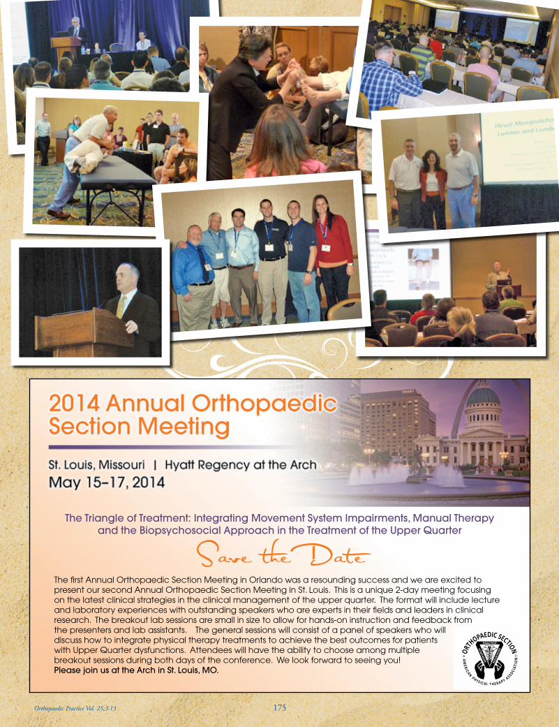
The Triangle of Treatment: Integrating Movement System Impairments, Manual Therapyand the Biopsychosocial Approach in the Treatment of the Upper Quarter
Save the DateThe first Annual Orthopaedic Section Meeting in Orlando was a resounding success and we are excited to present our second Annual Orthopaedic Section Meeting in St. Louis. This is a unique 2-day meeting focusingon the latest clinical strategies in the clinical management of the upper quarter. The format will include lectureand laboratory experiences with outstanding speakers who are experts in their fields and leaders in clinical research. The breakout lab sessions are small in size to allow for hands-on instruction and feedback from the presenters and lab assistants. The general sessions will consist of a panel of speakers who will discuss how to integrate physical therapy treatments to achieve the best outcomes for patients with Upper Quarter dysfunctions. Attendees will have the ability to choose among multiple breakout sessions during both days of the conference. We look forward to seeing you! Please join us at the Arch in St. Louis, MO.
175Orthopaedic Practice Vol. 25;3:13
�on�ratu�ation�to the 2013
Honors and AwardsRecipients
Catherine Worthingham Fellow of APTAMark W. Cornall, PT, PhD, CPed, FAPTABabette Sanders, PT, DPT, MS, FAPTAKathleen A. Sluka, PT, PhD, FAPTA
Lucy Blair Service AwardTara Jo Manal, PT, DPT, OCS, SCS David A. Pariser, PT, PhD* Rebecca G. Stephenson, PT, DPT, MS, WCS
Margaret L. Moore Award for Outstanding Contributions to Professional LiteratureJoel Bialosky, PT, PhD, FAAOMPT, OCS
Helen J. Hislop Award for Outstanding Contributions to Professional LiteratureCatherine C. Goodman, PT, MBA, CBP
Dorothy E. Baethke-Eleanor J. Carlin Award for Excellence in Academic TeachingAnne L. Harrison, PT, PhD
Jules R. Rothstein Golden Pen Award for Scientific WritingDennis L. Hart, PT, PhD*
Henry O. and Florence P. Kendall Practice AwardSteven A. Hoffman, PT, ATC, SCS
Jack Walker AwardJennifer E. Stevens-Lapsley, PT, MPT, PhD
Mary McMillan Scholarship Award, Physical Therapist Education ProgramJohn James Kuczynski, SPT
Minority Scholarship Award, Physical Therapist Education ProgramToki Tahara, SPT
Mary McMillan Scholarship Award, Physical Therapist Assistant Education ProgramDebora A. Lasure, SPTA
The Orthopaedic Section would like to congratulate those Section members who have been selected by the American Physical Therapy Association’s Board of Directors to receive the
following awards from the APTA’s 2013 Honors and Awards Program:
�
*Awarded posthumously
176 Orthopaedic Practice Vol. 25;3:13
OR
TH
OPA
ED
IC S
EC
TIO
N,
AP
TA
, IN
C.
OC
CU
PA
TIO
NA
L H
EA
LTH
SP
EC
IAL I
NT
ER
ES
T G
RO
UP
S
178 Orthopaedic Practice Vol. 25;3:13
PRESIDENT’S MESSAGELorena Pettet Payne, PT, OCS
Rick Wickstrom PT, DPT, CPE, CDMS submitted this summary of the latest activity to replace the Dictionary of Occupational Titles.
In July 2012, SSA signed an interagency agreement with the Bureau of Labor Statistics (BLS) to test occupational data col-lection methods that could lead to the development of a new Occupational Information System (OIS). The new OIS will replace the outdated Dictionary of Occupational Titles (DOT) in our disability determination process. In fiscal year 2013, BLS began testing the feasibility of using the National Compensa-tion Survey (NCS) platform as a means to gather the occupa-tional data needed by SSA for the OIS.
The Occupational Requirements Survey (ORS) is under development by the Bureau of Labor Statistics' (BLS) National Compensation Survey (NCS) program in association with the Social Security Administration (SSA). The ORS seeks to pro-vide job characteristics data to help the SSA in their disability determination process. Specifically, the ORS will gather job-related information regarding physical demands, environmental conditions, and vocational preparation requirements.
The NCS recently completed Phase 1 of the ORS tests con-ducted in cooperation with the SSA. The main objective of the 3 ORS tests in fiscal year 2013 is to assess whether it is feasible for BLS to collect data relevant to the SSA’s disability program using the NCS platform. The results of the Phase 1 proof-of-concept test suggest that this approach is viable. Respondents agreed to participate in the test; BLS field economists were able to capture the required data from traditional NCS respondents, and individual data element response rates were very high. The full report may be accessed at: http://www.ssa.gov/disabilityre-search/documents/Phase%20I%20Report%20Final.pdf.
I made a follow-up request and obtained information from the tech memo that describes the survey factors and scaling. No changes were made to the load ranges for the strength factor. The survey separated out the posture tolerances and established separate scales for occasional, frequent, and constant. There was no guidance on repetitions for each level of frequency. This is a quick survey approach, 10 to 12 minutes, rather than actual observation via a functional job analysis.
For more information on the latest activities related to SSA’s development of a new occupational system, Go to: http://www.ssa.gov/disabilityresearch/occupational_info_systems.html
OCCUPATIONAL HEALTHSPECIAL INTEREST GROUP
Common Industrial Ergonomics Assessment Tools for Physical TherapistsChristopher Studebaker, PT, DPT, OCS, Brian Murphy, MPT, OCSConcentra Medical Centers, Southeast Zone ADApt Lead
The term ergonomics or as it is often termed, human factors, is commonly defined as the process of fitting the workplace to the worker.1 A more precise description of human factors/ergo-nomics by Chapanis is that it “discovers and applies information about human behavior, abilities, limitations, and other charac-teristics to the design of tools, machines, systems, tasks, jobs, and the environments for productive, safe, comfortable, and effective human use.”2 From toothbrush handles to airline cock-pits, ergonomics principles are used to create user-friendly and safe interactions between a human and their environment. The field of ergonomics has historically been more the domain of the engineering and psychology professions than that of health care. However, with the growing emphasis on employer health care costs, more and more companies are beginning to use the unique skills and knowledge of physical therapists to provide ergonomic assessments in the work environment.3-5 Whether it be assembly line layouts, tool selection or station designs, physi-cal therapists may provide analyses of the work environment from the biomechanical and pathophysiological perspective that can assist engineers and safety personnel in the design or modi-fication of equipment to reduce the likelihood or work-related musculoskeletal disorders.
Physical therapists that work in the occupational health envi-ronment have extensive knowledge of anatomy, biomechanics, and common risk factors for work-related injuries. These skills, while a good foundation for a clinician in the industrial set-ting, do not by themselves constitute proficiency in ergonomics assessment. Knowledge of the standard assessment tools within the ergonomics field is imperative for the physical therapist if he or she wishes to operate within this area. These tools, which range from simple checklists to sophisticated mathematical models, vary in their application and use.6 Some are easy to learn and can be executed by people without much experience in work analyses while others require extensive data collection and software and may be more suitable to devoted ergonomics professionals. Most of the commonly used tools, however, are easily accessible online for free.7,8
Some of the tools for ergonomic assessment have signifi-cant, direct evidence to support them, although many do not. Most have few peer-reviewed studies supporting their construct validity. Many rely instead on biomechanical models or other indirect rationale for purporting their effectiveness. Ergonomic assessment tools also vary widely in the type of the data upon which they are derived. Some use objective variables such as lift heights, pull forces, and object weights, while others use subjec-
179
OC
CU
PATION
AL H
EA
LTHO
RT
HO
PA
ED
IC S
EC
TIO
N, A
PTA
, INC
.O
CC
UPA
TIO
NA
L H
EA
LTH
SP
EC
IAL IN
TE
RE
ST
GR
OU
PS
Orthopaedic Practice Vol. 25;3:13
tive information like perceived difficulty ratings.9,10 Some tools assess the worker’s entire body or general metabolic demands. Others assess the risk for only one body part such as the hand and wrist or the lumbar spine. One ergonomics assessment tool for the lumbar spine is the Revised NIOSH Lifting Equation (RNLE). In 1981, in response to a growing number of lumbar injuries during the second half of the 20th century, the National Institute for Occupational Safety and Health (NIOSH) pub-lished the Work Practices Guide for Manual Lifting.11 This guide summarized the current published research at that time about lifting. In addition, the guide offered a mathematical model for calculating the risk of lumbar injury from lifting activities. This mathematical model was updated in 1991 as the RNLE.12 The RNLE enabled a more effective assessment of sce-narios involving asymmetrical lifting and lifting objects with variable “quality of hand-to-object coupling.” In addition, the revised equation provided a method to assess the impact on the worker of lifting tasks with variable durations and frequencies.13 Originally, the RNLE was released as a booklet and was gener-ally calculated by hand, though now it can be found on the internet and is easily executed using the various online NIOSH calculators.14
The theoretical basis behind the development of the RNLE rests on the consideration of lumbar injury epidemiology and the biomechanical, physiological, and psychophysical limita-tions of the worker.15 The biomechanical model maintains that increased compressive and tensile loads of the lumbar region will result in structural damage of the spine. Various math-ematical models have been used to formulate a theoretical compressive load on the L5-S1 intraspinal disc. Compressive forces that exceed threshold limits of 3400 N are purported to correlate with an increased risk of lumbar injuries, primarily that of end-plate fractures at the L5 and S1 vertebrae.16 The physiological approach of assessing low back pain risk takes into consideration the energy expenditure and fatigue that is asso-ciated with material handling. Physiological research was used to help formulate acceptable limits for the metabolic require-ments of repetitive lifting. The psychophysical model of lumbar pain takes into consideration the individual’s perception of an acceptable amount of exertion and assumes that the worker is able to estimate his or her own maximum acceptable weight that can be handled.12 All 3 of these paradigms contribute to the formation of the RNLE, in theory enabling the tool to capture the different physical and psychosocial aspects of the material handling environment during its use.
The Revised NIOSH Lifting Equation attempts to quantify the risk of a single lifting task or multiple lifting tasks for the majority of healthy adults. The tool is not meant to assess the lifting capabilities of individuals with underlying medical con-ditions that would predispose them to back injuries or those who have previous histories of lumbar disorders. The equation is based on a comparison between the actual weight that is lifted, the load constant (LC), versus a theoretical safe weight, the recommended weight limit (RWL), during a given material handling scenario. The RWL must be calculated by multiply-ing the LC by 6 different task multipliers. The task multipliers are found by taking the horizontal distance of the lift from the midline of the body, the frequency of lifting, the quality of hand coupling of the lift, the distance that the lifted object travels in the vertical plane, and the total time spent on the job per
day and cross-referencing them in the NIOSH lifting equation tables to find each task’s variable multiplier. Table 1 lists the “measureable task descriptors” that must be assessed for the lift-ing activity.
Task multipliers are provided by NIOSH in tabular form and will be a number between zero and one. The more the task variable deviates from an ideal lifting position, the smaller the task multiplier becomes. A task multiplier of 1.0 would have no ergonomic impact on the assessment while a task with a multi-plier of 0.5 would reduce the RWL by 50%, etc. For example, if a task requires one to lift an object from 24” away from the body, then the horizontal modifier assigned to this lift from the NIOSH tables would be 0.42. This means that the horizontal distance of the lift from the worker would be so great that only 42% of the load constant, or 21.4 lbs, would be considered safe for most of the population.
Therefore, each of the 6 task multipliers can reduce the RWL depending on the position, frequency, coupling, or duration of a given task. The RWL is the product of all six multipliers and the LC. The LC is set by NIOSH at 51 lbs. This means that under ideal conditions the maximum amount of weight that can be lifted by the majority of the healthy working population is 51 lbs. Anything in excess of this number is thought to exceed the safe lifting capabilities of at least part of the population. Any deviation from ideal lifting conditions then reduces this theoretical weight limit.
RWL = HM x DM x AM x CM x FM x VM x LC (51 lbs)
The actual weight lifted during the assessed task is divided by the RWL to create a ratio, the lifting index (LI), for an activ-ity. The LI is the final output of the equation, allowing the con-sumer to rate the relative risk for lumbar injury with a numerical value. Lifting indices below 1.0 are thought to be relatively safe for most of the working population. As LIs exceed 1.0, the risk for lumbar injuries increases. While exact cut-offs for what is safe or unsafe is equivocal within the ergonomics world, most agree that as the LI exceeds 1.0 and approaches 3.0 there exists significant risk for most of the population.17 Lifting Index = (Actual Weight Lifted)/(Recommended Weight Limit)
The LI can be used to rank different activities within the same facility, for example, or used as a mechanism to aid in the design or alteration of equipment and material handling tasks. When designing an assembly line station or setting the maximum allowable carrying capacity of a bin for example, the equation can be used in reverse with the LI set to 1.0 or lower to establish an acceptable dimension of design, such as the hori-zontal distance.
When there are multiple lifting tasks that vary from one another significantly within the same job, an alternate version of the equation that uses the cumulative lifting index (CLI) should be used. The CLI replaces the LI for jobs with multi-ple lifting tasks. One may be tempted to use average weights, heights, and frequencies of multiple, disparate lifting activities to create a mean RWL. Unfortunately, this can yield an errone-ous LI. For instance, if a 20" and a 40" vertical lift were averaged together the result would be 30" which is considered optimal by the single-lift equation. In fact both the 20" and the 40" lift, both deviate significantly from the ideal lift height of 30". The CLI calculation then can be used to appropriately combine the relative risk of separate lifts without underestimating the LI. It
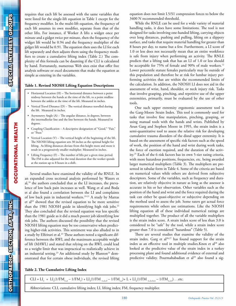
OR
TH
OPA
ED
IC S
EC
TIO
N,
AP
TA
, IN
C.
OC
CU
PA
TIO
NA
L H
EA
LTH
SP
EC
IAL I
NT
ER
ES
T G
RO
UP
S
180 Orthopaedic Practice Vol. 25;3:13
requires that each lift be assessed with the same variables that were listed for the single-lift equation in Table 1 except for the frequency modifier. In the multi-lift equation, the frequency of each lift assumes its own modifier, separate from that of the other lifts. For instance, if Worker A lifts a widget once per minute and a gidget twice per minute, then the frequency of the widget lift would be 0.94 and the frequency multiplier of the gidget lift would be 0.91. The equation then uses the LI for each lift separately and then adjusts them using the frequency modi-fiers to create a cumulative lifting index (Table 2.) The com-plexity of this formula can be daunting if the CLI is calculated by hand. Fortunately, numerous Web sites exist that offer free analysis software or excel documents that make the equation as simple as entering in the variables.
Several studies have examined the validity of the RNLE. In an expanded cross sectional analysis performed by Waters et al,18 the authors concluded that as the LI increases, the preva-lence of low back pain increases as well. Wang et al and Boda et al also found a correlation between the LI and complaints of low back pain in industrial workers.19-21 A study by Marras et al22 showed that the revised equation to be more sensitive than the 1981 NIOSH guide in identifying high risk jobs.22
They also concluded that the revised equation was less specific than the 1981 guide as it did a much poorer job identifying low risk jobs. The authors discussed the possibility that the revised NIOSH lifting equation may be too conservative when predict-ing higher-risk activities. This sentiment was also alluded to in a study by Elfeituri et al.23 These authors noted a significant dif-ference between the RWL and the maximum acceptable weight of lift (MAWL) and stated that relying on the RWL could lead to a weight limit that was impractical to realistically achieve in an industrial setting.23 An additional study by Blanton24 dem-onstrated that for certain obese individuals, the revised lifting
equation does not limit L5/S1 compression forces to below the 3400 N recommended threshold.
While the RNLE can be used for a wide variety of material handling tasks, it does have some limitations. The tool is not designed for tasks involving one-handed lifting, carrying objects over long distances, pushing and pulling, lifting on a slippery surface, and tasks that require material handling for greater than 8 hours per day, to name but a few. Furthermore, a LI score of 1.0 or less does not necessarily mean that an entire workforce is safe from injury when performing an activity. The RNLE predicts that a lifting task that has an LI of 1.0 or less should be acceptable for 75% of female and 90% of male workers.12 Lower percentile stature females particularly may lie outside of this population and therefore be at risk for lumbar injury per-forming activities that are within the recommended limits of the calculation. In addition, the NIOSH LI does not aid in the assessment of wrist, hand, shoulder, or neck injury risk. Tasks that involve grasping, pinching, and repetitive use of the upper extremities, primarily, must be evaluated by the use of other tools.
One such upper extremity ergonomic assessment tool is the Garg-Moore Strain Index. This tool is commonly used for tasks that involve fine manipulation, pinching, grasping, or using manual tools with the hands and wrists. Published by Arun Garg and Stephen Moore in 1995, the strain index is a semi-quantitative tool to assess the relative risk for developing cumulative trauma disorders of the distal upper extremity. It is based on the assessment of specific risk factors such as the speed of work, the position of the hand and wrist during work tasks, the force of exertion required, and the duration of the activ-ity.25 Each of the 6 risk factors has its own 5-tier rating criteria, with more hazardous positions, frequencies, etc. being awarded larger numerical multipliers (Table 3). The multipliers are pre-sented in tabular form in Table 4. Some of the criteria are based on numerical values while others are derived from subjective descriptors. Some of the variables, such as frequency and dura-tion, are relatively objective in nature as long as the assessor is accurate in his or her observation. Other variables such as the position of the hand and wrist and the force required during the task can either be quasi-objective or subjective depending on the method used to assess the job. Some raters get actual force requirements while others use estimations. Like the NIOSH lifting equation all of these individual numerical ratings are multiplied together. The product of all the variable multipliers is the strain index score. A strain index score of less than 3.0 is considered to be “safe” by the tool, while a strain index score greater than 7.0 is considered “hazardous” (Table 5).
There are several studies that examine the validity of the strain index. Garg et al26,27 has found support for the strain index as an effective tool in multiple studies.Knox et al28 also looked at the predictive value of the strain index in a turkey processing plant and found additional evidence of external and predictive validity. Pourmahabadian et al29 also found a sig-
Table 1. Revised NIOSH Lifting Equation Descriptions
• Horizontal Location (H) – The horizontal distance between a point midway between the hands at the time of the lift, to a point midline between the ankles at the time of the lift. Measured in inches.
• Vertical Travel Distance (D) – The vertical distance travelled during the lift. Measured in inches.
• Asymmetry Angle (A) – The angular distance, in degrees, between the intermalleolar line and the line between the hands. Measured in degrees.
• Coupling Classification – A descriptive designation of “Good,” “Fair,” or “Poor.”
• Vertical Location (V) – The vertical height of the beginning of the lift. The NIOSH lifting equation sets 30 inches as the optimal height for lifting. As lifting distances deviate from this height more and more it result in a progressively smaller multiplier. Measured in inches.
• Lifting Frequency (F) – The number of lifts per a given time period. The FM is also adjusted for the total duration that the worker spends at the station up to 8 hours in a shift.
Table 2. The Cumulative Lifting Index
CLI = LI1 + LI2(1/FM1+2 – 1/FM1) + LI3(1/FM 1+2+3 – 1/FM1,2)+ L + LI4(1/FM 1+2+3+4 – 1/FM1,2,3). . .etc.
Abbreviations: CLI, cumulative lifting index; LI, lifting index; FM, frequency multiplier.
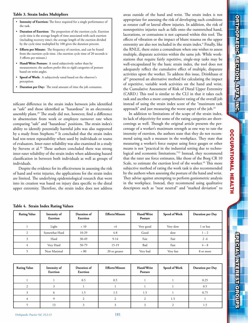
181
OC
CU
PATION
AL H
EA
LTHO
RT
HO
PA
ED
IC S
EC
TIO
N, A
PTA
, INC
.O
CC
UPA
TIO
NA
L H
EA
LTH
SP
EC
IAL IN
TE
RE
ST
GR
OU
PS
Orthopaedic Practice Vol. 25;3:13
nificant difference in the strain index between jobs identified as “safe” and those identified as “hazardous” in an electronics assembly plant.29 The study did not, however, find a difference in absenteeism from work or employee turnover rate when comparing “safe” and “hazardous” positions. The strain index’s ability to identify potentially harmful jobs was also supported by a study from Stephens.29 It concluded that the strain index had test-retest repeatability when used by individuals or teams of evaluators. Inter-rater reliability was also examined in a study by Stevens et al.30 These authors concluded there was strong inter-rater reliability of the strain index when addressing hazard classification in between both individuals as well as groups of individuals.
Despite the evidence for its effectiveness in assessing the risk of hand and wrist injuries, the applications for the strain index are limited. The underlying epidemiological research that went into its creation was based on injury data specific to the distal upper extremity. Therefore, the strain index does not address
areas outside of the hand and wrist. The strain index is not appropriate for assessing the risk of developing such conditions as rotator cuff or lateral elbow injuries. In addition, the risk of nonrepetitive injuries such as falls onto the outstretched hand, lacerations, or contusions is not captured within this tool. The effects of vibration or the impact of blunt trauma on the upper extremity are also not included in the strain index.6 Finally, like the RNLE, there exists a conundrum when one wishes to assess multiple, disparate activities within the same job. While work-stations that require fairly repetitive, single-step tasks may be well-encapsulated by the basic strain index, the tool does not adequately reflect the cumulative effect of multiple, disparate activities upon the worker. To address this issue, Drinkhaus et al33 presented an alternative method for calculating the impact of repetitive, variable work activities on the hand and wrist, the Cumulative Assessment of Risk of Distal Upper Extremity (CARD.) This tool is similar to the CLI in that it takes each task and ascribes a more comprehensive rating of the overall job instead of using the strain index score of the “maximum task approach” and just measuring the worst aspect of the job.33
In addition to limitations of the scope of the strain index, its lack of objectivity for some of the rating categories are short-comings as well. Though the original article presents the per-centage of a worker’s maximum strength as one way to rate the intensity of exertion, the authors state that they do not recom-mend using such a measure in the workplace. They state that measuring a worker’s force output using force gauges or other means is not “practical in the industrial setting due to techno-logical and economic limitations.”25 Instead, they recommend that the rater use force estimates, like those of the Borg CR 10 Scale, to estimate the exertion level of the worker.34 This more subjective method of rating the work task is also recommended by the authors when assessing the posture of the hand and wrist. They advise against attempting to perform goniometric analysis in the workplace. Instead, they recommend using qualitative descriptors such as “near neutral” and “marked deviation” to
Table 3. Strain Index Multipliers
• Intensity of Exertion: The force required for a single performance of the task.
• Duration of Exertion: The proportion of the exertion cycle. Exertion cycle time is the average length of time associated with each exertion (including recovery time); the average length of the exertion divided by the cycle time multiplied by 100 gives the duration percent.
• Efforts per Minute: The frequency of exertion, and can be found from the exertion cycle time. (An exertion cycle time of 20 seconds is 3 efforts per minute.)
• Hand/Wrist Posture: A rated subjectively rather than by measurement; the authors prefer this to rigid categories of posture based on wrist angles.
• Speed of Work: A subjectively rated based on the observer's perception.
• Duration per Day: The total amount of time the job consumes.
Table 4. Strain Index Rating Values
Rating Value Intensity of Duration of Efforts/Minute Hand/Wrist Speed of Work Duration per Day Exertion Exertion Posture
1 Light < 10 <4 Very good Very slow 1 or less
2 Somewhat Hard 10-29 4-8 Good slow 1 – 2
3 Hard 30-49 9-14 Fair Fair 2 -4
4 Very Hard 50-79 15-19 Bad Fast 4 – 8
5 Near Maximal > 80 20 or greater Very bad Very fast 8 or more
Rating Value Intensity of Duration of Efforts/Minute Hand/Wrist Speed of Work Duration per Day Exertion Exertion Posture
1 1 0.5 0.5 1 1 0.25
2 3 1 1 1 1 0.5
3 6 1.5 1.5 1.5 1 0.75
4 9 2 2 2 1.5 1
5 13 3 3 3 2 1.5
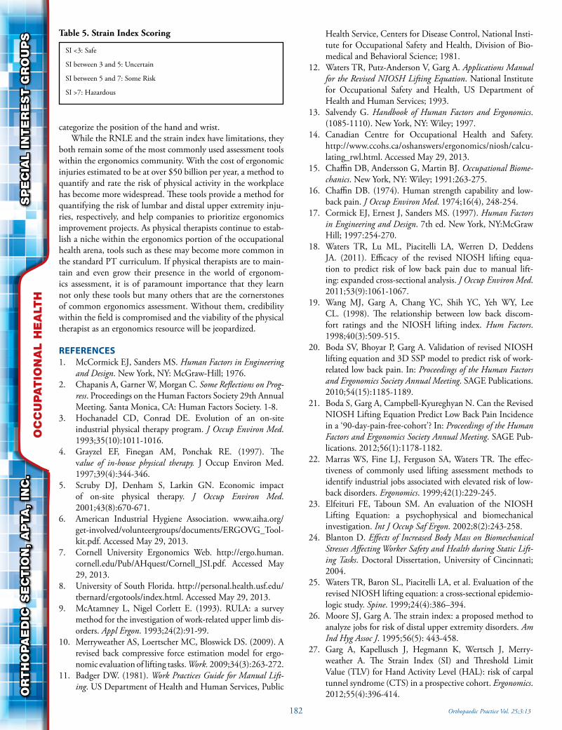
OR
TH
OPA
ED
IC S
EC
TIO
N,
AP
TA
, IN
C.
OC
CU
PA
TIO
NA
L H
EA
LTH
SP
EC
IAL I
NT
ER
ES
T G
RO
UP
S
182 Orthopaedic Practice Vol. 25;3:13
categorize the position of the hand and wrist. While the RNLE and the strain index have limitations, they
both remain some of the most commonly used assessment tools within the ergonomics community. With the cost of ergonomic injuries estimated to be at over $50 billion per year, a method to quantify and rate the risk of physical activity in the workplace has become more widespread. These tools provide a method for quantifying the risk of lumbar and distal upper extremity inju-ries, respectively, and help companies to prioritize ergonomics improvement projects. As physical therapists continue to estab-lish a niche within the ergonomics portion of the occupational health arena, tools such as these may become more common in the standard PT curriculum. If physical therapists are to main-tain and even grow their presence in the world of ergonom-ics assessment, it is of paramount importance that they learn not only these tools but many others that are the cornerstones of common ergonomics assessment. Without them, credibility within the field is compromised and the viability of the physical therapist as an ergonomics resource will be jeopardized.
REFERENCES1. McCormick EJ, Sanders MS. Human Factors in Engineering
and Design. New York, NY: McGraw-Hill; 1976.2. Chapanis A, Garner W, Morgan C. Some Reflections on Prog-
ress. Proceedings on the Human Factors Society 29th Annual Meeting. Santa Monica, CA: Human Factors Society. 1-8.
3. Hochanadel CD, Conrad DE. Evolution of an on-site industrial physical therapy program. J Occup Environ Med. 1993;35(10):1011-1016.
4. Grayzel EF, Finegan AM, Ponchak RE. (1997). The value of in-house physical therapy. J Occup Environ Med. 1997;39(4):344-346.
5. Scruby DJ, Denham S, Larkin GN. Economic impact of on-site physical therapy. J Occup Environ Med. 2001;43(8):670-671.
6. American Industrial Hygiene Association. www.aiha.org/get-involved/volunteergroups/documents/ERGOVG_Tool-kit.pdf. Accessed May 29, 2013.
7. Cornell University Ergonomics Web. http://ergo.human.cornell.edu/Pub/AHquest/Cornell_JSI.pdf. Accessed May 29, 2013.
8. University of South Florida. http://personal.health.usf.edu/tbernard/ergotools/index.html. Accessed May 29, 2013.
9. McAtamney L, Nigel Corlett E. (1993). RULA: a survey method for the investigation of work-related upper limb dis-orders. Appl Ergon. 1993;24(2):91-99.
10. Merryweather AS, Loertscher MC, Bloswick DS. (2009). A revised back compressive force estimation model for ergo-nomic evaluation of lifting tasks. Work. 2009;34(3):263-272.
11. Badger DW. (1981). Work Practices Guide for Manual Lift-ing. US Department of Health and Human Services, Public
Health Service, Centers for Disease Control, National Insti-tute for Occupational Safety and Health, Division of Bio-medical and Behavioral Science; 1981.
12. Waters TR, Putz-Anderson V, Garg A. Applications Manual for the Revised NIOSH Lifting Equation. National Institute for Occupational Safety and Health, US Department of Health and Human Services; 1993.
13. Salvendy G. Handbook of Human Factors and Ergonomics. (1085-1110). New York, NY: Wiley; 1997.
14. Canadian Centre for Occupational Health and Safety. http://www.ccohs.ca/oshanswers/ergonomics/niosh/calcu-lating_rwl.html. Accessed May 29, 2013.
15. Chaffin DB, Andersson G, Martin BJ. Occupational Biome-chanics. New York, NY: Wiley; 1991:263-275.
16. Chaffin DB. (1974). Human strength capability and low-back pain. J Occup Environ Med. 1974;16(4), 248-254.
17. Cormick EJ, Ernest J, Sanders MS. (1997). Human Factors in Engineering and Design. 7th ed. New York, NY:McGraw Hill; 1997:254-270.
18. Waters TR, Lu ML, Piacitelli LA, Werren D, Deddens JA. (2011). Efficacy of the revised NIOSH lifting equa-tion to predict risk of low back pain due to manual lift-ing: expanded cross-sectional analysis. J Occup Environ Med. 2011;53(9):1061-1067.
19. Wang MJ, Garg A, Chang YC, Shih YC, Yeh WY, Lee CL. (1998). The relationship between low back discom-fort ratings and the NIOSH lifting index. Hum Factors. 1998;40(3):509-515.
20. Boda SV, Bhoyar P, Garg A. Validation of revised NIOSH lifting equation and 3D SSP model to predict risk of work-related low back pain. In: Proceedings of the Human Factors and Ergonomics Society Annual Meeting. SAGE Publications. 2010;54(15):1185-1189.
21. Boda S, Garg A, Campbell-Kyureghyan N. Can the Revised NIOSH Lifting Equation Predict Low Back Pain Incidence in a ‘90-day-pain-free-cohort’? In: Proceedings of the Human Factors and Ergonomics Society Annual Meeting. SAGE Pub-lications. 2012;56(1):1178-1182.
22. Marras WS, Fine LJ, Ferguson SA, Waters TR. The effec-tiveness of commonly used lifting assessment methods to identify industrial jobs associated with elevated risk of low-back disorders. Ergonomics. 1999;42(1):229-245.
23. Elfeituri FE, Taboun SM. An evaluation of the NIOSH Lifting Equation: a psychophysical and biomechanical investigation. Int J Occup Saf Ergon. 2002;8(2):243-258.
24. Blanton D. Effects of Increased Body Mass on Biomechanical Stresses Affecting Worker Safety and Health during Static Lift-ing Tasks. Doctoral Dissertation, University of Cincinnati; 2004.
25. Waters TR, Baron SL, Piacitelli LA, et al. Evaluation of the revised NIOSH lifting equation: a cross-sectional epidemio-logic study. Spine. 1999;24(4):386–394.
26. Moore SJ, Garg A. The strain index: a proposed method to analyze jobs for risk of distal upper extremity disorders. Am Ind Hyg Assoc J. 1995;56(5): 443-458.
27. Garg A, Kapellusch J, Hegmann K, Wertsch J, Merry-weather A. The Strain Index (SI) and Threshold Limit Value (TLV) for Hand Activity Level (HAL): risk of carpal tunnel syndrome (CTS) in a prospective cohort. Ergonomics. 2012;55(4):396-414.
Table 5. Strain Index Scoring
SI <3: Safe
SI between 3 and 5: Uncertain
SI between 5 and 7: Some Risk
SI >7: Hazardous
183
OC
CU
PATION
AL H
EA
LTHO
RT
HO
PA
ED
IC S
EC
TIO
N, A
PTA
, INC
.O
CC
UPA
TIO
NA
L H
EA
LTH
SP
EC
IAL IN
TE
RE
ST
GR
OU
PS
Orthopaedic Practice Vol. 25;3:13
28. Garg A, Hegmann K, Wertsch J, et al. (2012). The WISTAH hand study: a prospective cohort study of distal upper extremity musculoskeletal disorders. BMC Musculo-skel Disord. 2012;13(1):90.
29. Knox K, Moore JS. Predictive validity of the Strain Index in turkey processing. J Occup Environ Med. 2001;43(5):451-462.
30. Pourmahabadian M, Saraji JN, Saddeghi-Naeeni MAH. (2005). Risk assessment of developing distal upper extrem-ity disorders by strain index method in an assembling elec-tronic industry. Acta Medica Iranica. 2005;43(5):347-354.
31. Stephens JP, Vos, GA, Steves EM Jr, Moore JS. Test-retest repeatability of the Strain Index. Appl Ergon. 2006;37(3):275-281.
32. Stevens EM, Vos GA, Stephens JP, Moore JS. (2004). Inter-rater reliability of the strain index. J Occup Environ Hyg. 2004;1(11):745-751.
33. Finnish Institute of Occupational Health. www.ttl.fi/work-loadexposuremethods. Accessed May 29, 2013.
34. Borg E, Borg G. A comparison of AME and CR100 for scal-ing perceived exertion. Acta Psychol. 2002;109:157-175.
22.3 FOOT AND ANKLE• Biomechanics of the Foot and Ankle for
the Physical Therapist—Jeff Houck, PT, PhD (Subject Matter Expert: Christopher R. Carcia, PT, PhD, SCS, OCS)
• Adult Acquired Flatfoot Disorders—Bran-don E. Crim, DPM, and Dane K. Wukich, MD (Subject Matter Expert: Christopher R. Carcia, PT, PhD, SCS, OCS)
• Examination of the Ankle and Foot—Todd E. Davenport, PT, DPT, OCS (Subject Matter Expert: RobRoy Martin, PT)
• Exercise Progressions for the Foot and Ankle—Clarke Brown, PT, DPT, OCS, ATC (Subject Matter Expert: Christopher R. Carcia, PT, PhD, SCS, OCS)
• Taping, Mobilization, and Exercises for the Foot and Ankle—Stephen Paulseth, PT, DPT, SCS, ATC, and RobRoy Martin, PhD, PT, CSCS (Subject Matter Expert: Todd E. Daven-port, PT, DPT, OCS)
• The Effectiveness of Foot Orthoses for the Treatment and Prevention of Lower Ex-tremity Overuse Injuries—James W. Mathe-son, PT, DPT, MS, OCS, SCS (Subject Matter Expert: Deb Nawoczenski, PT, PhD)
Register now! 800/444-3982
OR
TH
OPA
ED
IC S
EC
TIO
N,
AP
TA
, IN
C.
FO
OT
AN
D A
NK
LE
SP
EC
IAL I
NT
ER
ES
T G
RO
UP
S
184 Orthopaedic Practice Vol. 25;3:13
ENTRY-LEVEL CURRICULUM-PROGRESS CONTINUESClarke Brown PT, DPT, OCS, ATCPresident, FASIG
Background: In 2011, FASIG created a task force as an ini-tial step in a process that will culminate with “Minimum Stan-dards for Foot and Ankle Content in an Entry-level Physical Therapy Curriculum.” Last October 2012, our Entry-level Cur-riculum Task Force of 15 foot and ankle experts (mostly FASIG members!) gathered at APTA headquarters in Washington, DC.
Led by Chris Neville, this group generated an impressive and comprehensive outline that will serve as the template for the eventual curriculum recommendation. This document, still early in its development, boasts a surprisingly detailed array of foot and ankle examination, differential diagnosis, assessment, and interventions. The FASIG’s intent is to provide the ortho-paedic educator with evidence-based information regarding study of the foot and ankle that will be readily inserted into any orthopaedic curriculum.
To date, the document is undergoing further review. It is anticipated that a more formal and functional edition can be presented at CSM 2014!
FASIG SPURS RESEARCH WITH SECOND GRANTAt CSM 2013, the FASIG was informed by the Orthopaedic
Section’s Research Committee that the following authors had been awarded FASIG’s $15,000 research grant: Shane McClin-ton, Timothy Flynn, and Bryan Heiderscheit. Their study is titled, “Comparison of Usual Podiatric Care and Early Physical Therapy for Plantar Heel Pain.”
This grant, the second of an equal amount in the past 4 years, represents the commitment to research that symbolizes the FASIG and the Orthopaedic Section. Todd Davenport, Research Chair for the FASIG, adds, “Our membership meets at CSM each year and no other agenda item creates more excite-ment and pride than to vote on the contribution to research from our hard-earned funds.”
The FASIG looks forward to the results of this study that should help all of us better understand plantar heel pain and the treatment strategies used by foot care practitioners.
“FASIG TALKING POINTS…INTERESTING FODDER FOR RESEARCH AND PRACTICE”
The following commentaries have been selected from research journals from around the world. Consider their con-tent and bounce these ideas around the office! Then, create your own research study to answer some of these enigmatic issues. Finally, respond to this column at [email protected] with research ideas, questions, responses to these talking points, or any foot and ankle topic!
EXERCISE WITH MINIMALIST SHOESChances are that you have had patients ask for your opinion
on the use of minimalist shoes during running and walking, or
even about barefoot running. Recently, I came across a podiatric round table to ascertain the thoughts of a podiatrist. Mostly, this group of foot doctors cautioned against the use of minimal-ist shoes, particularly with or on feet that were new to running/walking or were mechanically compromised.
This panel of podiatrists agreed that some people can toler-ate less support at the foot during high-impact foot-strike. They acknowledged, however, that while the minimalist runner may move his foot contact more to the forefoot and realize less force transmission to the knee, the Achilles and metatarsals may pay a price. Almost never do they recommend training without shoes for more than 10% of anyone’s exercise volume. Citing studies, which place the vast majority of runners in the group of rear foot strikers, podiatrists are reticent to encourage a dramatic change in biomechanics.
In addition, the use of lightweight shoes for exercise other than running such as fitness classes (Cross Fit or Zumba) and home video exercising (Insanity or P90X) may also explain an upward trend in Achilles tendinopathy and plantar heel pain.
So, what foot and ankle injuries are you treating in your office today, due to minimalist or barefoot exercising, perhaps not seen 10 years ago? Are these new shoes actually creating patients? Or, as some say, is it merely a throwback to the super-light track shoes worn by many runners in the 70s during high-speed workouts?
What do you think?
CUBOID SYNDROME (RE-VISITED)In a previous OPTP issue, Dr. Matthew Kearns presented an
interesting case of cuboid syndrome in a 14-year-old girl who is now 16. This case was made interesting by the frequency of manipulation required to maintain joint stability and foot func-tion. Initially diagnosed as a lateral ankle sprain and following 8 weeks of immobilization, Dr. Kearns removed the cast-boot and resolved pain immediately using a cuboid-whip manipulation. Relief and functional return to all activities (except competi-tive soccer) were attained for one year. High-speed lateral move-ments caused a second event. Manipulation was again effective in resolving pain.
Over the course of the next 9 months, the patient could not effectively return to running/jumping activities without relapse or fear of injury. In particular, lateral cutting and jump-ing during soccer participation was most problematic. Three subsequent manipulations were performed, all eliciting com-plete relief of lateral foot pain. Strengthening and stabilization exercises were ongoing, and external taping including low-dye, strapping, and ankle taping with stirrups were trialed.
After the 4th manipulation, a semi-rigid, neutral orthotic was introduced. Sources of ‘extra’ support at or about the cuboid bone via padding, cookies, or wedges were not used. Orthoplast orthotics were ground for walking shoes and soccer cleats, so that she rarely exercised without orthotics.
After 3 months of exercise, including soccer participation, no events of lateral foot pain have occurred. Was this coinci-
FOOT & ANKLESPECIAL INTEREST GROUP
OC
CU
PATION
AL H
EA
LTHO
RT
HO
PA
ED
IC S
EC
TIO
N, A
PTA
, INC
.FO
OT
AN
D A
NK
LE
SP
EC
IAL IN
TE
RE
ST
GR
OU
PS
185Orthopaedic Practice Vol. 25;3:13
dental? Since radiographs rarely demonstrate malalignment at the cuboid articulations, is the cuboid subluxed? Where are the sounds that accompany this manipulation coming from?
SEVERE ONYCHOMYCOSISIt seems more and more patients are presenting with moder-
ate to severe sub-ungual onychomycosis, noticed during rou-tine evaluations for injuries or orthotics. I came across this very interesting intervention. The following is an abstract from the American Journal of Podiatric Medicine Association:
Onychomycosis, most commonly caused by two species of dermatophyte fungi--Trichophyton rubrum and Trichophyton mentagrophytes--is primarily treated with regimens of topical and systemic antifungal medications. This study was undertaken to evaluate in vitro the efficacy of low-voltage direct current as an antifungal agent for treating onychomycosis. Agar plate cul-tures of T rubrum and T mentagrophytes were subjected to low-voltage direct current electrostimulation, and antifungal effects were observed as zones in the agar around the electrodes lacking fungal growth. Zones devoid of fungal growth were observed for T rubrum and T mentagrophytes around anodes and cath-odes in a dose-dependent manner in the current range of 500
microA to 3 mA. Low-voltage direct current electrostimulation has great clinical potential for the treatment of onychomycosis and perhaps other superficial maladies of fungal etiology.
This study was originated in a physical therapy wound care clinic, in an attempt to address the commonly encountered infection and treat it with increased efficiency and efficacy. Could low-voltage direct current be used for other infectious processes? If so, why is electrical stimulation contraindicated for all forms of infections?
REFERENCES1. Blake R, Johncock D, Kirby K, Richie D. When patients ask
you about barefoot running and minimalist shoes. Podiatry Today. 2013;26(5):36-45.
2. Durall C. Examination and treatment of cuboid syndrome: A literature review. Sports Phys Ther. University of Wiscon-sin: La Crosse, WI. 2011:514-519.
3. Kalinowski DP, Edsberg LE, Hewson RA, Johnson RH, Brogan MS. Low-voltage direct current as a fungicidal agent for treating onychomycosis. J Am Podiatr Med Assoc. 2004;94(6):565-572.
186
OR
TH
OPA
ED
IC S
EC
TIO
N,
AP
TA
, IN
C.
PE
RFO
RM
ING
AR
TS
SP
EC
IAL I
NT
ER
ES
T G
RO
UP
S
Orthopaedic Practice Vol. 25;3:13
PERFORMING ARTS SPECIAL INTEREST GROUP
PRESIDENT’S MESSAGEThe PASIG continues to work on our resource center
located on the PASIG webpage of the www.orthopt.org. We are seeking authors for content related to the performing arts specialties such as dance, music, gymnastics, and figure skating. Please review the current content and reach out to me if you are interested in assisting with creating content.
We want to share our continued commitment towards research and evidence-based practice with you through our cita-tion blasts. Our research committee prepares a citation blast each month, which is an annotated bibliography on a specific topic area related to the performing arts. We are always seeking authors to assist us with this process. If you are interested in contributing, please contact our research committee chairper-son, Annette Karim at [email protected]. Please check out our current listing at:
http://www.orthopt.org/content/special_interest_groups/performing_arts/citations_endnotes
We are approaching our deadline of August 1, 2013, for nominations for 2014 PASIG officers where we are seeking nominees for President and a Nominating Committee member. If you are interested in nominating a candidate, please contact the Nominating Committee Chairperson, Amanda Blackmon at [email protected].
For students interested in the performing arts, we have updated our clinical affiliations list on our Web site. If you are a student and are interested in finding a performing arts spe-cific clinical, please check out the listing at www.orthopt.org on the PASIG page. We also offer a scholarship to a student who has been accepted to present at CSM 2014 to help defray the travel costs. If you are interested in applying for this scholarship, please contact our Student Scholarship Committee Chair, Amy Humphrey at [email protected].
Sincerely,Julie O’Connell, PT, DPT, ATC
PASIG President
PERFORMING ARTS CONFERENCES AND RESOURCESOrthopaedic Section-American Physical Therapy Association,Performing Arts SIGhttp://www.orthopt.org/content/special_interest_groups/performing_arts
Performing Arts Citations and Endnoteshttp://www.orthopt.org/content/special_interest_groups/performing_arts/citations_endnotes
ADAM Centerhttp://www.adamcenter.net/
Publications:http://www.adamcenter.net/#!vstc0=publications
Conference abstracts:http://www.adamcenter.net/#!vstc0=conferences
Dance USA Annual Conference: Philadelphia, PA, June 12-15, 2013http://www.danceusa.org/
Research resources:http://www.danceusa.org/researchresources
Professional Dancer Annual Post-Hire Health Screen:http://www.danceusa.org/dancerhealth
Dancer Wellness Projecthttp://www.dancerwellnessproject.com/
Becoming an affiliate:http://www.dancerwellnessproject.com/Information/Become-Affiliate.aspx
PERFORMING ARTSCONTINUING EDUCATION
Performing Arts IndependentStudy Courses Orthopaedic Section Independent Study Course. 20.3 Physical Therapy for the Performing Artist Monographs are available for: • Figure Skating (J. Flug, J. Schneider, E. Greenberg)• Artistic Gymnastics (A. Hunter-Giordano, Pongetti-Angeletti, S. Voelker, TJ Manal)• Instrumentalist Musicians (J. Dommerholt, B. Collier)
Orthopaedic Section Independent Study Course.Dance Medicine: Strategies for the Prevention and Care of Injuries to Dancers This is a 6-monograph course and includes many PASIG members as authors. • Epidemiology of Dance Injuries: Biopsychosocial
Considerations in the Management of Dancer Health (MJ Liederbach)• Nutrition, Hydration, Metabolism, and
Thinness (B Glace)• The Dancer’s Hip: Anatomic, Biomechanical, and
Rehabilitation Considerations (G. Grossman)• Common Knee Injuries in Dance (MJ Liederbach)• Foot and Ankle Injuries in the Dancer: Examination
and Treatment Strategies (M. Molnar, R. Bernstein, M. Hartog, L. Henry, M. Rodriguez, J. Smith, A. Zujko)
• Developing Expert Physical Therapy Practice in Dance Medicine – (J. Gamboa, S. Bronner, TJ Manal)
Contact the Orthopaedic Section at:
www.orthopt.orgOr call 1-800-444-3982
187
OC
CU
PATION
AL H
EA
LTHO
RT
HO
PA
ED
IC S
EC
TIO
N, A
PTA
, INC
.P
ER
FO
RM
ING
AR
TS
SP
EC
IAL IN
TE
RE
ST
GR
OU
PS
Orthopaedic Practice Vol. 25;3:13
Harkness Center for Dance Injuries, Hospital for Joint Diseaseshttp://hjd.med.nyu.edu/harkness/
Continuing education:h t t p : / / h j d . m e d . n y u . e d u / h a r k n e s s / e d u c a t i o n /h e a l t h c a r e - p r o f e s s i o n a l s / c o n t i n u i n g e d u c a t i o n -courses-cme-and-ceu
Resource papers:http://hjd.med.nyu.edu/harkness/dance-medicine-resources/resource-papersand-forms
Links:http://hjd.med.nyu.edu/harkness/dance-medicine-resources/links
Informative list of common dance injuries:h t t p : / / h j d . m e d . n y u . e d u / h a r k n e s s / p a t i e n t s /common-dance-injuries
Research publications:h t t p : / / h j d . m e d . n y u . e d u / h a r k n e s s / r e s e a r c h /research-publications
International Association for Dance Medicine and Science (IADMS)http://www.iadms.org/
The 23rd Annual Meeting of the International Association for Dance Medicine & Science (IADMS) will be held in Seattle, Washington, USA from October 17 - 19, 2013. Meeting activi-ties and sessions will be held at the Renaissance Seattle Hotel. On Sunday, October 20, 2013, Special Interest Groups (SIG) Day will be held, with special programs available.
Resource papers:h t t p : / / w w w . i a d m s . o r g / d i s p l a y c o m m o n .cfm?an=1&subarticlenbr=186
Links:http://www.iadms.org/displaycommon.cfm?an=5
Medicine, arts medicine, and arts education organization links:h t t p : / / w w w . i a d m s . o r g / d i s p l a y c o m m o n .cfm?an=1&subarticlenbr=5
Publications:http://www.iadms.org/displaycommon.cfm?an=3
Performing Arts Medicine Association (PAMA)http://www.artsmed.org/
Annual symposium: July 20-23, 2013 Medical Problems of Per-forming Artists:“Maximizing Performance, Artistry, Implementation, and Empowerment”http://www.artsmed.org/symposium.html
Interactive bibliography site:http://www.artsmed.org/bibliography.html
OR
TH
OPA
ED
IC S
EC
TIO
N,
AP
TA
, IN
C.
PA
IN M
AN
AG
EM
EN
TS
PE
CIA
L I
NT
ER
ES
T G
RO
UP
S
188 Orthopaedic Practice Vol. 25;3:13
PAIN MANAGEMENTSPECIAL INTEREST GROUP
PRESIDENT’S MESSAGEJohn E. Garzione, PT, DPT, DAAPM
The Brain…Another large piece of the persistent pain puzzle.Researchers have been looking at the brain by neuroimaging
to explain persistent pain and pain severity.1-3
There are many helpful techniques that Physical Therapists can use to help treat our patients who have persistent pain such as cognitive behavior, relaxation training, and meditation. Asso-ciative Awareness Technique (AAT) is another modality that can be added to our toolbox for patient care. If you are interested in exploring this technique further, you can visit their website at www.wellnessandperformance.com.
Hope you enjoy the rest of the summer.John
REFERENCES1. Robinson ME, Staud R, Price DD. Pain measurement and
brain activity: will neuroimaging replace pain ratings? J Pain. 2013;14(4):328-331.
2. Mackey SC. Central neuroimaging of pain. J Pain. 2013;14(4):334-335.
3. Sullivan MD, Cahana A, Derbyshire S, Loeser JD. What does it mean to call chronic pain a brain disease? J Pain. 2013;14(4):332-333.
Associative Awareness TechniqueScott Musgrave, MSPT Ernie Quinlisk, PT
In the last decade, there has been a tremendous amount of research and a plethora of books written about the brain. Neu-roplasticity has emerged as a term that defines this new era of understanding learning, memory, and behavior and sheds light on the perplexing issue of chronic pain like never before. Neu-roplasticity refers to the concept that the brain is not a static organ physiologically, but can change with training throughout life.
The idea of neuroplasticity was first proposed in 1892 by a Spanish physician named Santiago Ramon y Cajal, whose revo-lutionary ideas were rejected for the next 50 years! Many years later, a Polish neurophysiologist named Jerzy Konorski actually coined the term neuroplasticity prior to his death in 1973 as he further developed the work of Ivan Pavlov and Donald Hebb.
As we fast forward to current times, President Obama’s administration also understands the vast potential available within this new understanding of the brain, which has been demonstrated in the launch of his BRAIN initiative on April 5, 2013 (WhiteHouse.gov/infographics/brain-initiative). Even though this initiative has its allies and adversaries, the concept
of an ever-changing brain has created a novel understanding of healing potential that can alleviate or eliminate human suffering that is as vast as the brain itself.
Since 2007, Scott Musgrave, MSPT, and Ernie Quinlisk, PT, through their company Wellness and Performance have been studying and researching the frustrating and fluctuating cycle of chronic pain. As a result, they are revealing an enlight-ened understanding of the brain, which has culminated in the creation of a simple method of treatment that in many cases can resolve the vicious cycle that defines this problem. It is called Associative Awareness Technique (AAT). This technique teaches patients how to be aware of past traumatic events that caused them pain previously and continues to trigger chronic pain pat-terns. There are currently over 100 AAT medical practitioners around the country that are now enjoying a new sense of profes-sional satisfaction, by allowing their intellectual curiosity to be ignited by an innovative way of thinking and understanding, based on current scientific knowledge.
One of the many confounding physical problems that frus-trates and confuses most medical practitioners and millions of people is the problem of chronic pain. According to the Ameri-can Academy of Pain Medicine, 100 million people in the United States have chronic pain and more people suffer from chronic pain than diabetes, heart disease, and cancer combined! The economic impact on the United States is a startling $600 billion annually.
Musgrave and Quinlisk have determined that the key to understanding chronic pain lies within the form and function of the base functional unit of the brain: the neuron. Associa-tive Awareness Technique is designed around how information is processed in all 3 levels of the brain based upon neuronal function. By altering synaptic communication, we can extin-guish the causative aspects of chronic conditions by using the scientific concept of neuroplasticity.
In chapter 6 of his book, The Sensitive Nervous System, Australian Physiotherapist, David Butler1 talks about the grey zone of practice: “The grey zone is massive. This is an era of new, chronic and stress related disorders where there is neither vaccine nor cure. At this stage, best clinical reasoning must be applied to traverse the grey zones reasoning which includes, integrates and contributes to relevant evidence based work as it comes about.”
Butler goes on to say, “the outcomes movement has brought about another compelling issue for manual therapists. Clinicians who have followed the content of mainstream journals such as Spine and Pain and even a past issue of Manual Therapy (Vol. 4, 1999) will have noted increasing support for the contention that chronic pain development and responses to treatment may have more to do with psychosocial factors than physical factors. These include pain beliefs, movement fears, job satisfaction, and childhood experiences. Some clinicians in the musculoskeletal management area may well begin to ponder their effectiveness in helping persistent pain patients. We may all go through this, but on the up side, the outcomes movement and the infor-
OC
CU
PATION
AL H
EA
LTHO
RT
HO
PA
ED
IC S
EC
TIO
N, A
PTA
, INC
.PA
IN M
AN
AG
EM
EN
TS
PE
CIA
L IN
TE
RE
ST
GR
OU
PS
189Orthopaedic Practice Vol. 25;3:13
mation it brings, combined with the biological revolution is probably providing the most powerful stimulus for change and adaptation of practice ever. It can embellish existing successful management strategies and provide fresh and novel strategies.
Chronic pain patients live in the grey zone. If those of us in the medical profession who treat chronic pain are truly honest, we must admit our lack of comprehension in regards to the roots of chronic pain and the type of treatment that would best suit a particular patient. We prefer to not treat these patients as they come to us with varying pain complaints, depending on the day, in the hope of an answer because we are the ‘experts.’ In this respect, we are far from experts, causing patients to leave our offices like they have left so many offices in the past with no answers and no hope.
The dilemma of chronic pain has been proposed by the American Fibromyalgia Association as an abnormality in the central nervous system that causes widespread muscular pain, sleep, digestive disorders, chronic headaches, memory and con-centration difficulties, and many other body-wide symptoms. No doubt this is an indication of an autonomic nervous system gone awry, causing an abnormal cycling that has no end in sight for these unfortunate individuals. And where do we start with treatment? If we think the problem is peripherally based, we do not stand a chance in helping these patients.
Just like Dr. Santiago Ramon y Cajal proposed in 1892, a disruptive technology is required to improve the current medi-cal model and get the respective brain experts (scientific and medical) to work together as proposed currently by President Obama similar to the way all the systems in the body work together, not separately. In order to be even remotely effec-tive, this new technology must be easy to learn and apply. Care must also be affordable since many of the afflicted patients have spent thousands of dollars searching for answers. Unfortunately, physical therapists have too many questions of their own about chronic pain and therefore cannot supply an answer to deserv-ing patients.
Fortunately, AAT is this new affordable disruptive technol-ogy that follows the known form and function of the central nervous system as it scientifically explains the human traumatic experience that is the root of chronic pain. It requires no medi-cine, expensive equipment purchases, or frequent visits to your medical practitioner(s).
Associative Awareness Technique is a unique and innova-tive treatment process developed specifically by physical thera-pists for chronic pain. It is unique not only within the physical therapy profession but in the entirety of medicine, because there is no current way to physically apply the current scientific knowledge. Associative Awareness Technique is an innovative treatment process that melds current understandings in neuro-science and behavioral medicine to correspond with the 3 levels of the human brain to change the negative autonomic physical reactions that are the hallmark of chronic pain.
Associative Awareness Technique is designed to be self-applied, which is critical to create the neuroplastic changes that chronic pain patients require to make lasting change. Each level of AAT contains two steps. The first two steps of Level 1 (one self-applied, one hands on) target restoration of homeostasis to the autonomic nervous system (ANS). You can’t intellectual-ize your survival instincts! No wonder these chronic patterns continue to reoccur. The two self-applied steps of Level 2 are
designed specifically to target the limbic system and emotional associations that abnormally cycle the ANS. The final two self-applied steps that are part of Level 3 are designed specifically to address neocortical associations, anticipatory expectations, and memory that trigger emotional associations and abnormally cycle the ANS.
Associative Awareness Technique has been successfully used by many AAT practitioners to treat chronic conditions and is designed to follow the concept of neuroplasticity within the framework of its self-application model. Patients can make positive changes to all 3 levels of their brain by using these self-applied steps. Associative Awareness Technique is used in the situational events of their daily lives that have the potential to trigger the patient and reinforce previously conditioned nega-tive physical responses and their chronic pattern of pain.
Unfortunately, the physical therapy profession does not understand how the brain is functioning in the chronic pain patient, and this is clearly evident in why patients do not respond to our constant efforts of administering evidence based peripheral approaches to our chronic pain patients. We need to be leaders and educators in the study of chronic pain and that starts with a better understanding of the available science that we can ascertain to provide appropriate care for this difficult patient population.
Associative Awareness Technique provides a comprehensive understanding of the human nervous system as it addresses the interrelationships of the triune brain and its profound effects on the rapid cycling noted in almost every chronic pain patient. Associative Awareness Technique also addresses the myriad of complicating factors that these patients live with 24/7 like insomnia, post traumatic stress disorder, irritable bowel syn-drome, irrational fears, anxiety, and depression. By educating health care professionals, we will achieve our goal of remov-ing hopelessness from the lives of many chronic pain patients and their families and provide all practitioners tools that will only enhance the reputation of the physical therapy profession within the health care community.
REFERENCE1. Butler D. The Sensitive Nervous System. Adelaide, Australia:
Noigroup Publications; 2000.
RECOMMENDED READINGS• Doidge N. The Brain that Changes Itself. New York, NY:
Penguin Books; 2007.• Kolb B, Whishaw I. 2011. An Introduction to Brain and
Behavior. New York, NY: Worth Publishers; 2011.• Ledoux J 2002. Synaptic Self. New York, NY: Penguin
Group; 2002 • Scaer R. 2007. The Body Bears the Burden. 2nd ed. New
York, NY: The Hawthorne Medical Press; 2007.
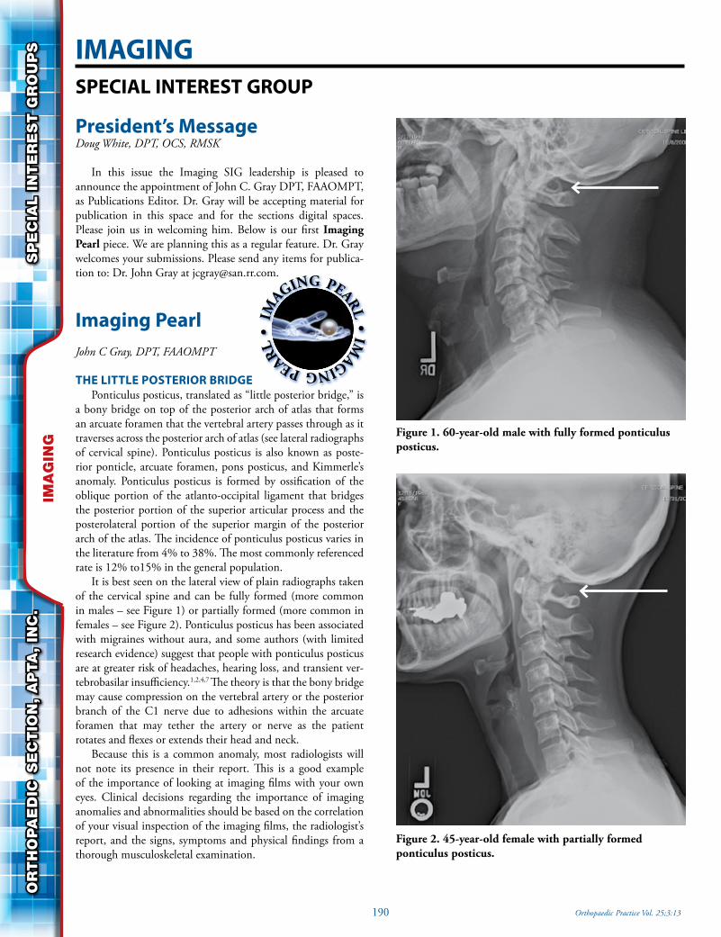
OR
TH
OPA
ED
IC S
EC
TIO
N,
AP
TA
, IN
C.
IMA
GIN
GS
PE
CIA
L I
NT
ER
ES
T G
RO
UP
S
190 Orthopaedic Practice Vol. 25;3:13
President’s Message Doug White, DPT, OCS, RMSK
In this issue the Imaging SIG leadership is pleased to announce the appointment of John C. Gray DPT, FAAOMPT, as Publications Editor. Dr. Gray will be accepting material for publication in this space and for the sections digital spaces. Please join us in welcoming him. Below is our first Imaging Pearl piece. We are planning this as a regular feature. Dr. Gray welcomes your submissions. Please send any items for publica-tion to: Dr. John Gray at [email protected].
Imaging PearlJohn C Gray, DPT, FAAOMPT
THE LITTLE POSTERIOR BRIDGEPonticulus posticus, translated as “little posterior bridge,” is
a bony bridge on top of the posterior arch of atlas that forms an arcuate foramen that the vertebral artery passes through as it traverses across the posterior arch of atlas (see lateral radiographs of cervical spine). Ponticulus posticus is also known as poste-rior ponticle, arcuate foramen, pons posticus, and Kimmerle’s anomaly. Ponticulus posticus is formed by ossification of the oblique portion of the atlanto-occipital ligament that bridges the posterior portion of the superior articular process and the posterolateral portion of the superior margin of the posterior arch of the atlas. The incidence of ponticulus posticus varies in the literature from 4% to 38%. The most commonly referenced rate is 12% to15% in the general population.
It is best seen on the lateral view of plain radiographs taken of the cervical spine and can be fully formed (more common in males – see Figure 1) or partially formed (more common in females – see Figure 2). Ponticulus posticus has been associated with migraines without aura, and some authors (with limited research evidence) suggest that people with ponticulus posticus are at greater risk of headaches, hearing loss, and transient ver-tebrobasilar insufficiency.1,2,4,7 The theory is that the bony bridge may cause compression on the vertebral artery or the posterior branch of the C1 nerve due to adhesions within the arcuate foramen that may tether the artery or nerve as the patient rotates and flexes or extends their head and neck.
Because this is a common anomaly, most radiologists will not note its presence in their report. This is a good example of the importance of looking at imaging films with your own eyes. Clinical decisions regarding the importance of imaging anomalies and abnormalities should be based on the correlation of your visual inspection of the imaging films, the radiologist’s report, and the signs, symptoms and physical findings from a thorough musculoskeletal examination.
IMAGINGSPECIAL INTEREST GROUP
IM
AGING PEARL • IMAGING PEARL
•
Figure 1. 60-year-old male with fully formed ponticulus posticus.
Figure 2. 45-year-old female with partially formed ponticulus posticus.
OC
CU
PATION
AL H
EA
LTHO
RT
HO
PA
ED
IC S
EC
TIO
N, A
PTA
, INC
.IM
AG
ING
SP
EC
IAL IN
TE
RE
ST
GR
OU
PS
191Orthopaedic Practice Vol. 25;3:13
REFERENCES1. Buna M, Coghlan W, deGruchy M, et al. Ponticles of
the atlas: a review and clinical perspective. J Manipulative Physiol Ther. 1984;7:261-266.
2. Greiner HM, Abruzzo TA, Kabbouche M, Leach JL, Zuc-carello M. Rotational vertebral artery occlusion in a child with multiple strokes: a case-based update. Childs Nerv Syst. 2010;26(12):1669-1674.
3. Hasan M, Shukla S, Siddiqui MS, Singh D. Posterolateral tunnels and ponticuli in human atlas vertebrae. J Anat. 2001;199(Pt 3):339-343.
4. Koutsouraki E, Avdelidi E, Michmizos D, Kapsali SE, Costa V, Baloyannis S. Kimmerle's anomaly as a possible causative factor of chronic tension-type headaches and neurosensory hearing loss: case report and literature review. Int J Neurosci. 2010;120(3):236-239.
5. Lambarty BGH, Zivanovic S. The retroarticular vertebral artery ring of the atlas and its significance. Acta Anatomica. 1973;85:113–122.
6. Stubbs DM. The arcuate foramen: variability in distribution related to race and sex. Spine. 1992;17:1502–1504.
7. Wight S, Osborne N, Breen AC. Incidence of ponticulus posterior of the atlas in migraine and cervicogenic headache. J Manipulative Physiol Ther. 1999;22:15-20.
8. Young JP, Young PH, Ackermann MJ, Anderson PA, Riew KD. The ponticulus posticus: implications for screw inser-tion into the first cervical lateral mass. J Bone Joint Surg Am. 2005;87:2495–2498.
Accurately Assess Balance & Sway
MatScan®
with Sway Analysis
• Evaluate weight bearing, balance and sway
• Set baselines and re-test post injury or treatment
• Portable, affordable tool for assessing patients
Contact Us for more information
www.tekscan.com/orthopt
IMAGING SIG LEADERSHIP
Douglas M. White, DPT, OCS – President [email protected] @Douglas_M_White Deydre Teyhen, PT, PhD, OCS – Vice President
Nominating Committee Wayne Smith, DPT, Med, ATCr, SCS, RMSK - Chair James “Jim” Elliot, PhD, PT Richard Souza, PT, PhD, ATC, CSCS
John C. Gray, DPT FAAOMPT – Publications EditorGerard Brennan, PT, PhD - Orthopaedic Section Board Liaison
OC
CU
PATION
AL H
EA
LTHO
RT
HO
PA
ED
IC S
EC
TIO
N, A
PTA
, INC
.A
NIM
AL R
EH
AB
ILIT
AT
ION
SP
EC
IAL IN
TE
RE
ST
GR
OU
PS
193Orthopaedic Practice Vol. 25;3:13
ANIMAL REHABILITATIONSPECIAL INTEREST GROUP
LETTER FROM THE PRESIDENTKirk Peck, PT, PhD, CSCS, CCRT
REFLECTIONS ON THE EVOLUTION OF ANIMAL REHABILITATION
Recently I was confronted with a rather interesting question of current relevance. In fact the question was so compelling it prompted me to reflect on the current state of physical therapy practice and animal rehabilitation. I was asked by a physical therapist, who does not treat animals by trade, if I was personally concerned that animals were not represented or even implied as part of scope of practice in the newly proposed APTA 2020 Vision Statement slated for debate during the 2013 House of Delegates. After mulling it over for a while, I offered the follow-ing commentary based on my perspective of current practice.
I do not believe the number of physical therapists (PTs) and physical therapist assistants (PTAs) treating animals on a national scale has achieved a high enough critical mass at this point in time to even be considered as part of the new Vision Statement. In fact I would surmise that a good majority of APTA members might even protest any motion to include ani-mals as part of the new vision. That may change in the future, but for now it is the current state of affairs even though physical therapists have been treating animals in the United States as far back as the 1970s and most likely in prior decades as well to some degree. Ann Downer, a PT faculty member in the Physi-cal Therapy program at The Ohio State University, no doubt raised a few eyebrows with the publication of her revolutionary book published in 1978 entitled, Physical Therapy for Animals: Selected Techniques. Although basic in premise, Downer’s book provided sound advice on indications and contraindications for a variety of physical agents still in use today.
The profession of physical therapy has advanced a great deal since the 70s in terms of skill development in clinical reasoning and manual therapy techniques, but the evolution of animal rehabilitation in the United States remains relatively low at best. Within the professional association, there are two primary levels of recognition: (1) An APTA position statement that sup-ports PTs forming relationships with veterinarians, and (2) The Animal Rehabilitation Special Interest Group, serving under the guidance of the APTA Orthopaedic Section, also providing support for PTs and PTAs who treat animals. Neither level of recognition however is significant enough at this point in time to warrant inclusion in the APTA Vision Statement. So there you have it...the omission of animals in the proposed APTA Vision Statement therefore does not bother me only because I am fully cognizant and accepting of the current environment within the association and within our profession as a whole related to animal rehab. But we as a collective group of thera-pists who carry a bonded interest in the animal kingdom, also possess the power of influence to change perceptions so maybe in another 5, 10, or even 15 years, the environment of rehabili-tation will be viewed through a different lens.
So why do I share this personal reflection? Simply put, good
leadership of any organization should on occasion provide members with some level of philosophical meanderings with the intent to educate and potentially motivate others toward action. So consider what I just said, and regardless of whether you agree or disagree, please keep in mind that the ARSIG rep-resents a unique group of therapists who are true pioneers in the profession…and we are still blazing new trails for others to follow. This level of responsibility is both a privilege and an honor, and yet its future hinges on your personal perspective and integrity as practitioners, educators, and scholars.
ARSIG LEGISLATIVE LIAISON: ROLE & RESPONSIBILITIES
Another topic that I must continue to hound (pun intended) is the dire need for the ARSIG to update and educate on the role of a Legislative Liaison. The basic role of a Legislative Liaison serving the SIG is to monitor all legislative and regu-latory happenings related to animal rehab from both the PT and veterinary perspectives, and be responsive to taking action when issues of concern arise. Of course this requires knowledge of how legislative and regulatory processes work in respective states, but as PTs and PTAs we are already accountable to know our own laws and regulations in order to be licensed, so I am simply asking designated liaisons to start tracking agendas, and reading the minutes from veterinary and PT board meetings as a starting point. In the majority of states, the process of track-ing political activities has been streamlined with use of online technology so it requires little time and effort once you catch on. You might also be surprised to learn how many communica-tions are happening in other professions related to animal rehab as well, eg, check out Occupational Therapists, Massage Therapists, Chiropractors, and Athletic Trainers.
What is most important, however, is for leaders of the ARSIG to remain ‘in the know’ regarding key political happen-ings in other states if the organization is to fulfill one of its pri-mary functions of lending support to colleagues when needed. This is how the APTA serves its members on a national level, and it is how state chapter associations serve their members on a state by state level. The ARSIG needs to also act with some level of homogeneity if it is to gain greater notoriety, and maybe someday become even more explicitly recognized within the profession.
IMPORTANT UPDATESThe California Saga
The California Veterinary Medical Board (VMB) met in April and unfortunately voted to retain the direct supervision requirement. So now the issue will move to a public hearing tentatively scheduled for January 2014. If Vet Board members cannot be convinced to alter their position on supervision in January, then PTs in California will lose a great deal of privilege they have enjoyed for many years. This is a very serious issue my fellow colleagues, and I am not sure how else to get that point across beyond the use of the written language. With that said,
OR
TH
OPA
ED
IC S
EC
TIO
N,
AP
TA
, IN
C.
AN
IMA
L R
EH
AB
ILIT
AT
ION
SP
EC
IAL I
NT
ER
ES
T G
RO
UP
S
194 Orthopaedic Practice Vol. 25;3:13
I urge our good friends in California to please review the lan-guage on direct supervision and the CA Vet Board agendas and meeting minutes as they become public. These documents are available at the following Web site: http://www.vmb.ca.gov/about_us/meetings.shtml. As ARSIG President, I have been in communications with individuals in California from both PT and Vet perspectives so I am abreast of the issues involved, and they are complex to say the least.
ARSIG WEB SITE:
The glitch with the ARSIG member directory, available on the SIG Web site, has been fixed. The list of current SIG mem-bers should now be accurate and serves as a nice resource for members seeking contact information about colleagues located across the country.
MASTER LIST OF ARSIG LEGISLATIVE LIAISONS:Tanya, Robyn, and I continue our efforts to update the
ARSIG Legislative Liaison list. However, we still need individu-als from some states to serve as volunteer state liaisons. In the near future I will post (for members only) a spreadsheet that lists all current state liaisons. This will help members identify the missing slots needing to be filled to achieve a goal of 50 liaisons for 50 states. Of course a few states are currently void of any PTs or PTAs practicing on animals, but I assume that statis-tic will eventually change as the profession continues to evolve.
Contact: Kirk Peck (President ARSIG): (402) 280-5633 Office; Email: [email protected]
Explore opportunities in this exciting field at the Canine Rehabilitation Institute.Take advantage of our:• World-renowned faculty • Certification programs for physical therapy and
veterinary professionals• Small classes and hands-on learning• Continuing education“I am a changed PT since taking the CRI course. It was an experience
that I will use every day in practice and will always remember!”Nancy Keyasko, MPT, CCRT, Stone Ridge, New York
HAVE YOU EVER THOUGHT ABOUTADDING CANINE REHABILITATION
TO YOUR PHYSICAL THERAPY SKILLS?
The physicaltherapists in our classes tellus that working with four-legged companions isboth fun and rewarding.
LEARN FROM THE BEST IN THE BUSINESS.www.caninerehabinstitute.com

Index to Advertisers
AAOMPT ............................................................................... 195www.aaompt.org
ActivaTek, Inc. ...................................................................... 141Ph: 800/680-5520www.activatekinc.com
Active Ortho ...........................................................................C2Ph: 877/477-3248ActiveOrtho.com
BTE Technologies ...................................................................C3Ph: 410/850-0333btetech.com
BackProject Corp. ................................................................ 138Ph: 888/470-8100E-mail: [email protected]
Canine Rehab Institute ........................................................ 194www.caninerehabinstitute.com
Duquesne University ............................................................ 161Ph: 412/396-5545www.duq.edu/rehabilitaton-science
Duquesne University ............................................................ 169Ph: 412/396-5541www.duq.edu/physicaltherapy
Evidence in Motion .............................................................. 137Ph: 888/709-7096www.EvidenceInMotion.com
Kinetacare ........................................................................... 154Ph: 877/573-7036www.KinetaCore.com
MGH Institute ....................................................................... 194www.mghihp.edu
Motivations, Inc. ................................................................... 187Ph: 800/791-0262www.motivationsceu.com
Myopain Seminars ............................................................... 183Ph: 301/656-0220Fax: 301/654-0333E-mail: [email protected]
NZ Manufacturing Inc. ......................................................... 152Ph: 800/886-6621www.nzmfg.com
OPTP .................................................................................... 185Ph: 763/553-0452Fax: 763/553-9355www.optp.com
Phoenix Core Solutions/Phoenix Publishing ........................ 152Ph: 800/549-8371www.phoenixcore.com
SacroWedgy ......................................................................... 161Ph: 800/737-9295www.sacrowedgy.com
Serola Biomechanics .............................................................C4 Ph: 815/636-2780Fax: 815/636-2781www.serola.net
Tekscan................................................................................ 191www.tekscan.com/orthopt
The Barral Institute .............................................................. 192Ph: 866/522-7725Barralinstitute.com
University of St. Augustine................................................... 177Ph: 800/241-1027www.usa.edu
WebPT .................................................................................. 153Ph: 877/622-0327www.webpt.com
196 Orthopaedic Practice Vol. 25;3:13
Scan for Video
• FOAM PAD Eliminates buckle irritation• Narrow band allows closer placement to crease of elbow• Comfortable, durable, breathable, & light
Orthopaedic Physical Ther a py Prac ticeOrthopaedic Section, APTA, Inc.2920 East Avenue South, Suite 200La Crosse, WI 54601
NON PROFIT ORG
U.S. POSTAGE
PAID
PERMIT NO. 2000
EAU CLAIRE, WI