Embed Size (px)
Citation preview

10.1128/MCB.21.16.5488-5499.2001.
2001, 21(16):5488. DOI:Mol. Cell. Biol. Kelley Rogers-Graham and Channing J. DerAidan McFall, Aylin Ülkü, Que T. Lambert, Andrea Kusa, 3-KinaseIndependent of PhosphatidylinositolActivation of a Novel Effector Pathway Oncogenic Ras Blocks Anoikis by
http://mcb.asm.org/content/21/16/5488Updated information and services can be found at:
These include:
REFERENCEShttp://mcb.asm.org/content/21/16/5488#ref-list-1at:
This article cites 57 articles, 27 of which can be accessed free
CONTENT ALERTS more»articles cite this article),
Receive: RSS Feeds, eTOCs, free email alerts (when new
http://journals.asm.org/site/misc/reprints.xhtmlInformation about commercial reprint orders: http://journals.asm.org/site/subscriptions/To subscribe to to another ASM Journal go to:
on July 22, 2014 by guesthttp://m
cb.asm.org/
Dow
nloaded from
on July 22, 2014 by guesthttp://m
cb.asm.org/
Dow
nloaded from
MOLECULAR AND CELLULAR BIOLOGY,0270-7306/01/$04.0010 DOI: 10.1128/MCB.21.16.5488–5499.2001
Aug. 2001, p. 5488–5499 Vol. 21, No. 16
Copyright © 2001, American Society for Microbiology. All Rights Reserved.
Oncogenic Ras Blocks Anoikis by Activation of a Novel EffectorPathway Independent of Phosphatidylinositol 3-Kinase
AIDAN MCFALL,1* AYLIN ULKU,1 QUE T. LAMBERT,1 ANDREA KUSA,1
KELLEY ROGERS-GRAHAM,1 AND CHANNING J. DER2
Lineberger Comprehensive Cancer Center1 and Department of Pharmacology,2 University of North Carolinaat Chapel Hill, Chapel Hill, North Carolina 27599
Received 12 January 2001/Returned for modification 19 February 2001/Accepted 23 May 2001
Activated Ras, but not Raf, causes transformation of RIE-1 rat intestinal epithelial cells, demonstrating theimportance of Raf-independent effector signaling in mediating Ras transformation. To further assess thecontribution of Raf-dependent and Raf-independent function in oncogenic Ras transformation, we evaluatedthe mechanism by which oncogenic Ras blocks suspension-induced apoptosis, or anoikis, of RIE-1 cells. Wedetermined that oncogenic versions of H-, K-, and N-Ras, as well as the Ras-related proteins TC21 and R-Ras,protected RIE-1 cells from anoikis. Surprisingly, our analyses of Ras effector domain mutants or constitutivelyactivated effectors indicated that activation of Raf-1, phosphatidylinositol 3-kinase (PI3K), or RalGDS aloneis not sufficient to promote Ras inhibition of anoikis. Treatment of Ras-transformed cells with the U0126 MEKinhibitor caused partial reversion to an anoikis-sensitive state, indicating that extracellular signal-regulatedkinase activation contributes to inhibition of anoikis. Unexpectedly, oncogenic Ras failed to activate Akt, andtreatment of Ras-transformed RIE-1 cells with the LY294002 PI3K inhibitor did not affect anoikis resistanceor growth in soft agar. Thus, while important for Ras transformation of fibroblasts, PI3K may not be involvedin Ras transformation of RIE-1 cells. Finally, inhibition of epidermal growth factor receptor kinase activity didnot overcome Ras inhibition of anoikis, indicating that this autocrine loop essential for transformation is notinvolved in anoikis protection. We conclude that a PI3K- and RalGEF-independent Ras effector(s) likelycooperates with Raf to confer anoikis resistance upon RIE-1 cells, thus underscoring the complex nature bywhich Ras transforms cells.
Anoikis means “homelessness” in Greek (18). It is a termused to describe the observation that normal epithelial cellsare dependent upon an appropriate extracellular basementmembrane, or home, to be viable. When epithelial cells losecontact with their basement membrane, they undergo anoikis,also known as suspension-induced apoptosis (17). This allowsthe body to rid itself of cells that are no longer needed and,presumably, protects tissues from inappropriate colonizationby nonadherent cells. In adult organisms, suspension-inducedapoptosis is commonly observed during regeneration of skin orcolonic epithelia or during involution of the mammary gland(6, 23, 40).
Gaining resistance to anoikis may be a general prerequisitefor the development and progression of cancers of epithelialorigin, or carcinomas. Acquiring independence from adhesionis a hallmark of the transformed cell, and most cell lines de-rived from human tumors are capable of growing in the ab-sence of adhesion (49). This characteristic of transformationlikely imparts a significant, and clearly abnormal, survival ad-vantage to cells. Cells in primary tumors, for example, oftenlack contact with an organized basement membrane and thusmust adapt to growth in matrix-poor or disorganized extracel-lular environments (39). Traversing the blood and lymph sys-tems during metastasis also requires that cells survive in theabsence of appropriate matrix contacts.
In vitro, a variety of immortalized but phenotypically normal
cell lines can be made adhesion independent by expression ofthe dominant positive oncoprotein Ras. Aberrant activation ofRas is common in human cancers, both by direct mutation andby indirect stimulation via deregulated cell surface receptorsignaling (1, 4, 10). Thus, understanding how Ras signal trans-duction imparts adhesion independence in vitro may revealcrucial targets for pharmacologic intervention and cancertreatment in vivo.
Understanding the mechanisms by which Ras promotes ad-hesion independence is complicated by the fact that Ras signaltransduction is much more complex than originally envisioned(51). First, there are currently over 18 known proteins thatbind Ras in its GTP-bound or activated state and thus have thepotential to serve as downstream effectors of Ras (7, 33). Theseproteins include lipid kinases, protein kinases, GTPase-activat-ing proteins, guanine nucleotide exchange factors (GEFs), andproteins with no known enzymatic function. For many of theseproteins, it is unknown what role they play in Ras transforma-tion. Second, oncogenic Ras can exert different biological ef-fects depending on the genetic context in which it is expressed.For example, while primary mouse fibroblasts undergo senes-cence in response to activated Ras expression, the additionalloss of p53 or Rb-1 tumor suppressor function allows Ras tocause growth transformation (22, 50). Third, the mechanismsof Ras transformation may vary as a function of cellular con-text. For example, the signaling pathways by which Ras causestransformation of NIH 3T3 mouse fibroblasts and RIE-1 ratintestinal epithelial cells are strikingly different (20, 31, 35).While aberrant activation of the Ras effector Raf alone issufficient to transform fibroblasts, Raf activation alone is in-
* Corresponding author. Mailing address: University of North Caro-lina at Chapel Hill, CB 7295, Chapel Hill, NC 27599. Phone: (919)962-1057. Fax: (919) 966-0162. E-mail: [email protected].
5488
on July 22, 2014 by guesthttp://m
cb.asm.org/
Dow
nloaded from
sufficient to transform RIE-1 cells. Furthermore, Ras transfor-mation of RIE-1 cells is critically dependent on a Raf-inde-pendent transforming growth factor a (TGF-a)–epidermalgrowth factor receptor (EGFR) autocrine signaling mecha-nism not required for fibroblast transformation.
Despite the complexity of Ras signal transduction, there arethree well-characterized Ras effectors that play establishedroles in Ras transformation of rodent fibroblasts. The bestcharacterized are the Raf serine/threonine protein kinases c-Raf-1, A-Raf, and B-Raf (7). These kinases activate MEK1/2and in turn activate the extracellular signal-regulated kinase(ERK) mitogen-activated protein kinases (MAPKs). The Raf-MEK-ERK pathway has been shown to be necessary and suf-ficient to promote Ras transformation of rodent fibroblasts.The second best characterized effectors of Ras are the class Iphosphatidylinositol 3-kinases (PI3Ks) p110a, p110b, p110g,and p110d (42–44). A major function for these lipid kinases isthe phosphorylation of phosphatidylinositol (4,5)-bisphosphate(PIP2) to produce phosphatidylinositol (3,4,5)-triphosphate(PIP3). Accumulation of PIP3 in Ras-transformed cells canfacilitate activation of the Akt/protein kinase B (PKB) proteinkinases. Akt, in turn, promotes cell survival by directly regu-lating the machinery of apoptosis as well as by causing changesin gene expression (12, 30, 45). PIP3 levels are elevated inRas-transformed rodent fibroblasts, and dominant negativePI3K can block Ras transformation of NIH 3T3 cells (43). Thethird best characterized effectors are GEFs for the Ral smallGTPases (RalGDS, Rgl, and Rlf/Rgl2) (15, 57). These proteinsstimulate formation of the active, GTP-bound forms of RalAand RalB, and dominant negative Ral can block Ras transfor-mation (54). Aside from these three main classes of effectors,the roles of other proteins in Ras transformation are less wellcharacterized.
Given that ras is most commonly mutated in carcinomas (4,10), we sought to expand our understanding of the role of Rasin epithelial cell transformation. Previous studies had docu-mented that oncogenic Ras blocks anoikis of MDCK caninekidney epithelial cells (17, 29). Furthermore, it was determinedthat the Ras effector PI3K and its downstream target Akt wereboth necessary and sufficient for Ras-mediated anoikis protec-tion of these cells. To assess whether these results are appli-cable to other Ras-transformed epithelial cell lines andwhether other Ras effectors contribute to anoikis resistance,we evaluated the mechanism of anoikis resistance in Ras-trans-formed RIE-1 epithelial cells. Surprisingly, we found thatPI3K-Akt signaling was neither necessary nor sufficient forRas-mediated anoikis resistance, or growth transformation, ofRIE-1 cells. Instead, we determined that anoikis resistance ofRas-transformed RIE-1 cells is complex and caused by thecombined actions of Raf and an unknown PI3K- and RalGEF-independent effector(s). Cumulatively, these data indicate thatthe Ras oncoprotein has multiple signaling properties thatpromote anoikis resistance and transformation of epithelial cells.
MATERIALS AND METHODS
Molecular constructs. cDNA sequences encoding constitutively activated Rasproteins [H-Ras(12V), H-Ras(61L), K-Ras(12V), and N-Ras(13D)]; activatedRas-related proteins [TC21(23V) and R-Ras(38V)]; hemagglutinin (HA)epitope-tagged and activated Rho family GTPases [Rac1(61L) and RhoA(63L)];and activated Ras effectors Raf-1 (Raf-22W and Raf-CAAX), PI3K (p110-
CAAX), and RalGDS (RalGDS-CAAX) and HA-tagged Rlf (HA-Rlf-CAAX)were cloned into the BamHI or EcoRI sites of the pBabe-puro and pZIP-NeoSV(x)1 retroviral expression vectors. The amino acid substitutions shown forthe GTPases render them constitutively GTP bound and thus activated. Activa-tion of Raf-1 was achieved by NH2-terminal truncation (designated Raf-22W) ormembrane targeting (Raf-CAAX) (52) and the PI3K, RalGDS, and Rlf wereactivated by addition of the COOH-terminal plasma membrane targeting se-quences from H- or K-Ras4B and designated p110-CAAX (43), RalGDS-CAAX(41), and HA-Rlf-CAAX (58), respectively. These Ras sequences signal post-translational modifications that cause constitutive membrane localization. Mem-brane localization, in turn, promotes activation of the catalytic functions of Raf-1and p110. The pDCR eukaryotic expression vectors encoding effector domainmutants of H-Ras(12V) (provided by M. White), which are impaired in specificeffector interactions, have been described previously (28, 43, 56). In brief, theE37G mutant retains binding of RalGEFs but is reduced in its ability to bind Rafor PI3K. Similarly, the Y40C mutant retains PI3K binding but is reduced in Rafand RalGEF binding, while the T35S mutant retains Raf binding but is reducedin PI3K and RalGEF binding.
Inhibitors. Chemical inhibitors used in this study are specific to MEK1/2(U0126) (provided by J. Trzaskos, Dupont) (13), PI3K (LY294002) (A.G. Sci-entific) (55), caspase-1-like proteases (Z-VAD-FMK) (Calbiochem) (14), andthe EGFR kinase (PD153035) (Tocris Cookson) (19). All inhibitors were dis-solved in dimethyl sulfoxide for use, and their effects were measured relative todimethyl sulfoxide (vehicle)-treated controls.
Cell culture, retroviral infection, and transfection. RIE-1 cells were obtainedfrom Robert J. Coffey (Vanderbilt University, Nashville, Tenn.) (3). RIE-1 orBosc23 and ROSE 199 cells (provided by R. Schafer) were grown in Dulbecco’smodified Eagle’s medium supplemented with 5 or 10% fetal calf serum, respec-tively. Mass populations of stably infected [pBabe-puro and pZIP-NeoSV(x)1] ortransfected (pDCR) cell populations were selected by the supplementation ofthe growth medium with 400 mg of G418/ml [for pDCR and pZIP-NeoSV(x)1expression constructs] or 2 mg of puromycin/ml (for pBabe-puro expressionconstructs).
Production of infectious, replication-incompetent retrovirus was achieved bytransfection of pZIP-NeoSV(x)1 and pBabe-puro expression constructs into theBosc23 ecotropic packaging cell line (37). RIE-1 cells seeded 24 h in advance ata density of 105 cells per 60-mm dish were infected by exposure to 1.5 ml ofretroviral supernatant, together with 1.5 ml of growth medium, and Polybrenewas added to a final concentration of 4 mg/ml. After 5 h, fresh growth mediumwas added, and drug selection was initiated 24 h later. Cells expressing pDCRconstructs were obtained by transfection with Effectene per the manufacturer’sinstructions (Qiagen).
SDS-polyacrylamide gel electrophoresis and Western blot analyses. Cell ly-sates were generated by lysis directly into sodium dodecyl sulfate (SDS) samplebuffer (for Akt analysis) or buffer containing 20 mM Tris (pH 7.4), 0.5% NP-40,and 250 mM NaCl (for Akt analysis and other analyses). The latter lysates wereclarified by centrifugation at 12,000 3 g for 10 min at 4°C prior to use. Proteinswere separated by SDS-polyacrylamide gel electrophoresis in 14, 7.5, or 10%gels; transferred to Immobilon P (Millipore) membranes; and incubated withprimary antibodies per the manufacturer’s instructions. Antibodies used forimmunoblotting are specific to ERK (sc93R; Santa Cruz Biotechnology), Akt,phospho-Akt, phospho-ERK (New England Biolabs, Inc./Cell Signaling Tech-nology), Ras (LA045; Viromed Biosafety), and the HA epitope tag (BabCO).Secondary antibodies were either horseradish peroxidase or alkaline phospha-tase conjugated for detection by enhanced chemiluminescence (Amersham Phar-macia Biotech) or phosphorimaging (Molecular Dynamics), respectively.
Apoptosis assays. RIE-1 cells were plated 36 h in advance of suspension at adensity of 4 3 106 cells per T185 flask (Nalge Nunc). To suspend cells, cells weretreated with 0.25% trypsin dissolved in phosphate-buffered saline containing 2.5mM EDTA for 8 min at 37°C. Cells were washed with either growth medium or0.5 mg of soybean trypsin inhibitor (Sigma)/ml depending on whether cells wereto be suspended in growth medium or in Dulbecco’s modified Eagle’s mediumsupplemented with 0.5 mg of bovine serum albumin/ml, respectively.
Cells were plated in poly(2-hydroxyethyl methacrylate) (poly-HEME)-coateddishes, prepared as described previously, in the presence of growth mediumunless otherwise noted (17). Treatment with the LY294002 inhibitor involvedsuspending cells in the absence of serum for 30 min prior to the addition of 10mM LY294002. After an additional 30 min of incubation, cells were stimulatedwith 5% fetal bovine serum when indicated. The MEK and EGFR kinase inhib-itors were used at 30 and 2 mM, respectively, and added to cells after a 30-minincubation in growth medium in suspension.
We used three types of assays to measure anoikis: DNA laddering, [3-(4,5-dimethylthiazol-2-yl)-5-(3-carboxymethoxyphenyl)-2-(4-sulfophenyl)-2H-
VOL. 21, 2001 Ras SIGNALING IN ANOIKIS RESISTANCE 5489
on July 22, 2014 by guesthttp://m
cb.asm.org/
Dow
nloaded from
tetrazolium) (MTS) tetrazolium-based viability assays, and DNA fragmentationenzyme-linked immunosorbent assay (ELISA). For viability assays, 106 cells weresuspended in poly-HEME-coated petri dishes for 15 h prior to replating afraction of the suspended samples in 96-well dishes. Viability was measured bythe conversion of MTS tetrazolium to formazan per the manufacturer’s instruc-tions (CellTiter AQueous kit; Promega). For DNA fragmentation ELISA, cellswere suspended as described above and subsequently lysed in 1.5 ml of a lysisbuffer containing 10 mM Tris (pH 8.0), 10 mM EDTA, and 0.5% Tx-100. Twentymicroliters of this lysate was analyzed as recommended by the manufacturer(DNA Death ELISA 10x; Boehringer Mannheim). For DNA laddering, 3 3 106
cells were processed by extraction and processing of low-molecular-weight DNAessentially as described previously (29). In brief, cells were lysed in the samebuffer used for DNA fragmentation ELISAs. Subsequently, the soluble fractionwas subjected to phenol-chloroform extraction and RNase A treatment prior toelectrophoresis of the samples in a 1.5% agarose gel.
Soft agar colony formation. To assess growth of RIE-1 cells in soft agar, cellswere seeded at 103 to 104 cells per 60-mm dish in growth medium containing0.3% agar over a base layer of 0.6% agar (9). LY294002 was added at a finalconcentration of 10 mM when indicated.
RESULTS
Oncogenic Ras promotes anoikis resistance of RIE-1 cells.Previous studies showed that oncogenic Ras conferred anoikisresistance upon MDCK canine kidney epithelial cells (17, 29).It was determined that Ras activation of PI3K, and not Raf,was necessary and sufficient to block anoikis. We showed pre-viously that oncogenic Ras caused anchorage-independentgrowth of RIE-1 rat intestinal epithelial cells by activation ofRaf-dependent and Raf-independent effectors (35). Further-more, we determined that the anchorage-independent growthof Ras-transformed RIE-1 cells was also dependent on theactivation of TGF-a via Raf-dependent and Raf-independentpathways (20). These observations suggested that Ras mayblock anoikis, a critical requirement for anchorage-indepen-dent growth, by a more complex mechanism in RIE-1 cells.Therefore, we initiated studies to determine if oncogenic Ras
rendered RIE-1 cells insensitive to anoikis, and if so, whateffectors were important in mediating this activity.
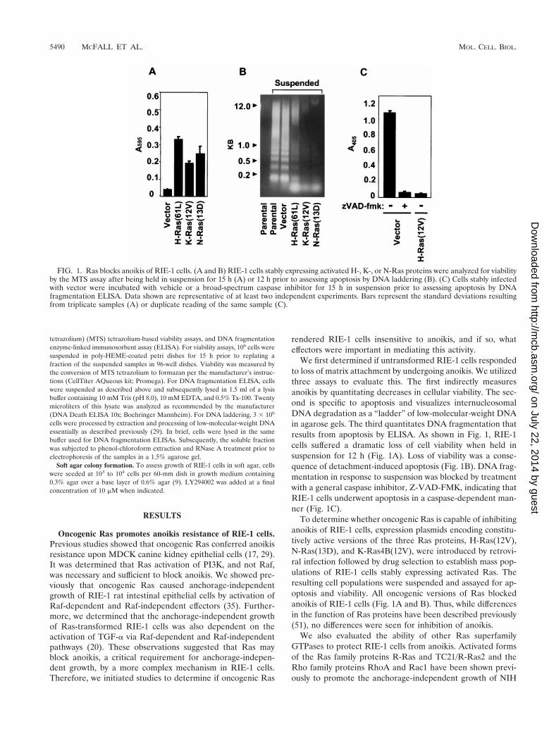
We first determined if untransformed RIE-1 cells respondedto loss of matrix attachment by undergoing anoikis. We utilizedthree assays to evaluate this. The first indirectly measuresanoikis by quantitating decreases in cellular viability. The sec-ond is specific to apoptosis and visualizes internucleosomalDNA degradation as a “ladder” of low-molecular-weight DNAin agarose gels. The third quantitates DNA fragmentation thatresults from apoptosis by ELISA. As shown in Fig. 1, RIE-1cells suffered a dramatic loss of cell viability when held insuspension for 12 h (Fig. 1A). Loss of viability was a conse-quence of detachment-induced apoptosis (Fig. 1B). DNA frag-mentation in response to suspension was blocked by treatmentwith a general caspase inhibitor, Z-VAD-FMK, indicating thatRIE-1 cells underwent apoptosis in a caspase-dependent man-ner (Fig. 1C).
To determine whether oncogenic Ras is capable of inhibitinganoikis of RIE-1 cells, expression plasmids encoding constitu-tively active versions of the three Ras proteins, H-Ras(12V),N-Ras(13D), and K-Ras4B(12V), were introduced by retrovi-ral infection followed by drug selection to establish mass pop-ulations of RIE-1 cells stably expressing activated Ras. Theresulting cell populations were suspended and assayed for ap-optosis and viability. All oncogenic versions of Ras blockedanoikis of RIE-1 cells (Fig. 1A and B). Thus, while differencesin the function of Ras proteins have been described previously(51), no differences were seen for inhibition of anoikis.
We also evaluated the ability of other Ras superfamilyGTPases to protect RIE-1 cells from anoikis. Activated formsof the Ras family proteins R-Ras and TC21/R-Ras2 and theRho family proteins RhoA and Rac1 have been shown previ-ously to promote the anchorage-independent growth of NIH
FIG. 1. Ras blocks anoikis of RIE-1 cells. (A and B) RIE-1 cells stably expressing activated H-, K-, or N-Ras proteins were analyzed for viabilityby the MTS assay after being held in suspension for 15 h (A) or 12 h prior to assessing apoptosis by DNA laddering (B). (C) Cells stably infectedwith vector were incubated with vehicle or a broad-spectrum caspase inhibitor for 15 h in suspension prior to assessing apoptosis by DNAfragmentation ELISA. Data shown are representative of at least two independent experiments. Bars represent the standard deviations resultingfrom triplicate samples (A) or duplicate reading of the same sample (C).
5490 MCFALL ET AL. MOL. CELL. BIOL.
on July 22, 2014 by guesthttp://m
cb.asm.org/
Dow
nloaded from
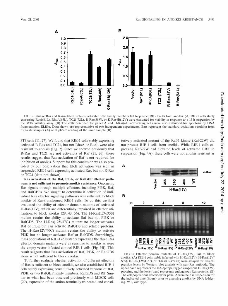
3T3 cells (11, 27). We found that RIE-1 cells stably expressingactivated R-Ras and TC21, but not RhoA or Rac1, were alsoresistant to anoikis (Fig. 2). Since we showed previously thatR-Ras and TC21 are not activators of Raf (21, 26), theseresults suggest that Ras activation of Raf is not required forinhibition of anoikis. Support for this conclusion was also pro-vided by our observation that ERK activation was seen insuspended RIE-1 cells expressing activated Ras, but not R-Rasor TC21 (data not shown).
Ras activation of the Raf, PI3K, or RalGEF effector path-ways is not sufficient to promote anoikis resistance. OncogenicRas signals through multiple effectors, including PI3K, Raf,and RalGEFs. We sought to determine if activation of indi-vidual Ras effector signaling pathways was sufficient to blockanoikis of Ras-transformed RIE-1 cells. To do this, we firstevaluated the ability of effector domain mutants of activatedH-Ras(12V), which are differentially impaired in effector uti-lization, to block anoikis (28, 43, 56). The H-Ras(12V/35S)mutant retains the ability to activate Raf but not PI3K orRalGDS. The H-Ras(12V/37G) mutant no longer activatesRaf or PI3K but can activate RalGDS and related proteins.The H-Ras(12V/40C) mutant retains the ability to activatePI3K but no longer activates Raf or RalGDS. Surprisingly,mass populations of RIE-1 cells stably expressing the three Raseffector domain mutants were as sensitive to anoikis as werethe empty vector-infected control RIE-1 cells (Fig. 3B). Thisresult suggests that Ras activation of Raf, PI3K, or RalGDSalone is not sufficient to block anoikis.
To further evaluate whether activation of different effectorsof Ras is sufficient to block anoikis, we also established RIE-1cells stably expressing constitutively activated versions of Raf,PI3K, or two RalGEF family members, RalGDS and Rlf. Sim-ilar to what had been observed previously with MDCK cells(29), expression of the amino-terminally truncated and consti-
tutively activated mutant of the Raf-1 kinase (Raf-22W) didnot protect RIE-1 cells from anoikis. While RIE-1 cells ex-pressing Raf-22W had elevated levels of activated ERK insuspension (Fig. 4A), these cells were not anoikis resistant as
FIG. 2. Unlike Ras and Ras-related proteins, activated Rho family members fail to protect RIE-1 cells from anoikis. (A) RIE-1 cells stablyexpressing Rac1(61L), RhoA(63L), TC21(72L), R-Ras(38V), or K-Ras4B(12V) were evaluated for viability in response to a 15-h suspension bythe MTS viability assay. (B) The cells described for panel A and H-Ras(61L)-expressing cells were also evaluated for apoptosis by DNAfragmentation ELISA. Data shown are representative of two independent experiments. Bars represent the standard deviations resulting fromtriplicate samples (A) or duplicate reading of the same sample (B).
FIG. 3. Effector domain mutants of H-Ras(12V) fail to blockanoikis. (A) RIE-1 cells stably infected with H-Ras(12V), H-Ras(12V/S35), H-Ras(12V/G37), or H-Ras(12V/C40) were assayed for Ras ex-pression levels by Western blot analysis with pan-Ras antibody. Theupper band represents the HA epitope-tagged exogenous H-Ras(12V)proteins, and the lower band represents endogenous Ras proteins. (B)The cell populations described for panel A were held in suspension forthe indicated time (hours) prior to assessing anoikis by DNA ladder-ing. WT, wild type.
VOL. 21, 2001 Ras SIGNALING IN ANOIKIS RESISTANCE 5491
on July 22, 2014 by guesthttp://m
cb.asm.org/
Dow
nloaded from
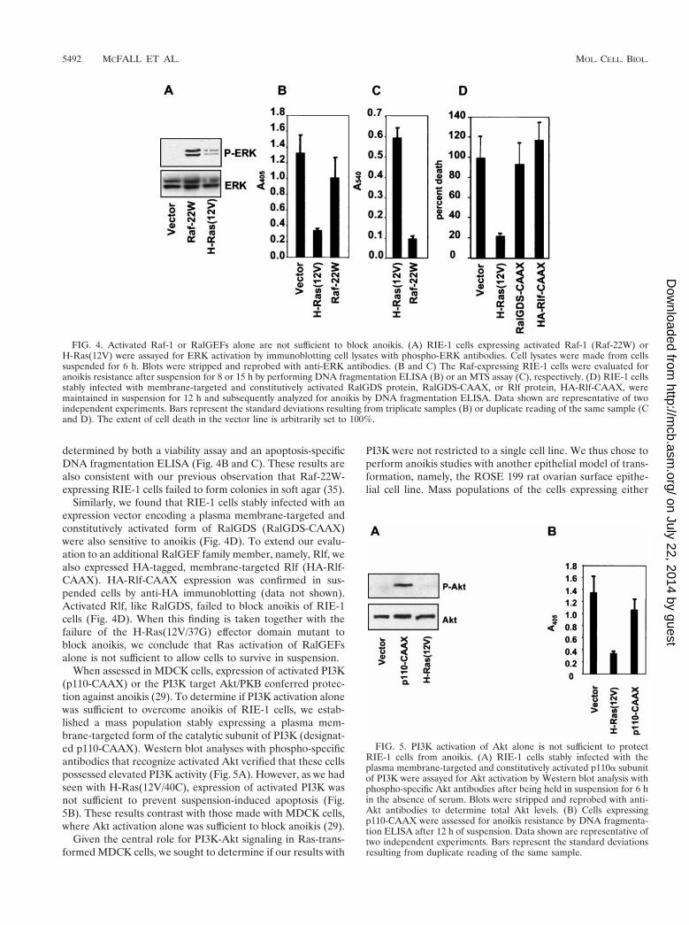
determined by both a viability assay and an apoptosis-specificDNA fragmentation ELISA (Fig. 4B and C). These results arealso consistent with our previous observation that Raf-22W-expressing RIE-1 cells failed to form colonies in soft agar (35).
Similarly, we found that RIE-1 cells stably infected with anexpression vector encoding a plasma membrane-targeted andconstitutively activated form of RalGDS (RalGDS-CAAX)were also sensitive to anoikis (Fig. 4D). To extend our evalu-ation to an additional RalGEF family member, namely, Rlf, wealso expressed HA-tagged, membrane-targeted Rlf (HA-Rlf-CAAX). HA-Rlf-CAAX expression was confirmed in sus-pended cells by anti-HA immunoblotting (data not shown).Activated Rlf, like RalGDS, failed to block anoikis of RIE-1cells (Fig. 4D). When this finding is taken together with thefailure of the H-Ras(12V/37G) effector domain mutant toblock anoikis, we conclude that Ras activation of RalGEFsalone is not sufficient to allow cells to survive in suspension.
When assessed in MDCK cells, expression of activated PI3K(p110-CAAX) or the PI3K target Akt/PKB conferred protec-tion against anoikis (29). To determine if PI3K activation alonewas sufficient to overcome anoikis of RIE-1 cells, we estab-lished a mass population stably expressing a plasma mem-brane-targeted form of the catalytic subunit of PI3K (designat-ed p110-CAAX). Western blot analyses with phospho-specificantibodies that recognize activated Akt verified that these cellspossessed elevated PI3K activity (Fig. 5A). However, as we hadseen with H-Ras(12V/40C), expression of activated PI3K wasnot sufficient to prevent suspension-induced apoptosis (Fig.5B). These results contrast with those made with MDCK cells,where Akt activation alone was sufficient to block anoikis (29).
Given the central role for PI3K-Akt signaling in Ras-trans-formed MDCK cells, we sought to determine if our results with
PI3K were not restricted to a single cell line. We thus chose toperform anoikis studies with another epithelial model of trans-formation, namely, the ROSE 199 rat ovarian surface epithe-lial cell line. Mass populations of the cells expressing either
FIG. 4. Activated Raf-1 or RalGEFs alone are not sufficient to block anoikis. (A) RIE-1 cells expressing activated Raf-1 (Raf-22W) orH-Ras(12V) were assayed for ERK activation by immunoblotting cell lysates with phospho-ERK antibodies. Cell lysates were made from cellssuspended for 6 h. Blots were stripped and reprobed with anti-ERK antibodies. (B and C) The Raf-expressing RIE-1 cells were evaluated foranoikis resistance after suspension for 8 or 15 h by performing DNA fragmentation ELISA (B) or an MTS assay (C), respectively. (D) RIE-1 cellsstably infected with membrane-targeted and constitutively activated RalGDS protein, RalGDS-CAAX, or Rlf protein, HA-Rlf-CAAX, weremaintained in suspension for 12 h and subsequently analyzed for anoikis by DNA fragmentation ELISA. Data shown are representative of twoindependent experiments. Bars represent the standard deviations resulting from triplicate samples (B) or duplicate reading of the same sample (Cand D). The extent of cell death in the vector line is arbitrarily set to 100%.
FIG. 5. PI3K activation of Akt alone is not sufficient to protectRIE-1 cells from anoikis. (A) RIE-1 cells stably infected with theplasma membrane-targeted and constitutively activated p110a subunitof PI3K were assayed for Akt activation by Western blot analysis withphospho-specific Akt antibodies after being held in suspension for 6 hin the absence of serum. Blots were stripped and reprobed with anti-Akt antibodies to determine total Akt levels. (B) Cells expressingp110-CAAX were assessed for anoikis resistance by DNA fragmenta-tion ELISA after 12 h of suspension. Data shown are representative oftwo independent experiments. Bars represent the standard deviationsresulting from duplicate reading of the same sample.
5492 MCFALL ET AL. MOL. CELL. BIOL.
on July 22, 2014 by guesthttp://m
cb.asm.org/
Dow
nloaded from
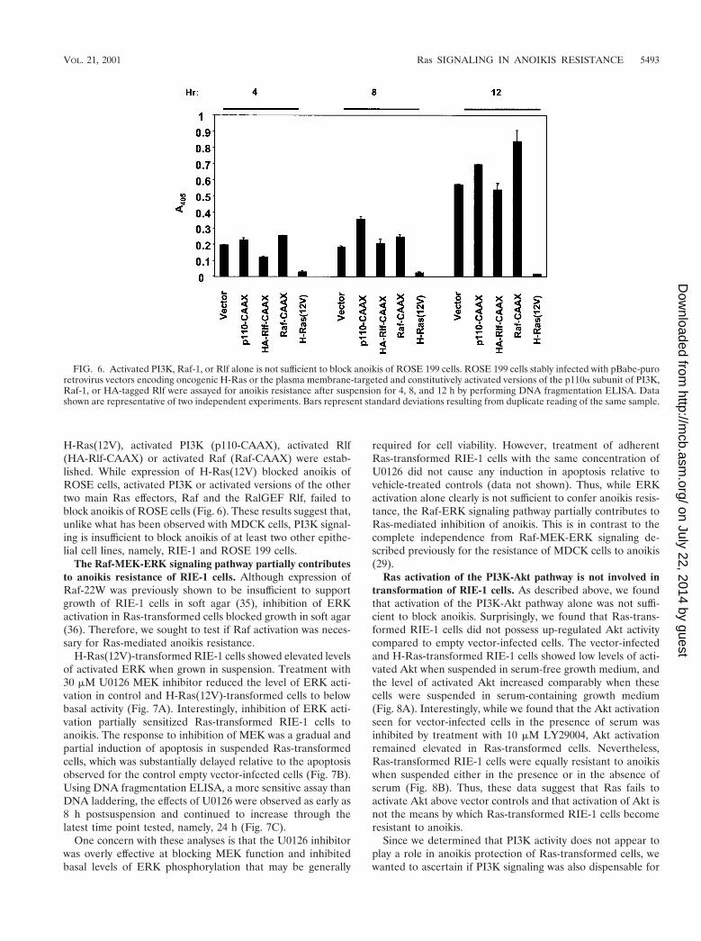
H-Ras(12V), activated PI3K (p110-CAAX), activated Rlf(HA-Rlf-CAAX) or activated Raf (Raf-CAAX) were estab-lished. While expression of H-Ras(12V) blocked anoikis ofROSE cells, activated PI3K or activated versions of the othertwo main Ras effectors, Raf and the RalGEF Rlf, failed toblock anoikis of ROSE cells (Fig. 6). These results suggest that,unlike what has been observed with MDCK cells, PI3K signal-ing is insufficient to block anoikis of at least two other epithe-lial cell lines, namely, RIE-1 and ROSE 199 cells.
The Raf-MEK-ERK signaling pathway partially contributesto anoikis resistance of RIE-1 cells. Although expression ofRaf-22W was previously shown to be insufficient to supportgrowth of RIE-1 cells in soft agar (35), inhibition of ERKactivation in Ras-transformed cells blocked growth in soft agar(36). Therefore, we sought to test if Raf activation was neces-sary for Ras-mediated anoikis resistance.
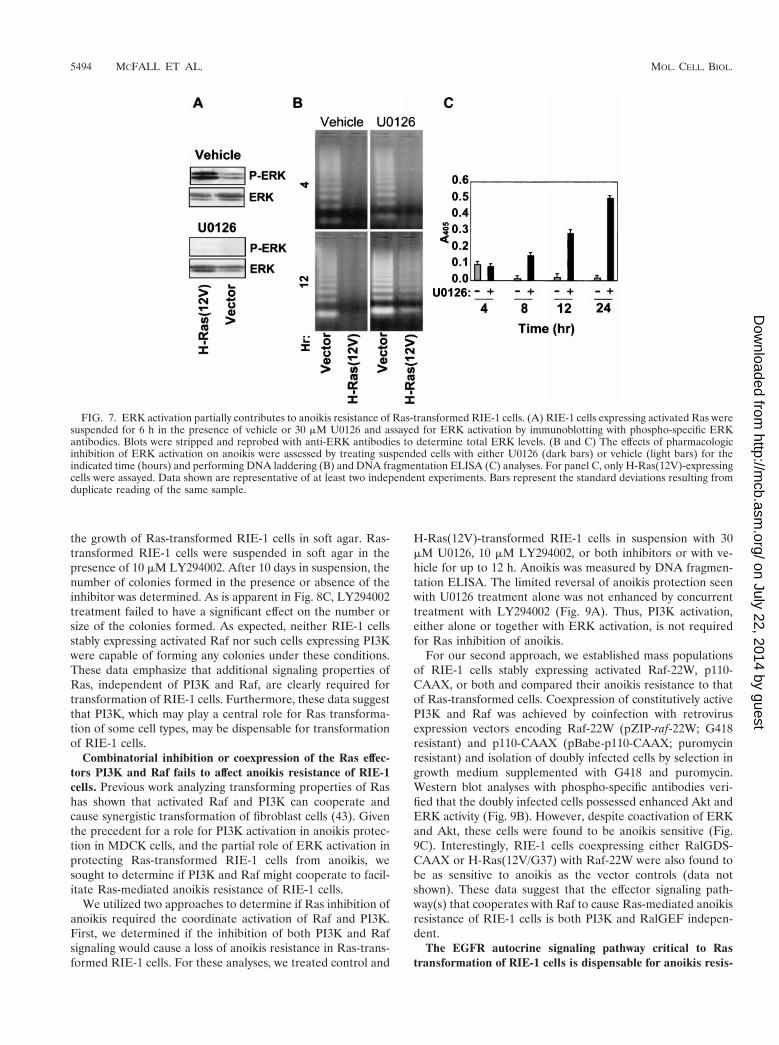
H-Ras(12V)-transformed RIE-1 cells showed elevated levelsof activated ERK when grown in suspension. Treatment with30 mM U0126 MEK inhibitor reduced the level of ERK acti-vation in control and H-Ras(12V)-transformed cells to belowbasal activity (Fig. 7A). Interestingly, inhibition of ERK acti-vation partially sensitized Ras-transformed RIE-1 cells toanoikis. The response to inhibition of MEK was a gradual andpartial induction of apoptosis in suspended Ras-transformedcells, which was substantially delayed relative to the apoptosisobserved for the control empty vector-infected cells (Fig. 7B).Using DNA fragmentation ELISA, a more sensitive assay thanDNA laddering, the effects of U0126 were observed as early as8 h postsuspension and continued to increase through thelatest time point tested, namely, 24 h (Fig. 7C).
One concern with these analyses is that the U0126 inhibitorwas overly effective at blocking MEK function and inhibitedbasal levels of ERK phosphorylation that may be generally
required for cell viability. However, treatment of adherentRas-transformed RIE-1 cells with the same concentration ofU0126 did not cause any induction in apoptosis relative tovehicle-treated controls (data not shown). Thus, while ERKactivation alone clearly is not sufficient to confer anoikis resis-tance, the Raf-ERK signaling pathway partially contributes toRas-mediated inhibition of anoikis. This is in contrast to thecomplete independence from Raf-MEK-ERK signaling de-scribed previously for the resistance of MDCK cells to anoikis(29).
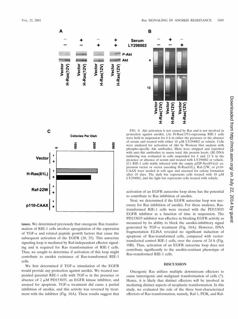
Ras activation of the PI3K-Akt pathway is not involved intransformation of RIE-1 cells. As described above, we foundthat activation of the PI3K-Akt pathway alone was not suffi-cient to block anoikis. Surprisingly, we found that Ras-trans-formed RIE-1 cells did not possess up-regulated Akt activitycompared to empty vector-infected cells. The vector-infectedand H-Ras-transformed RIE-1 cells showed low levels of acti-vated Akt when suspended in serum-free growth medium, andthe level of activated Akt increased comparably when thesecells were suspended in serum-containing growth medium(Fig. 8A). Interestingly, while we found that the Akt activationseen for vector-infected cells in the presence of serum wasinhibited by treatment with 10 mM LY29004, Akt activationremained elevated in Ras-transformed cells. Nevertheless,Ras-transformed RIE-1 cells were equally resistant to anoikiswhen suspended either in the presence or in the absence ofserum (Fig. 8B). Thus, these data suggest that Ras fails toactivate Akt above vector controls and that activation of Akt isnot the means by which Ras-transformed RIE-1 cells becomeresistant to anoikis.
Since we determined that PI3K activity does not appear toplay a role in anoikis protection of Ras-transformed cells, wewanted to ascertain if PI3K signaling was also dispensable for
FIG. 6. Activated PI3K, Raf-1, or Rlf alone is not sufficient to block anoikis of ROSE 199 cells. ROSE 199 cells stably infected with pBabe-puroretrovirus vectors encoding oncogenic H-Ras or the plasma membrane-targeted and constitutively activated versions of the p110a subunit of PI3K,Raf-1, or HA-tagged Rlf were assayed for anoikis resistance after suspension for 4, 8, and 12 h by performing DNA fragmentation ELISA. Datashown are representative of two independent experiments. Bars represent standard deviations resulting from duplicate reading of the same sample.
VOL. 21, 2001 Ras SIGNALING IN ANOIKIS RESISTANCE 5493
on July 22, 2014 by guesthttp://m
cb.asm.org/
Dow
nloaded from
the growth of Ras-transformed RIE-1 cells in soft agar. Ras-transformed RIE-1 cells were suspended in soft agar in thepresence of 10 mM LY294002. After 10 days in suspension, thenumber of colonies formed in the presence or absence of theinhibitor was determined. As is apparent in Fig. 8C, LY294002treatment failed to have a significant effect on the number orsize of the colonies formed. As expected, neither RIE-1 cellsstably expressing activated Raf nor such cells expressing PI3Kwere capable of forming any colonies under these conditions.These data emphasize that additional signaling properties ofRas, independent of PI3K and Raf, are clearly required fortransformation of RIE-1 cells. Furthermore, these data suggestthat PI3K, which may play a central role for Ras transforma-tion of some cell types, may be dispensable for transformationof RIE-1 cells.
Combinatorial inhibition or coexpression of the Ras effec-tors PI3K and Raf fails to affect anoikis resistance of RIE-1cells. Previous work analyzing transforming properties of Rashas shown that activated Raf and PI3K can cooperate andcause synergistic transformation of fibroblast cells (43). Giventhe precedent for a role for PI3K activation in anoikis protec-tion in MDCK cells, and the partial role of ERK activation inprotecting Ras-transformed RIE-1 cells from anoikis, wesought to determine if PI3K and Raf might cooperate to facil-itate Ras-mediated anoikis resistance of RIE-1 cells.
We utilized two approaches to determine if Ras inhibition ofanoikis required the coordinate activation of Raf and PI3K.First, we determined if the inhibition of both PI3K and Rafsignaling would cause a loss of anoikis resistance in Ras-trans-formed RIE-1 cells. For these analyses, we treated control and
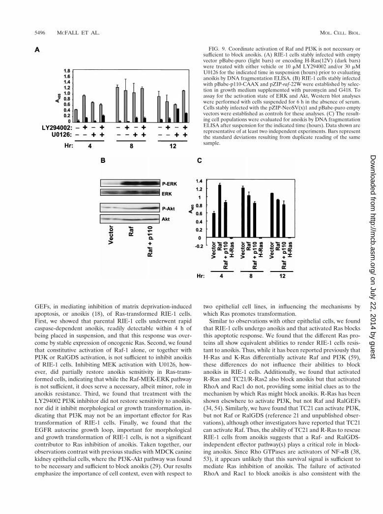
H-Ras(12V)-transformed RIE-1 cells in suspension with 30mM U0126, 10 mM LY294002, or both inhibitors or with ve-hicle for up to 12 h. Anoikis was measured by DNA fragmen-tation ELISA. The limited reversal of anoikis protection seenwith U0126 treatment alone was not enhanced by concurrenttreatment with LY294002 (Fig. 9A). Thus, PI3K activation,either alone or together with ERK activation, is not requiredfor Ras inhibition of anoikis.
For our second approach, we established mass populationsof RIE-1 cells stably expressing activated Raf-22W, p110-CAAX, or both and compared their anoikis resistance to thatof Ras-transformed cells. Coexpression of constitutively activePI3K and Raf was achieved by coinfection with retrovirusexpression vectors encoding Raf-22W (pZIP-raf-22W; G418resistant) and p110-CAAX (pBabe-p110-CAAX; puromycinresistant) and isolation of doubly infected cells by selection ingrowth medium supplemented with G418 and puromycin.Western blot analyses with phospho-specific antibodies veri-fied that the doubly infected cells possessed enhanced Akt andERK activity (Fig. 9B). However, despite coactivation of ERKand Akt, these cells were found to be anoikis sensitive (Fig.9C). Interestingly, RIE-1 cells coexpressing either RalGDS-CAAX or H-Ras(12V/G37) with Raf-22W were also found tobe as sensitive to anoikis as the vector controls (data notshown). These data suggest that the effector signaling path-way(s) that cooperates with Raf to cause Ras-mediated anoikisresistance of RIE-1 cells is both PI3K and RalGEF indepen-dent.
The EGFR autocrine signaling pathway critical to Rastransformation of RIE-1 cells is dispensable for anoikis resis-
FIG. 7. ERK activation partially contributes to anoikis resistance of Ras-transformed RIE-1 cells. (A) RIE-1 cells expressing activated Ras weresuspended for 6 h in the presence of vehicle or 30 mM U0126 and assayed for ERK activation by immunoblotting with phospho-specific ERKantibodies. Blots were stripped and reprobed with anti-ERK antibodies to determine total ERK levels. (B and C) The effects of pharmacologicinhibition of ERK activation on anoikis were assessed by treating suspended cells with either U0126 (dark bars) or vehicle (light bars) for theindicated time (hours) and performing DNA laddering (B) and DNA fragmentation ELISA (C) analyses. For panel C, only H-Ras(12V)-expressingcells were assayed. Data shown are representative of at least two independent experiments. Bars represent the standard deviations resulting fromduplicate reading of the same sample.
5494 MCFALL ET AL. MOL. CELL. BIOL.
on July 22, 2014 by guesthttp://m
cb.asm.org/
Dow
nloaded from
tance. We determined previously that oncogenic Ras transfor-mation of RIE-1 cells involves upregulation of the expressionof TGF-a and related peptide growth factors that cause thesubsequent activation of the EGFR (20, 35). This autocrinesignaling loop is mediated by Raf-independent effector signal-ing and is required for Ras transformation of RIE-1 cells.Thus, we sought to determine if activation of this loop mightcontribute to anoikis resistance of Ras-transformed RIE-1cells.
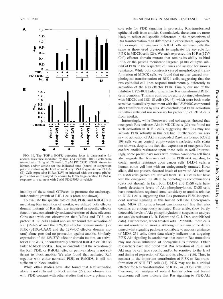
We first determined if TGF-a stimulation of the EGFRwould provide any protection against anoikis. We treated sus-pended parental RIE-1 cells with TGF-a in the presence orabsence of 2 mM PD153035, an EGFR kinase inhibitor, andassayed for apoptosis. TGF-a treatment did cause a partialinhibition of anoikis, and this activity was reversed by treat-ment with the inhibitor (Fig. 10A). These results suggest that
activation of an EGFR autocrine loop alone has the potentialto contribute to Ras inhibition of anoikis.
Next, we determined if the EGFR autocrine loop was nec-essary for Ras inhibition of anoikis. For these analyses, Ras-transformed RIE-1 cells were treated with the PD153035EGFR inhibitor as a function of time in suspension. ThePD153035 inhibitor was effective in blocking EGFR activity asmeasured by its ability to block the anoikis-inhibitory signalgenerated by TGF-a treatment (Fig. 10A). However, DNAfragmentation ELISA revealed no significant induction ofapoptosis of Ras-transformed cells, compared with vector-transfected control RIE-1 cells, over the course of 24 h (Fig.10B). Thus, activation of an EGFR autocrine loop does notcontribute significantly to the anoikis-resistant phenotype ofRas-transformed RIE-1 cells.
DISCUSSION
Oncogenic Ras utilizes multiple downstream effectors tocause tumorigenic and malignant transformation of cells (7).Hence, it is likely that distinct effectors will be involved inmediating distinct aspects of neoplastic transformation. In thisstudy, we evaluated the role of the three best-characterizedeffectors of Ras transformation, namely, Raf-1, PI3K, and Ral-
FIG. 8. Akt activation is not caused by Ras and is not involved inprotection against anoikis. (A) H-Ras(12V)-expressing RIE-1 cellswere held in suspension for 6 h in either the presence or the absenceof serum and treated with either 10 mM LY294002 or vehicle. Cellswere analyzed for activation of Akt by Western blot analysis withphospho-specific Akt antibodies. Blots were stripped and reprobedwith anti-Akt antibodies to assess total Akt protein levels. (B) DNAladdering was evaluated in cells suspended for 4 and 12 h in thepresence or absence of serum and treated with LY294002 or vehicle.(C) RIE-1 cells stably infected with the empty pZIP-NeoSV(x)1 ex-pression vector or vector encoding H-Ras(61L), Raf-22W, or p110-CAAX were seeded in soft agar and assessed for colony formationafter 10 days. The dark bar represents cells treated with 10 mMLY294002, and the light bar represents cells treated with vehicle.
VOL. 21, 2001 Ras SIGNALING IN ANOIKIS RESISTANCE 5495
on July 22, 2014 by guesthttp://m
cb.asm.org/
Dow
nloaded from
GEFs, in mediating inhibition of matrix deprivation-inducedapoptosis, or anoikis (18), of Ras-transformed RIE-1 cells.First, we showed that parental RIE-1 cells underwent rapidcaspase-dependent anoikis, readily detectable within 4 h ofbeing placed in suspension, and that this response was over-come by stable expression of oncogenic Ras. Second, we foundthat constitutive activation of Raf-1 alone, or together withPI3K or RalGDS activation, is not sufficient to inhibit anoikisof RIE-1 cells. Inhibiting MEK activation with U0126, how-ever, did partially restore anoikis sensitivity in Ras-trans-formed cells, indicating that while the Raf-MEK-ERK pathwayis not sufficient, it does serve a necessary, albeit minor, role inanoikis resistance. Third, we found that treatment with theLY294002 PI3K inhibitor did not restore sensitivity to anoikis,nor did it inhibit morphological or growth transformation, in-dicating that PI3K may not be an important effector for Rastransformation of RIE-1 cells. Finally, we found that theEGFR autocrine growth loop, important for morphologicaland growth transformation of RIE-1 cells, is not a significantcontributor to Ras inhibition of anoikis. Taken together, ourobservations contrast with previous studies with MDCK caninekidney epithelial cells, where the PI3K-Akt pathway was foundto be necessary and sufficient to block anoikis (29). Our resultsemphasize the importance of cell context, even with respect to
two epithelial cell lines, in influencing the mechanisms bywhich Ras promotes transformation.
Similar to observations with other epithelial cells, we foundthat RIE-1 cells undergo anoikis and that activated Ras blocksthis apoptotic response. We found that the different Ras pro-teins all show equivalent abilities to render RIE-1 cells resis-tant to anoikis. Thus, while it has been reported previously thatH-Ras and K-Ras differentially activate Raf and PI3K (59),these differences do not influence their abilities to blockanoikis in RIE-1 cells. Additionally, we found that activatedR-Ras and TC21/R-Ras2 also block anoikis but that activatedRhoA and Rac1 do not, providing some initial clues as to themechanism by which Ras might block anoikis. R-Ras has beenshown elsewhere to activate PI3K, but not Raf and RalGEFs(34, 54). Similarly, we have found that TC21 can activate PI3K,but not Raf or RalGDS (reference 21 and unpublished obser-vations), although other investigators have reported that TC21can activate Raf. Thus, the ability of TC21 and R-Ras to rescueRIE-1 cells from anoikis suggests that a Raf- and RalGDS-independent effector pathway(s) plays a critical role in block-ing anoikis. Since Rho GTPases are activators of NF-kB (38,53), it appears unlikely that this survival signal is sufficient tomediate Ras inhibition of anoikis. The failure of activatedRhoA and Rac1 to block anoikis is also consistent with the
FIG. 9. Coordinate activation of Raf and PI3K is not necessary orsufficient to block anoikis. (A) RIE-1 cells stably infected with emptyvector pBabe-puro (light bars) or encoding H-Ras(12V) (dark bars)were treated with either vehicle or 10 mM LY294002 and/or 30 mMU0126 for the indicated time in suspension (hours) prior to evaluatinganoikis by DNA fragmentation ELISA. (B) RIE-1 cells stably infectedwith pBabe-p110-CAAX and pZIP-raf-22W were established by selec-tion in growth medium supplemented with puromycin and G418. Toassay for the activation state of ERK and Akt, Western blot analyseswere performed with cells suspended for 6 h in the absence of serum.Cells stably infected with the pZIP-NeoSV(x)1 and pBabe-puro emptyvectors were established as controls for these analyses. (C) The result-ing cell populations were evaluated for anoikis by DNA fragmentationELISA after suspension for the indicated time (hours). Data shown arerepresentative of at least two independent experiments. Bars representthe standard deviations resulting from duplicate reading of the samesample.
5496 MCFALL ET AL. MOL. CELL. BIOL.
on July 22, 2014 by guesthttp://m
cb.asm.org/
Dow
nloaded from
inability of these small GTPases to promote the anchorage-independent growth of RIE-1 cells (data not shown).
To evaluate the specific role of Raf, PI3K, and RalGEFs inmediating Ras inhibition of anoikis, we utilized both effectordomain mutants of Ras that are impaired in specific effectorfunction and constitutively activated versions of these effectors.Consistent with our observation that R-Ras and TC21 canprotect RIE-1 cells against anoikis, we found that activation ofRaf (Raf-22W and the 12V/35S effector domain mutant) orPI3K (p110a-CAAX and the 12V/40C effector domain mu-tant) alone provided no protection against anoikis. Similarly,expression of the 12V/37G effector domain mutant, an activa-tor of RalGEFs, or constitutively activated RalGDS or Rlf alsofailed to block anoikis. Thus, we conclude that the activation ofthe Raf, PI3K, or RalGEF effector pathway alone is not suf-ficient to block anoikis. We also found that activated Raf,together with either activated PI3K or RalGDS, is still notsufficient to block anoikis.
While other studies have also found that Raf activationalone is not sufficient to block anoikis (29), our observationswith PI3K contrast with other studies that show a primary or
sole role for PI3K signaling in protecting Ras-transformedepithelial cells from anoikis. Cumulatively, these data are morelikely to reflect cell-specific differences in the mechanisms ofRas transformation than differences in experimental approach.For example, our analyses of RIE-1 cells are essentially thesame as those used previously to implicate the key role forPI3K in MDCK cells (29). We each expressed the H-Ras(12V/C40) effector domain mutant that retains its ability to bindPI3K or the plasma membrane-targeted p110a catalytic sub-unit of PI3K in the respective cell lines and assayed for anoikisresistance. While both constructs caused morphological trans-formation of MDCK cells, we found that neither caused mor-phological transformation of RIE-1 cells, suggesting that thetwo epithelial cell lines respond fundamentally differently toactivation of the Ras effector PI3K. Finally, our use of theinhibitor LY294002 failed to sensitize Ras-transformed RIE-1cells to anoikis. This is in contrast to results obtained elsewherewith MDCK and IEC-18 cells (29, 46), which were both madesensitive to anoikis by treatment with the LY294002 compoundafter transformation by Ras. We conclude that PI3K activationis neither sufficient nor necessary for protection of RIE-1 cellsfrom anoikis.
Interestingly, while Downward and colleagues showed thatoncogenic Ras activates Akt in MDCK cells (29), we found nosuch activation in RIE-1 cells, suggesting that Ras may notactivate PI3K robustly in this cell line. Furthermore, we alsosaw no activation of Akt in suspended Ras-transformed ROSE199 cells versus control empty-vector-transfected cells (datanot shown), despite the fact that expression of oncogenic Rasconfers anoikis resistance upon these cells as well. Interest-ingly, some preliminary work with human carcinoma cell linesalso suggests that Ras may not utilize PI3K-Akt signaling toconfer anoikis resistance upon cancer cells. DLD-1 cells, ahuman colon cell line with an endogenously mutated K-rasallele, did not possess elevated levels of activated Akt relativeto DkS8 cells (which are derived from DLD-1 cells but havelost the oncogenic ras allele by homologous recombination)(data not shown). In fact, both DLD-1 and DkS8 cells havebarely detectable levels of Akt phosphorylation. DkS8 cellshave nonetheless regained some sensitivity to anoikis relativeto DLD-1 cells, suggesting that Ras promotes PI3K-indepen-dent survival signaling in this human cell line. Correspond-ingly, MDA 231 cells, a breast carcinoma cell line that alsocontains an endogenously activated K-ras allele, show barelydetectable levels of Akt phosphorylation in suspension and yetare anoikis resistant (L. B. Eckert and C. J. Der, unpublisheddata). Furthermore, when treated with LY294002, these cellsare not sensitized to anoikis. Although it remains to be deter-mined what signaling pathways contribute to anoikis resistanceof MDA 231 cells, these data clearly indicate that targetingPI3K-Akt signaling in carcinomas that contain Ras mutationsmay not cause inhibition of oncogenic Ras function. Otherresearchers have also noted that Ras activation of PI3K andAkt may be cell type specific and/or be sensitive to the leveland timing of expression of Ras and its effectors (16). Thus, incontrast to the important contribution of PI3K to Ras trans-formation of NIH 3T3 cells (43), PI3K may not be a criticaleffector for Ras transformation of RIE-1 or ROSE cells. Fur-thermore, our analyses of several human colon and breastcarcinoma cell lines indicate that Ras signaling to PI3K-Akt
FIG. 10. The TGF-a–EGFR autocrine loop is dispensable foranoikis resistance mediated by Ras. (A) Parental RIE-1 cells weretreated with 10 ng of TGF-a/ml, 2 mM PD153035 EGFR kinase in-hibitor, and/or vehicle for the indicated time (hours) in suspensionprior to evaluating the level of anoikis by DNA fragmentation ELISA.(B) Cells expressing H-Ras(12V) or infected with the empty pBabe-puro vector were assayed for anoikis by DNA fragmentation ELISA inresponse to treatment with 2 mM PD153035 or vehicle.
VOL. 21, 2001 Ras SIGNALING IN ANOIKIS RESISTANCE 5497
on July 22, 2014 by guesthttp://m
cb.asm.org/
Dow
nloaded from
does not play a role in anoikis resistance of at least somecancers that suffer Ras mutations in vivo.
Like the Ras effector PI3K, we observed a different role forRaf in mediating anoikis resistance of Ras-transformed RIE-1cells compared to other epithelial cells. Unlike what has beenreported elsewhere for MDCK and IEC-18 rat intestinal epi-thelial cells (29, 46), inhibiting MEK in Ras-transformedRIE-1 cells caused a partial reversion to an anoikis-sensitivestate. Expression of activated Raf, however, was not sufficientto cause anoikis resistance of RIE-1 cells, even when coex-pressed with activated PI3K or RalGEFs. Thus, Raf is likely tocooperate with an unknown PI3K- and RalGEF-independenteffector(s) to block anoikis. Interestingly, cooperation betweentwo or more Ras effectors also appears to be required forRas-mediated anoikis resistance of IEC-18 cells (47), althoughPI3K, and not Raf, appears to play a pivotal role in anoikisresistance of these cells. Cooperation between Ras effectorshas also been observed for mediating other facets of transfor-mation, for example, focus formation (43, 56). Thus, perhaps itis not surprising that cooperativity exists between Ras effectorsin mediating anoikis resistance.
We found previously that a TGF-a–EGFR autocrine loop iscritical for complete transformation of RIE-1 cells, includinggrowth in soft agar (20). Consistent with this observation, wefound that activation of the EGFR with TGF-a is sufficient tocause partial resistance to anoikis of suspended parental RIE-1cells. Surprisingly, however, inhibition of EGFR signaling hasvery little effect on anoikis resistance of Ras-transformedRIE-1 cells, at least over the span of 24 h. Perhaps this signifiesthat EGFR signaling plays a redundant role with a Ras effec-tor(s) in inhibiting anoikis of Ras-transformed RIE-1 cells.
We have not yet identified what Ras effector(s) cooperateswith Raf to induce anoikis resistance of RIE-1 cells. In addi-tion to Raf, PI3K, and RalGEFs, there is an expanding rosterof other proteins that interact with Ras in its activated, GTP-bound state (51). These include AF-6, Nore-1, Rin1, PKCz,MEKK1, RASSF1, and the Ras GTPase-activated proteins(p120 and NF1). Although some of these proteins are unlikelyto play a role in inhibition of anoikis, for example, MEKK1,whose activation is associated with induction of apoptosis (8),none have thus far been tested for their ability to cooperatewith Raf in our study or other anoikis studies. However, sinceRin1 association is retained in the 37G effector domain mutant(24) and AF-6 binding is retained in the 37G and 40C mutants(28), it is unlikely that engagement of Rin1 and AF-6 is suffi-cient to block anoikis. Perhaps the key effector for Ras inhibi-tion of anoikis in RIE-1 cells remains to be identified.
How might Raf activity promote anoikis resistance of RIE-1cells? Activation of the MAPK signaling pathway can inhibitapoptosis in response to growth factor withdrawal, death re-ceptor activation, and, as very recently described for fibroblastsand MDCK cells, detachment from extracellular matrix (25,32). Given the gradual and delayed sensitization of Ras-trans-formed RIE-1 cells to anoikis in response to MEK inhibition,one possible mechanism of action of this inhibitor is to causechanges in gene expression that regulate apoptosis. Alongthese lines, inhibition of MEK in mammalian cells can reduceexpression of several antiapoptotic proteins of the Bcl-2 familyas well as reduce expression of the antiapoptotic gene par-4 (2,5). Alternatively, MEK signaling may have immediate effects
on the apoptosis machinery, for example, by modulating theactivation state of Bad (48). Whether the above-mentionedconsequences of MEK activation play a role in anoikis resis-tance of Ras-transformed RIE-1 cells remains to be deter-mined.
In summary, we have determined that PI3K is not a keyeffector for Ras inhibition of anoikis or transformation inRIE-1 cells. Furthermore, oncogenic Ras did not cause up-regulation of the PI3K-Akt pathway in RIE-1 cells. Our resultsemphasize that Ras promotes transformation in multiple waysin a cell-context-dependent fashion. We are currently assessingthe importance of the Raf-ERK, PI3K-Akt, and other Rassignaling pathways in protecting human tumor cells fromanoikis. We suspect that our studies will find a similar com-plexity, with different Ras effector pathways playing importantroles in some, but not all, tumor cells. Thus, ultimately inhib-iting oncogenic Ras function in various human tumors mayrequire effectively targeting a variety of Ras effector signalingpathways.
ACKNOWLEDGMENTS
We thank Julian Downward for providing the p110a-CAAX cDNAconstruct, Johannes Bos for providing the pMT2-HA-Rlf-CAAX con-struct, James Trzaskos (Dupont) for providing U0126, Heena Mehtaand Staeci Morita for technical support, and Misha Rand for prepa-ration of figures.
Our research was supported by grants from the National Institutesof Health (NIH) to C.J.D. (CA42978, CA55008, and CA63071). A.M.was additionally supported by an NIH training grant fellowship and anNIH National Research Service Award (CA84633-02).
REFERENCES
1. Barbacid, M. 1987. ras genes. Annu. Rev. Biochem. 56:779–827.2. Barradas, M., A. Monjas, M. T. Diaz-Meco, M. Serrano, and J. Moscat.
1999. The downregulation of the pro-apoptotic protein Par-4 is critical forRas-induced survival and tumor progression. EMBO J. 18:6362–6369.
3. Blay, J., and K. D. Brown. 1984. Characterization of an epithelioid cell linederived from the rat small intestine. Cell Biol. Int. Rep. 8:551–560.
4. Bos, J. L. 1989. ras oncogenes in human cancer: a review. Cancer Res.49:4682–4689.
5. Boucher, M. J., J. Morisset, P. H. Vachon, J. C. Reed, J. Laine, and N.Rivard. 2000. MEK/ERK signaling pathway regulates the expression of bcl-2,bcl-X(L), and mcl-1 and promotes survival of human pancreatic cancer cells.J. Cell. Biochem. 79:355–369.
6. Boudreau, N., C. J. Sympson, Z. Werb, and M. J. Bissell. 1995. Suppressionof ICE and apoptosis in mammary epithelial cells by extracellular matrix.Science 267:891–893.
7. Campbell, S. L., R. Khosravi-Far, K. L. Rossman, G. J. Clark, and C. J. Der.1998. Increasing complexity of Ras signaling. Oncogene 17:1395–1413.
8. Cardone, M. H., G. S. Salvesen, C. Widmann, G. Johnson, and S. M. Frisch.1997. The regulation of anoikis: MEKK-1 activation requires cleavage bycaspases. Cell 90:315–323.
9. Clark, G. J., A. D. Cox, S. M. Graham, and C. J. Der. 1995. Biological assaysfor Ras transformation. Methods Enzymol. 255:395–412.
10. Clark, G. J., and C. J. Der. 1993. Oncogenic activation of Ras proteins, p.259–288. In B. F. Dickey and L. Birnbaumer (ed.), GTPases in biology I.Springer Verlag, Berlin, Germany.
11. Cox, A. D., T. R. Brtva, D. G. Lowe, and C. J. Der. 1994. R-Ras inducesmalignant, but not morphologic, transformation of NIH3T3 cells. Oncogene9:3281–3288.
12. Downward, J. 1998. Mechanisms and consequences of activation of proteinkinase B/Akt. Curr. Opin. Cell Biol. 10:262–267.
13. Favata, M. F., K. Y. Horiuchi, E. J. Manos, A. J. Daulerio, D. A. Stradley,W. S. Feeser, D. E. Van Dyk, W. J. Pitts, R. A. Earl, F. Hobbs, R. A.Copeland, R. L. Magolda, P. A. Scherle, and J. M. Trzaskos. 1998. Identi-fication of a novel inhibitor of mitogen-activated protein kinase kinase.J. Biol. Chem. 273:18623–18632.
14. Fearnhead, H. O., D. Dinsdale, and G. M. Cohen. 1995. An interleukin-1beta-converting enzyme-like protease is a common mediator of apoptosis inthymocytes. FEBS Lett. 375:283–288.
15. Feig, L. A., T. Urano, and S. Cantor. 1996. Evidence for a Ras/Ral signalingcascade. Trends Biochem. Sci. 21:438–441.
5498 MCFALL ET AL. MOL. CELL. BIOL.
on July 22, 2014 by guesthttp://m
cb.asm.org/
Dow
nloaded from
16. Franke, T. F., S. I. Yang, T. O. Chan, K. Datta, A. Kazlauskas, D. K.Morrison, D. R. Kaplan, and P. N. Tsichlis. 1995. The protein kinase en-coded by the Akt proto-oncogene is a target of the PDGF-activated phos-phatidylinositol 3-kinase. Cell 81:727–736.
17. Frisch, S. M., and H. Francis. 1994. Disruption of epithelial cell-matrixinteractions induces apoptosis. J. Cell Biol. 124:619–626.
18. Frisch, S. M., and E. Ruoslahti. 1997. Integrins and anoikis. Curr. Opin. CellBiol. 9:701–706.
19. Fry, D. W., A. J. Kraker, A. McMichael, L. A. Ambroso, J. M. Nelson, W. R.Leopold, R. W. Connors, and A. J. Bridges. 1994. A specific inhibitor of theepidermal growth factor receptor tyrosine kinase. Science 265:1093–1095.
20. Gangarosa, L. M., N. Sizemore, R. Graves-Deal, S. M. Oldham, C. J. Der,and R. J. Coffey. 1997. A Raf-independent epidermal growth factor receptorautocrine loop is necessary for Ras transformation of rat intestinal epithelialcells. J. Biol. Chem. 272:18926–18931.
21. Graham, S. M., A. B. Vojtek, S. Y. Huff, A. D. Cox, G. J. Clark, J. A. Cooper,and C. J. Der. 1996. TC21 causes transformation by Raf-independent sig-naling pathways. Mol. Cell. Biol. 16:6132–6140.
22. Hahn, W. C., C. M. Counter, A. S. Lundberg, R. L. Beijersbergen, M. W.Brooks, and R. A. Weinberg. 1999. Creation of human tumour cells withdefined genetic elements. Nature 400:464–468.
23. Hall, P. A., P. J. Coates, B. Ansari, and D. Hopwood. 1994. Regulation of cellnumber in the mammalian gastrointestinal tract: the importance of apopto-sis. J. Cell Sci. 107:3569–3577.
24. Han, L., D. Wong, A. Dhaka, D. Afar, M. White, W. Xie, H. Herschman, O.Witte, and J. Colicelli. 1997. Protein binding and signaling properties ofRIN1 suggest a unique effector function. Proc. Natl. Acad. Sci. USA 94:4954–4959.
25. Holmstrom, T. H., S. C. Chow, I. Elo, E. T. Coffey, S. Orrenius, L. Sistonen,and J. E. Eriksson. 1998. Suppression of Fas/APO-1-mediated apoptosis bymitogen-activated kinase signaling. J. Immunol. 160:2626–2636.
26. Huff, S. Y., L. A. Quilliam, A. D. Cox, and C. J. Der. 1997. R-Ras is regulatedby activators and effectors distinct from those that control Ras function.Oncogene 14:133–143.
27. Khosravi-Far, R., P. A. Solski, G. J. Clark, M. S. Kinch, and C. J. Der. 1995.Activation of Rac1, RhoA, and mitogen-activated protein kinases is requiredfor Ras transformation. Mol. Cell. Biol. 15:6443–6453.
28. Khosravi-Far, R., M. A. White, J. K. Westwick, P. A. Solski, M. Chrza-nowska-Wodnicka, L. Van Aelst, M. H. Wigler, and C. J. Der. 1996. Onco-genic Ras activation of Raf/mitogen-activated protein kinase-independentpathways is sufficient to cause tumorigenic transformation. Mol. Cell. Biol.16:3923–3933.
29. Khwaja, A., P. Rodriguez-Viciana, S. Wennstrom, P. H. Warne, and J.Downward. 1997. Matrix adhesion and Ras transformation both activate aphosphoinositide 3-OH kinase and protein kinase B/Akt cellular survivalpathway. EMBO J. 16:2783–2793.
30. Kops, G. J., N. D. de Ruiter, A. M. de Vries-Smits, D. R. Powell, J. L. Bos,and B. M. Burgering. 1999. Direct control of the Forkhead transcriptionfactor AFX by protein kinase B. Nature 398:630–634.
31. Leevers, S. J., H. F. Paterson, and C. J. Marshall. 1994. Requirement forRas in Raf activation is overcome by targeting Raf to the plasma membrane.Nature 369:411–414.
32. Le Gall, M., J. C. Chambard, J. P. Breittmayer, D. Grall, J. Pouyssegur, andE. Obberghen-Schilling. 2000. The p42/p44 MAP kinase pathway preventsapoptosis induced by anchorage and serum removal. Mol. Biol. Cell 11:1103–1112.
33. Marshall, C. J. 1996. Ras effectors. Curr. Opin. Cell Biol. 8:197–204.34. Marte, B. M., P. Rodriguez-Viciana, S. Wennstrom, P. H. Warne, and J.
Downward. 1996. R-Ras can activate the phosphoinositide 3-kinase but notthe MAP kinase arm of the Ras effector pathways. Curr. Biol. 7:63–70.
35. Oldham, S. M., G. J. Clark, L. M. Gangarosa, R. J. Coffey, Jr., and C. J. Der.1996. Activation of the Raf-1/MAP kinase cascade is not sufficient for Rastransformation of RIE-1 epithelial cells. Proc. Natl. Acad. Sci. USA 93:6924–6928.
36. Oldham, S. M., A. D. Cox, E. R. Reynolds, N. S. Sizemore, R. J. Coffey, Jr.,and C. J. Der. 1998. Ras, but not Src, transformation of RIE-1 epithelial cellsis dependent on activation of the mitogen-activated protein kinase cascade.Oncogene 16:2565–2573.
37. Pear, W. S., G. P. Nolan, M. L. Scott, and D. Baltimore. 1993. Production ofhigh-titer helper-free retroviruses by transient transfection. Proc. Natl. Acad.Sci. USA 90:8392–8396.
38. Perona, R., S. Montaner, L. Saniger, I. Sanchez-Perez, R. Bravo, and J. C.Lacal. 1997. Activation of the nuclear factor-KB by Rho, CDC42, and Rac-1proteins. Genes Dev. 11:463–475.
39. Petersen, O. W., L. Ronnov-Jessen, V. M. Weaver, and M. J. Bissell. 1998.Differentiation and cancer in the mammary gland: shedding light on an olddichotomy. Adv. Cancer Res. 75:135–161.
40. Polakowska, R. R., M. Piacentini, R. Bartlett, L. A. Goldsmith, and A. R.Haake. 1994. Apoptosis in human skin development: morphogenesis, peri-derm, and stem cells. Dev. Dyn. 199:176–188.
41. Ramocki, M. B., M. A. White, S. F. Konieczny, and E. J. Taparowsky. 1998.A role for RalGDS and a novel Ras effector in the Ras-mediated inhibitionof skeletal myogenesis. J. Biol. Chem. 273:17696–17701.
42. Rodriguez-Viciana, P., P. H. Warne, R. Dhand, B. Vanhaesebroeck, I. Gout,M. J. Fry, M. D. Waterfield, and J. Downward. 1994. Phosphatidylinositol-3-OH kinase as a direct target of Ras. Nature 370:527–532.
43. Rodriguez-Viciana, P., P. H. Warne, A. Khwaja, B. M. Marte, D. Pappin, P.Das, M. D. Waterfield, A. Ridley, and J. Downward. 1997. Role of phospho-inositide 3-OH kinase in cell transformation and control of the actin cy-toskeleton by Ras. Cell 89:457–467.
44. Rodriguez-Viciana, P., P. H. Warne, B. Vanhaesebroeck, M. D. Waterfield,and J. Downward. 1996. Activation of phosphoinositide 3-kinase by interac-tion with Ras and by point mutation. EMBO J. 15:2442–2451.
45. Romashkova, J. A., and S. S. Makarov. 1999. NF-kappaB is a target of AKTin anti-apoptotic PDGF signalling. Nature 401:86–90.
46. Rosen, K., J. Rak, J. Jin, R. S. Kerbel, M. J. Newman, and J. Filmus. 1998.Downregulation of the pro-apoptotic protein Bak is required for the ras-induced transformation of intestinal epithelial cells. Curr. Biol. 8:1331–1334.
47. Rosen, K., J. Rak, T. Leung, N. M. Dean, R. S. Kerbel, and J. Filmus. 2000.Activated Ras prevents downregulation of Bcl-X(L) triggered by detachmentfrom the extracellular matrix. A mechanism of Ras-induced resistance toanoikis in intestinal epithelial cells. J. Cell Biol. 149:447–456.
48. Scheid, M. P., K. M. Schubert, and V. Duronio. 1999. Regulation of badphosphorylation and association with Bcl-x(L) by the MAPK/Erk kinase.J. Biol. Chem. 274:31108–31113.
49. Schwartz, M. A. 1997. Integrins, oncogenes, and anchorage independence.J. Cell Biol. 139:575–578.
50. Serrano, M., A. W. Lin, M. E. McCurrach, D. Beach, and S. W. Lowe. 1997.Oncogenic ras provokes premature cell senescence associated with accumu-lation of p53 and p16INK4a. Cell 88:593–602.
51. Shields, J. M., K. Pruitt, A. McFall, A. Shaub, and C. J. Der. 2000. Under-standing Ras: “it ain’t over ’til it’s over.” Trends Cell Biol. 10:147–153.
52. Stanton, V. P., Jr., D. W. Nichols, A. P. Laudano, and G. M. Cooper. 1989.Definition of the human raf amino-terminal regulatory region by deletionmutagenesis. Mol. Cell. Biol. 9:639–647.
53. Sulciner, D. J., K. Irani, Z.-X. Yu, V. J. Ferrans, P. Goldschmidt-Clermont,and T. Finkel. 1996. rac1 regulates a cytokine-stimulated, redox-dependentpathway necessary for NF-kB activation. Mol. Cell. Biol. 16:7115–7121.
54. Urano, T., R. Emkey, and L. A. Feig. 1996. Ral-GTPases mediate a distinctdownstream signaling pathway from Ras that facilitates cellular transforma-tion. EMBO J. 15:810–816.
55. Vlahos, C. J., W. F. Matter, K. Y. Hui, and R. F. Brown. 1994. A specificinhibitor of phosphatidylinositol 3-kinase, 2-(4-morpholinyl)-8-phenyl-4H-1-benzopyran-4-one (LY294002). J. Biol. Chem. 269:5241–5248.
56. White, M. A., C. Nicolette, A. Minden, A. Polverino, L. Van Aelst, M. Karin,and M. H. Wigler. 1995. Multiple Ras functions can contribute to mamma-lian cell transformation. Cell 80:533–541.
57. Wolthuis, R. M., and J. L. Bos. 1999. Ras caught in another affair: theexchange factors for Ral. Curr. Opin. Genet. Dev. 9:112–117.
58. Wolthuis, R. M., N. D. de Ruiter, R. H. Cool, and J. L. Bos. 1997. Stimulationof gene induction and cell growth by the Ras effector Rlf. EMBO J. 16:6748–6761.
59. Yan, J., S. Roy, A. Apolloni, A. Lane, and J. F. Hancock. 1998. Ras isoformsvary in their ability to activate Raf-1 and phosphoinositide 3-kinase. J. Biol.Chem. 273:24052–24056.
VOL. 21, 2001 Ras SIGNALING IN ANOIKIS RESISTANCE 5499
on July 22, 2014 by guesthttp://m
cb.asm.org/
Dow
nloaded from


