Embed Size (px)
Citation preview
Kidney International, Vol. 59 (2001), pp. 1498–1509
Obesity-related glomerulopathy: An emerging epidemic
NEERAJA KAMBHAM, GLEN S. MARKOWITZ, ANTHONY M. VALERI, JULIE LIN,and VIVETTE D. D’AGATI
Departments of Pathology and Medicine, Columbia University, College of Physicians and Surgeons, New York, New York USA
cess fusion. The ten-fold increase in incidence over 15 yearsObesity-related glomerulopathy: An emerging epidemic.suggests a newly emerging epidemic. Heightened physicianBackground. We report the first large renal biopsy-basedawareness of this entity is needed to ensure accurate diagnosisclinicopathologic study on obesity-related glomerulopathy.and appropriate therapy.Methods. Obesity was defined as body mass index (BMI)
.30 kg/m2. Obesity-related glomerulopathy (ORG) was de-fined morphologically as focal segmental glomerulosclerosisand glomerulomegaly (O-FSGS; N 5 57) or glomerulomegaly
Cross-sectional epidemiologic studies indicate that thealone (O-GM; N 5 14).prevalence of obesity in the United States and otherResults. Review of 6818 native renal biopsies received from
1986 to 2000 revealed a progressive increase in biopsy incidence Western countries has been steadily rising over the pastof ORG from 0.2% in 1986–1990 to 2.0% in 1996–2000 (P 5 two decades, a trend that has been linked to changing0.0001). Mean BMI in ORG was 41.7 (range 30.9 to 62.7). dietary habits and sedentary lifestyle [1–3]. This in-Indications for renal biopsy included proteinuria (N 5 40)
creased prevalence has been reported in both childrenor proteinuria and renal insufficiency (N 5 31). Seventy-oneand adults, and across diverse ethnic groups [1]. Ac-patients with ORG were compared to 50 patients with idio-
pathic FSGS (I-FSGS). Patients with ORG were older (mean cording to the National Health and Nutrition Examina-42.9 vs. 32.6 years, P , 0.001) and more often Caucasian (75% tion Survey, the prevalence of obesity (defined as bodyvs. 52%; P 5 0.003). ORG patients had a lower incidence of mass index, BMI .30) has increased from 14.1% tonephrotic range proteinuria (48% vs. 66%; P 5 0.007) and
22.5% between 1971 and 1994, and 54.9% of the U.S.nephrotic syndrome (5.6% vs. 54%; P , 0.001), with higherpopulation is overweight (BMI .25) [1]. Obese patientsserum albumin (3.9 vs. 2.9 g/dL; P , 0.001), lower serum
cholesterol (229 vs. 335 mg/dL; P , 0.001), and less edema are at greater risk to develop sleep apnea, hyperlipid-(35% vs. 68%; P 5 0.003). On renal biopsy, patients with ORG emia, hypertension, coronary vascular disease, insulinhad fewer lesions of segmental sclerosis (10 vs. 39%; P ,
resistance and diabetes [4–6].0.001), more glomerulomegaly (100% vs. 10%; P , 0.001), andThe renal effects of obesity in experimental animalsless extensive foot process effacement (40 vs. 75%; P , 0.001).
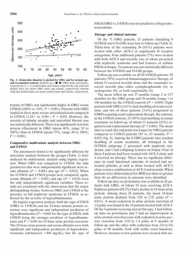
Glomerular diameter in ORG (mean 226 m) was significantly and humans include both structural and functional adap-larger than age- and sex-matched normal controls (mean 168 m; tations, such as increased glomerular filtration rate, in-P , 0.001). Follow-up was available in 56 ORG patients (mean creased renal blood flow, and renal hypertrophy [7, 8].27 months) and 50 idiopathic FSGS controls (mean 38 months).
In 1974, an association between massive obesity andA total of 75% of ORG patients received angiotensin-con-nephrotic-range proteinuria was first reported [9]. Sinceverting enzyme (ACE) inhibition or A2 blockade while 78%
of the I-FSGS patients received immunosuppressive therapy. that time, the development of glomerulomegaly and fo-ORG patients had less frequent doubling of serum creatinine cal segmental glomerulosclerosis (FSGS) has been linked(14.3% vs. 50%; P , 0.001) and progression to ESRD (3.6% to massive obesity [8, 10–14]. Most of these associationsvs. 42%; P , 0.001). On multivariate analysis, presenting serum
have been limited to case reports or small autopsy series.creatinine and severity of proteinuria were the only predictorsThus, little is known about the overall prevalence ofof poor outcome in ORG.
Conclusion. ORG is distinct from idiopathic FSGS, with a biopsy-documented obesity-related glomerulopathy, itslower incidence of nephrotic syndrome, more indolent course, presenting clinical features, morphologic manifestations,consistent presence of glomerulomegaly, and milder foot pro- or natural history.
The current study was designed to determine thechanging biopsy incidence of obesity-related glomeru-Key words: focal segmental glomerulosclerosis, nephrotic syndrome,
overweight, glomerulomegaly. lopathy (ORG) over the past 15 years. To better definethe spectrum of clinical and pathologic features of thisReceived for publication June 27, 2000entity, a cohort of ORG was compared to controls withand in revised form October 23, 2000
Accepted for publication October 26, 2000 idiopathic FSGS (I-FSGS). Our findings indicate thatORG is an increasingly prevalent disease that is clinically 2001 by the International Society of Nephrology
1498
Kambham et al: Obesity-related glomerulopathy 1499
and pathologically distinct from I-FSGS. The differenti- (excluding cellular and collapsing variants) diagnosedating clinicopathologic features of ORG have important from 1980 to 1992.implications for patient management and prognosis. Renal biopsies for the study cohort and controls were
processed for light microscopy, immunofluorescence(IF) and electron microscopy (EM) according to stan-METHODSdard techniques. At least 11 serial sections (3 mm thick)
All native renal biopsies accessioned in the Renal Pa- were stained with hematoxylin and eosin, periodic-acidthology Laboratory of Columbia Presbyterian Medical Schiff (PAS), Masson’s trichrome and Jones methena-Center from January 1986 to April 2000 were reviewed mine-silver stains (JMS). Routine IF was performed onretrospectively for evidence of ORG. Obesity was de- 3 mm cryostat sections using polyclonal FITC-conjugatedfined as a BMI .30, calculated as body weight in kilo- antibodies to IgG, IgM, IgA, C3, C1q, k, l, fibrinogengrams divided by the square of height in meters [1, 15]. and albumin (Dako Corporation, Carpenteria, CA,ORG was defined morphologically as (1) obesity-associ- USA). In 61 of 71 ORG cases, adequate glomerularated FSGS with glomerulomegaly (O-FSGS) or (2) obe- tissue was available for EM.sity-associated glomerulomegaly alone (O-GM). Renal Juxtaglomerular apparatus (JGA) hyperplasia was de-biopsies from obese patients with other underlying con-
fined as .8 nuclei per cross section of JGA [18]. Tubularditions that could cause secondary FSGS (such as HIV
atrophy, interstitial fibrosis and inflammation wereinfection, heroin abuse, solitary kidney, congenital heartgraded semiquantitatively on a scale of 0 to 31 baseddisease, sickle cell disease, renal dysplasia, or any otheron the % cortical area affected: 0 5 absent; 11 5 1–25%;pre-existing renal disease with loss of renal mass) were21 5 26–50% and 31 . 50%. Arteriosclerosis and arte-carefully excluded. Moreover, other defined primary andriolosclerosis were graded as follows: 0 5 absent; 11 5secondary glomerular diseases including diabetic ne-mild; 21 5 moderate; 31 5 severe. The intensity ofphropathy and hypertensive nephrosclerosis occurringstaining by immunofluorescence was graded on a scalein obese patients were eliminated. Of all 6818 nativeof trace (1/2) to 31. The percentage of foot processbiopsies received during this period, a total of 103 caseseffacement was estimated based on examination of allmet the entry criteria. From this group, 71 cases withnonsclerotic glomerular capillaries in all fields.adequate clinical information were studied. Because of
Measurement of glomerular diameter was performedthe limited follow-up, newly diagnosed cases of obesity-using a light microscopic eyepiece with built-in micro-related glomerulopathy from the year 2000 were ex-meter (Olympus Scientific, Tokyo, Japan). The conver-cluded.sion factor for each objective was calibrated against aPatient charts were reviewed for age, sex, race, andstandard by the manufacturer. Four levels of biopsy tis-presenting clinical and laboratory features at the timesue (PAS and JMS stains) were studied in each case. Allof renal biopsy. From the height and weight at the timeglomeruli cut at or near the hilus were measured. Twoof renal biopsy, BMI was calculated. Obesity was definedperpendicular diameters extending between the farthestas BMI .30 kg/m2: BMI 30.0 to 34.9 kg/m2, class I obesity;glomerular basement membrane points were measuredBMI 35.0 to 39.9 kg/m2, class II obesity; BMI $40 kg/m2,per glomerulus. The mean glomerular diameter (mm)class III or “morbid” obesity. The following definitionswas calculated from a total of 5 to 30 (mean 15) glomeruliwere used: hypertension (HTN), systolic pressure .140examined per case. Glomerular diameter was comparedmm Hg and diastolic pressure .90 mm Hg; nephroticto that of 21 age- and sex-matched normal controls ob-range proteinuria (NRP), 24-hour urine protein excre-tained from percutaneous renal biopsies of patients withtion $3.5 g; hematuria, presence of .5 red blood cellsisolated asymptomatic hematuria or subnephrotic pro-per high power field on microscopic examination of theteinuria who had no evidence of glomerular disease byurinary sediment; hypoalbuminemia, serum albumin lev-light microscopy, IF, or EM.els #3.5 g/dL; hypercholesterolemia, serum cholesterol
The following clinical and laboratory data at follow-.200 mg/dL; nephrotic syndrome, the combination ofup were analyzed for the group with ORG and I-FSGS:NRP, hypoalbuminemia and edema; and renal insuffi-Therapy directed to renal disease or obesity (includingciency, serum creatinine .1.2 mg/dL on two separateweight reduction, sleep apnea therapy, angiotensin-con-determinations. Creatinine clearances calculated fromverting enzyme inhibitors (ACE-I) or angiotensin II re-24-hour urine collections were available in 58 patients.ceptor antagonists, steroids or other immunosuppressiveClearances were adjusted for body surface area usingagents) was recorded for each patient. Measures of se-height and weight according to the following formula:rum creatinine, 24-hour urine protein, and body weight[(height in centimeters)(weight in kilograms)/3600]1/2
at last available follow-up were analyzed. Life-table anal-[16, 17]. Presenting clinical and laboratory features wereysis was performed using the outcome points of doublingcompared to a well-characterized historical control
group of 50 patients with I-FSGS of the classic type, of serum creatinine and end-stage renal disease.
Kambham et al: Obesity-related glomerulopathy1500
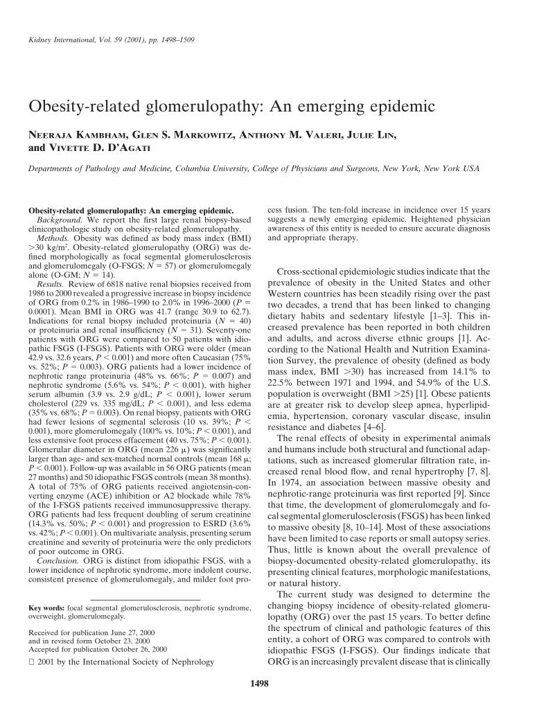
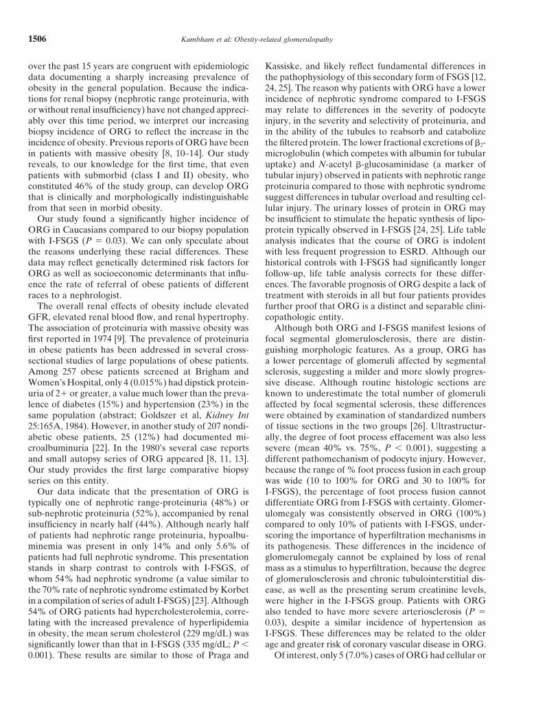
8-year-old girl [height 4.0 ft (121.9 cm), weight 190 lbs(86.4 kg)] and three patients in their second decade oflife; the oldest patients included seven in their sixth de-cade and one in his seventh decade. The patients’ weightsranged from 180 lbs to 410 lbs (81.8 kg to 186.4 kg). Themean BMI was 41.7 kg/m2 (range of 30.9 to 62.7); 38patients had BMI .40 kg/m2 and 33 had BMI ,40 kg/m2.The male-to-female ratio was 44:27 (1.6). The majorityof patients were Caucasian (75%), followed by AfricanAmerican (21%) and Hispanic (4%). Nine patients hadhistory of diabetes mellitus (12.7%) for a mean durationof 4.8 years (range 0.5 to 26 years). However, none ofthese patients had renal biopsy findings of diabetic ne-phropathy. Forty-four patients had a prior history ofhypertension of a mean 9 years’ (range from 0.2 to 40Fig. 1. The increased incidence of obesity-related glomerulopathyyears) duration. Eight patients carried a diagnosis of(ORG) is plotted as a percentage of total native renal biopsies received
over a 15-year period. obstructive sleep apnea syndrome prior to renal biopsy;however, sleep apnea studies were not performed on themajority of patients.
The indications for renal biopsy were isolated protein-Statistical analysisuria in 40 patients (56%) and proteinuria and renal insuf-
Statistical analysis was performed by exact statistical ficiency in 31 patients (44%). The mean serum creatinineinference using the following nonparametric methods: at presentation was 1.5 mg/dL (range 0.6 to 6.3 mg/dL).Fisher’s exact test, the Mann-Whitney U test, and the The creatinine clearance (CCr) was available in 58 pa-Wilcoxon signed ranks test, as appropriate. The Monte tients and the mean value was 113 cc/min (range 20 toCarlo approximation method was used where the statisti- 240 cc/min). The mean CCr adjusted for BSA was 82.9cal package could not solve the analysis by exact method- mL/min/1.73 m2 (range 13 to 186). Twenty-four of 58ology within 15 minutes or iteration processing time. patients (41.4%) had CCr .130 cc/min. Based on an ad-SPSSt 10.0.5 was used to perform the analysis. Multivari- justed CCr, 8 of 58 patients (13.8%) had a CCr .130 mL/ate analysis was performed by logistic regression analy- min/1.73 m2 and 32 patients (55%) had CCr ,75 mL/min/sis. Patient and renal outcome survival analysis was per- 1.73 m2. The mean 24-hour urine protein was 4.1 g (rangeformed by the method of Kaplan and Meier with 1 to 32 g). Although 34 patients (48%) had nephrotic-statistical comparison by the log rank test. A multivariate range proteinuria at presentation, only 4 patients (5.6%)analysis for predictors of outcome was performed by had nephrotic syndrome. The mean serum albumin levelCox regression analysis. Statistical significance was as- was 3.9 g/dL (range 1.5 to 5.0 g/dL) and only ten patientssumed at P , 0.05. (14%) had hypoalbuminemia. Twenty-five patients
(35%) had pedal edema at presentation. The serum cho-lesterol levels ranged from 152 to 303 mg/dL (mean 229RESULTSmg/dL) and 38 patients (54%) were hypercholesterol-
Biopsy incidence of obesity-related glomerulopathy emic. Microhematuria was documented in 15 patients.The biopsy incidence of ORG was determined for the When the presenting clinical features of the ORG
15-year period of January 1986 to April 2000 (Fig. 1). group were compared with I-FSGS controls, many sig-Among the 103 cases of ORG identified from 1986 to nificant differences were found (Table 1). Patients with2000, only two cases (2 of 956 native kidney biopsies; ORG were more likely to be Caucasian (74.6 vs. 52%;0.2%) were identified from January 1986 to December P 5 0.003) and tended to be older (mean age 42.9 vs.1990; 24 cases (24 of 2013; 1.2%) were identified from 32.6 years; P , 0.001) compared to historical controlsJanuary 1991 to December 1995; and 77 cases (77 of with I-FSGS. Although nearly half of the ORG patients3849; 2%) were identified from January 1996 to April presented with nephrotic-range proteinuria (48% vs.2000. This represents a ten-fold increase in biopsy inci- 66%; P 5 0.007), the incidence of pedal edema (35%dence of ORG over 15 years (P 5 0.0001). vs. 68%; P 5 0.003) and nephrotic syndrome (5.6% vs.
60%; P , 0.001) was significantly lower in the ORGClinical features at presentation group. Correspondingly, the mean serum albumin levels
The mean age of patients at the time of biopsy diagno- were higher (3.9 g/dL vs. 2.9 g/dL; P , 0.001) and thesis of ORG was 42.9 years (range 8–71 years; Table 1). mean 24-hour urine protein excretion was lower (4.1 g
vs. 6.9 g; P 5 0.002) than in I-FSGS controls. The meanThe youngest patients in the study group included an
Kambham et al: Obesity-related glomerulopathy 1501
Table 1. Clinical parameters and pathological findings in patients with ORG, O-FSGS and I-FSGS
ORG O-FSGS I-FSGS ORG vs. I-FSGS O-FSGS vs. I-FSGSParameter (N 5 71) (N 5 57) (N 5 50) (P value) (P value)
Mean age at presentation years 42.93 43.82 32.57 ,0.001 ,0.001Sex male:female 44:27 37:20 25:25 0.186 0.092Race/ethnicity N (%) 0.003 0.018
Caucasian 53 (74.6) 40 (70.2) 26 (52)African American 15 (21.1) 14 (24.5) 11 (22)Hispanic 3 (4.2) 3 (5.3) 11 (22)Other 0 0 2 (4)
HTN 1 N (%) 44 (62) 37 (64.9) 28 (56) 0.574 0.428Mean SCr at biopsy mg/dL 1.47 1.55 1.96 0.8 0.903Proteinuria 0.007 0.012
Non-nephrotic proteinuria N (%) 37 (52.1) 30 (52.6) 17 (34)Nephrotic range proteinuria N (%) 34 (47.9) 27 (47.4) 33 (66)
Nephrotic parametersNephrotic syndrome N (%) 4 (5.6) 4 (7) 27 (54) ,0.001 ,0.001Mean 24-hour urine protein g 4.09 4.24 6.89 0.002 0.004Mean serum albumin g/dL 3.87 3.8 2.9 ,0.001 ,0.001Mean serum cholesterol mg/dL 229.2 231.65 335.13 ,0.001 ,0.001Presence of edema N (%) 25 (35) 23 (40.4) 34 (68) 0.003 0.01
PathologyMean % global sclerosis 20% 25% 18% 0.423 0.027Mean % segmental sclerosis 10% 12% 39% ,0.001 ,0.001% of cases with glomerulomegaly 100% 100% 10% ,0.001 ,0.001Mean TA/IF (grade 0–3) mild (1.08) mild (1.26) mild (1.22) 0.238 0.912Mean arteriolosclerosis (grade 0–3) mild (1.34) mild (1.42) mild (0.98) 0.032 0.014Foot process fusion 40% 41% 75% ,0.001 ,0.001
Abbreviations are: ORG, obesity-related glomerulopathy; FSGS, focal segmental glomerulosclerosis; O-FSGS, obesity-related FSGS; I-FSGS, idiopathic FSGS;HTN, hypertension; TA/IF, tubular atrophy and interstitial fibrosis. Terms are defined in the text.
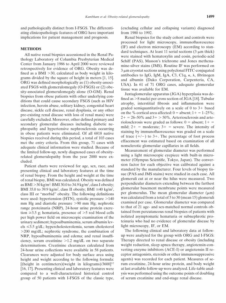
serum cholesterol level, although elevated, was signifi- and the percentage of segmentally sclerotic glomeruliranged from 3% to 50% (mean 12%; Table 1). All casescantly lower than in I-FSGS patients (229 mg/dL vs. 335
mg/dL; P , 0.001). Although patients with ORG were had lesions of segmental sclerosis of the “classic” orusual type, characterized by segmental obliteration andmore likely to be hypertensive (62% vs. 56%) and had
slightly lower mean serum creatinine at biopsy (1.47 vs. solidification of glomerular capillary lumina by increasedmatrix, forming expansile scars, often associated with1.96 mg/dL), these differences were not statistically sig-
nificant. inframembranous hyaline, endocapillary foam cells andBowman’s capsular adhesions (Fig. 2 A–C). In someBecause the ORG group contained some patients with
glomerulomegaly alone (O-GM) as well as the larger cases, the hypertrophied podocytes overlying the lesionswere segmentally detached from the glomerular base-group with glomerulomegaly and FSGS (O-FSGS), com-
parative analyses were also performed for O-FSGS pa- ment membranes and aligned over the sclerotic segment,forming a cellular “cap.” In addition to these classictients (N 5 57) versus I-FSGS controls (N 5 50). As
shown in Table 1, the differences were similar to those lesions, four cases also displayed focal cellular lesions ofobserved between the ORG and I-FSGS groups. Also, FSGS involving from 4 to 17% of glomeruli, one casewithin the study group of ORG, no statistically significant had a glomerular tip lesion, and a single case had onedifferences were found between those with O-FSGS and glomerulus with global collapse of the tuft and overlyingO-GM with respect to any clinical parameter listed in prominent podocyte hyperplasia [19–21]. Of note, 2 ofTable 1 (data not shown). the 4 patients with full nephrotic syndrome had lesions
of cellular or collapsing FSGS.Renal biopsy findings On analysis of the distribution of segmental lesions
relative to the vascular pole, 11 O-FSGS cases (19%)Light microscopic findings. Among the 71 cases ofORG, 57 had lesions of FSGS with glomerulomegaly had exclusively perihilar lesions. In the remainder (81%),
the lesions were in mixed perihilar and peripheral loca-(O-FSGS) and 14 cases had glomerulomegaly alone (O-GM). By light microscopy, the total number of glomeruli tions. Within the O-FSGS group, 16 cases (28%) also
had one or more glomeruli with slightly increased matrixsampled ranged from 5 to 30 (mean 6.4 in O-GM; mean14.2 in O-FSGS). about the vascular pole, with or without associated hyali-
nosis; however, the size of these perihilar lesions did notIn the O-FSGS group, the percentage of globally scle-rotic glomeruli ranged from 0% to 73% (mean 25%) reach the threshold for diagnosis of segmental sclerosis.
Kambham et al: Obesity-related glomerulopathy1502
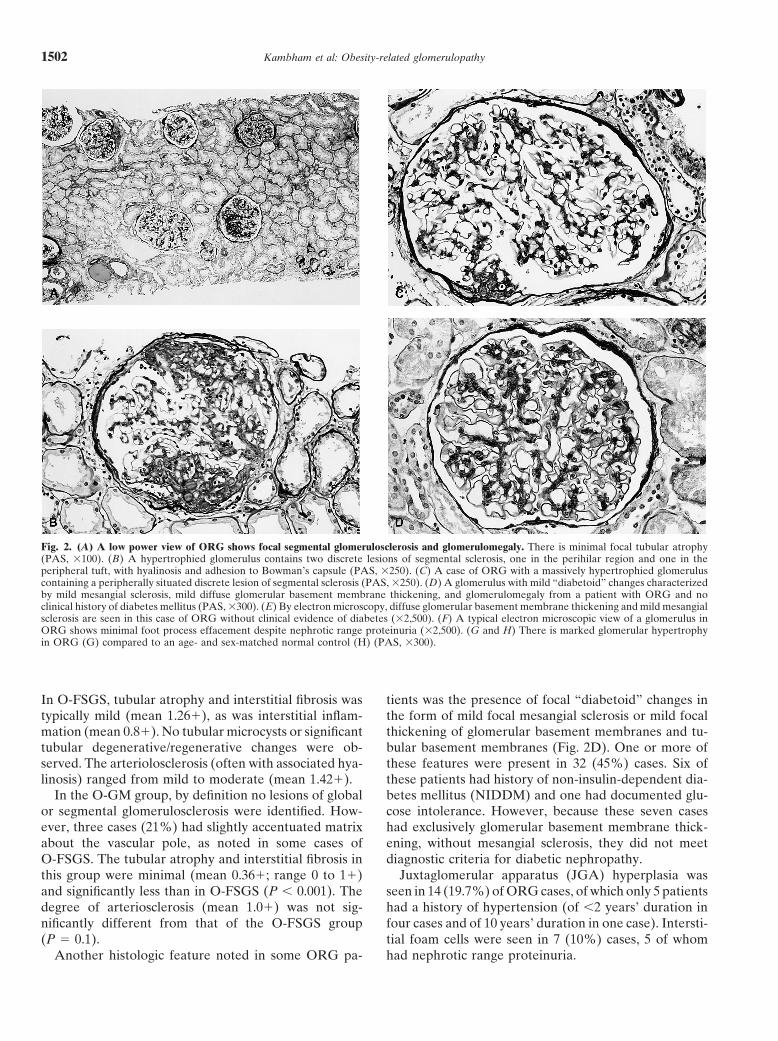
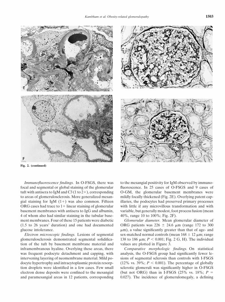
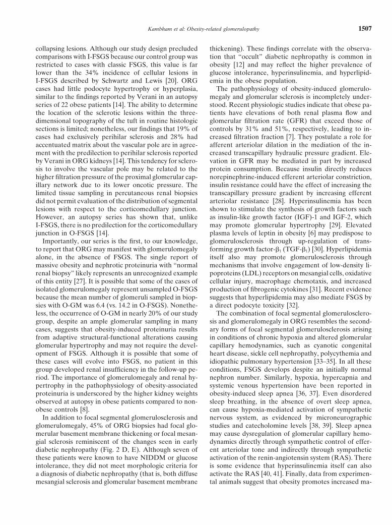
Fig. 2. (A) A low power view of ORG shows focal segmental glomerulosclerosis and glomerulomegaly. There is minimal focal tubular atrophy(PAS, 3100). (B) A hypertrophied glomerulus contains two discrete lesions of segmental sclerosis, one in the perihilar region and one in theperipheral tuft, with hyalinosis and adhesion to Bowman’s capsule (PAS, 3250). (C) A case of ORG with a massively hypertrophied glomeruluscontaining a peripherally situated discrete lesion of segmental sclerosis (PAS, 3250). (D) A glomerulus with mild “diabetoid” changes characterizedby mild mesangial sclerosis, mild diffuse glomerular basement membrane thickening, and glomerulomegaly from a patient with ORG and noclinical history of diabetes mellitus (PAS, 3300). (E) By electron microscopy, diffuse glomerular basement membrane thickening and mild mesangialsclerosis are seen in this case of ORG without clinical evidence of diabetes (32,500). (F) A typical electron microscopic view of a glomerulus inORG shows minimal foot process effacement despite nephrotic range proteinuria (32,500). (G and H) There is marked glomerular hypertrophyin ORG (G) compared to an age- and sex-matched normal control (H) (PAS, 3300).
In O-FSGS, tubular atrophy and interstitial fibrosis was tients was the presence of focal “diabetoid” changes inthe form of mild focal mesangial sclerosis or mild focaltypically mild (mean 1.261), as was interstitial inflam-
mation (mean 0.81). No tubular microcysts or significant thickening of glomerular basement membranes and tu-bular basement membranes (Fig. 2D). One or more oftubular degenerative/regenerative changes were ob-
served. The arteriolosclerosis (often with associated hya- these features were present in 32 (45%) cases. Six ofthese patients had history of non-insulin-dependent dia-linosis) ranged from mild to moderate (mean 1.421).
In the O-GM group, by definition no lesions of global betes mellitus (NIDDM) and one had documented glu-cose intolerance. However, because these seven casesor segmental glomerulosclerosis were identified. How-
ever, three cases (21%) had slightly accentuated matrix had exclusively glomerular basement membrane thick-ening, without mesangial sclerosis, they did not meetabout the vascular pole, as noted in some cases of
O-FSGS. The tubular atrophy and interstitial fibrosis in diagnostic criteria for diabetic nephropathy.Juxtaglomerular apparatus (JGA) hyperplasia wasthis group were minimal (mean 0.361; range 0 to 11)
and significantly less than in O-FSGS (P , 0.001). The seen in 14 (19.7%) of ORG cases, of which only 5 patientshad a history of hypertension (of ,2 years’ duration indegree of arteriosclerosis (mean 1.01) was not sig-
nificantly different from that of the O-FSGS group four cases and of 10 years’ duration in one case). Intersti-tial foam cells were seen in 7 (10%) cases, 5 of whom(P 5 0.1).
Another histologic feature noted in some ORG pa- had nephrotic range proteinuria.
Kambham et al: Obesity-related glomerulopathy 1503
Fig. 2. (continued)
Immunofluorescence findings. In O-FSGS, there was to the mesangial positivity for IgM observed by immuno-fluorescence. In 25 cases of O-FSGS and 9 cases offocal and segmental or global staining of the glomerular
tuft with antisera to IgM and C3 (1 to 21), corresponding O-GM, the glomerular basement membranes weremildly focally thickened (Fig. 2E). Overlying patent cap-to areas of glomerulosclerosis. More generalized mesan-
gial staining for IgM (11) was also common. Fifteen illaries, the podocytes had preserved primary processeswith little if any microvillous transformation and withORG cases had trace to 11 linear staining of glomerular
basement membranes with antisera to IgG and albumin, variable, but generally modest, foot process fusion (mean40%, range 10 to 100%; Fig. 2F).4 of whom also had similar staining in the tubular base-
ment membranes. Four of these 15 patients were diabetic Glomerular diameter. Mean glomerular diameter ofORG patients was 226 6 24.6 mm (range 172 to 300(1.5 to 26 years’ duration) and one had documented
glucose intolerance. mm), a value significantly greater than that of age- andsex-matched normal controls (mean 168 6 12 mm; rangeElectron microscopic findings. Lesions of segmental
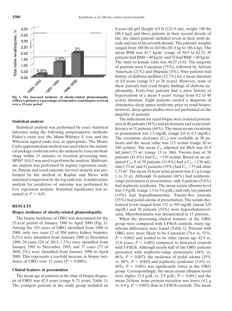
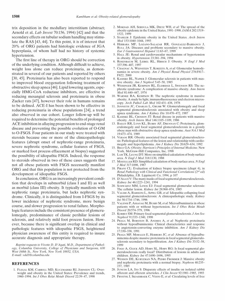
glomerulosclerosis demonstrated segmental solidifica- 138 to 186 mm; P , 0.001; Fig. 2 G, H). The individualvalues are plotted in Figure 3.tion of the tuft by basement membrane material and
inframembranous hyaline. Overlying these areas, there Comparative morphologic findings. On statisticalanalysis, the O-FSGS group had significantly fewer le-was frequent podocyte detachment and capping, with
intervening layering of neomembrane material. Mild po- sions of segmental sclerosis than controls with I-FSGS(12% vs. 39%; P , 0.001). The percentage of globallydocyte hypertrophy and intracytoplasmic protein resorp-
tion droplets were identified in a few cases. Few small sclerotic glomeruli was significantly higher in O-FSGS(but not ORG) than in I-FSGS (25% vs. 18%; P 5electron dense deposits were confined to the mesangial
and paramesangial areas in 12 patients, corresponding 0.027). The incidence of glomerulomegaly, a defining
Kambham et al: Obesity-related glomerulopathy1504
FSGS (ORG vs. I-FSGS) was not predictive of hypercho-lesterolemia.
Therapy and clinical outcome
Of the 71 ORG patients, 15 patients (including 9O-FSGS and 6 O-GM) were lost to follow-up (Table 2).Thirty-four of the remaining 56 (61%) patients weretreated with either ACE-I or angiotensin II receptorantagonists. Four additional patients (7%) were treatedwith both ACE-I and steroids, one of whom presentedwith nephrotic syndrome and had features of cellularFSGS on biopsy. Treatment was not statistically differentbetween the O-FSGS and O-GM groups.
Follow-up was available on all 50 I-FSGS patients: 39patients (78%) received immunosuppressive therapy, ofFig. 3. Glomerular diameter is plotted for ORG and for normal age-
and sex-matched controls. Symbols are: (j, d) ORG male and female, whom 21 received steroids alone and the remainder re-respectively; (h, s) control male and female respectively; top solid and ceived steroids plus either cyclophosphamide (6), ordashed lines are mean ORG male and female, respectively; bottom
cyclosporine (8), or both sequentially (4).solid and dashed lines are mean control male and female, respectively.The mean follow-up was 27 months (range 2 to 117
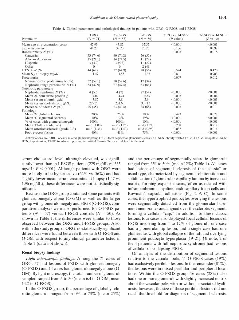
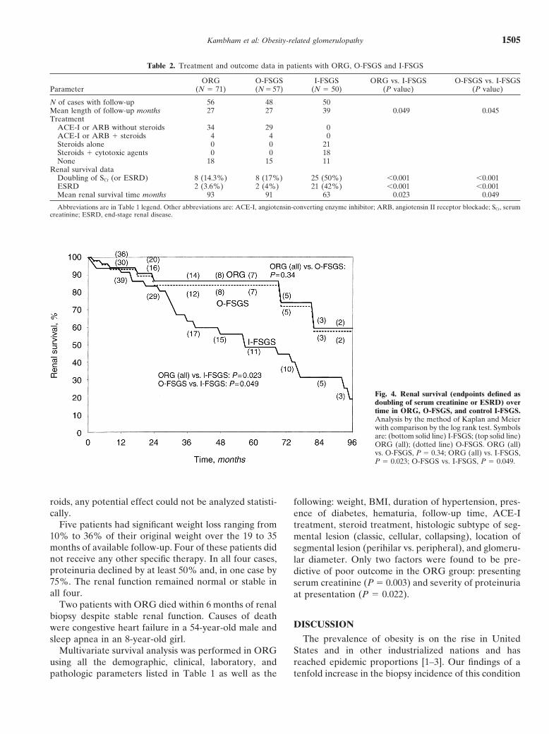
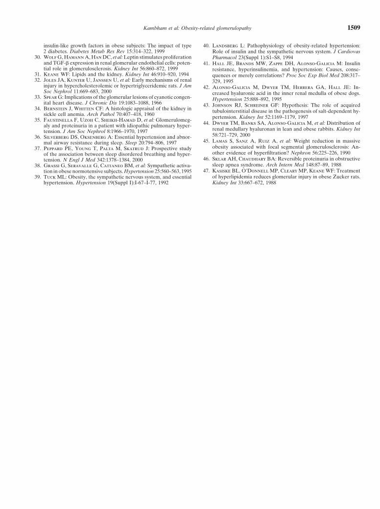
months) for the ORG group and 39 months (range 1 to140 months) for the I-FSGS controls (P 5 0.049). Eight
feature of ORG, was significantly higher in ORG versus patients with ORG (14.3%) had doubling of serum creat-I-FSGS (100% vs. 10%; P , 0.001). Patients with ORG inine and two of these patients (3.6%) progressed totended to have more severe arteriolosclerosis compared ESRD requiring renal replacement therapy. By contrast,to I-FSGS (1.341 vs. 0.981; P 5 0.03). However, the in the I-FSGS controls, 25 (50%) had doubling of serumseverity of tubular atrophy and interstitial fibrosis was creatinine on follow-up, of whom 21 patients (42%) re-not statistically different. There was significantly less foot quired renal replacement therapy (P , 0.001). The meanprocess effacement in ORG (mean 40%, range 10 to time to reach this end point was longer for ORG patients100%) than in I-FSGS (mean 75%, range 30 to 100%; compared to I-FSGS patients (93 vs. 63 months; P 5P , 0.001).
0.023; Fig. 4). Among the 8 ORG patients who reacheddoubling of serum creatinine, all belonged to theComparative multivariate analysis between ORGO-FSGS subgroup, 2 presented with nephrotic syn-and I-FSGSdrome, and 1 had collapsing features on biopsy. Four ofThe parameters found to be significantly different bythese 8 patients had been treated with ACE-I alone andunivariate analysis between the groups (Table 1) were4 received no therapy. There was no significant differ-analyzed by multivariate analysis using logistic regres-ence in renal functional outcome of treated and un-sion. When ORG was compared to I-FSGS, the onlytreated patients, as well as those treated with ACE-Iparameters that were independently significant were se-alone versus a combination of ACE-I and steroids. Whenrum albumin (P , 0.001) and age (P 5 0.032). Whenpatients were dichotomized for BMI less than or greaterthe O-FSGS and I-FSGS groups were compared, again,than 40, no differences in outcome were identified.serum albumin (P , 0.001) and age (P 5 0.018) were
Follow-up data on proteinuria was available in 26 pa-the only independently significant variables. These re-tients with ORG, of whom 18 were receiving ACE-I.sults are consistent with the observation that the majorEighteen patients (69.2%) had a decline in 24-hour urinedistinguishing feature between ORG and I-FSGS is theprotein. Among these 26 patients, the mean 24-hourpresence of full nephrotic syndrome in I-FSGS, as re-urine protein declined from 3.92 to 2.55 g/day (P 5flected by the severity of hypoalbuminemia.0.011). A mean reduction in urine protein excretion ofBy logistic regression analysis, both the type of FSGS1.0 g/day was found in the 18 patients treated with ACE-I.(ORG vs. I-FSGS) and the 24-hour urinary protein ex-Of the 4 patients receiving steroid therapy, 3 had follow-cretion were significant and independent predictors ofup data on proteinuria and 2 had an improvement inhypoalbuminemia (P 5 0.002 for the type of FSGS, withurine protein excretion (one with reduction in urine pro-I-FSGS being the stronger predictor of hypoalbumin-tein excretion from 13.0 to 1.4 g/day at 6 months ofemia and P , 0.001 for 24-hour urine protein excretion.follow-up and the other with reduction from 2.0 to 0.6The 24-hour urine protein and serum albumin were bothg/day at 98 months, both with stable renal function).significant and independent predictors of hypercholes-
terolemia (cholesterol .300 mg/dL), but the type of However, because so few patients were treated with ste-
Kambham et al: Obesity-related glomerulopathy 1505
Table 2. Treatment and outcome data in patients with ORG, O-FSGS and I-FSGS
ORG O-FSGS I-FSGS ORG vs. I-FSGS O-FSGS vs. I-FSGSParameter (N 5 71) (N 5 57) (N 5 50) (P value) (P value)
N of cases with follow-up 56 48 50Mean length of follow-up months 27 27 39 0.049 0.045Treatment
ACE-I or ARB without steroids 34 29 0ACE-I or ARB 1 steroids 4 4 0Steroids alone 0 0 21Steroids 1 cytotoxic agents 0 0 18None 18 15 11
Renal survival dataDoubling of SCr (or ESRD) 8 (14.3%) 8 (17%) 25 (50%) ,0.001 ,0.001ESRD 2 (3.6%) 2 (4%) 21 (42%) ,0.001 ,0.001Mean renal survival time months 93 91 63 0.023 0.049
Abbreviations are in Table 1 legend. Other abbreviations are: ACE-I, angiotensin-converting enzyme inhibitor; ARB, angiotensin II receptor blockade; SCr, serumcreatinine; ESRD, end-stage renal disease.
Fig. 4. Renal survival (endpoints defined asdoubling of serum creatinine or ESRD) overtime in ORG, O-FSGS, and control I-FSGS.Analysis by the method of Kaplan and Meierwith comparison by the log rank test. Symbolsare: (bottom solid line) I-FSGS; (top solid line)ORG (all); (dotted line) O-FSGS. ORG (all)vs. O-FSGS, P 5 0.34; ORG (all) vs. I-FSGS,P 5 0.023; O-FSGS vs. I-FSGS, P 5 0.049.
roids, any potential effect could not be analyzed statisti- following: weight, BMI, duration of hypertension, pres-cally. ence of diabetes, hematuria, follow-up time, ACE-I
Five patients had significant weight loss ranging from treatment, steroid treatment, histologic subtype of seg-10% to 36% of their original weight over the 19 to 35 mental lesion (classic, cellular, collapsing), location ofmonths of available follow-up. Four of these patients did segmental lesion (perihilar vs. peripheral), and glomeru-not receive any other specific therapy. In all four cases, lar diameter. Only two factors were found to be pre-proteinuria declined by at least 50% and, in one case by dictive of poor outcome in the ORG group: presenting75%. The renal function remained normal or stable in serum creatinine (P 5 0.003) and severity of proteinuriaall four. at presentation (P 5 0.022).
Two patients with ORG died within 6 months of renalbiopsy despite stable renal function. Causes of death
DISCUSSIONwere congestive heart failure in a 54-year-old male andThe prevalence of obesity is on the rise in Unitedsleep apnea in an 8-year-old girl.
States and in other industrialized nations and hasMultivariate survival analysis was performed in ORGreached epidemic proportions [1–3]. Our findings of ausing all the demographic, clinical, laboratory, and
pathologic parameters listed in Table 1 as well as the tenfold increase in the biopsy incidence of this condition
Kambham et al: Obesity-related glomerulopathy1506
over the past 15 years are congruent with epidemiologic Kassiske, and likely reflect fundamental differences inthe pathophysiology of this secondary form of FSGS [12,data documenting a sharply increasing prevalence of
obesity in the general population. Because the indica- 24, 25]. The reason why patients with ORG have a lowerincidence of nephrotic syndrome compared to I-FSGStions for renal biopsy (nephrotic range proteinuria, with
or without renal insufficiency) have not changed appreci- may relate to differences in the severity of podocyteinjury, in the severity and selectivity of proteinuria, andably over this time period, we interpret our increasing
biopsy incidence of ORG to reflect the increase in the in the ability of the tubules to reabsorb and catabolizethe filtered protein. The lower fractional excretions of b2-incidence of obesity. Previous reports of ORG have been
in patients with massive obesity [8, 10–14]. Our study microglobulin (which competes with albumin for tubularuptake) and N-acetyl b-glucosaminidase (a marker ofreveals, to our knowledge for the first time, that even
patients with submorbid (class I and II) obesity, who tubular injury) observed in patients with nephrotic rangeproteinuria compared to those with nephrotic syndromeconstituted 46% of the study group, can develop ORG
that is clinically and morphologically indistinguishable suggest differences in tubular overload and resulting cel-lular injury. The urinary losses of protein in ORG mayfrom that seen in morbid obesity.
Our study found a significantly higher incidence of be insufficient to stimulate the hepatic synthesis of lipo-protein typically observed in I-FSGS [24, 25]. Life tableORG in Caucasians compared to our biopsy population
with I-FSGS (P 5 0.03). We can only speculate about analysis indicates that the course of ORG is indolentwith less frequent progression to ESRD. Although ourthe reasons underlying these racial differences. These
data may reflect genetically determined risk factors for historical controls with I-FSGS had significantly longerfollow-up, life table analysis corrects for these differ-ORG as well as socioeconomic determinants that influ-
ence the rate of referral of obese patients of different ences. The favorable prognosis of ORG despite a lack oftreatment with steroids in all but four patients providesraces to a nephrologist.
The overall renal effects of obesity include elevated further proof that ORG is a distinct and separable clini-copathologic entity.GFR, elevated renal blood flow, and renal hypertrophy.
The association of proteinuria with massive obesity was Although both ORG and I-FSGS manifest lesions offocal segmental glomerulosclerosis, there are distin-first reported in 1974 [9]. The prevalence of proteinuria
in obese patients has been addressed in several cross- guishing morphologic features. As a group, ORG hasa lower percentage of glomeruli affected by segmentalsectional studies of large populations of obese patients.
Among 257 obese patients screened at Brigham and sclerosis, suggesting a milder and more slowly progres-sive disease. Although routine histologic sections areWomen’s Hospital, only 4 (0.015%) had dipstick protein-
uria of 21 or greater, a value much lower than the preva- known to underestimate the total number of glomeruliaffected by focal segmental sclerosis, these differenceslence of diabetes (15%) and hypertension (23%) in the
same population (abstract; Goldszer et al, Kidney Int were obtained by examination of standardized numbersof tissue sections in the two groups [26]. Ultrastructur-25:165A, 1984). However, in another study of 207 nondi-
abetic obese patients, 25 (12%) had documented mi- ally, the degree of foot process effacement was also lesssevere (mean 40% vs. 75%, P , 0.001), suggesting acroalbuminuria [22]. In the 1980’s several case reports
and small autopsy series of ORG appeared [8, 11, 13]. different pathomechanism of podocyte injury. However,because the range of % foot process fusion in each groupOur study provides the first large comparative biopsy
series on this entity. was wide (10 to 100% for ORG and 30 to 100% forI-FSGS), the percentage of foot process fusion cannotOur data indicate that the presentation of ORG is
typically one of nephrotic range-proteinuria (48%) or differentiate ORG from I-FSGS with certainty. Glomer-ulomegaly was consistently observed in ORG (100%)sub-nephrotic proteinuria (52%), accompanied by renal
insufficiency in nearly half (44%). Although nearly half compared to only 10% of patients with I-FSGS, under-scoring the importance of hyperfiltration mechanisms inof patients had nephrotic range proteinuria, hypoalbu-
minemia was present in only 14% and only 5.6% of its pathogenesis. These differences in the incidence ofglomerulomegaly cannot be explained by loss of renalpatients had full nephrotic syndrome. This presentation
stands in sharp contrast to controls with I-FSGS, of mass as a stimulus to hyperfiltration, because the degreeof glomerulosclerosis and chronic tubulointerstitial dis-whom 54% had nephrotic syndrome (a value similar to
the 70% rate of nephrotic syndrome estimated by Korbet ease, as well as the presenting serum creatinine levels,were higher in the I-FSGS group. Patients with ORGin a compilation of series of adult I-FSGS) [23]. Although
54% of ORG patients had hypercholesterolemia, corre- also tended to have more severe arteriosclerosis (P 50.03), despite a similar incidence of hypertension aslating with the increased prevalence of hyperlipidemia
in obesity, the mean serum cholesterol (229 mg/dL) was I-FSGS. These differences may be related to the olderage and greater risk of coronary vascular disease in ORG.significantly lower than that in I-FSGS (335 mg/dL; P ,
0.001). These results are similar to those of Praga and Of interest, only 5 (7.0%) cases of ORG had cellular or
Kambham et al: Obesity-related glomerulopathy 1507
collapsing lesions. Although our study design precluded thickening). These findings correlate with the observa-tion that “occult” diabetic nephropathy is common incomparisons with I-FSGS because our control group was
restricted to cases with classic FSGS, this value is far obesity [12] and may reflect the higher prevalence ofglucose intolerance, hyperinsulinemia, and hyperlipid-lower than the 34% incidence of cellular lesions in
I-FSGS described by Schwartz and Lewis [20]. ORG emia in the obese population.The pathophysiology of obesity-induced glomerulo-cases had little podocyte hypertrophy or hyperplasia,
similar to the findings reported by Verani in an autopsy megaly and glomerular sclerosis is incompletely under-stood. Recent physiologic studies indicate that obese pa-series of 22 obese patients [14]. The ability to determine
the location of the sclerotic lesions within the three- tients have elevations of both renal plasma flow andglomerular filtration rate (GFR) that exceed those ofdimensional topography of the tuft in routine histologic
sections is limited; nonetheless, our findings that 19% of controls by 31% and 51%, respectively, leading to in-creased filtration fraction [7]. They postulate a role forcases had exclusively perihilar sclerosis and 28% had
accentuated matrix about the vascular pole are in agree- afferent arteriolar dilation in the mediation of the in-creased transcapillary hydraulic pressure gradient. Ele-ment with the predilection to perihilar sclerosis reported
by Verani in ORG kidneys [14]. This tendency for sclero- vation in GFR may be mediated in part by increasedprotein consumption. Because insulin directly reducessis to involve the vascular pole may be related to the
higher filtration pressure of the proximal glomerular cap- norepinephrine-induced efferent arteriolar constriction,insulin resistance could have the effect of increasing theillary network due to its lower oncotic pressure. The
limited tissue sampling in percutaneous renal biopsies transcapillary pressure gradient by increasing efferentarteriolar resistance [28]. Hyperinsulinemia has beendid not permit evaluation of the distribution of segmental
lesions with respect to the corticomedullary junction. shown to stimulate the synthesis of growth factors suchas insulin-like growth factor (IGF)-1 and IGF-2, whichHowever, an autopsy series has shown that, unlike
I-FSGS, there is no predilection for the corticomedullary may promote glomerular hypertrophy [29]. Elevatedplasma levels of leptin in obesity [6] may predispose tojunction in O-FSGS [14].
Importantly, our series is the first, to our knowledge, glomerulosclerosis through up-regulation of trans-forming growth factor-b1 (TGF-b1) [30]. Hyperlipidemiato report that ORG may manifest with glomerulomegaly
alone, in the absence of FSGS. The single report of itself also may promote glomerulosclerosis throughmechanisms that involve engagement of low-density li-massive obesity and nephrotic proteinuria with “normal
renal biopsy” likely represents an unrecognized example poproteins (LDL) receptors on mesangial cells, oxidativecellular injury, macrophage chemotaxis, and increasedof this entity [27]. It is possible that some of the cases of
isolated glomerulomegaly represent unsampled O-FSGS production of fibrogenic cytokines [31]. Recent evidencesuggests that hyperlipidemia may also mediate FSGS bybecause the mean number of glomeruli sampled in biop-
sies with O-GM was 6.4 (vs. 14.2 in O-FSGS). Nonethe- a direct podocyte toxicity [32].The combination of focal segmental glomerulosclero-less, the occurrence of O-GM in nearly 20% of our study
group, despite an ample glomerular sampling in many sis and glomerulomegaly in ORG resembles the second-ary forms of focal segmental glomerulosclerosis arisingcases, suggests that obesity-induced proteinuria results
from adaptive structural-functional alterations causing in conditions of chronic hypoxia and altered glomerularcapillary hemodynamics, such as cyanotic congenitalglomerular hypertrophy and may not require the devel-
opment of FSGS. Although it is possible that some of heart disease, sickle cell nephropathy, polycythemia andidiopathic pulmonary hypertension [33–35]. In all thesethese cases will evolve into FSGS, no patient in this
group developed renal insufficiency in the follow-up pe- conditions, FSGS develops despite an initially normalnephron number. Similarly, hypoxia, hypercapnia andriod. The importance of glomerulomegaly and renal hy-
pertrophy in the pathophysiology of obesity-associated systemic venous hypertension have been reported inobesity-induced sleep apnea [36, 37]. Even disorderedproteinuria is underscored by the higher kidney weights
observed at autopsy in obese patients compared to non- sleep breathing, in the absence of overt sleep apnea,can cause hypoxia-mediated activation of sympatheticobese controls [8].
In addition to focal segmental glomerulosclerosis and nervous system, as evidenced by microneurographicstudies and catecholomine levels [38, 39]. Sleep apneaglomerulomegaly, 45% of ORG biopsies had focal glo-
merular basement membrane thickening or focal mesan- may cause dysregulation of glomerular capillary hemo-dynamics directly through sympathetic control of effer-gial sclerosis reminiscent of the changes seen in early
diabetic nephropathy (Fig. 2 D, E). Although seven of ent arteriolar tone and indirectly through sympatheticactivation of the renin-angiotensin system (RAS). Therethese patients were known to have NIDDM or glucose
intolerance, they did not meet morphologic criteria for is some evidence that hyperinsulinemia itself can alsoactivate the RAS [40, 41]. Finally, data from experimen-a diagnosis of diabetic nephropathy (that is, both diffuse
mesangial sclerosis and glomerular basement membrane tal animals suggest that obesity promotes increased ma-
Kambham et al: Obesity-related glomerulopathy1508
2. Mokdad AH, Serdula MK, Dietz WH, et al: The spread of thetrix deposition in the medullary interstitium (abstract;obesity epidemic in the United States, 1991–1998. JAMA 282:1519–
Arnold et al, Lab Invest 70:156, 1994) [42] and that the 1522, 19993. Stamler J: Epidemic obesity in the United States. Arch Internsecondary effects on tubular sodium handling may stimu-
Med 153:1040–1044, 1993late the RAS [43, 44]. To this point, it is of interest that4. Herrara MF, Lozano-Salazar RR, Gonzalez-Barranco J,
20% of ORG patients had histologic evidence of JGA Rull JA: Diseases and problems secondary to massive obesity.hyperplasia, of whom half had no history of systemic Eur J Gastroenterol Hepatol 11:63–67, 1999
5. Hall JE: Renal and cardiovascular mechanisms of hypertensionhypertension.in obesity. Hypertension 23:381–394, 1994The first line of therapy in ORG should be correction 6. Rosenbaum M, Leibel RL, Hirsch J: Obesity. N Engl J Med
of the underlying condition. Although difficult to achieve, 337:396–407, 19977. Chagnac A, Weinstein T, Korzets A, et al: Glomerular hemody-weight loss alone can reduce proteinuria, as demon-
namics in severe obesity. Am J Physiol Renal Physiol 278:F817–strated in several of our patients and reported by others F822, 2000[30, 45]. Proteinuria has also been reported to respond 8. Kasiske BL, Napier J: Glomerular sclerosis in patients with mas-
sive obesity. Am J Nephrol 5:45–50, 1985to improved blood oxygenation following treatment of9. Weisinger JR, Kempson RL, Eldridge L, Swenson RS: The ne-obstructive sleep apnea [46]. Lipid lowering agents, espe- phrotic syndrome: A complication of massive obesity. Ann Intern
cially HMG-CoA reductase inhibitors, are effective in Med 81:440–447, 197410. Warnke RA, Kempson R: The nephrotic syndrome in massivereducing mesangial sclerosis and proteinuria in obese
obesity. A study by light, immunofluorescence and electron micros-Zucker rats [47], however their role in humans remains copy. Arch Pathol Lab Med 102:431–438, 1978to be defined. ACE-I has been shown to be effective in 11. Jennette JC, Charles L, Grubb W: Glomerulomegaly and focal
segmental glomerulosclerosis associated with obesity and sleepreducing proteinuria in obese populations [24], a trendapnea syndrome. Am J Kidney Dis 10:470–472, 1987also observed in our cohort. Longer follow-up will be 12. Kasiske BL, Crosson JT: Renal disease in patients with massive
required to determine the potential benefits of prolonged obesity. Arch Intern Med 146:1105–1109, 198613. Bailey RR, Lynn KL, Burry AF, Drennan C: Proteinuria, glom-ACE inhibition in allaying progression to end-stage renal
erulomegaly and focal segmental glomerulosclerosis in a grosslydisease and preventing the possible evolution of O-GMobese man with obstructive sleep apnea syndrome. Aust NA J Med
to O-FSGS. Four patients in our study were treated with 19:473–474, 198914. Verani RR: Obesity associated focal segmental glomerulosclero-steroids because one or more of the clinicopathologic
sis: Pathological features of the lesion and relationship with cardio-features (abrupt onset of nephrotic-range proteinuria,megaly and hyperlipidemia. Am J Kidney Dis 20:629–634, 1992
severe nephrotic syndrome, cellular features of FSGS, 15. Bray GA: Obesity: Harrison’s Principles of Internal Medicine. NewYork, McGraw-Hill Companies, Inc, 1998or marked foot process effacement at biopsy) suggested
16. Lam TK, Leung DT: More on simplified calculation of body surfacethe possibility of idiopathic FSGS. Indeed, the responsearea. N Engl J Med 318:1130, 1988
to steroids observed in two of these cases suggests that 17. Mosteller RD: Simplified calculation of body surface area. N EnglJ Med 317:1098, 1987not all obese patients with FSGS necessarily manifest
18. Pirani CL: Evaluation of kidney biopsy specimens (chapt 4), inORG and that this population is not protected from theRenal Pathology with Clinical and Functional Correlations (2nd ed)
development of idiopathic FSGS. Philadelphia, J.B. Lippincott Co, 1994, p 107In conclusion, ORG is an increasingly prevalent condi- 19. D’Agati V: The many masks of focal segmental glomerulosclerosis.
Kidney Int 46:1223–1241, 1994tion that develops in submorbid (class I and II) as well20. Schwartz MM, Lewis EJ: Focal segmental glomerular sclerosis:as morbid (class III) obesity. It typically manifests with The cellular lesion. Kidney Int 28:968–874, 1985
nephrotic range proteinuria, but lacks nephrotic syn- 21. Valeri A, Barisoni L, Appel GB, et al: Idiopathic collapsing focalsegmental glomerulosclerosis: A clinicopathologic study. Kidneydrome. Clinically, it is distinguished from I-FSGS by itsInt 50:1734–1746, 1996lower incidence of nephrotic syndrome, more benign 22. Valensi P, Assayag M, Busby M, et al: Microalbuminuria in obese
course, and slower progression to renal failure. Morpho- patients with or without hypertension. Int J Obes Relat MetabDisord 20:574–579, 1996logic features include the consistent presence of glomeru-
23. Korbet SM: Primary focal segmental glomerulosclerosis. J Am Soclomegaly, predominance of classic perihilar lesions ofNephrol 9:1333–1340, 1998
sclerosis, and relatively mild foot process fusion. How- 24. Praga M, Borstein B, Andres A, et al: Nephrotic proteinuriawithout hypoalbuminemia: Clinical characteristics and responseever, because there is significant overlap in clinical andto angiotensin-converting enzyme inhibition. Am J Kidney Dispathologic features with idiopathic FSGS, heightened17:330–338, 1991
physician awareness of this entity is required to insure 25. Praga MP, Morales E, Herrero JC, et al: Absence of hypoalbu-accurate diagnosis and appropriate therapy. minemia despite massive proteinuria in focal segmental glomerulo-
sclerosis secondary to hyperfiltration. Am J Kidney Dis 33:52–58,Reprint requests to Vivette D. D’Agati, M.D., Department of Pathol- 1999
ogy, Columbia University, College of Physicians and Surgeons, 630 26. Fogo A, Glick AD, Horn SL, Horn RG: Is focal segmental glo-West 168th St., New York, New York 10032, USA. merulosclerosis really focal? Distribution of lesions in adults andE-mail: [email protected] children. Kidney Int 47:1690–1696, 1995
27. Wesson DE, Kurtzman NA, Pedro Frommer J: Massive obesityand nephrotic proteinuria with a normal biopsy. Nephron 40:235–REFERENCES237, 1985
28. Juncos LA, Ito S: Disparate effects of insulin on isolated rabbit1. Flegal KM, Carroll MD, Kuczmarski RJ, Johnson CL: Over-afferent and efferent arterioles. J Clin Invest 92:1981–1985, 1993weight and obesity in the United States: Prevalence and trends,
1960–1994. Int J Obes Relat Metab Disord 22:39–47, 1998 29. Frystyk J, Skjaerbaek C, Vesto E, et al: Circulating levels of free
Kambham et al: Obesity-related glomerulopathy 1509
insulin-like growth factors in obese subjects: The impact of type 40. Landsberg L: Pathophysiology of obesity-related hypertension:2 diabetes. Diabetes Metab Res Rev 15:314–322, 1999 Role of insulin and the sympathetic nervous system. J Cardiovas
30. Wolf G, Hamann A, Han DC, et al: Leptin stimulates proliferation Pharmacol 23(Suppl 1):S1–S8, 1994and TGF-b expression in renal glomerular endothelial cells: poten- 41. Hall JE, Brands MW, Zappe DH, Alonso-Galicia M: Insulintial role in glomerulosclerosis. Kidney Int 56:860–872, 1999 resistance, hyperinsulinemia, and hypertension: Causes, conse-
31. Keane WF: Lipids and the kidney. Kidney Int 46:910–920, 1994 quences or merely correlations? Proc Soc Exp Biol Med 208:317–32. Joles JA, Kunter U, Janssen U, et al: Early mechanisms of renal 329, 1995
injury in hypercholesterolemic or hypertriglyceridemic rats. J Am 42. Alonso-Galicia M, Dwyer TM, Herrera GA, Hall JE: In-Soc Nephrol 11:669–683, 2000 creased hyaluronic acid in the inner renal medulla of obese dogs.
33. Spear G: Implications of the glomerular lesions of cyanotic congen- Hypertension 25:888–892, 1995ital heart disease. J Chronic Dis 19:1083–1088, 1966 43. Johnson RJ, Schreiner GF: Hypothesis: The role of acquired
34. Bernstein J, Whitten CF: A histologic appraisal of the kidney in tubulointerstitial disease in the pathogenesis of salt-dependent hy-sickle cell anemia. Arch Pathol 70:407–418, 1960 pertension. Kidney Int 52:1169–1179, 199735. Faustinella F, Uzoh C, Sheikh-Hamad D, et al: Glomerulomeg-
44. Dwyer TM, Banks SA, Alonso-Galicia M, et al: Distribution ofaly and proteinuria in a patient with idiopathic pulmonary hyper-renal medullary hyaluronan in lean and obese rabbits. Kidney Inttension. J Am Soc Nephrol 8:1966–1970, 199758:721–729, 200036. Silverberg DS, Oksenberg A: Essential hypertension and abnor-
45. Lamas S, Sanz A, Ruiz A, et al: Weight reduction in massivemal airway resistance during sleep. Sleep 20:794–806, 1997obesity associated with focal segmental glomerulosclerosis: An-37. Peppard PE, Young T, Palta M, Skatrud J: Prospective studyother evidence of hyperfiltration? Nephron 56:225–226, 1990of the association between sleep disordered breathing and hyper-
46. Sklar AH, Chaudhary BA: Reversible proteinuria in obstructivetension. N Engl J Med 342:1378–1384, 2000sleep apnea syndrome. Arch Intern Med 148:87–89, 198838. Grassi G, Seravalle G, Cattaneo BM, et al: Sympathetic activa-
47. Kasiske BL, O’Donnell MP, Cleary MP, Keane WF: Treatmenttion in obese normotensive subjects. Hypertension 25:560–563, 1995of hyperlipidemia reduces glomerular injury in obese Zucker rats.39. Tuck ML: Obesity, the sympathetic nervous system, and essential
hypertension. Hypertension 19(Suppl I):I-67–I-77, 1992 Kidney Int 33:667–672, 1988


